5 End-of-chapter test - Macmillan Academy
5 End-of-chapter test - Macmillan Academy
5 End-of-chapter test - Macmillan Academy

You also want an ePaper? Increase the reach of your titles
YUMPU automatically turns print PDFs into web optimized ePapers that Google loves.
5 <strong>End</strong>-<strong>of</strong>-<strong>chapter</strong> <strong>test</strong><br />
1 a Explain why a large multicellular organism such as a human needs a transport system,<br />
whereas a single-celled organism such as Paramecium can exist without one. [3]<br />
b Explain the difference between:<br />
i a single and a double circulatory system<br />
ii a closed and an open circulatory system. [4]<br />
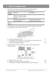
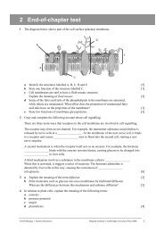
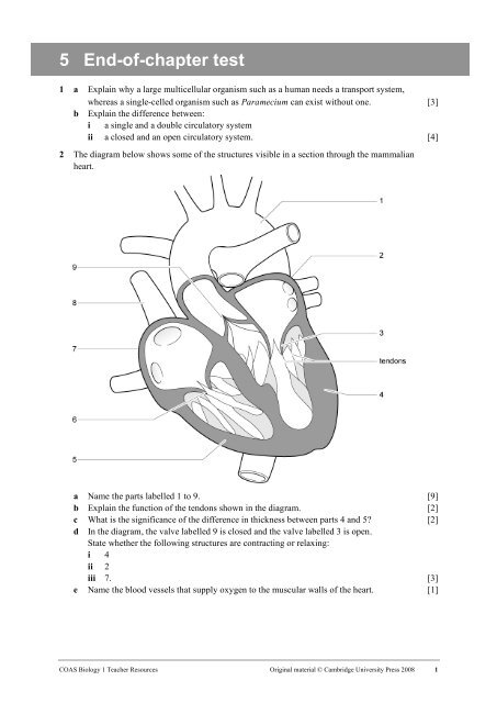
2 The diagram below shows some <strong>of</strong> the structures visible in a section through the mammalian<br />
heart.<br />
a Name the parts labelled 1 to 9. [9]<br />
b Explain the function <strong>of</strong> the tendons shown in the diagram. [2]<br />
c What is the significance <strong>of</strong> the difference in thickness between parts 4 and 5 [2]<br />
d In the diagram, the valve labelled 9 is closed and the valve labelled 3 is open.<br />
State whether the following structures are contracting or relaxing:<br />
i 4<br />
ii 2<br />
iii 7. [3]<br />
e Name the blood vessels that supply oxygen to the muscular walls <strong>of</strong> the heart. [1]<br />
COAS Biology 1 Teacher Resources Original material © Cambridge University Press 2008 1
5 <strong>End</strong>-<strong>of</strong>-<strong>chapter</strong> <strong>test</strong><br />
3 The diagram below shows a section through a mammalian heart and some <strong>of</strong> the structures<br />
involved in coordination <strong>of</strong> the heart beat.<br />
Structure X is the origin <strong>of</strong> waves <strong>of</strong> electrical excitation that spread through the heart muscle. The<br />
time taken for a wave <strong>of</strong> excitation to spread across the heart from X was measured. Recordings<br />
were made at positions A to G. The results are shown in the table below.<br />
Position<br />
Time/ms<br />
A 0<br />
B 12<br />
C 32<br />
D 45<br />
E 130<br />
F 150<br />
G 175<br />
a Name the structures labelled X, Y and Z. [3]<br />
b Using the information in the diagram and table, explain how the route taken by the wave <strong>of</strong><br />
excitation coordinates the contraction <strong>of</strong> the heart muscle. [6]<br />
COAS Biology 1 Teacher Resources Original material © Cambridge University Press 2008 2
4 The diagram below shows the pressure changes in the heart during the cardiac cycle.<br />
5 <strong>End</strong>-<strong>of</strong>-<strong>chapter</strong> <strong>test</strong><br />
a Identify the letters on the graph that correspond to the following events:<br />
i the ventricle beginning to contract<br />
ii the aortic semilunar valve opening<br />
iii the aortic semilunar valve closing<br />
iv the bicuspid valve opening. [4]<br />
b From the graph, calculate the heart rate in beats per minute. [2]<br />
c Explain what an electrocardiogram (ECG) is. [2]<br />
5 The diagram below shows cross-sections <strong>of</strong> three different types <strong>of</strong> mammalian blood vessel,<br />
A, B and C.<br />
a Name the three types <strong>of</strong> blood vessel. [3]<br />
b Explain how the middle layer <strong>of</strong> tissue in blood vessel A is adapted for the function <strong>of</strong><br />
the vessel. [2]<br />
c State two ways in which blood vessel C is adapted to allow the formation <strong>of</strong> tissue fluid. [2]<br />
d What is the function <strong>of</strong> tissue fluid [2]<br />
e Approximately 80% <strong>of</strong> tissue fluid is reabsorbed by the venous end <strong>of</strong> a capillary bed.<br />
What happens to the remaining tissue fluid [2]<br />
COAS Biology 1 Teacher Resources Original material © Cambridge University Press 2008 3
5 <strong>End</strong>-<strong>of</strong>-<strong>chapter</strong> <strong>test</strong><br />
6 Explain how each <strong>of</strong> the following features <strong>of</strong> a red blood cell is an adaptation for its function.<br />
a its small size (diameter about 7 µm) [2]<br />
b its biconcave disc shape [2]<br />
c the lack <strong>of</strong> a nucleus [2]<br />
d the presence <strong>of</strong> the enzyme carbonic anhydrase in the cell [2]<br />
7 The diagram below shows oxygen dissociation curves for human haemoglobin at two partial<br />
pressures <strong>of</strong> carbon dioxide. The effect <strong>of</strong> a high partial pressure <strong>of</strong> carbon dioxide on<br />
the oxygen dissociation <strong>of</strong> haemoglobin is called the Bohr effect.<br />
a Explain how the shape <strong>of</strong> the oxygen dissociation curve at low partial pressure <strong>of</strong> carbon<br />
dioxide is related to the function <strong>of</strong> the haemoglobin in carrying oxygen from the lungs to<br />
the respiring tissues. [3]<br />
b If the partial pressure <strong>of</strong> oxygen in the lungs is 11 kPa, use the graph to find the percentage<br />
saturation <strong>of</strong> the haemoglobin with oxygen in the lungs. [1]<br />
c In an actively respiring muscle, the partial pressure <strong>of</strong> oxygen is 2 kPa. Use the graph to<br />
find the percentage saturation <strong>of</strong> the haemoglobin with oxygen in the muscle tissue. [1]<br />
d Explain why the Bohr effect is <strong>of</strong> importance to actively respiring tissues. [3]<br />
e Copy the axes in the diagram and sketch the curve for haemoglobin at a low partial pressure<br />
<strong>of</strong> carbon dioxide. On your sketch graph, add another curve that shows the expected<br />
dissociation curve for the haemoglobin in the blood <strong>of</strong> a llama, a mammal species adapted<br />
to living at high altitudes. [2]<br />
Total:<br />
70<br />
Score: %<br />
Grade boundaries: 80% A, 70% B, 60% C, 50% D, 40% E<br />
COAS Biology 1 Teacher Resources Original material © Cambridge University Press 2008 4