Applications of Doppler ultrasound during labor - Medical ...
Applications of Doppler ultrasound during labor - Medical ...
Applications of Doppler ultrasound during labor - Medical ...

Create successful ePaper yourself
Turn your PDF publications into a flip-book with our unique Google optimized e-Paper software.
Review<br />
<strong>Medical</strong> Ultrasonography<br />
2011, Vol. 13, no. 2, 141-149<br />
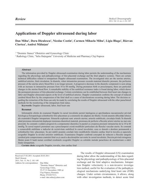
<strong>Applications</strong> <strong>of</strong> <strong>Doppler</strong> <strong>ultrasound</strong> <strong>during</strong> <strong>labor</strong><br />
Dan Mihu 1 , Doru Diculescu 1 , Nicolae Costin 1 , Carmen Mihaela Mihu 2 , Ligia Blaga 1 , Răzvan<br />
Ciortea 1 , Andrei Măluţan 1<br />
1<br />
”Dominic Stanca” Obstetrics and Gynecology Clinic<br />
2<br />
Radiology Clinic, ”Iuliu Haţieganu” University <strong>of</strong> Medicine and Pharmacy Cluj-Napoca<br />
Abstract<br />
The information provided by <strong>Doppler</strong> <strong>ultrasound</strong> examination <strong>during</strong> <strong>labor</strong> permits the understanding <strong>of</strong> the mechanisms<br />
regarding the physiology and pathophysiology <strong>of</strong> feto-placental exchange and the fetal adaptive systems. There are certain<br />
technical difficulties related to intrapartum <strong>Doppler</strong> <strong>ultrasound</strong> examination. The investigated sites are the uterine arteries,<br />
umbilical arteries, fetal circulation. In diastole, when intrauterine pressure exceeds maternal diastolic pressure, the perfusion<br />
pressure <strong>of</strong> the uterine artery blood flow is no longer present. A progressive decrease in the diastolic component is seen along<br />
with an increase in intrauterine pressure from 10 to 60 mmHg. During premature birth or preeclampsia, there are particular<br />
changes in the uterine blood flow. A remarkable stability <strong>of</strong> the umbilical resistance index is found <strong>during</strong> <strong>labor</strong>, which shows<br />
the permanent presence <strong>of</strong> feto-placental exchange. Certain correlations can be established between fetal heart rate changes in<br />
<strong>labor</strong> and <strong>Doppler</strong> <strong>ultrasound</strong> aspects at the level <strong>of</strong> umbilical arteries. <strong>Doppler</strong> examination confirms the concept <strong>of</strong> reduced<br />
cerebral blood flow by the compression <strong>of</strong> the fetal skull as a cause <strong>of</strong> decelerations occurring <strong>during</strong> <strong>labor</strong>. The decision regarding<br />
the extraction <strong>of</strong> the fetus can only be made by correlating the results <strong>of</strong> <strong>Doppler</strong> <strong>ultrasound</strong> with the other paraclinical<br />
methods for the monitoring <strong>of</strong> the intrapartum fetal status.<br />
Keywords: <strong>Doppler</strong> <strong>ultrasound</strong>, <strong>labor</strong>, fetal heart rate<br />
Rezumat<br />
Informaţiile <strong>of</strong>erite de examenul <strong>Doppler</strong> în cursul travaliului permit înţelegerea şi apr<strong>of</strong>undarea mecanismelor privind<br />
fiziologia şi fiziopatologia schimburilor feto-placentare şi a sistemelor de adaptare ale fătului. Există anumite dificultăţi tehnice<br />
ale examinării <strong>Doppler</strong> intrapartum. Situsurile explorate sunt: arterele uterine, arterele ombilicale, circulaţia fetală. În diastolă<br />
când presiunea intrauterină depăşeşte tensiunea diastolocă maternă, presiunea de perfuzie a fluxului arterei uterine nu mai este<br />
prezentă. Se constată o scădere progresivă a componentei diastolice în paralel cu creşterea presiunii intrauterine de la 10 la 60<br />
mmHg. În cursul naşterii premature sau a preeclampsiei există modificări particulare ale fluxului vascular uterin. Se constată<br />
o remarcabilă stabilitate a indicelui de rezistivitate ombilical în cursul travaliului, ceea ce denotă o derulare permanentă a<br />
schimburilor feto- placentare. Se pot stabili anumite corelaţii între modificările ritmului cardiac fetal în travaliu şi aspectele<br />
ecografiei <strong>Doppler</strong> la nivelul arterelor ombilicale. Examinarea <strong>Doppler</strong> confirmă conceptul de reducere a fluxului sanguin<br />
cerebral prin compresiunea craniului fetal ca şi cauză a deceleraţiilor survenite în cursul travaliului. Decizia privind extragerea<br />
fătului poate fi luată numai corelând rezultatele ecografiei <strong>Doppler</strong> cu celelalte metode paraclinice de monitorizare a stării<br />
fetale intrapartum.<br />
Cuvinte cheie: ecografie <strong>Doppler</strong>, travaliu, ritm cardiac fetal<br />
Received 27.02.2011 Accepted 28.03.2011<br />
Med Ultrason<br />
2011, Vol. 13, No 2, 141-149<br />
Address for correspondence: Dan Mihu<br />
”Dominic Stanca” Obstetrics and<br />
Gynecology Clinic,<br />
55-57 21 Decembrie 1989 str<br />
Cluj-Napoca, Romania<br />
Phone: 0722837213,<br />
e-mail: dan.mihu@yahoo.com<br />
The results <strong>of</strong> <strong>Doppler</strong> <strong>ultrasound</strong> (US) examination<br />
<strong>during</strong> <strong>labor</strong> allow the understanding <strong>of</strong> the data regarding<br />
the physiology and pathophysiology <strong>of</strong> feto-placental<br />
exchange and the fetal adaptive mechanisms. Intrapartum<br />
<strong>Doppler</strong> velocimetry is a non-invasive investigation<br />
method, useful for the evaluation <strong>of</strong> the pathophysiological<br />
mechanisms underlying fetal heart rate (FHR)<br />
changes. Under certain circumstances, it allows, along<br />
with other investigation methods, to detect acute fetal
142 Dan Mihu et al <strong>Applications</strong> <strong>of</strong> <strong>Doppler</strong> <strong>ultrasound</strong> <strong>during</strong> <strong>labor</strong><br />
asphyxia, as well as its effects on fetal hemodynamics.<br />
There are currently few studies regarding the utility<br />
<strong>of</strong> <strong>Doppler</strong> examination <strong>during</strong> <strong>labor</strong>, due to certain technical<br />
particularities and difficulties <strong>of</strong> intrapartum <strong>Doppler</strong><br />
examination. The most significant problems related<br />
to the exploration technique under <strong>labor</strong> conditions can<br />
be synthesized as follows [1]:<br />
• Uterine contractions (UC) <strong>during</strong> <strong>labor</strong> cause<br />
changes in the maternal circulation parameters.<br />
• Maternal respiratory movements, which are ample<br />
and more frequent <strong>during</strong> uterine contractions,<br />
make difficult the continuous recording <strong>of</strong> the<br />
<strong>Doppler</strong> signal.<br />
• The volume <strong>of</strong> amniotic fluid is reduced in term<br />
pregnancy, particularly after the rupture <strong>of</strong> membranes,<br />
causing difficulties <strong>of</strong> the <strong>Doppler</strong> examination<br />
<strong>of</strong> fetal vessels.<br />
• Uterine contractions change the aspect <strong>of</strong> the abdominal<br />
wall, the position <strong>of</strong> the transducer and<br />
the fetus, which can result in the loss <strong>of</strong> the <strong>Doppler</strong><br />
signal.<br />
• In the case <strong>of</strong> a fetal skull deeply engaged in the<br />
pelvic excavation, there are difficulties in recording<br />
transabdominal <strong>Doppler</strong> waves <strong>of</strong> the fetal cerebral<br />
vessels.<br />
The sites explored by intrapartum <strong>Doppler</strong> <strong>ultrasound</strong><br />
are uterine arteries, umbilical vessels, fetal circulation.<br />
Intrapartum <strong>Doppler</strong> <strong>ultrasound</strong> examination <strong>of</strong><br />
uterine arteries<br />
Examination technique<br />
The ascending branch <strong>of</strong> the uterine artery is identified<br />
by color <strong>Doppler</strong> US, and pulsed <strong>Doppler</strong> for recording<br />
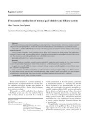
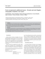
Fig 1. Normal pulsed and color <strong>Doppler</strong> <strong>of</strong> uterine artery (37<br />
WG)<br />
the <strong>Doppler</strong> spectrum. Peripheral resistance is assessed<br />
by the aspect <strong>of</strong> telediastolic flow (fig 1). When the angle<br />
between the vessel and the <strong>Doppler</strong> signal is constantly<br />
maintained, telediastolic flow is proportional to uterine<br />
perfusion, supposing that the low flow velocities that<br />
contribute to the spectrum are uniformly distributed and<br />
consequently, they should not be taken into consideration<br />
when evaluating a relative change in perfusion [2]<br />
For the quantification <strong>of</strong> the blood flow impedance,<br />
the resistance index (RI) or the systolic-diastolic index<br />
(S/D) is used [2].<br />
Uterine blood flow changes <strong>during</strong> uterine contractions<br />
During normal pregnancy, there is a progressive increase<br />
in uteroplacental blood flow related to the trophoblast<br />
invasion <strong>of</strong> spiral arteries. This process occurs<br />
in two stages: the first stage after 12 weeks <strong>of</strong> amenorrhoea<br />
(WA), and the second one after 18 WA, the process<br />
being considered completed at 22 WA. At the end <strong>of</strong><br />
the trophoblast invasion, the endothelial cells from the<br />
distal portion <strong>of</strong> the intervillous space arteries will also<br />
be replaced by trophoblast cells. The increase in diastolic<br />
blood flow is related to the vascular compliance and<br />
uteroplacental bed development [3,4].<br />
The normal uterine spectrum is defined by the increased<br />
residual blood flow in diastole (about 40% <strong>of</strong><br />
maximum systolic blood flow) and systolic peak with<br />
a vertical ascending phase and a less abrupt descending<br />
phase, followed by a second change in the descending<br />
slope to a pseudoplateau. So, the ascending and the descending<br />
parts <strong>of</strong> the systolic phase are not symmetrical<br />
[5-8].<br />
A mean 40-60% reduction in blood flow velocity was<br />
reported compared to the initial value, for a maximum<br />
intrauterine pressure <strong>of</strong> approximately 60 mmHg. The<br />
systolic peak decreases by only 25%, while the diastolic<br />
peak is extremely low or absent. This aspect was<br />
explained by a significant reduction in the perfusion<br />
pressure <strong>of</strong> the uterine artery blood flow at the maximum<br />
pressure <strong>of</strong> the uterine contraction. The perfusion<br />
pressure <strong>during</strong> systole is approximately 60-70 mmHg,<br />
when intrauterine pressure is 50-60 mmHg [9]. In diastole,<br />
when intrauterine pressure exceeds maternal diastolic<br />
pressure, the perfusion pressure <strong>of</strong> the uterine<br />
artery blood flow is no longer present. A progressive<br />
decrease in the diastolic component is found in parallel<br />
to an increase in intrauterine pressure from 10 mmHg to<br />
60 mmHg [9].<br />
An obvious correlation between the reduction <strong>of</strong> diastolic<br />
blood flow and the intensity <strong>of</strong> uterine contraction<br />
was evidenced [10]. Diastolic flow decreases to 0<br />
(null diastolic flow), when intrauterine pressure reaches
80 mmHg, without other changes in the spectrum (protodiastolic<br />
notch). However, systolic blood flow can be<br />
evidenced up to an intrauterine pressure <strong>of</strong> 130 mmHg<br />
[11].<br />
These aspects suggest that uterine diastolic flow reflects<br />
the blood flow from arcuate and spiral arteries <strong>during</strong><br />
uterine contraction. The compression <strong>of</strong> these vessels<br />
<strong>during</strong> normal <strong>labor</strong> under the influence <strong>of</strong> uterine contractions<br />
results in reduction or even disappearance <strong>of</strong> the<br />
diastolic component [12]. During the uterine contraction<br />
test (oxytocin test), there are similar uterine blood flow<br />
changes to those <strong>of</strong> spontaneous <strong>labor</strong> [13].<br />
Uterine blood flow changes <strong>during</strong> premature delivery<br />
During uterine contractions occurring before term or<br />
<strong>during</strong> premature <strong>labor</strong>, there is a non-physiological reduction<br />
in uterine blood flow. These changes, which can<br />
result in the suppression <strong>of</strong> diastolic flow or the aspect<br />
<strong>of</strong> reversed diastolic flow can develop even when the<br />
patient does not perceive uterine contractions as painful.<br />
The increase in vascular resistance in uterine arteries<br />
confirms uterine hyperactivity in the context <strong>of</strong> premature<br />
delivery, being an obvious argument for tocolytic<br />
treatment [14-16]. Systolic blood flow present up to an<br />
intrauterine pressure <strong>of</strong> 130 mmHg confirms the presence<br />
<strong>of</strong> a sufficient minimum blood flow in the intervillous<br />
space [11,17].<br />
Intrapartum uterine blood flow changes in patients<br />
with preeclampsia<br />
In the case <strong>of</strong> preeclampsia, the trophoblast invasion<br />
<strong>of</strong> uterine spiral arteries is limited and incomplete, affecting<br />
only their decidual segment, which results in a<br />
diminution <strong>of</strong> uteroplacental vascularization.[18]<br />
During pregnancies complicated by severe preeclampsia,<br />
the reduced diastolic flow and the presence<br />
<strong>of</strong> the protodiastolic notch are related to a compliance<br />
defect <strong>of</strong> uterine vessels [19]. Under the conditions <strong>of</strong><br />
preeclampsia, the disappearance <strong>of</strong> diastolic blood flow<br />
or the aspect <strong>of</strong> reversed flow can be found. The spectral<br />
aspect with maximum systolic velocity and reversed<br />
diastolic flow is similar to that <strong>of</strong> the right iliac artery<br />
[1,20].<br />
The blood flow in the intervillous space <strong>during</strong> uterine<br />
contractions is almost completely maintained. This<br />
phenomenon is caused by the unchanged systolic flow<br />
velocity, through the increase in the perfusion pressure<br />
determined by maternal arterial hypertension. These considerations<br />
recommend caution in the use <strong>of</strong> aggressive<br />
antihypertensive therapy in patients with preeclampsia<br />
[7,19]. In cases with abnormal uterine velocimetry prior<br />
to <strong>labor</strong>, a higher increase in vascular resistance is found<br />
<strong>during</strong> <strong>labor</strong> compared to normal pregnancy, determining<br />
a reserved fetal prognosis [12].<br />
<strong>Medical</strong> Ultrasonography 2011; 13(2): 141-149<br />
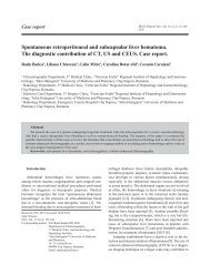
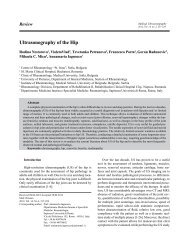
Fig 2. Normal pulsed and color <strong>Doppler</strong> <strong>of</strong> umbilical artery (38<br />
WG)<br />
Intrapartum <strong>Doppler</strong> <strong>ultrasound</strong> examination <strong>of</strong><br />
umbilical vessels<br />
The umbilical artery, the first vessel explored by <strong>Doppler</strong><br />
<strong>ultrasound</strong> in obstetrics, is the last vessel before the<br />
”placental obstacle”. As the placenta is the only organ<br />
downstream <strong>of</strong> this vessel, the umbilical artery is the<br />
privileged site <strong>of</strong> exploration <strong>of</strong> ”placental resistances”<br />
[21].<br />
Examination technique<br />
The umbilical cord is preferably visualized at its placental<br />
insertion (lower mobility area). The pulsed <strong>Doppler</strong><br />
window is fixed at the level <strong>of</strong> one <strong>of</strong> the umbilical<br />
arteries under a favorable angle (below 60°) with an<br />
opening <strong>of</strong> approximately 5 mm, incorporating the vessel<br />
[7].<br />
<strong>Doppler</strong> <strong>ultrasound</strong> examination <strong>of</strong> umbilical arteries<br />
<strong>during</strong> <strong>labor</strong><br />
The aspect <strong>of</strong> the spectrum <strong>of</strong> the umbilical artery<br />
corresponds to a vessel supplying an organ with relatively<br />
low vascular resistance, diastolic flow representing<br />
about 30% <strong>of</strong> the systolic flow value (fig 2).<br />
Residual diastolic flow increases <strong>during</strong> normal pregnancy<br />
[22]. When the amniochorionic membranes are intact<br />
and the amount <strong>of</strong> amniotic fluid is normal, the uterus<br />
can be considered as an empty sphere as in the model<br />
proposed by Kunzel et al [23]. According to this model,<br />
pressure in umbilical vessels increases in response to<br />
uterine contraction. Consequently, the perfusion pressure<br />
in the placental vascular tree is unchanged. This state is<br />
maintained after the rupture <strong>of</strong> membranes until a certain<br />
moment, because the fetal head covers the cervical orifice<br />
<strong>during</strong> <strong>labor</strong> [10].<br />
143
144 Dan Mihu et al <strong>Applications</strong> <strong>of</strong> <strong>Doppler</strong> <strong>ultrasound</strong> <strong>during</strong> <strong>labor</strong><br />
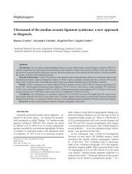
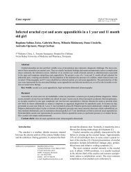
Fig 3. Abnormal pulsed and color <strong>Doppler</strong> <strong>of</strong> umbilical artery<br />
with reversed flow (36 WG)<br />
It is considered that umbilical <strong>Doppler</strong> spectrum undergoes<br />
no obvious changes as long as the fetal heart<br />
rate is maintained within normal limits (120-160 beats/<br />
minute) [1]. There is a remarkable stability <strong>of</strong> umbilical<br />
RI values <strong>during</strong> <strong>labor</strong>, which shows the permanent<br />
presence <strong>of</strong> feto-placental exchange <strong>during</strong> this period.<br />
Umbilical circulation maintains a certain independence<br />
in relation to the active phenomena <strong>of</strong> delivery.<br />
The aspect <strong>of</strong> umbilical diastolic flow can be assessed<br />
in four stages <strong>of</strong> uterine contraction: outside uterine contraction,<br />
in the ascending phase <strong>of</strong> uterine contraction, in<br />
the peak phase <strong>of</strong> uterine contraction, and in the descending<br />
phase <strong>of</strong> uterine contraction. Umbilical RI values<br />
undergo no significant changes <strong>during</strong> these phases <strong>of</strong><br />
uterine contraction [24]. In patients with ruptured membranes<br />
or those with oxytocin-induced <strong>labor</strong>, no significant<br />
variations in placental resistances were found, while<br />
the fetal heart rate was maintained within normal limits<br />
[13]. The position <strong>of</strong> the patient (dorsal decubitus or left<br />
lateral decubitus) at the time <strong>of</strong> examination does not influence<br />
the values <strong>of</strong> umbilical RI or the accuracy <strong>of</strong> the<br />
measurements [21].<br />
Correlations between fetal heart rate changes in <strong>labor</strong><br />
and <strong>Doppler</strong> <strong>ultrasound</strong> aspects in umbilical arteries<br />
At the level <strong>of</strong> the umbilical cord and in fetal vessels,<br />
direct mechanical effects due to vascular compression<br />
under the influence <strong>of</strong> uterine contractions should<br />
be differentiated from other causes <strong>of</strong> acute changes in<br />
vascular resistance.<br />
There are studies that attempt to establish certain correlations<br />
between fetal heart rate (FHR) variations and<br />
umbilical RI values [24]. The most frequent aspects <strong>of</strong><br />
FHR <strong>during</strong> <strong>labor</strong> are normal trajectory, early decelerations,<br />
late decelerations, variable decelerations, prolonged<br />
decelerations, accelerations, bradycardia [22,25].<br />
A progressive diminution in the umbilical diastolic<br />
flow is found <strong>during</strong> intrapartum decelerations and in fetal<br />
bradycardia, tending to become null, if the fetal heart<br />
rate decreases to less than 80 beats/minute. Fetal bradycardia<br />
with less than 100 beats/minute is associated with<br />
a significant prolongation <strong>of</strong> diastolic flow, as a compensation<br />
mechanism. This aspect <strong>of</strong> prolonged diastole is<br />
obvious in a normal fetal heart rate trajectory and less<br />
obvious in a changed trajectory, or imperceptible in a<br />
null diastolic flow record. In cases with reversed diastolic<br />
flow, the volume <strong>of</strong> the reversed flow will be theoretically<br />
increased, due to prolonged diastole (fig 3).<br />
The umbilical diastolic flow variation in <strong>labor</strong> under<br />
conditions <strong>of</strong> altered FHR is rather cardiogenic in origin<br />
and is less influenced by the temporary increase in<br />
placental resistances. The cardiac ejection volume (represented<br />
by systolic peak) and placental resistance (represented<br />
by residual diastolic flow) remain stable in spite<br />
<strong>of</strong> the increase in intrauterine pressure from 10 mmHg to<br />
60 mmHg <strong>during</strong> UC [25,26].<br />
In cases in which reversed diastolic flow is found <strong>during</strong><br />
decelerations, this reversed flow can have different<br />
aspects:<br />
• The reversed diastolic flow formed at the beginning<br />
<strong>of</strong> diastole may reflect very high placental<br />
resistance and umbilical artery occlusion.<br />
• The reversed holodiastolic flow, which tends to<br />
increase in telediastole, will be interpreted as a<br />
compensating flow from the peripheral expansion<br />
chamber (the placenta) and indicates an umbilical<br />
vein occlusion.<br />
The probability <strong>of</strong> an isolated umbilical vein occlusion<br />
or mixed umbilical vein and artery occlusion exists<br />
in the case <strong>of</strong> an acute reversed diastolic flow. In both<br />
cases, the outcome is the suppression <strong>of</strong> feto-placental<br />
perfusion, with the presence <strong>of</strong> an inefficient movement<br />
<strong>of</strong> fetal blood flow in umbilical arteries [27].<br />
Experiments in animals have demonstrated that the<br />
oxygen supply to the fetus remains almost constant even<br />
at a 50% reduction in umbilical artery blood flow, but the<br />
oxygen supply decreases exponentially when the umbilical<br />
blood flow decreases to less than 50%. Intrapartum<br />
<strong>Doppler</strong> <strong>ultrasound</strong> is a valuable method for the assessment<br />
<strong>of</strong> variable FHR decelerations, which are associated<br />
with reversed diastolic flow, indicating an obvious<br />
decrease in oxygen supply [27]. Under the conditions <strong>of</strong><br />
the association <strong>of</strong> pathological FHR decelerations with<br />
reversed diastolic flow in the spectrum <strong>of</strong> umbilical arteries,<br />
the risk <strong>of</strong> fetal hypoxia and acidosis increases.<br />
Because fetal oxygen supply significantly decreases
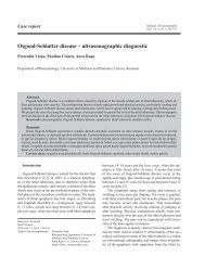
Fig 4. Normal pulsed and color <strong>Doppler</strong> <strong>of</strong> umbilical vein (37<br />
WG)<br />
Fig 5. Abnormal pulsed and color <strong>Doppler</strong> <strong>of</strong> umbilical vein<br />
with pulsations (36 WG)<br />
<strong>Medical</strong> Ultrasonography 2011; 13(2): 141-149<br />
when umbilical blood flow is reduced by more than 50%<br />
<strong>of</strong> the initial value, each deceleration should be considered<br />
as fetal ”stress”, particularly in fetuses with null or<br />
reversed diastolic flow. Considering these aspects, it can<br />
be stated that spontaneous or (oxytocin) induced <strong>labor</strong><br />
should be avoided in the case <strong>of</strong> a pregnancy with antepartum<br />
null or reversed diastolic flow in the umbilical<br />
arteries.<br />
Uterine contractions might cause a dangerous decrease<br />
in the oxygen supply to the intervillous spaces,<br />
and decelerations might determine a reduction in the<br />
umbilical blood flow, which can all result in severe fetal<br />
hypoxia [28]. The establishing <strong>of</strong> possible effects <strong>of</strong> fetal<br />
asphyxia on the umbilical artery blood flow have been attempted.<br />
One minute <strong>of</strong> asphyxia has been found to cause<br />
an approximately 35% decrease in the umbilical artery<br />
blood flow, mainly due to the decrease in FHR [10].<br />
However, <strong>Doppler</strong> velocimetry could not demonstrate<br />
acute fetal hypoxia in animal models. These results<br />
are not surprising if we consider the fact that placental<br />
perfusion cannot acutely change, probably because <strong>of</strong> the<br />
lack <strong>of</strong> innervation <strong>of</strong> intraplacental arteries, although<br />
perfusion might be affected by fetal epinephrine levels<br />
[28,29]. In the case <strong>of</strong> pregnancies with a risk for developing<br />
intrapartum fetal hypoxia, <strong>Doppler</strong> waves reflect<br />
a chronic increase in resistance in feto-placental circulation,<br />
due to either defective placentogenesis or an occlusion<br />
<strong>of</strong> the placental vascular tree. In fetuses with late<br />
decelerations and signs <strong>of</strong> hypoxemia in the intrapartum<br />
period, <strong>Doppler</strong> velocimetry in umbilical arteries can detect<br />
a high S/D index, both <strong>during</strong> and between UC [30].<br />
In 90% <strong>of</strong> the fetuses with late decelerations, umbilical<br />
vein pulsations were evidenced <strong>during</strong> UC, which indicates<br />
a possible short duration overloading <strong>of</strong> the right<br />
heart (fig 4, fig 5) [31,32].<br />
In cases with normal FHR, the umbilical vein blood<br />
flow was unchanged <strong>during</strong> <strong>labor</strong>. These findings demonstrate<br />
that acute hypoxia also affects fetal venous blood<br />
flow [33,34].<br />
145<br />
Intrapartum <strong>Doppler</strong> <strong>ultrasound</strong> examination <strong>of</strong><br />
fetal circulation<br />
Fig 6. Normal pulsed and color <strong>Doppler</strong> <strong>of</strong> fetal aortic artery<br />
(37 WG)<br />
Fetal aortic artery<br />
During normal pregnancy, a progressive increase in<br />
aortic blood flow is found until 36 WA, followed by a<br />
slight decrease starting with 39 WA. The aortic blood<br />
flow is characterized by a diphasic aspect with a sharp<br />
systolic peak, with increased amplitude, followed by a<br />
diastole, which starts with a protodiastolic notch, more<br />
obvious with the increase in gestational age (fig 6). The<br />
diastole is constantly positive due to the permanent open-
146 Dan Mihu et al <strong>Applications</strong> <strong>of</strong> <strong>Doppler</strong> <strong>ultrasound</strong> <strong>during</strong> <strong>labor</strong><br />
ing <strong>of</strong> the arterial duct on the one hand, and to the low<br />
downstream resistances on the other hand [21,35].<br />
In the case <strong>of</strong> normal FHR, UC do not cause significant<br />
changes in <strong>Doppler</strong> waves at the level <strong>of</strong> the fetal<br />
aorta. In contrast, decelerations result in a decrease in<br />
telediastolic aortic flow [36].<br />
Acute fetal hypoxia is considered to have no direct effect<br />
on the fetal aorta spectrum. On the other hand, a redistribution<br />
<strong>of</strong> fetal heart blood flow in favor <strong>of</strong> cerebral perfusion<br />
can have an indirect effect on the aortic spectrum,<br />
which is demonstrated by experiments in animals [11]. The<br />
major vasoconstriction <strong>of</strong> the aorta exposes the fetus to an<br />
increased risk <strong>of</strong> ulcero-hemorrhagic enterocolitis. The increased<br />
vascular resistance in the fetal aorta can induce a<br />
dilation <strong>of</strong> the arterial duct, pulmonary hypertension, with<br />
right heart decompensation and decreased cardiac blood<br />
flow, which precede fetal agony. There is an obvious correlation<br />
between aortic velocimetry on the one hand, and<br />
fetal venous pH, the importance <strong>of</strong> fetal hypoxia, hypercapnia<br />
and acidosis on the other hand [37].<br />
Fetal cerebral arteries<br />
The Circle <strong>of</strong> Willis, the site <strong>of</strong> the anastomoses <strong>of</strong> the<br />
main cerebral vessels, is visualized by using color <strong>Doppler</strong>.<br />
The section plane <strong>of</strong> the biparietal diameter allows<br />
to evidence the main components <strong>of</strong> the Circle <strong>of</strong> Willis<br />
(fig 7) [38].<br />
In normal pregnancies, the cerebral vascular system<br />
has an increased resistance, the value <strong>of</strong> the telediastolic<br />
flow being approximately 16% <strong>of</strong> that <strong>of</strong> maximum<br />
systolic flow (fig 8).<br />
There are only a few studies that have evaluated<br />
the changes in fetal cerebral vascularization <strong>during</strong> <strong>labor</strong>.<br />
The pressure <strong>of</strong> the transducer on the fetal head can<br />
cause a reduction <strong>of</strong> telediastolic flow velocity, and obvious<br />
pressure may induce null or reversed diastolic flow.<br />
These reductions in diastolic velocity have also been evidenced<br />
in oligohydramnios [39]. As long as the heart rate<br />
is maintained within normal limits, <strong>during</strong> UC the blood<br />
flow in the middle cerebral artery undergoes no obvious<br />
changes.<br />
Studies have evidenced a slight increase in vascular<br />
resistance in the internal carotid artery <strong>during</strong> the initial<br />
phase <strong>of</strong> the dilation period. As the skull descends, due<br />
to the pressure exerted by UC on the skull and to the<br />
increased intracranial pressure, the S/D index increases<br />
from 3.5 to 5.5 <strong>during</strong> UC [40].<br />
A normal fetus is considered to respond to a drastic<br />
heart rate decrease <strong>during</strong> isolated decelerations by a reduction<br />
in residual velocity at the level <strong>of</strong> cerebral circulation.<br />
This diminution <strong>of</strong> telediastolic velocity is purely<br />
cardiogenic in origin. Thus, <strong>Doppler</strong> examination has<br />
confirmed the concept <strong>of</strong> the reduction <strong>of</strong> cerebral blood<br />
Fig 7. Color <strong>Doppler</strong> – the Circle <strong>of</strong> Willis (38 WG)<br />
Fig 8. Normal pulsed and color <strong>Doppler</strong> <strong>of</strong> middle cerebral artery<br />
(37 WG)<br />
Fig 9. Abnormal pulsed and color <strong>Doppler</strong> <strong>of</strong> middle cerebral<br />
artery with vasodilation (38 WG)
flow by the compression <strong>of</strong> the fetal skull as a cause <strong>of</strong><br />
decelerations occurring <strong>during</strong> <strong>labor</strong> [39,41].<br />
Repeated decelerations trigger cerebral vasodilation,<br />
which is reflected by an increase in telediastolic velocity<br />
(fig 9).<br />
Thus, the fetal brain will be protected from blood flow<br />
and blood pressure variations [42]. When these adaptive<br />
phenomena are overcome, a correlation between fetal<br />
heart rate and diastolic cerebral blood flow can be detected<br />
(the decrease in FHR results in a reduction <strong>of</strong> diastolic<br />
cerebral flow) [39-41].<br />
<strong>Doppler</strong> <strong>ultrasound</strong> examination and peridural<br />
analgesia<br />
The sympathetic denervation <strong>of</strong> the uterus with the<br />
decrease <strong>of</strong> vascular resistances can be evidenced by<br />
<strong>Doppler</strong> <strong>ultrasound</strong> [43,44]. In this sense, the S/D index<br />
in the umbilical arteries shows a variable and inconsistent<br />
decrease <strong>during</strong> peridural analgesia with chlorprocaine.<br />
Under these conditions, the values <strong>of</strong> the S/D index<br />
in dorsal decubitus are higher than in the left lateral<br />
decubitus [11,45].<br />
Bupivacaine, which induces a negative inotropic<br />
effect, causes a constant and important decrease in the<br />
umbilical S/D index [46]. Fetal aortic blood flow may increase<br />
<strong>during</strong> <strong>labor</strong>, independently <strong>of</strong> peridural analgesia.<br />
In contrast, in the case <strong>of</strong> bethridine administration, fetal<br />
aortic blood flow is significantly reduced.<br />
However, there are studies that do not show significant<br />
blood flow changes <strong>during</strong> the course <strong>of</strong> different<br />
peridural analgesia methods [43]. These apparently contradictory<br />
aspects emphasize the complexity <strong>of</strong> interlacing<br />
mechanisms: the secretion <strong>of</strong> vasoactive substances<br />
<strong>during</strong> <strong>labor</strong> on the one hand, and the different actions <strong>of</strong><br />
the administered drugs on blood flow, on the other hand<br />
[11,45].<br />
Conclusions<br />
<strong>Doppler</strong> <strong>ultrasound</strong> examination <strong>during</strong> <strong>labor</strong>, unlike<br />
<strong>Doppler</strong> <strong>ultrasound</strong> <strong>during</strong> non-<strong>labor</strong>, can detect blood<br />
flow changes, which develop within seconds. Uterine<br />
muscle contraction leads to an increase in resistance in<br />
the uterine arteries, with a corresponding reduction in<br />
blood flow. The proportion <strong>of</strong> these changes depends on<br />
the intensity <strong>of</strong> UC, without significantly correlating with<br />
external tocography.<br />
On the examination <strong>of</strong> umbilical and fetal vessels, the<br />
direct mechanical effects due to the compression exerted<br />
by UC should be differentiated from heart rate changes<br />
or hypoxic factors, which can alter the characteristics <strong>of</strong><br />
<strong>Medical</strong> Ultrasonography 2011; 13(2): 141-149 147<br />
the blood flow. In the case <strong>of</strong> normal FHR, UC do not<br />
alter the characteristics <strong>of</strong> umbilical artery blood flow.<br />
When the antepartum umbilical spectrum is changed, decelerations<br />
can induce a drastic and exponential decrease<br />
in umbilical blood flow. These decelerations <strong>during</strong> spontaneous<br />
or induced <strong>labor</strong> should be avoided under these<br />
circumstances.<br />
The intrapartum <strong>Doppler</strong> examination <strong>of</strong> fetal vessels<br />
evidences and quantifies the blood flow redistribution<br />
mechanisms in the case <strong>of</strong> hypoxia – peripheral vasoconstriction<br />
in the mesenteric, renal, cutaneous territories<br />
and the preferential vasodilation <strong>of</strong> privileged territories,<br />
particularly the brain. The increased pressure on the fetal<br />
brain <strong>during</strong> UC may cause a decrease in diastolic flow<br />
velocity in cerebral vessels, as a result <strong>of</strong> increased intracranial<br />
pressure. Certain decisions regarding the extraction<br />
<strong>of</strong> the fetus can only be made by correlating <strong>Doppler</strong><br />
<strong>ultrasound</strong> results with the other paraclinical methods for<br />
the monitoring <strong>of</strong> the intrapartum fetal status.<br />
Conflict <strong>of</strong> interest: absence <strong>of</strong> conflict <strong>of</strong> interest<br />
References<br />
1. E. Weiss. Intrapartum Fetal Heart Rate Changes and <strong>Doppler</strong><br />
Sonography. In: Schmidt W, Kurjak A. Color <strong>Doppler</strong><br />
Sonography in Gynecology and Obstetrics Ed Thieme<br />
2005: 182-197.<br />
2. Rigano S, Ferrazzi E, Boito S, Pennati G, Padoan A, Galan<br />
H. Blood flow volume <strong>of</strong> uterine arteries in human pregnancies<br />
determined using 3D and bi-dimensional imaging, angio-<strong>Doppler</strong>,<br />
and fluid-dynamic modeling. Placenta 2010;<br />
31: 37–43.<br />
3. Groom KM, North RA, Stone PR, et al. Patterns <strong>of</strong> change<br />
in uterine artery doppler studies between 20 and 24 weeks<br />
<strong>of</strong> gestation and pregnancy outcomes. Obstet Gynecol<br />
2009; 113: 332-338.<br />
4. Burton GJ, Woods AW, Jauniaux E, Kingdom JC. Rheological<br />
and physiological consequences <strong>of</strong> conversion <strong>of</strong> the<br />
maternal spiral arteries for uteroplacental blood flow <strong>during</strong><br />
human pregnancy. Placenta 2009; 30: 473-482.<br />
5. Dane B, Dane C, Kiray M, Cetin A, Koldas M, Erginbas<br />
M. Correlation between first-trimester maternal serum<br />
markers, second-trimester uterine artery doppler indices<br />
and pregnancy outcome. Gynecol Obstet Invest 2010; 70:<br />
126–131.<br />
6. Tuuli MG, Odibo AO. The role <strong>of</strong> serum markers and uterine<br />
artery <strong>Doppler</strong> in identifying at-risk pregnancies. Clin<br />
Perinatol 2011; 38: 1–19.<br />
7. Ghosh G, Gudmundsson S. Uterine and umbilical artery<br />
<strong>Doppler</strong> are comparable in predicting perinatal outcome <strong>of</strong><br />
growth-restricted fetuses. BJOG 2009; 116: 424–430.
148 Dan Mihu et al <strong>Applications</strong> <strong>of</strong> <strong>Doppler</strong> <strong>ultrasound</strong> <strong>during</strong> <strong>labor</strong><br />
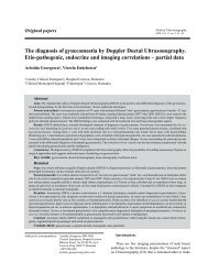
8. Gomez O, Figueras F, Fernandez S, et al. Reference ranges<br />
for uterine artery mean pulsatility index at 11-41 weeks <strong>of</strong><br />
gestation. Ultrasound Obstet Gynecol 2008; 32: 128-132.<br />
9. Li H, Gudmundsson S, Ol<strong>of</strong>sson P. Uterine artery blood<br />
flow velocity waveforms <strong>during</strong> uterine contractions. Ultrasound<br />
Obstet Gynecol 2003; 22: 578-585.<br />
10. Cooley SM, Donnelly JC, Walsh T, Macmahon C, Gillan<br />
J, Geary MP. The impact <strong>of</strong> umbilical and uterine artery<br />
<strong>Doppler</strong> indices on antenatal course, <strong>labor</strong> and delivery in<br />
a low-risk primigravid population. J Perinat Med 2011; 39:<br />
143-149.<br />
11. Kurjak A, Dudenhausen JW, Kos M, et al. <strong>Doppler</strong> information<br />
pertaining to the intrapartum period. J Perinat Med<br />
1996; 24: 271-276.<br />
12. Li H, Gudnason H, Ol<strong>of</strong>sson P, Dubiel M, Gudmundsson<br />
S. Increased uterine artery vascular impedance is related to<br />
adverse outcome <strong>of</strong> pregnancy but is present in only one<br />
– third <strong>of</strong> late third – trimester pre – eclamptic women. Ultrasound<br />
Obstet Gynecol 2005; 25: 459-463.<br />
13. Svardby K, Nordstrőm L, Sellstrőm E. Primiparas with or<br />
without oxytocin augumentation: a prospective descriptive<br />
study. J Clin Nurs 2007; 16: 179-184.<br />
14. Lima MM, Souza AS, Diniz C, Porto AM, Amorim MM,<br />
Moron AF. <strong>Doppler</strong> velocimetry <strong>of</strong> the uterine, umbilical<br />
and fetal middle cerebral arteries in pregnant women undergoing<br />
tocolysis with oral nifedipine. Ultrasound Obstet<br />
Gynecol 2009; 34: 311–315.<br />
15. Spong CY. Prediction and prevention <strong>of</strong> recurrent spontaneous<br />
preterm birth. Obstet Gynecol 2007; 110: 405-415.<br />
16. Sayin NC, Arda S, Varol FG, Sut N. The effects <strong>of</strong> ritodrine<br />
and magnesium sulfate on maternal and fetal <strong>Doppler</strong> blood<br />
flow patterns in women with preterm <strong>labor</strong>. Eur J Obstet<br />
Gynecol Reprod Biol 2010; 152: 50–54.<br />
17. Baschat AA. <strong>Doppler</strong> application in the delivery timing <strong>of</strong><br />
the preterm growth – restricted fetus: another step in the<br />
right direction. Ultrasound Obstet Gynecol 2004; 23; 111-<br />
118.<br />
18. Lovgren TR, Dug<strong>of</strong>f L, Galan HL. Uterine artery <strong>Doppler</strong><br />
and prediction <strong>of</strong> preeclampsia. Clin Obstet Gynecol. 2010;<br />
53: 888-898.<br />
19. Toal M, Keating S, Machin G, et al. Determinants <strong>of</strong> adverse<br />
perinatal outcome in high risk women with abnormal<br />
uterine artery <strong>Doppler</strong> images. Am J Obstet Gynecol 2008;<br />
198: 330.e1-7.<br />
20. Napolitano R,Rajakulasingam R, Memmo A, Bhide A,<br />
Thilaganathan B. Uterine artery <strong>Doppler</strong> screening for preeclampsia:<br />
comparison <strong>of</strong> the lower, mean and higher firsttrimester<br />
pulsatility indices. Ultrasound Obstet Gynecol<br />
2010 Sep 27. DOI:10.1002/uog.8848.<br />
21. Mihu D. Ecografia <strong>Doppler</strong> în Obstetrică şi Ginecologie.<br />
Ed. Clusium 2001: 89,119,206.<br />
22. Kiserud T. Physiology <strong>of</strong> the fetal circulation. Semin Fetal<br />
Neonatal Med 2005; 10: 493-503.<br />
23. Kunzel W. Uberwachung des Feten wahrend der Geburt. In<br />
Wulf KH, Schmidt-Matthiesen H (eds): Klinik der Frauenheilkunde<br />
und Geburt-shilfe. Bd. 7/I. Urban & Schwarzenberg,<br />
Munchen 1990: 91-134.<br />
24. Chan FY, Lam C, Lam YH, To WK, Pun TC, Lee CP. Umbilical<br />
artery <strong>Doppler</strong> velocimetry compared with fetal<br />
heart rate monitoring as a <strong>labor</strong> admission test. Eur J Obstet<br />
Gynecol Reprod Med 1994; 54: 1-6.<br />
25. Alfirevic Z, Devane D, Gyte GM. Continnous cardiotocography<br />
as a form <strong>of</strong> electronic fetal monitoring fetal assessment<br />
<strong>during</strong> <strong>labor</strong>. Cochrane Database Syst Rev 2006; 3:<br />
CD006066.<br />
26. Haws RA, Yakoob MY, Soomro T, Menezes EV, Darmstadt<br />
GL, Bhutta ZA. Reducing stillbirths: screening and monitoring<br />
<strong>during</strong> pregnancy and <strong>labor</strong>. BMC Pregnancy Childbirth<br />
2009; 9 (Suppl 1): S5.<br />
27. Somerset DA, Murrillis AJ, Wheeler T. Screening for fetal<br />
distress in <strong>labor</strong> using the umbilical artery blood velocity<br />
waveform. Br J Obstet Gynecol 1993; 100 : 55-59.<br />
28. Bathiyar MO, Copel JA. Cardiac changes in the intrauterine<br />
growth – restricted fetus. Semin Perinatol 2008; 32: 190-<br />
193.<br />
29. Kurmanavicius J, Florio I, Wisser J, et al. Reference resistance<br />
induce <strong>of</strong> the umbilical, fetal middle cerebral and<br />
uterine arteries at 24 – 42 weeks <strong>of</strong> gestation. Ultrasound<br />
Obstet Gynecol 1997; 10: 112-120.<br />
30. Skinner J, Greene RA, Gardeil F, Stuart B, Turner MJ. Does<br />
increased resistance on umbilical artery <strong>Doppler</strong> preclude a<br />
trial <strong>of</strong> labour. Eur J Obstet Gynecol Reprod Biol 1998; 79:<br />
35-38.<br />
31. Ozyüncü O, Saygan-Karamürsel B, Armangil D, et al. Fetal<br />
arterial and venous <strong>Doppler</strong> in growth restricted fetuses<br />
for the prediction <strong>of</strong> perinatal complications. Turk J Pediatr<br />
2010; 52: 384-392.<br />
32. Hasegawa J, Mimura T, T. Morimoto T, et al. Detection <strong>of</strong><br />
umbilical venous constriction by <strong>Doppler</strong> flow measurement<br />
at midgestation. Ultrasound Obstet Gynecol 2010; 36:<br />
196–201.<br />
33. Ghosh GS, Fu J, Ol<strong>of</strong>sson P, Gudmundsson S. Pulsations<br />
in the umbilical vein <strong>during</strong> <strong>labor</strong> are associated with increased<br />
risk <strong>of</strong> operative delivery for fetal distress. Ultrasound<br />
Obstet Gynecol 2009; 34: 177-181.<br />
34. Figueras F, Feranandez S, Hernandez-Andrade E, Gratacos<br />
E. Umbilical venous blood flow measurement: accuracy<br />
and reproducibility. Ultrasound Obstet Gynecol 2008; 32:<br />
587-591.<br />
35. Acharya G. Technical aspects <strong>of</strong> aortic isthmus <strong>Doppler</strong><br />
velocimetry in human fetuses. Ultrasound Obstet Gynecol<br />
2009; 33: 628-633.<br />
36. Fouron JC, Siles A, Montanari L, et al. Feasibility and reliability<br />
<strong>of</strong> <strong>Doppler</strong> flow recordings in the fetal aortic isthmus:<br />
a multicenter evaluation. Ultrasound Obstet Gynecol 2009;<br />
33: 690-693.<br />
37. Figueras F, Benavides A, Del Rio M, et al. Monitoring <strong>of</strong><br />
fetuses with intrauterine growth restriction: longitudinal<br />
changes in ductus venosus and aortic isthmus flow. Ultrasound<br />
Obstet Gynecol2009; 33: 39-43.<br />
38. Schenone MH, Mari G. The MCA <strong>Doppler</strong> and its Role in<br />
the Evaluation <strong>of</strong> Fetal Anemia and Fetal Growth Restriction.<br />
Clin Perinatol 2011; 38: 83–102.<br />
39. Kassanos D, Siristatidis C, Vitoratos N, Salamalekis E,
Creatsas G. The clinical signifiance <strong>of</strong> <strong>Doppler</strong> findings<br />
in fetal middle cerebral artery <strong>during</strong> <strong>labor</strong>. Eur J Obstet<br />
Gynecol Reprod Biol 2003; 109: 45-50.<br />
40. Oosterh<strong>of</strong> H, Dijkstra K, Aarnoudse JG. Fetal <strong>Doppler</strong> velocimetry<br />
in the internal carotid and umbilical artery <strong>during</strong><br />
Braxton Hicks contractions. Early Hum Dev 1992; 30: 33-<br />
40.<br />
41. Cheema R. Dubiel M, Gudmundsson S. Signs <strong>of</strong> fetal brain<br />
sparing are not related to umbilical cord blood gases at<br />
birth. Early Hum Dev 2009; 85: 467-470.<br />
42. Roza SJ, Steegers EA, Verburg BO, et al. What is spared<br />
by fetal brain-sparing Fetal Circulatory redistribution and<br />
behavioral problems in the general population. Am J Epidemiol<br />
2008; 168: 1154-1152.<br />
43. Ginosar Y, Nadjari M, H<strong>of</strong>fman A, et al; ACET study group.<br />
<strong>Medical</strong> Ultrasonography 2011; 13(2): 141-149<br />
Antepartum continuous epidural ropivacaine therapy reduces<br />
uterine artery vascular resistance in pre-eclampsia:<br />
a randomized, dose-ranging, placebo-controlled study. Br J<br />
Anaesth 2009; 102: 369–378.<br />
44. Reynolds F. Changes in <strong>Doppler</strong> velocimetry <strong>of</strong> uterine arteries<br />
<strong>during</strong> lbour analgesia. Br J Anaesth 2007; 98: 274-<br />
275.<br />
45. Fratelli N, Prefumo F, Andrico S, et al. Effects <strong>of</strong> epidural<br />
analgesia on uterine artery <strong>Doppler</strong> in labour. Br J Anaesth<br />
2011; 106: 221–224.<br />
46. Boulier V, Gomis P, Lautner C, Visseaux H, Palot M, Malinovsky<br />
JM. Minimum local analgesic concentrations <strong>of</strong><br />
ropivacaine and levobupivacaine with sufentanil for epidural<br />
analgesia in labour. Int J Obstet Anesth 2009; 18:<br />
226-230.<br />
149