Focal, asymptomatic epididymal masses - Medical Ultrasonography
Focal, asymptomatic epididymal masses - Medical Ultrasonography
Focal, asymptomatic epididymal masses - Medical Ultrasonography

You also want an ePaper? Increase the reach of your titles
YUMPU automatically turns print PDFs into web optimized ePapers that Google loves.
Case report<br />
<strong>Medical</strong> <strong>Ultrasonography</strong><br />
2010, Vol. 12, no. 2, 163-166<br />
<strong>Focal</strong>, <strong>asymptomatic</strong> <strong>epididymal</strong> <strong>masses</strong> – B-mode and color Doppler<br />
sonographic evaluation. Case report<br />
Vasiliki Bizimi 1 , Alexia P. Balanika 2 , Mariana Motogna-Kalokairinou 3 , Iovana Paianidi 1 ,<br />
Spiros Kardamis 1 , Christos Kominis 1 , Christos S. Baltas 1<br />
1<br />
Radiology Imaging Department, General Hospital of Athens „G. Gennimatas”, Athens, Greece<br />
2<br />
Computed Tomography Department, General Hospital „Asklipieio Voulas”, Athens, Greece<br />
3<br />
Radiology Department, General Hospital of Athens „Sotiria”, Athens, Greece<br />
Abstract<br />
We reported the case of an adult male with an extratesticular mass prove to be postoperative a chronic inflammatory process.<br />
The importance of B-mode and color Doppler sonography in the differential diagnosis and literature regarding <strong>epididymal</strong><br />
nodules and tumors of the paratesticular structures (epididymis) is reviewed and discussed.<br />
Keywords: epididymitis, paratesticular mass, ultrasonography<br />
Rezumat<br />
Prezentăm cazul unui pacient cu un nodul extratesticular dovedit a fi postoperator un proces inflamator cronic. Discutăm<br />
importanţa ecografiei B-mode şi a ecografiei color Doppler în diagnosticul diferenţial al nodulilor epididimali şi a tumorilor<br />
structurilor paratesticulare (epididim) însoţit de o trecere in revistă a datelor din literatură.<br />
Cuvinte cheie: epididimita, masă paratesticulară, ecografie<br />
Introduction<br />
High-resolution real-time sonography with color<br />
Doppler imaging is the technique of choice for imaging<br />
the epididymitis. Although only 3% of all solid extratesticular<br />
<strong>masses</strong> are malignant [1], previous studies<br />
have shown the malignancy rate for solid <strong>epididymal</strong><br />
<strong>masses</strong>, to be as high as 16% [2], whereas the rate for<br />
neoplastic processes within the epididymis was as high<br />
as 25% [3].<br />
Recent studies have suggested that scrotal sonography<br />
plays an important role in distinguishing solid<br />
<strong>masses</strong> from inflammatory <strong>masses</strong> of the epididymis. A<br />
small size, the presence of a hyperechoic or hypoechoic<br />
Received 6.04.2010 Accepted 14.04.2010<br />
Med Ultrason<br />
2010, Vol. 12, No 2, 163-166<br />
Address for correspondence: Vasiliki Bizimi<br />
9 Odysseos St, Heliopolis, 16346<br />
Athens, Greece<br />
Email: bizimi@otenet.gr<br />
rim circumscribing the lesion and little or absent blood<br />
flow are important findings when comparing benign<br />
neoplastic lesions with inflammatory lesions [4]. However,<br />
because of the very low prevalence of <strong>epididymal</strong><br />
malignancies, the sonographic characteristics of malignant<br />
<strong>epididymal</strong> lesions have not until recently been adequately<br />
studied.<br />
Presentation of the case<br />
A 34-year old male was referred to the Emergency<br />
Urological Department with sudden, painless, swelling<br />
of the right scrotum. Clinical examination revealed a<br />
small, solid, well defined palpable nodule attached to the<br />
external surface of the right testis. The patient reported<br />
no fever, and no history of trauma. <strong>Ultrasonography</strong> by<br />
B-mode and color Doppler, of the scrotum, detected a<br />
well circumscribed, ellipsoid, mass of mixed echostructure<br />
(with distinct calcifications) at the tail of the right<br />
epididymis. The mass measured 3.5/1.9 cm and had an<br />
increased vascularization. There was also a small con-
164 Vasiliki Bizimi et al <strong>Focal</strong>, <strong>asymptomatic</strong> <strong>epididymal</strong> <strong>masses</strong><br />
tralateral hydrocele (fig 1–3). Both testis and the left<br />
epididymis (head, body, and tail) were depicted with normal<br />
size echogenicity and vascularisation.<br />
Surgical excision was performed and a bulky right<br />
epididymis was revealed. Histological examination<br />
reported hyperplastic epithelial lining of the epididymis,<br />
with some areas of dilated ducts, suggestive of<br />
a chronic inflammatory process (chronic epididymitis).<br />
Discussion<br />
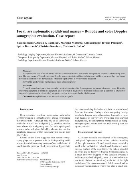
Fig 1. Gray scale ultrasonography of <strong>epididymal</strong> tale (longitudinal<br />
scan): the nodule is well circumscribed, ellipsoid, 3.5x1.9 cm,<br />
with mixed echostructure (with distinct calcification).<br />
Fig 2. Color Doppler sonography reveal increased vascularity of<br />
the <strong>epididymal</strong> nodule.<br />
Fig 3. Duplex ultrasonography depicting the tumoral monophasic<br />
arterial blood flow with low resisence index.<br />
According to a recent report by Alleman WG et<br />
al [5] most of the <strong>epididymal</strong> mass are benign. The<br />
most common is the cyst, reported in 20%-40% of<br />
<strong>asymptomatic</strong> individuals. They vary in size and almost<br />
always arise from the <strong>epididymal</strong> head. Spermatoceles<br />
(retention cysts), which appear in the same<br />
region, are more frequent but indistinguishable from<br />
the <strong>epididymal</strong> cysts at US. Spermatocele represents<br />
cystic dilatation of tubules of the efferent ductules in<br />
the head of the epididymis, and usually occur in middle<br />
aged men. They are often associated with a prior<br />
vasectomy. When the dimensions are large and the<br />
aspect is multilocular, differential diagnosis should<br />
be made from a large septated hydrocele (cysts displace<br />
the testis, whereas a hydrocele envelopes it)<br />
[6]. Sperm granuloma (in 42% of men with a previous<br />
vasectomy interpretated as an inflammatory<br />
<strong>masses</strong> resulting from a foreign body giant cell reaction<br />
to extravasated sperm) may be another cause of<br />
scrotal pain [7].<br />
Adenomatoid tumors, the most common benign neoplasm<br />
of the epididymis, account for approximately 30%<br />
of tumors, arise from the paratesticular region, usually at<br />
the lower pole, and may be solid or cystic [8]. Adenomatoid<br />
tumors have also been reported in the spermatic cord<br />
and tunica albuginea, where they can grow intratesticulary<br />
(indistinguishable in this case from testicular germ<br />
cells neoplasm) [6].<br />
Leiomyoma, the second most common benign tumor<br />
of the epididymis, may be solid or cystic, with or without<br />
calcifications. The tumor adheres to the testis, so clinical<br />
examination is unreliable in excluding malignancy; consenquently,<br />
removal is indicated [9].<br />
Papillary cystadenomas are strongly related with Von<br />
Hippel-Lindau disease. Up to 25% of men with the disease<br />
have an <strong>epididymal</strong> papillary cystadenoma and two<br />
thirds of men with papillary cystadenoma (40% of cystadenomas<br />
bilateral) have Von Hippel-Lindau disease.<br />
Practically the papillary cystadenomas are pathognomonic<br />
for Von Hippel-Lindau disease [6].
Invasive behavior is a feature of malignant lesions.<br />
Malignant tumors of the epididymis are rare and include<br />
sarcomas, metastases and adenocarcinoma. In adults,<br />
there are special features of liposarcoma and leiomyosarcoma<br />
on CT and MRI. Primary <strong>epididymal</strong> adenocarcinomas<br />
are very rare [5]. The most common cause<br />
of solid malignant neoplasms of the epididymis are metastasis<br />
(from prostate, stomach, colon and kidney, and<br />
leukemia). Lymphoma may also involve the epididymis<br />
in 60% of cases, even though genital tract lymphoma is<br />
mostly seen in the testis.<br />
Chronic inflammatory processes can result in<br />
secondary solid <strong>masses</strong> in up to 64% of cases due to<br />
granulomatous inflammatory reactions: tuberculosis,<br />
brucellosis, syphilis, parasitic and fungal infections<br />
[5]. The most common of the above is Mycobacterium<br />
tuberculosis. The <strong>epididymal</strong> infection is<br />
thought to result from renal disease which extends to<br />
the lower genitourinary tract, although hematogenous<br />
dissemination is also a possibility. Approximately<br />
25% of patients have bilateral involvement. The ultrasonographic<br />
findings include an enlarged epididymis<br />
which varies in appearance, ranging from hypoechoic<br />
to hyperechoic. Differential diagnosis from a<br />
primary testicular mass is often impossible. When the<br />
infection disseminates in testis, (epididymo-orchitis),<br />
imaging findings include diffuse testis enlargement,<br />
a solitary hypoechoic mass, or multiple hypoechoic<br />
nodules [3,6].<br />
Yang DM et al. (2003) used gray scale and color<br />
Doppler US together with some clinical features for<br />
the differential diagnosis of focal <strong>epididymal</strong> lesions.<br />
They found that lesions were larger in patients with tuberculous<br />
epididymitis, the hypoechoic or hyperechoic<br />
rim was more common in patients with benign <strong>epididymal</strong><br />
<strong>masses</strong> and the intralesional blood flow was<br />
greater in patients with nonspecific epididymitis [4].<br />
The characteristic finding in tuberculous epididymitis<br />
was heterogenous echogenicity of the lesion. This is<br />
due to caseation necrosis granuloma and fibrosis of the<br />
lesion. In contrast, sonographic findings in non specific<br />
epididymitis and benign <strong>epididymal</strong> <strong>masses</strong> were usually<br />
homogenous [4, 9]. Most of the focal <strong>epididymal</strong><br />
lesions were hypoechoic and located at the tail of the<br />
epididymis [4].<br />
Alleman WG et al. (2008), concluded, that the size<br />
of a neoplastic mass along with its vascularity, were significant<br />
malignancy markers. The above, combined with<br />
the clinical features, could limit the decision, as regard<br />
the differential diagnosis and treatment planning. The<br />
authors recommend US to be the first step in the evaluation<br />
of solid <strong>epididymal</strong> <strong>masses</strong>. If the mass is smaller<br />
<strong>Medical</strong> <strong>Ultrasonography</strong> 2010; 12(2): 163-166 165<br />
than 1.5 cm. and Doppler flow is absent, a follow up<br />
clinical evaluation is scheduled, with or without scrotal<br />
follow up sonography. If the size of a solid <strong>epididymal</strong><br />
mass is 1.5 cm, or greater, with a present Doppler flow,<br />
surgical exploration is considered. The presence of calcifications<br />
as well as the echogenicity of a mass was<br />
not a statistically significant marker, to determine the<br />
pathologic nature of the lesion [5].<br />
In our case, the aspect of the <strong>epididymal</strong> mass was<br />
uncertain, having large dimensions, mixed echostructure,<br />
and hypervascularization. This was the reason<br />
that the excision was justifiable, the patient having a<br />
high risk for a malignant lesion. The histological aspect<br />
of chronic epididymitis (excluding granulomatous<br />
inflammatory reactions) has to be associate with previous<br />
genital infection with chlamydia or other kind of<br />
bacteria.<br />
Conclusions<br />
Ultrasography can be used to accurately determine<br />
whether an abnormality is intratesticular or extratesticular.<br />
If the mass is extratesticular and cystic, a specific<br />
diagnosis can be made (hydrocele, <strong>epididymal</strong> cyst,<br />
spermatocele) and the mass can be interpreted as benign.<br />
Extratesticular solid lesions lack specific sonographic<br />
features and when the features are uncertainty, surgery<br />
should be planned.<br />
Bibliography<br />
1. Beccia DJ, Krane RJ, Olsson CA. Clinical management of<br />
non-testicular intrascrotal tumors. J Urol 1976; 116: 476-<br />
479.<br />
2. Frates MC, Benson CB, DiSalvo DN, Brown DL, Laing<br />
FC, Doubilet PM. Solid extratesticular <strong>masses</strong> evaluated<br />
with sonography: pathologic correlation. Radiology 1997;<br />
204: 43-46.<br />
3. Dogra VS, Gottlieb RH, Oka M, Rubens DJ. Sonography of<br />
the scrotum. Radiology 2003; 227: 18-36.<br />
4. Yang DM, Kim SH, Kim HN, et al. Differential diagnosis<br />
of focal <strong>epididymal</strong> lesions with gray scale sonographic,<br />
color doppler sonographic, and clinical features. J Ultrasound<br />
Med 2003; 22: 135-142.<br />
5. Alleman WG, Gorman B, King BF, Larson DR, Cheville<br />
JC, Nehra A. Benign and malignant <strong>epididymal</strong> <strong>masses</strong><br />
evaluated with scrotal sonography. Clinical and Pathologic<br />
Review of 85 Patients. J Ultrasound Med 2008; 27: 1195-<br />
1202.<br />
6. Woodward PJ, Schwab CM, Sesterhenn IA. From the archives<br />
of the AFIP: extratesticular scrotal <strong>masses</strong>: radio-
166 Vasiliki Bizimi et al <strong>Focal</strong>, <strong>asymptomatic</strong> <strong>epididymal</strong> <strong>masses</strong><br />
logic-pathologic correlation. Radiographics 2003; 23: 215-<br />
240.<br />
7. Greek Gg. Vasectomy. A safe, effective, economical means<br />
of sterilization. Postgrad Med 2000; 108: 173-179.<br />
8. Bostwick DG. Spermatic cord and testicular adnexa.<br />
In: Bostwick DG, Eble JN (eds). Urologic surgical pathology.<br />
St Louis, MO:Mosby-Year Book 1997: 647-<br />
674.<br />
9. Kim W, Rosen MA, Langer JE, Banner MP, Siegelman ES,<br />
Ramchandani P. US–MR imaging correlation in pathologic<br />
conditions of the scrotum. Radiographics 2007; 27: 1239-<br />
1253.