Ultrasound examination of normal gall bladder and biliary system
Ultrasound examination of normal gall bladder and biliary system
Ultrasound examination of normal gall bladder and biliary system

Create successful ePaper yourself
Turn your PDF publications into a flip-book with our unique Google optimized e-Paper software.
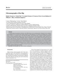
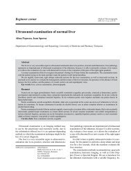
ody that joins the neck) <strong>and</strong> neck. It has a pear or teardropshape, laterally situated to the second part <strong>of</strong> theduodenum <strong>and</strong> anteriorly to the right kidney <strong>and</strong> transversecolon.Ideally a correct <strong>examination</strong> <strong>of</strong> the <strong>gall</strong><strong>bladder</strong> <strong>and</strong>the <strong>biliary</strong> tree should be performed on fasting patients(they should not eat or drink anything at least 8 hoursbefore ultrasound <strong>examination</strong>), because fasting distendsthe <strong>gall</strong><strong>bladder</strong> <strong>and</strong> reduces the bowel gas for an optimalvisualization. In emergency situations, however, the<strong>examination</strong> can be also performed if the <strong>gall</strong><strong>bladder</strong> ispartially contracted.It is recommended to take a short history <strong>of</strong> the patient<strong>and</strong> to palpate the abdomen before the <strong>examination</strong>,in order to complete the ultrasound information withclinical data.The “real-time” <strong>examination</strong> should be performed inall st<strong>and</strong>ard <strong>and</strong> any other necessary planes. Routinely,a convex multifrequency (2-5 MHz) transducer shouldbe used for the evaluation <strong>of</strong> the <strong>gall</strong><strong>bladder</strong>. The <strong>examination</strong>can be started in a supine position <strong>and</strong> continuedwith the patient in a left lateral decubitus (a mobile content<strong>of</strong> the <strong>gall</strong><strong>bladder</strong> will then move with the patientposition change). Sometimes, in order to demonstrate themobility <strong>of</strong> <strong>gall</strong><strong>bladder</strong> stones, prone or st<strong>and</strong>ing positionscan be used. The <strong>examination</strong> can start with a rightsubcostal oblique section, following the ribs, angling theprobe superiorly in order to avoid the bowel while thepatient is asked to take a suspended full inspiration. Longitudinalsections can be used in the same area as wellas intercostal sections (depending on the position <strong>of</strong> the<strong>gall</strong><strong>bladder</strong>).Useful l<strong>and</strong>marks for the evaluation <strong>of</strong> the <strong>gall</strong><strong>bladder</strong>are the edge <strong>of</strong> the right hepatic lobe <strong>and</strong> the liverhilum. In the right subcostal oblique section, the l<strong>and</strong>markstructure to be used is the interlobar fissure <strong>and</strong> the<strong>gall</strong><strong>bladder</strong> will be found by aligning the probe with thefissure <strong>and</strong> then tilting it. The <strong>gall</strong><strong>bladder</strong> is located inferiorlyor laterally to the fissure (between liver segmentsIV <strong>and</strong> V). It should be evaluated regarding the size, wallthickness <strong>and</strong> content. The <strong>normal</strong> <strong>gall</strong><strong>bladder</strong> will havean anechoic content, with thin (1-3 mm) echoic walls (fig1-3). If the patient is not fasting the <strong>gall</strong><strong>bladder</strong> will bepartially contracted <strong>and</strong> the walls will appear thicker (fig4, fig 5).The demonstration <strong>of</strong> the cystic duct is easiest in deepinspiration with the patient in supine or left lateral decubitus.It is visualized beginning from the infundibulum <strong>of</strong>the <strong>gall</strong><strong>bladder</strong>.The next step in the evaluation <strong>of</strong> the <strong>biliary</strong> <strong>system</strong>is the visualization <strong>of</strong> the main <strong>biliary</strong> duct (MBD). Itcan be demonstrated with the patient in supine or lat-Medical Ultrasonography 2010; 12(2): 150-152Fig 1, 2, 3. Normal <strong>gall</strong> <strong>bladder</strong> with thin walls <strong>and</strong> anechoiccontent151
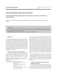
152 Alina Popescu, Ioan Sporea <strong>Ultrasound</strong> <strong>examination</strong> <strong>of</strong> <strong>normal</strong> <strong>gall</strong> <strong>bladder</strong> <strong>and</strong> <strong>biliary</strong> <strong>system</strong>eral decubitus by using a perpendicular on the ribs sectionin the right hipocondrum. The main <strong>biliary</strong> ductappears as a tube situated in front <strong>of</strong> the portal vein (fig6). In the same section the hepatic artery will appearas a round structure between the MBD <strong>and</strong> the portalvein. Sometimes, if there is a good acoustic window,the MBD can be also followed into the retro pancreaticportion. It must be evaluated regarding the size(<strong>normal</strong> up to 6 mm), wall thickness <strong>and</strong> content. Aftercolecystectomy the <strong>normal</strong> size <strong>of</strong> the MBD mayincrease.Normally, the intrahaepatic <strong>biliary</strong> ducts are notvisible (they become visible when they are dilated).Sometimes they can be also visualized in the left liverlobe in <strong>normal</strong> subjects, accompanying the portalbranches.The evaluation <strong>of</strong> the <strong>biliary</strong> <strong>system</strong> as presentedhere allows the ultrasonographists to answer thequestion if there is or not a suspicion <strong>of</strong> <strong>biliary</strong> disease.Selective referencesFig 4, 5. Partially contracted <strong>gall</strong><strong>bladder</strong>1. Anderhub B. Manual <strong>of</strong> Abdominal Sonography. Baltimore,University Park Press, 1983.2. Freitas ML, Bell RL, Duffy AJ. Choledocholithiasis: evolvingst<strong>and</strong>ards for diagnosis <strong>and</strong> management. World J Gastroenterol2006; 12: 3162-3167.3. Greiner L, Mueller J. Biliary tree <strong>and</strong> <strong>gall</strong><strong>bladder</strong>. InSchmidt G. Differential Diagnosis in <strong>Ultrasound</strong> Imaging,Thieme Medical Publishers, 2006.4. Hanbidge AE, Buckler PM, O’Malley ME, Wilson SR.From the RSNA refresher courses: imaging evaluation foracute pain in the right upper quadrant. Radiographics 2004;24: 1117-1135.5. Høgholm Pedersen M, Bachmann Nielsen M, Skjoldbye B.Basics <strong>of</strong> clinical ultrasound, Ed. Ultrapocketbooks, Copenhagen,2006: 41-53.6. Nuernberg D, Ignee A, Dietrich CF. <strong>Ultrasound</strong> in gastroenterology.Biliopancreatic <strong>system</strong>. Med Klin 2007; 102:112-126.7. Sporea I, Cijevschi Prelipcean C. Ecografia abdominală înpractica clinică, Ediţia a II-a, Editura Mirton, TimişoaraEditura Mirton, 2004: 9-99.Fig 6. The <strong>normal</strong> hilum with the main <strong>biliary</strong> duct <strong>and</strong> the portalvein