Download in pdf format - Computer Vision Lab - ETH Zürich
Download in pdf format - Computer Vision Lab - ETH Zürich
Download in pdf format - Computer Vision Lab - ETH Zürich
You also want an ePaper? Increase the reach of your titles
YUMPU automatically turns print PDFs into web optimized ePapers that Google loves.
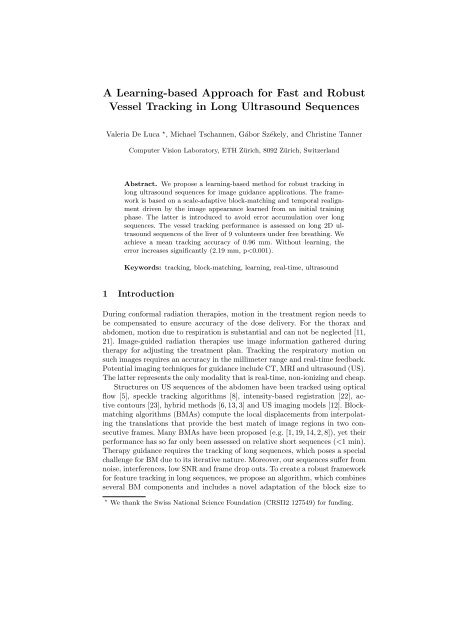
A Learn<strong>in</strong>g-based Approach for Fast and Robust<br />
Vessel Track<strong>in</strong>g <strong>in</strong> Long Ultrasound Sequences<br />
Valeria De Luca ⋆ , Michael Tschannen, Gábor Székely, and Christ<strong>in</strong>e Tanner<br />
<strong>Computer</strong> <strong>Vision</strong> <strong>Lab</strong>oratory, <strong>ETH</strong> <strong>Zürich</strong>, 8092 <strong>Zürich</strong>, Switzerland<br />
Abstract. We propose a learn<strong>in</strong>g-based method for robust track<strong>in</strong>g <strong>in</strong><br />
long ultrasound sequences for image guidance applications. The framework<br />
is based on a scale-adaptive block-match<strong>in</strong>g and temporal realignment<br />
driven by the image appearance learned from an <strong>in</strong>itial tra<strong>in</strong><strong>in</strong>g<br />
phase. The latter is <strong>in</strong>troduced to avoid error accumulation over long<br />
sequences. The vessel track<strong>in</strong>g performance is assessed on long 2D ultrasound<br />
sequences of the liver of 9 volunteers under free breath<strong>in</strong>g. We<br />
achieve a mean track<strong>in</strong>g accuracy of 0.96 mm. Without learn<strong>in</strong>g, the<br />
error <strong>in</strong>creases significantly (2.19 mm, p
2 V. De Luca et al.<br />
the feature scale. In addition, we exploit the approximate periodicity of breath<strong>in</strong>g<br />
motion for learn<strong>in</strong>g image appearance and correspond<strong>in</strong>g motion behavior<br />
(extracted by accurate but slow image registration) to allow frequent temporal<br />
realignment of the BMA for drift-free real-time track<strong>in</strong>g.<br />
2 Material<br />
US liver sequences of 9 volunteers dur<strong>in</strong>g free breath<strong>in</strong>g were acquired at the<br />
Geneva University Hospital [17]. To evaluate US track<strong>in</strong>g performance for hybrid<br />
US and MR guided treatments [17], an Acuson cl<strong>in</strong>ical scanner (Antares; Siemens<br />
Medical Solutions, Mounta<strong>in</strong> View, CA) was modified to be MR compatible,<br />
and US and MR images were simultaneously acquired. The US images (realtime<br />
second harmonic images with 1.8-2.2 MHz center frequency) were exported<br />
on-the-fly us<strong>in</strong>g a frame grabber device. 2D US images were acquired at a fixed<br />
location (longitud<strong>in</strong>al or <strong>in</strong>tercostal plane) over 5:21, 5:28 and 10:08 m<strong>in</strong> for 1,<br />
7 and 1 volunteer(s), respectively. The result<strong>in</strong>g 2650 to 14516 frames had a<br />
temporal and spatial resolution of 14-25 Hz and 0.3-0.7 mm, respectively.<br />
3 Method<br />
3.1 Scale-adaptive block-match<strong>in</strong>g<br />
The key components of our proposed scale-adaptive BMA (SA-BMA) are a novel<br />
adaptation of the block size to the feature scale and the new comb<strong>in</strong>ation of the<br />
<strong>in</strong>terpolation function from [14] with the temporal realignment from [19].<br />
Block configuration. Traditionally the size of the blocks is chosen empirically<br />
[16, 7] or equal to the US speckle size [10]. We adapt the block size to<br />
the feature size to ensure that every block conta<strong>in</strong>s a part of the feature, which<br />
limits the aperture problem and avoids ambiguities due to homogeneous blocks.<br />
The position of features to track, e.g. P j (t 0 ) for vessel j, are manually selected<br />
<strong>in</strong> I(t 0 ), see Fig. 1. BM is performed for a region of <strong>in</strong>terest (ROI j (t 0 )) around<br />
feature j, which covers a MxN grid of equally sized squares (called blocks)<br />
B i,j of size ∆b j with center po<strong>in</strong>ts G i,j , i ∈ [1, . . . , MN], def<strong>in</strong>ed at t 0 . ∆b j is<br />
determ<strong>in</strong>ed from the feature size. In details, as vessel cross sections are ellipticalshaped,<br />
we search for blob-like features centered at P j . A scale-space approach<br />
(local maxima of a Difference-of-Gaussian (DoG)) [15, 20] is used to detect the<br />
most likely blob <strong>in</strong> ROI j (t 0 ). The result<strong>in</strong>g scale s is related to the m<strong>in</strong>or semiaxis<br />
r j of an ellipse fitted to the vessel cut by r j = √ 2s and ∆b j = ⌈r j ⌉.<br />
Displacement calculation. We compute the motion field <strong>in</strong> each ROI j by<br />
determ<strong>in</strong><strong>in</strong>g the displacement at G i,j via BM, and use weighted <strong>in</strong>terpolation [14]<br />
to obta<strong>in</strong> the displacement of P j . At time step t ∗ the displacement of G i,j (t ref )<br />
<strong>in</strong> the reference frame t ref to G i,j (t ∗ ), denoted as d Gi,j (t ∗ ), is determ<strong>in</strong>ed by<br />
the displacement v which maximized the normalized cross-correlation (NCC)
Robust ultrasound track<strong>in</strong>g 3<br />
between B i,j (t ref ) and the block from I(t ∗ ) centered at G i,j (t ref )+v. The values<br />
of v are restricted to cover only a certa<strong>in</strong> search region. The reference frame is<br />
generally the previous frame (t ∗ −1). Other strategies for t ref are described <strong>in</strong> the<br />
next paragraph. The displacement of the tracked po<strong>in</strong>t from t ref to t ∗ (d j (t ∗ ))<br />
is deduced from the block displacements d Gi,j (t ∗ ) by weighted <strong>in</strong>terpolation:<br />
d j (t ∗ ) = ∑ î<br />
wîd Gî,j (t ∗ ), (1)<br />
where wî are the weights and î = {i|Q(i, t ∗ ) = 1}. Q(i, t ∗ ) is the filter<strong>in</strong>g mask<br />
for ROI j at time t ∗ , which is def<strong>in</strong>ed by Q(i, t ∗ ) = 1 for the 9 G i,j (t ref ) closest<br />
to P j (t ref ), and Q(i, t ∗ ) = 0 otherwise. We consider the weights wî [14]:<br />
1 1<br />
wî = 0.5<br />
D 2 + 1 ∑<br />
1<br />
+ 0.5∑<br />
î î D 2+1 î α , (2)<br />
î<br />
î<br />
with Dî the Euclidean distance from Gî,j (t ref ) to P j (t ref ), and αî = σ 2/µ î î<br />
the ratio between the variance (σ 2) and the mean (µ î î<br />
) of the pixel <strong>in</strong>tensities<br />
<strong>in</strong> Bî,j (t ∗ ). This <strong>in</strong>terpolation scheme has the advantage that it <strong>in</strong>corporates<br />
regularization (first term) and takes account of the relative image content (second<br />
term) [14]. The position of the tracked po<strong>in</strong>t is P j (t ∗ ) = P j (t ref ) + d j (t ∗ ).<br />
Reference frame def<strong>in</strong>ition. BMAs can generally only cope with small<br />
de<strong>format</strong>ions and appearance changes, as they are based on the translations<br />
of local regions. Hence BM is applied to temporally consecutive frames (i.e.<br />
t ref = t ∗ −1) for track<strong>in</strong>g. However, this strategy is subject to error accumulation<br />
lead<strong>in</strong>g to drift. Such errors are particularly relevant <strong>in</strong> long sequences. Yet the<br />
approximate periodic nature of respiratory motion provides frequently frames<br />
which are similar to the <strong>in</strong>itial frame and BM is aga<strong>in</strong> applicable for align<strong>in</strong>g<br />
these [19]. Errors occur also due to the quantization of d Gî,j . Hence we <strong>in</strong>troduce<br />
the follow<strong>in</strong>g strategy:<br />
if NCC(ROI j (t 0 ), ROI j (t ∗ )) > θ NCC,j then t ref = t 0<br />
else if ‖d j (t ∗ )‖ ≤ ɛ d then t ref = t ref<br />
prev<br />
else t ref = t ∗ − 1 end<br />
where NCC(A, B) is the NCC between image region A and B, θ NCC,j is the 84th<br />
percentile of the NCC values gathered from an <strong>in</strong>itial subset of the sequence,<br />
ɛ d = 0.01 pixel, and t ref<br />
prev denotes t ref from the previous image pair.<br />
3.2 Learn<strong>in</strong>g-based track<strong>in</strong>g<br />
Dur<strong>in</strong>g therapy, images are acquired cont<strong>in</strong>uously over several m<strong>in</strong>utes. Hence<br />
temporal realignment of the images is crucial to ensure robust track<strong>in</strong>g and to<br />
avoid error accumulation. For repetitive motion, such as breath<strong>in</strong>g, redundancy<br />
with<strong>in</strong> the images can be exploited [4]. Follow<strong>in</strong>g a similar strategy, we divide<br />
the method <strong>in</strong>to a tra<strong>in</strong><strong>in</strong>g and track<strong>in</strong>g phase. Dur<strong>in</strong>g tra<strong>in</strong><strong>in</strong>g we learn the<br />
relationship between image appearance and the correspond<strong>in</strong>g displacements,<br />
α î
4 V. De Luca et al.<br />
from a slower, but more robust track<strong>in</strong>g method. Dur<strong>in</strong>g the cl<strong>in</strong>ical application,<br />
the displacements are computed by the proposed SA-BMA (see Sec. 3.1), with<br />
the reference frame given by the closest frame from the tra<strong>in</strong><strong>in</strong>g set. This strategy<br />
allows temporal realignment over many more breath<strong>in</strong>g states than previously.<br />
Tra<strong>in</strong><strong>in</strong>g phase. In the tra<strong>in</strong><strong>in</strong>g phase we acquire a sequence cover<strong>in</strong>g 10<br />
breath<strong>in</strong>g cycle, result<strong>in</strong>g <strong>in</strong> T 10C images I(t i ), t i ∈ [t 0 , . . . , T 10C ]. The images<br />
I(t i ) are registered to I(t 0 ), to obta<strong>in</strong> spatial correspondence. The registration<br />
optimizes the parameters of an aff<strong>in</strong>e trans<strong>format</strong>ion with respect to NCC over<br />
a manually selected region around P j (t 0 ) and is <strong>in</strong>itialized by the result from<br />
I(t i−1 ) to I(t 0 ).<br />
To store the image appearance efficiently, we embed the images I(t i ) ∈ R D<br />
<strong>in</strong>to a low-dimensional representation S(t i ) = [s 1 (t i ); . . . ; s L (t i )] ∈ R L , with<br />
L ≪ D, us<strong>in</strong>g Pr<strong>in</strong>cipal Component Analysis (PCA) [4]. We select L such that<br />
the cumulative energy of the first L eigenvectors just exceeds 95%. In addition,<br />
we select the PCA component s B <strong>in</strong> S that captures the ma<strong>in</strong> breath<strong>in</strong>g motion,<br />
by comput<strong>in</strong>g the FFT of each s i and choos<strong>in</strong>g the one that has a power<br />
spectral density maximum at 0.15-0.4 Hz (2.5-6 s, common breath<strong>in</strong>g). S and<br />
the correspond<strong>in</strong>g registration results (e.g. P j ) are stored ∀t i .<br />
Track<strong>in</strong>g phase. New images are cont<strong>in</strong>uously acquired dur<strong>in</strong>g treatment.<br />
Given the current image I(t ∗ ), we first project it <strong>in</strong>to the PCA space (S(t ∗ ) =<br />
[s 1 (t ∗ ); . . . ; s L (t ∗ )]). Then, depend<strong>in</strong>g on its similarity to the tra<strong>in</strong><strong>in</strong>g data and<br />
the previous frame, a reference frame is chosen. The logic is as follows:<br />
outlierFlag = false<br />
if ||S(t ∗ ) − S(t 0 )|| 2 < θ 1 then t ref = t 0<br />
else if argm<strong>in</strong> tx∈[t 0,...,T 10C ]||S(t ∗ ) − S(t x )|| 2 < θ 2 then t ref = t x<br />
else if ||S(t ∗ ) − S(t ∗ − 1)|| 2 < θ 2 then t ref = t ∗ − 1<br />
else outlierFlag = true end<br />
if (outlierFlag == false) then do SA-BMA<br />
else do aff<strong>in</strong>e registration and update S end<br />
The threshold θ 1 is the 5th percentile of the Euclidean distance between S(t 0 )<br />
and S(t i ) ∀t 0 < t i ≤ T 10C . θ 2 is the 95th percentile of the distribution of the<br />
m<strong>in</strong>imum Euclidean distances between the S(t i ) <strong>in</strong> the tra<strong>in</strong><strong>in</strong>g set [4].<br />
3.3 Evaluation<br />
We compared the performance of SA-BMA (Sec. 3.1) and LB-MBA (Sec. 3.2).<br />
As basel<strong>in</strong>e BMA, we modified the SA-BMA to have fixed block size ∆b j = 16.<br />
The methods were tested for a total of 25 vessels <strong>in</strong> 9 sequences, see Fig. 1. We<br />
visually <strong>in</strong>spected the track<strong>in</strong>g quality for all vessels. We quantitatively evaluated<br />
the track<strong>in</strong>g error for the 15 vessels, which appeared to allow reliable annotations.<br />
We randomly selected 10% of the track<strong>in</strong>g phase images and manually annotated<br />
the position (denoted as ¯P j ) correspond<strong>in</strong>g to P j (t 0 ). For the annotated frame<br />
(ˆt), we calculated the track<strong>in</strong>g error TE j (ˆt) = ∥ ∥ Pj (ˆt) − ¯P (ˆt) ∥ ∥ . We summarize<br />
the results by the mean (MTE), standard deviation (SD) and 95th percentile of
Robust ultrasound track<strong>in</strong>g 5<br />
Fig. 1. I(t 0) of the 9 sequences and manual annotation of the tracked vessel centers<br />
P j(t 0), j ∈ [1, . . . , 25]. Quantitative evaluation was based on the 15 P j marked by ’x’.<br />
Visible artifacts <strong>in</strong>clude MR-RF <strong>in</strong>terferences (4), and small acoustic w<strong>in</strong>dows (2,3,6,8).<br />
all TE(ˆt), consider<strong>in</strong>g all landmarks as a s<strong>in</strong>gle distribution. We computed the<br />
MTE for each landmark j (MTE j ) and report the range for the 15 vessels. We<br />
<strong>in</strong>cluded the motion magnitude of the vessels, i.e. ∥ ∥P j (t 0 ) − ¯P (ˆt) ∥ ∥.<br />
We estimated the <strong>in</strong>ter-observer variability of the annotations. Two additional<br />
experts annotated 3% of randomly selected images from the track<strong>in</strong>g<br />
phase. We then def<strong>in</strong>ed as ground truth the mean position over the 3 annotations<br />
and calculated the track<strong>in</strong>g error as before.<br />
4 Results<br />
We tracked a total of ∼50000 frames, acquired over a total of ∼50 m<strong>in</strong>. ∆b j<br />
ranges <strong>in</strong> [4, 22] pixels and the size of the tracked vessels varies from 2 to 9 mm.<br />
The PCA space is characterized by L <strong>in</strong> the range of [86, 287], vs. D > 3660.<br />
We firstly evaluated the registration error for the tra<strong>in</strong><strong>in</strong>g images. The aff<strong>in</strong>e<br />
registration achieves an accuracy of 0.63 ± 0.36 mm (1.30 mm) on average (MTE<br />
± SD (95th percentile of TE)), with a MRE j range of [0.42, 0.84] mm.<br />
Table 1 lists the results for the proposed approaches, SA-BMA and LB-BMA.<br />
We compared LB-BMA consider<strong>in</strong>g S (LB-BMA 95 ) and s B (LB-BMA B ). The<br />
best performance is achieved by LB-BMA 95 with a MTE of 0.96 mm. Fig. 2 illustrates<br />
the benefit of the proposed methods for the worst case of BMA. For LB-<br />
BMA 95 (LB-BMA B ) and all 25 tracked vessels, t ref is picked from the tra<strong>in</strong><strong>in</strong>g<br />
set for 68.3% (99.0%) of the frames, while 1.4% (1.0%) require aff<strong>in</strong>e registration.<br />
The <strong>in</strong>ter-observer MTE varies from 0.30 to 0.34 mm (95th TE from 0.63<br />
to 0.68 mm, MTE j range [0.16, 0.70]). For the <strong>in</strong>ter-observer data set, the median<br />
TE j of BMA and SA-BMA, SA-BMA and LB-BMA B , and SA-BMA and<br />
LB-BMA 95 were statistically significantly different at the 0.001 level (Wilcoxon<br />
sign-rank test), while LB-BMA B and LB-BMA 95 were not (p=0.53). No other<br />
statistical tests were performed.
6 V. De Luca et al.<br />
Table 1. Track<strong>in</strong>g results (<strong>in</strong> mm) for the different methods w.r.t. manual annotation<br />
from one and three observers. Best results are <strong>in</strong> bold face.<br />
1 Obs - 10%, ∼7500 images 3 Obs - 3%, ∼2500 images<br />
MTE ±SD (95 th TE) rangeMTE j<br />
MTE ±SD (95 th TE) rangeMTE j<br />
VesselMotion 5.17 ± 3.21 (10.59) [2.81, 11.48] 5.22 ± 3.23 (10.57) [3.00, 11.30]<br />
BMA 3.22 ± 2.26 (7.24) [1.25, 12.35] 3.20 ± 2.26 (7.17) [1.26, 12.16]<br />
SA-BMA 2.19 ± 1.46 (4.90) [1.20, 5.79] 2.18 ± 1.45 (4.83) [1.22, 5.78]<br />
LB-BMA B 1.24 ± 1.41 (3.81) [1.04, 1.49] 1.21 ± 1.39 (3.67) [0.98, 1.49]<br />
LB-BMA 95 0.96 ± 0.64 (2.26) [0.38, 2.34] 0.97 ± 0.65 (2.20) [0.36, 2.24]<br />
The average time to compute the motion of the tracked vessel per frame<br />
was 100 ms (range [30, 350] ms). The PCA projection <strong>in</strong> LB-BMA required<br />
∼13 ms per frame. These measures were obta<strong>in</strong>ed us<strong>in</strong>g non-optimized Matlab<br />
software and no GPU parallel comput<strong>in</strong>g (s<strong>in</strong>gle PC with Intel R○Core TM i7-920<br />
at 2.66 GHz processor and 8 GB RAM), and exclude outliers, i.e. images that required<br />
aff<strong>in</strong>e registration. The latter was computed <strong>in</strong> approximately 0.8-2.5 s per<br />
image region, us<strong>in</strong>g the Insight Segmentation and Registration Toolkit (ITK).<br />
5 Conclusion<br />
We proposed a novel and robust framework for vessel track<strong>in</strong>g <strong>in</strong> long US sequences.<br />
The method is based on learn<strong>in</strong>g the relationship between image appearance<br />
and feature displacements to allow frequent re<strong>in</strong>itialization of a scaleadaptive<br />
block-match<strong>in</strong>g algorithm. The method was evaluated on long US sequences<br />
of the liver of 9 volunteers under free breath<strong>in</strong>g and achieved a mean<br />
accuracy of 0.96 mm for track<strong>in</strong>g vessels for 5-10 m<strong>in</strong>. To our knowledge, this is<br />
the first evaluation for track<strong>in</strong>g such long US sequences. Our performance also<br />
improves the state-of-the-art <strong>in</strong> 2D US track<strong>in</strong>g of the human liver (1.6 mm [23]).<br />
The proposed method is robust to <strong>in</strong>terference, noise (see Fig. 1), and frame drop<br />
outs. Moreover, it is potentially real-time [9, 18, 4].<br />
Standard BMA might fail <strong>in</strong> long sequences, due to an <strong>in</strong>appropriate block<br />
size, changes <strong>in</strong> the image similarity values and error accumulation. The <strong>in</strong>troduction<br />
of scale-adaptive blocks and the learn<strong>in</strong>g strategy were both significant<br />
for the improvement of the results. While adaption to the feature size reduces the<br />
error caused by ambiguous matches, the use of NCC for measur<strong>in</strong>g the opportunity<br />
of temporal realignment can be mislead<strong>in</strong>g. Even with adaptation to the<br />
<strong>in</strong>dividual US sequence, temporal realignment of the track<strong>in</strong>g was often either<br />
too sparse or too frequent. In contrast, the proposed learn<strong>in</strong>g based approach<br />
enables more frequent realignments to relevant images by exploit<strong>in</strong>g the repetition<br />
<strong>in</strong> the images and learn<strong>in</strong>g the ma<strong>in</strong> variation <strong>in</strong> image appearance. This<br />
allows us to detect outliers and then adapt to these previously unseen variations<br />
by aff<strong>in</strong>e registration, which is slow but able to handle larger displacements.
Robust ultrasound track<strong>in</strong>g 7<br />
Fig. 2. Comparison of the track<strong>in</strong>g performance for a failure sequence of BMA<br />
(MTE j=12.4 mm). (Top) Ma<strong>in</strong> motion component of manual annotation and d j from 3<br />
methods for a temporal subset. (Middle) Correspond<strong>in</strong>g NCC to first image. (Bottom,<br />
left to right) First image with annotation (P (t 0)), image with track<strong>in</strong>g results at last<br />
realignment (t a) of (SA-)BMA, at t a + 30 s and t a + 60 s. Drift occurs <strong>in</strong> a significant<br />
(moderate) way for BMA (SA-BMA) for t > t a, while LB-BMA rema<strong>in</strong>s robust.<br />
Reduc<strong>in</strong>g computational costs by us<strong>in</strong>g only the breath<strong>in</strong>g signal for measur<strong>in</strong>g<br />
image similarity <strong>in</strong>creased mean errors slightly (0.96 vs. 1.24 mm). While<br />
aff<strong>in</strong>e registration performed well on the tra<strong>in</strong><strong>in</strong>g set, it was only applied to<br />
outliers (1%) dur<strong>in</strong>g real-time track<strong>in</strong>g due to its computational complexity [4].<br />
The achieved accuracy and robustness of the proposed track<strong>in</strong>g method for<br />
long and very difficult US sequences makes us confident of its success for realtime<br />
US guidance dur<strong>in</strong>g radiation therapy under free-breath<strong>in</strong>g.<br />
References<br />
1. Boukerroui, D., Noble, J.A., Brady, M.: Velocity Estimation <strong>in</strong> Ultrasound Images:<br />
A Block Match<strong>in</strong>g Approach. In: Inf Process Med Imag<strong>in</strong>g, LNCS, vol. 2732, p.<br />
586 (2003)<br />
2. Byram, B., Holley, G., Giannantonio, D., Trahey, G.: 3-D phantom and <strong>in</strong> vivo<br />
cardiac speckle track<strong>in</strong>g us<strong>in</strong>g a matrix array and raw echo data. IEEE Trans<br />
Ultrason Ferroelectr Freq Control 57(4), 839 (2010)<br />
3. Cifor, A., Risser, L., Chung, D., Anderson, E.M., Schnabel, J.A.: Hybrid featurebased<br />
Log-Demons registration for tumour track<strong>in</strong>g <strong>in</strong> 2-D liver ultrasound images.<br />
In: Proc IEEE Int Symp Biomed Imag<strong>in</strong>g. p. 724 (2012)<br />
4. De Luca, V., Tanner, C., Szekely, G.: Speed<strong>in</strong>g-up Image Registration for Repetitive<br />
Motion Scenarios. In: Proc IEEE Int Symp Biomed Imag<strong>in</strong>g. p. 1355 (2012)<br />
5. Demi, M., Bianch<strong>in</strong>i, E., Faita, F., Gemignani, V.: Contour track<strong>in</strong>g on ultrasound<br />
sequences of vascular images. Pattern Recognition and Image Anal 18, 606 (2008)
8 V. De Luca et al.<br />
6. Foroughi, P., Abolmaesumi, P., Hashtrudi-Zaad, K.: Intra-subject elastic registration<br />
of 3D ultrasound images. Med Image Anal 10(5), 713 (2006)<br />
7. Harris, E.J., Miller, N.R., Bamber, J.C., Evans, P.M., Symonds-Tayler, J.R.N.:<br />
Performance of ultrasound based measurement of 3D displacement us<strong>in</strong>g a curvil<strong>in</strong>ear<br />
probe for organ motion track<strong>in</strong>g. Phys Med Biol 52(18), 5683 (2007)<br />
8. Harris, E.J., Miller, N.R., Bamber, J.C., Symonds-Tayler, J.R.N., Evans, P.M.:<br />
Speckle track<strong>in</strong>g <strong>in</strong> a phantom and feature-based track<strong>in</strong>g <strong>in</strong> liver <strong>in</strong> the presence<br />
of respiratory motion us<strong>in</strong>g 4D ultrasound. Phys Med Biol 55(12), 3363 (2010)<br />
9. Hsu, A., Miller, N.R., Evans, P.M., Bamber, J.C., Webb, S.: Feasibility of us<strong>in</strong>g<br />
ultrasound for real-time track<strong>in</strong>g dur<strong>in</strong>g radiotherapy. Med Phys 32(6), 1500 (2005)<br />
10. Kaluzynski, K., Chen, X., Emelianov, S.Y., Skovoroda, A.R., O’Donnell, M.: Stra<strong>in</strong><br />
rate imag<strong>in</strong>g us<strong>in</strong>g two-dimensional speckle track<strong>in</strong>g. IEEE Trans Ultrason Ferroelectr<br />
Freq Control 48(4), 1111 (2001)<br />
11. Keall, P.J., Mageras, G.S., Balter, J.M., Emery, R.S., Forster, K.M., Jiang, S.B.,<br />
Kapatoes, J.M., Low, D.A., Murphy, M.J., Murray, B.R., Ramsey, C.R., Van Herk,<br />
M.B., Vedam, S.S., Wong, J.W., Yorke, E.: The management of respiratory motion<br />
<strong>in</strong> radiation oncology report of AAPM Task Group 76. Med Phys 33, 3874 (2006)<br />
12. K<strong>in</strong>g, A.P., Rhode, K.S., Ma, Y., Yao, C., Jansen, C., Razavi, R., Penney, G.P.:<br />
Register<strong>in</strong>g preprocedure volumetric images with <strong>in</strong>traprocedure 3-D ultrasound<br />
us<strong>in</strong>g an ultrasound imag<strong>in</strong>g model. IEEE Trans Med Imag<strong>in</strong>g 29(3), 924 (2010)<br />
13. Leung, C., Hashtrudi-Zaad, K., Foroughi, P., Abolmaesumi, P.: A Real-Time Intrasubject<br />
Elastic Registration Algorithm for Dynamic 2-D Ultrasound Images.<br />
Ultrasound Med Biol 35(7), 1159 (2009)<br />
14. L<strong>in</strong>, C.H., L<strong>in</strong>, M.C.J., Sun, Y.N.: Ultrasound motion estimation us<strong>in</strong>g a hierarchical<br />
feature weight<strong>in</strong>g algorithm. Comput Med Imag<strong>in</strong>g Graph 31(3), 178 (2007)<br />
15. L<strong>in</strong>deberg, T.: Feature detection with automatic scale selection. Int J Comput<br />
<strong>Vision</strong> 30, 79 (1998)<br />
16. Morsy, A.A., Von Ramm, O.T.: FLASH correlation: a new method for 3-D ultrasound<br />
tissue motion track<strong>in</strong>g and blood velocity estimation. IEEE Trans Ultrason<br />
Ferroelectr Freq Control 46(3), 728 (1999)<br />
17. Petrusca, L., Catt<strong>in</strong>, P., De Luca, V., Preiswerk, F., Celican<strong>in</strong>, Z., Auboiroux, V.,<br />
Viallon, M., Arnold, P., Sant<strong>in</strong>i, F., Terraz, S., Scheffler, K., Becker, C.D., Salomir,<br />
R.: Hybrid Ultrasound/Magnetic Resonance Simultaneous Acquisition and Image<br />
Fusion for Motion Monitor<strong>in</strong>g <strong>in</strong> the Upper Abdomen. Invest Radiol 48, 333 (2013)<br />
18. P<strong>in</strong>ton, G.F., Dahl, J.J., Trahey, G.E.: Rapid track<strong>in</strong>g of small displacements with<br />
ultrasound. IEEE Trans Ultrason Ferroelectr Freq Control 53(6), 1103 (2006)<br />
19. Revell, J., Mirmehdi, M., McNally, D.: <strong>Computer</strong> <strong>Vision</strong> Elastography: Speckle<br />
Adaptive Motion Estimation for Elastography Us<strong>in</strong>g Ultrasound Sequences. IEEE<br />
Trans Med Imag<strong>in</strong>g 24(6), 755 (2005)<br />
20. Schneider, R.J., Perr<strong>in</strong>, D.P., Vasilyev, N.V., Marx, G.R., del Nido, P.J., Howe,<br />
R.D.: Real-time image-based rigid registration of three-dimensional ultrasound.<br />
Med Image Anal 16, 402 (2012)<br />
21. Shirato, H., Shimizu, S., Kitamura, K., Onimaru, R.: Organ motion <strong>in</strong> imageguided<br />
radiotherapy: lessons from real-time tumor-track<strong>in</strong>g radiotherapy. Int J<br />
Cl<strong>in</strong> Oncol 12, 8 (2007)<br />
22. We<strong>in</strong>, W., Cheng, J.Z., Khamene, A.: Ultrasound based Respiratory Motion Compensation<br />
<strong>in</strong> the Abdomen. In: MICCAI Workshop on Image Guidance and <strong>Computer</strong><br />
Assistance for Soft-Tissue Interventions (2008)<br />
23. Zhang, X., Günther, M., Bongers, A.: Real-Time Organ Track<strong>in</strong>g <strong>in</strong> Ultrasound<br />
Imag<strong>in</strong>g Us<strong>in</strong>g Active Contours and Conditional Density Propagation. In: Medical<br />
Imag<strong>in</strong>g and Augmented Reality, LNCS, vol. 6326, p. 286 (2010)


