Canon CXDI-55C - A Walsh Imaging
Canon CXDI-55C - A Walsh Imaging
Canon CXDI-55C - A Walsh Imaging

You also want an ePaper? Increase the reach of your titles
YUMPU automatically turns print PDFs into web optimized ePapers that Google loves.
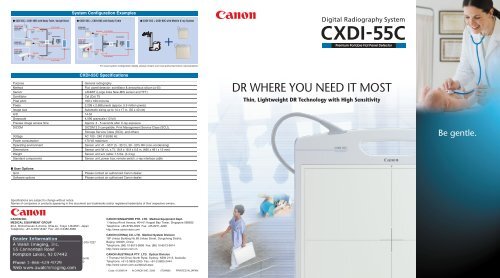
System Configuration Examples<br />
■ <strong>CXDI</strong>-<strong>55C</strong> + <strong>CXDI</strong>-40EC with Bucky Table, Upright Stand ■ <strong>CXDI</strong>-<strong>55C</strong> + <strong>CXDI</strong>-60C with Bucky Table ■ <strong>CXDI</strong>-<strong>55C</strong> + <strong>CXDI</strong>-60C with Mobile X-ray System<br />
Upright Stand<br />
<strong>CXDI</strong>-40EC<br />
Sensor Unit<br />
<strong>CXDI</strong>-<strong>55C</strong><br />
Portable Sensor Unit<br />
Power Box<br />
Sensor<br />
cable<br />
Power Box<br />
Hub<br />
Ethernet<br />
cable<br />
X-ray IF cable<br />
Control Station<br />
<strong>CXDI</strong>-60C<br />
Portable Sensor Unit<br />
Bucky Table<br />
<strong>CXDI</strong>-<strong>55C</strong><br />
Portable Sensor Unit<br />
Power Box<br />
Sensor<br />
cable<br />
X-ray IF cable<br />
Ethernet<br />
cable<br />
Ethernet<br />
cable<br />
Hub<br />
Control Station<br />
+<br />
Premium Portable Flat Panel Detector<br />
Bucky Table<br />
<strong>CXDI</strong>-40EC<br />
Sensor Unit<br />
Power Box<br />
Sensor<br />
cable<br />
Ethernet<br />
cable<br />
X-ray Generator<br />
Power Box<br />
Sensor<br />
cable<br />
X-ray IF cable<br />
X-ray Generator<br />
X-ray IF cable<br />
For exact system configuration details, please contact your local authorized <strong>Canon</strong> representative.<br />
<strong>CXDI</strong>-<strong>55C</strong> Specifications<br />
Purpose<br />
Method<br />
Sensor<br />
Scintillator<br />
Pixel pitch<br />
Pixels<br />
Image size<br />
A/D<br />
Grayscale<br />
Preview image access time<br />
DICOM<br />
Voltage<br />
Power consumption<br />
Operating environment<br />
Dimensions<br />
Weight<br />
Standard components<br />
General radiography<br />
Flat panel detector: scintillator & amorphous silicon (a-Si)<br />
LANMIT (Large Area New-MIS sensor and TFT)<br />
CsI (CsI: Tl)<br />
160 x 160 microns<br />
2,208 x 2,688 pixels (approx. 5.9 million pixels)<br />
Automatic sizing up to 14 x 17 in. (35 x 43 cm)<br />
14-bit<br />
4,096 grayscale (12-bit)<br />
Approx. 3 - 5 seconds after X-ray exposure<br />
DICOM 3.0 compatible, Print Management Service Class (SCU),<br />
Storage Service Class (SCU), and others<br />
AC 100 - 240 V 50/60 Hz<br />
170 VA maximum<br />
Sensor unit: 41 - 95˚F (5 - 35˚C), 30 - 50% RH (non-condensing)<br />
Sensor unit (W x L x T): 18.9 x 18.9 x 0.6 in. (480 x 481 x 15 mm)<br />
Sensor unit w/o cable: 7.5 lbs. (3.4 kg)<br />
Sensor unit, power box, remote switch, x-ray interface cable<br />
DR WHERE YOU NEED IT MOST<br />
Thin, Lightweight DR Technology with High Sensitivity<br />
Be gentle.<br />
■ User Options<br />
Grid<br />
Software options<br />
Please contact an authorized <strong>Canon</strong> dealer.<br />
Please contact an authorized <strong>Canon</strong> dealer.<br />
Specifications are subject to change without notice.<br />
Names of companies or products appearing in this document are trademarks and/or registered trademarks of their respective owners.<br />
CANON INC.<br />
MEDICAL EQUIPMENT GROUP<br />
30-2, Shimomaruko 3-chome, Ohta-ku, Tokyo 146-8501, Japan<br />
Telephone: +81-3-3757-8497 Fax: +81-3-5482-3960<br />
CANON U.S.A., Inc. Medical Systems Division<br />
15955 Alton Parkway, Irvine, CA 92618-3731, U.S.A.<br />
Telephone: (1) 949-753-4160, toll-free within U.S.A.: 1-800-970-7227<br />
Fax: (1) 949-753-4184<br />
http://www.usa.canon.com/DR<br />
CANON EUROPA N.V. Medical Systems Division<br />
Bovenkerkerweg 59-61, 1185 XB Amstelveen, The Netherlands<br />
Telephone: +31-(0) 20-545-8926 Fax: +31-(0) 20-545-8220<br />
http://www.canon-europa.com/Medical<br />
CANON SINGAPORE PTE. LTD. Medical Equipment Dept.<br />
1 HarbourFront Avenue, #04-01 Keppel Bay Tower, Singapore 098632<br />
Telephone: +65-6796-3549 Fax: +65-6271-4226<br />
http://www.canon-asia.com<br />
CANON (CHINA) CO, LTD. Medical System Division<br />
15F Jinbao Building No.89 Jinbao Street, Dongcheng District,<br />
Beijing 100005, China<br />
Telephone: (86) 10-8513-9999 Fax: (86) 10-8513-9914<br />
http://www.canon.com.cn<br />
CANON AUSTRALIA PTY. LTD. Optical Division<br />
1 Thomas Holt Drive, North Ryde, Sydney, NSW 2113, Australia<br />
Telephone: +61-2-9805-2000 Fax: +61-2-9805-2444<br />
http://www.canon.com.au/default.aspx<br />
Code: 0135W514 © CANON INC. 2009 0709AB0 PRINTED IN JAPAN
High-Quality Digital Radiography from a Sensitive,<br />
Thin and Lightweight Flat Panel Detector<br />
<strong>Canon</strong> has once again proven itself the leader in digital radiography. The lightweight <strong>CXDI</strong>-<strong>55C</strong> offers<br />
convenient DR imaging in a compact device with large area detection and a detachable cable for<br />
simple portability. This model also has a highly sensitive scintillator for obtaining high-quality<br />
diagnostic images with minimal X-ray exposure.<br />
High-Sensitivity DR Technology<br />
The Amorphous Silicon Flat Panel<br />
Detector of the <strong>CXDI</strong>-<strong>55C</strong> has a<br />
scintillator made of Cesium Iodide (CsI).<br />
The CsI crystals provide optimal lightchanneling<br />
properties for effective X-ray<br />
absorption and high signal-to-noise<br />
performance. The advanced LANMIT<br />
technology delivers high-quality<br />
diagnostic images with minimal X-ray<br />
exposure to patients, an ideal feature for<br />
pediatric and orthopedic purposes.<br />
Thin and Lightweight<br />
Sizeable Detection Area<br />
Impressively thin – the same thickness of standard<br />
film cassettes – this light and simple-to-use sensor<br />
can be utilized at a moment’s notice in trauma centers<br />
and ICUs. At only 7.5 lbs. (3.4 kg), the <strong>CXDI</strong>-<strong>55C</strong> is so<br />
easy to grip and handle that both<br />
the patient and X-ray technician<br />
can comfortably hold the unit in<br />
place during image capture.<br />
Only 0.6 in. (15 mm) thick<br />
With a large 14 x 17 in. (35 x 43 cm) imaging area, the generous size of <strong>CXDI</strong>-<strong>55C</strong><br />
accommodates a wide variety of radiographic applications, such as skull, spine,<br />
chest, abdomen, and extremity examinations.<br />
Superior Diagnostic <strong>Imaging</strong><br />
The <strong>CXDI</strong>-<strong>55C</strong> uses <strong>Canon</strong>’s Amorphous Silicon Flat Panel Detector. Known as<br />
LANMIT, it produces high-resolution, high-contrast diagnostic images. Multiobjective<br />
frequency processing by <strong>CXDI</strong>-<strong>55C</strong> can be optimally calibrated to view<br />
the images on LCD monitors.<br />
Cable Detachment for Mobile Convenience<br />
The <strong>CXDI</strong>-<strong>55C</strong> offers the benefits of true portability with a<br />
detachable sensor cable: time-effective transport and simple<br />
installation. Easily attach different types of <strong>CXDI</strong> sensors: <strong>55C</strong>,<br />
55G, 60C, and 60G.<br />
Results in Seconds<br />
A preview image is produced immediately after X-ray exposure, allowing for<br />
quick image confirmation, timely network distribution, and speedy diagnoses. If<br />
another image is required, the sensor is ready for the next X-ray exposure in<br />
moments thanks to its rapid refresh cycle.<br />
<strong>CXDI</strong>-<strong>55C</strong>/<strong>CXDI</strong>-55G<br />
<strong>CXDI</strong>-60C/<strong>CXDI</strong>-60G<br />
Extensive Network Capabilities<br />
DICOM 3.0 compatibility enables seamless data transfer to any DICOM devices,<br />
PACS, or RIS for efficient data management, printing, archiving, and remote image<br />
viewing. Such workflow efficiency means less wait time for patients, as well as<br />
higher patient throughput and less of a burden on staff.