Severe intracranial haemorrhage in neonatal alloimmune ... - RIHUC
Severe intracranial haemorrhage in neonatal alloimmune ... - RIHUC
Severe intracranial haemorrhage in neonatal alloimmune ... - RIHUC

You also want an ePaper? Increase the reach of your titles
YUMPU automatically turns print PDFs into web optimized ePapers that Google loves.
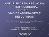
Figure 2<br />
Transfontanelar ultrasound with hyperechogenic <strong><strong>in</strong>tracranial</strong> lesions (arrows) suggest<strong>in</strong>g <strong>haemorrhage</strong>.<br />
Figure 3 Treatment and platelet count response. TP-Random donor platelet transfusion; TPΘ-Platelet transfusion from a negative HPA-1a<br />
donor; IVIG-Intravenous immunoglobul<strong>in</strong>; MPDN-methylprednisolone.<br />
This case remembers the complexity of this disease,<br />
the importance of its rapid recognition and prompt<br />
management.<br />
CASE PRESENTATION<br />
The authors present the case of a newborn term female,<br />
first birth from a second pregnancy of healthy non-consangu<strong>in</strong>eous<br />
parents, present<strong>in</strong>g generalised bruis<strong>in</strong>g, suffusions<br />
and petechias <strong>in</strong> different stages of development<br />
at birth.<br />
Family medical history was normal. Obstetric maternal<br />
antecedents <strong>in</strong>cluded a spontaneous abortion 1 year earlier,<br />
soon after an amniocentesis, performed at 16 weeks<br />
of pregnancy, due to mother’s age (38 years old). The previous<br />
fetus had a normal female karyotype (46, XX) and<br />
phenotype on necropsy, also show<strong>in</strong>g signs of <strong>in</strong>trauter<strong>in</strong>e<br />
growth restriction and acute retroplacental haematoma.<br />
The current pregnancy was uneventful. A female karyotype<br />
(46, XX) was obta<strong>in</strong>ed. Serological maternal <strong>in</strong>vestigation<br />
was irrelevant (negative hepatitis B virus, hepatitis<br />
C virus, HIV and venereal disease research laboratory;<br />
immune to toxoplasma, rubella and cytomegalovirus)<br />
and rout<strong>in</strong>e fetal ultrasounds were normal. There was no<br />
maternal history of <strong>haemorrhage</strong> nor thrombocytopenia<br />
(>230×10 9 /l); mother’s blood group was A Rh D positive.<br />
She was born from caesarean section at 40 weeks<br />
2 of 5<br />
BMJ Case Reports 2011; doi:10.1136/bcr.07.2011.4563