Severe intracranial haemorrhage in neonatal alloimmune ... - RIHUC
Severe intracranial haemorrhage in neonatal alloimmune ... - RIHUC
Severe intracranial haemorrhage in neonatal alloimmune ... - RIHUC
Create successful ePaper yourself
Turn your PDF publications into a flip-book with our unique Google optimized e-Paper software.
Rare disease<br />
<strong>Severe</strong> <strong><strong>in</strong>tracranial</strong> <strong>haemorrhage</strong> <strong>in</strong> <strong>neonatal</strong> <strong>alloimmune</strong><br />
thrombocytopenia<br />
Francisco Silva,<br />
1<br />
Sofi a Morais,<br />
2<br />
Teresa Sevivas,<br />
3<br />
Ricardo Veiga,<br />
4<br />
Ramon Salvado,<br />
3<br />
Adelaide Taborda<br />
2<br />
1<br />
Paediatric Department, Hospital Dr Nélio Mendonça, Funchal, Portugal ;<br />
2<br />
Neonatal Intensive Care Unit, Maternidade Bissaya Barreto, Coimbra, Portugal ;<br />
3<br />
Haematology Department, Hospital Professor Carmona da Mota, CHC, Coimbra, Portugal ;<br />
4<br />
Imagiology Department, Hospital Professor Carmona da Mota, CHC, Coimbra, Portugal<br />
Correspondence to Dr Francisco Silva, franciscoftsilva@hotmail.com<br />
Summary<br />
Neonatal <strong>alloimmune</strong> thrombocytopenia is a rare (1/1000–5000 births) life-threaten<strong>in</strong>g disorder, caused by fetomaternal <strong>in</strong>compatibility<br />
for a fetal human platelet alloantigen <strong>in</strong>herited from the father, with production of maternal alloantibodies aga<strong>in</strong>st fetal platelets, lead<strong>in</strong>g to<br />
severe thrombocytopenia and potential bleed<strong>in</strong>g. Intracranial <strong>haemorrhage</strong> is the most feared complication. This report presents the case of<br />
a term newborn <strong>in</strong>fant, born from caesarean section after a normal pregnancy, present<strong>in</strong>g signs of sk<strong>in</strong> bleed<strong>in</strong>g with different ages. Obstetric<br />
history <strong>in</strong>cluded a previous spontaneous abortion after amniocentesis. <strong>Severe</strong> thrombocytopenia (4×10 9 /l platelets) was found and bra<strong>in</strong><br />
ultrasound showed multiple <strong><strong>in</strong>tracranial</strong> <strong>haemorrhage</strong>s. Human platelet antigen (HPA) phenotyp<strong>in</strong>g showed maternal negative HPA-1a and<br />
paternal positive HPA-1a platelets. Strongly positive anti-HPA-1a and weakly positive anti-human leukocyte antigen class I alloantibodies were<br />
found <strong>in</strong> the mother. Multiple platelet transfusions, <strong>in</strong>travenous immunoglobul<strong>in</strong> and corticosteroid were given but favourable response was<br />
accomplished only after a compatible platelet transfusion. Bra<strong>in</strong> MRI showed multiple subacute and chronic <strong>haemorrhage</strong>s.<br />
BACKGROUND<br />
Neonatal <strong>alloimmune</strong> thrombocytopenia (NAIT) occurs<br />
when fetal platelets conta<strong>in</strong> an antigen that the mother<br />
lacks. The fetomaternal <strong>in</strong>compatibility for a platelet antigen<br />
is due to the passage of antigen-positive fetal platelets<br />
<strong>in</strong>to the circulation of an antigen-negative mother, caus<strong>in</strong>g<br />
maternal immunisation with production of immunoglobul<strong>in</strong><br />
G alloantibodies directed aga<strong>in</strong>st the ‘foreign’ antigen.<br />
These alloantibodies cross the placenta bidd<strong>in</strong>g to foetal<br />
platelets and caus<strong>in</strong>g its destruction, result<strong>in</strong>g <strong>in</strong> severe<br />
foetal and <strong>neonatal</strong> thrombocytopenia. 1 2<br />
Human platelet antigens (HPA) have different forms and<br />
its frequency varies with ethnicity. HPA-1a antigen is the<br />
commonest among caucasians, while the homozygous<br />
antigen for HPA-1b represents only 2.9%. 1 2<br />
The <strong>in</strong>cidence of NAIT is estimated to be 1/1000–5000<br />
births. It often develops <strong>in</strong> the first pregnancy of an<br />
unknown at-risk couple. Most NAIT newborn <strong>in</strong>fants appear<br />
healthy and have an unexpected severe thrombocytopenia<br />
(
Figure 2<br />
Transfontanelar ultrasound with hyperechogenic <strong><strong>in</strong>tracranial</strong> lesions (arrows) suggest<strong>in</strong>g <strong>haemorrhage</strong>.<br />
Figure 3 Treatment and platelet count response. TP-Random donor platelet transfusion; TPΘ-Platelet transfusion from a negative HPA-1a<br />
donor; IVIG-Intravenous immunoglobul<strong>in</strong>; MPDN-methylprednisolone.<br />
This case remembers the complexity of this disease,<br />
the importance of its rapid recognition and prompt<br />
management.<br />
CASE PRESENTATION<br />
The authors present the case of a newborn term female,<br />
first birth from a second pregnancy of healthy non-consangu<strong>in</strong>eous<br />
parents, present<strong>in</strong>g generalised bruis<strong>in</strong>g, suffusions<br />
and petechias <strong>in</strong> different stages of development<br />
at birth.<br />
Family medical history was normal. Obstetric maternal<br />
antecedents <strong>in</strong>cluded a spontaneous abortion 1 year earlier,<br />
soon after an amniocentesis, performed at 16 weeks<br />
of pregnancy, due to mother’s age (38 years old). The previous<br />
fetus had a normal female karyotype (46, XX) and<br />
phenotype on necropsy, also show<strong>in</strong>g signs of <strong>in</strong>trauter<strong>in</strong>e<br />
growth restriction and acute retroplacental haematoma.<br />
The current pregnancy was uneventful. A female karyotype<br />
(46, XX) was obta<strong>in</strong>ed. Serological maternal <strong>in</strong>vestigation<br />
was irrelevant (negative hepatitis B virus, hepatitis<br />
C virus, HIV and venereal disease research laboratory;<br />
immune to toxoplasma, rubella and cytomegalovirus)<br />
and rout<strong>in</strong>e fetal ultrasounds were normal. There was no<br />
maternal history of <strong>haemorrhage</strong> nor thrombocytopenia<br />
(>230×10 9 /l); mother’s blood group was A Rh D positive.<br />
She was born from caesarean section at 40 weeks<br />
2 of 5<br />
BMJ Case Reports 2011; doi:10.1136/bcr.07.2011.4563
Figure 4 Bra<strong>in</strong> MRI. Axial T1WI (A) and T2WI (B): right parietal <strong>haemorrhage</strong> with blood <strong>in</strong> early and late subacute stage. Axial T2<br />
gradient echo (C) and sagittal T1WI (D): there are signs of chronic bleed<strong>in</strong>g, with haemosider<strong>in</strong> deposits (C) and marked atrophy (D) of the<br />
cerebellar hemispheres and vermis.<br />
gestation, due to fetopelvic <strong>in</strong>compatibility and abnormal<br />
cardiotocographic record, need<strong>in</strong>g reanimation with bagmask<br />
ventilation with subsequent recovery (Apgar scores:<br />
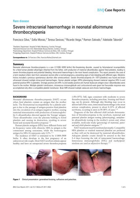
4/8/9). Generalised bruis<strong>in</strong>g, suffusions and petechias<br />
<strong>in</strong> different stages of development were noted ( figure 1 ).<br />
Hepatosplenomegaly was absent. Weight was adequate<br />
for gestational age (3330 g).<br />
INVESTIGATIONS<br />
Cord blood was obta<strong>in</strong>ed show<strong>in</strong>g a severe isolated<br />
thrombocytopenia of 16×10 9 /l platelets. Peripheral blood<br />
confirmed the thrombocytopenia (lowest value 4×10 9 /l)<br />
and excluded a possible <strong>in</strong>fectious cause (negative<br />
C-reactive prote<strong>in</strong> and blood culture). Because NAIT was<br />
suspected, newborn cranial ultrasound and parental blood<br />
test<strong>in</strong>g were performed. Cranial ultrasound ( figure 2 ) <strong>in</strong><br />
the first 2 h of life showed a right parieto-occipital hyperechoic<br />
and homolateral peri-sylvian lesions. Platelet HPA<br />
phenotyp<strong>in</strong>g <strong>in</strong> maternal and paternal blood showed<br />
negative HPA-1a and positive HPA-1a platelets, respectively;<br />
the mother had strongly positive anti-HPA-1a<br />
and weakly positive anti-HLA class I alloantibodies; the<br />
mother’s serum and father’s platelet crossmatch proved<br />
to be <strong>in</strong>compatible.<br />
BMJ Case Reports 2011; doi:10.1136/bcr.07.2011.4563 3 of 5
TREATMENT<br />
Medical treatment was started <strong>in</strong> the first hours of life<br />
( figure 3 ). Due to the unavailability of compatible platelets<br />
(negative HPA-1a), two platelet transfusions (20 ml/<br />
kg/dose) from random donors were adm<strong>in</strong>istered <strong>in</strong> day<br />
1 with count normalisation. IVIG (1 g/kg/day for 2 days)<br />
and <strong>in</strong>travenous methylprednisolone (3 mg/kg/day for 5<br />
days) were adm<strong>in</strong>istered as adjunctive therapies. Despite<br />
this management and over the next 3 days, platelet count<br />
decreased (28×10 9 /l platelets) lead<strong>in</strong>g to a third compatible<br />
platelet transfusion (negative HPA-1a donor) and IVIG<br />
adm<strong>in</strong>istration.<br />
OUTCOME AND FOLLOW-UP<br />
Platelet count reached a normal plateau (>198×10 9 /l platelets),<br />
signs of sk<strong>in</strong> bleed<strong>in</strong>g improved and cranial ultrasound<br />
was resemblant, at 5 days of life. Bra<strong>in</strong> MRI on day<br />
8 ( figure 4 ) showed multiple subacute cerebral <strong>haemorrhage</strong>s<br />
(1–4 weeks of development) <strong>in</strong> the right parietal<br />
lobe and <strong>in</strong> the posterior fossa, signs of chronic bleed<strong>in</strong>g<br />
(haemosider<strong>in</strong> deposits) <strong>in</strong> the occipital lobes and cerebellar<br />
hemispheres and vermis, these last also present<strong>in</strong>g<br />
marked atrophy.<br />
At day 11 of life, she was discharged home without<br />
cl<strong>in</strong>ical manifestations of neither bleed<strong>in</strong>g nor focal deficits<br />
on neurological exam<strong>in</strong>ation. Platelet count was normal<br />
(222×10 9 /l platelets).<br />
At 3 weeks life <strong>in</strong> the paediatric haematology consultation,<br />
molecular study was performed show<strong>in</strong>g homozygous<br />
platelet alloantigen genotypes 1b/1b <strong>in</strong> the mother and<br />
1a/1a <strong>in</strong> the father; the newborn was proved to be heterozygous<br />
HPA-1a/1b. Genetic counsell<strong>in</strong>g was offered.<br />
DISCUSSION<br />
Any newborn <strong>in</strong>fant with platelet count below 100×10 9 /l<br />
should be considered abnormal and promptly studied for<br />
an underly<strong>in</strong>g cause. 3 NAIT is a rare severe condition. It<br />
must be suspected if cl<strong>in</strong>ical manifestations such as sk<strong>in</strong><br />
or gastro<strong>in</strong>test<strong>in</strong>al bleed<strong>in</strong>g or <strong><strong>in</strong>tracranial</strong> <strong>haemorrhage</strong><br />
on ultrasound and severe thrombocytopenia are present,<br />
<strong>in</strong> the absence of maternal thrombocytopenia. Because<br />
maternal screen<strong>in</strong>g is rarely performed, couples usually<br />
do not know they are at risk until they have delivered an<br />
affected child. 1 3 5 The mother is asymptomatic, yet she or<br />
a sister may have a history of earlier affected pregnancies. 4<br />
In the previous case there was a history of spontaneous<br />
abortion 1 year earlier, after an amniocentesis at 16 weeks<br />
of pregnancy. This episode could have been related to foetal<br />
<strong>alloimmune</strong> thrombocytopenia. An explanation is that<br />
fetal platelet antigens are expressed s<strong>in</strong>ce week 16 of gestation<br />
enter<strong>in</strong>g the maternal circulation. Antiplatelet antibodies<br />
can endure <strong>in</strong> maternal circulation for many years<br />
and additional exposure <strong>in</strong> subsequent pregnancies can be<br />
a further stimulus. Severity of NAIT tends to worsen with<br />
subsequent pregnancies, lead<strong>in</strong>g to an <strong>in</strong>creased risk for<br />
<strong><strong>in</strong>tracranial</strong> <strong>haemorrhage</strong>. 1 4 In our case, bra<strong>in</strong> MRI showed<br />
that although undetected <strong>in</strong> fetal ultrasound, <strong><strong>in</strong>tracranial</strong><br />
<strong>haemorrhage</strong> occurred <strong>in</strong> utero.<br />
Infants with NAIT and <strong><strong>in</strong>tracranial</strong> <strong>haemorrhage</strong> have<br />
the poorest outcome. Mortality can range from 15% to<br />
30% of these cases. 4 6 Non-fatal cases usually are associated<br />
with neurologic sequelae <strong>in</strong>clud<strong>in</strong>g cerebral palsy,<br />
mental retardation, cortical bl<strong>in</strong>dness and seizures. When<br />
<strong><strong>in</strong>tracranial</strong> <strong>haemorrhage</strong> occurs <strong>in</strong> utero before week 20,<br />
abnormalities like porencephalic cysts, hydrocephaly, neuronal<br />
migrational disorders and superficial siderosis are<br />
often present. 4 Due to the severe central nervous system<br />
abnormalities affect<strong>in</strong>g the motor and coord<strong>in</strong>ation centers<br />
<strong>in</strong> our case, some compromise <strong>in</strong> the neurodevelopment is<br />
expected.<br />
The treatment of severe NAIT with platelet transfusion<br />
takes <strong>in</strong>to account the threshold of the platelet count. Term<br />
newborns with less than 30×10 9 /l platelets and preterm<br />
<strong>in</strong>fants with less than 50×10 9 /l platelets should be transfused,<br />
especially if bleed<strong>in</strong>g is present. Intravenous γ-globul<strong>in</strong> (1 g/<br />
kg/day for 1–4 days) is effective <strong>in</strong> 75% of cases but takes<br />
longer than does a platelet transfusion to achieve a safe<br />
platelet count. Intravenous methylprednisolone (1 mg/kg, q<br />
8 h, 1–3 days) lacks on efficacy studies. 1 4<br />
Because of the high risk of recurrence of NAIT, all women<br />
with a history of NAIT must be counselled. Sisters of HPA-<br />
1a–negative women should also be tested. 2 4 Attend<strong>in</strong>g the<br />
father’s homozygous genotype <strong>in</strong> the previous case, this<br />
couple will always produce HPA-1a/1b <strong>in</strong>fants, who will<br />
be affected by this condition if antenatal management is<br />
not fulfilled. Although the severity of NAIT is often difficult<br />
to predict, it is known that it tends to worsen with<br />
subsequent pregnancies. 4 Bussel et al. showed that a history<br />
of <strong><strong>in</strong>tracranial</strong> <strong>haemorrhage</strong> <strong>in</strong> a previously affected sibl<strong>in</strong>g<br />
is the only significant predictor of severity. 7 For this reason,<br />
if this couple decides to proceed with a future pregnancy<br />
they must be closely monitored and treatment should be<br />
offered. In a couple with a previous affected <strong>in</strong>fant, adequate<br />
therapy is maternal weekly IVIG from 20 weeks of<br />
gestation or earlier (12 weeks) if <strong><strong>in</strong>tracranial</strong> <strong>haemorrhage</strong><br />
is present. Increase of the IVIG dose or daily prednisone<br />
may be used when a more <strong>in</strong>tense prevention strategy is<br />
needed. Maternal anti-HPA-1a antibodies should be monitored.<br />
Antenatal management should avoid <strong>in</strong>vasive procedures<br />
such as <strong>in</strong> utero platelet transfusions because of<br />
the high risk of fetal loss (5.5% per affected pregnancy).<br />
Elective caesarean delivery should be performed, m<strong>in</strong>imis<strong>in</strong>g<br />
the haemorrhagic risks. 2 4 8<br />
Learn<strong>in</strong>g po<strong>in</strong>ts<br />
▶ Suspect of NAIT <strong>in</strong> a well newborn who presents with<br />
bruis<strong>in</strong>g, petechial rash and/or suffusions and who is<br />
found to have isolated severe thrombocytopenia.<br />
▶ Careful <strong>in</strong>quire about maternal personal and familial<br />
history and obstetric follow-up, <strong>in</strong>clud<strong>in</strong>g maternal<br />
platelet count.<br />
▶ Treatment should <strong>in</strong>clude adequate platelet transfusion<br />
(HPA-1a/ HPA-5b negative platelets) and IVIG as earlier<br />
as possible to prevent bleed<strong>in</strong>g complications. If<br />
adequate platelet transfusion is not available, random<br />
donor platelets should be used.<br />
▶ Intracranial <strong>haemorrhage</strong> is the most serious<br />
complication with catastrophic neurologic sequelae.<br />
▶ Genetic counsell<strong>in</strong>g should be offered to couples of<br />
affected children who want to have future pregnancies.<br />
Antenatal management is crucial <strong>in</strong> prevention of a<br />
poor outcome.<br />
4 of 5<br />
BMJ Case Reports 2011; doi:10.1136/bcr.07.2011.4563
Acknowledgements The authors thank Dr Manuela Benedito (haematology<br />
department), Dr Dolores Faria (<strong>neonatal</strong> <strong>in</strong>tensive care unit) and all health professionals<br />
<strong>in</strong>volved <strong>in</strong> the care of this patient.<br />
Compet<strong>in</strong>g <strong>in</strong>terests None.<br />
Patient consent Obta<strong>in</strong>ed.<br />
REFERENCES<br />
1. Fernandes CJ . Neonatal Thrombocytopenia . http://www.uptodate.com/<br />
contents/<strong>neonatal</strong>-thrombocytopenia ( accessed 15 Feb 2011 ).<br />
2. Blanchsette VS, Johnson J, Rand M . The management of <strong>alloimmune</strong><br />
<strong>neonatal</strong> thrombocytopenia. Baillieres Best Pract Res Cl<strong>in</strong> Haematol<br />
2000 ; 13 : 365 – 90 .<br />
3. Berkowitz RL, Bussel JB, McFarland JG . Alloimmune thrombocytopenia:<br />
state of the art 2006. Am J Obstet Gynecol 2006 ; 195 : 907 – 13 .<br />
4. Arnold DM, Smith JW, Kelton JG . Diagnosis and management of <strong>neonatal</strong><br />
<strong>alloimmune</strong> thrombocytopenia. Transfus Med Rev 2008 ; 22 : 255 – 67 .<br />
5. Mendes LR, Ferrão A, Malcata C, et al . trombocitopénia <strong>neonatal</strong> aloimune<br />
–apresentação clínica tardia . Acta Pediatr Port 2006 ; 7 : 27 – 9 .<br />
6. Castro Conde JR, Martínez ED, Rodríguez RC, et al . CNS siderosis and<br />
dandy-walker variant after <strong>neonatal</strong> <strong>alloimmune</strong> thrombocytopenia. Pediatr<br />
Neurol 2005 ; 32 : 346 – 9 .<br />
7. Bussel JB, Zabusky MR, Berkowitz RL, et al . Fetal <strong>alloimmune</strong><br />
thrombocytopenia. N Engl J Med 1997 ; 337 : 22 – 6 .<br />
8. Paidas MJ . Prenatal Management Of Neonatal Alloimmune<br />
Thrombocytopenia . http://www.uptodate.com/contents/prenatal-managementof-<strong>neonatal</strong>-<strong>alloimmune</strong>-thrombocytopenia<br />
(accessed 8 Feb 2011 ).<br />
This pdf has been created automatically from the fi nal edited text and images.<br />
Copyright 2011 BMJ Publish<strong>in</strong>g Group. All rights reserved. For permission to reuse any of this content visit<br />
http://group.bmj.com/group/rights-licens<strong>in</strong>g/permissions.<br />
BMJ Case Report Fellows may re-use this article for personal use and teach<strong>in</strong>g without any further permission.<br />
Please cite this article as follows (you will need to access the article onl<strong>in</strong>e to obta<strong>in</strong> the date of publication).<br />
Silva F, Morais S, Sevivas T, Veiga R, Salvado R, Taborda A. <strong>Severe</strong> <strong><strong>in</strong>tracranial</strong> <strong>haemorrhage</strong> <strong>in</strong> <strong>neonatal</strong> <strong>alloimmune</strong> thrombocytopenia.<br />
BMJ Case Reports 2011;10.1136/bcr.07.2011.4563, date of publication<br />
Become a Fellow of BMJ Case Reports today and you can:<br />
▶ Submit as many cases as you like<br />
▶ Enjoy fast sympathetic peer review and rapid publication of accepted articles<br />
▶ Access all the published articles<br />
▶ Re-use any of the published material for personal use and teach<strong>in</strong>g without further permission<br />
For <strong>in</strong>formation on Institutional Fellowships contact consortiasales@bmjgroup.com<br />
Visit casereports.bmj.com for more articles like this and to become a Fellow<br />
BMJ Case Reports 2011; doi:10.1136/bcr.07.2011.4563 5 of 5


