Scientific - myESR.org
Scientific - myESR.org
Scientific - myESR.org
- No tags were found...

You also want an ePaper? Increase the reach of your titles
YUMPU automatically turns print PDFs into web optimized ePapers that Google loves.
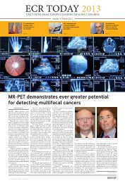
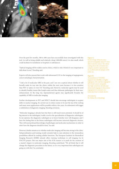
B1<br />
A<br />
B2<br />
Over the past few months, 300 to 400 cases have successfully been investigated with this<br />
tool. As well as being reliable and relatively cheap (400,000 euros) it is also small, which<br />
could facilitate its installation in hospitals or ambulances.<br />
“Optical imaging will be widely used in clinics, which is why I think it’s very important to<br />
talk about it now,” Kiessling said.<br />
Experts will also present their work with ultrasound (US) in the imaging of angiogenesis,<br />
cancer and plaque characterisation.<br />
“I did a lot of molecular MRI in the past, and I am very sceptical about whether it will<br />
broadly make its way into the clinics within the next years because it is less sensitive<br />
than PET or optics or even US,” Kiessling said. However, molecular agents may be used<br />
to identify healthy tissues like lymph nodes and thus delineate pathologies by their nonenhancement.<br />
In the long run, hyperpolarised agents may significantly broaden the<br />
capability of MRI in molecular imaging.<br />
Further developments in PET and SPECT should also encourage radiologists to acquire<br />
skills in nuclear imaging. Its current use in clinics seems to be just the tip of the iceberg<br />
and many more applications will be possible within a few years. Its refinement will trigger<br />
a redefinition of diagnostic imaging, Kiessling envisions.<br />
Figure A:<br />
Fusion image of fluorescence<br />
optical tomography and µCT of an<br />
atherosclerotic mouse. The colour<br />
coding shows the activation of a<br />
catepsin-sensitive optical probe<br />
in an atherosclerotic plaque in the<br />
aortic arc.<br />
Figure B:<br />
Accumulation of VEGFR2-<br />
targeted microbubbles in highly<br />
angiogenic (B1) and low angiogenic<br />
(B2) breast cancer xenografts.<br />
The high sensitivity of molecular<br />
ultrasound imaging to characterise<br />
angiogenesis in tumours at a<br />
molecular level is clearly shown.<br />
(Provided by<br />
Dr. Fabian M.A. Kiessling)<br />
“Molecular imaging is already here but there is still much more potential. It should be of<br />
big interest to the radiologist; it adds a tool to the specialisation of diagnostic radiologists.<br />
In my opinion, the diagnostic radiologist as we know him/her now will disappear, and I<br />
have the feeling that in the future radiologists will become universal diagnostic doctors.<br />
They will need profound knowledge of pathologies and molecular mechanisms in order to<br />
select how the diagnosis should be done,” he said.<br />
However, doubts remain as to whether molecular imaging will become strong in the clinic.<br />
Subspecialisation and training would certainly help to raise attention to the tremendous<br />
possibilities offered by reading cellular functions. The European Institute for Biomedical<br />
Imaging Research (EIBIR) already offers training workshops in cell imaging via its<br />
ENCITE project. But one major aim of the ECR session is to push for the creation of<br />
a master’s degree in molecular imaging, Kiessling underlined. “We all think that it will<br />
change the diagnostic procedure in the future, so it is very important that radiologists are<br />
going on with this,” he concluded.<br />
27