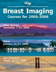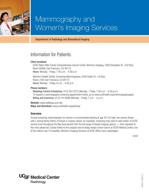
Mammography and Women's Imaging Services - UCSF Department ...
Mammography and Women's Imaging Services - UCSF Department ...
Mammography and Women's Imaging Services - UCSF Department ...

Create successful ePaper yourself
Turn your PDF publications into a flip-book with our unique Google optimized e-Paper software.
<strong>Mammography</strong> <strong>and</strong><br />
Women’s <strong>Imaging</strong> <strong>Services</strong><br />
<strong>Department</strong> of Radiology <strong>and</strong> Biomedical <strong>Imaging</strong><br />
Information for Patients<br />
Clinic locations:<br />
<strong>UCSF</strong> Helen Diller Family Comprehensive Cancer Center, Women’s <strong>Imaging</strong>, 1600 Divisadero St., 2nd floor,<br />
Room H2906, San Francisco, CA 94115<br />
Hours: Monday - Friday, 7:45 a.m. - 4:30 p.m.<br />
Women’s Health Center, Screening <strong>Mammography</strong>, 2356 Sutter St., 1st floor,<br />
Room J146, San Francisco, CA 94115<br />
Hours: Monday - Friday, 8 a.m. - 4:30 p.m.<br />
Phone numbers:<br />
Radiology Central Scheduling: (415) 353-2573 (Monday - Friday, 7:30 a.m. - 5:30 p.m.)<br />
To request a mammography screening appointment online, go to www.ucsfhealth.org/mammographyappts.<br />
Billing <strong>and</strong> insurance: (415) 514-8888 (Monday - Friday, 7 a.m. - 4 p.m.)<br />
Website: www.radiology.ucsf.edu<br />
Maps <strong>and</strong> directions: www.ucsfhealth.org/pathway<br />
Overview<br />
Annual screening mammography for women is recommended starting at age 40. For high-risk women (those<br />
with a strong family history of breast or ovarian cancer, for example), screening may need to start earlier. At <strong>UCSF</strong>,<br />
women from throughout the Bay Area benefit from the full range of breast imaging options — from st<strong>and</strong>ard to<br />
the most advanced. Closely linked to the surgical <strong>and</strong> oncology breast cancer teams at <strong>UCSF</strong> Medical Center, one<br />
of the nation’s top 10 hospitals, Women’s <strong>Imaging</strong> <strong>Services</strong> at <strong>UCSF</strong> offers many advantages.<br />
(over)<br />
2.12-RAD-11-01000
<strong>Mammography</strong> <strong>and</strong> Women’s <strong>Imaging</strong> <strong>Services</strong><br />
Breast imaging services highlights<br />
• Highly specialized team of expert physicians focused on<br />
efficient <strong>and</strong> accurate breast cancer screening,<br />
diagnosis, treatment <strong>and</strong> monitoring<br />
• Thirty-year record as a world-renowned breast imaging<br />
program continuing the model of renowned radiologist<br />
Dr. Edward A. Sickles<br />
• Speed of diagnosis is an essential, distinguishing<br />
feature of Women’s <strong>Imaging</strong> <strong>Services</strong> at <strong>UCSF</strong> with<br />
nearly all breast imaging exams reported within<br />
24 hours of the test being performed<br />
• <strong>UCSF</strong> performs 100 percent full-field digital<br />
mammography, a technology that has been proven<br />
to be of significant value in detecting additional cancers<br />
that were missed using older technologies <strong>and</strong> uses<br />
less radiation than older analog mammography<br />
• <strong>UCSF</strong> was among the first to perform breast MRI <strong>and</strong><br />
now has more than 15 years of experience with this<br />
sensitive imaging technique<br />
• Our high-field strength 3.0T breast MR provides<br />
superior image quality <strong>and</strong> a wider opening for<br />
improved patient comfort<br />
• Following the guidelines of the <strong>Mammography</strong> Quality<br />
St<strong>and</strong>ards Act, we are certified by the Food <strong>and</strong> Drug<br />
Administration <strong>and</strong> accredited by the American College<br />
of Radiology<br />
Breast imaging services offered<br />
Bone scanning with SPECT/CT<br />
• A st<strong>and</strong>ard imaging test to determine if breast cancer<br />
has spread to the bones<br />
• <strong>UCSF</strong> bone scanning has several advantages:<br />
- The exams use relatively low doses of radiation to<br />
more accurately locate where the cancer is located<br />
- The camera can acquire the image in about half the<br />
time of cameras at other imaging centers<br />
- <strong>UCSF</strong>'s advanced software enables sharper<br />
image quality<br />
• The Breast Cancer Prevention Program provides access<br />
to experts in the field of breast cancer prevention<br />
including physicians, nurse practitioners, genetic<br />
counselors <strong>and</strong> researchers<br />
• Expert interpretation of mammograms provides fewer<br />
false positives <strong>and</strong> a higher cancer detection rate<br />
• Coordinated Diagnostic <strong>and</strong> Evaluation Program,<br />
where patients with suspected breast cancer receive a<br />
radiology <strong>and</strong> pathology assessment <strong>and</strong> are seen by<br />
a top breast surgeon during a one-day coordinated<br />
visit to <strong>UCSF</strong><br />
• <strong>UCSF</strong> was the first location in the United States to<br />
offer breast MRI biopsy with the Sentinelle Vanguard<br />
dedicated biopsy table, which can prevent a return visit<br />
for the patient by allowing multi-site including bilateral<br />
biopsies<br />
• Using the world’s newest tools <strong>and</strong> methods, we can<br />
take samples as small as a group of cells to perform<br />
speedy <strong>and</strong> accurate breast biopsies when necessary<br />
• All physicians are certified by the American Board of<br />
Radiology<br />
• <strong>UCSF</strong> is a recognized leader in multi-institutional clinical<br />
breast cancer trials <strong>and</strong> breast imaging research<br />
Breast MRI<br />
• An advanced imaging technology using magnetic waves<br />
in conjunction with a contrast (dye) injection to take<br />
pictures of the breast<br />
• Can show lesions not visible at mammography or<br />
ultrasound<br />
• The American Cancer Society recommends that certain<br />
women with an especially high risk of developing breast<br />
cancer have an MRI scan along with their yearly<br />
mammogram
Breast ultrasound<br />
• Use of sound waves to evaluate breast lumps in<br />
conjunction with mammography<br />
Digital diagnostic mammography<br />
• A series of breast images to provide a more<br />
comprehensive <strong>and</strong> detailed view of the breast as<br />
prescribed <strong>and</strong> monitored by an interpreting radiologist<br />
• Appropriate for women with a prior history of breast<br />
cancer, a new breast symptom or abnormal screening<br />
mammogram<br />
Digital screening mammography<br />
• Used for annual mammograms to detect any overall<br />
change in the appearance of breast tissue<br />
• Appropriate as an annual check-up for women without<br />
breast symptoms<br />
• Screening mammography is an important tool for the<br />
early detection of breast cancer<br />
Lymphoscintigraphy with SPECT/CT<br />
• An imaging exam that uses a small amount of<br />
radioactive liquid to trace a path from the breast to the<br />
lymph nodes in the armpit<br />
• Allows the breast surgeon to perform a precise<br />
extraction of the lymph nodes for evaluation of<br />
possible disease<br />
MRI-guided core biopsy<br />
• An advanced imaging technology using MRI to guide<br />
the radiologist in obtaining a small tissue sample for<br />
evaluation of possible disease<br />
PET/CT imaging<br />
• An advanced technique that combines functional<br />
imaging <strong>and</strong> anatomic computed tomographic (CT)<br />
evaluation of the breast tissue, lymph nodes <strong>and</strong> solid<br />
organs to accurately identify sites of disease<br />
• <strong>UCSF</strong> has a very high volume of breast imaging<br />
procedures with PET/CT compared to most imaging<br />
centers<br />
• Unlike many imaging centers, <strong>UCSF</strong> routinely performs<br />
a diagnostic-quality whole body CT examination with<br />
contrast in conjunction with the PET imaging<br />
Stereotactic-guided core breast biopsy<br />
• <strong>Mammography</strong> <strong>and</strong> a computer-targeted guidance<br />
system to help the radiologist obtain a small tissue<br />
sampling for evaluation of possible disease<br />
Ultrasound-guided breast cyst aspiration<br />
• Use of ultrasound technology to guide the radiologist in<br />
removing fluid from a breast cyst<br />
Ultrasound-guided breast core biopsy <strong>and</strong> fine<br />
needle aspiration<br />
• Technology used to guide the radiologist in obtaining a<br />
small tissue sampling for evaluation of possible disease<br />
Wall motion study<br />
• A procedure (also called ERNA or MUGA) that is<br />
typically performed on patients with breast cancer to<br />
monitor whether certain high-dose chemotherapy<br />
regimens cause toxicity to the heart<br />
Wire localization procedure<br />
• <strong>Mammography</strong> ultrasound or MRI-guided pre-surgical<br />
procedure to mark the area of concern for the<br />
breast surgeon<br />
Billing <strong>and</strong> insurance<br />
We contract with most major health insurance companies. Many insurance plans (including PPOs) require prior<br />
authorization for imaging services. We will be happy to assist you with any questions regarding authorization, price<br />
quotes, procedure codes, statements, or billing. Please call (415) 514-8888.