2 0 1 1 UNIVERSITY OF CALIFORNIA SAN FRANCISCO - UCSF ...
2 0 1 1 UNIVERSITY OF CALIFORNIA SAN FRANCISCO - UCSF ...
2 0 1 1 UNIVERSITY OF CALIFORNIA SAN FRANCISCO - UCSF ...
You also want an ePaper? Increase the reach of your titles
YUMPU automatically turns print PDFs into web optimized ePapers that Google loves.
2 0 1 1<strong>UNIVERSITY</strong> <strong>OF</strong> <strong>CALIFORNIA</strong> <strong>SAN</strong> <strong>FRANCISCO</strong>imagesDEPARTMENT <strong>OF</strong> RADIOLOGY AND BIOMEDICAL IMAGING
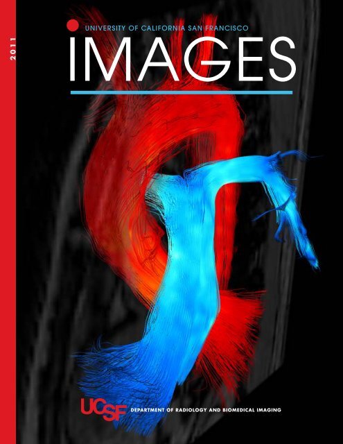
About the Cover:Systolic blood flow in the great vessels of a normal volunteer visualized by4D Flow. The 3D streamlines align with the local velocity vector field at a givenmoment in time, and provide a 3D perspective of instantaneous flow. Red = aorta,blue = pulmonary artery. The data for this image was collected in a single acquisitionof approximately 15 minutes. Cover image provided by Petter Dyverfeldt, PhD,a postdoctoral scholar, and Michael D. Hope, MD, an assistant professor inresidence in the Department of Radiology and Biomedical Imaging.Design: Irene Nelson Design Copyediting: DEF Communications Editorial Coordinator: Katie Murphy Printing: Advanced Printing, Pleasanton, Calif.
Table of Contentsletter from the chairman2 Excellence in Patient Care, Translational Research, and Educationclinical and research news4 4D Flow MR Imaging of the Thoracic Aorta8 MR-Guided Focused Ultrasound (MRg-FUS) Comes to <strong>UCSF</strong>11 High-Resolution Imaging of the Hippocampus in Temporal Lobe Epilepsy:Clinical and Functional Implicationsnew facilities and technology15 Capital Equipment and Technology: Past Year Overviewdepartment update17 Jung Becomes VAMC Chief of MRI17 New Faculty20 <strong>UCSF</strong> Launches New Master’s Degree Program in Biomedical Imaging21 Honors and Awards24 Partnership Brings Students to <strong>UCSF</strong> for Bioengineering Research24 Weiner Accepts Reagan Research Award25 Retired in 201128 In Memoriam30 Diagnostic Radiology Residency Program 201140 Clinical Fellows and Instructors 2011–201241 Diagnostic Radiology Residency Graduates–Class of 201142 The Margulis Society44 The Margulis Society Honor Roll of Donors46 Alumni News48 The Henry I. Goldberg Center for Advanced Imaging Education49 Radiology Postgraduate Education50 Radiology CME Calendar 201251 Annual Symposium and Hasegawa Award52 Surbeck Young Investigator Awards53 Department of Radiology Hosts Imaging Services Workshop54 Lanna Lee Award 201055 Faculty Roster60 The Year in Picturesradiology and biomedical imaging research63 Research Directions79 Grants and Fellowships
letter from the chairmanDear Colleagues and Friends,Another successful year has flown by in Radiology andBiomedical Imaging, and I am happy to report that we havesuccessfully navigated significant change across the universityand across the specialty of radiology, although muchmore is to come. As I sat down to write this year’s letter, Ireflected on what I wrote last year: first, that change is certain,although the form it will take is not; and second, thatfocus on excellence in patient care, translational research,and education will help us evolve and remain relevant andvaluable in the future. In this edition of Images, our 18thsince 1994, you will read about the Department’s achievementsin all of these areas, and I want to highlight a fewnotable accomplishments over the last year.This past year our Chancellor challenged us to excel intranslational research and I am happy to report thatRadiology more than contributed by achieving remarkableaccomplishments in moving research from bench tobedside. In late 2010, John Kurhanewicz, PhD, and SarahNelson, PhD, unveiled the first Phase 1 clinical trials usinghyper-polarized C13 and MRI to track the progress or inhibitionof prostate cancer. This new MRI technique allowstumor changes to be measured and visualized in real time,as the tumor responds or fails to respond to treatment.Their announcement at last year’s RSNA generated widemedia and public interest, and rightly so, as it will allowoncologists and patients to make quicker and better decisionstailored to each patient’s tumor type.Continuing with metabolic imaging, Daniel Vigneron,PhD, received a center grant to create a resource focusedon pushing the boundaries of hyper-polarized C13 for varioustypes of cancer imaging. These techniques will allowsignificant improvement in decision making and treatmentchoices for cancer patients. Sharmila Majumdar, PhD, ourVice-Chair for Research, obtained a Center for ResearchTranslation grant to use imaging in osteo-arthritis. Shecollaborates closely with Thomas Link, MD, PhD, and withcolleagues at <strong>UCSF</strong>’s new Orthopedic Institute (OI). Dr.Link’s efforts to establish excellent clinical service at theOI have not only improved patient care, but also openedup new avenues of translational research for imaging inmusculoskeletal applications.Finally, I am very proud to report that Dr. Fergus Coakleyreceived an NIH grant to purchase High Intensity FocusedUltrasound (HIFU) for use with MRI, and has establisheda program at China Basin to investigate the use of HIFUin uterine fibroid treatment. He looks forward to futurecollaborations with Dr. Link in the use of HIFU for metastaticbone cancer treatment, alleviating pain, and with Dr.Kurhanewicz for prostate cancer treatment.I announce these cutting-edge translational breakthroughswith great pride not only because of their scientific andclinical implications, but because they were achieved in thecontext of a tremendously difficult NIH funding environment.I am grateful to our entire faculty for the tremendouseffort that keeps us at the highest level of NIH rankings forradiology departments nationwide.This year, we also made our first exploratory steps intosocial media marketing. I invite you to read our blog atblog.radiology.ucsf.edu where you will find faculty commentson topics as diverse as “the thyroid shield controversycourtesy of the Dr. Oz Show” by Bonnie Joe, MD, PhD, to“radiology’s role in evaluating and treating obesity” fromAliya Qayuum, MBBS. We also used our blog, Twitter, andFacebook pages to educate our patients and the public onthe radiation scare that followed the terrible earthquake and2
tsunami in Japan earlier this year. I invite you to read ourblog, to “like” us on Facebook (facebook.com/<strong>UCSF</strong>imaging)and to follow us on Twitter (twitter.com/<strong>UCSF</strong>imaging).I can’t finish remarks about our year without complimentingour tremendous residents and thanking the MargulisSociety for their steadfast and loyal support of our residencyprogram. This year we were very fortunate to havea wildly successful Gala event, marking the Margulis Society’s20th anniversary and celebrating Dr. Alex Margulis’90th birthday! What an event—if you could not attend,you will find pictures and details in this magazine. In thecoming year, we are thrilled that Herbert Kressel, MD,the editor of Radiology, will be our guest speaker at theMargulis Society's biennial alumnus lecture, to be held onApril 3, 2012 at <strong>UCSF</strong>.I hope you enjoy this edition of Images, and please don’tforget to join us, as usual, at our RSNA reception for alumniand friends. We have a new and exciting venue this year, aChicago landmark building, the Chicago Cultural Center atthe corner of East Washington Street and Michigan Avenue.Please join us on Sunday, November 27 at 6:30 p.m.in theGAR Rotunda.Thank you for your ongoing interest and dedication to theDepartment of Radiology and Biomedical Imaging. I wishyou success and good health for 2012, and I hope to seeyou at RSNA.Sincerely,Ronald L. Arenson, MD3
clinical and research news4D Flow MR Imaging of the Thoracic AortaMichael D. Hope, MD; Petter Dyverfeldt, PhD; Monica Sigovan, PhD; Jing Liu, PhD; Karen Ordovas, MD;Jarrett Wrenn, MD, PhD; Elyse Foster, MD; Elaine Tseng, MD; Maythem Saeed, PhD; David Saloner, PhDBlood flow imaging with 4D Flow (time-resolved, 3Dphase-contrast MRI) is an innovative method for studyingcardiovascular disease that allows for striking blood flowvisualization (Figure 1). The full power of the techniquehas yet to be exploited in managing patients with cardiovasculardisease. Currently, a less robust, 2D phase-contrasttechnique is used in select clinical scenarios to quantifyblood velocity and flow in the cardiovascular system. Formany patients, it is an adjunct to echocardiography, whichis widely available and performed routinely. But echocardiographyhas weaknesses, including limited acoustic windowsand quantitative abilities, while 4D Flow has uniqueadvantages.Because its high-resolution 3D acquisition is unhinderedby acoustic windows, 4D Flow allows unrivaledvisualization of dynamic secondary blood flow features,including helices and vortices. It also allows key secondaryvascular parameters, including turbulence and vessel wallshear stress, to be quantified. We seek to capitalize on theseadvantages and to change the paradigm for risk-stratifyingpatients with cardiovascular disease. Today, cardiovascularpatients are risk-stratified most often using vessel dimensions.Altered blood flow is rarely considered, although substantialevidence demonstrates a link between abnormalflow and disease. Our projects focus on characterizing therole of abnormal flow in promoting or exacerbating aorticpathology.Vessel Wall Shear StressVessel wall shear stress (WSS) refers to the force per unitarea exerted on the vascular wall by blood moving in a tangentialplane. It can be estimated from the near-wall velocityFigure 1 Systolic blood flow in the greatvessels of a normal volunteer visualized by4D Flow. The 3D streamlines align with thelocal velocity vector field at a given momentin time, and provide a 3D perspective ofinstantaneous flow. Red = aorta, blue =pulmonary artery, maroon = pulmonaryveins. The data for this image was collectedin a single acquisition of approximately15 minutes.4
Figure 2 (A) Simulated flowprofiles with a mean velocity of0.5 m/s and different degreesof eccentricity. The centerline (•)and calculated displacements(*) are plotted on the abscissa.(B) WSS values plotted againstnormalized displacement fordifferent mean velocities: 0.5 m/s(*) corresponding to profiles in A,0.7 m/s (+), and 1 m/s (×).gradients captured by 4D Flow data sets. These gradients area function of both velocity and flow eccentricity (Figure 2).Wall shear stress is strongly linked to vascular disease.Low WSS promotes atherosclerosis through many well-documentedmechanisms. High WSS, especially positive sheargradients, contributes to pathologic vascular remodelingthat leads to aneurysm formation. Although MRI-derivedshear stress values are routinely lower than true values, acomparison of relative values allows altered shear stressprofiles to be identified and characterized. We are studyingthe link between altered shear profiles and the progressionof aortic aneurysm.Turbulence ImagingThe normal cardiovascular system maintains fluid transportat high efficiency. Disturbed and turbulent blood flow, however,is present with many cardiovascular diseases, and maycontribute to their progression. For example, turbulence isthe major cause of the pressure drop seen across a stenoticvessel or valve. Exposure of blood constituents to turbulentforces has been associated with hemolysis, platelet activation,and aggregation.Turbulence is a complex phenomenon, but one thatcommonly occurs in nature: seen in smoke coming out ofa chimney or experienced on an airplane. The presence ofapparently random velocity fluctuations is a typical featureof turbulence. The intensity of these velocity fluctuations canbe quantified by their standard deviation. Traditionally, invivo measurements of turbulence intensity have only beenpossible using invasive approaches. As a result, the extentand role of turbulence has not been fully investigated inhumans. We recently extended the 4D Flow imaging techniqueto estimate mean velocities and turbulence intensitywithin each image volume element. Turbulence intensitycan be estimated from the data acquired in a standard 4DFlow acquisition. We are using this approach to assess thehemodynamic impact of a wide range of vascular diseasestates, including aortic coarctation, carotid atherosclerosis,and aortic stenosis (Figure 3).Thoracic Aorta Flow ImagingDynamic blood flow imaging in the thoracic aorta withphase-contrast MRI has been a focus of considerableresearch interest for over 20 years. Starting in the late 1980s,normal blood flow in the thoracic aorta was studied in detailthroughout the cardiac cycle. Synthesizing observationsfrom 2D imaging planes acquired from multiple volunteers,“typical” flow patterns were reported, including aright-handed twist to flow in the ascending aorta duringlate systole, and retrograde flow streams during diastole.With the advent of 3D phase-contrast techniques, morecompelling visualization of complex flow patterns becamepossible. Software extended analysis beyond visualizationof aortic flow by allowing the estimation of secondary vascularparameters that can be correlated with aberrant flowpatterns. The goal of current research is to understand howabnormal flow may promote or worsen vascular diseaseso 4D Flow imaging can be used to evaluate and managepatients with aortic disease.Recent work has focused on gross pathologies of thethoracic aorta, such as coarctation and aneurysm. MRIflow evaluation has long been a component of the clinicalmanagement of patients with aortic coarctation. Our recentstudies suggest that 4D Flow imaging may expand this role.5
clinical and research newsFigure 3 Flow visualization at peak systole in the ascendingaorta of a 90-year-old male with severe aortic stenosis. Thesolid line indicates the location of the aortic valve. Sparsestreamlines (blue) outline the velocity field and volumerenderedturbulence intensity maps (red/yellow) show regionsand degrees of flow turbulence. Elevated turbulence intensity isseen in the proximal ascending aorta (solid arrow) where thepost-stenotic flow jet becomes incoherent. Turbulence intensityis elevated at the outer wall of the distal ascending aorta(hollow arrow).Not only can collateral flow be reliably calculated and aorticflow profiles readily assessed, abnormal 3D flow patternscan be identified that correlate with post-repair complications,including aneurysm and rupture.Aneurysms of the thoracic aorta are associated withcomplex abnormal flow patterns, mostly helical in nature.The significance of these patterns has been debated. Arethey simply the consequence of a dilated aorta, or do theyplay an active role in the progression of disease? In a subsetof cases with aortic valve disease, our recent work suggeststhat flow may play an active role. Flow similar to that seenwithin aortic aneurysms has been demonstrated in aortasthat are not (yet) dilated (Figure 4).Valve-Related Aortic DiseaseEvaluating valve-related disease of the ascending aorta with4D Flow is a promising clinical application. Many studieshave assessed flow alterations in patients who have undergoneaortic valve and/or ascending aortic replacement. Presurgically,4D Flow may prove useful by risk-stratifyingpatients and guiding the timing of intervention. Aorticvalve disease is relatively common, especially in the elderly,and is associated with the long-observed phenomenon ofpost-stenotic dilation. The mechanism is presumed to beflow-related. Without 4D Flow, however, the altered hemodynamicshave not been well characterized. The detailedassessment of 4D Flow reveals altered systolic flow resultingfrom eccentric flow jets with stenotic and deformed aorticvalves (Figure 4). The degree of flow eccentricity can bequantified, and has been shown to correlate with focallyelevated WSS and aortic dilation.We have focused our efforts on patients with bicuspidaortic valve (BAV), a defect found in 1-2% of the populationthat frequently results in significant aortic pathology,including aneurysm and dissection. We hypothesize thateccentric systolic flow jets with BAV, through the mechanismof elevated WSS, promote aortic dilation. The clinicalrelevance of proving this mechanism would be considerable,as it would allow a non-invasive means of risk-stratifying thesizable population of patients with BAV (up to four millionin the U.S. alone). For example, 4D Flow assessment couldbe performed for patients with BAV before valve or aorticdisease manifests. If normal systolic flow were identified,patients would need only occasional follow-up. If eccentricflow were identified, 1) closer follow-up with MRI wouldbe indicated, as follow-up echocardiography may be inadequateto assess the entire ascending aorta; 2) medicationwith beta-blockers, which lower WSS, could be initiated;and 3) depending on interval growth rates and the degreeof eccentricity, earlier intervention may be warranted (e.g.,at 4.5 cm for high-risk patients).With the help of our collaborators in Cardiology andCardiothoracic Surgery, we are collecting data on whetherincreased aortic growth rates are seen as a consequence ofthe elevated hemodynamic burden experienced by the aorticwall because of eccentric systolic flow. We are developinga time-resolved MRA sequence that will enable follow-upstudies to be co-registered spatially and temporally, so aorticdimensions can be evaluated at identical locations andorientations. We also are investigating animal models of6
PART 1 PART 2Figure 4 Part 1 Abnormal systolic blood flow with post-stenotic aortic dilation. Panel A exhibits normal blood flow in a healthyvolunteer. From left to right, magnetic resonance angiography (MRA), systolic streamlines in the ascending aorta, and cross-sectionalanalysis at the plane depicted in the proximal ascending aorta are provided.(The same sequence is found in each image panel.) TheMRA shows normal aortic geometry, the streamlines normal laminar systolic flow, and the cross-sectional analysis central fast flowand an even distribution of WSS around the aortic lumen; the green bars represent the relative magnitude of WSS. Panel B is froma 90-year-old man with severe aortic stenosis and aneurysmal dilation of the ascending aorta up to 5.3 cm. Systolic flow is eccentricwith streamlines that course through the ascending aorta in a right-handed helix. The cross-sectional analysis shows marginalized flowto the right-anterior quadrant where WSS is focally elevated. Panel C is from a 34-year-old woman with BAV, aortic stenosis, anddilation of the ascending aorta up to 4.6 cm. Similar eccentric flow with a right-handed helix of systolic streamlines is demonstrated.Shear stress is focally elevated where flow is marginalized against the aortic wall.Part 2 Similar eccentric systolic flow in patients without aortic dilation or aortic stenosis. Panel A is from a 64-ear-old man with TAV,aortic stenosis, and normal aortic dimensions. Eccentric flow and asymmetrically elevated shear stress is demonstrated. Withoutaortic dilation, it is more convincing that the aortic valve, and not aortic geometry, is causing the aberrant systolic flow. The helicalstreamlines appear to be in a more vertical orientation in cases of TAV compared to BAV. Panel B is from a 19-year-old woman withBAV, aortic stenosis, and normal aortic dimensions; the loss of MRA signal in the proximal descending aorta is due to a stent placedfor aortic coarctation. The abnormal flow pattern is seen without aortic dilation. Panel C is from a 19-year-old man with BAV, no aorticstenosis, and aneurysmal dilation of his aortic root up to 5.4 cm. The same abnormal systolic flow pattern is identified, suggesting thatpost-stenotic dilation can be seen in patients with BAV without aortic stenosis by conventional echocardiography criteria.valve-related aortic disease. If we can demonstrate acceleratedgrowth with abnormal flow, 4D Flow would become aclinically useful tool for risk-stratifying the sizable populationof patients with aortic valve disease for the likelihoodof developing an aneurysm.Michael D. Hope, MD, is an assistant professor in residence inthe Cardiac and Pulmonary Imaging section; Petter Dyverfeldt,PhD, and Monica Sigovan, PhD, are postdoctoral scholars;Jing Liu, PhD, is an assistant adjunct professor; KarenOrdovas, MD, is an assistant professor in residence in thesection of Cardiac and Pulmonary Imaging; Jarrett Wrenn,MD, PhD, is a PGY-4 diagnostic radiology resident ; MaythemSaeed, PhD, is an adjunct professor and David Saloner, PhDis a professor in residence in the Department of Radiology andBiomedical Imaging. Elyse Foster, MD, is a professor of Medicineand director of the Adult Echocardiography Laboratoryand Adult Congenital Heart Disease Service in the Departmentof Cardiology; Elaine Tseng, MD, is an assistant professorin residence in the Department of Surgery.7
clinical and research newsMR-Guided Focused Ultrasound (MRg-FUS) Comes to <strong>UCSF</strong>Fergus V. Coakley, MD; Christian Diederich, PhD; Vanessa Jacoby, MD; Thomas M. Link, MD, PhDWhat is MR-Guided Focused Ultrasound?MR-guided focused ultrasound (MRg-FUS), also known ashigh-intensity focused ultrasound, refers to the use of tightlyfocused high-energy ultrasound waves to heat and ultimatelykill tissue. Targeted and sustained energy depositionwith focused ultrasound waves heat the tissue at the focalzone to a threshold temperature of 65 to 85°C, resultingin coagulative necrosis. The use of focused ultrasound formedical therapy is not new. Frontal lobotomy using focusedultrasound through burr holes in animals was first describedin 1954. The FDA approved the use of extracorporeal shockwavelithotripsy (ESWL), also a form of focused ultrasound,in 1984. The use of focused ultrasound combined with MRIfor guidance and monitoring reemerged in the 1990s due toadvances in imaging, ultrasound technology, and focal therapy.MRI guidance provides three critical advantages duringfocused ultrasound treatment that can be summarized as the“three Ts”: Targeting, Thermometry, and (stereo)Taxis. Theexcellent soft tissue and multiparametric contrast propertiesof MRI allow for precise delineation and characterizationof the target lesion. Real-time MR thermal imaging duringthe procedure allows for immediate assessment of treatmentsuccess and adequacy. The physical linkage of the transducerwithin the fixed geometry of the MRI scanner provides astereotactic environment in which the three-dimensionallocation of the treated lesion is known, so treatment volumecan be accurately planned and the cumulative treatmentvolume can be mapped and displayed. To date, MRg-FUShas been used primarily to treat uterine fibroids, but ongoingresearch and developments promise much wider usagein tumor and cancer treatment, including applications inbrain, prostate, and bone disease. The recent acquisition ofFigure 1 Components of the MRg-FUS system.8
Figure 2 Photomontage showing a real-time MR thermal map obtained during a sonication, the temperature at three differentpoints in the field-of-view during the sonication, and the corresponding gross pathological specimen. Note the real-time, pixel-bypixeltemperature tracking and the excellent correlation between the temperature map and ablation zone.an MRg-FUS system at <strong>UCSF</strong> places us at the forefront ofthis exciting and cutting-edge technology.How Did We Get MRg-FUS at <strong>UCSF</strong>?The department has been interested in MR-g FUS for severalyears. In 2007, Dr. Fergus Coakley spent a six-week visitingobservership, funded by the Focused Ultrasound Foundation,with Dr. Wady Gedroyc, one of the recognized internationalleaders in this field, at St. Mary’s Hospital in London.In 2009, with the encouragement of Dr. Ron Arenson, ateam of multidisciplinary and interdepartmental investigatorsapplied for an S10 high-end instrumentation grantfrom the National Center for Research Resources for moneyunder PAR 09-118, a competitive program to support thepurchase a single major item of equipment for biomedicalresearch. In 2010, the department received $1,368,750from the NCRR to purchase the MRg-FUS system. The systemwas installed at China Basin in early 2011 and the firstpatient was treated on April 25, 2011.What Are We Doing With MRg-FUS?The MRg-FUS system installed at China Basin consists ofthree modules, for treatment of fibroids, prostate cancer, andpainful bony metastases.n The fibroid module combines a standard, dockable MRItabletop with a built-in, high-intensity focused ultrasoundtransducer. Patients lay prone in a water bath over thetransducer in the tabletop during imaging and treatment,which can take three to five hours. Patients receive conscioussedation and a Foley catheter during the procedure.Each focal treatment, known as a sonication, lasts 20 to30 seconds. Patients may experience some discomfort ora sensation of heating during sonication. Complicationsinclude skin burns in the near field and nerve stimulation,which may cause back or leg pain, in the far field. Publishedstudies indicate that treatment results in significantimprovement in both the bulk and bleeding symptomsrelated to fibroids. However, the existing data is derived9
clinical and research newsFigure 3 Photomontage of twogadolinium-enhanced T1-weighted imagesbefore and after MRg-FUS treatment oftwo intramural uterine fibroids (outlinedby dotted lines on the before image) ina 54-year-old woman complaining ofboth bulk symptoms and menorrhagia.Successful treatment is demonstrated asnon-enhancement (asterisks) of most of thefibroid volume after therapy. At follow-upafter three months, the patient reportedsignificant reduction in both bleeding andbulk symptoms.primarily from industry-funded single-arm trials, andthe potential biases of such data have limited communityacceptance of this management option by gynecologistsand also has limited widespread reimbursement by payers.Accordingly, in the summer of 2011, working in collaborationwith Dr. Vanessa Jacoby from the Departmentof Obstetrics, Gynecology, and Reproductive Sciences,<strong>UCSF</strong> opened a Committee on Human Researchapprovedand independently funded randomized doublearmsham-controlled study known as the PROMISe trial(Pilot Randomized trial Of MRI-guided focused ultrasoundIn Symptomatic uterine fibroids). Twenty patientswill be recruited and randomized to active or sham treatmentin a ratio of 2:1. The first patients in this trial weretreated in July 2011. Patients will be unblinded after threemonths, and those who underwent sham treatment willbe offered free active treatment.n The prostate module consists of an endorectal transducerwhich combines a phased-array ultrasound transducerfor precisely targeted treatment, an imaging coil, and acooling system to prevent rectal damage. A protocol fortreating selected patients with low-risk prostate cancer isunder FDA review, and treatment of patients at <strong>UCSF</strong> willlikely not occur until 2012.n The bone module consists of a dedicated circular transducerthat can be strapped to the body part being treated.Though external beam radiation is currently the standardof care for patients with localized bone pain, and results inthe palliation of pain for many of these patients, 20 to 30%of patients treated with radiation therapy do not experiencepain relief. In addition to relapse and re-treatment,there is an increased risk of pathologic fracture in theperi-radiation period. The fracture rate reported inradiation studies is generally in the range of 1% to 8%.Furthermore, patients who have recurrent pain at a sitepreviously irradiated may not be eligible for further radiationtherapy secondary to limitations in normal tissue tolerance.MRg-FUS may offer a viable treatment alternativein these cases, where external beam radiation encounterslimitations. A previous study showed that MRg-FUS canbe used to treat painful bony metastases that have failedradiation treatment with highly successful results. It isthought that the therapeutic mechanism is primarily thatof periosteal necrosis and denervation, although histopathologicalchanges can also be seen in the underlyingbone. A protocol for treating patients with painful bonymetastases has been approved by the FDA and the CHRat <strong>UCSF</strong>, and we plan to begin enrolling patients in thesecond half of 2011.Fergus V. Coakley, MD, is a professor of Radiology and Urology,chief of Abdominal Imaging and vice-chair of clinicalaffairs for the Department of Radiology and Biomedical Imaging.Christian Diederich, PhD, is a professor in residence inthe Department of Radiation Oncology, Vanessa Jacoby, MD,is an assistant adjunct professor in the Department of Obstetricsand Gynecology and Reproductive Sciences. Thomas M.Link, MD, PhD, is a Professor in Residence, chief of the MusculoskeletalSection, and co-director of the Musculoskeletaland Quantitative Imaging Research Interest Group in theDepartment of Radiology and Biomedical Imaging.10
High-Resolution Imaging of the Hippocampus in TemporalLobe Epilepsy: Clinical and Functional ImplicationsSusanne G. Mueller, MD, Kenneth D. Laxer, MDIts high concentration of glutamateric neurons, high plasticity,and life-long ability for neurogenesis render the hippocampusparticularly vulnerable to all kinds of insults.Consequently, hippocampal atrophy is a hallmark notonly of many brain diseases (Alzheimer’s disease, epilepsy,depression, and post-traumatic stress syndrome), but alsoof many non-brain diseases, such as diabetes and hypertension.In contrast to its macroscopic appearance, the hippocampusis not a homogeneous structure. Rather, it consistsof several histologically and functionally distinct, tightlyinterconnected subfields: Subiculum, cornu ammonis (CA)sectors 1–3 and dentate gyrus (DG). Histopathological studiesshow that these subfields vary in their vulnerability todifferent pathological processes, which produce characteristichippocampal atrophy patterns. For example, earlyAlzheimer’s is predominantly associated with neuron lossFigure 1 4T high-resolution image of the hippocampus of a healthy 60-year-old woman. Fast spin echo sequence (TR/TE: 3500/19ms, echo train length 15, 18.6 ms echo spacing, 160° flip angle, 100% oversampling in ky direction, 0.4 x 0.4 mm in planeresolution, 2 mm slice thickness, 24 interleaved slices without gap, acquisition time 5:30 min.)11
clinical and research newsin CA1, while PTSD is characterized by neuron loss in CA3and dentate gyrus. The ability to distinguish among differentpatterns of hippocampal atrophy in vivo on a MRIcould provide valuable information regarding the etiologyof hippocampal atrophy.Progress of Structural Imaging of the HippocampusOn conventional whole brain T1 or FLAIR images at 1.5T or 3T, the resolution (typically around 1×1×1 mm) andcontrast are usually not sufficient to appreciate subtle differenceswithin the hippocampus. Consequently, it appears tobe globally shrunken, often accompanied by an increasedFLAIR signal, regardless of the underlying disease process.If discernible at all, the internal structure often seems to beblurred or even lost. In comparison, the appearance of anatrophied hippocampus on a dedicated, high-resolutionT2 or PD-weighted fast-spin echo image obtained at 3 T orhigher is strikingly different, depicting details of its internalstructure (Figure 1). Even though the resolution is farfrom that of a histological preparation, a hypointense linerepresenting the myelinated fibers in the stratum lacunare,moleculare, and vestiges of the hippocampus sulcus areeasily and reliably recognized. The distance between thishypointense line and the outer boundary of the hippocam-Figure 2 Upper panel: Histological preparation of the hippocampus showing the hippocampal subfields. Lower panel:Manual labeling scheme used for subfield volumetry. The manual parcellation shows a reasonable correspondence with thehistological image. Some deviations from the histological scheme, e.g., part of the subiculum included in CA1, allow for moreconsistent marking between raters.12
Figure 3 Side-by-side comparison (T1 ((MPRAGE) TR/TE/TI = 2300/3/950 ms, 7° flip angle, 1.0 × 1.0 × 1.0 mm 3 resolution,acquisition time 5.17 min) with high resolution image) of the four atrophy types of MST seen in TLE.pus provides an excellent estimate of the thickness of thehippocampal cortex at this point. This permits identificationof circumscribed regions of hippocampal atrophy, particularlyif they are combined with patchy hypointensities.This hypointense line, along with external hippocampallandmarks, can also be used to subdivide the hippocampusinto subsections. These subsections correspond to the histologicalsubfields (Figure 2), so the volumes obtained bythis procedure can be used as surrogate subfield volumes.Clinical Applications of HippocampalHigh-Resolution ImagingIn some diseases the hippocampal volume losses are socharacteristic that their regional selectivity is easily detectedon visual inspection. A typical example is temporal lobeepilepsy (TLE) with mesial temporal sclerosis (TLE-MTS).In TLE-MTS the seizures arise from the hippocampus andneighboring mesial temporal structures, e.g., parahippocampus.Mesial temporal sclerosis (MTS), a specific formof hippocampal atrophy, is the hallmark of TLE-MTS. Largehistopathological studies of surgical specimens have shownthat MTS is not homogenous, but has four major subtypes:isolated DG atrophy, isolated CA1 atrophy, CA1 and DGatrophy with sparing of CA2, and global atrophy. CA1 andDG is the most common and is often referred to as “classicaltype” MTS.Figure 3 shows a side-by-side comparison of the hippocampusin a standard 4T T1 whole brain image and adedicated high-resolution image. While all four atrophytypes are easily identifiable by visual inspection in the highresolutionimage, this is difficult to do on the conventionalT1 image, despite the fact that the hippocampal atrophy isobvious to the experienced reader. A quantitative assessmentusing statistical thresholds to distinguish atrophiedsubfields from those in the normal range confirmed theexistence of atrophy patterns in a larger population of TLE-MTS patients.The ability to distinguish among different MTS patternson a MRI is not solely of academic interest. Largehistopathological series have shown that the different atrophypatterns may represent different clinicopathologicalTLE subtypes and may even have predictive value for the13
clinical and research newsoutcome of epilepsy surgery. Patients with severe CA1 andDG atrophy or global atrophy have a considerably betterchance of becoming seizure-free after temporal lobe resection(about 80% seizure-free) than patients with isolatedCA1 or DG atrophy (about 50% seizure-free). Since TLEis often difficult to control with antiepileptic drugs alone,for some patients surgery is sometimes the best chance forlasting seizure control. Any information that helps predictthe surgical success is highly welcome.Other forms of epilepsy also benefit from high-resolutionimaging. Temporal lobe epilepsy with temporolateralfocus and other forms of neocortical epilepsy withextratemporal onset can be associated with more subtle,but still significant, volume loss in the hippocampus andadjacent structures; up to 20% in the entorhinal cortex, eventhough the hippocampus is not involved in seizure generationand looks normal on visual inspection of conventionalor high-resolution MRI. Depending on the lateralizationof this structural abnormality in regard to the origin of theelectrophysiological seizure and the type of epilepsy, suchabnormalities can be a sign of subtle hippocampal pathologyand of possible suboptimal postsurgical outcomes inpatients if the hippocampus cannot be resected.Insight into the Functional Organizationof the HippocampusEvidence also exists for direct functional consequences ofdifferent atrophy patterns. Many TLE patients have subtlecognitive deficits compared to age- and education-matchedcontrols. Given the important role of the hippocampus inmemory function, it is not surprising that memory impairmentis one of the most prominent findings. Animal studiesand computational models using sophisticated memoryparadigms to tease out different aspects of memory processingsuggest a functional specialization of the subfields in thenormal hippocampus. These studies found that CA3 andDG might be primarily responsible for learning and earlyretrieval of new information, while CA1 has a major rolein delayed retrieval and recognition of already processedinformation. We tested whether this type of functional specializationcould be demonstrated in TLE-MTS patientsusing a standard clinical cognitive test. We chose the auditoryimmediate recall test of the Wechsler Memory Scale-IIIto measure learning/early retrieval, i.e., a task influencedby CA3 and DG atrophy, and the auditory delayed recognitiontest to measure delayed retrieval/recognition, whichrequires an intact CA1. All subfield volumes correlated tosome degree with memory function. However, the CA3and DG volumes showed the strongest correlation withthe immediate auditory recall performance. These volumesexplained about 20% of the variation of this score in a groupof healthy controls and people with TLE-MTS. The CA1showed the strongest correlation with the auditory delayedrecognition task and explained about 12% of its variationA similar relationship between CA3 and DG and immediateauditory recall was found for TLE with temporo lateralfocus, even though the subjects’ CA3 and DG sectors werecompletely normal on visual inspection and the quantitativeassessment excluded subtle atrophic changesIt is astonishing to be able to demonstrate this rathercomplex association using structural MRI and a simple clinicaltest. Such questions are usually investigated using fMRIand specially designed, sophisticated activation paradigms.Because many TLE patients have more or less frequent,isolated subclinical epileptic discharges in the larger hippocampalregion—which complicates functional studies—asimple structural-functional correlation approach might bemore advantageous for this patient population.Summary and OutlookPatients with TLE are not the only ones to benefit fromthis type of high-resolution imaging, particularly whencombined with quantitative measurements. This techniquehas been used successfully to differentiate between hippocampalvolume loss due to normal aging and early stageAlzheimer’s disease and to establish a hippocampal signatureof post-traumatic stress syndrome. At the moment,quantitative hippocampal volumetry still relies on an expertrater who identifies the crucial landmarks and labels thesubfields manually. This is likely to change in the future,due to ongoing development and evaluation of a more automatedapproach in a collaboration with the Penn Image andComputing & Science Lab at the University of Pennsylvania.This facilitates processing large data sets and introducingthese techniques into routine, clinical application.Susanne G. Mueller, MD, is an associate adjunct professor inthe Department of Radiology and Biomedical Imaging and aresearch scientist in radiology at the San Francisco VeteransAffairs Medical Center. Kenneth D. Laxer, MD, is a professorin the Department of Neurology, University of California, SanFrancisco and the Medical Director for the California PacificMedical Center Epilepsy Program.14
new facilities and technologyCapital Equipment and Technology: Past Year OverviewRobert G. Gould, ScDThis was a year in which we did relatively little site preparationconstruction; rather we spent time planning andpreparing documents, and waiting for state governmentapproval for construction. Radiology continues to succeedin obtaining Medical Center approval to replace imagingequipment, but it requires at least 18 months, and frequentlylonger, to install new hospital-based equipment. However,by mid-summer of 2011, construction was underway on thereplacement of the inpatient CT scanner at Mt. Zion. Withthis and other projects, we will be in a constant state of sitepreparation through at least midyear 2012.Parnassus AreaConstruction has begun on the third floor of the AmbulatoryCare Building along the main corridor of the Radiologyfacility to create a suite of ultrasound (US) rooms. Whencomplete, there will be three new imaging rooms added tothe two that already exist (formerly mammographic rooms)and a new, large US reading room. This will allow us to closethe cramped, outpatient US facility located on the PlazaLevel of the ACC complex. This third-floor space has notbeen remodeled in more than 30 years; the proof is two oldfilm darkrooms within the construction zone. When theconstruction is complete by the end of 2011, the space willmatch the décor of the surrounding area.Ultrasound also took delivery of 2 new GE Logig 9 USunits that will be used for inpatient imaging in Moffitt/Long.This allowed two existing units to be moved from the hospitalinto the new exam rooms in the ACC. The GE units havewireless connectivity and are relatively small and portable.Not easily seen, and not detectable, a significant improvementin the reliability of the PACS computer room, locatedon the first floor of the Kalmanovitz Library, was achievedby replacing the Uninterruptable Power Supply (UPS). Thisproject cost in excess of $200,000 and should prevent poweroutages from causing PACS to shut down. The UPS willsustain the PACS for more than 40 minutes, by which timebackup power should have been provided.In October, construction will begin to replace the8-slice CT scanner within Radiology on the third floor ofLong Hospital. A GE 750HD CT scanner with dual energycapability will be installed and in operation in the first quarterof 2012. The scanner room will have a display and allowtableside operation of the scanner for use in CT-guidedinterventional procedures.Late fall is also when construction should begin to preparethe space in the Nuclear Medicine area of Long for a newSPECT-CT scanner, to replace two old gamma cameras. Thenew device is a GE Discovery 670, a true multimodality devicewith a dual-head gamma camera combined with a 16-slice CT.This unit should be operational in the first quarter of 2012.Mt. Zion CampusMt. Zion’s projects have also been primarily in the planningstages. The exception is the installation of a new inpatient,GE 750HD CT scanner. The first patient scan was in mid-September. Like the planned CT scanner for Long, this unitwill be equipped for interventional procedure use, with displayin the scan room and tableside scanning control. TheMt. Zion area now has two 64-slice GE CT scanners, onean outpatient unit. Both have GE’s radiation dose-reducingreconstruction software, ASiR.Two replacement radiographic rooms are in the planningstage. A radiographic unit located in the DivisaderoStreet Medical Office Building will be upgraded with twoDR detectors, one in the table and the second in a wall stand.The equipment manufacturer is Philips. The detector currentlyused in this radiographic room is CR (computed radiography)and the new equipment will be the first DR (digitalradiography) room in the Mt. Zion area.The second replacement is of the only dedicated inpatientradiographic room at Mt. Zion Hospital. It will bereplaced by a GE Definium 6000 with digital tomographiccapabilities. This installation also switches the digital detectorfrom CR to DR.Finally at Mt. Zion, the image intensifier-based bodyinterventional room on the second floor will be replacedby a single-plane, Siemens Artis Zee angiographic unit thathas a flat panel detector. The plans for this installation havebeen reviewed by the state and this is another constructionproject that will start this fall with completion in the secondquarter of 2012.15
new facilities and technologyFigure 1 The new inpatient,GE 750HD CT scanner atMt. Zion Campus.China BasinThe second nuclear camera for China Basin, a GE InfiniaHawkeye, is the final project we anticipate completingbefore the end of 2011. Construction will start in October,and patients will be imaged early in December. The readingroom at China Basin has been re-arranged so it is now theprimary reading location for nuclear medicine studies.We also installed an Insightec ExAblate high-intensity,focused ultrasound (HIFU) unit on the research GE 3Tmagnet at China Basin. HIFU is a treatment device usedwith MR guidance for ablation procedures in a variety oftissues. It has FDA approval for ablation of uterine fibroidsand these procedures are currently being done.Approved ProjectsTwo major projects have been funded by the Medical Centerin Moffitt/Long. The first is replacement of the last remainingRadiology CT scanner with less than 64 slices. A GE750HD will be installed, the third such unit purchased byRadiology. Its installation will involve a significant change tothe central area of the Radiology Department within LongHospital. Architects have been hired and project planninghas begun.The second project is to replace an old bi-plane neuroangiographicroom, also located in Long. This will be thethird neurointerventional bi-plane room to be replaced inthe last two years and will eliminate all image-intensifierbasedimaging for this group.Lastly, the Department is actively working on replacingthe current PACS system, which uses an old version ofsoftware. No decision has been made on a vendor.Robert G. Gould, ScD, is a professor of radiology in residenceand vice-chair for Technology and Capital Projects. He overseesthe purchase of the department’s capital equipment.16
department updateJung Becomes VAMC Chief of MRINew FacultyNatasha Brasic, MDAssistant Professor of ClinicalRadiology, SFGHAdam Jung, MD, PhDIn July 2011, Adam Jung, MD, PhD, assistant professor of clinical radiology,was appointed Chief of the MRI section in the Department of Radiology atthe San Francisco Veterans Affairs Medical Center. According to ChairmanRon Arenson, MD, one of his goals is to develop a prostate imaging programat the VAMC.Jung completed his medical degree at the Texas A & M Health ScienceCenter, College Station, Texas, in 2003. He then participated in the AmericanBoard of Radiology’s Holman Pathway for Radiology residency. He completedboth his PhD and a diagnostic radiology residency at the Texas A &M Health Science Center at San Antonio, where he was chief resident, in2009. Jung came to <strong>UCSF</strong> for an abdominal imaging fellowship, completedin 2010. He accepted an assistant professor of Clinical Radiology position in2010. His area of research interest is prostate MRI, including endorectal MRIand MR spectroscopy of prostate cancer.In 2004, Natasha Brasic earned hermedical degree from the PritzkerSchool of Medicine, University ofChicago, Illinois. The following year,she completed a transitional year atMacNeal Hospital in Berwyn, Illinois,followed by a four-year diagnosticradiology residency at <strong>UCSF</strong>, whereshe served as chief resident from 2008-2009. Brasic completed two <strong>UCSF</strong> fellowshipsfollowing her residency, aWomen’s Imaging fellowship in 2010and an Interventional Radiology fellowshipin 2011. Brasic plans to “pursuemore in the field of women-basedinterventions, particularly in the areaof breast cancer diagnosis and minimallyinvasive treatment.”17
department updateDavid M. Naeger, MDAssistant Professor of ClinicalRadiologyCardiac and Pulmonary ImagingNuclear MedicineDavid M. Naeger received his medicaldegree in 2005 from Duke UniversitySchool of Medicine in Durham, NorthCarolina. He followed this with a oneyearinternship in Medicine at theCalifornia Pacific Medical Center inSan Francisco. During his diagnosticradiology residency at <strong>UCSF</strong>, Naegercompleted a research fellowship as arecipient of the NIH/NIBIB T32 TrainingGrant (2009–2010), and served aschief resident (2009–2010). In 2010,he received the Department of Radiologyand Biomedical Imaging’s ElmerNg Award. After completing his residencyin 2010, Naeger did a fellowshipin both Cardiac and Pulmonary Imagingand Nuclear Medicine sections at<strong>UCSF</strong>. In July 2011, Naeger acceptedthe position of assistant professor ofclinical radiology at <strong>UCSF</strong>.Elissa R. Price, MDAssistant Professor of ClinicalRadiologyWomen’s Imaging, Mt. ZionElissa R. Price received her medicaldegree in 2004, followed by a diagnosticradiology residency completed in2009, both at the University of TorontoMedical School, Canada. In 2010, shecompleted a year-long fellowship inBreast and Body Imaging at MemorialSloan-Kettering Cancer Center in NewYork, New York. From 2010–2011 shewas an attending radiologist in BreastImaging at Maimonides Medical Centerin Brooklyn, New York. In July2011, she accepted an assistant professorof clinical radiology position inWomen’s Imaging, <strong>UCSF</strong>. Her areas ofinterest include breast cancer, mammography,BRCA, breast ultrasound,and medical education.Viola Rieke, PhDAssistant Professor in ResidenceImage-Guided Therapy SpecialResource Group, China BasinViola Rieke, PhD, received her MS inElectrical Engineering from the Universityof Rhode Island, Providence,RI in 1999. In 2005, she received aPhD in Electrical Engineering fromStanford University, Palo Alto, Calif.,where she worked in the Departmentof Radiology as a researchassistant (2000–2005), a research associate(2005–2010), and senior researchassociate (2011). While at Stanford,she received the Electrical EngineeringDiversity Doctoral Fellowship in2000. In particular, Rieke’s researchfocuses on magnetic resonance guidedfocused ultrasound and MRI-guidedcardiac FUS. “This is a new and verypromising methodology for noninvasivetreatment of various diseases,but there are still many technologicalchallenges that have to be overcomefor a widespread adoption of FUS intoclinical routine,” she says. In November2011, Rieke accepted a positionas assistant professor in residence atChina Basin.18
Dorothy J. Shum, MDAssistant Clinical ProfessorUltrasound, SFGHDorothy J. Shum, MD, received hermedical degree from the Dartmouth-Brown Medical School in Hanover,New Hampshire and Providence,Rhode Island in 2005. After a oneyeartransitional internship at KaiserHospital in Los Angeles, Calif., shecompleted a four-year diagnostic radiologyresidency there in 2010. A yearlater, she completed an advanced bodyimaging fellowship at the Universityof Southern California in Los Angelesin 2011. She was a staff radiologist atKaiser Hospital in Los Angeles priorto accepting a position at <strong>UCSF</strong> as anassistant clinical professor in September2011. Shum’s research interests arein the areas of body MRI and ultrasoundfor oncologic imaging, hepatobiliaryimaging, and female pelvisimaging.Duygu Tosun, PhDAssistant Adjunct Professor, VAMCDuygun Tosun, PhD, is an associateresearch scientist under the mentorshipof Michael Weiner, MD, at theCenter of Imaging of NeurodegenerativeDiseases. In 1999, she receivedher BSc in Electrical and ElectronicsEngineering from Bilkent University,Turkey. Tosun earned an MA in Mathematics(2003) and a PhD (2005) inElectrical and Computer Engineeringfrom Johns Hopkins University, Maryland.She completed her postdoctoraltraining at the Laboratory of Neuro-Imaging, University of California LosAngeles. Tosun’s research focuses onthe use of multi-modality neuroimagingto improve the diagnostic accuracyin dementia and to study the biologyof aging and neurodegenerativediseases. She received the AFAR-GEHealthcare Junior Investigator Awardfor Excellence in Imaging and AgingResearch in 2010 and 2011 and thedeLeon NeuroImaging Prize for JuniorInvestigator at the 2011 Alzheimer’sAssociation International Conference.Tosun joined the Department of Radiologyand Biomedical Imaging in September2011 as an assistant professor.Alina Uzelac, DOAssistant Clinical ProfessorNeuroradiology, SFGHIn 2001, Alina Uzelac received herDoctor of Osteopathic Medicinedegree from the Western Universityof Health Sciences in Pomona, Calif.This was followed by a one-yearinternship at Chino Valley MedicalCenter in Chino, Calif. Uzelac completeda four-year diagnostic radiologyresidency in 2006 at Los AngelesCounty+University of Southern CaliforniaMedical Center in Los Angelesand completed a two-year clinical fellowshipin Neuroradiology at <strong>UCSF</strong>in 2011. In July 2011, she became anassistant clinical professor in the NeuroradiologySection, <strong>UCSF</strong>. Her areasof interest are trauma and central nervoussystem infection.19
department update<strong>UCSF</strong> Launches New Master’s Degree Programin Biomedical ImagingA new master’s degree program in Biomedical Imaging(MBI), which launched in September 2011, gives <strong>UCSF</strong> studentsthe opportunity to broaden their investigative projectswith a comprehensive understanding of imaging.One of the first programs of its kind, the MBI is intendedfor students with bachelor’s degrees, advanced pre-doctoralstudents, postdoctoral fellows, residents, researchers, andfaculty seeking a deeper knowledge of imaging techniques.“We are the leading health science campus for the UCsystem and are uniquely positioned to offer the MBI degreebecause of our resources and faculty expertise. Technologyhas greatly progressed in the last 20 years, increasing thespeed and quality of imaging and driving a greater need foradvanced education. Today, imaging technology is appliedto measure not just tissue structure, but also functionality,”said Sharmila Majumdar, PhD, professor of radiology andbiomedical imaging and co-chair of the MBI program committee,along with Professor David Saloner, PhD.Course work includes instruction in core theory drawnfrom imaging physics, engineering, and mathematics linkedto physiology and disease. In addition to learning the fundamentalsof image formation, students will participate inhands-on laboratory courses with experiments relevant toidentifying disease, assessing underlying causes, and monitoringresponse to therapy. The program may be completedin one year of full-time study or on a part-time scheduleover not more than three years.“The blend of theory with practical applications isimportant,” said Alastair Martin, PhD, professor of radiologyand biomedical imaging and director of graduateProfessors (left to right) David Saloner, Sharmila Majumdar, andAlastair Martin, chair the master’s degree program in biomedicalimaging, which launched in September 2011. Photo credit: SusanMerrell.studies for the MBI program. “We want students to gainboth an understanding of imaging principles and a strongfeel for how it is applied in the real world.”Because imaging is a major component of researchefforts in many disciplines at <strong>UCSF</strong>, students will have“a wealth of material to provide context for defining therequirements and challenges of using cutting-edge imagingmethods in relevant conditions,” said Saloner.Graduates of the MBI program will be well prepared forcareer options at the increasing number of companies usingimaging in research design, quality control, and in analyzinglarge trials that have major imaging components. Imagingscientists also are needed to support research programs inradiology departments and other disciplines.Incoming MBI program students, September 2011.20
Honors and AwardsCarina Mari Aparici, MDPromoted to Associate Professor in ResidenceA. James Barkovich, MDAwarded Honorary Membership, Turkish Society of Neuroradiology,Antalya, Turkey, April 2011Member, Scientific Board, European Society of MagneticResonance in NeuropediatricsMember, MRI Safety Committee, American Collegeof RadiologyChair, Honorary Member Committee, AmericanSociety of NeuroradiologyCo-Chair, Diagnostics and Therapeutics Commission,National Institute of Child Health and DevelopmentMember, Gold Medal Committee, American Society ofNeuroradiologyJay R. Catena, MDRecipient, First Prize, Education Exhibit Presentation,American Society of Head and Neck Radiology, 2010William P. Dillon, MD, received the J. Elliott Royer Awardfor his outstanding contributions to clinical neurology.Soonmee Cha, MDPromoted to Professor in ResidenceWilliam P. Dillon, MDRecipient, 2011 J. Elliott Royer AwardMember, Research Committee, American Society ofNeuroradiologySenior Editor, American Journal of NeuroradiologyRoy A. Filly, MDKeynote Speaker, Society of Radiologists in Ultrasound,2011 Annual MeetingRecipient, Bronze Award, Education Exhibit, AmericanRoentgen Ray Society Meeting, 2011Recipient, Summa Cum Laude award, American Societyof Neuroradiology, 2011Orit Glenn, MDRecipient, Outstanding Teacher Award, ISMRM AnnualMeeting, 2011Nominating member, Executive Committee, AmericanSociety of Pediatric Neuroradiology, June 2011Member, Editorial Board, American Journal of NeuroradiologyChristine Glastonbury, MBBSRecipient, First Prize, Scientific Exhibit, Combined OtolaryngologySpring Meeting, 2010Recipient, First Prize, Education Exhibit Presentation,American Society of Head and Neck Radiology, 2010Recipient, Cum Laude Award, Radiological Society ofNorth America Meeting, 2010Gretchen A.W. Gooding, MDMember, Editorial Advisory Board, Journal of Ultrasoundin MedicineChristopher P. Hess, MD, PhDMember, Editorial Board, American Journal of Neuroradiology21
department updateAdam Jung, MD, PhDPromoted to Chief of MRI, San Francisco Veterans AffairsMedical CenterChief, Gastrointestinal Subcommittee, AmericanRoentgen Ray SocietyRobert K. Kerlan, Jr., MDRecipient, Distinguished Reviewer Award, JVIRRecipient, Distinguished Service Award, AmericanBoard of RadiologyJeanne M. LaBerge, MDMember, ACGME Residency Review Committee for DiagnosticRadiology2011 Dotter Lecturer, Society of Interventional RadiologyHideyo Minagi award recipient, Terry C.P. Lynch, MD,selected by senior residents as ‘outstanding teacher’Thomas Lang, PhDMember, Editorial Board, Journal of Bone and MineralResearchSteven W. Hetts, MDRecipient, American Society of Neuroradiology FoundationScholar AwardRecipient, First Prize Poster Award, ISMRM AnnualMeeting, Interventional CategoryMember, Research, Clinical Practice, AudiovisualCommittees, ASNRMember, Neuroradiology Guidelines and Practice StandardsCommittee ASNR/ACRChair, Nominating Committee, Western NeuroradiologicalSocietySecretary, Scientific Committee, International Consortiumof Neuroendovascular CentresCharles Higgins, MDRecipient, Gold Medal, The Society for Cardiovascular MagneticResonance, 2011Nola Hylton, PhDAppointed, NIH NIBIB CouncilBonnie N. Joe, MD, PhDVisiting Professor, Grand Rounds, Emory University,Atlanta, Ga.Peder Larson, PhDRecipient, Junior Fellow Award, International Society forMagnetic Resonance in Medicine, May 2011Thomas M. Link, MD, PhDRecipient, Editors Award, Skeletal Radiology, 2011Recipient, Editor’s Recognition Award with distinction,Radiology, 2011Member, Editorial Board, Skeletal Radiology, 2011Chair-elect, Musculoskeletal Study Group, ISMRM,2011Recipient, Certificate of Distinction, Skeletal Radiology,2011Member, RSNA Musculoskeletal Scientific ProgramCommitteeTerry C.P. Lynch, MDRecipient, Hideyo Minagi Outstanding Teacher Award, 2011John D. Mackenzie, MDRecipient, Pacific Coast Pediatric Radiology AssociationAnnual Award for Research, August 201122
Sarah J. Nelson, PhDNIBIB Innovation Lecturer, World Molecular Imaging Congress,San Diego, Calif., September 2011Susan M. Noworolski, PhDDistinguished Reviewer, Journal of Magnetic ResonanceImaging, 2011Karen Ordovás, MDRecipient, American Roentgen Ray Society Scholar AwardAliya Qayyum, MBBSAuthor, MRI of the Liver, An Issue of Magnetic ResonanceImaging Clinics, Saunders (December 2010)John A. Shepherd, PhD, CCD, CDTPromoted to Associate Adjunct ProfessorProgram Chair, 2011 Annual Meeting, InternationalSociety for Clinical Densitometry, Miami, FlCo-Chair, 5th Breast Densitometry and Breast CancerRisk Workshop , San Francisco, Calif., 2011Judy Yee, MD (left) and Susan D. Wall, MD (right) onthe occasion of Wall being awarded the Cannon Medal,awarded annually to a distinguished gastrointestinalradiologist who has made an outstanding contribution tothe field of gastrointestinal and abdominal radiology.Lynne S. Steinbach, MDRecipient, Editor’s Recognition Award with Distinction,RadiologyDistinguished Reviewer, Journal of Magnetic ResonanceImagingRecipient, Certificate of Distinction, Skeletal RadiologySecretary, International Skeletal SocietyChair, Residency and Fellowship Education Committee,Society of Skeletal RadiologyRuedi F-L.Thoeni, MDMember, Committee on Abdominal Imaging for the AmericanCollege of RadiologyThomas H. Urbania, MDMember, Medical Imaging Resource Center Subcommitteeof the Radiology Informatics Committee, RSNAHenry VanBrocklin, PhDEditor-in-Chief, Molecular Imaging, 2012Susan D. Wall, MDRecipient, Cannon Medal, Society for Gastrointestinal RadiologyW. Richard Webb, MDIsaac Sanders Honorary Lecture, Los Angeles RadiologicalSociety, February 2011Michael Weiner, MDRecipient, Gold Medal of Paul Sabatier University, Toulouse,FranceRecipient, Gold Medal, City of Toulouse, FranceRecipient on behalf of the Alzheimer’s Disease NeuroimagingInitiative, Ronald and Nancy Reagan Award,Alzheimer’s AssociationNamed 2010 “Rock Star of Science” by the GeoffreyBeene Foundation, featured in GQ Magazine, November 23,2010. The Rock Stars of Science campaign brings rock starsand “rock star” scientists together to raise awareness of theimportant role of scientific research in our society.Judy Yee, MDDirector-at-Large, Society of Gastrointestinal RadiologistsBenjamin M. Yeh, MDRecipient, Visiting Professorship Award, The Society ofGastro intestinal Radiologists, 201123
department updatePartnership Brings Students to <strong>UCSF</strong>for Bioengineering ResearchAssociate Professor inResidence Tracy RichmondMcKnight, PhD,will mentor two studentsin the <strong>UCSF</strong>-TuskegeeSummer Internship in2012. The internship issupported by a $22,000grant from the Universityof California-HistoricallyBlack Colleges and UniversitiesInitiative. TheTracy Richmond McKnight, PhD interns, both students atTuskegee University’s School of Engineering and PhysicalSciences, will be part of <strong>UCSF</strong>’s highly successful SummerResearch Training Program. This funding is significant inthat of the 10 highly competitive grants awarded, it is 1 ofonly 2 grants geared toward science.“The strong mentorship, research experience, and exposureto biomedical applications of physics and engineeringdisciplines that I received as a student at Spelman College,and Howard University—both historically black institutions—andin the UC system at Davis and San Francisco, hada profound impact on my career path and was the impetusfor applying for this grant,” McKnight said. “I hope that the<strong>UCSF</strong>-Tuskegee Summer Internship in Bioengineering willhave a similar impact on the interns and will forge an ongoingrelationship between <strong>UCSF</strong> and Tuskegee University.”Weiner Accepts Reagan Research AwardOn April 6, 2011, Michael W. Weiner, MD, director of theCenter for Imaging of Neurodegenerative Diseases at the SFVeterans Affairs Medical Center’s accepted the 2011 Ronaldand Nancy Reagan Research Award from the Alzheimer’sAssociation on behalf of the Alzheimer’s Disease NeuroimagingInitiative (ADNI).The Association presented the award to ADNI “forits collaborative and innovative approaches to furtheringAlzheimer’s treatment, prevention and care,” citing Dr.Weiner for his “extraordinary leadership [which] has helpedmake ADNI the largest public-private Alzheimer’s diseaseresearch partnership in our country.”ADNI is a $140,000,000, multi-year clinical trial involvingmore than 1,000 patients at 55 centers in the US andCanada. It seeks to establish biomarkers for the progressionof Alzheimer’s disease based on markers in the brain, spinalfluid, and blood. Much of the project’s funding is administeredby NCIRE-The Veterans Health Research Institute.Weiner is the ADNI’s principal investigator.“Of course, none of this would be possible without thehuge support that our research group and I have receivedduring the past decades from the leadership of the VA,NCIRE, and <strong>UCSF</strong>,” said Dr. Weiner, who is a professorof Radiology, Medicine, Psychiatry, and Neurology at theUniversity of California, San Francisco.Michael Weiner (right) accepts the Ronald and Nancy ReaganResearch Award from Virginia Governor Bob McDonnell at the2011 National Alzheimer’s Gala in Washington, DC. The awardpays tribute to the Reagans for their courage and leadership in thefight against Alzheimer’s, and honors researchers who are leadingthe way in promising and innovative approaches to Alzheimer’streatment, prevention, and care.The award is “wonderful recognition of the great contributionto Alzheimer’s disease neuroimaging research madeby Dr. Weiner and his group,” said Judy Yee, MD, professorand vice-chair of Radiology and Biomedical Imagingat <strong>UCSF</strong> and chief of Radiology at SFVAMC. “We are veryproud of Dr. Weiner’s achievements and look forward to hiscontinued research success in this very important field. Ialso commend the dedication and hard work of the excellentinvestigators of ADNI.”24
Retired in 2011Richard S. Breiman, MDRichard S. Breiman, MD, retired inOctober 2011 after 10 years of serviceto the Department of Radiology andBiomedical Imaging.Breiman received his medicaldegree from <strong>UCSF</strong> in 1973. He completeda Diagnostic Radiology residencyat Stanford University in 1979,followed by CT and Ultrasound fellowships,also at Stanford University,in 1976 and 1978. From 1979-1981,Breiman was an assistant professor ofradiology at Duke University, Durham,North Carolina, and a ClinicalInstructor of Radiology at UC Berkeleyfrom 1982-1994. Concurrently heserved as volunteer clinical faculty at<strong>UCSF</strong> from 1984-1987. He worked inprivate practice as a radiologist andpartner at Pacific Imaging Consultantsfrom 1989-2001. He was appointedassistant clinical professor in theDepartment of Radiology and BiomedicalImaging in July 2001, becamean associate clinical professor in 2003,and was promoted to a clinical professorin 2007. He served as directorof the Henry I. Goldberg Center forAdvanced Imaging Education, andmore recently on the faculty at SanFrancisco General Hospital.“Dr. Breiman joined the Radiologyfaculty here at SFGH at a time ofneed for our department. His willingnessto cover several niches helpedus navigate through a rocky periodand to emerge as strong as ever,” saidMark Wilson, MD, chief of Radiologyat SFGH. ”His warm demeanor, consummateprofessionalism, and dedicationto radiology education will begreatly missed at SFGH.” Breiman willreturn to the department part-time ona recall appointment to provide clinicalcoverage at the <strong>UCSF</strong> AmbulatoryCare Center.Robert C. Brasch, MDAfter 25 years in the Department ofRadiology and Biomedical Imaging,Dr. Robert C. Brasch, professor inresidence, Radiology and Pediatrics,retired in July 2011.Brasch completed a medicaldegree at Washington University,St. Louis, Missouri, in 1970. From1973-1976, he was a <strong>UCSF</strong> Radiologyresident, concurrent with an NIHsponsoredresearch fellowship. Braschjoined the <strong>UCSF</strong> faculty in 1976 as aclinical instructor in the PediatricRadiology section, becoming an assistantprofessor the following year. In1982 he became an associate professor,and in 1986 was promoted to fullprofessor in residence.Brasch directed the Center forPharmaceutical and Molecular ImagingLaboratory (CPMI), which he createdin the early 1980s. In this capacity,he trained numerous research fellowsfrom the United States and around theworld in contrast medical research. Hereceived the RSNA 2003 OutstandingResearcher Award and in 2004 wasthe invited keynote speaker for theMadame Curie Lecture for the EuropeanCongress of Radiology, in Austria.He received the prestigious CaffeyAward for Outstanding Research fromthe Society of Pediatric Radiology ontwo occasions, 1992 and 1997, andin 1998 was named Alumnus of theYear by the Department of Radiology.He published extensively, with morethan 300 peer-reviewed manuscripts,and numerous book chapters in print.Brasch also served in many capacitiesfor <strong>UCSF</strong>’s Koret Family House, a notfor-profitorganization providing temporaryhousing to families of seriouslyill children receiving treatment at the25
department updateUniversity of California San FranciscoBenioff Children’s Hospital.Announcing Brasch’s retirement,Department Chairman Ron Arenson,MD, praised his “outstanding serviceas a faculty member in our department,”indicating that Brasch willreturn to the department part-time ona recall appointment to provide clinicalcoverage. Asked what he plannedto do when not at work, Brasch notedthat he plans to spend time “improvinghis golf game.”Philip A. Brodey, MD, FACRPhilip A. Brodey, MD, FACR, professorof radiology, retired in July2011 after more than 35 years in theDepartment of Radiology and BiomedicalImaging. In announcingBrodey’s retirement, Chairman RonArenson, MD, noted Brodey’s “excellentprofessional competence and verydedicated service to patient care andradiology at <strong>UCSF</strong>-Mt. Zion Hospitalover many years.”Brodey received his medicaldegree from Indiana UniversitySchool of Medicine, Indianapolis,Ind. in 1968. His postgraduate trainingincluded a one-year internship inRadiology in 1968 at Western ReserveUniversity, University Hospitals ofCleveland, Ohio, and a three-yearradiology residency, from 1969-72,completed at Wadsworth VA Hospital/UCLAMedical Center, which alsoincluded Harbor General Hospital andLos Angeles Children’s Hospital. From1972-74, Dr. Brodey served as a memberof the US Public Health Service inthe Department of Diagnostic Radiologyat the Clinical Center, NationalInstitutes of Health. From 1974-1985,he was Associate Chief of Radiologyat Mt. Zion Hospital in San Francisco,before becoming Chief of Radiologyat Mt. Zion in 1985, a position he helduntil 2003. Concurrently, Dr. Brodeyjoined the <strong>UCSF</strong> Radiology Departmentas clinical faculty in 1975. Hewas promoted to assistant clinicalprofessor in 1980 and associate clinicalprofessor in 1986. Brodey joinedthe full-time faculty in 1992 as part ofthe integration of the Department ofRadiology and Mt. Zion Hospital andMedical Center.Remarking on Brodey’s retirement,Executive Vice-Chair WilliamDillon praised his “tireless work in thereading rooms at Mt. Zion Hospitaland clinics” adding “We will miss himand his wry sense of humor. I believeyou will find him tending his grapesin Napa!”Steven H. Ominsky, MDAfter more than 25 years in theDepartment of Radiology and BiomedicalImaging. Steven H. Ominsky,MD, professor of radiology, retired inJuly 2011. Widely regarded as a superbclinical radiologist, Ominsky earnedextensive praise for his thoroughknowledge of radiology.“Dr. O was a dedicated, long-timemember of the <strong>UCSF</strong> radiology facultywho served as chief of the AmbulatoryCare Center for nearly 30 years,”said Helen Galvin, MD, clinical professorof radiology. “A tireless andastute radiologist, he loved to teach.He was a compassionate physician andcolleague and not least of all, a greatfriend and advocate for those of uswho worked with him for many years.”Ominsky received his medicaldegree at the University of Pennsylvania,Philadelphia, Penn., in 1966.He interned at Mt. Zion Hospital,San Francisco, Calif. anad served twoyears at Oak Ridge Associated Universityas a nuclear medicine fellow.He completed a diagnostic radiol-26
ogy residency at Beth Israel Hospital,Harvard University, where he servedas chief resident during the last year ofhis residency in 1972. Ominsky servedon the faculty at Hahnemann MedicalSchool in Philadelphia, from 1973-1976. He joined the <strong>UCSF</strong> faculty in1976, becoming a full professor in1986. He served as chief of radiologyfor the ACC from 1978 to 2007, wherehe had wide-ranging responsibilitiesfor patient care, equipment evaluation,and the extensive ramificationsof a large outpatient facility.“Dr. Ominsky was known for hisimpeccable standards and the greatconcern he had for his patients,” notedChairman Ron Arenson, MD. “Wewish him an enjoyable and fulfillingretirement.”Susanna LanzarinSue Lanzarin, academic personnelanalyst, has retired after more than 13years in the Department of Radiologyand Biomedical Imaging, and nearly30 years at <strong>UCSF</strong>.Soon after her 1981 graduationfrom San Francisco State University,where she earned a BS in HealthEducation, Lanzarin accepted a medicalsecretary position in the GastroenterologyClinic at <strong>UCSF</strong> where sheremained until 1989. Lanzarin thenworked for one year in the hospital’sAmbulatory Care Center, where shewas an input specialist for the STORclinical database system.From 1990–1998, Lanzarin servedas a program representative, administeringthe day-to-day operations of theStudent Programs Office in the Schoolof Medicine and coordinating fourthyear block electives at nine differenthospital sites.In 1998, Lanzarin took on newduties when she joined the Departmentof Radiology and BiomedicalImaging—first as an administrativeassistant, then, starting in 2002, as anacademic personnel analyst. In thisrole, Lanzarin provided critical supportand backup to the AcademicPersonnel Manager, applying hercomprehensive skills and knowledgeto all areas of personnel, includingrecruitment, appointments, merits andpromotions, payroll, appointmentsto medical staff, salary and benefitsadministration, and visa issues.Throughout her career, Lanzarinreceived many performance awardsas well as praise for her contributions.“I will miss the colleagues I hadhere in Radiology” said Lanzarin. “itwas great to work with people whowere supportive and positive. I havethe honor to say that I establishedgreat friendships with my co-workersand look forward to keeping in touch”.“We are so fortunate in Radiologyto have outstanding employees whohave a successful career in the department.We definitely benefited fromSue’s long tenure in the departmentand her depth of knowledge,” saidCathy Garzio, administrative director.“I appreciated and relied on Sue, andwe will miss her, but we wish her wellin retirement!”Lanzarin looks forward to relaxingand spending time with her husbandEd, her son Eddie and daughterAmanda and her family.27
department updateIn Memoriam: Patricia ByrdThe Department of Radiology and Biomedical Imaging losta dear friend and colleague with the passing of Pat Byrd,special projects administrator for the Musculoskeletal andQuantitative Imaging Group, who died suddenly on December30.Pat’s long history at <strong>UCSF</strong> began in 1976 in the VeteransAffairs Medical Center’s Department of Medicine, whereshe served as a staff research associate and grants managerfor more than 20 years. In 1988, while in the Department ofMedicine, Pat received the Chancellor’s Award for ExceptionalUniversity Service. In 1998 she joined the Departmentof Radiology and Biomedical Imaging as a research administrator,and was instrumental in organizing and developingthe department’s research administration infrastructure.“As many of you know, Pat was the department’s firstresearch administrator. She ‘tutored’ a huge number of radiologyinvestigators in the intricacies of applying for andmanaging grants, and she was highly respected by faculty,staff, and colleagues across campus,” said Cathy Garzio,administrative director for the Department of Radiologyand Biomedical Imaging. “We are deeply saddened that thisbright, funny, warm and intelligent woman has been takenfrom us too soon.”After retiring in 2005 after more than 30 years of service,Pat returned part-time to assist Sharmila Majumdar,PhD, and Thomas Link, MD, PhD, with the organizationand operations of their Musculoskeletal and QuantitativeImaging Research group and continued to be deeplyinvolved with the department administration.Pat was known for her love of travel, her spirit of adventure,and her enjoyment of good food and good wine. She issurvived by her husband of 38 years, David William “Bill”Byrd and her sister, Michele Demkowicz.28
In Memoriam: Gary Glazer, MDRon Arenson, MDGary Glazer, MD, was an extraordinary man, a visionaryand a pioneer in the field of radiology. Until earlier this year,Glazer served as chairman of the Department of Radiologyat the Stanford University School of Medicine and theEmma Pfeiffer Merner Professor in the Medical Sciences.He passed away on October 16, 2011, at age 61, after a longbattle with prostate cancer.Glazer received his undergraduate degree from theUniversity of Michigan in 1972, then attended medicalschool at Case Western Reserve University in Cleveland,Ohio, graduating in 1976. It is no surprise that he was bothPhi Beta Kappa in undergraduate school and Alpha OmegaAlpha in medical school. He came to <strong>UCSF</strong> in 1976 for amedicine internship and completed his diagnostic radiologyresidency here in 1980. He stayed at <strong>UCSF</strong> for a body CTand ultrasound fellowship—both exciting new modalitiesat that time.During his fellowship year (1980–81) at <strong>UCSF</strong>, Glazerserved as the Clarence Heller Fellow and the American CancerSociety Fellow. After finishing his fellowship at <strong>UCSF</strong>, hejoined the faculty at the University of Michigan. He becamechairman of the Stanford department of radiology in 1989.His prestigious awards include gold medals from theAssociation of University Radiologists in 2011 and theRadiological Society of North America in 2009. He heldhonorary membership in the Japanese Radiological Society,French Radiological Society, German Radiological Society,and the Chicago Radiological Society. He was the pastpresidentof the International Society of Strategic Studies inRadiology from 2003–2005. Glazer received the OutstandingTeacher Award from the Department of Radiology atthe University of Michigan in 1982, and the OutstandingAlumnus Award from the <strong>UCSF</strong> Department of Radiologyin 1991.Glazer will be remembered for many outstanding contributionsto our field and to medicine. At Stanford, he wasinstrumental in establishing the Richard M. Lucas Centerfor Magnetic Resonance Spectroscopy and Imaging, andfor bringing molecular imaging to Stanford. With thoseprograms and many others, Stanford is now among the topdepartments in the country in NIH funding.His long publication list and major scientific contributionsto our field—especially regarding chest disease and,most recently, magnetic resonance imaging—ensure hislegacy. Recently he published several very important editorials,including “Creating a patient-centered imaging service:determining what patients want,” “Decades of perceivedmediocrity: prestige and radiology,” and “The invisibleradiologist.”Perhaps most importantly, Gary Glazer was a devotedfamily man and a caring, compassionate individual. Hisclose friends all over the world already miss him dearly.29
department updateDiagnostic Radiology Residency Program 2011By Aliya Qayyum, MBBSResidency Program DirectorAs I write this, our new class is three weeks into theirresidency, and we have sent almost half of our graduatingseniors off to exciting opportunities in Boston, New York,and Stanford, with many staying home at <strong>UCSF</strong>. Our graduatingclass all entered fellowships in a range of subspecialties,including Interventional Radiology, Neuroradiology,Musculoskeletal, Pediatric Radiology, Nuclear Medicine,and Abdominal Imaging. Eight graduates of this unusuallylarge class are staying with us in IR, Neuro, Breast Imaging/Ultrasound, and Nuclear Medicine. You can read about theincoming residency class on page 34.Chief residents Jason Talbott, MD, PhD, Nazia Jafri, MD, IngridBurger, MD, PhDHighlightsThe new chief residents for 2011–2012 are Ingrid Burger,MD, PhD, Nazia Jafri, MD, and Jason Talbott, MD, PhD,who have already demonstrated their talent for organizationand diplomacy. The outgoing triumvirate of AndrewPhelps, MD, Fabio Settecase, MD, and Vinil Shah, MD, seta high bar. One of their enduring accomplishments wasthe acquisition and installation of an Audience ResponseSystem (ARS) to enhance the engagement and educationalvalue of our resident conferences. There is a modest adaptationand training requirement for faculty, and the resultshave been impressive.We had another very successful recruitment season forthe class to begin next year. It seems that the most attractivefeatures of the program are the clinical involvement,independence, and research opportunities. We had threeresidents pursue a full year of T32 research training, withfour more beginning this academic year. Many other residentsdesigned projects with mentors and received up to sixmonths of research time. As you can see from the list at theend of this article, the research productivity of the residentsthis year has been amazing.A nice by-product of this research is the huge residentparticipation at RSNA, which is a great experience. Twelveresidents attended, the vast majority involved with presentations,posters, exhibits, special programs, or awards.One major change this year is transitioning the residencyeducation and schedule to mesh with the new AmericanBoard of Radiology examination schedule, whichconsists of a comprehensive, computer-based exam at theend of the third year. This will include physics “in context”;the separate physics exam is being discontinued. The final,and somewhat subspecialty-specific, examination will be atthe end of fellowship or first year of practice. Our currentfirst- and second-year residents will follow this new ABRexam plan.The absolute highlight of the year was the warm welcomereception that included the entire residency groupincluding spouses, babies, toddlers, and others, hosted byChairman Ron Arenson, MD, and his gracious wife Ellenat their home.The year was capped by a wonderful graduation dinnerwith parents from all over the country and the world inattendance. Vinil Shah, MD, was selected for the Elmer Ngaward, and Gloria Chiang, MD, received the Margulis Societyresearch award. Resident teaching awards went to SFGHfaculty Terry Lynch, MD, (who was really surprised, speechlessalmost!) and Garney Fendley, MD, Abdominal Imagingfellow at the VAMC. Philip Goodman, MD, professor of30
adiology, Division of Cardiac and Thoracic Imaging, DukeUniversity, Residency Class of 1975 and former <strong>UCSF</strong> faculty,received the outstanding alumni award. I was touchedby a special recognition from the outgoing senior class.It was a really good year. I love this crew, and thoroughlyenjoy watching them transition from rookies toaccomplished radiologists.Resident Accomplishments 2010–11AwardsGloria Chiang, MD: Margulis Society Resident ResearchAward, 2011Akash Kansagra, MD: American Alliance of AcademicChief Residents in Radiology Advisor’s Award, Associationof University RadiologistsThomas A. Hope, MD: MRA Poster Award, The Effect ofOmniscan on Hypoxia inducible Factor-1α (HIF-1α) inMacrophages, MR Angio Club, 2010. Certificate of Merit,Clinical Evaluation of Cardiovascular Disease Using 4DFlow, RSNA, 2010Fabio Settecase, MD: RSNA Roentgen Resident/FellowResearch Award, 2011Vinil Shah, MD: Elmer Ng Award, presented to outstandingresident, 2011Timothy M. Shepherd, MD, PhD: Gabriel H. WilsonAward for best paper, “Reducing Patient Radiation ExposureDuring CT-Guided Injections for Spinal Pain,” WesternNeuroradiological Society, 2010GrantsAnia Azziz, MD: National Institute of Biomedical Imagingand Bioengineering, T32 Training Grant. Clinical andTranslational Science Institute Resident Research Award,2010–1011Thomas A. Hope, MD: RSNA Presidents Circle ResearchResident Grant, Validation of an NSF Model in Renal FailureRats and Evaluation of Imatinib as a Potential Treatment,2010–2011D. Thor Johnson, MD, PhD: National Institute of BiomedicalImaging and Bioengineering T32 Training GrantYuo-Chen Kuo, MD: 2011 Society of Interventional Radiology(SIR) Annual Scientific Meeting Resident-in-TrainingScholarshipGloria Chiang, MD, Margulis Society Resident Research AwardeeMichael Lu, MD: CTSI Resident Research Grant, “DoesImporting Outside Imaging to PACS Reduce the Rate ofRepeat Imaging?”Judong Pan, MD: RSNA Trainee Research Prize, November2010Anand Patel, MD: 2011 Society of Interventional Radiology(SIR) Annual Scientific Meeting Resident-in-TrainingScholarshipJason Talbott, MD, PhD: National Institute of BiomedicalImaging and Bioengineering T32 Training GrantPostersAnia Azziz, MD: Quantitative and Qualitative Assessmentof Breast MRI Background Enhancement in a Non-CancerPatient Population. Imaging Research Symposium, Radiologyand Biomedical Imaging, <strong>UCSF</strong>, 2011Marcel Brus-Ramer, MD: Idiopathic Thoracic Spinal CordHerniation: Retrospective Analysis Supporting a Mechanismof Dural Injury and Subsequent Tamponade. ImagingResearch Symposium, Radiology and Biomedical Imaging,<strong>UCSF</strong>, 2011Matthew Bucknor, MD: Extraspinal Sciatica in the Settingof Proximal Hamstring Injury: An Under Diagnosed ClinicalSyndrome. Imaging Research Symposium, Radiologyand Biomedical Imaging, <strong>UCSF</strong>, 2011Thomas A. Hope, MD: Evaluation of Gadolinium Accumulationand Fibrosis within the Liver after the Administration31
department updateof Gadoxetate in a Rat Model of Cirrhosis. Imaging ResearchSymposium, Radiology and Biomedical Imaging, <strong>UCSF</strong>,2011Akash Kansagra, MD: Simulation of Flow Mixing in theVertebrobasilar System. Bockman MD, Kansagra AP, WongEC, et al. CFD American Society of Mechanical EngineersSummer Bioengineering Conference, 2010Michael Lu, MD: Asymmetric Ascending Aortic Dilationwith Bicuspid Aortic Valve. Imaging Research Symposium,Radiology and Biomedical Imaging, <strong>UCSF</strong>, 2011John Mongan, MD: Methods for Radiation Dose Reductionin Abdominopelvic CT Imaging. Mongan J, Aslam R,Coakley F, Gould R, Shepherd J, Yeh B. RSNA, 2010PresentationsAnia Azziz, MD: Normal Variability of the QuantitativeAssessment of Breast Tissue by MRI. ISMRM, 2011Ramon Barajas, MD: Barajas RF Jr, Philips J, Hodgson JG,Chang JS, Vandenberg SR, Yeh RF, Parsa AT, McDermottMW, Berger MS, Dillon WP, Cha S. Anatomic and PhysiologicMR Imaging Characterizes Cellular and GeneticExpression Pattern of Angiogenesis in Glioblastoma Multiforme.RSNA, 2010. Barajas RF Jr, Butowski N, PhillipsJ, Nelson S, Aghi M, Chang S, Berger M, Cha S. RecurrentGlioblastoma Multiforme Following Bevacizumab Therapy:Unmasking of Highly Invasive Phenotype DifferentiatedBy Diffusion Weighted Imaging. RSNA, 2010. Barajas Jr.RF, Yu JP, Hess CP, von Morze C, Cha S. Super-ResolutionWhite Matter Track Density Imaging Correlates With In-Vivo Histopathologic Features of Glioblastoma MultiformeAggressiveness. RSNA, 2011. Barajas Jr. RF, Hess CP, vonMorze C, Cha S. Super Resolution White Matter Track-Density Imaging: Initial Clinical Feasibility Study in HumanBrain. ASNR, 2011Marjan Bolouri, MD: Bolouri, M, Courtier, J, Steinbach,L. To Touch or Not to Touch? Top 10 Normal PediatricMusculoskeletal Variants That Simulate Disease with TheirMimickers. RSNA, 2011Thomas A. Hope, MD: Hope TA, Brunwell AN, WheelerSE, Brasch RC, Chapman H. The Effect of Omniscan onHypoxia Inducible Factor-1α (HIF-1α) in Macrophages.MR Angio Club, Oct 2010. Hope TA, Hope MD, Miller DC,Markl M, Kvitting JE, Higgins CB, Herfkens RJ. Long TermFollow-Up of Patients Status Post Valve-Sparing AorticSurgery with 4D-Flow. ISMRM, 2010. Hope TA, Hope MD,Muzzarelli S, Ohliger M, Lu MT, Ordovas KG, Higgins CB.Clinical Evaluation of Cardiovascular Disease Using 4DFlow. RSNA, 2010Nazia Jafri, MD: Jafri NF, Clark J, Barani I, Weinberg V, ChaS. Relationship of Glioblastoma Multiforme (GBM) to theSubventricular Zone Predicts Survival. ASNR, 2011Vinil Shah, MD: Structured Reporting Versus ConventionalDictation for Complex Head and Neck Neoplasms: Whichdo Clinicians Prefer? ASNR, 2011Akash Kansagra, MD: Kansagra AP. A Novel Image-GuidedBalloon Vaginoplasty Method to Treat Obstructive VaginalAnomalies. Association of University Radiologists, 2011Michael Lu, MD: Lu MT, Crook S, Hope MD. AsymmetricAortic Dilation with Bicuspid Aortic Valve. RSNA, 2011. LuMT, Tellis W, Avrin DE. Impact of Importing Outside Imagingto PACS on Repeat Imaging. RSNA, 2011. Lu MT, TellisW, Avrin DE. Three-year Experience Importing OutsideHospital Imaging to PACS: Utilization, Storage, and FormalReinterpretation. RSNA, 2010Judong Pan, MD, PhD: Pan J, Stehling C, Muller-HockerC, Schwaiger BJ, Lynch J, Link TM. Vastus Lateralis/VastusMedialis Ratio Impacts Presence and Degree of KneeJoint Abnormalities and Cartilage T2 Determined with 3TMRI—An Analysis from the Incidence Cohort of the OsteoarthritisInitiative. RSNA, 2010. Pan J, Pialat J, Joseph T, KuoD, Nevitt MC, Link TM. Knee Cartilage T2 Characteristicsand Evolution In Relation To Morphological AbnormalitiesDetected By 3T MRI—A Longitudinal Study of TheNormal Control Cohort from the Osteoarthritis Initiative.RSNA, 2010PublicationsRamon Barajas, MD: Barajas RF Jr, Cha S. Diagnosis ofBrain Metastasis. Current and Future Management of BrainMetastasis. Karger. Pending Publication. Barajas Jr. RF, ChaS. MR Perfusion Imaging in Oncology: Neuro Applications.Clinical Perfusion MRI. Cambridge University Press. PendingPublication.Ingrid Burger, MD, PhD: “Minimal Menstrual Age” as ameasure to help assess early pregnancy failure. Accepted. JUltrasound in Med.Thomas A. Hope, MD: Hope MD, Hope TA, Crook SE,Ordovas KG, Urbania TH, Alley MT, Higgins CB. 4D Flow32
CMR in Assessment of Valve-Related Ascending AorticDisease. JACC Imaging. 2011 Jul;4(7):781-787.Nazia Jafri, MD: Jafri NF, Nadgir R, Slanetz PJ. Student-Facilitated Radiology-Pathology Correlation Conferences:An Experiential Educational Tool to Teach MultidisciplinaryPatient Care. J Am Coll Radiol. 2010 Jul;7(7):512-6.Akash Kansagra, MD: Coufal NG, Kansagra AP, Doucet J, etal. Gastric Trichobezoar. Causing Intermittent Small BowelObstruction: Report of a Case and Review of the Literature.Case Report Med. 2011;2011:217570. Epub Jun 7. KansagraAP, Miller CB, Roberts AC. A Novel Image-Guided BalloonVaginoplasty Method to Treat Obstructive Vaginal Anomalies.J Vasc Interv Radiol. 2011;22(5):691-694.Alexander Keedy, MD: Keedy AW, Durack JC, SandhuP, Chen EM, O’Sullivan PS, Breiman RS. Comparison ofTraditional Methods with 3D Computer Models in theInstruction of Hepatobiliary Anatomy. Anat Sci Educ. 2011Mar-Apr;4(2):84-91. Keedy AW, Yeh BM, Kohr JR, HiramotoJS, Schneider DB, Breiman RS. Evaluation of PotentialOutcome Predictors in Type II Endoleak: A RetrospectiveStudy With CT Angiography Feature Analysis. AJR Am JRoentgenol. 2011; 197:234-240. Keedy AW, Yee J, Aslam R,Weinstein S, Landeras LA, Shah JN, McQuaid KR, Yeh BM.Reduced Cathartic Bowel Preparation for CT Colonography:A Prospective Comparison of Two-Liter PolyethyleneGlycol and Magnesium Citrate. Radiology. 2011. Article inpress.Marc Laberge, MD: Laberge, MA, Baum, T, Virayavanich,W, Nardo, L, Nevitt, MC, Lynch, J, McCulloch, CE, Link,TM. Obesity Increases the Prevalence and Severity of FocalKnee Abnormalities Diagnosed Using 3T MRI In Middle-Aged Subjects—Data from The Osteoarthritis Initiative.Skeletal Radiol. Accepted.Michael Lu, MD: Lu MT, Tellis W, Fidelman N, QayyumA, Avrin DE. Importing Outside Imaging to PACS ReducesRepeat Imaging. AJR Am J Roentgenol. 2011. Article in press.Lu MT, Demehri S, Cai T, Parast L, Hunsaker AR, GoldhaberSZ, Rybicki FJ. Axial and Reformatted 4-ChamberRV/LV Diameter Ratios on CT pulmonary Angiography asPredictors of Death after Acute Pulmonary Embolism. AJRAm J Roentgenol. 2011. Article in press.Judong Pan, MD, PhD: Pan J, Pialat J, Joseph T, Kuo D,Nevitt MC, and Link TM. Knee Cartilage T2 Characteristicsand Evolution in Relation to Morphological AbnormalitiesDetected by 3T MRI—a Longitudinal Study of the NormalElmer Ng awardee Vinil Shah, MD, with Aliya Qayyum, MBBS, andDavid Avrin, MDControl Cohort from the Osteoarthritis Initiative. Radiology.Article in press. Pan J, Stehling C, Muller-Hocker C,Schwaiger BJ, Lynch J, McCulloch CE, Nevitt MC, Link TM.Vastus Lateralis/vastus Medialis Cross-sectional Area RatioImpacts Presence and Degree of Knee Joint Abnormalitiesand Cartilage T2 Determined with 3T MRI—An Analysisfrom the Incidence Cohort of the Osteoarthritis Initiative.Osteoarthritis Cartilage. 2011 Jan;19(1):65-73. StehlingC, Luke A, Stahl R, Baum T, Joseph G, Pan J, Link TM.Meniscal T1rho and T2 Measured with 3.0T MRI IncreasesDirectly after Running a Marathon. Skeletal Radiol. 2011Jun;40(6):725-35.Anand Patel, MD: Patel AS, Duan Q, Robson PM, McKenzieCM, Sodickson DK. A Simple Noniterative PrincipalComponent Technique for Rapid Noise Reduction in ParallelMR Images. NMR Biomed. 2011 May 4. doi: 10.1002/nbm.1716. Epub. Nguyen ML, Patel AS. Images in ClinicalMedicine: Plasmacytoma of the Skull. N Engl J Med. 2010Nov 25;363(22):e33.ServiceNazia Jafri, MD: Editorial Board, RSNA News Magazine.2011–2012 Secretary, AUR American Alliance of AcademicChief Residents in RadiologyMichael Lu, MD: American Roentgen Ray Society (ARRS)Scientific Program Subcommittee, Cardiopulmonary representative.<strong>UCSF</strong> Fellows and Residents Advisory Group,APEX EMR. Webmaster, <strong>UCSF</strong> diagnostic radiology residencywebsite, www.ucsfrad.comAnand Patel, MD: President-elect of the California RadiologicalSociety (CRS), Residents and Fellows Section33
department updateFirst-Year Diagnostic Radiology Residents 2011Mariam S. Aboian, MD, PhDMD 2010 Mayo Clinic, College ofMedicine, Rochester, Minn.2010–2011 Internal MedicineInternship, Dartmouth-HitchcockMedical Center, Lebanon, NHPhD 2008 Mayo Clinic, College ofMedicine, Pediatric and AdolescentMedicine, Rochester, Minn.Research:2009 Mayo Clinic, Rochester, Minn.Selected Publications:Aboian MS, Wong-Kisiel LC, RankM, Wetjen NM, Wirrell EC, Witte RJ.SISCOM in children with tuberoussclerosis complex-related epilepsy.Pediatr Neurol. 2011 Aug;45(2):83-8.Aboian MS, Daniels DJ, RammosSK, Pozzati E, Lanzino G. The putativerole of the venous system in thegenesis of vascular malformations.Neurosurg Focus. 2009 Nov;27(5):E9.Review.Aboian MS, Junna MR, Krecke KN,Wirrell EC. Mesial temporal sclerosisafter posterior reversible encephalopathysyndrome. Pediatr Neurol. 2009Sep;41(3):226-8.Jacob D. Brown, MDMD 2010 Georgetown University,School of Medicine, Washington,DCPhD 2010 Georgetown University,School of Medicine, Washington,DC2010–2011 Internal MedicineInternship, University of Utah, SaltLake City, UtahResearch:2009 Georgetown University,Department of Radiology,Washington DC2005–2008 National Institutes ofHealth, Bethesda, MDSelected Publications:Zeeberg BR, Liu H, Kahn AB, EhlerM, Rajapakse VN, Bonner RF, BrownJD, Brooks BP, Larionov VL, ReinholdW, Weinstein JN, Pommier YG.RedundancyMiner: De-replication ofredundant GO categories in microarrayand proteomics analysis. BMCBioinformatics. 2011 Feb 10;12:52.Brown JD, Dutta S, Bharti K, BonnerRF, Munson PJ, Dawid IB, AkhtarAL, Onojafe IF, Alur RP, Gross JM,Hejtmancik JF, Jiao X, Chan WY,Brooks BP. Expression profiling duringocular development identifies 2Nlz genes with a critical role in opticfissure closure. Proc Natl Acad Sci US A. 2009 Feb 3;106(5):1462-7.Karai L, Brown DC, Mannes AJ,Connelly ST, Brown J, Gandal M,Wellisch OM, Neubert JK, Olah Z,Iadarola MJ. Deletion of vanilloidreceptor 1-expressing primary afferentneurons for pain control. J ClinInvest. 2004 May;113(9):1344-52.Marcel Brus-Ramer, MDMD 2009 Columbia University,College of Physicians andSurgeons, New York, NY34
PhD 2009 Columbia University,College of Physicians andSurgeons, New York, NY2010–2011 Transitional Internship,St Luke’s Roosevelt Medical Center,New York, NYResearch:2009–2010 University of California,San Francisco, Departmentof Radiology and BiomedicalImaging, Neuroradiology Section2007–2010 New York PresbyterianHospital, New York, NYSelected Publications:Carmel JB, Berrol LJ, Brus-RamerM, Martin JH. Chronic electricalstimulation of the intact corticospinalsystem after unilateral injury restoresskilled locomotor control and promotesspinal axon outgrowth. J Neurosci.2010 Aug 11;30(32):10918-26.Brus-Ramer M, Starke RM, KomotarRJ, Meyers PM.Radiographic evidenceof cerebral hyperperfusion andreversal following angioplasty andstenting of intracranial carotid andmiddle cerebral artery stenosis: casereport and review of the literature.J Neuroimaging. 2010 Jul;20(3):280-3.Brus-Ramer M, Carmel JB, MartinJH. Motor cortex bilateral motor representationdepends on subcorticaland interhemispheric interactions.J Neurosci. 2009 May 13;29(19):6196-206.Nicholas Burris, MDMD 2010 University of Maryland,School of Medicine, Baltimore,MD2010–2011 Internal MedicineInternship, Mercy Medical Center,Baltimore, MDHonors and Awards2010 Joseph E. Whitley MemorialAward for Academic Excellence inRadiology, University of Maryland,School of Medicine, Baltimore,MD2010 Dean’s Award for Excellence inResearch, University of MarylandResearch:2005–2009 University of MarylandSchool of Medicine, Division ofCardiac Surgery, Baltimore, MD2004–2005 University of Maryland,School of Medicine, Division ofNeurology, Baltimore, MDSelected Publications:Brown EN, Burris NS, Kon ZN,Grant MC, Brazio PS, Xu C , Laird P ,Gu J , Kallam S , Desai P , Poston RS.Intraoperative detection of intimallipid in the radial artery predictsdegree of postoperative spasm. Atherosclerosis.2009; 205(2):466-71.Brazio PS , Laird PC , Xu C , Gu J,Burris NS , Brown EN , Kon ZN ,Poston RS. Harmonic scalpel versuselectrocautery for harvest of radialartery conduits: reduced risk ofspasm and intimal injury on opticalcoherence tomography. J Thorac CardiovascSur. 2008;136:1302-1308Grant MC , Kon Z , Joshi A , ChristensonE , Kallam S , Burris NS , GuJ , Poston RS. Is Aprotinin Safe toUse in a Cohort at Increased Risk forThrombotic Events: Results From ARandomized, Prospective Trial in OffPump Coronary Artery Bypass. AnnThorac Surg. 2008 Sep;86(3):815-22.Matthew L. Eltgroth, MDMD 2010 University of California,San Francisco, School of Medicine2010–2011 Transitional Internship,Santa Clara Valley Medical Center,San Jose, Calif.Research:2008–2010 University of California,San Francisco, Department ofRadiology and Biomedical Imaging2006–2008 Dean’s SummerResearch Fellowship, University ofCalifornia, San Francisco35
department updateSelected Publications:Chou D, Eltgroth M, Yang I, Lu D,Manley G. Rib head disarticulationfor multilevel transpedicular thoraciccorpectomies and expandable cagereconstruction. Neurol India. 2009Jul–Aug;57(4):469-74.Peng L, Eltgroth ML, LaTempa TJ,Grimes CA, Desai TA.The effectof TiO2 nanotubes on endothelialfunction and smooth muscleproliferation. Biomaterials. 2009Mar;30(7):1268-72.Popat KC, Eltgroth M, Latempa TJ,Grimes CA, Desai TA. DecreasedStaphylococcus epidermis adhesionand increased osteoblastfunctionality on antibiotic-loadedtitania nanotubes. Biomaterials. 2007Nov;28(32):4880-8.Robert R. Flavell, MD, PhDMD 2010 Weill Cornell MedicalCollege, New York, NY2010–2011 Transitional Internship,Memorial Sloan-Kettering CancerCenter, New York, NYPhD 2009 The Rockefeller University,New York, NYResearch:2009–2010 Memorial Sloan-Kettering Cancer Center, NewYork, NY2004–2008 The RockefellerUniversity, Dr. Tom MuirLaboratory, New York, NYSelected Publications:Ceccarini G, Flavell RR, ButelmanER, Synan M, Willnow TE, Bar-Dagan M, Goldsmith SJ, Kreek MJ,Kothari P, Vallabhajosula S, MuirTW, Friedman JM. PET imaging ofleptin biodistribution and metabolismin rodents and primates. CellMetab. 2009 Aug;10(2):148-59.Flavell RR, Muir TW. Expressedprotein ligation (EPL) in the studyof signal transduction, ion conduction,and chromatin biology. AccChem Res. 2009 Jan 20;42(1):107-16.Review.Flavell RR, Kothari P, Bar-Dagan M,Synan M, Vallabhajosula S, FriedmanJM, Muir TW, Ceccarini G.Sitespecific(18)F-labeling of the proteinhormone leptin using a general twostepligation procedure. J Am ChemSoc. 2008 Jul 16;130(28):9106-12.Elisabeth Garwood, MDMD 2010 Pennsylvania StateUniversity College of Medicine,Hershey, Penn.2010–2011 Internal MedicineInternship, George WashingtonUniversity Hospital, WashingtonDC2009 Alpha Omega AlphaResearch:2007–2008 Doris Duke ClinicalResearch Fellowship, Universityof California, San Francisco,Department of Surgery2006 University of California, SanFrancisco, Department of Surgery,Carol F. Buck Breast Care CenterSelected Publications:Christine CW, Garwood ER, SchrockLE, Austin DE, McCulloch, CE.Parkinsonism in patients with a historyof amphetamine exposure. MovDisord. 2010 Jan 30;25(2):228-31.Garwood ER, Kumar AS, BaehnerFl, Moore DH, Au A, Flowers CI,Garber J, Lesnikoski B, Olopade 0,Hylton N, Rush-Port E, Campbell M,Esserman LJ36
Fluvastatin reduces proliferation andincreases apoptosis in women withhigh grade breast cancer. Breast CancerRes Treat. 2010 Jan;119(1):137-44.with dyssynchronous heart failure.J Magn Reson Imaging. 2008Aug;28(2):375-81.Buchsbaum R, Kaufmann P, BarsdorfAI, QALS Study Group (115 collaboratorsincl. Garwood E). Web baseddata management for a phase II clinicaltrial in ALS. Amyotroph LateralScler. 2009 Oct–Dec;10(5-6):374-7.Patrick C. Gonzales, MDMD 2010 Emory University School ofMedicine, Atlanta, Georgia2010–2011 Transitional Internship,Kaiser Permanente, Oakland, Calif.Selected Publications:Dhanasekaran R, West JK, GonzalesPC, Subramanian R, Parekh S, SpiveyJR, Martin LG, Kim HS.Transjugularintrahepatic portosystemic shuntfor symptomatic refractory hepatichydrothorax in patients with cirrhosis.Am J Gastroenterol. 2010Mar;105(3):635-41.Fornwalt BK, Gonzales PC, DelfinoJG, Eisner R, Leon AR, Oshinski,IN. Quantification of left ventricularinternal flow from cardiac magneticresonance images in patientsRyan Kohlbrenner, MDMD 2010 University of Chicago,Pritzker School of Medicine,Chicago, Ill.2010–2011 Internal MedicineInternship, Washington Universityin St. Louis/Barnes-JewishHospital, St. Louis, Mo.2009 Alpha Omega AlphaResearch:2007–2010 University of Chicago,Department of Radiology,Chicago, Ill.2004–2006 University of SouthernCalifornia, NeurocognitiveDevelopment Laboratory, LosAngeles, Calif.Selected Publications:Suzuki K., Kohlbrenner R., ObajuluwaA. M., Epstein M. L., Garg S.,Hori M., and Baron R. L. Computeraidedmeasurement of liver volumesin CT by means of geodesic activecontour segmentation coupled withlevel-set algorithms. Medical Physics37(5), pp. 2159-2166, May 2010Valentin Lance, MDMD 2010 University of California,Los Angeles, David Geffen Schoolof Medicine2010–2011 Transitional Internship,Scripps Mercy Hospital, San Diego,Calif.Research:2009–2010 University of California,Los Angeles, David Geffen Schoolof Medicine, Department ofRadiology2007 University of California, LosAngeles, David Geffen School ofMedicine, Division of CardiologySelected Publications:London SE, Itoh Y, Lance VA, WisePM, Ekanayake PS, Oyama RK,Arnold AP, Schlinger BA. Neuralexpression and post-transcriptionaldosage compensation of the steroidmetabolic enzyme 17beta-HSD type4. BMC Neurosci. 2010 Apr 1;11:47.Lance V, Poommipanit P, Child JS,Aboulhosn J. Surgical Outcomes inAdults with Ebstein’s Anomaly of theTricuspid Valve. Journal of InvestigativeMedicine. 2008 Jan; 56(1):256-257.37
department updateMarc C. Mabray, MDMD 2010 University of New Mexico,School of Medicine, Albuquerque,New Mexico2010–2011 Transitional SurgeryInternship, University of NewMexico, Department of Surgery,Albuquerque, New Mexico2009 Alpha Omega AlphaResearch:2007–2010 University of NewMexico, School of Medicine,Department of Neurology2005–2009 University of NewMexico, School of Medicine,Departments of Otolaryngology,Orthopaedics and InternalMedicineAaron C. Miracle, MDMD 2010 University of MichiganMedical School, Ann Arbor, Mich.2010–2011 Preliminary MedicineInternship, St. Mary’s MedicalCenter, San Francisco, Calif.2010 Roger A. Berg Prize inRadiology, University of MichiganMedical School, Ann Arbor, Mich.Selected Publications:Miracle AC, Fox MA, AyyangarRN, Vyas A, Mukherji SK, QuintDJ. Imaging evaluation of intrathecalbaclofen pump-catheter systems.AJNR Am J Neuroradiol. 2011Aug;32(7):1158-64.Miracle AC, Mukherji SK. ConebeamCT of the head and neck, part 1:physical principles. AJNR Am J Neuroradiol.2009 Jun;30(6):1088-95.Miracle AC, Mukherji SK. ConebeamCT of the head and neck, part2: clinical applications. AJNR Am JNeuroradiol. 2009 Aug;30(7):1285-92.Sara K. Plett, MDMD 2010 Columbia College ofPhysicians and Surgeons, NewYork, NY2010–2011 Transitional MedicineInternship, Cottage Hospital, SantaBarbara, Calif.Research:2007–2008 Columbia KreitchmanPET Center, Columbia University,New York, NY2008 St. Luke’s-Roosevelt HospitalCenter, Department of RadiationOncology, New York, NYPublications:Saeed M, S Plett, GE Kim, HDaldrup-Link, J Courtier. Radiological-pathologicalcorrelation of pleomorphicliposarcoma of the anteriormediastinum in a 17-year-old girl.Pediatric Radiology. 2010 Dec; 40Suppl 1:S68-70.Plett, SK, WE Berdon, RA Cowles, ROklu, JB Campbell. Cushing proximalsymphalangism and the NOGand GDF5 genes. Pediatric Radiology.2008 Feb; 38(2):209-15.Plett, SK, DK Leung, A Jurewicz, MDFarwell, M Slifstein, R Manchanda,D Skovronsky, RL Van Heertum, HFKung, M Ichise. Quantification ofvesicular monoamine transportersin the primate brain with [ 18 F]9-fluoropropyl-(+)-DTBZ. Journal ofNuclear Medicine. 2008; 49 (Supplement1):80P38
Second-, Third-, and Fourth-YearDiagnostic Radiology Residents 2011–2012Second-Year ResidentsRamon F. Barajas, Jr., MDAmaya M. Basta, MDNancy J. Benedetti, MDStephanie Hou, MDAkash Kansagra, MDYuo-Chen Kuo, MDParham Moftakhar, MDDare Olorunsola, MDAnand S. Patel, MDRobin Price, MD, PhDRicky T. Tong, MD, PhDDavid N. Tran, MDJohn-Paul Yu, MD, PhDThird-Year ResidentsMarjan Bolouri, MDMatthew Bucknor, MDAbby Deans, MD, PhDD. Thor Johnson, MD, PhDLauren Hollowell, MDAlexander Keedy, MDKevin Koo, MDJohn Mongan, MD, PhDVictor Sai, MDRonnie Sebro, MDLeo Sugrue, MD, PhDS. Jarrett Wrenn, MD, PhDEtay Ziv, MD, PhDFourth-Year ResidentsVishal Agarwal, MDAnia J. Azziz, MDIngrid Burger, MD, PhD, ChiefRenu Chundru, MDThomas Hope, MDNazia Jafri, MD, ChiefMarc A. Laberge, MDMichael T. Lu, MDGinger Merry, MD, MPHMichael A. Ohliger, MD, PhDJ. Gabe Schneider, MDJason F. Talbott, MD, PhD, ChiefKiarash Vahidi, MD39
department updateClinical Fellows and Instructors 2011–2012Clinical FellowsMatthew Amans, MDNeuroradiologySpencer Behr, MDNuclear MedicineJohn Berry, MDAbdominal Imaging, SFGH/VAMCJonathan Blevins, MDMusculoskeletalWesley Chan, MDAbdominal Imaging, SFGH/VAMCMaison Chen, MDBreast Imaging, SFGH/UltrasoundGloria Chiang, MDNeuroradiologyDaniel Cooke, MDNeurointerventionalTrien Dang, MDAbdominal Imaging, SFGH/VAMCRob Dhillon, MDCardiac and Pulmonary ImagingRyan Downey, MDAbdominal Imaging, SFGH/VAMCAdam Farkas, MDInterventionalNidhi Gupta, MDAbdominal Imaging, SFGH/VAMCJoshua Hanelin, MDMusculoskeletalJames Hecksel, DOAbdominal Imaging, SFGH/VAMCWarren Kim, MD, PhDNeurointerventionalMaureen Kohi, MDBreast Imaging/UltrasoundPallav Kolli, MDInterventionalGerritt Lagemann, MDNeuroradiologyGrace Lee, MDBreast Imaging/UltrasoundSonia Lee, MDMusculoskeletalShlomo Leibowich, MDPediatricsJason Liu, MD, PhDNeuroradiologyNorna Luderman, MDBreast ImagingMonica Mishra, MDBreast Imaging, SFGH/UltrasoundMoira O’Riordan, MDBreast Imaging/UltrasoundTrushar Patel, MDInterventionalVijay Rao, MDBreast Imaging, SFGHSrikant Sadda, MDAbdominal Imaging, SFGH/VAMCFabio Settecase, MDNeuroradiologyTimothy Shepherd, MD, PhDNeuroradiologyBruno Soares, MDPediatric RadiologyYoung Song, MDAbdominal Imaging, SFGH/VAMCCharles Stout, MDNeurointerventionalJessica Tan, MDNeuroradiologyAndrew Taylor, MD, PhDInterventionalHuy Tran, MDAbdominal Imaging, SFGH/VAMCMax Wu, MD, PhDNuclear MedicineSilaja Yitta, MDBreast Imaging/UltrasoundClinical InstructorsJay Catena, MDNeuroradiologyBenjamin Cohen, MDNeuroradiologyPeter Jun, MDNeuroradiologyNayela Keen, MDNeuroradiologyRamin Naeini, MDNeuroradiologyPeter Shen, MD, PhDNeuroradiology40
Diagnostic Radiology Residency Graduates–Class of 2011Program Director: Aliya Qayyum, MBBSCongratulations to our 2011 graduates. We wish them success in their new fellowship positions.Gloria Chiang, MDFellowship, Neuroradiology, <strong>UCSF</strong>Jose Diaz-Hernandez, MDFellowship, Interventional Radiology,Mt. Sinai Hospital, NYAdam Farkas, MDFellowship, Interventional Radiology,<strong>UCSF</strong>Jeffrey Hom, MDFellowship, Body Imaging, Stanford,Calif.K. Pallav Kolli, MDFellowship, Interventional Radiology,<strong>UCSF</strong>Moira O’Riordan, MDFellowship, Breast Imaging/Ultrasound,<strong>UCSF</strong>Judong Pan, MD, PhDFellowship, Musculoskeletal Radiology,Massachusetts General Hospital,Boston, Mass.Maria Parayno, MDFellowship, Joint Program in NuclearMedicine, Harvard University/Brigham and Women’s Hospital,Boston, Mass.Andrew Phelps, MDFellowship, Pediatric Radiology,Children’s Hospital, Boston, Mass.Fabio Settecase, MDFellowship, Neuroradiology, <strong>UCSF</strong>Vinil Shah, MDFellowship, Neuroradiology,Massachusetts General Hospital,Boston, Mass.Timothy Shepherd, MD, PhDFellowship, Neuroradiology, <strong>UCSF</strong>Divya Sridhar, MDFellowship, Interventional Radiology,Mt. Sinai Hospital, New York, NYAndrew Taylor, MD, PhDFellowship, Interventional Radiology,<strong>UCSF</strong>Max Wu, MD, PhDFellowship, Nuclear Medicine, <strong>UCSF</strong>2011 Diagnostic Radiology Residency Graduates: (l–r, top row) Andrew Phelps, MD, Andrew Taylor, MD, PhD, K. Pallav Kolli, MD, DivyaSridhar, MD, Timothy Shepherd, MD, PhD, Moira O’Riordan, MD, Max Wu, MD, PhD, (l–r, bottom row) Judong Pan, MD, PhD, Fabio Settecase,MD, Maria Parayno, MD, Gloria Chiang, MD, Jeffrey Hom, MD, Jose Diaz-Hernandez, MD, Vinil Shah, MD, Adam Farkas, MD41
department updateThe Margulis Society“For 20 years, theMargulis Society hasexisted as a uniqueand successful organizationsupporting thedepartment, trainees,and graduates,” saidDiego E. Ruiz, MD,the new president ofthe Margulis Society.“We look forward tothe next 20 years ofservice and invite theDiego E. Ruiz, MDparticipation of all.”Ruiz’s two-yearterm follows that of Christopher J. Schultz, MD, who servedfrom July 2009 to June 2011. Ruiz received his MD from<strong>UCSF</strong> in 1999, then completed a four-year diagnostic radiologyresidency at <strong>UCSF</strong>. He completed a one-year fellowshipin Thoracic Imaging at <strong>UCSF</strong> in 2005. Ruiz is departmenthead of radiology at the Palo Alto Medical Clinic, and isan assistant clinical professor in Radiology and BiomedicalImaging at <strong>UCSF</strong>.Festive 20th Anniversary GalaOn March 12, the Margulis Society celebrated its 20th Anniversaryat the Olympic Club in San Francisco. It also wasa celebration of the 90th birthday of former departmentChairman Alexander R. Margulis, MD. He was accompaniedby his wife, Hedvig Hricak, MD, PhD, former Chief ofAbdominal Imaging at <strong>UCSF</strong>.Margulis said he was “deeply moved” to see so manyfriends and former colleagues among the party-goers.Reflecting on his time as chairman, he focused on theimportance of the <strong>UCSF</strong> radiology family. “Together,” hesaid, “we achieved much, but the most important accomplishmentwas that so many of the people that trained herecarried their acquired knowledge and the <strong>UCSF</strong> culturethroughout our country and the world. The positive relationshipsand achievements from that time are still presentin the department today.”Five of the alumni who have led the Margulis Society,Peter S. Moskowitz, MD, Edward Baker, MD, Richard Sollitto,MD, Donna Hoghooghi, MD, and Christopher Schultz,MD, presented Margulis with a plaque honoring his serviceand recognizing him as a “visionary leader, scholar, mentor,and friend.”The next Margulis Gala will be held in 2013, completewith dinner, dancing, and a silent auction.Informative Career EveningMore than 40 trainees gathered on August 10 for the 2011Margulis Society Career Evening for Residents and Fellows,held at the home of board member Donna Hoghooghi,MD. Lively discussions covered private practice and academiccareer options and the current job climate for radiologists.This year, two panelists, Maitray Patel, MD, of the MayoClinic in Arizona and Anna Meyerson, MD, of Atlanta,Georgia, connected via Skype. According to panel moderatorErik Gaensler, MD, their participation, “allowed us totell trainees about opportunities far afield. As the Bay Areajob market has gotten tighter, it’s important to keep traineesinformed about opportunities in other locations.”San Francisco panelists included Ron Arenson, MD,David Avrin, MD, PhD, Jim Chen, MD, PhD, Gina Song,MD, Chris Sonne MD, Aliya Qayyum, MBBS, and StephanieWeinstein, MD.Audience Response System InstalledThe Society’s big project for the year was the purchase of anaudience response system for the residency program. Thesystem, which arrived in June 2011, allows residents to giveimmediate feedback and reply to multiple-choice questionsduring a lecture.“The goal is to engage residents in active learning duringconferences. While the art of oral case taking will alwaysbe an important part of resident case conferences, we feltthat engaging the entire resident audience in critical thinking,especially during didactic conferences, will go a longway in enforcing key concepts,” said Vinil Shah, MD, formersenior resident. “Furthermore, in 2013 the [diagnostic42
adiology] board exam will be all multiple choice; this systemwill help residents prepare for that.”The $3,000 system operates at the Parnassus campus,the Veterans Affairs Medical Center, and San FranciscoGeneral Hospital.Margulis Society Board of Directors:Diego E. Ruiz, MDPresidentChristopher J. Schultz, MDImmediate Past-presidentJames S. Chen, MD, PhDPresident-electChiang Receives Resident Research AwardSenior resident Gloria Chiang, MD, was honored with the2011 Margulis Society Outstanding Resident ResearchAward, presented at the commencement dinner on June 3.Chiang accomplished an impressive amount of researchduring her time as a resident with multiple published papersin peer-reviewed journals, a research grant, and several presentations.Ronald L. Arenson, MDNick G. Costouros, MDJeffrey D. Dieden, MDErik H. L. Gaensler, MDChristopher P. Hess,MD, PhDDonna Hoghooghi, MDCamilla E. Lindan, MDVincent D. McCormick, MDPeter S. Moskowitz, MDDerk D. Purcell, MDAliya Qayyum, MBBSGautham P. Reddy,MD, MPHVolney F. Van Dalsem, MDBenjamin M. Yeh, MD20th Anniversary Margulis GalaMarch 12, 2011The 2011 Gala honored Dr. Alexander Margulis and celebrated 20 years of Margulis Society support for theDepartment of Radiology and Biomedical Imaging.43
department updateThe Margulis Society Honor Roll of DonorsThe Margulis Society gratefully acknowledges the following individuals for their generous contributions. This list reflectsgifts made between July 1, 2010 and June 30, 2011.Alexander Adduci, MD, PhDStephen F. Albert, MDAvanti Ambekar, MDJohn R. Amberg, MDRonald L. Arenson, MD *David E. Avrin, MD, PhDJohn J. Baehr, III, MDBrent J.H. Baker, MDIrene Balcar, MDRamon Barajas, Jr., MDA. James Barkovich, MDAndrea Baron, MDRobert M. Barr, MDCourtnay Bloomer, MDJohn F. Bokelman, MDWilliam G. Bradley, MD, PhD *Robert C. Brasch, MDMiriam A. Bredella, MDBen Maurice Brown, MDThomas J. Bryce, MDElizabeth S. Burnside, MD, MPHMichael A. Carducci, MDJohn J. Carolan, MDJay Catena, MDFrank S.H. Chang, MDSaadia Chaudhary, MDJames S. Chen, MD, PhDJing Chen, MDIris B.S. Choo, MDNathaniel A.H. Chuang, MD *Fergus V. Coakley, MDGranville C. Coggs, MD, FACRNick G. Costouros, MDGeoffrey I. Criqui, MDMs. Cathy Ann Cronin-PastranoLawrence E. Crooks, PhD *Wm. James DeMartini, MDTracey A. Dellaripa, MDJeffrey D. Dieden, MDWilliam P. Dillon, MDChristopher F. Dowd, MDTerril A. Efird, MDNazih Farah, MDThomas H. Farquhar, MDVickie A. Feldstein, MDCharles E. Fiske, MDRussell C. Fritz, MDErik H.L. Gaensler, MD *Helen B. Galvin, MDJohn S. Gletne, MDMorton G. Glickman, MDJames L. Gorder, MDRoy L. Gordon, MDElizabeth A. Guillaumin, MD*Stanley F. Handel, MDLawrence P. Harter, MDNorman Paul Herman, MDChristopher P. Hess, MD, PhDSteven W. Hetts, MDChristopher K. Hoffman, MD *Donna Hoghooghi, MDJulian B. Holt, MD*Donation of $1000 or more44
Joseph M. Hoxworth, MDHedvig Hricak, MD, PhDPatricia Rhyner Hudgins, MD *Pamela S. Jensen, MDBonnie N. Joe, MD, PhDJames E. Johnson, MDJohn E. Jordan, MDPeter J. Julien, MDJacque R. Jumper, MDStephanie Jun, MDCarl Kalbhen, MDLeon G.Kaseff, MDJane J. Kim, MD *Donald R. Kirks, MDJeffrey S. Klein, MDNaveen N. Kumar, MDSharon Kwan, MDAnthony G. Laglia, MDAnnie P.W. Lai, MDTheodore C. Larson, III, MDEdward A. Lebowitz, MDF. Chaney Li, MDCamilla E. Lindan, MDCarmen Lydell, MDMargaret A. Lynch, MDJay C. Mall, MDFrederick R. Margolin, MDAlexander R. Margulis, MDVincent D. McCormick, MDAnna Meyerson, MDHideyo Minagi, MD *Jerrold H. Mink, MDKirk L. Moon, MDJames M. Moorefield, MDPeter S. Moskowitz, MDReema Munir, MDBrian K. Nagai, MDJames A. Nelson, MDJohn D. Noonan, MDDavid Norman, MD *Steven H. Ominsky, MDKaren Ordovás, MDKent D. Pearson, MDKathryn L. Pearson Peyton, MDMark J. Popovich, MDDerk Purcell, MDAliya Qayyum, MBBSGautham Reddy, MD, MPHRichard Rhee, MDDiego E. Ruiz, MD *Andrew J. Ruppert, MDWarren M. Russell, MDJohn D. Schrumpf, MDChristopher J. Schultz, MDDavid M. Scovill, MDClark L. Searle, MDDouglas J. Sheft, MDEdward A. Sickles, MD *Rita E. Sohlich, MDMr. Robert StaffordLynne S. Steinbach, MDSusan K. Stevens, MD *Lori M. Stachowski, MDYee-Li Sun, MDDaniel Y. Sze, MDJudy K. Tam, MDFrank R. Tamura, MDRobert C. Taylor, DDSRonald R. Townsend, MDKim H. Tran-Fontaine, MDHilda Tso, MDVolney F. Van Dalsem, III, MDSusan D. Wall, MDNikunj P. Wasudev, MDAntonio Westphalen, MDA. Alan White, MDMichael Williams, MDBenjamin M. Yeh, MDKyle K. Yu, MD *Gregory G. Zaharchuk, MD, PhD45
department updateAlumni News1972Gretchen A.W. Gooding,MD, Mill Valley,Calif, writes that she andhusband Charles Gooding,MD, (1967) spenttwo weeks in England andWales in late spring “visitingcastles and gardensand having a great time.”The Goodings at the historic Astorhome, Cliveden, England.1975Paul R. Carpenter, MD,Whitefish, Montana, continuesto practice diagnostic neuroradiology at Mercy MedicalCenter in Sioux City, Iowa and its associated CNOSImaging Center in nearby Dakota Dunes, South Dakota.Working on a high-tech workstation in his home officeallows him to keep up with his very active outdoor lifestyle.He and his wife of 42 years, Penny, hike, snow shoe, ridemountain and road bikes, kayak, ski, and swim with theirMontana pals, and visiting “kids” and grandkids. DaughterDawn practices real estate law in Atlanta, Ga, and is a busymom to 3 boys, ages 9, 7, and 3. Dana is now an assistantprofessor in Biomechanical Engineering at the University ofColorado, Denver, where he, his wife and 2-year-old daughterare looking for a new home. Younger son Graham is avice president with a real estate group in Atlanta.Philip Goodman, MD,Chapel Hill, North Carolina,professor of radiologyat Duke University,received the OutstandingAlumni Award at<strong>UCSF</strong>’s Department ofRadiology and BiomedicalImaging 2011 commencement.Department Chairman Ron Aren sonpresenting the Outstanding AlumniAward to Goodman.1979R. James “Jim” Brenner, MD, JD, FACR, FCLM, San Francisco,Calif., survived the B’nai Mitvah of his twin sons andcontinues to serve as director of Breast Imaging for BayImaging Consultants, a consortium of radiologists in theEast Bay. He volunteers faculty time at <strong>UCSF</strong> as a professorof Radiology. Jim is working on the third novel of a trilogyand remains open to offering a finder’s fee if someone cansecure a desirable publishing contract.1981Irene Balcar, MD, San Francisco, Calif., retired in 2011from Kaiser Permanente, Oakland, Calif. Her colleague atKaiser Oakland, Jeffrey Dieden, MD, described the “wonderfulsurprise party at Spruce restaurant in Laurel Heights,SF, featuring a rendition of Katie Perry’s ‘Teenage Dream’with appropriately altered lyrics.”The Carpenters in Napa: (l–r) Dana holding Layla, MaryBeth, Penny,and Paul.(l–r) <strong>UCSF</strong> Radiology alumni Marianna Caponigro, MD, (1998),Krammie Chan, MD, (1996), Jeff Dieden, MD, (1986), Peggy Lynch,MD, (1988) celebrate with Irene Balcar.46
1989Jeffrey S. Klein, MD, Williston, Vt., former fellow in ThoracicImaging and faculty member at <strong>UCSF</strong> and SFGH(1989-1993), is the editor-designate of RadioGraphics, theRSNA’s continuing education journal.1992Eric Stern, MD, Seattle, Wash, is the editor GO RAD, aglobal outreach project developed by the International Societyof Radiology. Its purpose is to advance radiology educationby aggregating current radiology literature with contentdedicated to developing nations and underserved populations.While most major radiology journals publish online,open access to just-published content is often available onlyto subscribers. The GO RAD platform provides open accessto a limited amount of otherwise restricted-access content.Learn more at http://www.isradiology.org/gorad.1993Howard A. Rowley, MD, Madison, Wis., was appointed tothe 2011 RSNA’s International Visiting Professor program.His first destination is Myanmar (formerly Burma), on avisit for the RSNA Committee on International Relationsand Education.2001D. Chris Sonne, MD,of San Francisco, Calif.received the OutstandingClinical Faculty Award atthe Department of Radiologyand BiomedicalImaging 2011 commencement.This summer,Sonne relocated fromScottsdale Medical Imagingin Scotts dale, Ariz, toaccept a position at KaiserPermanente, Oakland.Sonne receiving the OutstandingClinical Faculty Award fromChairman Ron Arenson.2002Harold Litt, MD, PhD, Wynnewood, Penn., was promotedto associate professor of Radiology and Medicine at PerelmanSchool of Medicine of the University of Pennsylvania,Philadelphia, where he is chief of the Cardiovascular ImagingSection.2006Brian Ching, MD, Honolulu,Hawaii, former fellow in AbdominalImaging, sent a recent familyphoto and writes “tell everyone Isay Aloha.”2008Derk Purcell, MD, Mill Valley,Calif., and his wife Kristen McCarthyannounce the birth of theirson, August McCarthy Purcell,who was born March 27, 2011.2009Sapna Jain Palrecha, MD, LosAngeles, Calif., writes “After myresidency and neuro fellowshipat <strong>UCSF</strong>, I did a musculoskeletalradiology fellowship at the Hospitalfor Special Surgery in NewYork City. I then got marriedin June (in Michigan). NowI’m an attending at RenaissanceImaging Medical Associatesin Los Angeles.”2010Reema Munir, MD, Pasadena,Calif., accepted aposition as a radiologist atHuntington Memorial Hospitalin Pasadena.2011Garney Fendley, MD, New Orleans, La, received the OutstandingFellow/Clinical Fellow Teaching Award, presentedby the chief residents,at <strong>UCSF</strong>’s Departmentof Radiologyand Biomedical Imaging2011 commencement.He has accepteda staff position at theOschner Clinic inNew Orleans.Virginie and Brian Chingwith sons Mattias (6) andCarsten (4).Gus Purcell with bigsister Eliza.Sapna Jain and GaganPalrecha were married in atraditional ceremony.Fendley with chief residents.47
department updateThe Henry I. Goldberg Center forAdvanced Imaging EducationThe Henry I. Goldberg Center for Advanced Imaging Educationis the headquarters for all medical student educationin the Department of Radiology and Biomedical Imaging.Under the steady hand of Education Coordinator MelindaParangan-Chu, the Center oversees radiology instructionin the pre-clinical core curriculum, provides imaging workshopsduring clinical clerkships, offers a variety of radiologyelectives spanning both clinical applications and imagingresearch, and offers career advising and mentoring to <strong>UCSF</strong>medical students.The year kicked off with extensive physical renovationsto the Goldberg Center. Ongoing digitization of the filmbasedteaching file made it possible to replace light boxesand hardcopy films with new computers and a state-of-theartdigital projection system. New furniture and new paintcreated an attractive, modern, and comfortable space formedical students to gather and learn.The Medical Student Education Committee supervisesthe Goldberg Center’s academic activities. The committee’smembers, both faculty and resident educators, are dedicatedto integrating Radiology into physician training at <strong>UCSF</strong>.New faculty members on the Committee this year includeStefanie Weinstein, MD, and Jeremy Durack, MD, both ofDavid Naeger, MD, teaching medical students in the 140.03radiology elective.whom have demonstrated dedication to medical studentteaching in their clinical work and academic pursuits. Weare excited to work with Victor Sai, MD, the new ResidentLiaison for Medical Student Education over the comingyear. He brings an impressive teaching background to hisposition. Continuing committee members are Brett Elicker,MD, Vickie Feldstein, MD, Christine Glastonbury, MBBS,David Naeger, MD, Gabe Schneider, MD, Lynne Steinbach,MD, Khai Vu, MD, PhD, and Emily Webb, MD. A big thanksto this team for their continued efforts.Minagi Chair UpdateRecently, the committee has undertaken an exciting project:conceptualizing an innovative use of the Hideyo MinagiEndowed Chair in Radiology. Traditionally, this positionhas been awarded to a senior faculty member who is anestablished and well-regarded medical student educator. Inthe next two years, the funds will instead be used to supporta group of selected faculty on a rotating basis. This willhelp develop a new Radiology curriculum for the Schoolof Medicine, foster the development of teaching skills, andpromote educational research. Just as importantly, it willleverage the varied and multiple talents of our diverse facultyto the benefit of medical student education at <strong>UCSF</strong>. Theultimate goal of this award program is to develop a cadre ofskilled educators who want to pursue medical education asa primary focus in their career.Many <strong>UCSF</strong> faculty, volunteer faculty, fellows, and residentsgive generously of their time in the programs administeredby the Goldberg Learning Center. All of their effortsare very appreciated by the members of the Medical StudentEducation Committee and by the <strong>UCSF</strong> medical studentswho benefit directly from their contributions and time.For more information about the Goldberg LearningCenter’s activities, please contact Melinda Parangan-Chu(melinda.parangan-chu@ucsf.edu), or visit our websiteat: www.radiology.ucsf.edu/education/medical-students.48
Radiology Postgraduate Education2011 HighlightsIn 2011, Postgraduate Education revisited popular destinations—Hawaii,St. John, Scottsdale, and Yosemite—andadded offerings in two new locations. The June internationalBody and Bone Imaging course took place in Vancouver,British Columbia. Chaired by Thomas M. Link, MD, PhD,chief of Musculoskeletal Imaging, the course featured threespeakers from the University of British Columbia, includingthe head of its Radiology Department, Bruce B. Forster, MSc,MD. Forster’s keynote address recounted his experiences asthe head of radiology services for the 2010 Winter Olympics,which interested both attendees and their families.An additional summer offering in August took us toJackson Hole, Wyoming to enjoy the natural beauty andgrandeur of the Grand Tetons National Park. Tutorials inthe Tetons: Neuroradiology, Thoracic Imaging, Ultrasoundwas chaired by Christopher P. Hess, MD, PhD, chiefof Neuroradiology, VAMC and featured some of our mostexperienced and well-known faculty members.Looking Ahead to 2012The <strong>UCSF</strong> Radiology Annual Review (March 4–9, 2012)is moving to Union Square. The course will be held at thenewly renovated Grand Hyatt San Francisco. The GrandHyatt offers completely updated meeting space and sleepingrooms and is conveniently located close to numerous restaurants,attractions, and public transportation. This flagshipcourse is perfect for senior residents and practicing radiologistswho are interested in a thorough review of radiologyfundamentals. It’s also a convenient venue for earning 46CME credits in just one week.Canada beckons us once again for our June internationalmeeting, but this time we will journey east to Québec,the historic heart of the country. Quintessential Imaging inQuébec will be held at the Fairmont Le Château Frontenacfrom June 17–22, 2012 and chaired by Cynthia Chin, MD,neuroradiology section. Standing high on a bluff overlookingthe mighty St. Lawrence River, the Fairmont Le ChâteauFrontenac is one of Québec’s most historic and well-knownlandmarks. Old Québec has been designated as a UN WorldHeritage Site, and the Fairmont is conveniently located in itsThe majestic Fairmont Le Château Frontenac overlooks Cap Diamant,Québec.center within easy walking distance of all of the wonderfulsites and experiences that Old Québec has to offer.Our faculty continues to develop and offer new ABRapprovedSAMs (self-assessment modules). This year, newSAM activities in ultrasound, pulmonary, skeletal, and neuroimaging are included in our course offerings at no additionalcharge. As the number of radiologists certified since 2002continues to increase, we look forward to expanding ourSAM offerings in 2012 and beyond.Opportunities to Save<strong>UCSF</strong> Radiology alumni are eligible for a $50 savings on thefull registration fee and if you register by the early registrationdeadline, your combined savings will be $125.We alsooffer a $100 discount per course for attendees who registerfor our back-to-back January Hawaii courses: Breast Imaging(January 8–13), followed by Body Imaging (January15–20, 2012). Both courses will be held at the FairmontOrchid Resort on the sunny west Kohala Coast of the BigIsland of Hawaii.Become a “Frequent Attendee” and save even more byearning a free course registration. Attend just four courseswithin three consecutive years and your enrollment for thefifth course is free. Find more details online at our website,www.radiology.ucsf.edu/postgrad or email us at cme@radiology.ucsf.edu.Your former teachers, as well as our newer facultymembers, look forward to having you, your fellow alumni,and your colleagues join us at one of our courses.49
department updateRadiology CME Calendar 2012January 8–13, 2012Breast Imaging and Digital MammographyThe Fairmont Orchid – Kohala Coast, HIJanuary 15–20, 2012Body Imaging: Hot Topics in the TropicsThe Fairmont Orchid – Kohala Coast, HIJanuary 29–31, 2012Musculoskeletal MRIMiramonte Resort and Spa – Indian Wells, CAFebruary 1–3, 2012Abdominal and Pelvic Imaging: CT/MR/USMiramonte Resort and Spa – Indian Wells, CAFebruary 9–11, 2012Virtual Colonoscopy Workshop<strong>UCSF</strong> China Basin Research Center – San Francisco, CAFebruary 19–24, 2012Neuro and Musculoskeletal ImagingThe Fairmont Orchid – Kohala Coast, HIMarch 4–9, 2012<strong>UCSF</strong> Radiology Annual ReviewGrand Hyatt San Francisco – San Francisco, CAMarch 12–16, 2012Spring Training for RadiologistsThe Fairmont Scottsdale Resort – Scottsdale, AZMarch 23–25, 2012Breast Imaging UpdateThe Westin San Francisco Market St. – San Francisco, CAMay 20–25, 2012Practical Applications in Diagnostic RadiologyTenaya Lodge at Yosemite National Park – Fish Camp, CAMay 31–June 2, 2012Virtual Colonoscopy Workshop<strong>UCSF</strong> China Basin Research Center – San Francisco, CAJune 17–22, 2012Quintessential Imaging in QuébecFairmont Le Château Frontenac – Québec, CanadaSeptember 10–13, 2012Neuroradiology UpdateGrand Hyatt San Francisco – San Francisco, CASeptember 10–14, 2012Interventional Radiology Review<strong>UCSF</strong> Parnassus Campus – San Francisco, CASeptember 20–22, 2012Virtual Colonoscopy Workshop<strong>UCSF</strong> China Basin Research Center – San Francisco, CASeptember 30–October 5, 2012Women’s Imaging in Wine CountryThe Fairmont Sonoma Mission Inn & Spa – Sonoma, CAOctober 22–26, 2012<strong>UCSF</strong> Radiology HighlightsSan Francisco, CAOctober 28–November 2, 2012Diagnostic Radiology SeminarsThe Fairmont Kea Lani Resort – Maui, HINovember 5–9, 2012Breast Imaging and Digital MammographyRancho Las Palmas Resort – Rancho Mirage, CADecember 2–7, 2012Imaging Warm-Up in the CaribbeanThe Westin Resort St. John – St. John, US Virgin IslandsFor further information please contact:Radiology Postgraduate Education, <strong>UCSF</strong> School of Medicine3333 California Street, Suite 375, San Francisco, CA 94143-0629Tel: 415/476-5731 Fax: 415/476-9213 E-mail: cme@radiology.ucsf.edu Web: http://radiology.ucsf.edu/postgradCourse dates and locations are subject to change without notice before publication of a final brochure.Please visit our website for the most current information.50
Cutting-Edge Research and Hasegawa AwardHighlighted at Annual SymposiumThe Department of Radiology and Biomedical Imaging heldits eighth annual imaging research symposium on August 31,2011. The symposium has grown in scope each year, servingas a touchstone for the array of research performed in thedepartment. Moderated by faculty Valerie Cardenas-Nicholson,PhD, Galateia Kazakia, PhD, Z. Jane Wang, MD, andEsther Yuh, MD, PhD, this year’s oral presentations covereda variety of research focused on imaging of cancer, the brainreward system, traumatic brain injury, cartilage degeneration,aging, white matter injury, and other topics. The symposium’soral component, attended by 180, was followed bya large poster session in <strong>UCSF</strong>’s Millberry Union, where theBruce Hasegawa Award, as well as oral presentations andposter awards, were presented at a catered reception.Third Annual Hasegawa Award Presented to HuSimon Hu, PhD, a postdoctoral scholar, received the thirdannual Hasegawa Award for Excellence in Biomedical Imaging.Hu, who received his PhD from <strong>UCSF</strong> in 2009, wasselected for his accomplished research on hyperpolarizedcarbon-13 techniques for cancer imaging. Hasegawa, a scientist,teacher, and mentor in the department, died in 2008.Symposium organizers (l–r) Xiaojuan Li, PhD, Esther Yuh, MD, PhD,and Galateia Kazakia, PhD, with Valerie Cardenas-Nicholson, PhD,moderator and committee member.Dr. Gordon Honda, a childhood friend, generously fundsthe award in honor of Hasegawa.In accepting the award, Hu spoke about BruceHasegawa, describing him as “a warm caring individual,whose enthusiasm for science was evident to anyone whosaw him bouncing around in the hallways. He was a pioneerin combining anatomical imaging and functional imagingwith SPECT-CT.” Hu’s work follows along the same lines andcontinues Hasegawa’s vision by combining MR anatomicimaging and hyperpolarized C-13 metabolic imaging. “As isthe case with most scientific endeavors, I’m only a small partof a larger effort,” Hu said. “My work would not be possiblewithout the support of the Department, my co-workers atQB3, research collaborators, and my advisor Dan Vigneron.”Simon Hu accepts the Hasegawa Award from Chair Ron Arenson.Oral Presentation and Poster AwardsJason Talbott, MD, PhD, a senior resident and Olga Tymofiyeva,PhD, a postdoctoral scholar, received awards foroutstanding oral presentations for their symposium talks.Llewelyn (Trey) Jalbert, a staff research associate, and JanineLupo, PhD, an assistant research scientist, received posterawards.51
department updateSurbeck Young Investigator AwardsThree talented and intrepid researchers sharedtop honors in the 2011 Surbeck Young InvestigatorsAwards, presented by Professor SarahNelson, PhD, Department of Radiology andBiomedical Imaging and Dr. Richard Gowen,president, INDNJC Foundation, at a ceremonyheld on March 11 in Genentech Hall on theMission Bay Campus.The top three papers were authored by:n Myriam Chaumeil, PhD, “Hyperpolarized13C MR spectroscopic imaging can be usedto monitor Everolimus treatment in vivo inan orthotopic rodent model of glioblastoma,”Myriam Chaumeil, Tomoko Ozawa, Il Woo Park, KristenScott, C. David James, Sarah Nelson, Sabrina Ronen.Chaumeil is a post-doctoral scholar in the lab of SabrinaRonen, PhD, since 2008. She received her PhD in MedicalPhysics from the University of Paris XI, France.n Peder Larson, PhD, “Stimulated-Echo 13C MetabolicImaging of Transgenic Murine Cancer Models,” PederLarson, Ralph Hurd, Adam Kerr, John Pauly, Robert Bok,John Kurhanewicz, Daniel Vigneron. Larson graduatedfrom Stanford University’s Department of ElectricalEngineering, and has worked as a post-doctoral scholarin the lab of Daniel Vigneron, PhD, since 2007. This is histhird consecutive Surbeck Award.n Eugene Ozhinsky, “Improved Spatial Coverage for Brain3D PRESS MRSI by Automatic Placement of Outer-VolumeSuppression Saturation Bands,” Eugene Ozhinsky,Daniel Vigneron, Sarah Nelson. Ozhinsky, a fifth-yeargraduate student in the <strong>UCSF</strong>/UCB Program in Bioengineering,studied Applied Mathematics at St. PetersburgState Polytechnical University, Russia and received hisbachelor’s degree in Computer Science from UC Berkeleyin 2001.Second place awards were given to three authors of twopapers:n Adam Elkhaled and Llewellyn (Trey) Jalbert, “Presence of2-Hydroxyglutarate in IDH-mutated Low-grade Glioma,”4th Annual Surbeck Young Investigators Awards, March 11, 2011Adam Elkhaled, Llewellyn (Trey) Jalbert, Joanna Phillips,Hikari Yoshihara, Rupa Parvataneni, Radhika Srinivasan,Gabrielle Bourne, Susan Chang, Soonmee Cha, SarahNelson. Jalbert is a research associate in the laboratoryof Dr. Sarah Nelson and a graduate of UC Berkeley. Heis continuing his research as a student in the <strong>UCSF</strong>/UCBBioengineering PhD program this fall. Elkhaled is aresearch associate in the Nelson laboratory who receivedhis BS in Bioengineering from UC San Diego. He plansto attend medical school in the future.n Olga Tymofiyeva, PhD, “Baby Connectome: Mappingthe Structural Connectivity of the Newborn Brain,”Olga Tymofiyeva, Christopher Hess, Nan Tian, SoniaBonifacio, Hannah Glass, Patrick McQuillen, DonnaFerriero, A. James Barkovich, Duan Xu. Tymofiyeva, apost-doctoral scholar, has worked in the lab of Duan Xu,PhD, since 2010. She holds a PhD in Physics from theUniversity of Wuerzburg, Germany and an MSc in ElectricalEngineering from Karlsruhe University of AppliedSciences. She earned her BSc from the NTUU “KPI” inKyiv, Ukraine.The Margaret Hart Surbeck Laboratory of Advanced Imagingis dedicated to advancing imaging techniques for biologicaland medical applications. The Young InvestigatorAwards provide small grants for career development and arefunded through the INDNJC Foundation honoring MargaretHart Surbeck.52
Department of Radiology Hosts Imaging Services WorkshopA group of 35 post-docs, clinical fellows, and junior facultyfrom across <strong>UCSF</strong>—all of them early-stage investigators—learnedabout the use of in vivo preclinical and clinicalimaging as a tool in translational research in an August 24workshop presented by the <strong>UCSF</strong> Clinical and TranslationalScience Institute (CTSI), the Department of Radiology andBiomedical Imaging, and the HIV Research Section of theSan Francisco Department of Public Health.The workshop was organized by the Methods ineLearning for Translational Science Project (METiS), whichsupports collaborative workshops for early-stage investigatorswho may be unaware of the availability and utilityof emerging laboratory technologies. METiS is fundedthrough a grant from the NIH/National Center for ResearchResources.A panel of imaging experts, John Gore, PhD, of VanderbiltUniversity, Nashville, Tenn., and Pratik Mukherjee, MD,PhD, Henry VanBrocklin, PhD, and David Saloner, PhD, of<strong>UCSF</strong>’s Department of Radiology and Biomedical Imaging,discussed various imaging modalities, such as MR, US, PETandSPECT-CT, quantitative micro-imaging, and high-fieldNMR. Other topics also covered imaging applications forcardiovascular disease, neuroscience, and oncology/drugdevelopment research.Attendees at the collaborative imaging services workshopParticipants toured the department’s facilities at ChinaBasin, with emphasis given to the imaging equipment availablefor research.“We are excited about this collaboration with CTSI andwith the opportunity to expose early-stage investigatorsfrom other research areas across <strong>UCSF</strong> to the services andequipment available through our department,” said EllaJones, PhD, assistant adjunct professor.CTSI is a cross-school, campus-wide institute at <strong>UCSF</strong>,whose goal is to translate research into improvements inpatient and community health.53
department updateJuanita Nevarez Honored with 2010 Lanna Lee AwardPatients at the Mount Zion Breast Imaging Center are inthe caring hands of Juanita Nevarez, RT, the 2010 recipientof the Lanna Lee Award, given annually to the outstandingtechnologist in the Department of Radiology and BiomedicalImaging. “Juanita is not only an excellent mammographytechnologist who produces high-quality images, she demonstratescaring and sensitivity when dealing with patients,”said Operations Director Kathy Knoerl.Juanita joined the department in 1998 as a mammographytechnologist. Over her years of service at the mammographycenter, many patients have requested her for theirexaminations because of her skill in mammography and herfriendly approach to patient care. Extremely sensitive topatient needs, Juanita always knows the right thing to say.She contributed to the <strong>UCSF</strong> Mammography Department’stransition from a film-based department to a digitaldepartment. She serves as a quality control technologistand generously applies her knowledge and dedication to thefield of mammography in the mammography accreditationprocess.Juanita Nevarez (left) with Nemia Ah, Lanna Lee’s sisterThe Lanna Lee Award was established in memory ofLanna Lee, a senior radiology technologist who died on herway home from work in 1989 during the Loma Prieta earthquake.Lee was a role model for others, always working witha smile and delivering excellent care to her patients. Since herdeath, this award is given annually in her honor at the department’sHoliday Party. Her family regularly attends the awardcelebration to share in the knowledge that her spirit lives on.54
Faculty RosterChairmanRonald L. Arenson, MDAlexander R. Margulis DistinguishedProfessorExecutive Vice-ChairWilliam P. Dillon, MDProfessor of RadiologyElizabeth A. Guillaumin Chair inNeuroradiologyVice-ChairsDavid E. Avrin, MD, PhDProfessor of Clinical RadiologyVice-Chair, InformaticsFergus V. Coakley, MDProfessor in ResidenceVice-Chair, Clinical AffairsRobert G. Gould, ScDProfessor in ResidenceVice-Chair, Technology and CapitalProjectsSharmila Majumdar, PhDProfessor in ResidenceVice-Chair, ResearchSusan D. Wall, MDProfessor EmeritusVice-Chair, Academic AffairsMark W. Wilson, MDProfessor in ResidenceVice-Chair, San Francisco GeneralHospitalJudy Yee, MDProfessor in ResidenceVice-Chair, Veterans Affairs MedicalCenterAbdominal ImagingRizwan Aslam, MDAssociate Clinical ProfessorDavid E. Avrin, MD, PhDProfessor of Clinical RadiologyFergus V. Coakley, MDProfessor in Residence and ChiefBonnie N. Joe, MD, PhDAssociate Professor in ResidenceAdam Jung, MDAssistant Professor of Clinical RadiologyLiina Poder, MDAssociate Professor of Clinical RadiologyAliya Qayyum, MBBSProfessor in ResidenceDirector, Residency ProgramRichard Sollitto, MDClinical ProfessorRuedi F.-L. Thoeni, MDProfessor in ResidenceZ. Jane Wang, MDAssistant Professor in ResidenceEmily (Emma) M. Webb, MDAssistant Professor of Clinical RadiologyStefanie Weinstein, MDAssistant Clinical ProfessorAntonio C. Westphalen, MDAssistant Professor in ResidenceBenjamin M. Yeh, MDProfessor in ResidenceAmbulatory Care CenterRichard S. Breiman, MDClinical Professor EmeritusHelen B. Galvin, MDClinical ProfessorRichard A. Sollitto, MDClinical Professor and ChiefBrain Behavior ResearchInterest GroupLinda Chao, PhDAssociate Adjunct ProfessorRoland G. Henry, PhDProfessor in ResidenceChristopher P. Hess, MD, PhDAssistant Professor in ResidenceTracy Luks, PhDAssistant Adjunct ProfessorPratik Mukherjee, MD, PhDAssociate Professor in Residence andCo-DirectorSrikantan S. Nagarajan, PhDProfessor in Residence and Co-DirectorBrain Cancer ResearchInterest GroupSoonmee Cha, MDProfessor In Residence and Co-DirectorRoland G. Henry, PhDProfessor in ResidenceTracy R. McKnight, PhDAssociate Professor in ResidenceSarah J. Nelson, PhDProfessor of Radiology and Co-DirectorMargaret Hart Surbeck DistinguishedProfessor in Advanced ImagingSabrina M. Ronen, PhDAssociate Professor in Residence55
department updateBreast Cancer ResearchInterest GroupBelinda Chang, MDAssistant Professor of Clinical RadiologyNola M. Hylton, PhDProfessor in Residence and Co-DirectorBonnie N. Joe, MD, PhDAssociate Professor in Residence andCo-DirectorJohn A. Shepherd, PhDAssociate Adjunct Professor in ResidenceCardiac And Pulmonary ImagingBrett M. Elicker, MDAssistant Professor of Clinical Radiologyand ChiefCharles B. Higgins, MDProfessor EmeritusMichael Hope, MDAssistant Professor in ResidenceDavid M. Naeger, MDAssistant Professor of Clinical RadiologyKaren Ordovás, MDAssistant Professor in ResidenceThomas Urbania, MDAssistant Professor of Clinical RadiologyW. Richard Webb, MDProfessor EmeritusCardiovascular ImagingResearch Interest GroupElias H. Botvinick, MDProfessor in ResidenceMichael W. Dae, MDProfessor in ResidenceBrett M. Elicker, MDAssistant Professor of Clinical RadiologyRobert G. Gould, ScDProfessor in ResidenceMiguel Hernandez Pampaloni,MD, PhDAssistant Professor in ResidenceCharles B. Higgins, MDProfessor EmeritusMichael Hope, MDAssistant Professor in ResidenceJing Liu, PhDAssistant Adjunct ProfessorCarina Mari Aparici, MDAssociate Professor in ResidenceAlastair J. Martin, PhDAdjunct ProfessorDavid M. Naeger, MDAssistant Professor of Clinical RadiologyKaren Ordovás, MDAssistant Professor in Residence andCo-DirectorMathem Saeed, PhDAdjunct ProfessorDavid A. Saloner, PhDProfessor in Residence and Co-DirectorYoungho Seo, PhDAssistant Adjunct ProfessorCenter For Imaging ofNeurodegenerative Disease—VAMCLinda L. Chao, PhDAssociate Adjunct ProfessorDieter J. Meyerhoff, PhDProfessor in ResidenceSusanne G. Mueller, MDAssociate Adjunct ProfessorNorbert Schuff, PhDAdjunct ProfessorDuygu Tosun-Turgut, PhDAssistant Adjunct ProfessorMichael W. Weiner, MDProfessor in Residence and DirectorImage-Guided TherapySpecial Resource GroupFergus V. Coakley, MDProfessor in ResidenceChristopher F. Dowd, MDClinical ProfessorJeremy Durack, MDAssistant Professor of Clinical RadiologyJoey English, MD, PhDAssistant Clinical ProfessorNicholas Fidelman, MDAssistant Professor of Clinical RadiologyVan V. Halbach, MDClinical ProfessorSteven W. Hetts, MDAssistant Professor in Residence andCo-DirectorRandall T. Higashida, MDClinical Professor and ChiefNola M. Hylton, PhDProfessor in ResidenceJohn Kurhanewicz, PhDProfessor in ResidenceAlastair J. Martin, PhDAdjunct Professor and Co-DirectorViola Rieke, PhDAssistant Professor in ResidenceMathem Saeed, PhDAdjunct ProfessorDavid A. Saloner, PhDProfessor in ResidenceMark W. Wilson, MDProfessor in Residence and Co-DirectorInformatics and Image Processing/Display Special Resource GroupRonald L. Arenson, MDAlexander R. Margulis DistinguishedProfessorDavid E. Avrin, MD, PhDProfessor of Clinical Radiology andDirectorJeremy Durack, MDAssistant Professor of Clinical RadiologyRobert G. Gould, ScDProfessor in ResidenceJudy Yee, MDProfessor in ResidenceInterventional RadiologyDavid E. Avrin, MD, PhDProfessor of Clinical RadiologyMiles Conrad, MDAssistant Clinical ProfessorJeremy Durack, MDAssistant Professor of Clinical RadiologyNicholas Fidelman, MDAssistant Professor of Clinical Radiology56
Roy L. Gordon, MDProfessor in ResidenceAssociate Chair, Quality Care and PatientSafetyRobert K. Kerlan, Jr., MDProfessor of Clinical Radiology and ChiefJeanne M. LaBerge, MDProfessor in ResidenceErnest Ring, MDProfessor EmeritusRajiv Sawhney, MDClinical ProfessorMark W. Wilson, MDProfessor in ResidenceMount Zion Medical CenterBelinda Chang, MDAssistant Professor of Clinical RadiologyHelen B. Galvin, MDClinical ProfessorBonnie N. Joe, MD, PhDAssociate Professor in ResidenceRobert K. Kerlan, Jr., MDProfessor of Clinical RadiologyEdward A. Sickles, MDProfessor EmeritusRichard A. Sollitto, MDClinical Professor and ChiefAssociate Chair, Radiology, Mt. ZionMedical CenterDorota Wisner, MD, PhDAssistant Professor in ResidenceMRI/MRS Special Resource GroupNola M. Hylton, PhDProfessor in ResidenceJohn Kurhanewicz, PhDProfessor in ResidenceSharmila Majumdar, PhDProfessor in ResidenceAlastair J. Martin, PhDAdjunct ProfessorSarah J. Nelson, PhDMargaret Hart Surbeck DistinguishedProfessor in Advanced ImagingSabrina Ronen, PhDAssociate Professor in ResidenceDavid A. Saloner, PhDProfessor in ResidenceDaniel B. Vigneron, PhDProfessor in Residence and DirectorDuan Xu, PhDAssistant Professor in ResidenceXiaoliang Zhang, PhDAssociate Professor in ResidenceMusculoskeletal and QuantitativeImaging Research Interest GroupGalateia J. Kazakia, PhDAssistant Professor in ResidenceRoland Krug, PhDAssistant Professor in ResidenceThomas F. Lang, PhDProfessor in ResidenceXiaojuan Li, PhDAssociate Adjunct ProfessorThomas M. Link, MD, PhDProfessor in Residence and Co-DirectorSharmila Majumdar, PhDProfessor in Residence and Co-DirectorDavid Sandman, MDAssistant Professor of Clinical RadiologyMusculoskeletal ImagingThomas M. Link, MD, PhDProfessor in Residence and ChiefDavid Sandman, MDAssistant Professor of Clinical RadiologyLynne S. Steinbach, MDProfessor of Clinical RadiologyNeurointerventionalRadiologyChristopher F. Dowd, MDClinical ProfessorVan V. Halbach, MDClinical ProfessorSteven W. Hetts, MDAssistant Professor in ResidenceRandall T. Higashida, MDClinical Professor and ChiefNeurodegenerative DiseasesResearch Interest GroupValerie Cardenas-Nicholson, PhDAssociate Adjunct ProfessorLinda Chao, PhDAssociate Adjunct ProfessorTimothy Durazzo, PhDAssistant Adjunct ProfessorChristopher P. Hess, MD, PhDAssistant Professor in ResidenceDieter J. Meyerhoff, PhDProfessor in ResidenceSusanne G. Mueller, MDAssociate Adjunct ProfessorNorbert Schuff, PhDAdjunct Professor and Co-DirectorDuygu Tosun-Turgut, PhDAssistant Adjunct ProfessorMichael W. Weiner, MDProfessor in Residence andCo-DirectorNeuroradiologyA. James Barkovich, MDProfessor in ResidenceSoonmee Cha, MDAssociate Professor In ResidenceCynthia T. Chin, MDAssociate Professor of ClinicalRadiologyWilliam P. Dillon, MDProfessor and ChiefAlisa D. Gean, MDClinical ProfessorChristine Glastonbury, MDAssociate Professor of ClinicalRadiologyOrit A. Glenn, MDAssociate Professor in ResidenceChristopher P. Hess, MD, PhDAssistant Professor in ResidencePratik Mukherjee, MD, PhDAssociate Professor in ResidenceDavid Norman, MDClinical Professor Emeritus57
department updateAlina Uzelac, DOAssistant Clinical ProfessorEsther L. Yuh, MD, PhDAssistant Professor in ResidenceDavid M. Wilson, MD, PhDAssistant Professor in ResidenceNuclear MedicineElias H. Botvinick, MDProfessor in ResidenceMichael W. Dae, MDProfessor in ResidenceRandall A. Hawkins, MD, PhDProfessorCarina Mari Aparici, MDAssociate Professor in ResidenceDavid M. Naeger, MDAssistant Professor of Clinical RadiologyMiguel Hernandez Pampaloni,MD, PhDAssistant Professor in Residence andChiefNuclear-Optical SpecialResource GroupStephen Bacharach, PhDAdjunct ProfessorElias H. Botvinick, MDProfessor in ResidenceMichael W. Dae, MDProfessor in ResidenceRobert G. Gould, ScDProfessor in ResidenceRandall A. Hawkins, MD, PhDProfessorJiang He, PhDAssistant Adjunct ProfessorElla Fung Jones, PhDAssistant Adjunct ProfessorMiguel Hernandez Pampaloni,MD, PhDAssistant Professor in ResidenceCarina Mari Aparici, MDAssociate Professor in Residence andCo-DirectorHenry F. VanBrocklin, PhDProfessor in Residence and Co-DirectorYoungho Seo, PhDAssistant Adjunct ProfessorPediatric RadiologyRobert C. Brasch, MDProfessor EmeritusPierre-Alain Cohen, MDClinical ProfessorJesse Courtier, MDAssistant Clinical ProfessorJohn MacKenzie, MDAssistant Professor in Residence andChiefPediatric/Fetal ResearchInterest GroupA. James Barkovich, MDProfessor in Residence and DirectorVickie A. Feldstein, MDProfessor of Clinical RadiologyOrit A. Glenn, MDAssociate Professor in ResidenceRuth B. Goldstein, MDProfessor and ChiefRoland G. Henry, PhDProfessor in ResidencePratik Mukherjee, MD, PhDAssociate Professor in ResidenceDaniel B. Vigneron, PhDProfessor in ResidenceDuan Xu, PhDAssistant Professor in ResidenceProstate Cancer ResearchInterest GroupFergus V. Coakley, MDProfessor in Residence and Co-DirectorJohn Kurhanewicz, PhDProfessor in Residence and Co-DirectorSusan Noworolski, PhDAssociate Adjunct ProfessorAliya Qayyum, MBBSProfessor in ResidenceSabrina Ronen, PhDAssociate Professor in ResidenceDaniel B. Vigneron, PhDProfessor in ResidenceAntonio C. Westphalen, MDAssistant Professor in ResidenceSan Francisco General HospitalNatasha Brasic, MDAssistant Professor of ClinicalRadiologyMiles Conrad, MDAssistant Clinical ProfessorPierre-Alain Cohen, MDClinical ProfessorAlisa D. Gean, MDClinical ProfessorSteven W. Hetts, MDAssistant Professor in ResidenceTerry C.P. Lynch, MDClinical ProfessorHideyo Minagi, MDClinical Professor EmeritusSujal M. Nanavati, MDAssistant Clinical ProfessorAlexander V. Rybkin, MDAssociate Clinical ProfessorMathem Saeed, PhDAdjunct ProfessorDorothy Shum, MDAssistant Clinical ProfessorLori M. Strachowski, MDClinical ProfessorRuedi F.-L. Thoeni, MDProfessor in ResidenceThomas Urbania, MDAssistant Professor of ClinicalRadiologyAlina Uzelac, DOAssistant Clinical ProfessorThienkhai Vu, MD, PhDAssistant Clinical ProfessorW. Richard Webb, MDProfessor Emeritus58
Mark W. Wilson, MDProfessor in Residence and ChiefEsther L. Yuh, MD, PhDAssistant Professor in ResidenceSurbeck Laboratory forAdvanced ImagingRobert Bok, MD, PhDAssistant Adjunct ProfessorChristopher P. Hess, MD, PhDAssistant Professor in ResidenceDouglas Kelley, PhDAssociate Adjunct ProfessorJohn Kurhanewicz, PhDProfessor in ResidenceSharmila Majumdar, PhDProfessor in ResidenceSarah J. Nelson, PhDMargaret Hart Surbeck DistinguishedProfessor in Advanced Imaging andDirectorSabrina M. Ronen, PhDAssociate Professor in ResidenceDaniel B. Vigneron, PhDProfessor in ResidenceDuan Xu, PhDAssistant Professor in ResidenceXiaoliang Zhang, PhDAssociate Professor in ResidenceUltrasoundPeter W. Callen, MDProfessor in ResidenceVickie A. Feldstein, MDProfessor of Clinical RadiologyRoy A. Filly, MDProfessor EmeritusRuth B. Goldstein, MDProfessor and ChiefLiina Poder, MDAssociate Professor of Clinical RadiologyDorothy Shum, MDAssistant Clinical ProfessorRebecca Smith-Bindman, MDProfessor in ResidenceLori M. Strachowski, MDClinical ProfessorVeterans Affairs Medical CenterRizwan Aslam, MDAssociate Clinical Professor and Chiefof CTLinda L. Chao, PhDAssociate Adjunct ProfessorTimothy Durazzo, PhDAssistant Adjunct ProfessorAdam Jung, MDAssistant Professor of ClinicalRadiologyChristine M. Glastonbury, MBBSAssociate Professor and Chief ofNeuroradiologyVirginia J. Griswold, MDAssociate Clinical ProfessorCarina Mari Aparici, MDAssociate Professor in ResidenceMarcia J. McCowin, MDClinical ProfessorDieter J. Meyerhoff, PhDProfessor in ResidenceSusanne Mueller, MDAssociate Adjunct ProfessorDavid A. Saloner, PhDProfessor in ResidenceRajiv Sawhney, MDClinical ProfessorNorbert Schuff, PhDAdjunct ProfessorColin Studholme, PhDAssociate Professor in ResidenceMichael W. Weiner, MDProfessor in ResidenceStefanie Weinstein, MDAssistant Clinical ProfessorJudy Yee, MDProfessor in Residence and ChiefBenjamin M. Yeh, MDAssociate Professor in Residence andAssistant ChiefWomen’s Imaging at Mt. ZionBelinda Chang, MDAssistant Professor of ClinicalRadiologyBonnie N. Joe, MD, PhDAssociate Professor in Residence andChiefElissa R. Price, MDAssistant Professor of ClinicalRadiologyEdward A. Sickles, MDProfessor EmeritusDorota Wisner, MD, PhDAssistant Professor in Residence59
department updateThe Year in Pictures60
62department update
adiology and biomedical imaging researchResearch DirectionsAbdominal ImagingFergus V. Coakley, MD, ChiefResearch Directions:n The promotion of evidence-based abdominal imaging, includingsystematic validation or debunking of commonly heldopinions and assumptionsn Advanced modifications of MRI and CT techniques to optimizeassessment of hepatic, biliary, and renal diseasen Combined MRI and MR spectroscopic imaging (MRSI) in localizingand staging prostate cancern Advanced hepatic imaging, including multi-detector CT, CTcholangiography, new hepatobiliary MR contrast agents, andMR cholangiopancreatographyn Radiological evaluation of diffuse liver disease, including cirrhosis,pseudocirrhosis, and nonalcoholic hepatosisn Dynamic contrast-enhanced MRI and CT for assessment ofsolid organs and tumors in the abdomen and pelvisn 3D rendering of CT and MR images, including projectionaland volumetric applications, and CT colonographyRecent Key References:Keedy AW, Yee J, Aslam R, Weinstein S, Landeras LA, Shah JN,McQuaid KR, Yeh BM. Reduced Cathartic Bowel Preparation forCT Colonography: Prospective Comparison of 2-L PolyethyleneGlycol and Magnesium Citrate. Radiology. 2011 Oct;261(1):156-64.Naeger DM, Phelps A, Shah V, Avrin D, Qayyum A. Clinicianeducatorpathway for radiology residents. Acad Radiol. 2011May;18(5):640-4.Webb EM, Nguyen A, Wang ZJ, Stengel JW, Westphalen AC,Coakley FV. The Negative Appendectomy Rate: Who BenefitsFrom Preoperative CT? AJR Am J Roentgenol. 2011Oct;197(4):861-6.Westphalen AC, Koff WJ, Coakley FV, Muglia VF, Neuhaus JM,Marcus RT, Kurhanewicz J, Smith-Bindman R. Prostate Cancer:Prediction of Biochemical Failure after External-Beam RadiationTherapy—Kattan Nomogram and Endorectal MR ImagingEstimation of Tumor Volume. Radiology. 2011 Aug 24. [Epubahead of print]Brain Behavior Research Interest GroupSrikantan Nagarajan, PhD, Co-DirectorPratik Mukherjee, MD, PhD, Co-DirectorResearch Directions:The vision of the Brain-Behavior RIG is to:n Understand the relationship between brain and behavior inhealth and diseasen Integrate information from molecules to mindn Translate neuroimaging advances to the clinicOur specific mission is to:n Map and analyze functional activation in the brainn Map and analyze structural and functional network connectivityin the brainn Identify neurophysiological and neuroanatomical correlatesof behavior in health and diseaseSpecific projects involve:n Understanding the neural bases of sensory and motor function,speech, language, learning, memory, attention, executivefunction, and social cognition as measured by brainstructure, function, and connectivity in the healthy and in avariety of diseasesn Developing biological, brain-based markers for diagnosis,monitoring disease progression, and response to therapiesn Developing and disseminating powerful, state-of-the-artcomputational tools and resources for multimodal structuraland functional brain imagingn Developing novel brain-based therapiesThe RIG’s activities are currently involved with these specificpopulations:n Healthy young adults, normally developing children, andnormal aging adults63
adiology and biomedical imaging researchBreast Cancer Research Interest GroupNola Hylton, PhD, Co-DirectorBonnie N. Joe, MD, PhD, Co-DirectorResearch Directions:n Patients with:• Epilepsy• Traumatic brain injury• NeuroENT (tinnitus, spasmodic dysphonia)• Neuropsychiatric illnesses (schizophrenia, depression,PTSD, lupus, Gulf War Syndrome)• Multiple sclerosis, movement disorders (Parkinson’s disease,focal hand dystonia), prion diseases (CJD)• Neurodevelopmental disorders (autism, agenesis of thecorpus callosum, cerebral palsy)• Neurodegenerative diseases (Alzheimer’s/MCI, FTD, ALS,semantic dementia, PPA)• Brain tumors• Cerebrovascular disease (stroke, AVM, sickle cell disease)Recent Key References:Galantucci S, Tartaglia MC, Wilson SM, Henry ML, Filippi M,Agosta F, Dronkers NF, Henry RG, Ogar JM, Miller BL, Gorno-Tempini ML. White matter damage in primary progressive aphasias:a diffusion tensor tractography study. Brain. 2011 Jun 11.[Epub]Hinkley LB, Vinogradov S, Guggisberg AG, Fisher M, Findlay AM,Nagarajan SS. Clinical symptoms and alpha band resting-statefunctional connectivity imaging in patients with schizophrenia:implications for novel approaches to treatment. Biol Psychiatry.2011 Aug 19. [Epub]Li YO, Yang FG, Nguyen CT, Cooper SR, Lahue SC, Venugopal S,Mukherjee P. Independent component analysis of DTI revealsmultivariate microstructural correlations of white matter in thehuman brain. Hum Brain Mapp. 2011 May 12. [Epub]Martino J, Honma SM, Findlay AM, Guggisberg AG, Owen JP,Kirsch HE, Berger MS, Nagarajan SS. Resting functional connectivityin patients with brain tumors in eloquent areas. AnnNeurol. 2011 Mar;69(3):521-32. [Epub]Raj A, Hess C, Mukherjee P. Spatial HARDI: improved visualizationof complex white matter architecture with Bayesian spatialregularization. Neuroimage. 2011 Jan 1;54(1):396-409.The Breast RIG’s research aims are to advance imaging-basedapproaches for breast cancer diagnosis, leading to earlier detection,reduction of disease recurrence, and improved survival. Ourmajor research areas include:n MRI and spectroscopy to assess breast tumor response toneoadjuvant chemotherapy. <strong>UCSF</strong> is the lead institution forthe national ACRIN 6657/I-SPY breast cancer clinical trialtesting MRI and molecular biomarkers for the prediction oftreatment response and survival for women receiving neoadjuvantchemotherapy for locally advanced breast cancern Computer-aided tools for real-time measurement of MRI biomarkersfor breast cancern MRI of ductal carcinoma in situ (DCIS) for staging and assessingresponse to hormonal treatmentn Quantitative mammographic breast density measurement forbreast cancer risk assessmentn MRI-directed tissue biopsy for radiologic-pathologic correlationof imaging and molecular biomarkersn MRI measurement of breast density and tissue compositionRecent Key References:Arasu VA, Chen RC, Newitt DN, Chang CB, Tso H, Hylton NM, JoeBN. Can signal enhancement ratio (SER) reduce the number ofrecommended biopsies without affecting cancer yield in occultMRI-detected lesions? Acad Radiol. 2011 Jun;18(6):716-21.McLaughlin R, Hylton N. MRI in breast cancer therapy monitoring.NMR Biomed. 2011 Jul;24(6):712-20.Partridge SC, Singer L, Sun R, Wilmes LJ, Klifa CS, Lehman CD,Hylton NM. Diffusion-weighted MRI: influence of intravoxelfat signal and breast density on breast tumor conspicuity andapparent diffusion coefficient measurements. Magn ResonImaging. 2011 Sep 13. [Epub]Peng C, Chang CB, Tso HH, Flowers CI, Hylton NM, Joe BN.MRI appearance of tumor recurrence in myocutaneous flapreconstruction after mastectomy. AJR Am J Roentgenol. 2011Apr;196(4):W471-5.Shepherd JA, Kerlikowske K, Ma L, Duewer F, Fan B, WangJ, Malkov S, Vittinghoff E, Cummings SR. Volume of mammographicdensity and risk of breast cancer. Cancer EpidemiolBiomarkers Prev. 2011 Jul;20(7):1473-82.64
Cardiac and Pulmonary ImagingBrett M. Elicker, MD, ChiefResearch Directions:n Cardiac CT angiography (CTA)• CTA assessment of coronary allograft vasculopathy afterheart transplantation• Use of cardiac CTA for pre-surgical clearance• Use of cardiac CTA for definitive emergency room evaluationof atypical chest pain• Evaluation of coronary atherosclerosis in patients with HIVn Cardiac CT• Evaluation of pulmonary venous anatomy in atrial fibrillation• Characterization of myocardial ischemic injury by contrastenhancedMRI and CTn High-resolution CT• High-resolution CT diagnosis of lung disease• Clinical outcomes following negative CT for acute pulmonaryembolism• Predictors of poor outcome in patients with acute PE diagnosedby helical CTn Cardiac MRI• Use of novel cardiac MRI techniques and computationalmodeling for the quantitative assessment of ventricularperformance in congenital heart disease• Use of multidimensional flow techniques for quantitativeassessment of flow dynamics in congenital heart disease• MRI to assess cardiac function after repair of tetralogy ofFallot; correlation with clinical outcomes• MRI to assess cardiac function in the single ventriclepatient after Fontan palliation; correlation with clinicaloutcomes• Endovascular therapy and hemodynamic assessment usingMRI guidanceRecent Key References:Hope MD, Dyverfeldt P, Acevedo-Bolton G, Wrenn J, Foster E,Tseng E, Saloner D. Post-stenotic dilation: Evaluation of ascendingaortic dilation with 4D flow MR imaging. Int J Cardiol. 2011Sep 8. [Epub]Hope MD, Hope TA, Crook SE, Ordovas KG, Urbania TH, AlleyMT, Higgins CB. 4D flow CMR in assessment of valve-relatedascending aortic disease. JACC Cardiovasc Imaging. 2011Jul;4(7):781-7.Lee JS, Ryu JH, Elicker BM, Lydell CP, Jones KD, Wolters PJ, KingJr TE, Collard HR. Gastroesophageal Reflux Therapy is Associatedwith Longer Survival in Idiopathic Pulmonary Fibrosis. AmJ Respir Crit Care Med. 2011 Jun 23. [Epub]Muzzarelli S, Ordovas KG, Cannavale G, Meadows AK, HigginsCB.Tetralogy of Fallot: impact of the excursion of the interventricularseptum on left ventricular systolic function and fibrosisafter surgical repair. Radiology. 2011 May;259(2):375-83.Muzzarelli S, Ordovas KG, Cannavale G, Naeger D, MichaelsAD, Higgins CB. Potential of Delayed Gadolinium EnhancementMagnetic Resonance Imaging for Quantification of ReverseRemodeling of the Peri-Infarct Zone in Patients With IschemicCardiomyopathy Treated With Chronic Vasodilator Therapy:Initial Experience. J Thorac Imaging. 2011 May 5. [Epub]Cardiovascular ImagingResearch Interest GroupKaren Ordovás, MD, Co-DirectorDavid Saloner, PhD, Co-DirectorThe overall vision of the Cardiovascular Imaging RIG is to serveas a world leader in providing early diagnosis and improvedoutcomes for patients suffering from cardiovascular diseases.The CVRIG will develop evolving imaging technologies andapply currently used imaging methods to determine the etiologyof cardiovascular conditions and to provide early diagnosisof cardiovascular diseases, with the ultimate goal of reducing65
adiology and biomedical imaging researchoverall and cardiovascular mortality. The combination of theseelements, together with a program for educating and trainingpractitioners and scientists, will provide measurable benefitsto patients.In summary, our mission is to:n Use state-of-the-art imaging to understand the etiology ofmultiple cardiovascular diseasesn Investigate the scientific basis for new imaging modalitiesand their applicationsn Apply cardiovascular imaging modalities to evaluate thephysiologic, pharmacologic, and molecular basis of diseasen Develop tools for early detection of cardiovascular diseasesn Assess the role of cardiac imaging to predict cardiovascularoutcomes to reduce overall and cardiac-related mortality.Recent Key References:Hope MD, Dyverfeldt P, Acevedo-Bolton G, Wrenn J, Foster E,Tseng E, Saloner D. Post-stenotic dilation: Evaluation of ascendingaortic dilation with 4D flow MR imaging. Int J Cardiol. 2011Sep 8. [Epub].Muzzarelli S, Meadows AK, Ordovas KG, Hope MD, Higgins CB,Nielsen JC, Geva T, Meadows JJ. Prediction of HemodynamicSeverity of Coarctation by Magnetic Resonance Imaging. Am JCardiol. 2011 Aug 20. [Epub]Ordovás, KG, Carlsson, M, Lease, KE, Foster, E, Meadows, AK,Martin, AJ, Hope, M, Do, L, Higgins, CB, Saeed, M. Impairedregional left ventricular strain after repair of tetralogy of fallot.JMRI (in press).Saeed M, Saloner D, Martin A, Wilson M. Cardiovascular magneticresonance imaging in delivering and evaluating the efficacyof hepatocyte growth factor gene in chronic infarct scarCardiovasc Revasc Med. 2011 Mar-Apr;12(2):111-22.Sayre GA, Bacharach SL, Dae MW, Seo Y. Combining dynamicand ECG-gated 82Rb-PET for practical implementation in theclinic. Nucl Med Commun. 2011 Sep 19. [Epub]Image-Guided TherapySpecialized Resource GroupAlastair J. Martin, PhD, Co-DirectorSteven W. Hetts, MD, Co-DirectorMark W. Wilson, MD, Co-DirectorResearch Directions:The IGT SRG aims to be a world leader in developing newand improved guidance for a wide array of surgical and interventionalprocedures. The administration of therapy is evolvingand several common themes are emerging: (1) therapiesmust be delivered in an efficacious manner; (2) therapies mustbe administered in a minimally invasive fashion; (3) noveltherapies must achieve demonstrable benefits over existingapproaches; and (4) therapy delivery must be cost effective.Imaging is central to all these goals and the IGT SRG aims tobring together the clinical and technical expertise within ourdepartment and in collaboration with external departments,institutions, and industrial partners to develop delivery methodsthat achieve optimized therapeutic results.Our key objectives are to:n Provide improved guidance and evaluation of therapyn Perform interventions and therapy delivery in a more minimallyinvasive fashionn Develop image guidance for evolving medical therapies forwhich there may not now exist acceptable delivery mechanismsn Develop pre-clinical devices in collaboration with industrialpartnersn Conduct clinical trials that provide guidance to the medicalcommunity as to best practices in the therapeutic managementof patientsRecent Key References:Bernhardt AF, Wilson MW, Settecase F, Evans L, Malba V, MartinAJ, Saeed M, Roberts TPL, Arenson RL, Hetts SW. Steerable66
catheter microcoils for interventional MRI: reducing resistiveheating. Acad Radiol. (in press).Dicks D, Saloner D, Martin A, Carlsson M, Saeed M. Percutaneoustransendocardial VEGF gene therapy: MRI guided delivery andcharacterization of 3D myocardial strain. Int J Cardiol. 2010;143, 255-263.Larson PS, Starr PA, Bates G, Tansey L, Richardson RM, MartinAJ. An optimized system for interventional MRI guided stereotacticsurgery: preliminary evaluation of targeting accuracy.Neurosurgery. Jul 25, 2011. [Epub]Examples include deformable anatomic modeling and fitting,statistical/probabilistic pattern matching, 3D visualization,and diffusion techniques.Recent Key References:Avrin DE, Flanders AE. Moving the mission forward: annualreport from the RSNA Radiology Informatics Committee for 2010.Radiographics. 2011. Jul-Aug;31(4):1169-72.Settecase F, Hetts SW, Martin AJ, Roberts TP, Bernhardt AF,Evans L, Malba V, Saeed M, Arenson RL, Kucharzyk W, WilsonMW. Heating of MRI-assisted catheter steering coils for interventionalMRI. Acad Radiol. 2011 Mar;18(3):277-85.Starr PA, Martin AJ, Ostrem JL, Talke P, Levesque N, Larson PS.Subthalamic nucleus deep brain stimulator placement usinghigh-field interventional magnetic resonance imaging and askull-mounted aiming device: technique and application accuracy.J Neurosurg. 2010 Mar;112(3):479-90.Informatics and Image Processing/DisplaySpecial Resource GroupDavid E. Avrin, MD, PhD, DirectorResearch and Development Directions:Our group encompasses three areas of applied research anddevelopment:n PACS, RIS, Workflow, and Integration: Software developmentrelated to acquiring, storing, and displaying digital images inclinical radiology and health care enterprise environments.Developing tools and integrating with other components ofthe electronic medical record are two specific focuses. Integratedtools for education and research, such as the <strong>UCSF</strong>Teaching File and the wet-read module are examples ofsuccessful projects. We are also one of the five institutionsparticipating in the NIBIB-supported RSNA contract demonstrationproject on cross-enterprise patient controlled imagesharing. We also oversee research PACS to support collaborativeprojects that involve imaging.n Informatics: The intersection of the broad category of informaticswith medical imaging. Examples include knowledgemanagement, clinical decision support, standards such asRadLex and XDS-i, informatics for patient safety and quality,translational imaging, and datamining (including naturallanguage processing).n Image Processing: The broad range of post-acquisition imageprocessing for MR and CT that is not specific to other RIGs.Interventional RadiologyRobert K. Kerlan, Jr., MD, ChiefResearch Directions:n Joint project with Transplant Service for implantation of pancreaticislet cellsn Joint project with Transplant Service for downstaging hepatocellularcarcinoma in potential transplant candidatesn Joint project with Abdominal Imaging in using MR diffusionimaging to differentiate flow abnormalities from hepatocellularcarcinoman Joint project with Pediatric Surgery to create gastrojejunostomiesand percutaneous jejunostomies using magnetsn Assessing the role of interventional radiology in managingcomplications related to the creation of ileal pouches followingproctectomyn Use of expandable metallic stents in the airwaysn Joint project with Urology on RF ablation of small renalmassesn Assessing the safety of transdiaphragmatic drainagesRecent Key References:Durack JC, Westphalen AC, Kekulawela S, Bhanu SB, Avrin DE,Gordon RL, Kerlan RK. Perforation of the IVC: Rule Rather ThanException After Longer Indwelling Times for the Günther Tulipand Celect Retrievable Filters. Cardiovasc Intervent Radiol. 2011Mar 30. [Epub]67
adiology and biomedical imaging researchDurack JC, Thor Johnson D, Fidelman N, Kerlan RK, LabergeJM. Entrapment of the StarClose Vascular Closure System AfterAttempted Common Femoral Artery Deployment. CardiovascIntervent Radiol. 2011 Sep 23. [Epub]Farrelly C, Fidelman N, Durack JC, Hagiwara E, Kerlan RK Jr.Transcatheter arterial embolization of spontaneous life-threateningextraperitoneal hemorrhage. J Vasc Interv Radiol. 2011Oct;22(10):1396-402.Kwan SW, Fidelman N, Durack JC, Roberts JP, Kerlan RK Jr. Rexshunt preoperative imaging: diagnostic capability of imagingmodalities. PLoS One. 2011;6(7):e22222.Tan JH, Fidelman N, Durack JC, Hays SR, Leard LL, Laberge JM,Kerlan RK, Golden JA, Gordon RL. Management of recurrent airwaystrictures in lung transplant recipients using AERO coveredstents. J Vasc Interv Radiol. 2010 Dec;21(12):1900-4.Margaret Hart Surbeck Laboratory ofAdvanced ImagingSarah J. Nelson, PhD, DirectorDaniel B. Vigneron, PhD, Associate DirectorResearch Directions:Development of high-field, 3 Tesla (3T) and 7 Tesla (7T) MagneticResonance (MR) techniques with improved sensitivity andspecificity that more effectively address fundamental problemsin biology and medicine, most notably:n New algorithms for reconstructing spatial and temporalresponses of biological systems and quantifying the resultantmulti-dimensional and multi-spectral imagesn New strategies for designing high-frequency RF coils and coilarrays that address electromagnetic problems and computationalelectromagnetism in in vivo MR at high fields using theFDTD and other finite element methodsn Applications of novel RF coil designs for in vivo MRI andspectroscopyn Implementing parallel imaging strategies for anatomic, vascular,and spectroscopic imaging sequences in the musculoskeletalsystem, prostate, and brainn Dynamic contrast-enhanced and perfusion-weighted imagingn Phase and susceptibility-weighted imagingn High-resolution angiography of neurovascular diseasen Developing faster, more reliable methods to acquire and processdiffusion MRIn Integrating studies on the human scanners with ex vivo analysesof tissue samples using high-resolution magic anglespinning NMR spectroscopyn Improving and translating 3T MR spectroscopy sequences forprostate and brain in routine clinical usen Applying and developing high-resolution MRI, MR spectroscopy,and MR diffusion imaging techniques at 7Tn Developing hyperpolarized C-13 agents and integrating noveldata acquisition and analysis proceduresn Applying hyperpolarized C-13 metabolic imaging in cell systemsand pre-clinical models to evaluate cancer and otherdiseasesn Developing new methods for hyperpolarized C-13 metabolicimaging in patientsScientists in the Surbeck Lab continue to develop hands-oneducational programs in high-field MR that are available toundergraduate and graduate students, medical students, andresearch fellows.Recent Key References:Chaumeil MM, Ozawa T, Park I, Scott K, James CD, Nelson SJ,Ronen SM.(13)C MR spectroscopic imaging can be used to monitorEverolimus treatment in vivo in an orthotopic rodent modelof glioblastoma. Neuroimage. 2011 Jul 23. [Epub]68
Hu S, Balakrishnan A, Bok RA, Anderton B, Larson PE, NelsonSJ, Kurhanewicz J, Vigneron DB, Goga A. 13C-pyruvateimaging reveals alterations in glycolysis that precede c-Mycinducedtumor formation and regression. Cell Metab. 2011 Jul6;14(1):131-42.Hu S, Zhu M, Yoshihara HA, Wilson DM, Keshari KR, Shin P, ReedG, von Morze C, Bok R, Larson PE, Kurhanewicz J, Vigneron DB.In vivo measurement of normal rat intracellular pyruvate andlactate levels after injection of hyperpolarized [1-(13)C]alanine.J Magn Reson Imaging. 2011 Oct;29(8):1035-40.Ozhinsky E, Vigneron DB, Nelson SJ. Improved spatial coveragefor brain 3D PRESS MRSI by automatic placement of outervolumesuppression saturation bands. J Magn Reson Imaging.2011 Apr;33(4):792-802.Park I, Bok R, Ozawa T, Phillips JJ, James CD, Vigneron DB, RonenSM, Nelson SJ. Detection of early response to temozolomidetreatment in brain tumors using hyperpolarized 13C MR metabolicimaging. J Magn Reson Imaging. 2011 Jun;33(6):1284-90.n Strategies for non-invasive monitoring of cartilage and discregenerationn Microscopic characterization of bone, cartilage, disc, andother tissues, using methodologies such as computed tomography,Fourier Transform Infra-red imaging, high-resolutionNMR spectroscopy, and confocal laser microscopyn Development of high-resolution and quantitative computedtomography for characterizing bone geometry, micro-architecture,and density aimed at understanding aging, ethnicdifferences in the skeleton, osteoporosis, metal artifact reduction,and orthopedic implantsRecent Key References:Joseph GB, Baum T, Carballido-Gamio J, Nardo L, VirayavanichW, Alizai H, Lynch JA, McCulloch CE, Majumdar S, Link TM. Textureanalysis of cartilage T2 maps: individuals with risk factorsfor OA have higher and more heterogeneous knee cartilage MRT2 compared to normal controls—data from the osteoarthritisinitiative. Arthritis Res Ther. 2011 Sep 20;13(5):R153. [Epub]Karampinos DC, Yu H, Shimakawa A, Link TM, Majumdar S. T(1)-corrected fat quantification using chemical shift-based water/fat separation: Application to skeletal muscle. Magn Reson Med.2011 Mar 30. [Epub]Krug R, Larson PE, Wang C, Burghardt AJ, Kelley DA, Link TM,Zhang X, Vigneron DB, Majumdar S. Ultrashort echo time MRI ofcortical bone at 7 tesla field strength: A feasibility study. J MagnReson Imaging. 2011 Jul 18. [Epub]Zuo J, Joseph GB, Li X, Link TM, Hu SS, Berven SH, KurhanewitzJ, Majumdar S. In-vivo intervertebral disc characterization usingmagnetic resonance spectroscopy and T1ρ imaging: associationwith discography and Oswestry disability index and SF-36.Spine (Phila Pa 1976). 2011 Jun 20. [Epub]Musculoskeletal and Quantitative ImagingResearch Interest GroupSharmila Majumdar, PhD, Co-DirectorThomas M. Link, MD, PhD, Co-DirectorResearch Directions:n High-field and high-resolution MRI for quantitative characterizationof the morphology and function of the musculoskeletalsystemn Identification of biomarkers for degeneration in bone, cartilage,and inter-vertebral disc, and diseases such as osteoporosis,spinal disorders, and osteoarthritisn MR spectroscopy methods for characterizing muscle in diabetes,HIV disease, and other diseasesMusculoskeletal RadiologyThomas M. Link, MD, PhD, ChiefResearch Directions:Bone Marrow Imagingn Monitoring the progress of the treatment of Gauchers diseasen Cartilage and osteoarthritis MRIn Imaging osteoarthritis-related changes in the OsteoarthritisInitiative cohortsn Osteoarthritis, obesity, and physical activityn Cartilage imaging of marathoners and physically active peoplen Optimizing MR protocols for the knee at 3T and 7Tn Assessing menisci and cartilage with matrix-sensitive MRIsequences69
adiology and biomedical imaging researchKeen NN, Chin CT, Engstrom JW, Saloner D, Steinbach LS. Diagnosingulnar neuropathy at the elbow using magnetic resonanceneurography. Skeletal Radiol. 2011 Aug 16. [Epub]Pan J, Pialat JB, Joseph T, Kuo D, Joseph GB, Nevitt MC, LinkTM. Knee Cartilage T2 Characteristics and Evolution in Relationto Morphologic Abnormalities Detected at 3-T MR Imaging: ALongitudinal Study of the Normal Control Cohort from the OsteoarthritisInitiative. Radiology. 2011 Sep 7. [Epub]High-field MRI for musculoskeletal applicationsn In vitro and in vivo comparison of cartilage imaging at 1.5T,3T, and 7Tn Comparing 1.5T with 3T MRI for the evaluation of smallerjoints and the spineImaging of the Kneen ACL grafts and popliteomeniscal fascicle tears witharthroscopic correlationImaging of the Shouldern Optimizing MRI for visualizing metal-on-metal surfacereplacementsn Evaluating fatty infiltration of muscles of the rotator cuffMR Arthrographyn Evaluating the complications of MR arthrographyOsteopaorosis Imagingn Evaluating insufficiency fractures of the pelvis, CT vs. MRIn Contrast-enhanced, multi-slice-spiral CT for assessing bonedensity and structuren Diabetic bone disease and bone structuren CT and radiograph-based trabecular bone structure measuresto predict implant failure in patients undergoing internal fixationof proximal femur fracturesNew MRI Techniquesn Use of CUBE and IDEAL sequences at 3T to image the kneen Application of MAVRIC sequence for metal suppressionn MR neurographyRecent Key References:Giaconi JC, Link TM, Vail TP, Fisher Z, Hong R, Singh R, SteinbachLS. Morbidity of Direct MR Arthrography. AJR Am J Roentgenol.2011 Apr;196(4):868-74.Hovis KK, Stehling C, Souza RB, Haughom BD, Baum T, Nevitt M,McCulloch C, Lynch JA, Link TM. Physical activity is associatedwith MR-based knee cartilage T2 measurements in asymptomaticsubjects with and without osteoarthritis risk factors.Arthritis Rheum. 2011 May 2. [Epub]Virayavanich W, Ringler MD, Chin CT, Baum T, Giaconi JC,O’Donnell RJ, Horvai AE, Jones KD, Link TM. CT-guided Biopsyof Bone and Soft-tissue Lesions: Role of On-site Immediate CytologicEvaluation. J Vasc Interv Radiol. 2011 May 12. [Epub]Neurodegenerative Diseases Research InterestGroupNorbert Schuff, PhD, Co-DirectorMichael W. Weiner, MD, Co-DirectorResearch Directions:n Studying the causes and effects of neurodegenerative andpsychiatric disorders, using MRI as a surrogate markern Developing powerful, new brain MR techniques for earlydetection, improved diagnosis, and assessment of therapeuticinterventions of neurodegenerative and psychiatric disordersn Developing more powerful multimodal brain image processingand multivariate statistical imaging analysis techniquesn Highlights include:• Ultra-high resolution structural MRI• Diffusion spectrum imaging• Dynamic, arterial-spin-labeling imaging• Susceptibility-weighted imaging• Spectroscopic imaging and j-modulated spectroscopy• Bayesian image reconstruction• Multivariate image analysis methods• MRI protocols and processing pipelines for multicentertrials• Standards for imaging neurodegenerative diseases thatcan be transferred into clinical practice and multi-centerclinical trialsRecent Key References:Chao LL, Abadjian L, Hlavin J, Meyerhoff DJ, Weiner MW. Effectsof low-level sarin and cyclosarin exposure and Gulf War Illnesson brain structure and function: a study at 4T. Neurotoxicology.2011 Jun 29. [Epub]Chiang GC, Insel PS, Tosun D, Schuff N, Truran-Sacrey D, RaptentsetsangST, Jack, CR, Weiner MW. Identifying cognitively70
healthy elderly individuals with subsequent memory declineby using automated MR temporoparietal volumes. Radiology.2011 Jun;259(3):844-851.Durazzo TC, Tosun D, Buckley S, Gazdzinski S, Mon A, Fryer SL,Meyerhoff D. Cortical thickness, surface area, and volume ofthe brain reward system in alcohol dependence; relationshipsto relapse and extended abstinence. Alcoholism: Clinical andExperimental Research. 2011 Jun. 32 (6) 1-14.Duygu Tosun, Norbert Schuff, Chester A. Mathis, William Jagust,Michael W. Weiner, and Alzheimer’s Disease NeuroImaging Initiative.spatial patterns of brain amyloid-b burden and atrophyrate associations in MCI. Brain 2011 Apr;134(Pt 4):1077-88.Mueller SG, Chao LL, Berman B, Weiner MW. Evidence for functionalspecialization of hippocampal subfields detected by MRsubfield volumetry on high resolution images at 4 T. Neuroimage.2011 Jun 1;56(3):851-7.n DTI and fiber tractography processing for a multi-center consortiumn Study of mild TBICardiovascular Disease and Stroken Use of 64-slice CT to detect cardiovascular disease and strokeand a functional mapping and scoring system to predict theoutcome of ischemic stroken Use of perfusion and CTA imaging to detect ongoing hemorrhagesin the brain of patients presenting with acute intracerebralhematoman Use of permeability image mapping to detect stroke patientsat risk of subsequent hemorrhagen Automated software for the outcome classification of patientswith acute subarachnoid hemorrhageBrain Tumorsn Use of permeability and perfusion imaging to guide operativebiopsyn Correlation of genetic markers and imaging markers fromtissue obtained by image-guided biopsyHead and Neckn The utility of PET/CT in follow-up of patients with head andneck cancern The use of advanced imaging techniques in the detection ofrecurrent head and neck cancerNeuroradiologyWilliam P. Dillon, MD, ChiefResearch Directions:Neuropediatricsn Cause of cerebellar hypoplasia in some prematurely bornneonates and the effects of brain cooling on CNS injury interm neonates suffering hypoxic-ischemic injuryn Embryogenesis of disorders of the midbrain and hindbrainn Normal and abnormal development of the cerebral cortexn Fetal MR Neuroimaging: development and application ofadvanced MRI techniques to study normal and abnormalfetal brain developmentTraumatic Brain Injuryn DTI and fiber tractography, fMRI, 3D MRSI, and deformationmorphometry as imaging biomarkers for mild TBI to predictclinical outcomes in post-concussive syndrome, with correlationto neurocognitive testing and genomic analysis for TBIn Susceptibility genes such as ApoESpinen CT-guided back pain managementn Use of image guidance to improve the accuracy of injectionsn Utility of gadolinium MR myelography to detect CSF leaksn MR neurography for peripheral nerve diagnosisNeurodegenerative Diseasesn New imaging biomarkers for neurodegenerative diseasesusing 7T MRIn 7T imaging of patients with intractable epilepsyn Characterization of multimodal diffusion data using highangular,resolution-diffusion imagingRecent Key References:Corbett-Detig J, Habas PA, Scott JA, Kim K, Rajagopalan V,McQuillen PS, Barkovich AJ, Glenn OA, Studholme C. 3D globaland regional patterns of human fetal subplate growth determinedin utero. Brain Struct Funct. 2011 Jan;215(3-4):255-63.Hess CP, Mukherjee P, Barbaro NM. Epileptic foci. J Neurosurg.2011 Jun;114(6):1691-2; discussion 1692.Hu S, Zhu M, Yoshihara HA, Wilson DM, Keshari KR, Shin P, ReedG, von Morze C, Bok R, Larson PE, Kurhanewicz J, Vigneron DB.In vivo measurement of normal rat intracellular pyruvate and71
adiology and biomedical imaging researchlactate levels after injection of hyperpolarized [1-13)C]alanine.Magn Reson Imaging. 2011 Oct;29(8):1035-40.Li Y, Lupo JM, Polley MY, Crane JC, Bian W, Cha S, Chang S,Nelson SJ. Serial analysis of imaging parameters in patientswith newly diagnosed glioblastoma multiforme. Neuro Oncol.2011 May;13(5):546-57.Patel ND, van Zante A, Eisele DW, Harnsberger HR, GlastonburyCM. Oncocytoma: the vanishing parotid mass. AJNR Am J Neuroradiol.2011 Jul 14. [Epub]Shepherd TM, Hess CP, Chin CT, Gould R, Dillon WP. Reducingpatient radiation dose during CT-guided procedures: demonstrationin spinal injections for pain. AJNR Am J Neuroradiol.2011 Sep 15. [Epub]Yuh EL, Dillon WP. Intracranial hypotension and intracranialhypertension. Neuroimaging Clin N Am. 2010 Nov;20(4):597-617. Review.Key Recent References:Aparici CM, Bains S. Hypertrophic Osteoarthropathy SeenWith NaF18 PET/CT Bone Imaging. Clin Nucl Med. 2011Oct;36(10):928-9.Kumar R, Hawkins RA, Yeh BM, Wang ZJ. Focal fluorine-18fluorodeoxyglucose-avid lesions without computed tomographycorrelate at whole-body positron emission tomographycomputedtomography in oncology patients: how often are theymalignant? Nucl Med Commun. 2011 Sep;32(9):802-7.Sayre GA, Bacharach SL, Dae MW, Seo Y. Combining dynamicand ECG-gated 82Rb-PET for practical implementation in theclinic. Nucl Med Commun. 2011 Sep 19. [Epub]Seo Y, Aparici CM, Chen CP, Hsu C, Kased N, Schreck C, CostourosN, Hawkins R, Shinohara K, Roach Iii M. Mapping of lymphaticdrainage from the prostate using filtered 99mTc-sulfurnanocolloid and SPECT/CT. J Nucl Med. 2011 Jul;52(7):1068-72.Epub 2011 Jun 16.Cardiovascular Council Board of Directors (includes Botvinick,EH). Nuclear imaging: balancing proven clinical value andpotential radiation risk. J Nucl Med. 2011 Jul;52(7):1162-4.Nuclear MedicineMiguel Hernandez Pampaloni, MD, PhD, ChiefResearch Directions:n Cardiac and vascular applications of clinical SPECT-CT, PET,and PET-CT• Applications of SPECT-CT for cardiac synchrony• Dementia imaging with SPECT-CT• Clinical PET and PET-CT studies of cancer, cardiovascular,and neurological diseases• Feasibility of PET and MRI to characterize myocardialmetabolism and flow• Use of PET in monitoring therapy for breast and ovariancancers• Conformal radiation treatment planning with PET-CT• Imaging structure and function in small animals with CT/SPECT• Molecular probe development for SPECT and PETNuclear-Optical Specialized Resource GroupHenry F. VanBrocklin, PhD, Co-DirectorCarina Mari Aparici, MD, Co-DirectorResearch Directions:n Evaluation of molecular probes for mesothelioma imagingn Preparation of phosphoramidate imaging agents for prostatecancern Identifying breast cancer premalignancy with molecularprobesn Noninvasive detection of heart transplant rejection withmolecular probes72
n Characterization of atherosclerotic plaquesn Hypoxia as a biomarker for breast cancer and gliomasn Development of an automated system for the preparation offluorine-18 fluorine gas for PET radiochemistryn Development of a quantitative multipinhole SPECT/CT technologyfor highly sensitive targeted volume imagingn Quantitative SPECT/CT and PET/CT imaging of prostate cancerusing molecular probesn Development of a patient-specific pretherapy dosimetry toolfor targeted radiotherapy of neuroblastoman Development of quantitative dynamic SPECT/CT and PET/CTtechniques for myocardial perfusion imagingn Development of dual isotope simultaneous acquisition of myocardialperfusion imagingn Development of novel radionuclide detector technologies forsmall animal imagingn Development of quantitative dynamic imaging techniques formicroPET/CT imaging of cardiovascular and cancer researchn Molecular imaging of metastatic lymph nodes in breast cancern Preparation of tungsten-based nanomaterials for imagingapplicationsn Tracking distribution of labeled stem cells targeting the myocardiumand assessment of their physiologic effects on myocardialperfusion and functionRecent Key References:Junttila MR, Karnezis A, Garcia D, Madriles F, Kortlever RM,Rostker F, Brown-Swigart L, Pham DM, Seo Y, Evan GI, MartinsCP. Selective activation of p53-mediated tumor suppression inhigh grade tumours. Nature. 2010;468:567-571. (Journal Article)Iyer AK, Su Y, Feng J, Lan X, Zhu X, Liu Y, Gao D, Seo Y, Van-Brocklin HF, Broaddus VC, Liu B, He J. The effect of internalizinghuman single chain antibody fragment on liposome targetingto epithelioid and sarcomatoid mesothelioma. Biomaterials.2011;32:2605-2613.Iyer AK, Zhu X, Su Y, Feng J, Gao D, Seo Y, VanBrocklin HF, BroaddusVC, Liu B, He J. Novel human single chain antibody fragmentsthat are rapidly internalizing effectively target epithelioidand sarcomatoid mesotheliomas. Cancer Res. 2011;71:2428-2432. (Journal Article)More SS, Itsara M, Yang X, Geier EG, Tadano MK, Seo Y, Van-Brocklin HF, Weiss WA, Mueller S, Haas-Kogan DA, DuBois SG,Matthay KK, Giacomini KM. Vorinostat increases expression offunctional norepinephrine transporter in neuroblastoma in vitroand in vivo model systems. Clin Cancer Res. 2011;17:2339-2349. (Journal Article)Seo Y, Mari Aparici C, Chen P, Hsu C, Kased N, Schreck C, CostourosN, Hawkins R, Shinohara K, Roach M. Lymphatic drainagemapping of prostate using filtered 99mTc-sulfur nanocolloid andSPECT/CT. J Nucl Med. 2011;52:1068-1072.Pediatric/Fetal Research Interest GroupA. James Barkovich, MD, DirectorResearch Directions:n Developing new imaging techniques to assess normal andabnormal development, including MRSI and DTIn Developing new technology for imaging fetuses and neonatesand adapting state-of-the-art techniques for application inthe developing fetus and infantn Using imaging techniques to diagnose and study malformationsof the brainn Using imaging to assess injury in premature and term neonatesn Using imaging to assess new therapies for injured fetuses andneonatesn Using imaging to assess brain injury in neonates and infantswith severe congenital heart diseaseRecent Key References:Duhaime AC, Gean AD, Haacke EM, Hicks R, Wintermark M,Mukherjee P, Brody D, Latour L, Riedy G; Common Data ElementsNeuroimaging Working Group Members, Pediatric WorkingGroup Members. Common data elements in radiologicimaging of traumatic brain injury. Arch Phys Med Rehabil. 2010Nov;91(11):1661-6. Review.Esakoff TF, Sparks TN, Kaimal AJ, Kim LH, Feldstein VA, GoldsteinRB, Cheng YW, Caughey AB. Diagnosis and morbidity of placentaaccreta. Ultrasound Obstet Gynecol. 2011 Mar;37(3):324-7.Glass HC, Berman JI, Norcia AM, Rogers EE, Henry RG, Hou C,Barkovich AJ, Good WV. Quantitative fiber tracking of the opticradiation is correlated with visual-evoked potential amplitudein preterm infants. AJNR Am J Neuroradiol. 2010 Sep;31(8):1424-9.Jacob FD, Habas PA, Kim K, Corbett-Detig J, Xu D, Studholme C,Glenn OA. Fetal hippocampal development: analysis by magneticresonance imaging volumetry. Pediatr Res. 2011 May;69(5Pt 1):425-9.Xu D, Bonifacio SL, Charlton NN, P Vaughan C, Lu Y, FerrieroDM, Vigneron DB, Barkovich AJ. MR spectroscopy of normativepremature newborns. J Magn Reson Imaging. 2011 Feb;33(2):306-11.73
adiology and biomedical imaging researchShin DS, Poder L, Courtier J, Naeger DM, Westphalen AC, CoakleyFV. CT and MRI of early intrauterine pregnancy. AJR Am JRoentgenol. 2011 Feb;196(2):325-30.Prostate Cancer Research Interest GroupJohn Kurhanewicz, PhD, Co-DirectorFergus V. Coakley, MD, Co-DirectorPediatric RadiologyJohn MacKenzie, MD, ChiefResearch Directions:The mission of the Pediatric Radiology section is to improvethe health of children through advanced clinical imaging andresearch. The section studies pediatric disease through the lensof imaging and is focused on the development of new imagingtechnologies. Several ongoing basic science and clinical studiesare underway with collaborations with MRI physics, pediatriconcology, pediatric gastroenterology, and pediatric surgery.Examples of research in the Pediatric Radiology section include:n Novel contrast media for use in tumor detection and angiogenesisn Hyperpolarized 13C MRSI for detection and treatment monitoringof inflammatory arthritisn High-resolution MRI for characterization of congenital rectalfloor abnormalitiesRecent Key References:Courtier J, Poder L, Wang ZJ, Westphalen AC, Yeh BM, CoakleyFV. Fetal tracheolaryngeal airway obstruction: prenatalevaluation by sonography and MRI. Pediatr Radiol. 2010Nov;40(11):1800-5.Cyran CC, Sennino B, Fu Y, Rogut V, Shames DM, ChaopathomkulB, Wendland MF, McDonald DM, Brasch RC, Raatschen HJ.Permeability to macromolecular contrast media quantified bydynamic MRI correlates with tumor tissue assays of vascularendothelial growth factor (VEGF). Eur J Radiol. 2011 Sep 1.[Epub]MacKenzie JD, Yen YF, Mayer D, Tropp JS, Hurd RE, SpielmanDM. Detection of inflammatory arthritis by using hyperpolarized13C-pyruvate with MR imaging and spectroscopy. Radiology.2011 May;259(2):414-20.Research Directions:n Developing an optimized and clinically feasible multiparametricMR protocol for prostate cancer and diseases of the livern Rigorous histopathological correlative studies for validationof MR biomarkersn Developing ways to analyze multiparametric imaging datan Developing clinical predictive nomograms that incorporateimaging variablesn Image-guided biopsy and therapyn Identifying, validating and implementing robust, quantitative,noninvasive magnetic-resonance-based metabolomic biomarkersof human disease and therapeutic response using exvivo tissues, biofluids, and preclinical cell and murine modelsof human diseasen Developing targeted contrast agents for prostate cancer andother diseasesn Developing and implementing hyperpolarized 13C magneticresonance spectroscopic imaging in prostate cancer patientsRecent References:Chitkara M, Westphalen A, Kurhanewicz J, Qayyum A, Poder L,Reed G, Coakley FV. Magnetic resonance spectroscopic imagingof benign prostatic tissue: findings at 3.0 T compared to 1.5T-initial experience. Clin Imaging. 2011 Jul-Aug;35(4):288-93.Chung HT, Noworolski SM, Kurhanewicz J, Weinberg V, RoachIII M. A pilot study of endorectal magnetic resonance imagingand magnetic resonance spectroscopic imaging changes withdutasteride in patients with low risk prostate cancer. BJU Int.2011 Mar 24. [Epub]Ratiney H, Albers MJ, Rabeson H, Kurhanewicz J. Semi-parametrictime-domain quantification of HR-MAS data from prostatetissue. NMR Biomed. 2010 Dec;23(10):1146-57.Tiwari P, Viswanath S, Kurhanewicz J, Sridhar A, MadabhushiA. Multimodal wavelet embedding representation for data combination(MaWERiC): integrating magnetic resonance imagingand spectroscopy for prostate cancer detection. NMR Biomed.2011 Sep 30. [Epub]Westphalen AC, Koff WJ, Coakley FV, Muglia VF, Neuhaus JM,Marcus RT, Kurhanewicz J, Smith-Bindman R. Prostate Cancer:74
Prediction of Biochemical Failure after External-Beam RadiationTherapy—Kattan Nomogram and Endorectal MR Imaging Estimationof Tumor Volume. Radiology. 2011 Aug 24. [Epub]n Exploring MR sequences before and after gadolinium for focalhepatic lesionsn Imaging and computer-aided assessment of traumatic braininjuryn Optimizating hepatic MRI and CT imaging parametersn Transcatheter treatment of pelvic hemorrhage: post-traumatic,post-partum, and post-abortion.Recent Key References:Gean AD, Fischbein NJ. Head trauma. Neuroimaging Clin N Am.2010 Nov;20(4):527-56. Review.Keedy AW, Yeh BM, Kohr JR, Hiramoto JS, Schneider DB, BreimanRS. Evaluation of potential outcome predictors in type IIEndoleak: a retrospective study with CT angiography featureanalysis. AJR Am J Roentgenol. 2011 Jul;197(1):234-40.San Francisco General HospitalMark W. Wilson, MD, ChiefResearch Directions:n Imaging evaluation of pulmonary embolism, particularly theability of CT pulmonary angiography to predict outcomes inpatients with pulmonary embolismn Utility of imaging for diagnosis in AIDS patientsn Functional evaluation of pulmonary nodules in patients withsuspected lung carcinoma, imaging of mesothelioman Imaging recurrent pyogenic cholecystitis and cholangitisn Imaging trauma to the spine and spinal cord, chest, abdomen,and extremititesn Exploring MR sequences before and after gadolinium for focalhepatic lesionsn Neutral versus positive oral contrast in abdominal imagingn Evaluating and maintaining atypical dialysis access grafts andfistulasn Outcomes of transcatheter embolization for treatment of hemorrhagiccomplications of pregnancy terminationn Evaluating evolving techniques for transcatheter embolizationfor pelvic trauman Magnetic catheter manipulation in the MRI environmentn Proliferation of ultrasound in underdeveloped countriesn Global health care initiativesn Internet applications in radiologyn Evaluating patterns of infection by atypical mycobacterian Evaluating HRCT features of interstitial lung disease in thesetting of hypersensitivity pneumonitisKim JJ, Gean AD. Imaging for the diagnosis and managementof traumatic brain injury. Neurotherapeutics. 2011 Jan;8(1):39-53. Review.Naeger DM, Chang SD, Kolli P, Shah V, Huang W, Thoeni RF.Neutral vs positive oral contrast in diagnosing acute appendicitiswith contrast-enhanced CT: sensitivity, specificity,reader confidence and interpretation time. Br J Radiol. 2011May;84(1001):418-26.Rybkin AV, Wilson M. A web-based flexible communicationsystem in radiology. J Digit Imaging. 2011 Oct;24(5):890-6.Saeed M, Hetts SW, Ursell PC, Do L, Kolli KP, Wilson MW. Evaluationof the acute effects of distal coronary microembolizationusing multidetector computed tomography and magnetic resonanceimaging. Magn Reson Med. 2011 Sep 28. [Epub]Yuh EL, Cooper SR, Ferguson AR, Manley GT. Quantitative CTimproves outcome prediction in acute traumatic brain injury. JNeurotrauma. 2011 Oct 4. [Epub]UltrasoundRuth B. Goldstein, MD, ChiefResearch Directions:n Prenatal diagnosis of CNS anomalies with ultrasound and MRIn Further investigation of clinical manifestations and treatmentof twin transfusion syndromen Prospective, randomized trial of repair of fetal myelomeningocelen Prospective, randomized trial for selective ablation of connectingvessels in twin transfusion syndrome75
adiology and biomedical imaging researchn Advances in quantification of bone mineral densityn Stroke prediction using intimal thickness on carotid ultrasoundn Quantification of myocardial perfusion with multipurposeadvanced SPECT/CTRecent Key References:Bao DH, Mari C, Tseng JR, Quon A, Rosenberg J, Biswal S. Patternof 18F-FDG uptake in the spinal cord in patients with noncentralnervous system malignancy. Spine 2011 Feb 9. [Epub].Recent Key References:Aziz S, Wild Y, Rosenthal P, Goldstein RB. Pseudo gallbladdersign in biliary atresia—an imaging pitfall. Pediatr Radiol. 2011May;41(5):620-6; quiz 681-2.Courtier J, Poder L, Wang ZJ, Westphalen AC, Yeh BM, CoakleyFV. Fetal tracheolaryngeal airway obstruction: prenatalevaluation by sonography and MRI. Pediatr Radiol. 2010Nov;40(11):1800-5.Esakoff TF, Sparks TN, Kaimal AJ, Kim LH, Feldstein VA, GoldsteinRB, Cheng YW, Caughey AB. Diagnosis and morbidity of placentaaccreta. Ultrasound Obstet Gynecol. 2011 Mar;37(3):324-7.Filly RA, Baskin LS. Multifocal nephrogenic adenoma ofthe bladder in a pediatric patient. J Ultrasound Med. 2010Aug;29(8):1239-41.Jelin EB, Schecter SC, Gonzales KD, Hirose S, Lee H, Machin GA,Rand L, Feldstein VA. Guide wire assisted catheterization andcolored dye injection for vascular mapping of monochorionictwin placentas. J Vis Exp. 2011 Sep 5;(55).Shoo BA, Kangelaris G, Callen PW, Kashani-Sabet M, Leong SP.Detection of occult melanoma by preoperative positron emissiontomography-computed tomography and intraoperativeultrasonography. J Cutan Med Surg. 2010 May-Jun;14(3):130-5.Berland LL, Silverman SG, Gore RM, Mayo-Smith WW, MegibowAJ, Yee J et al. Managing incidental findings on abdominal CT:white paper of the ACR incidental findings committee. J Am CollRadiol 2010; 7:754-773.Berrington de Gonzalez A, Kim PK, Knudsen AB, Lansdorp-Vogelaar I, Rutter FM, Smith-Bindman R, Yee J, Kuntz KM, vanBallegooijen M, Zauber AG, Berg CD. Radiation-rated cancerrisks from CT colonography screening: a risk-benefit analysis,AJR 2011; 196:816-823.Chou SH, Wang ZJ, Kuo J, Cabarrus M, Fu Y, Aslam R, Yee J,Zimmet JM, Shunk K, Elicker B, Yeh BM. Persistent renalenhancement after intra-arterial versus intravenous iodixanoladministration. Eur J Radiol. 2011 Apr 4. [Epub].Chu L, Weinstein S, Yee J. Colorectal cancer screening in women:an underutilized life saver. AJR 2011; 196:303-310.Keedy A, Yee J, Aslam R, Weinstein S, Landeras L, Shah J,McQuaid K, Yeh B. Reduced Cathartic Bowel Preparation forCT Colonography: Prospective Comparison of 2-L PolyethyleneGlycol and Magnesium Citrate. Radiology 2011; 261:156-64.Prevrhal S, Forsythe CH, Harnish RJ, Saeed M, Yeh BM. CT AngiographicMeasurement of Vascular Blood Flow Velocity by UsingProjection Data. Radiology October 3, 2011. [Epub]Veterans Affairs Medical CenterDiagnostic RadiologyJudy Yee, MD, ChiefResearch Directions:n Iterative reconstruction for radiation dose reductionn Dose reduction for screening and diagnostic CT colonographyn Spectral imaging, dual energy, and low kVp CT imagingn Perfusion imaging in the abdomen and pelvisn CT and MR contrast timing and delivery in the abdomen andpelvisn Eovist MR for the detection of hepatocellular carcinoman High-field MR imaging of the prostate76
Veterans Affairs Medical CenterCenter for Imaging ofNeurodegenerative DiseasesMichael W. Weiner, MD, DirectorResearch Directions:n Studying the causes and effects of neurodegenerative andpsychiatric disorders, using MRI as a surrogate markern Developing powerful, new brain MR techniques for earlydetection, improved diagnosis, and assessment of therapeuticinterventions of neurodegenerative and psychiatric disordersn Developing more powerful multimodal brain image processingand multivariate statistical imaging analysis techniquesn Highlights include:• Ultra-high resolution structural MRI• Diffusion spectrum imaging• Dynamic, arterial-spin-labeling imaging• Susceptibility-weighted imaging• Spectroscopic imaging and j-modulated spectroscopy• Bayesian image reconstruction• Multivariate image analysis methods• MRI protocols and processing pipelines for multicenter trials• Standards for imaging neurodegenerative diseases thatcan be transferred into clinical practice and multi-centerclinical trialsRecent Key References:Chao LL, Abadjian L, Hlavin J, Meyerhoff DJ, Weiner MW. Effectsof low-level sarin and cyclosarin exposure and Gulf War Illnesson brain structure and function: a study at 4T. Neurotoxicology.2011 Jun 29. [Epub]Chiang GC, Insel PS, Tosun D, Schuff N, Truran-Sacrey D, RaptentsetsangST, Jack, CR, Weiner MW. Identifying cognitivelyhealthy elderly individuals with subsequent memory declineby using automated MR temporoparietal volumes. Radiology.2011 Jun;259(3):844-851.Durazzo TC, Tosun D, Buckley S, Gazdzinski S, Mon A, Fryer SL,Meyerhoff D. Cortical thickness, surface area, and volume ofthe brain reward system in alcohol dependence; relationshipsto relapse and extended abstinence. Alcoholism: Clinical andExperimental Research. 2011 Jun. 32 (6) 1-14.Duygu Tosun, Norbert Schuff, Chester A. Mathis, William Jagust,Michael W. Weiner, and Alzheimer’s Disease NeuroImaging Initiative.spatial patterns of brain amyloid-b burden and atrophyrate associations in MCI. Brain 2011 Apr;134(Pt 4):1077-88.Mueller SG, Chao LL, Berman B, Weiner MW. Evidence for functionalspecialization of hippocampal subfields detected by MRsubfield volumetry on high resolution images at 4 T. Neuroimage.2011 Jun 1;56(3):851-7.Veterans Affairs Medical CenterVascular Imaging Research CenterDavid Saloner, PhD, DirectorResearch Directions:n Development of methods for visualization of complex flow inintracranial aneurysmsn Assessment of novel contrast agents in MR angiographyn Development of patient-specific models for review of endovasculartherapiesn Analysis of plaque vulnerability using patient-specific, imagebasedcomputational methodsn Development of 4-D MR velocimetry methods for determinationin analyzing the impact of hemodynamics on vasculardisease progression77
adiology and biomedical imaging researchn Digital breast tomosynthesisn MRI/MRS for assessing tumor response to neo-adjuvant chemotherapyfor patients with locally advanced breast cancern Biomarker development using advanced breast MR techniquesRecent Key References:Doyle GP, Onysko J, Pogany L, Major D, Caines J, Shumak R,Wadden N, Carney PA, Sickles EA, Monsees BS, Bassett LW,Miglioretti DL. Limitations of minimally acceptable interpretiveperformance criteria for screening mammography. Radiology.2011 Mar;258(3):960-1.Recent Key References:Hope MD, Dyverfeldt P, Acevedo-Bolton G, Wrenn J, Foster E,Tseng E, Saloner D. Post-stenotic dilation: Evaluation of ascendingaortic dilation with 4D flow MR imaging. Int J Cardiol. 2011Sep 8. [Epub]Rayz VL, Boussel L, Ge L, Leach JR, Martin AJ, Lawton MT,McCulloch C, Saloner D. Flow residence time and regions ofintraluminal thrombus deposition in intracranial aneurysms.Ann Biomed Eng. 2010 Oct;38(10):3058-69.Sigovan M, Hope MD, Dyverfeldt P, Saloner D. Comparison offour-dimensional flow parameters for quantification of floweccentricity in the ascending aorta. J Magn Reson Imaging.2011 Sep 16. [Epub]Soleimani M, Khazalpour M, Cheng G, Zhang Z, Acevedo-BoltonG, Saloner DA, Mishra R, Wallace AW, Guccione JM, Ge L, RatcliffeMB. Moderate mitral regurgitation accelerates left ventricularremodeling after posterolateral myocardial infarction.Ann Thorac Surg. 2011. Ann Thorac Surg. 2011 Sep 24. [Epub]Houssami N, Abraham LA, Miglioretti DL, Sickles EA, KerlikowskeK, Buist DS, Geller BM, Muss HB, Irwig L. Accuracy and outcomesof screening mammography in women with a personal historyof early-stage breast cancer. JAMA. 2011 Feb 23;305(8):790-9.Itakura K, Lessing J, Sakata T, Heinzerling A, Vriens E, WisnerD, Alvarado M, Esserman L, Ewing C, Hylton N, Hwang ES. Theimpact of preoperative magnetic resonance imaging on surgicaltreatment and outcomes for ductal carcinoma in situ. Clin BreastCancer. 2011 Mar;11(1):33-8.Meyerson AF, Lessing JN, Itakura K, Hylton NM, Wolverton DE,Joe BN, Esserman LJ, Hwang ES. Outcome of long term activesurveillance for estrogen receptor-positive ductal carcinoma insitu. Breast. 2011 Aug 13. [Epub]Price ER, Morris EA. Magnetic resonance imaging-guided breastbiopsies: tips and tricks. Can Assoc Radiol J. 2011 Feb;62(1):15-21.Sickles EA. The use of breast imaging to screen women at highrisk for cancer. Radiol Clin North Am. 2010 Sep;48(5):859-78.Review.Sughrue ME, Saloner D, Rayz VL, Lawton MT. Giant intracranialaneurysms: evolution of management in a contemporary surgicalseries. Neurosurgery. 2011 Jun 30 [Epub]Women’s ImagingBonnie N. Joe, MD, PhD, ChiefResearch Directions:n MRI, optical imaging, and X-ray mammography for breastcancer screening and surveillance, diagnosis and tissue characterizationfor risk assessment, cancer staging, and treatmentresponse assessmentn New techniques in MRI-guided biopsy and imaging protocolsn Quantitative assessment of breast density and breast cancerrisk models78
Grants and FellowshipsGRANTSElias H. Botvinick, MDn Lawrence Berkeley National Laboratory; Dynamic CardiacSPECT Imaging, 7/1/11–4/30/12, $238,109.Fergus V. Coakley, MDn InSightec Ltd.; MR-Guided Focused Ultrasound Treatment ofUterine Fibroids, 5/11/11–5/10/14, $247,900.Michael W. Dae, MDn UC Discovery-Sonogenix, Inc.; Shear Stress Mediated RenalProtection, 4/1/11–3/31/12, $432,073.Jeremy C. Durack, MDn Society of Interventional Radiology; Radiation Dose Associatedwith Renal Ablation Procedures, 5/1/11–4/30/12,$97,512.Nicholas Fidelman, MDn MDS Nordion; Safety and Efficacy of Selective Internal RadiationTherapy, 11/24/10–10/31/11, $40,540.Steven W. Hetts, MDn NIH National Institute of Biomedical Imaging and Bioengineering;Endovascular Magnetic Catheter for InterventionalMRI, 8/1/11–5/31/12, $543,353.Nola M. Hylton, PhDn Susan G. Komen Breast Cancer Foundation; MR Imaging Phenotypesof Breast Cancer, 7/1/10–6/30/11, $250,000.n NIH National Cancer Institute; Quantitative Imaging for AssessingBreast Cancer Response, 9/26/11–8/31/12, $635,744.John Kurhanewicz, PhDn GE Global Research; <strong>UCSF</strong> Radiology Pathology Linkage forProstate Cancer Studies, 3/31/11–12/31/11, $26,078.Jeanne M. Laberge, MDn W.L. Gore & Associates, Inc.; The GORE ® VIATORR ® TIPSEndoprosthesis versus Large-Volume Paracentesis for theTreatment of Ascitesin in Patients with Chronic Liver Disease,5/24/11–5/23/14, $116,000.Thomas F. Lang, PhDn Columbia University, Bone Properties in Hypoparathyroidism:Effects of PTH, 7/1/10–6/30/11, $11,996.n Mayo Foundation/Mayo Clinic; Epidemiology of Age-RelatedBone Loss and Fractures, 7/2/10–5/31/11, $62,021.n NIH National Institute of Arthritis and Musculoskeletal andSkin; Bone quality by vQCT and HR-pQCT: Translation toMulti-Center Clinical Research, 8/1/11–7/31/12, $557,851.John D. Mackenzie, MDn National Academies Keck Futures Initiative, Multiscale BiomedicalImaging for Autoimmune Disease, 5/3/11–4/30/13,$50,000.Sharmila Majumdar, PhDn Merck & Co., Inc.; Non-invasive Assessment of Bone Microarchitectureand Strength Changes in Androgen DeprivationTherapy in Prostate Cancer as a Model for Male SecondaryOsteoporosis, 5/9/11–5/8/13, $112,140.n NIH National Institute of Arthritis and Musculoskeletal andSkin; Loaded and Unloaded MR Imaging of Meniscus-Cartilage-TrabecularBone in OA, 6/20/11–5/31/12, $550,360.n NIH National Institute of Arthritis and Musculoskeletal andSkin; Translation of Quantitative Imaging in Osteoarthritis,8/1/11–7/31/12, $1,301,037.Tracy R. McKnight, PhDn NIH National Cancer Institute; Impact of Molecular Phenotypeon Glioma Metabolism and Growth, 4/1/11–3/31/12, $414,281.Dieter Meyerhoff, PhDn NIH; Neuroimaging and Cognition for Predicting TobaccoDependence Treatment Outcomes, 09/30/10–05/31/12,$819,858.Srikantan S. Nagarajan, PhDn NIH National Institute of Neurological Disorders and Stroke;Fusion of Electromagnetic Brain Imaging and fMRI, 9/1/11–8/31/12, $231,750.Sarah J. Nelson, PhDn American College of Radiology; ACRIN Advanced DCE MRSImaging Core Lab, 1/1/10–12/31/10, $14,977.Susan Noworolski, PhDn Touro University; Metabolic Impact of Fructose Restriction inObese Children, 7/1/10–6/30/11, $64,429.Sabrina M. Ronen, PhDn NIH National Cancer Institute; MR Metabolic Imaging ofResponse to Targeted Therapies iin GBM (Glioblastoma),9/8/11–6/30/12, $639,992.79
adiology and biomedical imaging researchNorbert Schuff, PhDn Department of Defense; Multivariate Brain Imaging Analysisin PTSD, 9/30/10–10/1/12, $249,700.n NIH; 4R Tauopathy Clinical Trial Biomarker Development,12/1/10–8/31/15, $548,541.n Michael J. Fox Foundation for Parkinson’s Research; DiffusionTensor Imaging Processing and Group Analysis, 9/9/10–9/8/15, $519,599.n Michael J. Fox Foundation for Parkinson’s Research; MRI Signatureof Parkinson’s Disease Heterogeneity, 8/15/11–8/14/12,$241,415.n Michael J. Fox Foundation for Parkinson’s Research; MRI Signatureof Parkinson’s Disease Heterogeneity, 1/1/12–12/31/17$487,209.Youngho Seo, PhDn GE Healthcare; Characterization and Analysis of Pinhole Collimationfor Cardiac Imaging, 9/1/10–10/15/11, $97,103.n NIH National Cancer Institute; Pretherapy 124I-MIBG Dosimetryfor Planning 131I-MIBG Neuroblastoma Therapy, 4/1/11–3/31/12, $376,486.n NIH National Institute of Biomedical Imaging and Bioengineering;Energy-Independent Single Photon Molecular ImagingTechnology, 5/1/11–4/30/12, $589,394.John A. Shepherd, PhDn California Breast Cancer Research Program; Fifth InternationalWorkshop on Breast Cancer Risk Assessment, 1/15/11–1/14/12, $20,000.n California Pacific Medical Center Research Institute, Mammography-BasedRisk Assessment for Breast Cancer, 1/1/11–6/30/11, $29,117.n Merck & Co., Inc.; Advanced Body Composition SignaturesRelated to Fracture, 3/3/11–3/2/13, $85,680.n Social & Scientific Systems, Inc.; PROMISE P1084, 6/1/10–5/31/11, $95,745.n PHS Centers for Disease Control; Dual Energy X-Ray Absorptiometry(DXA) Scan Analysis and Contract, 4/12/11–12/31/11,$146,715.Rebecca Smith-Bindman, MDn PHS Agency for Healthcare Research and Quality; RCT of USversus CT for Patients in the ED with Suspected Renal Colic,9/30/10–9/29/13, $9,154,210.Richard B. Souza, PhDn NIH National Institute of Arthritis and Musculoskeletal andSkin; Mechanics, Neuromuscular Control, and Cartilage Compositionin Knee OA, 9/1/11–8/31/12, $280,974.Henry F. VanBrocklin, PhDn University of Montana; In vivo Disposition of BiologicallyPotent Phosphonate Chemical Agents, 9/30/10–8/31/11,$74,354.n Cancer Targeted Technology, LLC; Probe Development forProstate Cancer, 2/1/11–7/1/11, $89,271.n UC Davis; CARE California Alliance Radiotracer Education,3/1/11–8/31/11, $131,151.n Bayer Schering Pharma AG; BSP Research Agreement,11/17/10–11/16/12, $1,018,210.Daniel B. Vigneron, PhDn NIH National Institute of Biomedical Imaging and Bioengineering;Hyperpolarized MRI Technology Resource Center,8/1/11–7/31/12, $1,576,106.Z. Jane Wang, MDn Department of Defense, US Army Medical Research AcquisitionActivity; Noninvasive Assessment of Renal Tumor AggressivenessUsing Hyperpolarized 13C MR, 6/15/11–6/14/12,$115,875.Michael Weiner, MDn NIH; Alzheimer's Disease Neuroimaging Initiative 2, 9/1/10–8/31/15, $41,130,459.n Department of Defense; 4T Magnet Upgrade, 10/1/10–09/30/12, $3,393,000.Benjamin M. Yeh, MDn American College of Radiology; ACRIN 6690 A Prospective,Multicenter Comparison of Multiphase, Contrast-EnhancedCT and Multiphase Contrast-Enhanced MRI for Diagnosis ofHepatocellular Carcinoma and Liver Transplant Allocation,5/1/11–5/1/13, $130,010.FELLOWSHIPSMyriam M. Chaumeil, PhDn American Brain Tumor Association; Imaging Response to P13KPathway Inhibition in Glioblastoma, 7/1/11–6/30/12, $50,000.Shorouk F. Dannoon, PhDn Department of Defense, US Army Medical Research AcquisitionActivity; Phosphoramidate-based Peptidomimetic ProstateCancer PET; 6/20/11–6/19/12, $60,207.Frederick W. Duewer, PhDn NIH, National Cancer Institute; Subregional Measurements ofBreast Features to Assess Breast Cancer Risk, 8/1/11–7/31/12,$65,462.Il Woo Park, PhDn American Brain Tumor Association; ABTA Biomarker of MGMTusing C13 MRI, 7/1/11–6/30/12, $50,000.80
The Department of Radiology and Biomedical Imagingis grateful to the many alumniwho give back with a gift to the department.“I feel lucky to have been a part of <strong>UCSF</strong> Radiology for my residency and fellowship.I think about all the ways the Margulis Society supported me during residency, and Ijust like to return the favor by supporting the current residents. I only wish I couldteach more, too.”—Greg Sabo, MD, ’07Diagnostic RadiologistKaiser PermanenteHayward, California“I give because the training I received has helped my career tremendously. <strong>UCSF</strong>remains a leading radiology department, where the faculty teach with a combinationof cutting-edge equipment and knowledge, and integrity. Residents leave knowledgeable,of course, and with a sense of how radiology fits in the overall scheme ofcompassionate medicine. I have remained friends with several of my ‘attendings’and frequently reflect on how they affected my professional life. There is no betterway to say thank you than to support a program that helped develop the basis ofone’s own career.”—Patricia Hudgins, MD, FACR, ’85Professor of Radiology/OtolaryngologyDirector of Head and Neck Radiology, Department of Radiology,Emory University School of Medicine, Atlanta, Georgia“<strong>UCSF</strong> Radiology and Biomedical Imaging has always been an enduring leader inclinical excellence. I feel so fortunate for the privilege of spending my formativetraining years there. The residents and fellows are the face of our future, and theircontributions greatly enhance the UC experience. Giving back is an honor thatkeeps me connected with this incredible institution.”—Avanti Ambekar, MD, ’06Diagnostic RadiologistCalifornia Advanced Imaging Medical AssociatesBurlingame, California


