MRI EQUIPMENT Essentials
MRI EQUIPMENT Essentials
MRI EQUIPMENT Essentials

You also want an ePaper? Increase the reach of your titles
YUMPU automatically turns print PDFs into web optimized ePapers that Google loves.
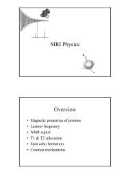
<strong>MRI</strong> <strong>EQUIPMENT</strong><br />
SG 2002<br />
• Room<br />
<strong>Essentials</strong><br />
– Radiofrequency shielding (Faraday Cage)<br />
– Fringe Field Containment<br />
• Static magnetic field (0.15 - 2.0 Tesla)<br />
• Variable magnetic field - gradients<br />
– field coils and amplifiers<br />
• Radiofrequency transmitter/receiver<br />
coils and amplifiers<br />
• Computer processing and display<br />
1
Virtually <strong>Essentials</strong><br />
• Magnet shielding<br />
– passive or active<br />
•Shim coils<br />
• MR compatible trolley<br />
2
Not essential but nice to have<br />
• MR compatible GA and monitoring<br />
equipment<br />
• ECG leads and expansion bellows for<br />
cardiac and respiratory gating<br />
3
Room requirements<br />
• Large enough to contain fringe field up<br />
to 5 Gauss (0.5mT) line<br />
– pacemakers<br />
5 Gauss<br />
Line<br />
• Completely enclosed in wire mesh -<br />
excludes radio waves from outside -<br />
zipper artefact.<br />
• Obvious practical considerations<br />
– room for trolleys etc<br />
4
Static magnetic field<br />
• Main field, B o responsible for lining<br />
up magnetic moment hydrogen<br />
protons, with (parallel) or against<br />
field (antiparallel)<br />
• Slightly greater than half are lined up with the field.<br />
Magnetic moments almost cancel out. The slight excess<br />
are the ones we can image (7 in 10million)<br />
• Proportion increases with increased field strength<br />
• Improved SNR<br />
• Is bigger always better<br />
– Increase in some artefacts and T1 acquisition times<br />
Types of magnet<br />
• Permanent (up to 0.35 T)<br />
– uses magnetised core - like horseshoe magnet<br />
–permanently on<br />
– field strength limited by weight of magnet<br />
– 0.2T field strength<br />
• iron 23 tons<br />
• neodymium alloy 9000 lbs<br />
5
Electromagnet<br />
• Resistive (up to 0.5 T)<br />
– normal electromagnet (solenoid)<br />
– windings have resistance therefore lots of heat<br />
generated. Limits the amount of current possible and<br />
consequently the strength of the magnet is less.<br />
– Cooling required.<br />
– may be switched on and off<br />
– use lots of electricity - expensive to run<br />
– Power =I 2 R<br />
Example: 0.15T magnet<br />
Requires 200 Amps<br />
Equivalent to 50kW<br />
6
• Superconducting (
Gradients<br />
• Frequency of precession w o dependent<br />
on field strength<br />
–w o is given by the Larmor equation w o = Bo x g<br />
where Bo is the magnetic field strength in Tesla and g is the Gyromagnetic ratio<br />
for hydrogen (42.57 MHz/T)<br />
• Gradients impose additional graded<br />
magnetic field on main field<br />
8
Magnet isocentre<br />
0.98T 1.0T 1.02T<br />
Gradients<br />
• This means we can define where an MR<br />
signal comes from in space because its<br />
frequency will depend on the local field<br />
strength<br />
• Gradient strength measured in mT/m<br />
– 20mT/m average<br />
• Very rapid switching<br />
–
• Three sets<br />
required for<br />
x,y,z directions<br />
• z - direction of<br />
main field<br />
•Helmholtz<br />
configuration<br />
Gradient Coils<br />
• x and y direction<br />
• Golay or saddle<br />
coils<br />
Gradient Coils<br />
10
Radiofrequency (RF) coils<br />
• Essential to put radiowaves into patient<br />
and to detect signal from precessing<br />
protons<br />
• Should only excite protons within<br />
volume of interest<br />
• Ideally in close proximity to maximise<br />
signal detection<br />
• May be transmit/receive or receive only<br />
• RF amplifiers - 20kW<br />
Radiofrequency (RF) coils<br />
• Built-in body coil<br />
• Anatomy specific coils - better SNR, less<br />
heating<br />
–Head<br />
–Neck<br />
–Extremity<br />
–Spine<br />
–Breast<br />
11
Surface coils<br />
• Aerial loops placed next to region if<br />
interest<br />
– High SNR next to coil<br />
– Small field of view<br />
– Best for superficial anatomy - TMJ, eye<br />
Volume Coils<br />
• Better RF homogeneity<br />
•Even SNR<br />
• Saddle, Birdcage configurations<br />
12
Phased Array Coils<br />
• Array of surface coils joined together<br />
• Avoids problem of poor coverage<br />
• Maintains SNR<br />
• Spine coils<br />
Open low field or closed high field<br />
• Closed - advantages<br />
– Usually high field<br />
– Better field<br />
homogeneity<br />
– Larger field of view<br />
– Better SNR<br />
– Better image quality<br />
– Faster scan times<br />
– Thinner slices improved<br />
spatial resolution<br />
– Spectroscopy, f<strong>MRI</strong>,<br />
EPI<br />
• Closed - disadvantages<br />
– Patient unfriendly<br />
–Claustrophobic<br />
– Limited access makes<br />
interventional techniques<br />
difficult<br />
– Positioning can be difficult<br />
– Usually noisy<br />
– Tissue heating (SAR limits)<br />
– Cryogen refills<br />
– cooling of gradient power<br />
supply<br />
13
• Open - advantages<br />
– less claustrophobic<br />
– Patient friendly<br />
– easier monitoring<br />
– easier positioning<br />
– intervention e.g.<br />
biopsy, tumour<br />
ablation possible<br />
–quiet<br />
– low tissue heating<br />
• Open - disadvantages<br />
–poor SNR<br />
– longer scan times<br />
– thicker slices<br />
– small FOV<br />
– not capable of some<br />
techniques<br />
– expensive on electricity<br />
– cooling of magnet power<br />
supply<br />
14
Field homogeneity<br />
• Uniformity of magnetic field<br />
– important for accurate spatial registration<br />
of MR signal<br />
• Better than 1ppm<br />
• Achieved by addition of pieces of iron<br />
(passive shimming) or solenoids (active<br />
shimming)<br />
Peripherals<br />
• Life sign monitoring<br />
• Anaesthetic trolley<br />
•ECG gating leads<br />
• Respiratory gating bellows<br />
15
Cardiac Gating<br />
Non ECG gated<br />
ECG gated<br />
Respiratory Gating<br />
Non respiratory gated<br />
Respiratory gated<br />
16
References<br />
• Boddy J (1999) High Field Closed <strong>MRI</strong> Scanners<br />
Clinical <strong>MRI</strong> 8(4) p124-126<br />
• Dunne S (1999) Low Field Open <strong>MRI</strong> Scanners<br />
Clinical <strong>MRI</strong> 8(4) p117-123<br />
• Keen M (1999) Lecture Notes on the Physics of <strong>MRI</strong><br />
Clinical <strong>MRI</strong> 9(3) p70-81<br />
17