ý.,,: V. ý ýý . - Nottingham eTheses - University of Nottingham
ý.,,: V. ý ýý . - Nottingham eTheses - University of Nottingham
ý.,,: V. ý ýý . - Nottingham eTheses - University of Nottingham

Create successful ePaper yourself
Turn your PDF publications into a flip-book with our unique Google optimized e-Paper software.
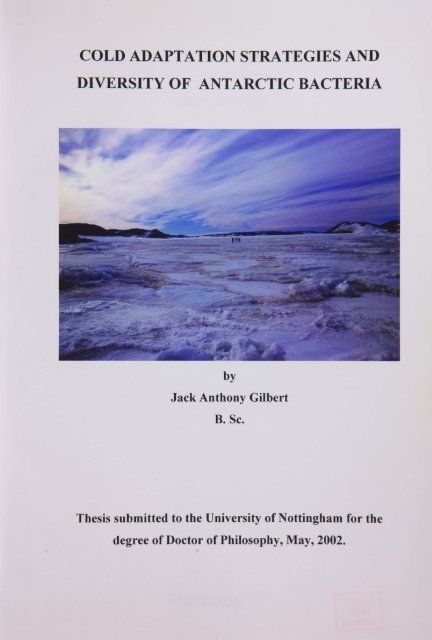
COLD ADAPTATION STRATEGIES AND<br />
DIVERSITY OF ANTARCTIC BACTERIA<br />
<strong>ý</strong>.,,: V. <strong>ý</strong><br />
<strong>ý</strong>.<br />
M<br />
<strong>ý</strong><strong>ý</strong> .<br />
FliY<strong>ý</strong>x<br />
<strong>ý</strong><strong>ý</strong><br />
<strong>ý</strong>'<br />
-7"<br />
'GY.<br />
.- -z'<br />
44<br />
Ir*<br />
1-* al.,<br />
1<br />
<strong>ý</strong>.<br />
. A* -ft<br />
.. > .<br />
v <strong>ý</strong>iein<strong>ý</strong>'<br />
by<br />
Jack Anthony Gilbert<br />
B. Sc.<br />
Thesis submitted to the <strong>University</strong> <strong>of</strong> <strong>Nottingham</strong><br />
for the<br />
degree <strong>of</strong> Doctor <strong>of</strong> Philosophy, May, 2002.
DECLARATION<br />
I hereby declare that the work presented in this thesis is my own and has not been<br />
submitted for any other degree. All sources <strong>of</strong> information have been acknowledged by<br />
reference to<br />
e au ors.<br />
Jack Anthony Gilbert
, 71<br />
"If you march your Winter<br />
Journeys you will have your reward, so long<br />
as all you want is a penguin's egg"<br />
Apsley Cherry-Garrard<br />
(The Worst Journey in the World)<br />
1<br />
w 4,
Contents<br />
List <strong>of</strong> Figures<br />
................................................................................<br />
i<br />
List <strong>of</strong> Tables .........................................................................................<br />
v<br />
List <strong>of</strong> Abbreviations<br />
Acknowledgements<br />
..............................................................................<br />
...............................................................................<br />
vii<br />
ix<br />
Abstract<br />
.........................................................................................<br />
x<br />
Chapter 1:<br />
INTRODUCTION<br />
..............................................................<br />
1<br />
1.1. Temperature as a regulatory factor<br />
....................................<br />
1<br />
1.2. Cold Environments<br />
..............................................................<br />
1<br />
1.3. Psychrophiles and psychrotrophs<br />
1.4. Antarctic oases - ice-free refugia<br />
............................................<br />
............................................<br />
2<br />
3<br />
1.5. Methods <strong>of</strong> cold adaptation<br />
......................................................<br />
4<br />
1.5.1. Physio-chemical properties <strong>of</strong> water in confined spaces<br />
........ 5<br />
1.5.2. Lower growth-viable temperature limit <strong>of</strong> psychrophiles<br />
........ 5<br />
1.5.3. Cold adaptation in proteins<br />
............................................<br />
7<br />
1.5.3.1. Structural adaptations in psychrophilic enzymes<br />
1.5.3.2. Cold induced psychrophilic protein production<br />
1.5.3.3. Ice nucleation proteins<br />
...................................<br />
........ 7<br />
........ 8<br />
9<br />
1.5.3.4. Antifreeze proteins<br />
..........................................<br />
10<br />
1.5.3.4.1. AFP structure and function<br />
...............<br />
10<br />
1.5.3.4.2. Bacterial AFPs<br />
.................................<br />
13<br />
1.5.3.4.3. AFPs in biotechnology<br />
1.5.3.4.4. Evolution <strong>of</strong> AFPs<br />
........................<br />
........................<br />
13<br />
14<br />
1.5.3.5. Membrane bound solute transport proteins<br />
................<br />
17<br />
1.5.4. Cold adaptation in membrane lipids<br />
.................................<br />
17<br />
1.5.4.1. Biochemical mechanisms <strong>of</strong> fatty acid induction<br />
...... 19<br />
1.6<br />
Biodiversity and biogeography <strong>of</strong> bacteria .................................<br />
20<br />
1.6.1. Bacterial biodiversity<br />
1.6.2. Bacterial biogeography<br />
...................................................<br />
..........................................<br />
21<br />
22
1.6.3. Molecular typing techniques<br />
..........................................<br />
22<br />
1.6.3.1.16S rDNA sequencing ..........................................<br />
22<br />
1.6.3.2. Amplified ribosomal DNA restriction analysis (ARDRA) .<br />
24<br />
1.6.3.3. Southern hybridisation (ribotyping)<br />
........................<br />
24<br />
1.6.3.4. Restriction fragment length polymorphism (RFLP/T-<br />
RFLP) ............................................................<br />
24<br />
1.6.3.5. Amplified fragment length polymorphism (AFLP)<br />
...... 25<br />
1.6.3.6. AP-PCR and RAPD<br />
..........................................<br />
25<br />
1.6.3.7. rep-PCR, ERIC-PCR and BOX-PCR ........................<br />
25<br />
1.6.3.8. DGGE and TGGE<br />
..........................................<br />
26<br />
1.6.4. Numerical taxonomy<br />
...................................................<br />
26<br />
Chapter 2: MATERIALS AND METHODS<br />
..........................................<br />
32<br />
2.1. Study sites<br />
.....................................................................<br />
32<br />
2.2. Sampling<br />
.....................................................................<br />
32<br />
2.3. Water sample processing<br />
...................................................<br />
37<br />
2.4. Culturing and maintenance <strong>of</strong> isolates .................................<br />
37<br />
2.5. Preservation and transportation <strong>of</strong> the microbial collection<br />
...... 38<br />
2.6. Gram stain technique<br />
...................................................<br />
38<br />
2.7. Water chemistry - analytical procedures<br />
.................................<br />
39<br />
2.7.1. Chlorophyll a analysis<br />
..........................................<br />
39<br />
2.7.2. Inorganic chemical analysis<br />
..........................................<br />
40<br />
2.7.2.1. Ammonium (NH4-N)<br />
2.7.2.2. Nitrite (NO2-N)<br />
2.7.2.3. Nitrate (N03-N)<br />
..........................................<br />
..........................................<br />
..........................................<br />
40<br />
40<br />
40<br />
2.7.2.3. Soluble reactive phosphorus (PO4-P) ........................<br />
40<br />
2.7.3. Dissolved organic carbon analysis<br />
.................................<br />
40<br />
2.8. Bacterial and flagellate counts<br />
2.9. Molecular typing techniques<br />
..........................................<br />
..........................................<br />
41<br />
41<br />
2.9.1. Chromosomal DNA extraction<br />
.................................<br />
41<br />
2.9.2. Preparation, electrophoresis and recording <strong>of</strong> agarose gels<br />
...... 42
2.9.3. Amplified ribosomal DNA restriction analysis<br />
...............<br />
43<br />
2.9.3.1. PCR amplification <strong>of</strong> 16S rRNA gene ........................<br />
43<br />
2.9.3.2. Restriction endonuclease digestion <strong>of</strong> 16S rDNA<br />
fragment<br />
...................................................<br />
45<br />
2.9.4. Computer enhanced phenetic analysis <strong>of</strong> ARDRA patterns<br />
2.9.5. Denaturing gradient gel electrophoresis<br />
........................<br />
...... 45<br />
46<br />
2.9.5.1. Collection <strong>of</strong> bacterial community<br />
........................<br />
46<br />
2.9.5.2. Genomic extraction for DGGE<br />
........................<br />
46<br />
2.9.5.3. Nested V3 region PCR amplification for DGGE<br />
...... 50<br />
2.9.5.4. Denaturing gradient gel electrophoresis<br />
...............<br />
51<br />
2.9.6. Sequencing <strong>of</strong> bacterial 16S rDNA .................................<br />
52<br />
2.9.6.1. Purification <strong>of</strong> 16S rDNA PCR products<br />
...............<br />
52<br />
2.9.6.2. Purification <strong>of</strong> PCR products by gel extraction ...... 53<br />
2.9.6.3. PCR product cloning<br />
..........................................<br />
53<br />
2.9.6.4. Sequencing reaction<br />
..........................................<br />
54<br />
2.10. Protein extraction and antifreeze protein assessment technique<br />
2.10.1. Total cellular protein extraction<br />
.................................<br />
...... 55<br />
55<br />
2.10.2. `SPLAT' analysis<br />
...................................................<br />
56<br />
Chapter 3: BIOLOGICAL AND PHYSICO-CHEMICAL CHARACTERISTICS<br />
OF THE STUDY LAKES<br />
3.1. Introduction<br />
3.2. Results<br />
.....................................................................<br />
.....................................................................<br />
.....................................................................<br />
58<br />
5 8<br />
59<br />
3.2.1. Summer sampled lakes<br />
..........................................<br />
59<br />
3.2.1.1. Physico-chemical characteristics<br />
........................<br />
60<br />
3.2.1.1.1. Ice cover ..........................................<br />
60<br />
3.2.1.1.2. Temperature ..........................................<br />
60<br />
3.2.1.1.3. Salinity<br />
..........................................<br />
60<br />
3.2.1.2. Inorganic nutrients ..........................................<br />
61<br />
3.2.1.2.1. Nitrate (N03-N) and nitrite (N02-N) ...... 61<br />
3.2.1.2.2. Ammonium (NH4-N) and SRP (P04-P) ...... 61
3.2.1.2.3. DOC and chlorophyll a<br />
........................<br />
61<br />
3.2.2. Focus study lakes<br />
...................................................<br />
65<br />
3.2.2.1. Physico-chemical characteristics ........................<br />
65<br />
3.2.2.1.1. Ice cover ..........................................<br />
65<br />
3.2.2.1.2. Temperature ..........................................<br />
65<br />
3.2.2.1.3. Salinity<br />
..........................................<br />
65<br />
3.2.2.2. Inorganic nutrients<br />
..........................................<br />
70<br />
3.2.2.2.1. Nitrate<br />
3.2.2.2.2. Nitrite<br />
3.2.2.2.3. Ammonium<br />
..........................................<br />
..........................................<br />
..........................................<br />
70<br />
70<br />
70<br />
3.2.2.2.4. Soluble reactive phosphate ........................<br />
70<br />
3.2.2.2.5. Dissolved organic carbon<br />
........................<br />
71<br />
3.2.2.3. Biological characteristics<br />
3.2.2.3.1. Chlorophyll a<br />
.................................<br />
.................................<br />
77<br />
77<br />
3.2.2.3.2. Bacterial cell concentrations<br />
...............<br />
77<br />
3.2.2.3.3. HNAN abundance .................................<br />
77<br />
3.2.2.3.4. PNAN abundance .................................<br />
78<br />
3.3. Discussion<br />
.....................................................................<br />
81<br />
3.3.1. Summer sampled lakes<br />
..........................................<br />
81<br />
3.3.2. Detailed study lakes ...................................................<br />
88<br />
3.3.2.1. Ace Lake<br />
...................................................<br />
88<br />
3.3.2.2. Pendant Lake ...................................................<br />
97<br />
3.3.2.3. Triple Lake<br />
..................................................<br />
102<br />
3.3.2.4. Deep Lake and Club Lake<br />
................................<br />
106<br />
3.3.3. Oval Lake<br />
...........................................................<br />
111<br />
3.4. Conclusions<br />
....................................................................<br />
112<br />
Chapter 4: ANTIFREEZE<br />
PROTEIN ANALYSIS<br />
................................<br />
114<br />
4.1. Introduction<br />
....................................................................<br />
114<br />
4.1.1. Disadvantages <strong>of</strong> the SPLAT assay 115<br />
4.1.2. Development <strong>of</strong> the high-throughput AFP analysis protocol
(HTAP)<br />
...........................................................<br />
115<br />
4.1.2.1. Premise <strong>of</strong> methodology ................................<br />
115<br />
4.1.2.2. Development <strong>of</strong> assay .........................................<br />
116<br />
4.1.2.3. Advantages and disadvantages <strong>of</strong> the assay<br />
..............<br />
120<br />
4.2. Results<br />
....................................................................<br />
121<br />
4.2.1. Sampling and bacterial isolation<br />
................................<br />
121<br />
4.2.2. Gram staining and cellular/colony morphology<br />
..............<br />
121<br />
4.2.3. High-throughput AFP assay<br />
.........................................<br />
122<br />
4.2.4. SPLAT assay ...........................................................<br />
122<br />
4.2.5. Crystal morphology<br />
..................................................<br />
123<br />
4.3. Discussion<br />
4.4. Conclusions<br />
....................................................................<br />
....................................................................<br />
127<br />
130<br />
Chapter 5: BACTERIAL MOLECULAR TYPING<br />
................................<br />
131<br />
5.1. Introduction<br />
5.2. Results<br />
....................................................................<br />
....................................................................<br />
13 1<br />
132<br />
5.2.1. Amplified ribosomal DNA restriction analysis (ARDRA) ..... 132<br />
5.2.1.1-Analysis <strong>of</strong> Hpa II and A lu I restriction digestion<br />
patterns<br />
..................................................<br />
132<br />
5.2.1.2. Relatedness <strong>of</strong> AFP active isolates from ARDRA<br />
..... 134<br />
5.2.2.16S<br />
rRNA nucleotide sequencing<br />
................................<br />
136<br />
5.2.3. Phylogenetic tree<br />
..................................................<br />
139<br />
5.3. Discussion<br />
....................................................................<br />
143<br />
5.3.1. ARDRA limitations<br />
..................................................<br />
143<br />
5.3.2. Identification <strong>of</strong> the AFP active isolates<br />
.......................<br />
145<br />
5.3.2.1. The Marinomonas protea group, 124,154,214,54,744,<br />
794 and 5ice3 ..................................................<br />
145<br />
5.3.2.2. The Pseudoalteromonas group, isolates 39 and 86<br />
5.3.2.3. The Pseudomonas group, isolates 302 and 33<br />
..... 146<br />
..... 146<br />
5.3.2.4. The Stenotrophomonas maltophilia group, isolates 492<br />
and 47 ...........................................................<br />
148
5.3.2.5. The Sphingomonas group, isolate 494<br />
5.3.2.6. The Psychrobacter group, isolate 583<br />
..............<br />
..............<br />
149<br />
151<br />
5.3.2.7. The Enterobacter agglomerans group, isolate 732<br />
..... 153<br />
5.3.2.8. The Idiomarina loihiensis group, isolate 53 ..............<br />
154<br />
5.3.2.9. The Halomonas sp. group, isolate 213<br />
..............<br />
156<br />
5.3.2.10. Isolate 466<br />
..................................................<br />
157<br />
5.4. Conclusions<br />
....................................................................<br />
15 8<br />
Chapter 6: BACTERIAL COMMUNITY ANALYSIS<br />
.......................<br />
159<br />
6.1. Introduction<br />
6.2. Results<br />
....................................................................<br />
....................................................................<br />
15 9<br />
160<br />
6.2.1. Detection <strong>of</strong> AFP active isolates in DGGE community pr<strong>of</strong>iles 161<br />
6.2.1.1. Assessment <strong>of</strong> 16S rRNA operon heterogeneity in<br />
M. protea<br />
..................................................<br />
162<br />
6.2.2. Temporal and spatial variation in microbial communities<br />
..... 167<br />
6.3. Discussion<br />
....................................................................<br />
173<br />
6.3.1. Detection <strong>of</strong> AFP active isolates in community pr<strong>of</strong>iles<br />
..... 173<br />
6.3.2. DGGE bias<br />
...........................................................<br />
175<br />
6.3.2.1. Species composition <strong>of</strong> the community<br />
6.3.2.2. Sample isolation and maintenance<br />
..............<br />
.......................<br />
175<br />
176<br />
6.3.2.3. Cellular lysis and DNA purification<br />
.......................<br />
177<br />
6.3.2.4. Polymerase chain reaction (PCR) amplification<br />
..... 178<br />
6.3.2.4.1. Amplification inhibition<br />
.......................<br />
178<br />
6.3.2.4.2. Preferential amplification<br />
.......................<br />
178<br />
6.3.2.4.3. Artefactual PCR products .......................<br />
179<br />
6.3.2.4.4. PCR contamination<br />
.......................<br />
180<br />
6.3.2.4.5.16S rRNA operon heterogeneity ..............<br />
181<br />
6.4. Conclusion<br />
....................................................................<br />
184<br />
Chapter 7:<br />
GENERAL DISCUSSION<br />
..................................................<br />
186<br />
7.1. Discussion<br />
....................................................................<br />
186
7.2. Future work<br />
....................................................................<br />
193<br />
REFERENCES<br />
APPENDICES<br />
.............................................................................<br />
.............................................................................<br />
195<br />
235
LIST OF FIGURES<br />
Figure I. I. Diagrammatic representation demonstrating the relationship between the<br />
different symbols used in the formulae in Table 1.2. Reproduced from Priest and Austin<br />
(1995) ................................................................................................<br />
28<br />
Figure 1.2. Representation <strong>of</strong> molecular taxonomy. Diagram outlining the molecular<br />
technique <strong>of</strong> identifying relationships between species<br />
.................................<br />
29<br />
Figure 2.1a A map <strong>of</strong> the Vestfold Hills, Eastern Antarctica, showing location <strong>of</strong> the 38<br />
sampled lakes (reproduced with permission <strong>of</strong> the Australian Antarctic data centre) ....<br />
34<br />
Figure 2.1b A map <strong>of</strong> the Larsemann Hills, Eastern Antarctica, showing the location <strong>of</strong><br />
the 2 sampled lakes (reproduced with permission from the Australian Antarctic data<br />
centre) ................................................................................................<br />
35<br />
Figure 2.1c A map <strong>of</strong> the epishelf lake, Beaver Lake, MacRobertson Land (modified<br />
from Laybourn-Parry et al., 2001b) ............................................................<br />
36<br />
Figure 2.2. Diagrammatic representation <strong>of</strong> the ARDRA procedure<br />
...............<br />
44<br />
Figure 2.3. Lane delineation image from 1D elite gel analysis s<strong>of</strong>tware (Amersham<br />
Pharmacia Biotech)<br />
..............................................................................<br />
47<br />
Figure 2.4a Image showing how the DNA bands in one lane are established by<br />
measuring pixel intensity .....................................................................<br />
47<br />
Figure 2.4b Image showing how individual bands can be seen more clearly by removing<br />
the background pixel intensity using a rolling disc algorithm with a disc radius <strong>of</strong> 20 ... 47<br />
Figure 2.5a Image showing automatic band detection s<strong>of</strong>tware prior to artefact<br />
Removal<br />
.......................................................................................<br />
48<br />
Figure 2.5b Image showing bands detected after deletion <strong>of</strong> artefactual bands ...... 48<br />
Figure 2.6a Image showing standardisation across the gel suing retardation factor, which<br />
alleviated any distortion due to uneven running <strong>of</strong> warping <strong>of</strong> the gel ...............<br />
48<br />
Figure 2.6b Image showing molecular weights assigned to each <strong>of</strong> the detectable bands<br />
within the three 100 bp ladders, from which a curve is computed to allows cross gel<br />
comparison<br />
.......................................................................................<br />
49<br />
Figure 2.7. Image showing band matching s<strong>of</strong>tware (1 D elite gel analysis s<strong>of</strong>tware<br />
(Amersham Pharmacia Biotech)<br />
............................................................<br />
49<br />
i
Figure 3.1. Ice thickness and date <strong>of</strong> recording for three <strong>of</strong> the five detailed study lakes<br />
throughout the 2000 sampling period ............................................................<br />
67<br />
Figure 3.2. Temperature measurements for Ace Lake, Pendant Lake and Triple Lake ..<br />
68<br />
Figure 3.3. Temperature and salinity pr<strong>of</strong>iles for Deep and Club Lakes with sampling<br />
dates<br />
.................................................................................................<br />
69<br />
Figure 3.4. Salinity measurements for Ace Lake, Pendant Lake and Triple Lake<br />
....... 68<br />
Figure 3.5. Nitrate concentration (µg L") for the five detailed study lakes ................<br />
72<br />
Figure 3.6. Nitrite concentration (µg L1) for the five detailed study lakes<br />
...............<br />
73<br />
Figure 3.7. Ammonia concentration (µg L-) for the five detailed study lakes<br />
...... 74<br />
Figure 3.8. Soluble reactive phosphate (SRP) concentration (µg L"1) for the five detailed<br />
study lakes .......................................................................................<br />
75<br />
Figure 3.9. Dissolved organic carbon (DOC) concentration (mg L') for the five detailed<br />
study lakes, standard deviation (mg mL-1) error bars are also given for each data point .<br />
76<br />
Figure 3.10. Chlorophyll a concentration (pg L1) for the five detailed study lakes ......<br />
78<br />
Figure 3.11. Bacterial abundance (x 106 ml-1) for the five detailed study lakes<br />
...... 79<br />
Figure 3.12. HINAN abundance<br />
(x 106 L) for<br />
the three detailed study lakes for which<br />
these measurements were taken<br />
............................................................<br />
80<br />
Figure 3.13. PNAN abundance (x 106 L)<br />
for the three detailed study lakes for which<br />
these measurements were taken<br />
............................................................<br />
80<br />
Figure 3.14a Air Temperature (°C), surface water temperature (°C) and salinity (ppt) for<br />
the 12 summer sampled hypersaline lakes<br />
...................................................<br />
84<br />
Figure 3.14b Air temperature (°C), surface water temperature (°C) and salinity (ppt) for<br />
the 13 summer sampled saline lakes<br />
............................................................<br />
84<br />
Figure 3.14c Air temperature (°C), surface water temperature (°C) and salinity (ppt) for<br />
the 13 summer sampled freshwater lakes<br />
...................................................<br />
84<br />
Figure 3.15a The correlation between salinity (ppt) and DOC (mg L-1) concentrations in<br />
the summer sampled hypersaline lakes<br />
...................................................<br />
87<br />
Figure 3.15b DOC (mg U) levels for the summer sampled hypersaline, saline and<br />
freshwater lakes<br />
..............................................................................<br />
87<br />
Figure 3.16. Shows how temperature fluctuations in Ace Lake correlate with salinity<br />
fluctuations during January. March and July ...................................................<br />
90<br />
11
Figure 3.17a Mean bacterial abundance against mean ammonia concentrations for Ace<br />
Lake<br />
................................................................................................<br />
95<br />
Figure 3.17b Relationship between average bacterial abundance with depth and average<br />
DOC concentration with depth over four sampling dates in Ace Lake<br />
...............<br />
95<br />
Figure 3.18. Relationship between bacterial abundance, PNAN abundance and HNAN<br />
abundance in Ace Lake<br />
.....................................................................<br />
97<br />
Figure 3.19. Diagrammatic representation <strong>of</strong> the pr<strong>of</strong>ile <strong>of</strong> Pendant Lake indicating the<br />
anoxic sump<br />
.......................................................................................<br />
98<br />
Figure 3.20. Relationship between bacterial, PNAN and HNAN abundances for Pendant<br />
Lake over time as averages <strong>of</strong> depth (0-1 Om) ..................................................<br />
101<br />
Figure 3.22a, b&c<br />
Correlation between bacterial abundance and chlorophyll a<br />
concentration for three sampling dates in 2000, July (a), September (b) and November<br />
(c), in Triple Lake.<br />
.............................................................................<br />
105<br />
Figure 3.21. Ambient air temperature graph for Davis station, Vestfold Hills, Antarctica<br />
(reproduced with the permission <strong>of</strong> the Bureau <strong>of</strong> Meteorology, Melbourne,<br />
Australia)<br />
......................................................................................<br />
107<br />
Figure 4.1. Ice recrystalisation at -6°C for 30 minutes in a (A) AFP solution <strong>of</strong> 30%<br />
sucrose and I mg/ml Fish AFP III, and (B) non-AFP solution <strong>of</strong> 30% sucrose<br />
(Reproduced from Mills, 1999)<br />
...........................................................<br />
116<br />
Figure 4.2. Fryka KP 281 cold block (CamLab) in situ with HTAP microtitre plates<br />
during the recrystalisation step at -6°C in cold room .........................................<br />
117<br />
Figure 4.3. Photographic image <strong>of</strong> a high-throughput AFP assay in a 96 well microtitre<br />
plate. Demonstrating technique and sample colouration<br />
................................<br />
117<br />
Figure 4.4. HTAP assay in a 96 well microtitre plate. Demonstrating lack <strong>of</strong> contrast 119<br />
Figure 4.5. Percentages <strong>of</strong> `SPLAT' assay confirmed AFP active bacteria isolated from<br />
each lake ......................................................................................<br />
123<br />
Figure 4.6. Total cellular protein extraction concentration (mg mUl) with SPLAT score<br />
with error bars for AFP active isolates<br />
..................................................<br />
125<br />
Figure 4.7. Photography <strong>of</strong> SPLAT analysis <strong>of</strong> isolate 494 ................................<br />
125<br />
Figure 5.1. Photographs <strong>of</strong> ARDRA gel patterns used for phenetic analysis <strong>of</strong> band<br />
position along gel (a) Hpa II and (b) Alu I<br />
..................................................<br />
133<br />
iii
Figure 5.2. Dendrogram calculated using the UPGMA algorithm from the similarity<br />
matrices <strong>of</strong> (a) Hpa II and (b) Alu I digest patterns<br />
.........................................<br />
135<br />
Figure 5.3. Photograph <strong>of</strong> an over-exposed gel image (b) next to a normal exposure gel<br />
image (A), indicating the low molecular weight bands <strong>of</strong> bands <strong>of</strong> low DNA<br />
concentration which can be better identified when the image is over-exposed<br />
..... 135<br />
Figure 5.4. Maximum likelihood phylogenetic tree <strong>of</strong> relatedness for 13 AFP active<br />
isolates aligned against 18 published close relatives .........................................<br />
142<br />
Figure 5.5. ARDRA analysis <strong>of</strong> gel <strong>of</strong> Hpa II restriction digest <strong>of</strong> (1) isolate 494. (2)<br />
M3 C203B-B, (3) Marinomonas protea and (4) SIA 181-1 A1....................... 152<br />
Figure 6.1. Denaturing gradient gel electrophoresis acrylamide gel showing 10 highly<br />
AFP active Antarctic lake isolates against 3 community pr<strong>of</strong>iles from the lakes <strong>of</strong> their<br />
isolation<br />
......................................................................................<br />
165<br />
Figure 6.2. Denaturing gradient gel electrophoresis acrylamide gel showing 9<br />
representatives <strong>of</strong> phena identified within the AFP active isolate grouping (except M<br />
protea related species) against 4 community pr<strong>of</strong>iles from the lakes <strong>of</strong> their isolation<br />
. 165<br />
Figure 6.3a DGGE gel 1. Matching analysis by Rf values to assess for the presence <strong>of</strong><br />
pure culture AFP active isolates within community fingerprints .......................<br />
166<br />
Figure 6.3b DGGE gel 2. Matching analysis by Rf value to assess for the presence <strong>of</strong><br />
pure culture AFP active isolates within community fingerprints .......................<br />
166<br />
Figure 6.4a DGGE community pr<strong>of</strong>iles for Ace Lake and Pendant Lake<br />
..............<br />
168<br />
Figure 6.4b DGGE community pr<strong>of</strong>iles for Pendant Lake, Triple Lake, Deep Lake and<br />
Club Lake<br />
......................................................................................<br />
168<br />
Figure 6.4c Gel 1 image (Fig. 6.4a) showing band matching <strong>of</strong> Rf values ..............<br />
169<br />
Figure 6.4d Gel 2 image (Fig. 6.4b) showing band matching <strong>of</strong> Rf values ..............<br />
169<br />
Figure 6.5a Similarity matrix for the community fingerprint patterns <strong>of</strong> DGGE analysis<br />
gels from fig 6.4a and fig 6.4b<br />
...........................................................<br />
170<br />
Figure 6.5b Dendrogram showing phenetic relatedness <strong>of</strong> DGGE community pr<strong>of</strong>iles<br />
for Ace Lake, Pendant Lake, Triple Lake, Deep Lake and Club Lake ..............<br />
171<br />
IN'
LIST OF TABLES<br />
Table 1.1. Types <strong>of</strong> AFPs and AFGP and their structures. Demonstrating the structural<br />
diversity in AFPs. Types I, II, III and AFGP descriptions were taken from Meyer et al.<br />
(1999), Type IV description taken from Zhao et al. (1998) .................................<br />
12<br />
Table 1.2. Three <strong>of</strong> the most commonly used similarity coefficients in bacterial<br />
taxonomy (after Priest and Austin, 1995)<br />
...................................................<br />
28<br />
Table 2.1. Location, selected physico-chemical properties <strong>of</strong> the study lakes. All<br />
salinities based on current study<br />
............................................................<br />
33<br />
Table 2.2. `SPLAT' scoring key. The size and density <strong>of</strong> ice crystals relates to<br />
observations made after 1 hour <strong>of</strong> recrystalisation at -6°C. .................................<br />
57<br />
Table 3.1a Physical and inorganic data for the hypersaline lakes sampled during January.<br />
February and March 2000<br />
.....................................................................<br />
62<br />
Table 3.1b Physical and inorganic data for the saline lakes sampled during January and<br />
February 2000 .......................................................................................<br />
63<br />
Table 3.1c Physical and inorganic data for the freshwater lakes sampled during January<br />
and February 2000<br />
..............................................................................<br />
64<br />
Table 3.2. Shows literature in which lakes for the current study only sampled during the<br />
austral summer <strong>of</strong> 1999/2000 are mentioned ...................................................<br />
82<br />
Table 3.3. Comparison <strong>of</strong> the major recorded physico-chemical factors for the lakes from<br />
which AFP active bacteria were isolated. All measurements are from Om during the<br />
summer<br />
......................................................................................<br />
1 12<br />
Table 4.1. The proportion <strong>of</strong> HTAP positive bacteria isolated from each lake<br />
..... 124<br />
Table 4.2. Isolates which tested positive for some level <strong>of</strong> RI activity. Also provided are<br />
the location from which the bacteria were isolated, the agar which was used to culture<br />
them, the colony and cellular morphologies<br />
..................................................<br />
126<br />
Table 5.1. Original isolate number, Genbank accession number, the size <strong>of</strong> the sequenced<br />
16S rDNA fragment used for identification and the closest relative identified by FASTA<br />
search <strong>of</strong> the EMBL prokaryote nucleotide database .........................................<br />
138<br />
V
Table 5.2. Growth results for suspected pseudomonad species isolates on pseudomonad<br />
selective agar and fluorescein selective agar ..................................................<br />
139<br />
Table 5.3. Table shows the close relatives used for creation <strong>of</strong> a maximum likelihood<br />
phylogenetic tree<br />
.............................................................................<br />
141<br />
Table 5.4. Isolate number with closest relative from the EMBL prokaryotic nucleotide<br />
database using FASTA search engine, lake <strong>of</strong> isolation and the depth in the water column<br />
or ice core <strong>of</strong> isolation .............................................................................<br />
144<br />
Table 7.1. AFP active bacterial species, their isolated location and study from which they<br />
are cited<br />
......................................................................................<br />
188<br />
vi
List <strong>of</strong> Abbreviations<br />
AFP<br />
AFGP<br />
AFLP<br />
AP-PCR<br />
ATCC<br />
- Antifreeze protein<br />
- Antifreeze glycoprotein<br />
- Amplified fragment length polymorphism<br />
- Arbitrarily primed polymerase chain reaction<br />
- American type culture collection<br />
ATP<br />
-<br />
Adenine 5'-triphosphate<br />
BLAST<br />
CAP<br />
CSP<br />
CTAB<br />
CTP<br />
DAPI<br />
DCM<br />
DDBJ<br />
DFAA<br />
DFCHO<br />
DGGE<br />
DNA<br />
DOC<br />
EMBL<br />
FASTA<br />
FISH<br />
GES<br />
GM<br />
GTP<br />
HNAN<br />
HTAP<br />
INA<br />
INP<br />
LRR<br />
NCBI<br />
OUT<br />
PCR<br />
PE<br />
PGIPs<br />
PNAN<br />
PUFAs<br />
RAPD<br />
RFLP<br />
R1<br />
RNA<br />
SIMCO<br />
SPLAT<br />
- Basic local alignment search tool for DNA DNA sequence comparisons<br />
- Cold acclimation protein<br />
- Cold shock protein<br />
- Cetyltrimethylammonium bromide<br />
- Cytosine 5' -triphosphate<br />
- 4', 6-diamidino-2-phenylindole<br />
- Deep chlorophyll maximum<br />
- DNA databank <strong>of</strong> Japan<br />
- Dissolved free amino acids<br />
- Dissolved free monosaccharides<br />
- Denaturing gradient gel electrophoresis<br />
- Deoxyribonucleic acid<br />
- Dissolved organic carbon<br />
- European molecular biology laboratory<br />
- DNA DNA sequence comparison program for computing similarity.<br />
- Fluorescent in situ hybridisation<br />
- Guanidium isothiocyanate<br />
- Genetically modified<br />
- Guanine 5'-triphosphate<br />
- Heterotrophic nan<strong>of</strong>lagellates<br />
- High-throughput antifreeze protein assay<br />
- Ice nucleation agent<br />
- Ice nucleation protein<br />
- Leucine rich repeat proteins<br />
- National centre for biotechnology information<br />
- Operational taxanomic unit<br />
- Polymerase chain reaction<br />
- Protein extract<br />
- Polygalacturonase inhibitor proteins<br />
- Phototrophic nan<strong>of</strong>lagellates<br />
- Polyunsaturated fatty acids<br />
- Random primed amplified polymorphic DNA<br />
- Restriction fragment length polymorphism<br />
- Recrystalisation inhibition, the act <strong>of</strong> inhibition <strong>of</strong> ice crystal growth<br />
- Ribonucleic acid<br />
- Sea ice microbial community<br />
- Ice recrystalisation inhibition assay determined using crossed polarised<br />
microscopy <strong>of</strong> a single crystal width sample<br />
vii
SRP<br />
SWA<br />
- Soluble reactive phosphate<br />
- Sea water agar<br />
'/2 SWA<br />
- '/2 sea water salinity agar<br />
T-RFLP -Termination restriction fragment length polymorphism<br />
TGGE<br />
TOC<br />
TSA<br />
TTY<br />
UPGMA<br />
YSA<br />
- Temperature gradient gel electrophoresis<br />
- Total organic carbon<br />
- Tryptic soya agar<br />
- thymine 5'-triphosphate<br />
- Unweighted pair group method with arithmetic averages<br />
- Yeast selective agar<br />
viii
ACKNOWLEDGEMENTS<br />
This study was funded by Unilever. The Antarctic phase <strong>of</strong> the research was<br />
supported by an Australian Antarctic Science Advisory Committee grant to Pr<strong>of</strong>essor J.<br />
Laybourn-Parry. I would firstly like to thank my supervisors, Pr<strong>of</strong>essor Johanna<br />
Laybourn-Parry, Dr. Christine Dodd and Dr. Phil Hill for their support, guidance and<br />
patience throughout the study, as well as for their un-ending enthusiasm for the research<br />
which helped me through the long Antarctic winter.<br />
I would like to extend my thanks Dr. David Bell for his invaluable help with the<br />
phylogenetic analysis, David Fowler and John Corrie for their patience with my<br />
impatience, various people in Food Microbiology<br />
for helping me to understand molecular<br />
biology, including Dr. Sharon Johnson, Dr. Timothy Aldsworth, Tania Perehenic, Danilo<br />
Ercolini, Dr. Sarah Mills, Nicola Cummings and especially Catherine Loc-Carrillo for<br />
never giving up on a pseudo-microbiologist.<br />
I would like to thank a number <strong>of</strong> the staff at<br />
Unilever, including Joy Wilkinson, Patricia Quail and Allen Griffiths for their support,<br />
Sarah Twigg for her help with the SPLAT analysis and Mark Berry for overseeing the<br />
project.<br />
I must also show my great appreciation to the staff <strong>of</strong> the Australian Antarctic<br />
Division, particularly to Trevor Bailey, Dr. Harvey Marchant, Debbie Lang and Vicki<br />
Norris. I would also like to thank the Davis Station ANARE expeditioners (1999-2000)<br />
for their assistance with fieldwork and all things musical, as well as for making my time<br />
in Antarctica one <strong>of</strong> the best <strong>of</strong> my life. Special thanks to Ray Bajinskis, Frederick Jobin<br />
and Brendan Hill for their exemplary effort on field trips and to Brad French for always<br />
being one step lower.<br />
Finally, I would like to thank my family for their never ending emotional support<br />
and friendship throughout my life and for giving me the power to fulfil my dreams.<br />
ix
ABSTRACT<br />
Bacteria have been isolated from virtually every environment on Earth. The<br />
Antarctic continent is no exception. In this extremely cold and dry environment bacteria<br />
have colonised various refugia and have evolved a number <strong>of</strong> strategies for coping with<br />
the extreme physico-chemical fluctuations they are exposed to within the environment.<br />
These psychrophilic adaptations include cold adapted proteins and lipids which are<br />
interest for biotechnology in areas such as frozen foods, agriculture and cryogenic<br />
storage. One type <strong>of</strong> cold adapted protein <strong>of</strong> particular interest is the antifreeze protein<br />
(AFP) for its recrystalisation inhibition and thermal hysteresis activity. It was first<br />
isolated from Antarctic fish in the 1970, but has since been found in plants, fungi, insects<br />
and bacteria.<br />
Over 800 bacterial isolates were cultured from lakes <strong>of</strong> the, Vestfold Hills,<br />
Larsemann Hills and MacRobertson Land, Antarctica. Approximately 87% were Gram<br />
negative rods. A novel AFP assay designed for high-throughput analysis in Antarctica,<br />
demonstrated putative activity in 187 isolates. Subsequent SPLAT analysis (qualification<br />
assessment <strong>of</strong> recrystalisation inhibition activity) <strong>of</strong> the putative positive isolates showed<br />
19 isolates with significant recrystalisation inhibition activity. These 19 isolates were<br />
cultured from five separate lakes with substantial physico-chemical differences.<br />
The 19 AFP active isolates were characterised, using amplified ribosomal DNA<br />
restriction analysis (ARDRA) and 16S rDNA sequencing, as predominantly belonging to<br />
genera from the a- and y-Proteobacteria, although they were more prominent in the<br />
gamma subdivision. One <strong>of</strong> these isolates (213, Halomonas sp. ) was shown as dominant<br />
within its community by DGGE analysis, indicating a possible selective advantage for<br />
AFP active bacteria.<br />
This is the first report <strong>of</strong> the phylogenetic distribution <strong>of</strong> AFP activity within<br />
bacteria, thus providing information which could enable future bacterial AFP assessments<br />
to be aimed at specific taxonomic groups.<br />
X
Chapter 1: INTRODUCTION<br />
1.1 -<br />
Temperature as a regulatory factor<br />
Temperature is a fundamental factor in the regulation <strong>of</strong> metabolism in living<br />
organisms. Its influence can be seen in all cellular processes directly in terms <strong>of</strong> growth<br />
rates, enzyme activity and cell composition (Herbert, 1986), and in its effect on aqueous<br />
systems (solute concentrations, diffusion rates and osmotic effects) (Oppenheimer, 1970;<br />
Herbert, 1986; Russell, 1990). Organisms which survive, or even thrive, at extremes <strong>of</strong><br />
temperature are known as extremophiles (Huber & Stetter, 1998; Russell and Hamamoto,<br />
1998; Stetter, 1999). The major aim <strong>of</strong> the current investigation was to investigate the<br />
adaptations employed by organisms that enable them to proliferate and function in the<br />
extremely low temperatures <strong>of</strong> the Antarctic environment, with the goal <strong>of</strong> isolating novel<br />
anti-freeze proteins from a bacterial source and delineating the environmental parameters<br />
which have promoted such activity. Further to this, the study investigated the taxonomy,<br />
phylogeny and ecology <strong>of</strong> bacterial assemblages in Antarctic lacustrine ecosystems.<br />
1.2 -<br />
Cold environments<br />
The large global extent <strong>of</strong> cold environments (
environments. Cold adapted bacteria have since been isolated from many permanently<br />
cold environments such as Arctic and Antarctic fish, deep ocean waters. polar soils and<br />
Antarctic marine and fresh water systems (Herbert, 1986).<br />
One <strong>of</strong> the most important conditions imposed by low temperature is the freezing<br />
point <strong>of</strong> water, which can fluctuate due to solute concentrations in the environment (pure<br />
water = 0°C, sea water (38 parts per thousand (ppt) salinity) = -1.9°C) (Feller and<br />
Gerday, 1997). The transition from fluid to crystalline state limits essential enzymatic<br />
reactions, and ice crystal growth can cause irreparable cellular damage (Russell, 1992).<br />
The following sections will describe the adaptive mechanisms used to survive in extreme<br />
low temperature environments, the evolution <strong>of</strong> such mechanisms and their<br />
biotechnological potential.<br />
1.3 -<br />
Psychrophiles and psychrotrophs<br />
Microorganisms constitute the most temperature-adapted organisms on the planet<br />
(Russell, 1990; Russell & Hamamoto, 1998). The majority <strong>of</strong> known bacterial<br />
phenotypes demonstrate optimal growth within the 30-40°C range, however, as a group,<br />
they show a continuum <strong>of</strong> thermal adaptation growing at temperatures from sub-zero to<br />
boiling point (Russell, 1990). In terms <strong>of</strong> temperature response, microorganisms can be<br />
classified into three groups, psychrophiles, mesophiles and thermophiles.<br />
Psychrophilic microorganisms are further classified into two groups,<br />
psychrophiles and psychrotrophs. Individuals in both groups are capable <strong>of</strong> growing at or<br />
close to 0°C (Russell & Hamamoto, 1998). Morita (1975) defines psychrophiles as<br />
organisms having an optimal growth temperature
1999 ; Hand & Burton, 1981) or identified using molecular techniques (e. g. DGGE.<br />
Murray et al., 1998).<br />
1.4 -<br />
Antarctic oases -<br />
ice-free refugia.<br />
The Antarctic continent is one <strong>of</strong> the driest and coldest places on Earth. Less than<br />
5% <strong>of</strong> the continent is free <strong>of</strong> ice. Seventy-five percent <strong>of</strong> the world's freshwater is<br />
locked up in its vast ice cap, which in places can reach a thickness <strong>of</strong> 4km. However.<br />
liquid water is available in its ice-free areas. Some areas have been ice-free for up to five<br />
million years such as the southern Victoria Land desert oasis (Parker et al., 1982); others<br />
such as the Larsemann Hills, Syowa Oasis, Schirmacher Oasis, Bunger Hills and the<br />
Vestfold Hills are all considerably younger (Hand & Burton, 1981; Laybourn-Parry,<br />
1997; Bell & Laybourn-Parry, 1999b) and mostly formed by iso-static uplift caused by<br />
the retreat <strong>of</strong> the Antarctic ice-cap approximately 9,000 years ago after the Winconsonian<br />
glaciation (Burton, 1981; Franzmann, 1991; Burgess et al., 1994).<br />
This study concentrates on the lakes <strong>of</strong> the Vestford Hills, Eastern Antarctica<br />
(68°S, 77-78°E, Fig. 2.1a), but also includes some lakes from the Larsemann Hills<br />
(69°23' S, 76°23'E, Fig 2.1 b) and Beaver Lake, an epi-glacial lake in MacRobertson Land<br />
(70° 48'S, 68°15'E, Fig 2.1c). The Vestfold Hills contain around 300 lakes and ponds,<br />
most <strong>of</strong> which are <strong>of</strong> marine origin. During isostatic uplift pockets <strong>of</strong> seawater<br />
in hollows<br />
and fjords were cut <strong>of</strong>f. Those lakes with inflows and outflows were progressively<br />
flushed by glacial and snow meltwaters and evolved into freshwater systems. However,<br />
lakes without outlets (i. e. endorehic basins) evolved into brackish, saline and hypersaline<br />
lakes some <strong>of</strong> which have undergone meromixis (Laybourn-Parry et al., 2002).<br />
Therefore, the action <strong>of</strong> evaporation, freezing concentration and marine and melt water<br />
influx have altered the salinity and anion/cation ratio <strong>of</strong> each <strong>of</strong> the lakes (James et al.,<br />
1994; Gore et al., 1996a). The lakes vary from freshwater to 350 0o salinity (Pickard,<br />
1986; Laybourn-Parry et al, 2002). Some lakes also show thermal and chemical<br />
stratification, which is either seasonal or meromictic (Burton, 1981; Pickard, 1986).<br />
The microbial diversity <strong>of</strong> the lakes and soil in the Vestford Hills has been<br />
researched (Wright & Burton, 1981; Burke & Burton, 1988; Franzmann, 1991;<br />
Franzmann & Rohde, 1991; Franzmann & Rohde, 1992; Laybourn-Parry & Marchant,<br />
1992a & 1992b; Laybourn-Parry et al., 1992; Franzmann & Dobson, 1993 ; Perriss et al..<br />
3
1993; Bayliss & Laybourn-Parry, 1995; Laybourn-Parry et al., 1995; Cavanagh et al.,<br />
1996; Franzmann, 1996; Laybourn-Parry & Bayliss, 1996; Laybourn-Parry et al.. 1996:<br />
Perriss & Laybourn-Parry, 1997; Bowman et al., 2000a & b; Laybourn-Parry et al., 2000:<br />
Labrenz & Hirsch, 2001; Laybourn-Parry et al., 2001 a; Laybourn-Parry et al., 2002:<br />
Pearce & Butler, 2002), and microorganisms (bacteria, yeasts, fungi, algae and lichens)<br />
have been isolated from most Antarctic environments, such as lake water (permanently<br />
and seasonally ice covered), sheet ice, sea ice, glacial ice, soil, rocks, benthic sediments<br />
and snow (Ellis-Evans, 1985a & b; Franzmann et al, 1990; James et al., 1994; Laybourn-<br />
Parry et al., 1996; Priscu et al., 1998; Fritsen & Priscu, 1999; Murray et al, 1998; Mills.<br />
1999; Staley & Gosink, 1999; Christner et al., 2001). The planktonic and benthic biota <strong>of</strong><br />
the lakes are dominated by bacteria and protozoa (Laybourn Parry et al.,<br />
1991; Laybourn-<br />
Parry, 1997). Most <strong>of</strong> the lakes are oligotrophic to ultra-oligotrophic (Laybourn-Parry &<br />
Marchant, 1992a). The microbial population densities are several orders <strong>of</strong> magnitude<br />
lower than oligotrophic lakes from lower latitudes (Laybourn-Parry & Marchant, 1992a).<br />
Also, the biodiversity <strong>of</strong> the plankton in these lakes is extremely low compared to<br />
tropical or temperate lakes (Laybourn-Parry et al., 1995; Parker et al., 1982). Although<br />
bacterial species isolated from the lakes have been noted (Kerry et al., 1977; Burton,<br />
1980; Hand & Burton, 1981; Wright & Burton, 1981; Burke & Burton, 1988; McMeekin<br />
& Franzmann, 1988; Franzmann et al., 1990; Mancuso et al., 1990; Franzmann, 1991;<br />
Franzmann & Rohde, 1991; Franzmann & Rohde, 1992; Laybourn-Parry & Marchant,<br />
1992b; Franzmann & Dobson, 1993; Franzmann, 1996; Bowman et al., 2000a & b;<br />
Labrenz & Hirsch, 2001), few comprehensive biodiversity or biogeography assessments<br />
have been made <strong>of</strong> the bacterial populations to date.<br />
1.5 -<br />
Methods <strong>of</strong> cold adaptation<br />
There are many different methods used by living organisms to survive in cold<br />
environments. Organisms must contend with decreased growth rates, slowed enzyme<br />
activity, alteration <strong>of</strong> cellular composition and changes in nutritional requirements. They<br />
must also be able to cope with fluctuations in solubility <strong>of</strong> solute molecules, ion transport<br />
and diffusion, osmotic effects on membranes, surface tension, density and the colloidal<br />
properties <strong>of</strong> aqueous matter (Oppenheimer, 1970; Wilkins, 1973).<br />
4
Cold adaptation is dependant on the adaptive changes in the cellular proteins and<br />
lipids in response to the conditions imposed on life by such extremely low temperatures<br />
(Russell, 1990). The following sections deal with the definition <strong>of</strong> lower growth-viable<br />
temperature and the range <strong>of</strong> different adaptations to allow psychrophiles to grow at these<br />
temperatures. Mindock et al. (2001) suggest two major physical or physio-chemical<br />
threats to bacterial survival when undergoing freezing:<br />
1. Cell lysis due to increased water volume as ice.<br />
2. Increased salinity outside <strong>of</strong> cell leading to an osmotic gradient across the<br />
membrane.<br />
1.5.1 -<br />
Physio-chemical properties <strong>of</strong> water in confined spaces<br />
Most theories on cold adaptation generally assume that water within a bacterial<br />
cell behaves in the same way as bulk water (Mindock et al., 2001). This however is a<br />
fallacy. Water in confined spaces (e. g. a bacterial cell) has very different properties to<br />
those <strong>of</strong> bulk water systems (Mindock et al., 2001). This is caused by the effect <strong>of</strong> an<br />
ordered surface upon water. The water molecules become highly ordered inducing a<br />
lower freezing point, which alters ionic solubilities, increases viscosity and reduces the<br />
dielectric constants compared to those <strong>of</strong> bulk water (Etzler, 1983). The ordering effect<br />
can extend up to 1 µm from the surface, therefore organising the majority <strong>of</strong> the water<br />
within a cell. The ordering <strong>of</strong> water on a cell membrane can dramatically reduce the<br />
freezing point <strong>of</strong> that water and as such acts as a form <strong>of</strong> cryo-protection. The highly<br />
ordered water near the cell membrane also has reduced solvent properties, thereby<br />
forcing solutes towards the centre <strong>of</strong> the cell causing an increase in solute concentration,<br />
thereby lowering the freezing point <strong>of</strong> the centre <strong>of</strong> the cell (Mindock et al., 2001).<br />
1.5.2 -<br />
Lower growth-viable temperature limit <strong>of</strong> psychrophiles<br />
Reductions in temperature tend to reduce the conformation <strong>of</strong> cellular<br />
macromolecules and other constituents thereby determining the enzymatic reaction rate<br />
(Russell, 1990). This is described in the Arrhenius equation (Herbert, 1986):<br />
K=Ae<br />
-EST<br />
where, Ea is the activation energy, A is a constant relating to steric factors and collision<br />
frequency, R is the universal gas constant, T is absolute temperature (K) and K is the<br />
relation between reaction rate and temperature for a given enzyme.<br />
5
The activation energy for most enzymes is 420 KJ mol"', hence a fall in<br />
temperature from +20°C to 0°C would cause a four fold decrease in activity (enzyme rate<br />
constant). This means that temperature decrease does not completely inhibit activity<br />
therefore, why do psychrophiles grow at 0°C and mesophiles not In order to answer this<br />
question, it is important to look at the physical lower temperature limit for life.<br />
There have been no reports <strong>of</strong> viable growth below -12°C (Russell, 1990). This is<br />
consistent with the known physical state <strong>of</strong> aqueous solutions at sub-zero temperatures<br />
(Mazur, 1980). Supercooling <strong>of</strong> intracellular fluid medium due to the exclusion <strong>of</strong> ice-<br />
nuclei (ice forming particles) via the plasma membrane, can result in an intracellular fluid<br />
state at -10 to -15°C (Russell, 1990). However, the intracellular supercooled water does<br />
have a higher vapour pressure than extracellular ice, resulting in water moving out <strong>of</strong> the<br />
cell, which causes an increase in solute concentration within the cellular medium (Mazur,<br />
1980). Also extracellular ice formation causes an increase in salinity outside <strong>of</strong> the cell<br />
inducing an osmotic gradient across the cell membrane (Mindock et al., 2001). The<br />
generally accepted adaptive response to this is to increase intracellular metabolites, such<br />
as glycine betaine, glycerol, mannitol and sorbitol both as cryo-protectants and osmolytes<br />
(Franks et al., 1990).<br />
At temperatures below -15<br />
°C the cell fluid begins to freeze causing super<br />
concentration (up to 3 Molar) <strong>of</strong> intracellular salts (Russell, 1990). This causes ionic<br />
imbalance, altered pH and a reduction in water activity, all <strong>of</strong> which has a toxic effect on<br />
the cell, which either inhibits biological function or kills it (Russell, 1990). Therefore, the<br />
lower growth-viable temperature limit is set by the physical properties <strong>of</strong> the aqueous<br />
solute system and not by the conformation properties <strong>of</strong> the cellular macromolecules<br />
(Russell, 1990).<br />
It is the adaptive changes in proteins and lipid molecules, that is the maintenance<br />
<strong>of</strong> functional enzymes and structural molecules, within a cell which form the basis <strong>of</strong> all<br />
cold adaptations (Wilkins, 1973; Russell, 1990; Nichols et al., 1993; Rotert et al., 1993;<br />
Feller et al., 1996; Feller & Gerday, 1997; Narinx et al.. 1997; Russell & Nichols. 1999).<br />
Even though the lower temperature limit to life is not a function <strong>of</strong> the chemical<br />
properties <strong>of</strong> the intracellular molecules, the ability <strong>of</strong> psychrophiles to grow at 0°C and<br />
6
not at 25°C is a function <strong>of</strong> the adaptive changes <strong>of</strong> those molecules. Hochachka and<br />
Somero (1984) suggested three central biochemical aims for cold adaptation within a cell:<br />
1. Preservation <strong>of</strong> the structural integrity <strong>of</strong> macromolecules and assemblies such as<br />
membranes.<br />
2. Provision <strong>of</strong> the necessary energy, metabolites and intracellular environment for<br />
metabolism.<br />
3. Regulation <strong>of</strong> the metabolism in response to the changing needs <strong>of</strong> the organism.<br />
1.5.3 -<br />
Cold adaptation in proteins<br />
The following sections outline the major adaptations in cellular proteins which<br />
permit biochemical function at cold temperatures. Anti-freeze proteins will be considered<br />
in detail because <strong>of</strong> their central role in this investigation.<br />
1.5.3.1 -<br />
Structural adaptations in psychrophilic enzymes<br />
Protein stability depends on the combined effect <strong>of</strong> many weak non-covalent<br />
interactions between the polypeptide backbone and between the amino-acid side chains<br />
(Russell, 1990). The interactions include hydrogen bonds, salt bridges, van der Waals<br />
interactions and hydrophobic bonding. Proteins inactivate and eventually destabilize due<br />
to loss <strong>of</strong> conformational entropy between these forces (Mathews, 1987; Gerday et al.,<br />
1997). Metabolic enzymes associated with psychrophilic organisms are heat labile<br />
(Burton & Morita, 1963). However, proteins are shown to have thermal optima far in<br />
excess <strong>of</strong> the upper growth-viable temperature limit <strong>of</strong> the organisms themselves (Morita,<br />
1975). The ability <strong>of</strong> these enzymes to function at temperatures close to or at 0°C appears<br />
to be inherent in their structure and not due to the action <strong>of</strong> other<br />
factors (Russell &<br />
Hamamoto, 1998). For example, studies <strong>of</strong> specific Vibrio strains have shown a series <strong>of</strong><br />
amylase enzymes which show deactivation at temperatures in excess <strong>of</strong> 30°C, optimal<br />
activity at 25°C, whilst still providing 10-15% activity at 0°C (Hamamoto & Horikoshi,<br />
1991).<br />
Chessa et al. (2000) suggest that `cold adapted enzymes' are usually characterised<br />
by a higher specific activity at low temperature when compared to their mesophilic<br />
counterparts, and also by a lower stability against temperature and denaturing agents.<br />
Russell & Hamamoto (1998) suggest that `enzymes from psychrophilic microorganisms<br />
should be adapted to remain flexible at low temperatures in order to carry out the<br />
7
conformational changes needed for catalytic action. ' This was corroborated<br />
by Zuber<br />
(1988), who demonstrated that the psychrophilic lactate dehydrogenase (LDH) had more<br />
polar and charged residues and less hydrophobic and ion-paring residues than its<br />
mesophilic counterpart, which should give the active site more flexibility,<br />
but greater<br />
stability, at low temperatures. Recent investigations using protein modelling and X-racy<br />
crystallography, have shown that the most frequently encountered changes in protein<br />
structure consist <strong>of</strong> a decrease in the strength <strong>of</strong> hydrophobic clusters and in the number<br />
<strong>of</strong> salt bridges (Chessa et al., 2000). Other investigations have shown further adaptations<br />
including additional glycine residues, a low arginine/lysine ratio, more hydrophilic<br />
surfaces and fewer aromatic interactions (Russell & Hamamoto, 1998). Some evidence<br />
suggests that the adaptation to low temperature is through alteration in the helix-capping<br />
residues (Rentier-Delrue et al., 1993).<br />
Ribosomal proteins in psychrophiles must be specially adapted to cope with<br />
protein production rates whilst under cold stress. It has been demonstrated that ribosomes<br />
from psychrophiles have special properties which allow them to function efficiently<br />
(Krajewska & Szer, 1967). Initialisation <strong>of</strong> protein synthesis rather than the ribosome<br />
itself is the more susceptible factor, as shown when E. coli is grown at 0°C causing a<br />
block in initiation (Russell, 1990). However, initialisation <strong>of</strong> protein synthesis is much<br />
more robust in response to a sudden drop in temperature in the psychrophile<br />
Pseudomonasfluorescens (Broeze et al., 1978).<br />
1. S. 3.2 -<br />
Cold induced psychrophilic protein production.<br />
A sudden, rapid decrease in temperature `cold shock' will induce the expression<br />
<strong>of</strong> a series <strong>of</strong> cold-shock proteins (CSPs). These are initiated when the house-keeping<br />
genes are shut down and the cold-shock genes are transcribed and translated (Russell &<br />
Hamamoto, 1998). A similar situation occurs with heat stress, where a heat shock<br />
response causes the expression <strong>of</strong> heat-tolerant proteins. However, whereas the heat<br />
shock response, being a general stress response, can be induced by multiple stress events<br />
(ethanol, heavy metals, anaerobiosis, etc. ), the cold-shock response is only triggered by<br />
cold (Russell, 1990). CSPs are expressed not only by psychrophiles, but also mesophiles<br />
and thermophiles though at correspondingly higher temperatures.<br />
8
The most prolific literature on cold-shock proteins relates to the major CSP <strong>of</strong><br />
E. coli, CspA, which is a transcriptional regulator <strong>of</strong> other cold-shock genes (Jones et al..<br />
1992; Jones & Inouye, 1994; Lee et al., 1994). The synthesis <strong>of</strong> CspA is tightly regulated<br />
at the transcriptional level by low temperature (Tanabe et al., 1992). Homologous<br />
proteins have been identified in other bacteria, e. g. CspB in B. subtilis (Willemsky er al.,<br />
1992) and Antarctic psychrotolerant species (Ray et al., 1994). The CspA protein<br />
achieves regulation by binding to the ATTGG sequences in the promoters <strong>of</strong> cold-shock<br />
genes (Russell & Hamamoto, 1998).<br />
After long periods <strong>of</strong> cold exposure (isothermal growth at low temperature) the<br />
initial buffer zone created by CSPs is replaced by a semi-permanent adaptation, identified<br />
by cold-acclimation proteins (CAPs) (Russell & Hamamoto, 1998). For more information<br />
please refer to Whyte & Innis (1992) and Robert & Innis (1992).<br />
1. S. 3.3 -<br />
Ice nucleation proteins<br />
Ice nucleation proteins or agents (INPs or INAs) act to promote freezing <strong>of</strong> super-<br />
cooled liquids by nucleating embryonic ice crystals before the homogenous ice<br />
nucleation event (spontaneous formation <strong>of</strong> ice nuclei at sub-zero temperatures during<br />
supercooling) (Warren & Wolber, 1987; Feeney, 1988; Warren & Wolber, 1991;<br />
Edwards et al., 1994; INPs reviewed in Warren, 1994; Gehring et al., 1998). Small<br />
volumes <strong>of</strong> water, such as those in cells, will not spontaneously freeze until they reach a<br />
temperature <strong>of</strong> -38°C (Cwilong, 1945); at this temperature the physiochemical processes<br />
in the cell will be irreparably damaged. To prevent this, the cell needs to initiate freezing<br />
at a higher temperature (Feeney and Yeh, 1993). The ability to nucleate ice at warmer<br />
temperatures gives the bacteria a selective advantage by allowing sufficient cellular<br />
dehydration (Edwards et al., 1994) and warmer temperature will allow controlled<br />
propagation <strong>of</strong> freezing instead <strong>of</strong> a rapid freezing event which could cause cell rupture<br />
(Baertlein et al., 1992). However, in order to initiate the phase transition from liquid to<br />
solid there needs to be the chance formation <strong>of</strong> a homogenous ice nucleation event in<br />
which water molecules align as they would in ice (Edwards et al., 1994). This transition<br />
can occur at warmer temperatures (-12°C, Yank<strong>of</strong>sky et al., 1997) when catalysed by a<br />
non-aqueous agent in a heterogeneous ice-nucleation event (Duman et al.,<br />
1985; Edwards<br />
et al., 1994, Wilson & Leader, 1995). The ice-forming agents within ice-nucleation active<br />
9
(INA+ phenotype) bacteria are the most temperature-effective natural ice nuclei after ice<br />
itself (which nucleates at 0°C under standard conditions (Yank<strong>of</strong>sky et al., 1997)). There<br />
are a large number <strong>of</strong> other naturally occurring ice nucleators including detritus (Upper &<br />
Vali, 1995), spider silk (Murase et al., 2001) and soot particles (Gorbunov et al., 2001).<br />
The INA+ phenotype has been identified in three Gram negative genera: Erwina,<br />
Pseudomonas and Xanthomonas. The protein itself is homologous between the genera,<br />
forming a beta-helical structure which could interact with water through the repetitive<br />
TXT motif (Graether & Jia, 2001). Water molecules are arranged into an ice-like array as<br />
the membrane bound heteronucleator (INP) acts as a template that mimics the structure <strong>of</strong><br />
ice. INA+ bacteria are generally epiphytic and are <strong>of</strong>ten ubiquitous in agriculturally<br />
important crops. They play a significant role in causing frost damage by promoting ice<br />
formation at a higher temperature than would occur if normal super cooling temperatures<br />
(-4°C to-12°C) were allowed to occur, accounting for an excess <strong>of</strong> $1 billion in crop<br />
injury in the United States per year (Edwards et al., 1994). As a result <strong>of</strong> this, much<br />
research has been performed to elucidate the mechanism behind ice nucleation in an<br />
effort to control or prevent crop damage by these bacteria. Research into crop protection<br />
led to the marketing <strong>of</strong> a number <strong>of</strong> commercial products including those used in the<br />
prevention <strong>of</strong> frost (FrostbanTM), manufacture <strong>of</strong> snow (Snomax®), reduction <strong>of</strong> the<br />
incidence <strong>of</strong> fire blight (Blightban®) and as an aid for food concentration and texturing<br />
(Skirvin et al., 2000). For a comprehensive review <strong>of</strong> the history <strong>of</strong> ice nucleation and<br />
nucleation function please refer to Skirvin et al. (2000).<br />
1. S. 3.4 -<br />
Antifreeze proteins<br />
1.5.3.4.1- AFP structure and function<br />
Ice-nucleating proteins induce ice formation but anti-freeze proteins inhibit ice<br />
formation, and in some cases have been shown to inhibit the action <strong>of</strong> INPs (Parody-<br />
Morreale et al., 1988). Anti-freeze proteins (also known as thermal hysteresis proteins)<br />
are a structurally diverse class <strong>of</strong> proteins, which all have the ability to bind to the surface<br />
<strong>of</strong> ice and inhibit its recrystalisation (Yeh & Feeney, 1996; Davies & Sykes, 1997;<br />
Smaglik, 1998). They act at the surface <strong>of</strong> ice by lowering the freezing point below the<br />
melting point (Davies & Sykes, 1997; Knight & DeVries, 1994; Haymet et al., 1998), a<br />
10
process known as thermal hysteresis. This mechanism is achieved non-colligatively.<br />
that<br />
is, due to the mechanism <strong>of</strong> the protein not by the concentration <strong>of</strong> particles. However,<br />
AFPs generally show a concentration-dependant effect on ice-crystal morphology, where<br />
low concentrations tend to produce hexagonal crystal morphologies and high<br />
concentrations tend to produce hexagonal bi-pyramidal and spicule morphologies (Meyer<br />
et al., 1999). AFPs were first identified in Antarctic notothenoid fish in 1968 by DeVries,<br />
who noted that the fish were able to depress their freezing point below that <strong>of</strong> the<br />
surrounding seawater using non-colligative mechanisms (DeVries, 1971). This led to the<br />
isolation <strong>of</strong> the antifreeze glycoproteins (AFGPs) found in Southern ocean fish <strong>of</strong>f the<br />
coast <strong>of</strong> Antarctica (Feeney & Yeh, 1993). Further work isolated four different AFPs,<br />
Type I, II, III and IV (which lack the glycotripeptide unit found in AFGPs) from polar<br />
fish (DeVries, 1971; Evans & Fletcher, 2001) and two types <strong>of</strong> thermal hysteresis<br />
proteins in some insects (Duman, 1977; Duman et al., 1991; Graham et al., 1997; Barrett,<br />
2001), plants (Griffith et al., 1992; Hon et al., 1994; Meyer et al., 1999), fungi (Duman<br />
and Olsen, 1993; Griffith & Ewart, 1995) and bacteria (Duman and Olsen, 1993; Sun et<br />
al., 1995) (Table 1.1 outlines the five major AFPs and their structural<br />
diversity). The<br />
most researched AFP is the 37-residue, 3-5 kDa alanine rich (>60 mol%, Evans &<br />
Fletcher, 2001), amphipathic a-helical type I polypeptide from the winter flounder<br />
(Pseudopleuronectes americanus) (DeVries and Lin, 1977), shorthorn sculpin<br />
(Myoxocephalus scorpius) (Scott et al., 1987) and grubby sculpin (Myoxocephalus<br />
aeneus) (DeVries & Cheng, 1992), and more recently the Atlantic snailfish (Liparis<br />
atlanticus) and dusky snailfish (L. gibbus) (Evans & Fletcher, 2001). In the winter,<br />
flounder AFP I is synthesized in the liver and then excreted into the blood to provide<br />
extracellular cryo-protection. This fish also produces the type I protein in its skin (Gong<br />
et al., 1996).<br />
The ice-crystal growth inhibition mechanism <strong>of</strong> Type I AFPs can be used to help<br />
describe the inhibitory mechanism for all AFPs. Firstly, it may be necessary to redefine<br />
the characteristics <strong>of</strong> type I AFPs. These AFPs have been described as small helical<br />
proteins/polypeptides that have a specific 11 amino acid repeating motif (Thr-X2-<br />
Asn/Asp-X7), where X is usually alanine but can be replaced by any amino acid that<br />
favours a helical formation (Fletcher et al., 2001). However, based on current e\ idence<br />
11
which shows type I AFPs considerably larger than previously thought and without the 11<br />
amino acid repeating motif (Evans & Fletcher, 2001), it is best to simplify the<br />
characteristics that allow differentiation <strong>of</strong> type I proteins to those with a high alanine<br />
content and an a-helical secondary structure. Despite the structurally diverse nature <strong>of</strong><br />
AFPs, they all tend to present a large proportion <strong>of</strong> their surface area exclusively<br />
for ice<br />
binding (Jia & Davies, 2002). It has been suggested that ice binding in AFPs is achieved<br />
by the matching <strong>of</strong> the ice-crystal lattice structure with the repeating amino acid sequence<br />
via polar residue interactions through hydrogen bonding, while the hydrophobic side is<br />
exposed to the water molecules inhibiting crystal growth (Fletcher et al., 2001).<br />
<strong>of</strong> Protein Structure<br />
_Type Type I<br />
Low molecular weight proteins consisting <strong>of</strong> alanine rich amphipathic<br />
a-helices.<br />
Type II<br />
Similar to the carbohydrate-recognition domain <strong>of</strong> C-type lectins (see<br />
Drickamer, 1999), they are extensively disulphide bonded and<br />
contain two (x-helices and two P-sheets.<br />
Type III<br />
Small, non-helical, globular proteins with a large hydrophobic core,<br />
several short ß-sheets and one helical turn.<br />
Type IV<br />
108 amino acids with a pyroglutamyl group blocking the end<br />
terminus. It is a four-helix bundle structure.<br />
AFGPs Polymers <strong>of</strong> an Ala-Ala-Thr repeat motif that is glycosylated at every<br />
threonyl hydroxy group with a disaccharide residue essential for AFP<br />
activity.<br />
Table 1.1 -<br />
Types <strong>of</strong> AFPs and AFGP and their structures. Demonstrating the structural diversity in<br />
AFPs. Types I, II, III and AFGP descriptions were taken from Meyer et aA (1999), Type IV<br />
description taken from Zhao et aA (1998).<br />
However, more recent investigations have shown that non-polar residues may also be<br />
involved in the ice binding mechanism (Chao et al., 1997). The most recent studies have<br />
postulated that the motif, previously suggested as directly associated with ice binding<br />
may actually play a functional role in protein solubility, and the hydrophobic site <strong>of</strong> the<br />
protein may bind to the ice via hydrophobic interactions <strong>of</strong> alanine residues (Evans &<br />
Fletcher, 2001). Zhang and Laursen (1999) suggested that the hydrogen bonding and<br />
hydrophobic interactions <strong>of</strong> the threonine sidechains and the interactions <strong>of</strong> the lysine<br />
residues may play specific roles in antifreeze activity. Haymet et al. (2001) also suggest<br />
that detailed simulations <strong>of</strong> the interactions <strong>of</strong> the diffuse ice/water interface do not<br />
support the original hydrogen bonding theory. In fact. through the creation <strong>of</strong><br />
hydrophobic mutants they show that hydrophobic interactions play a significant, perhaps<br />
12
dominant, role in the ice-growth inhibition mechanism (Warren et al., 1993), whereas<br />
Baardsines et al. (1999) and Jia and Davies (2002) both suggest that methyl groups on the<br />
threonine residues might interact with ice via Van der Waals forces and play a greater<br />
role in binding that hydrogen bonding. There is still no conclusive theory to encapsulate<br />
the mechanism <strong>of</strong> AFP activity, however, there are many suggested theories (Burcham et<br />
al., 1986; Knight et al., 1988; Parody-Morreale et al., 1988; Feeney & Yeh, 1993; Wilson<br />
et al., 1993; Chao et al., 1994; Laursen et al., 1994; Dunker et al., 1995; Sicheri & Yang,<br />
1995; Wilson & Leader, 1995; Jia et al., 1996; Madura et al., 1996; Davies & Sykes,<br />
1997; Haymet et al., 1998; Meyer et al., 1999; Grandum et al., 1999, Evans and Fletcher,<br />
2000; Jia & Davies, 2002).<br />
1.5.3.4.2 -<br />
Bacterial AFPs<br />
To date, only four bacterial strains have been characterised with AFP activity,<br />
Pseudomonas putida (Sun et al., 1995), Micrococcus cryophilus and Rhodococcus<br />
erythropolis (Duman & Olsen, 1993) and Marinomonas protea (Mills, 1999). Sun et al<br />
(1995) showed that P. putida GR12-2 isolated from the rhizobia <strong>of</strong> plants in the high<br />
arctic, Canada, synthesised a protein with antifreeze activity. It was also suggested that<br />
the P. putida AFP may be similar to type II AFPs found in insects and plants, because it<br />
is a glycine- and alanine-rich glycoprotein (164kDa) with<br />
intermolecular disulfide<br />
bridges (Sun et al., 1995; Ewart & Fletcher, 1990). The P. putida AFP was not fully<br />
characterised by Sun et al. (1995) and as such no definitive statements can be made.<br />
Duman and Olsen (1993) have also shown AFP activity in the well known psychrophiles<br />
M. cryophilus and R. erythropolis. Mills (1999) shows activity in the Antarctic<br />
psychrophile M protea. To date there has been no analysis <strong>of</strong> AFP type from either<br />
M cryophilus, R. erythropolis or Mprotea (Duman & Olsen, 1993; Mills, 1999; Barrett,<br />
2001). It is interesting to state that P. putida, M cryophilus and M protea all belong to<br />
the gamma sub-division <strong>of</strong> the superclass Proteobacterium (purple bacteria and relatives).<br />
1.5.3.4.3 -<br />
AFPs in biotechnology<br />
Biotechnology companies value AFPs for use in food such as ice cream (Sutton et<br />
al., 1994; Feeney & Yeh, 1998), frozen vegetables and meats (to maintain texture and<br />
prevent nutrient leakage) (Payne et al., 1994; Griffith and Ewart, 1995; Barrett, 2001).<br />
13
cryopreservation and hypothermic storage <strong>of</strong> cold sensitive cells (Arav et al.. 1993: Wu<br />
& Fletcher, 2000; Barrett, 2001), farming (to improve cold tolerance in plants and<br />
animals) (Hew et al., 1992; Griffith and Ewart, 1995), cryo-surgery (Koushafar et al..<br />
1997) and industry, for example the maintenance <strong>of</strong> acceptable ice-slurry characteristics<br />
for low-temperature energy storage and transport systems (Inada et al., 2000). AFPs are<br />
very expensive to farm from fish, for example AFPs from Newfoundland fish are around<br />
$50 per gram (Feeney and Yeh, 1998). Carrot AFPs have been expressed in certain<br />
transgenic crops endowing the plants with thermal hysteresis activity and hence<br />
resistance to frost damage (Griffith and Ewart, 1995). Both these methods are<br />
problematic in terms <strong>of</strong> financial cost, sustainability or the controversial issue <strong>of</strong><br />
genetically modified foods and fish stocks (based on ecological impact <strong>of</strong> GM fish farms<br />
(Feeney & Yeh, 1998)). Artificial<br />
type I AFPs have been synthesised (Barrett, 2001) and<br />
types I, II and III have been manufactured using recombinant techniques in both<br />
prokaryotic and eukaryotic systems (Duncker et al., 1995). Transgenic expression <strong>of</strong><br />
AFPs have been used to limited success because firstly there is a lack <strong>of</strong> suitable<br />
processing enzymes to convert the primary protein to its active form, and secondly there<br />
are generally low levels <strong>of</strong> protein production (winter flounder AFP levels -<br />
8-15mg/ml<br />
<strong>of</strong> blood; transgenic fish -<br />
2-30µg/ml) (Barrett, 2001). Bacterial AFPs would be an<br />
overall more attractive alternative. Not only are they cheap to produce and<br />
environmentally friendly (thousands <strong>of</strong> gallons <strong>of</strong> culture can be produced in hermetically<br />
sealed units at low production cost), but they are also an easy to cultivate natural source<br />
<strong>of</strong> AFP. However, only four AFP active bacterial strains have been isolated and<br />
characterised to date (Duman and Olsen, 1993; Sun et al., 1995; Mills, 1999). The<br />
discovery <strong>of</strong> further AFP active bacteria would be extremely beneficial to the<br />
biotechnology industry. For a detailed review <strong>of</strong> the biotechnological applications <strong>of</strong><br />
AFPs please refer to Griffith & Ewart (1995) or Barrett (2001).<br />
1.5.3.4.4 -<br />
Evolution <strong>of</strong>AFPs<br />
The five categorised AFP types are remarkably diverse in their 3D structures,<br />
having completely dissimilar folds and no sequence homology (Barrett, 2001). Even<br />
more distinct AFPs exist in plants and insects (Baardsines & Davies. 2001). One<br />
14
explanation for this diversity is the number <strong>of</strong> different faces ice can present for binding.<br />
allowing for a great number <strong>of</strong> different protein structures to evolve for the same purpose<br />
(Baardsines & Davies, 2001).<br />
The evolution <strong>of</strong> fish AFPs has been extensively researched (Scott et al., 1985:<br />
Scott et al., 1987; Davies et al., 1993; Chen et al., 1997; Davies and Sykes, 1997:<br />
Logsdon & Doolittle, 1997; Cheng, 1998; Meyer et al., 1999; Baardsines & Davies,<br />
2001). There are only a small number <strong>of</strong> structurally distinct proteins that act as AFPs.<br />
Their random distribution among bony Teleosts has been suggested as evidence for their<br />
recent, parallel and convergent evolution (Scott et al., 1986; Davies et al., 1993; Barrett,<br />
2001). This was probably in response to the relatively recent cooling <strong>of</strong> the planet,<br />
leading to extensive glaciation around 106 _<br />
107 years ago (Davies and Sykes, 1997).<br />
Davies and Sykes (1997) suggest that certain fish proteins probably had an affinity for<br />
ice, and then by gene mutation and/or gene amplification in combination with natural<br />
selection, AFPs evolved within certain taxa. The appearance <strong>of</strong> two different types <strong>of</strong><br />
AFP within two closely related taxa (as seen with AFP IV in Myoxocephalus spp. )<br />
suggests difficulties in predicting the presence <strong>of</strong> specific AFPs based on phylogenetic<br />
relationships (Davies and Sykes, 1997). There are also numerous examples <strong>of</strong> convergent<br />
evolution, for example AFGPs in Antarctic and Arctic species (Chen et al., 1997; Cheng,<br />
1998). The implication is that there are only a certain number <strong>of</strong> protein structures that<br />
are capable <strong>of</strong> altering the structure <strong>of</strong> ice to produce thermal hysteresis. The number <strong>of</strong><br />
possible progenitors is therefore limited, since the majority <strong>of</strong> proteins have no affinity<br />
for ice. It is suggested that AFGPs in Antarctic notothenoid fish were derived from the<br />
pancreatic trypsinogen gene (Chen et al., 1997). A recent discovery <strong>of</strong> a chimaeric<br />
antifreeze glyco-protein (AFGP)-protease gene in the giant Antarctic toothfish<br />
(Dissostichus mawsoni) could represent an evolutionary intermediate between the<br />
trypsinogen and AFGP (Cheng & Chen, 1999). High sequence homology between AFGP<br />
and notothenoid trypsinogen suggests a recent evolution (5-14 mya), this time frame<br />
being consistent with the divergence <strong>of</strong> notothenoids between 7-15 mya, which coincided<br />
with the cooling <strong>of</strong> the Antarctic ocean (10-14 mya) (Barrett, 2001). The AFGP in the<br />
northern cod shows homology with the AFGP in the Antarctic notothenoid fish, save the<br />
occasional Thr to Arg substitution in the northern cod (Chen et al., 1997). However, in<br />
15
the northern cod the AFGP shows no sequence homology with the trypsinogen gene (the<br />
precursor <strong>of</strong> the northern cod AFGP is therefore unknown), suggesting remarkable<br />
convergent evolution <strong>of</strong> AFGP from the northern cod and Antarctic notothenoid from two<br />
totally different precursor genes (Barrett, 2001). Type III fish AFPs show remarkable<br />
similarity to the C-terminal region <strong>of</strong> the mammalian sialic acid synthetase protein<br />
(Baardsines & Davies, 2001), type II show similarity to the carbohydrate recognition<br />
domain <strong>of</strong> the Ca2+ dependant lectins (Meyer et al., 1999), and type IV proteins are<br />
closely related to a helix-bundle serum apolipoprotein (Baardsines & Davies, 2001).<br />
These protein progenitors <strong>of</strong> the AFPs all have interactions with sugars and<br />
polysaccharides (Baardsines & Davies, 2001).<br />
An example <strong>of</strong> plant AFP evolution was shown by Meyer et al. (1999) who<br />
demonstrated that carrot AFP shared significant sequence identity (50-65% <strong>of</strong> the<br />
sequence was shared between the proteins) with the polygalacturonase inhibitor proteins<br />
(PGIPs) family <strong>of</strong> plant leucine-rich repeat (LRR) proteins. Pathogenic resistance in<br />
plants is mainly the result <strong>of</strong> selection for hypervari ability in the LRR domain <strong>of</strong> R-genes<br />
(those concerned with pathogenic resistance) (Meyers et al., 1998). Therefore, if there is<br />
selection for variability in the LRR domain <strong>of</strong> resistance genes, there could also be an<br />
evolutionary selection for variability in the LRR region <strong>of</strong> PGIPs. Meyer et al. (1999)<br />
suggested that this evolutionary mechanism for divergent selection has at some point<br />
produced a PGIP-derived protein, which binds to the non-proteinaceous ligand ice, and<br />
therefore would have acted as an ideal template for a progenitor <strong>of</strong> AFPs (Meyer et al,<br />
1999). Meyer et al. (1999) have also isolated a cold-induced PGIP-like protein from<br />
carrot that has 75% homology with the carrot AFP gene. This suggests that this protein<br />
could be a progenitor <strong>of</strong> carrot AFPs.<br />
The apparent lack <strong>of</strong> taxa-specific AFP activity in fish does not preclude the<br />
possibility that bacteria may show taxa-specific AFP activity. Understanding<br />
phylogenetic relationships between AFP producers will enable further isolation <strong>of</strong> novel<br />
AFP active bacteria. If AFP activity is shown to be within distinct taxa then it may be<br />
possible to identify bacteria from environmental samples using genus or species specific<br />
DNA probes with techniques such as fluorescent in situ hybridisation (FISH) (Amann et<br />
al., 1995, Ekong & Wolfe, 1998)<br />
16
1.5.3.5 -<br />
Membrane bound solute transport proteins<br />
Most microorganisms alter their membrane lipid composition as an adaptation to<br />
ambient temperature fluctuations. Such adaptations allow regulation <strong>of</strong> the solute<br />
transport system and the continued functioning <strong>of</strong> essential membrane bound proteins. It<br />
has been suggested that the minimum temperature for cell growth in mesophilic<br />
organisms is controlled by the low temperature inhibition <strong>of</strong> the solute transport<br />
mechanism (Morita, 1975). Baxter and Gibbons (1962) have demonstrated this in a<br />
comparison <strong>of</strong> respiratory activity between a psychrotrophic Candida sp. and a<br />
mesophilic strain <strong>of</strong> C. lipolytica. The psychrotroph was able to oxidize exogenous<br />
glucose at 0°C, where the mesophile showed no metabolic activity below 5°C, but was<br />
able to metabolise the glucose endogenously. This indicates that the low temperature had<br />
an inhibitory effect on transportation <strong>of</strong> glucose into the cell (Herbert, 1986).<br />
Most studies since have shown that temperature has little or no effect on sugar<br />
transportation in the psychrophilic Candida spp. These studies have been confirmed to a<br />
wide range <strong>of</strong> Gram negative and positive bacteria with the uptake <strong>of</strong> 2-deoxy glucose<br />
and L-leucine (Wilkins, 1973). However, Herbert and Bell (1977) reported that Vibrio<br />
AF- 1, a psychrophilic bacterium isolated from an Antarctic lake, demonstrated maximal<br />
[14C]glucose and [14C] lactose uptake rates at 0°C which decreased with an increase in<br />
temperature (Herbert, 1986).<br />
Farrel and Rose (1967) postulated a series <strong>of</strong> mechanisms which might help to<br />
describe the differences <strong>of</strong> transport processes between mesophiles and psychrophiles.<br />
Only one <strong>of</strong> these postulates can be substantiated; it states that `solute carrier proteins in<br />
mesophilic microorganisms are not abnormally cold sensitive but, owing to changes in<br />
the spatial organisation <strong>of</strong> the lipid bilayer, solute molecules are unable to combine with<br />
their respective carrier proteins'. As a result, most studies to date have concentrated not<br />
on the cold adaptability <strong>of</strong> the membrane bound proteins themselves but instead on the<br />
adaptations concerning membrane lipid composition and the liquid-crystalline<br />
state <strong>of</strong> the<br />
lipid bilayer <strong>of</strong> the cell membrane<br />
(Herbert, 1986).<br />
1.5.4 -<br />
Cold adaptation in membrane lipids<br />
Maintaining the optimal degree <strong>of</strong> fluidity <strong>of</strong> the lipid component <strong>of</strong> the cell<br />
membrane is important for normal functioning (Rotert et al., 1993; Chattopadhyay &<br />
17
Jagannadham, 2001). A decrease in temperature can cause formation <strong>of</strong> deleteriously<br />
strong lipid-lipid<br />
interactions (Russell & Hamamoto, 1998) causing rigidity through loss<br />
<strong>of</strong> liquid-crystalline<br />
phase which leads to loss <strong>of</strong> membrane transport mechanisms<br />
(impairing ion and solute regulation) and can cause membrane rupture.<br />
The most important response <strong>of</strong> membrane lipids to temperature change are<br />
alterations in the fatty acyl composition (Russell, 1984,1990). A decrease in growth<br />
temperature can cause one or a combination <strong>of</strong> the following responses:<br />
9 Increase in unsaturation<br />
" Decrease in average chain length<br />
" Increase in methyl branching<br />
" Increase in the ratio <strong>of</strong> anteiso branching relative to iso branching. (Russell &<br />
Hamamoto, 1998).<br />
Unsaturation <strong>of</strong> the fatty acids is the most common adaptation to cold stress (Rotert et<br />
al., 1993; Russell & Hamamoto, 1998; Russell & Nichols, 1999, Mindock et al., 2001),<br />
but changes in chain length frequently occur. Changes in methyl branching are found<br />
only in bacteria and mostly in Gram positive genera. As a rule Gram negative genera<br />
employ unsaturation and acyl chain length alteration in cold adaptation (there are<br />
relatively few Gram negative genera containing branched fatty acids), whereas Gram<br />
positive genera tend to alter the amount and type (anteiso/iso) <strong>of</strong> methyl branching whilst<br />
also utilising changes in chain length and unsaturation.<br />
The majority <strong>of</strong> bacteria do not contain polyunsaturated fatty acids (PUFAs; Rotert et<br />
al., 1993; Russell & Hamamoto, 1998; Russell & Nichols, 1999) therefore the above<br />
changes relate to monounsaturated fatty acids. Until 1977 it was considered that nonphotosynthetic<br />
bacteria did not produce PUFAs (for a review <strong>of</strong> the history <strong>of</strong> discovery<br />
<strong>of</strong> polyunsaturated fatty acids refer to Russell & Nichols, 1999). However, since 1977<br />
there have been numerous reports <strong>of</strong> PUFA in a range <strong>of</strong> eubacterial species (Johns &<br />
Perry, 1977; Delong & Yayanos, 1985; Wirsen et al., 1987; Temara et al.,<br />
1991; Intrigo,<br />
1992). PUFAs are mainly found in psychrophilic bacteria, examples <strong>of</strong> which show an<br />
increase in PUFA with a decrease in temperature (Russell, 1990). Some examples <strong>of</strong> the<br />
complexity <strong>of</strong> adaptive mechanisms shown by Antarctic psychrophilic bacteria is<br />
provided by Nichols et al (1997). The presence <strong>of</strong> PUFAs in Antarctic psychrophiles has<br />
18
een well established (Russell & Nichols, 1999), however, not all marine psychrophiles<br />
have PUFAs. It is therefore doubtful that this is a response to exposure to constant low<br />
temperature (-1.9°C). It seems more likely that, because the proportion <strong>of</strong> PUFAs in<br />
membrane lipids change with variations in temperature and may be maximal at the lowest<br />
temperature (Hamamoto, et al., 1994; Russell & Nichols, 1999), they are part <strong>of</strong> a<br />
homoviscous adaptive response to maintain the correct membrane fluidity phase (Russell<br />
& Nichols, 1999). It appears that the increase in double bond formation with the PUFAs<br />
causes a `hairpin' effect which reduces the effective acyl chain length (Keough & Kariel,<br />
1987) providing a high degree <strong>of</strong> packing order whilst preventing the formation <strong>of</strong> an<br />
hexagonal-II phase packing order (non-bilayer phase, which would inhibit membrane<br />
permeability and kill the cell) and allowing sufficient molecular motion to lower the<br />
liquid-crystalline phase transition temperature (Russell & Nichols, 1999).<br />
Another thermal adaptive mechanism for microbial lipids is the isomeric distribution<br />
<strong>of</strong> acyl chains between the sn-1 and sn-2 positions <strong>of</strong> the phospholipids which can alter<br />
their gel-to-liquid-crystalline phase transition temperature by up to 7°C (Russell, 1990).<br />
This combined with a high level <strong>of</strong> unsaturation can reduce the phase transition<br />
temperature to well below 0°C. An example <strong>of</strong> this adaptive ability can be seen in the<br />
psychrophilic bacterium, Psychrobacter urativorans (isolated from the Vestfold Hills,<br />
Antarctica), which shows the sn-1 /sn-2 distribution <strong>of</strong> fatty acyl chains (permanently not<br />
just in response to temperature change) which provides it with a maintained lower-<br />
melting-point phospholipid state even after a sudden reduction <strong>of</strong> temperature<br />
(McGibbon & Russell, 1983).<br />
1.5.4.1 -<br />
Biochemical mechanisms <strong>of</strong> fatty acid induction<br />
Adaptations <strong>of</strong> the membrane lipid composition are important for enabling them<br />
to compete successfully within their ecological niche (Russell & Hamamoto, 1998). The<br />
ability to alter fatty acyl composition and the speed with which it can be achieved, are<br />
related to the biomechanism. Short term changes (aerobic desaturation) and long term<br />
changes (anaerobic desaturation, chain length and branching) can all be understood by<br />
looking at the biomechanism (Russell, 1984).<br />
The anaerobic desaturation pathway involves the insertion <strong>of</strong> a double bond using<br />
fatty acid synthetase producing both saturated and unsaturated acids (Harwood and<br />
19
Russell, 1984). The most important low temperature active thermo-regulation enzyme in<br />
this pathway is ß-ketoacyl-ACP synthetase II, which elongates palmitoleate to cis-<br />
vaccenate. As the temperature falls, more carbon flows down the unsaturated arm relative<br />
to the saturated arm <strong>of</strong> the split pathway (Russell & Hamamoto, 1998). This increases<br />
unsaturated fatty acids and consequently diunsaturated phospholipids, causing the<br />
transition temperature <strong>of</strong> the membrane to fall (de Mendoza & Cronan, 1983). The<br />
pathway divergence occurs within seconds <strong>of</strong> any temperature reduction. However, once<br />
fatty acid synthetase products have been produced, they must be incorporated into the<br />
membrane lipids and this takes much longer (due to cellular growth and membrane<br />
formation) (Russell & Hamamoto, 1998).<br />
Desaturases (aerobic pathway) can introduce double bonds into fatty acids much<br />
faster than the anaerobic pathway. They have been shown to use intact acyl lipids as their<br />
substrate for double bond insertion in bacteria and algae (Foot et al., 1983). Therefore,<br />
because the fatty acids do not have to be made de novo the system is more rapid. The<br />
desaturase is activated by a decrease in membrane fluidity which is rapidly restored as<br />
exisiting fatty acids are desaturated (Russell & Hamamoto, 1998).<br />
Other mechanisms for transition temperature reduction (i. e. changes in fatty acyl<br />
chain length and the amount or type (anteiso/iso) <strong>of</strong> methyl branching) must be<br />
introduced by the production <strong>of</strong> new fatty acids, because methyl branching is reliant upon<br />
the primer molecule and the chain length is governed by the termination mechanism<br />
(Harwood & Russell, 1984). The mechanism <strong>of</strong> temperature regulation <strong>of</strong><br />
activity/specificity <strong>of</strong> fatty acid synthetase is not well understood (Kaneda, 1991),<br />
however for a review <strong>of</strong> current theories refer to Russell & Hamamoto (1998) and Russell<br />
& Nichols (1999).<br />
1.6 -<br />
Biodiversity and biogeography <strong>of</strong> bacteria<br />
There have been numerous extensive studies on microbial diversity and ecology within<br />
Antarctic ecosystems (Hand, 1980; Hand & Burton, 1981; Wright & Burton, 1981; Burke<br />
& Burton, 1988; Franzmann et al., 1990; Mancuso et al., 1990; Franzmann & Rohde,<br />
1991,1992; Laybourn-Parry & Marchant, 1992b; James et al., 1994. Ellis-Evans, 1985a:<br />
Laybourn-Parry et al., 1996; Laybourn-Parry, 1997; Ellis-Evans et al., 1998; Murray et<br />
al., 1998. Nichols et al., 1999, Bowman et al., 2000a and b; Waters et al., 2000; Brown<br />
20
& Bowman, 2001; Labrenz & Hirsch, 2001). The current study attempts to describe the<br />
phenetic relationships between numerous bacteria from a suite <strong>of</strong> lakes in relation to cold<br />
adaptation. The following sections describe the molecular methods used to assess<br />
bacterial biodiversity and biogeography.<br />
1.6.1 -<br />
Bacterial biodiversity<br />
Biodiversity is the irreducible complexity <strong>of</strong> life. Staley and Gosink (1999) refer<br />
to it as the variety and abundance <strong>of</strong> life forms that live on Earth. Biodiversity is the<br />
diversity <strong>of</strong> units <strong>of</strong> life, the basic unit being the species. However, it is also measured as<br />
intraspecific genetic variability and by the richness <strong>of</strong> evolutionary lineages at high<br />
taxonomic levels (Staley & Gosink, 1999). Bacterial biodiversity is one <strong>of</strong> the most<br />
challenging aspects <strong>of</strong> microbiology. Traditionally, microbial diversity has been assessed<br />
using phenetic characterisation <strong>of</strong> a pure culture by determination <strong>of</strong> physiology and<br />
morphology (Priest & Austin, 1995) and by the use <strong>of</strong> biochemical tests, serotyping and<br />
enzymatic products (Jayarao et al., 1992a). This approach is limited because <strong>of</strong> its<br />
inability to determine inter-species evolutionary relatedness (Staley & Gosink, 1999) and<br />
since traditional culturing techniques are not capable <strong>of</strong> isolating all bacteria from an<br />
environmental sample, there results a massive under-estimation <strong>of</strong> diversity in natural<br />
habitats (Staley & Gosink, 1999). Within the past twenty years there has been a gradual<br />
shift towards using molecular techniques to identify bacterial strains, which has<br />
revolutionised our concept <strong>of</strong> microbial ecology (Olsen et al., 1986; Pace et al., 1986).<br />
Woese (1987) pioneered the use <strong>of</strong> the RNA from the small ribosomal subunit, 16s or 18s<br />
rRNA. This allowed not only a phylogenetic tree to be produced to show bacterial<br />
evolutionary relatedness, but also provided a basis for revolutionising phenetic<br />
identification (Priest & Austin, 1995). Environmental monitoring has been dramatically<br />
improved with the use <strong>of</strong> direct nucleic acid assessment from environmental samples<br />
without prior culturing (van Elsas et al., 2000). Woese (1987) described 12 divisions<br />
within the Eubacteria based on the analysis <strong>of</strong> cultivated organisms, but with the use <strong>of</strong><br />
culture-independent phylogenetic surveys <strong>of</strong> environmental microbial communities the<br />
number <strong>of</strong> identifiable bacterial divisions has increased three fold to 36 (Pace, 1997;<br />
Hugenholtz et al., 1998). There are a number <strong>of</strong> areas which have been significantly<br />
stimulated by the introduction and development <strong>of</strong> nucleic acid approaches:<br />
21
1. Monitoring indigenous microorganisms <strong>of</strong> interest from an ecological or<br />
biotechnological perspective.<br />
2. The study <strong>of</strong> natural microbial diversity and <strong>of</strong> factors that disturb that diversity,<br />
3. The need to monitor genetically modified microorganisms based on their unique<br />
nucleic acid sequences.<br />
4. The assessment <strong>of</strong> the expression <strong>of</strong> specific microbial genes in the environment<br />
(van Elsas et al., 2000).<br />
These molecular techniques are used to supplement the standard physiological<br />
information, which can then be used to improve accuracy <strong>of</strong> bacterial taxonomy. A more<br />
accurate understanding <strong>of</strong> bacterial taxonomy will enable a more effective understanding<br />
<strong>of</strong> microbial ecology, total global biodiversity and species richness, and will help to<br />
target bacterial taxa for exploitation by the biotechnological industries.<br />
1.6.2 -<br />
Bacterial biogeography<br />
Biogeography is the study <strong>of</strong> the global distribution <strong>of</strong> species living or extinct<br />
(Staley & Gosink, 1999). The basic premise is to map out the location <strong>of</strong> individual<br />
species within the geographic area. Species which are shown to exist in more than one<br />
geographic location are known as `cosmopolitan' and those which are found in only one<br />
area are known as `endemic' (Staley & Gosink, 1999). The smaller the area the better the<br />
bio-geographical resolution. Biogeography enables the study <strong>of</strong> how the environmental<br />
conditions influences species distribution. In reference to this current study, it can<br />
demonstrate how bacteria are distributed among the different Antarctic lakes based on the<br />
different environmental parameters associated with each body <strong>of</strong> water.<br />
1.6.3 -<br />
Molecular typing techniques<br />
The following section describes some <strong>of</strong> the major molecular techniques available<br />
for the phenetic/phylogenetic analysis <strong>of</strong> bacteria.<br />
1.6.3.1 -16S rDNA sequencing<br />
Using sequencing techniques it is possible to elucidate the precise order <strong>of</strong><br />
nucleotides in a block <strong>of</strong> DNA (Brown, 1996). The most common DNA sequencing<br />
method is the chain termination method developed by Sanger et al. in the late seventies<br />
(Sanger et al., 1977, QIAGEN,<br />
1995). The DNA <strong>of</strong> interest acts as a template for the<br />
enzymatic synthesis <strong>of</strong> new DNA starting at a defined primer binding site (Brown, 1996).<br />
<strong>ý</strong><strong>ý</strong>
The reaction mix contains both deoxynucleotides (A, T, C& G) and dideoxynucleotides<br />
at concentrations which produce a finite probability that a dideoxynucleotide will be<br />
incorporated into each nucleotide position in a growing chain. The incorporation <strong>of</strong> a<br />
dideoxynucleotide inhibits further elongation, hence chain termination. This produces a<br />
number <strong>of</strong> truncated chains <strong>of</strong> varying length. This can then be read in four different<br />
reactions (ddATP, ddCTP, ddTTP, and ddGTP) to produce the order <strong>of</strong> nucleotides in the<br />
original template. Alternatively,<br />
a nucleotide-specific label can be attached to each<br />
dideoxynucleotides and the reaction read <strong>of</strong>f on a polyacrylamide gel (QIAGEN, 1995).<br />
The template used is generally the 16s rRNA gene. This gene is involved in the<br />
structure <strong>of</strong> ribosomes and the reading <strong>of</strong> mRNA for the production <strong>of</strong> proteins. There are<br />
three rRNAs used for taxonomic identification <strong>of</strong> bacteria; the 5S, 16S and 23S<br />
molecules. They are used to indicate relatedness in bacteria because they are present in<br />
all bacterial species. They have extensive regions <strong>of</strong> conserved sequence allowing<br />
relatedness to be assessed between species which otherwise share no sequence similarity,<br />
showing enough variation to allow differentiation <strong>of</strong> even closely related species. This<br />
allows phylogenetic relationships (suggested evolutionary relationships) to be inferred<br />
from the variations <strong>of</strong> these genes (Priest and Austin, 1995). The 16S rRNA molecule is<br />
generally accepted as the molecule upon which most <strong>of</strong> modern bacterial taxonomy is<br />
based (Priest & Austin, 1995; Palys et al., 1997; Dalevi et al., 2001). This is because the<br />
5S molecule is too small (116-120bp) as it undergoes marked mutational changes which<br />
would be obscured by long sections <strong>of</strong> conserved sequence in the 16S rRNA, and the 23 S<br />
molecule is too large to be easily sequenced (Priest and Austin, 1995). Over the last<br />
seven years, since the publication <strong>of</strong> the first bacterial genome sequence, over 60<br />
bacterial genome sequences have been published and there are more than 200 current<br />
research projects concerning bacterial genome sequencing (Suerbaum et al., in press).<br />
However, the 16S rRNA gene is still used as the primary identification tool for culture-<br />
dependant and -independent<br />
bacterial characterisation, this is exemplified by the<br />
presence <strong>of</strong> more than 30,000 16S rDNA sequences available in the main ribosomal<br />
databases, GenBank, EMBL and DDBJ (Dalevi et al., 2001). When using the 16s rDNA<br />
gene, the amplified product can either be directly sequenced (Bevan et al., 1992; Kocher,<br />
23
1992), or cloned into another bacterium (e. g. E. coli) via a plasmid, and then sequenced<br />
directly from the plasmid (Brown, 1996).<br />
1.6.3.2 -<br />
Amplified ribosomal DNA restriction analysis (ARDRA)<br />
ARDRA uses the sub-species specific variations in the highly conserved 16S<br />
ribosomal RNA gene to delineate phenetic relationships between bacteria (Vaneechoutte<br />
et al., 1995; Vila et al., 1996; Ingianni et al., 1997; Jawad et al., 1998; Vinuesa et al.,<br />
1998; Versalovic et al., 1998). The 16S rDNA fragment is amplified from genomic DNA<br />
using PCR. The fragment is then digested using endonucleases to produce a sub-species<br />
specific DNA pr<strong>of</strong>ile (Rademaker & de Bruijn, 1997). ARDRA was chosen based on its<br />
simplicity and flexibility<br />
because a large number <strong>of</strong> unknown specimens needed to be<br />
typed. ARDRA is an ideal tool for typing novel bacterial species (Ingianni et al., 1997)<br />
and can be <strong>of</strong> help in elucidating the ecological significance <strong>of</strong> specific bacteria<br />
(Vaneechoutte et al., 1993).<br />
1.6.3.3 -<br />
Southern hybridisation (ribotyping)<br />
Genomic DNA is digested using endonucleases and then run on an<br />
electrophoresis gel which is transferred (blotted) onto a nylon membrane. The DNA on<br />
the `blot' is then hybridised with labelled 16S / 23S rDNA (32P or digoxigenin). The<br />
labelled DNA is then detected providing a unique fingerprint pattern for individual<br />
species or even strains. The technique was pioneered by Grimont and Grimont (1986).<br />
Whereas other randomly primed or restriction enzyme methods <strong>of</strong> total bacterial DNA<br />
(RFLP, rep-PCR, AP-PCR, etc. ) produce numerous bands which all have to be resolved<br />
to enable sufficient species differentiation to be assessed, ribotyping produces only a<br />
small number <strong>of</strong> distinct bands allowing discrimination <strong>of</strong> individual species and sub-<br />
species (Grimont & Grimont, 1986).<br />
1.6.3.4 -<br />
Restriction fragment length polymorphism (RFLP/T-RFLP)<br />
Comparative analysis <strong>of</strong> 16S rDNA has shown that highly conserved sequences<br />
are interspersed with regions <strong>of</strong> variable sequences (Jayarao et al., 1992b). Analysis <strong>of</strong><br />
the variable regions can be used to determine the phylogenetic and evolutionary<br />
relationships between bacteria. This technique can be used to determine strain specific<br />
sequences between bacteria (Woese, 1987). Termination restriction fragment length<br />
polymorphism analysis (T-RFLP) is used to analyse the variable sequence regions <strong>of</strong><br />
24
gene mixtures and hence can be used to assess diversity within a simple bacterial<br />
community (Dunbar et al., 2000).<br />
1.6.3.5 -<br />
Amplified fragment length polymorphism (AFLP)<br />
AFLP involves the digestion <strong>of</strong> total purified genomic DNA using a restriction<br />
endonucleases (Vos et al., 1995; Janssen et al., 1996; McLauchlin et al., 2000:<br />
Rademaker et al., 2000). The digest products are then ligated to a double stranded<br />
oligonucleotide adapter which is complementary to the base sequence <strong>of</strong> the restriction<br />
site (Zabeau & Vos, 1993). Selective PCR <strong>of</strong> the fragments is achieved using primers<br />
which correspond to the adapter/restriction site sequence. These sequences are then<br />
analysed by gel electrophoresis (McLaughlin et al., 2000; Zhao et al., 2000). This allows<br />
characterisation <strong>of</strong> the bacterial isolates under investigation to the species, sub-species<br />
and strain level (Rademaker & de Bruijn, 1997).<br />
1.6.3.6 -<br />
AP-PCR and RAPD<br />
Arbitrarily primed PCR (AP-PCR) (Welsh & McClelland, 1990) and random<br />
primed amplified polymorphic DNA (RAPD) (Williams et al., 1990) are based on the<br />
amplification (using the polymerase chain reaction) <strong>of</strong> multiple DNA fragments from<br />
genomic DNA with random or arbitrary primers. This results in a characteristic pattern <strong>of</strong><br />
amplicons for each strain <strong>of</strong> a given species. Thus these techniques permit the<br />
differentiation <strong>of</strong> isolates to the species, subspecies and strain level.<br />
1.6.3.7 - rep-PCR, ERIC-PCR and BOX-PCR<br />
These techniques rely on the amplification <strong>of</strong> conserved repetitive sequences<br />
found within the intergenic regions at sites dispersed throughout the genome <strong>of</strong> bacteria<br />
(Versalovic et al., 1994). Such regions <strong>of</strong> noncoding, repetitive sequences can be used for<br />
multiple genetic targets for oligonucleotide primers, enabling the generation <strong>of</strong> unique<br />
DNA pr<strong>of</strong>iles or fingerprints for individual bacterial strains (Versalovic et al., 1994;<br />
Rademaker & de Bruijn, 1997; Louws et al., 1998; Rademaker et al., 1998; Rademaker et<br />
al., 2000). ERIC-PCR utilises amplification <strong>of</strong> the enterobacterial repetitive intergenic<br />
consensus, rep-PCR utilises the repetitive extragenic palindromes and BOX-PCR<br />
amplifies a BOX element found mainly in Gram positive bacteria (Tas & Lindström,<br />
2000). The genomic fingerprints produced using these techniques allow the determination<br />
25
<strong>of</strong> bacterial isolates to the species, subspecies and strain level (Rademaker & de Bruijn.<br />
1997).<br />
1.6.3.8 -<br />
DGGE and TGGE<br />
The differentiation <strong>of</strong> multiple species from an environmental sample can be<br />
performed by analysing base pair variations within a specific gene common to all species.<br />
Microbial community diversity can be analysed by PCR amplification <strong>of</strong> the variable V3<br />
region <strong>of</strong> the 16S rRNA gene (Neefs et al., 1990) from the total community genomic<br />
DNA. This provides numerous copies <strong>of</strong> the V3 regions from every bacterial species in<br />
the community (unless selective amplification occurs), which are then separated on a gel<br />
by denaturing or temperature gradient gel electrophoresis (DGGE or TGGE) (Muyzer et<br />
al., 1993; Muyzer & Smalla, 1998). The resulting fingerprint indicates the major species<br />
within a given ecosystem and can be used to show temporal changes in microbial<br />
community structure and dominance (Ovreäs et al., 1997; Murray et al., 1998; Muyzer &<br />
Smalla, 1998; Muyzer, 1999; Ampe & Miambe, 2000; Bosshard et al., 2000; Ampe et al.,<br />
2001; Cocolin et al., 2001; Coppola et al., 2001; Ercolini et al., 2001 a; Watanabe et al.,<br />
2001).<br />
1.6.4 -<br />
Numerical taxonomy<br />
In order to classify living organisms, it is necessary to compare them. Molecular<br />
techniques can provide information on each organism which can then be compared with<br />
all other organisms, thus providing a dataset which can demonstrate the relatedness <strong>of</strong> a<br />
group <strong>of</strong> organisms. This is the basic principle behind numerical taxonomy. However,<br />
there is no single correct way <strong>of</strong> characterising organisms (Priest & Austin, 1995). In fact<br />
there are three distinct ways in which bacteriologists classify bacteria:<br />
1. Special-purpose classifications, in which bacteria are classified based on<br />
characteristics which are ideal for their classification within a particular<br />
discipline. These are <strong>of</strong> no value to general microbiological classification (Priest<br />
& Austin, 1995).<br />
2. Phenetic classifications, which refers to relationships between organisms<br />
(Operational Taxonomic Units, OTUs) based on comparison <strong>of</strong> the individual<br />
characteristics <strong>of</strong> the complete organism, both its phenotype and genotype.<br />
Placement <strong>of</strong> an organism within a group or `phenon' is based on the organisms<br />
26
displaying a high number <strong>of</strong> common characteristics with other members <strong>of</strong> the<br />
group. This is known as a polythetic grouping (Priest and Austin, 1995).<br />
3. Phylogenetic classifications, which are based on genealogical relationships. These<br />
attempt to identify the true evolutionary tree that relates to the evolution <strong>of</strong> the<br />
organism or group <strong>of</strong> organisms in question. Phylogenetic classification is<br />
congruent with phenetic classification only if there is no parallel or convergent<br />
evolution and the rate <strong>of</strong> change proceeds constantly in all lineages (Priest &<br />
Austin, 1995).<br />
Classifications are achieved through numerical taxonomy, which is defined as `the<br />
grouping by numerical methods <strong>of</strong> taxonomic units into taxa on the basis <strong>of</strong> their<br />
characteristics' (Sneath & Sokal, 1973). The preference for numerical taxonomy as<br />
opposed to classical taxonomy comes from its equal weighting <strong>of</strong> all characteristics when<br />
constructing classifications (Priest & Austin, 1995). Ideally, classifications should be<br />
built on as many characteristics as it is possible to compile and compare. Therefore, using<br />
techniques which analyse a large number <strong>of</strong> characters such as DNA fingerprinting using<br />
molecular typing methods or by using total genomic or 16S rRNA sequence<br />
data, it is<br />
possible to assess the phenetic similarity between different organisms.<br />
Assessment <strong>of</strong> similarity can now be done by computer analysis <strong>of</strong> characteristics<br />
(Rademaker et al., 1999). A computer will assess the phenetic `distance' between two<br />
pairs <strong>of</strong> OTUs using coefficients <strong>of</strong> resemblance (Priest and Austin, 1995). Three <strong>of</strong> the<br />
most common coefficients are shown in table 1.2. The coefficients are based on the<br />
relationship between positive (a), negative (d) and dissimilar matches (b, c) between<br />
OTUs (Fig. 1.1). When assessing taxonomy using DNA fingerprints, it is <strong>of</strong>ten advised to<br />
use the Dice coefficient as it weights positive matches and ignores negative matches<br />
(Sackin, 1987). This is ideal, as, when dealing with DNA fingerprints, the individual<br />
bands are being matched to other bands or identified as not matching to a<br />
band (symbols<br />
a, b and c, Fig. 1.1), so organisms are grouped on their matching bands and lack <strong>of</strong><br />
matching bands, but not on the matching <strong>of</strong> spaces (symbol b, Fig. 1.1).<br />
When the relationship distances have been calculated for all OTUs, the taxonomic<br />
structure can be demonstrated using techniques such as hierarchical groupings, where<br />
strains are grouped into species, species into genera, genera into families, etc. (Priest and<br />
27
Austin, 1995), or ordination methods, which group organisms into taxa but with no<br />
hierarchical structure. There are three types <strong>of</strong> hierarchical structuring, these are:<br />
Results for OTU1<br />
Results for OTU2<br />
+ ab<br />
- cd<br />
Figure 1.1 -<br />
Diagrammatic<br />
representation demonstrating the relationships between the different<br />
symbols used in the formulae in Table 1.2. Reproduced from Priest and Austin (1995).<br />
Coefficient Abbreviation Formula<br />
Simple Matching SSM a+d/a+b+c+d<br />
Jaccard SJ a/a+b+c<br />
Dice SD 2a / 2a +b+c<br />
Table 1.2 -<br />
Three <strong>of</strong> the most commonly used similarity<br />
coefficients in bacterial taxonomy (after<br />
Priest and Austin, 1995).<br />
1. Single Linkage (Nearest Neighbour) Clustering, in which OTUs form groups<br />
at the highest similarity between the OTU and any one member <strong>of</strong> the group,<br />
with no account taken <strong>of</strong> the other members <strong>of</strong> the group (Priest & Austin,<br />
1995).<br />
2. Complete Linkage (Furthest Neighbour) clustering, in which OTUs will only<br />
form groups when all members <strong>of</strong> the group are linked, that is clusters are<br />
formed at the lowest similarity level between the OTU and any member <strong>of</strong> the<br />
cluster (Priest & Austin, 1995).<br />
3. Average linkage (UPGMA) clustering, or unweighted pair group method with<br />
arithmetic averages, is a compromise between single and complete linkage<br />
clustering. OTUs form groups at the average similarity between the OTU and<br />
all members <strong>of</strong> the group (Priest & Austin, 1995).<br />
It has been demonstrated that UPGMA clustering provides the most accurate<br />
representation <strong>of</strong> the taxonomic structure and introduces the least distortion into the lowdimensional<br />
taxonomic space (Priest and Austin, 1995; for a description <strong>of</strong> taxonomic<br />
space please refer to Priest and Austin, 1995). Classification data can be represented by<br />
one <strong>of</strong> several methods. Firstly, the Similarity matrix, which is constructed by listing the<br />
28
AACGTCGAAA<br />
AACCTCGAAA<br />
(Organism A)<br />
(Organism B)<br />
A('(CT<br />
4 GAAA<br />
(Organism C)<br />
A( G CT `<strong>ý</strong> GTAA<br />
(Organism D)<br />
(A) -<br />
Sequence data<br />
a<br />
(B) -<br />
DNA fingerprint<br />
BDA<br />
cB<br />
14<br />
12LD<br />
6'Ü<br />
Gi . fl<br />
(C) -<br />
Dendrograms <strong>of</strong> simi<br />
Figure 1.2 -<br />
Representation <strong>of</strong> molecular taxonomy. The sequence data (A) from organisms A, B, C<br />
and D, differs by only a few nucleotides. This arbitrary<br />
gene is digested by endonucleases to produce<br />
a DNA fingerprint (B). The similarity between these fingerprints can be calculated using coefficients<br />
<strong>of</strong> similarity to produce a similarity matrix, from which a dendrogram (C) can be calculated through<br />
hierarchical clustering (UPGMA). Two ways <strong>of</strong> representing the dendrogram are shown, (1) slanted,<br />
and (2) rectangular.<br />
29
OTUs according to the output from the cluster analysis (Priest and Austin. 1995). This<br />
can provide a shaded diagrammatic representation <strong>of</strong> the analysis; however, it is difficult<br />
to quickly assess the taxonomic structure from this. More common are the dendrograms<br />
which are produced from the cluster data via graph plotters or manual compilation.<br />
(Priest and Austin, 1995). They show relationships more clearly by indicating the joining<br />
<strong>of</strong> OTU clusters by a line (Fig. 1.2).<br />
This can be performed for any <strong>of</strong> the molecular typing techniques described in section<br />
1.5.3. For example, using ARDRA analysis to produce a sub-species specific fingerprint,<br />
which is compared with other fingerprints to produce a dendrogram <strong>of</strong> the relatedness<br />
between the bacterial isolates under investigation. These fingerprints can become so<br />
complex or numerous that interpretation and comparison by eye becomes too highly<br />
subjective and can lead to inconsistencies (Rademaker et al., 1999). In order to prevent<br />
these inconsistencies, computer programs such as ImageMaster 1D elite V3.01 gel<br />
analysis s<strong>of</strong>tware (Amersham Pharmacia Biotech & Non-Linear Dynamics) can be used<br />
to analyse the differences between large numbers <strong>of</strong> complex digest patterns. This<br />
numerical approach can greatly reduce the level <strong>of</strong> error experienced in such<br />
phylogenetic assessments (Rademaker et al., 1999).<br />
The functional analysis <strong>of</strong> bacterial taxonomy in the bacterial populations <strong>of</strong> the<br />
lakes <strong>of</strong> the Vestfold Hills, Antarctica, is best analysed by rapid DNA fingerprinting<br />
methods such as ARDRA and direct sequencing. These can be assessed using<br />
conventional numerical taxonomy tools such as clustering <strong>of</strong> OTUs using the Dice<br />
coefficient and UPGMA hierarchical groupings, which can be used to produce<br />
dendrograms to display interspecies relationships on the taxonomic scale. Analysis <strong>of</strong><br />
bacterial community structures and their temporal and spatial variability can be assessed<br />
using DGGE analysis and culturing techniques. Similar studies have already been<br />
completed on Antarctic lakes (Bowman et al., 2000a). This study, though, involves the<br />
estimation <strong>of</strong> the bacterial diversity <strong>of</strong> antifreeze protein (AFP) active bacteria in the<br />
lakes <strong>of</strong> the Vestfold Hills, Eastern Antarctica, as well as the demonstration <strong>of</strong> possible<br />
environmental impact on AFP activity, i. e. whether AFP activity is associated with a<br />
30
particular set <strong>of</strong> environmental parameters. It also demonstrates the first attempt to<br />
outline the taxanomic groupings <strong>of</strong> AFP active bacteria and to determine if AFP activity<br />
confers a selective advantage within a community by suggesting abundance <strong>of</strong> AFP<br />
active bacteria within the lacustrine communities.<br />
31
Chapter 2: MATERIALS<br />
AND METHODS<br />
2.1 -<br />
Study sites<br />
Forty-one Lakes from the Vestfold Hills (68°S 78°E) and Larsemann Hills<br />
(69°24'S, 76° 20'E), and a single sample from Beaver Lake (latitudes and longitudes, and<br />
depths are shown in Table 2.1) were sampled during 2000 (Figure 2.1 a, b and c). The<br />
salinity <strong>of</strong> these lakes ranged from freshwater to hypersaline (individual lake salinity is<br />
dealt with in Chapter 3). The lakes showed a range <strong>of</strong> stratification types from<br />
monomictic (Deep Lake), holomictic (Triple Lake) and meromictic (Ace Lake). Beaver<br />
Lake is a large epishelf lake, an unusual system found between ice shelves and<br />
continental land masses. They are freshwater lakes in hydrological contact with the sea,<br />
in that the lake sits on the colder denser seawater.<br />
2.2 -<br />
Sampling<br />
Water samples were obtained with a 2L Kemmener bottle through a<br />
hole drilled<br />
in the ice with a jiffy drill (AAA Products International, 7114 Harry Hines, Dallas, Texas<br />
75235, United States). When it was possible to take depth samples (i. e. lake had ice cover<br />
capable <strong>of</strong> supporting personnel and equipment) water was collected from Om, 2m, 4m,<br />
8m and l Om (depth <strong>of</strong> sampling was dictated by the depth <strong>of</strong> oxygenated waters or depth<br />
<strong>of</strong> lake). Om depth was taken as the base <strong>of</strong> the ice cover, and all other depths were taken<br />
from that point. In the absence <strong>of</strong> ice cover capable <strong>of</strong> supporting personnel and<br />
equipment, only surface water samples were taken from approximately 1-4m from the<br />
shore (depending on depth gradient <strong>of</strong> lake). For each sample, 4L <strong>of</strong> water was taken and<br />
stored in acid washed (10% HCl) polypropylene containers (for inorganic chemical<br />
analysis) and a further 100mL <strong>of</strong> water was taken in a sterilised glass Duran bottle (for<br />
microbiological<br />
analysis). Ice cores were taken where possible. The cores were obtained<br />
using a Sipre corer, which had been adapted for use with a Stihl drill (Stihl Inc, Virginia<br />
Beach, VA, USA) to provide faster and easier removal <strong>of</strong> ice cores. Holes were drilled<br />
into the ice using a Jiffy drill to approximately 1m above the ice-water interface. Then the<br />
ice core was taken from this depth to the ice-water interface.<br />
32
Lake<br />
Maximum<br />
Depth (m)<br />
Approximate<br />
Area (km2)<br />
Latitude (°S) :<br />
Longitude<br />
Average<br />
salinity<br />
Stratification<br />
type<br />
(°E)<br />
Ace 25 0.180 68.47: 78.19 20 %o a Meromictic<br />
Pendant 12 0.16 68.46: 78.24 18 %. o<br />
Triple 8 0.15 68.51: 78.23 170 %o Holomictic<br />
Holomictic<br />
Deep 36 0.64 68.56: 78.18 240 %o Monomictic<br />
Club na 0.91 68.54: 78.23 235 %o Monomictic<br />
Williams 7 0.171 68.48: 78.16 20 9<strong>ý</strong>Co a<br />
Meromictic<br />
Rookery 2 0.177 68.50: 78.05 50 %o Holomictic<br />
Watts 35 0.380 68.60: 78.19 4 %o Monomictic<br />
Crooked 160 9 68.35: 78.25 0 %o Monomictic<br />
Organic 7 0.047 68.46: 78.19 94 %. o a<br />
Stinear 18 0.83 68.55: 78.15 154%o na<br />
Meromictic<br />
Beaver<br />
Dingle 15 0.68 68.56: 78.05 136 %o na<br />
-435 150 70.80: 68.25 0 %o E pi-shelf<br />
Druzhby 40 4 68.58: 78.33 0 %. o<br />
<strong>Nottingham</strong> 18 0.22 68.45: 78.48 0 %o unknown<br />
unknown<br />
LH73<br />
-4 0.035 69.40: 76.38 0 %o Holomictic<br />
Pauk na na 68.56: 78.45 0%0 na<br />
Zvezda na na 68.53: 78.42 0 %o na<br />
Abraxas 24 0.077 68.49: 78.29 109'.<br />
o a<br />
Meromictic<br />
Anderson 21 0.119 68.61: 78.17 4 %o a Meromictic<br />
Ekho 43 0.444 68.51: 78.27 24 %o a Meromictic<br />
Oval 16 0.182 68.53: 78.28 34 %o a Meromictic<br />
Shield 39 0.203 68.53: 78.27 4 %o a Meromictic<br />
Reid 4 0.055 69.30: 76.30 0 %o unknown<br />
Verenteno na na 68.51: 78.18 4 %o na<br />
Collerson na na 68.58: 78.18 4%o na<br />
Jabs na na 68.55: 78.25 96 %o na<br />
Lebed 35 0.470 68.61: 78.21 195 %o Holomictic<br />
Oblong 15 0.185 68.63: 78.24 70 %o a Meromictic<br />
Scale 10 0.038 68.59: 78.18 4 %o a Meromictic<br />
Laternulla 9 0.363 68.65: 77.97 146 %o a Meromictic<br />
Tassie na na 68.53: 78.20 78 %o na<br />
Braunsteffer na na 68.52: 78.35 4%o na<br />
Tridant na na 68.53: 78.21 60%o na<br />
Pointed na na 68.51: 78.28 4 %o na<br />
Calendar na na 68.50: 78.43 4 %o na<br />
Hand na na 68.56: 78.28 2%o na<br />
Nicholson 13 0.100 68.60: 78.23 0 %o Holomictic<br />
Medusa na na 68.57: 78.23 2%o na<br />
Cemetery na na 68.62: 77.96 158%o na<br />
Angel `2' na na 68.64: 77.91 186%o na<br />
Table 2.1 -<br />
Location, selected physico-chemical properties <strong>of</strong> the study lakes All salinities based on<br />
current study. 'average salinity in mixolimnion<br />
from current study records. b estimate. na = not<br />
available.<br />
33
Vestfold Hills<br />
Ait<br />
ProdicedUy tie ROf tralbk A, itarctlc Gala Celtre,<br />
tralla l AIla rotio GIlIsb1,<br />
Gepartmettoltie Etulroomeitaid MerlbgeJo V2000<br />
0 Commomeafti orAuatralb<br />
* J-6<br />
mmr ZI =*<br />
uoraale<br />
I,<br />
lake<br />
" Retlige<br />
<strong>ý</strong><strong>ý</strong><br />
`º<strong>ý</strong><br />
anal<br />
A"<br />
14<br />
Rookery<br />
Let<br />
"<br />
<strong>ý</strong>.<br />
"<strong>ý</strong><br />
1' PI<strong>ý</strong>171a<br />
Ope<br />
<strong>ý</strong><strong>ý</strong>.<br />
d1<br />
4Y<br />
<strong>ý</strong>"+<strong>ý</strong>'<br />
flag ne Ic Island<br />
All<br />
ea<br />
Brookes<br />
44<br />
o71i<strong>ý</strong>{ lºbe r Na oe " r1<br />
_v<br />
f<br />
<strong>ý</strong> Davis Station 4<strong>ý</strong>" <strong>ý</strong>..<br />
<strong>ý</strong>'f<strong>ý</strong>j'<strong>ý</strong>f<br />
ä f<strong>ý</strong> X<br />
Wald<br />
Cr~4ake0 lake<br />
Bautler<br />
a °<strong>ý</strong><strong>ý</strong>j`<strong>ý</strong><br />
Ah<strong>ý</strong>f<br />
Ilarlne<br />
Plain<br />
<strong>ý</strong>j<br />
1n ,<br />
<strong>ý</strong> ~<strong>ý</strong><br />
"s<br />
°" SOrsdal Glacier<br />
Hortzo1taI Datim: VU GS84<br />
Prolectbi: UTH Zooe 44<br />
0l6$ 10 KIb me tre t<br />
NiiidT! n<br />
Figure 2.1 a-A map <strong>of</strong> the Vestfold Hills, Eastern Antarctica, showing the location <strong>of</strong> the 38 sampled<br />
lakes. (1) Ace Lake, (2) Pendant Lake, (3) Triple Lake, (4) Deep Lake, (5) Club Lake, (6) Williams<br />
Lake, (7) Rookery Lake, (8) Watts Lake, (9) Crooked Lake, (10) Organic Lake, (11) Lake Stinear,<br />
(12) Dingle Lake, (13) Druzhby Lake, (14) Lake <strong>Nottingham</strong>, (15) Pauk Lake, (16) Zvezda Lake, (17)<br />
Abraxas Lake, (18) Anderson Lake, (19) Ekho Lake, (20) Oval Lake, (21) Shield Lake, (22) Lake<br />
Verenteno, (23) Collerson Lake, (24) Lake Jabs, (25) Lake Lebed, (26) Oblong Lake, (27) Scale Lake,<br />
(28) Lake Laternulla, (29) Tassie Lake, (30) Braunsteffer Lake, (31) Tridant Lake, (32) Pointed Lake,<br />
(33) Calendar Lake, (34) Hand Lake, (35) Nicholson Lake, (36) Medusa Lake, (37) Cemetery Lake,<br />
(38) Angel `2' Lake. Reproduced with permission from the Australian Antarctic Data Centre.<br />
34
Larsemann Hills<br />
__<br />
rp3rtrnent <strong>of</strong> the Eru irurui rt and Heritage. Ju"iommorrww_31tt<br />
Of AuStr3II3<br />
FIFF<br />
DOJ<br />
<strong>ý</strong><strong>ý</strong><br />
' SBRO<strong>ý</strong>IN<strong>ý</strong>s<br />
&ZNngshan<br />
PENINSULA<br />
"Progress<br />
(PRC)<br />
II (Russia)<br />
L "Law (Austraua<strong>ý</strong>-<br />
I_.<br />
NS<br />
x<br />
ö<br />
Raute<br />
Rock<br />
ßXO G<br />
<strong>ý</strong>N<br />
Labe<br />
" Station<br />
I<strong>ý</strong>i<strong>ý</strong>ri: Yi ' ['t'i'-fl<br />
-4<br />
C-<br />
e<br />
Figure 2.1 b-A map <strong>of</strong> the Larsemann Hills, Eastern Antarctica, showing the location <strong>of</strong> the 2<br />
sampled lakes. (1) Lake Reid, (2) Lake LH 73. Reproduced with permission from the Australian<br />
Antarctic Data Centre.<br />
35
`"-<strong>ý</strong><strong>ý</strong><br />
la m1a1C11Ca<br />
t<br />
0 20<br />
kin<br />
\MV<br />
Loewe . '"<br />
r Ma sif.,<br />
i<br />
<strong>ý</strong>",<br />
ff-Ol<br />
manning<br />
Massif.<br />
'NICICod<br />
Massifi<br />
-%A<br />
71°S<br />
", Lake<br />
Padock<br />
,<br />
67°W 69°W<br />
Figure 2.1c -A<br />
map <strong>of</strong> the epishelf lake, Beaver Lake, MacRobertson Land (modified from<br />
Laybourn-Parry et al., 2001b) 36
Sampling trips took two days during July, due to the lack <strong>of</strong> daylight (approx 3<br />
hours) in the winter season, whilst in early summer (September and November) trips only<br />
took one day due to an increase in the number <strong>of</strong> hours <strong>of</strong> daylight per day.<br />
2.3 -<br />
Water sample processing<br />
On return to the laboratory, samples were stored at 1 °C until all initial processing<br />
was completed. Initially 500mL aliquots were taken for inorganic nutrient analysis and<br />
then frozen (-20°C) for ammonia analysis. Aliquots (IOmL) were taken for bacterial and<br />
flagellate counts; these were fixed in buffered glutaraldehyde to a final concentration <strong>of</strong><br />
2% (Appendix A2) and stored at 1 °C in the dark. For each sample, 120mL aliquots were<br />
vacuum-filtered through a 0.2µm mixed cellulose esters filter (nitrate and acetate,<br />
Millipore)<br />
and the filter was stored in TE buffer (Appendix A2) and frozen at -70°C for<br />
denaturing gradient gel electrophoresis (DGGE) analysis. Exactly 1L <strong>of</strong> the water was<br />
filtered through a 47mm GF/C (Whatman) glass micr<strong>of</strong>iber filter, which was stored at<br />
-70°C for subsequent chlorophyll a analysis. The remaining water was filtered through a<br />
47mm GF/F (Whatman, heated at 450°C for -5<br />
hours) micr<strong>of</strong>iber filter. The filtrate was<br />
decantered into a 21 polypropylene bottle (Acid washed, 10% HC1), 25m1 <strong>of</strong> which was<br />
dispensed into a sterile universal tube and frozen at -20°C for dissolved organic carbon<br />
analysis. The remainder <strong>of</strong> the GF/F filtrate was frozen at -20°C until inorganic chemical<br />
analysis was carried out.<br />
2.4 -<br />
Culturing and maintenance <strong>of</strong> isolates<br />
Microorganisms were cultured from lake water onto various non-selective solid<br />
media. The inoculation procedure was as follows: l00µ1 <strong>of</strong> lake water was pipetted onto<br />
four different agar media and spread using a flame sterilised glass spreader. Incubation<br />
temperature and nutrients were used which would allow rapid growth whilst providing<br />
similar conditions to the environment from which they were isolated. This prevented the<br />
chances <strong>of</strong> adaptive radiation that may cause the loss <strong>of</strong> AFP activity in a previously<br />
active bacterium. The four types <strong>of</strong> agar used were tryptic soya agar, yeast agar, seawater<br />
agar (3.8% salinity) and'/2 seawater agar (1.9% salinity) (TSA, YSA, SWA and<br />
1/2 SWA<br />
respectively, Appendix Al). For the ice cores, a 4cm thick cross section <strong>of</strong> ice was taken<br />
from the ice-water interface and from 50-54cm from the ice-water interface. Each section<br />
was cut with a sterilized ice saw blade and then sterilized by flaming the exterior surface<br />
37
with a Bunsen burner. The sections were then melted out at 1 °C in filtered (GF/F,<br />
Whatman) and sterilized (120°C for 20 minutes in autoclave) lake water from the lake <strong>of</strong><br />
origin, to prevent osmotic shock when the microbes were defrosted. The inoculation<br />
process was the same as for lake water. The plates were incubated at +17°C for 1 week,<br />
after which the plates were sub-cultured using a sterile streak plate technique, so that<br />
individual isolates (determined by colony morphology) were plated on to the media upon<br />
which they were originally grown. Each isolate was numbered, recorded in a database<br />
and then incubated at +17°C for 1 week. Original sample plates were stored at 1 °C until<br />
verification <strong>of</strong> isolate growth was made from sub-cultures. Once sub-cultures were grown<br />
and verified, they were Gram stained (refer to Gram stain - section 2.6). All Gram stain<br />
results were recorded into a database along with basic observations <strong>of</strong> cellular and colony<br />
morphology (Appendix B 1).<br />
2.5 -<br />
Preservation and transportation <strong>of</strong> the microbial collection.<br />
In order to maintain pure isolates and to transport them between Antarctica and<br />
England, it was necessary to place the cultures in cryogenic stasis. The isolates were<br />
cultured in their respective liquid media containing 10% glycerol (Appendix A 1). Each<br />
isolate was inoculated into 500µl <strong>of</strong> medium, which had been aliquoted individually into<br />
sterile 1.5m1 flip top Eppendorf tubes (autoclaved 121 °C, 20 minutes). Once inoculated,<br />
the tubes were incubated at +15°C for 1 week. Following incubation, the tubes were<br />
frozen at -20°C for 1 week. After one week <strong>of</strong> storage, plating them out onto solid media<br />
tested the viability <strong>of</strong> each <strong>of</strong> the cryo-cultures. Once viability and correct isolate<br />
identification (via colony morphology) had been assessed, the original plated cultures<br />
were destroyed. All isolates were duplicated and putative AFP active isolates were<br />
quadruplicated.<br />
2.6 -<br />
Gram stain technique<br />
All isolates were Gram stained for basic phenetic classification using a basic<br />
Gram method (Rodina, 1972). Mid log phase growth cultures provide the best results<br />
with the Gram stain as variability can occur in very young or very old cells (Singleton &<br />
Sainsbury, 1997), therefore cultures incubated for up to 1 week were used. The isolates<br />
were tested using the following procedure. A heat-fixed smear <strong>of</strong> bacterial cells was<br />
stained for approximately I minute, using Crystal Violet dye (triphenylmethane dye 88%,<br />
38
Sigma, Sigma-Aldrich<br />
Company Ltd., Dorset, England). This was rinsed with Milli Q<br />
(Millipore)<br />
water, after which, the smear was fixed for 1 min using Gram's Iodine<br />
(Sigma). The stained smear was rinsed and then run briefly under flowing acetone until<br />
no dye was freely flowing.<br />
The smear was then immediately rinsed under Milli Q<br />
(Millipore)<br />
water, as too much acetone can cause total decolourisation. A counter stain<br />
(safranin 0, Sigma) was used for 30 seconds to stain any decolourised smears. The slide<br />
was then viewed under oil immersion at 100x magnification. The colouration <strong>of</strong> the cells<br />
and their cellular morphology was recorded.<br />
Those smears that appeared purple/violet (crystal violet stain) in colour were<br />
termed Gram positive and those that underwent decolourisation and subsequent red<br />
safranin staining were termed Gram negative. Grams iodine is initially used as a mordant<br />
to bind the crystal violet to the cell wall. Acetone allows the increased permeability <strong>of</strong> the<br />
lipid rich Gram-negative cell wall causing decolourisation. The increased number <strong>of</strong><br />
cross-linked teichoic acid residues found in the Gram-positive peptidoglycan boundary is<br />
believed to aid in initial dye retention during Gram staining (Singleton & Sainsbury,<br />
1997).<br />
2.7 -Water chemistry - analytical procedures<br />
2.7.1 -<br />
Chlorophyll a analysis<br />
All water samples were assessed for their chlorophyll a concentration using the<br />
following procedure adapted from Standard Methods for the Examination <strong>of</strong> Water and<br />
Wastewater (1998, section 10200H, Clesceri & Eaton (Eds. )). A litre <strong>of</strong> sample water<br />
was filtered through GF/C filter paper (45mm). The chlorophyll was then extracted using<br />
97.8% methanol (giving a final volume <strong>of</strong> 15m1 <strong>of</strong> 90% methanol allowing for water in<br />
paper) as a solvent at -20°C for 24h. After centrifugation (2000rpm for 10 minutes,<br />
Beckman J2-MC, JA-14 rotor) the absorbance (OD) <strong>of</strong> the supernatant was read at<br />
665nm and 750nm (as background) (4cm cuvette path length, GBC UV/VIS 916<br />
spectrometer). The concentration <strong>of</strong> chlorophyll was calculated using the following<br />
equation:<br />
Chlorophyll a (µg1-1) = 13.9 x OD665-750 X<br />
v/(Vx 1)<br />
Where v equals volume <strong>of</strong> methanol (ml), V equals volume <strong>of</strong> water (L) and `1"<br />
represents cuvette path length (cm).<br />
39
2.7.2 -<br />
Inorganic chemical analysis<br />
Analyses <strong>of</strong> ammonia, nitrate, nitrite and soluble reactive phosphorus (NH4-N,<br />
N03-N, N02-N, P04-P) concentrations were performed on the lake water samples.<br />
2.7.2.1 -Ammonia<br />
(NH4-N)<br />
This method was taken from Mackereth et al. (1988, after Chaney & Marbach.<br />
1962). The principle is that the ammonia reacts with phenol and hypochlorite to form<br />
indophenol blue (catalysed by nitroprusside). A calibration curve was established using<br />
an ammonium chloride standard. Absorbance was measured at 635nm in a 4cm cell.<br />
2.7.2.2- Nitrite (NO2-N)<br />
This method is taken from Mackereth et al. (1988, after Bendschneider and<br />
Robinson, 1952). In principle, nitrite yields nitrous acid in acid solution which diazotises<br />
sulphanilamide; the diazonium salt that is formed binds to N-1-napthylethylenediamine<br />
dihydrochloride and forms a red azo dye. A calibration curve was established using a<br />
sodium nitrite standard solution. Absorbance was measured at 543nm in a 4cm cell.<br />
2.7.2.3 -<br />
Nitrate (NO3-N)<br />
This method was taken from Mackereth et al. (1988). The principle is that nitrate<br />
is reduced to nitrite using spongy cadmium and the nitrite is then determined as shown<br />
above in the nitrite methodology. A calibration curve was established using a potassium<br />
nitrate standard solution. Absorbance was measured at 543nm in a 4cm cell.<br />
2.7.2.4 -<br />
Soluble reactive phosphorus (P04-P)<br />
This method was taken from Mackereth et al. (1988), (taken from Murphy &<br />
Riley, 1962). The principle is that an acidified phosphate solution reacts with molybdate<br />
to form molybdo-phosphoric<br />
acid. This is then reduced to form a coloured molybdenum<br />
blue complex, the absorption <strong>of</strong> which is determined spectrophotometrically.<br />
The<br />
concentration <strong>of</strong> ortho-phosphate was established by running the unknown samples<br />
against a calibration curve formed from standards <strong>of</strong> known concentration. The<br />
absorbance was measured at 880nm in a 4cm cell.<br />
2.7.3 -<br />
Dissolved organic carbon analysis<br />
DOC was analysed using a Total Carbon Analyser (Shimadzu). The analysis is<br />
based on the combustion/non-dispersive infrared gas analysis method (Najm &<br />
Marcinko, 1995). Non-purgeable organic carbon was measured. A liquid sample was<br />
40
oxidized and the sample then converted to CO2 and H2O. A dehumidifier removed the<br />
water and a carrier gas swept the C02 to a Non-Dispersive Infrared (NDIR) Detector,<br />
which measured the carbon concentration based on a Potassium Hydrogen Phthalate<br />
(KHP) standard.<br />
2.8 -<br />
Bacterial and flagellate counts<br />
Samples were stained with DAPI (4', 6-diamidino-2-phenylindole)<br />
prior to<br />
filtration through a 0.2µm polycarbonate filter (Poretics) using a pressure no greater than<br />
5 In Hg vacuum. Bacteria were enumerated under epifluoresence microscopy using UV<br />
excitation at x1200 magnification. Both UV and blue filter sets were used to discriminate<br />
the aut<strong>of</strong>louresence <strong>of</strong> chlorophyll in the autotrophic bacteria and flagellates.<br />
For each preparation, ten replicate Whipple grids were counted and mean values<br />
calculated. Flagellates were counted as heterotrophic (no auto-fluorescence) or<br />
autotrophic (via auto-fluorescent) in twenty fields <strong>of</strong> view from which the mean <strong>of</strong> each<br />
was calculated.<br />
2.9 -<br />
Molecular typing techniques<br />
To enable assessment <strong>of</strong> the distribution <strong>of</strong> AFP activity within the taxa <strong>of</strong><br />
isolated bacteria, molecular typing techniques were employed to produce a phenetic tree<br />
<strong>of</strong> relatedness and a phylogenetic tree to suggest evolutionary relationships between the<br />
bacterial isolates, which in turn provided insights not only into the relationships <strong>of</strong> AFP<br />
active bacterial strains, but also information to enable future expeditions more readily<br />
identify AFP active species in environmental samples.<br />
2.9.1 -<br />
Chromosomal DNA extraction<br />
The CTAB (Cetyltrimethylammonium<br />
bromide) method (Ausubel, 1999; from<br />
Wilson, 1987) was employed for chromosomal DNA extraction. 50ml <strong>of</strong> bacterial culture<br />
was grown in a conical flask to produce a turbid culture. The media upon which the<br />
bacteria were originally isolated (TSA, YSA, SWA, 1/2 SWA - section 2.4) were used for<br />
liquid media. The cultures were grown in a static incubator at 15°C. The cells were then<br />
centrifuged for 10 min at 4000 g (6000 rpm in JA-20 rotor). The supernatant was<br />
discarded and the pellet was resuspended in 9.5m1 <strong>of</strong> TE buffer (Appendix A2). To this<br />
were added 0.5m1 <strong>of</strong> 10% SDS (Sigma) and 50µl <strong>of</strong> 20mg/ml proteinase K (Sigma),<br />
which was then mixed. This was incubated for 1h at 37°C. Following incubation, I. 8ml<br />
41
<strong>of</strong> 5M NaCl (Ausubel et al., 1999) was added and the solution was mixed thoroughly.<br />
Then 3ml <strong>of</strong> CTAB/NaCI<br />
solution (Ausubel et al., 1999) was added and mixed. This was<br />
incubated for Ih in a 65°C waterbath. Following incubation an equal volume (14.85m1)<br />
<strong>of</strong> 24: 1 chlor<strong>of</strong>orm/isoamyl<br />
alcohol was added. The solution was allowed to extract for a<br />
few minutes and then centrifuged for 10 min at 6000 g (7000 rpm) at room temperature.<br />
The supernatant was transferred to a fresh tube with a wide bore pipette (5m1 Gilson<br />
pipette). Isopropanol (0.6 vol) was added to the supernatant and then mixed gently until<br />
the DNA precipitated. This was centrifuged at 10,000 g (9000 rpm) for 5 min and the<br />
supernatant discarded. The pellet was resuspended in 1 ml <strong>of</strong> 70% ethanol and washed<br />
and then centrifuged again at 10,000 g (9,000 rpm) for 10 min and the supernatant was<br />
discarded again. The pellet was resuspended in 3ml <strong>of</strong> TE buffer and stored at 5°C<br />
overnight to allow the pellet to resuspend in the buffer. The following day the solution<br />
was transferred to a glass bijou bottle and stored at 5°C to prevent degradation <strong>of</strong> DNA.<br />
To determine if chromosomal DNA was extracted, 20µ1 <strong>of</strong> the solution was mixed with a<br />
5x loading dye and then electrophoresed on a I% agarose gel (TAE buffer) at 80V for 30<br />
minutes (section 2.9.2).<br />
2.9.2 -<br />
Preparation, electrophoresis and recording <strong>of</strong> agarose gels<br />
This basic method (Sambrook et al., 1989) was used for agarose gel<br />
electrophoresis throughout the project. The running voltage and length <strong>of</strong> running period<br />
were <strong>of</strong>ten altered for the varying types <strong>of</strong> analysis this protocol was employed in. For<br />
example, when assessing chromosomal DNA, the voltage was high (e. g. 100V) and as a<br />
result the running time was short, but when analysing small DNA fragments from a<br />
restriction digest, it was necessary to run the gel at a low voltage (20V) <strong>of</strong>ten overnight to<br />
help maintain distinct bands for better analysis.<br />
For electrophoresis, 1x TAE electrophoresis buffer was prepared from 50x stock<br />
solution (Sambrook et al., 1989), which was used as running buffer and gel buffer. A 0.8-<br />
1% TAE agarose gel (Hi-pure, low EEO, Bio-Gene) was prepared and pre-stained using<br />
0.5 µg ml-1 ethidium bromide (Sigma) for DNA visualisation. Two sizes <strong>of</strong> gel casting<br />
tray (Anachem Origo), 50ml (6 x7 cm) and 150m1(13 x 15 cm) and 12 well and 16 well<br />
casting combs (Anachem Origo) were used. The samples and standards were prepared in<br />
sterile 1.5m1 Eppendorf tubes by mixing a 1: 5 ratio <strong>of</strong> 5x loading buffer (Roche) to<br />
42
sample. An appropriate volume was loaded depending on the expected DNA<br />
concentration, although it was usually between 5 and 50 µl. The gel was usually run at<br />
80V/cm (power supply 500/400 = Amersham Pharmacia Biotech, Davy Avenue,<br />
Knowlhill, Milton Keynes, MK5 8PH, United Kingdom) for 30 min or until the dye had<br />
migrated approximately three quarters <strong>of</strong> the length <strong>of</strong> the gel. However, this varied (as<br />
stated above) depending on the samples were being observed.<br />
Following electrophoresis, the gel was placed in the ImageMaster® VDS unit<br />
(Pharmacia Biotech) to be examined under a ultra-violet transilluminator and<br />
photographed using a video camera attached to the system and a computer, so that digital<br />
image could be taken.<br />
2.9.3 -<br />
Amplified ribosomal DNA restriction analysis<br />
2.9.3.1 -<br />
PCR amplification <strong>of</strong> 16S rRNA gene<br />
The following procedure was adapted from Vaneechoutte et al. (1995) (ARDRA<br />
methodology outlined in figure 2.2). Bacterial specimens were grown in 30m1 glass<br />
universal bottles in 10ml <strong>of</strong> Tryptic Soya Broth (Sigma). Of this culture, 1 µl was added<br />
to a 49µl PCR reaction mixture consisting <strong>of</strong> 21 µl sterile RO H2O, 16µ1 dNTP mixture<br />
(Abgene, 98 College Avenue, Rochester, New York 14607, United States) (6.26µl <strong>of</strong><br />
each dNTP made up to I ml in sterile H20), 5µl Buffer IV (ABgene), 4p1 25mM MgCl<br />
(ABgene), 1µl universal primer 1.5 F (5'-TGG CTC AGA TTG AAC GCT GGC G-3')<br />
(Sigma-Genosys, 1442, Lake Front Circle, The Woodlands, Tx, 77380), 1 µl universal<br />
primer 1.5 R (5'-TAC CTT GTT ACG ACT TCA CCC CA-3') (Sigma-Genosys)<br />
(Vaneechoutte et al., 1995), 1 µl Taq DNA Polymerase (ABgene).<br />
Both primers were diluted 1: 5 from their original supplied concentrations with<br />
sterile reverse osmosis water. Taq DNA polymerase was only added after `hotstart' on<br />
the thermal cycler or PCR block (Techne Progene, Techne (Cambridge) Ltd, Duxford,<br />
Cambridge, CB2 4PZ, England). Total volume <strong>of</strong> PCR reaction mixture with DNA<br />
template was 50µ1. Mixture was run on the PCR block using the following program.<br />
Initial denaturing step <strong>of</strong> 10 min at 95°C followed by the addition <strong>of</strong> ice cold Taq (1µl per<br />
reaction). This is followed by 30 cycles <strong>of</strong> 0.5 min at 95°C (denature), 1 min at 55°C<br />
(anneal), 1.5 min at 72°C (extension), after which there was a single cycle <strong>of</strong> 72°C for 8<br />
43
Whole Cells<br />
OR<br />
<strong>ý</strong><strong>ý</strong><br />
'loom<br />
OR<br />
<strong>ý</strong>=J<br />
UH<strong>ý</strong><strong>ý</strong>aa<br />
PCR Prep.<br />
Samples<br />
Isolated DNA<br />
r<strong>ý</strong>r<br />
F=b4bWe<br />
10<br />
IP v IV *0<br />
Electrophoresis<br />
<strong>ý</strong>owa")')9 o<strong>ý</strong><br />
0 qop ® oaro<br />
"<br />
Amplification<br />
Restriction Digest<br />
Image<br />
{ Hpa II, Alu I)<br />
, <strong>ý</strong><strong>ý</strong><br />
Figure 2.2 Diagrammatic<br />
- representation <strong>of</strong> the ARDRA procedure. Chromosomal DNA is extracted<br />
from liquid media cultures, or whole cells are added from agar plate cultures to a PCR preparation.<br />
The target sequence, 16S rDNA, is then amplified by thermal cycling. The 16S rDNA sequence is<br />
then digested overnight using restriction enzymes (Hpa II & Alu I). The resultant fragment patterns<br />
are visualised by electrophoresis.<br />
44
min to ensure complete extension <strong>of</strong> all strands. The PCR reaction amplified a 1500 bp<br />
16s rDNA fragment. To check for correct amplification, 5µl <strong>of</strong> PCR product was run on<br />
an electrophoresis gel (section 2.9.2). A 100bp DNA Ladder (molecular weight marker<br />
VIII -<br />
Boehringer Mannheim Roche Diagnostics Ltd., Bell Lane, Lewes, East Sussex<br />
BN7 1 LG, United Kingdom) molecular weight marker was run in lane 1 <strong>of</strong> every gel to<br />
provide a known standard for sizing <strong>of</strong> PCR product.<br />
2.9.3.2 -<br />
Restriction endonuclease digestion <strong>of</strong> 16S rDNA fragment.<br />
PCR products were digested overnight with two separate restriction endonuclease<br />
digest solutions using the endonucleases Alu I (5'-AG/CT-3')<br />
and Hpa II (5'-C/CGG-3')<br />
(BRL Gibco -<br />
Invitrogen Ltd., 3 Fountain Drive, Inchinnan Business Park, Paisley PA4<br />
9RF, UK). The solutions comprised 12µ1 sterile H2O, 1 µl Hpa II restriction enzyme<br />
(BRL Gibco) or 1 µl Alu I restriction enzyme (BRL Gibco), 2.0 µl appropriate enzyme<br />
buffer (I Ox React 1, BRL Gibco) and 5.0 µl PCR product. The solutions were mixed and<br />
incubated overnight at 37°C. The digest solutions were then run at 20V overnight on a<br />
2%, 150ml agarose gel (3: 1 Nu-Sieve, FMC 1735 Market Street, Philadelphia, PA 19103,<br />
USA). All ARDRA gels were run with 3 100bp MW markers (Roche) and one standard<br />
specimen allowing cross-gel and inter-gel comparison.<br />
2.9.4 -<br />
Computer enhanced phenetic analysis <strong>of</strong> ARDRA patterns<br />
The restriction digest images were imported into the ImageMaster 1D elite V3.01<br />
gel analysis s<strong>of</strong>tware (Amersham Pharmacia Biotech & Non-Linear Dynamics). This<br />
program uses pixel intensity mapping to delineate DNA bands on 1D gels for use in<br />
composing dendrograms <strong>of</strong> phenetic similarity from comparisons on different patterns.<br />
The analysis was divided into five stages: Firstly, Lane delineation (Fig. 2.3)<br />
where the lanes on the gel were outlined as specific areas for pixel intensity evaluation.<br />
Secondly, Background reduction (Fig. 2.4a & b) where a rolling disc algorithm, with a<br />
disc radius <strong>of</strong> 20 was used to digitally enhance the peaks <strong>of</strong> pixel intensity that relate to<br />
DNA bands. Thirdly, DNA band detection (Fig. 2.5a & b), where bands were detected<br />
automatically by computer assessment <strong>of</strong> their pixel intensity. It was possible to remove<br />
artefacts such as anomalous high intensity points, afterwards. Fourthly, Gel<br />
standardisation (Fig. 2.6a & b) was applied to the gels using retardation factor (Fig 2.6a)<br />
(Rf -a measurement <strong>of</strong> a bands position along a lane relative to it's length) and<br />
45
Molecular Weight (Fig 2.6b), three 100bp ladders were run with the restriction digests in<br />
the gel and were used to standardise MW across the gel. This standardisation allowed<br />
comparison between gels. Fifthly, Band matching (Fig. 2.7), where bands were matched<br />
to themselves and common bands in a computer generated reference lane that held all the<br />
standardised values for the bands in the gel being analysed. This provided data on<br />
similarities between lanes in relation to the position <strong>of</strong> bands, which allowed phenetic<br />
similarity calculations between the patterns based on band position.<br />
This procedure was employed to produce a dendrogram showing the phenetic<br />
relatedness <strong>of</strong> the AFP active isolates ARDRA patterns. The data were then transferred<br />
to ImageMaster 1D database V2.12 (Amersham Pharmacia Biotech) to calculate a<br />
phenetic dendrogram created from the comparison <strong>of</strong> all the bands from all the lanes from<br />
more than one gel. The ARDRA patterns were matched by Rf value using the Dice<br />
similarity coefficient (Equation 2.1) and were displayed in a UPGMA (Unweighted pair<br />
group method with arithmetic averages) clustered dendrogram.<br />
SD =<br />
2a<br />
2a+b+c<br />
Equation 2.1 -<br />
Dice Coefficient equation. SD = coefficient <strong>of</strong> similarity<br />
based on the Dice equation; a<br />
= the number <strong>of</strong> positive matches. b and c= the number <strong>of</strong> non-matching characters between pairs <strong>of</strong><br />
Operational Taxonomic Units (Priest and Austin, 1995).<br />
2.9.5 -<br />
Denaturing gradient gel electrophoresis<br />
2.9.5.1- Collection <strong>of</strong> bacterial community<br />
To obtain a community fingerprint it was necessary to obtain a concentration <strong>of</strong><br />
the bacterial community from each lake. This was performed by filtering 120mL <strong>of</strong> lake<br />
water through a 47mm diameter, 0.2 µm mixed cellulose esters (nitrate and acetate) filter<br />
(Millipore). The biologically inert mix <strong>of</strong> nitrate and acetate make the filters ideal for<br />
cellular harvesting. The filter was stored in 1 ml <strong>of</strong> TE buffer in a 1.5m1 Eppendorf tube at<br />
-70°C.<br />
2.9.5.2 -<br />
Genomic extraction for DGGE<br />
To provide a clean DNA template for the initial PCR step in DGGE analysis the<br />
DNA was extracted using a modified CTAB technique (Ausubel et al., 1999), which is<br />
outlined here.<br />
The stored filters in TE buffer were defrosted on ice. Once totally defrosted. the<br />
46
Figure 2.3 -<br />
Lane delineation image from 1D elite gel analysis (Amersham Pharmacia Biotech). Each<br />
<strong>of</strong> the lanes is boxed to detail the specific areas for pixel intensity analysis.<br />
250 90<br />
III 70<br />
I'I<br />
200<br />
<strong>ý</strong><strong>ý</strong>I<br />
I<br />
Ci'IC<br />
60 -<br />
CL) 150 ' CID -<br />
4w 50<br />
C<br />
40<br />
100<br />
CU<br />
Xxn<br />
-<br />
ä<br />
jj*<br />
50 20 1<br />
10 1<br />
0n<br />
0 50 100 150 0 50 100 150<br />
*1 1<br />
Pixel Position<br />
Pixel Position<br />
10<br />
- MY 117 --<br />
AB<br />
Figure 2.4a & b. Figure A shows how the DNA bands in one lane are established by measuring pixel<br />
intensity. Figure B shows how the bands can be more clearly seen by removing the background pixel<br />
intensity using a rolling disc algorithm with a disc radius <strong>of</strong> 20.<br />
47
"Ma<br />
J6<br />
90-<br />
90<br />
80 80<br />
C<br />
70J 70<br />
50 60<br />
<strong>ý</strong>0 C 50<br />
Z<br />
Xx<br />
40<br />
30-<br />
5 40<br />
IF 30-<br />
20 I" 20 <strong>ý</strong> `.<br />
10 10<br />
0 50 100 150 0 50 100 150<br />
0<br />
Pixel Position<br />
Pixel Position<br />
AB<br />
Figure 2.5a &b-<br />
Figure A shows automatic band detection, the computer has automatically detected<br />
the bands in the lane, however, it has also detected two artefacts and represented them as bands I&<br />
2; Figure B shows the same lane after the two artefacts have been de-marked.<br />
Figure 2.6a -<br />
Standardisation<br />
across the gel using Retardation Factor, this alleviates any distortion<br />
due to uneven running or warping <strong>of</strong> the gel. The Rf points were anchored to molecular weight bands<br />
within the gel to provide cross-gel comparison.<br />
48
Figure 2.6b -<br />
Molecular weights are assigned to each <strong>of</strong> the detectable bands within the three 100<br />
base pair ladders, a curve is then computed which runs across the whole gel allowing MW values to<br />
be applied to each <strong>of</strong> the bands in all <strong>of</strong> the gels.<br />
J<br />
Figure 2.7 -<br />
Each <strong>of</strong> the bands in each <strong>of</strong> the lanes is matched up to a band in a synthetic lane that is<br />
created by the program for the purpose <strong>of</strong> matching bands for comparison between lanes. Note only<br />
the specimens are matched and not the 100 base pair markers.<br />
49
tubes were vortexed for 1 minute three times, in order to dislodge as many bacterial cells<br />
as possible from the filter membrane into solution. The filter was then squeezed to retain<br />
as much liquid as possible in the tube and removed to a new sterile Eppendorf tube and<br />
stored on ice. The initial tube was centrifuged at 13,000 g for 15 minutes (Bi<strong>of</strong>uge pico,<br />
Heraeus) to form a pellet <strong>of</strong> available bacterial cells. If this pellet was not large enough<br />
for productive chromosomal extraction, then the filter was placed in a solution <strong>of</strong> 0.1 %<br />
SDS and 0.2M Tris/HCI. This helped to lyse all remaining cells on the filter and free all<br />
DNA from the membrane surface. This was incubated at 55°C for 1 hour and then<br />
centrifuged at 13,000 g for 15 minutes to form a pellet or added to the initial tube and<br />
centrifuged to increase the pellet size.<br />
Once the pellet was formed and the supernatant discarded, the pellet was<br />
resuspended in 567 µl TE buffer (Appendix A2) by repeated pipetting. Once in solution,<br />
30 pl <strong>of</strong> 10% SDS and 3 µl <strong>of</strong> 20mg/ml proteinase K (Sigma) was added and incubated<br />
for 1 hour at 37°C. After incubation 100µl <strong>of</strong> 5M NaCl was added and then the solution<br />
mixed thoroughly by quick vortexing. This was followed by the addition <strong>of</strong> 160 pl <strong>of</strong><br />
CTAB/NaCI solution (Ausubel et al., 1999), the solution was mixed and then incubated<br />
at 65°C for 1 hour. This step was used to remove the polysaccharides and other<br />
contaminating macromolecules. After incubation, an equal volume (860µ1) <strong>of</strong><br />
chlor<strong>of</strong>orm/isoamyl<br />
alcohol was added and the solution was mixed using vortexing. This<br />
was then centrifuged (13,000 g, 10 min) and the top aqueous layer was collected and<br />
transferred to a new clean tube. To this was added 0.6 vol isopropanol and the solution<br />
was mixed very gently by inversion until the DNA had precipitated. This was then<br />
centrifuged (13,000 g, 10 min). The supernatant was discarded and the pellet was washed<br />
with 1 mL <strong>of</strong> 70% ethanol. This was centrifuged (13,000 g, 10 min) and the supernatant<br />
was discarded and the pellet was air-dried. After drying the pellet was resuspended in 50<br />
µl <strong>of</strong> TE buffer and then stored at 4°C.<br />
2.9.5.3 Nested V3 - region PCR amplification for DGGE<br />
The variable V3 region <strong>of</strong> the bacterial 16S rRNA gene (Neefs et al., 1990) was<br />
amplified from a1 kb fragment <strong>of</strong> the 16S rRNA gene which was previously amplified<br />
from the community genomic DNA. The 1 kb fragment was amplified using the method<br />
outlined in section 2.9.3.1, except that two different universal primers were used, 16S<br />
50
RNA 41F (5'-GCT CAG ATT GAA CGC TGG CG-3') and 16S rRNA 1066R (5'-ACA<br />
TTT CAC AAC ACG AGC TG-3'), which amplified the 1025 bp fragment <strong>of</strong> the 16S<br />
rRNA gene which contained the V3 region. Amplification <strong>of</strong> a smaller fragment (i. e. not<br />
the whole gene) limited the possible amplification <strong>of</strong> non-specific products during PCR.<br />
The PCR reaction for the amplification <strong>of</strong> the V3 region was as follows: To 3 pl<br />
<strong>of</strong> genomic DNA the following reagents were added, 2.5 µl <strong>of</strong> PCR buffer (Gibco BRL),<br />
1.25 µl MgCl (Gibco BRL), 0.5 µl <strong>of</strong> dNTP mix (Gibco, BRL), 1 µl <strong>of</strong> primer V3F (5'-<br />
CCG CGC GCG GCG GGC GGG GCG GGG GCA CGG GGG GCC TAC GGG AGG<br />
CAG CAG-3' with GC clamp; Invitrogen), 1 µl <strong>of</strong> primer V3R (5'-ATT ACC CGC GCT<br />
GCT GG-3'; Invitrogen) (primers taken from Muyzer et al., 1993) and 2.5 pl <strong>of</strong> Taq<br />
polymerase (Invitrogen). This was made up to 25 µl. The reaction mix was placed in 0.25<br />
µl thin walled micro-Eppendorf tubes and positioned in a PCR block (Techne Progene).<br />
The following `step-down' thermal cyclic program was taken from Ercolini et al.<br />
(2001 a): 1 cycle <strong>of</strong> 5 min at 94°C; 10 cycles <strong>of</strong> 94°C (1 min), 66.0 -<br />
56.0 (1 min) and<br />
72°C (3 min); 20 cycles <strong>of</strong> 94°C (1 min), 56°C (1 min), 72°C (3 min); and finally one<br />
cycle <strong>of</strong> 72°C (10 minutes) to ensure complete extension <strong>of</strong> all strands. The V3 fragment<br />
was 233 bp long. The reaction was checked by gel electrophoresis <strong>of</strong> 2µl <strong>of</strong> PCR product<br />
(Section 2.9.2).<br />
2.9.5.4 -<br />
Denaturing gradient gel electrophoresis<br />
DGGE was performed using the D-code system apparatus (Bio-Rad, Bio-Rad<br />
Laboratories Ltd., Bio-Rad House, Maylands Avenue, Hemel Hempstead, Hertfordshire<br />
HP2 7TD) using a method from Muyzer et al. (1993). The PCR products were run in 8%<br />
(wt/vol) polyacrylamide gels in 1X TAE (Sambrook et al., 1989) with denaturing<br />
gradients formed with 8% (wt/vol) acrylamide stock solutions (acrylamide-N, N' -<br />
methylenebisacrylamide<br />
37: 1) containg 0% and 100% denaturants (7 M urea (Sigma)<br />
and 40% (vol/vol) deionised formamide (Sigma). Gels were formed by the setting <strong>of</strong> a<br />
1.5 mL 8% polyacrylamide solution with 0% denaturant as a base layer, upon which the<br />
15 mL denaturing gradient gel was poured to produce a 30%-50% gel. Electrophoresis<br />
consisted <strong>of</strong> 50 V voltage for 10 min which allowed the DNA to migrate slowly out <strong>of</strong><br />
the wells. Then electrophoresis was run at 140 V for 6 h. After electrophoresis, the gels<br />
were stained for 3 min in ethidium bromide (0.5 mg L-1) and then rinsed for 15 minutes in<br />
51
everse osmosis water and photographed using the ImageMaster® VDS unit (Pharmacia<br />
Biotech) with UV transillumination<br />
(302nm). Position <strong>of</strong> bands was analysed using the<br />
ImageMaster 1D elite V3.01 gel analysis s<strong>of</strong>tware (Amersham Pharmacia Biotech &<br />
Non-Linear Dynamics) as shown for ARDRA analysis (section 2.9.4).<br />
2.9.6 -<br />
Sequencing <strong>of</strong> bacterial 16S rDNA<br />
For each isolate <strong>of</strong> interest the 16S rRNA gene was amplified using PCR (section<br />
2.9.3.1) and the nucleotide sequence <strong>of</strong> the gene was determined using chain-termination<br />
sequencing (Sanger et al., 1977) performed at the biopolymer synthesis and analysis unit<br />
(BSAU) <strong>Nottingham</strong> <strong>University</strong>. For most <strong>of</strong> the isolates, sequencing was performed<br />
directly on the purified PCR product; however, in some cases, this did not produce<br />
satisfactory results and the product was cloned using the TOPO TA Cloning reaction<br />
(Invitrogen).<br />
2.9.6.1 -Purification <strong>of</strong> 16S rDNA PCR products<br />
It was necessary to purify the 16s rDNA PCR product prior to sequencing to<br />
remove any primers, excess salts and other contaminants (enzymes, unincorporated<br />
nucleotides, agarose dyes, etc. ) which could interfere with the sequencing reaction. The<br />
QlAquickTM PCR purification kit (Qiagen) was used to purify the PCR amplicons. The<br />
following information is modified from the QlAquickTM handbook (Qiagen). The kit used<br />
a system in which nucleic acids were absorbed to a silica-gel membrane while the<br />
contaminants were passed through during microcentrifugation.<br />
The QlAquick PCR<br />
purification protocol was as follows: 5 volumes <strong>of</strong> buffer PB were added to one volume<br />
<strong>of</strong> PCR product, the solution was then mixed. The solution was added to a QlAquick spin<br />
column that was placed inside a 2m1 collection tube. This was centrifuged at 10,000 g<br />
(13,000 rpm, Bi<strong>of</strong>uge pico, Heraeus) for 1 min. The flow-through was discarded and<br />
0.75m1 <strong>of</strong> PE buffer (wash buffer) was added to the spin column, which was placed back<br />
in the empty 2ml collection tube. This was then centrifuged (13,000 rpm, 1 min). The<br />
flow-through was discarded and the spin column was centrifuged for one minute to<br />
remove the entire wash buffer. The column was then placed in a 1.5m1 Eppendorf tube<br />
and 30µl <strong>of</strong> elution buffer (sterile reverse osmosis water) was added to the spin column<br />
(directly onto the membrane) and allowed to stand for 1 min. It was then centrifuged for<br />
52
I min. The spin column was removed and discarded and the purified product was frozen<br />
at -20°C to prevent autocatalytic degradation.<br />
2.9.6.2 -<br />
Purification <strong>of</strong> PCR products by gel extraction<br />
When spurious non-target DNA was found to occur within a PCR reaction, the<br />
target PCR product was purified by gel extraction, which was performed using the<br />
QlAquick gel extraction protocol (QIAGEN; QIAGEN, GmbH, Germany): The PCR<br />
reaction was electrophoresed on a 0.8% low-melting point agarose gel (Sigma) which<br />
was run at 50V for 1 h. The gel was then viewed using a UV transilluminator and the<br />
desired product was identified from the spurious product by molecular weight, using a<br />
100bp molecular weight marker (Promega) as a guide. The desired product was excised<br />
from the gel with a clean, sharp scalpel. Care was taken to remove as much agarose as<br />
possible without losing DNA. The gel slice was weighed (Sartorius Balance) and 3<br />
volumes <strong>of</strong> buffer QX1 were added to 1 weight <strong>of</strong> gel. This was then incubated at 70°C<br />
for 10 min to dissolve the gel fragment. Dissolution was aided by flicking the tube to mix<br />
the solution. When the gel was completely dissolved 1 vol <strong>of</strong> isopropanol was added and<br />
then mixed (the pH was checked at this time, if the pH was > 7.5 then 10µl <strong>of</strong> 3M sodium<br />
acetate, pH 5 was added). The solution was loaded into a QlAquick spin column and<br />
centrifuged (13,000 rpm, 1 min; Bi<strong>of</strong>uge pico, Heraeus). The flow-through was discarded<br />
and 0.5 mL <strong>of</strong> buffer QX1 was added to the column, which was then re-centrifuged<br />
(13,000 rpm, 1 min). The flow-through was discarded and 0.75ml <strong>of</strong> buffer PE (wash<br />
buffer) was added to the column and then centrifuged (13,000 rpm, 1 min). The flow-<br />
through was discarded and the column was re-centrifuged (13,000 rpm, 1 min) to remove<br />
all trace <strong>of</strong> the wash buffer. The spin column was then placed in a sterile 1.5 mL<br />
Eppendorf tube, and eluted using 30 µl <strong>of</strong> sterile reverse osmosis water. The flow-<br />
through was frozen at -20°C until sequencing, to prevent autocatalytic degradation.<br />
2.9.6.3 -<br />
PCR product cloning<br />
In the event that direct sequencing <strong>of</strong> the 1500 bp 16S rDNA PCR product did not<br />
produce a satisfactory result, the fragment was cloned into a pCR®2.1 - TOPO® plasmid<br />
vector using the TOPO TA Cloning<br />
kit (Invitrogen). TA cloning works by the ligation<br />
<strong>of</strong> a PCR product with a single deoxyadenosine (A) on the 3' ends (due to the terminal<br />
53
transferase activity <strong>of</strong> Taq polymerase) to a vector with single overhanging 3'<br />
deoxythymidine (T) residues.<br />
The following protocol is outlined in the TOPO TA Cloning Version M<br />
instruction manual (Invitrogen): 2 pl <strong>of</strong> PCR product was added to a solution <strong>of</strong> I µ1 salt<br />
solution, 1 µl TOPO vector and 2 pl sterile reverse osmosis water. This solution was<br />
incubated on ice for 5 min. Following incubation, 2 pl <strong>of</strong> the TOPO cloning mix was<br />
added to a vial <strong>of</strong> One Shot® chemically competent Escherichia coli (TOPO I OF) and<br />
mixed gently. This was then incubated on ice for 5 min, heat shocked at 42°C for 30 sec<br />
and then transferred back to ice and 250 pl <strong>of</strong> room temperature SOC medium<br />
(Invitrogen) was added and the tubes were shaken (200rpm) at 37°C for 1 h. Following<br />
incubation 50 µl was spread on pre-warmed selective agar plates (LB media (Oxoid) with<br />
10 g L-1 agar (Oxoid) and 50µg ml-1 ampicillin (Sigma) and 40 µ1 <strong>of</strong> 40 mg ml-1 X-gal<br />
(Sigma)) and incubated at 37°C overnight. Following incubation, white colonies were<br />
picked and cultured overnight in selective LB medium broth (Oxoid; 50µg ml-1<br />
ampicillin). Following incubation, the plasmid was isolated using the QIAprep® plasmid<br />
extraction kit (QIAGEN), which uses a protocol involving the alkaline lysis <strong>of</strong> bacterial<br />
cells (modified from Birnboim & Doly, 1979) followed by the adsorption <strong>of</strong> the DNA on<br />
to a silica membrane in the presence <strong>of</strong> a high salt concentration. DNA is then washed by<br />
buffers PB and PE which remove endonuclease and other contaminants. The plasmid was<br />
digested with EcoR I to excise the PCR product, this was then analysed on an<br />
electrophoresis gel (section 2.9.2) to determine if the cloning was successful. If cloning<br />
was successful then the plasmid extracts were sent for sequencing (see section 2.9.6.4).<br />
2.9.6.4 -<br />
Sequencing reaction<br />
The chain-termination method (Sanger et al., 1977) was used to sequence the<br />
purified 16S rDNA PCR products, this was performed by the <strong>Nottingham</strong> <strong>University</strong><br />
sequencing department. The chain-termination method requires single stranded DNA.<br />
The primers used for the amplification <strong>of</strong> the PCR product (1.5F and 1.5R - section<br />
2.9.3.1) were used in the sequencing reaction. For sequencing <strong>of</strong> the cloned PCR product,<br />
the M 13 forward and reverse primers were used.<br />
The sequencing data was edited using the computer program Seqman (DNAstar<br />
Inc. ) The data for the individual sequences and for the primed sequence areas were<br />
54
compiled using Seqman and any erroneous nucleotides, ambiguous sections or<br />
nucleotides relating to the plasmid in sequences derived from cloned inserts, were either<br />
corrected or deleted based on manual comparison with the electropherograms. Sequences<br />
were then compared against the Genbank database <strong>of</strong> 16S rDNA sequences using the<br />
BLAST search engine (http: //www. ncbi. nlm. nih. gov) to identify the nearest known<br />
phylogenetic relatives.<br />
2.10 -<br />
Protein extraction and anti-freeze protein assessment technique<br />
2.10.1 -<br />
Total cellular protein extraction<br />
In order to assess the thermal hysteresis activity <strong>of</strong> the isolated bacterium, it was<br />
necessary to obtain a solution containing a substantial concentration <strong>of</strong> the total cellular<br />
protein. The protein extraction protocol developed for the current study was as follows.<br />
Isolates were inoculated into 250ml <strong>of</strong> appropriate broth (based on original agar<br />
media) in 500m1 conical flasks. The cultures were incubated at 17°C for 1 week. The<br />
flasks were manually shaken once a day to allow adequate oxygen transfer to the broth<br />
solution. After 1 week <strong>of</strong> incubation, the cultures were transferred to 5°C for 1 week.<br />
This process is designed to cold shock the cells to induce cold adapted protein synthesis.<br />
Prior to protein extraction the cold shocked cultures were transferred to sterilized 250m1<br />
polypropylene centrifuge tubes. The glass bead protein extraction method was as follows:<br />
The cultures (200 mL vol) were centrifuged at 15,300 g (10,000rpm JA-14) at<br />
4°C, for 10 min (Beckman cooling centrifuge and JA-14 fixed angle rotor). The<br />
supernatant was then carefully removed so as not to disturb the pellet, and<br />
discarded. The<br />
pellet was then resuspended in 1 ml <strong>of</strong> ice-cold Tris/HCl buffer (pH 7.0) and then<br />
centrifuged using the same centrifuge conditions stated above. The supernatant was<br />
carefully removed and discarded. The pellet was then resuspended in 1 ml <strong>of</strong> ice-cold<br />
Protein Extraction Buffer (Appendix A2) and kept on ice for 5 minutes to allow cellular<br />
lysis due to osmotic shock. This suspension was then transferred to a sterile, ice-cold<br />
1.5ml Eppendorf tube, to which was added 500mg <strong>of</strong> 106 microns or finer glass beads<br />
(Sigma). This was then shaken using a Mini BeadbeaterTM (Biospec Products) for 1 min<br />
at 5000rpm and then chilled on ice for 1 minute to prevent heat denaturing <strong>of</strong> the protein.<br />
This was repeated 5 times for each suspension. To this solution was added 500µ1 <strong>of</strong> ice-<br />
cold protein extraction buffer, which was then vortexed to bring the cellular lysis<br />
55
components into suspension. The Eppendorf tube was then centrifuged (13.000rpm, 4°C,<br />
10 min, Bi<strong>of</strong>uge pico, Heraeus) and the supernatant was transferred to a clean sterile ice-<br />
cold Eppendorf tube and frozen at -20°C until ready for AFP analysis.<br />
All protein extracts were tested for protein using the BioRad Protein Assay Kit<br />
(BioRad). The assay was based on the Bradford method. It utilizes Coomassie Blue (G-<br />
250) as a dye reagent. The absorbance maximum shifts from 465nm to 565nm in an<br />
acidic solution as the dye binds to protein. Lyophilised Bovine Serum Albumin and Fish<br />
AFP III (10mg/ml) was used as a positive control, while Protein Extraction Buffer was<br />
used as a negative control. The sample (l00µ1) was transferred into a clean test tube to<br />
which 5ml <strong>of</strong> dilute BioRad dye reagent was added. The sample was vortexed and then<br />
measured at OD595 verses a reagent blank using a spectrophotometer (Cecil CE 2021,<br />
2000 series). The positive controls both showed a deep blue colouration, while both<br />
negative controls showed a brown/red colouration consistent with the cationic, double<br />
protonated state <strong>of</strong> the dye at the assay pH.<br />
2.10.2 - `SPLAT' analysis<br />
The protein extractions (PE) were taken on ice to Unilever's Colworth laboratory<br />
where they were tested for recrystalisation inhibition activity using the `SPLAT' assay<br />
equipment. The protocol is modified from Knight et al. (1988). PE solutions were<br />
standardised by adding 50 µl <strong>of</strong> 60% sucrose into solution with 50 µl <strong>of</strong> PE. These were<br />
made up in 1.5m1 fixed lid Eppendorf tubes. The 30% sucrose/PE solutions were spun in<br />
an MSE Micro Centaur desktop micro-centrifuge for 10 sec (to make sure all liquid was<br />
at bottom <strong>of</strong> the tube and mixed well). Of the resulting solution, 5µl was placed between<br />
two circular 16mm diameter, numbered coverslips, which were blotted dry to remove any<br />
excess fluid. The coverslips were placed in 2,2,4 - trimethyl pentane pre-cooled to -70°C<br />
(using dry ice) for 2 minutes, to super-cool the samples. A circulating solvent bath was<br />
filled 3/4 full <strong>of</strong> 2,2,4 - trimethyl pentane and pre-chilled to -6°C using a Haake C water<br />
bath circulator and two Haake Temperature control units (PG40 and F4). The coverslips<br />
were taken directly from the -70°<br />
C solvent baths to the -6°C solvent bath (as quickly as<br />
possible to prevent melting) and held there for one hour to allow for recrystalisation. The<br />
coverslips were then viewed whilst in the bath using an EF L 20/0.30 160/0-2 objective<br />
on a Leitz Dialux 20 EB stage. The observed crystal shape, size and density were<br />
56
ecorded and related to the level <strong>of</strong> AFP activity using the splat scoring system (Table<br />
2.2).<br />
Observed crystal structure Slat Score<br />
Very small, very dense (e. g. Fish AFP III) +++++<br />
Small not dense or small quite dense with<br />
mediums (Marinomonas protea AFP) ++++<br />
Small not dense with medium-large or smallmedium<br />
not dense +++<br />
Mediums or Large with some smalls ++<br />
Large round discrete crystals (e. g. 30% sucrose) +<br />
Table 2.2 - `SPLAT' scoring key. The size and density <strong>of</strong> ice crystals relates to observations made<br />
after 1 hour <strong>of</strong> recrystalisation<br />
at -6°C.<br />
57
Chapter 3: BIOLOGICAL<br />
AND<br />
PHYSICO/CHEMICAL CHARACTERISTICS OF<br />
THE STUDY LAKES<br />
3.1 -<br />
Introduction<br />
The lakes sampled during this study were Beaver Lake (MacRobertson Land,<br />
70°48' S, 68'1 5'E, Fig. 2.1 c), and 2 lakes from the Larsemann Hills (69°23' S, 76°23' E,<br />
Fig. 2.1 b) and 39 lakes from the Vestfold Hills (68°30' S, 78°E, Fig. 2.1 a). The vast<br />
majority <strong>of</strong> study was focused on the Vestfold Hills, an ice free `oasis' created by<br />
isostatic uplift, which formed approximately 300 lakes and ponds from the sea as the land<br />
rose in the wake <strong>of</strong> the retreating polar ice cap. The marine origin <strong>of</strong> the lake water is still<br />
obvious in the ion ratios <strong>of</strong> a number <strong>of</strong> the saline and hypersaline lakes (Pickard, 1986).<br />
Evaporation, sublimation, melt-water input and various other factors have produced a<br />
wide variety <strong>of</strong> different limnological environments, including various stages <strong>of</strong><br />
stratification (for example monomixis and meromixis (<strong>of</strong> which the Vestfold Hills may<br />
have the largest concentration in the world), various salinities (from freshwater to saline<br />
and hypersaline) and various levels <strong>of</strong> trophy (for example the eutrophic Rookery Lake<br />
and ultra-oligotrophic<br />
Crooked Lake).<br />
Over the last thirty years the lakes and fjords <strong>of</strong> the Vestfold Hills have been<br />
extensively researched (Kerry et al., 1977; Burton & Barker, 1979; Hand, 1980; Burton,<br />
1981; Hand & Burton, 1981; Burke & Burton, 1988; McMeekin & Franzmann, 1988;<br />
Tucker & Burton, 1988; Volkman et al., 1988; Franzmann et al., 1990; Gibson et al.,<br />
1990; Mancuso et al., 1990; Franzmann & Rohde, 1991; Franzmann & Rohde, 1992;<br />
Laybourn-Parry & Marchant, 1992a; Laybourn-Parry & Marchant, 1992b; Laybourn-<br />
Parry et al., 1992; Perriss et al., 1993; Bayliss & Laybourn-Parry, 1995; Laybourn-Parry<br />
et al., 1995; Perriss et al., 1993; Perriss et al., 1995; Laybourn-Parry & Bayliss, 1996:<br />
Bayliss et al., 1997; Grey et al., 1997; Laybourn-Parry, 1997; Tong et al., 1997;<br />
Laybourn-Parry et al., 1998; Bell & Laybourn-Parry, 1999b; McMinn et al., 2000;<br />
Waters et al., 2000; Labrenz & Hirsch, 2001). Much <strong>of</strong> the literature is focused on<br />
biological production rates, organism diversity and microbial ecology. There is paucity <strong>of</strong><br />
58
information regarding the cold adaptation mechanisms <strong>of</strong> bacteria from many <strong>of</strong> the 300+<br />
lakes from this region. The current study was conducted on 41 lakes during January to<br />
November, 2000, to provide information on the antifreeze protein (AFP) activity status <strong>of</strong><br />
bacteria from the lakes <strong>of</strong> the Vestfold Hills. AFPs act to inhibit the recrystalisation <strong>of</strong><br />
ice, prevent cellular disruption, and induce thermal hysteresis, i. e. lowering the freezing<br />
point <strong>of</strong> the cell below the melting point (Knight & DeVries, 1994). AFPs are <strong>of</strong> interest<br />
to biochemical industries for uses such as frozen food preservation and transplant organ<br />
storage (Barrett, 2001).<br />
As an integral part <strong>of</strong> the study, bacterial and flagellate abundance and the<br />
physico-chemical and biological characteristics <strong>of</strong> each lake were assessed. This enabled<br />
a relatively detailed analysis <strong>of</strong> the biogeography <strong>of</strong> the AFP active bacteria and provided<br />
information as to the physical, chemical and biological attributes <strong>of</strong> a lake system which<br />
might have forced the evolution <strong>of</strong> AFP activity in bacteria. All bacteria isolated from the<br />
1999/2000 austral summer sampling trips were analysed for AFP activity (refer to<br />
Chapter 4). Those lakes from which high numbers <strong>of</strong> AFP active bacteria were isolated<br />
were chosen for a detailed study during the winter and spring <strong>of</strong><br />
2000 (June- November).<br />
The aim <strong>of</strong> this investigation was to provide information on the spatial and temporal<br />
variability <strong>of</strong> physical, chemical and biological characteristics for these lakes, and to try<br />
and isolate more AFP active bacteria. The five lakes chosen were, Ace Lake, Pendant<br />
Lake, Triple Lake, Deep Lake, and Club Lake. Oval Lake was not selected for the focus<br />
study but one bacteria isolated from the lake has subsequently been shown to be AFP<br />
active (Chapter 4). The focus study lakes as well as Oval Lake are dealt with in detail<br />
within this chapter; summer study lakes are analysed in less detail.<br />
3.2 Results -<br />
The results are divided into two sections, section 3.2.1 deals with those lakes<br />
sampled during the 1999/2000 austral summer, while section 3.2.2 deals with the focus<br />
study lakes as outlined in the introduction (3.1).<br />
3.2.1 Summer - sampled lakes<br />
The following lakes were only sampled during the summer <strong>of</strong> 1999/2000, they are<br />
divided into hypersaline lakes (Rookery Lake, Lake Trident, Lake Tassie, Oblong Lake,<br />
Organic Lake, Lake Jabs, Dingle Lake, Lake Stinear, Lake Lebed, Lake Laternula, Angel<br />
59
`2' Lake and Cemetery Lake), saline lakes (Pointed Lake, Lake Verenteno, Calendar<br />
Lake, Lake Williams, Ekho Lake, Shield Lake, Lake Anderson, Watts Lake, Scale Lake,<br />
Collerson Lake, Lake Braunsteffer, Lake Abraxas and Oval Lake) and freshwater lakes<br />
(<strong>Nottingham</strong> Lake, Lake Druzhby, Crooked Lake, Nicholson Lake, Pauk Lake, Lake<br />
Zvezda, Hand Lake, Lake Medusa (all in the Vestfold Hills) Larsemann Lake 73, Lake<br />
Reid (in the Larsemann Hills) and Beaver lake (MacRobertson Land)). For locations <strong>of</strong><br />
these lakes refer to Chapter 2. The physical, chemical and biological characteristics as<br />
recorded during the current study are dealt with here.<br />
3.2.1.1 -<br />
Physico-chemical characteristics<br />
3.2.1.1.1 -<br />
Ice cover<br />
The percentage ice cover or ice thickness <strong>of</strong> the 36 hypersaline, saline and<br />
freshwater lakes sampled during the summer period, varied considerably between the<br />
lakes (Tables 3.1 a, b and c). Several lakes (e. g. Lake <strong>Nottingham</strong> and Beaver Lake) had<br />
permanent ice cover. Other lakes had no ice cover during the summer only forming a thin<br />
ice sheet during the winter (e. g. Dingle Lake, Lake Stinear, Lake Lebed). All the other<br />
lakes showed some extent <strong>of</strong> melt-out from moating (e. g. Shield Lake and Oval Lake) to<br />
complete loss <strong>of</strong> ice-cover during the summer (e. g. Rookery Lake). Oval Lake showed an<br />
20% loss <strong>of</strong> ice cover due to moating (Table 3.1 b) but it is known that there was<br />
approximately 80% melt before refreezing.<br />
3.2.1.1.2 -<br />
Temperature<br />
Water temperature varied significantly between the lakes (Table 3.1 a, b and c).<br />
All lake systems whether hypersaline, saline or freshwater were observed to be colder in<br />
January than February and then significantly colder in March. The saline and hypersaline<br />
lakes showed significant variation and those without ice cover the greatest variation. The<br />
freshwater systems were all slightly above freezing. Oval Lake had the coldest water<br />
temperature recorded during the summer, -1.9°C (Table 3.1b).<br />
3.2.1.1.3 -<br />
Salinity<br />
The salinity for the lakes ranged from extremely hypersaline to freshwater (Tables<br />
3.1 a, b and c). Twelve are considered hypersaline (>35ppt) with salinities ranging from<br />
60
50ppt to 186ppt. Thirteen were saline (4-35ppt) and the remaining 11 lakes were<br />
freshwater (
O<br />
rf N 1,0 M M -t M 00 00 O<br />
0 .4 -Ö-<br />
N<br />
C<br />
M Q<br />
<strong>ý</strong><br />
00 kn 00 N<br />
Aa<br />
M `O O N N O<strong>ý</strong><br />
O<br />
00<br />
v) M N - M C 00<br />
Q<br />
A<br />
^<br />
00 M<br />
O<br />
M<br />
O<br />
N_ <strong>ý</strong>C M 01<br />
O<strong>ý</strong> O M N N v<strong>ý</strong> N <strong>ý</strong>p<br />
M<br />
N<br />
00<br />
N<br />
M<br />
N<br />
pp 00<br />
Qu<br />
0.4 ''4 O<br />
`' t \<br />
. -<strong>ý</strong><br />
C-i N N M<br />
M<br />
Ö Ö N N<br />
M<br />
O<br />
M<br />
\O<br />
00<br />
M<br />
C<br />
N<br />
\<br />
W) M<br />
1<br />
00<br />
(ON<br />
O N -<br />
L<br />
N<br />
qj<br />
E .. <strong>ý</strong>s<br />
su<br />
uy<br />
L -2<br />
CC L<br />
aj<br />
yC<br />
m<br />
e:<br />
ö0<br />
va<br />
u<br />
M<br />
00 <strong>ý</strong>t<br />
N<br />
M<br />
O<strong>ý</strong><br />
C<br />
N<br />
00<br />
kr) M 00<br />
'C<br />
N<br />
N<br />
00 , --,<br />
N N O O<br />
L<br />
r-4 N O O N c "--4 N N N<br />
N' 't N 00 kn Vn N<br />
It<br />
N `O - O O O', 6 "O 00 O<br />
00<br />
CO<br />
.G eC v,<br />
u a<strong>ý</strong><br />
wö3<br />
1, 4 4 <strong>ý</strong> <strong>ý</strong> <strong>ý</strong> <strong>ý</strong> <strong>ý</strong> <strong>ý</strong> <strong>ý</strong> M kn<br />
O O O O O<br />
O<br />
O<br />
<strong>ý</strong>O<br />
M<br />
O<br />
M<br />
O<br />
O<br />
aC<br />
'u<br />
CC<br />
r. +<br />
,a -0 0<br />
LV<br />
GU<br />
a C 00<br />
M<br />
O<br />
N<br />
V) 00 00<br />
01 M<br />
'o E<br />
c. L<br />
N<br />
\lo<br />
Euä<br />
V1<br />
4-0 110 Itt kn "Ic 00 <strong>ý</strong>10<br />
ä<br />
I<br />
O O 00 O d' <strong>ý</strong>O M tr) 01 IRT k 00<br />
kf) l<strong>ý</strong> a1 O<strong>ý</strong> --q ---I ---I<br />
o 0<br />
o O o o o \° o o o 0<br />
y<br />
c<br />
ea u<br />
°<strong>ý</strong>s<br />
Eu<br />
v M C1 O '-- O O O O O O<br />
O)<br />
CL) ä Z:<br />
o<br />
00 00 O INC N N N O M M M<br />
V1 S- M N M , ,<br />
>'p<br />
Z= ö 00 O O M M M M M M N N N<br />
u<br />
,<strong>ý</strong>L<br />
03<br />
r.<br />
(ii 16. d<br />
N. O<br />
OQ r0+<br />
CC<br />
CC<br />
V<br />
d: v) M<br />
'<br />
kr<strong>ý</strong><br />
kn V<strong>ý</strong><br />
.<strong>ý</strong>L<br />
LE-<br />
V') kn w)<br />
CO ,<br />
M<br />
VC<br />
-s su<br />
1-<br />
E-r<strong>ý</strong> E
M N N 't M M \p \p 00 kf) M kf)<br />
bA 00 O O<strong>ý</strong> --+ ON N m O 00<br />
11 Cl) O<strong>ý</strong><br />
V N O "<strong>ý</strong> O O O N O N O O<br />
O<br />
p +r O Ö r- M CIA<br />
m 00<br />
;4 kf)<br />
p<br />
O 1 Cl<strong>ý</strong><br />
!A<br />
- O, N<br />
c" r' O ". O . <strong>ý</strong>o - <strong>ý</strong>O N N N <strong>ý</strong>t O" r-<br />
cc > t..<br />
<strong>ý</strong>i°' E - N M - M v<strong>ý</strong> .-<strong>ý</strong> N<br />
lll:<br />
w ö a ; ö ö ö C )I C )<br />
t 'r kf<strong>ý</strong> 'O r<br />
ö<br />
oc "-<br />
N<br />
'C<br />
'C<br />
Ii<br />
vM<br />
CO<br />
A<br />
<strong>ý</strong><br />
O N N N t <strong>ý</strong> M<br />
kr)<br />
_<br />
Cl<br />
M<br />
06 ll<strong>ý</strong><br />
N N M O<br />
.^ M N M N N N M<br />
"--<br />
O<br />
M N C N<br />
ct &r O E M<br />
kr; N O<br />
[<strong>ý</strong> tt N <strong>ý</strong>O N<br />
00<br />
rq<br />
'C7<br />
ü<br />
os<br />
"r <strong>ý</strong>4 M O\ \O M --- t N 01 M 00 01<br />
'A' bA N Vr N \O r N M (:: > C 00 Cll<strong>ý</strong> N 00<br />
=L<br />
M O<strong>ý</strong> M . .<br />
l<strong>ý</strong> <strong>ý</strong>t oo O O<br />
Lso<br />
r, a N N 00 N 00 N 't 00 N "--'<br />
Q bD N M \O W) N O\ O M 00 M lzt<br />
Z M N N N 4 M N M N<br />
kf)<br />
.C<br />
L30<br />
L<br />
oO4,<br />
b0<br />
N<br />
O<br />
O O O O O O O O N O O<br />
(U<br />
rA<br />
L. 3<br />
-.<br />
a<strong>ý</strong>U<br />
E-_ °_. N O<br />
N kn N<br />
M<br />
[<br />
a, L 00 <strong>of</strong><br />
Ö N O M<br />
N<br />
O<strong>ý</strong><br />
L<br />
ö (A<br />
CM -<br />
4.0 Q<br />
M<br />
\lo<br />
3ö<br />
C_ ^<br />
N N M<br />
I- I: t d' "I- zt d' "t zt<br />
qt<br />
ö<br />
<strong>ý</strong>' o 0 0 0 0 0 0 0 0 0 0 0<br />
O 'O O <strong>ý</strong>n 'f O O O O O vn<br />
SC Q1 O <strong>ý</strong>10 p1 C C p1 O O O O 00<br />
u - - - -<br />
<strong>ý</strong> t 4 L L L L<br />
-<br />
=Sä<br />
Äw<br />
00 00 00 "-<strong>ý</strong> - N N N N N N <strong>ý</strong>O N<br />
ws<br />
tu . -<br />
R CM =<br />
Vl \j<strong>ý</strong><br />
a° O 00 m v) O O N%c C<br />
110 1 11C i kn I kn \O `O<br />
Cv<br />
CL4<br />
Q.<br />
E --, "l;<br />
O', c., m<br />
V<strong>ý</strong> llO llc<strong>ý</strong> M<br />
M<br />
.<strong>ý</strong> .CO<br />
^d<br />
-4<br />
O<br />
N<br />
o<br />
l<br />
O<br />
cn<br />
71 $-4<br />
7<strong>ý</strong><br />
Cd <strong>ý</strong><br />
Cd<br />
.<br />
` u<br />
-<br />
x<br />
C: C3 <strong>ý</strong>-4<br />
u<br />
Ü<br />
. 1Z <strong>ý</strong> i<strong>ý</strong> Ü<br />
"c- - Q C)<br />
ä > c C<br />
C<br />
cßä<br />
><br />
aL<br />
o=c<br />
cu cu CL,
,E<br />
N<br />
00 v') N 00<br />
\O \O<br />
tm L N 1<strong>ý</strong>0 V) M N 'tt m M N -<br />
U ö ö ö ö O o 0 0 0<br />
4.6<br />
s<br />
<strong>ý</strong>O 00<br />
. --, N M - 00<br />
Ix ' c ö CIA<br />
00<br />
v<strong>ý</strong> ö 0 0 ö ö 0 0 ä<br />
a<strong>ý</strong>-<br />
E<br />
.. C<br />
M<br />
00<br />
l<br />
tfi c: ><br />
Ö<br />
rf)<br />
Ö<br />
N<br />
N N "O N N<br />
Cuu<br />
E<br />
4. Z. <strong>ý</strong>i<br />
ö<br />
LyÖ<strong>ý</strong><br />
"l,<br />
00<br />
-<br />
116 1<br />
O --<strong>ý</strong> O O N<br />
M 0-4 16. m<br />
C^u<br />
Ca ye<br />
, bi<br />
f<br />
bA<br />
,<strong>ý</strong><br />
r--4 00<br />
00 kn M <strong>ý</strong>, O 00<br />
M<br />
M M - \O 00 16 \p<br />
öv<br />
N O<strong>ý</strong> d' W) \O N -<br />
Q bo N l "4 - N --+ 00 c oo<br />
Z M<br />
<strong>ý</strong>, O N N N M M N M<br />
1-1<br />
,'<br />
ec<br />
03<br />
_- 3<br />
^<br />
Qu<br />
l l<strong>ý</strong><br />
Z O O O 0 0 0 0 0 0 0<br />
O<br />
D<br />
I.<br />
8<br />
tj O<strong>ý</strong> N <strong>ý</strong>O O<strong>ý</strong> N N O<strong>ý</strong> 00<br />
H °... O O Ö O O N N <strong>ý</strong>t O O<br />
=cö<br />
LC v<strong>ý</strong> O G<strong>ý</strong><br />
<strong>ý</strong><br />
CC bD<br />
3vC<br />
ü<br />
(A "- "C<br />
CU e.. ' cu g.<br />
Nm 4 -m OJ<br />
,T<br />
ll<strong>ý</strong>o<br />
0 .C<br />
Uu<br />
-- a V n'<br />
<strong>ý</strong>.. O O O O O O O O N N<br />
"O c<strong>ý</strong> eu<br />
c<br />
O<br />
I<br />
SO -1 0<br />
tt<br />
0<br />
\<br />
O O<br />
O<br />
O O O <strong>ý</strong><strong>ý</strong>""1<strong>ý</strong> tf)<br />
}yy<br />
<strong>ý</strong><br />
0<br />
O<br />
/1<br />
--1 --1 --1 ^<br />
<strong>ý</strong><br />
V )<br />
y .L .L<br />
"<br />
.L<br />
ON,<br />
öca<br />
O<br />
V<br />
,-<br />
W t"<br />
1-1<br />
x<br />
C/D M M M --<strong>ý</strong> '-+<br />
cu -CJ<br />
Lzr O<br />
<strong>ý</strong>+<br />
CNy<br />
Ou<br />
. a; OO<br />
Ls<strong>ý</strong><strong>ý</strong><br />
s<br />
ä<strong>ý</strong> ö M 00 M 'O "C M "O<br />
\, c ,c 00 <strong>ý</strong>O <strong>ý</strong>O IC <strong>ý</strong>O<br />
L<br />
r. +<br />
CC<br />
CLQC<br />
w<br />
ae<br />
CL<br />
N v<strong>ý</strong> <strong>ý</strong>O<br />
a<br />
<strong>ý</strong>a CE<br />
ät<br />
OC<br />
C. 4<br />
"v c ) <strong>ý</strong> <strong>ý</strong> N<br />
.<strong>ý</strong><br />
N -<strong>ý</strong><br />
'5 cl<br />
Q U M a N C'-4 ,<br />
b<br />
"b<br />
0L<br />
(I)<br />
OqEx<br />
ti<br />
H<br />
O G. V=i
as would normally be expected, as biologically available nutrients are locked up in the<br />
increased biomass.<br />
3.2.2 -<br />
Focus study lakes<br />
Only five lakes were chosen for the detailed study from January to November<br />
2000, these were Ace Lake, Pendant Lake, Club Lake, Triple Lake and Deep Lake. They<br />
were chosen because they revealed the highest number <strong>of</strong> AFP positive isolates (refer to<br />
chapter 4).<br />
3.2.2.1 -<br />
Physico-chemical characteristics<br />
3.2.2.1.1 -<br />
Ice cover<br />
Ice cover on Ace Lake and Pendant Lake thickened during winter. with some<br />
fluctuation. This continued into November, with no observed melt (Fig 3.1 a& b). Triple<br />
showed total melt-out during the summer months and the early winter months but ice<br />
thickness increased to 0.4m during the winter, followed by a rapid thinning in November<br />
(Fig 3.1 c). Deep and Club had no ice cover throughout the year, apart from occasional<br />
greasy ice on extremely cold and still days (
3.4b) also showed an increase in salinity with depth on all sampling dates except<br />
November where a slight decrease in salinity with increasing depth was recorded. The<br />
average salinity was shown to increase from January to November in both lakes. The<br />
salinity pr<strong>of</strong>ile for Triple Lake (Fig. 3.4c) is virtually isohaline; however, there was<br />
generally a small increase in salinity with depth. Deep Lake (Fig. 3.3a) and Club Lake<br />
(Fig. 3.3b) showed no more than a 7% fluctuation in surface water salinity throughout the<br />
year. Salinity was generally lower during the summer, indicating melt water input<br />
causing a temporary dilution.<br />
66
1444<br />
6<strong>ý</strong><br />
L<br />
CC<br />
v<strong>ý</strong><br />
O<br />
Q<br />
O<br />
O<br />
NO<br />
u<br />
'c<br />
z<br />
++ u<br />
O<br />
W<br />
bD<br />
N<br />
O<br />
W) "a<br />
10 -<br />
Ri<br />
0<br />
0<br />
0<br />
z<br />
0<br />
0<br />
L<br />
a)<br />
O<br />
O<br />
-<strong>ý</strong><br />
C)<br />
0<br />
m<br />
2<br />
C)<br />
c',<br />
o<br />
0<br />
c<br />
-)<br />
o<br />
L() LI) N L[)<br />
O r- N<br />
(w) y; dea<br />
--<br />
O<br />
m<br />
o<br />
C)<br />
O<br />
O t! ) L() N In CO LO - LU<br />
OONOMOd<br />
OOoOO<br />
(w) LI; dad<br />
<strong>ý</strong>,<br />
cC<br />
t-<br />
u3<br />
cä<br />
"° ö<br />
u CC<br />
6p.<br />
O<br />
u<strong>ý</strong>l<br />
L<br />
V<br />
<strong>ý</strong><br />
^C<br />
CC<br />
O<br />
CO<br />
Lu<br />
OD<br />
a<br />
a<br />
Cu<br />
u<br />
sa<br />
a<strong>ý</strong> ca<br />
c<br />
u<br />
ar c
2a<br />
Temperature<br />
("C)<br />
2b<br />
Temperature<br />
("C)<br />
2c<br />
Temperature<br />
("C)<br />
-4 048 12 16<br />
0. -<br />
0<br />
-2<br />
02468 10 12 14<br />
0<br />
15<br />
-10 -5<br />
05<br />
2.<br />
3<br />
2<br />
4<br />
z<br />
Depth 5<br />
(m)<br />
6<br />
,<br />
Depth<br />
(m)<br />
Depth<br />
(mJ<br />
7<br />
4<br />
8<br />
8<br />
9<br />
101<br />
10<br />
8-<br />
--- _ -;<br />
Figure 3.2 -<br />
Temperature<br />
measurements for Ace Lake (a), Pendant Lake (b) and Triple Lake (c). Ace<br />
Lake sampling date key: 14/01/00 (-"-), 04/03/00 (-'-), 15/07/00 ( ), 07/09/00 () and 15/11/00 ( ).<br />
Pendant Lake sampling date key: 23/02/00 (-*-), 17/07/00 (- -), 07/09/00 () and 13/11/00 ( ). Triple<br />
Lake sampling date key: 23/01/00 (-"-), 17/07/00 (- -), 07/09/00 () and 10/11/00 ( ).<br />
Salinity<br />
[PDtI<br />
10 15 20 25<br />
0_<br />
Salinitg (PPt)<br />
100 120 140 160 180 200<br />
0<br />
1<br />
2<br />
3<br />
4<br />
3<br />
Depth 5<br />
(m)<br />
Depth 4<br />
(m)<br />
5<br />
7<br />
6<br />
8<br />
9<br />
1<br />
7<br />
10<br />
8<br />
Figure 3.4 Salinity - measurements for Ace Lake (a), Pendant Lake (b) and Triple Lake (c). Ace Lake<br />
sampling date key: 14/01/00 (-"-), 04/03/00 (- ), 15/07/00 ( ), 07/09/00 () and 15/11/00 ( ).<br />
Pendant Lake sampling date key: 23/02/00 (-*-), 17/07/00 (- -), 07/09/00 () and 13/11/00 ( ). Triple<br />
Lake sampling date key: 23/01/00 (-"-), 17/07/00 (- -), 07/09/00 () and 10/11/00 ( ).<br />
68
5<br />
- 250<br />
3<br />
245<br />
240<br />
G<br />
(, -3<br />
235 a 0-<br />
m -5<br />
230<br />
E<br />
-7<br />
m<br />
U)<br />
-9<br />
225<br />
-11<br />
220<br />
-13<br />
-15<br />
24/01/00<br />
04/02/00 14/07/00<br />
05/09/00<br />
215<br />
--<br />
10/11/00<br />
L<br />
Date <strong>of</strong> Sample<br />
3b<br />
5<br />
240<br />
U<br />
0<br />
0<br />
a> -5<br />
m<br />
c.<br />
-10<br />
E<br />
0<br />
235<br />
230 ä<br />
e<br />
'c<br />
225 ro<br />
-15<br />
220<br />
-20 L-<br />
23/01/00<br />
14/07/00 07/09/00<br />
1 215<br />
10/11/00<br />
Date <strong>of</strong> Sample<br />
Figure 3.3 -<br />
Temperature (<strong>ý</strong>)<br />
and salinity (-"-) pr<strong>of</strong>iles for Deep (a) and Club (b) lakes with sampling<br />
dates.<br />
69
3.2.2.2 -<br />
Inorganic nutrients<br />
3.2.2.2.1 -<br />
Nitrate<br />
Ace Lake (Fig 3.5a) and Pendant Lake (Fig 3.5b) both had low concentrations during<br />
the 1999/2000 summer, but those for Pendant Lake were higher. Both lakes had<br />
concentrations that were very low or below the level <strong>of</strong> detection during July, but increased<br />
by September. During November, Pendant Lake levels increased again but Ace Lake levels<br />
decreased to below the detection threshold. Triple Lake (Fig 3.5c) and Deep Lake (Fig 3.5d)<br />
showed an increase in levels from January to July. Club Lake (Fig 3.5e) showed a decrease<br />
over the same period. Deep Lake showed a reduction in levels during September whilst<br />
Triple Lake increased. The reverse was seen during November. The concentration in Club<br />
Lake was relatively uniform throughout the sampling period.<br />
3.2.2.2.2 -<br />
Nitrite<br />
Nitrite concentrations in all five lakes (Fig 3.6a-e) were high during the 1999/2000<br />
summer, but decreased considerably through the winter, and remained low through to<br />
November.<br />
3.2.2.2.3 -<br />
Ammonium<br />
Ammonium concentration pr<strong>of</strong>iles were similar for all five lakes. Low<br />
concentrations during the first summer followed by a huge peak in Club (10x, Fig 3.7e),<br />
Deep (7x, Fig 3.7d), Triple (5x, Fig 3.7c) and Pendant (7x, Fig 3.7b), and a small increase<br />
(4µg LI<br />
- 7µg L") in Ace Lake (Fig 3.7a) in July. The concentration then decreased to near<br />
the level <strong>of</strong> detection in September and remained low in November, except Ace Lake which<br />
had its highest concentrations during September (Fig. 3.7a).<br />
3.2.2.2.4 -<br />
Soluble reactive phosphate<br />
Soluble reactive phosphate (SRP) showed a similar pr<strong>of</strong>ile to ammonium with<br />
concentrations higher in January to March than September to November, and peaking in<br />
July, this was indicative <strong>of</strong> all five lakes. Generally concentrations in November were the<br />
lowest recorded for the whole sampling season (Ace (Fig 3.8a); Pendant (Fig 3.8b): Triple<br />
(Fig 3.8c); Deep (Fig 3.8d); Club (Fig 3.8e)).<br />
70
3.2.2.2.5 -<br />
Dissolved organic carbon<br />
Concentration pr<strong>of</strong>iles for DOC were similar for both Ace Lake (Fig 3.9a) and<br />
Pendant Lake (Fig 3.9b), with a relatively constant level throughout the sampling season.<br />
However, concentrations were lower in July than September, and November had the lowest<br />
concentration for the year. Triple (Fig 3.9c), Deep (Fig 3.9d) and Club (Fig 3.9e) lakes all<br />
showed similar pr<strong>of</strong>iles with concentrations highest in January, lowest in July and all<br />
showed a steady increase in concentration from July to November. Out <strong>of</strong> these three<br />
hypersaline lakes, Triple (Fig 3.9c) had the lowest concentration, then Club (Fig 3.9e), and<br />
Deep (Fig 3.9d) had the highest.<br />
71
Nitrate<br />
(pg L-1)<br />
c<br />
Nitrate (pg L-1)<br />
0<br />
0 10 20 30 40 50<br />
0<br />
0 10 20 30 40<br />
1<br />
1<strong>ý</strong><br />
2<br />
2<br />
3<br />
4<br />
3<br />
Depth 5<br />
(m)<br />
Depth<br />
II<br />
s<br />
5<br />
7<br />
I<strong>ý</strong><br />
l<br />
6<br />
8<br />
9<br />
7<br />
10<br />
8<br />
d<br />
e<br />
tu<br />
60<br />
50<br />
1 40<br />
so<br />
so<br />
40<br />
30<br />
r<br />
;r<br />
:1<br />
30<br />
20<br />
10<br />
0<br />
06/12/99 25/01/00 15103/00 04/05/00 23/06/00 12/08/00 01/10/00 20111/00 09/01101<br />
Sampling Date<br />
Z<br />
20<br />
10<br />
0<br />
06/12/9 25/01/0 15/03/0 04/05/0 23/06/0 12/08/0 0111010 20/11/0 0910110<br />
900000001<br />
Sampling Date<br />
Figure 3.5 -<br />
Nitrate concentration<br />
(µg L-') for the five detailed study lakes. Ace Lake (a), Pendant Lake<br />
(b) and Triple Lake (c) were all depth sampled over several sampling dates. Deep Lake (d) and Club<br />
Lake (e) were only surface water sampled (0m) over the sampling dates Ace Lake sampling date key:<br />
14/01/00 (-"-), 04/03/00 (- -), 15/07/00 ( ), 07/09/00 () and 15/11/00 ( ). Pendant Lake sampling<br />
date key: 23/02/00 (-"-), 17/07/00 (- -), 07/09/00 () and 13/11/00 ( ). Triple Lake sampling date<br />
key: 23/01/00 (-"-), 17/07/00 (- -), 07/09/00 () and 10/11/00 ( ).<br />
72
d<br />
14<br />
12<br />
10<br />
14<br />
12<br />
7 10<br />
8<br />
6<br />
4<br />
2<br />
0<br />
00/01/00 01/01/00 02/01/00 03101/00 04/01/00 05/01/00 06/01/00<br />
Sampling Date<br />
6<br />
4<br />
2<br />
0<br />
00/01/00 01101/00 02/01/00 03101100 04101/00 05/01/00<br />
Sampling Date<br />
Figure 3.6 -<br />
Nitrite concentration<br />
(µg L-) for the five detailed study lakes. Ace Lake (a), Pendant Lake<br />
(b) and Triple Lake (c) were all depth sampled over several sampling dates. Deep Lake (d) and Club<br />
Lake (e) were only surface water sampled (0m) over the sampling dates. Ace Lake sampling date key:<br />
14/01/00 (-"-), 04/03/00 (- -), 15/07/00 ( ), 07/09/00 () and 15/11/00 ( ). Pendant Lake sampling<br />
date key: 23/02/00 (-"-), 17/07/00 (- ), 07/09/00 () and 13/11/00 ( ). Triple Lake sampling date<br />
key: 23/01/00 (-"-)' 17/07/00 (-N-), 07/09/00 () and 10/11/00 ( ).<br />
73
c<br />
Ammonia<br />
(pg L-1)<br />
0<br />
0 10 20 30 40<br />
2<br />
3.<br />
Depth<br />
(m)<br />
5<br />
6<br />
7<br />
8<br />
d<br />
e<br />
7 30<br />
= 25<br />
40<br />
35<br />
20<br />
c<br />
0<br />
E 15<br />
E<br />
10<br />
5<br />
0<br />
30<br />
25<br />
20<br />
'" 15<br />
0<br />
10<br />
Dec-99 Jan-00 Mar-00 May-00 Jun-00 Aug-00 Oct-00 Nov-00 Jan-01 90000000<br />
E<br />
a5<br />
0<br />
06112/92510110<br />
15103/04/05/023106/012108/0111010<br />
20/111 0ero110<br />
Sampling Date<br />
Sampling Date<br />
Y<br />
Ii<br />
Figure 3.7 -<br />
Ammonium concentration (µg L-') for the five detailed study lakes. Ace Lake (a), Pendant<br />
Lake (b) and Triple Lake (c) were all depth sampled over several sampling dates. Deep Lake (d) and<br />
Club Lake (e) were only surface water sampled (0m) over the sampling dates. Ace Lake sampling date<br />
key: 14/01/00 (-"-), 04/03/00 (- -), 15/07/00 ( ), 07/09/00 () and 15/11/00 ( ). Pendant Lake<br />
sampling date key: 23/02/00 (-"-), 17/07/00 (- -), 07/09/00 () and 13/11/00 ( ). Triple Lake<br />
sampling date key: 23/01/00 (-"-), 17/07/00 (- -), 07/09/00 () and 10/11/00 ( ).<br />
74
SRP ('g L-<br />
05 10 15 20<br />
2<br />
3<br />
Depth4<br />
5<br />
6<br />
7<br />
II<br />
8<br />
.1<br />
de<br />
7300<br />
70<br />
60<br />
50<br />
40<br />
30<br />
20<br />
10<br />
0<br />
06/12/9 25/01/0 15/03/0 04/05/0 23/06/0 1210810 01110/0 20/11/0 09/0110 06112/99 25/01/00 1510310004/05/00 23/06/00 12/08/00 01/10/00 20111/00 09101/01<br />
900000001<br />
4b<br />
40<br />
35<br />
30<br />
25<br />
20<br />
N 15<br />
10<br />
5<br />
0<br />
..,<br />
Sampling Date Sampling Date<br />
" '''<br />
Figure 3.8 -<br />
Soluble reactive phosphate (SRP) concentration<br />
(µg L-) for the five detailed study lakes.<br />
Ace Lake (a), Pendant Lake (b) and Triple Lake (c) were all depth sampled over several sampling dates.<br />
Deep Lake (d) and Club Lake (e) were only surface water sampled (0m) over the sampling dates. Ace<br />
Lake sampling date key: 14/01/00 (-"-), 04/03/00 (- -), 15/07/00 ( ), 07/09/00 () and 15/11/00 ( ).<br />
Pendant Lake sampling date key: 23/02/00 (-"-), 17/07/00 (- -), 07/09/00 () and 13/11/00 ( ). Triple<br />
Lake sampling date key: 23/01/00 (-"-), 17/07/00 (- -), 07/09/00 () and 10/11/00 ( ).<br />
75
DOC (mg L-1)<br />
0<br />
0 10 20 30 40 50<br />
1<br />
2<br />
3<br />
Depth 4<br />
(m)<br />
5<br />
9<br />
7<br />
81<br />
--<br />
I<br />
e<br />
70 60<br />
60 50<br />
iJ<br />
50<br />
E 40<br />
40<br />
30<br />
30<br />
70<br />
U<br />
ö 20<br />
,M<br />
r 1A{<br />
g<br />
20 10<br />
10 n<br />
0I 06/12/9 25/01/0 15/0310 04/05/0 23/06/0 12/06/0 01/10/0 20/11/0 09/0110<br />
01/01/00 20/02/00 10/04/00 30/05/00 19/07/00 07/09/00 27/10/00 16/12/00 1900000001<br />
Sampling Date Sampling Dates<br />
Figure 3.9 -<br />
Dissolved organic carbon concentration (mg L-') for the five detailed study lakes, standard<br />
deviation (mg/ml) error bars are also given for each data point.<br />
Ace Lake (a), Pendant Lake (b) and<br />
Triple Lake (c) were all depth sampled over several sampling dates. Deep Lake (d) and Club Lake (e)<br />
were only surface water sampled (0m) over the sampling dates. Ace Lake sampling date key: 14/01/00 (-<br />
"-), 04/03/00 ( -), 15/07/00 ( ), 07/09/00 () and 15/11/00 ( ). Pendant Lake sampling date key:<br />
23/02/00 (-"-), 17/07/00 (r-), 07/09/00 () and 13/11/00 ( ). Triple Lake sampling date key: 23/01/00<br />
(- " 17/07/00 (- 07/09/00 ( )and 10/11/00 ( ).<br />
-), -),<br />
76
3.2.2.3 -<br />
Biological characteristics<br />
3.2.2.3.1 -<br />
Chlorophyll a<br />
The chlorophyll a concentrations showed significant variation across the five lakes;<br />
however, the depth pr<strong>of</strong>ile <strong>of</strong> concentration change is similar in Ace Lake, Pendant Lake and<br />
Triple Lake (Figs. 3.1Oa, b and c). Levels in the hypersaline Club Lake (Fig 3.1 Oe) and Deep<br />
Lake (Fig 3. l Od) did not exceed 1µg L-1. Triple Lake (Fig 3.1 Oc) showed concentrations<br />
ranging between 1 and 5µg L-1. Levels recorded for Triple Lake during July were the highest<br />
for all the detailed study lakes throughout the sampling period. Pendant Lake (Fig 3.1 Ob)<br />
showed an increase in concentration <strong>of</strong> chlorophyll a from February to November. Whereas<br />
Ace Lake (Fig 3.1 Oa) showed extremely low concentrations (
a<br />
0<br />
Bacterial Abundance<br />
(x 106 MI-1)<br />
0 10 20 30<br />
b<br />
0<br />
Bacterial Abundance<br />
106mh 1)<br />
0 10 15 20<br />
c Bacterial Abundance<br />
0<br />
(x 106 ml-1)<br />
05 10 15 20<br />
1<strong>ý</strong><br />
1<br />
2<br />
2<br />
2<br />
3<br />
3<br />
4<br />
4<br />
3<br />
Depth<br />
(m)<br />
5<br />
Depth<br />
(m)<br />
5<br />
Depth<br />
(m)<br />
4<br />
6<br />
6<br />
5<br />
7<br />
7<br />
6<br />
6<br />
8<br />
9`<br />
7<br />
10<br />
10<br />
L<br />
8<br />
de<br />
ö<br />
xJ<br />
2.5 ö1I<br />
2 0.8<br />
1.5<br />
0u<br />
E1M0.4<br />
to co "0<br />
0.6<br />
--- --- --- - ---- --- ---- --<br />
0.5 , 0.2 3<br />
0 --<br />
ä- 0<br />
23/06/00 12/08/00 01/10/00 20/11/00 23/06/00 12/08/00 01/10100 20/11/00<br />
Sampling Date Depth (m)<br />
Figure 3.11 -<br />
Bacterial abundance (x 106 ml-1) for the five detailed study lakes. Ace Lake (a), Pendant<br />
Lake (b) and Triple Lake (c) were all depth sampled over several sampling dates. Deep Lake (d) and<br />
Club Lake (e) were only surface water sampled (0m) over the sampling dates. Ace Lake sampling date<br />
key: 14/01/00 (-"-), 04/03/00 (- ), 15/07/00 ( ), 07/09/00 () and 15/11/00 ( ). Pendant Lake<br />
sampling date key: 23/02/00 (-"-), 17/07/00 (- -), 07/09/00 () and 13/11/00 ( ). Triple Lake<br />
sampling date key: 23/01/00 (-"-), 17/07/00 (- -), 07/09/00 () and 10/11/00 ( ).<br />
79
a<br />
0<br />
Bacterial Abundance<br />
(x 106 ml-1)<br />
0 10 20 30<br />
b<br />
0<br />
0<br />
Bacterial<br />
Abundance<br />
10610 ml- 1) 15 20<br />
2<br />
2<br />
3<br />
37<br />
4<br />
4<br />
.'<br />
Depth 5<br />
(m)<br />
Depth<br />
(m)<br />
5<br />
!<br />
I<strong>ý</strong><br />
6<br />
6<br />
7<br />
7<br />
8<br />
8<br />
9<br />
10<br />
ti<br />
ti<br />
9<br />
10<br />
d<br />
e<br />
2.5<br />
CD<br />
2<br />
<strong>ý</strong>o -<br />
0.8<br />
0.6<br />
1<br />
L) ME1<br />
0.5<br />
m 0.2<br />
0.4<br />
0<br />
23/06/00 12/08/00 01/10/00 20/11/00<br />
0<br />
23/06/00 12/08/00 01/10/00 20/11/00<br />
Sampling<br />
Date<br />
Depth (m)<br />
Figure 3.11 -<br />
Bacterial abundance (x 106 ml-1) for the five detailed study lakes. Ace Lake (a), Pendant<br />
Lake (b) and Triple Lake (c) were all depth sampled over several sampling dates. Deep Lake (d) and<br />
Club Lake (e) were only surface water sampled (0m) over the sampling dates. Ace Lake sampling date<br />
key: 14/01/00 (-"-), 04/03/00 (- -), 15/07/00 ( ), 07/09/00 () and 15/11/00 ( ). Pendant Lake<br />
sampling date key: 23/02/00 (-"-), 17/07/00 (- -), 07/09/00 () and 13/11/00 ( ). Triple Lake<br />
sampling date key: 23/01/00 (-"-), 17/07/00 (- -), 07/09/00 () and 10/11/00(<br />
).<br />
79
HNAN Abundance (s 105 L-<br />
1)<br />
0<br />
0 50 100 150 200<br />
I<br />
2<br />
3<br />
4<br />
Depth<br />
(m)<br />
5<br />
6<br />
7<br />
8<br />
9<br />
10<br />
Figure 3.12 -<br />
HNAN abundance (x 105 L-) for three detailed study lakes. Ace Lake (a), Pendant Lake<br />
(b) and Triple Lake (c) were all depth sampled over several sampling dates. Deep Lake and Club Lake<br />
are not included because virtually<br />
no flagellates were observed in the samples. Ace Lake sampling date<br />
key: 14/01/00 (-"-), 04/03/00 15/07/00 ( ), 07/09/00 () and 15/11/00 ( ). Pendant Lake<br />
sampling date key: 23/02/00 (-"-), 17/07/00 (-s ), 07/09/00 () and 13/11/00 ( ). Triple Lake<br />
sampling date key: 23/01/00 (-4-), 17/07/00 (-s-), 07/09/00 () and 10/11/00 ( ).<br />
0<br />
PNAN Abundance (s 105 L-<br />
1)<br />
0 500 1000 1500<br />
2<br />
3<br />
4<br />
Dept15<br />
(m)<br />
6<br />
7<br />
<strong>ý</strong>J-<br />
9<br />
10 Is<br />
Figure 3.13 PNAN - abundance (x 105 L-') for three detailed study lakes. Ace Lake (a), Pendant Lake<br />
(b) and Triple Lake (c) were all depth sampled over several sampling dates. Deep Lake and Club Lake<br />
are not included because no flagellates were observed in the samples. Ace Lake sampling date key:<br />
14/01/00 (-"-), 04/03/00 (- -), 15/07/00 ( ), 07/09/00 () and 15/11/00 ( ). Pendant Lake sampling<br />
date key: 23/02/00 (-"-), 17/07/00 (-"-), 07/09/00 () and 13/11/00 ( ). Triple Lake sampling date<br />
key: 23/01/00 (-"-), 17/07/00 (- -), 07/09/00 () and 10/11/00 ( ).<br />
80
3.3 -<br />
Discussion<br />
3.3.1 -<br />
Summer sampled lakes<br />
The forty one lakes investigated displayed a wide range <strong>of</strong> characteristics. including<br />
extremely variable salinities, temperatures and inorganic nutrient conditions. The majority<br />
<strong>of</strong> these lakes have been sampled to some extent in the past as shown in the literature (Table<br />
3.2). Seven <strong>of</strong> the sampled lakes are not mentioned in any available literature, these are<br />
Trident, Pointed, Calendar, Hand, Nicholson, Medusa and Cemetery.<br />
Ice cover was assessed in all the lakes. During the summer (1999/2000) ambient<br />
temperatures ranged from -6.2°C to 3.3°C (Tables 3.1 a, b and c). Sustained periods <strong>of</strong><br />
ambient temperature above zero combined with high incident solar radiation caused ice melt<br />
leading to partial or complete loss <strong>of</strong> ice cover. Saline and hypersaline lakes (Tables 3.1 a<br />
and b) showed a large reduction in the extent <strong>of</strong> ice cover or complete loss <strong>of</strong> cover (e. g.<br />
Oval, Lebed, and Cemetery, see table 1 a). Salinity alters the freezing point <strong>of</strong> water<br />
(Williams and Sherwood, 1994), for example sea water has a salinity <strong>of</strong> 3.8% or 38ppt and<br />
does not freeze until the temperature falls to -1.9°C.<br />
This is because salinity increases the<br />
solute potential and allows better thermal conductivity within water (Williams and<br />
Sherwood, 1994), therefore extremely saline water will heat up or cool down much faster<br />
than freshwater (Ferris & Burton, 1988). Hence, the saline (4-35ppt) and hypersaline<br />
(>35ppt) lakes (Tables 3.1a and b) are more prone to melt out during `warm' spells in the<br />
Antarctic summer (Franzmann, 1991; Gore, 1996a). Oval Lake at the time <strong>of</strong> sampling had<br />
-85% ice cover and a water temperature <strong>of</strong> -1.9°C: this temperature was probably an<br />
isolated event due to a combination <strong>of</strong> high thermal conductivity (salinity) and loss <strong>of</strong><br />
thermal insulation (ice cover). Ice cover has an extensive impact on the water chemistry <strong>of</strong> a<br />
lake. Maintenance <strong>of</strong> ice cover throughout most <strong>of</strong> the year as seen in many <strong>of</strong> the<br />
freshwater lakes (Laybourn-Parry et al., 1992; Laybourn-Parry & Marchant, 1992b; Bayliss<br />
& Laybourn-Parry, 1995) isolates the water body from most external influences, such as<br />
wind induced mixing (Laybourn-Parry et al., 2001 a) and allochthonous input <strong>of</strong>, for<br />
example animal faeces and melt water (Bell & Laybourn-Parry, 1999a).<br />
Surface water temperatures (Table 3.1 a, b& c) are directly dependant on air<br />
temperature where ice cover is lacking. Periods <strong>of</strong> maintained high air temperature lead<br />
81
Lake<br />
Published Information<br />
Williams<br />
Gibson (1999); Laybourn-Parry et al. (2001 a); Laybourn-Parry et al.<br />
(2002)<br />
Rookery Bell & Laybourn-Parry (1999a); Laybourn-Parry et al. (2002).<br />
Watts Pickard et al., (1986); Gore et al. (1996b); Gibson (1999).<br />
Crooked<br />
Laybourn-Parry et al. (1992,1995,2001 a); Laybourn-Parry & Marchant<br />
(1992a & b); Bayliss & Laybourn-Parry (1995); Gore et al. (1996b):<br />
Tong et al. (1997).<br />
Organic<br />
Burke & Burton (1988); McMeekin & Franzmann (1988); Bird et al.<br />
(1991); Roberts et al. (1993); Roberts & Burton (1993), Gibson &<br />
Burton (1996); Rogerson & Johns, 1996; Gibson (1999).<br />
Stinear McLeod (1964); Kerry et al. (1997); Gibson (1999)<br />
Beaver Lake Bayly & Burton (1993); Laybourn-Parry et al. (2001 b)<br />
Druzhby Laybourn-Parry & Marchant (1992b); Bayliss & Laybourn-Parry (1995);<br />
Laybourn-Parry et al. (1996); Tong et al.<br />
(1997); Gibson (1999);<br />
Laybourn-Pa et al. (2002).<br />
<strong>Nottingham</strong><br />
Laybourn-Parry et al. (2001 a).<br />
LH73 Ellis-Evans (1998).<br />
Pauk<br />
Zvezda<br />
Laybourn-Parry & Marchant (1992a).<br />
Laybourn-Parry & Marchant (1992b).<br />
Abraxas Burke & Burton (1988); Perriss et al. (1995); Gibson (! 999).<br />
Anderson Burke & Burton (1988); Gibson (1999).<br />
Ekho Burke & Burton (1988); Gibson (1999); Labrenz & Hirsch (2001).<br />
Oval Burke & Burton (1988); Gibson (1999).<br />
Shield Burke & Burton (1988); Gibson (1999).<br />
Reid Ellis-Evans (1998).<br />
Verenteno Perriss et al. (1995).<br />
Collerson Perriss et al. (1995).<br />
Jabs Perriss et al. (1995); Gibson (1999).<br />
Lebed Perriss et al. (1995); Gibson (1999).<br />
Oblong Gibson (1999).<br />
Scale Gibson (1999).<br />
Laternula Gibson (1999).<br />
Tassie Gibson (1999).<br />
Braunsteffer<br />
Laybourn-Parry & Marchant (1992b).<br />
Table 3.2 -<br />
Shows literature in which lakes for the current study only sampled during the austral<br />
summer <strong>of</strong> 1999/2000 are mentioned.<br />
82
to high surface water temperatures (Fig 3.14a, b& c), but salinity acts as a buffer to the<br />
water temperature. Figure 3.14a, b and c shows how water temperature correlated to the air<br />
temperature fluctuations. However, it also shows how salinity modifies the water<br />
temperature, e. g. Lake Tassie, on the 31St January 2001, had a water temperature peak which<br />
correlates not to air temperature but to a peak in salinity on the graph. This is also shown by<br />
the more saline lakes (Lebed, Organic, Laternula, Cemetery and Angel 2) where a<br />
significant decrease in air temperature through February and March does not cause a<br />
significant decrease in water temperature. The high salinity has a higher latent heat potential<br />
than the less saline lakes, which helps to maintain water temperature significantly higher<br />
than the air temperature (Fig 3.14a). Watts Lake had a surface temperature <strong>of</strong> +5°C, this is<br />
significantly higher than the ambient temperature (-4.5°C) for the date <strong>of</strong> sampling<br />
(14/02/00), indicating that ambient air temperature on day <strong>of</strong> sampling had little affect on<br />
the water temperature. Therefore, most lakes require prolonged exposure to a constantly<br />
high or low ambient temperature to significantly modify water temperature, although other<br />
factors are important, e. g. wind speed and topography. The water temperatures seen in the<br />
saline lakes, Anderson, Scale, Collerson and Braunsteffer vary between 0°C and 5°C,<br />
whereas air temperature and salinity are constant. All four <strong>of</strong> these lakes had ice cover<br />
which isolates the surface waters from external meteorological influence. Therefore, water<br />
temperature could be affected by solar heating <strong>of</strong> the surrounding hills or thermal<br />
stratification (see below). This variation due to ice cover can also be seen in the freshwater<br />
lakes which apart from Druzhby, Crooked and Hand, were all covered in a relatively thick<br />
ice sheet for the whole year.<br />
Water temperature is also affected by thermal stratification <strong>of</strong> the water column<br />
which is induced by salinity and temperature gradients. A number <strong>of</strong> the lakes <strong>of</strong> the<br />
Vestfold Hills are permanently stratified, or `meromictic' (Gibson, 1999; Labrenz & Hirsch,<br />
2001; Laybourn-Parry et al., 2002). Meromixis is a condition in which distinct layers within<br />
the water remain permanently unmixed, separated by strong physical and chemical<br />
gradients. Meromictic lakes have an upper mixolimnion and a lower monimolimnion, which<br />
remain permanently unmixed. The two layers are separated by a steep density gradient or<br />
chemocline, which prevents mixing <strong>of</strong> the two layers. The monimolimnion<br />
<strong>of</strong> these lakes is<br />
usually warmer than the surface waters, but the temperature usually peaks at close to the<br />
83
3<br />
250<br />
2<br />
1 200<br />
O<br />
1<br />
150<br />
E3<br />
-2<br />
1°O<br />
-4<br />
5<br />
50<br />
jÖ<br />
-6<br />
a.<br />
2CCD<br />
2i 2 <strong>ý</strong>- z^<br />
<strong>ý</strong>_<br />
(V (h vÖÖONRÖNp0<br />
G<br />
LL . LL LL_ (6 LL C<br />
. 19<br />
0<br />
NN<br />
=0<br />
o<br />
-mai<br />
Qa<br />
Lakes and Sampling Dates<br />
Figure 3.14a- Air temperature (°C -<br />
), surface water temperature (°C -"-) and salinity (ppt<br />
) for<br />
the 12 summer sampled hypersaline lakes.<br />
6<br />
I~<br />
v s<br />
03<br />
E<br />
y<br />
d<br />
a<br />
17<br />
0<br />
-6<br />
1 r-i rr r--- ri°<br />
mmm<br />
'ryc memönnn "r ä<br />
NN<br />
1)0<br />
N<br />
(") ÜQN<br />
LL 0<br />
in<br />
N<br />
E0xmm<br />
OOO ' >Q "'<br />
Wüö3<br />
0<br />
NyU<br />
rn<br />
ry<br />
Lakes and Dates <strong>of</strong> Sampling<br />
Figure 3.14b- Air temperature (°C<br />
surface water temperature (°C -"-) and salinity (ppt<br />
) for<br />
the 13 summer sampled saline lakes.<br />
6<br />
25<br />
4<br />
2<br />
2<br />
0<br />
15<br />
-2<br />
I<br />
-4<br />
05<br />
-6<br />
-8<br />
EnBZ<br />
<strong>ý</strong>' LL LL LL_ ll<br />
NOO<br />
t<strong>ý</strong>I<br />
OOOO<br />
NNN<br />
LL"'<br />
ry %<br />
c<br />
U<br />
OT<br />
ZLDOOVO<br />
ndv0N<br />
jOyOOOO<br />
In<br />
aNJ<br />
I-<br />
v<br />
Lake and Sampling Dates<br />
Figure 3.14c Air -- temperature (°C<br />
-), surface water temperature (°C -"-) and salinity (ppt<br />
the 11 summer sampled freshwater lakes.<br />
) for<br />
84
chemocline (Rankin et al., 1999). Meromixis has been observed in the following sampled<br />
Lakes: Organic, Williams, Abraxas, Ekho, Shield, Oval, Scale, Anderson, Oblong. Laternula<br />
and Angel `2' (Gibson, 1999; Labrenz & Hirsch, 2001). Most <strong>of</strong> the lakes do demonstrate<br />
some form <strong>of</strong> thermal stratification during the year (Table 2.1), generally monomixis. For<br />
example the majority <strong>of</strong> the hypersaline lakes are monomictic (Burton & Barker, 1979;<br />
Hand, 1980; Hand & Burton, 1981; Burton, 1981), being stratified during the summer, due<br />
to salinity and temperature gradients and then mixing in the winter when the upper warm<br />
water is cooled to the temperature <strong>of</strong> the lower waters. The freshwater lake, Crooked Lake.<br />
becomes inversely stratified when ice covered and only mixes in summer if the ice breaks<br />
out (Laybourn-Parry et al., 1992); it is, therefore, variably monomictic or amictic.<br />
Inorganic nutrients showed variability between the lakes (Tables 3.1 a, b and c).<br />
Dissolved organic carbon showed a correlation coefficient <strong>of</strong> r=0.895 with salinity (Fig<br />
3.15a) indicating lakes with high salinities generally had a higher DOC concentration (Fig<br />
3.15b). However, the correlation between salinity and DOC concentration was stronger at<br />
lower salinities. The correlation coefficient (r value) was only 0.70 for the hypersaline lakes.<br />
DOC is exuded by the phytoplankton during photosynthesis (Chrost & Faust, 1983),<br />
therefore high PNAN populations may cause DOC levels to increase (PNAN abundance was<br />
not recorded for the summer lakes). Autotrophic bacterioplankton may also exude DOC and<br />
contribute to the DOC pool. The heterotrophic bacterioplankton are the main consumers <strong>of</strong><br />
DOC, but HNAN have also been known to exploit it (Laybourn-Parry, 1997), although the<br />
majority <strong>of</strong> HNAN are consumers <strong>of</strong> bacterioplankton. The oligotrophic nature <strong>of</strong> these<br />
lakes coupled with low temperatures results in low microbial biomass and low turnover <strong>of</strong><br />
the DOC pool. The higher DOC concentrations recorded in the saline and<br />
hypersaline lakes<br />
could be due to accumulation <strong>of</strong> photosynthate in the absence <strong>of</strong> significant microbial<br />
decomposition (Hand, 1980; Barker, 1981; Burton, 1981). Viral lysis <strong>of</strong> bacteria and<br />
phytoplankton short circuits the carbon cycle in aquatic systems, preventing transfer <strong>of</strong><br />
carbon to higher trophic levels (Fuhrman, 1999). Viruses have been identified in the lakes <strong>of</strong><br />
the Vestfold Hills (Laybourn-Parry et al., 2001 a) but their role in DOC dynamics has yet to<br />
be determined. However, in microbially dominated communities like those <strong>of</strong> Antarctic<br />
lakes, viruses are likely to have a significant impact. This same trend can be seen for the<br />
major inorganic nutrients (Tables 3.1a, b and c), nitrate, nitrite, ammonium. and SRP, all <strong>of</strong><br />
85
which were, on average, highest in the hypersaline lakes, and lowest in the freshwater lakes.<br />
Therefore, the major limiting factor for metabolic processes in the hypersaline lakes was not<br />
nutrient availability but extreme high salinities. Oval Lake, although having a high salinity<br />
(34ppt), shows very similar inorganic and organic nutrient concentrations to the other saline<br />
lakes, except for a low concentration in ammonium, which is also seen in Lake Abraxas and<br />
Lake Williams as well as two hypersaline lakes (Table 3.1 a and b).<br />
86
a<br />
250<br />
200<br />
150<br />
100<br />
50<br />
70<br />
60<br />
50<br />
40 <strong>ý</strong>+<br />
E<br />
30 U<br />
20 0<br />
10<br />
0<br />
E<br />
+o, 4<br />
a, D o<br />
p<br />
co- -1D4-0"1M c -_ -0 +4 <strong>ý</strong>o 'o<br />
0' a,, c4=°, c vn aca to VI aa --v<strong>ý</strong> a_ c_ 1n a +v<br />
(<strong>ý</strong> a o<br />
_E+<br />
-o äx E 0aa, ä Inc in<br />
,<br />
ad, IL } E<strong>ý</strong> =. c c<br />
r0 4-j<br />
4fi<br />
.:<br />
-", +v<strong>ý</strong>. <strong>ý</strong> cc<br />
aa (D-T 0) ä, ß co Q, ßyß<br />
H<br />
C0<br />
=O- }<br />
H<br />
'v v' O 15-<br />
( <br />
+1 dü Ü4Ü O Q ö<br />
1<br />
Q><br />
-C<br />
J<br />
><br />
< Üm wO J U<br />
ä E<br />
_EN" -V<br />
, 4, -Z<br />
ä. <strong>ý</strong>a<br />
E QJ<br />
0<br />
Lake Names<br />
Figure 3.15a The - correlation between salinity (-"-) and DOC (a ) concentrations in the summer<br />
sampled lakes. Note DOC concentration for Cemetery Lake was too high to record using the TOC<br />
analyser and is noted here as 9999 mg 1-'. The correlation coefficient for the two variables is r=0.895.<br />
140<br />
120<br />
100<br />
p' 80<br />
E<br />
0<br />
ö 60<br />
40<br />
20<br />
0<br />
Figure 3.15b -<br />
DOC levels for the summer-sampled hypersaline (-"-), saline ( ) and freshwater ()<br />
lakes. The lakes are shown in the order (left to right)<br />
in which they were shown in tables la, b and c. The<br />
last hypersaline lake (Cemetery Lake) recorded <strong>of</strong>f the readable scale and is noted here as 9999µg I-'.<br />
87
3.3.2 -<br />
Detailed study lakes<br />
3.3.2.1 -<br />
Ace Lake<br />
Ace Lake is the most studied lake in the Vestfold Hills, being first investigated in<br />
1974 (Burton & Barker, 1979; Burton, 1980; Hand, 1980; Hand & Burton, 1981: Butler et<br />
al., 1988; Volkman et al., 1988; Mancuso et al., 1990; Bird et al., 1991: Franzmann &<br />
Rohde, 1991,1992; Perriss et al., 1995; Gibson & Burton, 1996; Bell & Laybourn-Parry,<br />
1999b; Gibson et al., 1999; Rankin et al., 1999; Laybourn-Parry et al., 2000; Schouten et<br />
al., 2001; Laybourn-Parry et al., 2002). This is probably due to its ease <strong>of</strong> access and interest<br />
in its meromictic state, which exhibits a complex system with several marked chemoclines<br />
throughout its 25m depth (Burton and Barker, 1979). Meromictic lakes are <strong>of</strong>ten <strong>of</strong> great<br />
interest because <strong>of</strong> the way in which the complex chemical environment stratifies the<br />
microbiota (Franzmann & Dobson, 1993). Ace Lake was the only meromictic lake in the<br />
focus study, but approximately 34 permanently stratified basins have been identified within<br />
the Vestfold Hills (Gibson, 1999); this number, however, may not be accurate (refer to<br />
section 3.3.2.2). There are approximately 40 described meromictic lakes in Antarctica which<br />
represents a large proportion <strong>of</strong> the global number (Rankin et al., 1999).<br />
The lake remained ice-covered during the study (Fig 3.1 a), with a maximum<br />
thickness <strong>of</strong> 2m in September and November. Although Ace Lake has been known to remain<br />
ice covered through an entire year (Burton, 1980), it usually melts out for at least 2-4 weeks<br />
during January or February (Bell & Laybourn-Parry, 1999b; Rankin et al, 1999). The<br />
differences in the time at which the lake-ice melts out depends on an equilibrium between<br />
the inverse relationship <strong>of</strong> under-ice salinity, ice thickness and the warming <strong>of</strong> under-ice<br />
water by solar radiation through clear ice (Gibson, 1999). There was an unusually<br />
high level<br />
<strong>of</strong> precipitation during the year 2000 recorded in the Vestfold Hills; in fact more snow fell in<br />
April than previously recorded for any month. Although strong winds did tend to prevent<br />
massive build-up <strong>of</strong> snow on the lake ice, there was <strong>of</strong>ten at least 60% snow cover on the<br />
ice, which caused severe light attenuation. This was demonstrated by Burch (1988) who<br />
showed that approximately 21 % <strong>of</strong> the incident radiation passed through 1.6m <strong>of</strong> snow-free<br />
ice, but only 7% when there was 30cm <strong>of</strong> snow cover. Hand and Burton (1981) show that<br />
only 20% <strong>of</strong> incident light penetrated the ice cover, but in times <strong>of</strong> melt out 10% <strong>of</strong> the light<br />
reaches 9m. 88
The temperature (Fig 3.2a) through the water column varied very little over depth<br />
and time. The temperature was always between -3 and 3°C, which is substantiated by<br />
previous studies (Laybourn-Parry et al., 2002). Ace Lake's meromictic status greatly affects<br />
the water temperature. The monimolimnion<br />
is reported to start around 10-12m (Gibson.<br />
1999; Laybourn-Parry, unpublished A), but has been reported as high as 7-9m (Hand. 1980:<br />
Bell & Laybourn-Parry,<br />
1999b). An anomalous water temperature peak <strong>of</strong> 15.7°C was noted<br />
at l Om during the September sampling trip; this temperature is very unlikely and was<br />
probably due to equipment malfunction. The previous maximum temperature recorded in the<br />
lake was 11.42°C (24th Feb, 1992, Rankin et al., 1999). Fluctuations in temperature correlate<br />
well with fluctuations in salinity throughout the mixolimnion during January, March and<br />
July (Fig. 3.16a, b and c). There tends to be an increase in temperature with depth, which<br />
should act to destabilise the stratified layers but proves insufficient to overcome the salinity<br />
gradient (Gibson, 1999; Rankin et al., 1999). The salinity gradient (by virtue <strong>of</strong> its density)<br />
is the dominant force behind the maintenance <strong>of</strong> stratification (Gibson, 1999). The<br />
permanent stratification <strong>of</strong> the lake means that the mixolimnion does not undergo thermal<br />
inversion <strong>of</strong> its water column during the year, although mixing does occur as demonstrated<br />
by the iso-haline condition <strong>of</strong> the mixolimnion<br />
during September and November (Fig 3.4a).<br />
Annual temperature fluctuations in Ace Lake are discussed by Hand and Burton (1981) and<br />
Rankin (1999) who suggest that the mixolimnion reaches an isohaline state in winter when it<br />
is cooled to about -1°C. It heats during the spring and summer and heat is transferred to the<br />
thermocline at around 10m, where temperature increases sharply. Temperature at this depth<br />
is balanced between radiative input from heat at the surface and conductive loss to the<br />
surrounding cooler waters (Rankin et al., 1999). This seasonal heat transfer to 1Om results in<br />
the highest temperatures at this depth which are maintained throughout the year (Rankin et<br />
al., 1999). This corresponds with the data from the current work (Fig. 3.2a and 3.16a, b and<br />
c).<br />
Salinity within Ace Lake (Fig 3.4a) tends to remain relatively constant throughout<br />
the year (Bell & Laybourn-Parry, 1999b), due to a balance between an increase in salinity<br />
caused by evaporation and sublimation, and a decrease in salinity caused by melt water<br />
input, both from ice cover melt out and melt stream input from the catchment area (Roberts<br />
89
Temperature (IC)<br />
C<br />
Temperature ("C)<br />
0123<br />
-2.5 -1.5 -0.5 0.5<br />
0=.<br />
2<br />
2<br />
4<br />
4<br />
Depth<br />
(m)<br />
Depth<br />
(m)<br />
6s6<br />
888<br />
10 10 10<br />
0 10 20 30 0 10 20 30 0 10 20 30<br />
Salinity (ppt) Salinity (ppt) SaIln t (ppt)<br />
Figure 3.16 -<br />
Shows how temperature fluctuations (- -) in Ace Lake correlate with salinity fluctuations<br />
(-"-) during January (a), March (b) and July (c). Correlation coefficients are (a) r=0.927, (b) r=<br />
0.955, (c) r=0.899.<br />
90
et al., 1999). No melt out was observed in Ace Lake and as such no known wind mixing<br />
occurred. However, mixing <strong>of</strong> the mixolimnion<br />
does occur with ice formation. A<br />
thermohaline convection cell is set up under new ice formation, due to the exclusion <strong>of</strong><br />
brine increasing sub-ice salinity and thus density, producing a cell which is heated by<br />
transfer from deeper waters resulting in a convection cell (Gibson, 1999). Such a<br />
situation was seen with the increase in ice cover from July through November (Fig 3.1 a);<br />
a layer <strong>of</strong> isohaline water forms, which becomes more saline and penetrates deeper as the<br />
winter progresses (Gibson & Burton, 1996), leading to complete mixing <strong>of</strong> the<br />
mixolimnion.<br />
This explains the isohaline state during September and November (Fig<br />
3.4a).<br />
Due to the lack <strong>of</strong> atmospheric CO2 input and allochthonous input from the<br />
catchment area, DOC is entirely derived from carbon fixation by photosynthesis<br />
(Laybourn-Parry et al., 2002). Parker et al. (1977), working on Lake Hoare (Dry Valleys)<br />
showed that 75% <strong>of</strong> the total photosynthetically fixed organic matter appeared as<br />
extracellular products. In the current study, DOC ranged from an annual average <strong>of</strong> 2mg<br />
L-1 at the surface to 9mg L-1 at 10m (Fig 3.9a), except for a peak in concentration (30.079<br />
mg U) at IOm during January, which was possibly due to accidental inclusion <strong>of</strong> water<br />
from the monimolimnion,<br />
but high DOC concentrations in January have been reported by<br />
Laybourn-Parry et al. (2002). DOC concentration reaches peaks at l Om, probably due to<br />
an increase in photosynthetic activity, producing DOC as by-products from<br />
photosynthesis. This peak may be brought on by deep light penetration to l Om in January<br />
when the snow coverage <strong>of</strong> the lake ice was minimal (Laybourn-Parry et al., 2002).<br />
Photosynthetically active radiation (PAR) levels are much higher in summer (11.5m)<br />
compared with winter (2m), due to changes in incident light levels. High PAR during the<br />
summer may inhibit photosynthetic production in the surface waters. Therefore, motile<br />
organisms e. g. flagellates and ciliates (e. g. Mesodinium rubidium), move to a depth<br />
where they are not light inhibited (Wright & Burton, 1981; Bell & Laybourn-Parry.<br />
1999b). The majority <strong>of</strong> phytoplankton are shade adapted due to the low light levels<br />
experienced for long periods during the Antarctic year. This is substantiated by the fact<br />
that the phytoplankton at 10m can still photosynthesise at
limicola, Rhodospirillaceae, Chromatium and Desulfovibrio (Hand, 1980; Hand &<br />
Burton, 1981; Volkman et al., 1988) produce a deep chlorophyll maximum (DCM) at<br />
around 10-12m. This dense band <strong>of</strong> bacteria prevent light penetration deeper than 11.5m<br />
(Rankin et al., 1999). The high DOC concentrations at 8-1 Om depth are greatly reduced<br />
with progression <strong>of</strong> the summer into autumn (Jan-Mar), possibly due to bacterial<br />
exploitation <strong>of</strong> the abundant carbon pool (Laybourn-Parry, unpublished A). DOC within<br />
the mixolimnion<br />
is primarily cycled by heterotrophic bacteria which return the carbon<br />
back to the dissolved inorganic carbon pool, by the `sloppy' feeding <strong>of</strong> HNAN,<br />
degradation by decomposers and excretion. These bacteria are then consumed by HNAN.<br />
ciliates and possibly PNAN, due to mixotrophy during light limiting conditions (Rankin<br />
et al., 1999; Laybourn-Parry et al., 2000).<br />
Inorganic nitrogen (NO2, NO3, NH3) fluctuated with depth and time. Nitrate and<br />
ammonium increased with depth (Fig 3.5a and 3.7a), whilst nitrite showed a decrease in<br />
depth (Fig 3.6a). Total nitrogen has been shown to increase with depth (Hand and Burton,<br />
1981). Nitrate was generally below the level <strong>of</strong> detection in the upper waters peaking at<br />
8-1Om (Fig 3.5a). Total oxidised nitrogen is generally low in the oxygenated waters<br />
(
increase in ammonium is probably due to the deamination <strong>of</strong> proteins which accumulate<br />
due to a slow organic matter mineralisation rate (Rankin et al.. 1999). Figure 3.8a shows<br />
levels <strong>of</strong> ammonium to be close to the level <strong>of</strong> detection, except in July and at l Om in<br />
September, when massive increases in ammonium concentration are seen (Fig 3.7a).<br />
Increases in bacterial abundance from March to July could have been influenced by-<br />
increasing ammonium concentrations (Fig 3.17a); however, the peak in bacterial<br />
abundance during September seems to coincide with a reduction in ammonium and other<br />
nitrogen compounds to levels below the limit <strong>of</strong> detection, which might indicate that<br />
bacteria were using their preferred nitrogen source, free amino acids.<br />
Inorganic phosphate (SRP, Fig. 3.8a) is relatively high in the upper mixolimnion<br />
and peaks at 1 Om. SRP has been shown to not limit biological activity in the upper<br />
mixolimnion and to increase in concentration after 7m (Hand & Burton, 1981; Bell &<br />
Laybourn-Parry, 1999b; Rankin et al., 1999).<br />
DOC generally shows an increase with depth, but remains relatively stable<br />
throughout the year (Fig 3.9a). Hand and Burton (1981) also show an increase in TOC<br />
(total organic carbon) with depth. In February, the DOC was comprised <strong>of</strong> only 28% <strong>of</strong><br />
dissolved free amino acids (DFAA) and dissolved free monosaccharides (DFCHO), much<br />
lower than Pendant Lake (Laybourn-Parry et al., 2002), which may explain why bacterial<br />
abundance was considerably lower in Ace Lake compared to Pendant Lake despite the<br />
high concentrations <strong>of</strong> available DOC. This probably relates to the state <strong>of</strong> meromixis in<br />
the lake, which imposes severe nutrient restrictions on the euphotic zone (Laybourn-Parry<br />
et al., 2002). High bacterial abundance appeared to coincide with high DOC<br />
concentration (Fig. 3.17b), possibly because bacterial production relies on concentration<br />
and composition <strong>of</strong> the DOC pool as well as nitrogen and phosphorus (Rankin et al.,<br />
1999). Also, high bacterial abundance at l Om (Fig 3.11 a) coincides with<br />
high DOC,<br />
which is released from the metabolism <strong>of</strong> H2S by the anaerobic sulphate reducers (e. g.<br />
Desulfovibrio). This DOC pool produced by the sulphate reducers near l Om is also fed on<br />
by other heterotrophs. It is also exploited by heterotrophic bacteria as a direct or indirect<br />
food source and fixed by phototrophic bacteria (Hand & Burton, 1981).<br />
Bacterial abundance was low in the upper mixolimnion (>6m) (Fig. 3.11 a)<br />
compared with abundance in the lower mixolimnion (6-lOm), rarely peaking above 5.5<br />
93
x106 cells mL"1. Previous abundance measurements have confirmed this with
10<br />
7<br />
9<br />
6<br />
8<br />
E<br />
ö<br />
7<br />
5<br />
Co<br />
u<br />
c<br />
m<br />
c<br />
40<br />
w<br />
95<br />
n<br />
m<br />
c<br />
t4<br />
m<br />
c<br />
m<br />
>3<br />
34<br />
2<br />
0<br />
0<br />
EE<br />
2<br />
1<br />
0<br />
0<br />
03/00 07/00 09/00 11/00<br />
Month <strong>of</strong> Sampling<br />
il<br />
Figure 3.17a -<br />
Mean bacterial abundance ( -)<br />
against mean ammonium concentrations (-"-) for Ace<br />
Lake.<br />
i<br />
E<br />
c<br />
D<br />
U<br />
E<br />
0<br />
u<br />
c<br />
0<br />
u<br />
J<br />
]<br />
q<br />
C<br />
a<br />
q<br />
c<br />
E<br />
Month <strong>of</strong> Sampling<br />
Figure 3.17b -<br />
Relationship between average bacterial abundance with depth (- -) and average DOC<br />
concentration with depth (-"-) over four sampling dates in Ace Lake.<br />
95
1999b), which suggests a PNAN peak in early summer and maximum abundance at 8-<br />
10m. PNAN abundances in Ace Lake tended to be low (
main pigments were chlorophylls a and c, the latter <strong>of</strong> which is bacteriochlorophyll<br />
(Burke & Burton, 1988). This corroborates the peak in bacterial abundance at l Om noted<br />
in the current study.<br />
LO<br />
0<br />
120<br />
10<br />
9<br />
U<br />
m 80<br />
C<br />
Q 60<br />
8 co<br />
0<br />
7X<br />
6<br />
5c<br />
z<br />
z<br />
40<br />
4C<br />
3<<br />
°ö 20<br />
z<br />
z0<br />
1Ü fü<br />
2<br />
a)<br />
o CO<br />
Mar July Sept Nov<br />
Sampling Date<br />
Figure 3.18 -<br />
Relationship between bacterial abundance ( ), PNAN abundance (-m-) and HNAN<br />
abundance (-"-) in Ace Lake<br />
3.3.2.2 -<br />
Pendant Lake<br />
Compared with Ace Lake, Pendant Lake is relatively unstudied (Burke & Burton,<br />
1988; Perriss et al., 1995; Gibson, 1999; Laybourn-Parry et al., 2002). It is, therefore, not<br />
a surprise that the physico-chemical properties <strong>of</strong> Pendant Lake are not well<br />
defined; for<br />
example, Gibson (1999) suggests that Pendant Lake is a 23m deep, meromictic lake with<br />
a salinity <strong>of</strong> 120 ppt. This does not correspond with the current study or those <strong>of</strong><br />
Laybourn-Parry et al. (2002), in which Pendant Lake has a maximum depth <strong>of</strong> 12m and a<br />
maximum recorded salinity <strong>of</strong> 20 ppt (Fig 3.4b; Laybourn-Parry et al., 2002). The lake is<br />
also not considered meromictic, although there is a small anoxic sump at the bottom <strong>of</strong><br />
the main southern depression in it (Fig 3.19)(Laybourn-Parry, per. com. ) Based on the<br />
current study, Pendant Lake is considered unstratified (amictic) as no stratification was<br />
observed in the water column throughout the year.<br />
In common with Ace Lake, Pendant Lake had thick ice cover for the entire study<br />
period. A maximum thickness <strong>of</strong> 2.75m was recorded in September and November, 2000<br />
(Fig. 3.1 b). Ice thickness records for the previous summer (Nov, '99 Feb, 2000) - show<br />
97
that the ice did not melt out and had a thickness <strong>of</strong> 2m in November (Laybourn-Parry et<br />
al., 2002), indicating inter-annual variation in ice thickness. The increase could be due to<br />
lower temperatures for the 2000 winter compared to the 1999 winter. The 1999 winter<br />
was considered to be unusually mild. As previously mentioned, the Vestfold Hills saw<br />
unusually high precipitation throughout 1999 and 2000 (information courtesy <strong>of</strong> the<br />
Bureau <strong>of</strong> Meteorology, Melbourne, Australia).<br />
Epilininion<br />
Ice<br />
Anoxic Sump<br />
Figure 3.19 -<br />
Diagrammatic representation <strong>of</strong> the pr<strong>of</strong>ile <strong>of</strong> Pendant Lake indicating the anoxic<br />
sump.<br />
The annual water temperatures in Pendant Lake (Fig. 3.2b) were, on average,<br />
higher than those in Ace Lake. This may be due to the near complete mixing <strong>of</strong> the water<br />
column during the winter compared to the permanently stratified Ace Lake. Ace Lake<br />
had a higher summer temperature (Laybourn-Parry et al., 2002), possibly due to solar<br />
heating through the thinner ice cover and surrounding rocks, compared with the relatively<br />
thick ice cover on Pendant Lake which, due to whitening <strong>of</strong> the ice (produced by repeated<br />
freeze/thaw during summer), would act as a reflective barrier to solar heating.<br />
Salinity (Fig 3.4b) was on average lower in Pendant Lake (17.75ppt) than the<br />
mixolimnion<br />
<strong>of</strong> Ace Lake (19.66ppt). Also, salinity throughout the water column was<br />
generally more uniform, with only a small increase at l Om where the sampling bottle<br />
collected some water from the more saline anoxic sump. An exception was in November,<br />
2000, when there was a small decrease in salinity with depth, which could be caused by<br />
an increase in salinity in the surface waters due to exclusion <strong>of</strong> brine through increased<br />
ice formation (Gibson, 1999; Rankin et al., 1999). The salinity pr<strong>of</strong>ile during February<br />
shows the opposite trend, with fresher water at the surface due to ice melt (Fig 3.1 b;<br />
Laybourn-Parry et al., 2002). 98
Inorganic nitrogen (NO2, NO3 and NH4) was generally higher in Pendant Lake<br />
than Ace Lake (Gibson, 1999; Laybourn-Parry et al., 2002). Ammonium and nitrate<br />
concentrations were probably higher due to the complete mixing <strong>of</strong> the Pendant Lake<br />
water column; whereas in Ace Lake, inorganic nitrogen settles out <strong>of</strong> the mixolimnion<br />
into the monimolimnion<br />
through the breakdown and diffusion <strong>of</strong> organic matter down the<br />
water column (Hand & Burton, 1981). However, nutrients do diffuse upwards across the<br />
chemocline, providing a localised nutrient pool just above the chemocline (Bell &<br />
Laybourn-Parry, 1999b). Nitrite concentrations (Fig. 3.6b) were virtually the same in<br />
both lakes (2µg<br />
1-1). Generally, inorganic nitrogen species were always measurable, and<br />
therefore not limiting.<br />
Inorganic phosphate (SRP; Fig 3.8b) was higher in Pendant Lake compared to the<br />
mixolimnion <strong>of</strong> Ace Lake. It was not nutrient limiting, reaching its highest concentration<br />
during July at 8-1 Om. SRP dynamics are heavily influenced by biogeochemical processes<br />
in the sediment (Laybourn-Parry et al., 2002). Since Pendant Lake was not stratified, the<br />
levels <strong>of</strong> SRP in the water column would have been influenced by the sediment processes<br />
which would not have occurred in the permanently stratified<br />
Ace Lake.<br />
Dissolved organic carbon (DOC; Fig 3.9b) showed little fluctuation with depth<br />
and time. Overall there was less DOC in Pendant Lake than Ace Lake (Fig 3.9a & b;<br />
Laybourn-Parry et al., 2002). There was a slight decrease in concentration from Om to<br />
I Om during November, which might coincide with an increase in phytoplankton activity<br />
in the upper waters due to increased light levels during the early summer. Laybourn-Parry<br />
et al. (2002) recorded the highest concentrations in November during the summer <strong>of</strong><br />
1999/2000. However, the ice thickness during this month was only 72% <strong>of</strong> November<br />
2000 (Fig 3.1 b), therefore the greater ice thickness would cause greater light attenuation,<br />
which could reduce photosynthetic activity and thus DOC output. DOC was higher<br />
during February compared with the winter, due to an increase in light conditions and a<br />
reduction in ice thickness allowing increased light penetration and intensity.<br />
Chlorophyll a concentrations (Fig 3. l Ob) were generally higher in Pendant Lake<br />
than Ace Lake, showing that the meromictic status <strong>of</strong> Ace Lake probably limits primary<br />
productivity within the oxylimnion (Laybourn-Parry et al., 2002). Chlorophyll a peaked<br />
at 10m during September (-35µg 1-1) levels then decreased during November,<br />
99
presumably with increasing light levels or maintained ice thickness causing<br />
photosynthetic inhibition. During the 1999/2000 summer Pendant Lake showed<br />
significantly higher primary production than Ace Lake (Laybourn-Parry et al., 2002).<br />
However, chlorophyll a concentrations were also shown to decrease throughout the<br />
1999/2000 summer from November through February, which correlates with the data<br />
from the current study where February 2000 levels were lower than November 2000 (Fig<br />
3.1 Ob).<br />
Bacterial abundance (Fig 3.11 b) fluctuated considerably throughout the year.<br />
However, there was a general trend to an increase in abundance with depth. which might<br />
indicate the presence <strong>of</strong> a deep chlorophyll maxima at 1 Om, as was seen in Ace Lake.<br />
This correlates well with chlorophyll a data which shows a maximum concentration at<br />
l Om throughout the year (Fig 3.1 Ob).<br />
HNAN abundance showed a strong correlation with bacterial abundance (Fig<br />
3.20). HNAN utilise bacteria as a food source, but there was a peak in bacterial<br />
abundance during September which is not utilised by the HNAN (Fig 3.20). However, the<br />
peak is mainly influenced by a huge abundance <strong>of</strong> bacteria at l Om where no HNAN were<br />
isolated during September (Fig 3.12b). This point at 10m during September also shows<br />
no recorded PNAN. The PNAN population was virtually entirely made up <strong>of</strong><br />
Stichococcus bacillaris (Laybourn-Parry et al., 2002). PNAN were recorded at<br />
moderately low abundance during February (500 x105 L-) (Laybourn-Parry et al., 2002).<br />
Reasons for this low abundance are unknown.<br />
100
744<br />
-- 7<br />
644<br />
6<br />
19<br />
ü<br />
SE<br />
400-- 4 2s<br />
z<br />
A<br />
3<br />
200.<br />
1<br />
I 0<br />
<strong>ý</strong><br />
is<br />
y<br />
144<br />
4 4<br />
42A* OTM MW II<br />
Ga"pl"<br />
Date<br />
Figure 3.20 -<br />
Relationship between bacterial (<br />
), PNAN (- -) and HNAN (-+-) abundances for<br />
Pendant Lake over time as averages <strong>of</strong> depth (0-10m).<br />
101
3.3.2.3 -<br />
Triple Lake<br />
There is little published work on Triple Lake. In the available literature, there is<br />
only one reference (Perriss et al., 1995), in which certain physico-chemical parameters<br />
were outlined. Triple Lake is a tri-basin hypersaline lake, during the current stud` the<br />
most northerly basin was depth sampled to its maximum depth (8m) throughout the<br />
winter <strong>of</strong> 2000. During the summer there was no ice cover (Fig 3.1 c) due to the extreme<br />
hypersalinity <strong>of</strong> the lake water (I36ppt; Fig. 3.4c). As previously described, salinity<br />
inhibits the formation <strong>of</strong> ice during warm periods due to the lowered freezing point<br />
(Gibson, 1999). During the winter approximately 30cm <strong>of</strong> ice had formed over the lake<br />
(Fig 3.1 c) which was sufficient for depth sampling.<br />
Temperature was influenced considerably by the hypersaline condition. During<br />
the summer when the lake was ice free, the temperature reached 3.4°C, due to solar<br />
heating <strong>of</strong> the water body. The summer sample was taken on the 23rd January, 2000;<br />
which was preceded by a particularly warm 2 weeks (average temperature at 14.00 hrs<br />
(local time) = 2.0°C). This caused the high temperature within the surface water. During<br />
the winter the water temperature (Fig. 3.2c) fell to -14°C<br />
in July and -14.3°C in<br />
September due to the reduction in ambient temperature (Fig. 3.21). During November the<br />
increase in solar radiation and ambient air temperature caused the water temperature to<br />
increase to -9°C, which is similar to the temperature for November reported by Perriss et<br />
al. (1995).<br />
Salinity in Triple Lake varied considerably throughout the year (Fig 3.4c).<br />
Initially, during January, the salinity was 136ppt; however, this increased dramatically by<br />
July to a surface salinity <strong>of</strong> 188ppt. This increased with depth to 192ppt at 8m. This was<br />
probably due to an increase in ice thickness by 30cm from January to July, which<br />
excludes brine increasing the salinity <strong>of</strong> the water body. Due to the shallow depth <strong>of</strong> this<br />
basin the formation <strong>of</strong> ice would have caused an increase in salinity through the entire<br />
water column. The shallow link (
salinity decreased from July to September, but the difference in salinity between Om and<br />
8m increased dramatically from 4ppt to 15ppt. This could be due to settling out <strong>of</strong> denser<br />
saline waters, although overall salinity decreased from July to September despite an<br />
increase in ice thickness (Fig 3.1 c). Salinity showed a further decrease during November.<br />
as warmer ambient temperatures started to melt the ice cover and thickness halved from<br />
40cm to 20cm. This input <strong>of</strong> freshwater in to the lake decreased the overall salinity <strong>of</strong> the<br />
whole water column (Fig. 3.4c).<br />
Inorganic nitrogen (NO2, NO3 and NH4), e. g. nitrate, was much lower (35µg U')<br />
for Triple Lake than previously recorded nitrate concentrations <strong>of</strong> >I OOpg U1 (Perriss et<br />
al., 1995) (Fig 3.5c). However, it is not known which <strong>of</strong> the three basins that comprise<br />
Triple Lake was sampled by Perriss et al. (1995). It is thought to <strong>of</strong> been one <strong>of</strong> the other<br />
basins, as the maximum depth sampled during their study was 1 Om, when maximum<br />
depth during this study was 8m. As none <strong>of</strong> the other basins were measured in the current<br />
study and each basin is a virtually isolated system during the winter, it is possible that<br />
inorganic nitrogen concentrations could be significantly higher in them. Nitrite levels<br />
were uniform with time and depth, indicating a well mixed system. These concentrations<br />
were similar to those seen in Ace and Pendant Lakes during September (Fig 3.6c).<br />
Ammonium concentrations showed significant fluctuation throughout the year (Fig 3.7c).<br />
Concentrations were higher in January than those seen in Ace Lake and Pendant Lake<br />
during the first summer (Fig 3.7a and b). During July the concentrations increased more<br />
than 4x to an average <strong>of</strong> 26µg L-1. These fluctuated with depth, but similar concentrations<br />
were seen at Om and 8m while there was a small decrease between these depths. It is<br />
presumed that low biological activity during the winter enabled the ammonium to re-<br />
accumulate (Hand, 1980).<br />
Soluble reactive phosphate (SRP) concentration (Fig 3.8c) showed significant<br />
fluctuation during the year. Concentrations during January were initially high for Triple<br />
Lake (-12µg L-1), but low compared with Ace (-52µg L-1) and Pendant Lakes (-17µg U<br />
1), which may have been caused by high photosynthetic activity as seen by high<br />
chlorophyll a levels ('-1.6µg U) in the surface water during January (Fig 3.1Oc)<br />
compared with Ace Lake (-0.7µg U) or Pendant Lake (-0.2µg L-1). This may be a<br />
consequence <strong>of</strong> the warm temperatures seen in Triple Lake during January (Fig 3.2c).<br />
103
During July SRP increased, along with chlorophyll a concentration (Fig 3.8c & Fig<br />
3. l Oc). However, SRP was still lower than in Ace Lake or Pendant Lake while, overall,<br />
chlorophyll a was higher. By September SRP concentrations dropped considerably', but<br />
the mean chlorophyll a concentration in September was approximately 50% that <strong>of</strong> July<br />
and remained low through November (Fig 3. l Oc). It is possible that bacteriochlorophv II<br />
could be responsible for the decrease in SRP. As bacterial abundance increases,<br />
phosphate decreases, despite the decrease in chlorophyll a. Therefore it is suggested that<br />
bacterioplankton, not the PNAN, have the biggest impact on phosphate concentrations in<br />
Triple Lake.<br />
DOC concentrations (Fig 3.9c) were significantly higher than in Ace or Pendant<br />
Lake (the correlation between high salinity and high DOC levels is shown in section<br />
3.3.1, figure 3.15a). It is believed this is because <strong>of</strong> the lack <strong>of</strong> biological activity in<br />
hypersaline lakes such as Triple Lake, which causes a reduction in the utilization <strong>of</strong> DOC<br />
leading to accumulation (Hand, 1980).<br />
Chlorophyll a shows an increase in concentration with depth throughout the year<br />
(Fig 3.22a). This indicates a shade adapted system. With similar inorganic nutrient<br />
concentrations throughout the lake, it is more likely that the phototrophic organisms are<br />
functioning with greater efficiency at depth due to reduced light intensities (Fig 3.1 Oc).<br />
Bacterial abundance correlates well with chlorophyll a concentrations during July and<br />
November, 2000, with r values <strong>of</strong> 0.96 and 0.95 respectively (Fig 3.22a). However,<br />
during November there is no correlation between the two variables, although they still<br />
show a similar relationship, i. e. an increase with depth.<br />
HNAN and PNAN (Fig 3.12c & 3.13c) abundances were similar to those found in<br />
Pendant Lake and Ace Lake. HNAN abundance correlated well with the bacterial<br />
abundance (September r=0.97, November r=0.99), which was expected, as HNAN use<br />
bacteria as a food source (Hand & Burton, 1981).<br />
104
a Bacterial abundance (x 106<br />
1.1)<br />
05 10<br />
0<br />
0<br />
Bacterial Abundance<br />
(x106I. 1)<br />
0 10 20<br />
C<br />
0<br />
Bacterial Abundance (x 106<br />
I. 1)<br />
05 10<br />
II<br />
2<br />
2<br />
2<br />
Depth<br />
Depth<br />
(m)<br />
Depth<br />
(m)<br />
4<br />
4<br />
ö<br />
8<br />
05 10 15<br />
0246<br />
05 10<br />
Chlorophyll a (ug 1.1)<br />
Chlorophyll a (ug I-1)<br />
Chlorophyll a (ug 1.1)<br />
Figure 3.22 -<br />
Correlation between bacterial abundance (- -) and chlorophyll a concentration (-"-)<br />
for three sampling dates in 2000, July (a), September (b) and November (c), in Triple Lake.<br />
Correlation coefficients are as follows, (a) r=0.96, (b) r=0.95 and (c) r=0.61.<br />
105
3.3.2.4 -<br />
Deep Lake and Club Lake<br />
Both Deep Lake and Club Lake are hyper-saline (246ppt & 231 ppt respectively<br />
(Fig 3.3a & b)). Only exceptionally halotolerant bacterial species can survive in these<br />
lakes (Hand, 1980; Ferris & Burton, 1988; McMeekin & Franzmann, 1988), therefore<br />
most propagules brought into the lakes by wind, melt streams or animal deposits will die<br />
immediately. Several desiccated penguin carcasses are to be found on the western most<br />
beach <strong>of</strong> Deep Lake, therefore penguins do travel to the lake, albeit infrequently. The<br />
majority <strong>of</strong> Deep Lake is too cold and too saline to support life (- -15°C<br />
below 15m<br />
throughout the year (Kerry et al., 1977)), however, the top lm <strong>of</strong> the lake has suitable<br />
temperatures during the summer to support bacterial growth (--+3°C) (McMeekin &<br />
Franzmann, 1988). Therefore, the surface water was ideal for bacterial isolation.<br />
Fourteen bacterial isolates were cultured from Deep Lake throughout the<br />
sampling season (Chapter 4). Eleven <strong>of</strong> the 14 isolates cultured from Deep Lake were<br />
from summer samples when the surface water was relatively warm<br />
(Fig 3.3a). One<br />
bacterium was isolated during July, none during September and then three during<br />
November when surface water began to warm again. Fifteen bacteria were isolated from<br />
Club Lake (Chapter 4). Eleven <strong>of</strong> these isolates were cultured from summer samples as<br />
with Deep Lake. This suggests that low temperature, not high salinity is the main<br />
inhibitory factor for bacterial growth in the hypersaline lakes (Ferris & Burton, 1988).<br />
However, attempts to culture bacteria from Deep and Club Lake water samples on solid<br />
media at the same salinity as the lake water itself were not effective. Subsequent attempts<br />
to culture bacteria in liquid enriched media (nutrient broth) using Deep Lake water at<br />
varying concentrations (100%, 50% and 25% <strong>of</strong> original 246ppt sterilized and GF/F<br />
filtered lake water) proved negative. Hand (1980) also showed that bacteria could not be<br />
cultured in vitro from Deep Lake if the salinity <strong>of</strong> the media was more than half <strong>of</strong> the<br />
lake itself. Of course the numbers <strong>of</strong> bacterial isolates found from direct culturing may<br />
not represent the entire bacterial assemblage. As has been suggested previously (Muy'zer<br />
et al., 1993, Amann et al., 1995; Hugenholtz & Pace, 1996. Hugenholtz et al.. 1998), it is<br />
estimated that >99% <strong>of</strong> bacteria in the environment are not cultivatable using traditional<br />
techniques. Attempts were made to address this problem by looking at community'<br />
pr<strong>of</strong>iles for 16s rDNA DGGE-PCR (Chapter 6).<br />
106
8<br />
ö<br />
a N<br />
Ö<br />
o<strong>ý</strong> L<br />
_><br />
Q<br />
O<br />
O<br />
4<br />
0<br />
rn<br />
O<br />
O<br />
N<br />
O<br />
O<br />
0<br />
co<br />
0<br />
0<br />
rn<br />
N-<br />
0<br />
0<br />
o3<br />
oc<br />
`n<br />
oE<br />
P<br />
ä<br />
m<br />
C<br />
r- 0<br />
m<br />
E<br />
o<br />
oC<br />
Mm<br />
Nm<br />
o<br />
CO<br />
ü')<br />
Co<br />
0<br />
C<br />
C" ee<br />
as-<br />
L<br />
C_<br />
O<br />
CC<br />
Q<br />
VL<br />
EO<br />
Cs<br />
00 4+<br />
CC<br />
C >><br />
o<br />
HO<br />
..<br />
CC<br />
CJ<br />
<strong>ý</strong>. r<br />
y<br />
CC ,<strong>ý</strong><br />
üc<br />
LL<br />
=ü<br />
+<strong>ý</strong> m CV<br />
. "<strong>ý</strong><br />
yL'<br />
Oi<br />
3<br />
V -0<br />
ac<br />
O'<br />
OL<br />
CC<br />
Gr<br />
Cl.<br />
E<br />
4.0<br />
O<br />
.<strong>ý</strong><br />
0<br />
""<br />
1-<br />
0<br />
U)<br />
<strong>ý</strong>3c<br />
ea<br />
üv<br />
a0<br />
O<br />
O<br />
s<br />
aL<br />
OO<br />
a'<br />
yam +C<br />
<strong>ý</strong>n<br />
O<br />
O<br />
O<br />
V<br />
L<br />
<strong>ý</strong>O<br />
LOy<br />
Gs NO<br />
t<br />
bD<br />
4! O.<br />
yO . r<br />
O<br />
Lyy<br />
O<br />
O<br />
N<br />
y :C<br />
o <strong>ý</strong>n o U) o<br />
O<br />
M<br />
N N M<br />
,<br />
O<br />
O<br />
N<br />
O<br />
Z<br />
cO<br />
Q<br />
MV<br />
Gr<br />
<strong>ý</strong>. + O<br />
O v<strong>ý</strong><br />
-OL<br />
NNy<br />
M 7, C<br />
VL<br />
(0, ) empedwal<br />
iz<br />
>>c<br />
<strong>ý</strong><br />
V<br />
aL.
Temperature in the two lakes showed significant seasonal fluctuation (Fig 3.3a &<br />
b). Both lakes were ice free throughout the year, therefore surface waters were subject to<br />
considerable seasonal temperature changes. The temperature pr<strong>of</strong>iles in both lakes (Fig<br />
3.3a & b) shows a close relationship with the seasonal ambient temperature chart (Fig<br />
3.21). Extreme low temperatures recorded during the winter (Deep = -13.8°C,<br />
July 2 000;<br />
Club = -14.5°C, July, 2000) probably had an inhibitory effect on biological activity.<br />
Previously, the lowest recorded surface temperatures were -19.6°C (9th' August 1973) for<br />
Club Lake and -18.3°C (9th August 1973) for Deep Lake (Kerry et al., 1977). Deep Lake<br />
water has been shown to freeze at about -28°C<br />
(Kerry et al., 1977) and exhibits a<br />
monomictic stratification cycle (Ferris & Burton, 1988). Increase in temperature in the<br />
surface waters during summer produces a pycnocline with the colder denser water and<br />
the lakes becomes thermally stratified (Kerry et al., 1977). Summer temperatures<br />
recorded between the surface and bottom <strong>of</strong> the lake can differ by up to 26°C (Ferris &<br />
Burton, 1988). Surface water reached temperatures <strong>of</strong> 3.9°C in Deep Lake and 3.7°C in<br />
Club Lake. However, temperatures have been shown to exceed 11 °C in Deep Lake (Kerry<br />
et al., 1977). As surface waters cool during autumn and winter the pycnocline increase in<br />
depth until the column is isothermal, allowing vertical mixing <strong>of</strong> the whole water column<br />
possibly due to wind induced mixing (Kerry et al., 1977).<br />
Salinity in Deep Lake and Club Lake (Fig 3.3a & b) was relatively stable<br />
throughout the year (McLeod, 1964; Kerry et al., 1977), but it was generally lower during<br />
summer (Jan-Feb, 2000), probably due to snowdrift and melt water input - these produce<br />
a thin layer <strong>of</strong> less dense, lower salinity water over the main body <strong>of</strong> water, which is<br />
quickly assimilated with the surface waters due to wind induced mixing (Kerry et al.,<br />
1977; Hand, 1980; Ferris & Burton, 1988). Burton & Campbell (1980) suggest that Deep<br />
Lake is a snow-drift-trap, which might explain why there is a small decrease in surface<br />
salinity from February to July, a period which comprised the highest snow fall for the<br />
year 2000 (courtesy <strong>of</strong> the Bureau <strong>of</strong> Meteorology, Melbourne, Australia). The extremely<br />
high salinity causes interesting density variations within the water column. Compared<br />
with freshwater, density change in water from Deep Lake is 11 times greater over a<br />
temperature increase <strong>of</strong> 4 to 9°C (Ferris & Burton, 1988). This means that density change<br />
per °C is more rapid in Deep Lake compared with lower salinities. the same is<br />
108
undoubtedly true <strong>of</strong> Club Lake. The exceptional thermal gradients set up during the<br />
summer combined with the rapid change in density found in Deep Lake produces a very<br />
stable stratification. This explains why only the surface waters <strong>of</strong> the lake are ever heated<br />
above 0°C. The stability <strong>of</strong> the thermal stratification means that heating <strong>of</strong> the water<br />
column is exceptionally inefficient (Ferris & Burton, 1988). This is despite the retention<br />
<strong>of</strong> 50% <strong>of</strong> solar radiation between September-December, 1977 (Ferris & Burton, 1988).<br />
Inorganic nitrogen (NO2, NO3, NI-I1) was generally higher than in Ace Lake,<br />
Pendant Lake or Triple Lake (Fig. 3.5,3.6 & 3.7) possibly due to low biological activity<br />
allowing accumulation (Hand, 1980). It has been suggested (McLeod, 1964; Kerry et al.,<br />
1977) that inorganic nitrogen is added through melt water streams, which<br />
have been<br />
shown to have considerably higher concentrations <strong>of</strong> these nutrients (Kerry et al., 1977).<br />
It is not thought that decomposition <strong>of</strong> mummified remains <strong>of</strong> seals and penguins found<br />
on the west shore <strong>of</strong> Deep Lake contributes any significant input (Kerry et al., 1977).<br />
Ferris & Burton (1988) suggest that nitrogen is not limiting within the Deep Lake water<br />
column - this is supported by the current data. However, they also suggest that the<br />
inorganic nitrogen may not be available to photosynthetic organisms and absence <strong>of</strong><br />
detectable ammonium, as shown during September and November in the current study,<br />
may inhibit photosynthesis, as ammonium is probably the preferred form <strong>of</strong> nitrogen for<br />
the photosynthetic organisms (Brezonik, 1972).<br />
Soluble reactive phosphate (SRP) concentration was generally lower in Deep<br />
Lake and Club Lake than in Ace Lake or Pendant Lake, but higher than was<br />
found in<br />
Triple Lake (Fig. 3.8). SRP has an inverse relationship with bacterial abundance (r =<br />
-0.97); thus as bacterial abundance increased, SRP concentration decreased, presumably<br />
due to exploitation <strong>of</strong> SRP by photosynthetic bacteria. Kerry et al. (1977) suggest that in<br />
Deep Lake the maximum production occurs when water temperatures are between 3 and<br />
6°C. Although this is not supported by the chlorophyll a or SRP data it is supported by<br />
the DOC data (Fig 3.9d) and by the number <strong>of</strong> bacteria cultured from water samples<br />
during this period. DOC is generally higher in the summer when surface water<br />
temperatures are within this range. DOC concentrations (Fig 3.9d & e) were considerably<br />
higher in Deep and Club Lake than the other focus study lakes. Although the majority <strong>of</strong><br />
this large DOC pool is probably recalcitrant, a proportion <strong>of</strong> it is probably due to lo«<br />
109
iological activity leading to accumulation (Hand, 1980). The seasonal fluctuation in<br />
both lakes was minimal, although generally concentrations were lower during the winter.<br />
possibly due to vertical mixing <strong>of</strong> the water column.<br />
Chlorophyll a concentrations (Fig 3. l Od & e) fluctuated dramatically for each<br />
lake. However, the overall concentrations were considerably lower than in other focus<br />
study lakes, suggesting inhibition <strong>of</strong> autotrophs, due to low temperatures and high<br />
salinity. In Deep Lake concentrations were very low during January but doubled during<br />
February, even though there was only an 11 day gap between these two sampling dates.<br />
Reasons for this apparent increase are unknown. Concentrations remained close to I µg U<br />
1 throughout the winter, falling again during November, possibly due to light inhibition<br />
with the increased solar radiation during the summer. Club Lake showed a similar plot<br />
with concentrations high from February through September, falling to almost half the<br />
concentration during November. Deep Lake and Club Lake are both very clear and in<br />
Deep Lake 45 to 50% <strong>of</strong> incident light has been shown to penetrate to 2m (Kerry et al.,<br />
1977) which would cause severe inhibition to shade tolerant photosynthetic species<br />
(Kerry et al., 1977).<br />
Bacterial abundance was significantly lower in Deep and Club Lakes throughout<br />
the year, compared with Ace, Pendant and Triple Lakes (Fig 3.11). This is probably due<br />
to inhibition <strong>of</strong> bacterial growth by high salinities and extremely low temperatures. Only<br />
two species <strong>of</strong> the genus Halobacterium have been isolated in the lake previously (Ferris<br />
and Burton, 1988); however, as previously suggested, the culture bias must be taken into<br />
consideration. McMeekin & Franzmann (1988) indicate that Halobacterium sp. may be<br />
<strong>of</strong> significant abundance within Deep Lake, as ether-linked isoprenoid glycerol lipids,<br />
which are specific to this genus, are found in high concentrations within the sediment.<br />
Hand (1980) suggests that bacterial abundance is generally around 105 cells per mL<br />
during the summer and reaches 106 cell mU' during winter, this data correlates with the<br />
current study where a greater bacterial abundance is shown during the winter (Fig 3.11 d<br />
& e). Hand (1980) also suggests that this increase in bacterial abundance might be due to<br />
resuspension <strong>of</strong> sedimented cells during holomixis. There were no PNAN or HNAN<br />
recorded in either lake. Although Kerry et al. (1977) did report `relative abundance <strong>of</strong><br />
phytoplankton'<br />
they found no HNAN which correlates with the current study. It is<br />
110
suggested that input <strong>of</strong> bacteria and PNAN may occur from melt water streams in which<br />
PNAN are especially abundant (Kerry et al., 1977; Hand, 1980). but the 1999/2000<br />
summer was abnormally cold and overcast (Laybourn-Parry et al., 2002) and most lakes<br />
did not melt out during this season. It is therefore possible that there was a severe<br />
reduction in melt water input into Deep Lake which would have meant a severe reduction<br />
in biotic input during favourable growth conditions. This would have left the lake<br />
impoverished and could account for the overall low bacterial population and absence <strong>of</strong><br />
PNAN.<br />
3.3.3 -<br />
Oval Lake<br />
Oval Lake was not studied during the focus study but has subsequently been<br />
shown to support one AFP+ bacterial isolate within its culturable biota (Chapter 4).<br />
Therefore it was considered prudent to compare this lake to Ace Lake, Pendant Lake and<br />
Triple Lake (Table 3.2 - subsequent AFP analysis has shown that only these four lakes<br />
have AFP active bacteria in their populations, refer to chapter 4), to note any similarities<br />
between them.<br />
Oval Lake is meromictic (permanently stratified) (Burke and Burton, 1988;<br />
Gibson, 1999), therefore its salinities and temperatures in the mixolimnion were expected<br />
to increase with depth as is seen in Ace Lake (Laybourn-Parry et al., 2000).<br />
Inorganic nitrogen (NO2, NO3, NH4) (Table 3.2) showed a similar concentration<br />
at Om during the summer as in Pendant Lake, Ace Lake and Triple Lake (Fig 3.5-3.7).<br />
There was no detectable nitrate, and ammonium was significantly lower than Pendant<br />
Lake or Triple Lake. This is indicative <strong>of</strong> Oval Lake's meromictic status. Nitrite<br />
concentrations were very similar to those <strong>of</strong> Ace Lake, Pendant Lake and Triple Lake.<br />
Oval Lake had an identical concentration <strong>of</strong> SRP to Ace Lake, strengthening the<br />
similarity between these two meromictic lakes. Higher levels <strong>of</strong> DOC in Oval Lake<br />
compared with Ace Lake could be due to inefficient degradation <strong>of</strong> DOC due to the high<br />
salinity inhibition <strong>of</strong> activity as seen in Triple Lake.<br />
Chlorophyll a concentration was similar to Ace Lake, although slightly higher,<br />
possibly due to increased nutrient levels as seen in higher salinity lakes, e. g. Triple Lake<br />
(Table 3.2) (Laybourn-Parry et al., 2002). There are no records for bacterial, PNAN or<br />
HNAN abundance as these were not recorded for the summer lake samples. Further study
will have to be performed on Oval Lake, as a single surface water measurement is not<br />
sufficient to understand the complex chemistry <strong>of</strong> its circulating mixolimnion.<br />
Ice Cover<br />
(thickness & Salinity Tempera Nitrate Nitrite Ammoniu SRP DOC Chi a<br />
Lake percentage) (p t ture °C m (pg L"' (pg L (m g LL-1) (pg L'<br />
Pendant ,<br />
Lake 1.5m, 100% 12 0.5 0 3.44 5.9 17.96 3.045 0.243<br />
Ace Lake 1.2m, 100% 14<br />
Oval Lake Nr, 85% 34<br />
-0.9 0 3.84 1.39 52.5 1.079 0.74<br />
-1.9 0 5.41 0.89 52.45 10.607 0.95<br />
Triple Lake 0% 136 3.4 0 7.22 6.69 12.17 38.676 1.68<br />
Table 3.3 -<br />
Comparison <strong>of</strong> the major recorded physico-chemical factors for the lakes from which<br />
AFP active bacteria were isolated. All measurements are from Om during the summer. This allows<br />
comparison between the lakes because Oval Lake was only sampled at Om.. \"r - not recorded<br />
3.4 -<br />
Conclusions<br />
Only four <strong>of</strong> the lakes were shown to contain AFP active bacteria within their<br />
biota -<br />
Pendant Lake, Ace Lake, Oval Lake and Triple Lake (refer to Chapter 4). Table<br />
3.2 outlines the differences between these four lakes. However, apart from the two<br />
meromictic lakes (Ace Lake and Oval Lake), there were very few similarities between<br />
them; they have different salinities, stratification types and inorganic / organic chemical<br />
concentrations.<br />
Ace Lake is a meromictic, saline lake. Its meromixis induces a very interesting<br />
water chemistry pr<strong>of</strong>ile. The bacterial populations within the lake were zoned due to the<br />
permanent stratification. The mixolimnion<br />
appears to have lower concentrations <strong>of</strong><br />
ammonium and DOC and a higher SRP concentration than Pendant Lake or<br />
Triple Lake,<br />
possibly due to meromixis.<br />
Oval Lake is also a meromictic, saline lake. Only a surface water sample was<br />
collected from the lake during the summer, so there was a lack <strong>of</strong> information regarding<br />
the physico-chemical characteristics <strong>of</strong> the water column. However, Oval Lake had a<br />
moderately high salinity and low ammonium concentrations, with high DOC and<br />
chlorophyll a concentrations. As observed in Ace Lake, SRP was also relatively abundant<br />
compared to Pendant Lake and Triple Lake.<br />
112
Pendant Lake is an unstratified, saline lake. It had a similar salinity to Ace Lake<br />
but generally had higher primary production, and hence a more abundant biota. It had a<br />
small anoxic sump at the bottom <strong>of</strong> the lake.<br />
Triple Lake is a shallow hypersaline holomictic lake. It had moderately high<br />
bacterial and flagellate abundance due to high nutrient concentrations which were higher<br />
than Ace Lake or Pendant Lake, probably due to low levels <strong>of</strong> biotic degradation and<br />
therefore accumulation.<br />
Deep Lake and Club Lake were not shown to have AFP active bacteria in their<br />
bacterial populations, but were part <strong>of</strong> the detailed study and so are still summarised here.<br />
They had similar surface water environments; they were both extremely hypersaline and<br />
had high nutrient concentrations, which suggests that nutrient levels did not inhibit<br />
biological activity within the lakes. Biological activity during the summer period<br />
indicated that some organisms were capable <strong>of</strong> surviving the high salinity. However, for<br />
about 9 months <strong>of</strong> the year the surface water temperature reached approximately -14°C -<br />
this is thought to be the main inhibitory factor to life in the lakes.<br />
113
Chapter 4: ANTIFREEZE<br />
PROTEIN ANALYSIS<br />
4.1 -<br />
Introduction<br />
Bacteria were isolated from a series <strong>of</strong> lakes located in the Vestfold Hills<br />
(68°30' S, 78°E), Larsemann Hills (69°23' S, 76°23' E) and MacRobertson Land (70°48' S.<br />
68°15'E), Eastern Antarctica .<br />
These bacterial isolates were analysed for antifreeze<br />
protein (AFP) activity. AFPs have the ability to inhibit the growth <strong>of</strong> ice (recrystallisation<br />
inhibition (RI)) by binding on to its surface and inhibiting further interaction with water<br />
molecules (Smaglik, 1998) and to depress the freezing point <strong>of</strong> an aqueous solution<br />
below that <strong>of</strong> the melting point (thermal hysteresis) (Davies and Sykes, 1997). Thermal<br />
hysteresis can be assessed by analysing the reduction in freezing temperature <strong>of</strong> a sample<br />
in comparison to pure water (Barrett, 2001). Recrystallisation inhibition activity can be<br />
assessed by observing the growth <strong>of</strong> ice crystals, following supercooling, as the sample<br />
temperature is increased towards 0°C (Barrett, 2001).<br />
In the current study, cell lysates from Antarctic bacteria were assessed for AFP<br />
activity for potential use in the food industry. The most important property <strong>of</strong> AFPs for<br />
the food industry is their ability to inhibit recrystallisation, because ice crystal growth<br />
during defrosting <strong>of</strong> frozen foods causes loss <strong>of</strong> texture, nutrients and flavour (Feeney &<br />
Yeh, 1993). Therefore, the recrystallisation-inhibition<br />
activity screening method was used<br />
to assess the Antarctic bacterial isolates for AFP activity.<br />
The basic premise behind the assay is that if a sample is supercooled to -70°C then<br />
embryonic ice crystals will form. These crystals will undergo recrystallisation when the<br />
solution is held at -6°C, because recrystallisation <strong>of</strong> ice is most potent at temperatures just<br />
below freezing due to the presence <strong>of</strong> slow moving free water molecules (Smaglik,<br />
1998). In the presence <strong>of</strong> anti-freeze proteins the crystals show limited recrystallisation.<br />
The basic assay technique for conformation <strong>of</strong> crystal-growth inhibition activity is the<br />
`SPLAT' assay. This is based on a method developed by Knight et al. (1988) and utilises<br />
a single layer <strong>of</strong> ice crystals whose growth is assessed using crossed polarized<br />
microscopy to distinguish the crystals. This method involves standardisation <strong>of</strong> all protein<br />
samples to 30% sucrose concentrations to, firstly. provide enough free liquid to prevent<br />
114
non-AFP active peptides from inhibiting ice-crystal growth (Knight et al.. 1995) and,<br />
secondly, to prevent false positive results.<br />
4.1.1 -<br />
Disadvantages <strong>of</strong> the SPLAT assay<br />
The `SPLAT' assay, although not as accurate as the `Colworth RI assay-' (which<br />
employs computer analysis <strong>of</strong> photomicrographs <strong>of</strong> ice crystals to ascertain quantitative<br />
measurements <strong>of</strong> recrystallisation),<br />
is still a relatively rapid means <strong>of</strong> qualifying the<br />
recrystallisation inhibition activity <strong>of</strong> a protein sample. However, the equipment required<br />
is both expensive and cumbersome to transport. Samples also need to be supercooled to<br />
-70°C in dry-ice cooled solvent, which for field conditions such as Antarctica where drv-<br />
ice has to be made in situ, means the transportation <strong>of</strong> CO2 cylinders which<br />
is both<br />
dangerous and expensive. The protocol is also labour intensive and time consuming. All<br />
<strong>of</strong> which make the `SPLAT' assay unfavourable for rapid field analysis in `difficult to get<br />
to' environments such as Antarctica.<br />
4.1.2 -<br />
Development <strong>of</strong> the high-throughput AFP analysis protocol (HTAP)<br />
A protocol was required which would overcome the disadvantages <strong>of</strong> the<br />
`SPLAT' assay. The new protocol had to be cheaper, more easily transportable and able<br />
to analyse large numbers <strong>of</strong> bacterial protein extracts in a short period <strong>of</strong> time in order to<br />
make the most <strong>of</strong> the valuable field sampling time.<br />
4.1.2.1 -<br />
Premise <strong>of</strong> methodology<br />
As it was still necessary to record a sample's RI activity, the new assay was based<br />
on the `SPLAT' assay. Whereas the SPLAT assay uses crossed polarized microscopy to<br />
qualify samples for gradated AFP activity, the new assay utilised direct visual<br />
identification <strong>of</strong> activity by increasing the volume <strong>of</strong> the sample and observing the<br />
refraction <strong>of</strong> light through the frozen sample. A supercooled sample has very small<br />
crystals. When a non-AFP active solution is recrystallised, the lack <strong>of</strong> ice-growth<br />
inhibition means that the crystals increase in size (Fig. 4.1 a) as opposed to solutions<br />
containing AFPs which show a limited increase in crystal size (Fig. 4.1 b). The highly<br />
dense, small crystals resulting from AFP activity, cause a high level <strong>of</strong> light refraction so<br />
that they appear opaque as opposed to the transparent non-AFP active solutions.<br />
115
4.1.2.2 -<br />
Development <strong>of</strong> assay<br />
Through a series <strong>of</strong> tests using 1 mg mL-1 solutions <strong>of</strong> Fish AFP III (acquired from<br />
Unilever) as a positive control and total cellular protein extraction from the AFP negative<br />
E. coli strain JM 109 and sterile reverse osmosis (RO) water as a negative controls, it was<br />
found that 100 µL solutions (50 µL <strong>of</strong> protein extract + 50 pL <strong>of</strong> 60% sucrose solution)<br />
in 96 well microtitre plates provide the most effective, practical guide for rapid visual<br />
identification <strong>of</strong> AFP activity. This volume was a compromise between the visual acuity<br />
<strong>of</strong> activity (small volumes, 25-50 µL, show greater contrast between positive and<br />
negative) and the melting-time for the sample (large volumes, 150-200 µL, melt slower).<br />
JU<br />
Ö ö°<strong>ý</strong><br />
ÖC<br />
Figure 4.1 -<br />
Ice recrystallised at -6°C for 30 minutes in a (A) AFP solution <strong>of</strong> 30% sucrose and<br />
1 mg/ml Fish AFP III, where the crystals are very small and dense; and (B) non-AFP solution <strong>of</strong> 30%<br />
sucrose and sterile H2O where the ice crystals are large and round (reproduced from Mills, 1999).<br />
Melting time was important<br />
in trials as the sample had to be viewed and recorded, during<br />
which time it was not possible to maintain it at -6°C. However, in field trials a dedicated<br />
cold room was available at -6°C, so observation could be performed without the sample<br />
melting (Fig. 4.2). Sample size was not decreased from 100 . tL in this situation, so the<br />
samples could be directly compared with those assayed without a dedicated cold room.<br />
When a light was shone from below the microtitre plate the wells with AFP active<br />
solutions appear turbid, whereas the AFP inactive solutions appear clear (Fig. 4.3). To<br />
make sure that high levels <strong>of</strong> light refraction were due to AFP activity, two tests were<br />
performed. Firstly, serial dilutions were made <strong>of</strong> fish AFP III (1 / 10,1 / 100 and<br />
1/ 1000);<br />
these were compared against the standard 1mg mUl fish AFP III solution. This<br />
confirmed that a decrease in AFP concentration produces a decrease in RI activity<br />
(indicated by a decrease in opacity <strong>of</strong> solution) and that, even at low concentrations <strong>of</strong> the<br />
116
. ter<br />
r<strong>ý</strong> :.<br />
'<strong>ý</strong><br />
Figure 4.2 -Fryka<br />
recrystallisation<br />
KP 281 cold block (Cam Lab) in situ with HTAP microtitre plates during<br />
in a -6°C cold room.<br />
i"<br />
Jam.<br />
. __<br />
<strong>ý</strong>IAI i idab.<br />
-ø10<br />
a<br />
<strong>ý</strong>, 1 00 4- 0 00 Ni<br />
r<br />
* 0.0<br />
9.00<br />
i<br />
1<strong>ý</strong><br />
00<br />
0 0.0 0 00O (i00<br />
0 0.<br />
00000! lee I<br />
Figure 4.3 -<br />
Photographic image <strong>of</strong> high-throughput<br />
AFP assay in 96 well microtitre plate. Wells<br />
surrounded in red contain fish AFP III; these wells are therefore nearly completely opaque due to<br />
high refraction <strong>of</strong> light through the sample. Wells surrounded in blue are 30% sucrose negative<br />
controls; these have low refraction and thus appear clear. Wells surrounded in green are isolates<br />
with SPLAT confirmed AFP activity; all appear active or coloured. Those samples not surrounded<br />
are APP negative protein extracts; some appear positive - this is an artefact <strong>of</strong> the protocol. All false<br />
positives must be confirmed using the SPLAT assay.<br />
117
protein (as might be found in a total cellular protein extract). the inhibitory activity was<br />
still present and could be visualised against a negative control. This suggests that while<br />
the mechanism <strong>of</strong> action is non-colligative, the level <strong>of</strong> activity and ice crystal<br />
morphology are altered by AFP concentration. Secondly, Proteinase K (20mg mL-1.<br />
Sigma) was added to a fish AFP III sample (incubated at 37°C for 1 hour) to destroy the<br />
protein. After this treatment no diffraction was seen in the sample, indicating that ice-<br />
growth inhibition was a result <strong>of</strong> the fish AFP III.<br />
False negatives were ruled out based on the fact that positive controls never<br />
appeared negative, unless there was some practical error when running the protocol. To<br />
make sure that there were no errors, the experiment was always duplicated. However, it is<br />
possible to concede that some bacteria that were AFP active may have appeared falsely<br />
negative because <strong>of</strong> some step <strong>of</strong> the protein extraction protocol or indeed in the HTAP<br />
analysis itself (this will be discussed more in section 4.3).<br />
In field trials using Antarctic bacterial isolates, 1 mg mL-l fish AFP III was used<br />
as a positive control and as an indicator <strong>of</strong> the opacity which AFP activity might produce<br />
in the test solutions. The negative control was a sterile solution <strong>of</strong> 30% sucrose. If the test<br />
solutions were darker/more opaque than the negative control then it was assumed that<br />
these solutions had possible activity. Distinguishing the solutions was <strong>of</strong>ten difficult as<br />
activity in the total cellular protein extracts was <strong>of</strong>ten extremely low (Fig. 4.4).<br />
The actual methodology for the new protocol, named the high-throughput AFP<br />
analysis protocol (HTAP), is as follows. 50 µL <strong>of</strong> 60% sucrose and 50 µL <strong>of</strong> protein<br />
extract were aliquoted into 96 well microtitre plate. Fish AFP III (lmg mL-' with 30%<br />
sucrose) was used as a positive control and 30% sucrose solution as a negative control.<br />
The plates were placed at -70°C (cryo-freezer) for 10 minutes to supercool the samples<br />
(producing embryonic ice crystals). The microtitre plate was then placed on a cold plate<br />
(CamLab, Fryka KP 281) for recrystallisation. The cold plate was pre-cooled to -6°C and<br />
stored in a cold room with an ambient temperature <strong>of</strong> -6°C, this ensured that there Was no<br />
thermal difference (caused by ambient air temperature) within the solution. The plate was<br />
viewed after 5 days using a light box in the cold room. so observation could occur at -6°C<br />
to prevent temperature change within the sample altering ice-crystal size. The microtitre<br />
plates were placed on a transparent plastic platform 15cm from the surface<br />
118
-<strong>ý</strong>- ----<br />
,. r. __-_-. ___. r.<br />
<strong>ý</strong><strong>ý</strong>-<br />
_ __<br />
---<br />
--<br />
--<br />
E<br />
F<br />
G<br />
12345678g 10.11 12<br />
Figure 4.4 HTAP - assay in a 96 well microtitre plate. The low contrast between negative and<br />
positive samples means that analysis for this plate is difficult. However, direct visual analysis can be<br />
more effective at discerning positive from negative, the following were considered positive, A 12, B7,<br />
C4, D3, D6, D10, D12, E5, E8, F3, F5, F6, F10, G4, G8, H7, H10.<br />
<strong>of</strong> the light box to prevent radiated heat from the light source altering the temperature <strong>of</strong><br />
the samples. Visual observations were recorded and a digital photograph was taken <strong>of</strong><br />
each plate.<br />
This protocol was used when in the field; however, as previously stated, a dedicated<br />
cold room was not always available and as such the freeze plate apparatus had to be<br />
stored in a 4°C cold room and the metal surface <strong>of</strong> the plate sealed from the external<br />
environment. A polystyrene ice-box was used to isolate the air space above the cold plate<br />
and the air temperature was recorded using a thermometer. When the internal temperature<br />
had reached -10°C, the microtitre plates were taken from the -70°C<br />
freezer and quickly<br />
placed on the cold-plate. On average, the internal temperature rose only 4°C. After the<br />
plates were placed on the block, the temperature control was set at -6°C and the apparatus<br />
was left for 5 days. Observation and recording <strong>of</strong> results was carried out in the cold room<br />
by quickly placing the microtitre plate onto a light source. making a rapid visual<br />
assessment <strong>of</strong> the refraction status <strong>of</strong> each sample, then taking a photograph using a<br />
119
digital camera (Fig. 4.3 & 4.4). After several pictures were taken, the plate was placed<br />
back on the cold block in case the digital pictures were not satisfactory.<br />
4.1.2.3 -<br />
Advantages and disadvantages <strong>of</strong> the assay<br />
The HTAP assay is much cheaper than the `SPLAT' assay as the only pieces <strong>of</strong><br />
specialised equipment necessary were the freeze plate (CamLab, Fryka KP 281) and<br />
-70°C freezer (which is common place in most well equipped laboratories). Apart from<br />
consumables (sucrose, microtitre plates, etc. ) and the equipment involved in the protein<br />
extraction methodology, there are no further costs. The `SPLAT' assay required<br />
expensive CO2 cylinders, solvents (2,2,4 - trimethyl pentane) and microscope objectives<br />
(EF L 20/0.30 160/0-2). Although the -70°C<br />
freezer was an advantage in the HTAP assay,<br />
tests using a -20°C freezer proved successful at establishing activity, although the test<br />
was not as discriminatory as when using -70°C to supercool the samples.<br />
Identification <strong>of</strong> AFP activity within the protein samples was sometimes hindered<br />
by colouration, caused by carotenoid pigments (found extensively in Antarctic bacteria as<br />
protection against UV damage; Vincent & Quesada, 1994, George et al., 2001), which<br />
were not removed by the protein extraction protocol. Cultured Antarctic bacterial samples<br />
within this study showed high levels <strong>of</strong> carotenoid pigmentation. Colouration does not<br />
affect a `SPLAT' assay because the sample used is so thin and analysis is done at the<br />
microscopic level, but with a HTAP assay the large volume <strong>of</strong> sample required (50 µL)<br />
means that coloration can make it very difficult to differentiate between an active or non-<br />
active sample. Normally a darkened sample, i. e. one which has greater opacity, would be<br />
considered positive, but, if a sample has colouration, it is difficult to ascertain the level <strong>of</strong><br />
refraction. This artefact meant that samples with refraction inhibitory colouration were<br />
recorded as positive for AFP activity and then assessed using the `SPLAT' assay at a<br />
later date. Due to the large sample size and uncertainty <strong>of</strong> location <strong>of</strong> pigment within the<br />
cell, fractionation <strong>of</strong> the solution to remove pigmentation was not performed. Despite the<br />
fact that a large number <strong>of</strong> false positive are incurred using this protocol. it is still more<br />
advantageous as a first step field screen for reducing the number <strong>of</strong> isolates which require<br />
' SPLAT' analysis. It therefore provides more time for field analysis and bacterial<br />
isolation and prevents the need for costly and cumbersome `SPLAT' analysis in the field.<br />
120
4.2 -<br />
Results<br />
4.2.1 -<br />
Sampling and bacterial isolation<br />
During a two month sampling period over the 1999/2000 austral summer, 41<br />
lakes were sampled with the aid <strong>of</strong> helicopter support. Two <strong>of</strong> these lakes, Ace and<br />
Pendant, were depth sampled because they had sufficient ice cover thickness to support<br />
the necessary<br />
ice drilling/coring equipment,<br />
depth sampling equipment and personnel.<br />
The majority <strong>of</strong> other lakes did not have sufficient ice cover thickness to support depth<br />
sampling activity. These were, Williams, Rookery, Club, Ekho, Triple, Deep, Shield,<br />
Oval, Druzhby, Tassie, Trident, Pointed, Verenteno, Calendar, Stinear, Jabs, Hand,<br />
Dingle, Crooked, Oblong, Lebed, Anderson, Scale, Collerson, Medusa, Pauk, Zve: da.<br />
Braunsteffer, Abraxas, Organic, Cemetery, Laternula, and Angel 2. For a number <strong>of</strong><br />
lakes, i. e. Watts, Nicholson, <strong>Nottingham</strong>, the lakes from the Larsemann Hills region<br />
(Larsemann 73 and Reid), and Beaver lake, samples were taken during other sampling<br />
programmes and as such these were not targeted for depth analysis. Surface water (within<br />
the top 1 meter from the surface) sampling provided a cursory analysis <strong>of</strong> bacterial AFl'<br />
activity from each lake, a spot test from which a more thorough, focused study could be<br />
developed on those lakes which demonstrated surface bacterial populations with a high<br />
instance <strong>of</strong> AFP activity (Chapter 3).<br />
Over the whole sampling period 866 bacterial cultures were isolated from surface<br />
water samples <strong>of</strong> the 41 individual lakes and depth samples with replicate samples on five<br />
detailed study lakes.<br />
4.2.2 -<br />
Gram staining and cellular/colony morphology<br />
Approximately<br />
87% <strong>of</strong> bacterial isolates were Gram negative. Colony<br />
morphology was dominated by mucoid colonies. These polysaccharide rich colonies<br />
could indicate a UV protective strategy. Noted morphologies included irregular. undulate,<br />
dry colonies, and punctiform, entire, moist/dry colonies.<br />
Gram negative cellular morphology was dominated by rods (-98%), however,<br />
these rod morphologies did vary in length (actual sizes were not recorded) and type, such<br />
as straight, club-shaped and long, thin curved rods. The remaining bacterial isolates were<br />
reported as coccoid (Appendix BI). It was possible that some <strong>of</strong> the Gram positive<br />
bacteria could have been contaminants.<br />
121
4.2.3 -<br />
High-throughput AFP assay<br />
The assay was applied to all 866 isolates from the Antarctic lake environments.<br />
Of these, 187 (21.6%) proved to be positive for AFP activity by this assay. Activit\<br />
ww as<br />
demonstrated by a reduction in transparency <strong>of</strong> the protein extracts when compared with<br />
a 30% sucrose solution without protein extract. The samples could not be quantified or<br />
qualified for level <strong>of</strong> activity as with a `SPLAT' assay, however, protein extracts were<br />
marked as either active or non-active. Those with reduced transparency (increased<br />
opacity) or colouration which were difficult to confirm were classed as positive, and<br />
those extracts with refraction equal or less than the negative control (30% sucrose) were<br />
classed as negative. The 187 bacteria strains, which were identified as positive using the<br />
HTAP assay, are shown in table 4.1. These isolates were returned to England and<br />
analysed using the `SPLAT' assay to confirm AFP activity within the bacteria. Of the<br />
187 bacteria identified as AFP active using the HTAP assay, only 12 were Gram positive.<br />
4.2.4 -<br />
SPLAT assay<br />
The 187 bacterial isolates from which positive protein extracts (using the HTAP<br />
assay) were extracted, were brought back to Colworth (Unilever) and assessed for<br />
activity using the `SPLAT' assay. Nineteen <strong>of</strong> the bacterial isolates were confirmed to<br />
have some level <strong>of</strong> AFP activity within their total cellular extracts (Table 4.2), which<br />
equates to 10.2% <strong>of</strong> those isolates showing a positive protein extract using the HTAP<br />
assay. Ten <strong>of</strong> these 19 isolates showed a SPLAT score <strong>of</strong> 4 or 5 (Section 2.10.2).<br />
indicating high AFP activity, comparable with pure fish AFP III protein (1 mg mU 1) or<br />
total cellular protein extracts <strong>of</strong> Marinomonasprotea<br />
(Mills, 1999). The rest <strong>of</strong> the<br />
samples had an activity <strong>of</strong> 3. Samples with an activity <strong>of</strong> less than 3 were considered to<br />
have insufficient activity for further analysis. AFP active bacteria with a score >3 were<br />
isolated from Ace Lake, Triple Lake, Pendant Lake, Pendant Ice cores and<br />
Oval Lake<br />
(Table 4.2 and Fig. 4.5). All AFP active bacteria, as confirmed by the SPLAT assay <strong>of</strong><br />
total cellular protein extracts, were Gram negative.<br />
The concentrations <strong>of</strong> protein extracts, prepared from each putative AFP active<br />
isolate, were assessed using the `SPLAT' analysis and were calculated using the BioRad<br />
Protein Kits (BioRad, see Chapter 2 section 2.10.1). Figure 4.6 shows that there was no<br />
direct relationship between total cellular protein concentration used for anale sis and<br />
122
SPLAT score. It has been shown that activity level is affected by AFP concentration.<br />
However, the AFP active quotient <strong>of</strong> these samples does not appear to be affected by the<br />
total cellular protein concentration.<br />
Oval<br />
5%<br />
e<br />
2T6riPl%<br />
Ace<br />
42<br />
Pendant<br />
(ice)<br />
Pendant<br />
16% 11%<br />
Figure 4.5 -<br />
Percentages <strong>of</strong> `SPLAT' assay confirmed AFP active bacteria isolated from each lake.<br />
4.2.5 -<br />
Crystal morphology<br />
The majority <strong>of</strong> the AFP positive protein samples produced small round crystal<br />
morphologies, as can be seen in figures 4.1 and 4.7. This morphology is consistent with<br />
crystal morphologies seen in SPLAT analysis, as crystals are formed and recrystalise<br />
after crash-cooling in a liquid medium. The interface between ice and water is<br />
molecularly rough and the transport <strong>of</strong> water molecules to the growing crystal surface is<br />
limited by diffusion, therefore circular discs <strong>of</strong>ten form (Whitworth & Petrenko, 1999).<br />
Isolate number 494 had a SPLAT activity <strong>of</strong> between 4 and 5 and produced small round<br />
crystals as far as could be ascertained (Fig. 4.7).<br />
123
E<br />
L<br />
at<br />
C<br />
O<br />
u<br />
-_<br />
ö<br />
. (A I<br />
-<br />
u<br />
öH<br />
y<br />
^_<br />
.C<br />
E .j<br />
Z l<strong>ý</strong> oo O N M v<strong>ý</strong> O O OOOOO O O C) l O O O ^_ O O O p<br />
CC<br />
L<br />
V<br />
r. +<br />
VL<br />
CC<br />
i<br />
V<br />
<strong>ý</strong><br />
a, U<br />
oa a<br />
c<br />
>p<br />
<strong>ý</strong>c<br />
E<br />
O<br />
d<strong>ý</strong><br />
4J<br />
G<strong>ý</strong><br />
ii<br />
Va<br />
O<br />
a<br />
L<br />
60<br />
E<br />
2<br />
O<br />
Z<br />
00 v)<br />
i tr ) k n N <strong>ý</strong> 10 M Vl W) M M M N N N N N N N N<br />
- - - -<br />
eC<br />
<strong>ý</strong><br />
V y<strong>ý</strong><br />
80<br />
N<br />
. -<strong>ý</strong><br />
b2<br />
E<br />
O<br />
ö<br />
ö<br />
(Z .<br />
a<strong>ý</strong><br />
O<br />
ü<br />
ea c<br />
cu<br />
°u<br />
.<br />
O<br />
ILI<br />
L.<br />
°uy<br />
aQ<br />
O<br />
QEö<br />
F-O=<br />
4.. -<br />
°ü<br />
e:<br />
at<br />
te<br />
Ol<strong>ý</strong><br />
_<br />
M (=<br />
--<br />
O<br />
_<br />
n<br />
p<strong>ý</strong><br />
V) v')<br />
--<br />
N<br />
N<br />
kn<br />
- N<br />
N<br />
N<br />
pp<br />
O <strong>ý</strong>O O N<br />
x X O<strong>ý</strong> -t<br />
*Z m<br />
p,<br />
L<br />
L$L<br />
Gu<br />
1A<br />
E<br />
Ö V .r .+<br />
C<br />
C C<br />
.. <strong>ý</strong> LC C<strong>ý</strong> 6<strong>ý</strong> O<br />
w<br />
Y GJ Co C C O w<br />
W<br />
cQ<br />
.a<br />
u<br />
Q<br />
u<br />
r<br />
y<br />
a<br />
y<br />
a<br />
v C<br />
W<br />
(M - <strong>of</strong> '<br />
L<br />
O<br />
L<br />
.<br />
V<br />
h<br />
(X<br />
C C 6<strong>ý</strong> i! w W m<br />
*'<br />
O<br />
Z<br />
=<br />
ß<br />
6)<br />
L<br />
.0<br />
v,<br />
m<br />
L<br />
O<br />
O<br />
CA co Q E- ix<br />
L.<br />
C<br />
,<br />
rA<br />
, fl<br />
C<br />
eC<br />
><br />
O<br />
CC<br />
v<br />
C<br />
L<br />
y<br />
y<br />
w<br />
y *<br />
C<br />
O<br />
L<br />
y<br />
0<br />
O<br />
L<br />
u O<br />
O cR<br />
,<br />
C<br />
<strong>ý</strong><br />
M<br />
1<br />
"vl<br />
vI
3'E t <strong>ý</strong><br />
_d<br />
M!<br />
.PJ r rn<br />
15<br />
3.5<br />
4<br />
3<br />
cl)<br />
0<br />
U<br />
N<br />
H<br />
3<br />
2.5 E<br />
rn<br />
E<br />
C<br />
2O<br />
c0<br />
1.5<br />
C<br />
c<br />
a<br />
co<br />
0<br />
U<br />
C<br />
1<br />
1<br />
0<br />
CL<br />
0.5<br />
0<br />
N<br />
C)<br />
-f)<br />
L<br />
NN^<br />
cY) O 'IT <strong>ý</strong>T VV<br />
RR--<br />
(D<br />
r- M<strong>ý</strong><br />
qlT<br />
000 c0<strong>ý</strong><br />
If) ß) "p co<br />
LL1<br />
N<br />
0<br />
10<br />
Isolate Number<br />
Figure 4.6 -<br />
Total cellular protein extraction concentration (mg ml-) (blue line) with SPLAT score<br />
(red bars) with error bars (I = no activity,<br />
5= activity <strong>of</strong> 1 mg ml-' fish AFP I11) for AFP active<br />
isolates.<br />
Figure 4.7 -<br />
Photograph <strong>of</strong> SPLAT analysis <strong>of</strong> isolate 494. This isolate was designated with a SPLAT<br />
score <strong>of</strong> 4-5 based on the small size and extreme density <strong>of</strong> the ice crystals. There are several artefacts<br />
in the picture due to the preparation <strong>of</strong> the protein extract for SPLAT analysis. Ice crystal size was<br />
not recorded.<br />
125
16. Ö<br />
O b O<br />
O<br />
i,<br />
`)<br />
L<br />
~~ Q<br />
<strong>ý</strong>+ O<br />
V<strong>ý</strong><br />
<strong>ý</strong><br />
O<br />
T.. i<br />
v<strong>ý</strong><br />
ö<br />
ö<br />
<strong>ý</strong>i <strong>ý</strong> O<br />
i,<br />
V<br />
O<br />
L<br />
O<br />
in<br />
O<br />
L<br />
V<br />
O 0<br />
i. i<br />
<strong>ý</strong>ii<br />
0<br />
<strong>ý</strong><br />
0 0 0 0<br />
<strong>ý</strong> L<strong>ý</strong> <strong>ý</strong> <strong>ý</strong><br />
i i i i r<br />
V) C/D<br />
r<br />
<strong>ý</strong>-" <strong>ý</strong> O<br />
. f: u<br />
41<br />
W.<br />
E<br />
+<br />
LL<br />
<strong>ý</strong><br />
O<br />
r- +<br />
uc<strong>ý</strong>,<br />
y<br />
3-"<br />
u<br />
"ß<br />
L<br />
L<br />
w4<br />
'A E<br />
ä O G<strong>ý</strong> .<strong>ý</strong> "<br />
<strong>ý</strong><br />
u .p \<br />
= 4w z .-<br />
110 A" U<br />
0 . a)<br />
+- bb<br />
Ü<br />
73<br />
r<br />
ö<br />
uy<br />
ea<br />
<strong>ý</strong> Ö<br />
Q"<br />
qj<br />
<strong>ý</strong> L. (A<br />
w+ O s_<br />
Q = v<br />
z<strong>ý</strong> o 0 0 <strong>ý</strong>s b<br />
C.<br />
uc<br />
30<br />
b t)k<br />
°' it, Q Q Q °' U a<strong>ý</strong> 3<br />
3 3<br />
Ems'<br />
öde<br />
O<br />
O <strong>ý</strong>r = - = = U = 0 0 = U aa 0 0 O r<strong>ý</strong> 0<br />
CC<strong>ý</strong>E<br />
OE<br />
Ec_<br />
CSC L.<br />
I 'C<br />
Ob+<br />
-u+E<br />
u I" +<br />
s3++<br />
w ". +<br />
L G<strong>ý</strong> `~<br />
<strong>ý</strong>i<br />
O<br />
\<br />
N<br />
V1<br />
l<br />
r<br />
Vl<br />
co<br />
r1<br />
l<br />
\ N \ N<br />
V1<br />
rl<br />
\<br />
N<br />
V1<br />
cn CI<strong>ý</strong> Cn<br />
r<strong>ý</strong><br />
Vl<br />
r<br />
Vf1 I<br />
N N N<br />
^O ö Q.<br />
44<br />
W<strong>ý</strong><br />
4, <strong>ý</strong>m<br />
0 0 0 0 0 0 0 0 O O 0 0 0 0 0 0 O O O<br />
r O O O O O O O O O O 0 0 0 0 0 0 O O O<br />
a<strong>ý</strong><br />
<strong>ý</strong>= c<br />
N<br />
O ;r C1 N 01 M <strong>ý</strong>--i . --i T--i . --i . --i . -<strong>ý</strong> N - N , - "- p<strong>ý</strong> O<strong>ý</strong> . -, . -<br />
<strong>of</strong> ct O O O O O r-O " <strong>ý</strong>--i r--i - 0 0 0 0 0 0 O<br />
A" o .--+ o o N . -+ .-+ .-+ ,-<strong>ý</strong> <strong>ý</strong>-+ o ,.- - .-. - o o .. -<br />
a, o<br />
c<br />
öc<br />
I& ö E E E E E E <strong>ý</strong> ö a e -5 E E E E E E C><br />
"`.<br />
A<br />
t I 00 N O '[f O<br />
j<br />
00. E O <strong>ý</strong>O N <strong>ý</strong>- O vl 00 ("l<br />
e<br />
ö "o<br />
L<strong>ý</strong>E<br />
VEö<br />
V<br />
V<br />
oc<strong>ý</strong><strong>ý</strong><br />
uo+<br />
't7<br />
<strong>ý</strong><br />
- - CAS C<strong>ý</strong> C<strong>ý</strong> e y N<br />
coy +E<br />
,<br />
P"<br />
14<br />
S6<br />
g<br />
<strong>ý</strong>4- N <strong>ý</strong> F <strong>ý</strong> F <strong>ý</strong> ä äu ä äu ö <strong>ý</strong> <strong>ý</strong>- ,<strong>ý</strong> -! <strong>ý</strong> ä < <<br />
V<br />
C. 0<br />
;_Q<br />
+ + + +<br />
+ + + +<br />
+ + + + + + + + + + + + + + + + + + +<br />
+ + + + + + + + + + + + + + + + + +<br />
+<br />
VO<br />
-S+<br />
N<br />
r. r. c<br />
uL<br />
<strong>ý</strong>O<br />
V i, i<br />
+<br />
r i.<br />
IM<br />
OL<br />
ti<br />
r<br />
O<br />
M N c<strong>ý</strong><br />
Kj N N <strong>ý</strong>"<br />
C1 M O<br />
<strong>ý</strong>! C<strong>ý</strong> C<strong>ý</strong> N O \C O 00 <strong>ý</strong>: N ^<br />
N N M <strong>ý</strong>i N N <strong>ý</strong>t c+<strong>ý</strong> oo M kn v) -- N<br />
,v<br />
:Cr
4.3 -<br />
Discussion<br />
Of the 866 bacteria isolated from the 41 lakes <strong>of</strong> the Vestfold Hills. Larsemann<br />
Hills and MacRobertson Land, 86.5% were Gram negative. This high instance <strong>of</strong> Gram<br />
negative bacteria in Antarctic bacterial collections has been noted in previous literature<br />
(Herbert, 1986; Russell, 1992). The fact that all the AFP active bacteria, as confirmed by<br />
the SPLAT assay, were also Gram negative supports the fact that Gram negative bacteria<br />
are generally well adapted to extreme conditions. However, the presence <strong>of</strong> Gram<br />
positive bacteria in Antarctica is also supported (Franzmann, 1996; Sheridan &<br />
Brenchley, 2000).<br />
Despite the limitations <strong>of</strong> the HTAP assay, it is inexpensive, transportable and<br />
provides an effective first-stage high through-put analysis technique. However, the<br />
method still involves cellular protein extraction, which is the rate limiting step in any<br />
protein analysis methodology. During the current study, HTAP analysis was developed<br />
and employed as a first-stage AFP analysis protocol (recrystallisation inhibition analysis<br />
protocol) for analysing bacterial samples in Antarctica. Total cellular protein extractions<br />
were performed for 866 bacterial isolates cultured from the study lakes in the Vestfold<br />
Hills. 50m1 liquid media cultures were grown for each isolate, each took approximately<br />
one week to produce a turbid culture. This was followed by one week <strong>of</strong> cold storage<br />
(1 °C), in an attempt to induce cold shock gene transcription (Russell & Hamamoto,<br />
1998). Due to limited numbers <strong>of</strong> glassware for culture growth, it took approximately 1<br />
month to extract the protein from all 866 isolates. It took approximately three hours to set<br />
up all 866 isolates in the HTAP assay which was then allowed to recrystalise for 5 days at<br />
-6°C. Therefore, although the actual protocol is rapid and labour free, the protein<br />
extraction protocol limits the speed with which isolates can be analysed for activity. The<br />
same is true for the SPLAT assay, which also requires the same protein extraction<br />
protocol. However, the actual SPLAT protocol can take up to 7-8 hours to analyse 50<br />
isolates. Thus, the minimum time needed for analysis <strong>of</strong> 866 isolates would be 138.5 man<br />
hours.<br />
The disadvantages <strong>of</strong> the HTAP assay include problems with analysing protein<br />
extracts with colouration. Figure 4.3 shows some extreme forms <strong>of</strong> colouration in crude<br />
protein extracts and, as can be seen, it is virtually impossible to discern the level <strong>of</strong><br />
127
efraction. They are, therefore, considered positive and analysed using the SPLAT assay<br />
to ascertain AFP activity. Such colouration existed within the protein extracts because it<br />
was necessary to analyse all the proteins from the bacterial cells as it is unknown where<br />
within the cell the AFP resides. However, the inclusion <strong>of</strong> all cellular proteins meant that<br />
any pigment-protein complexes, such as cytochrome or carotenoid complexes (Poggese<br />
et al., 1997; Boronowsky et al., 2001) were also included. Hence, the colouration within<br />
the crude protein extracts could only be removed by degrading these various protein<br />
complexes, which may inactivate the AFPs. It is possible to suggest the location <strong>of</strong> the<br />
AFP by SPLAT analysing fractionations <strong>of</strong> the different cellular components, but this<br />
was not performed for the current study due to time constraints.<br />
Only 10.2% <strong>of</strong> the HTAP positive isolates proved to be RI active using the<br />
SPLAT assay. That equates to 2.1 % <strong>of</strong> the isolated bacteria from Antarctic lakes showing<br />
some degree <strong>of</strong> RI activity. This seems a very small percentage, unless it is considered<br />
that, until a phenetic assessment <strong>of</strong> all 866 isolates is produced, it will be impossible to<br />
suggest how many species comprise the 866 isolates. It is not expected that there are 866<br />
individual species; indeed, phenetic analysis <strong>of</strong> the 19 AFP active isolates has identified<br />
only 11 individual taxa (Chapter 5). Therefore, it may be suggested that only a small<br />
percentage <strong>of</strong> the isolates are actually individual species. This is because bacterial species<br />
within a particular lake are not likely to vary considerably throughout the year, as only a<br />
limited number <strong>of</strong> taxa can actually survive and thrive in such a harsh environment, as<br />
suggested by a relatively low species diversity and biomass within Antarctic lakes<br />
(Laybourn-Parry, 1997). Of the bacteria isolated, there was still only a very small<br />
percentage which showed AFP activity. This may suggest that AFP is not the dominant<br />
method <strong>of</strong> cold adaptation in Antarctic bacteria. There are a number <strong>of</strong> other cold<br />
adaptation techniques, as reviewed in the introduction (Chapter 1). These mainly involve<br />
structural adaptation in enzymes and other proteins to maintain functionality at cold<br />
temperatures, as well as specific proteins which manipulate and control ice formation.<br />
e. g. ice nucleation proteins. They also involve cold adaptations in membrane lipids. It is<br />
possible, therefore, that the vast majority <strong>of</strong> bacteria in Antarctic lakes utilise one or more<br />
<strong>of</strong> these cold adaptations to allow for growth and multiplication in the extreme cold. This<br />
would mean that AFP activity is actually a relatively rare adaptation which has only<br />
1218
arisen in very few species due to chance mutation. During this study AFP activity has<br />
been shown to occur in strains <strong>of</strong> common bacterial species, such as Pseudomonas<br />
fluorescens (Chapter 5), which is known to inhabit household refrigeration systems<br />
(Dodd, pers. com. ). It is assumed that the P. flourescens isolated in Antarctica is a<br />
separate strain from that isolated from refrigeration systems, but it suggests that AFP<br />
activity might not be a rare condition, but in fact a cold induced protein that might occur<br />
in all psychrophilic bacteria. It is indeed possible that all <strong>of</strong> the Antarctic isolated bacteria<br />
may be genotypically AFP positive, but the conditions under which they were cultured<br />
may not have been those necessary for AFP induction. If this is correct, then there could<br />
be a very high proportion <strong>of</strong> false negatives within the 866 bacteria cultivated. There<br />
could also be bacteria which do express the protein under the culture conditions but the<br />
protein is degraded or inactivated in someway during protein extraction or during the<br />
AFP analysis itself. However, there is no evidence to suggest this. Isolates known to have<br />
activity, e. g. Marinomonas protea, do show activity using these conditions and these<br />
assays. As AFP from other sources (e. g. fish, insects, plants) are known to be extremely<br />
variable in structure (Yeh & Feeney, 1996; Davies & Sykes, 1997; Smaglik, 1998), it is<br />
possible that the conditions used to assess Antarctic bacteria during the current study,<br />
may have been ineffective at identifying activity <strong>of</strong> certain types <strong>of</strong> AFP. More detailed<br />
analysis will have to be performed to isolate the proteins and, subsequently, the genes<br />
which code for them, to help assess whether AFP activity is present within these putative<br />
non-active isolates, using specific oligonucleotide probes which identify specific<br />
conserved regions <strong>of</strong> the AFP gene. To date there has been no known successful<br />
published work on a bacterial AFP gene and, therefore, it is unknown as to whether there<br />
are conserved regions.<br />
AFPs have shown an ability to alter the structure <strong>of</strong> ice crystals. Barrett (2001)<br />
suggests that AFPs alter the ice growth morphology significantly. producing bypy'rimidal<br />
crystallites and columnar spicules. The visualisation required to observe crystal shapes<br />
was not performed for the current study due to time constraints. It is suggested that AFPs<br />
bind to the bypyrimidal planes. so that when crystal growth occurs close to the hN steresis<br />
temperature (lowered freezing point), it occurs along the c-axis <strong>of</strong> the crystal (Grandum<br />
et al.. 1999). Therefore, AFPs bind to a particular face <strong>of</strong> the embryonic ice crystal and<br />
129
inhibit growth along the axis upon which they act perpendicularly. Thus when the ice<br />
crystal is held at a temperature, close to its hysteresis temperature the corresponding<br />
increase in the rate <strong>of</strong> crystal growth produces extensive growth along the axis not<br />
`protected' by the influence <strong>of</strong> AFPs (Grandum et al, 1999; Barrett, 2001; Jia & Davies.<br />
2002).<br />
4.4 -<br />
Conclusions<br />
To increase the number <strong>of</strong> culturable isolates which could be screened for AFPs<br />
when in Antarctica, a new protocol was developed based on the SPLAT assay. but<br />
without the disadvantages which made SPLAT analysis in inaccessible field locations<br />
unfavourable. The new assay, the high-throughput AFP protocol (HTAP). was cheaper.<br />
faster, less labour intensive and was able to analyse large numbers <strong>of</strong> bacterial isolates at<br />
one time. 866 isolates were screened for AFP activity using the HTAP assay, only 187<br />
showed RI activity. One <strong>of</strong> the drawbacks to using the HTAP assay is that it produces<br />
false positives due to sample colouration. Therefore, all putative positive isolates had to<br />
be re-analysed using the conventional SPLAT assay upon return to England. Only 19 <strong>of</strong><br />
the HTAP positive isolates were proved to be positive using the SPLAT assay.<br />
The HTAP assay is an excellent first stage assessment technique for reducing the<br />
number <strong>of</strong> isolates which have to be assessed using the more accurate, but slower and<br />
more labour intensive, SPLAT assay.<br />
130
Chapter 5: BACTERIAL MOLECULAR TYPING<br />
5.1 -<br />
Introduction<br />
Bacterial isolates that were shown to be antifreeze protein (AFP) active (Chapter<br />
4) were characterised using a polyphasic taxonomic approach. combining amplified<br />
ribosomal DNA restriction analysis (ARDRA) with 16S rRNA molecular sequencing.<br />
These molecular phenetic and phylogenetic techniques were used to identify the isolates<br />
that show AFP activity, to enable an understanding <strong>of</strong> the taxonomic distribution <strong>of</strong><br />
Antarctic bacterial AFP activity.<br />
Characterisation <strong>of</strong> the isolates was performed using ARDRA, in which<br />
endonucleases restriction <strong>of</strong> the 16S rRNA gene was used to delineate phenetic<br />
relationships between bacterial isolates. The 16S rRNA gene is highly conserved,<br />
approximately 1500bp long and contains sub-species specific variations (Vaneechoutte et<br />
al., 1995; Vila et al., 1996; Rademaker & de Bruijn, 1997; Jawad et al., 1998). ARDRA<br />
was used because it is a well established, rapid and repeatable technique which enables<br />
sub-species identification between isolates (Vaneechoutte et al., 1995, Rademaker & de<br />
Bruijn, 1997) and therefore could determine the phenetic relatedness <strong>of</strong> the AFP active<br />
species, down to the sub-species level.<br />
Sequencing <strong>of</strong> the 16S rRNA gene was performed using the chain-termination<br />
method (Chapter 2, section 2.9.6.4; Sanger et al., 1977; Brown, 1996). The unidentified<br />
sequence can then be compared against 16S rRNA sequences from known species and a<br />
percentage similarity can be calculated to identify the isolate. By comparing the sequence<br />
<strong>of</strong> an unidentified isolate with those <strong>of</strong> a number <strong>of</strong> similar identified sequences,<br />
it is<br />
possible to infer phylogenetic relationships between the sequences. These relationships<br />
can be represented in a phylogenetic tree (Priest & Austin, 1995), which is used to<br />
graphically represent a possible evolutionary pathways which link the represented species<br />
to the unidentified isolate.<br />
In the current study all 19 AFP active isolates were characterised using ARDRA<br />
and molecular sequencing <strong>of</strong> the 16S rRNA gene to ascertain if AFP activity is present<br />
F )II
only in specific taxa or if it is found in many divergent taxa. By identifying taxa which<br />
have a propensity for AFP activity, it may be possible to aid future isolation <strong>of</strong> AFP<br />
active bacteria.<br />
5.2 -<br />
Results<br />
5.2.1 -<br />
Amplified ribosomal DNA restriction analysis (ARDRA)<br />
ARDRA analysis was performed on the 19 Antarctic bacterial isolates with AFP<br />
activity (Chapter 4). Two separate restriction digest enzymes, Hpa II and Alu I (Gibco<br />
BRL) were used to demonstrate the repeatability <strong>of</strong> digestion, in terms <strong>of</strong> the<br />
relationships derived between isolates, regardless <strong>of</strong> the restriction site.<br />
5.2.1.1 -<br />
Analysis <strong>of</strong> Hpa II and Alu I restriction digestion patterns.<br />
The Hpa II and Alu I digestions <strong>of</strong> the 1500bp 16S rDNA fragments from the 19<br />
AFP active isolates were analysed using the ImageMaster 1D elite V3.01 gel analysis<br />
s<strong>of</strong>tware (Amersham Pharmacia Biotech & Non-Linear Dynamics) (Fig 5.1). The<br />
procedure for analysis is outlined in Chapter 2 (section 2.9.4). A matrix <strong>of</strong> relatedness<br />
was calculated from the comparison <strong>of</strong> the ARDRA patterns <strong>of</strong> each isolate to every other<br />
isolate using the Dice coefficient <strong>of</strong> relatedness. A dendrogram was calculated from the<br />
matrix to graphically represent the relationships between the isolates. However, for the<br />
Hpa II and Alu I digestions it was necessary to run each digestion on two gels (Fig 5.1),<br />
therefore each gel from the Image Master 1D elite V3.01 gel analysis s<strong>of</strong>tware<br />
(Amersham Pharmacia Biotech & Non-Linear Dynamics) was exported to a database<br />
program, ImageMaster 1D database V2.12 (Amersham Pharmacia Biotech). This allowed<br />
the information from each gel to be combined to produce a similarity matrix (Appendix<br />
B2) and a dendrogram (Fig 5.2a & b, Hpa II & Alu I respectively) for all the isolates from<br />
a single digest.<br />
Twenty restriction digests were run, 19 individual isolates (494,213,302,22.5 1,<br />
86,39,732,492,47,583,124,154,214,5ice3,54,744,794<br />
and 466) and Marinomonas<br />
protea (Mills, 1999) which was an AFP active y-Proteobacteria which was isolated from<br />
Ace Lake during 1996/97. Ace Lake was one <strong>of</strong> the detailed study lakes from the current<br />
study and the lake from which 8 <strong>of</strong> the AFP active bacteria were isolated. Hence it \v as<br />
132
Fig 5.1 -<br />
Photographs <strong>of</strong> ARDRA gel patterns used for phenetic analysis <strong>of</strong> band<br />
position along gel. (a) and (b) gels resulting from Hpa II restriction<br />
digestion <strong>of</strong><br />
isolates. (c) and (d) gels resulting from Alu I restriction<br />
digestion <strong>of</strong> isolates. On both<br />
gels a control digestion <strong>of</strong> Marinomonas protea was used to enable inter-gel<br />
comparisons. Gels A and C: Lanel: molecular weight (MW) marker, Lane2: 466,<br />
Lane3: 47, Lane 4: M. protea, Lane5: 154, Lane6: 124, Lane7: MW marker, Lane8:<br />
5ice3, Lane9: 744, Lane10: 794, Lanell:<br />
214, Lane12: 54, Lane 13: MW marker.<br />
Gels B and D: Lanel:<br />
MW marker, Lane2: 494, Lane3: 583, Lane4: 213, Lane5:<br />
302, Lane6: 33, Lane7: MW marker, Lane8: 492, Lane9: 53, Lane10: 732, Lanel l:<br />
39, Lanel2: 86, Lane13: M. protea, Lane14: MW marker. MW marker in gels A and<br />
B comprises 100 bp increments from 100 bp to 1000 bp then a 1500 bp fragment at<br />
the top. The MW marker in gels C and D comprises 5 bands, 250,500,750,1000<br />
and 1500 bp.<br />
133
considered important to run this species with the un-characterised isolates. M. protea<br />
as<br />
also used as a standard for inter-gel analysis.<br />
Some small molecular weight bands on the gels were very faint due to opposite<br />
migration <strong>of</strong> ethidium bromide during electrophoresis and low DNA concentration during<br />
initial restriction digestion. If DNA concentration was deemed to be too low then the<br />
volume <strong>of</strong> 1500bp 16S rDNA PCR product added to the digestion reaction was increased.<br />
However, very small molecular weight bands (
Coefficient: Dice Algorithm: lPGMAA<br />
53<br />
732<br />
302<br />
33 I-<br />
583<br />
213---<strong>ý</strong><br />
466<br />
39<br />
86<br />
494 L<br />
aI Coefficient: Dice<br />
Alporithri UPaw,<br />
47<br />
492<br />
732<br />
39<br />
86<br />
583<br />
213<br />
53<br />
494<br />
466<br />
b<br />
nip<br />
154<br />
124<br />
mp<br />
154<br />
124<br />
Site -<strong>ý</strong><br />
5zce3<br />
I<br />
1744 794<br />
214<br />
54<br />
mP<br />
744<br />
794<br />
214<br />
54<br />
mp<br />
47 I<br />
302<br />
rim<br />
492<br />
0.4C,<br />
33<br />
f<br />
Figure 5.2 (a) Dendrogram<br />
- calculated using the unweighted pair group method<br />
with arithmetic averages (UPGMA) from the similarity matrix produced by<br />
similarity<br />
assessment <strong>of</strong> Hpa II digestions <strong>of</strong> AFP active<br />
isolates; (b) Dendrogram<br />
calculated using UPGMA from the similarity matrix produced by similarity<br />
assessment <strong>of</strong> Alu I digestions <strong>of</strong> AFP active isolates. mp - Marinomonas protea.<br />
Figure 5.3 Photograph<br />
- <strong>of</strong> an over exposed gel image (B) next to a normal exposure<br />
gel image (A), indicating the low molecular weight bands or bands <strong>of</strong> low DNA<br />
concentration (red box) which can be better identified when the image is<br />
overexposed.<br />
135
The two restriction enzyme digest patterns were very similar but did show some<br />
differences. The isolates which clustered with M protea (154,124.5ice3.744.794.214<br />
and 54), showed the same clustering for both digests (Figs. 5.2a & b) indicating they<br />
were probably all the same species. Isolates 39 and 86 were probably also identical as<br />
they formed a single taxon with both restriction enzymes (Figs. 5.2a & b). Isolates 302<br />
and 33 appeared to form a single taxon with Hpa II (Fig 5.2a), however, with Alu I they<br />
formed two separate taxa while still clustering together (Fig 5.2b), indicating that these<br />
two isolates had a higher similarity to each other than to any <strong>of</strong> the other isolates. The<br />
same situation was also shown by isolates 492 and 47. Isolate 494 appears as a direct<br />
outlier to the M protea taxon in both digest patterns indicating a distant relationship.<br />
Isolates 583 and 213 appear within the same group in both patterns, however, the degree<br />
<strong>of</strong> relatedness differs for the different enzymes. Using Alu I (Fig 5.2b) they clustered<br />
together with isolate 53 which when digested with Hpa II was found within the same<br />
group but at a different degree <strong>of</strong> relatedness (Fig 5.2a). Isolate 466 moved from an<br />
outlier <strong>of</strong> the 39/86 taxon using Hpa II (Fig 5.2a), to an outlier <strong>of</strong> the M protea taxon<br />
when using Alu I (Fig 5.2b). Isolate 732 shows a similar jump in position from a<br />
taxonomic grouping with the 302/33 taxon when using Hpa II (Fig 5.2a), to a grouping<br />
with the 39/86 taxon when using Alu I (Fig 5.2b).<br />
5.2.2 -16S rRNA nucleotide sequencing<br />
PCR products were purified using the QlAquick PCR purification centrifugation<br />
protocol (QIAGEN),<br />
eluted in water and then sequenced directly (Chapter 2, section<br />
2.9.6.4). The majority <strong>of</strong> the isolates provided approximately Ikb <strong>of</strong> nucleotide sequence<br />
directly from the PCR product. However, some <strong>of</strong> the PCR products failed to produce<br />
adequate sequence using this method; these sequences were cloned into the TOPO 2.1<br />
vector (Chapter 2, section 2.9.6.3) using the TOPO TA cloning kit (Invitrogen). and<br />
sequenced using the M 13 forward and reverse primers. This provided better sequencing<br />
results for the remaining isolates. Isolates 494,213,492,5(ice3)<br />
and 466 proved difficult<br />
to clone and so these sequences have not be adequately sequenced, as time constraints did<br />
not allow further attempts.<br />
The available sequence data for the 16S rDNA gene was entered into the Genbank<br />
database (http: //wNti-w. ncbi. nlm. nih. ) and assigned an accession number (Table 5.1).<br />
136
The closest published relative for each isolate was determined by comparison with the<br />
Genbank nucleotide database using the FASTA (http: //www. ebi. ac. uk) and BLAST<br />
(Basic Local Alignment Search Tools) search algorithms which compare multiple<br />
nucleotide sequences for sequence similarity and homology (Altschul et al., 1990).<br />
Isolates, 124,154,214,5ice3,54,744<br />
and 794 were all 100% identical to<br />
Marinomonasprotea<br />
(AJ238597), confirming that these are all one species. Similarly<br />
isolates, 86 and 39 were 99.4% and 99.6% identical respectively to an unspeciated<br />
Pseudoalteromonas sp. (U85856); therefore it is probable that these two isolates were the<br />
same species. Isolates 33 and 302 were identified as an Antarctic seawater bacterium<br />
(AJ293825) and Pseudomonas fluorescens (AF228367) respectively. However, isolate<br />
33 was also 97.8% related to an unspeciated Pseudomonas sp. (AB021318) and therefore<br />
shows a close relationship with isolate 302. Isolate 302 has been further identified as<br />
Pseudomonasfluorescens by culturing the bacterium on Pseudomonas agar F<br />
(fluorescens agar) and CFC agar (Table 5.2; Bridson, 1998). Isolate 33 was identified as a<br />
pseudomonad but did not produce fluorescein pigment. Pseudomonas agar F stimulates<br />
the production <strong>of</strong> fluorescein and reduces that <strong>of</strong> pyocyanin and/or pyorubin and<br />
therefore delineates between pseudomonads which produce fluorescein and those which<br />
produce pyocyanin and/or pyorubin. CFC agar selects for pseudomonads whilst<br />
inhibiting most other bacteria.<br />
Isolates 492 and 47 showed more than 99% similarity to Stenotrophomonas<br />
maltophilia (Ac. No. AJ293464). However, the sequence data for isolate 492 only<br />
comprised 408 bp and so were not sufficient to make a definitive identification (genus<br />
identification is supported by ARDRA, discussed in section 5.3.2.4). However.<br />
identification <strong>of</strong> isolate 47 was based on 1182 bp and was therefore sufficient for putati\ e<br />
speciation. Isolate 732 showed 99.5% similarity to Enterobacter agglomerans<br />
(AF348161) based on 1493 bp, which is nearly a complete gene sequence, it is therefore<br />
unlikely that this identification would be inaccurate. Isolate 53 was identified as 99.3%<br />
related to Idiomarina loihiensis, which is a hydrothermal vent bacterium. Isolate 494 was<br />
100% related to an unidentified Sphingomonas sp (AF395031). However. this<br />
137
Bacterial Genbank Size <strong>of</strong> sequenced Closest relative (EMBL prokaryote<br />
Isolate accession 16S rDNA nucleotide database, using FASTA<br />
Number number fragment (bp) search engine)<br />
494 na 153 Sphingomonas sp. Accession number<br />
AF395031 (100%)<br />
213 na 179 Halomonas sp. Acc. No. ABO 16049<br />
(97.2%)<br />
302 AY092072 1370 Pseudomonas fluorescens Acc. No.<br />
AF228367 (99.92%)<br />
33 AY092073 1370 Antarctic Seawater Bacterium Acc.<br />
No. AJ293825 (99.927%)<br />
53 AY092077 1381 Idiomarina loihiensis Acc. No.<br />
AF288370 (99.348%)<br />
86 AY092080 962 Pseudoalteromonas sp. Acc. No.<br />
U85856 (99.476%)<br />
39 AY092074 1376 Pseudoalteromonas sp. Acc. No.<br />
U85856 (99.636%)<br />
732 AY092079 1493 Enterobacter agglomerans Acc. No.<br />
AF348161 (99.512%)<br />
492 AY092076 408 Stenotrophomonas maltophilia. Acc.<br />
No. AJ293464 (99.020%)<br />
47 AY092075 1182 Stenotrophomonas maltophilia Acc.<br />
No. AJ293466 (99.833%)<br />
583 AY092078 814 Psychrobacter sp. Acc. No. AJ272303<br />
(94.711%)<br />
124 AY092065 886 Marinomonas protea, Acc. No.<br />
AJ238597 (100%)<br />
154 AY092066 1372 Marinomonas protea, Acc. No.<br />
AJ238597 (100%)<br />
214 AY092067 972 Marinomonas protea, Acc. No.<br />
AJ238597 (100%)<br />
5ice3 AY092068 277 Marinomonas protea, Acc. No.<br />
AJ238597 (100%)<br />
54 AY092069 965 Marinomonas protea, Acc. No.<br />
AJ238597 (100%)<br />
744 AY092070 1019 Marinomonas protea, Acc. No.<br />
AJ238597 (100%)<br />
794 AY092071 970 Marinomonas protea. Acc. No.<br />
AJ238597 (100%)<br />
466 na 567 Bacillus aquamarinus Acc. No.<br />
AF202056 (87.223%)<br />
Table 5.1 Original isolate - number, Genbank accession number, the size <strong>of</strong> the sequenced 16S rDNA<br />
fragment used for identification and the closest relative identified by FASTA search <strong>of</strong> the EMBL<br />
prokaryote nucleotide database. na = not available, these sequences were not entered into the<br />
Genbank database due to their shortness <strong>of</strong> length. Isolate 466 shows only 87% similarity to Bacillus<br />
aquamarinus, thus although included here it is not considered to be an accurate identification.<br />
138
identification was made based on 153 bp <strong>of</strong> sequence and therefore the identification is<br />
considered to be <strong>of</strong> low confidence. Isolate 583 showed a 94.7% similarity to an<br />
unspeciated Psychrobacter sp. (AJ272303). Isolates 213 had a sequence similarity with<br />
an unspeciated Halomonas sp. (AB016049) <strong>of</strong> 97.2% from a sequence length <strong>of</strong> 179bp.<br />
Isolate 466 showed no close similarity to any sequence in the database for the 567bp<br />
entered. Therefore, isolate 466 will require further investigation to provide an accurate<br />
identification.<br />
Growth media Isolate 302 Isolate 33<br />
Fluorescens Agar +<br />
-<br />
CFC Agar + +<br />
Table 5.2 -<br />
Growth results for suspected pseudomonad species isolates on<br />
pseudomonad selective agar and fluorescein selective agar.<br />
5.2.3 -<br />
Phylogenetic tree<br />
16S rDNA sequences for each <strong>of</strong> the 19 AFP active bacterial isolates from this<br />
study were aligned and analysed against the electropherograms from the sequencing<br />
results (see chapter 2). The consensus sequences for each <strong>of</strong> the 19 isolates were then<br />
compared against the Genbank nucleotide database and the nearest known phylogenetic<br />
relatives for each isolate were determined (Table 5.3). Only 13 <strong>of</strong> the isolates had<br />
sufficient sequence length for analysis, which was determined as being over 800 bp <strong>of</strong><br />
contiguous sequence. Therefore, isolates 494 (153bp), 213 (179bp), 492 (408bp), 466<br />
(567bp) and 5ice3 (277bp) could not be included in the alignment. Also removed was<br />
isolate 47 as although it comprised 1182bp <strong>of</strong> sequence, that sequence was not<br />
contiguous, it included a 300bp `gap' in the middle and so was not <strong>of</strong> sufficient<br />
contiguous length for alignment. An 887 bp consensus sequence for each <strong>of</strong> the<br />
remaining 13 isolates were re-aligned with the sequences <strong>of</strong> the nearest known relatives<br />
using PileUp (GCG package), which aligns the sequences using progressive pairwise<br />
alignment. This was the shortest contiguous sequence length in the analysis. The same<br />
887 bp region was used for all isolates to enable an accurate phylogeny to be calculated.<br />
A total <strong>of</strong> 18 published species were aligned (Table 5.3) these included species that Nt ere<br />
putatively related to the 6 isolates which could not be included in the alignment. Isolate<br />
494 was identified as Sphingomonas sp, which is the only identification belonging to a<br />
139
group other than the y-Proteobacteria. Therefore, this species was used as the outlier<br />
group in the phylogeny to root the tree. The multiple sequence alignment from PileUp<br />
was transferred into ClustalX 1.64b (Higgins et al., 1996). which was used to create a<br />
Phylip file for the production <strong>of</strong> the phylogenetic tree using the default weight parameters<br />
to produce a sequence distance matrix, which estimated the evolutionary distance <strong>of</strong> a<br />
particular pair <strong>of</strong> sequences based on differences in base pair substitutions. GeneDoc<br />
(Nicholas and Nicholas, 1997) was then used to examine and demonstrate that the<br />
alignment was correct by directly comparing the aligned sequences. Tree Puzzle version<br />
4 (Strimmer and von Haeseler, 1996) was then used to analyse the phylogeny and<br />
compute the maximum likelihood tree, i. e. the tree that best fits the sequence data. The<br />
maximum likelihood algorithm corrects for the superposition <strong>of</strong> multiple mutations at a<br />
single sequence position, so that the observed number <strong>of</strong> nucleotide differences do not<br />
underestimate the number <strong>of</strong> mutational events that may have occurred since the<br />
evolutionary separation <strong>of</strong> the genes (Olsen et al., 1986). The tree was created using the<br />
assumption that all the genes used in the alignment are mutating at a similar rate. The<br />
maximum likelihood tree was then viewed using TreeView 1.6.6 (Page, 2001) (Fig 5.4).<br />
It must be stressed that due to time limitations this may not be the most optimal tree, i. e.<br />
it may not be the best representation <strong>of</strong> the data. However, it is a good approximation <strong>of</strong><br />
the phylogenetic relatedness <strong>of</strong> the taxa.<br />
The phylogenetic tree (Fig 5.4) shows a similar pattern <strong>of</strong> relatedness to the<br />
ARDRA analysis (Fig 5.2a & b). Both isolates 302 and 33 cluster together in the<br />
Pseudomonas group. Isolate 732 clusters with the Enterobacteriaceae indicating its near<br />
identical similarity with Enterobacter agglomerans (syn. Pantoea agglomerans). also<br />
being closely related to Escherichia coll. Both isolates 86 and 39 cluster with<br />
Pseudoalteromonas sp. (Alteromonadaceae) as the most closely related group to the<br />
Enterobacteriaceae. The next group to join this clustering is the Idiomarina loihiensisf<br />
isolate 53 group (Alteromonadeceae). The Alteromonadeceae / Enterobacteriaceae group<br />
then joins on to the rest <strong>of</strong> the phylogeny, which comprises the Moraxellaceae family<br />
(Psychrobacter, Moraxella<br />
and isolate 583), Oceanospirillium group family<br />
(Marinotnonas protea taxon) and the Halomonadaceae family (Halomonas sp. and Arctic<br />
seawater bacterium). The Halomonadaceae family represent isolate 213 which could not<br />
140
Isolate number<br />
Closest phylogenetic relatives used in<br />
phylogenetic tree analysis.<br />
124,154,214,5ice3,54,744,794 Marinomonas protea (AJ2 X8597)<br />
86,39 Pseudoalteromonas sp (U85856)<br />
494 Sphin omonas sp (AF395031)<br />
213 Halomonas sp (ABO 16049)<br />
Arctic seawater bacterium (AJ293828 )<br />
302 Pseudomonas fluorescens (AF228 367)<br />
Pseudomonas azot<strong>of</strong>ormans (D84009)<br />
Pseudomonas putida (ABO16428)<br />
33 Pseudomonas sp MBIC3 (AB021318)<br />
53 Idiomarina loihiensis (AF288370)<br />
492,47 Stenotrophomonas maltophilia (AJ293464)<br />
Xanthomonas sp 1018 (AB054969)<br />
583 Psychrobacter sp (AJ272303)<br />
Moraxella lacunata (AF05169)<br />
732 Enterobacter agglomerans (AF348161)<br />
Pantoea agglomerans (AJ233423)<br />
Citrobacter freundii (AF025365)<br />
Escherichia coli (JO1859)<br />
Table 5.3 Table - shows the close relatives used for creation <strong>of</strong> a maximum likelihood phylogenetic<br />
tree. Also shows which isolates (from the current study) that the relatives relate to. Isolate 466 was<br />
not used in the phylogenetic tree as no close relative could be ascertained from the available<br />
sequence.<br />
141
Sphingomonas sp<br />
J33<br />
Pseudomonas<br />
sp<br />
Pseudomonas<br />
putida<br />
- Pseudomonas azot<strong>of</strong>ormans<br />
302<br />
Pseudomonas<br />
fluorescens<br />
Arctic Seawater Bacterium<br />
Halomonas sp<br />
124<br />
154<br />
214<br />
54<br />
744<br />
794<br />
Marinomonas<br />
protea<br />
583<br />
Moraxella lacunata<br />
Psychrobacter<br />
sp<br />
53<br />
Idiomarina loihiensis<br />
Pseudoalteromonas<br />
sp<br />
39<br />
86<br />
- Escherichia coil<br />
Citrobacter freundii<br />
Pantoea agglomerans<br />
Enterobacter agglomerans<br />
732<br />
Xanthomonas sp 1018<br />
Stenotrophomonas<br />
maltophilia<br />
0.1<br />
Figure 5.4 -<br />
Maximum likelihood phylogenetic tree <strong>of</strong> relatedness for 13 AFP active isolates aligned<br />
against 18 published close relatives. Sequence comparisons comprise a common 887 bp region from<br />
the 16S rDNA <strong>of</strong> all strains in analysis. Stenotrophomonas maltophilia (AJ293464); Xanthomonas sp<br />
1018 (AB054969); Sphingomonas sp. (AF395031); Pseudomonas sp MBIC3 (AB021318);<br />
Pseudomonas putida (A B016428); Pseudomonas azot<strong>of</strong>ormans (D84009); Pseudomonasfluorescens<br />
(AF288367); Arctic Seawater Bacterium (Halomonas sp - AJ293828); Halomonas sp. (AB016049);<br />
Marinomonas protea (AJ238597); Moraxella lacunata (AF05169); Psychrobacter sp (AJ272303);<br />
Idiomarina loihiensis (A F288370); Pseudoalteromonas sp (U85856); Escherichia coli (JO 1859);<br />
Citrobacter freundii<br />
(AF025365); Pantoea agglomerans (syn. Enterobacter agglomerans - AJ233423);<br />
Enterobacter agglomerans (AF34$161). All isolate numbers are referred to in the text. Scale shown at<br />
bottom <strong>of</strong> picture equates to a Jukes-Cantor coefficient <strong>of</strong> 0.1. Note isolates 5ice3,494,213,492 & 47<br />
were not included in this analysis due to insufficient size <strong>of</strong> their sequence data which would not <strong>of</strong><br />
clustered accurately.<br />
142
e included due to its small sequence length. Xanthomonas sp 1018 and<br />
Stenotrophomonas maltophilia (Xanthomonas group family)are still members <strong>of</strong> the<br />
hence is shown here as relatively distant outlier (compared with the Xanthomonas group<br />
gamma sub-group <strong>of</strong> the Proteobacteria but are represented here as a closely related<br />
outlier to the main group. The Xanthomonas group family represent isolates 47 and 492<br />
which could not be included due to their small sequence length. Sphingomonas<br />
(Sphingomonadaceae family) belongs to the alpha sub-group <strong>of</strong> the Proteobacteria and<br />
family) to the main group (Fig 5.4).<br />
5.3 -<br />
Discussion<br />
5.3.1 -<br />
ARDRA limitations<br />
The phenetic similarity between the AFP active isolates was determined using<br />
multiple ARDRA analysis with two restriction enzymes to generate two distinct<br />
molecular `fingerprints'. The ARDRA analysis proved successful at delineating 11<br />
groups, 8 <strong>of</strong> which were single isolate groups. Subsequent 16S rDNA sequences proved<br />
that the proposed phenetic similarities (Fig 2a & b) represent the taxanomic structure<br />
with reasonable accuracy. This is especially shown by the delineation <strong>of</strong> the<br />
Marinomonas protea grouping (section 5.3.2.1) and the Pseudoalteromonas grouping<br />
(isolates 39 and 86, section 5.3.2.2), both <strong>of</strong> which are robust with both endonucleases.<br />
However, the phenetic relatedness <strong>of</strong> some other <strong>of</strong> the isolates showed variation between<br />
the two restriction digestions (Hpa II & Alu I). These fluctuations in the relatedness <strong>of</strong><br />
species between the digests are based on small variations in the way the taxa clustered for<br />
the dendrogram, and some small differences in the restriction sites for the different<br />
endonucleases that induce variation in the relatedness <strong>of</strong> two isolates when using<br />
different enzymes. Variability<br />
in clustering patterns is noted by Jawad et al. (1998) who<br />
showed that variation in clustering did occur when five enzymes were used to analyse 32<br />
strains <strong>of</strong> Acinetobacter spp. This is a basic limitation <strong>of</strong> the ARDRA protocol: it is<br />
recommended that additional phenotypic tests be used to reconcile these variations<br />
(Vaneechoutte et al., 1995).<br />
143
Isolate<br />
Closest relative (EMBL<br />
prokaryotic<br />
nucleotide<br />
Lake <strong>of</strong><br />
isolation<br />
Depth <strong>of</strong><br />
isolation<br />
Date <strong>of</strong><br />
isolation<br />
database, using FASTA<br />
search engine)<br />
124 Marinomonas protea Ace Lake 8m 13/11/00<br />
154 Marinomonas protea Ace Lake 4m ]3/11/00<br />
214 Marinomonas protea Ace Lake 2m 131/11/00<br />
5ice3 Marinomonas protea Pendant Ice<br />
Core<br />
50cm from<br />
ice/water<br />
interface<br />
07/09/00<br />
54 Marinomonas protea Pendant Lake 4m 13/11/00<br />
744 Marinomonas protea Pendant Ice<br />
Core<br />
794 Marinomonas protea Pendant Ice<br />
Core<br />
50cm from<br />
ice/water<br />
interface<br />
Ice/Water<br />
interface<br />
13/11/00<br />
13/11/00<br />
39 Pseudoalteromonas sp. Ace Lake 4m 14/01/00<br />
86 Pseudoalteromonas sp. Ace Lake 4m 14/01/00<br />
302 Pseudomonasfluorescens Triple Lake Om 23/01/00<br />
33 Antarctic seawater<br />
Triple Lake 2m 17/07/00<br />
bacterium<br />
53 Idiomarina loihiensis Triple Lake 8m 07/09/00<br />
732 Enterobacter agglomerans Ace Lake 2m 04/03/00<br />
47 Stenotrophomonas<br />
Ace Lake 6m 14/01/00<br />
maltophilia<br />
492 Stenotrophomonas<br />
Ace Lake Om 15/07/00<br />
maltophilia<br />
583 Psychrobacter sp. Triple Lake Om 07/09/00<br />
494 Sphingomonas sp Pendant Lake Om 13/11/00<br />
213 Halomonas sp. Triple Lake 4m 07/09/00<br />
466 Bacillus sp. * Oval Lake Om 04/02/00<br />
Table 5.4 -<br />
Isolate number with closest relative from the EMBL prokaryotic nucleotide database<br />
using the FASTA search engine, lake <strong>of</strong> isolation and the depth in the water column or ice core <strong>of</strong><br />
isolation. *This identification is extremely tenuous.<br />
144
5.3.2 -<br />
Identification <strong>of</strong> the AFP active isolates<br />
5.3.2.1 -<br />
The Marinomonas protea group, 124,154,214,54, -44,794 and 5 icC3.<br />
Both the M protea taxon (which comprises isolates 124,154,214,54,744,794 & 5ice3 )<br />
and the Pseudoalteromonas sp. taxon (comprising isolates 86 and 39) have a robust<br />
phenetic relationship when compared using ARDRA, demonstrated by the fact that the<br />
grouping remains the same using the 2 different restriction enzymes. Based on<br />
subsequent 16S rRNA analysis it can be stated that these isolates are identical species,<br />
124,154,214,5ice3,54,744<br />
and 794, they are all Marinomonas protea (Fig 5.4). This<br />
species was first isolated from Ace Lake (Vestfold Hills, Antarctica) in 1996 (Mills.<br />
1999). However, these bacterial strains were not all isolated from Ace Lake (Table 5.4).<br />
Although three <strong>of</strong> these isolates, 124,154 and 214 were isolated from various depths in<br />
Ace Lake on the 13th November 2000, the rest were isolated from Pendant Lake on the<br />
same date except 5ice3 which was isolated in September 2000. Three <strong>of</strong> these isolates<br />
came from the Pendant Lake ice cores (5ice3,744 & 794). These lakes were sampled<br />
throughout the year 2000 and yet the majority <strong>of</strong> AFP active M. protea was isolated on<br />
the 13th November 2000. This suggests that M. protea is more abundant within the<br />
bacterial population around late winter/early summer and hence is more readily isolated<br />
(Dang & Lovell, 2002). However, it is also possible that experimental procedure could<br />
have caused such an anomaly, for example through unintentional preferential culturing <strong>of</strong><br />
M. protea. Until the remainder <strong>of</strong> the bacterial isolates, which were isolated from the<br />
lakes <strong>of</strong> the Vestfold Hills for the current study, are characterised to species level it will<br />
be impossible to determine whether the dominance <strong>of</strong> M. protea during the November<br />
sampling period for AFP active bacteria is an experimental artefact or represents actual<br />
variation in bacterial dominance. Two bacterial isolates (AF277471 & AF277469)<br />
closely related (97%) to M. protea, have also been isolated from the sea ice microbial<br />
communities (SIMCO, Brown and Bowman, 2001), and the majority <strong>of</strong> Marinomonax<br />
species are found directly associated with the marine environment (Van Landschoot and<br />
De Ley, 1983) suggesting that the genus could have evolved from a marine based<br />
organism, which is supported by the fact that both Ace and Pendant Lakes are marine<br />
145
derived. The phylogeny <strong>of</strong> the M protea group can be seen in the phylogenetic tree (Fig<br />
5.4). All the isolates (except 5ice3, see section 5.3.2) are shown to group as an identical<br />
taxon with M protea.<br />
5.3.2.2 -<br />
The Pseudoalteromonas group, isolates 39 and 86.<br />
Isolates 86 and 39 show repeatable identical ARDRA patterns using both<br />
restriction enzymes and therefore appear to be the same species. Comparison <strong>of</strong> the 16s<br />
rDNA sequence with the EMBL prokaryotic nucleotide database identifies both isolates<br />
as Pseudoalteromonas sp. (U85856). Phylogenetic comparison <strong>of</strong> these two isolates<br />
against Pseudoalteromonas sp. showed that they were identical and shared a near<br />
identical relationship with Pseudoalteromonas sp (Fig 5.4). The statistical estimate <strong>of</strong> the<br />
likelihood <strong>of</strong> this branch being accurate was 99%. It is therefore suggested that these two<br />
isolates are an identical species. Isolate 86 and 39 were both isolated from the same lake,<br />
at the same depth, on the same date, which may explain their equal sequence similarity.<br />
The genus Pseudoalteromonas is mainly associated with marine ecosystems and is<br />
particularly prevalent in Antarctic coastal bacterial communities, most notably within the<br />
SIMCO (Bowman et al., 1997; Bozal et al., 1997; Bowman, 1998; Ivanova et al., 1998;<br />
Nichols et al., 1999; Brown & Bowman, 2001). As Ace Lake, from which 39 and 86<br />
were isolated, is marine-derived (refer to chapter 1) the presence <strong>of</strong> isolates belonging to<br />
the genus Pseudoalteromonas is not unexpected. Species belonging to the genus<br />
Pseudoalteromonas are also generally psychrotrophic (Bozal et al., 1997), therefore the<br />
presence <strong>of</strong> AFP activity within these isolates is also not remarkable.<br />
. 5.3.2.3 - The Pseudomonas group, isolates 302 and 33.<br />
Isolates 302 and 33 showed a varying level <strong>of</strong> relatedness between the ARDRA<br />
gels, this would appear to indicate that they were not identical species but were closely'<br />
related. Characterisation <strong>of</strong> the isolates using 16S rDNA sequence data indicated that<br />
isolate 302 is a strain <strong>of</strong> Pseudomonasfluorescens this was further substantiated by<br />
growth <strong>of</strong> isolate 302 on pseudomonad selective agar and fluorescens agar. It has also<br />
been shown that isolate 33 is 97% related to a Pseudomonas sp.. whilst being most<br />
closely related to an unidentified Antarctic marine bacterium (Mergaert et al., 2001.<br />
AJ293825). It therefore can be considered to be related to the Pseudomona. s genus. This<br />
146
explains the fluctuation in relatedness between the two digest patterns using the ARDRA<br />
technique. In the phylogenetic tree (Fig 5.4) isolate 302 forms a virtually identical taxon<br />
with Pseudomonas f uorescens, and shows a close relationship with P. azot<strong>of</strong>orman. s and<br />
P. putida. This confirms that 302 is closely related to the pseudomonads. Isolate 33<br />
groups in a phenon with its closest relative, Pseudomonas sp (AB021318). which in turn<br />
groups with the remaining Pseudomonas species. This confirms the relationship between<br />
isolate 33 and the pseudomonads. However, it also highlights the evolutionary distance<br />
between isolates 302 and 33, which pushed the identification <strong>of</strong> isolate 33 towards the<br />
unidentified Antarctic marine pseudomonad (AJ293825) (not included in the phylogram).<br />
Examples <strong>of</strong> the genus Pseudomonas are readily isolated from the Antarctic environment<br />
(Shivaji et al., 1989; Meyer et al., 1998; Voss et al., 1999; Chessa et al., 2000; Brown &<br />
Bowman, 2001; Mergaert et al., 2001; Panicker et al. ,<br />
2002) and have been shown to be<br />
capable <strong>of</strong> growth at temperatures less than 5°C (Yabuuchi et al., 1993) and hence are<br />
known to be psychrotrophic and psychrophilic. Cold adaptation is therefore well<br />
documented in the pseudomonads (Krajewska & Szer, 1967; Kozl<strong>of</strong>f et al., 1983,<br />
Deininger et al., 1988; Turner et al., 1991; Chessa et al., 2000), especially Pseudomonas<br />
fluorescens which has been shown to demonstrate ice-nucleation activity and cold<br />
adapted gene expression (Deininger et al., 1988; Obata et al., 1998). It is therefore not<br />
remarkable that AFP activity should be present in this genus. Indeed, AFP activity has<br />
been shown to exist in Pseudomonas putida GR12-2, which was isolated from the<br />
rhizobium <strong>of</strong> plants growing in the Canadian high arctic (Sun et al., 1995). The AFP<br />
protein from this isolate was shown to be similar to AFP II which is also found in plants<br />
and insects (section 1.4.3.4) it would therefore be prudent to characterise the AFP active<br />
protein from isolates 302 and 33 to see if it correlates with the protein characterised by<br />
Sun et al. (1995) from P. putida, because as shown by the phylogenetic tree isolate 302<br />
and 33 have a close relationship with Pseudomonas putida.<br />
Pseudomonas f uorescens has been previously isolated from soil samples around<br />
Antarctic Lakes <strong>of</strong> the Schirmacher oasis in Antarctica (Shivaji et al., 1989). There are no<br />
known Antarctic P. fluorescens strains on any public access nucleotide databases and as<br />
such it was not possible to compare the sequence similarity <strong>of</strong> this strain against an<br />
Antarctic strain compared to strains isolated from more common environments such as<br />
147
aquifers (AF336349) and river water (AF228367) to determine whether there is a specific<br />
Antarctic strain. It would be valuable to assess non-Antarctic isolated P. fluorescens<br />
strains for AFP activity to determine whether AFP activity is solely a characteristic <strong>of</strong><br />
Antarctic strains <strong>of</strong> P. fluorescens. Suspected regions for the isolation <strong>of</strong> non-Antarctic<br />
AFP active strains would be the Arctic, as previously stated one AFP active species <strong>of</strong> P.<br />
putida was isolated from plant roots in the Canadian high Arctic (Sun et al., 1995).<br />
However, to date no Arctic P. fluorescens strains have been submitted onto the public<br />
domain nucleotide databases.<br />
5.3.2.4 -<br />
The Stenotrophomonas maltophilia group, isolates 492 and 47<br />
Isolates 47 and 492 also showed fluctuation in relatedness with different<br />
restriction enzymes using the ARDRA technique (Fig 5.2a & b). Both isolates were<br />
identified as Stenotrophomonas maltophilia by 16S rDNA sequencing. However, the<br />
identification for isolate 492 was based solely on a 408bp fragment <strong>of</strong> the 16S rRNA<br />
gene, which was considered too small for absolute identification. Therefore based on the<br />
ARDRA analysis it is more likely that 492 is in fact a separate species <strong>of</strong> the<br />
Stenotrophomonas genus as it exhibits the same fluctuation <strong>of</strong> relatedness as 302 and 33<br />
when digested with different enzymes (Fig 5.2 a& b). Both isolates were cultured from<br />
Ace Lake. This constitutes the first isolation <strong>of</strong> the Stenotrophomonas genus from Ace<br />
Lake water column. The only other recorded isolation <strong>of</strong> S. maltophilia from Antarctica<br />
is from the hypersaline lake sediments <strong>of</strong> Deep Lake (Bowman et al., 2000a), also<br />
located in the Vestfold hills (during the current study water samples were taken from the<br />
surface <strong>of</strong> Deep Lake, but S. maltophilia<br />
was not<br />
isolated; Chapter 2). However,<br />
Bowman et al. (2000a) have suggested that the clone they identified as having close<br />
sequence identity with S. maltophilia is possibly a PCR contamination, because it was<br />
cloned from a `very low yield' <strong>of</strong> DNA suggesting that it might be more vulnerable to<br />
contamination by common lab based genera, e. g. Pseudomonas, Stenotrophomonas,<br />
Enterobacter and Delftia (Tanner et al., 1998; Bowman et al., 2000a). However. they<br />
also state that their PCR negative controls were negative and therefore there is no pro<strong>of</strong><br />
<strong>of</strong> contamination (Bowman et al., 2000a). The actual isolation <strong>of</strong> the strains as a pure<br />
culture <strong>of</strong> S. maltophilia in the current study meant that the susceptibility to PCR<br />
148
contamination was lower as the PCR was prepared from high quality genomic DNA. and<br />
the negative controls on the 16S rRNA PCRs were also negative. Therefore, as this was<br />
the second isolation <strong>of</strong> S. maltophilia from a related environment in close proximit\ to<br />
the first site <strong>of</strong> isolation, it was possible to suggest that the isolate is present in that<br />
environment. The AFP activity <strong>of</strong> isolates 47 and 492 demonstrates a cold adapted<br />
strategy presumably in response to a cold environment. Therefore, it is unlikely that these<br />
two isolates were contaminants, but it would be necessary to determine if non-Antarctic<br />
S. maltophilia strains are AFP active before AFP activity can be used to confirm isolates<br />
47 and 492 as Antarctic strains. Further identification <strong>of</strong> isolates 492 and 47 is required to<br />
confirm that PCR contamination did not occur. It would be prudent to perform standard<br />
biochemical characterisation <strong>of</strong> the original isolate to confirm it as S. maltophilia.<br />
Phylogenetic analysis <strong>of</strong> isolates 47 and 492 was not possible as the 16S rRNA<br />
sequence length was not adequate for inclusion in the phylogenetic alignment. Ho w-ever,<br />
two examples <strong>of</strong> the Xanthomonas group family were included (Fig 5.4),<br />
Stenotrophomonas maltophilia (AJ293466) and Xanthomonas sp 1018 (AB054969). This<br />
group clusters outside <strong>of</strong> the main y-Proteobacteria phylogeny, and constitute a partial<br />
outlier to the main grouping, This is also shown by the ARDRA analysis, where isolates<br />
492 and 47 continually show a low level <strong>of</strong> relatedness with the rest <strong>of</strong> the patterns (Fig<br />
5.2a & b). This suggests that this phylogeny is robust and that the identification <strong>of</strong> these<br />
isolates is accurate.<br />
S. maltophilia is a pseudomonad like bacterium that is commonly found as a<br />
commensal organism <strong>of</strong> birds (as well as aquatic ecosystems), however, there are very<br />
few bird species in Antarctica; only three species were observed around the Ace Lake<br />
sampling site, Adelie penguins (Pygoscelis adeliae. ), South Polar Skuas (Catharacta<br />
maccormicki) and the Snow Petral (Pagodroma nivea). Although it is not currently<br />
known whether Stenotrophomonas maltophilia could have been introduced into Ace Lake<br />
from avian fauna, it is still a possibility.<br />
5.3.2.5 -<br />
The Sphingomonas group, isolate 494.<br />
Isolate 494 was identified as Sphingomonas sp. (Ac No. AF395031) using 16S<br />
rRNA sequencing data. However this identification was based on 153 bp which as with<br />
149
isolate 492, was considered too small a fragment for confident identification. Isolate 494<br />
produced the most AFP active protein extract <strong>of</strong> the 19 isolates identified by SPLAT<br />
analysis as being AFP active (Chapter 4). Therefore it was considered important to<br />
identify at least the genus <strong>of</strong> the isolate. Isolate 494 showed an identical relationship with<br />
two other bacterial species using multiple restriction enzymes for ARDRA analysis (Fig<br />
5.5). These two bacterial strains M3C203B-B (AF395031) and SIA181- IAI (AF395032)<br />
(Christner et al., 2001) were also sequenced and shown to be members <strong>of</strong> the genus<br />
Sphingomonas. This suggested that isolate 494 was probably also a member <strong>of</strong> the<br />
Sphingomonas genus. This genus is characterised as Gram negative, non-spore forming<br />
rods, with yellow pigmentation (Yabuuchi et al., 1990) which correlates with the<br />
phenotypic characteristics <strong>of</strong> isolate 494. The genus Sphingomonas. which belongs to the<br />
a-Proteobacteria, has been readily identified in certain Antarctic environments,<br />
specifically oil contaminated soil (Panicker et al., 2002) and the accretion ice above Lake<br />
Vostok (Christner et al., 2001). Bowman et al. (2000b) have also shown that a-<br />
Proteobacteria are one <strong>of</strong> the most common groups <strong>of</strong> eubacteria isolated from the saline<br />
lake sediments <strong>of</strong> the Vestfold Hills, Antarctica, although they did not isolate any<br />
examples <strong>of</strong> the genus Sphingomonas. Strains <strong>of</strong> the genus Sphingomonas have been<br />
isolated from a wide variety <strong>of</strong> sources including soil, marine and fresh water, marine<br />
organisms, and from plants (http: //www. jgi. doe. gov/JGI_microbial). Isolate 494, being<br />
the only AFP active isolate from the current study which did not belong to the y-<br />
Proteobacteria, was isolated from Pendant Lake, which is brackish and marine derived<br />
and therefore the presence <strong>of</strong> this isolate in such a lacustrine environment is not<br />
remarkable. Isolate 494 showed 99.02% sequence similarity with a Sphingomonas sp<br />
(AF39503 1) which was isolated from samples <strong>of</strong> melt water from the accretion ice above<br />
Lake Vostok, this ice originated in the lake, indicating that the bacterial isolate may also<br />
have originated in the lake (Christner et al., 2001), which further supports the isolation <strong>of</strong><br />
494 from a lacustrine system. In the phylogenetic tree analysis (Fig 5.4) the<br />
Sphingomonas species, which was entered to propose the positioning <strong>of</strong> isolate 494, was<br />
a distantly related outlier to the main group. This is as expected because Sphingomonas is<br />
a genus belonging to the a-Proteobacteria, whereas the rest <strong>of</strong> the sequences in the<br />
phylogenetic alignment belong to the y-Proteobacteria as already mentioned. This was<br />
150
seen to a certain extent in the ARDRA analysis (Fig. 5.2a & b) where isolate 494 forms<br />
an outlier to the main Marinomonas group but shows a distant relationship with all other<br />
groups in the assemblage.<br />
5.3.2.6. -<br />
The Psychrobacter group, isolate 583.<br />
Isolate 583 was identified as belonging to the genus Psychrobacter using 16S rRNA<br />
sequence analysis (Table 5.1). The closest relative was identified by comparison with the<br />
Genbank database as Psychrobacterproteolyticus<br />
(AJ272303), however this was<br />
identified at 94% similarity (Table 5.1). P. proteolyticus was isolated from Antarctic<br />
Krill, it is a 7-Proteobacterium belonging to the family Moraxellaceae (Denner et al..<br />
2001). Therefore, the closest relative to this isolate was found in association with the<br />
Antarctic marine environment and does exhibit the characteristics suitable for a cold-<br />
adapted AFP active bacterium from a saline lake; it is psychrotrophic and halotolerant.<br />
Isolate 583 was isolated from Triple Lake with a water temperature <strong>of</strong> -14°C and a<br />
salinity <strong>of</strong> 175ppt, therefore the bacterium would need to be psychrotrophic and<br />
halotolerant. However, it must be stated that this identification is based on the sequence<br />
similarity <strong>of</strong> only 814bp (Table 5.1), which limited the confidence <strong>of</strong> this identification.<br />
In the phylogenetic tree, isolate 583 was aligned against two close relatives from the<br />
family Moraxellaceae (Psychrobacter sp. (AJ272303) and Moraxella lacunata<br />
(AJ247228), Table 5.3) and clustered with the Moraxellaceae group. It was not closely<br />
related to either <strong>of</strong> the two relatives but was closer to the Psychrobacter sp. compared<br />
with Moraxella lacunata (Fig 5.4). This indicated that isolate 583 is probably an<br />
unidentified member <strong>of</strong> the Psychrobacter genus, but until the complete 16S rRNA gene<br />
has been sequenced this cannot be known. Duman & Olsen (1993) showed that<br />
Micrococcus cryophilus, which is synonymous with Psychrobacter urativorans<br />
(Cavanagh et al., 1996), demonstrated thermal hysteresis activity. M. cryophilus was<br />
originally identified as a Gram positive large coccus (McLean et al., 1951), however, the<br />
Gram stain was variable and was shown to be predominantly Gram negative and as such<br />
was re-grouped into the Psychrobacter genus, which is characterised by Gram negative.<br />
aerobic, oxidase positive coccobacilli (Juni & Heym. 1986; Cavanagh et al., 1996).<br />
Psvchrobacter urathvorans was isolated from ornithogenic soils (derived from the<br />
151
Figure 5.5 -<br />
ARDRA analysis gel <strong>of</strong> Hpa II restriction digest <strong>of</strong> (1) isolate 494, (2) M3C203B-B<br />
(AF395031), (3) Marinomonas protea, (4) SIA181-IAI<br />
(Ac. No. AF395032). M3C203B-B and SIA181-<br />
IAI have both been putatively identified as Sphingomonas sp. based on sequence similarity<br />
assessments (Christner et a/., 2001). 152
American Type Culture Collection (ATCC) (Duman & Olsen, 1993) and isolated from<br />
fresh pork sausages (McLean et al., 1951) and therefore although the Antarctic strain <strong>of</strong><br />
P. urativorans has not been identified as AFP active, there is a possibility it could<br />
demonstrate activity based on the apparent AFP activity <strong>of</strong> isolate 583.<br />
The grouping <strong>of</strong> isolate 583 with the other taxon within the phylogenetic tree (Fig,<br />
5.4) also resembled the grouping shown between the associated isolates using the<br />
ARDRA analysis, especially the Alu I digestion (Fig 5.2b). This further substantiates the<br />
identification <strong>of</strong> isolate 583 as belonging to the genus Psychrobacter. Examples <strong>of</strong> the<br />
Psychrobacter genus have been isolated from such diverse environments as freshwater<br />
fish and vacuum-packed beef in cold storage (Sakala et al., 2002; Gonzalez et al., 2000)<br />
and deep sea trenches whose deep-ocean circulation currents link it to surface waters in<br />
Antarctica (Maruyama et al., 2000). There have been several isolations <strong>of</strong> Psychrobacter<br />
from different Antarctic marine and terrestrial environments (Cavanagh, 1996: Duilio et<br />
al., 2001; Camardella et al., 2002) indicating that Psychrobacter is readily associated<br />
with the Antarctic marine environment, which links this genus to the marine derived<br />
lakes <strong>of</strong> the Vestfold Hills.<br />
5.3.2.7 -<br />
The Enterobacter agglomerans group, isolate 732.<br />
Isolate 732 shows a 99.5% similarity with Enterobacter agglomerans<br />
(AF348161), which is a member <strong>of</strong> the Enterobacteriaceae family. When aligned with<br />
Escherichia coli (JO 1859), Pantoea agglomerans/Enterobacter agglomerans (syn.<br />
AJ233423/AF348161)<br />
and Citrobacter freundii (AF025365), 732 clustered in a single<br />
taxon with E. agglomerans (AF348161) and P. agglomerans (AJ233423). and showed a<br />
close relationship with E. coli and C. freundii (Fig 5.4). In the cluster alignment the<br />
Enterobacteriaceae family phenon showed close relationship with the Pseudoalteromona. s<br />
genus (Fig 5.4), the statistical estimate <strong>of</strong> the likelihood <strong>of</strong> branching for the<br />
Pseudoalteromonas / Enterobacter branch is 93% which suggests this is rather robust<br />
153
phylogeny. This same relationship was demonstrated by the Alu I ARDRA analysis.<br />
where isolate 732 (Enterobacter) demonstrated a close relationship with isolates 39 and<br />
86 (Pseudoalteromonas; Fig 5.2c). Bowman et al. (2000) identified the species<br />
Enterobacter aerogenes from a clone library <strong>of</strong> the hypersaline sediment community <strong>of</strong><br />
Deep Lake (Vestfold Hills), but it was suggested that this clone might be a PCR<br />
contaminant (Bowman et al., 2000; as with Stenotrophomonas maltophilia. section<br />
5.3.2.4, isolates 492 & 47). However, the isolation <strong>of</strong> E. agglomerans. which has a 9411/o<br />
homology with E. aerogenes, from the nearby Ace Lake in the current study adds further<br />
precedent to the presence <strong>of</strong> this genus in the Antarctic environment. Further to this, the<br />
PCR preparations in the current study were made from high quality genomic DNA, and<br />
therefore were not subject to the same level <strong>of</strong> PCR contamination probability as with the<br />
Bowman et al. (2000) study where PCR preparations where made from `very low yield'<br />
DNA (Bowman et al., 2000). Enterobacter agglomerans or as it is now known Pantoea<br />
agglomerans, is more commonly found in associations with plant materials, animals and<br />
humans (Lindh et al., 1991), however it has been shown to be ice-nucleation active and<br />
be cold adapted, showing a wide range <strong>of</strong> cold acclimation protein changes (Kozl<strong>of</strong>f et<br />
al., 1983; Deininger et al., 1988; Koda et al., 2000; Koda et al., 2001). It is also known to<br />
have been isolated from a saline pond (Francis et al., 2000) and therefore has shown the<br />
ability to be halotolerant as well as psychrotrophic. Therefore, this identification is not<br />
remarkable, as E. agglomerans has shown the ability to adapt to such an environment as<br />
Ace Lake. However, there is also a possibility that this isolate could be a PCR<br />
contamination but there is nothing to support this.<br />
5.3.2.8 -<br />
The Idiomarina loihiensis group, isolate 53<br />
Isolate 53 was identified as being 99.3% related to Idiomarina loihiensi. s'. a<br />
bacterial isolate which was first isolated from the hydrothermal fluids <strong>of</strong> a submarine<br />
volcano in Hawaii (Donachie et al., unpublished). Isolate 53 was isolated from Triple<br />
Lake on September 7t" 2000 at 8m. The water temperature was -13.3°C and the salinity<br />
was 185ppt. Although the sequence relationship between isolate 53 and I. loihiensi. s<br />
suggests they are the same species it is almost certain that they are different strains as<br />
they exhibit an ability to survive in two very different extreme environments.<br />
154
Phylogenetic cluster alignment <strong>of</strong> these two strains suggests they are very closely related<br />
but not identical (Fig 5.4). The alignment places isolate 53 and I. Loihiensis as an<br />
outgroup to the Pseudoalteromonas and Enterobacteriaceae family phena. This<br />
relationship was also demonstrated by the Alu I ARDRA analysis (Fig 5.2b), suggesting<br />
this is a stable relationship. The genus Idiomarina was first described by Nanova et al.<br />
(2000) with two species, I. abyssalis and I. zobellii. Both were isolated from 4000-5000m<br />
in the north-western Pacific Ocean (Ivanova et al., 2000). Phylogenetic characterisation<br />
<strong>of</strong> these two novel species placed them in a Glade containing the genera Alteromonu. s.<br />
Colwellia and Pseudoalteromonas (Ivanova et al., 2000; Ivanova & Mikhailov, 2001).<br />
This same Glade formation is also shown in the current study with isolate 53 forming a<br />
close relationship with Idiomarina loihiensis, which groups in a Glade with<br />
Pseudoalteromonas (Fig. 5.4). Therefore based on the sequence analysis and phenotypic<br />
characterisation (Gram negative rods, as demonstrated by Ivanova et al., 2000) it could<br />
be suggested that isolate 53 is a species <strong>of</strong> Idiomarina. Further to this, to date all<br />
examples <strong>of</strong> the Idiomarina genus have been isolated from saline, marine environments,<br />
which correlates with the marine derivation <strong>of</strong> Triple Lake from which isolate 53 was<br />
cultured. It is possible that isolate 53 is a novel strain <strong>of</strong> Idiomarina loihiensis based on<br />
the high sequence similarity between them. The extreme differences in their respective<br />
environments suggests that possibility that they are separate species, as no single species<br />
can survive across the entire temperature range found in the biosphere (Fields, 2001).<br />
This is because certain adaptations which allow the species to survive at extreme high<br />
temperatures conflict with those required for extreme low temperatures, for example<br />
homeoviscous adaptation in membrane fluidity. At high temperatures adaptations to<br />
increase membrane viscosity include increasing the acyl-chain length, saturation, and the<br />
ratio <strong>of</strong> iso branching (Tolner et al., 1997; Stetter, 1999). In contrast at extreme low<br />
temperatures adaptations to increase the fluidity <strong>of</strong> the membrane include decreasing the<br />
acyl chain length, decreasing saturation and increasing the ratio <strong>of</strong> anteiso branching<br />
(Russell & Hamamoto, 1998). This would all lead to suggest that it would be impossible<br />
for isolate 53 to be I. loihiensis, insinuating a misidentification.<br />
However. this is not<br />
supported by the sequence data. By comparison <strong>of</strong> the sequences <strong>of</strong> the 16S rRNA genes<br />
for the four Idiomarina<br />
species available on the Genbank database (I. loihiensis<br />
155
(AF288370), I. abyssalis (AF052740), I. zobellii (AF052741). I. baltica (AJ440214)) it<br />
was possible to identify the regions <strong>of</strong> the 16S rRNA gene which showed the greatest<br />
level <strong>of</strong> differentiation between species,. These were identified as bp 424-472. bp 1114-<br />
1123 and bp 1242-1257 (Escherichia coli numbering). These regions <strong>of</strong> variation were<br />
all included within the 16S rDNA sequenced from isolate 53. Therefore, further<br />
characterisation <strong>of</strong> this species is necessary to determine its complete identification and<br />
discern whether this is a highly cold adapted strain <strong>of</strong> Idiomarina loihiensis or a different<br />
species <strong>of</strong> Idiomarina.<br />
5.3.2.9 -<br />
The Halomonas sp group, isolate 213.<br />
Isolate 213 could not be definitely identified due to the lack <strong>of</strong> 16S rRNA<br />
sequence; only 179bp were retrieved for this particular isolate. However, those 179 bp<br />
did identify the bacterial strain as belonging to the genus Halomonas (Table 5.1). The<br />
ARDRA analysis clustered isolate 213 into a group with isolates 53 (Idiomarina<br />
loihiensis), 583 (Psychrobacter sp. ), 732 (Enterobacter agglomerans), 39 and 86<br />
(Pseudoalteromonas sp. ). The relationship between these isolates varied with the two<br />
different restriction enzymes. Although isolate 213 could not be utilized for direct<br />
assessment in the phylogenetic alignment (Fig 5.4) the species Halomonas variablis<br />
(AJ306893) was included and indeed it did show a relationship with the genera<br />
Psychrobacter, Idiomarina, Pseudoalteromonas, Marinomonas and Enterobacter (Fig<br />
5.4). Halomonas showed a closer grouping with Marinomonas in the phylogenetic tree<br />
(Fig 5.4), however, the estimated statistical likelihood <strong>of</strong> the occurrence <strong>of</strong> this grouping<br />
was 53%. Therefore there is a possibility that the Halomonas sp and the Arctic seawater<br />
bacterium grouping could <strong>of</strong> clustered with the Psychrobacter, Idiomarina,<br />
Pseudoalteromonas, Enterobacter Glade if a more robust branching pattern was<br />
calculated, however time constraints would not allow for this.<br />
The genus Halomonas has been readily identified as ubiquitous in the marine<br />
habitat (Kaye & Baross, 2000, Maruyama et al., 2000; Teske et al.. 2000; Sass et al..<br />
2001; Ivanova et al.. 2002), and has shown particular abundance in the deep-sea habitat<br />
and hydrothermal vents (Kaye & Baross, 2000; Teske et al.. 2000). The Halomona. s<br />
genus has also been identified in lakes <strong>of</strong> the Vestfold Hills (Bowman et al., 2000; James<br />
156
et al., 1994) and the Antarctic SIMCO (Brown & Bowman; 2001). However, it has never<br />
been isolated from Triple Lake, Vestfold Hills, which demonstrates such an extreme<br />
hypersaline and cold environment, and it may therefore be a novel species or strain.<br />
Bowman et al.. (2000) did demonstrate a 16S rRNA clone from the sediments <strong>of</strong> Deep<br />
Lake (the most hypersaline lake in the Vestfold Hills) which showed a close relationship<br />
with Halomonas subglaciescola. This indicates not only that Halomonas species can exist<br />
in extremely saline and cold environments, but also that Halomonas species have been<br />
isolated from the lakes <strong>of</strong> the Vestfold Hills, thus increasing the likelihood <strong>of</strong> the<br />
identification <strong>of</strong> isolate 213 as a member <strong>of</strong> the genus Halomonas. The identification <strong>of</strong><br />
isolate 213 as Halomonas sp is therefore not unexpected, but complete sequencing <strong>of</strong> the<br />
16S rRNA gene would be necessary for this identification to be definitive.<br />
5.3.2.10 -<br />
Isolate 466<br />
Isolate 466 is shown using ARDRA analysis to be a distant relative <strong>of</strong> the ;t<br />
protea grouping this was demonstrated in both enzymatic digestions, but was more<br />
obvious in the Alu I digestion, where the phenetic similarity was -75%. Due to this<br />
assessment it was considered that it is possible that this strain does belong to the gamma<br />
Proteobacteria, however, sequence assessment <strong>of</strong> the 153bp 16S rRNA gene fragment for<br />
isolate 466 demonstrated a very weak relationship (87.2%) with Bacillus aquamarinus<br />
(AF202056) which is part <strong>of</strong> the Bacillus / Clostridium group, which has been noted in<br />
Antarctic lakes (Brambilla et al., 2001) and psychrophilic species have been noted in<br />
deep-sea sediments (Rüger et al., 2000), and while it is a marine based species (Yoon et<br />
al., 2001) the sequence similarity is to small to realistically consider it as a possibility.<br />
More conclusively isolate 466 was Gram negative whereas members <strong>of</strong> the genus<br />
Bacillus are Gram positive, and hence it is extremely unlikely that it could be related to<br />
this genus. It is thus possible that the sequence obtained is the result <strong>of</strong> PCR<br />
contamination. Hence no definitive identification <strong>of</strong> isolate 466 can be given at this point:<br />
further work was required to identify this isolate, but as it had a low level <strong>of</strong> AFP activity<br />
it was considered to be superfluous and so was not pursued further.<br />
157
5.4 -<br />
Conclusions<br />
The ARDRA patterns demonstrated putative relationships between the isolates<br />
but did not show a robust pattern; there was fluctuation between the relatedness <strong>of</strong><br />
isolates and their position within the dendrogram. This is probably due to the way the<br />
DNA fingerprints produced from the different restriction sites for the different enz mes<br />
cluster in the analysis. DNA sequencing was used to identify the bacterial taxa.<br />
Phylogenetic assessment using the 16S rRNA gene sequences for the bacterial isolates<br />
showed a more similar pattern <strong>of</strong> relatedness between the isolates to the Alu I ARDRA<br />
pattern, suggesting that this pattern may be a more accurate representation <strong>of</strong> the<br />
relatedness <strong>of</strong> the isolates. The 19 bacterial isolates that demonstrated AFP activity from<br />
the 866 isolates cultured and screened for activity were found predominantly within the y-<br />
Proteobacteria, with one isolate from the a-Proteobacteria. To date, all characterised AFP<br />
active bacteria have been shown to belong to the y-Proteobacteria (Sun et al., 1991;<br />
Duman & Olsen, 1993; Mills, 1999) except Rhodococcus erythropolis which belongs to<br />
the Actinobacteria (High G+C Gram-positive class <strong>of</strong> the phylum Firmicutes: Duman &<br />
Olsen, 1993) suggesting that either there is a phylogenetic relationship for AFP proteins<br />
within the y-Proteobacteria or that there is a bias towards AFP activity assessment <strong>of</strong> y-<br />
Proteobacteria through their psychrophilic marine and lacustrine environmental isolation.<br />
This shows a relatively close phylogenetic relatedness for AFP activity, suggesting that<br />
the gene for the antifreeze protein has evolved in only one limited eubacterial phylum.<br />
However, y- and a-Proteobacteria are considered to be the dominant eubacterial groups<br />
within the Antarctic marine and aerobic lacustrine systems. Therefore, if environmental<br />
factors are selecting for these groups based on halotolerant and psychrotrophic function,<br />
then it is possible that AFP would evolve only in these groups. However, selective<br />
culturing techniques should not be ruled out. Other dominant groups within the Antarctic<br />
ecosystems e. g. the order Verrucomicrobiales, the class Actinobacteria, the<br />
Clostridium/Bacillus<br />
subphylum <strong>of</strong> the Gram positives and the Coophage-<br />
Flavobacterium-Bacteroides<br />
phylum, are not represented within the characterised AFP<br />
strains. It is therefore possible that the culturing techniques used may have inadvertently<br />
selected for Proteobacteria. Further discussion <strong>of</strong> this will be presented in Chapter 7.<br />
158
Chapter 6: BACTERIAL COMMUNITY ANALYSIS<br />
6.1 -<br />
Introduction<br />
To assess whether antifreeze protein (AFP) activity provided a selective<br />
advantage to those bacterial isolates that were shown to have activity, and to identify the<br />
spatial and temporal fluctuations in the lacustrine bacterial communities, a rudimentar\<br />
molecular study <strong>of</strong> the eubacterial community was done.<br />
The application <strong>of</strong> molecular biological techniques to microbial ecology- has<br />
revolutionized our view <strong>of</strong> microbial diversity. It is now generally accepted that only a<br />
small fraction <strong>of</strong> the actual bacterial diversity has been identified using traditional<br />
culturing techniques (Muyzer et al., 1993; Ovreas et al., 1997: Hugenholtz et al., 1998:<br />
Muyzer & Smalla, 1998; Bosshard et al., 2000). The apparent underestimation <strong>of</strong><br />
bacterial diversity and abundance within a community has been called "the great plate<br />
count anomaly" whereby the use <strong>of</strong> viable plate counting and most-probable-number<br />
techniques has proved inadequate to assess bacterial communities because current<br />
culturing techniques fail to isolate the majority <strong>of</strong> environmental bacterial species<br />
(Amann et al., 1995). Using molecular techniques the number <strong>of</strong> characterised bacterial<br />
divisions has tripled to about 40 due to the identification <strong>of</strong> novel taxa without the need<br />
for cultivation (Hugenholtz & Pace, 1996; Hugenholtz et al., 1998). Studying the<br />
bacterial diversity <strong>of</strong> a community has therefore been dramatically advanced by the use<br />
<strong>of</strong> environmental clone libraries (Bano & Hollibaugh, 2002) and techniques such as<br />
fluorescent in situ hybridisation (FISH, Amann et al., 1995; Ekong & Wolfe. 1998;<br />
Hugenholtz et al., 1998; Bano & Hollibaugh, 2002), which enable the study <strong>of</strong> un-<br />
culturable bacterial species. Using techniques such as denaturing gradient gel<br />
electrophoresis (DGGE. Muyzer et al., 1993) it is possible to rapidly analyse structural<br />
and species composition change within a community over time and along<br />
physicochemical gradients, such as those found within the lakes <strong>of</strong> the current stud)<br />
(Chapter 3).<br />
The use <strong>of</strong> DGGE in studying complex microbial populations was developed by<br />
Muyzer et al. (1993) and has since been used to study microbial communities from many<br />
different environments by separation <strong>of</strong> the variable V3 region <strong>of</strong> the 16S rRNA gene<br />
159
(Neefs et al., 1990), amplified by PCR from community genomic preparations. The DNA<br />
is separated through the mobility <strong>of</strong> partially melted DNA during electrophoresis in a<br />
polyacrylamide gel, which will cease migration at specific locations along a denaturing<br />
gradient (Muyzer et al., 1993; Muyzer & Smalla, 1998). DGGE can detect up to 95% <strong>of</strong><br />
all possible single base substitutions in sequences up to 1000bp (Myers et al., 1985). As<br />
stated this has been applied to a wide variety <strong>of</strong> microbial populations including<br />
meromictic lacustrine systems (f vreas et al., 1997; Bosshard et al.,<br />
1999). hypersaline<br />
pools (Kübel et al., 2000), Antarctic coastal waters (Murray et al., 1998). hot springs<br />
(Ferris et al., 1996), varied marine systems (Muyzer et al., 1995; Schäfer et al., 2001;<br />
Campbell & Cary, 2001), food products (Cocolin et al., 2001; Coppola et al., 2001;<br />
Ercolini et al. 2001a & 2001b) and agriculture (Ampe & Miambi. 2000; Ampe el al.,<br />
2001; Duineveld et al., 2001). DGGE analysis has also been used to study the diversity <strong>of</strong><br />
specific species groups (e. g. ammonia-oxidizing bacteria, Nicolaisen & Ramsing, 2002).<br />
analyse sequence variation between type strains <strong>of</strong> the human immunodeficiency virus<br />
(Andersson et al., 1993) and for the identification <strong>of</strong> specific genes <strong>of</strong> interest from<br />
natural ecosystems (Muyzer, 1999).<br />
In the current study, community samples were taken at 2m intervals from three <strong>of</strong><br />
the detailed study lakes (Ace Lake, Pendant Lake and Triple Lake, Chapter 3) and the<br />
surface water <strong>of</strong> Deep Lake and Club Lake, at two time intervals between the late austral<br />
winter and early austral summer. This was to determine the presence and nature <strong>of</strong> spatial<br />
and temporal fluctuation within the microbial population and to observe how the AFP<br />
active bacterial strains were represented in the community.<br />
6.2 -<br />
Results<br />
DGGE analysis was performed using the method outlined in Muyzer et al.,<br />
(1993; Section 2.9.5.4). Community and pure culture chromosomal DNA was extracted<br />
using the CTAB protocol (Ausubel et al., 1999). Nested PCR <strong>of</strong> the V3 region<br />
for DGGE<br />
analysis was performed using a protocol outlined in Ercolini et al. (2001 a), from a1 kb<br />
16S rDNA fragment (PCR protocol outlined in section 2.9.5.3).<br />
160
6.2.1 -<br />
Detection <strong>of</strong> AFP active isolates in DGGE community pr<strong>of</strong>iles<br />
Fourteen AFP active isolates were compared against 7 community pr<strong>of</strong>iles taken<br />
from the lakes from which the cultivated strains were isolated. Two DGGE gels were run.<br />
firstly a gel which comprised the 10 isolates which demonstrated an AFP activity <strong>of</strong> 4-is<br />
(SPLAT scoring, section 2.10.2) with 3 community pr<strong>of</strong>iles (Fig 6.1); secondly a gel<br />
comprising 9 isolates, which represented the taxonomic groups demonstrated by the<br />
amplified ribosomal DNA restriction analysis (ARDRA, refer to Chapter >) with 4<br />
community pr<strong>of</strong>iles (Fig 6.2). The seven community pr<strong>of</strong>iles were not taken from the<br />
depths and times from which the majority <strong>of</strong> the AFP active strains were isolated,<br />
because community samples were only isolated during September and November, 2000<br />
and eight <strong>of</strong> the AFP active bacteria were isolated from dates other than these. Five <strong>of</strong> the<br />
remaining isolates were taken from depths at which due to the lack <strong>of</strong> community pr<strong>of</strong>ile<br />
DNA, it was not possible to include the appropriate community pr<strong>of</strong>ile in this analysis.<br />
Only one isolate, 213, was compared against a community pr<strong>of</strong>ile from the lake, depth<br />
and time from which it was isolated. However, it was assumed that the other 13 isolates<br />
may have been represented in the communities throughout the depth <strong>of</strong> their respective<br />
lakes.<br />
In DGGE gel 1 (Fig 6.3a), lanes 4,5 and 8 each showed a single band match with<br />
bands in the community pr<strong>of</strong>iles. However, none <strong>of</strong> the lanes show complete matching<br />
which indicates that these isolates were either not present within the selected community<br />
pr<strong>of</strong>iles (Fig 6.3a, Lanes 1,2 & 3) or were not represented by the DGGE community<br />
analysis due to biases inherent in the protocol (see section 6.3.2 for a discussion <strong>of</strong> bias<br />
within DGGE analysis). DGGE gel 2 (Fig 6.2/Fig 6.3b) indicated that 5 <strong>of</strong> the AFP active<br />
isolates were present within the community pr<strong>of</strong>iles. It was observed that the majority <strong>of</strong><br />
the isolates showed more than one putative 16S rDNA copy (putative because they were<br />
not confirmed through 16S rDNA sequencing). Only isolates 54 (Fig 6.1, Lane 10). 794<br />
(Fig 6.1, Lane 13) and 583 (Fig 6.2, Lane 9) showed a single copy <strong>of</strong> the 16S rRNA gene.<br />
Isolate 302 (Fig. 6.3b. Lane 3) showed 2 bands that were also present in the<br />
community pr<strong>of</strong>ile for Ace Lake, 4m from September (Fig. 6.3b, Lane 10), even though<br />
this isolate was isolated from Triple Lake. Om during January (Table 5.2). Isolate 732<br />
(Fig. 6.3b, Lane 6) was isolated from Ace Lake at 2m in March (Table 5.2). but matched<br />
161
with two bands in the Triple Lake, 4m September community pr<strong>of</strong>ile (Fig. 6.3b, Lane<br />
13). Isolate 494 (Fig. 6.3b, Lane 7) showed three bands present within the community<br />
pr<strong>of</strong>ile <strong>of</strong> Ace Lake, 4m September (Fig. 6.3b, Lane 10), but was isolated from Pedant<br />
Lake at Om during November. Isolate 583 (Fig 6.3b, Lane 9) matched with a single band<br />
from the Pendant Lake, Om September community pr<strong>of</strong>ile (Fig 6.3b, Lane 11), however.<br />
it was isolated from Triple Lake at Om during September (Table 5.2). Isolate 213 (Fig<br />
6.3a, Lane 4) matched with two bands from the Triple Lake. 4m September pr<strong>of</strong>ile (Fig<br />
6.3b, Lane 13). As previously stated, this is the only isolate which was identified as being<br />
present within a community pr<strong>of</strong>ile from the lake, depth and time from which it was<br />
actually isolated.<br />
The pure culture AFP pr<strong>of</strong>iles which were present within the community pr<strong>of</strong>iles<br />
(Fig 6.3b) were not represented by high intensity bands, possibly indicating that the AFP<br />
active isolates were generally not the dominant bacteria within the DGGE community<br />
pr<strong>of</strong>iles, except isolate 213 which was represented by the most dominant bands in the<br />
Triple Lake, 4m September pr<strong>of</strong>ile (Fig 6.3b, Lane 13).<br />
In figure 6.3a, there is a single band present in multiple lanes (Lanes 4.5,6,7,8,<br />
9,10,11,13 & 14) which contain pure culture pr<strong>of</strong>iles. The presence <strong>of</strong> this band was<br />
considered to be artefactual as it was not present in any <strong>of</strong> the replicate pr<strong>of</strong>iles<br />
in figure<br />
6.3b. It is possible that this band could represent a PCR contaminant that affected all<br />
these isolates during PCR (Section 6.3.2.4.4).<br />
6.2.1.1 -<br />
Assessment <strong>of</strong> 16S rRNA operon heterogeneity in M. protea<br />
Isolates 154,54,744,794 and Marinomonas protea (Fig 6.1, Lanes, 9-11 & 13 -<br />
14<br />
respectively) all demonstrated 100% 16S rDNA sequence similarity to each other<br />
(Chapter 5), however, they show variation in the number <strong>of</strong> 16S rRNA genes. Isolates<br />
154,744 and M. protea all showed four putative copies <strong>of</strong> the gene whereas, as already<br />
stated, isolates 54 and 794 showed only a single copy.<br />
A DGGE gel was run to compare isolates 154,54,744 and 794 against<br />
Marinomonas protea (Fig 6.3a -<br />
lanes 9-11.13 & 14 respectively). The resultant bands<br />
for each isolate were excised from the DGGE gel, the DNA was extracted (by suspension<br />
<strong>of</strong> the excised band in water, at 4°C, overnight) and the V3 region was then re-amplified<br />
162
using the same PCR protocol as originally used (Section 2.9.5.3). The top two bands from<br />
isolates 154,744 and M protea (Fig. 6.3a) both re-amplified all four <strong>of</strong> the bands from<br />
the original pr<strong>of</strong>ile, except that the top two bands were less intense. The bottom two<br />
bands (Fig 6.3a) could both be isolated and re-amplified separately without inclusion <strong>of</strong><br />
any other bands in the resultant pr<strong>of</strong>ile. This indicated that the top two bands contained<br />
trace DNA from the bottom two bands, which was causing re-amplification <strong>of</strong> the bottom<br />
two bands. It is possible that the top two bands were single stranded artefacts because no<br />
single PCR product could be obtained when the bands are excised and re-amplified.<br />
However, it is unlikely that the same size single stranded DNA artefact would continue to<br />
appear in repeated experiments. Another hypothesis is that these bands relate to<br />
amplicons from a secondary binding site within the 1 kb 16S rRNA gene fragment used<br />
for nested PCR. This unspecific product would therefore be repeatably amplified in each<br />
PCR. This could explain why the bands were not amplified to the same intensity as the<br />
bottom two bands, which would have been preferentially amplified as they contained the<br />
exact target sequence. However, without further analysis this cannot be proven, and<br />
it is<br />
possible that the top two bands while still containing DNA from the bottom two bands<br />
could be 16S rDNA, with a quite a different sequence (based on location in DGGE gel<br />
compared to bottom two bands), but it was not possible to isolate and identify them<br />
definitively.<br />
The bottom two bands were both excised from the DGGE gel. extracted and<br />
purified. They were then sequenced using the chain-termination method (Sanger et al.,<br />
1977). Both <strong>of</strong> the single bands from isolates 54 and 794 proved to be identical showing<br />
100% similarity to each other over the 172 bp <strong>of</strong> the sequenced V3 region (230bp) <strong>of</strong> the<br />
16S rRNA gene. They also showed 100% sequence similarity to M. protea when<br />
compared using a BLAST search on the Genbank ribonucleotide database (Altschul et<br />
al., 1990). The lower band <strong>of</strong> the sequenced pair <strong>of</strong> bands from isolates 154,744 and M.<br />
protea showed 100% sequence similarity to the single band from isolates 54 and 794.<br />
Therefore, this band which is in the same position in the DGGE gel is identical for all<br />
five isolates. The upper band <strong>of</strong> the pair <strong>of</strong> sequenced bands in isolates 154.744 and M.<br />
protea, showed a 95% sequence similarity to the lower band and to the single band <strong>of</strong><br />
isolate 54 and 794. It also showed only a 98% similarity to . ll. protea, using a Genbank<br />
163
BLAST search. In fact it showed a 100% sequence similarity to :I primoryensis.<br />
However, it was not possible to make a definitive identification based on only 173 bp <strong>of</strong><br />
the 16S rRNA gene (Ercolini et al., 2001 a).<br />
164
Figure 6.1 -<br />
Denaturing gradient gel electrophoresis acrylamide gel showing 10 highly AFP active<br />
Antarctic lake bacterial isolates against 3 community pr<strong>of</strong>iles from the lakes from which they were<br />
isolated. Lanes 1-3 comprise community pr<strong>of</strong>iles: Lane I- Ace Lake 8m November; Lane 2-<br />
Pendant Lake lOm November; Lane 3- Triple Lake 8m November. Lanes 4-14 comprise 16S rDNA<br />
V3 region patterns for individual cultivated species: Lane 4- isolate 213; Lane 5- isolate 492; Lane<br />
6- isolate 53; Lane 7- isolate 732; Lane 8- isolate 302; Lane 9- isolate 154; Lane 10 -<br />
isolate 54;<br />
Lane 11 -<br />
isolate 744; Lane 12 -<br />
isolate 494; Lane 13 -<br />
isolate 794; Lane 14 -<br />
Marinomonas proiea.<br />
For details <strong>of</strong> gel composition and running conditions refer to section 2.9.5.4.<br />
Figure 6.2 Denaturing - gradient gel electrophoresis acrylamide gel showing 9 representatives <strong>of</strong><br />
phena identified within the AFP active isolate grouping (except Marinomonasprolea related species)<br />
against 4 community pr<strong>of</strong>iles from the lakes from which they were isolated. Lanes 1-9 comprise 16S<br />
rDNA V3 region patterns for individual cultivated species: Lane I- isolate 466; Lane 2- isolate 47;<br />
Lane 3-<br />
isolate 302; Lane 4- isolate 492; Lane 5- isolate 53; Lane 6- isolate 732; Lane 7- isolate<br />
494; Lane 8- isolate 86; Lane 9- isolate 583. Lanes 10-13 comprise DGGE community pr<strong>of</strong>iles:<br />
Lane 10 -<br />
Ace Lake 4m September; Lane 11 -<br />
Pendant Lake Om September; Lane 12 -<br />
Pedant Lake<br />
I Om September; Lane 13 -<br />
Triple Lake 4m September.<br />
165
Figure 6.3a DGGE - gel 1. Matching analysis by Rf values to assess for the presence <strong>of</strong> pure culture<br />
AFP active isolates within community fingerprints. Note single band in lanes 4-11 and 13-14 which<br />
was not included in the analysis as it was considered to be a contaminant. Lane I- Ace Lake 8m<br />
November; Lane 2- Pendant Lake 10m November; Lane 3- Triple Lake 8m November. Lanes 4-14<br />
comprise 16S rDNA V3 region patterns for individual cultivated species: Lane 4- isolate 213; Lane 5<br />
- isolate 492; Lane 6- isolate 53; Lane 7- isolate 732; Lane 8- isolate 302; Lane 9- isolate 154;<br />
Lane 10 -<br />
isolate 54; Lane 11 -<br />
isolate 744; Lane 12 -<br />
isolate 494; Lane 13 -<br />
isolate 794; Lane 14 -<br />
Marinomonas protea.<br />
Figure 6.3b -<br />
DGGE gel 2. Matching analysis by Rf value to assess for the presence <strong>of</strong> pure culture<br />
AFP active isolates within community fingerprints. Lane I- isolate 466; Lane 2- isolate 47; Lane 3-<br />
isolate 302; Lane 4- isolate 492; Lane 5- isolate 53; Lane 6- isolate 732; Lane 7- isolate 494; Lane<br />
8- isolate 86; Lane 9- isolate 583. Lanes 10-13 comprise DGGE community pr<strong>of</strong>iles: Lane 10 - Ace<br />
Lake 4m September; Lane 11 -<br />
Pendant Lake Om September; Lane 12 -<br />
Pedant Lake lOm<br />
September; Lane 13 -<br />
Triple Lake 4m September.<br />
166
6.2.2 -<br />
Temporal and spatial variation in microbial communities<br />
Community samples were taken for each lake at 2m depth intervals on two<br />
different dates and then analysed by computer assessment <strong>of</strong> the DGGE pr<strong>of</strong>iles for the<br />
eubacterial communities <strong>of</strong> each sample (Fig 6.4a & b). Both images show complex<br />
microbial communities. However, there is some distinct change between the pr<strong>of</strong>iles in<br />
each lane. The original pr<strong>of</strong>ile images were analysed using the Image Master ID elite<br />
V3.01 gel analysis s<strong>of</strong>tware (Amersham Pharmacia Biotech & Non-Linear Dynamics) to<br />
demark lanes within the pr<strong>of</strong>iles and then analyse the similarity between the pr<strong>of</strong>iles by<br />
matching the Rf values <strong>of</strong> each band (Fig 6.4c, Gel 1 and Fig 6.4d, Gel 2). Similarity<br />
matrices were based on the Dice coefficient which weights positives matches (refer to<br />
section 2.9.4).<br />
Inter-lane comparison <strong>of</strong> the two gels was undertaken by standardisation <strong>of</strong> two<br />
replicates present in both gels, the V3 community fingerprint for Ace Lake I Om during<br />
September (Fig 6.4a Lane 8; Fig 6.4b Lane 4) and Pendant Lake 4m during September<br />
(Fig 6.4a, Lane 13; Fig 6.4b, Lane 5) were standardised using Rf (retardation factor)<br />
values so that the two gels could be compared. This inter-gel comparison was performed<br />
using ImageMaster 1D database V2.12 (Amersham Pharmacia Biotech). A similarity<br />
matrix was produced (Fig 6.5a) using the Dice coefficient and a dendrogram was derived<br />
(Fig. 6.5b).<br />
For the following descriptions <strong>of</strong> temporal and spatial variation <strong>of</strong> similarity<br />
values between communities refer to figure 6.5a. Ace Lake showed a similarity between<br />
communities throughout its mixolimnion during September, 2000, ranging from 0.72<br />
between 4m and 8m, to 0.9 between Om and 2m. The only exception was 8m to l Om<br />
which was only 0.61 which correlated with the introduction <strong>of</strong> the bacterial community<br />
associated with the chemocline. The temporal variation between depths. however.<br />
showed a low similarity indicating a change in the community between September and<br />
November, 2000. During November, 2000 the similarity between communities at<br />
different depths throughout the mixolimnion<br />
in Ace Lake was very low compared with<br />
September, suggesting that the communities had undergone zonation due to an increase<br />
in the salinity gradient and temperature gradient caused by changing environmental<br />
conditions with the start <strong>of</strong> the austral summer.<br />
167
Figure 6.4a -<br />
DGGE community pr<strong>of</strong>iles for Ace Lake and Pendant Lake. Lanes 1-9 are Ace Lake<br />
pr<strong>of</strong>iles, Lane I= Om, September, followed sequentially paired lanes <strong>of</strong> 2m, 4m, 8m and 10m. First <strong>of</strong><br />
each pair relates to a September sample, second <strong>of</strong> each pair relates to a November sample. Lane 10-<br />
15 are Pendant Lake pr<strong>of</strong>iles, 10 & 11 are Om, Sept/Nov; 12 is 2m, Sept; 13 & 14 are<br />
4m, SeptlNov,<br />
and Lane 15 is 8m, Sept. Note loss <strong>of</strong> Ace, Om, November and Pendant Lake, 2m, November. Lost in<br />
transit from Antarctica.<br />
Pendant Lake 10m, September and November and Pendant Lake, 8m, November<br />
are shown<br />
in figure 6.4b.<br />
Figure 6.4b DGGE - community pr<strong>of</strong>iles for Pendant Lake (Pe), Triple Lake jr), Deep Lake (De)<br />
and Club Lake (CI). Lanes: I- Pe Om Sept; 2- Pe 10m Nov; 3- Pe 8m Nov; 4- Ac 10m Sept; 5- Pe<br />
4m Sept; 6- Tr Om Sept; 7- Tr 2m Sept; 8- Tr 4m Sept; 9- Tr 4m Nov; 10 Tr 8m Sept; 11 Tr<br />
- -<br />
8m Nov; 12 De Om Sept; 13 De Om Nov; 14 Cl Om Sept; 15 Cl Om Nov. Triple Lake, Om & 2m,<br />
- - - -<br />
November, had identical patterns as indicated by previous DGGE assessment (not shown) and as such<br />
only one <strong>of</strong> each were included. 168
Figure 6.4c -<br />
Gel I image showing band matching <strong>of</strong> Rf values using the Image Master ID elite V3.01<br />
gel analysis s<strong>of</strong>tware (Amersham Pharmacia Biotech & Non-Linear Dynamics). Red dots denote<br />
bands which were selected automatically based on a threshold pixel intensity. Layout as <strong>of</strong> Figure<br />
6.4a.<br />
Figure 6.4d -<br />
Gel 2 image showing band matching <strong>of</strong> Rf values using the Image Master 1D elite<br />
V3.01 gel analysis s<strong>of</strong>tware (Amersham Pharmacia Biotech & Non-Linear Dynamics). Red dots<br />
denote bands which were selected automatically<br />
based on a threshold pixel intensity. Layout as <strong>of</strong><br />
figure 6.4b.<br />
169
L<br />
rte' ^ N- G N- 'r pt1r1MMv, MNO 1r r- rJ v, NN<br />
N<br />
'S <strong>ý</strong>C<br />
r<strong>ý</strong> t'1 MM Cl 7r! r^ . r, oC r, N-<br />
-<strong>ý</strong><br />
NI<br />
<strong>ý</strong>M^N,<br />
rV<br />
.N -7 v', -NI: '7 r1 rl<br />
,-; r r! --<br />
0001OOOOOOOOOOOOOOOOOCOOO<br />
zi<br />
OG rL ir, .ZI 00 v) '7 M - O 00 1,0 M 00 v) 1- i "O N' <strong>ý</strong>O NM r-<br />
r! rte, 7hOM0NNMN<br />
C<br />
^N<br />
N<br />
<strong>ý</strong>O <strong>ý</strong>'M'<strong>ý</strong>. p <strong>ý</strong>, t<br />
COcCOO<br />
OOO O<br />
CD CD CD OOOOO<br />
t 00 DODM 00 Cl O 00 v<strong>ý</strong> 00 ', 0 1M <strong>ý</strong>O<br />
00 C-p<br />
Cl<br />
v-,<br />
- -<br />
G-<br />
(-<br />
<strong>ý</strong>COO<br />
OÖ<br />
O'<br />
0000O<br />
-Y<br />
NMM NN v Nv', M -e IO 100 I <strong>ý</strong>o MN1 N- N- 1 v-, e N1 M zc cO1^<br />
O<br />
OM<br />
1M<br />
. f, e <strong>ý</strong>D 00 M<br />
rl<br />
MOONN<br />
CD 0OOO<br />
00 't<br />
O}O C><br />
j<br />
c; c> CD i c: > c><br />
-CD -<br />
tN<br />
-O<br />
±O<br />
00 NOM<br />
I- MAM<br />
v) 00 "<br />
Z-<br />
O<strong>ý</strong> NMMCN<br />
c5 NC Wn 'C<br />
OOOOOO0 i0 OOO<br />
-r v, M <strong>ý</strong>D M 00 r- "0 MMNNMMM I00 'n w<strong>ý</strong> 00<br />
CIOOONO v<strong>ý</strong> <strong>ý</strong>O MV<br />
OM! <strong>ý</strong>n MON N t" t 'f v (<strong>ý</strong><br />
OOOOOOOOOOO<br />
OIO<br />
<strong>ý</strong><br />
00 --<br />
O<br />
M<br />
H<br />
r- ä, f<br />
14<br />
<strong>ý</strong>c<br />
iNNN -i 00 NNNMOOO V) , "0<br />
O{<br />
-. -.<br />
00 kn NN "D v --<br />
OOOO1OOOOOOOÖOÖIÖOÖ<br />
,4M 00 'O r) M t- V') N 00 e 1- V'! 00 '. c N- kr) '. N- r- --'<br />
(! NN-MM ^- 10 - CD M- NN mt -M<br />
00 O ON CD CD CD CD CD CD CD CD CD CD OOO<br />
}<br />
rq (n rn r- "0 000 00 t- OM N- 000 "0 N C-.<br />
NNO V) C, 4 Irl fl<strong>ý</strong><br />
N N-<br />
Irl C~<br />
OOOOOOOOÖOOOOOOOOOO<br />
----<br />
- --- _+ .<br />
O (fl 00 '. c NM'. o v) N- N- 00 00 nN ,-,<br />
NpNee v'! e<br />
CD O O 00 "-" CD<br />
00 Kn N-<br />
1ööö, öOööööö ööö C CI<br />
O' rNMh 00 '. 0 N- 00 '. 0 '. 0 M N- 'O V-' 00 M '. O -<br />
M 111 CD Irl '10 v'l 00 1.0 "O O O"0 OOO<br />
OOOOOOOOOOOO<br />
00 ' Vl "0 V) 00 \O 00 1,0 Vl 00 'tY V) N- M<br />
NNNN 11) NNN le OOÖ<br />
OÖOOOOOOOOOOOO<br />
cE<br />
c=<strong>ý</strong>a<br />
I a<strong>ý</strong> -<br />
u=Z<br />
.r<strong>ý</strong><br />
c"=<br />
c<strong>ý</strong>"<br />
O<br />
N 00 '. 0 00 "0 N 00 M 00 N 00 M 00 N- '. 0 V-<strong>ý</strong> ,--'<br />
^ (', l NNN N- N<br />
CD V) e V) (n l- kr<strong>ý</strong> v) '. D N-<br />
OOOOOOOOOOOOOOO<br />
on<strong>ý</strong>w E<br />
<strong>ý</strong>O M <strong>ý</strong>O <strong>ý</strong>D M 1,0 MM1,0 N N- M kf)<br />
rn 't - c> t- - rn W, l'n '10 C> ", l") (Z le<br />
OOOOOOOOOOOO<br />
-[<br />
"0 -<br />
-i<br />
i<br />
Fi<br />
11<br />
<strong>ý</strong>cuy<br />
C I<strong>ý</strong> M0 Vl l<strong>ý</strong> N l<strong>ý</strong> 00 C<br />
OOOO000000<br />
(N N 'z e3N r-4'<br />
CD vgl c r"0
Coefficient: Dice Algorithm: UPGMA<br />
Trdrn3<br />
TrOm3<br />
D0rn3<br />
Tr2rn3<br />
Tr4m4<br />
D0m4<br />
C10rn3<br />
CIOrn4<br />
Tr8rn3<br />
Tr8rn4<br />
Pe8rn4<br />
Ac4rn5<br />
Pe 1 OnO<br />
PelOm4<br />
Ac 1 OrTO<br />
Ac10rn4<br />
- replicate<br />
Pe4rTO<br />
Pe8rn3<br />
Pe2rn3<br />
PeOm3<br />
Pe4m3<br />
Pe4rn3<br />
- replicate<br />
Ac2rTO<br />
TC<br />
AcOrn4<br />
Ac4rn4<br />
Ac8rn5<br />
PeOrn4<br />
Ac2rn5<br />
AcBrn4<br />
0. 1<br />
Ac 10m5<br />
C I, 4 Ci, 5 0,6 0,7 0,8 0; 9<br />
Figure 6.5b -<br />
UPGMA Dendrogram showing phenetic relatedness <strong>of</strong> DGGE community pr<strong>of</strong>iles for<br />
Ac = Ace Lake, Pe = Pendant Lake, Tr = Triple Lake, D= Deep Lake and Cl = Club Lake (Vestfold<br />
Hills Antarctica),<br />
From September to November. For Ace Lake: Ac10m4 relates to Ace Lake, 10m<br />
depth sampled during September; Ac10m5 relates to Ace Lake, 10m during November. All the other<br />
lakes utilise the end code `3' for September sampling and 14' for November sampling. For<br />
descriptions <strong>of</strong> red box and green box refer to text.<br />
171
Ace Lake, I Om, September replicates showed 0.88 similarity, and the Pendant<br />
Lake 4m September replicates showed 0.87 similarity. Therefore any similarity value<br />
equal or higher than this would have been considered to be an identical community. For<br />
example, during September, Pendant Lake Om, 2m and 4m showed between 0.8 and 0.87<br />
similarities suggesting that these communities were extremely similar to identical, which<br />
in turn indicated that there was significant mixing in this part <strong>of</strong> the water column,<br />
possible caused by saline convection current induced by ice cover melt.<br />
Pendant Lake showed a similar pattern to Ace Lake, in that spatial similarity was<br />
higher between the depths during September than November. The exception to this was<br />
between 8m and 10m which during September showed a similarity <strong>of</strong> 0.46 indicating a<br />
significantly different community. In November the similarity between these two depths<br />
was 0.73, indicating a possible converging <strong>of</strong> the different communities at this depth.<br />
Temporal variation was significant at Om and 8m, but considerably less variable at 4m<br />
and 10m. This suggests that the community at Om and 8m changed considerably<br />
compared to 4m and l Om possibly caused by changing salinity due to ice melt and an<br />
increase in light levels at the surface.<br />
The dendrogram <strong>of</strong> relatedness for all the community pr<strong>of</strong>iles (Fig. 6.5b) indicates<br />
two distinct taxonomic groupings separated by a similarity level <strong>of</strong> 0.31. The first<br />
grouping (encircled in red) relates to the community pr<strong>of</strong>iles <strong>of</strong> the hypersaline lakes,<br />
Triple Lake, Deep Lake and Club Lake. The second grouping (encircled in green) relates<br />
to the community pr<strong>of</strong>iles <strong>of</strong> the brackish lakes, Ace Lake and Pendant Lake. This<br />
demonstrates a clear differentiation between the communities adapted to the extremely<br />
hypersaline environment and those which have moderate saline tolerance. Salinity is not<br />
the only variable between these two lake types, the general physicochemical properties<br />
vary significantly between the brackish and hypersaline lakes (refer to Chapter 3).<br />
The similarity values for Om and 2m in Triple Lake (Fig 6.5b) were calculated<br />
based on the community fingerprints being identical between September and November<br />
(data not shown). Therefore, only one <strong>of</strong> each was used for the community DGGE<br />
assessment because it was considered important to perform the DGGE analysis for all<br />
lake samples at the same time and there were only 15 viable wells on the DGGE gels.<br />
The spatial variation for each date showed a similar pr<strong>of</strong>ile with depth. The similarity<br />
172
etween Om and 2m was obviously the same during September and November at 0.7.<br />
which was probably caused by mixing due to saline convection cells caused by the<br />
melting <strong>of</strong> the ice cover. Between 2m and 4m the similarity varied from 0.5 in September<br />
to 0.8 in November, which suggested that the bacterial community in the top four meters<br />
was showing greater mixing during November, possible due to more ice-melt. The<br />
similarity between 4m and 8m was low in both September and November, indicating a<br />
substantially different bacterial population at the bottom <strong>of</strong> the lake compared with the<br />
water column, possibly caused by the interaction with the bacterial population in the<br />
sediment. There was high temporal variation for 4m and 8m suggesting that at these<br />
depths the population was undergoing significant change between September and<br />
November, possibly caused by increases in temperature and<br />
light levels.<br />
The spatial fluctuation in the community composition <strong>of</strong> Deep Lake and Club<br />
Lake could not be recorded because there was no ice cover on these lakes throughout the<br />
year and therefore depth sampling could not be performed. However the temporal<br />
fluctuation at Om in Deep Lake between September and November was 0.46 indicating<br />
that there was significant change in community between these two dates, possibly caused<br />
by influx <strong>of</strong> bacterial species via melt streams during November, 2000. This is<br />
substantiated by an increase in the number <strong>of</strong> bands detected in the Deep Lake<br />
community in November compared with September (Fig. 6.4d). There was no temporal<br />
fluctuation in the community in Club Lake.<br />
6.3 -<br />
Discussion<br />
6.3.1 -<br />
Detection <strong>of</strong> AFP active isolates in community pr<strong>of</strong>iles.<br />
Only five out <strong>of</strong> fourteen AFP active isolates analysed using DGGE were shown<br />
to be present in the DGGE community pr<strong>of</strong>iles <strong>of</strong> the lakes <strong>of</strong> the Vestfold Hills (Figs<br />
6.3a & b). Only one <strong>of</strong> the five (isolate 213) was represented by particularly intense<br />
bands within the pr<strong>of</strong>ile, which is commonly associated with abundance <strong>of</strong> template.<br />
Template abundance is believed to be proportional to the abundance <strong>of</strong> a particular<br />
species within the original community (Muyzer et al., 1993). Based on this assumption.<br />
that band intensity equates with abundance, the majority the AFP active strains 'V ere not<br />
abundant within the communities in which they were identified. Isolate 213 was the only<br />
173
AFP active bacterium which was represented by bands <strong>of</strong> the greatest intensit\ within its<br />
community. This suggests that AFP activity within this bacterium did pros ide it with a<br />
selective advantage within its community. However, as for all the isolates detected ww ithin<br />
the community pr<strong>of</strong>iles, the link between band intensity and abundance relies on the<br />
absence <strong>of</strong> bias which, as described later (section 6.3.2), can significantly affect the<br />
authenticity <strong>of</strong> such an assumption (Ercolini et al., 2001 b).<br />
As previously stated, the DGGE pr<strong>of</strong>iles against which the isolates (except isolate<br />
213) were analysed were not the communities from which they were cultured. Thus it is<br />
possible that the 13 isolates were not present within those communities, as they were not<br />
represented in the community pr<strong>of</strong>iles. This would mean that the initial assumption that<br />
those 13 isolates may have been represented in the communities throughout the depth <strong>of</strong><br />
their respective lakes is possibly false. The similarity scores for the DGGE community<br />
pr<strong>of</strong>ile analysis (Fig 6.5a & b, section 6.2.2) show that there is considerable change<br />
within these communities over short periods <strong>of</strong> time. For example, Isolate 53 was isolated<br />
from Triple Lake at 8m during September. It was analysed against the community pr<strong>of</strong>ile<br />
for Triple Lake at 8m from November (Fig 6.3a). The community pr<strong>of</strong>iles <strong>of</strong><br />
Triple Lake<br />
at 8m between September and November had a similarity <strong>of</strong> 0.61 %, indicating the<br />
community had changed considerably. This change in community is probably due to the<br />
changing environmental conditions during the period September - November; the water<br />
temperature in Triple Lake increased by nearly 7°C from -14°C to -7.5°C.<br />
The ice cover<br />
was halved due to melt and therefore the salinity decreased from 170 ppt to 160 ppt<br />
(Section 3.3.2.3). This probably had a substantial impact on the bacterial community<br />
composition. A similar situation was shown for isolates 154,54,494 and 583 (all isolated<br />
from different depths or times), which suggests why they were not present in the<br />
community pr<strong>of</strong>iles selected for analysis.<br />
Isolates 732,302,494<br />
and 583 were represented in the community pr<strong>of</strong>iles <strong>of</strong><br />
lakes from which they were not isolated (section 6.2.1). It is possible that even though<br />
they were not isolated from those lakes they may have been present within the<br />
community and the culturing conditions used may not have been ideal to isolate them.<br />
This could have been because they may have been competitively excluded from the<br />
initial environmental culture by a species which could utilise the culture medium more<br />
174
All PCR-based rRNA molecular techniques have substantial bias (Amann et al.,<br />
efficiently (Amann et al., 1995). This is one reason for the culture bias seen in<br />
environmental sampling, it is also another reason why traditional culturing techniques<br />
should always be used in conjunction with molecular techniques (Hugenholtz et al..<br />
1998).<br />
6.3.2 -<br />
DG GE bias<br />
1995). This is exemplified by the current DGGE study. As stated, 9 AFP active isolates<br />
were not present within the community pr<strong>of</strong>iles <strong>of</strong> the lakes from which they were<br />
isolated, which suggests that the DGGE analysis does not represent the full diversity <strong>of</strong><br />
the community. As has already been suggested it is possible that, because the community<br />
pr<strong>of</strong>iles selected for analysis were not directly sampled from the communities from which<br />
the AFP active isolates were cultured, the AFP active isolates may in fact not be present<br />
within those communities. However, the ubiquitous presence <strong>of</strong> Marinomonas protect<br />
within Ace Lake during November (as indicated by its isolation in Ace Lake at 8m, 4m<br />
and 2m during November (Table 2- Chapter 5)) suggests it is unusual that this species<br />
should not be identified in the Ace Lake community pr<strong>of</strong>iles from November.<br />
DGGE as with all molecular rRNA analyses has bias at virtually every physical,<br />
chemical and biological step, all <strong>of</strong> which can cause artefacts and distortion <strong>of</strong> the<br />
observed community structure (van Wintzingerode et al., 1997; Ercolini et al., 2001 b).<br />
This can be seen in the current study, where the cultivated record does not correlate with<br />
the molecular `view' <strong>of</strong> the community. There are five major steps in all small subunit<br />
ribosomal molecular analysis and all involve some level <strong>of</strong> bias; the following sections<br />
outline the areas <strong>of</strong> bias in the context <strong>of</strong> the current study.<br />
6.3.2.1 -<br />
Species composition <strong>of</strong> community<br />
The selection <strong>of</strong> a molecular technique is largely dependant on knowing what<br />
it is<br />
that the technique will be selecting for, e. g. Archaea or Eubacteria. This is the first step<br />
where bias can occur. It is important to design a protocol which will select for the species<br />
present in that community. This can be determined by analysis <strong>of</strong> the environment, for<br />
example. observation <strong>of</strong> the different morphotypes present within the community b<strong>ý</strong><br />
fluorescent microscopy and physical and chemical analysis <strong>of</strong> the environment. which<br />
175
can help to predict the types <strong>of</strong> bacteria that will be found there (van Wintzin, -, erode er<br />
al., 1997). During the current study a detailed analysis <strong>of</strong> the environment from which the<br />
communities were isolated was performed (Chapter 3). The main assessment <strong>of</strong> these<br />
analyses is that the communities were dominated by Gram-negative rods which were<br />
psychrotrophic or psychrophilic and halotolerant. Characterisation <strong>of</strong> the AFP active<br />
isolates using ARDRA and molecular sequencing (Chapter 5) also provided information<br />
on what types <strong>of</strong> bacteria were present in the community. Therefore DGGE was<br />
performed using nucleic acid extraction techniques and PCR universal primers (Section<br />
2.9.5.3) that would select for these bacterial communities, i. e. cold. saline, aquatic<br />
ecosystem communities (Ovreäs et al., 1997; Bosshard et al., 1999, Schäfer er al., 2001).<br />
6.3.2.2 -<br />
Sample isolation and maintenance<br />
The second step where bias can occur is during sample collection and<br />
maintenance <strong>of</strong> the samples until analysis (Amann et al., 1995; Hugenholtz et al., 1998).<br />
Certain environments, especially extreme ecosystems contain bacterial communities that<br />
are specifically adapted to that environment, therefore, changing that environment, i. e.<br />
changing the salinity, pressure, temperature in which a community is maintained, can<br />
cause the enrichment <strong>of</strong> certain bacterial groups within the community, thus changing the<br />
abundance ratios <strong>of</strong> the groups within the community or more drastically changing the<br />
composition <strong>of</strong> the community so that it no longer reflects the original structure (Rochelle<br />
et al., 1994). During the current study water samples were taken directly from the lake<br />
and stored in a thermally insulated `cool box', to prevent them from freezing, for<br />
transportation back to the laboratory, where they were immediately processed and then<br />
frozen. It was necessary to prevent the samples from initially freezing so that the bacteria<br />
were kept viable. It is likely that the temperature <strong>of</strong> the water samples did change from<br />
the point <strong>of</strong> their isolation to when they were processed. However. because during<br />
transportation (
6.3.2.3 -<br />
Cellular lysis and DNA purification<br />
The third step where bias can occur is in the act <strong>of</strong> lysing the cells and the<br />
subsequent extraction <strong>of</strong> DNA from the cells (Hugenholtz & Pace, 1996). It is recognized<br />
as important that all the cells within a community must be lysed if the true community<br />
structure is to be discerned. If total cellular lysis can be confirmed then it is just as<br />
important that the DNA thus extracted must be <strong>of</strong> high quality, i. e. free <strong>of</strong> contaminants<br />
which could inhibit or effect subsequent PCR analysis (Amann et al., 1995). Many varied<br />
lysis techniques have been developed to deal with the different bacterial types (e. g. Gram<br />
negatives and positives) from different environments (e. g. inorganic particles and organic<br />
matter can affect lysis efficiency (van Wintzingerode et al.. 1997)). Aquatic systems are<br />
relatively devoid <strong>of</strong> inorganic and organic particles compared to soil ecosystems and so<br />
generally lysis is more efficient (Somerville et al., 1989), this is especially true <strong>of</strong><br />
Antarctic aquatic systems (Laybourn-Parry et al., 2000). During the current study<br />
complete cellular lysis was determined by observation <strong>of</strong> cell suspensions under a<br />
microscope prior to DNA purification. Two main methods were considered for DNA<br />
isolation, the guanidium isothiocyanate (GES) method and the<br />
hexadecyltrimethylammonium<br />
bromide (CTAB) method (Ausubel, 1999; after Wilson.<br />
1987). The CTAB method was chosen because it is applicable to most bacterial species,<br />
whereas the GES method can fail to eliminate DNAase produced by some bacterial<br />
strains (Owen & Hernandez, 1993). As the majority <strong>of</strong> the bacterial species were<br />
probably unidentified it was considered that CTAB would be a more appropriate method.<br />
The method was adapted from Ausubel et al. (1999) using SDS, in a high salt buffer in<br />
the presence <strong>of</strong> CTAB and Proteinase K and then purified by chlor<strong>of</strong>orm extraction and<br />
precipitation with isopropanol (Section 2.9.1). A similar protocol developed by<br />
Somerville et al. (1997) was used for the isolation <strong>of</strong> bacteria from Antarctic coastal<br />
waters (Murray et al., 1998). CTAB is a quaternary ammonium compound. which<br />
purifies the DNA by removing the polysaccharide compounds. This was important in the<br />
current study due to the high polysaccharide content <strong>of</strong> psychrophilic bacteria (Mindock<br />
et al., 2001). The DNA had to be <strong>of</strong> high quality (i. e. not fragmented) as fragmentation <strong>of</strong><br />
DNA can cause artefacts in subsequent PCR. and not associated with contaminants.<br />
177
which could have inhibited PCR by interfering with Taq polymerase and DNA<br />
hybridisation efficiencies (Steffan & Atlas, 1988; Porteous & Armstrong. 1991).<br />
6.3.2.4 -<br />
Polymerase chain reaction (PCR) amplification<br />
The fourth step and probably the most bias inducing step in any molecular<br />
protocol is the use <strong>of</strong> the polymerase chain reaction (PCR) to amplify small DNA<br />
fragments for analysis. The major elements which induce bias in PCR are dealt <strong>ý</strong> ith the<br />
following sub-sections.<br />
6.3.2.4.1 -Amplification<br />
inhibition<br />
Firstly, PCR amplification can be inhibited by contaminants. but these should be<br />
removed by the DNA purification steps. However, in the current study PCR reactions<br />
were performed in a final volume <strong>of</strong> 50µl, which should <strong>of</strong> diluted any contaminating<br />
compounds to below their inhibitory concentration (Tsai & Olsen, 1991).<br />
6.3.2.4.2 -<br />
Preferential amplification<br />
Secondly, there is the possibility <strong>of</strong> preferential amplification within the<br />
community. Amplified<br />
DNA can only reflect the true diversity and abundance ratios <strong>of</strong> a<br />
community if amplification efficiencies are identical for all species (van Wintzingerode<br />
et al., 1997). The possibility <strong>of</strong> preferential amplification during PCR was virtually<br />
impossible to ascertain for the current study due to the lack <strong>of</strong> information regarding the<br />
bacterial population under assessment. Van Wintzingerode et al. (1997) have suggested<br />
that the lack <strong>of</strong> information on genome size and rrn copy number (section 6.3.2.4.5) for<br />
uncultured bacteria means that currently it is impossible to determine the true abundance<br />
ratios for a microbial community. Suzuki and Giovannoni (1996) have suggested that<br />
amplified DNA, for example in a DGGE pr<strong>of</strong>ile, can only reflect species abundance if the<br />
following are assumed: firstly that all DNA molecules are equally accessible to primer<br />
hybridisation; secondly that all primer-template hybrids form with equal efficiency;<br />
thirdly that the extension efficiency <strong>of</strong> the Taq polymerase is identical for all templates:<br />
and fourthly that limitations to amplification by substrate exhaustion are equal for all<br />
templates. No degenerices were used in the primers for amplification <strong>of</strong> the V3 region for<br />
DGGE (Section 2.9.5.3) in the current study and as such formation <strong>of</strong> primer-template<br />
178
hybrids should be equal. However, Suzuki and Giovannoni (1996) have shown that bias<br />
can occur due to successive reannealing <strong>of</strong> a single template, which would progressively<br />
inhibit the formation <strong>of</strong> other primer-template hybrids. Van Wintzingerode et al. (1997)<br />
have suggested that this should not occur if the community genomic DNA template<br />
contains sufficient diversity. Hansen et al. (1998) suggested that DNA bias during PCR<br />
originated in the DNA which flanked the PCR amplicons, i. e. some species may have<br />
DNA inserts prior to the 16S rDNA which inhibits the amplification <strong>of</strong> the target DNA.<br />
For the current project it is unknown if this bias has affected the results. It is possible that<br />
preferential amplification <strong>of</strong> the gene products <strong>of</strong> specific species within the community<br />
acted to inhibit the amplification <strong>of</strong> other species, which could explain the absence AFP<br />
active cultured isolates from the community pr<strong>of</strong>iles. Bosshard et al. (1999) have<br />
suggested that only numerically dominant groups will be amplified in community PCR.<br />
Ercolini et al. (2001b) have suggested that DGGE using the variable V3 region is prone<br />
to selective amplification, they suggested using culture dependant analysis to support<br />
culture-independent analysis due to the bias present in both analyses. This has been<br />
previously substantiated by other research (Liesack et al., 1997). During the current study<br />
nested PCR was used, which incorporates two PCR steps (Section 2.9.5.3). This was<br />
performed as V3 PCR amplification from genomic DNA was not providing repeatable<br />
results, possibly due to contamination in the genomic DNA preparation. The inclusion <strong>of</strong><br />
multiple PCR steps in the current study would have increased the likelihood <strong>of</strong> bias. The<br />
probability <strong>of</strong> differential amplification could also suggest why, for example.<br />
isolate 302<br />
was represented in the community pr<strong>of</strong>ile for Ace Lake, 4m from September despite not<br />
being cultured from this community. It may not have been cultivatable from the<br />
environmental conditions in Ace Lake, but was preferentially amplified by the multiple<br />
DGGE-PCRs.<br />
6.3.2.4.3 -<br />
Artefactual PCR products<br />
Thirdly the production <strong>of</strong> artefactual PCR products such as chimeric molecules<br />
and erroneous base pair insertions during the extension phase can affect a DGGE pr<strong>of</strong>ile<br />
by altering the composition <strong>of</strong> the target sequence. During the current study a 'pro<strong>of</strong><br />
reading' enzyme was associated with the Taq polymerase which should have<br />
179
significantly reduced the likelihood <strong>of</strong> erroneous insertions during the extension phase <strong>of</strong><br />
PCR. However, there were still bands within the pr<strong>of</strong>iles which could not be explained.<br />
These bands were usually very faint and so were not designated for analysis by the<br />
automated band selection s<strong>of</strong>tware. The majority <strong>of</strong> artefactual bands on a DGGE gel<br />
especially high up in the gel (closer to the wells) are <strong>of</strong>ten single stranded DNA due to<br />
incomplete extension during PCR amplification (Muyzer & Smalla, 1998). The final<br />
extension time for the DGGE-PCR in the current study was 10 minutes which was<br />
considered long enough to ensure complete extension <strong>of</strong> all PCR products (Ercolini c't al.,<br />
2001 a). Incomplete extension <strong>of</strong> the DNA strand during PCR can be caused by the GC-<br />
clamp, which leads to multiple bands from a single product (Nübel et al., 1996). The<br />
confirmation <strong>of</strong> the absence <strong>of</strong> incomplete strand synthesis by attempted re-amplification<br />
<strong>of</strong> bands within the DGGE pr<strong>of</strong>iles (incomplete strands would not re-amplify) was not<br />
determined in the current study due to time constraints which prevented assessment <strong>of</strong><br />
each band within the community pr<strong>of</strong>iles.<br />
6.3.2.4.4. -<br />
PCR contamination<br />
Fourthly, the possibility <strong>of</strong> contaminating DNA being amplified within the PCR<br />
which would cause a complete misrepresentation <strong>of</strong> the community diversity. The<br />
sensitivity <strong>of</strong> PCR required strict aseptic technique to prevent the amplification <strong>of</strong> DNA<br />
from bacterial contaminants (van Wintzingerode et al., 1997). However, bacterial<br />
contaminants in the current study cannot be ruled out as time constraints prevented full<br />
analysis <strong>of</strong> the DNA bands present within the DGGE pr<strong>of</strong>iles. Characterisation <strong>of</strong> PCR<br />
contaminants in DGGE pr<strong>of</strong>iling can be achieved by sequence identification <strong>of</strong> the bands<br />
within a pr<strong>of</strong>ile. Identification <strong>of</strong> a common PCR contaminant (e. g. Bacillus sp. ) would<br />
mean re-amplification<br />
<strong>of</strong> the DGGE pr<strong>of</strong>iles. To prove a putative contaminant<br />
conclusively would require the use <strong>of</strong> techniques such as FISH, which utilise labelled<br />
species specific DNA probes applied in situ to confirm or deny the presence <strong>of</strong> an isolate<br />
in a community (Dang & Lovell, 2002). The diversity <strong>of</strong> the DGGE community pr<strong>of</strong>iles<br />
obtained during the current study and the distinct division <strong>of</strong> community pr<strong>of</strong>iles betx\ecn<br />
brackish and hypersaline lakes (section 6.2.2) infers that the community pr<strong>of</strong>iles do not<br />
180
contain contaminants. If the pr<strong>of</strong>iles were contaminated they may show a closer<br />
similarity to each other.<br />
6.3.2.4.5. -<br />
16S rRNA operon heterogeneity<br />
Lastly, sequence heterogeneity within multiple 16S rRNA operons can lead to a<br />
biased reflection <strong>of</strong> microbial diversity (van Wintzingerode et al., 1997). As<br />
demonstrated by figures 6.1 and 6.2, most <strong>of</strong> the AFP active species studied as pure<br />
culture DGGE pr<strong>of</strong>iles demonstrate more than one copy <strong>of</strong> the rRNA gene. Multiple rrn<br />
copies have been identified in many bacterial species (Condon et al., 1995, Fancily et al..<br />
1995; Nübel et al., 1996; Afseth & Mallavia, 1997; Lin & Tseng, 1997, Fegatella et al.,<br />
1998; Ueda et al., 1999; Klappenbach et al., 2000; Klappenbach et al., 2001: Moreno ct<br />
al., 2002). Farrelly et al. (1995) showed that genomic properties such as genome size and<br />
rrn gene copy number can have an effect on PCR amplification efficiencies. Nicolaisen<br />
and Ramsing (2002) have suggested that it is important to evaluate any gene used in<br />
DGGE analysis for the presence <strong>of</strong> multiple copies <strong>of</strong> that gene before it can be used<br />
effectively in community fingerprinting. The number <strong>of</strong> rRNA operons varies<br />
significantly between taxa, for example the pathogenic bacteria Rickettsia prowa: keii<br />
only has one copy, whereas the Escherichia coli has seven copies and Clostridium<br />
paradoxum possess 15 copies (Klappenbach et al., 2001). It is generally assumed that<br />
multiple copies <strong>of</strong> rRNA operons in prokaryotes are indicative <strong>of</strong> high growth rates<br />
(Farrelly et al., 1995), however, inactivation <strong>of</strong> operons in multiple copy number species<br />
shows marginal impact on growth rates, and species with a single copy still have short<br />
doubling rates (Condon et al., 1995, Klappenbach et al.. 2001). It has been suggested<br />
that the number <strong>of</strong> rRNA operons may indicate the ability <strong>of</strong> a bacterium to adapt to<br />
favourable changes in growth conditions by the rapid synthesis <strong>of</strong> ribosomes (Condon et<br />
al., 1995). Klappenbach et al. (2000) demonstrated this, showing direct correlation<br />
between the rates at which a variety <strong>of</strong> phylogenetically diverse bacterial species respond<br />
to changes in resource availability and the number <strong>of</strong> rRNA genes in the genome,<br />
suggesting that the number <strong>of</strong> rRNA operons in a bacterial genome represents an adaptive<br />
strategy to changing resource availability. It was therefore considered that Antarctic<br />
bacterial species should contain multiple copies <strong>of</strong> the 16S rRNA gene as the`, have to<br />
181
adapt to the extreme environmental conditions prevalent in Antarctica, which are<br />
accompanied by, for example, rapid changes in temperature and light availability<br />
throughout a year (Russell & Hamamoto, 1998). This was substantiated by the current<br />
study in which all but three <strong>of</strong> the assessed AFP active isolates showed multiple copies <strong>of</strong><br />
the 16S rRNA gene.<br />
One genus isolated in the current study, Sphingomonas (isolate 494), an<br />
ultramicrobacterium<br />
((x-Proteobacteria) usually found in oligotrophic marine waters<br />
(Fegatella et al., 1998), has been twice isolated in Antarctica, once in the accreted ice<br />
above the sub-glacial Lake Vostok (Christner et al., 2001) and then in the current study<br />
from the sub-ice, surface water <strong>of</strong> Pendant Lake. This isolate was shown to have 3 copies<br />
<strong>of</strong> 16S rRNA operon (Fig 6.1 & 6.2, lanes 7 and 12 respectively). The Sphingomona, s sp.<br />
strain RB2256, which is undoubtedly a different species from that <strong>of</strong> the current study,<br />
was isolated from cold marine waters in Resurrection Bay, Alaska, and has been shown<br />
to have only 1 copy <strong>of</strong> the rrn operon (Fegatella et al., 1998). Despite this it has also been<br />
shown to maintain a higher concentration <strong>of</strong> ribosomes per cell volume than Escherichia<br />
coli, and maintained the ability to show rapid response to nutrient fluctuation within its<br />
substrate (Fegatella et al., 1998). Fegatella et al. (1998) has suggested that this may be a<br />
characteristic <strong>of</strong> the oligotrophic ultramicrobacteria, but the current study suggests that<br />
this may be inaccurate, unless the multiple-banding pattern shown in figures 6.1 and 6.2<br />
was not accurate. It is possible that two <strong>of</strong> the bands in the tri-banded pr<strong>of</strong>ile may be<br />
artefactual (refer to section 6.3.2.4.3).<br />
This possibility was demonstrated in the heterogeneity in the 16S rRNA gene<br />
from the Marinomonas protea strain patterns in the current study (section 6.2.1.1). Where<br />
three <strong>of</strong> the strains produced a four banded pr<strong>of</strong>ile, which by further investigation through<br />
sequencing was considered to be only a two banded pr<strong>of</strong>ile. The fact that three <strong>of</strong> these<br />
five strains produced a possible two banded pr<strong>of</strong>ile and the other two strains produced a<br />
single band pr<strong>of</strong>ile suggests that there was operon copy number heterogeneity within a<br />
single species from the same and similar environments. The presence <strong>of</strong> two bands within<br />
three <strong>of</strong> the strains could be due to a mixed culture, however, this was not substantiated<br />
by analysis <strong>of</strong> cellular and colony morphology which indicated pure cultures. The<br />
production <strong>of</strong> a second band by erroneous nucleotide insertions introduced during PCR<br />
182
(Section 6.3.2.4.3 ) could not be ruled out, but the use <strong>of</strong> a 'pro<strong>of</strong> reading' Taq enzyme<br />
and maintenance <strong>of</strong> heterogeneity through repetition does suggest that PCR error was<br />
unlikely. It was not ascertained why isolates which proved by near complete 16S rRNA<br />
sequencing and ARDRA (Chapter 5) to be identical species, should have one strain <strong>ý</strong>t ith<br />
one 16S rDNA copy and one strain with two copies. This suggested that there were two<br />
separate strains. It also suggested however, that during PCR, the gene represented by the<br />
lower band (in isolates 154,744 & M. protea) and the single band (in isolates 54 & 794)<br />
is preferentially amplified. This was demonstrated by comparison <strong>of</strong> the sequence for this<br />
band with the complete 16S rDNA sequence from each isolate, which found that only this<br />
band showed 100% similarity. The upper band, from isolates 154,744 and 11 protea<br />
could not be found within the complete sequence for the 16S rRNA gene from these<br />
isolates, which suggested that this copy was not amplified during PCR <strong>of</strong> the 16S rRNA<br />
gene.<br />
Heterogeneity between copies <strong>of</strong> the 16S rRNA gene within a single species <strong>of</strong><br />
bacteria could interfere with the interpretation <strong>of</strong> a DGGE pr<strong>of</strong>ile, because individual<br />
bands within a DGGE pattern would not necessarily equate to individual species. This is<br />
demonstrated with the pure culture DGGE analysis <strong>of</strong> M. protea (Section 6.2.1.1). If the<br />
two bands present in 3 <strong>of</strong> M. protea isolates were sequenced for identification from a<br />
DGGE pr<strong>of</strong>ile they would suggest the presence <strong>of</strong> two different strains, which would<br />
cause inaccuracies in the assessment <strong>of</strong> diversity (Priest & Austin, 1995. Nicolaisen &<br />
Ramsing, 2002). This is dramatic flaw in the analysis. The presence <strong>of</strong> rRNA operon<br />
heterogeneity within a single species also has consequences for other methods in<br />
microbial ecology as well as placing doubt on the use <strong>of</strong> 16S rRNA as an identification<br />
tool in unculturable microorganisms (Nübel et al., 1996).<br />
Multiple copies <strong>of</strong> the 16S rRNA gene have been shown to have a sequence<br />
variability <strong>of</strong>
Paenibacillus polymyxa. In the current study the two 16S rRNA operons <strong>of</strong> M. protea<br />
showed a sequence dissimilarity <strong>of</strong> 4.7% over 179bp. It is generally accepted that 95%<br />
sequence similarity and above is sufficient to indicate a single species with 16S rDN. -%<br />
comparisons, therefore, all the percentage dissimilarities mentioned above are still within<br />
the single species similarity threshold. The presence <strong>of</strong> heterogeneity in multiple copies<br />
<strong>of</strong> a single gene has been suggested to be caused by selective pressure on RNA function<br />
which cause differential evolution on selective operons causing the fixation and spread <strong>of</strong><br />
sequence variants (Nübel et al., 1996). However, horizontal gene transfer between similar<br />
species has not been ruled out as a possible cause <strong>of</strong> heterogeneity (Mylvaganam &<br />
Dennis, 1992; Moreno et al., 2002), although this may cause greater sequence divergence<br />
than has been observed.<br />
The presence <strong>of</strong> a number <strong>of</strong> sequence variants <strong>of</strong> the 16S rRNA molecule with<br />
some degree <strong>of</strong> independent evolution in a single genome has a significant impact on<br />
prokaryotic systematics, which is based on the assumption that sequences <strong>of</strong> such genes<br />
reflect the evolution <strong>of</strong> the species <strong>of</strong> origin (Nübel et al., 1996). The use <strong>of</strong> DGGE for<br />
the analysis <strong>of</strong> pure cultures to detect sequence heterogeneity in single species is<br />
suggested as a support technique for phylogenetic determination, as it is important to<br />
know which copy <strong>of</strong> the gene is being used for determination (Nübel et al., 1996). This is<br />
obviously not possible for unculturable prokaryotes, and it is therefore suggested that<br />
community assessments using 16S rRNA sequences <strong>of</strong> unculturable microorganisms be<br />
augmented by traditional culture studies (Amann et al., 1995; Ercolini et al., 2001 b).<br />
6.4 -<br />
Conclusion<br />
Without further analysis <strong>of</strong> the DGGE community pr<strong>of</strong>iles it is not possible to<br />
make definitive statements about the results, due to the possibility <strong>of</strong> bias within this<br />
molecular technique. However, it is important to note that the majority <strong>of</strong> AFP active<br />
bacterial isolates were not present within the analysed DGGE community pr<strong>of</strong>iles. Those<br />
that were present were not represented by significantly bright bands and as such it could<br />
be assumed that they are not dominant within those communities. Isolate 213 was present<br />
within the DGGE community pr<strong>of</strong>ile from the community <strong>of</strong> its isolation. and was<br />
represented by the most intense bands in the pr<strong>of</strong>ile, possibly indicating that it was the<br />
184
most dominant species in the community. This may suggest that AFP activity did impart<br />
some level <strong>of</strong> selective advantage to that species within that community at the time <strong>of</strong><br />
isolation. Further analysis <strong>of</strong> all the bacterial strains which demonstrated AFP activity<br />
within their communities would need to be performed, using such techniques as DGGE<br />
and fluorescent in situ hybridisation (FISH), to determine if AFP activity did provide a<br />
selective advantage. The assessment <strong>of</strong> spatial and temporal variation <strong>of</strong> the different<br />
communities suggested that between September and November, 2000, the communities<br />
<strong>of</strong> Ace Lake, Pendant Lake, Triple Lake and Deep Lake generally underwent significant<br />
change in species composition, whereas Club Lake showed no change in its community<br />
pr<strong>of</strong>ile. During September the communities showed limited variation through the<br />
measured water column. However, the lowest depth recorded for Ace Lake, Pendant<br />
Lake and Triple Lake demonstrated a significant change in community composition<br />
compared with the rest <strong>of</strong> the measured water column, most likely due to the presence <strong>of</strong><br />
different bacterial communities in the sediments, in the case <strong>of</strong> Pendant and Triple Lakes,<br />
and the chemocline, in the case <strong>of</strong> Ace Lake. During November each lake showed<br />
significant variation in the communities recorded at each depth, indicating a zonation <strong>of</strong><br />
communities, possibly due to the increase in light levels (hence zonation based on light<br />
sensitivity), an overall decrease in the salinity due to ice melt (hence zonation along a<br />
steeper salinity gradient) and increase in temperature (hence zonation along a steeper<br />
temperature gradient).<br />
Denaturing gradient gel electrophoresis (DGGE) is a useful technique for<br />
identifying spatial and temporal dynamics in community structure. However, its<br />
limitations due to bias mean that it requires support from traditional culturing methods<br />
and the determination <strong>of</strong> 16S rRNA operon heterogeneity (polyphasic taxonomic<br />
approach).<br />
18>
Chapter 7: GENERAL DISCUSSION<br />
7.1 -<br />
Discussion<br />
The strategies adopted by micro-organisms in adapting to survive and, in some<br />
cases thrive, in cold environments is <strong>of</strong> great interest to biologists, not only from an<br />
academic perspective, but also for the novel compounds and biological pathways <strong>of</strong><br />
biotechnological interest, which can be gained from the study <strong>of</strong> such adaptations<br />
(Herbert, 1986; McMeekin & Franzmann, 1988; Nichols et al., 1993; Feller & Gerdav.<br />
1997; Russell & Hamamoto, 1998; Naganuma, 2000; Asgeirsson & Andresson, 2001:<br />
Demirjian et al., 2001; Duman & Serianni, 2002). Prokaryotes have colonised Antarctica<br />
through birds and air-borne transportation (Abyzov, 1993; Marshall, 1996) as well as<br />
transportation through the marine ecosystem. It is likely that the majority <strong>of</strong> bacterial<br />
isolates from the current study are marine derived, as the lacustrine systems under study<br />
were formed from isolated pockets <strong>of</strong> seawater (Laybourn-Parry & Marchant. 1992b). In<br />
Antarctica, the continuous exposure to extreme cold moulds not only the environment but<br />
also the microbial communities therein. The ubiquitous nature <strong>of</strong> bacteria within the<br />
Antarctic microbial community is well demonstrated by the characterisation <strong>of</strong> bacterial<br />
species from this extreme environment (Wright & Burton, 1981; Franzmann, 1991,<br />
Franzmann et al., 1990; Franzmann & Dobson, 1992; Dobson et al., 1993; Franzmann &<br />
Dobson, 1993; Franzmann, 1996; Bowman et al., 1997; Franzmann et al.,<br />
1997; Labrenz<br />
et al., 1998; McCammon et al., 1998; Labrenz et al., 1999; Bowman et al., 2000b;<br />
Labrenz et al., 2000; Lawson et al., 2000; McCammon & Bowman, 2000; Pearce &<br />
Butler, 2002; ). The methods which these bacteria employ for cold adaptation are largely<br />
unidentified. The current study demonstrated the occurrence <strong>of</strong> antifreeze proteins<br />
(AFPs) as a cold adaptive mechanism in the bacterial populations from the lakes <strong>of</strong> the<br />
Vestfold Hills, Eastern Antarctica (68°S 78°E).<br />
Nineteen bacterial isolates, comprising eleven characterised taxa. were identified<br />
as being positive for AFP activity during the current study. Previously, only four other<br />
bacterial species have been identified as AFP active, Psychrobacter uratilvorans,<br />
Rhodococcus erythropolis (Duman & Olsen, 1993), Pseudomonas putida (Sun er al.,<br />
1995) and A farinomonas protea (Mills, 1999). One <strong>of</strong> the eleven species from the current<br />
186
study was characterised as M protea, and comprised 7 <strong>of</strong> the 19 AFP active isolates.<br />
Although it was suggested, based on DGGE analysis <strong>of</strong> the variable V3 region <strong>of</strong> the 16S<br />
rRNA gene (Chapter 6), that the 7 isolates comprised two separate strains <strong>of</strong> this species.<br />
With the four previously identified AFP active species, the current study brings the total<br />
number <strong>of</strong> identified AFP active species up to 14 (Table 7.1).<br />
One isolate from the current study was identified as belonging to the<br />
Psychrobacter genus. Duman and Olsen (1993) identified M. cryophilus as being AFP<br />
active, but this species was re-characterised as Psychrobacter urativorans (McLean et al.,<br />
1951; Juni & Heym, 1986), which was isolated from ornithogenic soils in the Vestfold<br />
Hills (Cavanagh et al., 1996). It is possible that Psychrobacter urativorcnz5 (isolated by<br />
Cavanagh et al., 1996) and the Psychrobacter sp. isolated from Triple Lake during the<br />
current study may be a similar strain, but species identification <strong>of</strong> the isolate from the<br />
current study would be necessary to prove this.<br />
All the bacterial isolates in the current study were identified as belonging to the<br />
Proteobacteria (a and y subgroups) and both Pseudomonas putida (Sun et al., 1995) and<br />
Psychrobacter urativorans (Duman & Olsen, 1993) belong to the y-Proteobacteria. This<br />
may suggest that AFP active phenotype is present only in the Proteobacteria and<br />
prevalent in the y-subgroup. However, Rhodococcus erythropolis (Duman & Olsen,<br />
1993) belongs to the class Actinobacteria (high G+C gram positive bacteria), which<br />
suggests that the AFP active phenotype although abundant in the Proteobacteria, is not<br />
exclusively contained therein. The four studies performed on AFP activity bacteria to<br />
date (Duman & Olsen, 1993; Sun et al., 1995; Mills, 1999; current study) suggest that<br />
AFP activity in bacteria is solely contained within the a- and y- Proteobacteria and the<br />
Actinobacteria. However, the presence <strong>of</strong> many other bacterial genera from Antarctic<br />
ecosystems (e. g. Flavobacterium (Dobson et al., 1993; McCammon et al., 1998;<br />
McCammon & Bowman, 2000), Carnobacterium (Franzmann et al., 1991) and many<br />
other novel and existing genera (Franzmann & Dobson, 1992; Bowman et al., 2000;<br />
Labrenz et al., 2000; Lawson et al., 2000), provides the possibility that AFP activity is<br />
more widespread within bacterial taxonomy than is suggested from the characterised AFP<br />
active species. During the current study, when an assessment <strong>of</strong> AFP activity was made<br />
on 866 individual environmental isolates, only 19 isolates (which were characterised as<br />
187
AFP Active Species Location <strong>of</strong> Isolation Reference<br />
Pseudomonas putida Rhizosphere in high Arctic Sun et al., 1995<br />
Rhodococcus erythropolis Midgut <strong>of</strong> beetle larvae Duman & Olsen, 1993<br />
Micrococcus cryophilus Fresh pork sausages (ATCC) Duman & Olsen, 1993<br />
Marinomonas protea Ice/water interface, Ace Lake Mills 1999.<br />
, ,<br />
Vestfold Hills, Antarctica<br />
Pseudoalteromonas sp. 4m, Ace Lake, Vestfold Hills, Current Study<br />
Antarctica<br />
Pseudomonasfluorescens Om, Triple Lake, Vestfold Hills, Current Study<br />
Antarctica<br />
Antarctic sweater 2m, Triple Lake, Vestfold Hills, Current Study<br />
bacterium (Pseudomonas)<br />
Antarctica<br />
Idiomarina loihiensis 8m, Triple Lake, Vestfold Hills, Current Study<br />
Antarctica<br />
Enterobacter agglomerans 2m, Ace Lake, Vestfold Hills, Current Studs'<br />
Antarctica<br />
Stenotrophomonas 6m, Om, Ace Lake, Vestfold Current StudT'<br />
maltophilia<br />
Hills, Antarctica<br />
Psychrobacter sp. Om, Triple Lake, Vestfold Hills, Current Study<br />
Antarctica<br />
Sphingomonas sp Om, Pendant Lake, Vestfold Current Study<br />
Hills, Antarctica<br />
Halomonas sp. 4m, Triple Lake, Vestfold Hills, Current Study<br />
Antarctica<br />
Isolate 466 Om, Oval Lake, Vestfold Hills, Current Study<br />
Antarctica<br />
TnhIP 71- AFP arrive harterial<br />
cnpriec_ their isolated location and stud v from which they are cited.<br />
Micrococcus cryophilus is synonymous with Psychrobacter urativorans and is referred to as this in the<br />
text; P. urativorans was isolated from ornithogenic soils in the Vestfold Hills, Eastern Antarctica<br />
(Cavanagh et al., 1996). Isolate 466 was not definitively identified and so is not characterised here, for<br />
further information refer to section chapter 5, section 5.3.3.10.<br />
188
11 species) showed AFP activity. This suggests one <strong>of</strong> two possibilities. Firstly, that the<br />
culture conditions used to isolate the bacteria from these environments were<br />
unintentionally selecting for Proteobacteria, which are considered dominant in cold,<br />
aquatic ecosystems (Lopez-Garcia et al., 2001). Secondly, that all or the majority <strong>of</strong><br />
bacteria within the community had an AFP active phenotype, but the cold-shock<br />
conditions used to induce expression <strong>of</strong> the AFP gene were not sufficient. This second<br />
point is supported by the studies <strong>of</strong> Duman & Olsen (1993) and Sun et al. (1995), who<br />
both suggested that variation in the length <strong>of</strong> cold-shock exposure did affect the AFP<br />
activity <strong>of</strong> the respective bacterial species.<br />
The nineteen bacterial isolates which showed AFP activity were<br />
isolated from<br />
four different lakes. The majority <strong>of</strong> the isolates came from Ace Lake, the site <strong>of</strong> previous<br />
isolation <strong>of</strong> the AFP active bacterium Marinomonas protea (Mills, 1999). This is a<br />
meromictic (permanently stratified) saline lake with low inorganic nitrogen<br />
concentrations (N-NO2, N-NO2 and N-NH4) and high soluble reactive phosphate (SRP)<br />
concentrations, compared with the other lakes from which AFP active bacteria were<br />
isolated (Pendant, Triple and Oval). Pendant Lake, from which five AFP active bacteria<br />
were isolated, had a similar salinity to Ace Lake, but it generally had higher<br />
concentrations <strong>of</strong> inorganic nitrogen and lower concentrations <strong>of</strong> SRP. The variation<br />
between the two lakes is attributed to the meromictic status <strong>of</strong> Ace Lake, which<br />
accumulates nutrients within the anoxic monimolimnion due to downward flux <strong>of</strong><br />
particulate and dissolved matter across the chemocline (Laybourn-Parry et al., 2002).<br />
One <strong>of</strong> the AFP active isolates was isolated from Oval Lake. This isolate has not<br />
been fully characterised; however, in ARDRA analysis it does appear to have a Dice<br />
coefficient <strong>of</strong> similarity <strong>of</strong> approximately 75% with the Marinomonas group which could<br />
indicate that it belongs in the Proteobacteria. Oval Lake is also a meromictic<br />
lake (Burke<br />
and Burton, 1988; Gibson, 1999) and although at 8m it is considerably more shallow than<br />
Ace Lake (25m), it was expected to show a similar chemical pr<strong>of</strong>ile as Ace Lake. The<br />
current study demonstrated that inorganic nutrient concentrations in the surface waters<br />
during the summer were very similar between Oval Lake and Ace Lake. However, Oval<br />
Lake has a considerably higher salinity than Ace Lake, which at 34 ppt is close to marine<br />
salinity.<br />
189
The remaining five AFP active isolates were cultured from the extremely<br />
hypersaline Triple Lake. All five isolates were characterised as belonging to genera<br />
which have shown an association with extreme environments, i. e. Halomonas,<br />
Psychrobacter, Idiomarina and Pseudomonas (James et al., 1994; Meyer et al., 1998;<br />
Obata et al., 1998; Ivanova et al., 2000; Kaye & Baross, 2000; Maruyama et al., 2000;<br />
Teske et al., 2000; Brown & Bowman, 2001; Duilio et al., 2001; Mergaert et al., 2001;<br />
Panicker et al., 2002; Camardella et al., 2002) . The hypersalinity <strong>of</strong> Triple Lake causes<br />
severe temperature fluctuation throughout the year; during the summer the water<br />
temperature can reach 3 to 4°C and during the winter it can fall to -14°C.<br />
It has been<br />
suggested that the AFP activity <strong>of</strong> the characterised isolates should be considered one<br />
adaptation to survival <strong>of</strong> the bacterial cell in the extreme cold (Sun et al., 1995).<br />
However, due to the extreme hypersalinity <strong>of</strong> the environment the chances <strong>of</strong> freezing are<br />
low, unless the isolate exists in the surface water, where a 30cm thick ice cover forms<br />
during the winter. The possibility <strong>of</strong> the bacteria freezing is low, because adaptations to<br />
extreme salinity generally involve an increase in intracellular solute potential to allow<br />
biochemical functioning within the high osmotic stress environment (Kushner, 1978,<br />
Wright and Burton, 1981). Thus, AFP activity is possibly redundant in their current<br />
environment, but it is suggested that the bacteria evolved AFP activity during a time<br />
when they were exposed to freezing stresses. The salinities within Ace Lake, Pendant<br />
Lake and Oval Lake are low enough to allow substantial freezing <strong>of</strong> the surface waters<br />
during the winter and so AFP activity could have evolved in such an environment. To<br />
date there have been no recorded isolations <strong>of</strong> Pseudomonas, Halomonas, Psychrobacter<br />
or Idiomarina from either Ace Lake, Pendant Lake or Oval Lake. This could imply that,<br />
while the species from the hypersaline lakes have evolved AFP activity, they did not<br />
originate within the saline lakes. This in turn could suggest that the saline lakes - Ace<br />
Lake, Pendant Lake and Oval Lake - are not the only locations in which AFP activity has<br />
evolved. This is substantiated by Psychrobacter urativorans, which was isolated from<br />
ornithogenic soils in an Adelie penguin colony in the Vestfold Hills (Cavanagh et a!.,<br />
1996), and as such would have been exposed to freezing stresses. The close association <strong>of</strong><br />
the predatory South Polar Skua (Catharacta maccormicki) with the penguin colony.<br />
means that it could have acted as a vector for the bacteria to Triple Lake. This suggests<br />
190
that AFP activity could have evolved in any location in which freezing stress acted as a<br />
selective pressure for its evolution. However, it is also recognised that the lack <strong>of</strong><br />
isolation <strong>of</strong> Pseudomonas, Halomonas, Psychrobacter or Idiomarina from Ace Lake,<br />
Pendant Lake and Oval Lake does not necessarily mean that they do not exist within<br />
those communities, as culturing bias could have negated their isolation (Amann et al.,<br />
1995). Indeed, characterisation <strong>of</strong> the remainder <strong>of</strong> the bacterial isolates cultured from<br />
these lakes during the current study, may indicate the existence <strong>of</strong> these genera in these<br />
communities. However, the lack <strong>of</strong> AFP activity within the remainder <strong>of</strong> the isolates<br />
under the conditions used, suggests the absence <strong>of</strong> the AFP active species <strong>of</strong> these genera<br />
from the hypersaline lakes.<br />
DGGE community pr<strong>of</strong>iling <strong>of</strong> the Ace Lake, Pendant Lake and Triple Lake<br />
bacterial communities, demonstrated the distinct difference between saline (Ace Lake &<br />
Pendant Lake) and hypersaline (Triple Lake) Antarctic lake communities. Triple Lake<br />
showed a distinct clustering with two other hypersaline lake communities (Deep Lake<br />
and Club Lake) when the similarity coefficients for the DGGE community pr<strong>of</strong>iles were<br />
compared in an UPGMA dendrogram (Fig. 6.5b). Pseudomonas and Halomonas isolates<br />
have been cultured from Deep Lake, Organic Lake and Ekho Lake, which are all<br />
hypersaline lakes from the Vestfold Hills (Bowman et al., 2000b). However, if these<br />
species were cultured from these lakes during the current study (which cannot<br />
be known<br />
as the majority <strong>of</strong> the 866 environmental isolates were not characterised), then they did<br />
not demonstrate AFP activity under the culturing and AFP assessment conditions.<br />
The AFP activity within these species should provide freeze tolerance which<br />
would allow the bacteria to survive in a niche in which other species are unable to survive<br />
and thus provide a selective advantage. Due to its enhanced freeze-resistance, the AFP<br />
active species would be able to out-compete freeze-intolerant species. In the current<br />
study, the identification, using DGGE, <strong>of</strong> the Halomonas sp. within the community from<br />
which it was isolated (Triple Lake, 8m), confirmed the presence <strong>of</strong> that AFP active<br />
species within its community. The DGGE analysis also suggested that the Halonnonas sp.<br />
was the most dominant species in the Triple Lake, 8m community, suggesting AFP<br />
activity may have enabled it to become dominant. The inability to isolate AFP active<br />
species from some lake communities supports the hypothesis that these species have<br />
191
depths <strong>of</strong> each lake from which they were isolated suggests that some AFP active species<br />
evolved AFP activity as a selective advantage over other non-AFP active bacteria,<br />
suggesting that they were competitively excluded from environments which were warmer<br />
and had less freezing stress. However, the presence <strong>of</strong> AFP active species in various<br />
may also be able to successfully compete for nutrients in parts <strong>of</strong> the lake where AFP<br />
activity is redundant.<br />
Including the 19 AFP active environmental isolates thus far described, a further<br />
847 cultured isolates were maintained during the current study. The potential<br />
biotechnological resource is therefore considerable, as these psychrotrophic and<br />
psychrophilic bacterial species are possible sources <strong>of</strong> novel biochemical compounds.<br />
Not only have a series <strong>of</strong> bacterial species producing possibly novel AFPs been isolated<br />
and characterised, but also a huge potential reservoir <strong>of</strong> cold tolerant enzymes (e. g.<br />
proteinases, lipases and cellulases), polyunsaturated fatty acids (for food additives) and<br />
various pharmaceuticals (Feller et al., 1996; Feller & Gerday, 1997; Russell &<br />
Hamamoto, 1998; Gerday et al., 2000) is now available. The potential use <strong>of</strong> AFPs in<br />
industry is also well documented, e. g. in preservation <strong>of</strong> frozen foods, such as meat and<br />
ice cream (Griffith & Ewart, 1995; Feeney & Yeh, 1998), cryopreservation <strong>of</strong> living<br />
tissue (Wu & Fletcher, 2000), maintenance <strong>of</strong> ice slurries as cooling agents in industrial<br />
plants (Inada et al., 2000) and protection <strong>of</strong> freeze-intolerant food animals and<br />
agricultural crops (Feeney & Yeh, 1993). The development <strong>of</strong> the AFPs from the current<br />
study into working biotechnological products will require considerable future research to<br />
identify and purify the proteins with antifreeze activity, understanding <strong>of</strong> post-<br />
translational modifications and then research the possible applications <strong>of</strong> these purified<br />
proteins. Bacterial AFPs from the current study could provide a readily exploitable<br />
source <strong>of</strong> AFP. The bacterial species involved require no specific growth requirements<br />
for the production <strong>of</strong> AFP activity (save those reported in the current study) and as such<br />
could be easily produced on an industrial scale, especially if further research was<br />
conducted into the optimisation <strong>of</strong> AFP yield from bacterial cultures.<br />
192
7.2 -<br />
Future Work<br />
The 19 AFP active bacterial isolates were characterised using ARDRA and partial<br />
16S rDNA sequencing; however, it would be prudent to complete the characterisation <strong>of</strong><br />
these isolates through biochemical and molecular techniques to allow a complete<br />
taxonomic understanding <strong>of</strong> these species. This would involve phenotypic<br />
characterisation through biochemical tests including carbohydrate utilisation<br />
(differentiated into assimilation, oxidation and fermentation), oxidase, catalase and<br />
antibiotic susceptibility. Sequencing <strong>of</strong> the complete 1500bp 16S rRNA gene would<br />
allow complete identification <strong>of</strong> the bacterial species by comparison with the GenBank<br />
nucleotide database (NCBI, EMBI). Sequencing should be completed by cloning the<br />
1500bp product for subsequent sequencing. Further to this, utilisation <strong>of</strong> the 16S rDNA<br />
sequence for phylogenetic analysis to delineate evolutionary pathways for AFP isolates<br />
should also be conducted. Although this work has been carried out to a certain level in<br />
the current study, it would be prudent to complete the species identification to provide a<br />
clearer understanding <strong>of</strong> the taxonomy <strong>of</strong> the AFP active species.<br />
To increase understanding <strong>of</strong> Antarctic bacterial diversity, it would also be useful<br />
to perform molecular characterisation <strong>of</strong> the remaining 847 isolates from the current<br />
study. This would provide information on the spatial and temporal changes<br />
in diversity<br />
and distribution <strong>of</strong> culturable bacterial species within the lakes <strong>of</strong> the Vestfold Hills. If<br />
this was combined with the detailed physico-chemical analysis <strong>of</strong> each <strong>of</strong> the lakes from<br />
the current study, it would be possible to analyse the biogeography <strong>of</strong> the bacterial<br />
isolates. This would provide a more comprehensive understanding <strong>of</strong> the culturable<br />
bacterial communities <strong>of</strong> the diverse lacustrine ecosystems <strong>of</strong> the Vestfold Hills than is<br />
currently known. It might also be possible to discern how the different bacterial<br />
communities developed within the different ecosystems, and therefore track the evolution<br />
<strong>of</strong> the bacterial communities <strong>of</strong> this recently (-9000 years) formed environment (refer to<br />
Chapter 1 for a description <strong>of</strong> the formation <strong>of</strong> the Vestfold Hills).<br />
Identification <strong>of</strong> the protein responsible for AFP activity in each <strong>of</strong> the AFP acti\ c<br />
bacterial isolates is also considered important. Determining the nature <strong>of</strong> the protein and<br />
subsequent identification (through screening <strong>of</strong> gene libraries) <strong>of</strong> the gene which codes<br />
for the protein, would provide valuable information on bacterial AFPs. To date. 5 AFP<br />
193
types have been identified from fish, insects and plants (Chapter 1, section 14.3.4.1), and<br />
they show significant structural variation. Therefore, identification and characterisation<br />
<strong>of</strong> the bacterial AFPs from the current study would enable a greater understanding <strong>of</strong> the<br />
structure and function <strong>of</strong> all AFPs, providing a clearer overall picture <strong>of</strong> AFP diversity.<br />
Identification <strong>of</strong> the genes encoding the AFPs in the bacterial isolates <strong>of</strong> the<br />
current study would also enable the identification <strong>of</strong> bacterial species with the same AFP<br />
genes in situ, using fluorescently labelled DNA probes. This would provide a rapid<br />
method for the identification <strong>of</strong> potential AFP activity in different environments. It does<br />
rely, however, on the same AFP gene being present in different AFP active bacterial<br />
species.<br />
Further work on the DGGE pr<strong>of</strong>iles used for analysis <strong>of</strong> the temporal and spatial<br />
variability in the community composition within the lakes (Chapter 6) is also necessary.<br />
Sequence identification <strong>of</strong> the bacterial 16S rDNA fragments within the gel would<br />
provide more information on the location <strong>of</strong> confirmed AFP active species within the<br />
communities, as well as increasing the knowledge and understanding <strong>of</strong> Antarctic<br />
lacustrine bacterial diversity and ecological function.<br />
194
REFERENCES<br />
ABYZOV,<br />
S. S. (1993). Microorganisms in the Antarctic sea-ice. In: Antarctic<br />
Microbiology. (Ed. E. I. Friedmann). Wiley-Liss. N. Y.. pp. 265-295.<br />
AFSETH, G. and MALLAVIA,<br />
L. P. (1997). Copy number <strong>of</strong> the 16S rRNA gene in<br />
Coxiella burnetii. European Journal <strong>of</strong> Epidemiology. 13.6,729-731.<br />
ASGEIRSSON, B., ANDRESSON, O. S. (2001). Primary structure <strong>of</strong> cold -adapted<br />
alkaline phosphatase from a Vibrio sp. as deduced from the nucleotide gene<br />
sequence. Biochimica et Biophysica Acta. 1549,99-111.<br />
ALTSCHUL, S. F., GISH, W., MILLER, W., MYERS, E. W. and LIPMAN, D. J.<br />
(1990). Basic local alignment search tool. Journal <strong>of</strong> Molecular .<br />
1ficrohiology.<br />
215,3,403-410.<br />
AMANN, R. I., LUDWIG, W. and SCHLEIFER, K. -H.<br />
(1995). Phylogenetic<br />
identification and in situ detection <strong>of</strong> individual microbial cells without<br />
cultivation. Microbiological Reviews. 59,1,143-169.<br />
AMPE, F. and MIAMBI,<br />
E. (2000). Cluster analysis, richness and biodiversity indexes<br />
derived from denaturing gradient gel electrophoresis fingerprints <strong>of</strong> bacterial<br />
communities demonstrates that traditional maize fermentations are driven by the<br />
transformation process. International Journal <strong>of</strong> Food Microbiology. 60,91-97.<br />
AMPE, F., SIRVENT, A. and ZAKHIA, N. (2001). Dynamics <strong>of</strong> the microbial<br />
community responsible for traditional sour cassava starch fermentation studied by<br />
denaturing gradient gel electrophoresis and quantitative rRNA hybridisation.<br />
International Journal <strong>of</strong> Food Microbiology. 65,45-54.<br />
ANDERSSON, B., JIA-HSU, Y., LEWIS, D. E. and GIBBS. R. A. (1993). Rapid<br />
characterisation <strong>of</strong> HIV-1 sequence diversity using denaturing gradient gel<br />
electrophoresis and direct automated DNA sequencing <strong>of</strong> PCR products. PC'R<br />
Methods and Applications. 2,4,293-300.<br />
ARAV, A., RUBINSKY, B., FLETCHER, G. and SEREN, E. (1993). Cryogenic<br />
protection <strong>of</strong> oocytes with antifreeze proteins. Molecular Reproduction &<br />
Development. 36.488-493.<br />
AUSUBEL. F. M.. BRENT, R., KINGSTON, R. E.. MOORE. D. D.. SEIDMAN. J. G..<br />
195
SMITH, J. A. and STRUHL, K. (Eds. ). (1999). Short Protocols in _1folecular<br />
Biology. John Wiley & Sons, New York.<br />
BAARDSNES, J., KONDEJEWSKI, L. H., HODGES. R. S., CHAO, H.. KAY, C. and<br />
DAVIES, P. L. (1999). New ice-binding face for type I antifreeze protein. FEBS<br />
Letters. 463,87-91.<br />
BAARDSNES, J., and DAVIES, P. L. (2001). Sialic acid synthase: the origin <strong>of</strong> fish type<br />
III antifreeze protein Trends in Biochemical Sciences. 26,8,468-469.<br />
BAERTLEIN, D. A., LINDOW, S. E., PANOPOULOS, N. J., LEE, S. P.. MINDRINO<br />
M. N. and CHEN, T. H. H. (1992). Expression <strong>of</strong> a bacterial ice nucleation gene<br />
.<br />
in plants. Plant Physiology. 100,1730-1736.<br />
BANO, N. and HOLLIBAUGH, J. T. (2002). Phylogenetic composition <strong>of</strong><br />
bacterioplankton assemblages from the Arctic Ocean. Applied and Environmental<br />
Microbiology. 68,2,505-518.<br />
BARKER, R. J. (1981). Physical and chemical parameters <strong>of</strong> Deep Lake, Vestfold Hills,<br />
Antarctica. ANARE Science Report. 130. Australian Government Publishing<br />
Service, Canberra, 73 pp.<br />
BARRETT, J. (2001). Thermal hysteresis proteins. The International Journal <strong>of</strong><br />
Biochemistry and Cell Biology. 33,105-117.<br />
BAYLISS, P. R. and LAYBOURN-PARRY,<br />
J. (1995). Seasonal abundance and size<br />
variation in Antarctic populations <strong>of</strong> the Cladoceran Daphniopsis studeri.<br />
Antarctic Science. 7,4,393-394.<br />
BAYLISS, P. R., ELLIS-EVANS, J. C. and LAYBOURN-PARRY, J. (1997). Temporal<br />
patterns <strong>of</strong> primary production in a large ultra-oligotrophic Antarctic freshwater<br />
lake. Polar Biology. 18,363-370.<br />
BAYLY, I. A. E. and BURTON, H. R. (1993). Beaver Lake, greater Antarctica, and its<br />
population <strong>of</strong> Boeckella poppei (Mräzek) (Copepoda: Calanoida). Internationale<br />
Vareinigungfür Theoretische und Angewandte Limnologie. 25.97-5-978.<br />
BAXTER, R. M. and GIBBONS, N. E. (1962). Observation son the physiology <strong>of</strong><br />
psychrophilism in a yeast. Canadian Journal <strong>of</strong> Microbiology. 8.511-5 17.<br />
BELL, E. M. and LAYBOURN-PARRY.<br />
J. (1999a). The plankton community <strong>of</strong> a<br />
young, eutrophic, Antarctic saline lake. Polar Biolog.<br />
22.4,248-253i.<br />
196
BELL, E. M. and LAYBOURN-PARRY,<br />
J. (1999b). Annual plankton dynamics in an<br />
Antarctic saline lake. Freshwater Biology. 41.507-519.<br />
BENDSCHNEIDER, K. and ROBINSON, R. J. (1952) A new spectrophotometric<br />
method for the determination <strong>of</strong> nitrite in sea water. Journal <strong>of</strong> Alurine Research,<br />
11,87-96.<br />
BEVEN, I. S., RAPLEY, R. and WALKER, M. R. (1992). Sequencing <strong>of</strong> PCR amplified<br />
DNA. PCR Methods and Applications. 1,222-228.<br />
BIRD, M. I., CHIVAS, A. R., RADNELL, C. J. and BURTON, H. R. (1991).<br />
Sedimentological and stable-isotope evolution <strong>of</strong> lakes in the Vestfold Hills.<br />
Antarctica. Palaeogeography, Palaeclimatology, Palaeoecology. 84.109-130.<br />
BIRNBOLM, H. C. and DOLY, J. (1979). A rapid alkaline extraction procedure for<br />
screening recombinant plasmid DNA. Nucleic Acids Research. 7.1513-1522.<br />
BORONOWSKY, U., WENK, S., SCHNEIDER, D., JÄGER, C. and RÖGNER. M.<br />
(2001). Isolation <strong>of</strong> membrane protein subunits in their native state: evidence for<br />
selective binding <strong>of</strong> chlorophyll and carotenoid to the b(6) subunit <strong>of</strong> the<br />
cytochrome b(6)f complex. Biochimica et Biophysica Acta. 1506,1.55-66.<br />
BOSSHARD, P. P., STETTLER, R. and BACHOFEN, R. (2000). Seasonal and spatial<br />
community dynamics in the meromictic lake Cadagno. Archives <strong>of</strong> Microbiology .<br />
174,168-174.<br />
BOWMAN,<br />
J. P. (1998). Pseudoalteromonas prydzensis sp. nov., a psychrotrophic,<br />
halotolerant bacterium from Antarctic sea ice. International Journal <strong>of</strong> Systematic<br />
Bacteriology. 48,1037-1041.<br />
BOWMAN, J. P., McCAMMON, S. A., BROWN, M. V., NICHOLS, D. S. and<br />
McMEEKIN,<br />
T. A. (1997). Diversity and association <strong>of</strong> psychrophilic bacteria in<br />
Antarctic sea ice. Applied and Environmental Microbiology. 63.3068-3078.<br />
BOWMAN, J. P., REA, S. M., McCAMMON, S. A. and McMEEKIN. T. A. (2000a).<br />
Diversity and community structure within anoxic sediment from marine salinity<br />
meromictic lakes and a coastal meromictic marine basin. Vestfold Hills, Eastern<br />
Antarctica. Environmental Microbiology, 2,2.227-237.<br />
BOWMAN, J. P., McCAMMON. S. A., REA, S. M. and McMEEKIN. T. A. (2000h).<br />
The microbial composition <strong>of</strong> three limnologically<br />
disparate hypersaline Antarctic<br />
197
lakes. FEMS Microbiology Letters. 183,81-88.<br />
BOZAL, N., TUDELA, E., ROSSELLO-MORA, R.. LALUCAT. J. and GUINEA. J.<br />
(1997). Pseudoalteromonas Antarctica sp. nov., isolated from an Antarctic coastal<br />
environment. International Journal <strong>of</strong> Systematic Bacteriology. 47.345-151.<br />
BRAMBILLA,<br />
E., HIPPE, H., HAGELSTEIN, A., TINDALL, B. J. and<br />
STACKEBRANDT,<br />
E. (2001). 16S rDNA diversity <strong>of</strong> cultured and uncultured<br />
prokaryotes <strong>of</strong> a mat sample from Lake Fryxell, McMurdo Dry Valleys,<br />
Antarctica. Extremophiles: Life Under Extreme Conditions. 5,1.23-33.<br />
BREZONICK,<br />
P. L. (1972). Nitrogen: Sources and transformations in natural waters. In:<br />
Nutrients in Natural Waters. (Eds. H. E. Allen & J. R. Kramer). J. Wiley & Sons.<br />
N. Y. pp. 1-50.<br />
BRIDSON, E. Y. (1998). The Oxoid Manual, 8 `h Ed. Oxoid Ltd, UK.<br />
BROCK, T. D. (1985). Life at high temperatures. Science, Washington. 230,132-138.<br />
BROEZE, R. J., SOLOMON, C. J. and POPE, D. H. (1978). Effects <strong>of</strong> low temperature<br />
on in vivo and in vitro protein synthesis in Escherichia coli and Pseudomonas<br />
fluorescens. Journal <strong>of</strong> Bacteriology. 134,861-874.<br />
BROWN, T. A. (1996). Gene Cloning an Introduction, 3 `d Edition. Chapman & Hall.<br />
London.<br />
BROWN, M. V. and BOWMAN, J. P. (2001). A molecular phylogenetic survey <strong>of</strong> sea-<br />
ice microbial communities. FEMS Microbiology Ecology. 35,267-275.<br />
BURCH, M. D. (1988). Annual cycle <strong>of</strong> phytoplankton in Ace Lake, an ice covered<br />
saline meromictic lake. Hydrobiologia. 165,59-75.<br />
BURCHAM, T. S., OSUDA. D. T., YEH, Y. and FEENEY. R. E. (1986). A kinetic<br />
description <strong>of</strong> antifreeze glycoproteins activity. The Journal <strong>of</strong> Biological<br />
Chemistry. 261,14,6390-6397.<br />
BURGESS, J. S., SPATE, A. P. and SHEVLIN, J. (1994). The onset <strong>of</strong> deglaciation in<br />
the Larsemann Hills. Eastern Antarctica. Antarctica Science. 6,4,491-495.<br />
BURKE, C. M. and BURTON. H. R. (1988). Photosynthetic bacteria in meromictic lakes<br />
and stratified fjords <strong>of</strong> the Vestfold Hills, Antarctica. Hydrobiologia. 165.1 3- 3.<br />
BURTON. H. R. (1980). Methane in a saline Antarctic lake. In: BiogeochemislrY <strong>of</strong><br />
Ancient and Modern Environments. Proceedings <strong>of</strong> the Fourth International<br />
198
Symposium on Environmental Biogeochemistry (ISEB). (Eds. P. A. Trudinger<br />
and M. R. Walter). Australian Academy <strong>of</strong> Science, Canberra.<br />
BURTON, H. R. (1981). Chemistry, physics and evolution <strong>of</strong> Antarctic saline lakes.<br />
Hydrobiologia, 82,339-362.<br />
BURTON, H. R. and BARKER, R. J. (1979). Sulfur chemistry and microbiological<br />
fractionation <strong>of</strong> sulfur isotopes in a saline Antarctic Lake. Geomicrobiologv<br />
Journal. 1,4,329-340.<br />
BURTON, H. R. and CAMPBELL, P. J. (1980). The climate <strong>of</strong> the Vestfold Hills. Davis<br />
Station, Antarctica, with a note on its effects on the hydrology <strong>of</strong> hypersaline<br />
Deep Lake. ANARE. Scientific Report, Publication No. 1-29. Aust. Govt. Publ.<br />
Service, Canberra., 50 pp.<br />
BURTON, S. D. and MORITA,<br />
R. Y. (1963). Denaturation and renaturation <strong>of</strong> malic<br />
dehydrogenase in a cell-free extract from a marine psychrophile. Journal <strong>of</strong><br />
Bacteriology. 86,1019-1024.<br />
BUTLER, E. C. V., BURTON, H. R. and SMITH, J. D. (1988). Iodine distribution in an<br />
Antarctic meromictic saline lake. Hydrobiologia. 165,97-101.<br />
CAMARDELLA, L., DI FRAIA, R., ANTIGNANI, A., CIARDIELLO, M. A., DI<br />
PRISCO, G., COLEMAN, J. K., BUCHON, L., GUESPIN, J., RUSSELL, N. J.<br />
(2002). The Antarctic Psychrobacter sp. TAD 1 has two cold-active glutamate<br />
dehydrogenases with different c<strong>of</strong>actor specificities. Characterisation <strong>of</strong> the<br />
NAD+ -dependant enzymes. Comparative Biochemistry and Physiology Part . 1.<br />
131,559-567.<br />
CAMPBELL, B. J. and CARY, S. C. (2001). Characterization <strong>of</strong> a novel spirochete<br />
associated with the hydrothermal vent polychaete annelid, Alvinella<br />
pompejana. Applied and Environmental Microbiology. 67,1,110-117.<br />
CARBON, P., EHRESMANN, C., EHRESMANN, B. and EBEL, J. P. (1979). The<br />
complete nucleotide sequence <strong>of</strong> the ribosomal 16S rRNA from Esc'herichia coli.<br />
Experimental details and cistron heterogeneities. European Journal <strong>of</strong><br />
Biochemistry. 100,2,3 99-410.<br />
CARPENTER, C. M. and HOFMANN, G. E. (2000). Expression <strong>of</strong> 70 kDa heat shock<br />
proteins in Antarctic and New Zealand notothenoid<br />
fish. Comparative<br />
199
Biochemistry and Physiology Part A. 125,229-238.<br />
CAVANAGH. J., AUSTIN, J. J. and SANDERSON, K. (1996). Novel Psychrobacter<br />
species from Antarctic ornithogenic soils. Int. J. Syst. Bacteriol. 46.4.841-848.<br />
CHANEY. A. L. and MARBACH, E. P. (1962). Modified reagents for the determination<br />
<strong>of</strong> urea and ammonia. Clinical Chemistry, 8,130-132.<br />
CHAO, H., SONNICHSEN, F. D., DeLUCA, C. I., SYKES, B. D. and DAVIES. P. L.<br />
(1994). Structure-function relationship in the globular type II antifreeze protein;<br />
identification <strong>of</strong> a cluster <strong>of</strong> surface residues required for binding to ice. Protein<br />
Science. 3,1760-1769.<br />
CHAO, H., HOUSTON, M. E., HODGES, R. S., KAY. C. M., SYKES, B. D.,<br />
LOEWEN, M. C., DAVIES, P. L. and SÖNNICHSEN, F. D. (1997). A<br />
diminished role for hydrogen bonds in antifreeze protein binding to ice.<br />
Biochemistry. 36,14652-14660.<br />
CHATTOPADHYAY, M. K. and JAGANNADHAM, M. V. (2001). Maintenance <strong>of</strong><br />
membrane fluidity in Antarctic bacteria. Polar Biology. 24.386-388.<br />
CHEN, L., DEVRIES, A. L. and CHENG, C. -H.<br />
C. (1997). Convergent evolution <strong>of</strong> the<br />
antifreeze glycoproteins in Antarctic notothenoid fish and Arctic cod. Proc. Natl.<br />
Acad. Sci. USA. 94,3817-3822.<br />
CHENG, C. -H.<br />
C. (1998). Evolution <strong>of</strong> the diverse antifreeze proteins. Current Opinion<br />
in Genetics and Development. 8,715-720.<br />
CHENG, C. -H.<br />
C. and CHEN, L. (1999). Evolution <strong>of</strong> an antifreeze glycoprotein.<br />
Nature (London). 401,443-444.<br />
CHESSA, J. -P.,<br />
PETRESCU, I., BENTAHIR, M., VAN BEEUMEN, J. and GERDAY,<br />
C. (2000). Purification, physico-chemical characterisation and sequence <strong>of</strong> a heat<br />
labile alkaline metalloprotease isolated from a psychrophilic Pseudomonas<br />
species. Biocimica et Biophysica ACTA. 1479.265-274.<br />
CI-IRISTNER, B. C., MOSLEY-THOMPSON. E., THOMPSON. L. O. and REEVE, J.<br />
N. (2001). Isolation <strong>of</strong> bacteria and 16S rDNAs from Lake Vostok accretion ice.<br />
Environmental Microbiology. 3,9,570-577.<br />
CHROST, R. J. and FAUST, M. A. (1983). Organic carbon release by phytoplankton: its<br />
composition and utilization by aquatic bacterioplankton. Journal <strong>of</strong> Plankton<br />
200
Research. 5,477-493.<br />
CLESCERI, L. S. & EATON, A. D. (Eds. ). (1998). Standard Methods for the<br />
Examination <strong>of</strong> Water and Wastewater, 20th edition. American Public Health<br />
Association, American Water Works Association and Water Environment<br />
Federation.<br />
COCOLIN, L., MANZANO, M., AGGIO, D., CANTONI, C. and COMI, G. (2001).: \<br />
novel polymerase chain reaction (PCR) -<br />
denaturing gradient gel electrophoresis<br />
(DGGE) for the identification <strong>of</strong> Micrococcaceae strains involved in meat<br />
fermentations. Its application to naturally fermented Italian sausages. Meat<br />
Science. 57,59-64.<br />
CONDON, C., LIVERIS, D., SQUIRES, C., SCHWARTZ, I. and SQUIRES, C. L.<br />
(1995). rRNA operon multiplicity in Escherichia coli and the physiological<br />
implications <strong>of</strong> rrn inactivation. Journal <strong>of</strong> Bacteriology. 177,14,4152-4156.<br />
COPPOLA, S., BLAIOTTA, G., ERCOLINI, D. and MOSCHETTI, G. (2001).<br />
Molecular evaluation <strong>of</strong> microbial diversity occurring in different types <strong>of</strong><br />
Mozzarella cheese. Journal <strong>of</strong> Applied Microbiology. 90,414-420.<br />
CWILONG, B. M. (1945). Sublimation in a Wilson Chamber. Nature. 155,361-362.<br />
DALEVI, D., HUGENHOLTZ, P. and BLACKALL, L. L. (2001). A multiple-outgroup<br />
approach to resolving division-level phylogenetic relationships using 16S rDNA<br />
data. International Journal <strong>of</strong> Systematic and Evolutionary Microbiology. 51.2,<br />
385-391.<br />
DANG, H. and LOVELL, C. R. (2002). Numerical dominance and phylotype diversity <strong>of</strong><br />
marine Rhodobacter species during early colonization <strong>of</strong> submerged surfaces in<br />
coastal marine waters as determined by 16S ribosomal DNA sequence analysis<br />
and fluorescence in situ hybridisation. Applied and Environmental Microbiology.<br />
68,2,496-504.<br />
DAVIES, P. L., EWART, K. V. and FLETCHER, G. L. (1993). The diversity and<br />
distribution <strong>of</strong> fish antifreeze proteins: new insights into their origins. In: Fish<br />
Biochemistry and Molecular Biology, vol 2. (Eds. T. P. Mommsen and P. W.<br />
Hochachka). Amsterdam, Elsevier. Pp. 279-291.<br />
DAVIES, P. L. and SYKES, B. D. (1997). Antifreeze proteins. Current Opinion in<br />
201
Structural Biology. 7,828-834.<br />
DEININGER, C. A., MUELLER. G. M. and WOLBER, P. K. (1988). Immunolgical<br />
characterisation <strong>of</strong> ice nucleation proteins from Pseudomonas syringae.<br />
Pseudomonas f uorescens and Erwina herbicola. Journal <strong>of</strong> Bacteriology. 170.1<br />
669-675.<br />
DELONG, E. F. and YAYANOS, A. A. (1985). Adaptation <strong>of</strong> the membrane lipids <strong>of</strong> a<br />
deep-sea bacterium to changes in hydrostatic pressure. Science. 228,1101-1102.<br />
De MENDOZA, D. and CRONAN, J. E., Jr. (1983). Thermal regulation <strong>of</strong> membrane<br />
lipid fluidity by bacteria. Trends in Biochemical Science. 8,49-52.<br />
DEMIRJIAN, D. C., MORIS-VARAS, F. and CASSIDY, C. S. (2001). Enzymes from<br />
extremophiles. Current Opinion in Chemical Biology. 5.144-151.<br />
DENKER, E. B. M., MARK, B., BUSSE, H. J., TURKIEWICZ, M. and LUBITZ, W.<br />
(2001). Psychrobacter proteolyticus sp. nov., a psychrotrophic, halotolerant<br />
bacterium isolated from the Antarctic krill Euphausia superba Dana, excreting a<br />
cold-adapted metalloprotease. Systematic and Applied Microbiology. 24.44-53.<br />
DeVRIES, A. L. (1971). Glycoproteins as biological antifreeze agents in Antarctic fishes.<br />
Science. 172,1152-1155.<br />
DeVRIES, A. L. and LIN, Y. (1977). Structure <strong>of</strong> a peptide antifreeze and mechanism <strong>of</strong><br />
adsorption to ice. Biochimica et Biophysica Acta. 495,388-392.<br />
DeVRIES, A. L. and CHENG, C. -H.<br />
C. (1992). The role <strong>of</strong> antifreeze glycopeptides and<br />
peptides in the survival <strong>of</strong> cold water fishes. In: Water and Life. (Eds. G. N.<br />
Somero, C. B. Osmond and C. L. Bolis). Springer-Verlag, New York, Berlin. Pp.<br />
31-315.<br />
DOBSON, S. J., COLWELL, R. R., McMEEKIN, T. A. and FRANZMANN, P. D.<br />
(1993). Direct sequencing <strong>of</strong> the polymerase chain reaction-amplified 16S rRNA<br />
gene <strong>of</strong> Flavobacterium gondwanense sp. nov. and Flavobacterium salegens sp.<br />
nov., two new species from a hypersaline lake. International Journal <strong>of</strong><br />
Systematic Bacteriology. 43,77-83.<br />
DONACHIE, S. P.. HOU, S., GREGORY. T. S.. MALAHOFF. A. and ALAM. M.<br />
Bacterial diversity and identification <strong>of</strong> a new species in hydrothermal fluids at<br />
the Loihi submarine volcano, Hawaii. Unpublished.<br />
202
DRICKAMER,<br />
K. (1999). C-type lectin-like domains. Current Opinion in Structural<br />
Biology. 9,585-590.<br />
DUILIO, A. TUTINO, M. L., MATAFORA. V., SANNIA, G. and MARINO. G. (2001 )<br />
Molecular characterisation <strong>of</strong> a recombinant replication protein (Rep) from the<br />
Antarctic bacterium Psychrobacter sp. TA 144. FEMS Microbiology- Letters. 198.<br />
1,49-55.<br />
DUINEVELD, B. M., KOWALCHUK, G. A., KEIJZER, A., VAN ELSAS, J. D. and<br />
VAN VEEN, J. A. (2001). Analysis <strong>of</strong> bacterial communities in the Rhizosphere<br />
<strong>of</strong> chrysanthemum via denaturing gradient gel electrophoresis <strong>of</strong> PCR-amplified<br />
16S rRNA as well as DNA fragments coding for 16S rRNA. Applied and<br />
Environmental Microbiology. 67,1,172-178.<br />
DUMAN, J. G. (1977). Variations in macromolecular antifreeze levels in larvae <strong>of</strong> the<br />
darkling beetle, Meracantha contracta. The Journal <strong>of</strong> Experimental 7_oologi-.<br />
201,1,85-92.<br />
DUMAN, J. G., NEVEN. L. G., BEALS, J. M., OLSON, K. R. and CASTELLINO, F. J.<br />
(1985). Freeze-tolerance adaptations, including haemolymph protein and<br />
lipoprotein nucleators, in the larvae <strong>of</strong> the cranefly, Tipula trivittata. Journal <strong>of</strong><br />
Insect Physiology. 31,1-8.<br />
DUMAN, J. G., XU, L., NEVEN, L. G., TURSMAN, D. and WU, D. W. (1991).<br />
Haemolymph proteins involved in insect subzero temperature tolerance: Ice<br />
nucleators and antifreeze proteins. In: Insects at Low Temperatures. (Eds.) R. E.<br />
Lee and D. L. Denlinger. Chapman & Hall, New York/London. Pp. 94-127.<br />
DUMAN, J. G. and OLSEN, T. M. (1993). Thermal hysteresis protein activity in<br />
bacteria, fungi, and phylogenetically diverse plants. Cryobiology. 30.322-328.<br />
DUMAN, J. G. and SERIANNI, A. S. (2002). The role <strong>of</strong> endogenous antifreeze protein<br />
enhancers in the haemolymph thermal hysteresis activity <strong>of</strong> the beetle Dendroides<br />
canadensis. Journal <strong>of</strong> Insect Physiology. 48,1,103-111.<br />
DUNBAR, J., TICKNOR, L. O. and KUSKE, C. R. (2000). Assessment <strong>of</strong> microbial<br />
diversity in four South-western United States soils by 16S rRNA gene terminal<br />
restriction fragment analysis. Applied and Environmental Microbiologi'. 66.7,<br />
2943-2950.<br />
203
DUNKER, B. P., KOOPS, D. M., WALKER, V. K. and DAVIES. P. L. (1995). Low<br />
temperature persistence <strong>of</strong> type I antifreeze protein is mediated by cold-specific<br />
mRNA stability. FEBS Letters. 377,185-188.<br />
EDWARDS, A. R., VAN DEN BUSSCHE, R. A. and WICHMAN. H. A. (1994).<br />
Unusual pattern <strong>of</strong> bacterial ice nucleation gene evolution.. lioleculur Biology and<br />
Evolution. 11,6,911-920.<br />
EKONG, R. and WOLFE, J. (1998). Advances in fluorescent in situ hybridisation.<br />
Current Opinion in Biotechnology. 9,19-24.<br />
ELLIS-EVANS,<br />
J. C. (1985a). Interactions <strong>of</strong> bacterio- and phyto-plankton in nutrient<br />
cycling within eutrophic Heywood Lake, Signy Island. In: Antarctic Nutrient<br />
Cycles and Food Webs. (Eds. W. R. Siegfried, P. R. Condy. and R. M. Laws).<br />
Springer-Verlag, Berlin Heidelberg.<br />
ELLIS-EVANS,<br />
J. C. (1985b). Fungi from maritime Antarctic freshwater environnments.<br />
British Antarctic Survey Bulletin. 68,37-45.<br />
ELLIS-EVANS, J. C., LAYBOURN-PARRY, J., BAYLISS, P. R. and PERRISS, S. J.<br />
(1998). Physical, chemical and microbial community characteristics <strong>of</strong> the lakes<br />
<strong>of</strong> the Larsemann Hills, Continental Antarctica. Archives <strong>of</strong> Hydrobiology. 141,2,<br />
209-230.<br />
EKONG, R. and WOLFE, J. (1998). Advances in fluorescent in situ hybridisation.<br />
Current Opinion in Biotechnology. 9,19-24.<br />
ERCOLINI, D., MOSCHETTI, G., BLAIOTTA, G. and COPPOLA, S. (2001a).<br />
Behaviour <strong>of</strong> variable V3 region from 16S rDNA <strong>of</strong> lactic acid bacteria in<br />
denaturing gradient gel electrophoresis. Current Microbiology, 42.3.199-202.<br />
ERCOLINI, D., MOSCHETTI, G., BLAIOTTA. G. and COPPOLA. S. (2001b). The<br />
potential <strong>of</strong> a polyphasic PCR-DGGE approach in evaluating microbial diversity<br />
<strong>of</strong> natural whey cultures for water-buffalo Mozzarella cheese production: bias <strong>of</strong><br />
culture-dependent and culture-independent analyses. Systematic and Applied<br />
Microbiology, 24,4,610-617.<br />
ETZLER, F. M. (1983). A statistical thermodynamic model for water near solid<br />
interfaces. Journal <strong>of</strong> Colloid Interface Science. 92.43-56.<br />
EVANS, R. P. and FLETCHER, G. L. (2001). Isolation and characterisation <strong>of</strong> type I<br />
204
antifreeze proteins from Atlantic snailfish (Liparis atlanticus) and dusky snailfish<br />
(Liparis gibbus). Biochimica et Biophysica Acta. 1547,235-244.<br />
EWART, K. V. and FLETCHER, G. L. (1990). Isolation and characterisation <strong>of</strong><br />
antifreeze proteins from smelt (Osmerus mordax) and Atlantic herring (Clupea<br />
harengus). The Canadian Journal <strong>of</strong> Microbiology. 41.109-117.<br />
FARRELL, J. and ROSE, A. H. (1967). Temperature effects on microorganisms. In:<br />
Thermobiology. (Ed. A. H. Rose). Academic Press. London and New York. Pp.<br />
147-218.<br />
FARRELLY, V., RAINEY, F. A. and STACKERBRANDT, E. (1995). Effect <strong>of</strong> genome<br />
size and rrn gene copy number on PCR amplification <strong>of</strong> 16S rRNA genes from a<br />
mixture <strong>of</strong> bacterial species. Applied and Environmental Microbiology. 61.7,<br />
2798-2801.<br />
FEENEY, R. E. (1988). Inhibition and promotion <strong>of</strong> freezing: Fish anti-freeze proteins<br />
and ice-nucleating proteins. Comments on Agricultural Food Chemnistry. 1,147-<br />
181.<br />
FEENEY, R. E. and YEH, Y. (1993). Antifreeze Proteins: Properties, mechanism <strong>of</strong><br />
action, and possible applications. Food Technology. 47,1,82-90.<br />
FEENEY, R. E. and YEH, Y. (1998). Antifreeze proteins: Current status and possible<br />
food uses. Trends in Food Science and Technology. 9,102-106.<br />
FEGATELLA, F. I.<br />
LIM, J., KJELLEBERG, S. and CAVICCHIOLI, R. (1998).<br />
Implications <strong>of</strong> rRNA operon copy number and ribosome content in the marine<br />
oligotrophic Ultramicrobacterium<br />
Sphingomonas sp. strain RB2256. Applied und<br />
Environmental Microbiology. 64,11,4433-4438.<br />
FELLER, G., NARINX, E., ARPIGNY, J. L., ALTTALEB, M., BAISE. E., SABINE. G.<br />
and GERDAY, C. (1996). Enzymes from psychrophilic organisms. FEMS<br />
Microbiology Reviews. 18,189-202.<br />
FELLER, G. & GERDAY, C. (1997). Psychrophilic enzymes: molecular basis <strong>of</strong> cold<br />
adaptation. Cellular and Molecular Life Sciences. 53,830-841.<br />
FERRIS, M. J. and BURTON, H. R. (1988). The annual cycle <strong>of</strong> heat content and<br />
mechanical stability <strong>of</strong> hypersaline Deep Lake. Vestfold Hills, Antarctica.<br />
Hydrobiologia. 165,115-128.<br />
205
FERRIS, M. J., MUYZER, G. and WARD, D. M. (1996). Denaturing gradient gel<br />
electrophoresis pr<strong>of</strong>iles <strong>of</strong> 16S rRNA-defined populations inhabiting a hot spring<br />
microbial mat community. Applied and Environmental Microbiology. 62.40-<br />
346.<br />
FIELDS, P. A. (2001). Review: Protein function at thermal extremes: balancing stability<br />
and flexibility. Comparative Biochemistry and Physiology, Part A. 129.417-41.<br />
FLETCHER, G. L., HEW, C. L. and DAVIES, P. L. (2001). Antifreeze proteins <strong>of</strong> teleost<br />
fishes. Annual Review <strong>of</strong> Physiology. 63,359-390.<br />
FOOT, M., JEFFCOAT, R. and RUSSELL, N. J. (1983). Some properties, including the<br />
substrate in vivo, <strong>of</strong> the A9-desaturase in Micrococcus cryophilus. Biochemistry<br />
Journal. 209,345-353.<br />
FRANCIS, C. A., OBRAZTSOVA, A. Y. and TEBO, A. M. (2000). Dissimilatory<br />
metal reduction by the facultative anaerobe Pantoea agglomerans SP1. Applied<br />
and Environmental Microbiology. 66,2,543-548.<br />
FRANKS, F., MATHIAS, S. F. and HATLEY. R. H. M. (1990). Water, temperature and<br />
life. Philosophical transactions <strong>of</strong> the royal society <strong>of</strong> London B. 326,517-53 3.<br />
FRANZMANN, P. D. (1991). The microbiota <strong>of</strong> saline lakes <strong>of</strong> the Vestfold Hills,<br />
Antarctica. In: General and Applied Aspects <strong>of</strong> Halophilic Microorganisms.<br />
Rodrigues-Valera, F. (Ed. ) Plenum Press, New York.<br />
FRANZMANN,<br />
P. D. (1996). Examination <strong>of</strong> Antarctic prokaryotic diversity through<br />
molecular comparisons. Biodiversity and Conservation. 5,11.1295-1305.<br />
FRANZMANN, P. D., DEPREZ, P. P., McGUIRE, A. J., McMEEKIN, T. A. and<br />
BURTON, H. R. (1990). The heterotrophic. bacterial microbiota <strong>of</strong> Burton Lake,<br />
Antarctica. Polar Biology, 10,261-264.<br />
FRANZMANN, P. D. and ROHDE, M. (1991). An obligately anaerobic, coiled<br />
bacterium from Ace Lake, Antarctica. Journal <strong>of</strong> General Microbiology. 137,<br />
2191-2196.<br />
FRANZMANN, P. D., HÖPFL, P., WEISS, N. and TINDALL, B. J. (1991).<br />
Psychrotrophic. lactic acid-producing bacteria from anoxic waters in Ace Lake. #<br />
Antarctica; Carnobacteriumfunditum<br />
sp. nov. and C'arnobacterium altertunditian<br />
sp. nov. Archives <strong>of</strong> Microbiology. 156,4.215 5-262.<br />
206
FRANZMANN, P. D. and DOBSON, S. J. (1992). Cell wall-less. free-living spirochaetes<br />
FRANZMANN, P. D. and ROHDE, M. (1992). Characteristics <strong>of</strong> a novel. anaerobic.<br />
in Antarctica. FEMS Microbiology Letters. 97.289-262.<br />
mycoplasma-like bacterium from Ace Lake, Antarctica. Antarctic Science. 4. .<br />
155-162.<br />
FRANZMANN, P. D. and DOBSON, S. J. (1993). The phylogeny <strong>of</strong> bacteria from a<br />
modem Antarctic refuge. Antarctic Science, 5.3.267-270.<br />
FRANZMANN, P. D., DOBSON, S. J., NICHOLS, P. D. and McMEEKIN. T. A. (1997)<br />
Prokaryotic Antarctic biodiversity. In: Antarctic communities: Species, Structure<br />
and Survival. (eds. B. Battaglia, J. Valencia and D. W. H. Walton). <strong>University</strong><br />
Press, Cambridge. Chapter 8, pp. 51-56.<br />
FRITSEN, C. H. and PRISCU, J. C. (1999). Seasonal change sin the optical properties <strong>of</strong><br />
the permanent ice cover on Lake Bonney, Antarctica. Consequences for the lake<br />
productivity and phytoplankton dynamics. Limnol. Oceanogr. 44.447-454.<br />
FUHRMAN,<br />
J. A. (1999). Marine viruses and their biogeochemical and ecological<br />
effects. Nature, 399,541-548<br />
GEHRING, A. G., EHRENFELD,<br />
E., TU, S. I. and IRWIN, P. L. (1998). Limit <strong>of</strong><br />
detection for Salmonella spp. Using the bacteriophage-induced ice nucleation<br />
assay. TEKTRAN, Agricultural<br />
Research Service.<br />
GEORGE, A. L., MURRAY, A. W. and MONTIEL, P. O. (2001). Tolerance <strong>of</strong> Antarctic<br />
cyanobacterial mats to enhanced UV radiation.<br />
FEMS Microbiological Ecology.<br />
37.91-101.<br />
GERDAY, C., AITTALEB, M., ARPIGNY. J. -L.,<br />
BAISE. E., CHESSA, J. -J..<br />
GARSOUX, G., PETRESCU, I. and FELLER, G. (1997). Psychrophilic enzymes:<br />
a thermodynamic challenge. Biochimica et Biophysica. 1342,119-131.<br />
GERDAY, C., AITTALEB, M., BENTAHIR, M., CHESSA, J. -P.,<br />
CLAVERIE, P..<br />
COLLINS, T., D'AMICO, S., DURMONT. J.. GARSOUX, G.. GEORLETTE,<br />
D., HOYOUX, A., LONHIENNE, T., MUEWIS. M. -A. and FELLER. G.<br />
(2000). Cold-adapted enzymes: from fundamentals to biotechnology. Tibtech. 18.<br />
103-107.<br />
GIBSON, J. A. E. (1999). The meromictic lakes and stratified marine basins <strong>of</strong> the<br />
207
distinct and active polypeptides without the secretory signal and prosequences.<br />
Vestfold Hills, East Antarctica. Antarctic Science. 11.2,175-192.<br />
GIBSON, J. A. E. and BURTON, H. R. (1996). Meromictic Antarctic lakes as recorders<br />
<strong>of</strong> climate change: the structures <strong>of</strong> Ace and Organic Lakes, Vestfold Hills.<br />
Antarctica. Papers and Proceedings <strong>of</strong> the Royal Society <strong>of</strong> Tasmania. 130.2.73-<br />
78.<br />
GONG, Z., EWART, K. V., HU, Z., FLETCHER, G. L. and HEW, C. L. (1996). Skin<br />
antifreeze protein genes <strong>of</strong> the winter flounder, Pleuronectes americanus, encode<br />
The Journal <strong>of</strong> Biological Chemistry. 271,8,4106-4112.<br />
GONZALEZ, C. J., SANTOS, J. A., GARCIA-LOPEZ, M. L. and OTERO, A. (000).<br />
Psychrobacters and related bacteria in freshwater fish. Journal <strong>of</strong> Food<br />
Protection. 63,3,315-321.<br />
GORBUNOV, B., BAKLANOV, A., KAKUTKINA, N.. WINDSOR. H. L. and TOUMI.<br />
R. (2001). Ice nucleation on soot particles. Aerosol Sciences. 32,199-215.<br />
GORE, D. B., CREAGH, D. C., BURGESS, J. S., COLHOUN, E. A., SPATE. A. P. and<br />
BAIRD, A. S. (1996a). Composition, distribution and origin <strong>of</strong> surficial salts in<br />
the Vestfold Hills, East Antarctica. Antarctic Science. 8,1,73-84.<br />
GORE, D. B., PICKARD, J., BAIRD, A. S. and WEBB, J. A. (1996b). Glacial Crooked<br />
Lake, Vestfold Hills, East Antarctica. Polar Record. 32,180,19-24.<br />
GRAETHER, S. P. and JIA, Z. (2001). Modelling Pseudomonas syringae ice nucleation<br />
protein as a beta-helical protein. Biophysical Journal. 80,3,1169-1173.<br />
GRAHAM, L. A., LIOW, Y. -C.,<br />
WALKER, V. K. and DAVIES, P. L. (1997).<br />
Hyperactive antifreeze protein from beetles. Nature. 388,727-728.<br />
GRANDUM, S., YABE, A., NAKAGOMI, K., TANAKA, M., TAKEMURA, F..<br />
KOBAYASHI, Y. and FRIVIK, P. -E.<br />
(1999). Analysis <strong>of</strong> ice crystal growth for<br />
a crystal surface containing adsorbed antifreeze proteins. Journal <strong>of</strong> ('rvstal<br />
Growth. 205,382-390.<br />
GREY, J., LAYBOURN-PARRY. J., LEAKEY, R. J. G. and McMINN, A. (1997).<br />
Temporal patterns <strong>of</strong> protozooplankton abundance and their food in Ellis Fjord.<br />
Princess Elizabeth Land, Eastern Antarctica. Estuarine Coastal Shel I 'Science. 45.<br />
17-25.<br />
208
GRIFFITH, M., ALA, P., YANG. D. S. C., HON, W. C. and MOFFATT. B. A. (1992).<br />
Antifreeze protein produced endogenously in winter rye leaves. Plant Physiology.<br />
100,593-596.<br />
GRIFFITH, M. and EWART, V. (1995). Antifreeze proteins and their potential use in<br />
frozen foods. Biotechnological Advances. 13,3,375-402.<br />
GRIMONT, F. and GRIMONT, P. A. D. (1986). Ribosomal ribonucleic acid gene<br />
restriction patterns as potential taxonomic tools. Annals <strong>of</strong> the Pasteur<br />
Institute/Microbiology. 137B, 165-175.<br />
HAMAMOTO, T. and HORIKOSHI, K. (1991). Characterisation <strong>of</strong> an amylase from a<br />
psychrotrophic Vibrio isolated from a deep-sea mud sample. FEMS .l<br />
ticrobiology<br />
Letters. 84,79-84.<br />
HAMAMOTO, T., TAKATA, N., KUDO, T. and HORIKOSHI, K. (1994). Effect <strong>of</strong><br />
temperature and growth phase on fatty acid composition <strong>of</strong> the psychrophilic<br />
Vibrio sp. strain no. 5710. FEMS Microbiology Letters. 119,77-82.<br />
HAND, R. M. (1980). Bacterial populations <strong>of</strong> two saline Antarctic lakes. In:<br />
Biogeochemistry <strong>of</strong> Ancient and Modern Environments. Proceedings <strong>of</strong> the Fourth<br />
International Symposium on Environmental Biogeochemistry (ISEB). (Eds. P. A.<br />
Trudinger and M. R. Walter). Australian Academy <strong>of</strong> Science, Canberra.<br />
HAND, R. M. and BURTON, H. R. (1981). Microbial ecology <strong>of</strong> an Antarctic saline<br />
lake. Hydrobiologia, 82,363-374.<br />
HANSEN, M. C., TOLKER-NIELSEN, T., GIVSKOV, M. and MOLIN, S. (1998).<br />
Biased 16S rDNA PCR amplification caused by interference from DNA flanking<br />
the template region. FEMS Microbiology Ecology. 26,141-149.<br />
HARWOOD, J. L. and RUSSELL, N. J. (1984). Lipids in Plants and Microbes. George<br />
Allen & Unwin, London.<br />
HAYMET, A. D. J., WARD, L. G., HARDING, M. M. and KNIGHT, C. A. (1998).<br />
Valine substituted winter flounder `antifreeze': preservation <strong>of</strong> ice growth<br />
hysteresis. FEBS Letters. 430,301-306.<br />
HAYMET, A. D. J., WARD, L. G. and HARDING. M. M. (2001). Hydrophobic<br />
analogues <strong>of</strong> the winter flounder `antifreeze' protein. FEBS Letters. 491,285-288.<br />
HERBERT, R. A. (1986). The ecology and physiology <strong>of</strong> psychrophilic microorganisms.<br />
209
In: Microbes in Extreme Environments. Herbert. R. A. & Codd. G. A. (Eds. ).<br />
Special Publication <strong>of</strong> the Society for General Microbiology. Academic Press.<br />
HERBERT, R. A. and BELL, C. R. (1977). Growth characteristics <strong>of</strong> an obligatel\<br />
psychrophilic Vibrio sp. Archives <strong>of</strong> Microbiology. 113.215-220.<br />
HEW, C. L., DAVIES, P. L. and FLETCHER, G. L. (1992). Antifreeze protein gene<br />
transfer in Atlantic salmon. Molecular Marine Biology & Biotechnology. 1.309-<br />
317.<br />
HIGGINS, D. G., THOMPSON, J. D. and GIBSON, T. G. (1996). Using CLUSTAL for<br />
multiple sequence alignments. Methods in Enzymology. 266,383-402.<br />
HOCHACHKA, P. W. and SOMERO, G. N. (1984). Biochemical Adaptations. Princeton<br />
<strong>University</strong> Press. Princeton.<br />
HON, W. C., GRIFFITH, M., CHONG, P. and YANG, D. S. C. (1994). Extraction and<br />
isolation <strong>of</strong> antifreeze proteins from winter rye (Secale cereale L) leaves. Plant<br />
Physiology. 104,971-980.<br />
HUBER, H. and STETTER, K. 0. (1998). Hyperthermophiles and their possible potential<br />
in biotechnology. Journal <strong>of</strong> Biotechnology. 64,39-52.<br />
HUGENHOLTZ, P. and PACE, N. R. (1996). Identifying microbial diversity in the<br />
natural environment: a molecular phylogenetic approach. Trends in<br />
Biotechnology. 14,190-197.<br />
HUGENHOLTZ, P., GOEBEL, B. M. and PACE, N. R. (1998). Impact <strong>of</strong> culture-<br />
independent studies on the phylogenetic view <strong>of</strong> bacterial diversity. Journal <strong>of</strong>'<br />
Bacteriology. 180,18,4765-4774.<br />
INADA, T., TABE, A., GRANDUM, S. and SAITO, T. (2000). Control <strong>of</strong> molecular-<br />
level ice crystallization using antifreeze protein and silane coupling agent.<br />
Materials Science and Engineering. A292,149-154.<br />
INGIANNI, A., PETRUZZELLI, S., MORANDOTTI, G. and POMPEI. R. (1997).<br />
Genotypic differentiation<br />
<strong>of</strong> Gardnerella vaginalis by amplified ribosomal DNA<br />
restriction analysis (ARDRA). FEMS Immunology and Medical Microbiology. 18.<br />
61-66.<br />
INTRIAGO, P. (1992). The regulation <strong>of</strong> fatty acid biosynthesis in some estuarine strains<br />
<strong>of</strong> Flexibacter. Journal <strong>of</strong> General Microbiology-. 138.109-114.<br />
10
IVANOVA, E. P., KIPRIANOVA, E. A., MIKHAILOV, V. V.. LEVANOVA. G. F.<br />
GARAGULYA, GORSHKOVA, N. M., VYSOTSKIL M. V.. NICOL AU. D. V..<br />
YUMOTO, N., TAGUCHI, T. and YOSHIKAWA, S. (1998). Phenotypic<br />
diversity <strong>of</strong> Pseudoalteromonas citrea from different marine habitats and<br />
emendation <strong>of</strong> the description. International Journal <strong>of</strong> Systematic Bacteriolog..<br />
48,247-256.<br />
IVANOVA, E. P., ROMANENKO, L. A., CHUN. J., MATTE, M. H.. MATTE. G. R..<br />
MIKHAILOV, V. V., SVETASHEV, V. I., HUQ, A., MALIGEL, T. and<br />
COLWELL,<br />
R. R. (2000). Idiomarina gen. Nov., comprising novel indigenous<br />
deep-sea bacteria from the Pacific Ocean, including descriptions <strong>of</strong> two species.<br />
Idiomarina abyssalis sp. nov. and Idiomarina zobellii sp. nov. International<br />
Journal <strong>of</strong> Systematic and Evolutionary Microbiology. 50,901-907.<br />
IVANOVA, E. P. and MIKHAILOV, V. V. (2001). A new family <strong>of</strong> Alteromonadeceae<br />
fam. Nov., including the marine Proteobacteria species Alteromonas,<br />
Pseudoalteromonas, Idiomarina and Colwellia. Mikrobiologiia. 70.1.15-2 3.<br />
IVANOVA, E. P., KURILENKO, V. V., KURILENKO, A. V., GORSHKOVA. N. M.,<br />
SHUBIN, F. N., NICOLAU, D. V. and CHELOMIN, V. P. (2002). Tolerance to<br />
cadmium <strong>of</strong> free-living and associated with marine animals and eelgrass marine<br />
gamma-Proteobacteria. Current Microbiology. 44,357-362.<br />
JAMES, S. R., BURTON, H. R., McMEEKIN, T. A. and MANCUSO. C. A. (1994).<br />
Seasonal abundance <strong>of</strong> Halomonas meridiana. Halomonas subglaciescola,<br />
Flavobacterium gondwanense and Flavobacterium salegens in four Antarctic<br />
Lakes. Antarctic Science, 6,3,325-332.<br />
JANSSEN, R., COOPMAN, R., HUYS, G., SWINGS, J., BLEEKER, M.. VOS, P.,<br />
ZABEAU,<br />
M. and KERSTERS, K. (1996). Evaluation <strong>of</strong> the DNA fingerprinting<br />
method AFLP as a new tool in bacterial taxonomy. Microbiology. 142.7.1881-<br />
1893.<br />
JAWAD, A., SNELLING, A. M., HERITAGE, J. and HAWKEY. P. M. (1998).<br />
Comparison <strong>of</strong> ARDRA and recA-RFLP analysis for genomic species<br />
identification <strong>of</strong> Acinetobacter spp. FEMS Microbiology Letters. 165.35 7-362.<br />
JAYARAO, B. M., BASSAM, B. J.. CAETANO-ANOLLES. G.. GRESSHOFF. P. M.<br />
211
and OLIVER, S. P. (1992a). Subtyping <strong>of</strong> Streptococcus uberis by DNA<br />
amplification fingerprinting. Journal <strong>of</strong> Clinical Microbiology. 30.1347-1350.<br />
JAYARAO, B. M., DORE, J. J. Jr. and OLIVER, S. P. (1992b). Restriction fragment<br />
length polymorphism analysis <strong>of</strong> 16S ribosomal DNA <strong>of</strong> Streptococcus and<br />
Enterococcus species <strong>of</strong> bovine origin. Journal <strong>of</strong> Clinical Microbiology. 30.9.<br />
2235-2240.<br />
JIA, Z., DeLUCA, C. I., CHAO, H. and DAVIES, P. L. (1996). Structural basis for the<br />
binding <strong>of</strong> a globular antifreeze protein to ice. Nature. 384,285-288.<br />
JIA, Z. and DAVIES, P. L. (2002). Antifreeze proteins: an unusual receptor-ligand<br />
interaction. Trends in Biochemical Sciences. 27,2,101-106.<br />
JOHNS, R. B. and PERRY, G. L. (1977). Lipids <strong>of</strong> the bacterium Flexibacter<br />
polymorphus. Archives <strong>of</strong> Microbiology. 114,267-271.<br />
JONES, P. G., KRAH, R., TAFURI, S. R. and WOLFFE, A. P. (1992). DNA gyrase,<br />
CS7.4, and the cold shock response in Escherichia coli. Journal <strong>of</strong> Bacteriology.<br />
174,5798-5802.<br />
JONES, P. G. and INOUYE, M. (1994). Cenozoic evolution <strong>of</strong> Antarctic glaciation, the<br />
circum-Antarctic<br />
ocean and their impact on global palaeooceanography. Journal<br />
<strong>of</strong> Geophysical Research, 82,3843-3860.<br />
JUNI, E. and HEYM, G. A. (1986). Psychrobacter immonilis gen. nov.. sp. nov.:<br />
Genospecies composed <strong>of</strong> gram-negative aerobic, oxidase-positive Coccobacilli.<br />
International Journal <strong>of</strong> Systematic Bacteriology. 36,388-391.<br />
KANEDA,<br />
T. (1991). Iso- and anteiso-fatty acids in bacteria: biosynthesis, function and<br />
taxonomic significance. Microbiological Review. 55,288-302.<br />
KAYE, J. Z. and BAROSS, J. A. (2000). High incidence <strong>of</strong> halotolerant bacteria in<br />
Pacific hydrothermal-vent and pelagic environments. FEMS Microbiology<br />
Ecology. 32,3,249-260.<br />
KEOUGH, K. M. W. and KARIEL, N. (1987). Differential scanning calorimetric studies<br />
<strong>of</strong> aqueous dispersions <strong>of</strong> phosphatidylcholines containing two polyenoic chains.<br />
Biochimica et Biophysica Acta. 902.11-18.<br />
KERRY, K. R., GRACE, D. R., WILLIAMS, R. and BURTON. H. R. (1977). Studies on<br />
some saline lakes <strong>of</strong> the Vestfold Hills, Antarctica. In: Adaptations within<br />
212
Antarctic Ecosystems. G. A. Llano (Ed. )<br />
KLAPPENBACH, J. A., DUNBAR, J. M. and SCHMIDT. T. M. (2000). RRNA operon<br />
copy number reflects ecological strategies <strong>of</strong> bacteria. Applied and Environmental<br />
Microbiology, 66,4,1328-1333.<br />
KLAPPENBACH, J. A., SAXMAN, P. R., COLE, J. R. and SCHMIDT. T. N1. (2001)<br />
rrndb: the Ribosomal RNA operon copy number database. Nucleic .<br />
Acids<br />
Research (Online). 29,1,181-184.<br />
KNIGHT, C. A., HALLETT, J. and DEVRIES, A. L. (1988). Solute effects on ice<br />
recrystalisation: an assessment technique. Cryobiology, 25,55-60.<br />
KNIGHT, C. A. and DeVRIES, A. L. (1994). Effects <strong>of</strong> a polymeric, nonequilbrium<br />
"antifreeze" upon ice growth from water. Journal <strong>of</strong> Crystal Growth. 143,101-<br />
310.<br />
KNIGHT, C. A., WEN, D. and LAURSEN, R. A. (1995). Nonequilibrium antifreeze<br />
peptides. Cryobiology. 32,23-34.<br />
KOCHER, T. D. (1992). PCR, direct sequencing and the comparative approach. PCR<br />
Methods and Applications. 1,217-221.<br />
KODA, N., AOKI, M., KAWAHARA, H., YAMADE, K. and OBATA, H. (2000).<br />
Characterisation and properties <strong>of</strong> intracellular proteins after cold acclimation <strong>of</strong><br />
the ice-nucleating bacterium Pantoea agglomerans (Erwina herbicola) I FO 12686.<br />
Cryobiology. 41,3,195-203.<br />
KODA, N., ASAEDA, T., YAMADE, K., KAWAHARA, H. and OBATA, H. (2001). A<br />
novel cryoprotective protein (CRP) with high activity from the ice-nucleating<br />
bacterium, Pantoea agglomerans IFO 12686. Biosciences, Biotechnology and<br />
Biochemistry. 65,4,888-894.<br />
KOUSHAFAR, H., PHAM, L., LEE, C. and RUBINSKY, B. (1997). Chemical adjuvant<br />
cryosurgery with antifreeze proteins. Journal <strong>of</strong> Surgical Oncology. 66.114-121.<br />
KOZLOFF, L. M., SCHOFIELD, M. A. and LUTE, M. (1983). Ice nucleating activity <strong>of</strong><br />
Pseudomonas syringae and Erwina herbicola. Journal <strong>of</strong> Bacteriology-. 153,1.<br />
222-231.<br />
KRAJEWSKA, E. and SZER, W. (1967). Comparative studies <strong>of</strong> amino acid<br />
incorporation in a cell-free system from psychrophilic Pseudomonas sp. 412.<br />
213
European Journal <strong>of</strong> Biochemistry. 2,250-256.<br />
KUSHNER, D. J. (1978). Life in high salt and solute concentrations: halophilic bacteria.<br />
In: Microbial Life in Extreme Environments. (Ed. D. J. Kushner). Academic<br />
Press, N. Y.<br />
LABRENZ, M., COLLINS, M. D., LAWSON, P. A. ,<br />
TINDALL. B. J.. BRAKER. G.<br />
and HIRSCH, P. (1998). Antarctobacter heliothermus gen. Nov., sp. nov.. a<br />
budding bacterium from hypersaline and heliothermal Ekho Lake. International<br />
Journal <strong>of</strong> Systematic Bacteriology. 48,4,1363-1372.<br />
LABRENZ, M., COLLINS, M. D., LAWSON, P. A., TINDALL. B. J., SCHUMANN. P.<br />
and HIRSCH, P. (1999). Roseiovarius tolerans gen. nov.. sp. nov., a budding<br />
bacterium with variable bacteriochlorophyll a production from hypersaline Ekho<br />
Lake. International Journal <strong>of</strong> Systematic Bacteriology. 49,1.13 7-147.<br />
LABRENZ, M., TINDALL, B. J., LAWSON, P. A., COLLINS, M. D.. SCHUMANN, P.<br />
and HIRSCH, P. (2000). Staleya guttiformis gen. nov., sp. nov. and Sulfrtobacrcr<br />
brevis sp. nov., alpha-3-Proteobactria from hypersaline, heliothermal and<br />
meromictic Antarctic Ekho Lake. International Journal <strong>of</strong>. Systematic and<br />
Evolutionary Microbiology. 50,1,303-313.<br />
LABRENZ,<br />
M. and HIRSCH, P. (2001). Physiological diversity and adaptations <strong>of</strong><br />
aerobic heterotrophic bacteria from different depths <strong>of</strong> hypersaline, heliothermal<br />
and meromictic Ekho Lake (Eastern Antarctica). Polar Biology. 24,5,320-327.<br />
LAURSEN, R. A., WEN, D. and KNIGHT. C. A. (1994). Enantioselective adsorption <strong>of</strong><br />
the D- and L- forms <strong>of</strong> an a-helical antifreeze polypeptide to the {2021 } planes <strong>of</strong><br />
ice. Journal <strong>of</strong> the American Chemistry Society. 116,12057-12058.<br />
LAWSON, P. A., COLLINS, M. D., SCHUMANN. P., TINDALL, B. J., HIRSCH, P.<br />
and LABRENZ, M. (2000). New LL-diaminopimelic acid containing<br />
actinomycetes from hypersaline, heliothermal and meromictic Antarctic Ekho<br />
Lake: Nocardioides aquaticus sp. nov. and Friedmanniella [correction <strong>of</strong><br />
Friedmannielly] lacustris sp. nov. Systematic and Applied Microbiology. 23,2.<br />
219-229.<br />
LAYBOURN-PARRY,<br />
J. (1997). The microbial loop in Antarctic lakes. In: Ecosi stem<br />
Processes in Antarctic Ice free Landscapes, PP. 231-240. (Eds. W. B. Lyons. C.<br />
214
Howard-Williams and I. Hawes). Balkema, Rotterdam.<br />
LAYBOURN-PARRY, J. (2002). Unpublished A.<br />
LAYBOURN-PARRY,<br />
J. MARCHANT, H. J. and BROWN. P. (1991). The plankton <strong>of</strong><br />
a large oligotrophic freshwater lake. Journal <strong>of</strong> Plankton Research. 13.1137-<br />
1149.<br />
LAYBOURN-PARRY, J., MARCHANT, H. J. and BROWN, P. E. (1992). Seasonal<br />
cycle <strong>of</strong> the microbial plankton in Crooked Lake, Antarctica. Polar Biology. 12,<br />
411-416.<br />
LAYBOURN-PARRY,<br />
J. and MARCHANT, H. J. (1992a). Daphniopsis studeri<br />
(Crustacea: Cladocera) in lakes <strong>of</strong> the Vestfold Hills, Antarctica. Polar Biology.<br />
11,631-635.<br />
LAYBOURN-PARRY, J. and MARCHANT, H. J. (1992b). The microbial plankton <strong>of</strong><br />
freshwater lakes in the Vestfold Hills, Antarctica. Polar Biology. 12.405-410.<br />
LAYBOURN-PARRY, J., BAYLISS, P. and ELLIS-EVANS, J. C. (1995). The<br />
dynamics <strong>of</strong> heterotrophic nan<strong>of</strong>lagellates and bacterioplankton in a large ultra-<br />
oligotrophic Antarctic lake. I Plankton Res. 17,1835-1850.<br />
LAYBOURN-PARRY, J. and BAYLISS, P. (1996). Seasonal dynamics <strong>of</strong> the planktonic<br />
community in Lake Druzhby, Princess Elizabeth Land, Eastern Antarctica.<br />
Freshwater Biology. 35,57-67.<br />
LAYBOURN-PARRY, J., JAMES, M. R., McKNIGHT, D.. M, PRISCU, J..<br />
SPAULDING,<br />
S. A. and SHIEL, R. (1997). The microbial plankton <strong>of</strong> Lake<br />
Fryxell, southern Victoria Land, Antarctica, during the summers <strong>of</strong> 1992 and<br />
1994. Polar Biology. 17,54-61.<br />
LAYBOURN-PARRY, J., ROBERTS, E. C. and BELL, E. M. (1998). Mixotrophy as a<br />
survival strategy in Antarctic lakes. Submitted to: SCAR VII Biology Svtnpo. sium<br />
Proceedings, Christchurch, New Zealand.<br />
LAYBOURN-PARRY,<br />
J.. BELL, E. M. and ROBERTS, E. C. (2000). Protozoan growth<br />
rates in Antarctic lakes. Polar Biology. 23,7,445-451.<br />
LAYBOURN-PARRY,<br />
J. HOFER, J. S. and SOMMARUGA, R. (2001 a). Viruses in the<br />
plankton <strong>of</strong> freshwater and saline Antarctic lakes. Freshwater Biology. 46.1279-<br />
1287.<br />
215
LAYBOURN-PARRY,<br />
J., QUAYLE, W. C.. HENSHAW, T.. RUDDELL. A. and<br />
MARCHANT, H. J. (2001b). Life on the edge: the plankton and chemistry <strong>of</strong><br />
Beaver Lake, an ultra-oligotrophic epishelf lake, Antarctica. Freshwai r- Biology.<br />
46,1205-1217.<br />
LAYBOURN-PARRY,<br />
J., QUAYLE, W. and HENSHAW. T. (2002). The functional<br />
dynamics and evolution <strong>of</strong> Antarctic saline lakes in relation to salinity and trophy.<br />
Polar Biology. 25,542-552..<br />
LEE, S. J., XIE, A., JIANG, W., ETCHEGARAY, J-P., JONES. P. G. and INOUYE. M.<br />
(1994). Family <strong>of</strong> major cold-shock protein, CspA (CS7.4), <strong>of</strong> Escherichia coli,<br />
whose members show a high sequence similarity with the eukaryotic Y-box<br />
binding proteins. Molecular Microbiology. 11,833-839.<br />
LIESACK, L., JANSSEN, P. H., RAINEY, F. A., WARD-RAINEY.<br />
N. L. and<br />
STACKERBRANDT,<br />
E. (1997). Microbial diversity in soil: the need for a<br />
combined approach using molecular and cultivation techniques. In: ltlodt'rn Soil<br />
Microbiology.<br />
(Eds. J. D. van Elsas, J. T. Trevors, E. M. H. Wellington). Marcel<br />
Dekker Inc., New York, pp 375-439.<br />
LIN, N. T. and TSENG, Y. H. (1997). Sequence and copy number <strong>of</strong> the Xanthomonas<br />
campestris pv. Campestris gene encoding 16S rRNA. Biochemical and<br />
Biophysical Research Communications. 235,2,276-280.<br />
LINDH, E., KJAELDGAARD, P., FREDERIKSEN, W. and URSUG, J. (1991).<br />
Phenotypical properties <strong>of</strong> Enterobacter agglomerans (Pantoea agglomerans)<br />
from human, animal and plant sources. APMIS: Acta Pathologica Microbiologica<br />
at limnological Scandinavica. 99.4,347-352.<br />
LOGSDON, J. M. and DOOLITTLE, W. F. (1997). Origin <strong>of</strong> antifreeze protein genes: A<br />
cool tale in molecular evolution. Proceedings <strong>of</strong> the National Academy <strong>of</strong> Science.<br />
USA. 94,3485-3487.<br />
LOPEZ-GARCIA, P., LOPEZ-LOPEZ, A.. MOREIRA, D. and RODRIGUEZ-<br />
VALERA,<br />
F. (2001). Diversity <strong>of</strong> free-living prokaryotes from a deep-sea site at<br />
the Antarctic Polar Front. FEMS Microbiology Ecology. 36.193-202.<br />
LOUWS, F. J., BELLS, J., MEDINA-MORA,<br />
C. M., SMART. C. D.. OPGENORTH, D..<br />
ISHIMARU, C. A.. HAUSBECK, M. K.. De BRUIJN. F. J. and FULBRIGHT. D.<br />
216
W. (1998). Rep-PCR-mediated genomic fingerprinting: a rapid and effective<br />
method to identify Clavibacter michiganensis. Phytopathologv. 88.8.862-868.<br />
MACKERETH, F. J. H., HERON, J. & FALLING. J. I. (1988). Water Analysis.<br />
Freshwater Biol. Assoc. Sci. Publ. n° 36.<br />
MADURA, J. D., TAYLOR, M. S., WIERZBICKI, A.. HARRINGTON. J. P.. SIKES. C.<br />
S. and SÖNNICHSEN, F. (1996). The dynamics and binding <strong>of</strong> a type III<br />
antifreeze protein in water and on ice. Journal <strong>of</strong> Molecular Structure<br />
(Theochem). 388,65-77.<br />
MANCUSO, C. A., FRANZMANN, P. D., BURTON, H. R. and NICHOLS, P. D.<br />
(1990). Microbial community structure and biomass estimates <strong>of</strong> a methanogenic<br />
Antarctic lake ecosystem as determines by phosphlipid analysis. Aficrobial<br />
Ecology. 19,73-95.<br />
MARSHALL, W. A. (1996). Biological particles over Antarctica. Nature. 383,680.<br />
MARUYAMA, A., HONDA, D., YAMAMOTO, H., KITAMURA, K. and<br />
HIGASHIHARA,<br />
T. (2000). Phylogenetic analysis <strong>of</strong> psychrophilic bacteria<br />
isolated from the Japan Trench, including a description <strong>of</strong> the deep-sea species<br />
Psychrobacter pacificensis sp. nov. International Journal <strong>of</strong> Systematic and<br />
evolutionary Microbiology. 50,2,835-846.<br />
MATTHEWS,<br />
B. W. (1987). Genetic and structural analysis <strong>of</strong> the protein stability<br />
problem. Biochemistry. 26,6885-6888.<br />
MAZUR, P. (1980). Limits to life at low temperatures and reduced water contents and<br />
water activities. Origins <strong>of</strong> Life. 10,137-159.<br />
McCAMMON, S. A., INNES, B. H., BOWMAN. J. P., FRANZMANN. P. D..<br />
DOBSON, S. J., HOLLOWAY, P. E., SKERRATT, J. H.. NICHOLS, P. D. and<br />
RANKIN,<br />
L. M. (1998). Flavobacterium hibernum sp. nov., a lactose-utilizing<br />
bacterium from a freshwater Antarctic lake. International Journal <strong>of</strong>'Sy. stematic<br />
Bacteriology. 48,4.1405-1412.<br />
McCAMMON, S. A. and BOWMAN. J. P. (2000). Taxonomy <strong>of</strong> Antarctic<br />
Flavobacterium species: description <strong>of</strong> Flavobacterium gillisiae sp. nov..<br />
Flavobacterium tegetincola sp. nov.. and Flaw bacterium xanthum sp. nov.. nom.<br />
Rev. and reclassification <strong>of</strong> [Flavobacterium] salegens as Salcgentibacter<br />
217
salegens gen. nov., comb. nov. International Journal <strong>of</strong> Systematic and<br />
Evolutionary Microbiology. 50,3.1055-1063.<br />
McGIBBON, L. and RUSSELL, N. J. (1983). Fatty acid positional distribution in<br />
phospholipids <strong>of</strong> a psychrophilic bacterium during changes in growth.<br />
temperature. Current Microbiology. 9,241-244.<br />
McLAUCHLIN, J., RIPABELLI, G., BRETT, M. M. and THRELFALL. E. J. (2000).<br />
Amplified<br />
fragment length polymorphism (AFLP) analysis <strong>of</strong> Clostridium<br />
perfringens for epidemiological typing. International Journal <strong>of</strong> Food<br />
Microbiology. 56,21-28.<br />
McLEAN, R. A. (1951). Micrococcus cryophilus, spec. nov., a large coccus especially<br />
suitable for cytologic study. J. Bacteriol. 62.723-728.<br />
McLEOD, I. R. (1964). The saline lakes <strong>of</strong> the Vestfold Hills, Princess Elizabeth Land.<br />
In: Antarctic Geology. (Ed. R. J. Adie). North Holland Publishing Co.<br />
Amsterdam. pp. 65-72.<br />
McMEEKIN, T. A. and FRANZMANN, P. D. (1988). Effect <strong>of</strong> temperature on the<br />
growth rates <strong>of</strong> halotolerant and halophilic bacteria isolated from Antarctic saline<br />
lakes. Polar Biology. 8,281-285.<br />
McMINN, A., BLEAKLEY, N., STAINBURNER, K., ROBERTS. D. and TRENERRY,<br />
L. (2000). Effect <strong>of</strong> permanent sea ice cover and different nutrient regimes on the<br />
phytoplankton succession <strong>of</strong> fjords <strong>of</strong> the Vestfold Hills Oasis, eastern Antarctica.<br />
Journal <strong>of</strong> Plankton Research. 22,2,287-303.<br />
MERGAERT, J., VERHEIST, A., CNOCKAERT, M. C., TAN. T. -L.,<br />
SWINGS. J. and<br />
SWINGS, J. (2001). Characterisation <strong>of</strong> facultative oligotrophic bacteria from<br />
Polar seas by analysis <strong>of</strong> their fatty acids and 16S rDNA sequences. Systematic<br />
and Applied Microbiology. 24,1,98-107.<br />
MEYERS, B. C., SHEN, K. A., ROHANI, P., GAUT, B. S. and MICHELMORE. R. W.<br />
(1998). Receptor-like genes in the major resistance locus <strong>of</strong> the lettuce are subject<br />
to divergent selection. The Plant Cell. 10.11.1833-1846.<br />
MEYER, J. -M.,<br />
STINTZI, A., COULANGES, V.. SHIVAJI, S.. VOSS. J. A.. "I'ARAZ.<br />
K. and BUDZIKIWICZ, H. (1998). Siderotyping <strong>of</strong> fluorescent pseudomonads:<br />
characterisation <strong>of</strong> pyoverdines <strong>of</strong> Pseudomonas fluore. sc-L'n. s and P. sc'udomona. s<br />
218
putida strains from Antarctica. Microbiology, 144.3119-3126.<br />
MEYER, K., KEIL, M. and NALDRETT, M. J. (1999). A leucine-rich protein <strong>of</strong> carrot<br />
that exhibits antifreeze activity. FEBS Letters. 447,171-178.<br />
MILLS, S. V. (1999). Novel biochemical compounds from Antarctic microorganisms.<br />
Ph. D. Thesis, <strong>Nottingham</strong> <strong>University</strong>.<br />
MINDOCK, C. A., PETROVA, M. A. and HOLLINGSWORTH, R. I. (2001). Re-<br />
evaluation <strong>of</strong> osmotic effects as a general adaptive strategy for bacteria in sub-<br />
freezing conditions. Biophysical Chemistry. 89,13-24.<br />
MONTGOMERY, J. and CLEMENTS, K. (2000). Disaptation and recovery in the<br />
evolution <strong>of</strong> Antarctic fishes. Tree. 15,7,267-271.<br />
MORENO, C., ROMERO, J. and ESPEJO, R. T. (2002). Polymorphism in repeated 16S<br />
rRNA genes is a common property <strong>of</strong> type strains and environmental isolates <strong>of</strong><br />
the genus Vibrio. Microbiology. 148,1233-1239.<br />
MORITA, R. Y. (1975). Psychrophilic bacteria. Bacteriological Revicew. s, 39,146-167.<br />
MURASE, N., RUIKE, M., MATSUNAGA, N., HAYAKAWA, M., KANEKO, Y. and<br />
ONO, Y. (2001). Spider silk has an ice nucleation activity. Die<br />
Naturwiseenschaften. 88,3,117-118.<br />
MURPHY, J. and RELEY, J. P. (1962). The modified single solution method for the<br />
determination <strong>of</strong> phosphate in natural waters. Analyitica Chimica Acta, 1.<br />
MURRAY, A. E., PRESTON, C. M., MASSANA, R., TAYLOR, L. T., BLAKIS, A.,<br />
WU, K. and DELONG, E. F. (1998). Seasonal and spatial variability <strong>of</strong> bacterial<br />
and archaeal assemblages in the coastal waters near Anvers Island, Antarctica.<br />
Applied and Environmental Microbiology. 64,7,2585-2595.<br />
MUYZER, G. (1999). DGGE/TGGE a method for identifying genes from natural<br />
ecosystems. Current Opinion in Microbiology. 2,317-322.<br />
MUYZER, G., DE WAAL, E. C. and UITTERLINDEN, A. G. (1993). Pr<strong>of</strong>iling <strong>of</strong><br />
complex microbial populations by denaturing gradient gel electrophoresis analysis<br />
<strong>of</strong> polymerase chain reaction-amplified<br />
genes coding for 16S rRNA. ; Ipplied and<br />
Environmental Microbiology, 59.3.695-700.<br />
MUYZER, G.. TESKE, A.. WIRSEN, C. 0. and JANNASCH. H. W. (1995).<br />
Phylogenetic relationship <strong>of</strong> Thiomocrospira<br />
species and their identification in<br />
219
deep-sea hydrothermal vent samples by denaturing gradient gel electrophoresis <strong>of</strong><br />
16S rDNA fragments. Archives <strong>of</strong> Microbiology. 164.165-172.<br />
MUYZER, G. and SMALLA, K. (1998). Application <strong>of</strong> denaturing gradient gel<br />
electrophoresis (DGGE) and temperature gradient gel electrophoresis (TGGE) in<br />
microbial ecology. Antonie van Leeuwenhoek. 73.127-141.<br />
MYERS, R. M., FISCHER, S. G., LARMAN, L. S. and MANIATIS. T. (1985). Nearly<br />
all single base substitutions in DNA fragments joined to a GC-clamp can be<br />
detected by denaturing gradient gel electrophoresis. Nucleic Acids Research. 13,<br />
3131-3145.<br />
MYLVAGANAM,<br />
S. and DENNIS, P. P. (1992). Sequence heterogeneity between the<br />
two genes encoding 16S rRNA from the halophilic archaebacterium Haloarcula<br />
marismortui. Genetics, 130,399-410.<br />
NAGANUMA, T. (2000). Microbes on the edge <strong>of</strong> the global biosphere. Uchu Scihutsu<br />
Kagaku. 14,4,323-331.<br />
NAJM, I. & MARCINKO, J. (1995). Impact <strong>of</strong> Analytical Methodology and Water<br />
Quality on TOC Analytical Result. Proceedings <strong>of</strong> 1995 Water Quality<br />
Technology Conference, 71-77.<br />
NARINX, E., BAISE, E. and GERDAY, C. (1997). Subtilisin from psychrophilic<br />
Antarctic bacteria: characterization and site-directed mutagenesis <strong>of</strong> residues<br />
possibly involved in the adaptation to cold. Protein Engineering. 10.11.1271-<br />
1279.<br />
NEEFS, J. -M.,<br />
VAN DE PEER, Y., HENDRIKS, L. and DE WACHTER, R. (1990).<br />
Compilation <strong>of</strong> small ribosomal subunit RNA sequences. Nucleic Acids Res. 18,<br />
2237-2317.<br />
NICHOLAS, K. B. and NICHOLAS, H. B. Jr. (1997). Genedoc: A tool for editing and<br />
annotating multiple sequence alignments. Distributed by the Author.<br />
NICHOLS, D. S., NICHOLS, P. D. and McMEEKIN. T. A. (1993). Polyunsaturated fatty<br />
acids in Antarctic bacteria. Antarctic Science. 5,2,149-160.<br />
NICHOLS, D. S., BROWN. J. L., NICHOLS. P. D. and McMEEKIN. T. A. (1997).<br />
Production <strong>of</strong> eicosapentaenoic and arachidonic acids by an Antarctic bacterium:<br />
response to growth temperature. FEMS Microbiology Letters. 152.349- 354.<br />
220
NICHOLS, D., BOWMAN, J., SANDERSON, K., MANCUSO NICHOLS. C.. LEWIS.<br />
T., McMEEKIN,<br />
T. and NICHOLS, P. D. (1999). Developments with Antarctic<br />
microorganisms: culture collections, bioactivity<br />
screening, taxonom\ .<br />
PUFA<br />
production and cold-adapted enzymes. Current Opinion in Biotechnolog -. I0.<br />
240-246.<br />
NICOLAISEN, M. H. and RAMSING, N. B. (2002). Denaturing gradient gel<br />
electrophoresis (DGGE) approaches to study the diversity <strong>of</strong> ammonia-oxidizing<br />
bacteria. Journal <strong>of</strong> Microbiological<br />
Methods. 1618, In Press.<br />
NOBEL, U., ENGELEN, B., FELSKE, A., SNAIDR, J., WIESHUBER, A., AMANN, R.<br />
I., LUDWIG, W. and BACKHAUS, H. (1996). Sequence heterogeneities <strong>of</strong> genes<br />
encoding 16S rRNAs in Paenibacillus polymyxa detected by temperature gradient<br />
gel electrophoresis. Journal <strong>of</strong> Bacteriology. 178,19,5636-5643.<br />
NOBEL, U., GARCIA-PICHEL, F., CLAVERO, E. and MUYZER, G. (2000). Matching<br />
molecular diversity and ecophysiology <strong>of</strong> benthic cyanobacteria and diatoms in<br />
communities along a salinity gradient. Environmental Microbiology. 2.2,217-<br />
226.<br />
OBATA, H., ISHIGAKI, H. KAWAHARA, H. and YARMADE. K. (1998). Purification<br />
and characterization <strong>of</strong> a novel cold-regulated protein from an ice-nucleating<br />
bacterium, Pseudomonasfluorescens KUIN-1. Biosciences, Biotechnology and<br />
Biochemistry. 62,11,2091-2097.<br />
OGASAWARA, N., NAKAI, S. and JOSHIKAWA, H. (1994). Systematic sequencing <strong>of</strong><br />
the 180 kilobase region <strong>of</strong> the Bacillus subtilis chromosome containing the<br />
replication origin. DNA Research, 1,1-14.<br />
OLSEN, G. J., LANE, D. J., GIOVANNONI, S. J., PACE, N. R. and STAHL. D. A.<br />
(1986). Microbial ecology and evolution: a ribosomal RNA approach. Annual<br />
Review <strong>of</strong> Microbiology. 40.337-365.<br />
OPPENHEIMER, C. H. (1970). Temperature. In: Microbial Ecologn'. Volume I. pp. 47-<br />
361. (Ed. 0. Kinne). Wiley, New York.<br />
OVREAS, L., FORNEY, L.. DAAE, F. L. and TORSVIK, V. (1997). Distribution <strong>of</strong><br />
bacterioplankton in meromictic lake Saelenvannet. as determined by denaturing<br />
gradient gel electrophoresis <strong>of</strong> PCR-amplified gene fragments coding for 16S<br />
1
PACE, N. R. ,<br />
STAHL, D. A., LANE, D. J. and OLSEN, G. J. (1986). The analysis <strong>of</strong><br />
rRNA. Applied and Environmental Microbiology. 63,9.3367-3373.<br />
OWEN, R. J. and HERNANDEZ, J. (1993). Ribotyping and arbitrary-primer PCR<br />
fingerprinting<br />
Microbiology.<br />
<strong>of</strong> campylobacters. In: New Techniques in Food and Beverage<br />
(Eds. R. G. Kroll, A. Gilmour. and M. Sussman). SAB Technical<br />
Series 31. Blackwell Scientific Publ., Oxford.<br />
natural microbial populations by ribosomal RNA sequences. Advances in<br />
Microbial Ecology. 9,1-55.<br />
PACE, N. R. (1997). A molecular view <strong>of</strong> microbial diversity and the biosphere. Science.<br />
276,734-740.<br />
PAGE, R. D. M. (2001). TreeView (Win32) V I. 6.6.<br />
PALYS, T., NAKAMURA,<br />
L. K. and COHAN, F. M. (1997). Discovery and<br />
classifications <strong>of</strong> ecological diversity in the bacterial world: the role <strong>of</strong> DNA<br />
sequence data. International Journal <strong>of</strong> Systematic Bacteriology. 47,4,1145-<br />
1156.<br />
PANICKER, G., AISLABIE, J., SAUL, D. and BEJ, A. K. (2002). Cold tolerance <strong>of</strong><br />
Pseudomonas sp. 30-3 isolated from oil-contaminated soil, Antarctica. Polar<br />
Biology. 25,5-11.<br />
PARKER, B. C., HOEHN, R. C., PATTERSON, R. A., CROFT, J. A., SARVOS, R. W..<br />
SUGG, H. G., WHITHURST, J., FORTNER, R. D. and WEAND, B. J. (1977).<br />
Change sin dissolved organic matter, photosynthetic production and microbial<br />
community composition in Lake Bonney, Southern Victoria Land. In:<br />
Adaptations within the Antarctic Ecosystems. (Ed. G. A. Llano). Proceedings <strong>of</strong><br />
SCAR III Symposium. Smithsonian Institute, Washington DC, pp 873-880.<br />
PARKER, B. A., SIMMONS, Jr., G.. M., SEABURG, K. G., CATHEY, D. D. and<br />
ALLNUTT,<br />
F. C. T. (1982). Comparative ecology <strong>of</strong> plankton communities in<br />
seven Antarctic oasis lakes. Journal <strong>of</strong> Plankton Research. 4.2.271-285.<br />
PARODY-MORREALE, A.. MURPHY, K. P., DiCERA, E., FALL. R.. DeVRIIE. ti. A. L.<br />
and GILL, S. J. (1988). Inhibition <strong>of</strong> bacterial ice nucleators by fish antifrecic #<br />
glycoproteins. Nature. 333,782-783.<br />
PAYNE, S. R.. SANDFORD. D., HARRIS. A. and YOUNG. O. A. (1994). The effects<br />
<strong>ý</strong><strong>ý</strong><strong>ý</strong>
PEARCE, D. A. and BUTLER, H. G. (2002). Short-term stability <strong>of</strong> the microbial<br />
<strong>of</strong> antifreeze proteins on chilled and frozen meat. Meat Science. 37.429-4 'i8.<br />
community structure in a maritime Antarctic lake. Polar Biology. 25.479-487.<br />
PERRISS, S. J., LAYBOURN-PARRY,<br />
J. and MARCHANT. H. J. (1993).. t1CSOCh111111)t<br />
rubrum (Myrionecta rubra) in an Antarctic brackish lake. Archives o><br />
hydrobiology. 128,57-64.<br />
PERRISS, S. J., LAYBOURN-PARRY,<br />
J. and MARCHANT. H. (1995). Widespread<br />
occurrence <strong>of</strong> populations <strong>of</strong> the unique autotrophic ciliate )Jesodinium rubriml<br />
(Ciliophora: Haptorida) in brackish and saline lakes <strong>of</strong> the Vestfold Hills (eastern<br />
Antarctica). Polar Biology. 15,423-428.<br />
PERRISS, S. J. and LAYBOURN-PARRY, J. (1997). Microbial communities in the<br />
saline lakes <strong>of</strong> the Vestfold Hills (Eastern Antarctica). Polar Biology, 18,135-<br />
144.<br />
PICKARD, J. (1986). Antarctic oases, Davis station and the Vestfold Hills. In: Antarctic<br />
Oasis. (Ed. J. Pickard). Academic Press, Sydney.<br />
PICKARD, J., ADAMSON,<br />
D. A. and HEATH, C. W. (1986). The evolution <strong>of</strong> Watts<br />
Lake, Vestfold Hills, East Antarctica, from marine inlet to freshwater lake.<br />
Palaeogeography, Palaeoclimatology, Palaeoecology. 53.271-288.<br />
POGGESE, C., POLVERINO DE LAURETO, P., GIACOMETTI, G. M., RIGONI, I.<br />
and BARBATO,<br />
R. (1997). Cytochrome b6f complex from the cyanobacterium<br />
Synechocystis 6803: evidence <strong>of</strong> dimeric organisation and identification <strong>of</strong><br />
chlorophyll-binding subunit. FEBS Letters. 414,585-589.<br />
PORTEOUS, L. A. and ARMSTRONG, J. L. (1991). Recovery <strong>of</strong> bulk DNA from soil<br />
by a rapid, small-scale extraction method. Current Microbiology. 22,345-348.<br />
PRIEST, F. and AUSTIN, B. (1995). Modern Bacterial Taxonomy, 2 "d edition. Chapman<br />
& Hall, London.<br />
PRISCU, J. C., FRITSEN, C. H., ADAMS, E. E.. GIOVANNONI, S. J.. PAERL. H. \V..<br />
MCKAY, C. P., DORAN, P. T., GORDON. D. A., LANOIL. B. D., and<br />
PINCKNEY,<br />
J. L. (1998). Perennial Antarctic lake ice: an oasis for life in a polar<br />
desert. Science. 280,5372.2095-2098.<br />
RADEMAKER, J. L. W. and De BRUIJN. F. J. (1997). Characterisation and<br />
<strong>ý</strong><strong>ý</strong> <strong>ý</strong>
classification <strong>of</strong> microbes by rep-PCR genomic fingerprinting and computer<br />
assisted pattern analysis. In: DNA Markers: Protocols, Applications and<br />
Overviews. (Eds. G. Caetano-Anolles and P. Gressh<strong>of</strong>f). J Wiley & lions. Ne«<br />
York. Pp. 151-171.<br />
RADEMAKER, J. L. W.., LOUWS, F. J. and De BRUIJN. F. J. (1998). Characterisation<br />
<strong>of</strong> the diversity <strong>of</strong> ecologically important microbes by rep-PCR genomic<br />
fingerprinting. In: Molecular Microbial Ecology Manual. (Eds. A. D. L.<br />
Akkermans, J. D. van Elsas and F. J. de Bruijn). Kluwer Academic Publishers,<br />
Dordrecht, The Netherlands, 3.4.3 pp. 1-27.<br />
RADEMAKER, J. L. W., LOUWS, F. J., ROSSBACH. U.. VINUESA, P. and De<br />
BRUIJN, F. J. (1999). Computer assisted pattern analysis <strong>of</strong> molecular<br />
fingerprints and database construction. In: Molecular Microbial Ecology Manual.<br />
(Eds. A. D. L. Akkermans, J. D. van Elsas, F. J. de Bruijn). Kluwer Academic<br />
-Publishers,<br />
Dordrecht, The Netherlands, 7.1.3,<br />
RADEMAKER,<br />
J. L. W., HOSTE, B., LOUWS, F. J., KERSTERS. K., SWINGS, J.,<br />
VAUTERIN, L., VAUTERIN, P. and De BRUIJN, F. J. (2000). Comparison <strong>of</strong><br />
AFLP and rep-PCR genomic fingerprints with DNA-DNA homology studies:<br />
Xanthomonas as a model system. International Journal <strong>of</strong> Systematic and<br />
Evolutionary Microbiology. 50,665-677.<br />
RANKIN, L. M., GIBSON, J. A. E., FRANZMANN, P. D. and BURTON, H. R. (1999).<br />
The chemical stratification and microbial communities <strong>of</strong> Ace Lake, Antarctica:<br />
A review <strong>of</strong> the characteristics <strong>of</strong> a marine-derived meromictic lake.<br />
Polarforschung. 66,1-20.<br />
RAY, M. K., SITARAMAMMA, T., GHANDHI, S. and SHIVAJI, S. (19941).<br />
Occurrence and expression <strong>of</strong> cspA, a cold shock gene, in Antarctic psychrophilic<br />
bacteria. FEMS Microbiology Letters. 116.55-60.<br />
RENTIER-DELRUE, F., MANDE, S. C., MOYENS, S., TERPSTRA, P.. MAINFROID,<br />
V., GORAJ, K., LION, M., HOL, W. G. J. and MARTIAL.<br />
J. A. (1993). Cloning<br />
and overexpression <strong>of</strong> the triosephosphate isomerase genes from psy'chrophilic<br />
and thermophilic bacteria. Journal <strong>of</strong> Molecular Biology. 229.85-93.<br />
ROBERTS, D.. ROBERTS, J. L.. GIBSON, J. A. E., McMINN. A. and HEIJNIS. H.<br />
224
(1999). Palaeohydrological modelling <strong>of</strong> Ace Lake, Vestfold Hills. Antarctica.<br />
The Holocene. 9,5,515-520.<br />
ROBERTS, E. C. and LAYBOURN-PARRY.<br />
J. (1999). Mixotrophic cryptophvtes and<br />
their predators in the Dry Valley lakes <strong>of</strong> Antarctica. Freshwater Biology,. 41.<br />
737-746.<br />
ROBERTS, M. E. and INNISS, W. E. (1992). The synthesis <strong>of</strong> cold shock proteins and<br />
cold acclimation proteins in the psychrophilic bacterium Aquaspirillum arclicum.<br />
Current Microbiology. 25,275-278.<br />
ROBERTS, N. J. and BURTON, H. R. (1993). Sampling volatile organics from a<br />
meromictic Antarctic lake. Polar Biology. 13,359-361.<br />
ROBERTS, N. J., BURTON, H. R. and PITSON, G. A. (1993). Volatile organic<br />
compounds from Organic Lake, an Antarctic, hypersaline, meromictic lake.<br />
Antarctic Science. 5,4,361-366.<br />
ROCHELLE, P. A., CRAGG, B. A., FRY, J. C., PARKES, R. J. and WEIGHTMAN. A.<br />
J. (1994). Effect <strong>of</strong> sample handling on estimation <strong>of</strong> bacterial diversity in marine<br />
sediments by 16S rRNA gene sequence analysis. FEMS Microbiology Reviews,<br />
21,213-229.<br />
RODINA, A. G. 1972. Methods in Aquatic Microbiology. <strong>University</strong> Park Press,<br />
Baltimore.<br />
ROGERSON, J. H. and JOHNS, R. B. (1996). A geolipid characterisation <strong>of</strong> Organic<br />
Lake -a<br />
hypersaline meromictic Antarctic lake. Organic Geochemistry. 25,1 / 2.<br />
1-8.<br />
ROTERT, K. R., TOSTE, A. P. and STEIERT, J. G. (1993). Membrane fatty acid<br />
analysis <strong>of</strong> Antarctic bacteria. FEMS Microbiology Letters. 114.253-258.<br />
ROGER, H. J., FRITZE, D. and SPRÖER, C. (2000). New psychrophilic and<br />
psychrotolerant Bacillus marinus strains from tropical and polar deep-sea<br />
sediments and emended description <strong>of</strong> the species. International Journal <strong>of</strong><br />
Systematic and Evolutionary Microbiology. 50,3,1305-1313.<br />
RUSSELL, N. J. (1984). Mechanisms <strong>of</strong> thermal adaptation in bacteria: blueprints for<br />
survival. Trends in Biochemical Science. 9.108-112.<br />
RUSSELL, N. J. (1990). Cold adaptation <strong>of</strong> microorganisms. Phil Trans Roy Soc London<br />
225
Series B. 329,595-611.<br />
RUSSELL, N. J. (1992). Physiology and molecular biology <strong>of</strong> psychrophilic<br />
microorganisms. In: Molecular Biology and Biotechnology <strong>of</strong> Extremophiles.<br />
Herbert, R. A. & Sharp, R. J. (Eds. ) 203-224. Blackie. London.<br />
RUSSELL, N. J. and HAMAMOTO, T. (1998). Psychrophiles. In: Extremophile. ti.<br />
Microbial Life in Extreme Environments. Horikoshi. K. and Grant W. D. (Eds. )<br />
25-45. Wiley-Liss. Inc.<br />
RUSSELL, N. J. and NICHOLS, D. S. (1999). Polyunsaturated fatty acids in marine<br />
bacteria -a<br />
dogma rewritten. Microbiology. 45,767-779.<br />
SACKIN, M. J. (1987). Computer programs for classification and identification. . 1Ic'ihods<br />
in Microbiology. 19,459-494.<br />
SAKALA, R. M., HAYASHIDANI,<br />
H., KATO, Y., HIRATA, T.. MAKINO, Y<br />
FUKUSHIMA, A., YAMADA, T., KANEUCHI. C. and OGAWA, M. (2002).<br />
Change in the composition <strong>of</strong> the micr<strong>of</strong>lora on vacuum-packaged beef during<br />
chiller storage. International Journal <strong>of</strong> Food Microbiology. 74.1-2,87-99.<br />
SAMBROOK, J., FRITSCH, E. F. and MANIATIS, T. (1989). Molecular Cloning. A<br />
Laboratory Manual. 2nd edition. Cold Spring Harbour Press, U. S. A.<br />
SANGER, F., NICKLEN, S. and COULSON, A. R. (1977). DNA sequencing with chain-<br />
terminating inhibitors. Proceedings <strong>of</strong> the National Academy <strong>of</strong> Sciences <strong>of</strong> the<br />
United States <strong>of</strong> America, 74,12,5463-5467.<br />
SASS, A. M., SASS, H., COOLEN, M. J., CYPIONKA, H., OVERMANN, J. (2001)<br />
Microbial communities in the chemocline <strong>of</strong> a hypersaline deep-sea basin (Urania<br />
basin, Mediterranean Sea). Applied and Environmental Microbiology. 67,12.<br />
5395-5402.<br />
SCHÄFER, H., BERNARD, L., COURTIES, C., LEBARON, P., SERVAIS. P..<br />
PUKALL, R., STACKEBRANDT, E., TROUSSELLIER, M.. GUINDULAIN.<br />
T., VIVES-REGO,<br />
J. and MUYZER, G. (2001). Microbial community dynamics<br />
in Mediterranean nutrient-enriched seawater mesocosms: changes in the genetic<br />
diversity <strong>of</strong> bacterial populations. FEATS Microbiology Ecology. 34.243-253.<br />
SCHOUTEN, S., RUPSTRA, W. I. C., KOK, M.. HOPMANS. E. C., SUMMONS, R. F.,<br />
VOLKMAN, J. K. and SINNINGHE DAMSTE. J. S. (2001). Molecular organic<br />
226
tracers <strong>of</strong> biogeochemical processes in a saline meromictic lake (Ace Lake).<br />
Geochimica et Cosmochimica Acta. 65,10,1629-1640.<br />
SCOTT, G. K., HEW, C. L. and DAVIES, P. L. (1985). Antifreeze protein genes are<br />
tandemly linked and clustered in the genome <strong>of</strong> the winter flounder. Proceedings<br />
<strong>of</strong> the National Academy <strong>of</strong> Science. USA. 82,2613-2617.<br />
SCOTT, G. K., DAVIES, P. L., SHEARS, M. A. and FLETCHER. G. L. (1987).<br />
Structural variation in the alanine-rich antifreeze proteins <strong>of</strong> the Pleuronectidae.<br />
European Journal <strong>of</strong> Biochemistry. 168,629-633.<br />
SCOTT, G. K., HAYES, P. H., FLETCHER, G. L. and DAVIES, P. L. (1988). Wolffish<br />
antifreeze protein genes are primarily organised as tandem repeats that each<br />
contain two genes in inverted orientation. Molecular Cell Biology. 8,3670-3675.<br />
SHERIDAN, P. and BRENCHLEY, J. E. (2000). Characterization <strong>of</strong> a salt tolerant<br />
family 42 U-galactosidase from a psychrophilic Antarctic Planococcus isolate.<br />
Applied and Environmental Microbiology. 66,2438-2444.<br />
SHIVAJI, S., RAO, N. S., SAISREE, L., SHETH, V., REDDY, G. S. and BHARGAVA,<br />
P. M. (1989). Isolation and identification <strong>of</strong> Pseudomonas spp. From Schirmacher<br />
Oasis, Antarctica. Applied and Environmental Microbiology. 55,3,767-770.<br />
SICHERT, F. and YANG, D. S. C. (1995). Ice-binding structure and mechanism <strong>of</strong> an<br />
antifreeze protein from winter flounder. Nature. 375,427-431.<br />
SINGLETON, P. & SAINSBURY, D. 1997. Dictionary <strong>of</strong> Microbiology and Molecular<br />
Biology, second edition. John Wiley Sons., Chichester.<br />
SKIRVIN, R. M., KOHLER, E., STEINER, H., AYERS, D., LAUGHNAN,<br />
A.,<br />
NORTON, M. A. and WARMUND, M. (2000). The use <strong>of</strong> genetically<br />
engineered bacteria to control frost on strawberries and potatoes. Whatever<br />
happened to all <strong>of</strong> that research Scientia Horticulturae. 84,179-189.<br />
SMAGLIK, P. (1998). How do antifreeze proteins work The Scientist. 12.18,4.<br />
SNEATH, P. H. A. and SOKAL, R. R. (1973). Numerical Taxonomy: The Principles and<br />
Practice <strong>of</strong> Numerical Classification. W. H. Freeman. San Francisco.<br />
SOMERVILLE, C. C., NIGHT, I. T., STRAUBE, W. L. and COLWELL. R. R. (1989).<br />
Simple, rapid method fro direct isolation <strong>of</strong> nucleic acids from aquatic<br />
environments. Applied and Environmental Microbiology. 55.548-554.<br />
227
STEFFAN, R. J. and ATLAS, R. M. (1988). DNA amplification to enhance detection <strong>of</strong><br />
STALEY, J. T. and GOSINK, J. J. (1999). Poles apart: biodiversity and biogeograph% <strong>of</strong><br />
sea ice bacteria. Annual Review <strong>of</strong> Microbiology. 53,189-215.<br />
genetically engineered bacteria in environmental samples. Applied and<br />
Environmental Microbiology. 54,2185-2191.<br />
STETTER, K. 0. (1999). Extremophiles and their adaptation to hot environments. FEBS<br />
Letters. 452,22-25.<br />
STRIMMER, K. and VON HAESLER, A. (1996). Quartet puzzling: a quartet maximum<br />
likelihood method for reconstructing tree topologies. Alolecular Biology and<br />
Evolution. 13,4673-4680.<br />
SUERBAUM, S., JOSENHANS, C., CLAUS. H. and FROSCH, M. (2002). Bacterial<br />
genomics: seven years on. Trends in Microbiology. Article in press.<br />
SUN, X., GRIFFITH, M., PASTERNAK,<br />
J. J. and GLICK, B. R. (1995). Low<br />
temperature growth, freezing survival and production <strong>of</strong> antifreeze protein by the<br />
plant growth promoting rhizobacterium Pseudomonas putida GR12-2. Canadian<br />
Journal <strong>of</strong> Microbiology, 41,776-784.<br />
SUTTON, R., EVANS, I. D. and CRILLY, J. F. (1994). Modelling ice coarsening in<br />
concentrated disperse food systems. Journal <strong>of</strong> Food Science, 59,1227-1231.<br />
SUZUKI, M. T. and GIOVANNONI, S. J. (1996). Bias caused by template annealing in<br />
the amplification <strong>of</strong> mixtures <strong>of</strong> 16S rRNA genes by PCR. Applied and<br />
Environmental Microbiology. 62,625-630.<br />
TANABE, H., GOLDSTEIN,<br />
J., YANG, M. and INOUYE, M. (1992). Identification <strong>of</strong><br />
the promoter region <strong>of</strong> the Escherichia coli major cold shock gene. Journal <strong>of</strong><br />
Bacteriology. 174,3867-3873.<br />
TANNER, M. A., GOEBEL, G. M., DOJKA, M. A. and PACE, N. R. (1998). Specific<br />
ribosomal DNA sequences from diverse environmental settings correlate with<br />
experimental contaminants. Applied and Environmental Microbiology. 64,3110-<br />
3113.<br />
TAS, E. and LINDSTROM.<br />
K. (2000). Detection <strong>of</strong> bacteria by their intrinsic markers.<br />
In: Tracking Genetically Engineered Micro-organisms: alethod Development<br />
from Microcosms to the Field. (Eds. ). Jansson J. K.. van Elsas J. D. and Bailey %1. ).<br />
228
R. G. Landes Company, Austin, Texas. USA. pp. 53-68.<br />
TEMARA, A., DERIDDER, C. and KAISIN. M. (1991). Presence <strong>of</strong> an essential<br />
polyunsaturated fatty acid in intradigestive bacterial symbionts <strong>of</strong> a deposit feeder<br />
echinoid (Echinodermata). Comparative Biochemistry and Physiology. 100B.<br />
503-505.<br />
TESKE, A., BRINKHOFF, T., MUYZER, G., MOSER, D. P., RETHMEIER. J. and<br />
JANNASCH, H. W. (2000). Diversity <strong>of</strong> thiosulfate-oxidizing bacteria from<br />
marine sediments and hydrothermal vents. Applied and Environmental<br />
Microbiology. 66,8,3125-3133.<br />
TOLNER, B., POOLMAN,<br />
B. and KONINGS, W. N. (1997). Adaptation <strong>of</strong><br />
microorganisms and their transport systems to high temperatures. Comparative<br />
Biochemical Physiology. 118A, 3,423-428.<br />
TONG, S., VORS, N. and PATTERSON, D. J. (1997). Heterotrophic flagellates,<br />
centrohelid heliozoa and filose amoebae from marine and freshwater sites in the<br />
Antarctic. Polar Biology. 18,91-106.<br />
TSAI, Y. L. and OLSEN, B. H. (1991). Rapid method for direct enumeration <strong>of</strong> DNA<br />
from soils and sediments. Applied and Environmental Microbiology. 57.1070-<br />
1074.<br />
TUCKER, M. J. and BURTON, H. R. (1988). The inshore marine ecosystem <strong>of</strong>f the<br />
Vestfold Hills, Antarctica. Hydrobiologia. 165,129-139.<br />
TURNER, M. A. ARELLANO,<br />
F. and KOZLOFF, L. M. (1991). Components <strong>of</strong> ice<br />
nucleation structure <strong>of</strong> bacteria. Journal <strong>of</strong> Bacteriology. 173,20,6515-6527.<br />
UEDA, K., SEKI, T., KUDO, T., YOSHIDA, T. and KATAOKA, M. (1999). Two<br />
distinct mechanisms cause heterogeneity <strong>of</strong> 16S rRNA. Journal <strong>of</strong> Bacteriology.<br />
181,1,78-82.<br />
UPPER, C. D. And VALI, G. (1995). The discovery <strong>of</strong> bacterial ice nucleation and its<br />
role in the injury <strong>of</strong> plants by frost. In: Biological Ice Nucleation and Its<br />
Applications. (Eds) Lee Jr., R. E., Warren, G. J., Gusta, L. V. APS Press, St Paul.<br />
MN, pp. 29-39.<br />
VAN ELSAS, J. D., DUARTE, G. F.. KEIJZER-WOLTERS, G. F. and SMIT. E. (2000).<br />
Analysis <strong>of</strong> the dynamics <strong>of</strong> fungal communities in soil via fungal specific PCR<br />
229
<strong>of</strong> soil DNA followed by denaturing gradient gel electrophoresis. Journal <strong>of</strong><br />
Microbiological Methods. 43,2,133-151.<br />
VAN LANDSCHOOT, A. and DE LEY, J. (1983). Intra- and intergenic similarities <strong>of</strong><br />
the rRNA cistrons <strong>of</strong> Alteromonas, Marinomonas (gen. nov. ) and some other<br />
gram-negative bacteria. Journal <strong>of</strong> General Microbiology. 129.3057-3074.<br />
VAN WINTZINGERODE, F., GÖBEL, U. B. and STACKEBRANDT. E. (1997).<br />
Determination <strong>of</strong> microbial diversity in environmental samples: pitfalls <strong>of</strong> PCR-<br />
based rRNA analysis. FEMS Microbiology Reviews, 21.213-229.<br />
VANEECHOUTTE, M., DIJKSHOORN, L., TJERNBERG, I., ELAICHOUNI. A.. DE<br />
VOS, P., CLAEYS, G. and VERSCHRAEGEN. G. (1995). Identification <strong>of</strong><br />
Acinetobacter genomic species by amplified ribosomal DNA restriction analysis.<br />
Journal <strong>of</strong> Clinical Microbiology, 33,1,11-15.<br />
VERSALOVIC, J., SCHNEIDER, M., De BRUIJN, F. J. and LUPSKI, J. R. (1994).<br />
Genomic fingerprinting <strong>of</strong> bacteria using repetitive sequence-based polymerase<br />
chain reaction. Methods in Molecular and Cellular Biology. 5,25-40.<br />
VERSALOVIC,<br />
J., De BRUIJN, F. J. and LUPSKI, J. R. (1998). Repetitive sequence-<br />
based PCR (rep-PCR) DNA fingerprinting <strong>of</strong> bacterial genomes.<br />
In: Bacterial<br />
Genomes, Physical Structure and Analysis. (Eds. F. J. de Bruijn, J. R. Lupski and<br />
G. M. Weinstock). Chapman & Hall, London. Pp. 437-454.<br />
VILA, J., MARCOS, M. A. and JIMENEZ DE ANTA, M. T. (1996). A comparative<br />
study <strong>of</strong> different PCR-based DNA fingerprinting techniques for typing <strong>of</strong> the<br />
Acinetobacter calcoaceticus-A. baumannii complex. Journal <strong>of</strong> Medical<br />
Microbiology. 44,482-489.<br />
VINCENT, W. F. and QUESADA, A. (1994). Ultraviolet radiation effects on<br />
cyanobacterial: implications for Antarctic microbial communities. Antarct. Res.<br />
Ser. 62,111-124.<br />
VINUESA, P., RADEMAKER, J. L., De BRUIJN. F. J. and WERNER, D. (1998).<br />
Genotypic characterisation <strong>of</strong> Bradyrhizobium strains nodulating endemic '. 'N-oody<br />
legumes <strong>of</strong> the Canary Islands by PCR-restriction fragment length polymorphism<br />
analysis <strong>of</strong> genes encoding 16S rRNA (16S rDNA) and 16S-23S rDNA intergenic<br />
spacers, repetitive extragenic palindromic PCR genomic fingerprinting, and<br />
230
partial 16S rDNA sequencing. Applied and Environmental Ilicrohiol<br />
. 64.6.<br />
2096-2104.<br />
VOLKMAN, J. K., BURTON, H. R., EVERITT, D. A. and ALLEN. D. I. (1988).<br />
Pigment and lipid compositions <strong>of</strong> algal and bacterial communities in Ace Lake.<br />
Vestfold Hills, Antarctica. Hydrobiologia. 165,41-57.<br />
VOS, P., HOGERS, R., BLEEKER, M., REIJANS, M., VANDE LEE, T., HORNE, 'fit..<br />
FRIJTERS, A., POT, J., PELEMAN, J., KUIPER, M. and ZABEAU. M. (1995).<br />
AFLP: a new technique for DNA fingerprinting. Nucleic Acid Research. 23.<br />
4407-4414.<br />
VOSS, J., TARAZ, K. and BUDZIKIEWICZ, H. (1999). A pyoverdin from the Antarctic<br />
strain 51 W <strong>of</strong> Pseudomonasfluorescens. Z. Naturforsch. 54c. 156-162.<br />
WARREN, G. J. (1994). A bibliography <strong>of</strong> biological ice nucleation. ( 'ryo-Letters, 15,<br />
323-331.<br />
WARREN, G. J. and WOLBER, P. K. (1987). Heterogeneous ice nucleation by bacteria.<br />
Cryo-Letters. 8,204-215.<br />
WARREN, G. J. and WOLBER, P. K. (1991). Molecular aspects <strong>of</strong> microbial ice<br />
nucleation. Molecular Microbiology. 5,239-243.<br />
WARREN, G. J., HAGUE, C. M., COROTTO, L. V. and MUELLER. G. M. (1993).<br />
Properties <strong>of</strong> engineered antifreeze peptides. FEBS. 321,2-3,116-120.<br />
WATANABE, K., KODAMA, Y. and HARAYAMA,<br />
S. (2001). Design and evaluation<br />
<strong>of</strong> PCR primers to amplify bacterial 16S ribosomal DNA fragments used for<br />
community fingerprinting. Journal <strong>of</strong> Microbiological Methods. 44.253-262.<br />
WATERS, R. L., VAN DEN ENDEN. R. and MARCHANT, H. J. (2000). Summer<br />
microbial ecology <strong>of</strong> East Antarctica (80-150°E): protistan community structure<br />
and bacterial abundance. Deep-sea Research II. 47,2401-2435.<br />
WELSH, J. and McCLELLAND, M. (1990). Fingerprinting genomes using PCR with<br />
arbitrary primers. Nucleic Acids Research. 18,24,7213-7218.<br />
WHITWORTH, R. W. and PETRENKO, V. F. (1999). Physics <strong>of</strong> Ice. Oxford <strong>University</strong><br />
Press.<br />
WHYTE, L. G. and INNIS, W. E. (1992). Cold shock proteins and cold acclimation<br />
proteins in a psychrotrophic bacterium. Canadian Journal <strong>of</strong> tlicrobiologi . 38.<br />
'31
1281-1285.<br />
WILKINS,<br />
P. 0. (1973). Psychrotrophic Gram-positive bacteria: temperature effects on<br />
growth and solute uptake. Canadian Journal <strong>of</strong> Microbiology. 19,8,909-915.<br />
WILLIAMS, J. G., KUBELIK, A. R., LIVAK, K. J.. RAFALSKI. J. A. and TINGEY". S.<br />
V. (1990). DNA polymorphisms amplified by arbitrary primers are useful as<br />
genetic markers. Nucleic Acids Research. 18,22.6531-6535.<br />
WILLIAMS, W. D. and SHERWOOD, J. E. (1994). Definition and measurement <strong>of</strong><br />
salinity in salt lakes. International Journal <strong>of</strong> Salt Lake Research. 3,53-63 .<br />
WILLEMSKY, G., BANG, H., FISCHER, G. and MARAHIEL, M. A. (I 992).<br />
Characterisation <strong>of</strong> csp B, Bacillus subtilis inducible cold shock gene affecting<br />
cell viability at low temperatures. Journal <strong>of</strong> Bacteriology. 174.6326-6335.<br />
WILSON, K. (1987). Preparation <strong>of</strong> genomic DNA from bacteria. In: Current Protocols<br />
in Molecular Biology. (Eds. F. M. Ausubel, R. Brent, R. E. Kingston, D. D.<br />
Moore, J. A. Smith, J. G. Seidman, and<br />
K. Struhl). Wiley, N. Y.<br />
WILSON, P. W., BEAGLEHOLE,<br />
D. and DeVRIES, A. L. (1993). Antifreeze<br />
glycopeptide adsorption on a single crystal ice surfaces using ellipsometry.<br />
Biophysical Journal. 64,1878-1884.<br />
WILSON, P. W. and LEADER, J. P. (1995). Stabilization <strong>of</strong> supercooled fluids by<br />
thermal hysteresis proteins. Biophysical Journal. 68.2098-2107.<br />
WIRSEN, C. 0., JANNASCH, H. W., WAKEHAM, S. G. and CANUEL, E. A. (1987).<br />
Membrane lipids <strong>of</strong> a psychrophilic and barophilic deep-sea bacterium. Current<br />
Microbiology. 14,319-322.<br />
WOESE, C. R. (1987). Bacterial evolution. Microbiological Review. 51,221-271.<br />
WRIGHT, S. W. and BURTON, H. R. (1981). The biology <strong>of</strong> Antarctic saline lakes.<br />
Hydrobiologia, 82,319-338.<br />
WU, Y. and FLETCHER, G. L. (2000). Efficacy <strong>of</strong> antifreeze protein types in protecting<br />
liposome membrane integrity depends on phospholipid class. Biochimica ci<br />
Biophysica Acta. 1524,11-16.<br />
YABUUCHI. E., YANO, I., OYAIZU, H., HASHIMOTO, Y.. EZAKI. T. and<br />
YAMAMOTO.<br />
H. (1990). Proposal <strong>of</strong> Sphingomonas paucumoblis gen. no\. and<br />
comb. nov., Sphingomonas parapaucimoblis<br />
sp. nov., Sphingomonas I"unc)ikUUi'uc<br />
<strong>ý</strong>t<strong>ý</strong>.,
YABUUCHI, E., WANG, L., ARAKAWA, M. and TANO, I. (1993). Survival <strong>of</strong><br />
sp. nov., Sphingomonas adhaesiva. Sp. nov.. Sphingomonas capsulata comb.<br />
nov., and two Genospecies <strong>of</strong> the genus Sphingomonas.. tlicrobial Immunology.<br />
34,99-119.<br />
Pseudomonas psedomallei strains at 5 degrees C. Kansenschgaku Zasshi. 67.4.<br />
331-335.<br />
YANKOFSKY, S. A., NADLER, T. and KAPLAN, H. (1997). The presence <strong>of</strong> complete<br />
but masked freezing nuclei in various artificially constructed ice nucleation-active<br />
Proteobacteria. Current Microbiology. 34,5,318-325.<br />
YEH, Y. and FEENEY, R. E. (1996). Antifreeze proteins: structures and mechanisms <strong>of</strong><br />
functions. Chemistry Review. 96,601-617.<br />
YOON, J. H., LEE, K. C., WEISS, N., KHO, Y. H., KANG, K. H. and PARK, Y. H.<br />
(2001). Sporosarcina squimarina sp. nov., a bacterium isolated from seawater in<br />
Korea, and transfer <strong>of</strong> Bacillus globisporus (Larkin and Stokes, 1967), Bacillus<br />
psychrophilus<br />
(Nakamura, 1984) and Bacillus pasteurii (Chester, 1898) to the<br />
genus Sporosarcina as Sporosarcina globispora comb. Nov., Sporosarcina<br />
psychrophila comb. Nov. and Sporosarcinapasteurii,<br />
comb. Nov., and emended<br />
description <strong>of</strong> the genus. International Journal <strong>of</strong> Systematic and Evolutionary<br />
Microbiology. 51,3,1079-1086.<br />
ZABEAU,<br />
M. and VOS, P. (1993). Selective restriction fragment amplification: a general<br />
method for DNA fingerprinting. European Patent Office, Munich, Germany. EP 0<br />
534 858.<br />
ZHANG, W. and LAURSEN, R. A. (1999). Artificial<br />
antifreeze polypeptides: a-helical<br />
peptides with KAAK motifs have antifreeze and ice crystal morphology<br />
modifying properties. FEBS Letters. 455,372-376.<br />
ZHAO, Z., DENG, G., LUI, Q. and LAURSEN. R. A. (1998). Cloning and sequencing <strong>of</strong><br />
cDNA encoding the LS-12 antifreeze protein in the longhorn sculpin,<br />
Myoxocephalus octodecimspinosis. Biochimica et Biophysica Acta. 1382.177-<br />
180.<br />
ZHAO, S., MITCHELL, S. E.. MENG, J.. KRESOVICH. S.. DOYLE. M. P.. DEAN. R.<br />
E., CASA, A. M. and WELLER. J. W. (2000). Genomic typing <strong>of</strong> Escherichia<br />
233
coli 0157: H7 by semi-automated fluorescent AFLP analysis. Microbcs and<br />
Infection. 2,107-113.<br />
ZUBER, H. (1988). Temperature adaptation <strong>of</strong> lactate dehydrogenase. Structural,<br />
functional and genetic aspects. Biophysical Chemistry. 29,171-179.<br />
24
APPENDICES<br />
Al. Media<br />
Tryptic Soya Agar (TSA)<br />
Tryptic Soya Broth (Sigma)<br />
Agar (Difco)<br />
Sterile RO Water<br />
30 g<br />
15 g<br />
1L<br />
Yeast Malt Agar (YMA)<br />
Yeast Extract (Sigma)<br />
Malt (Sigma)<br />
Peptone (Sigma)<br />
Dextrose (Sigma)<br />
Agar (Difco)<br />
Sterile RO Water<br />
3g<br />
3g<br />
5g<br />
10 g<br />
20 g<br />
1L<br />
Seawater Aizar (SWA<br />
Yeast Extract (Sigma)<br />
Peptone (Sigma)<br />
Salt (Coral Life)<br />
Agar (Difco)<br />
Sterile RO Water<br />
1g<br />
1g<br />
38 g<br />
15 g<br />
1L<br />
'/2 Seawater Agar (1/2 SWA<br />
Yeast Extract (Sigma)<br />
Peptone (Sigma)<br />
Salt (Coral Life)<br />
Agar (Difco)<br />
Sterile RO Water<br />
1g<br />
1g<br />
19 g<br />
15 g<br />
1L<br />
For each recipe the corresponding liquid media was made without the agar.<br />
For each recipe the corresponding cryo-preservation liquid medium was made containing<br />
10% glycerol (Sigma), which was added after incubation time immediately prior to<br />
freezing.<br />
A2 Buffers and Reagents<br />
Protein Extraction Buffer (PEB)<br />
25 mM Tris (Tris{hydroxymethyl}amino-methane)/HC1 pH 7.0<br />
1 mM EDTA (Ethylenediamine-tetraacetic acid)<br />
1 mM PMSF* (Phenylmethylsulphonyl fluoride) from a 100mM stock in ethanol.<br />
2 µg ml-1 Pepstatin A* from a 2.5mg stock in methanol.<br />
235
* added prior to use.<br />
PEB recipe above was used in Antarctica, but PMSF and Pepstatin A were replaced with<br />
a Complete Jj protease inhibitor tablet (Roche) when the protocol was performed in<br />
laboratories in <strong>Nottingham</strong> <strong>University</strong>.<br />
20% Buffered Glutaraldehyde<br />
Na2HPO4 -<br />
14.145g in 500m1 (A)<br />
NaH2PO4 -<br />
10.585g in 500m1 (B)<br />
Then: 152.5m1 A+ 97.5m1 B+ 250m1 distilled H2O = 500m1 buffer<br />
Then: 800m1 glut + 200ml Buffer = 20% Buffered gluteraldehyde<br />
Adjust Ph to 7.0 with NaOH<br />
TE Buffer (for protein and genomic DNA extractions)<br />
10 mM Tris-HC1<br />
1 mM EDTA<br />
Adjust pH to 8<br />
236
"D<br />
C<br />
'O<br />
I.<br />
.. r<br />
.. r<br />
6<br />
rr<br />
E<br />
, 4mo .0<br />
"<br />
N M O - 1 N- 00 C N r 't In N M<br />
N M Zt 0 0 0 0 0 0 0 0 C) O - - - - - - N N N N<br />
=<br />
<strong>ý</strong> b - ,--4 - r--. ,--. ,--. r-+ --,<br />
it<br />
u<br />
00<br />
v<strong>ý</strong> vD vI<br />
71<br />
b<br />
yO b b O<br />
5 O O<br />
p<br />
a/<br />
vi<br />
p <strong>ý</strong>" _-" C O<br />
i<br />
O<br />
.<strong>ý</strong><br />
"d C$<br />
O '0 "o 7C 'C t<strong>ý</strong> t<strong>ý</strong> O 7 7d b <strong>ý</strong>-<br />
O O O 0 0 0 0 0 0 0 0<br />
U<br />
U<br />
ti<br />
U<br />
O<br />
cn<br />
p<br />
U<br />
a<br />
O O O<br />
'C1<br />
Au<br />
O<br />
Ü Ü Ü<br />
.<strong>ý</strong><br />
V O Ü Ü<br />
4- "<br />
Q<br />
ü<br />
M<br />
N<br />
bu<br />
O ^'<br />
.. a<br />
0 0<br />
u<strong>ý</strong><br />
Qö<br />
00<br />
° Q<br />
°<br />
o a<strong>ý</strong> o "-<strong>ý</strong> p<br />
o ö p o Q Q ö U o Q ö<br />
V "<strong>ý</strong> o ö o Q 'b b .<strong>ý</strong> 'b ".<br />
U U U U U U U U U -d U U<br />
yam'.., c3 " "<strong>ý</strong> c<strong>ý</strong>'+-, c<strong>ý</strong>+-, c<strong>ý</strong><strong>ý</strong>, c<strong>ý</strong><strong>ý</strong> c`<strong>ý</strong><strong>ý</strong>, '<strong>ý</strong> '<strong>ý</strong> .<br />
Uv 0 = ö 4 = = W al O 0 a1 0 <strong>ý</strong>4 0 c<strong>ý</strong> 0 0 = c) = = 0 O =<br />
<strong>of</strong><br />
an<br />
o<br />
cc<br />
a., G;,<br />
ac<br />
<strong>ý</strong>a<br />
v<br />
(X<br />
au<br />
L<br />
00 d d d d d d d d d d d d d d <strong>ý</strong><br />
1<br />
N VA 0 1 N CA<br />
N<br />
d d <strong>ý</strong><br />
d d<br />
Ln N N N<br />
u .Q<br />
. oce Eö<br />
<strong>ý</strong>o<br />
c<strong>ý</strong><br />
u<strong>ý</strong><br />
C I 6 z 0 0 ö<br />
c<br />
00 00 00 00 -- sz r. 00 00 00<br />
00 OCc<br />
c<br />
ME<br />
0<br />
(A c u<br />
C O <strong>ý</strong>' <strong>ý</strong>"<br />
.<strong>ý</strong> U N<br />
<strong>ý</strong><br />
º. a U<br />
d<br />
y<br />
a<br />
U<br />
d<br />
U<br />
a<br />
U<br />
N<br />
U<br />
d<br />
N<br />
U<br />
d<br />
y<br />
a<br />
' J.<br />
-'., y<br />
H a<br />
U U U U y v J.<br />
'b<br />
N G) N U U U n J<br />
U<br />
d<br />
U<br />
d<br />
U<br />
d<br />
U<br />
d<br />
U<br />
d<br />
U<br />
d<br />
J<br />
d<br />
C<br />
U<br />
c..<br />
7<br />
Z: L<br />
c<strong>ý</strong><br />
J<br />
^J<br />
J<br />
r < E<br />
^J ^J I.<br />
JJ<br />
^L<br />
G-I J<br />
'<strong>ý</strong>!
i<br />
i<br />
+<br />
i<br />
+ + <strong>ý</strong>. <strong>ý</strong> i i<br />
+<br />
03<br />
+<br />
i i i i<br />
03<br />
L: "<br />
+<br />
'<br />
i<br />
O N M d' [-- O\ N M d' O N M 'Tt yr <strong>ý</strong>lO 00 C<br />
N N N N N N N N N M M M M M M M M M M M M<br />
N M O N M 00 00<br />
.--<br />
lD<br />
O<br />
<strong>ý</strong><br />
U b "d "d 'd v "d b d b "d v "d "d v - - U 173 T3<br />
p<br />
O 0 0 0 O O O O O O O O O O O C O O O O O O O O O O<br />
a.. t-, t"-4 s., t-, C $-4<br />
Ü<br />
Ü<br />
+<br />
U<br />
b<br />
U<br />
U<br />
U >, U<br />
00<br />
"<br />
Z OU<br />
p p p<br />
:C<br />
p<br />
Ö<br />
C) O Z.<br />
' .<br />
E0 O O c 7: 1 Ö<br />
O<br />
Q Q<br />
O D U U U <strong>ý</strong><br />
U """ U U U U U U U<br />
I)<br />
C)<br />
4.<br />
3 U U U U U G) N<br />
i +'<br />
3 U > t, U N U U v Z U U N U N C<br />
> 3 3<br />
°<br />
U<br />
u<br />
aio n a'°<br />
, or, an t0<br />
. CA) . to to<br />
. an .° . .<br />
N CA C! ) V1 i/) cn cn<br />
E<br />
ctt c<strong>ý</strong> cd c<strong>ý</strong> c<strong>ý</strong> c<strong>ý</strong> c<strong>ý</strong> cd cd<br />
cti c13 m M c13 cd m cä c<strong>ý</strong> cd cd «i<br />
G<br />
Ö N N<br />
C C C<br />
A N N 00 00<br />
V) vl l! 1 tf)<br />
r! -<br />
<strong>ý</strong><br />
U U U N U U U U N<br />
- C<strong>ý</strong> U U U U U U N<br />
Ü<br />
IJJ<br />
U U U U U U U U<br />
U<br />
J J<br />
U U U U U U U U U<br />
y<br />
_ _<br />
_ ,__<br />
U ^J U ^J JJ<br />
U G) O U O N U U U N cl. rn.<br />
N O N U U U a) ^J<br />
' "<strong>ý</strong> ' '<strong>ý</strong> U U U U U U U U U i<br />
U<br />
rOj<br />
U U U U U U U U U U s .,<br />
<strong>ý</strong> ,<br />
b E E o f E 0 Q Q. 1 <strong>ý</strong> w 1 E t w<br />
<strong>ý</strong> <strong>ý</strong> 3 m <strong>ý</strong> aa O <strong>ý</strong> v 3 3 r<strong>ý</strong> c<strong>ý</strong> <strong>ý</strong> <strong>ý</strong> aa c<strong>ý</strong> <strong>ý</strong> a<strong>ý</strong> r<strong>ý</strong> c7 U c<strong>ý</strong> o 0<br />
<strong>ý</strong> c<strong>ý</strong><br />
.<strong>ý</strong><br />
M M M M M<br />
(-^1<br />
N
.<br />
+ + +<br />
I I I I I I<br />
+<br />
I<br />
+ +<br />
I I I I I.<br />
O<strong>ý</strong> N M N N M N M d- 11O l-- N M <strong>ý</strong>t O r- N M O<strong>ý</strong> N M<br />
t kn kn v) kn kn `O "c "C \O "O tl- l- l- [l- l- l-- l- t, - l- [, - l- t- 00 00 00<br />
. -- . -<strong>ý</strong> , --4 - -4 1-4<br />
In "Cl V)<br />
Ö<br />
Ö<br />
U. -0<br />
O 0 0<br />
^, i-4<br />
1-4<br />
03<br />
--<br />
1--4 "-<br />
U<br />
cd<br />
.<strong>ý</strong><br />
En<br />
O Ö Ö Ö<br />
/<br />
..<br />
U<br />
-,<br />
C's<br />
.<strong>ý</strong> v<strong>ý</strong><br />
,1<br />
U<br />
"<br />
U<br />
,--4<br />
"<br />
U<br />
,-,<br />
rn U v<strong>ý</strong> "b v<strong>ý</strong><br />
"<br />
U<br />
..<br />
rn<br />
.5 -<br />
U<br />
" U U<br />
cv -<br />
03 v<strong>ý</strong><br />
O p Ö Ö Ö p p Ö p O p Ö p Ö Ö O Ö Ö p O<br />
,<strong>ý</strong><br />
i. U V] C4 U U U U r4<br />
4 s-. 4 i.. <strong>ý</strong> i.. <strong>ý</strong> 7.. t.. i., U s- U U U U U v] i.<br />
U<br />
Ü<br />
O<br />
O<br />
M<br />
N<br />
.d .% Q .% " sue. a<strong>ý</strong> c<br />
b b 'CS <strong>ý</strong>' 4-j 'd b v<strong>ý</strong> 71 v) v) b v) r) +- .. <strong>ý</strong> <strong>ý</strong>, ..., cn v) 'O<br />
.<br />
O O 'p O Q Q "-<br />
O O O Q Q O " O Q "- "- O O Q "- O Q Q O<br />
b - 1:: )<br />
U U U U '<strong>ý</strong><br />
"~ U U U<br />
a) 0<br />
U U U U U +-<br />
cw an on bo oA bO<br />
C.<br />
~<br />
I) a) a) a) a<strong>ý</strong> 4--<strong>ý</strong><br />
r<strong>ý</strong> r<strong>ý</strong> aa r<strong>ý</strong> ai o a) 4--<strong>ý</strong><br />
r<strong>ý</strong> o a) I)<br />
ai<br />
a<strong>ý</strong> o 4 <strong>ý</strong> o --<strong>ý</strong><br />
<strong>ý</strong>n<br />
4<br />
4<br />
o a<strong>ý</strong> a<strong>ý</strong><br />
aa a<strong>ý</strong> ..<br />
aA<br />
a)<br />
an<br />
a<strong>ý</strong><br />
o a<strong>ý</strong> a<strong>ý</strong> o 3 a<strong>ý</strong> o c o o o o w <strong>ý</strong><br />
y<br />
d ¢ d d ¢ ¢ d ¢ d d d ¢ ¢ ¢ d d d d d<br />
<strong>ý</strong><br />
`<br />
3 3 C! <strong>ý</strong> C/<strong>ý</strong> N C/<strong>ý</strong> ci <strong>ý</strong>%1 C/<strong>ý</strong> N C 1 C/1 C/D<br />
C<strong>ý</strong> C/<strong>ý</strong> C/<strong>ý</strong> C/<strong>ý</strong> N N C/<strong>ý</strong> ',<br />
3 N N<br />
¢ III C/<strong>ý</strong> <br />
N N It<br />
-- -:<br />
I -t >~ N N <strong>ý</strong>t O C s~ >~ s~ N O d' O O >~ = O! t<br />
N<br />
M<br />
Q) <strong>ý</strong>' N N G<strong>ý</strong><br />
U U y<br />
N N (-I c- I N C`I N r'',<br />
'J<br />
J<br />
J<br />
J<br />
^J<br />
U<br />
^J<br />
U<br />
^!<br />
J<br />
J<br />
J<br />
'J<br />
J<br />
J<br />
J<br />
0-4 71<br />
a) N Q. N U U y<br />
a) U<br />
U O ' <strong>ý</strong>^'<br />
U U U ' <strong>ý</strong>~,<br />
U<br />
<strong>ý</strong><br />
Q d a<br />
G)<br />
U<br />
U<br />
U<br />
N<br />
U<br />
U<br />
U<br />
U<br />
U<br />
<strong>ý</strong>'"<br />
"<br />
<strong>ý</strong>.<br />
' s~., y J J J J<br />
E<br />
J<br />
<<br />
J<br />
E<br />
J J' y:.<br />
1 1 1<br />
+ +<br />
1 1 1 1 1<br />
+<br />
1 1 1 1 I 1 1 1 1 1 1 1 1 1 1 1<br />
O - N - N c0 O<strong>ý</strong> N M <strong>ý</strong>t O r-- M me- tr N M <strong>ý</strong>fi O -- M <strong>ý</strong>t t<strong>ý</strong> 00<br />
00 00 00 00 00 00 00 O<strong>ý</strong> O l <strong>ý</strong> Ol<strong>ý</strong> O'l\ CT O1\ O<strong>ý</strong> (01) O<strong>ý</strong> N -<br />
M 'Cl. O O O O O O O O O O<br />
r-" - --1 r"- --+ N N N N N N N N N N N N N N<br />
"Ü<br />
Ü<br />
yr<br />
"<br />
'<br />
cn<br />
"Ü "v<br />
'<br />
cn<br />
o -<br />
bA a$<br />
Cl)<br />
Ü<br />
vl<br />
Ü<br />
Cl) Cl)<br />
O O ^d d d p p ' d b E 77 p 't<strong>ý</strong> p "d "t<strong>ý</strong> "C<strong>ý</strong> p TJ 'd O p 73 't7 73 'd 'b 73<br />
O O O O O O O O O O O O O = 0 0 0 0 0 0<br />
0<br />
`n<br />
(n (n<br />
" r--' -" v U +' '0<br />
_<br />
<strong>ý</strong> v En U U U<br />
O<br />
d'<br />
N<br />
b 3 ; en<br />
3 <strong>ý</strong>, rv ä ä <strong>ý</strong>' -c: $ rd ö °<br />
v o s, b Ts<br />
s ö ö ö ö °'<br />
.<br />
is ö <strong>ý</strong> C<strong>ý</strong> J CQ Q<br />
,<br />
1)<br />
.,<br />
.....,..,. u<br />
,.<br />
.<strong>ý</strong> .C ti.,<br />
(U<br />
.,... .U .,..,........ ........ <strong>ý</strong>, <strong>ý</strong>1 <strong>ý</strong>1 .., a, -- ,<strong>ý</strong><br />
3 0 0 a<strong>ý</strong> a<strong>ý</strong> r<strong>ý</strong> c<strong>ý</strong> c<strong>ý</strong>a ocl a<strong>ý</strong> <strong>ý</strong> 0 0 0 W U <strong>ý</strong> O c<strong>ý</strong> 0 0<br />
d
Ö<br />
ON N M 1 O - N 'zt "o l'" 00 C N M<br />
I<strong>ý</strong>t<br />
N M <strong>ý</strong>t N M Tfi 00 N<br />
O - - r-' -" r --4 --1 -0 --1 --4 --+ -4 -4 N N N N N N N N N N M M M M<br />
N N N N N N N N N N (', I N N N N N N N Cl N N N N N N N N N N N<br />
Q<br />
.d 7: 1<br />
o<br />
cn<br />
Ö Ö<br />
"Ü<br />
v<strong>ý</strong> bO -d v<strong>ý</strong> v<strong>ý</strong> <strong>ý</strong>n v<strong>ý</strong> v<strong>ý</strong> v<strong>ý</strong> v<strong>ý</strong> v<strong>ý</strong> v<strong>ý</strong> v<strong>ý</strong> <strong>ý</strong>n "<strong>ý</strong> <strong>ý</strong>n v<strong>ý</strong> <strong>ý</strong>n <strong>ý</strong>n v<strong>ý</strong> <strong>ý</strong>n<br />
Ö Ö O Ö Ö Ö Ö Ö Ö Ö Ö Ö Ö Ö Ö O Ö Ö Ö Ö Ö Ö<br />
O<br />
cn<br />
Ö<br />
m<br />
O Ö<br />
C/)<br />
Ö<br />
or s<br />
<strong>ý</strong> U +-+ -0<br />
z<br />
" U " <strong>ý</strong>,<br />
U<br />
v<br />
: 1-4<br />
_<br />
.0<br />
-<br />
0<br />
O<br />
O -d p 4 U U U<br />
E<br />
<strong>ý</strong><br />
-<br />
- -<br />
4-<br />
-<br />
,. d<br />
0<br />
-<br />
.<strong>ý</strong><br />
-t<br />
c- <strong>ý</strong><br />
C: "3 U) "ß Q a) -':: I -- U) ,-) <strong>ý</strong> .b a: 0 3 -d <strong>ý</strong> -7;-<strong>ý</strong><br />
c)<br />
cn r.<br />
-4<br />
0<br />
1--I<br />
U<br />
O<br />
U<br />
<strong>ý</strong>3<br />
cu<br />
U<br />
b<br />
i-i<br />
:3<br />
Q<br />
"N<br />
.0<br />
CA<br />
73<br />
y<br />
<strong>ý</strong>"'<br />
:r<br />
<strong>ý</strong><br />
-<strong>ý</strong> ^'<br />
"<br />
vii<br />
m<br />
CIO<br />
-f-<br />
a;<br />
ll::: <strong>ý</strong> ä ä s<strong>ý</strong> .<strong>ý</strong> y<br />
<strong>ý</strong> O O O O O GQ 0 o 0 0 0 W U H <strong>ý</strong>4 0 0 <strong>ý</strong> <strong>ý</strong> 0 0 0 <strong>ý</strong> <strong>ý</strong> <strong>ý</strong> .<strong>ý</strong><br />
N V1 N N C/<strong>ý</strong> C/] C/<strong>ý</strong><br />
N N C/1 Cn Cl) V/ C/<strong>ý</strong><br />
3 3<br />
V) U5 C/<strong>ý</strong> f<br />
El ,<br />
O N O N O O O O O O O O O O N O <strong>ý</strong>- N O O O O O O<br />
^<br />
. _. -<br />
=O<br />
N C) C<strong>ý</strong> c<strong>ý</strong> C<strong>ý</strong> C<strong>ý</strong><br />
<strong>ý</strong> 1 y<br />
.1 .1 O O O _-....<br />
O
M M l<strong>ý</strong> CO O<strong>ý</strong> N M - N M k I- 00 N M O "-- l- 00 N<br />
d <strong>ý</strong>t <strong>ý</strong>t <strong>ý</strong> kn V) <strong>ý</strong>O ',. C I'D INO \O<br />
110<br />
110<br />
N N N N N N N N N N N N N N N N N N N Cl N N N N N N N N N<br />
Ii<br />
Ö<br />
(y<br />
0<br />
`n<br />
Ö<br />
(n bb cn c3 cn cn vD cn cn cn Cl) vD cn v, cn cn rn V, rn V, Cl)<br />
'd - - "Cl - - - r, "d E "d 'd "C "d -C<strong>ý</strong> 'd 'd 't1 "d 'd 'd 'C -0 b 'G 'G 'G<br />
0 0 0 0 0 0 0 0 0 0 0 0 0 0 0 0 0 0 0 0 01 0 0 0 0<br />
I<br />
I<br />
-4-j -4<br />
>, Q. CL4 cn I<br />
Q<br />
am<br />
'<br />
<strong>ý</strong><br />
U +-<br />
rl<br />
Cl<br />
' _ C<br />
6<br />
4<br />
4-<br />
4-<br />
"Cl cn cn 'd "C 'd v' -C<strong>ý</strong> "C C) A `n 'd p ^d 'd +<br />
"" O ' ^" Q Q O O O Q O O '<strong>ý</strong>, <strong>ý</strong> Q O U O O ;<strong>ý</strong><br />
O U p U U U U U<br />
,, p<br />
s<strong>ý</strong>' 3 -<br />
,<strong>ý</strong> cam,<br />
-<br />
.. ...<br />
b<br />
Q Q Q Q <strong>ý</strong> O<br />
U U U y y y<br />
<strong>ý</strong>+/<strong>ý</strong><br />
W<br />
-<br />
c<br />
4.. <strong>ý</strong><br />
C)<br />
<strong>ý</strong> ++<br />
4.. <strong>ý</strong> N N 4<br />
O M<strong>ý</strong> 0<br />
O M MN = MN<br />
F +- 1 W I- ß -1 1.<br />
.. <strong>ý</strong><br />
t<strong>ý</strong><br />
0<br />
03<br />
,<br />
<strong>ý</strong> 4<strong>ý</strong> U 4--<strong>ý</strong><br />
0 O <strong>ý</strong>. /<strong>ý</strong><br />
W<br />
O<br />
M<br />
V-<strong>ý</strong>-I<br />
M rt<br />
O<br />
" I N N N U5<br />
3 3 3<br />
O<br />
>. Q O O C)<br />
/<strong>ý</strong> w<br />
`J<br />
Z 1-1<br />
^ O O O O O O O N O O O O O O O O O N "-^ O O O O<br />
- r<br />
--<br />
tj)<br />
to<br />
C14<br />
0 L4<br />
" <strong>ý</strong> "r' O O O O :<strong>ý</strong><br />
V "r. , O O y..<br />
=:
1 T 1 1 1 1 1 1 1 1 1 1 1 1 1 1 1 1<br />
+<br />
1 1 1 1 1 1 1 1 11<br />
00 (14 M O V) \O 00 0 N M llt M v1 "O N 00 00 O<strong>ý</strong><br />
N N N N N N r- 00 00 00 00 00 00 00 00 O<strong>ý</strong> O<strong>ý</strong> c<strong>ý</strong> O<strong>ý</strong> O-, C O-, O<strong>ý</strong> O<strong>ý</strong> O<strong>ý</strong> c N<br />
N N N N N N N N N N N N N N N N N N N N N N N N N N r r^, M<br />
b<br />
U<br />
O 0<br />
U<br />
"Z U<br />
U<br />
rA<br />
"d<br />
c,<br />
'ti<br />
v]<br />
"d<br />
vA<br />
"d<br />
v]<br />
"d<br />
C/]<br />
"C<br />
V]<br />
"d<br />
v]<br />
"d<br />
V]<br />
"d<br />
(A<br />
"d<br />
C/]<br />
"d<br />
vD<br />
"C<br />
UD<br />
"d<br />
C) O O O O O O O O O O O O O Q O 0 O O O O O O O O O<br />
s-4 s-4 O<br />
-<br />
v)<br />
"U<br />
v<br />
-<br />
vD<br />
^'<br />
O<br />
-<br />
v)<br />
-<br />
vD<br />
(A<br />
"O<br />
C/)<br />
'Ci<br />
ri<br />
'C<br />
v)<br />
'zi<br />
-<br />
v)<br />
-<br />
C/I<br />
f/]<br />
"'d<br />
v)<br />
-O<br />
O<br />
0<br />
U<br />
b<br />
.<br />
O<br />
U -<br />
E<br />
'd<br />
-ci<br />
. Ö " . - ,<strong>ý</strong><br />
U<br />
O<br />
U<br />
<strong>ý</strong>.,<br />
U <strong>ý</strong>' U O<br />
'<br />
O<br />
M<br />
CA<br />
22<br />
U<br />
U<br />
b Q<br />
§<br />
Ü<br />
0<br />
Ü - u<br />
" <strong>ý</strong> " U<br />
-4- Cj / <strong>ý</strong> "<br />
S<br />
U y J<br />
N<br />
O<br />
O O Q '<br />
<strong>ý</strong>-+ O ; 0 O<br />
SCI<br />
p bp "r, ., -<br />
' <strong>ý</strong>+ '<strong>ý</strong> <strong>ý</strong>"<strong>ý</strong>'<br />
<strong>ý</strong> "0 " " O Q p O O Q :b 3 ,<strong>ý</strong> - d d U U O ' y U U<br />
3<br />
" -<br />
I-I I- I "d I--I<br />
<strong>ý</strong><br />
3 3 e en aA<br />
<strong>ý</strong><br />
SZ '` . Zi . t; OU<br />
o<strong>ý</strong>, an '<strong>ý</strong> -<br />
bn en <strong>ý</strong>w ago n "<br />
a<strong>ý</strong> <strong>ý</strong> 00 0 E-<strong>ý</strong> al O c<strong>ý</strong> GA U ö c<strong>ý</strong> W <strong>ý</strong>4 GA G<strong>ý</strong>l O O <strong>ý</strong> O G<strong>ý</strong>I a O W r<strong>ý</strong> 0 0 <strong>ý</strong>^<br />
N Cn Vn cn cn Gn N CID rID Gn ID cn N_ V1 N C! <strong>ý</strong> VD rID C/) N N<br />
3<br />
--<br />
N O O O O O O ct N N O O O O O N N N O O O O O O O O t<br />
E<br />
00<br />
O N U a<strong>ý</strong> U<br />
-. p . -<br />
O O O r'"<br />
N C C C C C <strong>ý</strong>,<br />
< H<br />
H<br />
H H H < W W W U U Q < <strong>ý</strong><br />
H Z Z Z Z Z Z "`''<strong>ý</strong>-
li<br />
1 1 1 1 1 1 1 1 1 I I 1 I I 1 1 I<br />
="r<br />
1 1 1 1 1 1 1 11<br />
M<br />
N M -- N M d' v<strong>ý</strong> <strong>ý</strong>p l<strong>ý</strong> 00 O<strong>ý</strong> N M d' O M <strong>ý</strong>D [<strong>ý</strong> 00 . NM<br />
O O 0 0 0 0 0 0 0 0 0 0<br />
"<br />
0 --<strong>ý</strong> - -4 -- "<strong>ý</strong> --4 - .-- .-- - NNN N<br />
M M M M M M M M M M M M M M M M M M M M M M M M M M MMM M<br />
v1 In v<strong>ý</strong> V) CD C/] CD CD M v] v] (ID vD vD VD (ID V) VD v) v) (ID (I) Cl) (ID v) cl) U<br />
"U<br />
vD<br />
"Cl 'L<strong>ý</strong> -0 'U 'd "d "d ''C '0 "d "d '0 "d 73 'b 't<strong>ý</strong> "C7 "0 'ti "d "C<strong>ý</strong> 'd "'d 'b 'd 'b U U 'd<br />
O O O O O O O O O O O O O O O O O Cd O O O O O O O O 0 O<br />
O<br />
J<br />
U<br />
U<br />
"<br />
O<br />
Ü es Ü Ü<br />
p ., .<br />
-4 . -4 ---4<br />
<strong>ý</strong><br />
§ E<br />
-+-j +<strong>ý</strong><br />
O<br />
Q<br />
V)<br />
4<strong>ý</strong> 4-1 4- Z<br />
O<br />
v<br />
+a<br />
-t<br />
ri<br />
<strong>ý</strong>,<br />
:.<br />
rte-!<br />
V<br />
F""i<br />
'., <strong>ý</strong>.<br />
I<br />
V<br />
'. <strong>ý</strong> <strong>ý</strong>' "<strong>ý</strong> F--1 Y--i I--"i "<strong>ý</strong> "<strong>ý</strong> 1--"1 <strong>ý</strong>,,,<br />
<strong>ý</strong> Q<br />
r"-<strong>ý</strong> I- 1--"i "+ "<strong>ý</strong> "-<br />
b<br />
<strong>ý</strong><br />
v "<strong>ý</strong> "d 't<strong>ý</strong> d d C "d " + 'd U b O 'C 3 b b b 't <strong>ý</strong> b b A b<br />
O "<br />
Q " " "<br />
^' O O O O Q Q "O "O ä<strong>ý</strong> _<br />
^"<br />
"~ "O "O_<br />
_ _<br />
O O O Q Q "O_<br />
Q ä<strong>ý</strong> _<br />
O<br />
O<br />
vii U O tom, '<strong>ý</strong> U U U U U U<br />
O D U U U U<br />
U<br />
,<strong>ý</strong><br />
.D<br />
" ^'<br />
O<br />
"'<br />
V<br />
Ü<br />
fl M<br />
.<br />
U<br />
ö 0 0 Cr1 a1 Cr1 G<strong>ý</strong> 0 0 a1 0 a1 ö ö <strong>ý</strong><br />
L<strong>ý</strong> W GQ L<strong>ý</strong> t<strong>ý</strong> 0 0 <strong>ý</strong>4 0 ö c<strong>ý</strong><br />
V1 Con Cn V] C/1 C/1 cn C/5 cn U) U) V) C6 N N<br />
E"'<strong>ý</strong> F--1 H C! <strong>ý</strong> C/<strong>ý</strong> C/] E'-<strong>ý</strong> F-" F"' <strong>ý</strong>"''<br />
<strong>ý</strong>+ C/1 F"' E-' F-'<br />
r- C/<strong>ý</strong> F--' E-"' E-"' E-" <strong>ý</strong>'' <strong>ý</strong>%'' <strong>ý</strong>/: <strong>ý</strong>-'<br />
"-- "-<strong>ý</strong><br />
O<br />
O<br />
N Ö N Ö Ö Ö Ö Ö Ö Ö<br />
Ö Ö Ö Ö Ö Ö Ö Ö Ö N Ö<br />
zT 'T - '<br />
N<br />
U<br />
0 C)<br />
U<br />
U<br />
" U " <strong>ý</strong> " <strong>ý</strong>, " <strong>ý</strong> " <strong>ý</strong><br />
i1<br />
r i -r<br />
<strong>ý</strong><br />
" '<br />
t'" `<br />
i S"r <strong>ý</strong><br />
. <strong>ý</strong>I <strong>ý</strong>+<br />
<strong>ý</strong> V ' U "3<br />
i""i<br />
:<strong>ý</strong> ^" ^" `<strong>ý</strong><br />
r .. -<br />
<strong>ý</strong>L<strong>ý</strong>L
1 1 1 1 1 1 1 1 1 1 1 1 1 1 1 1 1 1 1 1 1 1 1 1 1 1 1 1 1 1<br />
O - N M U') 1,0 00 N M I N M <strong>ý</strong>1 `1 110 l- 00 N M <strong>ý</strong>t O - N M et<br />
N N N N N N N N M M M M le let 1 t et et qe e v ) (Z) kf)<br />
M M M M M M M M M M M M M M M M M M M M M M M M M M M M M M<br />
O<br />
O<br />
Cl) v) vi v) vi vi v) Cl) Cn v) v]<br />
"Ü<br />
v) i: "-1 Cl) i-w Cl) Cl) Cl) v) c) Cn Cl) Cl) Cn cn Cl) Cl) Cl) Cn<br />
"d 'b b '<strong>ý</strong> b "C 'C3 'C - "d 'C3 v 'd O "ti O 'b 'ti - - - 'd - 'd 'ti 'd -ci -c<br />
O O O O O O O O O O O 0 O O O O O O O O O O O O O O O O<br />
Ü<br />
U<br />
Ü<br />
>b >b<br />
E<br />
$Z<br />
U<br />
i-<br />
...,<br />
r-d<br />
-may<br />
""<br />
U U U rn<br />
i i Q b Q<br />
" -d <strong>ý</strong> <strong>ý</strong>- -p O y<br />
CA U O (A<br />
1-<strong>ý</strong> 'b<br />
º-<strong>ý</strong><br />
<strong>ý</strong>/'<strong>ý</strong><br />
'*<br />
f1<br />
Q Q v O O O V O Q "O<br />
O O O O O d<br />
Z U U U Z U U U U v U U U "-<br />
<strong>ý</strong> .O<br />
3<br />
y<br />
O<br />
Ö<br />
U O U U v<br />
p<br />
Ö<br />
<strong>ý</strong><br />
. U<br />
a) p Z<br />
z <strong>ý</strong> ä Ö <strong>ý</strong> ä <strong>ý</strong> 3 <strong>ý</strong> <strong>ý</strong> ä ä<br />
o <strong>ý</strong> d 3<br />
O <strong>ý</strong> p c<strong>ý</strong> <strong>ý</strong> ä<br />
c a r c c c a : O oa csa c<strong>ý</strong> - c<strong>ý</strong> c<strong>ý</strong> a<strong>ý</strong> <strong>ý</strong> a<strong>ý</strong><br />
d d d ¢ ¢ d d d d d d ¢ ¢ ¢ ¢ Q d d d ¢ Q d d ¢ ¢
N 00 O N e O - N M v) N 00 O> N M O - N M v<strong>ý</strong> <strong>ý</strong>O N 00<br />
M M M M M M M M M M M M M M M M M M M M M M M M M M<br />
O<br />
fn v<strong>ý</strong> v] Cn v<strong>ý</strong> p Cl) Cl) Cl) Cl) Cl) Cl) Cl) cn U <strong>ý</strong>n v<strong>ý</strong> <strong>ý</strong>n <strong>ý</strong>n <strong>ý</strong>n cn v<strong>ý</strong> cn U v) v<strong>ý</strong> U <strong>ý</strong>n <strong>ý</strong>n<br />
b b b '<strong>ý</strong> ^d <strong>ý</strong> b 'U 'd b '<strong>ý</strong> 't<strong>ý</strong> '<strong>ý</strong> b U b b 'd b <strong>ý</strong> b b b 'C U b 'b U b 'b<br />
O O O O O O O O O O O O O O O O O<br />
,<strong>ý</strong><br />
O O O O C O O (Z O O<br />
Q.<br />
N<br />
Ü<br />
r,<br />
"<br />
O <strong>ý</strong> "O<br />
~ ~<br />
<strong>ý</strong> U U i U U U U<br />
-+<br />
z v<strong>ý</strong><br />
^"<br />
s ,<br />
15<br />
Ü<br />
-t<br />
O O O O p , O Q Q O O .b<br />
+<strong>ý</strong> O O O O O O O O<br />
U U ++ U U ., <strong>ý</strong> ". U U U p U U U rn<br />
U U U C) U<br />
U<br />
U<br />
U<br />
1)<br />
U U<br />
N U U N U U U U N N a) U a) N U U U N<br />
to bA bA bA to bA bA<br />
.<br />
bA bA bA bA<br />
,<br />
bA<br />
N N U U N N U N 4-<strong>ý</strong> 4-+ G<strong>ý</strong> N<br />
<strong>ý</strong>,<br />
C) N<br />
^, , '. }<br />
s, 4-<strong>ý</strong> - N U N U<br />
,<strong>ý</strong><br />
3<br />
cr<strong>ý</strong> m a<strong>ý</strong> <strong>ý</strong>4 <strong>ý</strong>4 GC1 a1 ö 0 CD CD 4 GQ p, H a1 c<strong>ý</strong> O C) CD Z M Z c<strong>ý</strong> cn ^<br />
,<br />
G)<br />
d d d d <strong>ý</strong> d <strong>ý</strong><br />
d d<br />
<strong>ý</strong><br />
Vl C/<strong>ý</strong> V<strong>ý</strong> C/<strong>ý</strong> C! <strong>ý</strong> N C/<strong>ý</strong> C/<strong>ý</strong> ',<br />
b d d d d d d d d d<br />
<strong>ý</strong><br />
N fD C/) C/) V<strong>ý</strong> C! <strong>ý</strong> V1<br />
d d d dl<strong>ý</strong>l<br />
V1<br />
d d<br />
3 N ' N (ID V1 C/<strong>ý</strong> C/<strong>ý</strong> C/<strong>ý</strong><br />
!<br />
O O O CD -e 'e O N O O O O O O O O O O CD -e O O O O O O O<br />
..<br />
N<br />
N<br />
N<br />
N<br />
U<br />
^' b N N v<strong>ý</strong> v<strong>ý</strong> v<strong>ý</strong> v<strong>ý</strong> v<strong>ý</strong><br />
a<br />
a)<br />
U<br />
"<br />
U<br />
Cl)<br />
cd<br />
G<strong>ý</strong> U U<br />
"<br />
v)<br />
c<strong>ý</strong><br />
v)<br />
c<strong>ý</strong><br />
Q Ca Ca Ca d H d a Q Q H N <strong>ý</strong> F- E-<br />
Cl)<br />
c<strong>ý</strong><br />
N 4)<br />
~<br />
Cl)<br />
cd<br />
c n<br />
Cl)<br />
c<strong>ý</strong><br />
U U y<br />
v<strong>ý</strong> v<strong>ý</strong> <strong>ý</strong>"<br />
v)<br />
cd<br />
c<strong>ý</strong><br />
<strong>ý</strong><strong>ý</strong>,<br />
l<strong>ý</strong>_)<br />
.<strong>ý</strong> .<strong>ý</strong>w.<br />
C<br />
.<strong>ý</strong><br />
= OIC C C :_<br />
v) U<br />
cCi , ,.., s. U y C O 0 ^"<br />
JJJ,<br />
E-" E-<strong>ý</strong> E- d<br />
_<br />
:<strong>ý</strong>
1 1 1 1 I 1<br />
+<br />
I I 1 I 1 1 I 1 1 1 I 1 1 1<br />
+<br />
1 1 1<br />
O<strong>ý</strong> (N e O --<strong>ý</strong> N M <strong>ý</strong>t <strong>ý</strong>C [<strong>ý</strong> oo p<strong>ý</strong><br />
l- 00 00 00 00 00 00 00 00 00 00 00 00 00 OO<br />
M M M M M M M M M M M M M M M M M M M M M M M M<br />
N M O "-- N M `p (<strong>ý</strong> pp<br />
v'<br />
M<br />
O<strong>ý</strong><br />
M<br />
C<br />
M<br />
O<strong>ý</strong><br />
M<br />
.<br />
r^,<br />
!n<br />
'd 'd<br />
'd<br />
Cn<br />
v]<br />
'b<br />
v)<br />
"Cd<br />
v]<br />
"C<br />
v]<br />
-d<br />
v)<br />
'd<br />
v)<br />
'd<br />
'd<br />
Cn<br />
'Ü<br />
v<br />
v)<br />
-d<br />
v)<br />
"Ti<br />
'Ü<br />
v<br />
C)<br />
'd<br />
'tj<br />
Cl)<br />
O<br />
-d O<br />
O O O O O O O O 'd<br />
O C O O C Z<br />
O O O O O O O O O O O O O O O<br />
In<br />
'd<br />
In<br />
'Tj<br />
v)<br />
'd<br />
cn<br />
'b<br />
vI<br />
'b<br />
c<strong>ý</strong><br />
-d<br />
<strong>ý</strong>n<br />
"C<br />
v)<br />
'c<br />
v)<br />
-d<br />
c)<br />
c)<br />
" )<br />
v)<br />
' b<br />
O<br />
Ö<br />
"'<strong>ý</strong> ^' Tr U<br />
. ...<br />
r-<br />
b a<br />
.b C C r -<br />
CD <strong>ý</strong><strong>ý</strong> zi<br />
<strong>ý</strong>i I-<strong>ý</strong><br />
. '<br />
.<br />
Q o<br />
; <strong>ý</strong>s U U<br />
.d<br />
o<br />
:d pa<br />
Q) Zi Zi C)<br />
o<br />
9<br />
'o<br />
.. 0<br />
'o<br />
a<strong>ý</strong><br />
Q Q Q<br />
Ln<br />
;<strong>ý</strong><br />
U<br />
;i<br />
Q<br />
u a) a) 2<br />
;b b<br />
-d b<br />
o o 'O C o<br />
Q Q Q Q<br />
U U U U U<br />
(D Q) Q) a)<br />
E<br />
N<br />
"<br />
5- 3<br />
Q)<br />
3 3 <strong>ý</strong> 3 3 3 3 ICZ<br />
3 °' u <strong>ý</strong> <strong>ý</strong> 3 3 3 3 <strong>ý</strong><br />
> o a<strong>ý</strong> ö CD CD o Z<br />
ce<br />
cn<br />
'U<br />
.<br />
U U U N <strong>ý</strong>4 -i<br />
'+ . + + <strong>ý</strong>+-<br />
rx az Z x o o o CD ö o Z Z a Z m CD CD CD o CD<br />
d d ¢ ¢ ¢ d ¢ d < d d ¢ ¢ d < <<br />
N º3 1) N N N N C/) V] C! ) C/) C/<strong>ý</strong> C/] C/<strong>ý</strong><br />
3 3<br />
N<br />
N<br />
3<br />
N C/) V1 C/) c cI<br />
3 3<br />
N N 1<br />
0 r O 0 0 0 0 0 0 0 0 0 0 ez- O rt O O O O O C C O<br />
^. 7<br />
.<br />
i<br />
V V V<br />
N G) Q) N N N C) N U<br />
Z :e<br />
(V<br />
V Q) Q) O<br />
U C<strong>ý</strong> C<strong>ý</strong> C<strong>ý</strong> CAS C<strong>ý</strong> C<strong>ý</strong> C<strong>ý</strong> C<strong>ý</strong> C<strong>ý</strong> " it<br />
V L<br />
'<br />
Z Z+ r<br />
^ <strong>ý</strong>+ {'=I<br />
d<br />
V<br />
U Q) i. it<br />
U d E-<strong>ý</strong> d A.. U U U U U U U U U H d : -<br />
d a F-<strong>ý</strong> =- -- ' `-- `-~<br />
O<br />
'<br />
_ LL<br />
<strong>ý</strong><br />
'<br />
I <strong>ý</strong><br />
_<br />
-j 71<br />
O<br />
_<br />
'J<br />
1- r>>,<br />
LLLL_<br />
c'<br />
>^<br />
J
i<br />
1 I 1 1 1 1 1<br />
+ +<br />
1 1<br />
+<br />
I<br />
+<br />
I I 1 1 1 I 1 1 I 1 I 1<br />
1<br />
U N M <strong>ý</strong>t O N M ' l- 00 C N M O - N M r- 00 C<br />
c <strong>ý</strong>t O O O O O O O O O O O O - - - - -- - "-<strong>ý</strong> - . -- - . -- . -<strong>ý</strong> .<strong>ý</strong><br />
rn<br />
0<br />
G)<br />
O,<br />
i-r 71 ( v<strong>ý</strong> v<strong>ý</strong> v) vi v) U U v] v<strong>ý</strong> U Cn U U cn En cn v) cn rn Cn V) I En Cn !n v]<br />
0<br />
p tip -d '0 'd 'd -CJ U U -c$ 'd U "d U U "<strong>ý</strong> -0 - - 'd 'd - - "Cl - - 'G 7J<br />
a <strong>ý</strong>/ . O O O O 0 0 O O O 0 O O O O O O O O o O O O O<br />
UO 0<br />
0<br />
U<br />
U<br />
d<br />
0 0<br />
...,<br />
00<br />
Ü<br />
.'<br />
d d<br />
.<strong>ý</strong> a) a<strong>ý</strong> a<strong>ý</strong> <strong>ý</strong>'<br />
C Ü Ü Ü<br />
N<br />
-4-0 4-j<br />
U + 'o<br />
cn v<strong>ý</strong><br />
.<strong>ý</strong><br />
U a) ' 0 u +<br />
-+<strong>ý</strong> 4-<br />
0 3<br />
Q<br />
o 3 3 .<br />
a, o " ö a<strong>ý</strong> a<strong>ý</strong> ö ö -<strong>ý</strong> U<br />
cf)<br />
a<strong>ý</strong> Q Q C<strong>ý</strong> o<br />
U U ,, y<br />
3 bn<br />
>, N U U y U<br />
,<strong>ý</strong> .<strong>ý</strong> "<strong>ý</strong>'<br />
O<br />
O<br />
D U Q. <strong>ý</strong>n v<strong>ý</strong> p Ln a<strong>ý</strong> U U<br />
on "on<br />
3 <strong>ý</strong> G<strong>ý</strong>1 GA N E-<strong>ý</strong> <strong>ý</strong><br />
o W W O a1 <strong>ý</strong> <strong>ý</strong> m 4 C 1 a, 4 0-4<br />
C al<br />
Cd<br />
"e'° 4.. 1 4.1 tir'<br />
-- <strong>ý</strong> 3 0 0 0 1<br />
N C/D N N N C/<strong>ý</strong> C/<strong>ý</strong> V1 C/<strong>ý</strong> V<strong>ý</strong> C/<strong>ý</strong> C/1 V<strong>ý</strong> V1 C/D C/1 (/<strong>ý</strong> (/D C/)<br />
3 N N<br />
00 00 "-- 0 0 0 0 0 0 0 0 0 0 r-- 0 0 0 0 0 0 0 0 0 0<br />
II<br />
~'"<br />
Ü<br />
0 0 0 0 0 0 0<br />
z<br />
0<br />
0<br />
- 1 <strong>ý</strong> 1<br />
. .<br />
O<br />
-<br />
%<strong>ý</strong> C<br />
==<br />
O<br />
S<br />
0<br />
.<strong>ý</strong> ^) CU ^J U HU<strong>ý</strong>, <strong>ý</strong>.<br />
<strong>ý</strong>-+<br />
. may<br />
V<br />
7r<br />
/<br />
<strong>ý</strong><br />
U 4)<br />
i<strong>ý</strong>"I V!<br />
U<br />
7) v<br />
<strong>ý</strong><br />
U<br />
<strong>ý</strong>1<br />
U<br />
4/ W V/ W V/<br />
<strong>ý</strong>"1 i<strong>ý</strong>l iM <strong>ý</strong>1 i.<br />
N N U 0<br />
U<br />
l<br />
<strong>ý</strong>I<br />
0<br />
<strong>ý</strong> <strong>ý</strong> V/ L/<br />
"1<br />
1<br />
0<br />
N U U<br />
<strong>ý</strong><br />
,.<br />
U<br />
U<br />
.J<br />
<strong>ý</strong><br />
n<br />
/'<br />
J<br />
J<br />
r+ ... <strong>ý</strong> ..<br />
<strong>ý</strong>, <strong>ý</strong>+<br />
r. . -r
1 1 1 1 1 1 1 I I 1 I 1 I 1 1 I T I 1 I 1 111I1 1<br />
N<br />
N<br />
N<br />
M<br />
N<br />
N<br />
O<br />
N<br />
"--<br />
N<br />
N<br />
N<br />
M<br />
N<br />
d'<br />
N<br />
to<br />
N<br />
o<br />
N<br />
t-<br />
"<br />
00<br />
CIA<br />
C<br />
" M<br />
N<br />
m<br />
M<br />
M<br />
M<br />
O<br />
M<br />
--+<br />
M<br />
N<br />
M<br />
M<br />
M<br />
M<br />
W)<br />
M<br />
,C<br />
M<br />
I- 00<br />
1-71<br />
M M M 'I T ' 7<br />
"Ü<br />
rA vD vD cn (n (n vD vD cn cn vD vD<br />
'Cd<br />
(n vn vD<br />
'd<br />
En vD<br />
"O "d<br />
cn cn<br />
"O 'd -0 -0 'd '0<br />
"Cl<br />
v<br />
Cl) v) vD<br />
73 - - 73<br />
O O '0<br />
O O 'b<br />
O 'b<br />
O - "d<br />
o C) 'd<br />
O 'O<br />
O o O O O o O O o O O O o O O O O O o<br />
U<br />
"d 22<br />
-7<br />
r<strong>ý</strong><br />
"o ld -d<br />
"<br />
ö ö ö<br />
U U<br />
"- (U -d -d -d <strong>ý</strong>- pa -d d "rs -d -v -v "v -v -v -<strong>ý</strong> -v -v '<br />
U ° o 0 0<br />
3<br />
o , 0 o a,<br />
Q<br />
o 0 0 o Q o 0 ä 0 0 :<strong>ý</strong><br />
U U U U cn U U U -C U U U U U U U U U<br />
U N bp U U N U v<strong>ý</strong> N U N N<br />
U U U U U 1. ) U U<br />
aA to 3 3<br />
g tin bn en bn zw a4 an do an bn bn , <strong>ý</strong>u cr,<br />
W c<strong>ý</strong> GA o r,. W L<strong>ý</strong> GQ 3 E-"l al O c<strong>ý</strong> <strong>ý</strong>4 <strong>ý</strong>1 0 0 o 0 a1 <strong>ý</strong>1 a1 a1 0 <strong>ý</strong>4 <strong>ý</strong>4 GQ w <strong>ý</strong><br />
ES C/] E-<strong>ý</strong> <strong>ý</strong><br />
<strong>ý</strong>' <strong>ý</strong>'' <strong>ý</strong>'' <strong>ý</strong>' <strong>ý</strong>' <strong>ý</strong>'' E"-<strong>ý</strong> E--<strong>ý</strong> H E--" H <strong>ý</strong><br />
co<br />
<strong>ý</strong>'' <strong>ý</strong> H E<strong>ý</strong> v<strong>ý</strong> = A N N E-- <strong>ý</strong>-- <strong>ý</strong>-', --I<strong>ý</strong><br />
<strong>ý</strong>o O d" 0 0 0 0 0 0 0 0 0 0 ,O O "-<strong>ý</strong> 0 0 0 0 0 0 0 0 CO <strong>ý</strong>O O<br />
C<strong>ý</strong> U<br />
<strong>ý</strong><br />
N<br />
z<br />
.< 0..<br />
4<br />
as as as as M m<br />
M m M cl Cd C, <strong>ý</strong> -<strong>ý</strong><br />
<strong>ý</strong>i <strong>ý</strong>"i S--i <strong>ý</strong>: Ci Li L: <strong>ý</strong>i <strong>ý</strong>r <strong>ý</strong>i G) G) N <strong>ý</strong> C C <strong>ý</strong>. " <strong>ý</strong><br />
4-4 +- 4-j 4-1 4-4 4- 4- -4- 4- -<br />
7D 7) 7D 7; --., I -,:; ,--:)<br />
"<strong>ý</strong>-<br />
<strong>ý</strong><strong>ý</strong> qC J :J<br />
--<strong>ý</strong>
kr)<br />
M -t O - N M , zt <strong>ý</strong>, O h 00 O., N M Rt O - N M V) I- 00 ^<strong>ý</strong> N<br />
,<br />
M <strong>ý</strong>t<br />
t -t d' I- mot d- d' d" I W) v) kn kn<br />
Lr)<br />
kn<br />
kr)<br />
kn kn Ln kn kn i<strong>ý</strong> ,L <strong>ý</strong>O ,O <strong>ý</strong>O<br />
cn<br />
"Cl<br />
'<br />
rn<br />
En<br />
-<br />
v)<br />
"d<br />
vD<br />
"d<br />
'd<br />
vD<br />
v]<br />
'd<br />
-<br />
vn<br />
v]<br />
"d<br />
v)<br />
'd<br />
cn<br />
"Cl<br />
vD<br />
'b<br />
v)<br />
-0<br />
'C3<br />
O<br />
i-r<br />
O<br />
v)<br />
"C<strong>ý</strong><br />
O O O O O O O O O O O O O O O O O O O 0 0 O O O 0 O O<br />
v]<br />
-e<br />
v,<br />
'd<br />
cn<br />
-<br />
f/)<br />
71<br />
v]<br />
'C<br />
U<br />
U<br />
Ü<br />
U<br />
v)<br />
-C<strong>ý</strong><br />
fn<br />
-<br />
cn<br />
^C<br />
Ü<br />
U<br />
cn<br />
'C<br />
cn<br />
-<br />
'L$<br />
t.<br />
p<br />
O<br />
U<br />
"O O "O "O<br />
Cl<br />
icy -i 1-<strong>ý</strong><br />
Z Z Z<br />
1-- i rl<br />
" " i;., tom.<br />
+--' Ti -CJ cn cn T0 -0 'b -C °O -0 7-j 'C<br />
" r--+ cl as C<strong>ý</strong> 03<br />
b C)<br />
O<br />
U<br />
. Q.<br />
5<br />
O<br />
U<br />
Q Q O<br />
U<br />
rU.<br />
O<br />
U<br />
O<br />
U<br />
`<br />
O<br />
U<br />
O<br />
U<br />
O<br />
U<br />
O<br />
U<br />
O<br />
U<br />
:S<br />
b<br />
U<br />
<strong>ý</strong><br />
Cam/] i-r<br />
S -<br />
C<br />
y<br />
Lct<br />
b<br />
O<br />
b<br />
O<br />
v0<br />
O<br />
<strong>ý</strong><br />
O<br />
<strong>ý</strong><br />
v0<br />
bA ;J<br />
<strong>ý</strong><br />
"<br />
/<strong>ý</strong><br />
v<br />
3<br />
a<strong>ý</strong><br />
a<strong>ý</strong><br />
3 5<br />
<strong>ý</strong> "to oA en lw cw en rw bn oA <strong>ý</strong>n bp ap an a4 --<br />
+-"<strong>ý</strong><br />
.D<br />
MN<br />
ICI<br />
MN<br />
F +-I<br />
<strong>ý</strong>+"-+ 4.. <strong>ý</strong><br />
0<br />
MN<br />
F+"I<br />
MU<br />
f-+-I<br />
MN<br />
i--1<br />
MN<br />
1 +-I<br />
MU<br />
lyl<br />
MU<br />
I"+ 1<br />
MU<br />
F<strong>ý</strong>""I MN lyl<br />
M<br />
Fil<br />
M<br />
F<br />
M<br />
V-1<br />
M<br />
V<strong>ý</strong> I<br />
<strong>ý</strong>lr a, M<br />
<strong>ý</strong>-+<br />
w<br />
M<br />
FV<br />
M<br />
W<br />
J<br />
i.<br />
d d d Q d d d d d d d d d d d
Cl M 'tt tn '. p t- 00 O\ N M O - (14 M -t kf) IC [-- x c N r^, -t = "--<br />
<strong>ý</strong>O ,c \, O \<strong>ý</strong>O \<strong>ý</strong>O `O [-- [- l- r- [- r- r- r- r- r- r- r- r- r x 00 x x x 00<br />
r.<br />
U cn vD vi v<strong>ý</strong><br />
0<br />
. U -<br />
& v<strong>ý</strong> U vi c, v<strong>ý</strong> ci <strong>ý</strong>n v<strong>ý</strong> "r. U v<strong>ý</strong> v<strong>ý</strong> vj cn v<strong>ý</strong> v<strong>ý</strong> cn<br />
V - 'b 7: $ b u "O 'b U "O -C<strong>ý</strong><br />
0<br />
0 6 6 0<br />
0<br />
0 0<br />
0<br />
0 o<br />
"<br />
,<strong>ý</strong><br />
ü<br />
<strong>ý</strong>n n cn v<br />
'd - - - U 71 - b b - - - b J 'b -0 'b -'J<br />
O O O O o O O O O O O O O J O L O L<br />
I s.<br />
N<br />
~<br />
<strong>ý</strong><br />
<strong>ý</strong> ~<br />
i". I i". 1<br />
. rl "<strong>ý</strong> <strong>ý</strong> 1<strong>ý</strong>1 <strong>ý</strong>yn<br />
"<br />
"<br />
Q. %<br />
('<br />
Vi<br />
""1<br />
rl " rl<br />
3 _ _- "<br />
"<br />
O ^' O O<br />
.<br />
<strong>ý</strong><br />
Y<strong>ý</strong>1<br />
.t<br />
V<br />
U <strong>ý</strong>_ <strong>ý</strong>_<br />
' W J<br />
M""<strong>ý</strong>i<br />
<strong>ý</strong> O<br />
--I<br />
. i. r Iii <strong>ý</strong>,<br />
<strong>ý</strong>y .. --i .. r<br />
"<br />
Z: l<br />
V<br />
V<br />
V V '. J "n<br />
Vi<br />
r<br />
O O <strong>ý</strong>' O O O +<strong>ý</strong> O O G <strong>ý</strong><br />
C<br />
° 3 3 3 0 a<strong>ý</strong> a<strong>ý</strong> 3 o a<strong>ý</strong> a<strong>ý</strong> 3 3 a<strong>ý</strong> 5<br />
<strong>ý</strong> GQ c c O <strong>ý</strong> <strong>ý</strong> O äa al c<strong>ý</strong> al 0 ö al c<strong>ý</strong> <strong>ý</strong>' aa c<strong>ý</strong> al 0 0 =- c<strong>ý</strong> C. <strong>ý</strong> O <strong>ý</strong><br />
o <strong>ý</strong> H<br />
O O O O O O O O O O O N Cl O O O O O O O O O O NC C C<br />
<strong>ý</strong><br />
ICI<br />
I<strong>ý</strong><br />
iII<br />
II<br />
<strong>ý</strong><br />
b<br />
b<br />
U<br />
b<br />
U<br />
b<br />
C)<br />
C<strong>ý</strong>j cv c as Cc as Q<strong>ý</strong> U<br />
~<br />
\<br />
i<br />
X., <strong>ý</strong><br />
0 0 0 0 0 0 d Q E-<strong>ý</strong> a<br />
0 0 0 0 0 O-<strong>ý</strong> <strong>ý</strong>i '<strong>ý</strong>
N M t kf) ,c r- CO O'<strong>ý</strong> N M O - N M d- r- 00 ON U N r)<br />
00 00 00 00 00 00 00 00 O<strong>ý</strong>, Q\ 01 O<strong>ý</strong> O<strong>ý</strong> O<strong>ý</strong> O, \ ON O<strong>ý</strong> O<strong>ý</strong> O', O<strong>ý</strong> O<strong>ý</strong> O<strong>ý</strong> N M<br />
O O O<br />
.u<br />
M<br />
cd<br />
Cl)<br />
<strong>ý</strong>n<br />
"d<br />
'Ü<br />
Ü<br />
Ü Ü<br />
v<strong>ý</strong> v<strong>ý</strong> v<strong>ý</strong> v<strong>ý</strong> rn v<strong>ý</strong> v<strong>ý</strong> v<strong>ý</strong> <strong>ý</strong>n v<strong>ý</strong> v<strong>ý</strong> v<strong>ý</strong><br />
v<strong>ý</strong> v<strong>ý</strong> cn v<strong>ý</strong><br />
'<strong>ý</strong> '<strong>ý</strong> 'd U 'd "d 'd 'd -C '71 C "Ci U '0 73 73 'd U C)<br />
O O O O O o O O O O o o c) O O O O o O O O<br />
'd<br />
0<br />
0<br />
-" v'<br />
. 0<br />
E<br />
O<br />
"Cl<br />
.E O<br />
CID<br />
03<br />
E =1<br />
O<br />
cam) 't<strong>ý</strong> <strong>ý</strong>, C<br />
I<br />
am<br />
C /)<br />
1<br />
1<br />
4-4 4-<br />
1 h<br />
r<br />
E<br />
Ü<br />
'<br />
-4+<br />
4"<br />
cn +-+<br />
-4-<br />
r1<br />
ri<br />
-<br />
.,<br />
'b<br />
'b "d "C<strong>ý</strong> bA 'L<strong>ý</strong> 'd "b<br />
.,., .,.. , .,..,..... '<br />
0 0 0 0 0 o o ö bn 0<br />
3<br />
°<br />
Cl)<br />
.<br />
..<br />
"<br />
Q<br />
"<br />
. C .<strong>ý</strong> -<strong>ý</strong> .<br />
.<strong>ý</strong>- 0 ö<br />
o o 0 0 b 3 "}, Q o 0<br />
<strong>ý</strong>;<br />
<strong>ý</strong><br />
U U<br />
°' 3<br />
cw on on on bO - bo bn <strong>ý</strong> , bL) - tin <strong>ý</strong> tin en bn bn <strong>ý</strong> <strong>ý</strong> o bO u<br />
C<strong>ý</strong> V1 C/) CA C/] V1 -_<br />
C40<br />
C/)<br />
C/<strong>ý</strong><br />
,<br />
N N N N V1 ( 1 rf<br />
<strong>ý</strong><br />
" <strong>ý</strong> r7l<br />
O O O O O O O O <strong>ý</strong>O O N O O O O O O O O O O O O<br />
00 T n<br />
cf)<br />
V) V) V)<br />
<strong>ý</strong>1<br />
U)<br />
<strong>ý</strong>1<br />
CA U G<strong>ý</strong> N U U U U U ^J<br />
^J J C`<br />
,
d' O - (N M 'nt V) \O N. 00 O> N M nt O - N M Kn \sO t- O<strong>ý</strong> N cn lP O<br />
O O O O O 0 0 0 0 0 0 - - - - r--- . -4 "-" . -<strong>ý</strong> " . -, -- - . N N N N N<br />
Gn<br />
0<br />
' Ü<br />
vn ' V '<br />
cn v2 r1 vn Gn <strong>ý</strong>n <strong>ý</strong>n v<strong>ý</strong> v<strong>ý</strong> <strong>ý</strong>n <strong>ý</strong>n v<strong>ý</strong> v<strong>ý</strong> <strong>ý</strong>n v<strong>ý</strong> <strong>ý</strong>n <strong>ý</strong>n Ü<br />
Ü<br />
cn <strong>ý</strong>n v<strong>ý</strong><br />
'd "d "d "0 -0 'Ci "C U "0 'U -0 "0 'd "d = "0 'd 'd ^d v 'C v -c 'd "0 ^d v<br />
O O O O O O O O O O O O O O O O O O O O O O O O O O O<br />
v<strong>ý</strong> v<strong>ý</strong><br />
"Ö<br />
Ü<br />
r<br />
Ü<br />
U<br />
rz G n<br />
-j<br />
e . ci<br />
V<br />
'l<strong>ý</strong> 9 9 g Ü Ü Ü O Ü Ü<br />
"<br />
"<strong>ý</strong> Gn TJ b ''d 'C 0 'C3 "<strong>ý</strong>, 'd b Ö Ö `n `n - -<br />
A b 2 0 ° _ ° v Q 1-) 0<br />
c ) °<br />
° C) -j o 0<br />
.,<br />
-4-0 U 4.. 1<br />
Z ° E E A Ca b Z<br />
v ,<strong>ý</strong><br />
M<br />
N<br />
ö<br />
ö<br />
3<br />
3 3<br />
a<strong>ý</strong><br />
öß a<strong>ý</strong><br />
a<strong>ý</strong> a<strong>ý</strong> a<strong>ý</strong><br />
en<br />
to<br />
öß on<br />
w o<br />
0 0 O U a1 W <strong>ý</strong> <strong>ý</strong> <strong>ý</strong> a1 G<strong>ý</strong>1 0 <strong>ý</strong> GA 0 <strong>ý</strong> <strong>ý</strong> 0 0 0 0 <strong>ý</strong> 0 0 O 0 0<br />
d <strong>ý</strong>C Q Q < < < < 9 d d d d < d d d d Q < d d<br />
<strong>ý</strong>" N N Gn Gn ch V1 cn Gn Gn UD UD C/1 V1 cn Vn Gn Gn N C/<strong>ý</strong> º3 N<br />
le O O O O O O O O O O N N<br />
nzt O O O O O O O O N N 00 O<br />
'C3 -0 "d "d 'd "d C 'Ci<br />
N N N N N<br />
U<br />
.<strong>ý</strong><br />
0<br />
U<br />
U<br />
.<strong>ý</strong><br />
V<br />
Q<br />
N<br />
<strong>ý</strong><br />
N<br />
pq<br />
N<br />
pq<br />
N<br />
pa<br />
<strong>ý</strong> N<br />
pq pq<br />
N N<br />
pq pq<br />
N <strong>ý</strong><br />
pq<br />
U U ' <strong>ý</strong>; U N N <strong>ý</strong>. s. f.. i., t.. ti.. ti., U U i..,<br />
'<br />
C <strong>ý</strong>. "<br />
pq d Q F-" d pQ 0<strong>ý</strong> U U U U U U U d d H E- U
"-<strong>ý</strong> N m d <strong>ý</strong>n v<strong>ý</strong> <strong>ý</strong>O <strong>ý</strong>O l- 00 O" N M 'It O -- N M to Ic r- 00 C N M O --<br />
N N N N N N N N N N M M M M M M M M M M M M M <strong>ý</strong>T V<br />
V7 kr) kn kn Vn V) kf) kn tf) V) kn Vl kn kn V) V) kr) kn kf) V) V) V') V) kI t V-) V')<br />
v1<br />
In v1 r/] V] v] cn v] (/] v] v] v] En r/] Cl)<br />
"U<br />
v) vD Cl) v) U V) cn v<strong>ý</strong> c/] U U<br />
"d "d "d "d 'd "d 'd "d "T<strong>ý</strong> 'd "Cl s 'd b u 73 "d '0 'b U 'd 'd 'L3 -0 O -U U<br />
0 0 0 0 0 0 0 0 0 0 0 0 O O O O O o o O O O O o o<br />
. -O OO<br />
U<br />
U<br />
+'<br />
U<br />
_<br />
'^' Q. V) Q. C b C U<br />
U U U -0 U U U v , v , U U<br />
3 r<br />
0 r<br />
.<br />
. r- Q Q $ <strong>ý</strong> v<br />
- b o<br />
"d - - <strong>ý</strong>s d a, v<br />
Q Q Q <strong>ý</strong>c O "- 'o "- o "- 'o <strong>ý</strong>o o U<br />
<strong>ý</strong> "- O Q - Ca Q " o "-<br />
U<br />
U U U U<br />
U<br />
Chi<br />
u U U<br />
+- +I +-b<br />
<strong>ý</strong>I<br />
<strong>ý</strong>--1<br />
u\r<br />
`<br />
1<strong>ý</strong>1 `<br />
<strong>ý</strong>I<br />
F--1<br />
<strong>ý</strong>"<strong>ý</strong>1<br />
F--1<br />
<strong>ý</strong><br />
<strong>ý</strong>I<br />
F--1<br />
W<br />
,<br />
<strong>ý</strong>d<br />
<strong>ý</strong><br />
<strong>ý</strong>,<br />
A`<br />
L/<br />
y<br />
^<br />
-b<br />
" .y .. ""4 Qj .4<br />
<strong>ý</strong><br />
<strong>ý</strong><br />
<strong>ý</strong><br />
<strong>ý</strong><br />
A,<br />
V<br />
<strong>ý</strong><br />
^l<br />
<strong>ý</strong>-I F-1 FI<br />
o " o a, o "<strong>ý</strong> :3 Ca<br />
71<br />
"- `<br />
"<br />
J<br />
<strong>ý</strong><br />
-Ä A, Q)<br />
<strong>ý</strong><br />
Vw/<br />
P'<br />
y ^, -<br />
<strong>ý</strong><br />
<strong>ý</strong><br />
<strong>ý</strong><br />
".<br />
+. r<br />
<strong>ý</strong>t<br />
N<br />
0 3<br />
0 0 3<br />
<strong>ý</strong>4 0 a1 GA a<strong>ý</strong> a<strong>ý</strong> O <strong>ý</strong> O r<strong>ý</strong> 3 3 0 0 0 0 a1 <strong>ý</strong>c1 3 0 <strong>ý</strong> 0 <strong>ý</strong> <strong>ý</strong>.., O<br />
4-4<br />
N N N N C/<strong>ý</strong> C/<strong>ý</strong> V] V1 C/<strong>ý</strong> V1 C/1<br />
-. 4 r-4<br />
<strong>ý</strong><br />
<strong>ý</strong><br />
<strong>ý</strong><br />
C400<br />
C%1 L/<strong>ý</strong> V<strong>ý</strong> N N C/<strong>ý</strong> C/<strong>ý</strong> C! <strong>ý</strong> C/<strong>ý</strong> C/<strong>ý</strong> N V<strong>ý</strong> N <strong>ý</strong>/; : ;,/;<br />
I<br />
O O O O O O O O O O O N N OO O O C O O O O O O O <strong>ý</strong>O r1xO =<strong>ý</strong><br />
I<br />
i<br />
'<strong>ý</strong> '<strong>ý</strong> b b b b b<br />
Q) Q) U N U Q) N Q) Q) Q) U U (: ) .)<br />
I<br />
IIj<br />
O O O O O O O O O O O O O U C. ) U 2 U 2 S'<br />
C) L C 0<br />
-C - - ^<strong>ý</strong><br />
O O O O O O O O O O O Ü<br />
U U U U U U U U U<br />
" ,., . r.<br />
1 1. I u<br />
m ,= "
1 1 T 1 1 1 T 1 1 1 1 1 1 1 1 1 1 1 1 1 1 1 1 1 T I 1<br />
N M \O r- 00 01 N M ct' O - N M d' v) IO [- 00 C N cn O r- N r'<br />
v'<strong>ý</strong> (n vn v) v-<strong>ý</strong> vn W<strong>ý</strong> vn v) vn vn kn V1 (r) v-<strong>ý</strong> v)<br />
r<br />
(Z -0 -CJ -C lz<br />
cn cn U ci v<strong>ý</strong> v<strong>ý</strong> v<strong>ý</strong> r' U cn <strong>of</strong> cn v) vD vD (n Cn to to cn U U cn U<br />
"d 'd u "d -ci -ci "d "d "d U<br />
rn -ci 'd 't'i "d "d 'ti 'd "d 'd 'd 'Li U D Ö Ö<br />
O O 0 0 0 0 O O O 0 O O O O O O O O O O O 0<br />
U <strong>ý</strong>n cn v<strong>ý</strong> U U <strong>ý</strong>n<br />
Ö<br />
E<br />
CD. l<strong>ý</strong> mm CD<br />
kr)<br />
O U U Q .<br />
C<br />
-<br />
r -<br />
r1<br />
<strong>ý</strong><br />
O O O O O U O O Q Q '" O O U O O O O U O<br />
O<br />
U U <strong>ý</strong>, U U U U U<br />
D U U U U U U<br />
r-" ^<strong>ý</strong> Q b<br />
' °<strong>ý</strong> 5<br />
oA o4 9 u bn ti4 <strong>ý</strong>4 e n c<strong>ý</strong><br />
.<strong>ý</strong><br />
N N N C/<strong>ý</strong><br />
C/<strong>ý</strong> C/<strong>ý</strong> C/<strong>ý</strong> V<strong>ý</strong> C/<strong>ý</strong> C/<strong>ý</strong> V1 V<strong>ý</strong> N C! <strong>ý</strong> C/<strong>ý</strong> U<strong>ý</strong> N C/<strong>ý</strong> C/<strong>ý</strong> V<strong>ý</strong> C/) J) <strong>ý</strong>] :<br />
O O 0 0 0 0 0 0 o0 N N N O O O O O O O O O C o0 N r<strong>ý</strong><br />
<strong>ý</strong><strong>ý</strong><br />
N C ^ =C<br />
II<br />
^J ^J ^J' J<br />
Z<br />
II<br />
O O O O p O O O ', r r. f.<br />
r.<br />
cn <strong>ý</strong>n cn cn bA bA <strong>ý</strong>A <strong>ý</strong>n vD cn v) U UO<br />
<strong>ý</strong>1<br />
al<br />
U U U U<br />
J Z O O<br />
! 0<br />
'cV N G% "0 ce M M Co<br />
, fr r.<br />
Lr<br />
it<br />
"c "Z -c<br />
. U U '"<br />
Z Z z z o o o ¢ ¢ ¢ <strong>ý</strong> H ¢ ¢ ¢<br />
t2 3z,<br />
I<br />
U :J<br />
3 3 3 3 3 <strong>ý</strong> <
<strong>ý</strong>O r- 00 00 O's O<strong>ý</strong>, N M lz- O O - - N N M cM -t I kn v) I. O r- 00 O<strong>ý</strong> (- I<br />
<strong>ý</strong>c 1%0 <strong>ý</strong>O 110 `O INC 'NO r- r- [- r- r- r- r- r-, r- r- r- r- r-- l- [- r-- h [` (- 00 00<br />
'd<br />
O<br />
V)<br />
p<br />
U <strong>ý</strong>n rn U v<strong>ý</strong> v<strong>ý</strong> v<strong>ý</strong> v<strong>ý</strong> v<strong>ý</strong><br />
U - "d U "Cl "d "d '"d "d<br />
0 O O 0 0 0 0 O<br />
O<br />
E<br />
cn <strong>ý</strong>n v<strong>ý</strong> v<strong>ý</strong> v<strong>ý</strong> to v<strong>ý</strong> <strong>ý</strong>n v<strong>ý</strong> <strong>ý</strong>n v<strong>ý</strong> <strong>ý</strong>n v<strong>ý</strong> v<strong>ý</strong> <strong>ý</strong>n <strong>ý</strong>n <strong>ý</strong>n cn<br />
7d _0 'd '"O 'd 'd 'd 'd "d 'd b 'O 71 77 'b 'd 71 'U<br />
O O O O O O O O O O O O O O O O O<br />
O<br />
J<br />
E<br />
U<br />
ö<br />
' 'ß "i -4 " u<br />
4 '.<br />
.4<br />
,<br />
<strong>ý</strong><br />
v<br />
Q U " p ' Q Q " "--i<br />
v<br />
v .,<br />
<strong>ý</strong><br />
U<br />
p<br />
a i °<br />
es<br />
(<strong>ý</strong><br />
N U U<br />
<strong>ý</strong>, ",<br />
O<br />
" .<strong>ý</strong><br />
O<br />
'<br />
O<br />
<strong>ý</strong><br />
Ü<br />
"<strong>ý</strong><br />
..<br />
O<br />
-<br />
--i<br />
U<br />
U U U 10<br />
O<br />
ö<br />
O<br />
O<br />
to +- R M ;. M -<br />
U<br />
+<strong>ý</strong> +<strong>ý</strong> . -. , -.<br />
73 U U U U U U J j, U<br />
'L3<br />
"<br />
"Cl<br />
cn<br />
'.",<br />
cn<br />
I- -I f---1 I-m I ^d ^d I--<strong>ý</strong> 4--<strong>ý</strong> {-!<br />
'ti rd 'i<strong>ý</strong> 'b " '" " '" 'L<strong>ý</strong> 'b <strong>ý</strong>-+ <strong>ý</strong>+ .. 7v 7b<br />
"d v n v n it p p p p U U<br />
t8<br />
p O r" L. "<br />
.<br />
y<br />
r <strong>ý</strong><br />
<strong>ý</strong>y<br />
( ß<br />
r +<br />
r-y<br />
C<br />
9 1-<br />
-<br />
as<br />
u<br />
U<br />
U<br />
Z$<br />
U<br />
-=<br />
U<br />
Z 5 2<br />
U<br />
5<br />
cn<br />
U<br />
- -<br />
-<br />
v-<br />
..<br />
O<br />
--<br />
U<br />
5<br />
.- O p<br />
U<br />
= =<br />
<strong>ý</strong><br />
7: <strong>ý</strong> <strong>ý</strong>5 E <strong>ý</strong><strong>ý</strong> 7-<br />
3 w, a<strong>ý</strong> a<strong>ý</strong> a<strong>ý</strong> a<strong>ý</strong> a<strong>ý</strong> a<strong>ý</strong> a<strong>ý</strong> a<strong>ý</strong> a<strong>ý</strong> a<strong>ý</strong> v ti<br />
fj en " bO bI bO <strong>ý</strong> bO bO bO bi CD to OJO th dn on "<strong>ý</strong> on b,<br />
<strong>ý</strong> 3 3 3<br />
O G<strong>ý</strong> <strong>ý</strong> O O W f<strong>ý</strong> GA <strong>ý</strong> O ö c<strong>ý</strong> G4 G<strong>ý</strong>1 Cr1 <strong>ý</strong>4 <strong>ý</strong> A ' <strong>ý</strong>4 t<strong>ý</strong> 0 0 04 <strong>ý</strong>; <strong>ý</strong> <strong>ý</strong><br />
.<strong>ý</strong><br />
110<br />
kn<br />
N<br />
<strong>ý</strong><strong>ý</strong> d d<br />
d d d d d d "d d d d d d d Q dd dd dd-<strong>ý</strong>I<strong>ý</strong>I<strong>ý</strong>!<br />
cf)<br />
Cn N N V<strong>ý</strong> cn Cn<br />
cn<br />
C<br />
V1 cn V) Cl<strong>ý</strong> N : C/<strong>ý</strong> V/<br />
f<br />
I<br />
<strong>ý</strong><br />
O 0 0 0 0 0 0 o0 N O N Cl Cl O O O O O O OO OOOO<br />
OOx<br />
l<br />
II I <strong>ý</strong><strong>ý</strong><br />
_ _ _<br />
R<strong>ý</strong> R3 <strong>ý</strong> <strong>ý</strong> U U U U U U<br />
f"-<br />
.<strong>ý</strong><br />
.. <strong>ý</strong><br />
s-<br />
U U N Ul i l U<br />
Cd<br />
U<br />
m<br />
U<br />
C<strong>ý</strong><br />
U<br />
C<strong>ý</strong><br />
U<br />
t<br />
^J<br />
J<br />
-<strong>ý</strong><br />
_U<br />
C<strong>ý</strong> Cd C<strong>ý</strong> <strong>ý</strong>3U =<strong>ý</strong> <strong>ý</strong><strong>ý</strong> UUJ<br />
C<strong>ý</strong><br />
U U UU 'J J<strong>ý</strong><strong>ý</strong><strong>ý</strong> <strong>ý</strong><strong>ý</strong><br />
<strong>ý</strong>j<br />
l<br />
a a a a cn cn cn<br />
d d b
+ +<br />
i i i i i i " i " .<br />
+ + + +<br />
"<br />
+<br />
" i " i i<br />
M MT O . N M 'Tt v) \p l` 00 p, N M <strong>ý</strong>t O , -+ Cl m 't v> OO t 00 p<strong>ý</strong><br />
00 00 00 00 00 00 00 00 00 00 00 00 O\ C\ O\ O\ O, \ O<strong>ý</strong> ON O<strong>ý</strong> O<strong>ý</strong> O'1, o C<strong>ý</strong> O', N M<br />
c/)<br />
'd<br />
"U<br />
"U<br />
vD<br />
O<br />
1-4 D<br />
vA v] r/)<br />
cn ri v] v) v)<br />
&<br />
''d 'd 'd U<br />
<strong>ý</strong>v<br />
<strong>ý</strong>. J - U<br />
r<strong>ý</strong>/D! Cl) U U vA U (# v) cn<br />
- 'd 'd<br />
<strong>ý</strong>ci!<br />
V<br />
-<br />
.V rv)<strong>ý</strong><br />
'd<br />
.. . rv) <strong>ý</strong> <strong>ý</strong><strong>ý</strong>r-<strong>ý</strong><br />
U U<br />
V<br />
U U<br />
V V<br />
_0 U - - -<br />
O O O O O O O O O O O O O O O O O O O O O O O O O O O O<br />
<strong>ý</strong>"1 $-1 i-"1 i-"1 U %1 {-"I U ; ;. -1 4 i-"I i-d " U i-1 i-r iß"-1 s. -I<br />
4 C<strong>ý</strong> U C) U i-i U i".. " $-r 3-r<br />
1<br />
"U<br />
"U<br />
vD<br />
'b<br />
O<br />
Ü<br />
4-j<br />
~<br />
<strong>ý</strong>.<br />
-+ .<br />
^<br />
"C/<strong>ý</strong><br />
V +-"+<br />
"Cam/]<br />
i-j<br />
71 U<br />
A U U<br />
"f 4- -0<br />
<strong>ý</strong>_<br />
U<br />
,<br />
r-<br />
kr)<br />
N<br />
V O O ' -0 ^" 0 C 0 0 0 Q O U<br />
., . 4<strong>ý</strong><br />
O C G C r. a<br />
+. - O O 1'-<br />
a) V U E 3 0<br />
3 u o °h v v<br />
el<strong>ý</strong><br />
o <strong>ý</strong> aa aa <strong>ý</strong> <strong>ý</strong> o <strong>ý</strong> <strong>ý</strong> <strong>ý</strong> aa 3 aa o a<strong>ý</strong> <strong>ý</strong> <strong>ý</strong> o a<strong>ý</strong> <strong>ý</strong> 3 3 3 <strong>ý</strong> o =- a<strong>ý</strong> <strong>ý</strong> .c<br />
N N N C/D N (ID VD C/D N N N N N 3 C/<strong>ý</strong> <strong>ý</strong> j<br />
O O 0 0 0 0 0 0 0 0 0 0 00 0 0 0 0 0 0 00000 <strong>ý</strong>- CC -T<br />
O<br />
C S '<strong>ý</strong><br />
0<br />
C<br />
0 0<br />
C<br />
0 0 0<br />
Z.<br />
0 0<br />
G<strong>ý</strong> O N O O a. ) V . -.<br />
<strong>ý</strong>.<br />
C<br />
O<br />
O<br />
C<br />
O<br />
U<br />
C<br />
O<br />
<strong>ý</strong>J<br />
<strong>ý</strong><br />
C<br />
.<strong>ý</strong> -<strong>ý</strong> I c"<strong>ý</strong> I -:<br />
cZ$<br />
rrirrn<br />
5 1<br />
-O ^O '<strong>ý</strong> 'J y= GI<br />
JJIJ<strong>ý</strong>Ji "<br />
<strong>ý</strong>U L ,<strong>ý</strong><br />
H H N N U U U U U
+ + + + +<br />
. +<br />
. . +<br />
N M<br />
'Tt O - N M <strong>ý</strong>t vn <strong>ý</strong>, O [-- 00 O<strong>ý</strong> N M Itt O ^ N M 'tt<br />
kn IO I- 00 N rn<br />
O O O O O O O O O O O O O O<br />
110 <strong>ý</strong>o 110 <strong>ý</strong>10 11.0 IO 110 <strong>ý</strong>O 1.0 <strong>ý</strong>lc `O `O <strong>ý</strong>O 110 - 110 <strong>ý</strong>C<br />
- - "--I - -4 --<br />
<strong>ý</strong>c `O 0O . 11O -<strong>ý</strong> . INC<br />
-<strong>ý</strong> .--.<br />
11O . llc<br />
--. - N N N<br />
\O 110 <strong>ý</strong>lO INC<br />
rf<br />
C)<br />
V<br />
V<strong>ý</strong><br />
-d<br />
V]<br />
U<br />
V<br />
V]<br />
U<br />
U<br />
zy<br />
-<br />
..<br />
V<br />
V<br />
o O o O 0 C) O O 0 o O<br />
Vi<br />
U<br />
U<br />
U<br />
U<br />
V<strong>ý</strong><br />
Vr C%<br />
E<br />
<strong>ý</strong>V<strong>ý</strong>. y]<br />
V<br />
UV<br />
V-cj l )<br />
a)<br />
V)<br />
V<strong>ý</strong>.. ]<strong>ý</strong><br />
".<br />
V<br />
V<strong>ý</strong> Cl)<br />
-v0<br />
7<strong>ý</strong> 7: j<br />
<strong>ý</strong>y V<br />
.. <strong>ý</strong>. y<br />
V<br />
0 O o o 0 o 0 o o 0 O o<br />
Cl)<br />
c/)<br />
U<br />
V<br />
c/) ..<br />
- . <strong>ý</strong>Vy)<br />
V<br />
V<br />
V<br />
i"<strong>ý</strong><br />
,'<br />
Ü v Ü U U >, . U 7<strong>ý</strong> U<br />
"<br />
O v _ U Ü<br />
V) Q " C J "_0 +<strong>ý</strong><br />
9<br />
00<br />
kr)<br />
N<br />
'b 0 to E 4; 7 - '<strong>ý</strong> bA -cl - =<br />
O +-' O Q "O<br />
O "d<br />
<strong>ý</strong><strong>ý</strong><br />
"d U O<br />
."d 'L7 "- "'d<br />
"- "O "O "- "Ö "<br />
U<br />
U<br />
U<br />
'tj<br />
",<br />
O<br />
U<br />
"Cl "1 V) 'd rA 'b 'b '0_ <strong>ý</strong>A r, p<br />
Ö<br />
Ö Q Q<br />
U<br />
O<br />
U<br />
U<br />
O<br />
U<br />
O<br />
U<br />
O<br />
U<br />
y<br />
O bb o o w w C's aA Cd Cd<br />
3 cU bO ^<strong>ý</strong> oc, <strong>ý</strong>s -<br />
3<br />
3 - c ö <strong>ý</strong> o aa a<strong>ý</strong> <strong>ý</strong> <strong>ý</strong> aa <strong>ý</strong>a <strong>ý</strong> 3 <strong>ý</strong>a<br />
li<br />
! II<br />
C/D N N i/<strong>ý</strong> V1 C/<strong>ý</strong> i/1 &D C/D C/D i/<strong>ý</strong> C/1 C/<strong>ý</strong> V1 ri C! D r%<strong>ý</strong> N N C/ V: V<strong>ý</strong> C/<strong>ý</strong><br />
00<br />
Itt O O O O O O O O O O O O ct 0 0 0 0 0 0<br />
E<br />
0 O O C CIO x<br />
I<br />
U<br />
U<br />
4!<br />
U<br />
^<strong>ý</strong><br />
N<br />
N<br />
_<br />
iil<br />
'i,<br />
CC3<br />
C<strong>ý</strong>3<br />
CA V)<br />
Cd C'3<br />
V) cn<br />
Cl Cl Cl<br />
V)<br />
Ct Cl U U U<br />
V)<br />
<strong>ý</strong><br />
cn<br />
C<br />
-0 "I=$ 03<br />
U N U U U U U U U<br />
N N .<br />
ill<br />
it U 1~<br />
' to ~<br />
U_<br />
C<br />
U<br />
C <strong>ý</strong>. "<br />
J<br />
C<br />
vU<br />
L 3-. 3, i-. i.. L. s <strong>ý</strong><br />
<strong>ý</strong><strong>ý</strong>+<br />
<strong>ý</strong>G<br />
iYk<br />
LSSJ: l 'L<br />
'J
e O --<strong>ý</strong> N M t Vn 1,0 [-- 00 O> N M ne O - N M vn <strong>ý</strong>O t--<br />
N N N N N N N N N N N M M M M M M M M M M M M<br />
<strong>ý</strong>c ', O 1,0 O<br />
1,0 'Z <strong>ý</strong>O `O \, O 1,0 1,0 <strong>ý</strong>, O `O \, O <strong>ý</strong>O <strong>ý</strong>, O <strong>ý</strong>O <strong>ý</strong>, O `O<br />
00 O> N M<br />
M<br />
<strong>ý</strong>O<br />
M<br />
O<br />
<strong>ý</strong>O <strong>ý</strong>, O<br />
,r<br />
<strong>ý</strong>, O<br />
cn rn Cl) v<strong>ý</strong> U v<strong>ý</strong> v<strong>ý</strong> v<strong>ý</strong> to U<br />
b<br />
rn <strong>ý</strong>n v<strong>ý</strong> U U<br />
'd "d 'C U<br />
v<strong>ý</strong> v<strong>ý</strong> cn<br />
"d "C<strong>ý</strong> b 'd U<br />
cn cn v<strong>ý</strong><br />
b<br />
<strong>ý</strong>n v<strong>ý</strong><br />
b b U U<br />
c<strong>ý</strong> v<strong>ý</strong> cn<br />
W C 'L3 b b 'L<strong>ý</strong> b b b 'O<br />
b<br />
O 'Ly<br />
O O O O O 0 O O O c) (Z O O O O O O O O O O O O<br />
O<br />
4e<br />
U<br />
Ü<br />
U<br />
J<br />
w<br />
Q u E: =<br />
r b t. C<br />
h-1 i--1 Z$ Q , <strong>ý</strong>--a Z<br />
-d<br />
-i .1 "<strong>ý</strong> W-i 4-<br />
rZ f<br />
, "<br />
kr)<br />
N<br />
(C r<br />
tip U <strong>ý</strong>; " <strong>ý</strong> U -CJ<br />
O O O Q O<br />
U U U U U U<br />
'4-- .<strong>ý</strong><br />
U<br />
Z<br />
O<br />
U O O O Q O O<br />
U U U U U U U<br />
,,<br />
'<strong>ý</strong><br />
b Q O<br />
p<br />
U<br />
U U N a<strong>ý</strong> 3 <strong>ý</strong>.. a<strong>ý</strong> a<strong>ý</strong> <strong>ý</strong> a<strong>ý</strong> s-. 3 a<strong>ý</strong> bA czA <strong>ý</strong> a<strong>ý</strong> p a<strong>ý</strong> a<strong>ý</strong> U<br />
3 al a<br />
<strong>ý</strong> <strong>ý</strong> O U ß<strong>ý</strong> <strong>ý</strong><br />
c)<br />
zz<br />
cu<br />
c2<br />
<strong>ý</strong> <strong>ý</strong>4 U 0 <strong>ý</strong>'A ö ö 0 <strong>ý</strong>1 a <strong>ý</strong>4 W al O a1 CA <strong>ý</strong> 0 <strong>ý</strong> O a1<br />
3<br />
a<strong>ý</strong> a<strong>ý</strong> p 5<br />
,.,.,<br />
a<strong>ý</strong><br />
d d d d d < d d d d d d d d d d d d<br />
C/<strong>ý</strong> C/<strong>ý</strong> Cl) cl) cl) cl) º3 Cl) cl) cl) Cl) "3 N<br />
<strong>ý</strong><br />
N<br />
<strong>ý</strong><br />
N<br />
<strong>ý</strong><br />
N<br />
<strong>ý</strong><br />
N N N<br />
F'-' <strong>ý</strong>"'' <strong>ý</strong>"' <strong>ý</strong>"' <strong>ý</strong>"' <strong>ý</strong>'' C/<strong>ý</strong> <strong>ý</strong>"' <strong>ý</strong>'' <strong>ý</strong>"' <strong>ý</strong>"' VA C/<strong>ý</strong> <strong>ý</strong>"' "-- <strong>ý</strong>"' E-'<strong>ý</strong> F-' E'-' E'-' F-' F-' <strong>ý</strong> <strong>ý</strong> <strong>ý</strong> -. <strong>ý</strong> j--<br />
il<br />
O<br />
i-1<br />
O<br />
0<br />
i-r<br />
i-1<br />
0<br />
<strong>ý</strong>-I<br />
0 0<br />
-1<br />
0<br />
0<br />
i--1<br />
i-I<br />
0<br />
-1<br />
0<br />
i--I<br />
0<br />
i<strong>ý</strong>l<br />
00<br />
i--I<br />
00<br />
i i i1 i-N 7<strong>ý</strong>1<br />
-1<br />
O<br />
E<br />
E<br />
it<br />
<strong>ý</strong>-r<br />
~. j<br />
.<br />
-<br />
O O O O O O C O O O C 00 O<br />
<strong>ý</strong>-".<br />
.<br />
~. ~<br />
.<br />
^. Cr<br />
.<br />
~r<br />
.<br />
"<br />
u<br />
"<br />
mt Ce Ce Ce Ce M$ Z Z Z r zi zz $rr rZ ice.<br />
i<br />
.<br />
>< %C<br />
c C(i<br />
i-4 3-4<br />
<strong>ý</strong>C<br />
c<br />
iC<br />
Cd<br />
<strong>ý</strong>C %C C<strong>ý</strong><br />
<strong>ý</strong><br />
H < d < . d - < . <<br />
. rC<strong>ý</strong>!<br />
C<strong>ý</strong><br />
<strong>ý</strong> 0 0<br />
U N U U U U " <strong>ý</strong><br />
U<br />
a a Z. Z. d <<br />
C<strong>ý</strong> U CCS Ce Ce m Ce '<strong>ý</strong> C<strong>ý</strong> CJ,<br />
-I:; "Z<br />
r" =J^,<br />
J<strong>ý</strong>'.<br />
^J 9<strong>ý</strong>i<strong>ý</strong>,<br />
H Q<br />
Q) N<br />
Z. a<br />
^J<br />
c.<br />
C)<br />
a<br />
U<br />
s :. =-! =i =-, =-<br />
J
N M It kr) 1,0 [- 00 CN N M "t O - N M <strong>ý</strong>, O ['- 00 O<strong>ý</strong> N M <strong>ý</strong>t O - N M "t<br />
ItT N' ItT 'IT d' 'IT ItT d' tn \O \O \O \O \O 110 \O 140<br />
11.0 1 <strong>ý</strong>O <strong>ý</strong>O INC `O 110 IC 110 1 `O <strong>ý</strong>O 110 110 110 110 110 <strong>ý</strong>C <strong>ý</strong>C 110 so `O IC <strong>ý</strong>O \O 110 INC \O 110 \C <strong>ý</strong>O \O<br />
U v) U v<strong>ý</strong> rn to v) v) v<strong>ý</strong> bA v<strong>ý</strong> U v<strong>ý</strong> U cn cn U v<strong>ý</strong> rn v) v) v) v<strong>ý</strong> cn cn cn cn<br />
b v 'd v 'd 'd d "d "d "d - ) - C) - - C) d -d 'd 'd 'd - - d 'd 'd<br />
O O C C 0 0 0 0 0 0 0 0 0 O O O 0 0 0 0 O O O O O O O O<br />
; -4 Q<br />
cn<br />
-cl<br />
IZL4<br />
4<br />
7: 1<br />
4-j<br />
+- i--+<br />
''<br />
" 4 0 " 1<br />
"<strong>ý</strong><br />
- 4.<br />
T$<br />
O<br />
'<br />
V)<br />
cn<br />
-4<br />
<strong>ý</strong> " T: $<br />
-+-+ .4<br />
0<br />
1-J ---+<br />
"<br />
U<br />
CA<br />
U<br />
U<br />
U ~ U U '<strong>ý</strong>' yA<br />
hr /<br />
"-4-1<br />
am<br />
C /) "- QC 71 -<br />
"r"''<br />
+--' -0<br />
<strong>ý</strong>+<br />
41<br />
C)<br />
N<br />
° ° 'ö Q<br />
3 3 3 , 3 on <strong>ý</strong>1 Q - 3<br />
."<br />
'ö ö<br />
. °o .<strong>ý</strong> ,<strong>ý</strong><br />
° ° °<br />
. <strong>ý</strong>' a 4., .<strong>ý</strong> "<strong>ý</strong><br />
o o o o ;<strong>ý</strong> o 0 0<br />
°'°<br />
v<br />
4,<br />
d° fi r '<br />
.<strong>ý</strong> .<br />
O <strong>ý</strong> O U O a1 a X 0 0 >, <strong>ý</strong> <strong>ý</strong> a. 0 a 0 4 U 3 a1 c<strong>ý</strong> 0 0 o <strong>ý</strong> r<strong>ý</strong> al aa c<strong>ý</strong><br />
Cln<br />
j<br />
C400 CA N N N N N N N Cl) (JD Cl) C/1 N N N<br />
o co 00 x<br />
N N N N N N N N 00 00 O O N N N N N o0 00 00 00 00 O o<br />
x<br />
'L3<br />
Cd as Cß Ri<br />
"d<br />
4J W 1J<br />
Q<strong>ý</strong><br />
4! l/ V!<br />
C<strong>ý</strong><br />
C<strong>ý</strong><br />
7;<br />
A,<br />
W<br />
N<br />
y<br />
C<strong>ý</strong> C<strong>ý</strong> Cl<strong>ý</strong> C<strong>ý</strong> CCM C<strong>ý</strong> C<strong>ý</strong><br />
7; 1 i<strong>ý</strong> aLI <strong>ý</strong>"<br />
N<br />
U U "<strong>ý</strong> U C) Q<strong>ý</strong> C) Q% C) Q<strong>ý</strong> C) Q<strong>ý</strong> C) Q<strong>ý</strong><br />
i<strong>ý</strong><br />
Q<strong>ý</strong> C) Q% C) ^. J ^..<br />
<strong>ý</strong><br />
Q<strong>ý</strong><br />
U<br />
.rJ<br />
II<br />
" <strong>ý</strong><br />
L"' <strong>ý</strong>' ^ ^r<strong>ý</strong><strong>ý</strong>/,<br />
" <strong>ý</strong><br />
<strong>ý</strong>..<br />
.<br />
''<br />
^<strong>ý</strong><br />
i ..<br />
v<br />
<strong>ý</strong> r<strong>ý</strong>
FT<br />
ce<br />
+<br />
I<br />
I<br />
r.<br />
to \, O t- 00 O\ N M - O - N M e &t <strong>ý</strong>, O [- 00 O> N M O - N M 9e vn<br />
<strong>ý</strong>O \O IO <strong>ý</strong>lO [- l- (- t- [- [- [- 00 00 00 00 00 00 00 00 00 00<br />
"0 110 IZ <strong>ý</strong>D C <strong>ý</strong>O \O<br />
'a<br />
U<br />
CD.<br />
Cli<br />
vi<br />
'd<br />
C/)<br />
vi<br />
vi<br />
C/]<br />
v]<br />
vi<br />
Cl)<br />
vi<br />
(I)<br />
Cl)<br />
v]<br />
cn<br />
b<br />
C/]<br />
'd "d "d "d 'U "d ld "d "d 'd 'b "d 'rs 'ri 'd 'd -c<br />
'd 'd 'd ci 'd Ci 'Ci<br />
O O (D 0 O O Cd O O O O O O O O 00 0 0 0 0 0 (Z O O<br />
O O O O O O O<br />
v]<br />
Cl)<br />
Cl)<br />
v)<br />
v)<br />
vi<br />
r- 'd<br />
vi<br />
vi<br />
v)<br />
" c<strong>ý</strong><br />
C/]<br />
"Ü<br />
C/]<br />
Cn<br />
'b<br />
u U U<br />
'<br />
"Z<br />
U U U U<br />
te<br />
Z zi r. ci -ci v) r .'<br />
<strong>ý</strong>'<br />
<strong>ý</strong>_ - U C U<br />
. ---q<br />
, IM<br />
N<br />
'<br />
-4.. 1<br />
Z<br />
*- ', -1-<br />
2<br />
U<br />
U<br />
j "O 'd<br />
"<br />
O<br />
U<br />
+,<br />
<strong>ý</strong>' -4- =<br />
p<br />
U O<br />
"'O -c -ci "O<br />
" O "<br />
O O<br />
U U U<br />
,<br />
v "p<br />
.<br />
p<br />
U<br />
"s; O U<br />
"d_ . 4-, i v gyn., v /<strong>ý</strong><br />
,<br />
Z<br />
O<br />
U -f-- O<br />
C O<br />
U ,C<br />
.<strong>ý</strong> U + U 3 <strong>ý</strong>, <strong>ý</strong>... <strong>ý</strong>,<br />
<strong>ý</strong> <strong>ý</strong> o <strong>ý</strong> <strong>ý</strong> <strong>ý</strong> <strong>ý</strong> aa 3 a<strong>ý</strong> aa aa r<strong>ý</strong> <strong>ý</strong>a aa o a<strong>ý</strong> a<strong>ý</strong> <strong>ý</strong> r<strong>ý</strong> o a<strong>ý</strong> <strong>ý</strong> p<br />
3 o 3 o _<br />
I<br />
I<br />
'ü.<br />
l<br />
N N N<br />
¢ d<br />
<strong>ý</strong><br />
<strong>ý</strong> d d ¢ d ¢ <strong>ý</strong> d ¢ Q<br />
<strong>ý</strong> <strong>ý</strong><br />
<strong>ý</strong><br />
N<br />
N<br />
C/<strong>ý</strong> i/<strong>ý</strong><br />
¢ ¢ ¢ <strong>ý</strong><br />
/<strong>ý</strong> C/<strong>ý</strong> C! <strong>ý</strong><br />
C/<strong>ý</strong> V1 V<strong>ý</strong> U<strong>ý</strong> <strong>ý</strong> N C/<strong>ý</strong> N <strong>ý</strong> C<br />
N<br />
N<br />
d <strong>ý</strong><br />
3 :!: :<br />
00 00 00<br />
Ö<br />
Ö<br />
- 00 'fi e O O O O O O O O O O - 0 = C C<br />
M M M M M M M M M<br />
_<strong>ý</strong>
IC [- 00 Q N M I- O - N M W) INC l- 00 O<strong>ý</strong>\ N M O - N M<br />
00 00 00 00 O<strong>ý</strong> C7) O<strong>ý</strong> O\ O<strong>ý</strong> (=*) Cl<strong>ý</strong> ON OIN OIN O', O<strong>ý</strong> O<strong>ý</strong> N M O O O O O O O O O<br />
11C I<br />
11O 110 110 <strong>ý</strong>10 "IO I-O IO \O INC <strong>ý</strong>. O <strong>ý</strong>O <strong>ý</strong>O <strong>ý</strong>O <strong>ý</strong>lO <strong>ý</strong>o I-- t- [- [-- l-- [. - l- [-- I-- l- [-- t-<br />
`n 7d O<br />
Ö<br />
p<br />
U<br />
v) v) v) cn v<strong>ý</strong> <strong>ý</strong>n v<strong>ý</strong> r<strong>ý</strong> v<strong>ý</strong> rn v<strong>ý</strong> <strong>ý</strong>n c<strong>ý</strong> v<strong>ý</strong> v<strong>ý</strong> v<strong>ý</strong> <strong>ý</strong>n v<strong>ý</strong> bA rn C <strong>ý</strong>n<br />
_ '<br />
O O O O O O O O O O O O O O O O O O O 0 0 O<br />
U<br />
03 zn v) to Cn<br />
E "<br />
)<br />
O O O O<br />
"d 7d "d "d ''d 71 'd 7d "d 'd 'd 'd 1: 3 "d 'b "d "d 0 L= d d b b b<br />
U<br />
U<br />
t3<br />
v<br />
ms<br />
Ü <strong>ý</strong>" C1+ P. r. r. s " , d<br />
" <strong>ý</strong> U Ü U "<strong>ý</strong>+ Ü Ü E<br />
E 7a<br />
. 4-0<br />
.<strong>ý</strong><br />
O<br />
zs<br />
N<br />
N<br />
En En -C<strong>ý</strong> y b `<strong>ý</strong> '<strong>ý</strong> '<strong>ý</strong> 'C ^d b '<strong>ý</strong> Ü 7d -4 Cl) b -p -d E<br />
p Q . Q O Ö "<strong>ý</strong> Q O O O O O O O :<strong>ý</strong><br />
L<br />
O<br />
'O -0<br />
0 0 0 0 '<strong>ý</strong> O O<br />
'd 'd G<strong>ý</strong> U<br />
4-1<br />
"U 4tß..,<br />
--<strong>ý</strong><br />
44--<strong>ý</strong> "dj '<strong>ý</strong><br />
4<strong>ý</strong>-<strong>ý</strong> <strong>ý</strong>"' 4<strong>ý</strong>--<strong>ý</strong> 4<strong>ý</strong>-<strong>ý</strong> ,C U<br />
"U 4<strong>ý</strong>-<strong>ý</strong> "U "U "N "N "N N U<br />
' Cj ...<br />
3 3 <strong>ý</strong> ..,<br />
<strong>ý</strong><br />
o <strong>ý</strong> o o aa aa a<strong>ý</strong> o a<strong>ý</strong> aa aa a<strong>ý</strong> aa r<strong>ý</strong> c<strong>ý</strong> r<strong>ý</strong> o o a<strong>ý</strong> <strong>ý</strong> <strong>ý</strong> r<strong>ý</strong> r<strong>ý</strong><br />
.<strong>ý</strong><br />
C<br />
<strong>ý</strong><br />
" 1<strong>ý</strong><br />
<strong>ý</strong><br />
C% cn N N C/D C/D C/D V1<br />
d<br />
kn 110 r- 00 O<strong>ý</strong> N M O - N M Itt vn IC r- 00 O<strong>ý</strong> N M d- O - N M 't v<strong>ý</strong> .. O l-<br />
O O O O <strong>ý</strong>--<strong>ý</strong> --<strong>ý</strong> . -<strong>ý</strong> --<strong>ý</strong> . --+ . -- , --<strong>ý</strong> , -<strong>ý</strong> . --<strong>ý</strong> "-- "-- "-- "--" N N N N N N N N N N<br />
) vJ v<strong>ý</strong> (/ v] fJ v) r/] v) v] vi Cl) cl) v] v] C/) C/) cn<br />
v) cn v<strong>ý</strong> c/<strong>ý</strong> Cn cn cn v1 cn Cn<br />
10 "d "C "d "d 'U 71 "d "d "d "d 7d 'b b b b 71 'd b U .0 'b 'd 'b "d 73 'd 'd 'd 73<br />
O O O O O O O O O O O O O O O O O O O 0 O O O O O O O O O O<br />
"Ü<br />
Ü<br />
E - U Ü<br />
CIO<br />
U<br />
"O<br />
''<br />
U<br />
M<br />
f` 1<br />
tom, ' cl " d ' d bA U U<br />
"<br />
b<br />
y<br />
U<br />
.b O N<br />
to C) -A o 9 (1) a) S 3: - ö <strong>ý</strong>' ä <strong>ý</strong><br />
c<strong>ý</strong> <strong>ý</strong> O <strong>ý</strong> Pa G<strong>ý</strong>I <strong>ý</strong> ö ö 0 <strong>ý</strong>4 <strong>ý</strong>4 <strong>ý</strong>1 <strong>ý</strong> U <strong>ý</strong> 3 0 0 0 ° <strong>ý</strong> <strong>ý</strong> O O <strong>ý</strong> O <strong>ý</strong> <strong>ý</strong> c<strong>ý</strong><br />
C/D V1 V1 C! ) C/D N N N N C/5 C/1 (JD V) ) C/1 N N N<br />
3 N N 3 V) 1 N<br />
o<br />
O o 0 -i<br />
o<br />
O O<br />
3<br />
O o o<br />
--<br />
--<strong>ý</strong> --i .-<strong>ý</strong> .-- .-<strong>ý</strong> 00 00 00 00 00 00 00 -<br />
. r -=<br />
,-- .-- .- . .<br />
00 00 OC C O<br />
N N N N N "<strong>ý</strong><br />
Q) N<br />
H `<strong>ý</strong><br />
N N N N N N N N N N "<br />
HIH<br />
NN<br />
N1<br />
00 Cl\ M O --<strong>ý</strong> N M N M N M N M M M M<br />
N N M M M M M M M M M M \<strong>ý</strong>p 110 110 [- [<strong>ý</strong> [- OO 00 OO cl, p<strong>ý</strong><br />
v] v1 v) v]<br />
"Ü<br />
vJ vD v) t/] v] vD<br />
O<br />
V v) r/] v) v<strong>ý</strong> Cn<br />
O 0<br />
1-+<br />
v) v<strong>ý</strong> v1 Cn CAA<br />
"o '0 "d "d U "d 'b '0 7C "o 'C3 d b b b b b r 110 'd 'd 'd 'd 'O C<br />
0 0 0 0 0 0 0 0 0 0 0 0 U o 0 0 0 0 o O o 0 0 0 0 0 0<br />
U<br />
03 u<br />
U U U v U<br />
v<br />
71<br />
le<br />
1110<br />
N<br />
o<br />
Q Q<br />
vI o U U U ., <strong>ý</strong><br />
Ej<br />
;<br />
c) 0<br />
v z ° 3<br />
U o U Ot, 3 3<br />
-0 = 3 3 E <strong>ý</strong> E<br />
9 9<br />
4Z<br />
1-4<br />
-- o<br />
tu to r. to ct3 th to m to<br />
5<br />
0<br />
0 m 0 O >. M 0 0 0 0 <strong>ý</strong> 0 0 <strong>ý</strong> >, al <strong>ý</strong> o cA U <strong>ý</strong> a1 U 0<br />
l ry"<strong>ý</strong><br />
-0 ,=<br />
1<br />
d i d d d d d d d d d d d <strong>ý</strong> d d d <strong>ý</strong> <strong>ý</strong> Q <strong>ý</strong> <strong>ý</strong>I<br />
c N c U5 cn N N N N N C! ) V1 CA C/] ci<br />
r - <strong>ý</strong><strong>ý</strong> E Q E E <strong>ý</strong>: e- ', E 1- E =<br />
! , E E <strong>ý</strong> O "--<br />
O N --<strong>ý</strong> O kn N N N N N N O kr)<br />
^ O<br />
--<br />
O º- = O --<br />
N<br />
Ü<br />
Ü<br />
UUU<br />
N<br />
N<br />
,<strong>ý</strong><br />
N N N N N N<br />
.b -O J<br />
y C J' =-'I JI'<br />
d d d d a a<br />
d d d d d d d a<br />
=- =- < =- <strong>ý</strong>--
, It N M "t N M I<strong>ý</strong>t N M M M M<br />
O<strong>ý</strong> N m "T O O O O - - - - N N N M M<br />
I- I-<br />
kn Vn I'O (-- 00 Q\ N M<br />
'T O<br />
[- 00 00 00 00 00 00 00 00 00 00 00 00 00 00 00 00 00 00 00 00 00 00 00 00 00<br />
<strong>ý</strong><br />
O<br />
b<br />
O<br />
<strong>ý</strong><br />
v<strong>ý</strong><br />
b<br />
v<strong>ý</strong><br />
b<br />
to<br />
b<br />
Ln<br />
V)<br />
Ü<br />
rA<br />
v)<br />
'$ 'd '3 V E<br />
'd '"d "O "d -0 '0 73 'b<br />
'b 'ß C b 171 b O<br />
O O O O O O C) O O O O O O O O v<strong>ý</strong> O O O O<br />
rA<br />
> r<br />
s-4 s-4<br />
O O<br />
<strong>ý</strong>A<br />
vl<br />
v<br />
m<br />
I<br />
pq<br />
cn<br />
U<br />
U<br />
+U<strong>ý</strong>-+<br />
"<strong>ý</strong> ^d 5<br />
.4 'd<br />
U<br />
0<br />
>,<br />
0 -0<br />
.. O<br />
>, U t<br />
"p >, C v mm'<br />
Q<br />
kr)<br />
N<br />
U<br />
-4<br />
'b<br />
"<strong>ý</strong><br />
4<br />
Q<br />
-4<br />
"Cl<br />
S<br />
y<br />
Q<br />
O<br />
U '-O<br />
U Q > <strong>ý</strong> Q<br />
V .<strong>ý</strong><br />
<strong>ý</strong>", r"-<strong>ý</strong> S S' <strong>ý</strong>l "Ö y , - d<br />
p<br />
v<br />
p<br />
O<br />
v<br />
7O<br />
bA .<strong>ý</strong><br />
O<br />
U<br />
O<br />
U ,<strong>ý</strong><br />
<strong>ý</strong><br />
O<br />
U<br />
p<br />
Q<br />
A<br />
cA<br />
O Q O O<br />
U v u<br />
cn<br />
Q ' <strong>ý</strong> O O<br />
,. +<br />
'' '-<br />
to to 50 to 3 cw bn 3 bn CIO CIO a_n<br />
_<br />
C = C1 0 = C = 0 o W CO U o al a<strong>ý</strong> c<strong>ý</strong> 0 0 c<strong>ý</strong> 0 a1 c<strong>ý</strong>a C <strong>ý</strong> - -i<br />
o 'o -<br />
-<br />
I<br />
N<br />
d<br />
<strong>ý</strong><br />
C/<strong>ý</strong> N<br />
d d % %<br />
d d<br />
<strong>ý</strong><br />
N L /<strong>ý</strong><br />
C/<strong>ý</strong> C<br />
N<br />
C<br />
N<br />
C/<strong>ý</strong> C /<strong>ý</strong><br />
d<br />
<strong>ý</strong><br />
d<br />
<strong>ý</strong><br />
N N<br />
Q<br />
V1 N N <strong>ý</strong><br />
<strong>ý</strong> Q 3 C/<strong>ý</strong><br />
d<br />
<strong>ý</strong><br />
.<strong>ý</strong> <strong>ý</strong>r. <strong>ý</strong>l. <strong>ý</strong><br />
<strong>ý</strong><strong>ý</strong> : % I : :/:<br />
. -<strong>ý</strong><br />
Ö<br />
00 00<br />
Ö<br />
Ö<br />
C)<br />
N<br />
r xxx ^<br />
O 00 W-) N 00 N 00 N N N N -<strong>ý</strong> 7 . .<br />
M O <strong>ý</strong>_ <strong>ý</strong>_<br />
<strong>ý</strong><br />
ä ä ä U a d d <strong>ý</strong> a d d<br />
-<br />
'----<br />
<strong>ý</strong>__
<strong>ý</strong><br />
Ol<strong>ý</strong><br />
M<br />
ON,<br />
<strong>ý</strong><br />
c*)<br />
<strong>ý</strong><br />
O><br />
<strong>ý</strong><br />
O<strong>ý</strong><br />
00<br />
c<br />
O\<br />
01<br />
0 0 0 0 0 0 0<br />
110<br />
ll<strong>ý</strong>c<br />
N<br />
Ö Q Ö Ö Q Ö Ö<br />
bbO bbO bbO bbO bbO<br />
L<strong>ý</strong> O al al <strong>ý</strong><br />
<strong>ý</strong>1 G<strong>ý</strong><br />
C% N c2 cn v3 Vn<br />
CD CD<br />
O 00 00 00 - -
gj<br />
a<br />
- - - - - - - - - - - - - - - - - -<br />
<strong>ý</strong> r<br />
GL<br />
.<br />
I<br />
fir,<br />
-<br />
Ö<br />
O<br />
O<br />
M 00 <strong>ý</strong>t <strong>ý</strong>7 <strong>ý</strong>'T<br />
z<br />
I.<br />
0-0<br />
ti<br />
M<br />
<strong>ý</strong>o co q <strong>ý</strong>r "t<br />
"" o ö ö 0 ö ö<br />
. V. I<br />
'ti ä<br />
CZ au<br />
cis NW<br />
N<br />
O <strong>ý</strong>o 00 "t -r -t<br />
M - - O<br />
Ö<br />
O O O O<br />
M ' M M<br />
N -<br />
Ö Ö<br />
O O<br />
Ö<br />
O O O<br />
<strong>ý</strong><br />
<strong>ý</strong>Ö<br />
II<br />
M<br />
<strong>ý</strong>D <strong>ý</strong>O <strong>ý</strong>O<br />
<strong>ý</strong>O <strong>ý</strong>O <strong>ý</strong>D<br />
V<strong>ý</strong><br />
00<br />
--<strong>ý</strong> Ö Ö Ö O<br />
M<br />
M 7<br />
Ö Ö<br />
O O O<br />
R 1f v.<br />
et <strong>ý</strong>--<strong>ý</strong> 0 0 0 0 0 0 0 0 0 0<br />
It<br />
<strong>of</strong> R V V V<br />
At) - o o O o ö o c o = = -<br />
N<br />
4,<br />
N<br />
. -" -<br />
v,<br />
Ö Ö Ö Ö Ö J J<br />
Ü<br />
C3<br />
,uZ:<br />
v<br />
AO<br />
-<br />
N<br />
r<br />
- <strong>ý</strong>--<strong>ý</strong> - O O O O O O<br />
Ö<br />
O C O --<br />
N - , -. -<br />
<strong>ý</strong>-"<br />
Ö<br />
Ö<br />
O<br />
O O O O O<br />
- - - - - - - -<br />
-<br />
-<br />
-<br />
-<br />
-<br />
Q<br />
II<br />
..,<br />
- - - - - - - - -<br />
v<strong>ý</strong> q <strong>ý</strong> <strong>ý</strong> g v<br />
- - - - -<br />
+r<br />
. . .<br />
.<br />
.<br />
. . ...<br />
vN<br />
VA<br />
e<strong>ý</strong><br />
E<br />
- - - - - - - - -<br />
- - -<br />
-<br />
'n -r -r -r -<br />
0<br />
- -<br />
-<br />
'n -r In Ir, 1r<br />
- ö © ö c o o<br />
E4<br />
- - - - - - - -<br />
- - - - - - - -<br />
*Z ce<br />
r<br />
r - o - - - c o 0 0 0 o c -<br />
i<br />
I<br />
. ^'<br />
Rt<br />
<strong>ý</strong>Q<br />
, l.<br />
`.<br />
d<br />
<strong>ý</strong><br />
..<br />
I_ ". . "" cri [<strong>ý</strong> [<strong>ý</strong> N<br />
v<br />
,<br />
I<br />
i<br />
M M<br />
V N M M t v. t1 <strong>ý</strong>C
I<br />
<strong>ý</strong>o<br />
00<br />
i i i i i i i i<br />
r<br />
'<strong>ý</strong><br />
- 0<br />
- -<br />
r<br />
O<br />
N<br />
-<br />
N<br />
O<br />
N<br />
O<br />
h<br />
Ö<br />
-<br />
000<br />
O<br />
r<br />
O<br />
1N<strong>ý</strong>,<br />
O<br />
h<br />
O<br />
v<strong>ý</strong><br />
0<br />
<strong>ý</strong>O<br />
<strong>ý</strong> O,<br />
0<br />
r<br />
0<br />
r<br />
0<br />
r<br />
ö<br />
M<br />
M<br />
<strong>ý</strong> N N N<br />
<strong>ý</strong> `j<br />
- O O O O<br />
N<br />
Ö<br />
O<br />
14 N r'<strong>ý</strong><br />
1ri<br />
M - O O O Ö<br />
N<br />
O<br />
N<br />
Ö<br />
MMf<br />
O<br />
N -<br />
Ö<br />
O O O O O O O<br />
M<br />
oo<br />
1fß -<br />
W) t-<br />
00<br />
oil<br />
n<br />
e1<br />
00 1n<br />
VJ r Q0 r<br />
Ö<br />
O O O O O O O O<br />
-<br />
R<br />
Ö<br />
V<strong>ý</strong><br />
O<br />
V<strong>ý</strong><br />
O<br />
<strong>ý</strong>A<br />
O<br />
<strong>ý</strong><br />
O<br />
<strong>of</strong><br />
Ö<br />
tM+3<br />
O<br />
<strong>ý</strong><br />
O<br />
R<br />
Ö<br />
V3<br />
O<br />
00<br />
-<br />
V<strong>ý</strong><br />
O<br />
r <strong>ý</strong> r r<br />
v i,<br />
O<br />
e +<strong>ý</strong><br />
O<br />
O<br />
Ö<br />
v i,<br />
O<br />
V i<br />
O<br />
h<br />
Ö<br />
O<br />
<strong>ý</strong>3<br />
O -<br />
N<br />
. - -<br />
v;<br />
O<br />
O<br />
M<br />
Ö<br />
M<br />
Ö<br />
110<br />
AO<br />
h<br />
O<br />
Ir- t-<br />
r-<br />
h<br />
O<br />
O<br />
r-<br />
O<br />
r<br />
Ö<br />
-<br />
r . -. - - O O O O O O O O O O -<br />
Ir-<br />
r M M 1O r r r r<br />
V<strong>ý</strong> 1n M<strong>ý</strong> t<strong>ý</strong> <strong>ý</strong>D v<strong>ý</strong> h Vl 1A Y<strong>ý</strong><br />
- - - - - O O O O O<br />
Iii<br />
O<br />
1W<br />
O<br />
h<br />
Ö<br />
h<br />
O<br />
h<br />
Ö<br />
-<br />
lot<br />
- ,. - - - - -"<br />
Ö<br />
t- m en 14 t- r- t-- t-<br />
O O O O O O O O O -<br />
.<strong>ý</strong> L<br />
r M M '. o r r<br />
on<br />
r<br />
r<br />
a<br />
- - - - - - - - o 0 0 0 0<br />
l<br />
o<br />
h<br />
O<br />
ri<br />
o<br />
It<br />
O<br />
n<br />
O -<br />
r<br />
IT -<br />
0o<br />
N<br />
Ö<br />
r<br />
try<br />
O<br />
r V<strong>ý</strong><br />
r<br />
V<strong>ý</strong><br />
=<br />
r<br />
III<br />
=<br />
r<br />
V<strong>ý</strong><br />
c<br />
r<br />
1/<strong>ý</strong><br />
o<br />
oo N<br />
o<br />
O<br />
h<br />
N<br />
=<br />
00 N<br />
Ö O O<br />
're<br />
1<br />
=<br />
't' N<br />
Ö<br />
°<br />
't<br />
O<br />
'r' r<br />
O<br />
V3<br />
Ö<br />
ry<br />
O<br />
m<br />
<strong>ý</strong><br />
-<br />
v<strong>ý</strong><br />
N<br />
Ö<br />
r<br />
1n<br />
O<br />
r<br />
1/<strong>ý</strong><br />
O<br />
r<br />
h<br />
Ö<br />
v<strong>ý</strong><br />
00<br />
0<br />
v<strong>ý</strong><br />
00<br />
<strong>ý</strong><br />
<strong>ý</strong>n<br />
00<br />
0<br />
<strong>ý</strong>n<br />
00<br />
<strong>ý</strong><br />
v<strong>ý</strong><br />
00<br />
0<br />
<strong>ý</strong><br />
O<br />
ir,<br />
N<br />
<strong>ý</strong><br />
r<br />
h<br />
0<br />
r<br />
V<strong>ý</strong><br />
O<br />
r<br />
V<strong>ý</strong><br />
O<br />
<strong>ý</strong>,<br />
N<br />
Ö<br />
wi<br />
O<br />
ao<br />
00<br />
Ö<br />
r,<br />
N<br />
O<br />
r,<br />
N<br />
O<br />
ir.<br />
00<br />
O<br />
IT<br />
ry<br />
r<br />
G.<br />
<strong>ý</strong>"<br />
N<br />
r.<br />
ü<br />
in r r<br />
^<br />
N Y,<br />
T<br />
R<br />
OMO<br />
V<br />
M<br />
N<br />
O<br />
M<br />
M<br />
<strong>ý</strong>f<br />
O<strong>ý</strong><br />
Q<br />
r<br />
<strong>ý</strong>f<br />
to)<br />
r<br />
O<strong>ý</strong><br />
Mf<br />
<strong>ý</strong>O<br />
OD<br />
a<br />
E