Kallmann Syndromes MR Findings - Neuroradiology
Kallmann Syndromes MR Findings - Neuroradiology
Kallmann Syndromes MR Findings - Neuroradiology

You also want an ePaper? Increase the reach of your titles
YUMPU automatically turns print PDFs into web optimized ePapers that Google loves.
842 YOOSEM<br />
14, July/August<br />
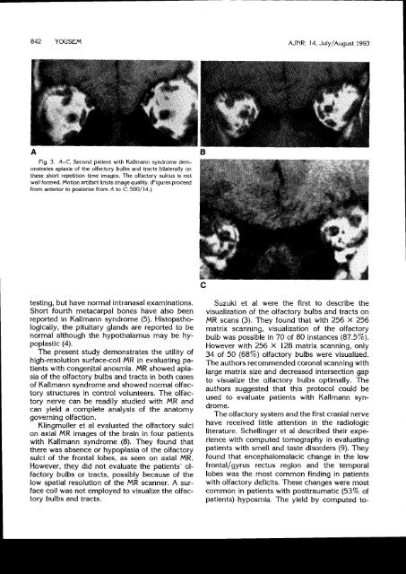
Fig. 3. A-C, Second patient with <strong>Kallmann</strong> syndrome demonstrates<br />
aplasia of the olfactory bulbs and tracts bilaterally on<br />
these short repetition time images. The olfactory sulcus is not<br />
well formed. Motion artifact limits image quality. (Figures proceed<br />
from anterior to posterior from A to C;500/14.\<br />
testing, but have normal intranasal examinations.<br />
Short fourth metacarpal bones have also been<br />
reported in <strong>Kallmann</strong> syndrome (5). Histopathologically,<br />
the pituitary glands are reported to be<br />
normal although the hypothalamus may be hypoplastic<br />
(4).<br />
The present study demonstrates the utility of<br />
high-resolution surface-coil <strong>MR</strong> in evaluating patients<br />
with congenital anosmia. <strong>MR</strong> showed aplasia<br />
of the olfactory bulbs and tracts in both cases<br />
of <strong>Kallmann</strong> syndrome and showed normal olfactory<br />
structutes in control volunteers. The olfactory<br />
nerve can be readily studied with <strong>MR</strong> and<br />
can yield a complete analysis of the anatomy<br />
governing olfaction.<br />
Klingmuller et al evaluated the olfactory sulci<br />
on axial <strong>MR</strong> images of the brain in four patients<br />
with <strong>Kallmann</strong> syndrome (B). They found that<br />
there was absence or hypoplasia of the olfactory<br />
sulci of the frontal lobes, as seen on axial <strong>MR</strong>.<br />
However, they did not evaluate the patients' olfactory<br />
bulbs or tracts, possibly because of the<br />
low spatial resolution of the <strong>MR</strong> scanner. A surface<br />
coil was not employed to visualize the olfactory<br />
bulbs and tracts.<br />
Suzuki et al were the first to describe the<br />
visualization of the olfactory bulbs and tracts on<br />
<strong>MR</strong> scans (3). They found that with 256 x 256<br />
matrix scanning, visualization of the olfactory<br />
bulb was possible in 70 of 80 instances (87.5%).<br />
However with 256 X 128 matrix scanning, only<br />
34 of 50 (68%) olfactory bulbs were visualized.<br />
The authors recommended coronalscanning with<br />
large matrix size and decreased intersection gap<br />
to visualize the olfactory bulbs optimally. The<br />
authors suggested that this protocol could be<br />
used to evaluate patients with <strong>Kallmann</strong> syndrome.<br />
The olfactory system and the first cranialnerve<br />
have received little attention in the radiologic<br />
literature. Schellinger et al described their experience<br />
with computed tomography in evaluating<br />
patients with smell and taste disorders (9). They<br />
found that encephalomalacic change in the low<br />
frontal/gyrus rectus region and the temporal<br />
lobes was the most common finding in patients<br />
with olfactory deficits. These changes were most<br />
common in patients with posttraumatic (53% of<br />
patients) hyposmia. The yield by computed to-