Clear Cell Papillary Renal Cell Carcinoma - NAJMS: The North ...
Clear Cell Papillary Renal Cell Carcinoma - NAJMS: The North ...
Clear Cell Papillary Renal Cell Carcinoma - NAJMS: The North ...
Create successful ePaper yourself
Turn your PDF publications into a flip-book with our unique Google optimized e-Paper software.
224 Oct 2012 Vol 5 No.4 <strong>North</strong> American Journal of Medicine and Science<br />
Review<br />
<strong>Clear</strong> <strong>Cell</strong> <strong>Papillary</strong> <strong>Renal</strong> <strong>Cell</strong> <strong>Carcinoma</strong><br />
– A New Emerging Entity<br />
Michael Sinnott, MD; 1 Frank Chen, MD, PhD; 2 Kazunori Kanehira MD; 1<br />
Nobert Sule MD, PhD; 1 Bo Xu, MD, PhD 1,2 *<br />
1<br />
Department of Pathology and Laboratory Medicine of Roswell Park Cancer Institute, Buffalo, NY<br />
2 Department of Pathology, State University of New York at Buffalo, Buffalo, NY<br />
<strong>Clear</strong> cell papillary renal cell carcinoma was initially reported in patients with end stage renal disease<br />
under name of “end stage renal disease-associated renal cell carcinoma”. However, subsequent studies<br />
show that the tumor is also seen in non-end stage settings with or without impaired renal function. Recent<br />
advances in molecular genetic techniques and immunohistochemical staining have showed that clear cell<br />
papillary renal cell carcinoma is a new distinct entity that has unique genetic, histomorphological and<br />
clinical characteristics. This review summarizes the most current views on clear cell papillary renal cell<br />
carcinoma with focus on histomorphological features, immunohistochemical profiles and molecular genetic<br />
characteristic of this new entity.<br />
[N A J Med Sci. 2012;5(4):224-227.]<br />
Key Words: clear cell papillary renal cell carcinoma, histomorphologic features, immunohistochemical<br />
and molecular-genetic characterization, pathoclinical correlation<br />
INTRODUCTION<br />
<strong>The</strong> first renal neoplasm was reported nearly 200 years ago. 1<br />
<strong>Clear</strong> cell renal cell carcinoma accounts for 60-70% of all<br />
cases of renal cell carcinoma and papillary renal cell<br />
carcinoma comprises approximately 10% of renal cell<br />
carcinoma. 2-3 Tumors arising in kidneys involved by end<br />
stage renal diseases may show distinctive histologic features<br />
not easily classified according to the current World Health<br />
Organization (WHO) classification System. 3-5 Tickoo et al<br />
described 15 cases of “clear-cell papillary renal cell<br />
carcinoma of end stage kidneys,” which were mainly cystic<br />
tumors and showed prominent papillary architecture with<br />
purely clear-cell cytology. 5 An association with end-stage<br />
renal disease was initially described for clear-cell papillary<br />
renal cell carcinoma, but many cases have now been<br />
documented outside of this setting. 6 This newly described<br />
low grade entity has been given many names such as “renal<br />
angiomyoadenomatous tumor”, “clear cell tubulopapillary<br />
renal cell carcinoma” and “clear cell papillary renal cell<br />
carcinoma”. 7-9 <strong>The</strong> nomenclature was discussed at the recent<br />
annual meeting of United States and Canadian Association of<br />
Pathologists (USCAP, 2011) and the name of “clear cell<br />
papillary renal cell carcinoma” was adopted. 10-11<br />
HISTOMORPHOLOGIC FEATURES OF CCPRCC<br />
Recently, three cases of clear cell papillary renal cell<br />
carcinoma were seen at our institution. Grossly, these tumors<br />
are well circumscribed and encapsulated with tan-pink to tanred,<br />
cystic cut surfaces. <strong>The</strong> tumors are localized to the renal<br />
Received 09/21/2012; Revised 10/10/2012; Accepted 10/11/2012<br />
*Corresponding Author: Department of Pathology and Laboratory<br />
Medicine, Roswell Park Cancer Institute, Elm and Carlton Streets, Buffalo,<br />
NY 14263. Tel: 716-845-5981. (Email: bo.xu@roswellpark.org)<br />
cortex without any infiltration into the perirenal or renal sinus<br />
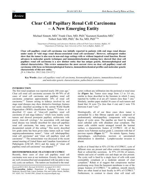
fat (Figure 1a). Tumor sizes range from 1.3 to 3.8 cm.,<br />
similar to those described in the literature in which 5 cases<br />
reported by Gobbo et al are pT1 tumors (less than 7 cm). 9<br />
Similarly, another paper studied 36 cases of such tumors and<br />
found that 34 were T1a (less than 4 cm) and 2 were T1b<br />
(betwenn 4 and 7 cm). 8<br />
Histologically, all of our three cases show tumor is<br />
surrounded by a thin fibrous capsule and is composed of<br />
predominantly tubulopapillary components with varying<br />
degree of cystic areas. <strong>The</strong> papillae are lined by medium<br />
sized cuboidal cells with abundant clear cytoplasm. <strong>The</strong><br />
tumor cells exhibit nuclei arranged toward the luminal<br />
surface, a characteristic feature of CCPRCC. All of the<br />
tumors were Fuhrman nuclear grade 2, consistent with that of<br />
previous reports (Figure 1). 8-12 No mitotic figures, foamy<br />
macrophages, calcifications, necrosis or sarcomatoid<br />
differentiation is identified. Lymphovascular invasion is<br />
absent.<br />
Aydin et al found that in 92% (33/36) of CCPRCC, cysts of<br />
different sizes are present and often contain serosanguinous<br />
fluid or colloid-like secretions. In 97% (35/36) of tumors,<br />
tubules and acini of variable size and shape are also present.<br />
<strong>The</strong> cystic wall is lined by a single layer of cells with scant<br />
eosinophilic cytoplasm or a moderate amount of clear<br />
cytoplasm. 8 Rohan et al suggest that conspicuous nuclear<br />
positioning away from the basement membrane is one of the<br />
unique features of CCPRCC. 13 In addition, smooth muscle<br />
fibers are often found at least focally in 69% of CCPRCC. 8<br />
Furthermore, in contrast to classic clear cell renal cell
<strong>North</strong> American Journal of Medicine and Science Oct 2012 Vol 5 No.4 225<br />
carcinoma, CCPRCC lacks a delicate sinusoidal vascular<br />
network. 9<br />
If one is not aware of this new entity, CCPRCC can be<br />
confused morphologically with other renal cell carcinomas,<br />
particularly clear cell carcinoma or papillary carcinoma and<br />
their variants. However, conventional clear cell RCC and<br />
papillary RCC often exhibit more aggressive pathologic<br />
features, including larger tumor size, higher Fuhrman grade<br />
nuclei (3 and 4), coagulative necrosis, perirenal fat<br />
involvement, vascular invasion, lymph node metastasis, and<br />
sarcomatoid differentiation. None of these are seen in<br />
CCPRCC. 14 Occasionally, however, immunohistochemistry<br />
studies may be needed for definitive classification.<br />
Figure 1. A. Gross image of clear cell papillary renal cell carcinoma. Tumor is well circumscribed and<br />
encapsulated with tan-pink to tan-red cut surface. A cystic area is also evident. B. At low power view, the tumor<br />
is tubulopapillary with microcysts. C and D. At higher power view, the tumor has a nested and papillary<br />
patterns. Tumor cells have abundant cytoplasm, low grade nuclei and nuclei located away from the basement<br />
membrane.<br />
IMMUNOHISTOCHEMISTRY PROFILES OF<br />
CCPRCC<br />
CCPRCC has a distinctive immunoprofile. Gobbo et al found<br />
that all tumors show strong and diffuse positive<br />
immunohistochemical staining for cytokeratin 7 and carbonic<br />
anhydrase IX. <strong>The</strong> tumor cells are negative for alphamethylacyl-CoA<br />
racemase (AMACR), CD10, and<br />
transcription factor E3. 6 Another study reported that most<br />
tumors are negative for CD10 except few are focally positive<br />
with expression limited to 1-10% of the tumor cells. 8 This<br />
study also found that staining for TFE3 protein, a distinctive<br />
and diagnostic feature of Xp11.2 translocation RCC, was not<br />
identified in any of their tumors. 15,16 Conventional clear cell<br />
renal cell carcinomas typically show immunoreactivity to<br />
antibodies against CA-IX and CD10 and lack<br />
immunoreactivity for CK7. 17-19 <strong>Papillary</strong> renal cell<br />
carcinomas characteristically show positive immunostaining<br />
for AMACR and CK7, 17-20 but less frequently express CA-IX<br />
and/or CD10. 21 Also of note, Xp11.2 translocation associated<br />
renal cell carcinomas do not express CK7 and usually show<br />
positive immunostaining for CD10. 16 In addition to CA IX,<br />
another study evaluated immunoexpression of two other<br />
markers of HIF pathway activation, HIF-1α and GLUT-1.<br />
<strong>The</strong> majority of CCPRCC express both of these markers as<br />
clear cell renal cell carcinoma does. 13 In addition, Wolfe et al<br />
reported focal to abundant disorganized bundles of spindle<br />
cells with eosinophilic cytoplasm and cigar-shaped nuclei<br />
that expressed desmin and h-caldesmon, consistent with<br />
smooth muscle in 4 out of 4 cases. 22<br />
In our recent cases, immunohistochemical studies were<br />
performed. As iilustrated in Figure 2, tumor cells are<br />
strongly and diffusely positive for CK7, PAX-2 and<br />
vimentin. Immunostain for high molecular weight cytokeratin<br />
was focally positive. Tumor cells are negative for AMACR<br />
and CD10 (Figure 2).
226 Oct 2012 Vol 5 No.4 <strong>North</strong> American Journal of Medicine and Science<br />
Figure 2. Immunohistochemical studies of CCPRCC. A. CK7; B. PAX-2; C. Vimentin; D. High molecular<br />
weight cytokeratin; E. CD10; F. Racemase.<br />
MOLECULAR GENETIC CHARACTERISTICS OF<br />
CCPRCC<br />
<strong>The</strong> genetic abnormalities of several types of renal cell<br />
carcinomas have been well described. <strong>Clear</strong> cell renal<br />
carcinoma often has deletions in chromosome 3p and /or<br />
mutations in VHL genes, while papillary renal cell carcinoma<br />
often has a trisomy of chromosomes 7 or 17 and loss of the Y<br />
chromosome. 3 Aydin et al, found that none of the 36 cases of<br />
CCPRCC display gains of chromosome 7 or loss of Y<br />
chromosome, a typical feature of papillary renal cell<br />
carcinoma (PRCC), and none of the cases has deletion of 3p<br />
a typical feature of clear cell renal cell carcinoma (CCRCC). 8<br />
Another study showed monoclonal gains of chromosomes 7,<br />
10 and 12 which were detected in one case of CCPRCC, and<br />
their presence was verified by fluorescence in situ<br />
hybridization (FISH) studies. 22 On the other hand, Michel et<br />
al found Trisomy 7 together with monosomy 17, 16 and 20 in<br />
one case of CCPRCC. Trisomy 17 was seen in another case<br />
of CCPRCC. No other chromosomal gains or losses were<br />
found. 7 In addition, Gobbo et al reported in contrast to the<br />
control cases of clear cell RCC and papillary RCC, 3p<br />
deletion was not observed in any case of CCPRCC.<br />
Furthermore, chromosomes 7 and Y were numerically<br />
normal in all of their cases and only 1 of 5 cases showed gain<br />
of chromosome 17. 7 Using quantitative RT-PCR approach,<br />
Rohan et al showed relative over expression of VHL mRNA<br />
in CCPRCC compared with non-neoplastic renal parenchyma<br />
and clear cell RCC cases harboring 3p losses andor VHL<br />
mutations. <strong>The</strong>se finding supports the notion that the<br />
molecular mechanism underlying CCPRCC is distinct from<br />
that of clear cell RCC or papillary RCC and is not related to<br />
abrogation of VHL signaling. 7
<strong>North</strong> American Journal of Medicine and Science Oct 2012 Vol 5 No.4 227<br />
SUMMARY<br />
CCPRCC is a new entity described since the 2004 World<br />
Health Organization classification of renal tumors. Due to its<br />
unique histomorphology, immunohistochemical staining<br />
pattern, molecular genetic features and favorable clinical<br />
outcome, CCPRCC deserves a separate entity and will be<br />
included in the newer edition of WHO classification of renal<br />
tumors. Anecdotally, the authors feel that CCPRCC is not as<br />
rare as one may expect since we have seen three<br />
immunohistochemically proven cases in a few months in our<br />
institution. We feel that pathologists need to be aware of this<br />
new entity. <strong>The</strong> morphology and immunohistochemical<br />
staining pattern is quite characteristic of this tumor, and it is<br />
relatively easy to confirm the diagnosis. It is worth noting<br />
that recognizing this entity is important as its prognosis is<br />
much better than that of both clear cell RCC and papillary<br />
RCC. No recurrence, lymph node or other metastases have<br />
been reported to date for CCPRCC.<br />
CONFLICT OF INTEREST<br />
<strong>The</strong> authors have no conflict of interest to disclose.<br />
REFERENCES<br />
1. Delahunt B, Eble JN. History of the development of the classification<br />
of renal cell neoplasia. Clin Lab Med. 2005;25(2):231-246.<br />
2. Reuter VE, Presti JC, Jr. Contemporary approach to the classification<br />
of renal epithelial tumors. Semin Oncol. 2000;27(2):124-137.<br />
3. Eble J, Sauter G, Epstein J, et al. World Health Organization<br />
Classification of Tumours: Pathology and Genetics of Tumours of the<br />
Urinary System and Male Genital Organs. Lyon: IARC Press; 2004.<br />
4. Sule N, Yakupoglu U, Shen SS, et al. Calcium oxalate deposition in<br />
renal cell carcinoma associated with acquired cystic kidney disease: a<br />
comprehensive study. Am J Surg Pathol. 2005;29(4):443–451.<br />
5. Tickoo SK, dePeralta-Venturina MN, Harik LR, et al. Spectrum of<br />
epithelial neoplasms in end-stage renal disease: an experience from 66<br />
tumor-bearing kidneys with emphasis on histologic patterns distinct<br />
from those in sporadic adult renal neoplasia. Am J Surg Pathol.<br />
2006;30(2):141–153.<br />
6. Michal M, Hes O, Nemcova J, et al. <strong>Renal</strong> angiomyoadenomatous<br />
tumor: morphologic, immunohistochemical, and molecular genetic<br />
study of a distinct entity. Virchows Arch. 2009; 454(1):89–99.<br />
7. Michal M, Hes O, Havlicek F. Benign renal angiomyoadenomatous<br />
tumor: a previously unreported renal tumor. Ann Diagn Pathol.<br />
2000;4(5):311–315.<br />
8. Aydin H, Chen L, Cheng L, et al. <strong>Clear</strong> cell tubulopapillary renal cell<br />
carcinoma: a study of 36 distinctive low-grade epithelial tumors of the<br />
kidney. Am J Surg Pathol. 2010;34(11):1608–1621.<br />
9. Gobbo S, Eble JN, Grignon DJ, et al. <strong>Clear</strong> cell papillary renal cell<br />
carcinoma: a distinct histopathologic and molecular genetic entity. Am<br />
J Surg Pathol. 2008;32(8):1239–1245.<br />
10. Behdad A, Monzon FA, Hes O, et al. Relationship between sporadic<br />
clear cell-papillary renal cell carcinoma (CP-RCC) and renal<br />
angiomyoadenomatous tumor (RAT) of the kidney: analysis by virtualkaryotyping,<br />
fluorescent in situ analysis and immunohistochemistry<br />
(IHC). Mod Pathol. 2011;24(Suppl):179A.<br />
11. Herrera LP, Hes O, Hirsch M, et al. <strong>Clear</strong> cell-papillary renal cell<br />
carcinoma (CP-RCC) not associated with end stage renal disease:<br />
clinicopathologic analysis of 50 tumors confirming a novel subtype of<br />
renal cell carcinoma (RCC) occurring in a sporadic setting. Mod<br />
Pathol. 2011;24(Suppl):197A.<br />
12. Fuhrman SA, Lasky LC, Limas C. Prognostic significance of<br />
morphologic parameters in renal cell carcinoma. Am J Surg Pathol.<br />
1982;6(7):655–663.<br />
13. Stephen M Rohan, Yonghong Xiao, et al. <strong>Clear</strong>-cell papillary renal cell<br />
carcinoma: molecular and immunohistochemical analysis with<br />
emphasis on the von Hippel–Lindau gene and hypoxia-inducible factor<br />
pathway-related proteins. Modern Pathol. 2011;24(9):1207–1220.<br />
14. Mai KT, Faraji H, Desantis D, et al. <strong>Renal</strong> cell carcinoma with mixed<br />
features of papillary and clear cell cytomorphology: a fluorescent in<br />
situ hybridization study. Virchows Arch. 2010;456(1):77–84.<br />
15. Argani P, Ladanyi M. Translocation carcinomas of the kidney. Clin<br />
Lab Med. 2005;25(2):363–378.<br />
16. Argani P, Olgac S, Tickoo SK, et al. Xp11 translocation renal cell<br />
carcinoma in adults: expanded clinical, pathologic, and genetic<br />
spectrum. Am J Surg Pathol. 2007;31(8):1149–1160.<br />
17. Avery AK, Beckstead J, Renshaw AA, et al. Use of antibodies to RCC<br />
and CD10 in the differential diagnosis of renal neoplasms. Am J Surg<br />
Pathol. 2000;24(2):203–210.<br />
18. Kim MK, Kim S. Immunohistochemical profile of common epithelial<br />
neoplasms arising in the kidney. Appl Immunohistochem Mol<br />
Morphol. 2002;10(4):332–338.<br />
19. Tretiakova MS, Sahoo S, Takahashi M, et al. Expression of alphamethylacyl-CoA<br />
racemase in papillary renal cell carcinoma. Am J Surg<br />
Pathol. 2004;28(1):69–76.<br />
20. Delahunt B, Eble JN. <strong>Papillary</strong> renal cell carcinoma: a<br />
clinicopathologic and immunohistochemical study of 105 tumors. Mod<br />
Pathol. 1997;10(6):537–544.<br />
21. Martignoni G, Brunelli M, Gobbo S, et al. Role of molecular markers<br />
in diagnosis and prognosis of renal cell carcinoma. Anal Quant Cytol<br />
Histol. 2007;29(1):41–49.<br />
22. Wolfe A, Dobin SM, Grossmann P, Michal M, Donner LR. Clonal<br />
trisomies 7, 10 and 12, normal 3p and absence of VHL gene mutation<br />
in a clear cell tubulopapillary carcinoma of the kidney. Virchows Arch.<br />
2011;459(4):457-463.


