Clear Cell Papillary Renal Cell Carcinoma - NAJMS: The North ...
Clear Cell Papillary Renal Cell Carcinoma - NAJMS: The North ...
Clear Cell Papillary Renal Cell Carcinoma - NAJMS: The North ...

Create successful ePaper yourself
Turn your PDF publications into a flip-book with our unique Google optimized e-Paper software.
<strong>North</strong> American Journal of Medicine and Science Oct 2012 Vol 5 No.4 225<br />
carcinoma, CCPRCC lacks a delicate sinusoidal vascular<br />
network. 9<br />
If one is not aware of this new entity, CCPRCC can be<br />
confused morphologically with other renal cell carcinomas,<br />
particularly clear cell carcinoma or papillary carcinoma and<br />
their variants. However, conventional clear cell RCC and<br />
papillary RCC often exhibit more aggressive pathologic<br />
features, including larger tumor size, higher Fuhrman grade<br />
nuclei (3 and 4), coagulative necrosis, perirenal fat<br />
involvement, vascular invasion, lymph node metastasis, and<br />
sarcomatoid differentiation. None of these are seen in<br />
CCPRCC. 14 Occasionally, however, immunohistochemistry<br />
studies may be needed for definitive classification.<br />
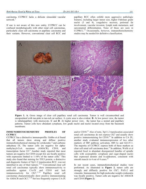
Figure 1. A. Gross image of clear cell papillary renal cell carcinoma. Tumor is well circumscribed and<br />
encapsulated with tan-pink to tan-red cut surface. A cystic area is also evident. B. At low power view, the tumor<br />
is tubulopapillary with microcysts. C and D. At higher power view, the tumor has a nested and papillary<br />
patterns. Tumor cells have abundant cytoplasm, low grade nuclei and nuclei located away from the basement<br />
membrane.<br />
IMMUNOHISTOCHEMISTRY PROFILES OF<br />
CCPRCC<br />
CCPRCC has a distinctive immunoprofile. Gobbo et al found<br />
that all tumors show strong and diffuse positive<br />
immunohistochemical staining for cytokeratin 7 and carbonic<br />
anhydrase IX. <strong>The</strong> tumor cells are negative for alphamethylacyl-CoA<br />
racemase (AMACR), CD10, and<br />
transcription factor E3. 6 Another study reported that most<br />
tumors are negative for CD10 except few are focally positive<br />
with expression limited to 1-10% of the tumor cells. 8 This<br />
study also found that staining for TFE3 protein, a distinctive<br />
and diagnostic feature of Xp11.2 translocation RCC, was not<br />
identified in any of their tumors. 15,16 Conventional clear cell<br />
renal cell carcinomas typically show immunoreactivity to<br />
antibodies against CA-IX and CD10 and lack<br />
immunoreactivity for CK7. 17-19 <strong>Papillary</strong> renal cell<br />
carcinomas characteristically show positive immunostaining<br />
for AMACR and CK7, 17-20 but less frequently express CA-IX<br />
and/or CD10. 21 Also of note, Xp11.2 translocation associated<br />
renal cell carcinomas do not express CK7 and usually show<br />
positive immunostaining for CD10. 16 In addition to CA IX,<br />
another study evaluated immunoexpression of two other<br />
markers of HIF pathway activation, HIF-1α and GLUT-1.<br />
<strong>The</strong> majority of CCPRCC express both of these markers as<br />
clear cell renal cell carcinoma does. 13 In addition, Wolfe et al<br />
reported focal to abundant disorganized bundles of spindle<br />
cells with eosinophilic cytoplasm and cigar-shaped nuclei<br />
that expressed desmin and h-caldesmon, consistent with<br />
smooth muscle in 4 out of 4 cases. 22<br />
In our recent cases, immunohistochemical studies were<br />
performed. As iilustrated in Figure 2, tumor cells are<br />
strongly and diffusely positive for CK7, PAX-2 and<br />
vimentin. Immunostain for high molecular weight cytokeratin<br />
was focally positive. Tumor cells are negative for AMACR<br />
and CD10 (Figure 2).