Examination of Foods for Staphylococcus aureus - DB Server Test ...
Examination of Foods for Staphylococcus aureus - DB Server Test ...
Examination of Foods for Staphylococcus aureus - DB Server Test ...

You also want an ePaper? Increase the reach of your titles
YUMPU automatically turns print PDFs into web optimized ePapers that Google loves.
Experiment 12: <strong>Examination</strong> <strong>of</strong> <strong>Foods</strong> <strong>for</strong><br />
<strong>Staphylococcus</strong> <strong>aureus</strong><br />
Based on U.S. FDA Bacteriological Analytical Manual, 8th Edition, Current through Revision A,<br />
1998, Chapter 12: <strong>Staphylococcus</strong> <strong>aureus</strong>, Reginald W. Bennett and Gayle A. Lancette<br />
Objective:<br />
1. To learn how to isolate, enumerate and confirm <strong>Staphylococcus</strong> <strong>aureus</strong> in foods.<br />
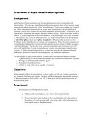
Background:<br />
<strong>Staphylococcus</strong> <strong>aureus</strong> is highly vulnerable to destruction by heat treatment and nearly all<br />
sanitizing agents. Thus, the presence <strong>of</strong> this bacterium or its enterotoxins in processed<br />
foods or on food processing equipment is generally an indication <strong>of</strong> poor sanitation. S.<br />
<strong>aureus</strong> can cause severe food poisoning. It has been identified as the causative agent in<br />
many food poisoning outbreaks and is probably responsible <strong>for</strong> even more cases in<br />
individuals and family groups than the records show. <strong>Foods</strong> are examined <strong>for</strong> the<br />
presence <strong>of</strong> S. <strong>aureus</strong> and/or its enterotoxins to confirm that S. <strong>aureus</strong> is the causative<br />
agent <strong>of</strong> foodborne illness, to determine whether a food is a potential source <strong>of</strong> "staph"<br />
food poisoning, and to demonstrate post-processing contamination, which is generally<br />
due to human contact or contaminated food-contact surfaces. Conclusions regarding the<br />
significance <strong>of</strong> S. <strong>aureus</strong> in foods should be made with circumspection. The presence <strong>of</strong> a<br />
large number <strong>of</strong> S. <strong>aureus</strong> organisms in a food may indicate poor handling or sanitation;<br />
however, it is not sufficient evidence to incriminate a food as the cause <strong>of</strong> food<br />
poisoning. The isolated S. <strong>aureus</strong> must be shown to produce enterotoxins. Conversely,<br />
small staphylococcal populations at the time <strong>of</strong> testing may be remnants <strong>of</strong> large<br />
populations that produced enterotoxins in sufficient quantity to cause food poisoning.<br />
There<strong>for</strong>e, the analyst should consider all possibilities when analyzing a food <strong>for</strong> S.<br />
<strong>aureus</strong>.<br />
Methods used to detect and enumerate S. <strong>aureus</strong> depend on the reasons <strong>for</strong> testing the<br />
food and on the past history <strong>of</strong> the test material. Processed foods may contain relatively<br />
small numbers <strong>of</strong> debilitated viable cells, whose presence must be demonstrated by<br />
appropriate means. Analysis <strong>of</strong> food <strong>for</strong> S. <strong>aureus</strong> may lead to legal action against the<br />
party or parties responsible <strong>for</strong> a contaminated food. The methods <strong>of</strong> analysis <strong>for</strong> S.<br />
<strong>aureus</strong> that have been studied collaboratively and found suitable <strong>for</strong> use in providing the<br />
type <strong>of</strong> in<strong>for</strong>mation necessary <strong>for</strong> FDA requirements are presented in this chapter.<br />
There has been considerable controversy about the significance and correct method <strong>of</strong><br />
reading the coagulase test. Research results have indicated that the weak coagulase<br />
activity represented by 1+, 2+, and 3+ reactions seldom corresponds with other criteria<br />
associated with S. <strong>aureus</strong> (4). A consensus <strong>of</strong> peers has established that a 4+ coagulase<br />
reaction is necessary <strong>for</strong> unquestioned identification <strong>of</strong> S. <strong>aureus</strong>. Those strains suspected<br />
<strong>of</strong> being S. <strong>aureus</strong> on the basis <strong>of</strong> coagulase reactions <strong>of</strong> less than 4+ should be confirmed<br />
by other tests, such as anaerobic glucose fermentation, lysostaphin sensitivity, and<br />
thermonuclease production. Studies <strong>of</strong> colonial morphology on Baird-Parker agar,<br />
1
lysostaphin sensitivity, coagulase and thermonuclease production, and glucose and<br />
mannitol fermentation were conducted on 100 enterotoxigenic and 51 nonenterotoxigenic<br />
strains <strong>of</strong> S. <strong>aureus</strong> (3). In all cases, the reactions <strong>of</strong> enterotoxigenic and<br />
nonenterotoxigenic strains varied by 12% or less. This research indicates that none <strong>of</strong><br />
these tests can be relied upon to differentiate toxic and nontoxic staphylococci.<br />
Experiment:<br />
Direct Plate Count Method<br />
This method is suitable <strong>for</strong> the analysis <strong>of</strong> foods in which more than 100 S. <strong>aureus</strong> cells/g<br />
may be expected. It con<strong>for</strong>ms to the method in ref. 1.<br />
A. Materials<br />
Tuesday, October 15: Plating on Baird Parker Medium plates, incubating plates at 35 o C<br />
<strong>for</strong> 48 hours, plating on 3M Petrifilm Rapid S. <strong>aureus</strong> Count Plates, incubating<br />
plates at 35 o C <strong>for</strong> 24 hours<br />
1. Food sample<br />
2. Sterile spatulas: 1 per team<br />
3. Scale<br />
4. Sterile stomacher bags: 1 per team<br />
5. Butterfield's phosphate-buffered dilution water: One 225-ml flask per team<br />
6. Butterfield's phosphate-buffered dilution water: Four 90-ml bottles per team<br />
7. Sterile 10-ml pipettes<br />
8. Sterile 1-ml tips (blue)<br />
9. Sterile 0.2-ml tips (yellow)<br />
10. Baird Parker Medium plates: 15 plates per team<br />
11. 3M Petrifilm Rapid S. <strong>aureus</strong> Count Plates: 5 plates per team<br />
Wednesday, October 16: Transferring 3M Petrifilm Rapid S. <strong>aureus</strong> Count Plates to<br />
a 62 o C incubator, incubating plates 1-4 hours, placing reactive disks, incubating plates<br />
with reactive disks at 35 or 37 o C <strong>for</strong> 1-3 hours, counting and recording colonies<br />
1. Sterile <strong>for</strong>ceps: 1 per team<br />
2. 62 o C incubator<br />
3. Colony counter<br />
Thursday, October 17: Counting and recording colonies, transferring suspect S. <strong>aureus</strong><br />
colonies into BHI broth, inoculating agar slants <strong>of</strong> TSA, incubating BHI culture<br />
suspension at 35 o C <strong>for</strong> 18-24 hours, incubating agar slants <strong>of</strong> TSA at room temperature<br />
<strong>for</strong> 18-24 hours<br />
1. Colony counters<br />
2. Brain Heart Infusion (BHI) Broth: 0.2-0.4 ml per tubes; 3 tubes per team<br />
3. Trypticase Soy Agar (TSA) slants: 3 slants per team<br />
2
Tuesday, October 22: Catalase test, adding coagulase plasma to the BHI culture and<br />
incubating at 35oC and examining periodically <strong>for</strong> clot <strong>for</strong>mation, Gram stain (from TSA<br />
slants), inoculating carbohydrate fermentation medium containing glucose (0.5%),<br />
inoculating carbohydrate fermentation medium containing mannitol (0.5%), testing <strong>for</strong><br />
lysostaphin sensitivity, testing <strong>for</strong> thermostable nuclease production<br />
1. 3% Hydrogen peroxide<br />
2. Reconstituted coagulase plasma(rabbit) with EDTA: 1.5 ml per team<br />
3. Gram stain materials<br />
4. Carbohydrate fermentation medium containing glucose (0.5%): 5 tubes per team<br />
5. Positive and negative controls (bacterial cultures that ferment and do not ferment<br />
glucose): One set per bench<br />
6. Carbohydrate fermentation medium containing mannitol (0.5%): 5 tubes per team<br />
7. Positive and negative controls (bacterial cultures that ferment and do not ferment<br />
mannitol): One set per bench<br />
8. Sterile paraffin oil: Enough to cover 100 tubes<br />
9. Phosphate-saline buffer: 0.2 ml per tube; 3 tubes per team<br />
10. Phosphate-saline buffer: 0.1 ml per tube; 3 tubes per team<br />
11. Lysostaphin (50 µg/ml) dissolved in 0.02 M phosphate-saline buffer containing<br />
1% NaCl: 0.3 ml per team<br />
12. DNase <strong>Test</strong> Agar with Methyl Green plates: 1 plate per team<br />
Thursday, October 24: Read the results <strong>of</strong> carbohydrate fermentation tests. No materials<br />
needed.<br />
B. Preparation <strong>of</strong> sample<br />
Using aseptic technique, weigh 25 g <strong>of</strong> sample into sterile stomacher bag. Add 225 ml<br />
Butterfield's phosphate-buffered dilution water (1:10 dilution) and blend <strong>for</strong> 1 min at high<br />
speed. Using the 1:10 dilution, make serial dilutions <strong>of</strong> sample <strong>for</strong> enumeration <strong>of</strong> S.<br />
<strong>aureus</strong>.<br />
C. Isolation and enumeration <strong>of</strong> S. <strong>aureus</strong><br />
1. For each dilution to be plated, aseptically transfer 1 ml sample suspension to 3 plates<br />
<strong>of</strong> Baird-Parker agar, distributing 1 ml <strong>of</strong> inoculum equitably to 3 plates (e.g., 0.4 ml, 0.3<br />
ml, and 0.3 ml). Spread inoculum over surface <strong>of</strong> agar plate, using sterile bent glass<br />
streaking rod. Retain plates in upright position until inoculum is absorbed by agar (about<br />
10 min on properly dried plates). If inoculum is not readily adsorbed, place plates upright<br />
in incubator <strong>for</strong> about 1 h. Invert plates and incubate 45-48 h at 35°C. Select plates<br />
containing 20-200 colonies, unless only plates at lower dilutions (>200 colonies) have<br />
colonies with typical appearance <strong>of</strong> S. <strong>aureus</strong>. Colonies <strong>of</strong> S. <strong>aureus</strong> are circular, smooth,<br />
convex, moist, 2-3 mm in diameter on uncrowded plates, gray to jet-black, frequently<br />
with light-colored (<strong>of</strong>f-white) margin, surrounded by opaque zone and frequently with an<br />
outer clear zone; colonies have buttery to gummy consistency when touched with<br />
inoculating needle. Occasionally from various foods and dairy products, nonlipolytic<br />
strains <strong>of</strong> similar appearance may be encountered, except that surrounding opaque and<br />
clear zones are absent. Strains isolated from frozen or desiccated foods that have been<br />
3
stored <strong>for</strong> extended periods frequently develop less black coloration than typical colonies<br />
and may have rough appearance and dry texture.<br />
2. For each dilution to be plated, place 1 ml <strong>of</strong> sample suspension onto center bottom film<br />
<strong>of</strong> 3M Petrifilm Rapid S. <strong>aureus</strong> Count Plates. Refer to the “Reminders <strong>of</strong> Use <strong>for</strong><br />
3M Petrifilm Rapid S. <strong>aureus</strong> Count Plates” that is attached to your handout.<br />
Incubate plates with clear side up in stacks <strong>of</strong> up to 10. Incubate at 35 o C or 37 o C <strong>for</strong> 24<br />
hours.<br />
3. Refer to the “Reminders <strong>of</strong> Use <strong>for</strong> 3M Petrifilm Rapid S. <strong>aureus</strong> Count<br />
Plates” that is attached to your handout. Transfer petrifilm plates to a 62 o C incubator.<br />
Incubate <strong>for</strong> 1-4 hours. With sterile <strong>for</strong>ceps, remove the round reactive disk from the<br />
outer square frame. Lift the top film <strong>of</strong> the Petrifilm plate and place the Petrifilm reactive<br />
disk in the well <strong>of</strong> the plate. Lower the top film. Apply gentle pressure across the reactive<br />
disk. Incubate plates with inserted Petrifilm reactive disks <strong>for</strong> 1-3 hours at 35 o C or 37 o C.<br />
4. Read and record results. Refer to the “Interpretation Guide Section” that is<br />
attached to your handout.<br />
Dilution 10 -1<br />
Dilution 10- 2<br />
Dilution 10 -3<br />
Dilution 10- 4<br />
Dilution 10 -5<br />
S. <strong>aureus</strong> count<br />
5. Count and record colonies on Baird-Parker Agar plates. If several types <strong>of</strong> colonies are<br />
observed which appear to be S. <strong>aureus</strong> on selected plates, count number <strong>of</strong> colonies <strong>of</strong><br />
each type and record counts separately. When plates <strong>of</strong> the lowest dilution contain 200 colonies have colonies with the<br />
typical appearance <strong>of</strong> S. <strong>aureus</strong> and typical colonies do not appear at higher dilutions, use<br />
these plates <strong>for</strong> the enumeration <strong>of</strong> S. <strong>aureus</strong>, but do not count nontypical colonies. Select<br />
> 1 colony <strong>of</strong> each type counted and test <strong>for</strong> coagulase production. Add number <strong>of</strong><br />
colonies on triplicate plates represented by colonies giving positive coagulase test and<br />
multiply by the sample dilution factor. Report this number as number <strong>of</strong> S. <strong>aureus</strong>/g <strong>of</strong><br />
food tested.<br />
cfu [Plate 1 (0.4<br />
ml)]<br />
cfu [Plate 2 (0.3<br />
ml)]<br />
cfu [Plate 3 (0.3<br />
ml)]<br />
cfu/g food<br />
Dilution 10 -2<br />
4
Dilution 10 -3<br />
Dilution 10 -4<br />
Dilution 10 -5<br />
D. Coagulase test and Gram staining<br />
Transfer suspect S. <strong>aureus</strong> colonies into small tubes containing 0.2-0.3 ml BHI broth and<br />
emulsify thoroughly. Inoculate agar slant <strong>of</strong> suitable maintenance medium, e.g., TSA,<br />
with loopful <strong>of</strong> BHI suspension. Incubate BHI culture suspension and slants 18-24 h at<br />
35°C. Retain slant cultures at room temperature <strong>for</strong> ancillary or repeat tests in case<br />
coagulase test results are questionable. Take 100 µl <strong>of</strong> BHI culture suspensions and<br />
keep it in a sterile tube <strong>for</strong> thermostable nuclease production test. Add 0.5 ml<br />
reconstituted coagulase plasma with EDTA to the BHI culture and mix thoroughly.<br />
Incubate at 35°C and examine periodically over 6 h period <strong>for</strong> clot <strong>for</strong>mation. Only firm<br />
and complete clot that stays in place when tube is tilted or inverted is considered positive<br />
<strong>for</strong> S. <strong>aureus</strong>. Partial clotting, <strong>for</strong>merly 2+ and 3+ coagulase reactions, must be tested<br />
further (4). <strong>Test</strong> known positive and negative cultures simultaneously with suspect<br />
cultures <strong>of</strong> unknown coagulase activity. Stain all suspect cultures with Gram reagent and<br />
observe microscopically. A latex agglutination test (AUREUS TEST TM , Trisum Corp.,<br />
Taipei, Taiwan) may be substituted <strong>for</strong> the coagulase test if a more rapid procedure is<br />
desired.<br />
Colony 1<br />
Colony 2<br />
Colony 3<br />
E. Ancillary tests<br />
Coagulase activity (clot <strong>for</strong>mation)<br />
1. Catalase test. Use growth from TSA slant <strong>for</strong> catalase test on glass slide or spot plate,<br />
and illuminate properly to observe production <strong>of</strong> gas bubbles.<br />
Colony 1<br />
Colony 2<br />
Colony 3<br />
Catalase activity (gas bubbles)<br />
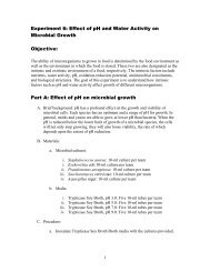
2. Anaerobic utilization <strong>of</strong> glucose. Inoculate tube <strong>of</strong> carbohydrate fermentation<br />
medium containing glucose (0.5%). Immediately inoculate each tube heavily with wire<br />
loop. Make certain inoculum reaches bottom <strong>of</strong> tube. Cover surface <strong>of</strong> agar with layer <strong>of</strong><br />
sterile paraffin oil at least 25 mm thick. Incubate 5 days at 37°C. Acid is produced<br />
anaerobically if indicator changes to yellow throughout tube, indicating presence <strong>of</strong> S.<br />
5
<strong>aureus</strong>. Run controls simultaneously (positive and negative cultures and medium<br />
controls).<br />
Colony 1<br />
Colony 2<br />
Colony 3<br />
Positive control<br />
Negative control<br />
Anaerobic utilization <strong>of</strong> glucose (yellow<br />
color)<br />
3. Anaerobic utilization <strong>of</strong> mannitol. Repeat 2, above, using mannitol as carbohydrate<br />
in medium. S. <strong>aureus</strong> is usually positive but some strains are negative. Run controls<br />
simultaneously.<br />
Colony 1<br />
Colony 2<br />
Colony 3<br />
Positive control<br />
Negative control<br />
Anaerobic utilization <strong>of</strong> mannitol (yellow<br />
color)<br />
4. Lysostaphin sensitivity. Transfer isolated colony from agar plate with inoculating<br />
loop to 0.2 ml phosphate-saline buffer, and emulsify. Transfer half <strong>of</strong> suspended cells to<br />
another tube (13 x 100 mm) containing 0.1 ml phosphate-saline buffer as control. Add<br />
0.1 ml lysostaphin (dissolved in 0.02 M phosphate-saline buffer containing 1% NaCl) to<br />
original tube <strong>for</strong> concentration <strong>of</strong> 25 µg lysostaphin/ml. Incubate both tubes at 35°C <strong>for</strong><br />
not more than 2 h. If turbidity clears in test mixture, test is considered positive. If clearing<br />
has not occurred in 2 h, test is negative. S. <strong>aureus</strong> is generally positive.<br />
Lysostaphin sensitivity<br />
(clearing <strong>of</strong> turbidity)<br />
Tube 1 (Phosphate-saline<br />
buffer + lysostaphin)<br />
Control (turbidity)<br />
Tube 2 (Phosphate-saline<br />
buffer)<br />
Colony 1<br />
Colony 2<br />
6
Colony 3<br />
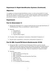
5. Thermostable nuclease production. Please note that we will per<strong>for</strong>m II. Section I is<br />
an alternative and is included <strong>for</strong> your in<strong>for</strong>mation.<br />
I. This test is claimed to be as specific as the coagulase test but less subjective, because it<br />
involves a color change from blue to bright pink. It is not a substitute <strong>for</strong> the coagulase<br />
test but rather is a supportive test, particularly <strong>for</strong> 2+ coagulase reactions. Prepare<br />
microslides by spreading 3 ml toluidine blue-deoxyribonucleic acid agar on the surface <strong>of</strong><br />
each microscope slide. When agar has solidified, cut 2 mm diameter wells (10-12 per<br />
slide) in agar and remove agar plug by aspiration. Add about 0.01 ml <strong>of</strong> heated sample<br />
(15 min in boiling water bath) <strong>of</strong> broth cultures used <strong>for</strong> coagulase test to well on<br />
prepared slide. Incubate slides in moist chamber 4 h at 35°C. Development <strong>of</strong> bright pink<br />
halo extending at least 1 mm from periphery <strong>of</strong> well indicates a positive reaction.<br />
II. Heat (15 min in boiling water bath) BHI culture suspensions (100 µl each). Mark<br />
bottom <strong>of</strong> a DNase <strong>Test</strong> Agar with Methyl Green plate into 3 equal sections with a<br />
marker and label each section. Spot 50 µl <strong>of</strong> heated sample on the plate (do not spread).<br />
Incubate plate at 4 o C <strong>for</strong> overnight. Development <strong>of</strong> a clearing (loss <strong>of</strong> blue-green color)<br />
around growth indicates a positive reaction.<br />
Colony 1<br />
Colony 2<br />
Colony 3<br />
Thermostable nuclease production (loss <strong>of</strong><br />
blue-green color around growth)<br />
F. Some typical characteristics <strong>of</strong> 2 species <strong>of</strong> staphylococci and the micrococci,<br />
which may be helpful in their identification, are shown in Table 1.<br />
7
Most Probable Number Method <strong>for</strong> <strong>Staphylococcus</strong> spp.<br />
The most probable number (MPN) method (2) is recommended <strong>for</strong> routine surveillance<br />
<strong>of</strong> products in which small numbers <strong>of</strong> S. <strong>aureus</strong> are expected and in foods expected to<br />
contain a large population <strong>of</strong> competing species. Please note that we will not per<strong>for</strong>m<br />
MPN. The following is included <strong>for</strong> your in<strong>for</strong>mation.<br />
A. Materials--Same as <strong>for</strong> Direct Plate Count Method, above. In addition: Trypticase<br />
(tryptic) soy broth (TSB) containing 10% NaCl and 1% sodium pyruvate.<br />
B. Preparation <strong>of</strong> sample--Same as <strong>for</strong> Direct Plate Count Method, above.<br />
C. Determination <strong>of</strong> MPN<br />
Inoculate 3 tubes <strong>of</strong> TSB containing 10% NaCl and 1% sodium pyruvate (A, above) with<br />
1 ml portions <strong>of</strong> decimal dilutions <strong>of</strong> each sample. Highest dilution must give negative<br />
endpoint. Incubate tubes 48 ± 2 h at 35°C. Using 3 mm loop, transfer 1 loopful from each<br />
tube showing growth (turbidity) to plate <strong>of</strong> Baird-Parker medium with properly dried<br />
surface. Vortex-mix tubes be<strong>for</strong>e streaking if growth is visible only on bottom or sides <strong>of</strong><br />
tubes. Streak inoculum to obtain isolated colonies. Incubate plates 48 h at 35°C. From<br />
each plate showing growth, transfer at least 1 colony suspected to be S. <strong>aureus</strong> to BHI<br />
broth (see C and D <strong>of</strong> Direct Plate Count Method, above). Continue procedure <strong>for</strong><br />
identification and confirmation <strong>of</strong> S. <strong>aureus</strong> (D and E, Direct Plate Count, above).<br />
Report S. <strong>aureus</strong>/g as MPN/g, according to tables in Appendix 2, MPN Determination.<br />
References:<br />
1. AOAC INTERNATIONAL. 1995. Official Methods <strong>of</strong> Analysis, 16th ed., sec. 975.55.<br />
AOAC INTERNATIONAL, Arlington, VA.<br />
8
2. AOAC INTERNATIONAL. 1995. Official Methods <strong>of</strong> Analysis, 15th ed., sec. 987.09.<br />
AOAC INTERNATIONAL, Arlington, VA.<br />
3. Bennett, R.W., M. Yeterian, W. Smith, C.M. Coles, M. Sassaman, and F.D. McClure.<br />
1986. <strong>Staphylococcus</strong> <strong>aureus</strong> identification characteristics and enterotoxigenicity. J. Food<br />
Sci. 51:1337-1339.<br />
4. Sperber, W.H., and S.R. Tatini. 1975. Interpretation <strong>of</strong> the tube coagulase test <strong>for</strong><br />
identification <strong>of</strong> <strong>Staphylococcus</strong> <strong>aureus</strong>. Appl. Microbiol. 29:502-505.<br />
9
Media and Reagents:<br />
From The Compendium <strong>of</strong> Analytical Methods <strong>for</strong> the Analysis <strong>of</strong> Food and Agricultural Products<br />
(a) Baird-Parker medium (egg tellurite glycine pyruvate agar, ETGPA).—(1) Basal<br />
medium.—Suspend 10.0 g tryptone, 5.0 g beef extract, 1.0 g yeast extract, 10.0 g sodium<br />
pyruvate, 12.0 g glycine, 5.0 g LiCl· 2O, and 20.0 g agar in 950 mL H 2 O. Heat to bp<br />
with frequent agitation to dissolve ingredients completely. Dispense 95 mL portions into<br />
screw-cap bottles. Autoclave 15 min at 121°C. Final pH should be 7.0 ± 0.2 at 25°C.<br />
Store 1 month at 4 ± 1°C.<br />
(b) Enrichment.—Bacto EY tellurite enrichment (Difco Laboratories or equivalent) or<br />
prepare as follows: Soak fresh eggs ca 1 min in dilution <strong>of</strong> saturated HgCl 2 solution (1 +<br />
1000). Aseptically crack eggs and separate yolks from whites. Blend yolk and<br />
physiological saline solution, (b), (3 + 7, v/v) in high-speed blender ca 5 s. To 50 mL egg<br />
yolk emulsion, add 10 mL filter-sterilized 1% potassium tellurite solution (w/v). Mix and<br />
store at 4 ±<br />
(c) Tryptic (trypticase) soy agar (TSA).—Suspend 15.0 g trypticase peptone (pancreatic<br />
digest <strong>of</strong> casein), 5.0 g phytone peptone (papaic digest <strong>of</strong> soya meal), 5.0 g NaCl, and<br />
15.0 g agar in l L H 2 O. Heat with agitation to dissolve agar. Dispense 200 mL portions<br />
into flasks. Autoclave 15 min at 121°C. Final pH should be 7.3 ± 0.2. Pour 15–18 mL<br />
medium into sterile 15 100 mm Petri dishes.1°C.<br />
(d) Brain-heart infusion (BHI) broth.—Dissolve infusion from 200 g calf brain and from<br />
250 g beef heart, 10.0 g proteose peptone or Gelysate, 5.0 g NaCl, 2.5 g<br />
Na 2 HPO 4·12H 2 O, and 2.0 g glucose in 1 L H 2 O, heating gently if necessary. Dispense<br />
5 mL portions into 16 150 mm test tubes and autoclave 15 min at 121°C. Final pH<br />
should be 7.4 ± 0.2.<br />
(e) Desiccated coagulase plasma (rabbit) with EDTA.—Reconstitute according to<br />
manufacturer's directions. If not available, reconstitute desiccated coagulase plasma<br />
(rabbit) and add Na 2 H 2 EDTA to final concentration <strong>of</strong> 0.1% in reconstituted plasma.<br />
(f) Butterfield's buffered phosphate diluent.—(1) Stock solution.—Dissolve 34.0 g<br />
KH 2 PO 4 in 500 mL H 2 O, adjust to pH 7.2 with ca 175 mL 1M NaOH, and dilute to 1 L.<br />
Store in refrigerator. (2) Diluent.—Dilute 1.25 mL stock solution to 1 L with H 2 O.<br />
Prepare dilution blanks with this solution, dispensing enough to allow <strong>for</strong> losses during<br />
autoclaving. Autoclave 15 min at 121°C.<br />
(g) DNase test agar with Methyl Green. See attached.<br />
10