Cardiac Monitoring(rev) - American Association of Critical-Care ...
Cardiac Monitoring(rev) - American Association of Critical-Care ...
Cardiac Monitoring(rev) - American Association of Critical-Care ...

Create successful ePaper yourself
Turn your PDF publications into a flip-book with our unique Google optimized e-Paper software.
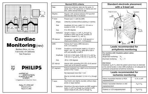
PR<br />
segment<br />
ST<br />
segment<br />
5 mm = 0.5 mV<br />
0.20 second<br />
0.1 mV<br />
0.04<br />
second<br />
Rate<br />
Rhythm<br />
P wave<br />
Normal ECG criteria<br />
Atrial and ventricular rates are the same. In<br />
adults, 60 to 100 cycles/min; in infants and children,<br />
within normal limits for age<br />
Regular, with variance between P-P and R-R<br />
intervals less than 0.16 second<br />
Present and 1:1 with the QRS<br />
Standard electrode placement<br />
with a 5-lead set<br />
Angle <strong>of</strong> Louis<br />
RA<br />
(white)<br />
LA (black)<br />
Shape<br />
Uniformly rounded without peaking or notches<br />
PR<br />
interval<br />
<strong>Cardiac</strong><br />
<strong>Monitoring</strong> (<strong>rev</strong>)<br />
Barbara Drew, RN, PhD<br />
University <strong>of</strong> California<br />
San Francisco<br />
Size<br />
Axis<br />
Deflection<br />
PR interval<br />
QRS<br />
Q wave<br />
Amplitude<br />
Axis<br />
Amplitude
Wide QRS tachycardias: distinguishing supraventricular tachycardia with bundle branch<br />
block or aberrant conduction from ventricular tachycardia (VT)<br />
Four-step approach to diagnosis<br />
V 1 or MCL 1 V 6 or MCL 6<br />
Using the bedside monitor:<br />
Ventricular tachycardia<br />
1. Presence <strong>of</strong> A-V dissociation Yes = VT<br />
*<br />
2. QRS width > 0.16 second Yes = VT Monophasic R<br />
Biphasic rS with<br />
R:S ratio 30 ms<br />
b) Slurred or notched<br />
S descent<br />
c) QRS onset to S nadir >60 ms<br />
Bimodal rR’ or<br />
triphasic rsR’<br />
Notched QS<br />
Biphasic qR<br />
Intrinsicoid<br />
deflection<br />
≥ 70 ms<br />
Supraventricular tachycardia with bundle branch<br />
block or aberration<br />
Triphasic qRs with<br />
R:S ratio >1.0<br />
All <strong>of</strong> the following in V 1 and V 2 :<br />
a) R ≤ 30 ms or no R<br />
Intrinsicoid<br />
b) Straight S descent<br />
deflection<br />
c) QRS onset to S<br />
≤ 50 ms<br />
nadir ≤ 60 ms<br />
And, no Q in V 6<br />
Unhelpful QRS morphologies<br />
Slurred or notched<br />
taller right peak<br />
Monophasic R<br />
Taller left or right<br />
peak<br />
Biphasic Rs with<br />
R:S ratio >1.0<br />
*Applies only to tachycardias with a positive waveform in V 1 .<br />
*<br />
ECG indicators <strong>of</strong> myocardial damage<br />
Ischemia<br />
Inverted T wave<br />
Mason-Likar (modified)<br />
Angle <strong>of</strong> Louis<br />
RA<br />
(white)<br />
ST depression<br />
Injury<br />
Elevated<br />
ST segment<br />
Infarction<br />
Q wave changes<br />
RL<br />
(green)<br />
3<br />
4<br />
2<br />
1<br />
V1<br />
V 2<br />
V 3<br />
V4<br />
• Inverted T waves in leads with upright<br />
QRS deflections.<br />
• Deeply inverted T waves in precordial<br />
leads.<br />
•Transient ST-segment depression<br />
reflects acute ischemia.<br />
• Permanent ST-segment depression may<br />
indicate digitalis effect, LVH.<br />
• Sign <strong>of</strong> an acute process; returns to<br />
baseline with time.<br />
•STelevation may indicate pericarditis.<br />
• Determine location <strong>of</strong> injury similar to MI<br />
location process.<br />
•STdepression that occurs in an ECG<br />
and that also has ST elevation in other<br />
leads reflects reciprocal changes.<br />
•Evaluate Q wave size—normally small<br />
in leads V 5 and V 6 ; normally deep in<br />
leads III and aVR<br />
• Prolonged Q wave is ≥ 0.04 second.<br />
• Loss <strong>of</strong> R wave V 1 through V 3<br />
12-lead placement<br />
V5<br />
V 6<br />
LA<br />
(black)<br />
LL<br />
(red)<br />
5th ICS midaxillary<br />
(white)<br />
References<br />
EASI TM<br />
5th ICS<br />
Level <strong>of</strong> sternum<br />
(brown)<br />
Top <strong>of</strong> sternum<br />
(black)<br />
5th ICS<br />
left<br />
midaxillary<br />
(red)<br />
1. Drew BJ. Bedside electrocardiographic monitoring: state <strong>of</strong> the art for<br />
the 1990s. Heart Lung. 1991;20:610-623.<br />
2. Drew BJ. Bedside electrocardiogram monitoring. AACN Clin Issues.<br />
1993;4:25-33.<br />
3. Drew BJ, Kruc<strong>of</strong>f MW, for the ST-Segment <strong>Monitoring</strong> Practice<br />
Guideline International Working Group. Multilead ST-segment<br />
monitoring in patients with acute coronary syndromes: a consensus<br />
statement for healthcare pr<strong>of</strong>essionals. Am J Crit <strong>Care</strong>. 1999;2:372-388.<br />
I<br />
S<br />
E<br />
A