EU Pediatric ECG Guidelines
EU Pediatric ECG Guidelines
EU Pediatric ECG Guidelines

Create successful ePaper yourself
Turn your PDF publications into a flip-book with our unique Google optimized e-Paper software.
Complete SummaryGUIDELINE TITLE<strong>Guidelines</strong> for the interpretation of the neonatal electrocardiogram.BIBLIOGRAPHIC SOURCE(S)Schwartz PJ, Garson A Jr, Paul T, Stramba-Badiale M, Vetter VL, Wren C.<strong>Guidelines</strong> for the interpretation of the neonatal electrocardiogram. A task force ofthe European Society of Cardiology. Eur Heart J 2002 Sep;23(17):1329-44. [52references] PubMedGUIDELINE STATUSThis is the current release of the guideline.COMPLETE SUMMARY CONTENTSCOPEMETHODOLOGY - including Rating Scheme and Cost AnalysisRECOMMENDATIONSEVIDENCE SUPPORTING THE RECOMMENDATIONSBENEFITS/HARMS OF IMPLEMENTING THE GUIDELINE RECOMMENDATIONSQUALIFYING STATEMENTSIMPLEMENTATION OF THE GUIDELINEINSTITUTE OF MEDICINE (IOM) NATIONAL HEALTHCARE QUALITY REPORTCATEGORIESIDENTIFYING INFORMATION AND AVAILABILITYDISCLAIMERSCOPEDISEASE/CONDITION(S)Electrocardiogram (<strong>ECG</strong>) abnormalitiesGUIDELINE CATEGORYDiagnosisEvaluationManagementRisk AssessmentScreeningTreatmentCLINICAL SPECIALTY1 of 22
MAJOR OUTCOMES CONSIDEREDDiagnosis• Predictive/interpretative value of diagnostic tests and assessments• Prevalence of electrocardiographic abnormalities and underlying diseases inthe neonatal and paediatric population• Morbidity and mortality related to arrhythmogenic disorders in the neonataland paediatric population• Side effects of maternal or fetal medicationMETHODOLOGYMETHODS USED TO COLLECT/SELECT EVIDENCESearches of Electronic DatabasesDESCRIPTION OF METHODS USED TO COLLECT/SELECT THE EVIDENCENot statedNUMBER OF SOURCE DOCUMENTSNot statedMETHODS USED TO ASSESS THE QUALITY AND STRENGTH OF THEEVIDENCEWeighting According to a Rating Scheme (Scheme Given)RATING SCHEME FOR THE STRENGTH OF THE EVIDENCEStrength of EvidenceA. Data derived from at least two randomized clinical trials or meta-analysesB. Data derived from a single randomized trial and/or meta-analysis from nonrandomizedstudiesC. Consensus opinion of the experts based on trials and clinical experienceMETHODS USED TO ANALYZE THE EVIDENCESystematic ReviewDESCRIPTION OF THE METHODS USED TO ANALYZE THE EVIDENCEThe procedure used for developing and issuing the guideline was in accordancewith the companion document to this guideline, the European Society ofCardiology "Recommendations for Task Force Creation and Report Writing"(available from the European Society of Cardiology Web site).3 of 22
METHODS USED TO FORMULATE THE RECOMMENDATIONSExpert ConsensusDESCRIPTION OF METHODS USED TO FORMULATE THERECOMMENDATIONSNot statedRATING SCHEME FOR THE STRENGTH OF THE RECOMMENDATIONSUsefulness or Efficacy of a Recommended TreatmentClass I = Evidence and/or general agreement that a given treatment is beneficial,useful and effectiveClass II = Conflicting evidence and/or a divergence of opinion about theusefulness/efficacy of the treatmentIIa: weight of evidence/opinion is in favour of usefulness/efficacyIIb: usefulness/efficacy is less well established by evidence/opinionClass III* = Evidence or general agreement that the treatment is notuseful/effective and in some cases may be harmful.* Use of Class III is discouraged by the ESCCOST ANALYSISA formal cost analysis was not performed and published cost analyses were notreviewed.METHOD OF GUIDELINE VALIDATIONExternal Peer ReviewInternal Peer ReviewDESCRIPTION OF METHOD OF GUIDELINE VALIDATIONThis document was reviewed and approved by the European Society of Cardiology(ESC) Committee for Practice <strong>Guidelines</strong> and Policy Conferences (W. Klein[Chairman], V. Dean, D. Jumeau, M.A. Alonso, C. Blomström-Lundqvist, G. DeBacker, M. Flather, J. Hradec, K. H. McGregor, A. Oto, A. Parkhomenko, S. Silber,A. Torbicki, G. Mazzotta, J. Deckers, H. Dargie, H-J. Trappe). The guideline wasendorsed by the Board of the European Society of Cardiology.RECOMMENDATIONSMAJOR RECOMMENDATIONS4 of 22
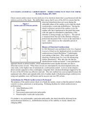
There are important differences between neonatal and adult electrocardiograms(<strong>ECG</strong>s). When a cardiologist examines the <strong>ECG</strong> of an apparently normal andhealthy infant the focus has to be on distinguishing between patterns that shouldcause no alarm and those that require action or additional investigations. Toprovide clues for this distinction is what the members of the Task Force haveattempted to do and, whenever possible or appropriate, steps in managementhave also been suggested.Normal Electrocardiogram in the NewbornNormal values• Normal electrocardiographic values in the paediatric population traditionallyderive from those published in 1979 by Davignon et al; thus, the percentiletables published by Davignon are recommended for use in clinical practice(see Table 1 in the original guideline document).Technology• The normal newborn <strong>ECG</strong> should include 12 leads. Other leads, V 3R , V 4R andV 7, may provide additional information to evaluate possible congenital heartlesions.• The current use of computerized digital <strong>ECG</strong> systems affects newborn <strong>ECG</strong>s toa greater extent than those of older children or adults. The newborn <strong>ECG</strong> mayhave a higher voltage and shorter duration QRS complexes resulting in ahigher percentage of high frequency components. The recommendations of anumber of groups vary as to the best bandwidth cutoffs and samplingfrequency to reduce error. Higher bandwidth cutoffs may alter amplitude ofsignals by as much as 46%. This would make standards determined fromanalogue signals or digitized signals at lower sampling rates and lowerfrequency cutoffs different from those at higher settings. The currentAmerican Heart Association recommendation for paediatric <strong>ECG</strong>s is 150 Hz asa minimum bandwidth cutoff and 500 Hz as a minimum sampling rate. TheRijnbeek study reported normals using a higher sampling rate of 1200 Hz.Compared to Davignon's study, which used a sampling rate of 333 Hz, thenewborn upper limits in Rijnbeek´s study were 12-25% higher than inDavignon´s.Artefacts• Artefacts are common in newborn <strong>ECG</strong>s and include limb lead reversal andincorrect chest lead positioning. In addition, electrical interference, usually 60cycles, can occur in hospital settings from bedside monitors, warmers or otherequipment. Other artefacts occur because of various types of patientmovement common in neonates. These artefacts may be random as withhiccoughs or limb movement. Normal complexes are seen along with theartefacts, and the intrinsic rhythm of the patient is not affected. Othercommon artefacts include a fine, often irregular undulation of the baselinefrom muscle tremors or jitteriness. Again, the intrinsic rhythm is not affected.The size of the QRS complex and the baseline may wander in a cyclic fashionwith respirations. It should be noted that the neonate breathes from 30-60times per min.5 of 22
• The main clue in determining the presence of an artefact is to evaluatewhether it affects the intrinsic rhythm and if it is timed such that it could be atrue depolarization. A signal within 80 ms from a true QRS complex could notoccur from an electrophysiologic point of view.Electrocardiographic measurements• Because of the current limitations of electronic measurements in newborn<strong>ECG</strong>s, intervals should be hand measured as the computerized systems areoften inaccurate in the newborn.• Intervals in children increase with increasing age, reaching most of the adultnormal values by 7-8 years of age.Heart rate• Heart rate can be determined by a variety of methods. Normal neonates mayhave heart rates between 150-230 beats. min –1 , especially if they are cryingor agitated. Over 200 beats. min -1 , one-half small box can make anappreciable difference in heart rates.• Heart rates between the 2nd and 98th percentile in the first year of life areshown in Table 1 of the original guideline document. The normal heart rateincreases from the first day of life, it reaches a peak between the first andsecond month and then declines returning to the values recorded at birth bythe sixth month. During the following six months, it remains rather stable andthen slowly declines after one year due to maturation of vagal innervation ofthe sinus node.• Clinically significant gender differences in heart rate are not seen in theneonatal period.P wave• The P wave axis is a vector indicating the direction of activation, which isaway from the site of origin. By identifying the quadrant location of the Pwave axis one can determine the site of origin of the rhythm. For example,sinus rhythm originates in the high right atrium transcribing a P wave with anaxis in the quadrant bordered by 0 and +90 degrees.• Measurements are available in Table 1 of the original guideline document forP wave amplitude. The P wave is generally pointed in lead II and aVF andmore rounded in other leads. Lead V 1 may be diphasic.• The PR interval is measured from the onset of the P wave to the Q or R waveif no Q wave is present. The PR interval, measured in lead II, increases withage and decreases with heart rate. The normal neonatal PR interval rangesfrom a minimum of 70 ms to a maximum of 140 ms, with a mean of 100 ms.QRS complex• The normal full-term neonate has an axis between 55 degrees and 200degrees but by one month, the normal upper limit has fallen to 160 degreesor less. Although one might identify an axis of 120 degrees as right axisdeviation in an adult, it is a normal finding in a newborn.• The QRS axis in the premature newborn <strong>ECG</strong> ranges between 65 degrees and174 degrees.6 of 22
• The duration of the QRS complex is measured from the beginning to the endof the ventricular depolarization complex and it should be measured in a leadwith an initial Q wave. QRS duration in the newborn and infant is narrow (30 ms isabnormal. The appearance of secondary r waves (r´ or R´) in the right chestleads is frequent in normal neonates.• Davignon et al. provided â ˜normal´ values in infants. The use of 2nd and98th percentiles to define normality implies that 4% of the population areâ ˜abnormal´ for any given single measurement, so â ˜normal´ rangeshave to be interpreted with caution (see Table 1 of the original guidelinedocument).QT interval• The QT interval is the interval between the beginning of the QRS complex andthe end of the T wave. The QT measurement should be made in leads II, V 5 ,and V 6 with the longest value being used. The main difficulty lies in identifyingcorrectly the point where the descending limb of the T wave intersects theisoelectric line. Due to the fast heart rate of infants the P wave may besuperimposed on the T wave, particularly when the QT interval is prolonged.In this case, the end of the T wave should be extrapolated by drawing atangent to the downslope of the T wave and considering its intersection withthe isoelectric line.• The QT interval duration changes with rate and it is usually corrected (QTc)by using Bazett´s formula. Correction of the QT interval requires a stablesinus rhythm without sudden changes in the RR interval. QTc is equal to QTinterval in seconds divided by the square root of the preceding RR interval inseconds. To avoid time-consuming calculations, a simple chart (see Figure 1of the original guideline document) where the value of QTc is easily obtainedby matching QT and RR interval in millimetres (given the paper speed 25 mm. s -1 ) has been produced. When heart rate is particularly slow or fast theBazett´s formula may not be accurate in the correction but it remains thestandard for clinical use.• The mean QTc on the 4th day of life is 400 plus or minus 20 ms and, atvariance with the adult, no gender differences are present. Therefore, theupper normal limit of QTc standard deviations above the mean, correspondingto the 97·5 percentile) is 440 ms. By definition, 2·5% of normal newbornsare expected to have a QTc greater than 440 ms. In healthy infants there is aphysiological prolongation of QTc by the second month (mean 410 ms)followed by a progressive decline, so that by the sixth month QTc returns tothe values recorded in the first week.• Pitfalls with QT measurement. Despite its apparent simplicity themeasurement of the QT interval is fraught with errors. The simple fact that a7 of 22
small square on the <strong>ECG</strong> paper is equivalent to 40 ms explains why healthyscepticism should accompany claims of clinical importance attached to verysmall degrees of â ˜QT prolongation´. An attempt should be made tomeasure with 10 ms (1/4 of a mm) while one recognizes that this may bewithin measurement error.ST segment and T wave• ST segment elevations >1 mm above the isoelectric line are uncommon in thenewborn. In neonates and infants it is better to consider as the isoelectric linethe TP segment instead of the PQ segment. T waves are normally quitevariable in the first week of life. After 1 week, the T wave is negative in leadV 1 and positive in V 5- V 6 .Abnormal Electrocardiogram in the NewbornHeart rateSinus arrhythmia• Since sinus arrhythmia is less pronounced at fast heart rate, neonates show amore regular rhythm than young children and adolescents, particularly in thefirst week of life. Sinus arrhythmia should be differentiated from wanderingpacemaker, which manifests itself with a gradual change of P wave axis andmorphology and that is due to a shift of the pacemaker from the sinus nodeto the atrium and the atrioventricular (AV) junction. Although wanderingpacemaker may accompany other types of bradyarrhythmia, it has nopathologic meaning.• Work up: No work-up should be necessary unless significant bradycardiacoexists.Sinus tachycardia• Sinus tachycardia is a sinus rhythm with a heart rate above the normal limitfor age. In the newborn the upper normal limit (98th percentile) is 166 beats.min –1 in the first week and 179 beats. min -1 in the first month. After the sixthmonth the upper normal limit declines to approximately 160 beats . min-1and at 1 year is 151 beats . min -1 . These values have been measured from<strong>ECG</strong>s recorded when infants were awake and quiet. It has to be noted thatnewborn infants may transiently reach a heart rate up to 230 beats . min -1 .• Work-up: The evaluation of these patients should be performed according tothe underlying condition. If myocarditis is suspected an echocardiogramshould be performed. Appropriate acute treatment of causes of tachycardiamay be considered. Persistence of elevated rates should be further evaluated.Sinus bradycardia• Sinus bradycardia is defined as a sinus rhythm with a heart rate below thenormal limit. In the neonatal period the lower normal limit (2nd percentile) is91 beats . min -1 during the first week and 107 beats . min -1 in the first monthof life. At the first month the lower limit increases to 121 beats . min -1 and8 of 22
declines to approximately 100 beats . min -1 in the following months. At 1 yearthe lower normal limit is 89 beats . min -1 . These values apply to an <strong>ECG</strong>recorded in the awake state when heart rate is measured over two respiratorycycles.• Work-up: 24-hour Holter monitoring may be helpful for further evaluationwhen a heart rate below 80-90 beats . min -1 is present on surface <strong>ECG</strong> duringinfancy. Evaluation for underlying conditions should be performed.Other bradycardias• Sinus pauses in newborns may last from 800 to 1000 ms. Pauses >2 s areabnormal. Sinus pauses may be followed by escape beats which arise fromthe atria or from the AV-junction. It has to be noted that even healthyneonates may show periods of junctional rhythm, i.e. a sequence of narrowQRS complexes in the absence of preceding P waves.• Work-up: 24-hour Holter monitoring may be useful for the assessment ofsignificant bradycardia. Long pauses secondary to excessive vagal tone maybe eliminated by the use of atropine, and rarely require pacemakers.Treatment of other underlying diseases should be undertaken.P wave• Abnormal P waves may be seen in infants with atrial enlargement or nonsinusorigin of the P wave. Ectopic atrial rhythms originate most commonlyfrom the low right atrium (0 to -90 degrees), high left atrium (+90 to +180degrees) or the low left atrium (+180 to +270 degrees).• Right atrial enlargement and/or hypertrophy typically produces increased Pwave amplitude with a normal P wave duration. The P wave axis usuallyremains normal so the effect is usually best seen in lead II.• Left atrial enlargement and/or hypertrophy typically produces an increasedand prolonged negative terminal deflection of the P wave in lead V 1 (generallyaccepted as >40 ms in duration and 0·1 mV in amplitude). Left atrialenlargement also causes exaggerated notching of the P wave in lead IIalthough this is not a specific sign.• Work-up: An echocardiogram should be performed when clinically indicated.Atrioventricular (AV) conduction• During atrial tachycardia, it is possible to observe 1/1 conduction through theatrioventricular node at rates over 300 beats . min -1 .Complete (third degree) atrioventricular (AV) block• Complete atrioventricular (AV) block implies complete absence of conductionfrom atrium to ventricle. <strong>ECG</strong> shows normal atrial activation and slowerdissociated regular QRS complexes. Congenital complete block is observed incomplex congenital heart malformations.First and second degree atrioventricular (AV) block9 of 22
• Neonates may present with first or second degree atrioventricular (AV) blockand rare reports exist demonstrating progression to complete AV block afterbirth in children with and without antibody mediated conduction disorders.• Long QT syndrome (LQTS) is occasionally complicated by impairedatrioventricular conduction, mostly 2:1 AV block. Functional AV block can beobserved in neonates because they have a fast atrial rate and the P wave fallswithin the very prolonged T wave. Cases of infra-Hisian block location at theHis-Purkinje level have been demonstrated.• Work-up: In neonates and infants with AV conduction abnormalities clinicalhistory of autoimmune disease and plasma titres of maternal antibodies (antiRo/SSA and antiLa/SSB) should be performed. When neonates have abnormalAV nodal conduction without maternal antibodies, an <strong>ECG</strong> should also beperformed on the parents and siblings (see intraventricular abnormalities).Neonates with first degree AV block should be followed with additional <strong>ECG</strong>sin the following months. Neonates and infants with second or third degree AVblock need a complete paediatric cardiologic work-up, including anechocardiogram. The only effective treatment of congenital complete AV blockin neonates with symptoms or a low ventricular escape rhythm is permanentartificial pacing.Intraventricular conductionBundle branch block• Congenital isolated complete right bundle branch block (RBBB) and leftbundle branch block are very rare in neonates. The classical <strong>ECG</strong> in Ebstein´sanomaly of the tricuspid valve displays a prolonged PR interval and wideRBBB.• Left anterior fascicular block is found in association with congenital heartmalformations such as atrioventricular canal defects and tricuspid atresia. Insevere cardiomyopathy, interruption of the left bundle, which results from theinvolvement of the left ventricle and/or its conduction system, has beenreported and carries a poor prognosis.• Hereditary bundle branch block is an autosomal dominant genetic disease thatwas mapped in some families to the long arm of chromosome 19. Affectedindividuals have various combinations of conduction defects such as RBBB,left or right QRS axis deviation or AV block; the r´ pattern may as well be theprelude to a conduction block. Abnormalities have been described in patientsas young as 15 days.• Non-specific intraventricular conduction abnormalities are very rare inneonates and infants with normal heart structures. They may be amanifestation of inflammation in myocarditis or endocarditis.• Work-up: Neonates and infants with intraventricular conduction abnormalitiesneed a complete paediatric cardiologic work-up. Evaluation of possibleunderlying causes should be performed. An <strong>ECG</strong> should also be performed onthe parents and siblings.Wolff-Parkinson-White syndrome (WPW)• The anatomical substrate of preexcitation in Wolf-Parkinson-White (WPW)syndrome is a direct muscular connection between the atria and ventricles.Since accessory pathways rarely show decremental conduction, the electrical10 of 22
impulse is conducted prematurely to the ventricles resulting in a short PRinterval. Conduction through the atrioventricular node and the accessorypathway results in collision of two electrical wavefronts at the ventricular levelcausing a delta wave and a fusion QRS complex with prolonged duration.• The diagnosis of preexcitation is solely based on the findings of the surface<strong>ECG</strong> (see Figure 2 of the original guideline document). Intermittentpreexcitation is not uncommon in newborns and infants. Depending on thelocation of the accessory pathway as well as the conduction properties of theatrioventricular node, even continuous preexcitation may be subtle and onlydetected in the mid-precordial leads.• Clinical counterparts: In WPW syndrome the typical form of paroxysmalsupraventricular tachycardia (orthodromic) results from reentry antegradelythrough the atrioventricular node and retrogradely through the accessorypathway. As digoxin shortens the antegrade effective refractory period of theaccessory pathway and promotes rapid atrioventricular conduction duringatrial flutter or atrial fibrillation over the pathway, the use of digoxin iscontraindicated at any age. Verapamil should also be avoided as it mayincrease the ventricular response rate during atrial fibrillation in thosepatients, and may cause cardiovascular collapse in infants and youngchildren.• Work-up: Congenital heart disease is more common in infants and youngchildren with preexcitation, with a prevalence as high as 45% for infants withan <strong>ECG</strong> pattern consistent with a right-sided accessory pathway. Thus, inevery young patient with a preexcitation pattern on surface <strong>ECG</strong>, a complete2-dimensional echocardiographic work-up is recommended to rule out anyintracardiac abnormality.Assessment of the conduction properties of the accessory pathway, i.e., theantegrade effective refractory period and the shortest RR-interval withpreexcitation, by transesophageal programmed stimulation may be useful inselected patients for risk stratification and mode of therapy.QRS axis and amplitude• Abnormal axis implies a mean frontal plane QRS vector outside the normalrange and must take into account the relative right axis deviation seen innormal neonates. Left axis deviation is seen in a variety of abnormalitiesincluding atrioventricular septal defect, ventricular septal defect, tricuspidatresia, and Wolff-Parkinson-White (WPW) syndrome, but may be occasionallyobserved in otherwise normal infants.Right ventricular hypertrophy• Right ventricular hypertrophy may be suspected from a QR complex in V 1 , anupright T wave in V 1 (normal in the first week of life), increased R waveamplitude in V 1 , and increased S wave amplitude in V 6 (according to theDavignon criteria). Sensitivity and specificity has not been tested in theneonate. QR patterns are commonly seen with pressure overload congenitallesions, rSR´ patterns are seen in volume overload lesions.Left ventricular hypertrophy11 of 22
• The performance of the <strong>ECG</strong> in recognition of left ventricular hypertrophy ispoorer than generally recognized and has not been specifically tested inneonates. Left ventricular hypertrophy is expected to produce increased leftsided voltages. Garson described the most helpful <strong>ECG</strong> signs in children asbeing T wave abnormalities in leads V 5 and V 6 , increased R wave amplitude inV 6 , increased S wave amplitude in V 1 (according to the Davignon criteria), anda combination of these last two variables. Left to right shunt lesions mayresult in left ventricular hypertrophy, but this may be in association with rightventricular hypertrophy and manifested as biventricular hypertrophy. Leftventricular hypertrophy in the newborn may be attenuated by the normalright-sided predominance of the newborn. The normal premature heart maynot have developed the right-sided predominance, especially if
amplitude. Central nervous system abnormalities can produce QTprolongation and T wave inversion.• Several drugs commonly used in the neonatal period and during infancy mayinduce QT interval prolongation; among them are macrolide antibiotics suchas spyramycin, erythromycin, clarithromycin and also trimethoprim.Prokinetics such as cisapride have been positively linked to QT intervalprolongation. All these drugs share one action: they block I Kr , one of the ioniccurrents involved in the control of ventricular repolarization.• Neonates born from mothers with autoimmune diseases and positive for theanti-Ro/SSA antibodies may also show QT interval prolongation, sometimeswith QTc values exceeding 500 ms, which tends to be transient and todisappear by the sixth month of life, concomitantly with the disappearance ofthe anti Ro/SSA antibodies.• Some of the neonates with QT interval prolongation may be affected by thecongenital LQTS. This possibility has to be carefully evaluated because of itsimplications for management.Long QT syndrome (LQTS)• Long QT syndrome (LQTS) is characterized by the occurrence of syncopalepisodes due to torsades de pointes ventricular tachycardia (VT) and by ahigh risk for sudden cardiac death among untreated patients. Importantly, in12% of patients with LQTS, sudden death was the first manifestation of thedisease and in 4% this happened in the first year of life. This point alonemandates the treatment of all those diagnosed as affected, even if there areno symptoms. Long QT syndrome is a genetic disease due to mutations ofseveral genes all encoding ionic (potassium or sodium) currents involved inthe control of ventricular repolarization. In most cases, several members ofthe same family are gene-carriers. Low penetrance exists in LQTS, whichmeans that gene-carriers may not show the clinical phenotype and may havea normal QT interval. Therefore a normal QT in the parents does not rule outfamilial LQTS. In addition, approximately 30% of cases are due to 'de novo'mutations which imply unaffected parents and no family history. 'De novo'LQTS mutations have been demonstrated in infant victims of cardiac arrestand sudden death diagnosed as Sudden Infant Death Syndrome.• Even though relatively few LQTS patients have cardiac events during the firstyear of life, the vast majority become symptomatic later on, either duringchildhood or adolescence according to genetic subgroups. Thereforetreatment must continue. Beta-blockers are the first choice therapy in LQTSand are effective in preventing recurrences in 80% of already symptomaticpatients; different degrees of protection exist according to genetic subgroups.If beta-blockers are unable to prevent new cardiac events, additional drugtherapy, left cardiac sympathetic denervation, pacemakers or the implantablecardioverter defibrillator should be considered based on evidence, with dueconsideration for body size.• <strong>ECG</strong> tracings of newborns with LQTS are shown in Figure 3 of the originalguideline document.• Work-up: It is well understood that the likelihood of having LQTS increaseswith increasing QTc; however, since a small percentage of LQTS patients hasa QTc
e updated frequently. Given the life-threatening potential of the disease,once the diagnosis of LQTS becomes probable, it is recommended that theseinfants are referred to a specialist as soon as possible.• First <strong>ECG</strong>: QTc above 440 ms, the upper limit of normal.Exclude other causes of acquired QT interval prolongation and obtain adetailed family history for the possibility of familial LQTS. Episodes ofearly sudden death, fainting spells, and seizures-epilepsy should alertto this possibility. The <strong>ECG</strong> should be repeated after a few days toconfirm the abnormal finding. Subsequent management depends on(1) presence or absence of family history suggestive for LQTS, and (2)the degree of QT interval prolongation.The presence of complex ventricular arrhythmias would haveadditional importance. The following stepwise approach involvesinfants with and without a family history for LQTS (see Figure 4 of theoriginal guideline document). If family history is positive, then--asLQTS is an autosomal dominant disease--the infant has a 50%probability of being affected and complete diagnostic proceduresshould be performed, as always with LQTS families.• The second <strong>ECG</strong> is normal.If the first QTc was
All diagnostic procedures listed above should be performed and a third<strong>ECG</strong> should be planned within a month. In case of a positive familyhistory, therapy should be initiated. Even without a family history,therapy should be considered.Even in infants with a very prolonged QTc in the first month of life, the<strong>ECG</strong> may normalize. If subsequent <strong>ECG</strong>s and diagnostic procedures donot confirm the presence of LQTS, it is logical to progressivelywithdraw therapy and to return to periodic observations.• The second <strong>ECG</strong> shows a QTc greater than or equal to 500 ms.ST segment elevationInfants with a QTc >500 ms are very likely to be affected by LQTS andto become symptomatic. All diagnostic procedures listed above shouldbe performed and these infants should be treated.Highest risk. The presence of QTc close to 600 ms, or of T wavealternans, or of 2:1 AV block secondary to major QT prolongation, orof hearing loss, identify infants at extremely high risk.• Work-up: Whenever the underlying cause has been identified, it should betreated. If the Brugada syndrome is suspected, careful family history shouldbe collected, 24-hour Holter monitoring obtained, and the patient should bereferred to a specialist.Atrial and ventricular arrhythmiasAtrial/junctionalPremature atrial beats• A premature atrial beat is a premature P wave. Premature atrial beats usuallyhave a different morphology and mean vector from sinus P waves. In regularsinus rhythm at a normal rate, a P wave that occurs before the next expectedP wave is a premature atrial beat. A premature atrial beat may be conductedto the ventricles normally, or with ventricular aberration or not conducted or'blocked'. Occasionally, blocked premature atrial beats occur in a bigeminalsequence, so-called 'blocked atrial bigeminy'. This rhythm simulates sinusbradycardia; it is important to examine the T waves carefully for blocked Pwaves (see Figure 5 of the original guideline document). In infants, since therefractory periods of the bundle branches are similar, premature atrial beatsmay be conducted with either RBBB or left bundle branch aberration. It isrelatively common in the same strip to see premature atrial beats conductednormally, aberrantly and blocked.• It is relatively uncommon for an infant to have both premature atrial beatsand premature ventricular beats, although it does occur. Therefore, in aninfant with premature P waves and wide QRS complexes on the same strip, acareful search should be made for a premature P wave preceding a wide QRS,15 of 22
• A premature ventricular beat is manifest on the surface <strong>ECG</strong> as a prematureabnormal QRS (not similar to the sinus QRS complex) that is not preceded bya premature P wave. It is important to recognize that in infants, the QRSduration may be normal--or slightly prolonged (i.e., less than 0·08 s)--but ifthe complex has a different morphology from the sinus, and is not precededby a premature P wave, the diagnosis is a premature ventricular beat.• The relationship between morphology and the site of origin is not exactenough to be able to predict which ventricle is causing the arrhythmia. It isnot possible to distinguish premature ventricular beats from premature atrialbeats with aberrancy on the basis of QRS morphology.• Work-up: The QT interval should be measured carefully during periods ofsinus rhythm (see section on repolarization abnormalities). In complexventricular arrhythmias, 24-hour Holter monitoring may be worthwhile. Anechocardiogram may be performed to determine ventricular function orstructural abnormalities. Occasionally maternal drugs that cause ventriculararrhythmias may be transferred in utero or post-natally in breast milk.Ventricular tachycardia (VT)• Ventricular tachycardia (VT) is a series of three or more repetitive complexesthat originate from the ventricles. The complexes are therefore different fromthe patient's normal QRS; usually, the QRS duration is prolonged for the ageof the patient (0·09 s or more in infants). It is therefore usually a 'wide QRS'tachycardia. However, infants may have a QRS duration in VT less than 0·09s but clearly different from the sinus complex (see Table 2 of the originalguideline document).• The specific morphology of the QRS is generally not helpful to distinguish VTfrom supraventricular tachycardia with aberration. However, SVT in infantswith a different QRS beyond the first 10-20 beats is rare, therefore, in thissituation, a diagnosis of VT should be strongly considered. In infants, the rateof VT may be from 200-500 beats . min-1.• There may be a slight variation in the R-R interval over several beats. Theremay be sinus P waves continuing unrelated to VT (AV dissociation),retrograde P waves or no visible P waves. There may also be fusion andcapture beats. The diagnosis of VT should be strongly considered if thepatient has premature ventricular beats during times of sinus rhythm with asimilar morphology to the tachyarrhythmia.• Work-up: An underlying cardiac or central nervous system abnormality maybe found in infants with VT. The QT interval should be carefully measured(see section on repolarization abnormalities), 24-hour Holter monitoring andechocardiogram should be obtained. Treatment is generally indicated.Accelerated ventricular rhythm• Accelerated ventricular rhythm is also known as 'slow VT'--generally the rateis
An algorithm on QT prolongation management is provided.EVIDENCE SUPPORTING THE RECOMMENDATIONSTYPE OF EVIDENCE SUPPORTING THE RECOMMENDATIONSThe type of supporting evidence is not specifically stated for eachrecommendation.BENEFITS/HARMS OF IMPLEMENTING THE GUIDELINE RECOMMENDATIONSPOTENTIAL BENEFITSIn general, early identification of life-threatening arrhythmogenic disorders, whichoften manifest in infancy, childhood or even later, may allow initiation of effectivepreventive therapy.POTENTIAL HARMSNot statedQUALIFYING STATEMENTSQUALIFYING STATEMENTSThis guideline document is not intended to be all-inclusive or to substitute fortextbooks on paediatric and neonatal electrocardiogram (<strong>ECG</strong>).IMPLEMENTATION OF THE GUIDELINEDESCRIPTION OF IMPLEMENTATION STRATEGYAn implementation strategy was not provided.IMPLEMENTATION TOOLSClinical AlgorithmPersonal Digital Assistant (PDA) DownloadsPocket Guide/Reference CardsFor information about availability, see the "Availability of Companion Documents" and "PatientResources" fields below.INSTITUTE OF MEDICINE (IOM) NATIONAL HEALTHCARE QUALITY REPORTCATEGORIESIOM CARE NEED18 of 22
Living with IllnessStaying HealthyIOM DOMAINEffectivenessIDENTIFYING INFORMATION AND AVAILABILITYBIBLIOGRAPHIC SOURCE(S)Schwartz PJ, Garson A Jr, Paul T, Stramba-Badiale M, Vetter VL, Wren C.<strong>Guidelines</strong> for the interpretation of the neonatal electrocardiogram. A task force ofthe European Society of Cardiology. Eur Heart J 2002 Sep;23(17):1329-44. [52references] PubMedADAPTATIONNot applicable: The guideline was not adapted from another source.DATE RELEASED2002 SepGUIDELINE DEVELOPER(S)European Society of Cardiology - Medical Specialty SocietySOURCE(S) OF FUNDINGEuropean Society of Cardiology (ESC). The Task Force for the Interpretation of theNeonatal Electrocardiogram Report was financed by the budget of the Committeefor Practice <strong>Guidelines</strong> of the European Society of Cardiology and was independentof any commercial, health, or governmental authorities.GUIDELINE COMMITTEETask Force for the Interpretation of the Neonatal ElectrocardiogramCOMPOSITION OF GROUP THAT AUTHORED THE GUIDELINETask Force members: P.J. Schwartz (Chair); A. Garson, Jr; T. Paul; M. Stramba-Badiale; V.L. Vetter; E. Villain; C. WrenFINANCIAL DISCLOSURES/CONFLICTS OF INTERESTNot statedENDORSER(S)19 of 22
Bulgarian Society of Cardiology - Medical Specialty SocietyCzech Society of Cardiology - Medical Specialty SocietyEstonian Cardiac Society - Medical Specialty SocietyFinnish Cardiac Society - Medical Specialty SocietyFrench Society of Cardiology - Medical Specialty SocietyHellenic Cardiological Society - Medical Specialty SocietyIrish Cardiac Society - Medical Specialty SocietyItalian Federation of Cardiology - Medical Specialty SocietyLatvian Society of Cardiology - Medical Specialty SocietyLebanese Society of Cardiology - Medical Specialty SocietyLithuanian Society of Cardiology - Medical Specialty SocietyPolish Cardiac Society - Medical Specialty SocietyPortuguese Society of Cardiology - Medical Specialty SocietySan Marino Society of Cardiology - Medical Specialty SocietySlovenian Society of Cardiology - Medical Specialty SocietySpanish Society of Cardiology - Medical Specialty SocietySwiss Society of Cardiology - Medical Specialty SocietyUkrainian Society of Cardiology - Medical Specialty SocietyGUIDELINE STATUSThis is the current release of the guideline.GUIDELINE AVAILABILITYElectronic copies: Available from the European Society of Cardiology (ESC) Website.Print copies: Available from Elsevier Science Ltd. European Heart Journal, ESC<strong>Guidelines</strong> - Reprints, 32 Jamestown Road, London, NW1 7BY, United Kingdom.Tel: +44.207.424.4422; Fax: +44 207 424 4433; Web site: www.eurheartj.org.AVAILABILITY OF COMPANION DOCUMENTSThe following is available:• Recommendations for Task Force creation and report production. SophiaAntipolis (France): European Society of Cardiology, 2002. Electronic copies:Available in Portable Document Format (PDF) from the European Society ofCardiology (ESC) Web site.• Interpretation of the neonatal electrocardiogram. Pocket guidelines. Orderform available in Portable Document Format (PDF) from the ESC Web site.Also available for PDA download from the ESC Web site.PATIENT RESOURCESNone availableNGC STATUSThis NGC summary was completed by ECRI on April 16, 2003.20 of 22
COPYRIGHT STATEMENTThis summary is based on the original guideline, which is subject to the guidelinedeveloper's copyright restrictions.DISCLAIMERNGC DISCLAIMERThe National Guideline Clearinghouse (NGC) does not develop, produce,approve, or endorse the guidelines represented on this site.All guidelines summarized by NGC and hosted on our site are produced under theauspices of medical specialty societies, relevant professional associations, publicor private organizations, other government agencies, health care organizations orplans, and similar entities.<strong>Guidelines</strong> represented on the NGC Web site are submitted by guidelinedevelopers, and are screened solely to determine that they meet the NGCInclusion Criteria which may be found athttp://www.guideline.gov/about/inclusion.aspx.NGC, AHRQ, and its contractor ECRI make no warranties concerning the contentor clinical efficacy or effectiveness of the clinical practice guidelines and relatedmaterials represented on this site. Moreover, the views and opinions of developersor authors of guidelines represented on this site do not necessarily state or reflectthose of NGC, AHRQ, or its contractor ECRI, and inclusion or hosting of guidelinesin NGC may not be used for advertising or commercial endorsement purposes.Readers with questions regarding guideline content are directed to contact theguideline developer.© 1998-2006 National Guideline ClearinghouseDate Modified: 7/10/200621 of 22
22 of 22