
Create successful ePaper yourself
Turn your PDF publications into a flip-book with our unique Google optimized e-Paper software.
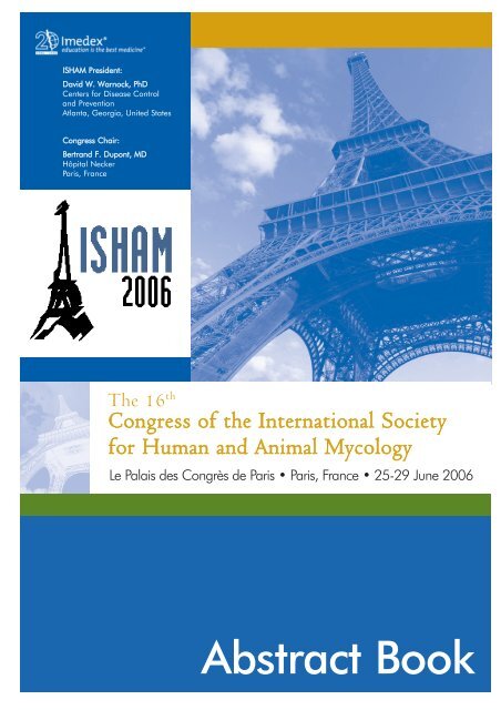
<strong>ISHAM</strong> President:<br />
David W. Warnock, PhD<br />
Centers for Disease Control<br />
and Prevention<br />
Atlanta, Georgia, United States<br />
Congress Chair:<br />
Bertrand F. Dupont, MD<br />
Hôpital Necker<br />
Paris, France<br />
The 16 th<br />
Congress of the International Society<br />
for Human and Animal Mycology<br />
Le Palais des Congrès de Paris • Paris, France • 25-29 June 2006<br />
<strong>Abstract</strong> <strong>Book</strong>
The 16 th<br />
Congress of the International Society<br />
for Human and Animal Mycology<br />
Le Palais des Congrès de Paris • Paris, France • 25-29 June 2006<br />
ABSTRACT TABLE OF CONTENTS<br />
Grouped by scientific topic<br />
Oral Presentations<br />
Animal Mycology . .O-0001 – O-0007<br />
Antifungals . . . . . . .O-0008 – O-0016<br />
Clinical Mycology . .O-0017 – O-0021<br />
Diagnostic Tools . . .O-0022 – O-0025<br />
Epidemiology . . . . .O-0026 – O-0030<br />
Genomics . . . . . . . .O-0031 – O-0035<br />
Immunology . . . . . .O-0036 – O-0038<br />
Molecular Biology . .O-0039 – O-0045<br />
Physiopathology . . . . . . . . . . .O-0046<br />
Poster Presentations<br />
Animal Mycology . . . .P-0001 – P-0043<br />
Antifungals . . . . . . . .P-0044 – P-0165<br />
Clinical Mycology . . . .P-0166 – P-0350<br />
Diagnostic Tools . . . . .P-0351 – P-0438<br />
Epidemiology . . . . . . .P-0439 – P-0573<br />
Genomics . . . . . . . . .P-0574 – P-0584<br />
Immunology . . . . . . .P-0585 – P-0626<br />
Molecular Biology . . .P-0627 – P-0709<br />
Other . . . . . . . . . . . .P-0710 – P-0738<br />
Taxonomy . . . . . . . . . .P-0739 – P0756<br />
©2006 Imedex ® , Inc. All rights reserved. Reproduction<br />
in whole or part is prohibited without prior written<br />
consent from Imedex and contributing faculty.
Abad A . . . . . . . . . .O-0017, P-0585,<br />
. . . . . . . . . . . . . . . . .P-0615, P-0685<br />
Abarca C . . . . . . . . . . . . . . . .P-0556<br />
Abbes S . . . . . . . . . . . . . . . . .P-0550<br />
Abboud P . . . . . . . . . . . . . . . .P-0230<br />
Abd El Kader A . . . . . . . . . . . .P-0499<br />
Abdelaziz H . . . . . . . . . . . . . . .P-0281<br />
Abdul Samads . . . . . . . . . . . . .P-0352<br />
Abe S . . . . . . . . . . . .O-029, P-0055,<br />
. . . . . . . . . . . . . . . . .P-0085, P-0707<br />
Abel P . . . . . . . . . . . . . . . . . . .P-0227<br />
Abia-Bassey L . . . . . . . . . . . . .P-0441<br />
Abli P . . . . . . . . . . . . . . . . . . .P-0483<br />
Abramson M . . . . . . . . . . . . . .P-0507<br />
Adachi Y . . . . . . . . . .P-0606, P-0612<br />
Adelantado C . . . . . . .P-0059, P-0202<br />
Adema G . . . . . . . . . . . . . . . .P-0597<br />
Adiri Y . . . . . . . . . . . . . . . . . .P-0507<br />
Adler-Moore J . . . . . . . . . . . .O-0013<br />
Afeltra J . . . . . . . . . . . . . . . . .P-0157<br />
Afioni F . . . . . . . . . . . . . . . . . .P-0213<br />
Afonso A . . . . . . . . . . . . . . . . .P-0346<br />
Afshar P . . . . . . . . . . . . . . . . .P-0493<br />
Afshari N . . . . . . . . . . . . . . . .P-0120<br />
Agabian N . . . . . . . . . . . . . .O-0033<br />
Aghamirian M . . . . . .P-0001, P-0442<br />
Aghapour R . . . . . . . . . . . . . . .P-0452<br />
Aghili R . . . . . . . . . . . . . . . . . .P-0493<br />
Agnetti F . . . .P-0007, P-0455, P-0521<br />
Agorio I . . . . . . . . . . . . . . . . .P-0382<br />
Aguiar L . . . . . . . . . . . . . . . . .P-0031<br />
Aguilar A . . . . . . . . . . . . . . . .P-0211<br />
Agyeman K . . . . . . . . . . . . . . .P-0533<br />
Ahmad S . . . . . . . . . .P-0249, P-0627<br />
Ahmed A . . . . . . . . . .P-0152, P-0167<br />
Ahn K . . . . . . . . . . . . . . . . . . .P-0502<br />
Aho S . . . . . . . . . . . . . . . . . . .P-0557<br />
Akcaglar S . . . . . . . . .P-0070, P-0071<br />
. . . . . . . . . . .P-0072, P-0168, P-0443<br />
Akeel R . . . . . . . . . . . . . . . . . .P-0169<br />
Akkurt L . . . . . . . . . . . . . . . . .P-0056<br />
Akoru C . . . . . . . . . . . . . . . . .P-0353<br />
Aktas E . . . . . . . . . . . .P-0056, P-0664<br />
Al Sharif A . . . . . . . . . . . . . . . .P-0499<br />
Al-Mohri H . . . . . . . . . . . . . . .P-0293<br />
Al-Sweih N . . . . . . . . . . . . . . .P-0249<br />
Alain T . . . . . . . . . . . . . . . . . .P-0171<br />
Albaina O . . . . . . . . . . . . . . . .P-0069<br />
Albarrag A . . . . . . . . . . . . . . .P-0044<br />
Albert N . . . . . . . . . . . . . . . . .P-0060<br />
Alcazar-Fuoli L . . . . . . . . . . . . .P-0628<br />
Alcoba-Flórez J . . . . . . . . . . . .P-0354<br />
Aldins P . . . . . . . . . . . . . . . . . .P-0444<br />
Alejandro S . . . . . . . .P-0254, P-0255<br />
Alexander C . . . . . . . .P-0045, P-0445<br />
Alexopoulos E . . . . . . .P-0049, P-0484<br />
Alhambra A . . . . . . . .P-0280, P-0355<br />
. . . . . . . . . . . . . . . . . . . . . . .P-0386<br />
Ali S . . . . . . . . . . . . . . . . . . . .P-0041<br />
Aliouat C . . . . . . . . . . . . . . . .P-0745<br />
Alizadeh H . . . . . . . . . . . . . . .P-0218<br />
Alkorta M . . . . . . . . . .P-0426, P-0536<br />
Aller A . . . . . . . . . . . .P-0096, P-0103,<br />
. . . . . . . . . . . . . . . . .P-0104, P-0368<br />
Allison M . . . . . . . . . . . . . . . . .P-0321<br />
Allombert C . . . . . . . . . . . . . .P-0301<br />
Almamary A . . . . . . . . . . . . . .P-0223<br />
Almeida S . . . . . . . . . . . . . . . .P-0586<br />
Alonso M . . . . . . . . . .P-0486, P-0653<br />
Alonso-Monge R . . . . . . . . . .O-0039<br />
Altas K . . . . . . . . . . . .P-0244, P-0245<br />
Altclas J . . . . . . . . . . . . . . . . .P-0382<br />
Altman S . . . . . . . . . . . . . . . . .P-0448<br />
Alvarado P . . . . . . . . . . . . . . .P-0516<br />
Alvarado Ramírez E . .P-0046, P-0342,<br />
. . . . . . . . . . . . . . . . .P-0385, P-0629<br />
Alvarez M . . . . . . . . . .P-0074, P-0410<br />
Alvarez S . . . . . . . . . .P-0078, P-0714<br />
Alvaro L . . . . . . . . . . . . . . . . .P-0683<br />
Alves C . . . . . . . . . . . . . . . . . .P-0034<br />
Alves S . . . . . . . . . . . . . . . . . .P-0406<br />
Alviano C . . . . . . . . .P-0253, P-0296,<br />
. . . . . . . . . . . . . . . . .P-0595, P-0625<br />
Alviano D . . . . . . . . . . . . . . . .P-0625<br />
Alviano D . . . . . . . . . . . . . . . .P-0595<br />
Aly R . . . . . . . . . . . . . . . . . . . .P-0343<br />
Amagai M . . . . . . . . . . . . . . . .P-0317<br />
Amel B . . . . . . . . . . . . . . . . . .P-0310<br />
Amendola L . . . . . . . . . . . . . .P-0259<br />
Amorim A . . . . . . . . . . . . . . . .P-0284<br />
Amorim C . . . . . . . . .P-0446, P-0515<br />
Amorim J . . . .P-0095, P-0221, P-0264<br />
Amrein C . . . . . . . . . . . . . . . .O-0008<br />
Anane S . . . .P-0170, P-0243, P-0246<br />
Ancelle T . . . . . . . . . . . . . . . . .P-0417<br />
Andaluz López E . . . . . . . . . . .P-0654<br />
Anderson F . . . . . . . . . . . . . . .P-0595<br />
Anderson M . . . . . . . . . . . . . .P-0044<br />
Anderson S . . . . . . . . . . . . . . .P-0474<br />
Andreopoulos A . . . . . . . . . . .P-0226<br />
Andreotti P . . . . . . . . .P-0216, P-0279<br />
Angelopoulou M . . . . . . . . . . .P-0226<br />
Angoulvant A . . . . . . .P-0356, P-0510<br />
Ann T . . . . . . . . . . . . . . . . . . .P-0254<br />
Anna C . . . . . . . . . . . . . . . . . .P-0254<br />
4 Le Palais des Congrès de Paris • Paris, France • 25-29 June 2006
Anne B . . . . . . . . . . . . . . . . . .P-0471<br />
Anne G . . . . . . . . . . . . . . . . . .P-0171<br />
Anne R . . . . . . . . . . . . . . . . . .P-0073<br />
Anne S . . . . . . . . . . . . . . . . . .P-0073<br />
Annemarie B . . . . . . . . . . . . . .P-0254<br />
Anonye N . . . . . . . . . . . . . . . .P-0286<br />
Antinori S . . . . . . . . . . . . . . . .P-0172<br />
Antoine B . . . . . . . . . . . . . . .O-0023<br />
Antoine H . . . . . . . . . . . . . . . .P-0369<br />
Antonopoulou S . . . . . . . . . . . .P-0047<br />
Anyfantis I . . . . . . . . . . . . . . . .P-0048<br />
Anzawa K . . .P-0173, P-0174, P-0447,<br />
. . . . . . . . . . .P-0519, P-0570, P-0752<br />
Aoki S . . . . . . . . . . . .P-0658, P-0666,<br />
. . . . . . . . . . . . . . . . .P-0673, P-0704<br />
Apaire-Marchais V . . .P-0357, P-0574<br />
Apitz-Castro R P-0092, P-0102, P-0127<br />
Arabatzis M . . . . . . . .P-0049, P-0358<br />
Arai M . . . . . . . . . . . . . . . . . .P-0116<br />
Araiza J . . . . . . . . . . . . . . . . .P-0195<br />
Arana D . . . . . . . . . . . . . . . .O-0039<br />
Arancia S . . . . . . . . . .P-0359, P-0596<br />
Arantes R . . . . . . . . . . . . . . . .P-0644<br />
Arathoon E . . . . . . . . . . . . . . .P-0422<br />
Araújo Resende L . . . .P-0481, P-0482<br />
Arcelli R . . . . . . . . . . . . . . . . .P-0521<br />
Arduino M . . . . . . . . . . . . . . . .P-0462<br />
Arechaval A . . . . . . . .P-0069, P-0341<br />
Arenas R . . . . . . . . . .P-0479, P-0496<br />
Arendrup M . . . . . . . . . . . . . . .P-0132<br />
Arias M . . . . . . . . . . . . . . . . . .P-0192<br />
Arlington-Skaggs B . . . . . . . . .P-0559<br />
Arné P . . . . . . . . . . . . . . . . . . .P-0020<br />
Aroch A . . . . . . . . . . . . . . . . .P-0469<br />
Arosemena L . . . . . . .P-0059, P-0202<br />
Arreguín R . . . . . . . . . . . . . . .O-0012<br />
Arruda-Neto J . . . . . . . . . . . . .P-0661<br />
Arsenis G . . . . . . . . . . . . . . . .P-0049<br />
Arthington-Skaggs B . . . . . . . .P-0422<br />
Arthur I . . . . . . . . . . . . . . . . . .P-0448<br />
Arévalo-Morales M . . . . . . . . .P-0354<br />
Asahi Y . . . . . . . . . . . . . . . . . .P-0611<br />
Asci Toraman Z . . . . . . . . . . . .P-0333<br />
Asgari G . . . . . . . . . . . . . . . . .P-0032<br />
Aspiroz C . . . . . . . . . . . . . . . .P-0304<br />
Asri Rezai S . . . . . . . . . . . . . .P-0043<br />
Ates A . . . . . . . . . . . . . . . . . .P-0495<br />
Aubert A . . . . . . . . . . . . . . . .P-0237<br />
Auger A . . . . . . . . . . . . . . . . .P-0383<br />
Auger I . . . . . . . . . . . . . . . . .P-0360<br />
Auler M . . . . . . . . . . .P-0292, P-0568<br />
Aurore K . . . . . . . . . . . . . . . .P-0175<br />
Aurélien F . . . . . . . . . . . . . . .P-0175<br />
Ausma J . . . .P-0176, P-0219, P-0576<br />
Aveskamp M . . . . . . . . . . . . .P-0743<br />
T Avšic- upanc T . . . . . . . . . .P-0351<br />
Ayadi A . . . .P-0208, P-0326, P-0523,<br />
. . . . . . . . . . .P-0550, P-0551, P-0604<br />
Aydil U . . . . . . . . . . . . . . . . . .P-0449<br />
Ayyildiz A . . . . . . . . . . . . . . . .P-0664<br />
Azaiz M . . . . . . . . . . . . . . . . . .P-0494<br />
Aze vedo M . . . . . . . . . . . . . . .P-0021<br />
Azevedo M . .P-0037, P-0038, P-0626<br />
Aznar C . . . . . . . . . . . . . . . . .P-0193<br />
Aznar J . . . . . . . . . . . . . . . . . .P-0368<br />
Aznar J . . . . . . . . . . . . . . . . . .P-0426<br />
Baccarin R . . . . . . . . . . . . . . . .P-0033<br />
Bachewich C . . . . . . . . . . . . .O-0016<br />
Bachhawat A . . . . . . . . . . . . . .P-0473<br />
Badali H . . . . . . . . . . . . . . . . .P-0450<br />
Bader O . . . . . . . . . . . . . . . . .P-0671<br />
Badre Eddine L . . . . . .P-0177, P-0178<br />
Badri T . . . . . . . . . . . . . . . . . .P-0179<br />
Baeza L . . . . . . . . . . .P-0180, P-0222<br />
Bagg J . . . . . . . . . . . . . . . . . .O-0018<br />
Bahadori F . . . . . . . . . . . . . . .P-0213<br />
Bahloul M . . . . . . . . . . . . . . . .P-0326<br />
Bahrami Samani H . . . . . . . . .P-0181<br />
bailly e . . . . . . . . . . . . . . . . . .P-0182<br />
Bailly E . . . . .P-0183, P-0184, P-0205<br />
Bailão A . . . . . . . . . . . . . . . . .P-0180<br />
Baixench M . . . . . . . . .P-0185, P-0186<br />
Baldo A . . . . . . . . . .O-0001, O-0005<br />
Ball L . . . . . . . . . . . . . . . . . . .P-0050<br />
Bamberg W . . . . . . . . . . . . . . .P-0463<br />
Banerjee D . . . . . . . . . . . . . .O-0031<br />
Banerjee U . . . . . . . . .P-0267, P-0314<br />
Banfi E . . . . . . . . . . . . . . . . . .P-0051<br />
Baptista R . . . . . . . . . . . . . . . .P-0361<br />
Baquero F . . . . . . . . .P-0074, P-0410<br />
Baran E . . . . . . . . . . .P-0084, P-0187<br />
Baran W . . . . . . . . . . . . . . . . .P-0187<br />
Barbey S . . . . . . . . . . . . . . . .O-0009<br />
Barboni A . . . . . . . . . . . . . . . .P-0015<br />
Barbosa - Sabanero G . . . . . . .P-0312<br />
Barbosa dos Santos C . . . . . .P-0481,<br />
. . . . . . . . . . . . . . . . .P-0482, P-0520<br />
Barbosa, Jr A . . . . . . .P-0451, P-0738<br />
Barchiesi F . . . . . . . . . . . . . . .P-0144<br />
Barker K . . . . . . . . . . .P-0579, P-0587<br />
Barnes P . . . . . . . . . . . . . . . . .P-0588<br />
Barrabes A . . . . . . . . . . . . . . .P-0183<br />
Barreira P . . . . . . . . . . . . . . .O-0044<br />
Barrenetxea G . . . . . .O-0017, P-0585<br />
The 16 th Congress of the International Society for Human and Animal Mycology<br />
5
Barreto L . . . . . . . . . . . . . . . . .P-0677<br />
Barreto-Bergter E . . . . . . . . . . .P-0613<br />
Barros G . . . . . . . . . . . . . . . . .P-0201<br />
Barros R . . . . . . . . . . . . . . . . .P-0505<br />
Barroso C . . . . . . . . . . . . . . . .P-0311<br />
Barton R . . . . . . . . . . . . . . . .O-0014<br />
Bastides F . . . . . . . . . . . . . . . .P-0184<br />
Batisse A . . . . . . . . . . . . . . . .O-0008<br />
Batista W . . . . . . . . . . . . . . . .P-0630<br />
Batura-Gabryel H . . . .P-0188, P-0189<br />
Bautista-Muñoz C . . . . . . . . . .P-0705<br />
Benjamin L . . . . . . . . . . . . . . .P-0422<br />
Benoit S . . . . . . . . . . . . . . . . .P-0463<br />
Bentubo H . . . . . . . . . . . . . . . .P-0014<br />
Benvenuto F . . . . . . . . . . . . . .P-0287<br />
Benvenuto Ferreira F . . . . . . . .P-0364<br />
Berdicevsky I . . . . . . . .P-0143, P-0631<br />
Berge M . . . . . . . . . . . . . . . .O-0008<br />
Bergmans A . . . . . . . . . . . . . .P-0363<br />
Bergès T . . . . . . . . . . . . . . . . .P-0154<br />
Berjeaud J . . . . . . . . . . . . . . . .P-0459<br />
Berk E . . . . . . . . . . . . . . . . . . .P-0449<br />
Billaud E . . . . . . . . . . . . . . . .O-0008<br />
Birinci A . . . . . . . . . . . . . . . . .P-0056<br />
Bisaro F . . . . . . . . . . . . . . . . .P-0458<br />
Bistoni F . . . . . . . . . . . . . . . . .P-0716<br />
Blancard A . . . . . . . . . . . . . . .P-0301<br />
Blanchet D . . . . . . . . . . . . . . .P-0193<br />
Blanco J . . . .P-0078, P-0365, P-0714<br />
Blanco Y . . . . . . . . . . . . . . . . .P-0472<br />
Blijlevens N . . . . . . . . . . . . . . .P-0161<br />
Blonde R . . . . . . . . . . . . . . . . .P-0194<br />
Bocanegra R . . . . . . . .P-0028, P-0110<br />
Bayram Ö . . . . . . . . . . . . . . . .P-0694 Berman J . . . . . . . . . . . . . . . .O-0003 Boekhout T . . . . . . .O-0027, P-0196,<br />
Becerril-Luján B . . . . . . . . . . . .P-0692 Bermúdez R . . . . . . . . . . . . . . .P-0470 . . . . . . . . . . .P-0244, P-0304, P-0367<br />
Beck S . . . . . . . . . . . . . . . . . . .P-0052 Bernadette L . . . . . . . .P-0512, P-0513 Boel A . . . . . . . . . . . . . . . . . . .P-0454<br />
Becker K . . . . . . . . . . .P-0134, P-0228 Bernard G . . . . . . . . . . . . . . .O-0037 Bogdanowicz E . . . . . . . . . . . .P-0209<br />
Becker W . . . . . . . . . . . . . . . .P-0739 Bernard S . . . . . . . . . . . . . . . .P-0205 Bogomolova T . . . . . .P-0155, P-0215,<br />
Begum Y . . . . . . . . . . . . . . . . .P-0362 Bernardes-Engemann A . . . . .P-0191,<br />
. . . . . . . . . . . . . . . . . . . . . . .P-0566<br />
Behzadi E . . . . . . . . . . . . . . . .P-0452 . . . . . . . . . . . . . . . . .P-0287, P-0364 Bogusz B . . . . . . . . . .P-0150, P-0461<br />
Behzadi P . . . . . . . . . . . . . . . .P-0452 Bernardino S . . . . . . . . . . . . . .P-0589 Bohse M . . . . . . . . . . . . . . . . .P-0633<br />
Belhadj S . . . .P-0170, P-0243, P-0246 Bernhardt H . . . . . . . . . . . . . .P-0053 Boiron P . . . . . . . . . . . . . . . . .P-0346<br />
Belleguic C . . . . . . . . . . . . . . .P-0210 Bernhardt J . . . . . . . . . . . . . . .P-0053 Bollo E . . . . . . . . . . . .P-0007, P-0635<br />
Bello N . . . . . . . . . . . . . . . . . .P-0209 Berriman M . . . . . . . . . . . . . .O-0035 Bolognini J . . . . . . . . . . . . . . .P-0356<br />
Bellucci C . . . . . . . . . . . . . . . .P-0591 Berry A . . . . . . . . . . . . . . . . . .P-0489 Bolognini J . . . . . . . . . . . . . . .P-0109<br />
Ben Hadj El Bechir A . . . . . . . .P-0453 Berrêdo Pinho M . . . . . . . . . . .P-0191 Bonaccorso C . . . . . . . . . . . . .P-0172<br />
Ben Osman Dhahri A P-0179, P-0217, Berthelemy M . . . . . . . . . . . .O-0002, Boncio L . . . . . . . . . .P-0007, P-0025,<br />
. . . .P-0234, P-0250, P-0453, P-0494 . . . . . . . . . . . . . . . . .P-0675, P-0745 . . . . . . . . . . . . . . . . .P-0455, P-0521<br />
Ben Tekaya N . . . . . .P-0179, P-0217, Bertout S . . . . . . . . . . . . . . . . .P-0054 Bonfim Carregaro F . . . . . . . . .P-0002<br />
. . . . . . . . . . . . . . . . .P-0234, P-0250 Betran A . . . . . . . . . . . . . . . . .P-0192 Bonifaz A . . . . . . . . . . . . . . . .P-0195<br />
Benada O . . . . . . . . . . . . . . . .P-0437 Beucher B . . .P-0407, P-0713, P-0729 Bonmarchand G . . . . . . . . . . .P-0230<br />
Benard G . . . . . . . . . .P-0216, P-0279 Bex V . . . . . . . . . . . . . . . . . . .P-0675 Bonnet E . . . . . . . . . . . . . . . . .P-0093<br />
Benboubker l . . . . . . . . . . . . . .P-0182 Bhardwaj S . . . . . . . . .P-0473, P-0632 Bonnin A . . . .P-0475, P-0557, P-0717<br />
Benderdouche M . . . . . . . . . . .P-0421 Bien C . . . . . . . . . . . . . . . . . .P-0637 Borbély Á . . . . . . . . . . . . . . . .P-0428<br />
Benetucci A . . . . . . . . . . . . . . .P-0190 Bignell E . . . . . . . . . . . . . . . .O-0042 Borgers M . . .P-0176, P-0219, P-0576<br />
Benites N . . . . . . . . . . . . . . . .P-0033 Bii C . . . . . . . . . . . . . . . . . . . .P-0055 Borges C . . . . . . . . . . . . . . . . .P-0180<br />
Benito-Ruesca R . . . . . . . . . . . .P-0309 Bilgin K . . . . . . . . . . . . . . . . . .P-0056 Borges R . . . . . . . . . . . . . . . . .P-0468<br />
6 Le Palais des Congrès de Paris • Paris, France • 25-29 June 2006
Bori A . . . . . . . . . . . . . . . . . . .P-0076<br />
Borman A . . .P-0456, P-0741, P-0743<br />
Borrell N . . . . . . . . . . . . . . . . .P-0426<br />
Bosch Alepuz M . . . . .P-0457, P-0465<br />
Bosshard P . . . . . . . . .P-0373, P-0374<br />
Botterel F . . . . . . . . . . . . . . . .P-0220<br />
Bouaziz M . . . . . . . . .P-0326, P-0550<br />
Brena S . . . . . . . . . . . . . . . . . .P-0401<br />
Brentrup A . . . . . . . . . . . . . . . .P-0229<br />
Bretagne S . . . . . . . . .P-0020, P-0220<br />
Briand J . . . . . . . . . . . . . . . . .O-0009<br />
Bricaire F . . . . . . . . . . . . . . . .P-0194<br />
Bridge P . . . . . . . . . . . . . . . . .P-0741<br />
Brightwell K . . . . . . . . . . . . . . .P-0432<br />
Busold C . . . . . . . . . . . . . . . . .P-0578<br />
Bustamante B .P-0113, P-0199, P-0464<br />
Buzanello Martins C . .P-0064, P-0065<br />
Buziba N . . . . . . . . . . . . . . . . .P-0353<br />
Buzina W . . . . . . . . . . . . . . . .P-0200<br />
Bykowska B . . . . . . . . . . . . . . .P-0573<br />
Béatrice S . . . . . . . . . . . . . . . .P-0073<br />
Bouchara J . . . . . . . .P-0153, P-0154,<br />
. . . . . . . . . . . . . . . . .P-0370, P-0649<br />
Boucharan J . . . . . . . . . . . . .O-0044<br />
Boudava S . . . . . . . . . . . . . . . .P-0551<br />
Boudreaux J . . . . . . . . . . . . . .P-0376<br />
Bougeret C . . . . . . . . . . . . . .O-0009<br />
Bougerol C . . . . . . . . . . . . . . .P-0020<br />
Bougnoux M . . . . . . . . . . . . . .P-0230<br />
Bourdeau P . . . . . . . . . . . . . . .P-0004<br />
Bourdeau P . . . . . . . .P-0003, P-0366<br />
Bourée P . . . . . . . . . . . . . . . . .P-0458<br />
Bouscary D . . . . . . . . . . . . . . .P-0186<br />
Boussaud V . . . . . . . . . . . . . .O-0008<br />
Bouyer S . . . . . . . . . . . . . . . . .P-0459<br />
Bovers M . . . . . . . . . .P-0196, P-0367<br />
Bovers M . . . . . . . . . . . . . . . .O-0027<br />
Boyce S . . . . . . . . . . . . . . . . . .P-0330<br />
Boyer J . . . . . . . . . . . . . . . . . .P-0109<br />
Bozonnet E . . . . . . . . . . . . . . .P-0237<br />
Bozza S . . . . . . . . . . . . . . . . . .P-0590<br />
Brahim A . . . . . . . . . . . . . . . . .P-0177<br />
Brajer B . . . . . . . . . . .P-0188, P-0189<br />
Branche J . . . . . . . . . . . . . . . .P-0734<br />
Brandt M . . .P-0422, P-0462, P-0463,<br />
. . . . . . . . . . . . . . . . . . . . . . .P-0546<br />
Brandão J . . .P-0034, P-0345, P-0460<br />
Brasch J . . . . . . . . . . .P-0715, P-0740<br />
Braus G . . . . . . . . . .O-0042, P-0694<br />
Brigitte W . . . . . . . . . . . . . . . .P-0073<br />
Briones M . . . . . . . . . .P-0446, P-0515<br />
Brito A . . . . . .P-0197, P-0212, P-0270<br />
Brito E . . . . . . . . . . . . . . . . . . .P-0064<br />
Bromuro C . . . . . . . . .P-0591, P-0594<br />
Brouwer C . . . . . . . . .P-0087, P-0161<br />
Brown D . . . . . . . . . . . . . . . . .P-0083<br />
Brown L . . . . . . . . . . . . . . . . .P-0445<br />
Brown T . . . . .P-0136, P-0320, P-0321<br />
Bruet V . . . . . . . . . . . .P-0003, P-0004<br />
Bruijnesteijn van Coppenraet L .P-0358<br />
Brusca M . . . . . . . . . . . . . . . . .P-0075<br />
Bruzual E . . . . . . . . . .P-0197, P-0212<br />
Brzozowski T . . . . . . . . . . . . .O-0021<br />
Bubnick M . . . . . . . . . . . . . . .O-0040<br />
Buchheidt D . . . . . . . . . . . . . .P-0391<br />
Buchta V . . . . . . . . . . . . . . . . .P-0232<br />
Budak A . . . .O-0021, P-0150, P-0461<br />
Buendia B . . . . . . . . . . . . . . . .P-0426<br />
Bueno J . . . . . . . . . . .P-0106, P-0107<br />
Bueno Sánchez J . . . . .P-0057, P-0058<br />
Bufler J . . . . . . . . . . . . . . . . . .P-0053<br />
Bujdakova H . . . . . . . .P-0105, P-0592<br />
Bulut Y . . . . . . . . . . . . . . . . . .P-0333<br />
Buot G . . . . . . . . . . .O-0010, P-0198<br />
Burger E . . . . . . . . . . .P-0133, P-0610<br />
Burwell L . . . .P-0462, P-0463, P-0474<br />
Bejar V . . . . . . . . . . . . . . . . . .P-0607<br />
Böttger E . . . . . . . . . .P-0373, P-0374<br />
C Sartorelli A . . . . . . . . . . . . . .P-0305<br />
C. da Silva B . . . . . . . . . . . . . .P-0439<br />
C. Franco M . . . . . . . . . . . . . .P-0439<br />
Cabagna M . . . . . . . . . . . . . .P-0308<br />
Caballero J . . . . . . . . . . . . . . .P-0365<br />
Cabezón V . . . . . . . . . . . . . .O-0041<br />
Cabrita J . . . . . . . . . . . . . . . . .P-0505<br />
Cacere C . . . . . . . . . . . . . . . .O-0037<br />
Cafarchia C . .P-0005, P-0006, P-0721<br />
Caillot D . . . . . . . . . .P-0475, P-0557<br />
Calabuig Munoz E . . .P-0457, P-0465<br />
Calderone R . . . . . . . .P-0654, P-0669<br />
Calich V . . . . . . . . . . . . . . . . .P-0589<br />
Caliendo A . . . . . . . . . . . . . . .P-0506<br />
Caligiorne R . . . . . . . .P-0201, P-0742<br />
Calinon C . . . . . . . . . . . . . . . .P-0557<br />
Calvo M . . . . . . . . . . .P-0059, P-0202<br />
Calvo R . . . . . . . . . . . . . . . . . .P-0202<br />
Camadro J . . . . . . . . . . . . . . .P-0693<br />
Camargo Z . . . . . . . .P-0031, P-0607<br />
Cammue B . . . . . . . . . . . . . . .P-0576<br />
Campbell C . . . . . . . .P-0456, P-0741<br />
Campolina S . . . . . . .P-0201, P-0742<br />
Campos C . . . . . . . . . . . . . . .P-0634<br />
Camus C . . . . . . . . . . . . . . . .P-0210<br />
The 16 th Congress of the International Society for Human and Animal Mycology<br />
7
Camus D . . . . . . . . . .P-0328, P-0665<br />
Candolfi E . . . . . . . . . . . . . . . .P-0421<br />
Canestri A . . . . . . . . . . . . . . . .P-0194<br />
Cannizzo F . . . . . . . .P-0007, P-0025,<br />
. . . . . . . . . . . . . . . . .P-0635, P-0704<br />
Cannon R . . . . . . . . . .P-0114, P-0115<br />
Cano J . . . . .P-0749, P-0304, P-0753<br />
Cano L . . . . .P-0593, P-0593, P-0621<br />
Canton E . . . . . . . . . .P-0103, P-0379<br />
Canton Lacasa E . . . . .P-0457, P-0465<br />
Carattoli A . . . . . . . . . . . . . . .P-0359<br />
Carbajosa J . . . . . . . . . . . . . .P-0736<br />
Cardoso J . . . . . . . . . . . . . . . .P-0346<br />
Cariani L . . . . . . . . . . . . . . . . .P-0697<br />
Carissimi M . . . . . . . .P-0696, P-0732<br />
Carmona A . . . . . . . . . . . . . . .P-0719<br />
Carmona O . . . . . . . . . . . . . .P-0212<br />
Carnovale S . . . . . . . . . . . . . .P-0636<br />
Caro-Gomez E . . . . . . . . . . . .P-0593<br />
Caron F . . . . . . . . . . . . . . . . .P-0230<br />
Carrillo-Muñoz A . . . . . . . . . .P, 0069,<br />
. . . . . . . . . . . . . . . . .P-0315, P-0316<br />
Carter D . . . . . . . . . . . . . . . . .P-0400<br />
Cartuyvels R . . . . . . . . . . . . . .P-0454<br />
Caruso P . . . . . . . . . . . . . . . . .P-0080<br />
Carvalho A . . . . . . . . .P-0203, P-0466<br />
Carver P . . . . . . . . . . . . . . . . .P-0467<br />
Casadevall A . . . . . . .P-0641, P-0716<br />
Casanova K . . . . . . . . . . . . . .P-0468<br />
Casanova M . . . . . . . . . . . . . .P-0684<br />
Casquero J . . . . . . . . . . . . . . .P-0113<br />
Cassaing S . . . . . . . . .P-0093, P-0489<br />
Cassone A . . . . . . . .O-0045, P-0359,<br />
. . . . . . . . . . .P-0591, P-0594, P-0596<br />
Cassoux N . . . . . . . . . . . . . . .P-0194<br />
Castañón L .O-0012, P-0469, P-0470<br />
Castel D . . . . . . . . . . . . . . . . .P-0054<br />
Castro C . . . . . . . . . .P-0103, P-0104,<br />
. . . . . . . . . . .P-0113, P-0368, P-0464<br />
Castro M . . . . . . . . . . . . . . . . .P-0509<br />
Castro N . . . . . . . . . . . . . . . . .P-0476<br />
Catena M . . . . . . . . . . . . . . . .P-0039<br />
Catherine D . . . . . . . . . . . . . .P-0369<br />
Catherine K . . . . . . . . . . . . . . .P-0471<br />
Caumes E . . . . . . . . . . . . . . . .P-0194<br />
Cavalheiro A . . . . . . . . . . . . . .P-0037<br />
Cavallini Sanches E . . . . . . . .P-0002,<br />
. . . . . . . . . . . . . . . . .P-0008, P-0042<br />
Cavelheiro A . . . . . . . . . . . . . .P-0038<br />
Cazzaniga R . . . . . . . . . . . . . .P-0509<br />
Cekovska Z . . . . . . . . . . . . . . .P-0276<br />
Celum C . . . . . . . . . . . . . . . . .P-0464<br />
Cenci E . . . . . . . . . . . . . . . . . .P-0716<br />
Cerbón M . . . . . . . . . . . . . . . .P-0265<br />
Cermeño J . . . . . . . . .P-0204, P-0472<br />
Cermeño J . . . . . . . . .P-0204, P-0472<br />
Cernila B . . . . . . . . . . . . . . . . .P-0351<br />
Cervantes-Olivares R . . . . . . .P-0009,<br />
. . . . . . . . . . . . . . . . .P-0010, P-0040<br />
Cervantes-Olivares R . . . . . . . .P-0563<br />
Cesar Montelli A . . . . . . . . . . .P-0126<br />
Cesar Viani F . . . . . . . . . . . . .P-0082<br />
Ceylan A . . . . . . . . . . . . . . . . .P-0449<br />
Chaabane T . . . . . . . . . . . . . .P-0246<br />
Chaari H . . . . . . . . . . . . . . . . .P-0550<br />
Chabasse D . .P-0153, P-0154, P-0370<br />
Chabbou A . . . . . . . . . . . . . . .P-0243<br />
Chakarian J . . . . . . . . . . . . . .P-0230<br />
Chaker E . . . . . . . . . .P-0170, P-0179,<br />
. . . . . . . . . . . . . . . . .P-0243, P-0246<br />
Chakrabarti A P-0473, P-0496, P-0632<br />
Chakrabarti A . . . . . . . . . . . . .P-0632<br />
Challa S . . . . . . . . . . . . . . . . .P-0290<br />
Chamaillard M . . . . . . . . . . . .P-0734<br />
Chambost H . . . . . . . .P-0275, P-0301<br />
Chamilos G . . . . . . . . . . . . . .P-0060<br />
Chan C . . . . . . . . . . . . . . . . . .P-0285<br />
Chandenier J . . . . . . . . . . . . .P-0182<br />
Chandenier J . . . . . . .P-0183, P-0184,<br />
. . . . . . . . . . . . . . . . .P-0205, P-0356<br />
Chander J . . . . . . . . . . . . . . . .P-0206<br />
Chandy R . . . . . . . . . . . . . . . .P-0249<br />
Chang D . . . . . . . . . . . . . . . . .P-0474<br />
Chang Y . . . . . . . . . . . . . . . . .P-0637<br />
Chanock S . . . . . . . . . . . . . . .O-0032<br />
Chantrey J . . . . . . . . . . . . . . .P-0456<br />
Chapman S . . . . . . . . . . . . . . .P-0322<br />
Chapnick E . . . . . . . . .P-0080, P-0225<br />
Charlier C . . . . . . . . . . . . . . . .P-0207<br />
Chartier L . . . . . . . . . . . . . . . .P-0207<br />
chavali P . . . . . . . . . . . . . . . . .P-0290<br />
Chayakulkeeree M . . .P-0638, P-0674<br />
Chebore S . . . . . . . . . . . . . . . .P-0353<br />
Cheickrouhou F . . . . . . . . . . . .P-0604<br />
Cheikh-rouhou F . . . . . . . . . . .P-0326<br />
Cheikhrouhou F . . . . .P-0208, P-0523,<br />
. . . . . . . . . . . . . . . . .P-0550, P-0551<br />
Chelly H . . . . . . . . . . . . . . . . .P-0550<br />
Chen C . . . . . . . . . . . . . . . . . .P-0660<br />
Chen H . . . . . . . . . . . . . . . . . .P-0708<br />
Chen H . . . . . . . . . . . . . . . . . .P-0402<br />
Chen S . . . .O-0015, P-0068, P-0122,<br />
. . . . . . . . . . . . . . . . .P-0400, P-0558<br />
Chen W . . . . . . . . . . . . . . . . .P-0402<br />
Chen Z . . . . . . . . . . . . . . . . . .P-0538<br />
8 Le Palais des Congrès de Paris • Paris, France • 25-29 June 2006
Cheng H . . . . . . . . . . . . . . . . .P-0571<br />
Cheng J . . . . . . . . . . . . . . . . .P-0324<br />
Cherif F . . . . . . . . . . . . . . . . .P-0179<br />
Cherif F . . . . . . . . . . . . . . . . .P-0453<br />
Cherif F . . . . .P-0217, P-0234, P-0250<br />
Cherif F . . . . . . . . . . . . . . . . .P-0494<br />
Chermette R . . . . . . . . . . . . . .P-0675<br />
Chevalier P . . . . . . . . . . . . . .O-0008<br />
Chevrier S . . . . . . . . .P-0210, P-0370<br />
Chewins J . . . . . . . . . . . . . . . .P-0655<br />
Chiani P . . . . . . . . . . .P-0591, P-0594<br />
Chibana H . . . . . . . . . . . . . .O-0035<br />
Chikamori M . . . . . . . . . . . . . .P-0639<br />
Chilina G . . . . . . . . . . . . . . . .P-0155<br />
Chindamporn A . . . . .P-0473, P-0498<br />
Cho H . . . . . . . . . . . . . . . . . . .P-0258<br />
Choe Y . . . . . . . . . . . . . . . . . .P-0502<br />
Choi E . . . . . . . . . . . . . . . . . .O-0032<br />
Choi J . . . . . . . . . . . .P-0519, P-0640<br />
Chou H . . . . . . . . . . . . . . . . . .P-0504<br />
Chowdhary A . . . . . . .P-0371, P-0539<br />
Chrisman C . . . . . . . . . . . . . .P-0641<br />
Christin M . . . . . . . . . . . . . . . .P-0209<br />
Christine I . . . . . . . . . . . . . . . .P-0718<br />
Christophe H . . . . . . . . . . . . . .P-0471<br />
Chryssanthou E . . . . . . . . . . . .P-0372<br />
Ciardo D . . . . . . . . . .P-0373, P-0374<br />
Claire L . . . . . . . . . . . . . . . . . .P-0471<br />
Claro R . . . . . . . . . . . . . . . . . .P-0096<br />
Clayton T . . . . . . . . . . . . . . . .P-0614<br />
Cleary J . . . . . . . . . . . . . . . . .P-0322<br />
Clemons K . . .P-0061, P-0564, P-0565<br />
Coccia E . . . . . . . . . . . . . . . . .P-0599<br />
Coco B . . . . . . . . . . .O-0018, P-0731<br />
Colella M . . .P-0375, P-0388, P-0514<br />
Coll-Daroca J . . . . . . . . . . . . .P-0342<br />
Collopy-Júnior Í . . . . . . . . . . . .P-0253<br />
Colom F . . . . . . . . . . . . . . . . .P-0096<br />
Colombel J . . . . . . . . .P-0429, P-0734<br />
Colombo A . .P-0446, P-0515, P-0644<br />
Connolly P . . . . . . . .O-0006, P-0431<br />
Connolly P . . . . . . . . . . . . . . .P-0293<br />
Contreras L . . . . . . . . . . . . . . .P-0733<br />
Cooke R . . . . . . . . . . . . . . . . .P-0330<br />
Cooper C . . . . . . . . . . . . . . . .P-0703<br />
Corbellini V .P-0138, P-0695, P-0696,<br />
. . . . . . . . . . . . . . . . .P-0711, P-0732<br />
Cordero M . . . . . . . . . . . . . . .P-0642<br />
Cordoliani Y . . . . . . . . . . . . . .P-0207<br />
Corneta E . . . . . . . . . . . . . . . .P-0643<br />
Cornillet A . . . . . . . . . . . . . . . .P-0210<br />
Corrales A . . . . . . . . . . . . . . .P-0058<br />
Correa B . . . . . . . . . . . . . . . . .P-0651<br />
Correa, S . . . . . . . . . . . . . . . .P-0014<br />
Correia-da-Silva F . . . . . . . . . .P-0253<br />
Corrêa Nunes F . . . . . . . . . . . .P-0651<br />
Corte A . . . . . . . . . . . . . . . . . .P-0031<br />
Cortez K . . . . . . . . . . . . . . . .O-0032<br />
Cortizo-Vidal S . . . . . .P-0404, P-0405<br />
Costa J . . . . . . . . . . . . . . . . . .P-0220<br />
Costa de Oliveira R . . . . . . . . .P-0577<br />
Costa-de-Oliveira S . . . . . . . . .P-0203<br />
Costantino P . . . . . . . . . . . . . .P-0591<br />
Costas E . . . . . . . . . . .P-0078, P-0714<br />
Coste A . . . .O-0003, P-0062, P-0691<br />
Cotteau A . . . . . . . . . . . . . . . .P-0328<br />
Cottin J . . . . . . . . . . .P-0357, P-0574<br />
Couble A . . . . . . . . . . . . . . . .P-0346<br />
Couillault G . . . . . . . . . . . . . .P-0557<br />
Countinho S . . . . . . . . . . . . . .P-0014<br />
Couppié P . . . . . . . . . . . . . . . .P-0193<br />
Coupé S . . . . . . . . . . . . . . . . .P-0421<br />
Cousson J . . . . . . . . . . . . . . . .P-0427<br />
Coutinho J . . . . . . . . . . . . . . .P-0346<br />
Coutinho T . . . . . . . . . . . . . . .P-0156<br />
Cova N . . . . . . . . . . . . . . . . . .P-0204<br />
Cowen L . . . . . . . . . . . . . . . . .P-0720<br />
Cox G . . . . . . . . . . . . . . . . . . .P-0622<br />
Cozzi J . . . . . . . . . . . . . . . . . .P-0382<br />
Craig J . . . . . . . . . . . . . . . . . .P-0535<br />
Cray C . . . . . . . . . . . . . . . . . .P-0011<br />
Crispim R . . . . . . . . . . . . . . . .P-0033<br />
Cristiano da Silva B . . . . . . . . .P-0126<br />
Cross L . . . . . . . . . . . . . . . . .O-0018<br />
Cruz H . . . . . . . . . . . . . . . . . .P-0681<br />
Cruz K . . . . . . . . . . . . . . . . . .P-0017<br />
Cruzado M . . . . . . . . . . . . . . .P-0078<br />
Cuenca-Estrella M . . . . . . . . . .P-0628<br />
Cui F . . . . . . . . . . . . . . . . . . .P-0402<br />
Cuisenier B . . . . . . . . . . . . . . .P-0475<br />
Cummings J . . . . . . . . . . . . . .P-0376<br />
Cunha F . . . . . . . . . . .P-0271, P-0272<br />
Cunha M . . . . . . . . . . . . . . . .P-0595<br />
Cunningham M . . . . . . . . . . . .P-0239<br />
Curiel-Quesada E . . . . . . . . . .P-0692<br />
Curtillet C . . . . . . . . . .P-0275, P-0301<br />
Cury V . . . . . . . . . . . . . . . . . .P-0644<br />
Cuétara M . . .P-0211, P-0280, P-0355<br />
Cyrillo M . . . . . . . . . . . . . . . . .P-0515<br />
Czaika G . . . . . . . . . . . . . . . .P-0135<br />
Czaika V . . . . . . . . . . . . . . . . .P-0135<br />
Cácere C . . . . . . . . . . . . . . . .P-0279<br />
The 16 th Congress of the International Society for Human and Animal Mycology<br />
9
Cândida do Amaral C . . . . . . .P-0482<br />
Cécile P . . . . . . . . . . . . . . . . .P-0369<br />
Córdoba S . . .P-0063, P-0106, P-0479<br />
Córdova-Martínez É . . . . . . . . .P-0265<br />
Côté I . . . . . . . . . . . . . . . . . . .P-0293<br />
Cýkman Toker S . . . . . . . . . . .P-0443<br />
D’alessandro D . . . . . . . . . . . .P-0430<br />
da Costa J . . . . . . . . . . . . . . .P-0695<br />
da Rosa A . . . . . . . . .P-0548, P-0549<br />
da Silva J . . . . . . . . . . . . . . . .P-0279<br />
da Silva Ruiz L . . . . . .P-0082, P-0126<br />
Daboit T . . . . . . . . . . .P-0696, P-0732<br />
Dai Y . . . . . . . . . . . . . . . . . . .P-0538<br />
Daikos G . . . . . . . . . .P-0048, P-0332<br />
Dalenda E . . . . . . . . . . . . . . . .P-0310<br />
Dalle F . . . . .P-0475, P-0557, P-0717<br />
Damasco P . . . . . . . . . . . . . . .P-0287<br />
Danaire V . . . . . . . . . . . . . . . .P-0557<br />
Daniault G . . . . . . . . . . . . . . .P-0459<br />
Dannaoui E . . . . . . . .P-0137, P-0193<br />
Daoud W . . . . . . . . . . . . . . . .P-0179<br />
Das S . . . . . . . . . . . . . . . . . . .P-0329<br />
Datry A . . . . . . . . . . .P-0194, P-0489<br />
Davel G . . . . . . . . . . . . . . . . .P-0063<br />
Davel G . . . . .P-0106, P-0157, P-0479<br />
Daveloese A . . . . . . . . . . . . . .P-0328<br />
David L . . . . . . . . . . . . . . . . . .P-0086<br />
David S . . . . . . . . . . . . . . . . . .P-0407<br />
Davis R . . . . . . . . . . . . . . . . .O-0033<br />
Davison J . . . . . . . . . . . . . . .O-0016<br />
de Albornoz M . . . . . . . . . . . .P-0516<br />
de Assis Baroni F . . . . . . . . . . .P-0082<br />
De Beenhouwer H . . . . . . . . . .P-0454<br />
De Bernardis F . . .O-0045, P-0359, P-<br />
0594, P-0596 . . . . . . . . . . . . . . . . . .<br />
de Capraris D . . . . . . . . . . . . .P-0006<br />
de Cassia Trindade R . . . . . . . .P-0451<br />
de Fatima Sugizaki M . . . . . . .P-0126<br />
de Fatima Travalia M . . . . . . . .P-0451<br />
De Garcia M . . . . . . . . . . . . . .P-0476<br />
de Hoog G . .P-0017, P-0244, P-0358,<br />
. . . . . . . . . .P-0377, P-0440, P-0739,<br />
. . . . . . . . . . . . . . . . .P-0750, P-0755<br />
de Hoog S . . . . . . . . .P-0157, P-0167,<br />
. . . .P-0358, P-0742, P-0743, P-0744<br />
De Jesus M . . . . . . . . . . . . . . .P-0716<br />
de Kat J . . . . . . . . . . . . . . . . .P-0152<br />
de la Parte M . . . . . . .P-0197, P-0212<br />
De la Peña-Moctezuma A . . . . .P-0040<br />
de Melo D . . . . . . . . . . . . . . . .P-0738<br />
de Melo D . . . . . . . . . . . . . . . .P-0451<br />
de Monbrison F . . . . . . . . . . . .P-0378<br />
de Monte M . . . . . . . . . . . . . .P-0205<br />
de Resende M . . . . . .P-0064, P-0065,<br />
. . . .P-0066, P-0451, P-0644, P-0738<br />
de Souza L . . . . . . . . .P-0548, P-0549<br />
de Souza T . . . . . . . . . . . . . . .P-0695<br />
de Souza W . . . . . . . . . . . . . .P-0595<br />
De Vedia L . . . . . . . . . . . . . . .P-0382<br />
De Zoppa A . . . . . . . . . . . . . .P-0033<br />
DeCaro G . . . . . . . . . . . . . . . .P-0225<br />
Deccache P . . . . . . . . . . . . . .O-0019<br />
Decroix J . . . . . . . . . . . . . . . . .P-0176<br />
Dei-Cas E . . . . . . . . . . . . . . . .P-0665<br />
del Castillo M . . . . . . . . . . . . .P-0341<br />
del Palacio A . . . . . . . . . . . . . .P-0280<br />
Del Palacio A P-0280, P-0355, P-0386,<br />
P-0401 . . . . . . . . . . . . . . . . . . . . . . .<br />
Delaporte E . . . . . . . . . . . . . . .P-0665<br />
Delhaes L . . . . . . . . . . . . . . . .P-0665<br />
Delplace F . . . . . . . . . . . . . . . .P-0702<br />
Demanche C . . . . . .O-0002, P-0745<br />
Denning D P-0044, P-0146, P-0158, P-<br />
0395 . . . . . . . . . . . . . . . . . . . . . . . . .<br />
Derossi A . . . . . . . . . . . . . . . .P-0222<br />
Derouin F . . . . . . . . . . . . . . . .P-0421<br />
Descamps F . . . . . . . . . . . . . .P-0303<br />
Descamps P . . . . . . . . . . . . . .P-0198<br />
Desreumaux P . . . . . . . . . . . .O-0004<br />
Devi S . . . . . . . . . . . . . . . . . . .P-0473<br />
Deville M . . . . . . . . .O-0002, P-0675<br />
Dewes L . . . . . . . . . . .P-0548, P-0549<br />
Diamanti M . . . . . . . . . . . . . . .P-0455<br />
Dias C . . . . . . . . . . . . . . . . . .P-0288<br />
Diaz E . . . . . .P-0269, P-0485, P-0516<br />
Diaz M . . . . .P-0012, P-0196, P-0367<br />
Diba K . . . . . .P-0213, P-0414, P-0645<br />
Dignard D . . . . . . . . . . . . . . .O-0035<br />
Dimitriadou E . . . . . . . . . . . . .P-0226<br />
Dinleyici E . . . . . . . . . . . . . . . .P-0252<br />
Dinulescu M . . . . . . . . . . . . . .P-0214<br />
Diot P . . . . . . . . . . . . . . . . . . .P-0205<br />
Dippon J . . . . . . . . . . . . . . . . .P-0578<br />
Dispersyn G . . . . . . . . . . . . . .P-0576<br />
Distel B . . . . . . . . . . . . . . . . . .P-0701<br />
Diza E . . . . . . . . . . . . . . . . . . .P-0270<br />
do Valle De Zoppa A . . . . . . . .P-0022<br />
Dokic-Trajkovska E . . . . . . . . . .P-0276<br />
Dolande M . . . . . . . . .P-0477, P-0478<br />
Dolgo-Saburova J . . . .P-0215, P-0618<br />
Domb A . . . . . . . . . . . . . . . . .P-0125<br />
Domingues M . . . . . . .P-0481, P-0482<br />
Dominguez S . . . . . . .P-0615, P-0685<br />
10 Le Palais des Congrès de Paris • Paris, France • 25-29 June 2006
Dominique T . . . . . . . . . . . . . .P-0471<br />
Donegan N . . . . . . . .P-0285, P-0330<br />
Dong P . . . . . . . . . . . . . . . . . .P-0067<br />
Donnelly P . . . . . . . . . . . . . . .P-0161<br />
Donofrio F . . . . . . . . . . . . . . .P-0216<br />
Dos Santos V . . . . . . . . . . . . . .P-0646<br />
Dostál J . . . . . . . . . . . . . . . . . .P-0647<br />
Dotis J . . . . . . . . . . . . . . . . . .P-0247<br />
Douchet C . . . . . . . . . . . . . . . .P-0356<br />
Doukas E . . . . . . . . . . . . . . . .P-0332<br />
Drago M . . . . . . . . . .P-0424, P-0697<br />
Drahosova M . . . . . . . . . . . . .P-0232<br />
Driemeier D . . . . . . . .P-0002, P-0008<br />
Driss G . . . . . . . . . . . . . . . . . .P-0178<br />
Dziadkowiec D . . . . . . . . . . . .P-0091<br />
Díaz M . . . . . . . . . . . . . . . . . .P-0733<br />
Díez-Orejas R . . . . . . . . . . . .O-0041<br />
E A Marques M . . . . . . . . . . . .P-0305<br />
E. Schwendler S . . . . .P-0021, P-0626<br />
Earhart K . . . . . . . . . .P-0575, P-0587<br />
Ecarnot-Laubriet A . . . . . . . . . .P-0475<br />
Echevarria H . . . . . . . . . . . . . .P-0039<br />
Eckhardt J . . . . . . . . .P-0037, P-0038<br />
Edlind T . . . . . . . . . . . . . . . . .P-0575<br />
Egan L . . . . . . . . . . . . . . . . . .P-0431<br />
Ehlert K . . . . . . . . . . .P-0228, P-0229<br />
Eisman B . . . . . . . . . . . . . . . .O-0039<br />
Ejzemberg R . . . . . . . .P-0613, P-0625<br />
Ernoult E . . . . . . . . . . . . . . . . .P-0154<br />
Erturan Z . . . . . . . . . . . . . . . . .P-0056<br />
Eskandarian M . . . . . . . . . . . .P-0648<br />
Espenshade P . . . . . . . . . . . . .P-0637<br />
Esperou H . . . . . . . . . . . . . . . .P-0088<br />
Espinel-Ingroff A . . . . . . . . . . .P-0379<br />
Espinosa-Texis A . . . . . . . . . . .P-0657<br />
Essendoubi M . . . . . . . . . . . . .P-0480<br />
Essig A . . . . . . . . . . . . . . . . . .P-0399<br />
Estelle C . . . . . . . . . . . . . . . . .P-0718<br />
Etienne M . . . . . . . . . . . . . . . .P-0230<br />
Evci C . . . . . .P-0070, P-0071, P-0072<br />
F. Lopez J . . . . . . . . . . . . . . . .P-0628<br />
Faber E . . . . . . . . . . . . . . . . . .P-0232<br />
Dromer F . . .P-0088, P-0137, P-0196,<br />
. . . . . . . . . . .P-0207, P-0295, P-0299<br />
Drusano G . . . . . . . . . . . . . . .P-0083<br />
Duarte A . . . . . . . . . . . . . . . .O-0037<br />
Duarte-Escalante E . . . . . . . . .P-0479<br />
Dubertret L . . . . . . . . .P-0501, P-0552<br />
Ducoing-Watty A . . . .P-0009, P-0040,<br />
. . . . . . . . . . . . . . . . . . . . . . .P-0563<br />
Ekmekci R . . . . . . . . . . . . . . . .P-0244<br />
El Euch D . . . .P-0179, P-0250, P-0453<br />
El Tounisy E . . . . . . . . . . . . . . .P-0499<br />
Elad D . . . . . . . . . . . . . . . . . .P-0013<br />
Eldon M . . . . . . . . . . . . . . . .O-0011<br />
Eleuch D . . . .P-0217, P-0234, P-0494<br />
Elguezabal N . . . . . . . . . . . . .P-0401<br />
Ellis D . . . . . . . . . . .O-0015, P-0068,<br />
Fabre A . . . . . . . . . . . . . . . . . .P-0649<br />
Fadaie fard F . . . . . . . . . . . . .P-0030<br />
Fadda G . . . . . . . . . . . . . . . . .P-0130<br />
Faergemann J . . . . . .P-0219, P-0484<br />
Fahal A . . . . . . . . . . . . . . . . . .P-0167<br />
Faika C . . . . . . . . . . . . . . . . . .P-0310<br />
Falcon D . . . . . . . . . . . . . . . . .P-0237<br />
Falegan A . . . . . . . . . . . . . . . .P-0530<br />
Dumon H . . . . . . . . .P-0274, P-0275,<br />
. . . . . . . . . . . . . . . . .P-0122, P-0558<br />
Falk R . . . . . . . . . . . . . . . . . . .P-0125<br />
. . . . . . . . . . .P-0299, P-0300, P-0301<br />
Ellis M . . . . . . . . . . . . . . . . . . .P-0218<br />
Falkiewicz-Dulik M . . . . . . . . . .P-0099<br />
Dunand J . . . . . . . . . . . . . . .O-0010<br />
Eltaeib N . . . . . . . . . . . . . . . . .P-0281<br />
Fallahyan F . . . . . . . . . . . . . . .P-0582<br />
Duncan G . . . . . . . . . . . . . . . .P-0239<br />
Emi Matsumoto F . . . .P-0082, P-0126<br />
Falusi E . . . . . . . . . . .P-0089, P-0428<br />
Dungan J . . . . . . . . . . . . . . .O-0033<br />
Emilie F . . . . . . . . . . . . . . . . . .P-0471<br />
Fanariotis D . . . . . . . . . . . . . .P-0047<br />
Dunsmuir R . . . . . . . . . . . . . . .P-0045<br />
Emily Cury A . . . . . . . . . . . . . .P-0082<br />
Faouzi S . . . . . . . . . . . . . . . . .P-0073<br />
Dupont B . . . . . . . . . .P-0097, P-0295<br />
Endo S . . . . . . . . . . . . . . . . . .P-0411<br />
Farahnejad Z . . . . . . . . . . . . .P-0164<br />
Durand E . . . . . . . . . . . . . . . .P-0650<br />
Ener B . . . . .P-0070, P-0071, P-0072,<br />
Faraut F . . . . . . . . . . . . . . . . .P-0299<br />
Durand P . . . . . . . . . . . . . . . .P-0339<br />
. . . . . . . . . . . . . . . . .P-0168, P-0443<br />
Farhat D . . . . . . . . . . . . . . . . .P-0249<br />
Duriez T . . . . . . . . . . . . . . . . .P-0745<br />
Englethaler D . . . . . . . . . . . . .P-0474<br />
Farmaki E . . . . . . . . . . . . . . . .P-0248<br />
Durkin M . . . . . . . . .O-0006, P-0431<br />
Enomoto U . . . . . . . . . . . . . . .P-0240<br />
Farnarier C . . . . . . . . . . . . . . .P-0273<br />
Durán C . . . . . . . . . . .P-0058, P-0107<br />
Eraso E . . . . . . . . . . . . . . . . . .P-0069<br />
Farrugia C . . . . . . . . . . . . . . .P-0220<br />
Dutta S . . . . . . . . . . . . . . . . . .P-0079<br />
Ereno Auler M . . . . . . . . . . . . .P-0082<br />
The 16 th Congress of the International Society for Human and Animal Mycology<br />
11
Fassier T . . . . . . . . . . . . . . . . .P-0339<br />
Fatma S . . . . . . . . . . . . . . . . .P-0278<br />
Fattouma M . . . . . . . . . . . . . .P-0604<br />
Faure O . . . .P-0237, P-0380, P-0421<br />
Favel A . . . . . . . . . . . . . . . . . .P-0273<br />
Favennec L . . . . . . . . . . . . . . .P-0230<br />
Fegeler W . . .P-0134, P-0228, P-0229<br />
Feierl G . . . . . . . . . . . . . . . . . .P-0200<br />
Felicissimo P . . . . . . . . . . . . . .P-0346<br />
Felipe M . . . . . . . . . . . . . . . . .P-0719<br />
Fell J . . . . . . . . . . . . . . . . . . . .P-0367<br />
Fellenberg K . . . . . . . . . . . . . .P-0578<br />
Fernandes E . . . . . . . . . . . . . .P-0466<br />
Fernandes Fontana F .P-0481, P-0482<br />
Fernandez A . . . . . . . . . . . . . .P-0485<br />
Fernandez N . . . . . . .P-0190, P-0209<br />
Fernandez V . . . . . . . .P-0242, P-0372<br />
Fernandez de Larrinoa I . . . . . .P-0401<br />
Fernandez-Olmos A . . . . . . . .P-0410<br />
Fernández-Arenas E . . . . . . . .O-0041<br />
Fernández-Olmos A . . . . . . . .P-0074<br />
Ferquel C . . . . . . . . . . . . . . . .P-0295<br />
Ferraro P . . . . . . . . . . . . . . . . .P-0259<br />
Ferreira K . . . . . . . . . . . . . . . .P-0586<br />
Ferreira M . . . . . . . . .P-0221, P-0264<br />
Ferreira Fernandes G . . . . . . . .P-0534<br />
Ferreiro L . . . .P-0002, P-0008, P-0042<br />
Ferretti A . . . . . . . . . . . . . . . . .P-0591<br />
Ferrão M . . . .P-0138, P-0695, P-0711<br />
Ferwerda . . . . . . . . . . . . . . . .P-0597<br />
Feuilhade M . . . . . . . . . . . . . .P-0501<br />
Feuilhade de Chauvin M . . . .O-0007,<br />
. . . . . . . . . . . . . . . . . . . . . . .P-0552<br />
Fica A . . . . . . . . . . . . . . . . . . .P-0733<br />
Fidel R . . . . . . . . . . . . . . . . . .P-0191<br />
Fierer J . . . . . . . . . . . . . . . . . .P-0598<br />
Fierro L . . . . . . . . . . . . . . . . . .P-0195<br />
Figueiredo C . . . . . . . . . . . . .O-0019<br />
Figueroa C . . . . . . . . . . . . . . .P-0315<br />
Filler S . . . . . . . . . . . . . . . . . .P-0395<br />
Finkelman M . . . . . . . . . . . . . .P-0381<br />
Finquelievich JP-0075, P-0124, P-0307,<br />
. . . .P-0308, P-0382, P-0636, P-0650<br />
Fior A . . . . . . . . . . . . . . . . . . .P-0193<br />
Fiori B . . . . . . . . . . . . . . . . . . .P-0130<br />
Fischer C . . . . . . . . . . . . . . . . .P-0225<br />
Fischman Gompertz O . . . . . .P-0014,<br />
. . . . . . . . . . . . . . . . .P-0076, P-0651<br />
Flageul B . . . . . . . . . . . . . . . .O-0007<br />
Florax A . . . . . . . . . . . . . . . . .P-0228<br />
Florek J . . . . . . . . . . . . . . . . . .P-0508<br />
Flores-Carreón A . . . .P-0098, P-0671,<br />
. . . . . . . . . . . . . . . . .P-0692, P-0736<br />
Fontanet A . . . . . . . . . . . . . . .P-0207<br />
Fontes C . . . . . . . . . . . . . . . .O-0037<br />
Fonteyne P . . . . . . . . . . . . . . .P-0383<br />
Forche A . . . . . . . . . . . . . . . .O-0003<br />
Fothergill A . .P-0077, P-0147, P-0166<br />
Fradin C . . . . . . . . . . .P-0667, P-0726<br />
Fragouli E . . . . . . . . . . . . . . . .P-0047<br />
Frampton C . . . . . . . . . . . . . . .P-0218<br />
Franco H . . . . . . . . . . . . . . . . .P-0128<br />
Franco M . . . . . . . . . . . . . . . .P-0305<br />
Francois-Xavier E . . . . . . . . . . .P-0086<br />
Frank M . . . . . . . . . . . . . . . . .P-0267<br />
Frans J . . . . . . . . . . . . . . . . . .P-0454<br />
François I . . . . . . . . . . . . . . . .P-0576<br />
François N . . . . . . . . .P-0328, P-0734<br />
Fregeau C . . . . . . . . . . . . . . . .P-0083<br />
Freire T . . . . . . . . . . . . . . . . . .P-0201<br />
Freitas G . . . .P-0284, P-0327, P-0505<br />
French Cryptococcosis Study Group . .<br />
. . . . . . . . . . . . . . . . . . . . . . .P-0207<br />
Fridkin S . . . .P-0462, P-0463, P-0474,<br />
. . . . . . . . . . . . . . . . .P-0533, P-0559<br />
Fries C . . . . . . . . . . . . . . . . . .P-0244<br />
Frimodt-Moller N . . . . . . . . . . .P-0132<br />
Froehlich B . . . . . . . . . . . . . . .P-0229<br />
Frolova E . . . . . . . . . . . . . . . . .P-0618<br />
Fréalle E . . . . . . . . . . . . . . . . .P-0665<br />
Frédéric D . . . . . . . . . . . . . . . .P-0471<br />
Frédéric G . . . . . . . . . . . . . . . .P-0513<br />
Frédérique F . . . . . . . . . . . . . .P-0427<br />
Fujisawa T . . . . . . . . . . . . . . . .P-0325<br />
Fukushima K . . . . . . .P-0025, P-0483,<br />
. . . . . . . . . . . . . . . . .P-0639, P-0746<br />
Fukushima K . . . . . . . . . . . . . .P-0755<br />
Funakoshi K . . . . . . . . . . . . . . .O-029<br />
Furlan Matos Aires W .P-0481, P-0482<br />
Fusco-Almeida A . . . . . . . . . . .P-0222<br />
Fétissof F . . . . . . . . . . . . . . . . .P-0205<br />
Gafa V . . . . . . . . . . . . . . . . . .P-0599<br />
Gagliardi C . . . . . . . . . . . . . . .P-0599<br />
Gaitanis G . . . . . . . . . . . . . . .P-0484<br />
Galanis Z . . . . . . . . . . . . . . . .P-0226<br />
Galbraith K . . . . . . . . . . . . . . .P-0652<br />
Galimberti L . . . . . . . . . . . . . .P-0172<br />
Gallego M . . . . . . . . .P-0057, P-0211<br />
Gallot N . . . . . . . . . . .P-0615, P-0685<br />
Gambale V . . . . . . . . .P-0082, P-0544<br />
Gambale W . . . . . . . .P-0568, P-0643<br />
Gamboa A . . . . . . . . . . . . . . .P-0485<br />
Gandemer V . . . . . . . . . . . . . .P-0210<br />
Gangneux J . .P-0210, P-0294, P-0370<br />
12 Le Palais des Congrès de Paris • Paris, France • 25-29 June 2006
Gantois N . . . . . . . . . . . . . . . .P-0665<br />
Garbervetsky L . . . . . . . . . . . .P-0341<br />
Garcia I . . . . . . . . . . . . . . . . .P-0486<br />
Garcia M . . . .P-0078, P-0365, P-0714<br />
Garcia P . . . . . . . . . . . . . . . .O-0017<br />
Garcia-Hermoso D . . . . . . . . .P-0300<br />
García I . . . . . . . . . . . . . . . . .P-0653<br />
García de Acevedo B . . . . . . . .P-0265<br />
García-Effrón G . . . . . . . . . . .P-0628<br />
García-Martínez J . . . .P-0486, P-0653<br />
Garg A . . . . . . . . . . . . . . . . . .P-0223<br />
Gargala G . . . . . . . . . . . . . . .P-0230<br />
Gargallo-Viola D . . . . . . . . . . .P-0365<br />
Garmendia Y . . . . . . . . . . . . .P-0121<br />
Gasperik J . . . . . . . . . . . . . . . .P-0592<br />
Gautam R . . . . . . . . . . . . . . . .P-0223<br />
Gautier M . . . . . . . . . . . . . . . .P-0427<br />
Gene J . . . . . . . . . . . . . . . . . .P-0753<br />
Genot S . . . . . . . . . . . . . . . . .P-0299<br />
Gené F . . . . . . . . . . . . . . . . .O-0010<br />
Gené J . . . . . .P-0192, P-0228 P-0749<br />
George A . . . . . . . . . . . . . . .O-0013<br />
Gerety R . . . . . . . . . . . . . . . .O-0011<br />
Gerrits van den Ende AP-0440, P-0739<br />
Gershman K . . . . . . . . . . . . . .P-0463<br />
Gersuk G . . . . . . . . . . . . . . . .P-0600<br />
Gervini R . . . . . . . . . .P-0548, P-0549<br />
Geunes-Boyer S . . . . . . . . . . . .P-0601<br />
Ghannoum M . . . . . . . . . . . . .P-0079<br />
Ghiasian S . . . . . . . . . . . . . . .P-0487<br />
Ghilardi A . . . . . . . . . . . . . . . .P-0224<br />
Ghitan M . . . . . . . . . .P-0080, P-0225<br />
Giacomini E . . . . . . . . . . . . . .P-0599<br />
Giakoumis X . . . . . . . . . . . . . .P-0226<br />
Gibas C . . . . . . . . . . . . . . . . .P-0747<br />
Gibbs D . . . . . . . . . . . . . . . . .P-0379<br />
Giddey . . . . . . . . . . . . . . . . . .P-0748<br />
Giddey K . . . . . . . . . . . . . . . . .P-0584<br />
Gil C . . . . . . . . . . . . . . . . . . .O-0041<br />
Gil M . . . . . . . . . . . . . . . . . . .P-0603<br />
Gil-Tomás J . . . . . . . . . . . . . . .P-0309<br />
Giles S . . . . . . . . . . . . . . . . . .P-0601<br />
Gilgado F . . . . . . . . . . . . . . . .P-0749<br />
Gilibert R . . . . . . . . . . . . . . . .P-0524<br />
Girouard G . . . . . . . . . . . . . . .P-0384<br />
Gliosca L . . . . . . . . . . . . . . . . .P-0075<br />
Gloeckner A . . . . . . . . . . . . . .P-0227<br />
Gniadek A . . . . . . . . . . . . . . .P-0508<br />
Gobernado M . . . . . . . . . . . . .P-0457<br />
Gobernado Serrano M . . . . . .P-0465<br />
Godoy G . . . . . . . . . . . . . . . .P-0472<br />
Godoy P . . . . . . . . . . .P-0446, P-0515<br />
Goldman G . . . . . . . .P-0577, P-0630<br />
Goldman M . . . . . . . . . . . . . .P-0577<br />
Goldman M . . . . . . . . . . . . . .O-0006<br />
Gomes Pìres H . . . . . .P-0481, P-0482<br />
Gomez B . . . . . . . . . . . . . . . . .P-0362<br />
Gomez B . . . . . . . . . . . . . . . . .P-0621<br />
Gomez M . . . . . . . . . . . . . . . .P-0567<br />
Gomez S . . . . . . . . . . . . . . . . .P-0316<br />
Gompertz O . . . . . . . . . . . . . .P-0343<br />
Goncalves G . . . . . . . . . . . . . .P-0451<br />
Gonsálvez M . . . . . . . . . . . . . .P-0472<br />
Gonzàlez G . . . . . . .O-0012, P-0081<br />
González R . . . . . . . . . . . . . .O-0012<br />
González-Ibarra J . . . . . . . . . .P-0098<br />
Gonçalves S . . . . . . . . . . . . . .P-0446<br />
Gonçalves da Silva E .P-0082, P-0126,<br />
. . . . . . . . . . . . . . . . . . . . . . .P-0439<br />
Gorostiaga J . . . . . . . . . . . . . .P-0341<br />
Gouy de Bellock J . . . . . . . . .O-0002<br />
Gow N . . . . . . . . . . . . . . . . . .P-0731<br />
Gow N . . . . . . . . . . . . . . . . . .P-0671<br />
Gozalbo D . . . . . . . . . . . . . . .P-0603<br />
Grammatikou M . . . . . . . . . . .P-0048<br />
Grant S . . . . . . . . . . . . . . . . . .P-0596<br />
Gravi E . . . . . . . . . . . . . . . . . .P-0719<br />
Graybill J . . . .P-0028, P-0110, P-0395<br />
Graziani S . . . . . . . . . . . . . . . .P-0359<br />
Grenouillet F . . . . . . . . . . . . . .P-0489<br />
Grillot R . . . . . . . . . .P-0237, P-0380,<br />
. . . . . . . . . . . . . . . . .P-0430, P-0599<br />
Groll A . . . . . . . . . . . .P-0228, P-0229<br />
Grosse V . . . . . . . . . . . . . . . .O-0042<br />
Gräser Y . . . . . . . . . . . . . . . . .P-0740<br />
Guareschi C . . . . . . . . . . . . . .P-0063<br />
Guarro J . . .O-0010, P-0282, P-0288,<br />
. . . . .P-0304,P-0311 P-0749, P-0753<br />
Gueit I . . . . . . . . . . . . . . . . . .P-0230<br />
Guelfand L . . . . . . . . . . . . . . .P-0636<br />
Guerardel Y . . . . . . . . . . . . . .P-0725<br />
Guereña D . . . . . . . . . . . . . .O-0012<br />
Guerrero H . . . . . . . . . . . . . . .P-0496<br />
Guerrini G . . . . . . . . . . . . . . .P-0382<br />
Guglielminetti M . . . . . . . . . . .P-0754<br />
Guida N . . . . . . . . . . .P-0015, P-0016<br />
Guiguen C . . . . . . . . .P-0210, P-0370<br />
Guillemain R . . . . . . . . . . . . .O-0008<br />
Guillot J . . .O-0002, P-0006, P-0020,<br />
. . . . . . . . . . .P-0665, P-0675, P-0745<br />
Guinard J . . . . . . . . . . . . . . . .P-0184<br />
Guinard j . . . . . . . . . . . . . . . .P-0182<br />
The 16 th Congress of the International Society for Human and Animal Mycology<br />
13
Guiu N . . . . . . . . . . . .P-0059, P-0202<br />
Gumbo T . . . . . . . . . . . . . . . .P-0083<br />
Gumma S . . . . . . . . . . . . . . . .P-0281<br />
Gundlapalli A . . . . . . . . . . . . .P-0334<br />
Guptasarma P . . . . . . . . . . . . .P-0632<br />
Gurr S . . . . . . . . . . . . . . . . . . .P-0050<br />
Guzman-Chavez R . . . . . . . . .P-0010<br />
Guzmán C . . . . . . . . .P-0615, P-0685<br />
Guérin J . . . . . . . . . . . . . . . . .P-0339<br />
Gómez de Ana S . . . .P-0385, P-0629<br />
Gómez Raja J . . . . . . . . . . . . .P-0654<br />
Göral G . . . . . . . . . . . . . . . . .P-0072<br />
Haas D . . . . . . . . . . . . . . . . . .P-0200<br />
Haase G . . . . . . . . . .P-0418, P-0750<br />
Hachicha M . . . . . . . . . . . . . .P-0208<br />
Hackett E . . . . . . . . . . . . . . . .P-0431<br />
Hadrich I . . . . . . . . . .P-0208, P-0604<br />
Haedersdal M . . . . . . . . . . . . .P-0132<br />
Hafida N . . . . . . . . . .P-0177, P-0178<br />
Hagblom P . . . . . . . . . . . . . . .P-0242<br />
Hagen F . . . . . . . . . . . . . . . . .P-0196<br />
Hagen F . . . . . . . . . . . . . . . .O-0027<br />
Haghnazari A . . . . . . . . . . . . .P-0043<br />
Hall L . . . . . . . . . . . . .P-0231, P-0655<br />
Halliday C . . . . . . . . . . . . . . . .P-0400<br />
Halos L . . . . . . . . . . . . . . . . . .P-0675<br />
Hamad S . . . . . . . . . . . . . . . .P-0386<br />
Hamal P . . . . . . . . . . .P-0232, P-0344<br />
Hamilton A . . . . . . . . . . . . . . .P-0621<br />
Hammadi K . . . . . . . . . . . . . .P-0233<br />
Hammer A . . . . . . . . . . . . . . .P-0497<br />
Hanchi I . . . . . . . . . . . . . . . . .P-0234<br />
Handwerger K . . . . . . . . . . . . .P-0720<br />
Haniloo A . . . . . . . . . . . . . . . .P-0450<br />
Hansen M . . . . . . . . . . . . . . . .P-0334<br />
Hao F . . . . . . . . . . . . . . . . . . .P-0235<br />
Haouet S . . . . . . . . . .P-0179, P-0217<br />
Harada S . . . . . . . . . . . . . . . .P-0387<br />
Harak H . . . . . . . . . . . . . . . . .P-0236<br />
Harding D . . . . . . . . . . . . . . . .P-0115<br />
Harmsen A . . . . . . . . . . . . . . .P-0605<br />
Harrak J . . . . . . . . . . . . . . . . .P-0017<br />
Harrison L . . . . . . . . . . . . . . . .P-0559<br />
Hartung C . . . . . . . . . . . . . . . .P-0477<br />
Hartung de Capriles C . . . . . .P-0375,<br />
. . . . . . . . . . . . . . . . .P-0388, P-0514<br />
Hartz Alves S . . . . . . .P-0037, P-0021,<br />
. . . . . . . . . . .P-0038, P-0042, P-0626<br />
Harvalou K . . . . . . . . . . . . . . .P-0047<br />
Hasegawa A . . . . . . . . . . . . . .P-0679<br />
Hashemi J . .P-0001, P-0018, P-0029,<br />
. . . . . . . . . . .P-0490, P-0491, P-0492<br />
Hashemi J . . . . . . . . . . . . . . . .P-0442<br />
Hashizume H . . . . . . . . . . . . .P-0336<br />
Hashizume T . . . . . . . . . . . . . .P-0746<br />
Hassan Q . . . . . . . . . . . . . . . .P-0178<br />
Hasselby J . . . . . . . . . . . . . . . .P-0132<br />
Hasumi Y . . . . . . . . . . . . . . . .P-0117<br />
Hata Y . . . . . . . . . . . . . . . . . .P-0317<br />
Hatamzadeh M . . . . . . . . . . . .P-0525<br />
Hauggaard A . . . . . . . . . . . . .P-0218<br />
Hauser N . . . .P-0389, P-0578, P-0656<br />
Hausstein U . . . . . . . . . . . . . .P-0052<br />
Haustein U . . . . . . . . . . . . . . .P-0522<br />
Havlicek V . . .P-0415, P-0425, P-0437<br />
Haw C . . . . . . . . . . . . . . . . . .P-0258<br />
Hay R . . . . . . . . . . . . . . . . . . .P-0343<br />
Hayden R . . . . . . . . . .P-0376, P-0390<br />
Haznedar R . . . . . . . . . . . . . . .P-0398<br />
Heath C . . . . . . . . . . . . . . . . .P-0122<br />
Hedayati M . . . . . . . . . . . . . . .P-0493<br />
Heep M . . . .P-0081, P-0136, P-0318,<br />
. . . . . . . . . . .P-0319, P-0320, P-0321<br />
Hegedûs J . . . . . . . . . . . . . . . .P-0089<br />
Hehlmann R . . . . . . . . . . . . . .P-0391<br />
Heitman J . . . . . . . . . . . . . . . .P-0503<br />
Heitman J . .O-0043, P-0120, P-0686<br />
Heitzmann A . . . . . . . . . . . . . .P-0295<br />
Helena da Silva E . . . . . . . . . .P-0126<br />
Hemadi D . . . . . . . . . . . . . . . .P-0316<br />
Hemashettar B . . . . . . . . . . . .P-0752<br />
Hennequin C . . . . . .O-0010, P-0109,<br />
. . . . . . . . . . . . . . . . .P-0198, P-0356<br />
Henzel A . . . . . . . . . .P-0021, P-0626<br />
Hepsert S . . . . . . . . . . . . . . . .P-0056<br />
Hermosilla G . . . . . . .P-0012, P-0556<br />
Hernandez C . . . . . . . . . . . . . .P-0681<br />
Hernandez R . . . . . . . . . . . . . .P-0656<br />
Hernandez-Romahn A . . . . . . .P-0009<br />
Hernando F . . . . .O-0017, P-0585, P<br />
. . . . . . . . . . . . . . . . . .-0615, P-0685<br />
Hernández I . . . . . . . . . . . . . .P-0472<br />
Hernández-Alcaraz J . . . . . . . .P-0536<br />
Hernández-Hernández F . . . . .P-0265,<br />
. . . . . . . . . . . . . . . . . . . . . . .P-0657<br />
Hernández-Ramírez A . . . . . . .P-0479<br />
Hernández-Rodríguez C . . . . . .P-0705<br />
Herrera F . . . . . . . . . . . . . . . .P-0382<br />
Hervé P . . . . . . . . . . .P-0512, P-0513<br />
Hicheri J . . . . . . . . . . . . . . . . .P-0494<br />
Higashiyama Y . . . . . . . . . . . .P-0392<br />
Hincky V . . . . . . . . . . . . . . . . .P-0237<br />
Hinojosa A . . . . . . . . . . . . . . .P-0361<br />
Hinojosa R . . . . . . . . . . . . . . .P-0567<br />
14 Le Palais des Congrès de Paris • Paris, France • 25-29 June 2006
Hipler C . . . . . . . . . . . . . . . . .P-0052<br />
Hirakata Y . . . . . . . . .P-0277, P-0392<br />
Hirose N . . . . . . . . . . . . . . . . .P-0555<br />
Hiroshima K . . . . . . . . . . . . . .P-0730<br />
Huang Y . . . . . . . . . . . . . . . . .P-0565<br />
Hube B . . . . .P-0667, P-0671, P-0717<br />
Huber F . . . . . . . . . . . . . . . . .P-0193<br />
Huck S . . . . . . . . . . . . . . . . . .P-0421<br />
Italhi M . . . . . . . . . . . . . . . . . .P-0233<br />
Itano E . . . . . . . . . . . .P-0031, P-0607<br />
Ito T . . . . . . . . . . . . . . . . . . . .P-0337<br />
Ito-Kuwa S . . .P-0658, P-0673, P0704<br />
Hiruma M . . . . . . . . .P-0160, P-0238,<br />
. . . . . . . . . . . . . . . . .P-0500, P-0555<br />
Hisajima T . . . . . . . . . . . . . . . .O-029<br />
Hocqueloux L . . . . . . . . . . . . .P-0295<br />
Hocquette A . . . . . . . . . . . . . .P-0054<br />
Hoepelman A . . . . . . . . . . . .O-0027<br />
Hoepelman I . . . . . . . . . . . . . .P-0367<br />
Hoinard D . . . . . . . . . . . . . . . .P-0193<br />
Holandino C . . . . . . . . . . . . . .P-0296<br />
Holl A . . . . . . . . . . . . . . . . . . .P-0200<br />
Holmes A . . . . . . . . . .P-0114, P-0115<br />
Holton J . . . . . . . . . . . . . . . . .P-0596<br />
Homayouni R .P-0575, P-0579, P-0587<br />
Hoogveld H . . . . . . . . . . . . . . .P-0196<br />
Horré R . . . . . . . . . . . . . . . . . .P-0739<br />
Horta G . . . . . . . . . . . . . . . . .P-0288<br />
Horwitz B . . . . . . . . . . . . . . . .P-0631<br />
Hoskin T . . . . . . . . . . . . . . . . .P-0569<br />
Hosoi M . . . . . . . . . . . . . . . . .P-0411<br />
Hosseinpur L . . . . . . . . . . . . . .P-0518<br />
Hot A . . . . . . . . . . . . . . . . . . .P-0097<br />
Hotta A . . . . . . . . . . . . . . . . . .P-0116<br />
Howard S . . . . . . . . . . . . . . . .P-0146<br />
Howell S . . . . . . . . . . . . . . . . .P-0239<br />
Howles M . . . . . . . . . . . . . . . .P-0376<br />
Hradílek M . . . . . . . . . . . . . . .P-0647<br />
Hrušková-Heidingsfeldová O . .P-0647<br />
Hryncewicz-Gwozdz A . . . . . . .P-0084<br />
Hsiung A . . . . . . . . . . . . . . . . .P-0379<br />
Hsu V . . . . . . . . . . . . . . . . . . .P-0083<br />
Huerga V . . . . . . . . . . . . . . . .P-0685<br />
Huie-White S . . . . . . . . . . . . . .P-0559<br />
Hull C . . . . . . . . . . . . . . . . . . .P-0699<br />
Hummel M . . . . . . . . . . . . . . .P-0391<br />
Hurst S . . . . . . . . . . . . . . . . . .P-0422<br />
Iatta R . . . . . . . . . . . .P-0005, P-0721<br />
Ichai P . . . . . . . . . . . . . . . . . .P-0220<br />
Iersel M . . . . . . . . . . .P-0318, P-0319<br />
Ikeda F . . . . . . . . . . . . . . . . . .P-0114<br />
Ikeda R . . . . . . . . . . . .P-0244, P-0722<br />
Ikeda S . . . . . . . . . . . .P-0238, P-0555<br />
Ilkit M . . . . . . . . . . . . . . . . . . .P-0495<br />
Imbert C . . . . . . . . . . . . . . . . .P-0459<br />
Immunair Studies Group . . . . .P-0294<br />
Inci E . . . . . . . . . . . . . . . . . . .P-0245<br />
Infante R . . . . . . . . . . . . . . . . .P-0464<br />
Inoue K . . . . . . . . . . . . . . . . . .P-0090<br />
Iorio E . . . . . . . . . . . . . . . . . . .P-0591<br />
Iovannitti C . .P-0075, P-0124, P-0650<br />
Iozumi K . . . . . . . . . . . . . . . . .P-0240<br />
Ischer F . . . . . . . . . . .O-0003, P-0062<br />
Isham N . . . . . . . . . . . . . . . . .P-0079<br />
Ishibashi H . . . . . . . . .O-029, P-0085<br />
Ishibashi K . . . . . . . . .P-0606, P-0612<br />
Ishibashi Y . . . . . . . . . . . . . . .P-0611<br />
Ishii M . . . . . . . . . . . . . . . . . . .P-0283<br />
Ishizaki H . . . . . . . . .P-0173, P-0174,<br />
. . . . . . . . . .P-0447, P-0496, P-0519,<br />
. . . . . . . . . . . . . . . . .P-0570, P-0752<br />
Isla G . . . . . . . . . . . . . . . . . . .P-0063<br />
Iumikawa K . . . . . . . . . . . . . . .P-0277<br />
Izumikawa K . . . . . . . .P-0392, P-0397<br />
J K . . . . . . . . . . . . . . . . . . . . .P-0251<br />
Jabali Y . . . . . . . . . . . . . . . . . .P-0241<br />
Jabbour A . . . . . . . . . . . . . . . .P-0125<br />
Jackson C . . . . . . . . .O-0014, P-0659<br />
Jacques R . . . . . . . . . . . . . . . .P-0369<br />
Jacques V . . . . . . . . . . . . . . . .P-0171<br />
Jagielski T . . . . . . . . . . . . . . . .P-0019<br />
Jahouhari C . . . . . . . . . . . . . .P-0472<br />
Jaime T . . . . . . . . . . . . . . . . . .P-0683<br />
Jain N . . . . . . . . . . . .P-0162, P-0314<br />
Jain S . . . . . . . . . . . . . . . . . . .P-0282<br />
Jakobson E . . . . . . . . . . . . . . .P-0242<br />
Jalalizand N . . . . . . .P-0412, P-0414,<br />
. . . . . . . . . . . . . . . . .P-0645, P-0687<br />
Jamal T . . . . . . . . . . . . . . . . . .P-0675<br />
Jamison G . . . . . . . . . . . . . . .P-0376<br />
Janbon G . . .P-0702, P-0725, P-0726<br />
Jankoska G . . . . . . . . . . . . . . .P-0276<br />
Jaqueti J . . . . . . . . . . . . . . . . .P-0653<br />
Jaquetti J . . . . . . . . . . . . . . . . .P-0486<br />
Jaramillo J . . . . . . . . . . . . . . .P-0035<br />
Javan H . . . . . . . . . . . . . . . . .P-0547<br />
Jawhara S . . . . . . . . . . . . . . .O-0004<br />
Jean baptiste S . . . . . . . . . . . .P-0171<br />
Jean Francois M . . . . . . . . . .O-0023<br />
Jean Gustave T . . . . . . . . . . . .P-0086<br />
Jean Paul D . . . . . . . . . . . . . . .P-0086<br />
Jean-Claude G . . . . . . . . . . . .P-0175<br />
Issa N . . . . . . . . . . . . . . . . . . .P-0569<br />
The 16 th Congress of the International Society for Human and Animal Mycology<br />
15
Jean-Louis Q . . . . . . .P-0512, P-0513<br />
Jean-Luc B . . . . . . . . .P-0512, P-0513<br />
Jean-Marc B . . . . . . . . . . . . . .P-0718<br />
Jean-Paul B . . . . . . . .P-0512, P-0513<br />
Jean-Yves C . . . . . . . . . . . . . .P-0512<br />
Jensen J . . . . . . . . . . . . . . . . .P-0715<br />
Julienne L . . . . . . . . . . . . . . . .P-0086<br />
K.S S . . . . . . . . . . . . . . . . . . . .P-0251<br />
Kabbara N . . . . . . . . . . . . . . .P-0088<br />
Kabukcuoglu S . . . . . . . . . . . .P-0252<br />
Kacalak Rzepka A . . . . . . . . . .P-0302<br />
Kadota T . . . . . . . . . . . . . . . . .O-029<br />
Katherina Z . . . . . . . . . . . . . . .P-0717<br />
Katiraee F . . . . . . . . . . . . . . . .P-0164<br />
Kato H . . . . . . . . . . . .P-0151, P-0611<br />
Katragkou A . . . . . . . . . . . . . .P-0247<br />
Katsampas A . . . . . . . . . . . . . .P-0047<br />
Kauffman S . . . . . . . . . . . . . . .P-0636<br />
Jewtuchowicz V . . . . .P-0075, P-0124, Kagawa S . . . . . . . . . . . . . . . .P-0752 Kauffman-Lacroix C . . . . . . . . .P-0665<br />
. . . . . . . . . . . . . . . . . . . . . . .P-0650<br />
Kajiwara S . . . . . . . . . . . . . . . .P-0581 Kaufman G . . . . . . . . . . . . . . .P-0631<br />
Jha M . . . . . . . . . . . . .P-0282, P-0331<br />
Kalkanci A . . . . . . . . .P-0398, P-0449 Kaushik R . . . . . . . . . . . . . . . .P-0206<br />
Jiang B . . . . . . . . . . . . . . . . .O-0016<br />
Kallel K . . . . .P-0170, P-0243, P-0246 Kawai M . . . . . . . . . . . . . . . . .P-0500<br />
Jimenez M . . . . . . . . . . . . . . . .P-0598<br />
Kaltseis J . . . . . . . . . . . . . . . . .P-0537 Kawamoto S . . . . . . . .P-0722, P-0728<br />
Jimenez O . . . . . . . . . . . . . . .P-0514<br />
Kamarloei S . . . . . . . . . . . . . .P-0554 Kawasaki M . . . . . . . .P-0173, P-0174,<br />
Jimenez R . . . . . . . . . . . . . . . .P-0211<br />
Kamei K . . . .P-0027, P-0036, P-0291, . . . . . . . . . .P-0447, P-0496, P-0519,<br />
Jiménez T . . . . . . . . . . . . . . . .P-0046 . . . . . . . . . . . . . . . . .P-0526, P-0730 . . . . . . . . . . .P-0570, P-0751, P-0752<br />
Joffroy C . . . . . . . . . . . . . . . . .P-0656 Kamiya S . . . . . . . . . . . . . . . . .P-0055 Kaytaz A . . . . . . . . . . . . . . . . .P-0245<br />
Johann S . . . . . . . . . .P-0064, P-0065 Kammarnjassadakul P . . . . . . .P-0498 Kazimierz K . . . . . . . . . . . . . . .P-0527<br />
Johansson A . . . . . . . . . . . . . .P-0242 Kammeyer P . . . . . . . . . . . . . .P-0497 Kchaou W . . . . . . . . . . . . . . . .P-0551<br />
Johnson E . . . . . . . . . .P-0456, P-0741 Kamule L . . . . . . . . . . . . . . . . .P-0353 Kechia F . . . . . . . . . . . . . . . . .P-0394<br />
Johnson S . . . . . . . . . . . . . . . .P-0497 Kanayama A . . . . . . . . . . . . . .P-0411 Kefala-Agoropoulou K . . . . . . .P-0248<br />
Johnson S . . . . . . . . . . . . . . . .P-0608 Kancharla A . . . . . . . . . . . . . .P-0290 Keiko L . . . . . . . . . . . . . . . . . .P-0643<br />
Joliffe K . . . . . . . . . . . . . . . . . .P-0118 Kandie S . . . . . . . . . . . . . . . . .P-0353 Kempf M . . . . . . . . . .P-0357, P-0574<br />
Jones M . . . . . . . . . . . . . . . . .P-0285 Kaneko A . . . . . . . . . . . . . . . .P-0581 Kentouche K . . . . . . . . . . . . . .P-0391<br />
Jones T . . . . . . . . . . . . . . . . .O-0033 Kaneko T . . . . . . . . . . . . . . . .P-0393 Kepil N . . . . . . . . . . . . . . . . . .P-0245<br />
Jonk L . . . . . . . . . . . .P-0087, P-0161 Kano R . . . . . . . . . . . . . . . . . .P-0679 Kesby C . . . . . . . . . . . . . . . . .P-0535<br />
Joossens M . . . . . . . . . . . . . . .P-0734 Kantarcioglu A . . . . . .P-0244, P-0245 Keshavarz D . . . . . . . . . . . . . .P-0442<br />
Joossens S . . . . . . . . . . . . . . . .P-0429 Kaocharoen S . . . . . . . . . . . . .P-0498 Ketela T . . . . . . . . . . . . . . . . .O-0016<br />
Jordan R . . . . . . . . . . . . . . . . .P-0382 Kaouech E . . .P-0170, P-0243, P-0246 Khalili A . . . . . . . . . . . . . . . . .P-0433<br />
Jorgensen I . . . . . . . . . . . . . . .P-0024 Kappe R . . . . . . . . . . . . . . . . .P-0306 Khan J . . . . . . . . . . . . . . . . . .O-0032<br />
Joseph C . . . . . . . . . . . . . . . . .P-0329 Karam El-Din A . . . . . . . . . . . .P-0499 Khan Z . . . . .P-0249, P-0539, P-0627<br />
Joseph J . . . . . . . . . . . . . . . . .P-0218 Karamifar K . . . . . . . . . . . . . .P-0547 Khelifa E . . . . . . . . . . . . . . . . .P-0250<br />
Joseph L . . . . . . . . . . . . . . . . .P-0627 Kardos G . . . . . . . . . .P-0089, P-0428 Khlif M . . . . . . . . . . . .P-0326, P-0550<br />
Jouault T . . .O-0004, P-0724, P-0725 Kasai T . . . . . . . . . . . . . . . . . .P-0090 Khodaverdi M . . . . . . . . . . . . .P-0450<br />
Jouy N . . . . . . . . . . . . . . . . . .P-0724 Kashman Y . . . . . . . . . . . . . . .P-0143 Khorramizadeh M . . . .P-0165, P-0582<br />
Judith F . . . . . . . . . . . . . . . . .O-0023 Katarzyna W . . . . . . . .P-0111, P-0527 Khosravi A . . . . . . . . . . . . . . .P-0616<br />
Khosravi A . . . . . . . . .P-0029, P-0313<br />
16 Le Palais des Congrès de Paris • Paris, France • 25-29 June 2006
Kibbler C . . . . . . . . . . . . . . . .P-0362<br />
Kiehl M . . . . . . . . . . . . . . . . . .P-0391<br />
Kilic Z . . . . . . . . . . . . . . . . . . .P-0252<br />
Kim H . . . . . . . . . . . . . . . . . .O-0032<br />
Kim H . . . . . . . . . . . . . . . . . . .P-0419<br />
Kim J . . . . . . . . . . . . . . . . . . .P-0348<br />
Kim K . . . . . . . . . . . . . . . . . . .P-0640<br />
Kim S . . . . . . . . . . . . . . . . . .O-0042<br />
Kim S . . . . . . . . . . . . . . . . . . .P-0580<br />
Kim Y . . . . . . . . . . . . . . . . . . .P-0419<br />
Kimura M . . . . . . . . . . . . . . . .P-0723<br />
Kindo A . . . . . . . . . . . . . . . . . .P-0251<br />
Kiraz N . . . . . . . . . . . . . . . . . .P-0252<br />
Kirel B . . . . . . . . . . . . . . . . . . .P-0252<br />
Kirkpatrick W . . . . . . .P-0028, P-0395<br />
Kleinert R . . . . . . . . . . . . . . . .P-0200<br />
Klimko N . . . . . . . . . . . . . . . . .P-0215<br />
Kneipp l . . . . . . . . . . . . . . . . .P-0253<br />
Kneipp L . . . . . . . . . . . . . . . . .P-0296<br />
Knoke M . . . . . . . . . . . . . . . . .P-0053<br />
Kobayashi H . . . . . . . .P-0283, P-0735<br />
Kobayashi I . . . . . . . . . . . . . . .P-0411<br />
Kodousek R . . . . . . . . . . . . . . .P-0232<br />
Kofronova O . . . . . . . . . . . . . .P-0437<br />
Koga H . . . . . . . . . . . . . . . . . .P-0090<br />
Kohno S . . . .P-0277, P-0392, P-0397<br />
Kolb Z . . . . . . . . . . . . . . . . . . .P-0215<br />
Kondori N . . . . . . . . . . . . . . . .P-0396<br />
Konieczna A . . . . . . . . . . . . . .P-0344<br />
Kono A . . . . . . . . . . . . . . . . .O-0037<br />
Kontoyiannis D . . . . . . . . . . . .P-0060<br />
Kopp E . . . . . . . . . . . . . . . . . .P-0307<br />
Kordbacheh P P-0165, P-0572, P-0709<br />
Koslu A . . . . . . . . . . . . . . . . . .P-0244<br />
Kosravi A . . . . . . . . . . . . . . . .P-0542<br />
Kotevska V . . . . . . . . . . . . . . .P-0276<br />
Kottilil S . . . . . . . . . . . . . . . . .O-0032<br />
Kouloumvaki A . . . . . .P-0165, P-0572<br />
Kowshik T . . . . . . . . . .P-0371, P-0539<br />
Kozovska Z . . . . . . . . . . . . . . .P-0691<br />
Krappmann S . . . . . .O-0042, P-0694<br />
Krasowska A . . . . . . . . . . . . . .P-0091<br />
Kristensen J . . . . . . . . . . . . . . .P-0218<br />
Kucharikova S . . . . . . . . . . . . .P-0592<br />
Kuchler K . . . . . . . . . . . . . . . .P-0105<br />
Kugler A . . . . . . . . . . . . . . . .O-0011<br />
Kuijper E . . . . . . . . . .O-0027, P-0358<br />
Kullberg B . . . . . . . . . . . . . . . .P-0597<br />
Kuramae E . . . . . . . . . . . . . . .P-0196<br />
Kurihara S . . . . . . . . .P-0277, P-0397<br />
Kurtzman C . . . . . . . . . . . . . . .P-0266<br />
Kustimur S . . . . . . . . .P-0398, P-0449<br />
Kwak T . . . . . . . . . . . . . . . . . .P-0640<br />
Kwon-Chung J . . . . . . . . . . . .P-0637<br />
Königsdorfer A . . . . . . . . . . . .P-0656<br />
L’Ollivier C . . . . . . . . . . . . . . .P-0475<br />
L. Batista W . . . . . . . . . . . . . . .P-0305<br />
La Sorda M . . . . . . . . . . . . . . .P-0130<br />
La Valle R . . .O-0045, P-0359 P-0596<br />
Labidi H . . . . . . . . . . . . . . . . .P-0494<br />
Labrousse H . . . . . . . . . . . . . .P-0303<br />
Lachance C . . . . . . . . . . . . . . .P-0384<br />
Lackner E . . . . . . . . . . . . . . . .P-0537<br />
Lacroix C . . . . . . . . .O-0007, P-0088,<br />
. . . . . . . . . . . . . . . . .P-0501, P-0552<br />
Lacube P . . . . . . . . . .P-0109, P-0356<br />
Lafon I . . . . . . . . . . . . . . . . . .P-0475<br />
Lagneau P . . . . . . . . . . . . . . . .P-0019<br />
Lagrou K . . . . . . . . . . . . . . . . .P-0454<br />
Lahat Y . . . . . . . . . . . . . . . . . .P-0129<br />
Lakhal S . . . . . . . . . . . . . . . . .P-0246<br />
Lakner A . . . . . . . . . . . . . . . . .P-0399<br />
Lamias M . . . . . . . . . . . . . . . .P-0533<br />
Lamping E . . . . . . . . .P-0114, P-0115<br />
Lan C . . . . . . . . . . . . . . . . . .O-0033<br />
Landaeta M . . . . . . . . . . . . . . .P-0514<br />
Lande R . . . . . . . . . . . . . . . . .P-0599<br />
Landry L . . . . . . . . . . . . . . . . .P-0360<br />
Lapière C . . . . . . . . . . . . . . .O-0005<br />
Larcher G . .O-0044, P-0153, P-0154<br />
Larriba Calle G . . . . . . . . . . . .P-0654<br />
Larsen R . . . . . . . . . . .P-0254, P-0255<br />
Lasala M . . . . . . . . . .P-0190, P-0209<br />
Latercia Tranches Dias A . . . . .P-0082<br />
Lattes R . . . . . . . . . . . . . . . . . .P-0209<br />
Lau A . . . . . . . . . . . . . . . . . . .P-0400<br />
Laudinet N . . . . . . . . .P-0294, P-0303<br />
Launay O . . . . . . . . . . . . . . . .P-0207<br />
Laurent F . . . . . . . . . . . . . . . . .P-0346<br />
Laverdière M . . . . . . . . . . . . . .P-0145<br />
Lavrijsen A . . . . . . . . . . . . . . .P-0358<br />
Lazari P . . . . . . . . . . . . . . . . . .P-0048<br />
Lazera M . . . . . . . . . .P-0451, P-0738<br />
Lazo Ch. J . . . . . . . . .P-0256, P-0257<br />
Lazo S. R . . . . . . . . . .P-0256, P-0257<br />
Lazzell A . . . . . . . . . . . . . . . . .P-0617<br />
Laín A . . . . . . . . . . . . . . . . . . .P-0401<br />
Le Loc’h G . . . . . . . . . . . . . . .P-0020<br />
Lebeau B . . . . . . . . . .P-0237, P-0380<br />
Ledezma E . . . . . . . . .P-0092, P-0102<br />
Lee J . . . . . . . . . . . . . . . . . . .O-0011<br />
Lee M . . . . . . . . . . . . .P-0258, P-0419<br />
The 16 th Congress of the International Society for Human and Animal Mycology<br />
17
Lee Y . . . . . . . . . . . . . . . . . . .P-0502<br />
Lefrancois C . . . . . . . .P-0357, P-0574<br />
LeMaile-Williams M . . . . . . . . .P-0462<br />
Lemen W . . . . . . . . . . . . . . . .P-0609<br />
Lemes R . . . . . . . . . . .P-0066, P-0644<br />
Lemieux C . . . . . . . . . . . . . . . .P-0259<br />
Lemieux S . . . . . . . . . . . . . . .O-0016<br />
LeMonte A . . . . . . . . . . . . . . . .P-0431<br />
Lempicki R . . . . . . . . . . . . . . .O-0032<br />
Lempitski S . . . . . . . . . . . . . . .P-0381<br />
Lemus E. D . . . . . . . . . . . . . . .P-0092<br />
Lemus Espinoza D . . . . . . . . . .P-0102<br />
Leon I . . . . . . . . . . . . . . . . . .O-0044<br />
Leray E . . . . . . . . . . . . . . . . . .P-0210<br />
Lessa R . . . . . . . . . . . .P-0271, P-0272<br />
Lesuisse E . . . . . . . . . . . . . . . .P-0693<br />
Lethuillier A . . . . . . . . . . . . . .O-0010<br />
Levitz S . . . . . . . . . . . . . . . . . .P-0623<br />
Levy A . . . . . . . . . . . . . . . . . .O-0007<br />
Lewis R . . . . . . . . . . . . . . . . . .P-0060<br />
Li C . . . . . . . . . . . . . .P-0262, P-0403<br />
Li D . . . . . . . . . . . . . .P-0260, P-0261<br />
Li L . . . . . . . . . . . . . . . . . . . . .P-0349<br />
Li M . . . . . . . . . . . . . . . . . . . .P-0706<br />
Li R . . . . . . . .P-0067, P-0094, P-0263<br />
Li S . . . . . . . . . . . . . . . . . . . . .P-0504<br />
Li X . . . . . . . . . . . . . .P-0402, P-0706<br />
Li Z . . . . . . . . . . . . . . . . . . . .O-0035<br />
Lia R . . . . . . . . . . . . . . . . . . . .P-0006<br />
Liames S . . . . . . . . . . . . . . . . .P-0462<br />
Libersa C . . . . . . . . . . . . . . . .P-0734<br />
Licea A . . . . . . . . . . . . . . . . .O-0012<br />
Licinio P . . . . . . . . . . . . . . . . .P-0742<br />
Licznar P . . . . . . . . . .P-0357, P-0574<br />
Lidia P . . . . . . . . . . . . . . . . . .P-0683<br />
Lim S . . . . . . . . . . . . . . . . . . .P-0502<br />
Lima Barros M . . . . . . . . . . . . .P-0364<br />
Linas M . . . . . . . . . . . . . . . . . .P-0093<br />
Lindquist S . . . . . . . . . . . . . . .P-0720<br />
Lindsley M . . . . . . . . . . . . . . . .P-0422<br />
Linnemann D . . . . . . . . . . . . .P-0132<br />
Linton C . . . . . . . . . . .P-0456, P-0741<br />
Litvintseva A . . . . . . . . . . . . . .P-0503<br />
Litvintseva A . . . . . . . . . . . . . .P-0024<br />
Liu H . . . . . . . . . . . . . . . . . . . .P-0596<br />
Liu T . . . . . . . . . . . . . .P-0579, P-0587<br />
Liu W P-0083, P-0094, P-0402, P-0706<br />
Liu Y . . . . . . . . . . . . . . . . . . . .P-0706<br />
Lizard S . . . . . . . . . . . . . . . . . .P-0475<br />
Llebaría R . . . . . . . . . . . . . . . .P-0404<br />
Llovo J . . . . . . . . . . . .P-0404, P-0405<br />
Lmimouni B . . . . . . . . . . . . . . .P-0489<br />
Lo H . . . . . . .P-0504, P-0571, P-0660<br />
Lobo I . . . . . . . . . . . . . . . . . . .P-0264<br />
Lock L . . . . . . . . . . . .P-0138, P-0711<br />
Lohoue J . . . . . . . . . . . . . . . . .P-0394<br />
Lopardo H . . . . . . . . . . . . . . .P-0315<br />
Lopes A . . . . . . . . . . .P-0271, P-0272<br />
Lopes M . . . .P-0284, P-0505, P-0327<br />
Lopes V . . . . .P-0095, P-0221, P-0264<br />
Lopes-Bezerra L . . .O-0019, O-0044,<br />
. . . . . . . . . . . .P-191, P-0287, P-0364<br />
Lopez Correia da Silva L . . . . .P-0022<br />
Lopez Rodas V . . . . . .P-0078, P-0714<br />
Lopez-Oviedo E . . . . . . . . . . . .P-0096<br />
Lopez-Ribot J .P-0587, P-0617, P-0684<br />
Lorenzo J . . . . . . . . . . . . . . . .P-0636<br />
Loreto É . . . . . . . . . . . . . . . . .P-0406<br />
Lortholary O .P-0097, P-0137, P-0207<br />
Losson B . . . . . . . . .O-0001, O-0005<br />
Lott T . . . . . . . . . . . . . . . . . . .P-0462<br />
Louie A . . . . . . . . . . . . . . . . . .P-0083<br />
Loulergue P . . . . . . . . . . . . . . .P-0097<br />
Loungnarath V . . . . . . . . . . . .P-0293<br />
Loureiro y Penha C . . . . . . . . .P-0364<br />
Louvison M . . . . . . . . . . . . . . .P-0661<br />
Lucas R . . . . . . . . . . . . . . . . . .P-0509<br />
Lucchetti A . . . . . . . . . . . . . . . .P-0464<br />
Ludovic D . . . . . . . . . . . . . . . .P-0171<br />
Ludwig K . . . . . . . . . . . . . . . . .P-0053<br />
Lupetti A . . . . . . . . . . . . . . . . .P-0087<br />
Lv G . . . . . . . . . . . . . . . . . . . .P-0402<br />
Lyman C . . . . . . . . . . . . . . . .O-0032<br />
Lyon G . . . . .P-0506, P-0507, P-0559<br />
Lyon J . . . . . . . . . . . .P-0064, P-0065<br />
Lyonnet D . . . . . . . . . . . . . . . .P-0340<br />
Lysiane M . . . . . . . . . . . . . . . .P-0513<br />
Léchenne B . . . . . . . . . . . . . . .P-0584<br />
Léon A . . . . . . . . . . . . . . . . . .P-0427<br />
Lévèque C . . . . . . . . . . . . . . . .P-0207<br />
López E . . . . . . . . . . . . . . . . . .P-0104<br />
López M . . . . . . . . . . . . . . . . .P-0426<br />
López-Martínez R . . . . . . . . . . .P-0265<br />
López-Oviedo E . . . . . . . . . . . .P-0103<br />
López-Romero E . . . . .P-0098, P-0671<br />
L. T. Dias A . . . . . . . . . . . . . . .P-0439<br />
Lütticken R . . . . . . . . . . . . . . . .P-0418<br />
M. de Siqueira A . . . . . . . . . . .P-0439<br />
M. Santurio J . . . . . . .P-0021, P-0626<br />
Maares J . . .P-0136, P-0318, P-0319,<br />
. . . . . . . . . . . . . . . . .P-0320, P-0321<br />
18 Le Palais des Congrès de Paris • Paris, France • 25-29 June 2006
MacCallum D . . . . . . . . . . . . .P-0662<br />
Macedo W . . . . . . . . . . . . . . .P-0541<br />
Mach T . . . . . . . . . . . . . . . . .O-0021<br />
Machova E . . . . . . . . . . . . . . .P-0592<br />
Macrì C . . . . . . . . . . . . . . . . .O-0045<br />
Macura A . . . . . . . . . .P-0099, P-0508<br />
Madani M . . . . . . . . . . . . . . . .P-0491<br />
Madeline B . . . . . . . . .P-0254, P-0255<br />
Madopoulou F . . . . . . . . . . . . .P-0048<br />
Madrid H . . . . . . . . . . . . . . . .P-0423<br />
Maes E . . . . .P-0702, P-0725, P-0726<br />
Maffei C . . . . . . . . . . . . . . . . .P-0100<br />
Maffei C . . . . . . . . . . . . . . . . .P-0509<br />
Magaldi S . . .P-0375, P-0388, P-0514<br />
Magee P . . . . . . . . . . . . . . . .O-0035<br />
Makni F . . . . . . . . . . .P-0208, P-0326,<br />
. . . . . . . . . . .P-0523, P-0550, P-0551<br />
Makri A . . . . . . . . . . . . . . . . . .P-0049<br />
Malanski L . . . . . . . . . . . . . . .P-0031<br />
Malcok H . . . . . . . . . . . . . . . .P-0664<br />
Maldonado A . . . . . . . . . . . . .P-0128<br />
Maleszka R . . . . . . . . . . . . . . .P-0302<br />
Maliji G . . . . . . . . . . . . . . . . .P-0101<br />
Malik R . . . . . . . . . . . . . . . . . .P-0400<br />
Mallaret M . . . . . . . . . . . . . . .P-0303<br />
Mallatova N . . . . . . . .P-0241, P-0297<br />
Mallet J . . . . . . . . . . . . . . . . . .P-0429<br />
Mallie M . . . .P-0054, P-0268, P-0369<br />
Maltusch C . . . . . . . . . . . . . . .P-0750<br />
Mandal P . . . . . . . . . . . . . . . .P-0267<br />
Markham A . . . . . . . .P-0564, P-0565<br />
Marot A . . . . . . . . . . . . . . . . .P-0357<br />
Marot-Leblond A . . . .P-0407, P-0713,<br />
. . . . . . . . . . . . . . . . . . . . . . .P-0729<br />
Marques Gomes M . . . . . . . . .P-0311<br />
Marr K . . . . . . . . . . . .P-0588, P-0600<br />
Marreckchi S . . . . . . .P-0523, P-0551<br />
Marriott D . . . . . . . .O-0015, P-0068,<br />
. . . . . . . . . . . . . . . . .P-0535, P-0558<br />
Martchenko M . . . . . . . . . . . .O-0035<br />
Martel N . . . . . . . . . . . . . . . .O-0016<br />
Martellos S . . . . . . . . . . . . . . .P-0754<br />
Marth E . . . . . . . . . . . . . . . . . .P-0200<br />
Martin P . . . . . . . . . . . . . . . . .P-0400<br />
Martin de la Escalera C . . . . .P-0103,<br />
. . . . . . . . . . . . . . . . . . . . . . .P-0104<br />
Mandarino G . . . . . . . . . . . . .P-0359<br />
Mageel B . . . . . . . . . . . . . . . .O-0035<br />
Martin Martin de la Escalera C .P-0096<br />
Maneiro E . . . . . . . . . . . . . . . .P-0714<br />
Maghsood A . . . . . . . . . . . . . .P-0487<br />
Martin-Mazuelos E . . .P-0096, P-0103,<br />
Manfait M . . . . . . . . . . . . . . . .P-0480<br />
Magill S . . . . . . . . . . . . . . . . .P-0266<br />
. . . . . . . . . . . . . . . . . . . . . . .P-0368<br />
Maniscalchi B. M . . . . . . . . . . .P-0092<br />
Magnaval J . . . . . . . . . . . . . . .P-0093<br />
Martine G . . . . . . . . .P-0175, P-0471<br />
Maniscalchi Badaoui M . . . . . .P-0102<br />
Magne D . . . . . . . . . . . . . . . .P-0510<br />
Martinez C . . . . . . . . . . . . . . .P-0486<br />
Manzano-Gayosso P . . . . . . . .P-0265<br />
Magni C . . . . . . . . . . . . . . . . .P-0172<br />
Martinez J . . . . . . . . . . . . . . . .P-0684<br />
Marasas W . . . . . . . . .P-0156, P-0487<br />
Mahagoub E . . . . . . . . . . . . . .P-0281<br />
Martinez M . . . . . . . . . . . . . . .P-0061<br />
Marchand A . . . . . . . .P-0003, P-0004<br />
Mahdavi Omran S . . . . . . . . . .P-0101<br />
Martinez W . . . . . . . . . . . . . . .P-0388<br />
Margueritte G . . . . . . . . . . . . .P-0268<br />
Mahfoudh M . . . . . . . . . . . . . .P-0208<br />
Martinez Godoy P . . . . . . . . . .P-0651<br />
Marie M . . . . . . . . . . . . . . . . .P-0471<br />
Mahgoub E . . . . . . . . . . . . . . .P-0511<br />
Martinez Vivot M . . . . . . . . . . .P-0016<br />
Marie Denise L . . . . . . . . . . .O-0023<br />
Mahmoodi Rad M . . . . . . . . . .P-0709<br />
Martins C . . . . . . . . . . . . . . . .P-0343<br />
Marie-Hélène R . . . . . . . . . . . .P-0718<br />
Mahmoud E . . . . . . . . . . . . . .P-0499<br />
Martins E . . . . . . . . . . . . . . . . .P-0343<br />
Marie-Pierre B P-0471, P-0512, P-0513<br />
Maidan M . . . . . . . . . . . . . . . .P-0648<br />
Martins M . . . . . . . . . .P-0203, P-0420<br />
Marie-Reine M . . . . . .P-0512, P-0513<br />
Majima T . . . .P-0116, P-0149, P-0666<br />
Martos A . . . .P-0096, P-0103, P-0104<br />
Marimon Pico R . . . . . . . . . . . .P-0753<br />
Majoros L . . . . . . . . . .P-0089, P-0428<br />
Martín-Mazuelos E . . . . . . . . . .P-0104<br />
Marin V . . . . . . . . . . . . . . . . . .P-0273<br />
Makhjiri S . . . . . . . . . . . . . . . .P-0029<br />
Martínez C . . .P-0057, P-0653, P-0733<br />
Marinho de Medeiros Camin P . .P-0022<br />
Maki K . . . . . . . . . . . . . . . . . .P-0114<br />
Martínez K . . . . . . . . . . . . . . . .P-0470<br />
Marino C . . . . . . . . . . . . . . . .P-0197<br />
Makimura K .P-0055, P-0240, P-0336,<br />
Martínez-Esparza M . . . . . . . . .P-0724<br />
. . . .P-0393, P-0412, P-0413, P-0414, Marion R . . . . . . . . . . . . . . . . .P-0665<br />
. . . . . . . . . . .P-0518 P-0663, P-0707<br />
Mary C . . . . . . . . . . . .P-0274, P-0299<br />
The 16 th Congress of the International Society for Human and Animal Mycology<br />
19
Massai L . . . . . . . . . . . . . . . . .P-0224<br />
Masui S . . . . . . . . . . .P-0149, P-0666<br />
Mata S . . . . . . . . . . . . . . . . . .P-0477<br />
Mata-Essayag S . . . . .P-0375, P-0388,<br />
. . . . . . . . . . . . . . . . . . . . . . .P-0514<br />
Matevish J . . . . . . . . . . . . . . . .P-0079<br />
Matos J . . . . . . . . . . . . . . . . . .P-0646<br />
Matos Santiago Reis A . . . . . . .P-0022<br />
Matsumoto F . . . . . . . . . . . . . .P-0568<br />
Matsumoto M . . . . . . . . . . . . .P-0222<br />
Matsumoto T . . . . . . . . . . . . . .P-0712<br />
Matthey T . . . . . . . . . . . . . . . .P-0227<br />
Mattsby-Baltzer I . . . . . . . . . . .P-0396<br />
Mavor A . . . . . . . . . . . . . . . . .P-0667<br />
May G . . . . . . . . . . . . . . . . . .P-0580<br />
Mayr A . . . . . . . . . . . .P-0408, P-0440<br />
Mazars E . . . . . . . . . . . . . . . . .P-0140<br />
McCarthy D . . . . . . . . . . . . . . .P-0147<br />
McCarthy K . . . . . . . . . . . . . . .P-0546<br />
McEween J . . . . . . . . . . . . . . .P-0689<br />
McNicholas P . . . . . . . . . . . . .P-0089<br />
Mehrabani D . . . . . . . . . . . . . .P-0032<br />
Meinberg R . . . . . . . . . . . . . . .P-0463<br />
Meissner N . . . . . . . . . . . . . . .P-0605<br />
Melkusova S . . . . . . . .P-0105, P-0592<br />
Mellado E . . . . . . . . . . . . . . . .P-0628<br />
Melo A . . . . . . . . . . . .P-0446, P-0515<br />
Melo T . . . . . . . . . . . .P-0271, P-0272<br />
Mena C . . . . . . . . . . . . . . . . .P-0195<br />
Mencl K . . . . . . . . . . . . . . . . .P-0409<br />
Mender T . . . . . . . . . . . . . . . .P-0472<br />
Mendes-Giannini M .O-0037, P-0180,<br />
. . . . . . . . . . .P-0216, P-0222, P-0279<br />
Mendoza L . . . . . . . . .P-0615, P-0685<br />
Mendoza M . . . . . . . .P-0197, P-0212,<br />
. . . .P-0269, P-0270, P-0485, P-0516<br />
Meneau I . . . . . . . . . . . . . . . .P-0668<br />
Menezes E . . . . . . . . .P-0271, P-0272<br />
Menon V . . . . . . . . . . . . . . . . .P-0669<br />
Mepham S . . . . . . . . . . . . . . .P-0123<br />
Mercadillo M . . . . . . .P-0074, P-0410<br />
Merkerová M . . . . . . . . . . . . . .P-0647<br />
Merz W . . . . . . . . . . . . . . . . . .P-0266<br />
Mesa A . . . . . . . . . . . .P-0058, P-0106<br />
Mesa Arango A . . . . .P-0057, P-0107<br />
Mesplet M . . . . . . . . .P-0015, P-0016<br />
Meyer W . . . . . . . . . . . . . . . . .P-0674<br />
Meyer-Fernandes J . . .P-0253, P-0296<br />
Meyer-Wentrup F . . . . . . . . . . .P-0597<br />
Meziou J . . . . . . . . . . . . . . . . .P-0523<br />
Mezlini S . . . . . . . . . .P-0250, P-0453<br />
Michallet M . . . . . . . . . . . . . . .P-0524<br />
Michaux J . . . . . . . . . . . . . . .O-0002<br />
Michel G . . . . . . . . . .P-0275, P-0301<br />
Michel-Nguyen A . . . .P-0273, P-0274,<br />
. . . . . . . . . . .P-0275, P-0300, P-0489<br />
Michel-Ngyuen A . . . . . . . . . . .P-0299<br />
Michev V . . . . . . . . . . . . . . . . .P-0614<br />
Mignon B . . . . . . . .O-0001, O-0005<br />
Miguel A . . . . . . . . . . . . . . . . .P-0157<br />
Migueís J . . . . . . . . . . . . . . . . .P-0311<br />
Mikaeili A . . . . . . . . . .P-0023, P-0517<br />
Mikami T . . . . . . . . . . . . . . . . .P-0712<br />
Mikawa T . . . . . . . . . . . . . . . .P-0411<br />
Milenkovik Z . . . . . . . . . . . . . .P-0276<br />
Milewski S . . . . . . . . . . . . . . . .P-0098<br />
Milioni A . . . . . . . . . . . . . . . . .P-0049<br />
Mille C . . . . .P-0295, P-0702, P-0725,<br />
. . . . . . . . . . . . . . . . . . . . . . .P-0726<br />
Miranda E . . . . . . . . . . . . . . . .P-0279<br />
Mircevska G . . . . . . . . . . . . . .P-0276<br />
Mirhendi H . . . . . . . .P-0412, P-0413,<br />
. . . .P-0414, P-0518, P-0645, P-0687<br />
Mirhendi S . . . .P-0525, P-572, P-0676<br />
Mirzabalaeva A . . . . .P-0215, P-0618<br />
Miszti C . . . . . . . . . . . . . . . . . .P-0428<br />
Mitchell T . . . . . . . . . .P-0024, P-0503<br />
Mitroussia-Ziouva A . .P-0048, P-0332<br />
Miura N . . . . . . . . . . . . . . . . .P-0612<br />
Miyaji M . . . . . . . . . . . . . . . . .P-0526<br />
Miyazaki Y . . .P-0277, P-0392, P-0397<br />
Mnichowska-Polanowska M . . .P-0108<br />
Mochizuki T . . . . . . .O-0014, P-0173,<br />
. . . .P-0174, P-0325, P-0447, P-0496,<br />
. . . . . . . . . . .P-0519, P-0570, P-0752<br />
Mogensen E . . . . . . . . . . . . . .P-0670<br />
Mohamadi G . . . . . . . . . . . . .P-0490<br />
Mohan H . . . . . . . . . . . . . . . .P-0206<br />
Mohseni S . . . . . . . . . . . . . . . .P-0450<br />
Mojal Garcia S . . . . . . . . . . . .P-0385<br />
Mokaddas E . . . . . . . .P-0249, P-0627<br />
Mokni M . . . .P-0217, P-0234, P-0250,<br />
. . . . . . . . . . . . . . . . .P-0453, P-0494<br />
Mokracek A . . . . . . . . . . . . . . .P-0297<br />
Moleta Colodel E . . . . . . . . . . .P-0008<br />
Molina A . . . . . . . . . .P-0074, P-0410<br />
Molina D . . . . . . . . . .P-0028, P-0110<br />
Moncef B . . . . . . . . . . . . . . . .P-0278<br />
Monk B . . . . . . . . . . .P-0114, P-0115<br />
Monod M . . .P-0584, P-0727, P-0748<br />
Montagna M . . . . . . .P-0005, P-0721<br />
Monteagudo C . . . . . . . . . . . .P-0617<br />
Monteavaro C . . . . . . . . . . . . .P-0039<br />
Monteiro da Silva J . . .P-0216, P-0279<br />
20 Le Palais des Congrès de Paris • Paris, France • 25-29 June 2006
Montharu J . . . . . . . . . . . . . . .P-0205<br />
Montiel J . . . .P-0058, P-0106, P-0107<br />
Moon S . . . . . . . . . . . . . . . . . .P-0640<br />
Moore C . . . . . . . . . . . . . . . . .P-0146<br />
Mora Amador Junior D . . . . . .P-0520<br />
Mora-Montes H . . . . .P-0671, P-0736<br />
Morace G . . . . . . . . . .P-0424, P-0697<br />
Moragues M . . . . . . . .P-0386, P-0401<br />
Morais F . . . . . . . . . . .P-0672, P-0689<br />
Morales B . . . . . . . . . .P-0451, P-0738<br />
Morand S . . . . . . . . . . . . . . .O-0002<br />
Morandi V . . . . . . . . . . . . . . .O-0019<br />
Moranova Z . . . . . . . . . . . . . .P-0728<br />
Moras E . . . . . . . . . . . . . . . . .P-0016<br />
Mordon S . . . . . . . . . . . . . . .O-0004<br />
Moreira D . . . . . . . . .P-0292, P-0568<br />
Moreira F . . . . . . . . . .P-0064, P-0065<br />
Moreira S . . . . . . . . . . . . . . . .P-0222<br />
Morel P . . . .O-0007, P-0501, P-0552<br />
Moreno B . . . . . . . . . . . . . . . .P-0128<br />
Moreno J . . . . . . . . . .P-0280, P-0355<br />
Moretti A . . . . . . . . . .P-0007, P-0025,<br />
. . . . . . . . . . . . . . . . .P-0455, P-0521<br />
Morgan J . . . . . . . . . . . . . . . .P-0422<br />
Mori T . . . . . . . . . . . . . . . . . . .P-0560<br />
Morin O . . . . . . . . . . . . . . . . .P-0370<br />
Morschhäuser J . . . . . . . . . . .O-0003<br />
Mosavi S . . . . . . . . . . . . . . . . .P-0101<br />
Mostafaei A . . . . . . . . . . . . . . .P-0023<br />
Mostagnei S . . . . . . . . . . . . . .P-0554<br />
Motazedian M . . . . . .P-0032, P-0289<br />
Motoi M . . . . . . . . . . . . . . . . .P-0606<br />
Moukhlis R . . . . . . . . . . . . . . .P-0109<br />
Mourad B . . . . . . . . . .P-0177, P-0178<br />
Mourad M . . . . . . . . . . . . . . . .P-0310<br />
Mourão Roque A . . . . . . . . . . .P-0619<br />
Moya M . . . . . . . . . . .P-0026, P-0477<br />
Mu, A . . . . . . . . . . . . . . . . . . .P-0015<br />
Muckensturm B . . . . . . . . . . . .P-0295<br />
Muellejans B . . . . . . . . . . . . . .P-0227<br />
Mugasia O . . . . . . . . . . . . . . .P-0055<br />
Mujica M . . . .P-0075, P-0124, P-0650<br />
Mukherjee S . . . . . . . . . . . . . .P-0632<br />
Munro C . . . . . . . . . . . . . . . . .P-0662<br />
Murata Y . . . . . . . . . .P-0027, P-0036<br />
Murayama S . . . . . . . . . . . . . .P-0581<br />
Murciano C . . . . . . . . . . . . . . .P-0603<br />
Murciano F . . . . . . . . . . . . . . .P-0629<br />
Murgich J . . . . . . . . . . . . . . . .P-0128<br />
Murgui A . . . . . . . . . . . . . . . . .P-0684<br />
Murillo L . . . . . . . . . . . . . . . .O-0033<br />
Murzyn A . . . . . . . . . . . . . . . .P-0091<br />
musa a . . . . . . . . . . . . . . . . . .P-0281<br />
Muñoz A . . . . . . . . . .P-0404, P-0405<br />
Méndez-Tovar L . . . . . . . . . . . .P-0265<br />
Méndez-Álvarez S . . . . . . . . . .P-0354<br />
Mônica de Oliveira R . . . . . . . .P-0520<br />
Mörz H . . . . . . . . . . . . . . . . . .P-0391<br />
Mügge C . . . . . . . . . . . . . . . .P-0522<br />
Mühlschlegel F . . . . . . . . . . . .P-0670<br />
Müller F . . . . . . . . . . .P-0141, P-0323<br />
N K . . . . . . . . . . . . . . . . . . . . .P-0251<br />
N Miura N . . . . . . . . . . . . . . .P-0606<br />
Na S . . . . . . . . . . . . .P-0338, P-0562<br />
Nabuco de Abreu A . . . . . . . . .P-0022<br />
Nack S . . . . . . . . . . . . . . . . . .P-0427<br />
Nagao K . . . . . . . . . .P-0245, P-0317<br />
Nagasaka S . . . . . . . . . . . . . .P-0149<br />
Naidu J . . . . . . . . . . .P-0282, P-0331<br />
Nail-Billaud S .P-0407, P-0713, P-0729<br />
Najvar L . . . .P-0028, P-0110, P-0395<br />
Nakamura K .P-0658, P-0673, P-0704<br />
Nakanishi T . . . . . . . . . . . . . . .P-0283<br />
Namaki A . . . . . . . . . .P-0213, P-0645<br />
Nancy C . . . . . . . . . . . . . . . . .P-0254<br />
Nanjoh Y . . . . . . . . . . . . . . . .P-0090<br />
Nantel A . . . . . . . . . . . . . . . .O-0035<br />
Naoufel B . . . . . . . . . . . . . . . .P-0310<br />
Nascimento T . . . . . . . . . . . . .P-0284<br />
Nasrollahi A . . . . . . .P-0018, P-0490,<br />
. . . . . . . . . . . . . . . . .P-0491, P-0492<br />
Nasrollahi Omran A . . . . . . . .P-0029<br />
Natanzian Ghahfarokhi M . . . .P-0131<br />
Nathalie C . . . . . . . . . . . . . . .P-0073<br />
Nather K . . . . . . . . . . . . . . . . .P-0662<br />
Navarro I . . . . . . . . . . . . . . . .P-0031<br />
Navas T . . . . . . . . . . . . . . . . .P-0468<br />
Navon A . . . . . . . . . . . . . . . . .P-0139<br />
Nawrot U . . . . . . . . . .P-0111, P-0527<br />
Nayak N . . . . . . . . . . . . . . . . .P-0112<br />
Nayeri F . . . . . . . . . . . . . . . . .P-0572<br />
Nedved J . . . . . . . . . .P-0415, P-0437<br />
Nefzi F . . . . . . . . . . . . . . . . . .P-0243<br />
Negishi Y . . . . . . . . . . . . . . . .P-0581<br />
Negroni R . . . . . . . . . . . . . . . .P-0341<br />
Neji S . . . . . . . . . . . . . . . . . . .P-0523<br />
Nenoff P . . . .P-0052, P-0108, P-0522<br />
Netea M . . . . . . . . . . . . . . . . .P-0597<br />
Neto T . . . . . . . . . . . . . . . . . .P-0505<br />
Neves Torres L . . . . . . . . . . . . .P-0022<br />
Newell V . . . . . . . . . . . . . . . . .P-0048<br />
Newman S . . . . . . . . . . . . . . .P-0609<br />
The 16 th Congress of the International Society for Human and Animal Mycology<br />
21
Newport G . . . . . . . . . . . . . .O-0033<br />
Neyra E . . . . .P-0046, P-0113, P-0464<br />
Ngamskulrungroj P . . .P-0543, P-0674<br />
Nguewou A . . . . . . . . . . . . . . .P-0285<br />
Nguyen C . . . . . . . . . . . . . . . .P-0580<br />
Nguyen Q . . . . . . . .O-0015, P-0068,<br />
. . . . . . . . . . .P-0122, P-0535, P-0558<br />
Nibbering P . . . . . . . . . . . . . . .P-0087<br />
Nicolle M . . . . . . . . . . . . . . . .P-0524<br />
Nieguitsila A . . . . . . . . . . . . . .P-0675<br />
Nielsen K . . . . . . . . . . . . . . . .O-0043<br />
Nierman W . . . . . . . . . . . . . . .P-0580<br />
Niggemann S . . . . . . . . . . . . .P-0391<br />
Niimi K . . . . . . . . . . . .P-0114, P-0115<br />
Niimi M . . . . .P-0114, P-0115, P-0581<br />
Nikaeen M . . .P-0518, P-0525, P-0676<br />
Niki Y . . . . . . . . . . . . . . . . . . .P-0434<br />
Nikookhah F . . . . . . . . . . . . . .P-0030<br />
Nimubona S . . . . . . . . . . . . . .P-0210<br />
Nino-Vega G .P-0677, P-0682, P-0690<br />
Nishida N . . . . . . . . .P-0116, P-0149<br />
Nishikaku A . . . . . . . . . . . . . .P-0610<br />
Nishikawa A . . . . . . .P-0151, P-0337,<br />
. . . . . . . . . . . . . . . . .P-0387, P-0611<br />
Nishikawa T . . . . . . . . . . . . . .P-0160<br />
Nishimura K . . . . . . .P-0027, P-0036,<br />
. . . . . . . . . . . . . . . . .P-0526, P-0755<br />
Nishiyama Y . . . . . . . .O-029, P-0117<br />
Niño-Vega G .P-0642, P-0678, P-0698<br />
Njikam N . . . . . . . . . . . . . . . .P-0394<br />
Noacco G . . . . . . . . . . . . . . . .P-0421<br />
Nobel-Wang J . . . . . .P-0462, P-0463<br />
Nobrega F . . . . . . . . . . . . . . .P-0577<br />
Nobrega M . . . . . . . . . . . . . . .P-0577<br />
Nolard N . . . . . . . . . . . . . . . .P-0383<br />
Nolte F . . . . . . . . . . . . . . . . . .P-0506<br />
Noman R . . . . . . . . . . . . . . . .P-0316<br />
Nombela C . . . . . . .O-0039, O-0041<br />
Noorbakhsh F . . . . . . . . . . . . .P-0582<br />
Nordenberg D . . . . . . . . . . . . .P-0533<br />
Noronha G . . . . . . . . . . . . . . .P-0460<br />
Nosanchuk J . . . . . . . . . . . . . .P-0267<br />
Noureddine E . . . . . . . . . . . . .P-0177<br />
Nourian A . . . . . . . . . . . . . . . .P-0450<br />
Novotny R . . . . . . . . . . . . . . . .P-0728<br />
Nowak P . . . . . . . . . . . . . . . . .P-0150<br />
Nowicka J . . . . . . . . .P-0111, P-0527<br />
Nowicki R . . .P-0350, P-0528, P-0573<br />
Nozawa A . . . . . . . . . . . . . . . .P-0116<br />
Nshiyama Y . . . . . . . . . . . . . .P-0085<br />
Nunes L . . . . . . . . . . . . . . . . .P-0577<br />
Nusgens B . . . . . . . . . . . . . . .O-0005<br />
Nweze E . . . . . . . . . . . . . . . . .P-0286<br />
Nóbrega F . . . . . . . . .P-0634, P-0672<br />
Nóbrega M . . . . . . . . . . . . . . .P-0634<br />
Núñez del arco A . . . . . . . . . .P-0035<br />
O’Connor J . . . . . . . . . . . . . . .P-0603<br />
O’Donnell K . . . . . . . . . . . . . .P-0497<br />
O’Mahony R . . . . . . . . . . . . . .P-0596<br />
O. Grimalt J . . . . . . . . . . . . . .P-0628<br />
Obando D . . . . . . . . . . . . . . .P-0118<br />
Ocampo-Candiani J . . . . . . . .P-0567<br />
Ochiai E . . . . . . . . . . . . . . . . .P-0730<br />
Ochoa D . . . . . . . . . . . . . . . .O-0017<br />
Odds F . . . . . . . . . . . . . . . . . .P-0662<br />
Ogasawara A . . . . . . . . . . . . .P-0712<br />
Ogawa H . . . . . . . . .P-0159, P-0160,<br />
. . . . . . . . . . .P-0238, P-0500, P-0555<br />
Ogbonnaya L . . . . . . . . . . . . .P-0286<br />
Ogreden S . . . . . . . . . . . . . . .P-0245<br />
Oguri T . . . . . . . . . . . . . . . . . .P-0560<br />
Oh D . . . . . . . . . . . . . . . . . . .P-0348<br />
Ohno N . . . . . . . . . . .P-0606, P-0612<br />
Okabayashi K . . . . . . . . . . . . .P-0679<br />
OKafor J . . . . . . . . . . . . . . . . .P-0286<br />
Okan T . . . . . . . . . . . . . . . . . .P-0443<br />
Okungbowa F . . . . . .P-0119, P-0529<br />
Okungbowa M . . . . . .P-0119, P-0529<br />
Olaizola C . . .P-0375, P-0388, P-0514<br />
Olawuyi O . . . . . . . . . . . . . . .P-0530<br />
Olawuyi T . . . . . . . . . . . . . . . .P-0530<br />
Oleksiak D . . . . . . . . . . . . . . .P-0150<br />
Olive D . . . . . . . . . . . . . . . . . .P-0275<br />
Oliveira L . . . . . . . . . .P-0271, P-0272<br />
Olivier B . . . . . . . . . . . . . . . . .P-0073<br />
Olivo M . . . . . . . . . . .P-0028, P-0110<br />
Olson J . . . . . . . . . . . . . . . . .O-0013<br />
Olsovska J . . . . . . . . .P-0415, P-0437<br />
Omidi K . . . . . . . . . . . . . . . . .P-0518<br />
Ono E . . . . . . . . . . . . . . . . . . .P-0607<br />
Ono M . . . . . . . . . . . . . . . . . .P-0031<br />
Onozaki M . . . . . . . . . . . . . . .P-0393<br />
Onyewu C . . . . . . . . . . . . . . . .P-0120<br />
Oosawa H . . . . . . . . . . . . . . .P-0411<br />
Oral B . . . . . . . . . . . . . . . . . .P-0072<br />
Orellán Y . . . . . . . . . . . . . . . .P-0472<br />
Oren G . . . . . . . . . . . . . . . . . .P-0125<br />
Oris E . . . . . . . . . . . . . . . . . . .P-0454<br />
Orofino Costa R . . . .P-0287, P-0288,<br />
. . . . . . . . . . . . . . . . . . . . . . .P-0364<br />
Orozco-Topete R . . . . . . . . . . .P-0265<br />
Ortega-Nava M . . . . . . . . . . . .P-0563<br />
Ortiz M . . . . . . . . . . .P-0280, P-0355<br />
22 Le Palais des Congrès de Paris • Paris, France • 25-29 June 2006
Osafune T . . . . . . . . . . . . . . . .P-0658<br />
Osborn J . . . . . . . . . . . . . . . . .P-0430<br />
Oshimi K . . . . . . . . . . . . . . . . .P-0560<br />
Ossadnik K . . . . . . . . . . . . . . .P-0135<br />
Otranto D . . .P-0005, P-0006, P-0721<br />
Otter J . . . . . . . . . . . . . . . . . .P-0655<br />
Oura T . . . . . . . . . . . . . . . . . .P-0581<br />
Ozgen H . . . . . . . . . . . . . . . . .P-0252<br />
Ozil S . . . . . . . . . . . . . . . . . . .P-0340<br />
Pabon de Santiago R . . . . . . . .P-0121<br />
Padhye A . . . . . . . . . . . . . . . . .P-0473<br />
Padovan A . . . . . . . . .P-0001, P-0515<br />
Pajdo R . . . . . . . . . . . . . . . . .O-0021<br />
Pakshir K . . .P-0032, P-0289, P-0547,<br />
. . . . . . . . . . . . . . . . . . . . . . .P-0554<br />
Palatka K . . . . . . . . . . . . . . . . .P-0232<br />
Palenzuela G . . . . . . . . . . . . . .P-0268<br />
Palomares J . . . . . . . . . . . . . . .P-0368<br />
Pamidimukkala U . . . . . . . . . .P-0290<br />
Panagiotakopoulou K . . . . . . . .P-0332<br />
Panayotidis P . . . . . . . . . . . . . .P-0226<br />
Pandey A . . . . . . . . . . . . . . . . .P-0531<br />
Panepinto J . . . . . . . . . . . . . . .P-0680<br />
Pangalis G . . . . . . . . . . . . . . .P-0226<br />
Panizo M . . . .P-0416, P-0468, P-0532<br />
Panovski N . . . . . . . . . . . . . . .P-0276<br />
Papalexis P . . . . . . . . . . . . . . .P-0047<br />
Papini M . . . . . . . . . . . . . . . . .P-0455<br />
Pappagianis D . . . . . . . . . . . . .P-0608<br />
Pappas P . . . . . . . . . . . . . . . . .P-0533<br />
Parada H . . . . . . . . . .P-0034, P-0345<br />
Pardini G . . . . . . . . . . . . . . . .P-0691<br />
Parente J . . . . . . . . . . . . . . . . .P-0222<br />
Park B . . . . . . . . . . . .P-0474, P-0533<br />
Park B . . . . . . . . . . . . . . . . . . .P-0291<br />
Park K . . . . . . . . . . . . . . . . . . .P-0348<br />
Park S . . . . . . . . . . . . . . . . . . .P-0640<br />
Parodi L . . . . . . . . . . . . . . . . .P-0016<br />
Parra B . . . . . . . . . . . . . . . . . .P-0681<br />
Parravicini C . . . . . . . . . . . . . .P-0172<br />
Pascale L . . . . . . . . . . . . . . . . .P-0427<br />
Paschoalin T . . . . . . . . . . . . . .P-0719<br />
Passos F . . . . . . . . . . . . . . . . .P-0031<br />
Paterson D . . . . . . . . . . . . . . .P-0533<br />
Patterson T . . . . . . . . .P-0028, P-0395<br />
Paugam A . . .P-0185, P-0186, P-0417<br />
Paula C . . . . .P-0292, P-0544, P-0661<br />
Paulinkevicius M . . . . . . . . . . .P-0682<br />
Paulovicova E . . . . . . . . . . . . .P-0592<br />
Pavlicek J . . . . . . . . . . . . . . . .P-0728<br />
Pawlik B . . . . . . . . . . . . . . . . .P-0099<br />
Pedros B . . . . . . . . . . . . . . . . .P-0684<br />
Pelletier R . . . . . . . . .P-0145, P-0293,<br />
. . . . . . . . . . . . . . . . .P-0360, P-0384<br />
Pelloux H . . . .P-0237, P-0380, P-0421<br />
Pelludo J . . . . . . . . . . . . . . . . .P-0014<br />
Peltroche-Llacsahuanga H . . . .P-0418<br />
Peman Garcia J . . . . . . . . . . . .P-0457<br />
Pemán J . . . . . . . . . . . . . . . . .P-0426<br />
Pemán García J . . . . . . . . . . . .P-0465<br />
Penha C . . . . . . . . . . . . . . . .O-0044<br />
Penna S . . . . . . . . . . . . . . . . . .P-0472<br />
Perales J . . . . . . . . . . . . . . . .O-0044<br />
Perazzi B . . . . . . . . . . . . . . . . .P-0190<br />
Pereira D . . . . . . . . . . . . . . . . .P-0038<br />
Pereira J . . . . . . . . . . .P-0095, P-0327<br />
Pereira M . . . . . . . . . . . . . . . .P-0180<br />
Pereira Chioccola V . . . . . . . . .P-0643<br />
Pereiro Jr M . . . . . . . .P-0298, P-0683<br />
Peres I . . . . . . . . . . . . . . . . . . .P-0505<br />
Perez A . . . . . . . . . . . . . . . . . .P-0684<br />
Perez C . . . . . . . . . . . . . . . . . .P-0269<br />
Perez Belles C . . . . . . . . . . . . .P-0457<br />
Perez Blanco M . . . . . . . . . . . .P-0157<br />
Perfect J . . . . .P-0622, P-0638, P-0674<br />
Pericolini E . . . . . . . . . . . . . . .P-0716<br />
Peron C . . . . . . . . . . . . . . . . . .P-0003<br />
Perraud M . . . . . . . . . . . . . . . .P-0340<br />
Perry R . . . . . . . . . . . . . . . . . .P-0122<br />
Persat F . . . . . . . . . . . . . . . . . .P-0339<br />
Petrikkos G . . . . . . . . .P-0048, P-0332<br />
Petrikkou E . . . . . . . . . . . . . . .P-0048<br />
Petrou M . . . . . . . . . . . . . . . . .P-0123<br />
Petrovska M . . . . . . . . . . . . . . .P-0276<br />
Pham M . . . . . . . . . . . . . . . .O-0008<br />
Philip-Joët F . . . . . . . . . . . . . .P-0273<br />
Philippe I . . . . . . . . . . . . . . . . .P-0073<br />
Pichová I . . . . . . . . . . . . . . . . .P-0647<br />
Picos J . . . . . . . . . . . . . . . . . . .P-0016<br />
Picot S . . . . . . . . . . . .P-0378, P-0524<br />
Piens M . . . . . . . . . . .P-0339, P-0524<br />
Pierre A . . . . . . . . . . . . . . . . . .P-0171<br />
Pina-Vaz C . . . . . . . . . . . . . . .P-0203<br />
Pineau L . . . . . . . . . . . . . . . . .P-0153<br />
Pinon J . . . . . . . . . . . .P-0427, P-0480<br />
Pinoni M . . . . . . . . . . . . . . . . .P-0124<br />
Pinto L . . . . . . . . . . . .P-0613, P-0625<br />
Pinto M . . . . . . . . . . . . . . . . . .P-0613<br />
Piolat C . . . . . . . . . . . . . . . . . .P-0237<br />
Pires C . . . . . . . . . . . . . . . . . .P-0643<br />
Pires de Camargo Z . .P-0482, P-0534<br />
Pitashny R . . . . . . . . . . . . . . . .P-0307<br />
The 16 th Congress of the International Society for Human and Animal Mycology<br />
23
Pitusi M . . . . . . . . . . . . . . . . . .P-0125<br />
Pivovarova Z . . . . . . . . . . . . . .P-0710<br />
Piyabongkarn D . . . . . . . . . . .P-0498<br />
Pizzolatti M . . . . . . . . . . . . . . .P-0065<br />
Pla J . . . . . . . . . . . . . . . . . . .O-0039<br />
Playford G . . . . . . . .O-0015, P-0535<br />
Plomer-Niezgoda E . . . . . . . . .P-0084<br />
Podo F . . . . . . . . . . . . . . . . . .P-0591<br />
Poirier C . . . . . . . . . . . . . . . . .P-0259<br />
Poirier L . . . . . . . . . . . . . . . . .P-0293<br />
Poirot J . . . .O-0010, P-0294, P-0303,<br />
. . . . . . . . . . . . . . . . . . . . . . .P-0356<br />
Poisson D . . . . . . . . . . . . . . . .P-0295<br />
Polacheck I . . . . . . . . . . . . . . .P-0125<br />
Pombo D . . . . . . . . . . . . . . . . .P-0334<br />
Ponce-Noyola P . . . . . . . . . . . .P-0671<br />
Ponton J .O-0017, P-0069, P-0280, P-<br />
0386, P-0401, P-0585, P-0704 . . . . . .<br />
Poppert S . . . . . . . . . . . . . . . .P-0399<br />
Portela M . . . . . . . . . . . . . . . .P-0296<br />
Pospisilova H . . . . . . . . . . . . . .P-0297<br />
Posteraro B . . . . . . . . . . . . . . .P-0130<br />
Poulain D . . . . . . . . .O-0004, P-0328,<br />
. . . .P-0357, P-0429, P-0604, P-0702,<br />
. . . .P-0724, P-0725, P-0726, P-0734<br />
Pounds S . . . . . . . . . . . . . . . . .P-0390<br />
Pozzatti P . . . . . . . . . . . . . . . . .P-0406<br />
Pradel P . . . . . . . . . . . . . . . . .P-0237<br />
Prat C . . . . . . . . . . . . . . . . . . .P-0378<br />
Prida M . . . . . . . . . . . . . . . . . .P-0308<br />
Prieto S . . . . . . . . . . . .P-0486, P-0653<br />
Priscila Garcia I . . . . . . . . . . . .P-0482<br />
Proksch E . . . . . . . . . . . . . . . .P-0715<br />
Puccia R . . . .P-0305, P-0577, P-0630,<br />
. . . . . . . . . . . . . . . . . . . . . . .P-0689<br />
Péchard K . . . . . . . . . . . . . . . .P-0458<br />
Pérez C . . . . . . . . . . .P-0375, P-0388,<br />
. . . . . . . . . . . . . . . . .P-0477, P-0514<br />
Pérez-Bellés C . . . . . . . . . . . . .P-0465<br />
Pérez-Mejía A . . . . . . . . . . . . .P-0736<br />
Pérez-Pérez L . . . . . . . . . . . . . .P-0298<br />
Pérez-Roth E . . . . . . . . . . . . . .P-0354<br />
Pérez-Torres A . . . . . . . . . . . . .P-0620<br />
Péricard J . . . . . . . . . . . . . . . .P-0020<br />
Querol I . . . . . . . . . . . . . . . . .P-0192<br />
Quindos G . . . . . . . . .P-0635, P-0704<br />
Quindós G . .P-0069, P-0386, P-0536<br />
Quinton J . . . . . . . . . . . . . . . .P-0020<br />
R. Borba M . . . . . . . . . . . . . . .P-0008<br />
R. Paula C . . . . . . . . . . . . . . . .P-0439<br />
Raclavsky V . .P-0232, P-0344, P-0728<br />
Radev S . . . . . . . . . . . . . . . . . .P-0614<br />
Rainer J . . . . . . . . . . .P-0408, P-0537<br />
Ramage G . .O-0018, P-0684, P-0731<br />
Ramirez Garcia A . . . .P-0615, P-0685<br />
Ramos R . . . . . . . . . . .P-0269, P-0270<br />
Ramírez A . . . . . . . . .O-0017, P-0585<br />
Ramírez-Gaona A . . . . . . . . . .P-0657<br />
Ran Y . . . . . . . . . . . . . . . . . . .P-0538<br />
Randhawa H . . . . . . .P-0371, P-0539<br />
Ranjana K . . . . . . . . . . . . . . . .P-0473<br />
Ranque S . . . . . . . . . .P-0274, P-0299<br />
Ranque S . . . .P-0275, P-0300, P-0301<br />
Rast T . . . . . . . . . . . . . . . . . .O-0035<br />
Ratajczak Stefanska V . . . . . . .P-0302<br />
Rato S . . . . . . . . . . . . . . . . . . .P-0420<br />
Raymond M . . . . . . . . . . . . . . .P-0579<br />
Reboux G . . . . . . . . . . . . . . . .P-0303<br />
Redetzke K . . . . . . . . . . . . . . .P-0306<br />
Reedy J . . . . . . . . . . . . . . . . . .P-0686<br />
Reichenpfader B . . . . . . . . . . .P-0200<br />
Reiffert, S . . . . . . . . . . . . . . . .P-0739<br />
Reinthaler F . . . . . . . . . . . . . . .P-0200<br />
Reis A . . . . . . . . . . . . . . . . . . .P-0033<br />
Reis C . . . . . . . . . . . .P-0540, P-0541<br />
Reis Filho E . . . . . . . . .P-0540 P-0541<br />
Reiss E . . . . . . . . . . . . . . . . . .P-0559<br />
Relloso S . . . . . . . . . . . . . . . . .P-0124<br />
Remoli M . . . . . . . . . . . . . . . .P-0599<br />
Renée G . . . . . . . . . . .P-0512, P-0513<br />
Restrepo A . . . . . . . . .P-0593, P-0621<br />
Reuter S . . . . . . . . . . . . . . . . .P-0391<br />
Revathi S . . . . . . . . . . . . . . . . .P-0055<br />
Reviakina V . .P-0416, P-0468, P-0532<br />
Revillo M . . . . . . . . . . . . . . . . .P-0192<br />
Reyes H . . . . . . . . . . . . . . . . . .P-0388<br />
Reyes-Montes M . . . . . . . . . . .P-0479<br />
Rezaei M . . . . . . . . . . . . . . . . .P-0517<br />
Rezaian M . . . . . . . . . . . . . . . .P-0165<br />
Rezaie A . . . . . . . . . . . . . . . . .P-0687<br />
Rezaie S . . . . . . . . . . .P-0165, P-0582<br />
Rezusta A . . . . . . . . . .P-0192, P-0304<br />
Rezusta-López A . . . . . . . . . . .P-0309<br />
Rheeder J . . . . . . . . . . . . . . . .P-0487<br />
Riazipour M . .P-0164, P-0542, P-0616<br />
Riazypoor M . . . . . . . . . . . . . .P-0433<br />
Ribaud P . . . . . . . . . . . . . . . . .P-0088<br />
Ribeiro J . . . . . . . . . . . . . . . . .P-0466<br />
Ribeiro M . . . . . . . . . .P-0543, P-0544<br />
Ribeiro S . . . . . . . . . . .P-0271, P-0272<br />
Ricci G . . . . . . . . . . . . . . . . . .P-0305<br />
Richard F . . . . . . . . . . . . . . . .O-0023<br />
Richard G . . . . . . . . . . . . . . . .P-0183<br />
24 Le Palais des Congrès de Paris • Paris, France • 25-29 June 2006
Richard-Lenoble D . . .P-0183, P-0182,<br />
. . . . . . . . . . . . . . . . .P-0184, P-0205<br />
Rimek D . . . . . . . . . . . . . . . . .P-0306<br />
Rinaldi M . . .P-0077, P-0147, P-0166,<br />
. . . . . . . . . . . . . . . . . . . . . . .P-0334<br />
Riquelme M . . . . . . . . . . . . . . .P-0361<br />
Risi E . . . . . . . . . . . . . . . . . . . .P-0020<br />
Ro B . . . . . . . . . . . . . . . . . . . .P-0419<br />
Robert R . . . .P-0357, P-0407, P-0574,<br />
. . . . . . . . . . . . . . . . .P-0713, P-0729<br />
Roberts G . . . . . . . . . . . . . . . .P-0231<br />
Robin M . . . . . . . . . . . . . . . . .P-0088<br />
Robinson Z . . . . . . . . . . . . . . .P-0050<br />
Rocha A . . . . . . . . . . .P-0481, P-0689<br />
Rocha N . . . . . . . . . . . . . . . . .P-0221<br />
Rochas V . . . . . . . . . . . . . . . . .P-0088<br />
Rochat L . . . . . . . . . . . . . . . . .P-0668<br />
Rodarte G . . . . . . . . . . . . . . .O-0033<br />
Rodero L . . . . . . . . . .P-0106, P-0222<br />
Rodier M . . . . . . . . . . . . . . . . .P-0459<br />
Rodrigues A . . . . . . . . . . . . . . .P-0203<br />
Rodrigues A . . . . . . . . . . . . . . .P-0021<br />
Rodrigues E . . . . . . . . . . . . . . .P-0719<br />
Rodrigues F . . . . . . . . . . . . . . .P-0203<br />
Rodrigues M . . . . . . . . . . . . . .P-0253<br />
Rodrigues Paula C . . .P-0082, P-0126,<br />
. . . . . . . . . . . . . . . . . . . . . . .P-0568<br />
Rodriguez M . . . . . . . . . . . . . .P-0011<br />
Rodriguez N . . . . . . . .P-0307, P-0308<br />
Rodriguez V . . . . . . . . . . . . . . .P-0426<br />
Rodriguez Rilo L . . . . . . . . . . . .P-0209<br />
Rodriguez-Brito S . . . . . . . . . . .P-0690<br />
Rodriguez-Nava V . . . . . . . . . .P-0346<br />
Rodriguez-Otero L . . .P-0404, P-0405<br />
Rodríguez-Andrés C . . . . . . . . .P-0536<br />
Rodríguez-Tudela J . . . . . . . . . .P-0628<br />
Roehrle M . . . . . . . . . .P-0136, P-0320<br />
Rogers D . . . .P-0575, P-0579, P-0587<br />
Rognon B . . . . . . . . . . . . . . . .P-0691<br />
Roilides E . . .O-0032, P-0247, P-0248<br />
Roither S . . . . . . . . . . . . . . . . .P-0432<br />
Roitt I . . . . . . . . . . . . . . . . . . .P-0596<br />
Rokoei F . . . . . . . . . . . . . . . . .P-0492<br />
Roll P . . . . . . . . . . . . . . . . . . .P-0200<br />
Rolland C . . . . . . . . . . . . . . . .P-0421<br />
Romano C . . . . . . . . . . . . . . .P-0224<br />
Romano R . . . . . . . . . . . . . . .O-0037<br />
Romero A . . .P-0096, P-0103, P-0104<br />
Romero H . . . . . . . . . . . . . . . .P-0127<br />
Romero L . . . . . . . . . . . . . . . .P-0545<br />
Romero-Martinez R . . . . . . . . .P-0692<br />
Romeuf N . . . . . . . . . . . . . . . .P-0378<br />
Romo-Lozano Y . . . . . . . . . . . .P-0657<br />
Román E . . . . . . . . . . . . . . . .O-0039<br />
Roos B . . . . . . . . . . . .P-0136, P-0318,<br />
. . . . . . . . . . .P-0319, P-0320, P-0321<br />
Roque H . . . . . . . . . . . . . . . . .P-0420<br />
Rosa A . . . . . . . . . . . . . . . . . .P-0075<br />
Rosa C . . . . . . . . . . . . . . . . . .P-0201<br />
Rosado C . . . . . . . . . . . . . . . .P-0460<br />
Rosado L . . . .P-0034, P-0345, P-0460<br />
Rosanova M . . . . . . . . . . . . . .P-0316<br />
Roselló A . . . . . . . . . . . . . . . . .P-0477<br />
Roselló A . . . .P-0375, P-0388, P-0514<br />
Rosenthal S . . . . . . . . . . . . . . .P-0595<br />
Rossano M . . . . . . . . . . . . . . .P-0016<br />
Rossodivita M . . . . . . . . . . . . .P-0521<br />
Rossouw J . . . . . . . . . . . . . . . .P-0546<br />
Roth D . . . . . . . . . . . . . . . . . .P-0143<br />
Rotter M . . . . . . . . . . . . . . . . .P-0432<br />
Roudbarmohammadi S . . . . . .P-0433<br />
Roux P . . . . . . . . . . . .P-0109, P-0510<br />
Rovegno D . . . . . . . . . . . . . . .P-0754<br />
Roy T . . . . . . . . . . . . . . . . . . .P-0267<br />
Rozentale B . . . . . . . . . . . . . . .P-0444<br />
Rozewicka M . . . . . . . . . . . . . .P-0302<br />
Rubio M . . . . . . . . . . . . . . . . .P-0304<br />
Rubio-Calvo C . . . . . . . . . . . .P-0309<br />
Rude T . . . . . . . . . . . . . . . . . .P-0638<br />
Ruiz L . . . . . . . . . . . . . . . . . . .P-0568<br />
Ruiz M . . . . . . . . . . . . . . . . . .P-0368<br />
Ruiz-Acevedo A . . . . . . . . . . . .P-0545<br />
Ruiz-Ortiz M . . . . . . . . . . . . . .P-0563<br />
Ruiz-Palacios G . . . . . . . . . . . .P-0470<br />
Rupp S . . . . .P-0389, P-0578, P-0656<br />
Ruschel L . . . . . . . . . . . . . . . . .P-0732<br />
Ruskova L . . . . . . . . . . . . . . . .P-0344<br />
Rym K . . . . . . . . . . . . . . . . . . .P-0310<br />
S. Argenta J . . . . . . . . . . . . . .P-0626<br />
S. Cavalheiro A . . . . . . . . . . . .P-0626<br />
S. Cavalhiro A . . . . . . . . . . . . .P-0021<br />
S. Perpétuo de Sequeira H . . . .P-0311<br />
S. Terceti M . . . . . . . . . . . . . . .P-0439<br />
S.Nikhat S . . . . . . . . . . . . . . . .P-0290<br />
Sabanero M . . . . . . . . . . . . . .P-0312<br />
Sabayan B . . . . . . . . .P-0547, P-0554<br />
Sabino R . . . .P-0034, P-0345, P-0460<br />
Sabokbar A . . . . . . . . . . . . . . .P-0313<br />
Sabuncu I . . . . . . . . . . . . . . . .P-0252<br />
Sabzevari Z . . . . . . . . . . . . . . .P-0525<br />
Sachanas S . . . . . . . . . . . . . . .P-0226<br />
Sadahiro A . . . . . . . . . . . . . . .P-0619<br />
Saghazadeh M . . . . . . . . . . . .P-0616<br />
The 16 th Congress of the International Society for Human and Animal Mycology<br />
25
Saha D . . . . . . . . . . . . . . . . . .P-0314<br />
Sahand I . . . . . . . . . . . . . . . . .P-0069<br />
Sahar Khiz M . . . . . . . . . . . . .P-0131<br />
Saijyo T . . . . . . . . . . . . . . . . . .P-0392<br />
Saito F . . . . . . . . . . . . . . . . . .P-0722<br />
Salah K . . . . . . . . . . . . . . . . . .P-0604<br />
Salas R . . . . . . . . . . . . . . . . . .P-0012<br />
Salas Tellez E . . . . . . . . . . . . . .P-0035<br />
Salehian Omran D . . . . . . . . .P-0452<br />
Sales Net M . . . . . . . . . . . . . .P-0272<br />
Sales Neto M . . . . . . . . . . . . . .P-0271<br />
Salet R . . . . . . . . . . . . . . . . . .P-0268<br />
Salhab A . . . . . . . . . . . . . . . . .P-0193<br />
Saliba F . . . . . . . . . . . . . . . . .P-0220<br />
Salisbury D . . . . . . . . . . . . . . .P-0462<br />
Sallami H . . . . . . . . . . . . . . . .P-0208<br />
Salle B . . . . . . . . . . . . . . . . . .P-0745<br />
Salmanpour R . . . . . . . . . . . . .P-0289<br />
Salvatori R . . . . . . . . .P-0007, P-0521<br />
Salvenmoser S . . . . . .P-0141, P-0323<br />
Samat m . . . . . . . . . . . . . . . . .P-0352<br />
Samayoa B . . . . . . . . . . . . . . .P-0422<br />
Samford L . . . . . . . . . . . . . . .O-0011<br />
San Segundo I . . . . .O-0017, P-0585<br />
San-Blas G . .P-0128, P-0642, P-0677,<br />
. . . .P-0678, P-0682, P-0690, P-0698<br />
Sanchez J . . . . . . . . . . . . . . . .P-0464<br />
Sanchez M . . . . . . . . .P-0435, P-0436<br />
Sanchez-Sousa A . . . . . . . . . . .P-0410<br />
Sanclemente G . . . . . . . . . . . .P-0057<br />
Sandini S . . .O-0045, P-0359, P-0596<br />
Sandocsky-Losica H . . . . . . . . .P-0143<br />
Sandoval-Bernal G . . . . . . . . .P-0312<br />
Sandovsky-Losica H . . . . . . . . .P-0129<br />
Sanglard D . . . . . . .O-0003, P-0062,<br />
. . . . . . . . . . .P-0130, P-0668, P-0691<br />
Sanguinetti M . . . . . . . . . . . . .P-0130<br />
Sano A . . . . . . . . . . .P-0027, P-0036,<br />
. . . . . . . . . . . . . . . . .P-0526, P-0607<br />
Santhanam j . . . . . . . . . . . . . .P-0352<br />
Santiago A . . . . . . . . . . . . . . .P-0121<br />
Santos P . . . . . . . . . .P-0315, P-0316,<br />
. . . . . . . . . . . . . . . . .P-0451, P-0738<br />
Santos R . . . . . . . . . . . . . . . . .P-0693<br />
Santos Neves de Barcellos D . .P-0002<br />
Santurio D . . . . . . . . . . . . . . . .P-0406<br />
Santurio J . . .P-0037, P-0038, P-0406<br />
Sanza L . . . . . . . . . . . . . . . . . .P-0559<br />
Sapieri A . . . . . . . . . . . . . . . . .P-0643<br />
Sar M . . . . . . . . . . . . . . . . . . .P-0245<br />
Sarafi A . . . . . . . . . . . . . . . . . .P-0553<br />
Sarazin A . . . . . . . . . . . . . . . .P-0724<br />
Sasanelli M . . . . . . . . . . . . . . .P-0006<br />
Sasse C . . . . . . . . . .O-0042, P-0694<br />
Sato A . . . . . . . . . . . . . . . . . . .P-0730<br />
Sato T . . . . . . . . . . . .P-0245, P-0317<br />
Sato Y . . . . . . . . . . . . . . . . . . .P-0411<br />
Satpathy G . . . . . . . . . . . . . . .P-0112<br />
Sattari M . . . . . . . . . . . . . . . . .P-0131<br />
Sauer J . . . . .P-0136, P-0318, P-0319,<br />
. . . . . . . . . . . . . . . . .P-0320, P-0321<br />
Sauer P . . . . . . . . . . . . . . . . . .P-0232<br />
Saunte D . . . . . . . . . . . . . . . . .P-0132<br />
Saville S . . . . . . . . . . .P-0587, P-0617<br />
Savin C . . . . . . . . . . . . . . . . . .P-0421<br />
Scalise G . . . . . . . . . . . . . . . . .P-0144<br />
Scaltrito M . . . . . . . . .P-0424, P-0697<br />
Scarano S . . . . . . . . . . . . . . . .P-0382<br />
Scavone R . . . . . . . . . . . . . . . .P-0133<br />
Schabereiter-Gurtner C . . . . . .P-0432<br />
Schaufele R . . . . . . . . . . . . . . .P-0390<br />
Scheel C . . . . . . . . . . . . . . . . .P-0422<br />
Scheid L . . . . . . . . . . . . . . . . .P-0406<br />
Scherer S . . . . . . . . . . . . . . . .O-0035<br />
Schettino A . .P-0015, P-0016, P-0039<br />
Schleck C . . . . . . . . . . . . . . . .P-0569<br />
Schmalreck A . . . . . . .P-0134, P-0135<br />
Schmitt-Hoffmann A . .P-0136, P-0318,<br />
. . . . . . . . . . .P-0319, P-0320, P-0321<br />
Schonfeld W . . . . . . . . . . . . . .P-0324<br />
Schouls L . . . . . . . . . . . . . . . . .P-0363<br />
Schroers H . . . . . . . . . . . . . . .P-0497<br />
Schubach A . . . . . . . . . . . . . . .P-0364<br />
Schwarz H . . . . . . . . . . . . . . . .P-0389<br />
Schwarz P . . . . . . . . . . . . . . . .P-0137<br />
Schwendler S . . . . . . .P-0037, P-0038<br />
Schwesinger G . . . . . . . . . . . .P-0053<br />
Schär G . . . . . . . . . . . . . . . . .P-0374<br />
Scialino G . . . . . . . . . . . . . . . .P-0051<br />
Scroferneker M . . . . .P-0138, P-0548,<br />
. . . . . . . . . .P-0549, P-0695, P-0696,<br />
. . . . . . . . . . . . . . . . .P-0711, P-0732<br />
Sealy P . . . . . . . . . . . . . . . . . .P-0322<br />
Seaton S . . . . . . . . . . . . . . . . .P-0362<br />
Sefidgar S . . . . . . . . . . . . . . . .P-0101<br />
Segal E . . . . .P-0129, P-0139, P-0143<br />
Segundo-Zaragoza C . . . . . . .P-0009,<br />
. . . . . . . . . . . . . . . . .P-0040, P-0563<br />
Segura G . . . . . . . . . . . . . . . .P-0629<br />
Segura-Roca G . . . . . . . . . . . .P-0342<br />
Seguy N . . . . . . . . . . . . . . . . .P-0140<br />
Seidler M . . . . . . . . . .P-0141, P-0323<br />
Seifeldin R . . . . . . . . . . . . . . . .P-0324<br />
Sein T . . . . . . . . . . . . . . . . . . .P-0390<br />
26 Le Palais des Congrès de Paris • Paris, France • 25-29 June 2006
Seishima M . . . . . . . . . . . . . . .P-0325<br />
Seki M . . . . . . . . . . . .P-0277, P-0392<br />
Sekkal N . . . . . . . . . . . . . . . . .P-0185<br />
Sellami A . . .P-0208, P-0326, P-0523,<br />
. . . . . . . . . . . . . . . . .P-0550, P-0551<br />
Sellami H . . .P-0326, P-0523, P-0550,<br />
. . . . . . . . . . . . . . . . . . . . . . .P-0551<br />
Selores M . . . . . . . . . .P-0221, P-0264<br />
Selselet G . . . . . . . . . . . . . . . .P-0233<br />
Semedo I . . . . . . . . . . . . . . . .P-0327<br />
Semelcki A . . . . . . . . . . . . . . .O-0003<br />
Sendid B . . .O-0004, P-0328, P-0429,<br />
. . . . . . . . . . .P-0489, P-0724, P-0734<br />
Senol E . . . . . . . . . . . . . . . . . .P-0398<br />
Senses E . . . . . . . . . . . . . . . . .P-0252<br />
Serelha M . . . . . . . . . . . . . . . .P-0505<br />
Sermondade N . . . . . . . . . . . .P-0552<br />
Setina M . . . . . . . . . . . . . . . . .P-0297<br />
Severa M . . . . . . . . . . . . . . . . .P-0599<br />
Seyed Gama H . . . . . . . . . . . .P-0041<br />
Seyed Masoud H . . . . . . . . . . .P-0041<br />
Shabashova N . . . . . . . . . . . . .P-0618<br />
Shaffer R . . . . . . . . . . . . . . . . .P-0330<br />
Shahi M . . . . . . . . . . . . . . . . .P-0142<br />
Shahi S . . . . . . . . . . . . . . . . . .P-0142<br />
Shammas V . . . . . . . . . . . . . . .P-0218<br />
Shankland G . . . . . . .P-0045, P-0445<br />
Sharbatkhori F . . . . . . . . . . . . .P-0553<br />
Sharbatkhori M . . . . . .P-0213, P-0553<br />
Sharbatkhori N . . . . . . . . . . . .P-0553<br />
Sharbatkhori Z . . . . . . . . . . . .P-0553<br />
Sharma M . . . . . . . . . . . . . . . .P-0473<br />
Sharma S . . . . . . . . . .P-0080, P-0632<br />
Sharp A . . . . . . . . . . . . . . . . .P-0158<br />
Sheehan D . . . . . . . . . . . . . . .P-0079<br />
Shekarkhar G . . . . . . . . . . . . .P-0554<br />
Shen Y . . . . . . . . . . . . . . . . . .P-0402<br />
Sheorey H . . . . . . . . . . . . . . . .P-0329<br />
Shephard G . . . . . . . . . . . . . .P-0487<br />
Sheppard D . . . . . . . . . . . . . .P-0395<br />
Shibata N . . . . . . . . . . . . . . . .P-0735<br />
Shibuya K . . . . . . . . . . . . . . . .P-0730<br />
Shields C . . . . . . . . . . . . . . . .P-0266<br />
Shih H . . . . . . . . . . . . . . . . . .P-0660<br />
Shikanai-Yasuda M . . . . . . . . .P-0435,<br />
. . . . . . . . . . . . . . . . .P-0436, P-0619<br />
Shimamura T . . . . . . . . . . . . .P-0149<br />
Shin D . . . . . . . . . . . . . . . . . .P-0640<br />
Shin Y . . . . . . . . . . . . . . . . . . .P-0640<br />
Shinohara H . . . . . . . . . . . . . .P-0612<br />
Shiota R . . . . . . . . . . . . . . . . .P-0393<br />
Shiozawa M . . . . . . . . . . . . . .P-0031<br />
Shiraishi K . . . . . . . . .P-0116, P-0149<br />
Shiraki Y . . . . . . . . . . .P-0238, P-0555<br />
Shiva C . . . . . . . . . . .P-0059, P-0202<br />
Shoham S . . . . . . . . . . . . . . . .P-0285<br />
Shoham s . . . . . . . . . . . . . . . .P-0330<br />
Shokohi T . . . . . . . . . . . . . . . .P-0493<br />
Shukla A . . . . . . . . . . . . . . . . .P-0632<br />
Shurpitskaja O . . . . . . . . . . . .P-0215<br />
Shwarzman R . . . . . . . . . . . . .P-0129<br />
Sicard P . . . . . . . . . . . . . . . . .P-0475<br />
Sigler L . . . . . . . . . . . . . . . . . .P-0747<br />
Sigrist H . . . . . . . . . . . . . . . . .P-0300<br />
Sillaots S . . . . . . . . . . . . . . . .O-0016<br />
Silva E . . . . . . . . . . . . . . . . . . .P-0568<br />
Silva I . . . . . . . . . . . . . . . . . . .P-0287<br />
Silva M . . . . . . . . . . . .P-0613, P-0625<br />
Silva V . . . . . .P-0423, P-0556, P-0733<br />
Silva-Vergara M . . . . .P-0481, P-0482,<br />
. . . . . . . . . . . . . . . . . . . . . . .P-0520<br />
Silveira V . . . . . . . . . . . . . . . . .P-0540<br />
Simmons K . . . . . . . . . . . . . . .P-0608<br />
Simpson V . . . . . . . . . . . . . . . .P-0456<br />
Singh S . . . . . . . . . . .P-0282, P-0331<br />
Sinha K . . . . . . . . . . .P-0371, P-0539<br />
Sinhorini I . . . . . . . . . . . . . . . .P-0033<br />
Sinitskaya I . . . . . . . . . . . . . . .P-0700<br />
Sionov E . . . . . . . . . . .P-0125, P-0143<br />
Sioud Dhrif A . . . . . . . . . . . . .P-0453<br />
Sisto F . . . . . . . . . . . .P-0424, P-0697<br />
Sixt N . . . . . . . . . . . . .P-0303, P-0557<br />
Skiada A . . . . . . . . . .P-0048, P-0332<br />
Sklenar J . . . .P-0415, P-0425, P-0437<br />
Slavin M . . .O-0015, P-0068, P-0122,<br />
. . . . . . . . . . . . . . . . . . . . . . .P-0558<br />
Slomianny M . . . . . . . . . . . . . .P-0726<br />
Slosar P . . . . . . . . . . . . . . . . . .P-0409<br />
Sluiter-van Dijk A . . . . . . . . . . .P-0161<br />
Smedema M . . . . . . . . . . . . .O-0006<br />
Smrcka V . . . . . . . . . . . . . . . .P-0241<br />
Smulian G . . . . . . . .O-0040, P-0609<br />
Soares C . . . . . . . . . .P-0180, P-0222<br />
Soares R . . . . . . . . . . . . . . . . .P-0296<br />
Soares Fº P . . . . . . . . . . . . . . .P-0343<br />
Socie G . . . . . . . . . . . . . . . . . .P-0088<br />
Sockalingum G . . . . . . . . . . . .P-0480<br />
Sofair A . . . . . . . . . . . . . . . . . .P-0559<br />
Sohn K . . . . . . . . . . . . . . . . . .P-0656<br />
Solomon S . . . . . . . . . . . . . . .P-0251<br />
Sonia M . . . . . . . . . . . . . . . .O-0023<br />
Sophie C . . . . . . . . . . . . . . . .O-0023<br />
Sopo L . . . . . . . . . . . . . . . . . .P-0476<br />
Sorais F . . . . . . . . . . .P-0678, P-0698<br />
The 16 th Congress of the International Society for Human and Animal Mycology<br />
27
Sorin V . . . . . . . . . . . . . . . . . .P-0073<br />
Sorrell T . . .O-0015, P-0068, P-0118,<br />
. . . . . . . . . . .P-0400, P-0535, P-0558<br />
Soto P . . . . . . . . . . . . . . . . . . .P-0039<br />
Souad C . . . . . . . . . . . . . . . . .P-0177<br />
Sousa O . . . . . . . . . . . . . . . . .P-0064<br />
Souza I . . . . . . . . . . . . . . . . . .P-0296<br />
Souza V . . . . . . . . . . . . . . . . .P-0643<br />
Souza Motta C . . . . . . . . . . . .P-0483<br />
Souza Carvalho Melhem M . . .P-0643<br />
Sowa J . . . . . . . . . . . . . . . . . .P-0283<br />
Soydan S . . . . . . . . . . . . . . . . .P-0333<br />
Spanamberg A . . . . . .P-0002, P-0042<br />
Spanish Network of Infection in Transpla<br />
P-0426 . . . . . . . . . . . . . . . . . . . . . . .<br />
Spanjaard L .O-0027, P-0196, P-0367<br />
Spanu T . . . . . . . . . . . . . . . . .P-0130<br />
Specht C . . . . . . . . . . . . . . . . .P-0649<br />
Speers D . . . . . . . . . . . . . . . . .P-0448<br />
Spickermann J . . . . . .P-0136, P-0318,<br />
. . . . . . . . . . .P-0319, P-0320, P-0321<br />
Spiess B . . . . . . . . . . . . . . . . .P-0391<br />
Spreghini E . . . . . . . . . . . . . . .P-0144<br />
Srebnik M . . . . . . . . . . . . . . . .P-0125<br />
Srikanth P . . . . . . . . . . . . . . . .P-0251<br />
Srinivasan A . . . . . . . .P-0462, P-0463<br />
St Sauver J . . . . . . . . . . . . . . .P-0569<br />
St-Germain G . . . . . . . . . . . . .P-0145<br />
Staab J . . . . . . . . . . . . . . . . . .P-0588<br />
Standaert-Vitse A . . .O-0004, P-0604,<br />
. . . . . . . . . . . . . . . . .P-0328, P-0734<br />
Stano P . . . . . . . . . . . . . . . . . .P-0424<br />
Stanton B . . . . . . . . . . . . . . . .P-0699<br />
Stashenko E . . . . . . . .P-0058, P-0107<br />
Staudt M . . . . . . . . . . . . . . . . .P-0699<br />
Stchingel A . . . . . . . . . . . . . . .P-0288<br />
Steel N . . . . . . . . . . . . . . . . . .P-0146<br />
Steele C . . . . . . . . . . . . . . . . .P-0622<br />
Stein A . . . . . . . . . . . . . . . . . .P-0299<br />
Stepanova A . . . . . . . . . . . . . .P-0700<br />
Stevens D . . .P-0061, P-0564, P-0565<br />
Stolhand M . . . . . . . . . . . . . . .P-0334<br />
Stopiglia C . . . . . . . . .P-0696, P-0732<br />
Strick L . . . . . . . . . . . . . . . . . .P-0464<br />
Strijbis K . . . . . . . . . . . . . . . . .P-0701<br />
Strompoulis P . . . . . . . . . . . . .P-0047<br />
Stéphane D . . . . . . . . . . . . . . .P-0073<br />
Su S . . . . . . . . . . . . . . . . . . . .P-0259<br />
Sudhadham M . . . . . . . . . . . .P-0440<br />
Sugi Y . . . . . . . . . . . . . . . . . . .P-0560<br />
Sugita T . . . .P-0151, P-0337, P-0387,<br />
. . . . . . . . . . .P-0393, P-0555, P-0611<br />
Sugita-Konishi Y . . . . . . . . . . .P-0291<br />
Sugiura M . . . . . . . . . . . . . . . .P-0317<br />
Sukhanova Y . . . . . . . . . . . . . .P-0566<br />
Sukran T . . . . . . . . . . . . . . . . .P-0443<br />
Šulc M . . . . . . . . . . . . . . . . . .P-0425<br />
Sulc M . . . . . . . . . . . .P-0415, P-0437<br />
Sullivan L . . . . . . . . . . . . . . . .P-0329<br />
Summerbell R . . . . . . .P-0358, P-0497<br />
Supaphan J . . . . . . . . . . . . . . .P-0335<br />
Sutton D . . . .P-0077, P-0147, P-0166,<br />
. . . . . . . . . . . . . . . . . . . . . . .P-0334<br />
Suzuki M . . . .P-0025, P-0411, P-0735<br />
Suzuki S . . . . . . . . . . . . . . . . .P-0735<br />
Suzuki Y . . . . . . . . . . . . . . . . .P-0336<br />
Suárez C . . . . . . . . . . . . . . . . .P-0107<br />
Suárez-Alvarez R . . . . . . . . . . .P-0620<br />
Svejgaard E . . . . . . . . . . . . . . .P-0132<br />
Svensson E . . . . . . . . . . . . . . .P-0396<br />
Svoboda W . . . . . . . . . . . . . . .P-0031<br />
Swain S . . . . . . . . . . . . . . . . . .P-0605<br />
Sweet L . . . . . . . . . . . . . . . . . .P-0330<br />
Swiatlo E . . . . . . . . . . . . . . . . .P-0322<br />
Swinne D . . . . . . . . . . . . . . . .P-0383<br />
Symoens F . . . . . . . . . . . . . . .P-0356<br />
Syouji T . . . . . . . . . . . . . . . . . .P-0116<br />
Szczepanik A . . . . . . .P-0188, P-0189<br />
Szymañska J . . . . . . . .P-0148, P-0561<br />
Sáez A . . . . . . . . . . . . . . . . . .P-0468<br />
Sánchez Reus F . . . . . . . . . . . .P-0426<br />
Sánchez-Aguilar D . . . . . . . . . .P-0298<br />
Sánchez-Sousa A . . . . . . . . . . .P-0074<br />
Sébastien B . . . . . . . . . . . . . . .P-0369<br />
Sóczó G . . . . . . . . . . . . . . . . .P-0089<br />
Tabart J . . . . . . . . . .O-0001, O-0005<br />
Taglialegna R . . . . . . . . . . . . .P-0100<br />
Taguchi H . . . . . . . . . . . . . . . .P-0055<br />
Tahsiro T . . . . . . . . . . . . . . . . .P-0392<br />
Tajima M . . . . . . . . . .P-0337, P-0387<br />
Tajudin T . . . . . . . . . . . . . . . . .P-0338<br />
Takahashi Y . . . . . . . . . . . . . . .P-0036<br />
Takarouri K . . . . . . . . . . . . . . .P-0125<br />
Takatori K . . . . . . . . . . . . . . . .P-0291<br />
Takayama A . . . . . . . .P-0027, P-0036<br />
Takeo K . . . . . . . . . . . . . . . . . .P-0673<br />
Takigawa M . . . . . . . . . . . . . . .P-0336<br />
Takiuchi I . . . . . . . . . . . . . . . . .P-0160<br />
Takizawa K . .P-0025, P-0483, P-0746,<br />
. . . . . . . . . . . . . . . . . . . . . . .P-0755<br />
Talebi A . . . . . . . . . . . . . . . . . .P-0043<br />
Tamiolaki M . . . . . . . . . . . . . .P-0247<br />
Tanabe H . . . .P-0447, P-0519, P-0752<br />
Tang yew huat H . . . . . . . . . . .P-0352<br />
28 Le Palais des Congrès de Paris • Paris, France • 25-29 June 2006
Tarazooie B . . . . . . . .P-0165, P-0572<br />
Tarazui B . . . . . . . . . . . . . . . . .P-0412<br />
Taron C . . . . . . . . . . . . . . . . . .P-0649<br />
Tashiro T . . . . . . . . . . . . . . . . .P-0277<br />
Tattevin P . . . . . . . . . . . . . . . . .P-0210<br />
Toriello C . . . . . . . . . .P-0496, P-0736<br />
Toro F . . . . . . . . . . . . . . . . . . .P-0375<br />
Torosantucci A . . . . . .P-0591, P-0594<br />
Torres J . . . . . . . . . . .P-0127, P-0257<br />
Torres M . . . . . . . . . . . . . . . . .P-0368<br />
Ullibarri B . . . . . . . . . . . . . . . .P-0536<br />
Umeyama T . . . . . . . . . . . . . .P-0581<br />
Underhill D . . . . . . . . . . . . . . .P-0600<br />
Ungpakorn R . . . . . . . . . . . . . .P-0496<br />
Uran-Jimenez M . . . . .P-0593, P-0621<br />
Tay k . . . . . . . . . . . . . . . . . . . .P-0338<br />
Tay s . . . . . . . . . . . . . . . . . . . .P-0562<br />
Taylor M . . . . . . . . . . .P-0620, P-0692<br />
Teixeira M . . . . . . . . . . . . . . . .P-0221<br />
Teixeira P . . . . . . . . . . . . . . . .O-0019<br />
Teixeira R . . . . . . . . . . . . . . . .P-0014<br />
Templeton I . . . . . . . . . . . . . . .P-0503<br />
Templeton K . . . . . . . . . . . . . .P-0358<br />
Thami G . . . . . . . . . . . . . . . . .P-0206<br />
Theelen B . . . . . . . . . .P-0244, P-0304<br />
Theodoro F . . . . . . . . .P-0540, P-0541<br />
Thevissen K . . . . . . . . . . . . . . .P-0576<br />
Thibout Y . . . . . . . . . . . . . . . .P-0339<br />
Thiebaut A . . . . . . . . .P-0340, P-0524<br />
Thirach S . . . . . . . . . . . . . . . . .P-0703<br />
Thomas D . . . . . . . . . . . . . . .O-0009<br />
Thomas H . . . . . . . . . . . . . . . .P-0254<br />
Thompson E . . . . . . . . . . . . . .P-0334<br />
Thomson P . . . . . . . . . . . . . . .P-0423<br />
Thuru X . . . . . . . . . . . . . . . . .O-0004<br />
Tibaldini P . . . . . . . . . . . . . . . .P-0521<br />
Tiraboschi I . .P-0190, P-0209, P-0341<br />
Tiraboschi J . . . . . . . . . . . . . . .P-0209<br />
Tissot-Dupont H . . . . . . . . . . . .P-0299<br />
Toffaletti D . . . . . . . . .P-0638, P-0674<br />
Tomiyama S . . . . . . . .P-0116, P-0149<br />
Toraman Z . . . . . . . . . . . . . . .P-0056<br />
Tore O . . . . . . . . . . . . . . . . . .P-0168<br />
Toribio J . . . . . . . . . . . . . . . . .P-0298<br />
Torres-Rodríguez J . . .P-0046, P-0342,<br />
. . . . . . . . . . . . . . . . .P-0385, P-0629<br />
Toubas D . . . . . . . . . .P-0427, P-0480<br />
Towersey L . . . . . . . . . . . . . . . .P-0343<br />
Travassos L . . . . . . . . .P-0577, P-0719<br />
Travelsi H . . . . . . . . . . . . . . . .P-0551<br />
Tremblay C . . . . . . . . . . . . . . .P-0293<br />
Trindade R . . . . . . . . . . . . . . . .P-0738<br />
Trinel P . . . . .P-0702, P-0725, P-0726<br />
Tristao F . . . . . . . . . . . . . . . . .P-0607<br />
Trojanowska D . . . . .O-0021, P-0150<br />
Tronchin G . . . . . . . . . . . . . . .P-0574<br />
Trosok S . . . . . . . . . . . . . . . .O-0016<br />
Trtkova J . . . . . . . . . . . . . . . . .P-0344<br />
Tschachler E . . . . . . . . . . . . . .P-0165<br />
Tsiouris I . . . . . . . . . . . . . . . . .P-0248<br />
Tsubakimoto W . . . . . . . . . . . .P-0283<br />
Tsuboi R . . . .P-0090, P-0151, P-0337,<br />
. . . . . . . . . . . . . . . . . . . . . . .P-0387<br />
Tsubuku H . . . . . . . . .P-0151, P-0387<br />
Tsutsumi V . . . . . . . . . . . . . . . .P-0312<br />
Tula L . . . . . . . . . . . . . . . . . . .P-0382<br />
Turac-Bicer A . . . . . . . . . . . . . .P-0495<br />
Turki H . . . . . . . . . . . .P-0523, P-0551<br />
Turkoz Sucak G . . . . . . . . . . . .P-0398<br />
Turner V . . . . . . . . . .O-0003, P-0062<br />
Tóth B . . . . . . . . . . . . . . . . . . .P-0428<br />
Ubukata K . . . . . . . . . . . . . . . .P-0581<br />
Uchevatkina A . . . . . . . . . . . . .P-0618<br />
Urban C . . . . . . . . . . . . . . . . .P-0667<br />
Uslu S . . . . . . . . . . . . . . . . . . .P-0449<br />
Uthman A . . . . . . . . . . . . . . . .P-0165<br />
V.V. S . . . . . . . . . . . . . . . . . . . .P-0290<br />
Vagefikia A . . . . . . . . . . . . . . .P-0554<br />
Vagner O . . . . . . . . . .P-0475, P-0557<br />
Vaiopoulos G . . . . . . . . . . . . .P-0226<br />
Vajpayee M . . . . . . . . . . . . . . .P-0162<br />
Vajpayee R . . . . . . . . . . . . . . .P-0112<br />
Valderrama B . . . . . . . . . . . . .P-0658<br />
Valdes-Martinez S . . . . . . . . . .P-0563<br />
Valente P . . . . . . . . . .P-0002, P-0042<br />
Valentín Martín A . . . . . . . . . . .P-0465<br />
Vallor A . . . . . . . . . . . . . . . . . .P-0110<br />
Valot S . . . . . . . . . . . . . . . . . .P-0557<br />
van Asbeck E . . . . . . .P-0564, P-0565<br />
van Belkum A . . . . . . .P-0152, P-0167<br />
van de Sande W . . . . .P-0152, P-0167<br />
Van den Abeele A . . . . . . . . . .P-0454<br />
van der Raaij-Helmer E . . . . . .P-0358<br />
van der Westhuizen L . . . . . . . .P-0487<br />
Van Dijck P . . . . . . . . . . . . . . .P-0648<br />
van het Hoog M . . . . . . . . . .O-0035<br />
van Leeuwen W . . . . . . . . . . . .P-0167<br />
Vandecandelaere P . . . . . . . . .P-0454<br />
Vandeplassche L . . . . .P-0176, P-0219<br />
Vandeputte P . . . . . . .P-0153, P-0154<br />
Vandercappellen J . . . . . . . . . .P-0576<br />
Vandewalle P .P-0429, P-0604, P-0734<br />
Uchida K . . . . . . . . . .P-0085, P-0090<br />
The 16 th Congress of the International Society for Human and Animal Mycology<br />
29
Vanhems P . . . . . . . . . . . . . . .P-0524<br />
Vanittanakom N . . . . .P-0335, P-0703<br />
Vasilyeva N . .P-0155, P-0215, P-0566<br />
Vassei M . . . . . . . . . . . . . . . . .P-0289<br />
Vecchiarelli A . . . . . . . . . . . . . .P-0716<br />
Veillette K . . . . . . . . . . . . . . .O-0016<br />
Velders M . . . . . . . . . .P-0087, P-0161<br />
Velegraki A . . . . . . . .P-0049, P-0226,<br />
. . . . . . . . . . .P-0248, P-0358, P-0484<br />
Velho G . . . . . . . . . . .P-0221, P-0264<br />
Vemu L . . . . . . . . . . . . . . . . . .P-0290<br />
Venâncio A . . . . . . . . . . . . . . .P-0646<br />
Vera L . . . . . . . . . . . . . . . . . . .P-0347<br />
Vera-Cabrera L . . . . . . . . . . . .P-0567<br />
Verbrugh H . . . . . . . . .P-0152, P-0167<br />
Vergara M . . . . . . . . . . . . . . . .P-0521<br />
Verissimo C . . . . . . . . . . . . . . .P-0034<br />
Vermeire S . . . . . . . . . . . . . . .P-0429<br />
Vermitsky J . . . . . . . . . . . . . . .P-0575<br />
Vermout S . . . . . . . .O-0001, O-0005<br />
Verrot D . . . . . . . . . . . . . . . . .P-0273<br />
Veríssimo C . . . . . . . .P-0345, P-0460<br />
Vescia N . . . . . . . . . . . . . . . . .P-0430<br />
Vettorato G . . . . . . . . .P-0548, P-0549<br />
Viani F . . . . . . . . . . . . . . . . . .P-0568<br />
Viani P . . . . . . . . . . . . . . . . . .P-0568<br />
Vicenti J . . . . . . . . . . . . . . . . .P-0695<br />
Vidal G . . . . . . . . . . . . . . . . . .P-0307<br />
Vidal-Vanaclocha F . . .P-0615, P-0685<br />
Vidotto V . . . . . . . . . .P-0007, P-0025,<br />
. . . .P-0455, P-0483, P-0521, P-0635,<br />
. . . . . . . . . . . . . . . . .P-0658, P-0704<br />
Vieira M . . . . . . . . . . . . . . . . .P-0346<br />
Vieira R . . . . . . . . . . . . . . . . . .P-0420<br />
Viguie C . . . . . . . . . . . . . . . . .P-0185<br />
Vijayan V . . . . . . . . . . . . . . . .P-0371<br />
Villa L . . . . . . . . . . . . . . . . . . .P-0681<br />
Villa-Tanaca L . . . . . . .P-0040, P-0705<br />
Villagómez-Castro J . . . . . . . . .P-0098<br />
Villamón E . . . . . . . . . . . . . . .P-0603<br />
Villar M . . . . . . . . . . . . . . . . . .P-0635<br />
Virtudazo E . . . . . . . . . . . . . . .P-0673<br />
Visbal G . . . . . . . . . . . . . . . . .P-0128<br />
Vismer H . . . . . . . . . .P-0156, P-0487<br />
Visser W . . . . . . . . . . . . . . . . .P-0701<br />
Vitale R . . . . . . . . . . . . . . . . . .P-0063<br />
Vitale R . . . . . . . . . . . . . . . . . .P-0157<br />
Vitse-Standaert A . . . . . . . . . . .P-0429<br />
Viudes Á . . . . . . . . . . .P-0457, P-0465<br />
Vivot W . . . . . . . . . . .P-0063, P-0106<br />
Voinovich D . . . . . . . . . . . . . . .P-0051<br />
Vormoor J . . . . . . . . .P-0228, P-0229<br />
Voyatzi A . . . . . . . . . . . . . . . . .P-0049<br />
Vrzalova A . . . . . . . . . . . . . . .P-0241<br />
Vyas J . . . . . . . . . . . . . . . . . . .P-0623<br />
Vybornova I . . . . . . . . . . . . . . .P-0215<br />
Wafa E . . . . . . . . . . . .P-0177, P-0178<br />
Wakasa A . . . . . . . . . . . . . . . .P-0752<br />
Walker R . . . . . . . . . . . . . . . . .P-0569<br />
Waller J . . . . . . . . . . . . . . . . . .P-0489<br />
Walls L . . . . . . . . . . . . . . . . . .P-0598<br />
Walsh T . . . .O-0032, P-0376, P-0390<br />
Wamae N . . . . . . . . . . . . . . . .P-0055<br />
Wan Z . . . . . . . . . . . .P-0067, P-0094<br />
Wanert F . . . . . . . . . . . . . . . . .P-0745<br />
Wang A . . . . . . . . . . . . . . . . .P-0379<br />
Wang D . . . . . . . . . . . . . . . . .P-0094<br />
Wang J . . . . .P-0349, P-0504, P-0660<br />
Wang L . . . . . . . . . . . . . . . . . .P-0583<br />
Wannemuehler K . . . . . . . . . . .P-0474<br />
Warn P . . . . . . . . . . . .P-0146, P-0158<br />
Watanabe A . . . . . . . . . . . . . .P-0730<br />
Watanabe S . . . . . . . .P-0159, P-0160,<br />
. . . . . . . . . . .P-0393, P-0570, P-0679<br />
Watanabe T . . . . . . . . . . . . . .P-0679<br />
Watanabe T . . . . . . . . . . . . . .P-0712<br />
Waters M . . . . . . . . . . . . . . . .P-0329<br />
Watson T . . . . . . . . . . . . . . . . .P-0011<br />
Welsh E . . . . . . . . . . . . . . . . . .P-0567<br />
Welsh O . . . . . . . . . . .P-0347, P-0567<br />
Weng X . . . . . . . . . . . . . . . . . .P-0349<br />
Wengenack N . . . . . . .P-0231, P-0655<br />
Wergikoskii B . . . . . . . . . . . . .P-0460<br />
Wheat L . . . .O-0006, P-0293, P-0431<br />
Whiteway M . . . . . . . . . . . . . .O-0035<br />
Whittle A . . . . . . . . . . . . . . . . .P-0448<br />
Widmer F . . . . . . . . . . . . . . . .P-0118<br />
Wiederhold N . . . . . . . . . . . . .P-0110<br />
Wilhelmi I . . . . . . . . . . . . . . . .P-0211<br />
Williamson P . . . . . . . . . . . . . .P-0680<br />
Willinger B . . . . . . . . . . . . . . .P-0432<br />
Wilson H . . . . . . . . . . . . . . . . .P-0507<br />
Winter S . . . . . . . . . . . . . . . . .P-0578<br />
Wintermans R . . . . . . . . . . . . .P-0363<br />
Witt J . . . . . . . . . . . . . . . . . . .P-0431<br />
Wohlfiel S . . . . . . . . . . . . . . . .P-0231<br />
Woods J . . . . . . . . . . .P-0633, P-0652<br />
Wormley Jr. F . . . . . . . . . . . . .P-0622<br />
Wouters L . . . . . . . . . .P-0176, P-0219<br />
Wozniak K . . . . . . . . . . . . . . . .P-0623<br />
Wright J . . . . . . . . . . . . . . . . .P-0601<br />
Wright L . . . . . . . . . . . . . . . . .P-0118<br />
Wuhrer E . . . . . . . . . . . . . . . . .P-0236<br />
30 Le Palais des Congrès de Paris • Paris, France • 25-29 June 2006
Wulferink M . . . . . . . .P-0087, P-0161<br />
Xess I . . . . . . .P-0162, P-0267, P-0314<br />
Xi L . . . . . . . . . . . . . .P-0483, P-0706<br />
Xiong L . . . . . . . . . . . . . . . . . .P-0538<br />
Xu D . . . . . . . . . . . . . . . . . . .O-0016<br />
Xu L . . . . . . . .P-0575, P-0579, P-0587<br />
Xu X . . . . . . . . . . . . . . . . . . . .P-0706<br />
Xu Y . . . . . . . . . . . . . . . . . . . .P-0403<br />
Yadegari M . .P-0131, P-0164, P-0433<br />
Yaguchi T . . . . . . . . . .P-0036, P-0317<br />
Yamada T . . . . . . . . . . . . . . . .P-0707<br />
Yamaguchi H . . . . . . .O-029, P-0085,<br />
. . . . . . . . . . .P-0090, P-0117, P-0393<br />
Yamaguchi M . . . . . . . . . . . . .P-0722<br />
Yamanaka S . . . . . . . . . . . . . .P-0325<br />
Yamashita J . . . . . . . . . . . . . . .P-0496<br />
Yanagihara K . . . . . . .P-0277, P-0392<br />
Yang Y . . . . . . . . . . .P-0504, P-0571,<br />
. . . . . . . . . . . . . . . . .P-0660, P-0708<br />
Yarita K . . . . . . . . . . .P-0027, P-0036<br />
Yazdanbod H . . . . . . . . . . . . .P-0452<br />
Yazdanpanah H . . . . . . . . . . . .P-0487<br />
Yazgi H . . . . . . . . . . . . . . . . . .P-0664<br />
Yelinov N . . . . . . . . . . . . . . . .P-0155<br />
Yim S . . . . . . . . . . . . . . . . . . .P-0502<br />
Yong S . . . . . . . . . . . . . . . . . .P-0497<br />
Yoshida K . . . . . . . . . . . . . . . .P-0434<br />
Yoshida M . . .P-0435, P-0436, P-0606<br />
Yoshihiro s . . . . . . . . . . . . . . .P-0737<br />
Yoshizawa N . . . . . . . . . . . . . .P-0317<br />
Youssef S . . . . . . . . . . . . . . . . .P-0178<br />
Youssefi M . . . . . . . . . . . . . . . .P-0101<br />
Yu H . . . . . . . . . . . . . . . . . . . .P-0348<br />
Yucowsky M . . . . . . . .P-0315, P-0316<br />
Yula E . . . . . . . . . . . . . . . . . . .P-0495<br />
Yumyourn P . . . . . . . . . . . . . . .P-0498<br />
Yvonne Arantes Baccarin R . . .P-0022<br />
Yücel A . . . . . . . . . . . .P-0244, P-0245<br />
abka M . . . . . . . . . . . . . . . .P-0425<br />
Zabka M . . . . . . . . . .P-0415, P-0437<br />
Zacke A . . . . . . . . . . . . . . . . .P-0537<br />
Zafirovik Z . . . . . . . . . . . . . . . .P-0276<br />
Zaini F . . . . . . . . . . . . . . . . . .P-0709<br />
Zaitz C . . . . . . . . . . . . . . . . . .P-0496<br />
Zanardi E . . . . . . . . . . . . . . . .P-0595<br />
Zancopé-Oliveira R . . . . . . . . .P-0222<br />
Zapata B . . . . . . . . . . . . . . . . .P-0057<br />
Zare P . . . . . . . . . . . . . . . . . . .P-0043<br />
Zareh O . . . . . . . . . . . . . . . . .P-0525<br />
Zarnowski R . . . . . . . . . . . . . .P-0652<br />
Zarrinfar H . . . . . . . . . . . . . . .P-0164<br />
Zarzov P . . . . . . . . . . . . . . . .O-0009<br />
Zaugg C . . . . . . . . . . . . . . . . .P-0584<br />
Zavrel M . . . . . . . . . . . . . . . . .P-0656<br />
Zelazny I . . . . . . . . . .P-0350, P-0573<br />
Zelt G . . . . . . . . . . . . . . . . . . .P-0656<br />
Zeng J . . . . . . . . . . . . . . . . . .P-0739<br />
Zeng J . . . . . . . . . . . .P-0377, P-0755<br />
Zeraati H . . . . . . . . . . . . . . . .P-0572<br />
Zhang J . . . . . . . . . . . . . . . . .P-0706<br />
Zhang Q . . . . . . . . . . . . . . . . .P-0349<br />
Zheng Y . . . . . . . . . . . . . . . . .P-0755<br />
Zhou G . . . . . . . . . . . . . . . . . .P-0538<br />
Zhu L . . . . . . . . . . . . . . . . . . .P-0349<br />
Zilberman M . . . . . . . . . . . . . .P-0139<br />
Zinke H . . . . . . . . . . . . . . . . . .P-0227<br />
Zinker-Ruzal S . . . . . . . . . . . . .P-0671<br />
Zitouna M . . . . . . . . . . . . . . . .P-0217<br />
Zmetek Granja L . . . . .P-0613, P-0625<br />
Znaidi S . . . . . . . . . . . . . . . . .P-0579<br />
Zomorodian K . . . . . .P-0165, P-0412,<br />
. . . . . . . . . . . . . . . . . . . . . . .P-0572<br />
Zomorodian S<br />
. . . . . . . . . . . .P-0572<br />
Zurak I . . . . . . . . . . . . . . . . . .P-0438<br />
Zwoliñska- Wcislo M . . . . . . .O-0021<br />
Zychlinsky A . . . . . . . . . . . . . .P-0667<br />
The 16 th Congress of the International Society for Human and Animal Mycology<br />
31
32 Le Palais des Congrès de Paris • Paris, France • 25-29 June 2006
ORAL PRESENTATIONS ANIMAL MYCOLOGY*<br />
O-0001. Kinetics of Microsporum canis adherence using an in vitro model of<br />
reconstituted feline epidermis<br />
Baldo A, Tabart (, Vermout ., /osson 2, Mignon 2<br />
6niversity of /iege, /iege, 2elgium<br />
!"#$%&'%$()*#+,"& is the main agent of dermatophytosis in dogs and cats and is<br />
responsible for a frequent zoonosis. The pathogenesis of the infection remains largely<br />
unBnown. The first pathogenic mechanism involved is the adherence of arthroconidia<br />
to the &-$+-()*#%$,.() followed by their germination and invasion of the cornified<br />
epidermis. Adherence of !/*#+,"& conidia to epidermis has never been investigated.<br />
The aim of this study was to determine the Binetics of fungal adherence using a new ",*<br />
0"-$% model consisting in reconstituted feline epidermis. Arthroconidia of the !/*#+,"&<br />
strain IHEM 212JK were produced ",*0"-$%. Arthroconidial suspension was spread over<br />
the surface of the cultured Beratinocytes and co-cultures were incubated for varying<br />
lengths of timeM 0, 2, O and 6 hours. The experiment was performed in triplicates. For<br />
each incubation time, adherent conidia were counted using a fluorescent staining. It<br />
was shown that the adherence of arthroconidia to feline Beratinocytes is a progressive<br />
phenomenon beginning as early as 2 hours post exposure (PE). The high quantity of<br />
adherent conidia at Oh PE suggests that the model could be useful to evaluate the<br />
potential effect of inhibitors of adherence. This could lead to the identification of fungal<br />
factors involved in !/*#+,"& adherence.<br />
O-0002. Incidence of porcine Pneumocystis carinii in slaughterhouse lungs in<br />
southern and mid-western regions of Brazil<br />
Cavallini Sanches E 1 , Ferreiro / 1 , Pescador C 2 , Rozza D 2 , Driemeier D 2<br />
1 6niversidade Federal do Rio Grande do .ul- Faculdade de VeterinZria-.etor<br />
Micologia, 2 6niversidade Federal do Rio Grande do .ul- Faculdade de VeterinZria-<br />
.etor Patologia, J 6niversidade Federal do Rio Grande .ul-Faculdade de VeterinZria-<br />
.etor Patologia, O 6niversidade Federal do Rio Grande do .ul-Faculdade de<br />
VeterinZria-.etor Patologia, [ 6niversidade Federal do Rio Grande do .ul-Faculdade<br />
de VeterinZria-.etor Patologia<br />
Summary: 1,.()%#2&-"&*#+$",""*is an opportunistic fungus currently considered a<br />
specific pulmonary pathogen in humans and animals. In pigs, pneumonia by 1/*#+$",""<br />
causes serious economical losses due to its detrimental effects on growth, food<br />
conversion, and carcass and viscera disposal after slaughter. The objective of this<br />
study was to evaluate the incidence of 1/*#+$",""*in two regions of 2razil. Grocott]s<br />
staining and nested PCR at mitochondrial large subnit (mt/.6) rRNA gene, revealed<br />
an overall incidence of J6.K_ of 1/*#+$",""3*in [6O slaughterhouse pigs. The incidence<br />
was of JK.K_ in pigs sampled from a slaughterhouse A located in the state of Rio<br />
Grande do .ul (R./.outhern region), and of JJ.K_ in pigs from a slaughterhouse 2<br />
located in the state of Mato Grosso (MT/Mid-aestern region), between the months of<br />
February and March of 200J. Among the positive cases diagnosed in R., 1/*#+$",""*<br />
was observed in O1.K_ of normal lungs and in [8.0_ of lungs presenting lesions,<br />
whereas in MT, those incidences were respectively of J6.J_ and 6J.[_. Hematoxylin<br />
c eosin (HE) staining of lungs positive to 1/*#+$",""3*revealed that JK.O_ had bronchointerstitial<br />
mononuclear pneumonia and J6.[_ interstitial mononuclear pneumonia,<br />
with only 1J.[_ considered normal. These results indicate the necessity of<br />
identification of the fungal agents and the evaluation of its importance for 2razil pig<br />
industry, which should allow the adoption of more efficient preventive and therapeutic<br />
measures, in order to minimize the incidence of pulmonary pathologies in the pigs,<br />
and consequently economical losses.<br />
The 16th Congress of the International Society for Human and Animal Mycology
O-0003. Parallel history of Pneumocystis micro-organisms and populations of<br />
the woodmouse, Apodemus sylvaticus<br />
Deville M 1 , 2erthelemy M 1 , Demanche C 2 , Gouy de 2ellocB ( J , Morand . J , Michaux ( J ,<br />
Guillot J 1<br />
1 6MR INRA, AF..A, ENVA, 6PVM 2iologie Moldculaire et Immunologie Parasitaires<br />
et Fongiques, Ecole Nationale Vdtdrinaire deAlfort, Maisons-Alfort Cedex, France, 2 EA<br />
J60K, Facultd de Pharmacie, 6niversitd /ille 2, /ille, France, J Centre de 2iologie et de<br />
Gestion des Populations, Montferrier-sur-/ez, France<br />
Recent phylogenetic studies revealed a fascinating congruence of 1,.()%#2&-"& spp<br />
evolutive history and that of their corresponding mammalian hosts. The present study<br />
was included in a large scientific project whose purpose was to show that parasite and<br />
fungal diversity in mammalian hosts could affect genetic diversity of a selected gene<br />
and could modify life history traits. Parasite and fungal diversity in island populations<br />
was studied on the micromammal species 4'%5.)(&*&260+-"#(&. For the detection<br />
and characterization of Pneumocystis organisms, a total number of 202 lung samples<br />
from woodmice were collected in islands (J2 in .icily, [ in Corsica, [6 in Majorca, 1[<br />
in Minorca) and continental areas in Europe (J8 in Italy, 1[ in France, O1 in .pain).<br />
Cystic forms of 1,.()%#2&-"& were detected by direct examination of impression<br />
smears of cross-sections of lungs stained with toluidine blue f/*The presence of*<br />
1,.()%#2&-"&*DNA was assessed by nested-PCR at mitochondrial large subunit<br />
(mt/.6) rRNA. Cystic forms*were observed in smears from 2O lung samples from<br />
woodmice collected on islands (28_) and from [6 lung samples from woodmice<br />
collected on the continent (O8_). Amplifications at mt/.6 rRNA gene were positive<br />
for 68 lung samples from woodmice collected on islands (78_) and from 10[ lung<br />
samples from woodmice collected on the continent (K1_). 1,.()%#2&-"& carriage was<br />
significantly lower in woodmice from islands than in those from the continent. Direct<br />
sequencing of nested-PCR products demonstrated a very high variability among<br />
woodmouse-derived 1,.()%#2&-"&*sequences with a total number of J7 distinct<br />
mt/.6 sequence-types. Co-infection with two sequence-types occurred in several<br />
animals. However, the genetic divergence among these sequence-types was very low<br />
and the presence of several 1,.()%#2&-"& species within 4'%5.)(&*&260+-"#(& was<br />
considered unliBely. The analysis of the genetic structure of woodmouse-derived<br />
1,.()%#2&-"&*revealed two distinct groups. The first one comprised 1,.()%#2&-"&<br />
from woodmice collected in continental .pain, France and 2alearic islands. The<br />
second one included 1,.()%#2&-"& from woodmice collected in continental Italy,<br />
Corsica and .icily. These two genetic groups were in accordance with the two<br />
lineages currently described within 4'%5.)(&*&260+-"#(& (haestern lineagei and<br />
hItalo-balBanici lineage).<br />
O-0004. Effects of galectin-3 and intestinal inflammation induced by DSS on<br />
gastrointestinal colonization with Candida albicans in mice<br />
Jawhara S 1, 2 , Thuru j 2 , .tandaert-Vitse A 1 , .endid 2 1 , (ouault T 1 , Mordon . J ,<br />
Desreumaux P 2 , Poulain D 1<br />
1/aboratoire de Mycologie Fondamentale et Appliqude, /ille, France, 2Research<br />
Center on Inflammatory 2owel Diseases, CH6, /ille, France, JFrench National<br />
Institute of Health c Medical Research, /ille 6niversity Hospital, /ille, France<br />
7+,5"5+*+68"#+,& is both a normal commensal of the digestive tract and an<br />
opportunistic pathogen capable of inducing mucocutaneous and systemic infections.<br />
As the digestive niche represent the most common source for dissemination, the<br />
understanding of the interactions between 7/*+68"#+,& and the gut is important for<br />
controlling infections. In parallel a role for 7/*+68"#+,& has been recently suggested for<br />
the generation of anti-oligomannose antibodies which are marBers of Crohn]s Disease,<br />
a severe inflammatory bowel disease whose dtiopathogeny is still unclear. In this<br />
study, we addressed experimentally two interconnected questions regarding 7/*<br />
+68"#+,& colonization and host response. ae examined first the role of galectin-J<br />
since this lectin, Bnown to be expressed at the intestinal level, is a specific ligand for<br />
adhesins prominently expressed in 7/*+68"#+,& which consists in !-1,2-linBed<br />
oligomannoses. Then we assessed the influence of an experimentally induced<br />
inflammation and galectin-J. aild-type and BnocBout 9+6: ;
O-0005. A new in vitro model of Microsporum canis dermatophytosis in<br />
reconstituted feline skin<br />
Tabart J 1 , 2aldo A 1 , Vermout . 1 , Nusgens 2 2 , /apimre C 2 , /osson 2 1 , Mignon 2 1<br />
1 Department of Infectious and Parasitic Deases, Faculty of Veterinary Medecine,<br />
6niversity of /imge, 2elgium, 2 /aboratory of Connective Tissues 2iology, Faculty of<br />
Medicine, 6niversity of /imge, 2elgium<br />
!"#$%&'%$()*#+,"& is a pathogenic fungus causing superficial cutaneous infection<br />
called dermatophytosis. Though cats are the natural and reservoir host of !/*#+,"&,<br />
the latter is frequently isolated from other pets and is a zoonotic agent. The complexity<br />
of mechanisms involved in dermatophytic infections maBes ",*0"0% relevant studies<br />
particularly difficult to perform. Consequently an ",*0"-$% model should be developed to<br />
investigate interactions between !/*#+,"& and Beratinocytes, the most abundant (K[_)<br />
cell type in epidermis. The aim of this study is to develop a new ",*0"-$% model of !/*<br />
#+,"& dermatophytosis using reconstituted feline sBin (RF.), and to validate it by<br />
comparison with ",*0"0% observations. leratinocytes (l) and fibroblasts (F) were<br />
obtained from feline foetal sBin.Epidermis was reconstructed from proliferating l<br />
seeded on an artificial dermis made of F embedded in collagen polymers and placed<br />
at air-liquid interface. Histological analysis of RF. revealed a fully differentiated<br />
epidermis with morphological features similar to those observed ",*0"0% and composed<br />
of the four (basal, spinous, granular and cornified) characteristic layers. A proliferation<br />
assay showed replicating cells in the basal layer, indicating that RF. is a living tissue<br />
leading to the formation of a horny layer. RF. was inoculated with an appropriate<br />
amount of !/*#+,"& arthroconidia. Histopathological analysis revealed that !/*#+,"&<br />
invaded the stratum corneum of RF. as observed in natural infection. A fungal<br />
inhibition test using miconazole nitrate added to the culture medium performed using<br />
the model revealed that RF. presents a good permeability to the medication as<br />
observed ",*0"0%. In view of these results, !/*#+,"& dermatophytosis model on RF.<br />
seems to be a suitable tool to investigate mechanisms involved in natural !/*#+,"&<br />
feline infections. Additionally, RF. could be useful for testing drug efficacy in various<br />
feline infectious dermatoses.<br />
O-0006. Two Microsporum canis dipeptidyl peptidase genes with possible<br />
involvement in fungal virulence<br />
Vermout S, 2aldo A, Tabart (, /osson 2, Mignon 2<br />
Department of Infectious and Parasitic Diseases, Parasitology, Faculty of Veterinary<br />
Medicine, 6niversity of /imge, /imge, 2elgium<br />
In addition to being a hard to eradicate feline dermatosis, !"#$%&'%$()*#+,"&*infection<br />
is transmissible to humans and to many animal species. Pathophysiologic<br />
mechanisms underlying !/*#+,"&*dermatophytosis are still poorly understood, and if<br />
several Beratinases, both metalloproteases and subtilases, are probably involved in<br />
the pathogenesis of the infection, none has been shown to play a direct role in fungal<br />
virulence. The aim of this study was to investigate the potential role of so far unBnown<br />
!/*#+,"& proteases, i.e. dipeptidyl peptidases (DPP)n indeed, it was previously shown<br />
from several fungal species and other organisms that these proteases are implicated<br />
in the nutrition of pathogens, as well as in the modulation of the host immune<br />
response . Two genes coding respectively for a DPP IV and V were isolated from a !/*<br />
#+,"& genomic library. They were completely sequenced, as well as the corresponding<br />
cDNAs. The two sequences show the presence of the Gly-j-.er-j-Gly consensus<br />
motif and of the putative catalytic triad .er, Asp, His of the serine proteases from the<br />
.K family. They also show important similarity with DPPs from*>$"#?%'?2-%,*&'/3*<br />
4&'.$@"66(&*&'/3 and various other organisms. DPPs IV and V are secreted proteins<br />
and are produced on a medium containing Beratin as the sole carbon source. 6sing a<br />
Reverse Transcriptase (RT)-PCR technique, the proteases were shown to be<br />
expressed ",*0"0%*in experimentally infected guinea pig and in naturally infected cat.<br />
These results are the first steps leading to further functional investigations on !/*#+,"&*<br />
dipeptidyl peptidases as potential virulence factors.<br />
The 16th Congress of the International Society for Human and Animal Mycology
O-0007. Purification and characterization of 55-kda serine protease from<br />
Histoplasma capsulatum culture supernatants<br />
Zarnowski R, aoods (<br />
6niversity of aisconsin, Madison, aI, 6.A<br />
A"&-%'6+&)+*#+'&(6+-() Darling is the etiologic agent of histoplasmosis, the most<br />
common pulmonary mycosis of humans and other mammals. Infection occurs through<br />
the inhalation of conidia or mycelial fragments. A/*#+'&(6+-() then converts to yeast<br />
form, infects pulmonary macrophages and replicates in phagolysosomal<br />
compartments. It is highly probable that the infection process and further<br />
dissemination of this dimorphic fungus might be escalated in the presence of tissuedisrupting<br />
proteolytic enzymes. However, extracellular proteases produced by A/*<br />
#+'&(6+-() have received relatively very little attention.<br />
A secreted protease was successfully purified from steady-state culture supernatants<br />
of A/*#+'&(6+-()*6C/A [J1.. The enzyme was isolated using a set of<br />
chromatographic techniques including anion exchange chromatography on DEAE FF,<br />
affinity chromatography on 2enzamidine-.epharose O FF, and size exclusion<br />
chromatography on .uperdex 200. The molecular mass of the protease was [[ BDa<br />
using gel filtration, which was in agreement with the results obtained from .D.-PAGE,<br />
indicating thereby the protease was a monomer protein. The proteolytic activity was<br />
assayed with various insoluble protein-dye conjugates including azocoll, azoalbumin,<br />
and azocasein. The optimum temperature and pH of the protease activity was O2 oC<br />
and pH 7.0, respectively. The enzyme was stable in a range of O to [0 oC and of pH<br />
from [ to 8. The enzyme was considerably inhibited in the presence of serine protease<br />
inhibitors liBe PM.F, leupeptin, chymostatin, and antipain, but not by T/Cl. The<br />
enzyme purified was thus a chymotrypsin-liBe serine protease. No inhibitory effect was<br />
caused by iodoacetamide indicating a lacB of cysteine/histidine residues in a catalytic<br />
center of this enzyme. The activity was only slightly inhibited by 1 _ .D. that enabled<br />
its in-gel detection. In fact, a single band with an apparent molecular mass of 60 BDa<br />
was observed on gelatin-supplemented zymograms. The protease could also cleave<br />
various substrates including collagen, albumin, and casein. The protease was<br />
expressed constitutively during the growth of A/*#+'&(6+-(), and was up-regulated<br />
under nitrogen-limited growth conditions. Iron had a minor effect, but its lacB<br />
negatively affected this activity. This results indicate this secreted serine protease may<br />
represent an important virulence factor in the development of histoplasmosis, but<br />
further ",*0"0% studies including construction of non-proteolytic mutants are necessary<br />
before final conclusions can be drawn.<br />
ANTIFUNGALS*<br />
O-0008. Voriconazole therapeutic drug monitoring (TDM) in lung transplanted<br />
patients<br />
Billaud E 1 , 2erge M 1 , Guillemain R 2 , 2atisse A 1 , Chevalier P 2 , Pham M 1 , Amrein C 2 ,<br />
2oussaud V 2<br />
1 Pharmacology, H. E. G. P., Paris, France., 2 Cardiovascular .urgery, H. E. G. P.,<br />
Paris, France<br />
Aspergillosis is a high risB complication in lung transplantation, specially in cystic<br />
fibrosis (CF) patients. Azole antifungals are currently used in prophylactic and curative<br />
treatments against invasive aspergillosis or 4&'.$@"66(& species colonisations. Azoles<br />
are highly metabolised and Bnown as CYPJAO inhibitors, with potential clinical<br />
implications via pharmacoBinetic metabolic interactions. Voriconazole (VRZ) was<br />
introduced without TDM recommendation, arguing its good bioavailability (K6_) and a<br />
classical half-life (6h) as compared to itraconazole (ITZ).<br />
ae introduced the 6V-HP/C determination of VRZ plasmatic concentrations in order<br />
to - manage the relative intensity of drug interactions between calcineurin inhibitors<br />
(CNI) and azolesn - assess the achievement of therapeutic VRZ levels in CF<br />
transplanted patients.<br />
The incidence of aspergillosis morbidity in our population was 20_. VRZ was<br />
administered to O8 patients since 200J. Treatment duration ranged from a few days<br />
up to 6 months. VRZ TDM data analysis concerned trough (C0) and peaB levels<br />
collected r 2h after dosing (C2). ae referred the therapeutic concentration range to<br />
the pivotal trials pharmacoBinetics data (C0 M 1.[r0.[ mg// - C2 M O.0r1.0 mg//).<br />
The recommended standard dosage (200 mg/day 2ID following loading dose) of this<br />
non linear pharmacoBinetics drug was able to achieve significant levels in 20_ CF<br />
patients sdetectable level t 0.2, 4'.$@"66(&*&&' MIC t 0.[ mg//u. Doses up to 800<br />
mg/day appeared necessary to reach detectable levels. Trough VRZ concentrations<br />
still remained v 0.[ mg// in 20_ patients after one weeB. This regimen was higher in<br />
CF as compared to non CF or non transplanted patients. The contribution of higher<br />
clearance and possible lacB of absorption in CF youngest patients were expected, but<br />
the variability justified an individualized approach. This also occurred using IV route,<br />
underlying VRZ pharmacoBinetics variability, maybe enhanced by CYP2C1K<br />
polymorphism. RisB of inefficacy during underdosed periods was supplied by<br />
antifungal combinations (caspofungin).<br />
VRZ concentrations correlated significantly (pv0.1) with aspartate aminotransferase.<br />
fther hepatic enzymes were related in case of previous hepatic alteration.<br />
Regarding the azoles-CNI interactions<br />
" VRZ acted as a significant although less stronger inhibitor than ITZ.*<br />
" >+#$%6")(&*#%,#.,-$+-"%,*-%*5%&.*$+-"%*B+&*C/D*#*E/5*G,HI5JL*+,5*5/D*#*M/N*<br />
G,HMDJ
into account when switching routes during co-prescriptions, in addition to the<br />
correction applied from relative oral bioavailibility.<br />
" 72#6%&'%$",*#%,#.,-$+-"%,*-%*5%&.*$+-"%*$.)+",.5*(,#?+,@.5*+-*RD*G,HI5J5($",@*<br />
KO*$%(-.*B"-?*K>L.<br />
" Dramatic changes in azole concentrations impact the interaction magnitude and<br />
subsequently adjustment requirements.<br />
Despite the lacB of TDM recommendation, we suggest the determination of VRZ<br />
concentrations in order to manage azoles interactions in lung transplantation or other<br />
patients such as HIV, exposed to strong CYPJAO inhibitors (protease inhibitors)<br />
and/or inducers (rifampicin) altogether with azoles. Antifungal combinations represent<br />
an intensive strategy, waiting for documented therapeutic azole levels. Despite the<br />
absence of recent invasive aspergillosis mortality in our population, further<br />
investigations are needed to validate this costly approach.<br />
O-0009. Inhibition of the ubiquitin-proteasome pathway as a new strategy for<br />
developing antifungal molecules<br />
Bougeret C, Zarzov P, 2riand (, 2arbey ., Thomas D<br />
Cytomics .ystems<br />
Cytomics .ystems is a biopharmaceutical company engaged in the discovery and<br />
development of new therapeutic molecules that control the degradation of proteins, to<br />
treat fungal infection and cancers. /everaging several years of academic research,<br />
Cytomics .ystems is utilizing T8"=#$.., w , a proprietary cell-based high-throughput<br />
screening technology to discover drugs controlling protein turnover by the 6biquitin<br />
Proteasome pathway. The 6biquitin-Proteasome pathway was recently demonstrated<br />
to control many important facets of the cell biology and dysfunctions of this pathway<br />
are now Bnown to be involved in many diseases such as cancer, inflammatory and<br />
neurodegenerative diseases. Cytomics .ystems T8"=#$.., w screening technology<br />
relies on the use of genetically engineered yeast cells and provides the company with<br />
functional screening assays that replicate the entire and complex activity of the<br />
cellular ubiquitin-proteasome pathway.<br />
The ubiquitin-proteasome pathway is essential for the growth and survival of<br />
pathogenic fungi such as 7+,5"5+*+68"#+,&, 7+,5"5+*@6+8$+-+ and 4&'.$@"66(&*<br />
U()"@+-(&. Cytomics .ystems has identified a fungal cell regulator whose degradation<br />
by the ubiquitin-proteasome pathway is absolutely required for the survival of fungal<br />
cells. Noteworthy, this fungal regulator is a fungal specific protein, found in most<br />
pathogenic fungal species and absent from human cells.<br />
Cytomics .ystems has developed a high-troughput screening assay to identify<br />
inhibitors of the degradation of this fungal specific target. 6sing its T8"=#$.., w<br />
technology, Cytomics .ystems performed a large scale screening of its diversified<br />
compound library and identified several series of molecules that were potent inhibitors<br />
of the degradation of the target protein. Analoging and hit to lead optimization studies<br />
allowed to select for J lead compounds that were next demonstrated to strongly inhibit<br />
the ",*0"-$% proliferation of a wide range of pathogenic fungi. Noteworthy, these<br />
molecules were shown to be more active than fluconazole (Diflucan w ). K,*0"0% studies<br />
demonstrated that these J molecules were well tolerated in animals and were active at<br />
a single dose of about 1 mg/Bg in an acute model of disseminated candidiasis in mice.<br />
These J leads compounds are now entering pre-clinical development.<br />
The 16th Congress of the International Society for Human and Animal Mycology
O-0010. A mutation in Tac1p, a transcription factor regulating CDR1 and CDR2,<br />
is coupled with loss of heterozygosity at chromosome 5 to mediate antifungal<br />
resistance in Candida albicans<br />
Coste A 1 , Turner V 1 , Ischer F 1 , Morschhxuser ( 2 , Forche A J , .emelcBi A J , 2erman ( J ,<br />
.anglard D 1<br />
1 Institute of Microbiology, /ausanne, .witzerland, 2 6niversitxt ayrzburg, Germany,<br />
J 6niversity of Minnesota, MN, 6.A<br />
Tac1p is a 7+,5"5+*+68"#+,& transcription factor necessary for the upregulation of the<br />
A2C-transporter genes 7VPI and 7VPM, which mediate azole resistance. ae<br />
showed previously the existence of both wild-type and hyperactive >47I alleles. aildtype<br />
alleles, isolated from azole-susceptible strains, mediate upregulation of 7VPI<br />
and 7VPM upon exposure to inducers such as fluphenazine, while hyperactive alleles,<br />
isolated from azole-resistant strains, result in high constitutive expression of 7VPI<br />
and 7VPM. In this study>47I alleles were recovered from two pairs of matched azolesusceptible<br />
(D.Y2KOn FH1M heterozygote at mating type locus) and azole-resistant<br />
isolates (D.Y2K6n FHJM homozygous at mating type locus). Five new >47I alleles<br />
were recovered. The susceptible D.Y2KO strain contains two wild-type*alleles >47IW:<br />
and >47IWE while the resistant D.Y2K6 strain contains a single hyperactive allele<br />
>47IW5. A single amino acid (aa) difference between >47IWE*and >47IW5 (NK77D)<br />
was observed in a region corresponding to the predicted activation domain of Tac1p.<br />
Two >47I alleles*>47IWN and >47IWC were recovered from FH1 and a single<br />
hyperactive allele >47IWC was recovered*from FHJ. The NK77D change was also<br />
seen in >47IWC. The importance of NK77D in conferring hyperactivity to >47I was<br />
confirmed by site-directed mutagenesis. Furthermore, drug resistance properties of<br />
the >47I alleles were not affected by the mating type*status of the strains. 2oth<br />
>47IW5 and >47IWC hyperactive alleles were co-dominant with wild-type alleles, and<br />
only had hyperactive phenotypes when homozygous liBe in strains D.Y2K6 and FHJ.<br />
To asB how /fH at the >47I locus and fluconazole resistance occurred, we followed<br />
the ",*0"-$% acquisition of fluconazole resistance in strain FH1 by isolation of two FH1-<br />
derived fluconazole-resistant strains from two successive rounds of incubation on<br />
fluconazole-containing medium. These two strains showed a stepwise increase in<br />
fluconazole resistance. Interestingly, the strain with highest fluconazole MIC was<br />
homozygous at the >47IWC and !>X$ loci. Comparative Genome Hybridization and<br />
.ingle Nucleotide Polymorphism analyses were performed on those two laboratory<br />
isolates but also on the 2 pairs of clinical isolates to determine the >47I and<br />
chromosome [ statuses of all these strains. ae demonstrated that /fH at the >47I<br />
and !>X loci occurred by recombination between portions of chromosome [ in the<br />
clinical isolates but by chromosome [ loss and duplication in the laboratory isolates.<br />
This difference might reflect a cost in fitness for such events to happen in the host<br />
conditions.<br />
O-0011. Amphotericin b inhalation powder (ABIP) achieves significant<br />
pulmonary and low systemic amphotericin b concentrations<br />
Kugler A, /ee (, .amford /, Gerety R, Eldon M<br />
NeBtar Therapeutics<br />
Background: A2IP is being developed as an effective, safe, and convenient therapy to<br />
prevent pulmonary fungal infections in immunosuppressed patients. A2IP is a highly respirable<br />
dry-powder, formulated to have an aerodynamic size to deliver amphotericin 2 (Am2) to the<br />
same airways and alveolar surfaces of the lungs on which fungal spores deposit and germinate.<br />
A2IP is administered with a proprietary, handheld, dry-powder inhaler device and doses<br />
containing up to 2[ mg Am2 have been well-tolerated. Targeted drug delivery to the lung is<br />
expected to maintain lung tissue Am2 concentrations sufficient to prevent opportunistic<br />
pulmonary fungal infections in immunosuppressed patients while avoiding the dose-limiting side<br />
effects and drug-drug interactions observed with systemic use. Proof that A2IP protects against<br />
4&'.$@"66(&*U()"@+-(& infection, while producing low systemic Am2 exposure, has been<br />
demonstrated in a persistently neutropenic efficacy model in rabbits. In this study, human<br />
pulmonary and systemic Am2 pharmacoBinetics following single inhaled doses of A2IP were<br />
investigated.<br />
Methods: Eight healthy subjects received single [-mg inhalation doses in this open-label, safety,<br />
tolerability, and pharmacoBinetic (Pl) study of A2IP. .tandard safety measurements, including<br />
pulmonary function, were monitored throughout the 17 weeBs of the study. .even venous blood<br />
(predose, 0.[, 1, 2, O, 8, 2O hr postdose) and 2 bronchoalveolar lavage (2A/n zearly] 1-12 hr and<br />
zlate] 21-J2 hr postdose) samples per subject were collected, and used to characterized systemic<br />
(plasma) and pulmonary (epithelial lining fluid sE/Fu) Am2 pharmacoBinetics. Plasma and 2A/<br />
Am2 concentrations were determined using validated /C-M./M. methods. .erum/2A/ urea<br />
concentration ratios were used to quantify E/F dilution.<br />
Results: No clinically relevant changes from baseline for serum chemistry parameters, vital<br />
signs, or spirometry (FVC and FEV 1 ) were observed. Adverse events (AEs) were rated as mild or<br />
moderate in severity with no reported serious AEs. Mean plasma Am2 Cmax was 21.7 ng/m/<br />
(rangeM 8.7 to JJ.1 ng/m/) and occurred at 8 hr. These Am2 concentrations were well below<br />
those associated with renal toxicity (generally sustained plasma Am2 concentrations of greater<br />
than 1000 ng/m/) and were largely consistent with laboratory animal results. The maximum<br />
observed E/F Am2 concentration was [0.O kg/m/ and concentrations declined with a 6-10 hr<br />
half-life, also consistent with animal results.<br />
Conclusions: This trial demonstrated that inhaled A2IP was well-tolerated at pulmonary Am2<br />
concentrations significantly above common 4&'.$@"66(& spp. minimum inhibitory concentrations<br />
and were accompanied by low systemic Am2 exposure. Maximal E/F Am2 concentrations were<br />
{1000-fold higher than systemic Am2 concentrations, demonstrating that A2IP pulmonary<br />
delivery achieves a localization of Am2 exposure to the site of greatest importance for preventing<br />
pulmonary fungal infection, the lung. Maintenance of pulmonary Am2 concentrations in excess of<br />
MICs during the entire period of pulmonary fungal infection risB in immunosuppressed patients,<br />
while minimizing systemic Am2 exposure, should be possible with repeated A2IP dosing. A2IP<br />
is a promising therapy for prevention of pulmonary fungal infections in patients at risB due to<br />
immunosuppressive therapy including those receiving organ or stem cell transplants, or treated<br />
with chemotherapy for hematologic malignancies.
O-0012. The development of an in vitro model of invasive aspergillosis to<br />
determine the effectiveness of voriconazole<br />
Mowat E 1 , 2utcherr ( 2 , ailliams C J , Ramage G O<br />
1 Glasgow Caledonian 6niversity, 2 Glasgow Caledonian 6niversity, J YorBhill Childrens<br />
Hospital, O Glasgow Caledonian 6niversity<br />
Purpose of the study: Aspergillosis is associated with high rates of mortality, ranging<br />
from J0 up to K0_ in some instances. Examination of bronchopulmonary lavage and<br />
histology shows numerous hyphae which intertwine forming a coherent networB of<br />
hyphal elements, which are similar to other fungal biofilms.<br />
Summarized description of the project: The objective of this study was to develop<br />
an ",*0"-$%*system which modelled the structural characteristics of invasive<br />
aspergillosis as a means of examining the effects of voriconazole under different<br />
experimental conditions. .pores were harvested from NCPF 7J67and other clinical<br />
isolates. These were standardised in RPMI at different densities (10 O , 10 [ , 10 6 c 10 7 )<br />
and seeded onto K6-well polystyrene microtitre plates for selected times (1, 2, O, 6, 8,<br />
2O h). 2iofilm growth Binetics were determined using a metabolic (jTT) and a biomass<br />
assay (crystal violet). Antifungal susceptibility testing was then performed with<br />
voriconazole. The effects upon biofilm viability and development were assessed when<br />
voriconazole was added at 0, 1, 2, O and 2O h post conidial inoculation. Confocal and<br />
scanning electron microscopy was also performed.<br />
Results and conclusions: Conidia concentrations of 1 % 10 [ /ml produced optimal<br />
biofilms structures, with similar appearance to ",*0"0%*histological data. 2iofilm depths<br />
ranged from 117 | J60 &m. Those biofilms with lower seeding density were thicBer<br />
and with longer hyphae. Microscopic and quantitative analysis demonstrated that all<br />
biofilms developed similarlyn phase 1) conidia germinated into hyphaen phase 2)<br />
hyphal elements intertwined, forming a monolayern phase J) biofilm forms with threedimensional<br />
structure and matrix material. Voriconazole treatment of mature biofilms<br />
(2O h) was ineffective (MIC [0 128 ! t [12 &g/ml), whereas pre-treatment with<br />
voriconazole exhibited dose dependent activity (MIC [0 0.[ ! O &g/ml). K0_ inhibition<br />
of biofilm biomass was demonstrated at all concentrations t 1 &g/ml. 2iofilms formed<br />
with lower conidial density were more susceptible to treatment, and once replenished<br />
with anti-fungal free media, the biofilm formation was defective and unstable. ae have<br />
developed a reproducible and robust assay to analyse treatment of 4/*U()"@+-(&*<br />
biofilms. The results suggest that treatment of mature 4/*U()"@+-(& biofilms with<br />
voriconazole is an ineffective approach. However, pre-treatment of conidia prevents<br />
hyphal formation and subsequent development of an invasive form of 4/*U()"@+-(&/*<br />
This indicates that a prophylactic use of voriconazole to susceptible patient groups<br />
may be an important measure to consider in the management of these patients.<br />
O-0013. Sequential combination therapy using liposomal amphotericin b and<br />
micafungin for systemic murine aspergillosis<br />
Olson J, George A, Adler-Moore (<br />
California .tate Polytechnic 6niversity, Pomona, 6.A<br />
Background: The incidence of invasive aspergillosis has continued to increase, with the<br />
mortality rate remaining high even with the introduction of new antifungal agents. For patients<br />
whose condition continues to worsen despite monotherapy, the addition of a second agent may<br />
be helpful. Previous studies have suggested that combination antifungal drug therapy can be<br />
helpful, but it can also be antagonistic, depending on the regimen. In this study we examined the<br />
sequential use of liposomal amphotericin 2 (/Am2, Am2isome') and the echinocandin,<br />
Micamine' (Mc) at different dose levels, in a murine model of systemic aspergillosis.<br />
Methods: .wiss aebster female mice (7 wBs old) were immunosuppressed IP with 100 mg/Bg<br />
cyclophosphamide J days prior to fungal challenge and every J days throughout the study. fn<br />
day 0, mice were challenged IV with 4&'.$@"66(&*U()"@+-(& ([.7 x 10exO conidia/mouse). Mice<br />
(1O/group) were treated IV, 2Oh post-challenge, with [_ dextrose (D[a controls), 10 or 1[<br />
mg/Bg /Am2 or [ mg/Bg Mc daily for 6 days (monotherapy). Mc at [mg/Bg was selected since<br />
previous studies had shown lower efficacy with 10 or 1[ Mc in this model. .equential therapy<br />
consisted of J daily treatments with /Am2 followed by J days of Mc (/Am2/Mc) or J daily<br />
treatments with Mc, followed by J days of /Am2 (Mc//-Am2). Mice (7/group) were followed for<br />
morbidity for 28 days and the other 7 mice/group were euthanized 2Oh after the last drug<br />
treatment. Their Bidneys were homogenized in 1.0 ml P2., and plated on .abouraud]s agar for<br />
determination of /og 10 CF6/g Bidney (/-CF6). Aliquots of Bidney homogenates were also<br />
evaluated for galactomannan index (GI) using the Platelia enzyme immunoassay. Results were<br />
evaluated with a laplan-Meier analysis, lrusBal-aallis one-way ANfVA and a Mann-ahitney<br />
two-tailed T-test.<br />
Results:<br />
Treatment<br />
mg/BgM<br />
/Am2<br />
10<br />
/Am2<br />
1[<br />
Mc<br />
[<br />
/Am210<br />
/Mc [<br />
/Am21[<br />
/Mc [<br />
Mc [<br />
//Am210<br />
Mc [<br />
//Am21[<br />
.urvival 86_ 100_ [7_ 100_ 100_ 86_ 86_ 0_<br />
/-CF6 0.J0 0.16 2.6K 0.KJ 0.26 2.KK 2.21 2.82<br />
GI 0.687 1.120 [.O0 1.O7 0.60 6.00 [.8J [.8[<br />
D[a<br />
Control mice were all moribund by day 7 while all treatments produced similar survival rates<br />
(86_-100_), except for Mc alone which resulted in [7_ survival. aith monotherapy, fungal<br />
burden was statistically significantly lower in mice given 10 or 1[ mg/Bg /Am2 versus [ Mc (/-<br />
CF6, P } 0.0006n GI, P v 0.001). .imilarly, mice given /Am2 or /Am2/Mc had statistically fewer<br />
CF6 and lower GI than mice treated with Mc alone or Mc//Am2 (P v 0.0[).<br />
Conclusions: Monotherapy with /Am2 was significantly more effective than Mc monotherapy for<br />
treating established systemic aspergillosis based on both survival and reduction of fungal burden.<br />
The combination of the two agents did not provide any additional therapeutic benefit compared to<br />
/Am2 alone, and showed antagonism when the Mc was given first.<br />
The 16th Congress of the International Society for Human and Animal Mycology
O-0014. BAL8557 a water-soluble azole in a phase II double-blind esophageal<br />
candidiasis trial – safety, QT analysis, and pharmacokinetics of three different<br />
dosing regimens<br />
.chmitt-Hoffmann A 1 , Hardenberg ( 1 , .auer ( 1 , Viljoen ( 2 , Mitha I J , Voiriot P O , Heep<br />
M 1<br />
1 2asilea Pharmaceutica /td, 2asel, .witzerland, 2 Genclin Corp., 2loemfontein, .outh<br />
Africa, J 2enmed/ Pentagon Hosp., Gauteng, .outh Africa, O Cardia2ase, Nancy,<br />
France<br />
Background: 2A/O81[ is a new extended-spectrum azole antifungal in development as a prodrug<br />
(2A/8[[7, water soluble azole, a.A) suitable for parenteral and oral application with high<br />
oral bioavailability. a.A is currently in phase III clinical development for treatment of invasive<br />
fungal disease.<br />
Methods: This double-blind, multi-center, randomized phase II trial studied three oral dose<br />
regimens of a.A of 100 mg daily, [0 mg daily or O00 mg weeBly (doses expressed as mg<br />
equivalent of 2A/O81[) versus a 100 mg daily oral dose of fluconazole (F/C) in 160 patients with<br />
endoscopically confirmed esophageal candidiasis. All a.A groups received a loading dose on<br />
day one and were treated for 1O or 21 days. 2lood samples for 2O-h pharmacoBinetic analysis<br />
were obtained at selected centers on day 1 and EfT, and blood samples for determination of<br />
drug trough levels were obtained before dosing on day J and EfT.<br />
Results: a.A and F/C were equally well tolerated. Most adverse events were intercurrent<br />
infectious diseases attributable to the immunocompromised status of the patients in the study.<br />
No prolongation of ~T was observed for a.A (reduction of 6.[ to 8.O msec) in contrast to the<br />
fluconazole group (O.J msec increase of ~T C FRIDERICIA ).<br />
Main pharmacoBinetic resultsM<br />
Maintenance Dose*<br />
Parameter<br />
WSA<br />
WSA 50 mg/day WSA 400 mg/week<br />
100 mg/day<br />
Number of .ubjects 1J 10 12<br />
A6C .. (&g.h/m/) 2[.8 (# 12.6) 212 (# 106) [6.6 (# 2[.J)<br />
Mean A6C .. (&g.h/m/)/ dose (mg) 0.[16 0.[J0 0.[66<br />
A6C 0-2O (&g.h/m/) 2[.8 (# 12.6) 77.1 (# 2J.[) [6.6 (# 2[.J)<br />
C max (&g/m/) 1.80 (# 0.67K) 7.O6 (# 1.K7) O.[O (# 2.06)<br />
t max (h) 1.[ (1.2[ | J.0) 2.0 (1.[ | J.0) 1.8 (1.0 | [.0)<br />
Values are presented as arithmetic mean # .Dn median and range are listed for t max<br />
Conclusions: 2oth daily regimens of a.A as well as the once weeBly dosing regimen were as<br />
well tolerated as fluconazole in the treatment of esophageal candidiasis in immunocompromised<br />
patients. No prolongation of ~T interval was observed. PharmacoBinetics were dose-proportional<br />
for all dose regimens in patients and in agreement with phase I results. PharmacoBinetic<br />
properties as well as safety and the excellent efficacy confirmed the potential for once-daily or<br />
intermittent (weeBly) dosing.<br />
O-0015. In vitro activity of a new triazole, BAL8557, against histoplasma<br />
capsulatum from patients failing fluconazole<br />
aheat / 1 , Connolly P 1 , .medema M 1 , DurBin M 1 , Goldman M 2,J<br />
1 MiraVista Diagnostics/Mira2ella Technologies, Indianapolis, IN 6.A, 2 Indiana<br />
6niversity .chool of Medicine, Indianapolis, IN 6.A, J AID. Clinical Trials Group,<br />
2ethesda, MD 6.A<br />
Background: Amphotericin 2 is effective for treatment of histoplasmosis, but iv<br />
administration and toxicity detract from its usefulness. Azole compounds hold<br />
promise for efficacious treatment with less toxicityn however varied absorption has<br />
proven to be a concern with itraconazole (ITZ), and development of resistance to<br />
fluconazole (F/6) has been documented in the scientific literature for several species<br />
of fungi. Patients with AID. who were treated for histoplasmosis in AID. Clinical<br />
Trials Group .tudy 17O failed fluconazole therapy at a higher rate than itraconazole<br />
therapy. Paired pre- and post-treatment isolates (failure isolates) were available for<br />
17 patients failing F/6 therapy. Two of the 17 patients were defined as treatment<br />
failures during the induction phase of treatment, and the remaining 1[ were<br />
considered to have relapsed on maintenance treatment. Antifungal susceptibility in<br />
vitro showed a O fold or more reduction in susceptibility to fluconazole in 10 of the 17<br />
failure isolates. In this study, we have evaluated the in vitro activity of a new water<br />
soluble triazole, 2A/8[[7, along with other newer azoles for these paired A"&-%'6+&)+<br />
isolates collected from patients who failed fluconazole treatment.<br />
*<br />
Method: ae performed in vitro antifungal susceptibility testing using NCC/.<br />
macrobroth procedures for susceptibility testing of yeasts modified for A"&-%'6+&)+*<br />
#+'&(6+-() to allow for a higher initial inoculum and an incubation time of 6 days.<br />
Antifungal susceptibility of 17 pairs of isolates was determined for three newer<br />
antifungal agentsM 2A/8[[7, posaconazole (Pf.) and voriconazole (VfR). The<br />
susceptibility testing with fluconazole was repeated alongside the newer agents. A<br />
significant change in susceptibility was defined as a four fold or greater increase in<br />
MIC.<br />
Results: Twelve of 17 paired isolates exhibited a four-fold or greater increase in MIC<br />
to F/6 between their pre-treatment and failure isolates. .even of 17 exhibited a fourfold<br />
or greater increase in MIC to VfR. No significant increase in MIC was seen in<br />
any of the isolates when tested with 2A/8[[7 or Pf.. The MIC K0 for all drugs is as<br />
followsM<br />
MIC K0 Pre-treatment MIC K0 Failure<br />
Fluconazole O.0 kg/ml J2.0 kg/ml<br />
2A/8[[7 0.007 kg/ml 0.01[ kg/ml<br />
Voriconazole 0.2[ kg/ml 1.0 kg/ml<br />
Posaconazole 0.007 kg/ml 0.007 kg/ml<br />
Conclusions: Isolates from patients that failed F/6 therapy exhibited reduced<br />
susceptibility to F/6 and VfR but showed no significant change in susceptibility to<br />
2A/8[[7 and Pf.. aith 2A/8[[7]s attractive solubility and bioavailability properties,<br />
studies in animals are warranted.
O-0016. Genome-wide heterozygote fitness test screening and mechanism of<br />
action studies of inhibitory compounds in Candida albicans<br />
ju D, Jiang B, letela T, /emieux ., 2achewich C, Veillette l, Martel N, Davison (,<br />
.illaots ., TrosoB .<br />
MercB Frosst<br />
7+,5"5+*+68"#+,& is the principal fungal pathogen of humans causing a spectrum of<br />
disease ranging from superficial sBin and mucosal infections to life threatening<br />
infections. Continued discovery of more efficacious antifungal agents is required to<br />
address this unmet medical need and alternative strategies which leverage the recent<br />
completion and annotation of the 7/*+68"#+,& genomic sequence provide a foundation<br />
for novel approaches. Here we describe a genome-wide approach to elucidating the<br />
mechanism of action of small molecules causing growth inhibitory effects in 7/*<br />
+68"#+,&. As first demonstrated in =+##?+$%)2#.&*#.$.0"&"+., heterozygote strains<br />
serve as sensitized target-based whole cell assays displaying chemically-induced<br />
haploinsufficiency to target-specific inhibitors. A 7/*+68"#+,&*Fitness Test (CaFT)<br />
approach was developed using a heterozygous deletion set of 2,876 individual strains<br />
which comprises {O[_ of the 7/*+68"#+,& genome. As each heterozygote mutant is<br />
molecularly tagged with unique bar-codes, the effects of inhibitory small molecules<br />
can be examined en masse following DNA microarray analyses. To validate this<br />
screening paradigm directly within a pathogenic fungus, we have examined the effects<br />
of a diverse set of Bnown antifungal agents (including inhibitors of lipid biosynthesis,<br />
RNA metabolism, cytosBeletal dynamics, translation, and secretion) whose drug target<br />
and mode of action are well characterized. ae also describe a novel class of<br />
antifungal agents predicted by the CaFT to affect microtubule dynamics and<br />
independently verified by genetic and biochemical means. This worB represents the<br />
first such demonstration of applying a genome-wide fitness test strategy outside of the<br />
model organism, =/*#.$.0"&"+., thus extending antifungal drug mechanism of action<br />
determination to the primary fungal pathogen.<br />
CLINICAL MYCOLOGY*<br />
O-0017. Liposomal amphotericin B standard dose in combination with<br />
caspofungin versus liposomal amphotericin B high dose regimen for the<br />
treatment of invasive aspergillosis in immunocompromised patients:<br />
randomized pilot study (combistrat trial).<br />
Caillot D 1 , Thidbaut A, Herbrecht R, Pigneux A, Alfandari ., Attal M, 2ernard F,<br />
/archd (, 2onnet 2, Mahi /<br />
1 CH6 de Dijon. France, 2 CH6 de /yon. France, J CHR6 de .trasbourg. France, O CH6<br />
de 2ordeaux France, [ CHR6 de /ille. France, 6 CH6 de Toulouse. France, 7 CH6 de<br />
Montpellier. France, 8 CH6 de Nancy. France, K Gilead .ciences France, 10 Gilead<br />
.ciences France<br />
Background: Monotherapy with polyenes, azoles, or echinocandins for Invasive<br />
Aspergillosis (IA) treatment is still associated with substantial mortality. Preclinical<br />
studies data suggest that combination therapy with polyenes and echinocandins may<br />
have additive activity against 4&'.$@"66(& species.<br />
Methods: ae assessed the efficacy of /iposomal Amphotericin 2 standard dose (J<br />
mg/Bg/d) in combination with caspofungin standard dose (CfM2f) 0.$&(&*/iposomal<br />
Amphotericin 2 high dose (10 mg/Bg/d) regimen (/-AM2 HD) in the treatment of IA.<br />
Patients with Proven or Probable IA by modified EfRTC/M.G criteria were<br />
randomized to receive CfM2f or /-AM2 HD until investigator-defined end of study<br />
drug treatment (EfT). The primary endpoint was favorable overall response (FfR)<br />
defined as partial or complete response assessed at EfT. .urvival was followed up to<br />
12 weeBs.<br />
Results: J0 patients with pulmonary IA diagnosis (probableM 26 | provenM O) were<br />
assessed in this preliminary analysisM 1[ received CfM2f and 1[ /-AM2 HD. Groups<br />
were well matched in terms of demographics and neutropenia was present at baseline<br />
in all patients. Median duration of study drug treatment was CfM2f 18 days s10-J[u<br />
and /-AM2 HD 17 days sO-2Ou. At day 1O, FfR was JJ_ for CfM2f and 27_ for /-<br />
AM2 HD (N.). At EfT, FfR was CfM2f 67_ 0&/ /-AM2 HD 27_ (p} 0.028). At<br />
weeB 12, FfR was 80_ for CfM2f and 67_ for /-AM2 HD (N.) and survival 100_<br />
CfM2f 0&/ 86,7_ /-AM2 HD (N.). Infusion related reactions were observed in J /-<br />
AM2 HD patients (20_) (Flush, cervical or thoracic pain, chills, nausea).<br />
Nephrotoxicity (serum creatinine t2x baseline) occurred in J /-AM2 HD patients<br />
(20_) 0&/ 1 CfM2f (6.7_). HypoBalemia (l r vJ.0 mmol//) developed in 2 /-AM2 HD<br />
(1J_) 0&/ 2 CfM2f (1J_).<br />
Conclusion: In immunocompromised patients with underlying hematological<br />
malignancies, /iposomal Amphotericin 2 at J mg/Bg/d in combination with<br />
caspofungin as initial treatment of invasive aspergillosis demonstrated significant<br />
favorable overall response rate at end of treatment compared to /-AM2 given as a<br />
high dose regimen, although there were no differences seen at day 1O and weeB 12,<br />
and had a 12 weeB survival rate of 100_. To our Bnowledge this is the first<br />
prospective randomised study comparing antifungal monotherapy 0.$&(& combination<br />
in invasive aspergillosis. A large randomised controlled trial is required to confirm this<br />
data.<br />
The 16th Congress of the International Society for Human and Animal Mycology
O-0018. Increasing incidence of Candida albicans and Candida glabrata mixed<br />
biofilm infections in denture stomatitis patients<br />
Coco B 1 , Cross / 2 , 2agg ( 2 , Ramage G 1<br />
1 Glasgow Caledonian 6niversity, Glasgow, 6l, 2 Glasgow 6niversity Dental .chool,<br />
Glasgow, 6l<br />
Purpose of the study: Denture stomatitis, a common form of oropharyngeal<br />
candidiasis (fPC), results from the development of biofilm growth against the upper<br />
palate in the oral cavity and is associated with high rates of morbidity. The denture<br />
acts a reservoir of infection and may contain other 7+,5"5+*spp. such as 7/*@6+8$+-+.<br />
Previous studies have only used oral rinses and swabs to assess the microbiology.<br />
Therefore, qualitatively and quantitatively, the ecology of denture stomatitis may be<br />
underreported due to the adhesive nature of 7+,5"5+ biofilms attached to the upper<br />
denture.<br />
Summarized description of the project: The objective of this study is to qualitatively<br />
and quantitatively assess the 7+,5"5+*spp. population from denture stomatitis patients,<br />
to examine the ",*0"0%*gene expression of Bey virulence factors and to examine the<br />
effects of antifungal agents. Forty denture-wearing patients were recruited at Glasgow<br />
6niversity Dental .chool, and graded for the level of palatal erythema (0 -J). Dentures<br />
were removed and sonicated in P2. for [ min, and swish and swab samples taBen.<br />
Microbiological assessment of all samples was performed by plating dilutions of the<br />
liquid samples onto ChromAgar plates to quantify and qualify the 7+,5"5+*spp. API<br />
J2C was used to validate the initial identifications. Denture and swish samples were<br />
subsequently processed for RNA isolation, cDNA produced and RT-PCR and Real-<br />
Time PCR performed. The genes analysed included 4X=I3*4X=53*=41I3*=41M3*<br />
AY1I3*1XZM and*47>I as a houseBeeping gene.<br />
Results and conclusions: ff the JJ culture-positive patients sampled, K patients<br />
showed no signs of erythema (score } 0). ff the clinically defined denture stomatitis<br />
patients, 12, 8 and O patients exhibited varying levels of erythema, with scores of 1, 2<br />
and J, respectively. A wide variety of 7+,5"5+ spp. were isolated from the dentures<br />
and oral cavity of the patients examined, including 7/*+68"#+,&3*7/*@6+8$+-+3*7/*[.2U$3*7/*<br />
-$%'"#+6"&*+,5*=+#?$%)2#.&*#.$.0"&"+.. The predominant species isolated in this<br />
investigation were 7/*+68"#+,& (78_) followed by 7/*@6+8$+-+ (J0_). These were found<br />
in combination in 21_ of the patients examined. ~uantitative analysis of isolated<br />
7+,5"5+*spp. demonstrated variable plate counts for the different grades of erythema,<br />
ranging from 1 % 10 J | 8.JO % 10 7 cfu/sample. K,*0"0%*gene expression analysis of 7/*<br />
+68"#+,&*infections showed the expression of =41M3*4X=I3*1XZI and 1!4I. ae<br />
confirm that the 7/*+68"#+,&*is the predominant pathogen in denture stomatitis, and the<br />
emerging trend of mixed 7/*+68"#+,& and 7/*@6+8$+-+ infections. This study is the first<br />
to show that sonication procedure yields higher quantities of 7+,5"5+*spp when<br />
compared to oral rinsing. The production of Bey virulence factors ",*0"0%*accounts for<br />
varying grades of erythema encountered. The interactions between 7/*+68"#+,&*and 7/*<br />
@6+8$+-+*biofilm infections requires further detailed investigation to determine if their<br />
coaggregation has a defined role in fPC infections.<br />
O-0019. Malassezia folliculitis: A new entity: Report of 26 cases<br />
Feuilhade de Chauvin M 1 2 , /evy A 2 , Flageul 2 2 , Morel P 2 , /acroix C 1<br />
1 Mycology hopital .t /ouis, 2 Dermatology Hopital .t /ouis<br />
Described by aeary in 1K6K, Malassezia folliculitis (MF) was recognized as an entity<br />
by Potter in 1K7J. Itching eruption consisting in follicular papules and follicular<br />
pustules represent the clinical signs. The lesions are distributed mainly over the upper<br />
bacB and chest, shoulders and to a lesser extend over the outers aspects of the upper<br />
arms and on the flanBs. The prevalence of this fungal infection is not Bnown. It is<br />
probably a common illness. Predisposing factors in the causation of MF are not really<br />
established. It does not exist guidelines for treatment. .ome good results are<br />
observed with systemic azoles drugs but relapses are very frequent.<br />
The aim of this retrospective study was to evaluate epidemiological data, diagnostic<br />
methods of MF and the response to therapy.<br />
Method: Twenty six patients with a dermatosis consistent with MF were seen in<br />
dermatological clinic of .aint /ouis Hospital in Paris from May 2002 to april 200O.<br />
Diagnosis was confirmed by mycological examination and/or histopahological findings.<br />
Mycological examination consisted of quarrying the follicle content with a scarificator.<br />
The product was placed on a drop of 2lacB Chloral E solution and examinated by<br />
microscopy. 2iopsy specimens were stained with periodic acid .chiff and Gomori<br />
Grocott stains.<br />
Elementary clinical lesions and their distribution, duration, concomitant itching, sex,<br />
age, geographic origin, drugs and medical history, antifungal treatment and its results<br />
were retrospectively revised for each patient.<br />
ResultsM Twenty two patients are males and four patients are women (sex ratio [/1).<br />
The age median was O6 years old. Itching was present in 18 patients (70_). Five<br />
patients were immunocompromised (2 renal transplants, 2 bone marrow transplants, 1<br />
HIV). fthers patients were immunocompetent. 2efore consulting, O patients received<br />
topic Betoconazole and 17 patients received antibacterial and/or antiacneic drugs<br />
without improvement.<br />
Mycological examination was performed for 2O patients and confirmed the diagnosis<br />
in 22 cases. .Bin biopsy and histological examination realized for 12 patients showed<br />
folliculitis in 11 cases but confirmed MF only in [ cases. The two technics were<br />
realized for ten patientsM diagnosis of MF was confirmed by mycological examination<br />
alone for 7, by histology alone for 2 and by both methods for 1.<br />
Eleven patients received topic antifungal drugs (often Betoconazole)M1 cured with<br />
relapse, O improved, J failed, and J missing. .ystemic antifungal therapy alone was<br />
given for O patientsM 1 cured with relapse, 2 improved and one missing. Nine patients<br />
were treated by topic and systemic antifungal drugsM J cured with relapse, 1 improved,<br />
and [ missing.<br />
ConclusionM Epidemiological and therapeutic prospective studies are necessary for a<br />
better Bnowledge of this fungal infection.
O-0020. Candida bloodstream infection in hemodialysis<br />
Pyrgos V 1 , Ratanavanich l 2 , Donegan N J , Veis ( 2 , aalsh T O , .hoham . 1,O<br />
1 .ection of Infectious Diseases, aashington Hospital Center, aashington, DC,<br />
2 .ection of Nephrology, aashington Hospital Center, aashington, DC, J Department<br />
of Infection Control, aashington Hospital Center, aashington, DC, O Pediatric<br />
fncology 2ranch, National Cancer Institute, 2ethesda, MD<br />
Purpose: 7+,5"5+ bloodstream infections (2.I) are a major cause of morbidity and<br />
mortality in patients undergoing hemodialysis. To date, candidemia has not been well<br />
defined in this population. ae sought to better characterize the epidemiology,<br />
microbiology and outcomes of hemodialysis-associated candidemia.<br />
Summary: This was a retrospective case control study. All culture proven cases of<br />
Candida 2.I*occurring at aashington Hospital Center were evaluated from (anuary 1,<br />
2000 to .eptember 1, 200O. For each case two non-candidemic adult hemodialysis<br />
patients who were admitted to the hospital during a contemporaneous J months<br />
interval were chosen as controls. Medical records were abstracted for demographics,<br />
underlying illnesses, surgery and use of total parenteral nutrition (TPN), immune<br />
suppressive medications, antibiotics, and mode of hemodialysis (fistula, graft,<br />
implanted vascular catheter). Crude mortality was assessed at the end of<br />
hospitalization.<br />
Results and conclusions: During the study period J[0 cases of 7+,5"5+ 2.I were<br />
identified. ff these 78 occurred in 77 adult hemodialysis patients (22_). Cases and<br />
controls were similar with respect to age, gender, receipt of corticosteroids or<br />
antibiotics, and prevalence of diabetes mellitus, liver cirrhosis, surgical procedures or<br />
cancer. .everal variables correlated strongly with hemodialysis associated<br />
candidemia. Multivariate analysis found TPN (fR, 18.JKn K[_ CI, J.8[-120.1J) and<br />
dialysis through a catheter (fR J.2[n K[_ CI, 1.71-6.1K) to be independently<br />
associated with candidemia. Absolute neutrophil count below [00 cells/$/ was<br />
identified in O cases and no controls. The percentage of candidemic patients with<br />
active neoplastic diseases was higher than in controls (10.O vs. [.2_) but this<br />
difference did not achieve statistical significance. The distribution of Candida species<br />
in the hemodialysis cohort was C. @6+8$+-+ 2[.6_, C*+68"#+,& 2J.1_, C*'+$+'&"6%&"&<br />
2J.1_, C -$%'"#+6"& 2J.1_, and C.*[$(&." [.J_. Hemodialysis recipients had<br />
significantly more infections with 7/*@6+8$+-+ and 7/*[$(&."*than non-hemodialysis<br />
candidemic patients (J1_ vs. 17_n p } 0.00K). Conversely, 7/*+68"#+,& bloodstream<br />
infections were more common in non-hemodialysis patients ([J_ vs. 2J_n pv 0.0001).<br />
In hospital mortality was significantly elevated for candidemic (O0/77n [2_) vs. noncandidemic<br />
(12/1[On 7.8_) hemodialysis recipients sfR 12.7Kn K[_ CI [.78-28.8u and<br />
tended to be higher in candidemic hemodialysis recipients (O0/77n [2_) compared<br />
with candidemic non-hemodialysis patients (11J/28Jn O0_n p } 0.0[8). In conclusion<br />
22_ of candidemic patients in our cohort were dialysis recipients. Dialysis via a<br />
catheter and TPN were major risB factors for hemodialysis-associated candidemia.<br />
The vast majority of such infections were due to non-+68"#+,& species and in-hospital<br />
mortality exceeded [0_.<br />
O-0021. Does probiotic therapy attenuate the delay in healing of colonic damage<br />
induced by fungi Candida<br />
ZwolisBa- acislo M 1 , Budak A 2 , 2rzozowsBi T J , TrojanowsBa D 2 , Pajdo R J , Mach T 1<br />
1 Dept of Gastroenterology and Hepatology (agiellonian 6niversity, lraBow, Poland,<br />
2 Dept of Pharmaceutical Microbiology (agiellonian 6niversity, lraBow, Poland, J Dept<br />
of Physiology (agiellonian 6niversity, lraBow, Poland<br />
IntroductionM C+,5"5+*+68"#+,& frequently inhabits the gastrointestinal tract of<br />
humans leading to gastrointestinal candidiasis, but the studies on the relation between<br />
the presence of fungi and colitis are limited due to the lacB of convenient animal model<br />
resembling 7+,5"5+ colonization in humans.<br />
Aims and Methods: ae evaluated the effect of 7+,5"5+*+68"#+,& and vehicle<br />
inoculation on the healing of colonic damage induced by 2,O,6- trinitrobenzenosulfonic<br />
acid( TN2.) intrarectally ( p.r.) in rats, with or without probiotic bacteria<br />
/acidofil( 10 6 CF6/ml) administered intragastrically. At day 0,O,8,and 1[ upon TN2.<br />
application, the distal 8 cm of colon was removed, the area of damage was measured<br />
by planimetry, the colonic blood flow ( C2F)( H2-gas clearance), the MPf activity and<br />
quantitative cultures of 7+,5"5+ and expression of mRNA for proinflammatory<br />
cytoBines I/-beta, TNF alpha( E/I.A) were estimated in the colonic mucosa.<br />
Results: TN2. caused colonic ulcers, that were accompanied by an increase in<br />
colonic tissue weight and the number of 7+,5"5+ colonies, the significant decrease in<br />
the C2F, J[-fold increase in MPf activity and plasma TNF alpha, I/-beta levels as<br />
compared to those in vehicle- control colonic mucosa. In control animals the area of<br />
colonic damage decreased at day 8 and lesions disappeared after 1[ days upon their<br />
induction. In contrast, the colonic damage was present till 1[ day in 7+,5"5+<br />
inoculated rats, followed by the fall of C2F and significant rise in MPf activity and the<br />
plasma TNF alpha and I/- beta levels. These effects were significantly reduced by the<br />
administration of /acidofil at each time intervals tested. Colonic 7+,5"5"+&"& was<br />
accompanied by the upregulation of I/-1 beta and TNF- alpha mRNA and this effect<br />
was counteracted ",*7+,5"5+- colonized rats treated with /acidofil.<br />
Conclusion:<br />
1. Candidiasis delays healing of colonic damage possibly due to the<br />
impairment in C2F in the ulcer area and enhanced expression and release of<br />
proinflammatory cytoBines such as I/-1! and TNF-$n<br />
Probiotic therapy with /acidofil could be useful in the treatment of ulcerative colitis<br />
associated with fungi infection.<br />
The 16th Congress of the International Society for Human and Animal Mycology
DIAGNOSTIC TOOLS*<br />
O-0022. Rapid detection and identification of six commonly encountered<br />
dermatophytes by multiplex real-time PCR<br />
Arabatzis M 1,2,J,O , 2ruijnesteijn van Coppenraet / 1 , luijper E 1 , de Hoog G 2 , /avrijsen<br />
A J , Templeton l 1 , van der Raaij-Helmer E J , VelegraBi A O , .ummerbell R 2<br />
1 Medical Microbiology Department, /eiden 6niversity Medical Center, /eiden,<br />
Netherlands, 2 Centraalbureau voor .chimmelcultures,6trecht, Netherlands,<br />
J Dermatology Department, /eiden 6niversity Medical Center, /eiden, Netherlands,<br />
O Microbiology Department, Athens Medical .chool, Athens, Greece<br />
Although*dermatophytic infections are regarded as relatively Bnown clinical entities,<br />
still a lot remains to be elucidated regarding their epidemiology and their respective<br />
response to treatment. Current diagnosis of dermatophyte infections essentially relies<br />
on microscopic evaluation of the isolate]s phenotypic characteristics. This is a lengthy,<br />
low sensitivity and expertise requiring process yet, identification of isolates to species<br />
level is a central component in understanding dermatophyte global epidemiology. fur<br />
objective was to develop a real-time PCR assay for rapid and reliable diagnosis of<br />
dermatophytoses and simultaneous species identification.<br />
Two assays were designed and optimisedM fne based on amplification of the IT.1<br />
region for detection of the >$"#?%'?2-%,*).,-+@$%'?2-.& species complex, >/*<br />
-%,&($+,&*and >/0"%6+#.() and a second based on amplification of the IT.2 region for<br />
detection of the >/*$(8$() species complex, !"#$%&'%$()*#+,"& and !/*+(5%(","".<br />
2oth assays employed Taqman and minor groove binding probes carrying different<br />
fluorophores thus allowing target discrimination. 1?%#"5*?.$'.&0"$(&*I (PhoHV-1) was<br />
used as internal control. .ensitivity was tested with serial DNA dilutions and specificity<br />
with a panel of J6 different species including all pathogenic and non pathogenic<br />
dermatophytes, sBin yeasts, bacteria as well as human DNA. The protocol was<br />
evaluated by testing blindly K2 specimens, collected prospectively from clinically<br />
suspicious sBin-nail-hair lesions over a period of 6 months.<br />
The system correctly identified all the above-mentioned dermatophyte species from<br />
pure culture. The analytical sensitivity of both assays was 0.1 pg, corresponding to 2.[<br />
genomes per sample. All microscopy- and/or culture-positive samples (n}O0) were<br />
PCR positive and species (>/*$(8$()3*>/*).,-+@$%'?2-.&3*!/*+(5%(",""3*>/*0"%6+#.())<br />
from positive cultures (n}2K) were correctly identified. The system also detected 7<br />
additional positive samples that were microscopy/culture-negative and identified 2 >/*<br />
$(8$() and >/*).,-+@$%'?2-.& mixed infections.<br />
The proposed real-time PCR assay has a high sensitivity, enables accurate diagnosis<br />
of six commonly encountered dermatophyte species and could potentially be<br />
incorporated in the clinical laboratory routine diagnostic procedures.<br />
O-0023. Diagnosis of pneumocystosis: significance of a positive result by realtime<br />
PCR<br />
Judith F, Antoine 2, .onia M, .ophie C, Richard F, Marie Denise /, (ean Francois M<br />
.ervice de Parasitologie-Mycologie, CH6 Rangueil, Toulouse, France<br />
OBJECTIVES:<br />
1. To assess the value of a routine real-time PCR technique for the diagnosis of 1,.()%#2&-"&*<br />
\"$%0.#" pneumonia (PCP)<br />
2. To compare the results with those provided by direct immunofluorescence (DIF), which is the<br />
standard method.<br />
J. For both diagnostic methods, to determine the relations between the type of result | positive or<br />
negative | and various clinical and laboratory parameters.<br />
Methods: All over 200O year, any bronchoalveolar lavage (2A/) sample received in our<br />
laboratory was investigated, prospectively and daily, for the presence of 1/*]"$%0.#"*DNA using a<br />
real-time PCR assay performed on a lightcycler w system (/arsen et al., 2002). This molecular<br />
diagnosis was carried out simultaneously along with DIF reference method. Finally, in order to<br />
assess PCRes sensitivity and specificity v. DIF, a receiving operating curve (RfC) was<br />
constructed.<br />
The second part of the study comprised J groups. First group included all subjects found positive<br />
by both DIF and PCR, and was termed ÄPCRrve/DIFrveÄ. The second group was made from<br />
patients having a negative DIF but a positive PCR result, and was coined as ÄPCRrve/DIF-veÄ.<br />
The third group included J[ patients randomly selected from all PCR-ve results.<br />
For every patient included in the study, clinical and laboratory data were retrospectively recorded<br />
from the personal file.<br />
Results: fut of O00 2A/ samples, J1 were found positive by DIF method, and 66 by PCR assay.<br />
No patient was found as having the pattern ÄPCR-ve/DIFrveÄ, so the first study group had J1<br />
subjects and the second group had J[ subjects.<br />
Area under RfC curve was 0.K8 with K[_ confidence interval (CI K[_ ) s0.K7-1u. The<br />
quantification of the 1,.()%#2&-"&*\"$%0.#" DNA was represented by the cycle threshold (Ct)<br />
calculated by the /ightCycler w .oftware. If the chosen Ct was 22, the specificity of real-time PCR<br />
was 100 _, but the sensitivity was only 7O.2 _. If the Ct was 27, the sensitivity of real-time PCR<br />
was 100 _, but specificity stopped at K0 _. 2etween these 2 cut-points, there was a grey zone<br />
in which PCR value and DIF result could be discordant, so the diagnosis of pneumocystosis only<br />
relied upon DIF.<br />
In comparison to the control group, patients in the PCRrve/DIF-ve group were more frequently<br />
carriers of neoplasia or haematological diseases (odds ratio sfRu}12.O, CI K[_ }s1.2-12[.2u).<br />
Patients from the PCRrve/DIFrve group were more frequently HIVrve (fR}22.8, CI K[_ }s1.1-<br />
[08.1u). Patients from both groups were more frequently hypoxemic than the control group<br />
(respective fR} 8.K and 11.6, CI K[_}s1.1-6J.1u and s1.1-1JJ.8u).<br />
In comparison to the PCRrve/IF-ve group, patients from the PCRrve/IFrve group had more<br />
frequently fever above J8Å[ (fR}JO.O, CI K[_}s1.1-1101.7u and every patients of this group had a<br />
radiological interstitial syndrome.<br />
Finally, every patient in the PCRrve/DIFrve group met all the diagnostic criteria of PCP.<br />
Conclusion: Real-time PCR was found to be an efficient routine method for PCP diagnosis, but<br />
some results were difficult to interpret since they were in the grey zone. A further prospective<br />
study, that will investigate the characteristics of patients displaying a PCRrve/DIF-ve pattern,<br />
appeared therefore to be indispensable.
O-0024. Molecular identification of Fusarium human clinical isolates<br />
Lethuillier A 1 , Gend F 2 , 2uot G 1 , Dunand ( J , Poirot ( 1 , Guarro ( 2 , Hennequin C 1<br />
1 /aboratoire de Parasitologie-Mycologie, Facultd de Mddecine P c M Curie, Paris,<br />
France, 2 Microbiology 6nit, Medicine .chool Rovira i Virgili 6niversity, Reus, .pain,<br />
J /aboratoire de Microbiologie, HÇpital Ambroise Pard, Paris, France<br />
Purpose of the study: ^(&+$"() are the molds the most frequently isolated in human<br />
pathogenic conditions after 4&'.$@"66(& spp. They are responsible for superficial<br />
infections such onychomycosis and Beratitis and more rarely deep-seated lifethreatening<br />
infections. ^(&+$"() identification at the species level is important for both<br />
a better Bnowledge of epidemiology and possibly for therapeutic adjustment<br />
considering the species-specific antifungal susceptibility. 6p to now, ^(&+$"()<br />
identification is based on morphologic traits, but is difficult due to the high degree of<br />
macroscopic and microscopic polymorphism or convergence within and between<br />
species. Following studies on plant pathogen isolates, partial sequence of Translation<br />
elongation factor 1 (EF1) has been proposed for ^(&+$"() species molecular<br />
identification. Here, we report on the use of this approach for identification of human<br />
clinical isolates.<br />
Description: Preliminary, J7 clinical isolates were examined both conventionally<br />
(macro and microscopic examination of colonies grown on PDA medium) and for their<br />
EF1 sequence (direct double strand sequencing). .equences were compared using<br />
blastn software against both a ^(&+$"()-specific nucleotide database and Gen2anB.<br />
Results and conclusions: 2ased on sequence comparison, sixteen isolates<br />
accounting for 6 different sequence types were identified as ^(&+$"()*%_2&'%$() with<br />
a percentage of identity % KK_ with sequences in the database. .imilarly, a %KK_<br />
identity was observed for isolates identified as ^(&+$"()*'$%6"U.$+-() (n}2) or<br />
^(&+$"()*0.$-"#"66%"5.& (n}2). In all these cases, microscopic examination was in<br />
accordance with molecular identification. For 16 isolates which morphological<br />
identification was compatible with ^(&+$"()*&%6+,", sequence identity with 10 different<br />
^/*&%6+," sequence types ranged from K6 to K8 _. Among these, J isolates appeared<br />
genetically closer to `.%#%&)%&'%$+*+U$"#+,+, a teleomorph of 4#$.)%,"() spp.<br />
These results demonstrate the usefullness of molecular identification of ^(&+$"() but<br />
also illustrate the intraspecific biodiversity of ^(&+$"() human isolates. They warrant<br />
the enlargement of genomic databases to facilitate the recognition of human clinical<br />
isolates.<br />
O-0025. Detection of paracoccidioides brasiliensis gp70/gp43 circulating<br />
antigens and follow up of patients under antimycotic therapy<br />
Pires de Camargo Z 1 , Marques da .ilva . 2 , Colombo A J , 2lotta M O , ~ueiroz-Telles F [ ,<br />
/opes ( 6<br />
1 6NIFE.P/ Cellular 2iology Division/.Éo Paulo, 2razil, 2 Evandro Chagas Institute,<br />
2eldm, ParZ, 2razil, J 6NIFE.P/ Division of Infectious Deseases, .Éo Paulo, 2razil,<br />
O 6NICAMP/Department of Clinical Pathology, Campinas, .P.,2razil, [ Department of<br />
Community Healthy, Medical .choll, Curitiba, ParanZ, 2razil, 6 6NIFE.P/ Immunology<br />
Division/.Éo Paulo, 2razil<br />
Paracoccidioidomycosis (PCM) is an important systemic fungal disease, particularly<br />
among individuals living and worBing in rural areas of endemicity in 2razil and /atin<br />
America, that without antifungal therapy, may develop fatal infection. In some invasive<br />
fungal disease, detection of circulating antigens is a useful approach to the<br />
serodiagnosis. Detection of 1/*8$+&"6".,&"&*circulating antigens in body fluids could<br />
facilitate the early diagnosis of the PCM and to confirm a preliminary diagnosis, when<br />
antibody detection is inconclusive. In this study, Inh-E/I.A was used to detect gpOJ<br />
and gp70 antigens, marBer molecules of PCM. Monoclonal antibodies produced<br />
against these molecules were used in independent analyses, in an attempt of<br />
detecting these antigens in different biological samples, such as serum,<br />
bronchoalveolar lavage and urine of patients with PCM, as well as in the study of<br />
follow-up of patients treated with Itraconazol (ITZ) or .ulfametoxazolrTrimetoprim<br />
(.MZrTMP). GpOJ and gp70 were detected in all the patients (81 samples) at the<br />
moment of diagnosis (K6.2K_ and K8.76_, respectively) with mean concentration of<br />
K,87&g/ml and 8,1K&g/ml, respectively. In the patients with acute and chronic unifocal<br />
forms, antigens were detected in 100_ of them and in chronic multifocal forms,<br />
antigens were detected in K[.J1_ and K8.OJ_, respectively. .era antibody titers from<br />
these patients were tested by ID test, and 7J patients (K0,12_) were positives.<br />
Positive correlation between ID and inb-E/I.A was found when antigen concentration<br />
and antibody titers were compared. Circulating antigens were detected in other<br />
biological samples. In urine samples, circulating antigens (gpOJ and gp70) were<br />
detected in 87,[_ of samples, ( mean concentration 8,6[ &g/ml and 8,K6&g/ml,<br />
respectively). Forty and four patients with PCM were follow-up during therapy. Twenty<br />
and three patients were treatment with ITZ (12 months) and 21 with .MZrTMP (up to<br />
O8 months). Circulating antigen levels decreased more quicBly in the group treated<br />
with ITZ (' v0,0001). At the end of the follow-up, circulating antigens were detected in<br />
[ patients in the group treated with ITZ and in all patients treated with .MZrTMP.<br />
fverall , the sensitivity and specificity of the Inb-E/I.A method showed sufficiently<br />
high in different biological samples, so that, we concluded that this test can be used<br />
for circulating antigen detection in different biological samples for PCM diagnosis and,<br />
also, for the follow up of patients under antimycotic therapy.<br />
AcknowledgementM This worB was supported by grants from FundaÑÉo de Amparo Ö<br />
Pesquisa do Estado de .Éo Paulo (FAPE.P).<br />
The 16th Congress of the International Society for Human and Animal Mycology
EPIDEMIOLOGY*<br />
O-0026. Circulating Coccidioides species in Mexico<br />
Castañón L 1 , Guerea D 2 , GonzZlez R J , /icea A O , GonzZlez G [ , ArreguÜn R 1<br />
1 6niversidad Nacional Autánoma de Mdxico, 2 /aboratorio de .oluciones Gendticas.<br />
Tijuana, 2aja California. Mdxico, J Hospital de Especialidades No. 71, IM... Torreán,<br />
Coahuila. Mdxico, O Centro de Investigacián CientÜfica y Educacián .uperior de Ensenada.<br />
2aja California. Mdxico, [ Hospital 6niversitario. 6NAN/. Monterrey, Nuevo /eán. Mdxico<br />
Purpose of Study: .everal studies are focused in the genealogy of the dimorphic fungi<br />
7%##"5"%"5.&*"))"-"&, suggesting the separation of this specie in two very defined groupsM<br />
Group I (non-Californian) and Group II (Californian). In year 2002, Fisher .-*+6., have<br />
demonstrated that in fact 7%##"5"%"5.&*"))"-"&, was a group conformed by two different<br />
speciesM 7%##"5"%"5.&*'%&+5+&"" sp. nov. (non-Californian), and the specie already well-<br />
Bnown 7/*"))"-"& (Californian), both are causal agents of the human coccidioidomycosis.<br />
Fisher .-*+6., have analyzed J6 Mexican strains of 7%##"5"%"5.& spp by means of PCR<br />
amplification of nine microsatellites regions, and have identified 28 strains as 7/*'%&+5+&""<br />
and eight as 7/*"))"-"&. During 200O 2ialeB .-*+6., have analyzed 120 Mexican clinical<br />
isolates by means of PCR, based on the Nucleotide .equence of Antigen2/Proline-Rich<br />
Antigen, identifying all those strains liBe 7/*'%&+5+&"".<br />
aith the purpose of contributing to the epidemic Bnowledge of the illness in our country, the<br />
aim of our investigation was to confirm, if one or bout 7%##"5"%"5.& species were present in<br />
Mexico as causal agent of coccidiodomycosis.<br />
Description: ae have analyzed 60 clinical isolates of 7%##"5"%"5.&*spp, from Mexican<br />
patients with coccidioidomycosis diagnostic. The specific identification of each strain was<br />
carried out by means of Real Time PCR using Taqman Probes designed to detect and<br />
amplify .ingle Nucleotide Polymorphisms (.NPes), which can differentiate between DNA of<br />
both species. ae have selected four gene targets that present the best potential for a<br />
successful detection of the corresponding .NP]s, four .NP taqman dual probe assays<br />
where designedM proline 1[7, proline 17O, hexoBinase 1OK and glucose-synthase 1K2. The<br />
dual probe assays were designed that for all 7/*'%&+5+&"" strains can be amplified with a<br />
VIC fluorescent marBer, and all 7/*"))"-"& targets be amplified with a FAM fluorescent<br />
marBer. All reactions were carried out using the A2I 7[00 Real Time PCR and A2I Taqman<br />
PCR Master Mix with Rfj as a passive reference was used. All samples were run by<br />
triplicate.<br />
Results and Conclusions: According to .NP detection analysis, three strains were<br />
identified as 7/*"))"-"& and [7 corresponded to 7/*'%&+5+&"". The assays proline 1[7,<br />
hexoBinase 1OK and glucose-synthase 1K2, give consistent results on .NP differentiation<br />
between the species. However with the template corresponding to proline 17O we<br />
encountered 60_ of the samples as negative for both species. ae suspect low<br />
amplification efficiency either because of poor correlation with primer/sequence design and<br />
also the specificity of taqman MG2 probes in this .NP, referring to a second mutation<br />
and/or a different nucleotide that the reported. ae intend to further this study via DNA<br />
sequencing for this particular gene region.<br />
According to other authors, we coincide that in Mexico exists more than one 7%##"5"%"5.&<br />
species that cause coccidioidomycosis and the agent more frequently associated to the<br />
illness is 7/*'%&+5+&"". ff the three identified cases as 7/*"))"-"&, the patients lived or they<br />
worBed in 6.A, for that is highly possible that the isolated of 7/ "))"-"& obtained are not<br />
endemic of our country.<br />
O-0027. Genotyping by amplified fragment length polymorphism (aflp), matingtype-<br />
and serotype diversity, and susceptibility to fluconazole among dutch<br />
isolates of cryptococcus neoformans<br />
Hagen F 1 , Hoepelman A 2 , 2overs M 1 , luijper E J , .panjaard / O , 2oeBhout T 1<br />
1 C2., dept. Yeast Comparative Genomics, 6trecht, The Netherlands, 2 6MC6, dept.<br />
Acute Medicine and Infectious Diseases, 6trecht, The Netherlands, J /6MC, dept.<br />
Medical Microbiology, /eiden, The Netherlands, O AMC, dept. Medical Microbiology,<br />
Amsterdam, The Netherlands<br />
Introduction: 7$2'-%#%##(&*,.%U%$)+,&, an encapsulated basidiomyceteous yeast, is<br />
frequently implicated in meningoencephalitis in HIV-positive individuals and other<br />
immunocompromised patients. In the Netherlands, 7/*,.%U%$)+,& was found to be<br />
implicated in 21J patients with cryptococcosis between 1K77-200O. A major increase<br />
in the incidence of the disease was observed from 1K87, due to the increasing number<br />
of AID. patients. The introduction of highly active antiretroviral therapy (HAART) in<br />
1KK6 resulted in a significant decrease of patients with cryptococcosis. Here we<br />
present data on AF/P genotypes, mating-types, and serotypes of these Dutch isolates.<br />
In addition, susceptibility to fluconazole was investigated for part of the isolates.<br />
Materials and methodsM 2OO isolates of 7/*,.%U%$)+,& from The Netherlands were<br />
analyzed with standard AF/P protocols. Mating- and serotypes were determined by<br />
PCR using primer sets derived from the =>aIM, =>aMQ and 914I genes.<br />
Fluconazole susceptibility of OJ ramdonly selected isolates was tested by the E-test<br />
method.<br />
Results: AF/P genotypes 1, 12, 2, J, O and 8 were present with 7K.2, O.7, 11.O, 2.1,<br />
0.[, and 1.0_ respectively. The majority of isolates (80.2_) belonged to serotype A<br />
MATalpha, whereas 11.[_ was found to belong to serotype D (86.O_ MATalpha,<br />
1J.6_ MATa). AD hybrids were present in O.6_ on the population with both subtypes<br />
alphaAaD (8[.7_) and alphaDaA (1O.6_). fne 7/*@+--"" was found and had matingtype<br />
alpha. Two patients were infected with an alpha2aD hybrid between 7/*<br />
,.%U%$)+,&*and*7/*@+--"". All sero- and mating-type combinations were observed<br />
between 1K87 and 1KK6, whereas lower diversity of mating- and serotype<br />
combinations were isolated during 1K77-1K81 and after 1KK7. Fluconazole resistance<br />
was observed in 28_ of OJ randomly selected isolates that mostly belonged to AF/P<br />
genotype 1.<br />
Conclusions: The 7/*,.%U%$)+,& population in The Netherlands consisted mainly of*<br />
7/*,.%U%$)+,& var. @$(8"" (serotype A) and to a lesser extent var. ,.%U%$)+,&<br />
(serotype D) and hybrids between these varieties. .urprisingly, we discovered<br />
previously unBnown hybrids between 7/*,.%U%$)+,& and 7/*@+--"". The largest<br />
variation in mating- and serotype diversity occurred during the time in which the<br />
highest number of AID. patients occurred. Fluconazole resistance was observed in<br />
almost J0_ of the isolates investigated.
EPIDEMIOLOGY*<br />
O-0028. Mycetoma in Iran<br />
Hashemi J 1 , Nasrollahi A 2 , Parsa . 1<br />
1 .cience c Research 2ranch-Islamic Azad 6niversity, Tehran, Iran, 2 ToneBabon<br />
Islamic Azad 6niversity<br />
Mycetoma is a chronic, granolumatous disease of sBin and subcutaneous tissue,<br />
which sometimes involves bones, muscle and neighboring organs.<br />
Generally infective agent presents in soil or on vegetable as saprophytes. Mycetoma<br />
usually occurs injury prone part of body. In 1K62 mycetoma has been first reported in<br />
Iran by Medical Mycology Department of Tehran 6niversity.<br />
ae retrospectively compared the overall prevalence of mycetoma and the prevalence<br />
of infective agent in Iran during O0 years.<br />
In our study age, sex, job, infective agent and the site of infective have been<br />
considered.<br />
It should be mentioned that the geographic distribution of the pathogenic agent<br />
determine by the annual rain fall. In north of Iran with rainy and humid status, the<br />
prevalent agent is Nocardia which leads to actinomycetoma.<br />
According to our study among 62 cases of mycetoma OO(71_) of them was fungi and<br />
1O(22.6_) was bacteria. The peaB age for infection was between O0-[0 years old but<br />
there is no a significant differences between age and infective agent (Pt0/0[). In our<br />
study among 62 cases which reported 6O.[_ of them were male and JJ/K_ of them<br />
were female.<br />
The single most common site of infection is foot but generally hand and other limb can<br />
infected. [O/O_of infected area is palm and it can infected other area with less<br />
frequent.<br />
fur results shows that farmers are at greater occupation risB of mycetoma. Among<br />
our cases O[/2_ of them were farmers.<br />
O-0029. Recent developments in the molecular subtyping of anthropophilic<br />
dermatophyte fungi<br />
Jackson C 1 , MochizuBi T 2 , 2arton R J<br />
1 Institute ff 2iological .ciences, Aberystwyth, 6l, 2 Department of Dermatology,<br />
lanazawa Medical 6niversity, 6chinada, (apan, J Mycology Reference Centre,<br />
Division of Microbiology, 6niversity of /eeds and General Infirmary, /eeds, 6l<br />
The ribosomal DNA (rDNA) spacer regions have well recognized applications in both<br />
the molecular taxonomy and strain identification of dermatophyte fungi. ae have<br />
developed novel molecular subtyping methods for two anthropophilic species based<br />
on polymorphic regions of the nontranscribed spacer (NT.) of the rDNA. In the first<br />
method, a 2K6bp region of the NT. of >$"#?%'?2-%,*$(8$() was identified which<br />
contained two variable polyguanine microsatellites. The first satellite had either K, 10<br />
or 11 guanine residues, whilst the larger satellite had five variants of between 18 | 22<br />
guanine residues. In addition, the DNA flanBing these microsatellites was found to<br />
contain 1O single nucleotide polymorphisms (.NPs). .even of the .NPs were<br />
transitions and seven transversions. A single J base indel was seen in one isolate.<br />
Typeabilty by this method was high, with each of ten random clinical isolates of >/*<br />
$(8$() sampled having a unique combination of microsatellite variations and .NPs.<br />
In the second method, existing RF/P]s in the rDNA of >$"#?%'?2-%,*).,-+@$%'?2-.&<br />
var ",-.$5"@"-+6.*(Tmi) were localized to the NT. region (1) , where sequence analysis<br />
identified three independant polymorphic subrepeat elements (.RE]s). The first<br />
variable locus, containing the .RE Tmi.0, was unique to Tmi whilst the second two<br />
.RE]s, Tmi.1 and Tmi.2, had counterparts in >/*-%,&($+,& and/or >/*$(8$()/ PCR<br />
fingerprinting of each polymorphic .RE produced in combination a total of 1O<br />
individual strain profiles from J0 random isolates of Tmi from the 6l, (apan and India.<br />
2oth the >/*$(8$() and Tmi typing systems provided a high index of discrimination,<br />
were reproducible and simple to interpret. These methods provide valuable<br />
epidemiological tools for the molecular subtyping of two dermatophyte species which<br />
often appear genetically invariant by many genotyping techniques.<br />
References:<br />
MochizuBi TIH, 2arton RC, Moore Ml, (acBson C(, lelly ./, Evans EG. Restriction<br />
fragment length polymorphism analysis of ribosomal DNA intergenic regions is useful<br />
for differentiating strains of >$"#?%'?2-%,*).,-+@$%'?2-.&. ( Clin Microbiol.<br />
200JnO1MO[8J-O[88.<br />
The 16th Congress of the International Society for Human and Animal Mycology
O-0030. Candidaemia amongst Australian intensive care unit patients: Results<br />
from a 3-year nationwide surveillance<br />
Playford G 1, 2 , Nguyen ~ J , Chen . 2,O , Ellis D [ , .lavin M 6 , .orrell T 2,O , Marriott D J<br />
1 Princess Alexandra Hospital, 2risbane, Australia, 2 6niversity of .ydney, .ydney,<br />
Australia, J .t Vincent]s Hospital, .ydney, Australia, O aestmead Hospital, .ydney,<br />
Australia, [ aomen]s and Children]s Hospital, Adelaide, Australia, 6 Royal Melbourne<br />
Hospital, Melbourne, Australia<br />
Background: The importance of Candidaemia amongst IC6 patients is increasing,<br />
although considerable geographic variations in the incidence, species distribution, and<br />
outcomes have been reported. As part of a Australian nationwide surveillance<br />
program, all episodes of IC6-associated Candidaemia were studied.<br />
Methods: The Australian Candidaemia .tudy involved a prospective populationbased<br />
surveillance of all episodes of Candidaemia occurring within Australia over a J-<br />
year period (August 2001 to August 200O). Clinical variables and blood culture<br />
isolates were collected. IC6-associated cases were defined as adult non-neutropenic<br />
patients with IC6 length of stay at least O8 hours.<br />
Results: Amongst K18 episodes of Candidaemia during the surveillance period, 20_<br />
(18J) were IC6-associated. The overall incidence of Candidaemia was 2.O8/1000<br />
admissions (K[_CI 1.8[-J.2[), with considerable institutional variation (0.O6-[.O1). No<br />
change in incidence occurred over the surveillance period. .pecies distribution<br />
included 7+,5"5+*+68"#+,& (62_ of isolates), 7/*@6+8$+-+ (17_), 7/*'+$+'&"6%&"& (8_),<br />
7/*-$%'"#+6"&*(6_), and 7/*[$(&."*(O_). Median time to development of Candidaemia<br />
was 8 days. Although intravascular devices were the source for J[_ of episodes, the<br />
source for O6_ remained undefined. Recognised risB factors for Candidaemia were<br />
common, including recent antimicrobial therapy (KK_), another healthcare-associated<br />
infection (7O_), recent surgery (68_), and TPN use (O[_). Prior use of systemic<br />
antifungal agents was uncommon (K_) and was associated with*7/*@6+8$+-+ and 7/*<br />
[$(&.". Fluconazole was used as empiric antifungal therapy in 7[_ of episodes. O6_<br />
of patients died, mostly (70_) within the first 10 days following diagnosis.<br />
Considerable variation in management following diagnosis was observedn including<br />
removal of intravascular devices (2[_), ophthalmological examination (26_), followup<br />
blood cultures (6O_), and duration of antifungal therapy (mean 10.8 days).<br />
Conclusions: Candidaemia in Australian critically-ill patients demonstrates<br />
institutional variation according to case-mix and local therapeutic practices. Although<br />
7+,5"5+*+68"#+,& remains the commonest species in Australian IC6s, the incidence of<br />
7/*@6+8$+-+ may require reconsideration of fluconazole for empiric therapy. Poor<br />
clinical outcomes may relate to the observed failure to identify high-risB patients who<br />
may benefit from early antifungal therapy and from variation in clinical management.<br />
GENOMICS*<br />
O-0031. Genes regulated by human steroid hormones in the human pathogenic<br />
fungus Candida albicans - a genome-wide study.<br />
Banerjee D<br />
(awaharlal Nehru 6niversity, New Delhi, India<br />
Purpose of study: The opportunistic fungal pathogen 7+,5"5+*+68"#+,& causes<br />
severe and recurrent human infections, especially in immuno-compromised patients.<br />
fne of the conditions that enhance such infection is the presence of human steroid<br />
hormones such as !-estradiol and progesterone as seen in cases of vulvo-vaginal<br />
candidiasis (VVC). The cascade of events leading to the induction of genes after<br />
steroid exposure in 7/*+68"#+,& is poorly understood. In the absence of a Bnown<br />
steroid receptor signalling in 7+,5"5+, in this study, an attempt was made to resolve<br />
the molecular basis of steroid signalling by employing genome-wide analyses.<br />
Summarized description of the project: 7/*+68"#+,& (.C[J1O) cultures in mid log<br />
phase (f.D 1.[-2.0) were either treated with 1mM concentration of progesterone<br />
dissolved in ethanol or mocB treated with ethanol alone. After J0 min of treatment, the<br />
cells were harvested at once at room temperature and snap-frozen in liquid nitrogen.<br />
Total RNA was isolated and transcriptional profiling was carried out by using cDNA<br />
microarrays obtained from Eurogentec containing K8_ of the 7/*+68"#+,& fRFs. The<br />
data analysed included O replicates, including 2 biological replicates and two dye<br />
swap experiments. Genes were defined as being significantly regulated, if they were<br />
regulated by at least a factor of 1.[. The gene annotation of Candida D2<br />
(httpM//genilist.fr/CandidaD2/) was used. Northern blots were done to confirm overand<br />
under-expression of some of the genes.<br />
Results and discussion: It was observed that human steroid hormones considerably<br />
enhanced the expression of multi-drug resistance (MDR) genes belonging to both<br />
ATP 2inding Cassette*G7VPI*and*7VPM) and Major Facilitator (Ca!VPI) superfamilies<br />
of multidrug transporters. Interestingly, in spite of the upregulation of MDR<br />
genes due to steroid exposure, 7/*+68"#+,& cells became sensitive to azoles and other<br />
tested drugs. As a consequence of steroid exposure, in our experiments, several<br />
genes were affected. Microarray results revealed that a total of KK genes were<br />
significantly regulated by progesterone, of which 60 were up regulated while JK were<br />
down-regulated. The steroid regulated genes included genes associated with hyphal<br />
induction, involved in evasion of host immune response and establishment of<br />
pathogenesis. An i,*&"6"#% search for various TF binding sites in the promoter of the<br />
affected genes revealed that a^9I3*71AI3*`P9I3*>T1I3*!K9I*and*41WI regulated<br />
genes are predominantly responsive to steroid treatment. The conserved steroid<br />
response region (.RR) reported earlier by us (AAGAA, CCGAA and ATTGG), and the<br />
stress responsive elements (.TREn AG O or C OT) were also found in the promoters of<br />
several responsive genes. fur present data sheds new light on the regulation of gene<br />
expression by human steroid hormones and its possible correlation with drug<br />
resistance and morphogenesis.
GENOMICS*<br />
O-0032. The functional genomics of innate host defense against important<br />
human pathogens: Candida albicans and Aspergillus fumigatus<br />
Cortez K 1 , lim H 1 , Choi E 1,2 , lhan ( 1 , /empicBi R J , Roilides E 1 , ChanocB . 1 , lottilil<br />
. O , /yman C 1 , aalsh T 1<br />
1<br />
Pf2, NCI, NIH, DHH., 2ethesda, Maryland, 6.A, 2 .eoul National 6niversity, College of<br />
Medicine, .eoul, lorea, J .AIC-FredericB, NCI, NIH, DHH., Maryland, 6.A, O /IR, NIAID, NIH,<br />
DHH., 2ethesda, Maryland, 6.A<br />
Purpose of the studyM Monocytes and macrophages play a pivotal role in host defense against<br />
invasive candidiasis and aspergillosis. The human innate host response against 7/*+68"#+,& and<br />
4/*U()"@+-(& is mediated by intracellular destruction of such pathogens and the release of<br />
immune-related molecules from phagocytic cells. The regulatory pathways and expression<br />
profiles of the multiples genes mediating the initial immune response to these fungal pathogens<br />
are not well understood. Applying DNA microarray technology to the molecular genetics of the<br />
innate immune system may provide new insights into the Binetics and profile of multiple<br />
regulatory pathways involved in the initial response of these cells to 7/*+68"#+,&*and 4/*U()"@+-(&/*<br />
DescriptionM Freshly isolated peripheral blood monocytes from ten healthy human donors were<br />
incubated with either opsonized*7/*+68"#+,& for 0 to 18 h or 4/*U()"@+-(& conidia for 0 to 6 h in<br />
parallel with time-matched uninfected controls. Total RNA was extracted, amplified and<br />
hybridized onto human genome cDNA or oligonucleotide microarray chips. After the chips were<br />
scanned, images obtained and normalized, differential expression of genes considered<br />
significant by 2 way ANfVA for both experiments were selected.<br />
Results and conclusionsM 7/*+68"#+,&-induced gene expression Binetics showed enhanced<br />
expression of proinflammatory cytoBines (TNF-$, I/-1, I/-6, and /IF) during the first 6 h<br />
corresponding to an increase in phagocytosisn these genes returned to near baseline by 18 h.*7/*<br />
+68"#+,&W",5(#.5*gene expression of chemoBines, (I/-8n MIP-1, MIP-J, MIP-O, and MCP-1), with<br />
peaB expression at O to 6 h, along with chemoBine receptor genes (CCR1, CCR[, CCR7, and<br />
CjCR[). Expression of genes involved in monocyte viability (2C/2-related protein,<br />
metallothioneins, CD71, and .fC.J) was up-regulated at O to 6 h and remained elevated<br />
throughout the 18-h time course. In contrast, expression of genes encoding T-cell-regulatory<br />
molecules (I/-12, IFN-(, and TGF-à) was unchangedn whereas, genes encoding I/-1[, I/-1J<br />
receptor (I/-1JRa1), and CD1O were suppressed during the 18-h exposure to 7/*+68"#+,&. 2y<br />
comparison,*4/*U()"@+-(& induced up-regulation of a different array of genes involved in innate<br />
host response, specifically I/-1!, I/-8, I/-10, CjC/2, CC/O, CC/J, CC/20, long pentraxin J, and<br />
matrix metalloproteinase 1 coinciding with an increase in phagocytosis. Expression of<br />
CC/[/RANTE., ficolin1 and MARCf were simultaneously down-regulated by 4/*U()"@+-(&*but<br />
not by 7/*+68"#+,&. *DNA microarray analysis of human monocytes was validated by real-time<br />
qPCR analysis of selected genes and the Binetics of gene expression was confirmed at the<br />
protein level by E/I.A (I/-8, CC/[, CC/O, and MMP-1).<br />
O-0033. Toward understanding regulatory networks affected by iron availability<br />
in Candida albicans<br />
LAN C 1 , Rodarte G 2 , Murillo / 2 , (ones T J , Davis R J , Dungan ( 2 , Newport G 2 , Agabian<br />
N 2<br />
1 Institute of Molecular and Cellular 2iology, National Tsing Hua 6niversity,<br />
HsinchuJ001J, Taiwan, 2 Department of Cell and Tissue 2iology, 6niversity of<br />
California, .an Francisco, CAKO1OJ, 6.A, J .tanford Genome Technology Center,<br />
Palo Alto, CAKOJ0O, 6.A<br />
Iron, an essential element for almost every organism, serves as a regulatory signal for<br />
the expression of virulence determinants in many proBaryotic and euBaryotic<br />
pathogens. 6sing a multiplex real time RT-PCR method, we detected the iron gene<br />
regulation from low-abundance transcripts derived from clinical isolated 7+,5"5+*<br />
+68"#+,&. The results indicate that several iron-related genes are differentially<br />
expressed in the isolates causing candidiasis vs. the commensal ones. In addition,<br />
using a custom Affymetrix GeneChip ) representing the entire 7/*+68"#+,& genome, we<br />
examined the changes in genome-wide gene expression in this pathogen as a<br />
function of alterations in concentrations of iron in the growth media. A total of [26<br />
fRF transcripts are more highly expressed when the levels of available iron are low,<br />
while 626 fRF transcripts are more highly expressed in high iron conditions. The<br />
transcripts dominantly affected by iron concentration range from those associated with<br />
cell surface properties to others which affect mitochondrial function, iron transport and<br />
virulence-related secreted hydrolases. Moreover gene expression as assayed in DNA<br />
microarrays confirms and extends reports of alterations in cell surface antigens and<br />
drug sensitivity correlated with iron availability. To understand how these genes and<br />
pathways might be regulated, we isolated a gene designated =^TI*that encodes a<br />
homolog of the T&-"6+@%*)+25"& 6R2.1, a transcriptional repressor of siderophore<br />
uptaBe/biosynthesis. Comparisons between wild-type and =^TI-null mutant strains<br />
revealed 1JK potential target genes of .fu1pn many of which are iron-responsive.<br />
.earching consensus sequences within the regulatory regions of iron-regulated and/or<br />
.fu1-regelated genes was also performed. Together, these results not only expand<br />
our understanding of global iron regulation in 7. +68"#+,&, but also provide insights into<br />
the potential role of iron availability in 7/*+68"#+,&*virulence.<br />
ReferencesM<br />
/an .-*+6. (200O) Mol Microbiol, [J M1O[1-1O6K.<br />
/an et al. (2006) In preparation.<br />
7/*+68"#+,&*and*4/*U()"@+-(& induced distinct patterns of genes encoding innate host defense<br />
molecules, including proinflammatory cytoBines, chemoBines, and receptors. ahile*7/*+68"#+,& is<br />
a potent inducer of a dynamic cascade of expression of genes whose products are related to the<br />
recruitment, activation, and protection of neutrophils and monocytes, exposure to conidia of 4/*<br />
U()"@+-(& resulted in the expression and secretion of different cytoBines, chemoBines and other<br />
molecules involved with phagocytosis and adherence, suggesting distinct biologic pathways of<br />
innate immune mechanisms involved in host defense to these pathogens.<br />
The 16th Congress of the International Society for Human and Animal Mycology
O-0034. Genome-wide screen identifies genes implicated in hypersensitivity to<br />
miconazole in Saccharomyces cerevisiae<br />
Thevissen K 1 , FranÑois I 1 , De 2rucBer l 1 , Dispersyn G 2 , Ausma ( 2 , 2orgers M 2 ,<br />
Cammue 2 1<br />
1 CMPG, latholieBe 6niversiteit /euven, Heverlee, 2elgium, 2 2arrierTherapeutics nv.,<br />
Geel, 2elgium<br />
Purpose of the study: Miconazole belongs to the azole antifungals, which inhibit<br />
ergosterol biosynthesis. 2esides inhibition of ergosterol biosynthesis, miconazole<br />
induces reactive oxygen species (Rf.) in susceptible fungi, leading to fungal cell<br />
death s1, 2u. Apparently, the observed Rf.-increase upon miconazole treatment<br />
results from inhibition of fungal catalase and peroxidase, enzymes implicated in Rf.<br />
detoxification s2u. It is currently not clear whether additional cellular processes are<br />
affected by the miconazole-induced Rf.-increase and how miconazole tolerance in<br />
yeast is regulated.<br />
Description of the project: ae screened the complete set of haploid<br />
=+##?+$%)2#.&*#.$.0"&"+. gene deletion mutants for hypersensitivity to miconazole.<br />
Results and Conclusion: ae identified 22 genes/open reading frames, which when<br />
deleted, conferred at least O-fold hypersensitivity to miconazole. These 22 genes were<br />
designated miconazole tolerance genes. Major functional groups encode proteins<br />
involved in tryptophan biosynthesis, transport (endocytosis/secretion), gene<br />
expression, cytosBeleton organization, sphingolipid/sterol biosynthesis and phosphate<br />
metabolism. fnly J of these 22 miconazole tolerance genes were implicated in<br />
hypersensitivity to the non-Rf.-inducing antifungal fluconazole. The remaining 1K<br />
genes were hence designated zmiconazole-specific] tolerance genes. The linB<br />
between tryptophan biosynthesis, transport, gene expression and cytosBeleton<br />
organization on one hand and miconazole antifungal action (comprising ergosterol<br />
biosynthesis inhibition and Rf.-inducing potential) on the other hand will be<br />
discussed further.<br />
References<br />
lobayashi et al., 2002. Antimicrob. Agents Chemother. O6MJ11J-J117<br />
FranÑois et al., 2006. Anti-Infect. Agents Med. Chem. [MJ-1J.<br />
O-0035. A complete haploid assembly of the Candida albicans genome<br />
van het Hoog M 2 , Rast T 1 , MartchenBo M 2 , .cherer . J , Mageel 2 1 , Dignard D 2 , /i Z 2 ,<br />
2erriman M O , Chibana H [ , ahiteway M 2 , Nantel A 2 , Magee P 1<br />
1 6niversity of Minnesota, Dept. of Genetics, Cell 2iology and Development, .t. Paul,<br />
6.A, 2 2iotechnology Research Institute, National Research Council, Montreal, ~C,<br />
Canada, J JKJ8 Paseo Grande, Moraga, 6.A, O aelcome Trust .anger Institute,<br />
Hinxton, 6l, [ Research Center for Pathogenic Fungi and Microbial Toxicoses, Chiba<br />
6niversity, Chiba, (apan<br />
The .tanford Genome Technology Center (.GTC) published a 10.7 fold whole<br />
shotgun sequence of the 7/*+68"#+,& .C[J1O genome in 200O ((ones et al., PNA.,<br />
200O). The large number of conserved gene families, repeated sequence elements,<br />
and divergent alleles, coupled with the absence of high-density genetic or physical<br />
maps had limited the extent of the genome assembly to 266 individual supercontigs.<br />
ae applied a variety of strategies to produce a final version of the 7/*+68"#+,& genome<br />
assembly with all its elements properly positioned along the eight chromosomes. 2y<br />
purifying individual chromosomes with pulse-field gel electrophoresis followed by<br />
hybridization to DNA microarrays, we were able to distribute 18J supercontigs<br />
(representing K8.O_ of the sequences) among eight distinct chromosomal groups.<br />
Furthermore, K supercontigs had been misassembled, containing marBers from 2<br />
different chromosomes. A superassembly (Assembly 20) was constructed using a<br />
synteny analysis with preliminary versions of the 7/*5(86",".,&"&*genome assembly<br />
being constructed at the .anger Center. The contigs were verified and their potential<br />
orientation and order on each chromosome was determined by using a sequencetagged<br />
site (.T.) map of overlapping fosmid clones. The release of all the individual<br />
sequence traces by the .GTC allowed the application of the Phrap assembly software<br />
to these smaller chromosome groups, with the closing of gaps and great reduction of<br />
the number of supercontigs. Repeat regions that were too large to be covered by a<br />
sequence run, such as the ribosomal DNA cluster and the MR. and its subunits, were<br />
attached to the unique sequence using the .T. map. The same procedure was used<br />
to place telomeres and to validate each potential junction. The overall assembly was<br />
compared to an optical map (fpGen, Madison, aI) which identified some<br />
misassembled contigs and gave a rough size estimate of the telomeres and rDNA<br />
region. Ambiguities among the three methods were resolved by PCR amplification and<br />
gap sequencing. ae have produced eight linear DNA sequences totaling 1[.8O6 Mb<br />
that are interrupted by K MR.s, 2 R22s, the rDNA and 1[ gaps. Gene annotation data<br />
from the community annotation (2raun et al., P/o.-Genetics, 200[) and subsequent<br />
updates produced by Candida Genome Database (Arnaud et al., Nucleic Acids Res,<br />
200[) have been mapped to these new sequences and are currently being validated.<br />
fne of the most interesting features of the 7/*+68"#+,& genome is the association of<br />
members of the 7>4 gene family with 1J out of 16 telomeres. These genes encode<br />
putative transcription factors and are absent from the genome of the closely related<br />
but less virulent species, 7/*5(86",".,&"&/<br />
.upported by Contract N01-0[O76 awarded to PTM, CIHR grant MfP-O2[16 to Ma<br />
and CIHR grant HfP-67260 to AN.
IMMUNOLOGY*<br />
O-0036. Characterization of proteins recognized by an anti-Candida albicans<br />
monoclonal antibody in human tumor tissues<br />
Abad A 1 , RamÜrez A 1 , .an .egundo I 1 , Garcia P 1 , fchoa D 1 , Ponton ( 1 , 2arrenetxea<br />
G 2,J , Hernando F<br />
1 Dpto. InmunologÜa, MicrobiologÜa y ParasitologÜa. 6niversidad del PaÜs Vasco. /eioa.<br />
.pain, 2 Dpto. Especialidades Mddico-~uirârgicas. 6niversidad del PaÜs Vasco. /eioa.<br />
.pain, J ClÜnica ~uirán 2ilbao .. /.<br />
Purpose: 2asing on the existence of a cross-reactivity between proteins of<br />
pathogenic yeasts and human tumor cells this project tries to identify the proteins of<br />
human ovarian and uterus tumor tissues that are recognized by a monoclonal<br />
antibody created against a mannoprotein of C. albicans, looBing for MAb recognition<br />
differences between healthy and tumor samples.<br />
Description: There have been described some monoclonal antibodies created<br />
against C. albicans mannoproteins which cross-react specifically with human tumor<br />
related proteins.<br />
ae used in this study the MAb C7, developed against a C. albicans mannoprotein of<br />
more than 200BDa, which has already demonstrated a cross-reaction with He/a cell<br />
line of cervix adenocarcinoma. They have been used human tissue samples of ovary<br />
and uterus and He/a cell line. Complete protein extractions were separated by twodimensional<br />
electrophoresis. The reactivity against the antibody was evaluated by<br />
aestern blotting with the chemiluminescence detection system EC/-plus.<br />
The results were analyzed with the Image Masters 2D Platinum program of<br />
Amersham 2iosciences, looBing for similarities between tumor tissues and the cell line,<br />
and differences of both with healthy tissues. After the analysis, those proteins<br />
recognized by the antibody in the tumor tissues were identified by MA/DI-TfFF/TfFF.<br />
The peptide mass fingerprint data were then searched in databases for final<br />
identification of peptides.<br />
Results and Conclusion: ae have demonstrated a cross-reaction of the MAb C7<br />
between Candida albicans and human ovarian and uterus proteins, by twodimensional<br />
electrophoresis (2DE) and immunobloting, and identified the proteins<br />
recognized in the tumor sample. According to a functional classification most of these<br />
proteins correspond to cytosBeletal proteins, and oxidorreductases, but there are also<br />
some isomerases and lyases, one nucleic acid-binding protein and one transfer/carrier<br />
protein.<br />
Two of the identified proteins in the tumor tissue were also identified and recognized<br />
by MAb C7 in He/a cell line. These proteins have a molecular weight of J[-[[BDa, but<br />
one of them is localized in a basic area and the other around [-[,[ pI.<br />
O-0037. Expression of the costimulatory molecules CD28/CTLA4, CD80/CD86,<br />
ICOS and PD-1 on T-cells and monocytes from paracoccidioidomycosis (PCM)<br />
patients<br />
Cacere C 1 , Romano R 1 , Mendes-Giannini M 2 , Fontes C J , lono A O , Duarte A 1 ,<br />
Bernard G 1<br />
1 /aboratorio de Dermatologia e Imunodeficiencias, FM6.P, .ao Paulo 2razil,<br />
2 /aboratorio de Micologia Clinica, 6NE.P-Araraquara, .ao Paulo, 2razil, J Hospital<br />
6niversitario, 6niversidade Federal do Mato Grosso, .ao Paul, 2razil, O Divisao de<br />
Doencas Infecciosas e Parasitarias, HC-FM6.P, .ao Paulo, 2razil<br />
Introduction: PCM patients present a variable degree of suppression of the antigen-specific<br />
cellular immune responses, which usually correlates with the severity of the clinical presentation.<br />
Aiming to understand better this T-cell anergy, we analyzed the expression of several<br />
costimulatory molecules on CD1Or and CDJr cells.<br />
Methods: P2MC isolated from patients with active disease (n}2[) and from healthy individuals<br />
cured of PCM (cured controls, n}1[), were cultivated in presence of gpOJ, the immunodominant<br />
1/*8$+&"6".,&"& antigen, and as a control fungal antigen, 7+,5"5+*+68"#+,&*metabolic*antigen<br />
sCMAu. CDJr and CD1Or cells were analyzed by flow cytometry for the expression of<br />
costimulatory molecules. /ymphoproliferation was assessed by tritiated thymidine incorporation.<br />
Results: /ymphoproliferative assays confirmed previous data showing decreased gpOJ- but not<br />
CMA-induced response in patients as compared to controls. .ignificantly more patients T-cells<br />
expressed CT/AO, either upon stimulation with gpOJ or CMA, or without any exogenous stimuli.<br />
However, neither gpOJ nor CMA could up-regulate CT/AO expression when compared to nonstimulated<br />
T-cells. CD28 expression was high and comparable among patients] and controls] T-<br />
cells. /ymphoproliferation assays were performed in controls] and patients] P2MC in the<br />
presence and absence of a neutralizing anti-CT/AO antibody. The depressed patients] responses<br />
could not be redressed with the antibody. This suggests that enhanced CT/AO expression does<br />
not play a major role in PCM T-cell anergy. The percentages of CD1Or cells expressing CD80<br />
and CD86 did not differ between patients and controls. However, significantly more patients] T-<br />
cells expressed these costimulatory molecules than controls] cells. Preliminary experiments show<br />
that blocBing of CD86 signaling with a neutralizing antibody did not enhance the proliferative<br />
responses to gpOJ by patients] cells, but decreased the positive responses from controls] P2MC<br />
to either CMA or gpOJ. 2locBing the CD80 signaling did neither affect the positive proliferative<br />
responses of patients] cells nor redressed the decreased responses of patients] cells.<br />
Expression of ICf. and PD-1 were also comparable between patients] and controls] T-cells, and<br />
were not enhanced with gpOJ or CMA stimulation.<br />
Conclusion: Previous studies suggest that CT/AO plays a down-regulatory role in T cell<br />
responses of patients with PCM. Although we observed increased expression of CT/AO in<br />
patients] T cells, this expression could not account for the diminished proliferative responses.<br />
The unexpected increased expression of CD80 and CD86 in patients] T-cells may represent an<br />
alternative immunosuppressive mechanism, although preliminary experiments using neutralizing<br />
anti-CD80 or anti-CD86 antibodies do not suggest this hypothesis. Further experiments with the<br />
combined used of the antibodies are warranted. fur data also suggest that PD-1 and ICf. do<br />
not play a major role in the T-cell anergy of PCM patients.<br />
ae have also noticed a differential recognition of one protein by the MAb C7 between<br />
tumor and nontumor samples, being higher in tumor tissues and He/a cell line,<br />
smaller or null in benign tumors and absolutely null in healthy tissues. This could be<br />
due to an overexpression of this protein in tumor tissues. These results give us the<br />
chance to study this protein role as a tumor marBer or therapeutic target.<br />
Financial supportM Fapesp, CNPq and /IM-HC.<br />
The 16th Congress of the International Society for Human and Animal Mycology
O-0038. Translationally controlled tumour protein from madurella mycetomatis,<br />
a marker for tumorous mycetoma progression<br />
van de Sande W 1 , (anse D 1 , Hira V 2 , Goedhart H J , van der Zee R O , Ahmed A [ , ftt A 1 ,<br />
Verbrugh H 1 , van 2elBum A 1<br />
1 Erasmus MC, 6niversity Medical Centre Rotterdam, Department of Medical<br />
Microbiology c Infectious Diseases, Dr. Molewaterplein O0, J01[ GD Rotterdam, The<br />
Netherlands, 2 Erasmus MC-.ophia Childrenes Hospital, 6niversity Medical Centre<br />
Rotterdam, Department of Paediatrics, The Netherlands, J .t. Elisabeth ZieBenhuis,<br />
Department of Pathology, HilvarenbeeBseweg 60, [022 GC Tilburg, The Netherlands,<br />
O 6trecht 6niversity, Department of Infectious Diseases and Immunology, P.f. 2ox<br />
80.16[, J[08 TD 6trecht, The Netherlands, [ Mycetoma Research Group, Institute of<br />
Endemic Diseases and Faculty of Medical /aboratory .ciences, 6niversity of<br />
lhartoum, lhartoum, .udan<br />
About forty years ago antibodies against the fungus !+5($.66+*)2#.-%)+-"& were first<br />
demonstrated in eumycetoma patients, who are suffering form a disease characterised by<br />
tumourous swellings. To date nothing is Bnown about the individual immuno-reactive antigens<br />
present in this fungus. ae here identify its first immuno-dominant antigen, a protein homologous<br />
to the translationally controlled tumour protein (TCTP), a well-conserved histamine release factor<br />
in a range of euBaryotes/*Many functions have been recorded for TCTP. This protein appears to<br />
be involved in cell cycle control, stress responses, histamine release and interleuBin production.<br />
It is also Bnown to bind the anti-malarial and anti-cancer drug artemisinin.<br />
The gene for this antigen was demonstrated to be present in two variants in !/*)2#.-%)+-"&, with<br />
1J_ amino acid difference between the two proteins encoded. Differences were also recorded<br />
for the two TCTP signature sequences. TCTP variant I had a deviated TCTP2 signature<br />
sequence and TCTP variant II had a deviated TCTP1 signature sequence. The two variants were<br />
similarly represented in our collection with variant I found in [J_ of our isolates and variant II in<br />
O7_/ The two proteins shared epitopes, since variant II of the protein could be recognised by<br />
antibodies raised against variant I. In vitro, TCTP was secreted into the culture medium. In vivo, it<br />
was found to be expressed at the fungal cell surface in developing stages of the eumycetomacharacteristic<br />
blacB grain but not in completely developed grains.<br />
MOLECULAR BIOLOGY*<br />
O-0039. The CEK1 and HOG1 MAP kinases play opposite roles in cell wall<br />
biogenesis and chlamidospores formation in Candida albicans<br />
Alonso-Monge R, Eisman 2, RomZn E, Arana D, Nombela C, Pla (<br />
6niversidad Complutense de Madrid<br />
The Hog1 MAP Binase mediates an adaptive response to both osmotic and oxidative<br />
stress in the fungal pathogen 7+,5"5+*+68"#+,&. This protein also participates in two<br />
distinct morphogenetic processes, namely the yeast-to-hypha transition (as a<br />
repressor) and chlamydospore formation (as an inducer). ae show here that<br />
repression of filamentous growth occurs both, under serum limitation and under other<br />
partially inducing conditions, such as low temperature, low pH or nitrogen starvation.<br />
To understand the relationship of the HfG pathway to other MAP Binase cascades<br />
that also play a role in morphological transitions, we have constructed and<br />
characterized a set of double mutants in which we deleted both the Ab9I gene and<br />
other signalling elements (the 7=>MQ, 7X4E and A=>C Binases, the 71AI and a^9I<br />
transcription factors and the 711I protein phosphatase). ae also show that Hog1<br />
prevents the yeast-to-hypha switch independently of all the elements analysed and<br />
that the inability of the ?%@I mutants to form chlamydospores is suppressed when<br />
additional elements of the 7acI- pathway (7=>MQ or A=>C) are altered. Finally, we<br />
report that Hog1 represses the activation of the CeB1 MAP Binase under basal<br />
conditions and that CeB1 activation correlates with resistance to certain cell wall<br />
inhibitors (such as Congo Red), demonstrating a role for this pathway in cell wall<br />
biogenesis.<br />
!/*)2#.-%)+-"&*TCTP was shown to be antigenic in mice, rabbits and humans. .ignificant IgG<br />
and IgM immune responses against the whole protein and selected !/*)2#.-%)+-"&Wspecific<br />
peptides were determined. The antibody levels correlated with lesion size and disease duration.<br />
fverall, the patients with the largest lesions had the highest antibody titre, the titre lowering with<br />
decreasing size of the lesion. After 6-1[ years of disease duration the antibody titres were the<br />
highest. 6nfortunately some cross-reactivity, especially with the whole protein, was observed<br />
between the mycetoma patients and the .udanese healthy control population. Cross-reactivity<br />
appeared to be less when peptides were used which were developed on !/*)2#.-%)+-"&*specific<br />
domains in the TCTP sequence. Cross-reactivity appeared to be associated with cross-reactivity<br />
against other endemic TCTP producing organisms.<br />
TCTP is the first well-characterised immuno-dominant antigen and although TCTP may not be<br />
the best diagnostic tool, the E/I.As presented here could be useful in seroprevalence studies. In<br />
addition, studies into the influence of TCTP on mycetoma development or its use as a vaccine in<br />
the prevention of infection are urgently warranted.
O-0040. Molecular characterization of mating in Histoplasma capsulatum<br />
2ubnicB M 1 , Smulian G 1,2<br />
1 6niversity of Cincinnati College of Medicine, Cincinnati, fH, 6.A, 2 VA Medical<br />
Center, Cincinnati, fH, 6.A<br />
Purpose of the Study: Although mating of the heterothallic dimorphic fungus,<br />
A"&-%'6+&)+*#+'&(6+-() has previously been reported and morphological changes<br />
associated with mating described, molecular characterization of mating has not been<br />
performed. ae sought to identify mating associated genes and characterize the<br />
molecular changes associated with mating.<br />
Description: Two mating types designated (|) and (r) were previously described<br />
based on phenotypic mating assays. Prior studies demonstrated a 1M1 ratio of (r) and<br />
(-) isolates in soil samples but a 7M1 ratio of (-) M (r) mating types in clinical isolates.<br />
Through syntenic analysis of the A"&-%'6+&)+*genomic sequence databases, we<br />
identified the mating type idiomorphs, a MAT1-1 locus in A/*#+'&(6+-() lab strains<br />
G2172 and a62O, and a MAT1-2 locus in the lab strain G186AR. The mating loci<br />
each contained a single open reading frame encoding a putative transcript encoding a<br />
transcription factor containing an alpha-box or and HMG domain respectively. Genes<br />
encoding putative pheromone receptors, a putative pheromone and a MAP Binase<br />
postulated to function in pheromone response pathway were identified. To further<br />
characterize the molecular events associated with mating, we sought to characterize<br />
expression of these genes. Transcription of each mating type gene was<br />
demonstrated by RT-PCR in a mating-type restricted manner. Transcription of both<br />
pheromone receptors, the putative pheromone and the pheromone responsive MAPl<br />
was demonstrated under basal conditions. /aboratory strains reportedly lose the<br />
ability to mate following prolonged passage, thus we sought to identify fresh isolates<br />
of opposite mating type. A PCR based assay was developed to amplify either the<br />
MAT1-1 or the MAT1-2 locus specifically, providing a mechanism to determine the<br />
mating locus types of unBnown A/*#+'&(6+-() strains. ae typed available clinical<br />
isolates and identified clinical isolates of both MAT1-1 and MAT1-2 types. These<br />
strains were able to mate when placed adjacent to each other on alpha celluloseyeast<br />
extract agar allowing molecular characterization of the mating process. To<br />
confirm the previously reported mating type disequilibrium in clinical isolates, the<br />
mating type PCR assay was applied to several paraffin-embedded lung sections with<br />
granulomatous lesions. A"&-%'6+&)+ infection was initially confirmed using<br />
A/#+'&(6+-() specific PCR primers. The mating type was then determined using a<br />
sensitive nested PCR approach. The MAT1-1 locus was identified in [ of K samples<br />
where A/*#+'&(6+-() DNA was detected while the MAT1-2 locus was not detected in<br />
any of the samples.<br />
Results and Conclusions: 2oth idiomorphs of the mating locus of A/*#+'&(6+-()<br />
were identified. The previously reported mating type disequilibrium was confirmed at<br />
a molecular level and suggests that the (-) mating type corresponds to the presence of<br />
the MAT1-1 mating type locus. Additional components of the signaling cascade<br />
associated with mating were identified and gene expression characterized. These<br />
studies will form the basis for future characterization of the mating response and allow<br />
examination of a mating type association with virulence in A/*#+'&(6+-()/<br />
O-0041. Quantitative proteomic and transcriptional analysis of C. albicans<br />
response after macrophage interaction<br />
Fernández-Arenas E, Cabezán V, Nombela C, Gil C, Díez-Orejas R<br />
Department of Microbiology, Pharmacy faculty, Complutense 6niversity of Madrid, .pain.<br />
Purpose of the study: The encounter between Candida albicans and phagocytes<br />
(macrophages) is considered the initial step in the development of host immune defenses. The<br />
single utility of proteomic and genomic approaches to study host - pathogen interaction has been<br />
previously described in the literature (1, 2, J)n but up to now few studies have employed both<br />
techniques as complementary approaches. 2ecause of the importance of the macrophages in<br />
the innate immune response against fungal infectious, we have investigated the transcriptional<br />
profiling and the differential protein expression (using a proteomic approach) of C. albicans after<br />
the interaction with these phagocytes.<br />
Description: ae have developed an in vitro system by employing the murine macrophage cell<br />
line RAa26O.7 and the wild-type Candida albicans yeast strain .C[J1O. The study of the<br />
differential C. albicans protein expression in these conditions (Jh. of interaction) has been<br />
performed by 2D-PAGE. The comparative analyse of the bidimensional silver stained gels<br />
obtained, was performed using the bioinformatic software Melanie J. A complementary approach<br />
for this proteomic worB is the study of the yeast transcriptional profiling after two time points of<br />
interaction (1.[ and Jh). ae have analyzed differential C. albicans gene expression using<br />
Microarrays from Eurogentec.<br />
Results and conclusions: A total of 1O0 protein species differentially expressed have been<br />
detectedn 8J with increased expression and [7 with their expression decreased. The most<br />
interesting proteins up and down regulated, have been identified by MA/DI-TfF TfF ([K<br />
proteins) and classified according to their biological function.The majority of these proteins<br />
belong to different metabolic pathways. 6p-regulated proteins belong to oxidation of fatty acids<br />
and detoxification, meanwhile, other metabolic proteins belonging to glycolysis, fermentation and<br />
gluconeogenesis are clearly down-regulated. Proteins belonging to cell rescue, defence and<br />
virulence, proteosome and oxidative stress response are also up-regulated. A total of [16 C.<br />
albicans differentially expressed genes have been detected after 1,[ and Jh of macrophage<br />
interaction (20K over-expressed and J07 under-expressed). The yeast transcriptional response<br />
pointed out an increase in genes belonging to oxidative stress, metal homeostasis, DNA damage<br />
repair, lipid metabolism and pathogenesis and a down-regulation profile in genes belonging to<br />
cytosBeleton, morphogenesis, metabolism (carbohydrate, amino acid and nucleotide) and<br />
vesicular-vacuolar transport.<br />
The results obtained after this proteomic/genomic analyses indicate that a great percent of these<br />
proteins/genes belong to different cellular pathways showing the attempt of C. albicans to avoid<br />
being engulfed by macrophages, and once inside, points out the hostile environment that<br />
surrounds the yeast.<br />
References:<br />
/orenz MC, 2ender (A, FinB GR. Transcriptional response of Candida albicans upon<br />
internalization by macrophages. EuBaryot Cell. 200O fctnJ([)M1076-87.<br />
Fradin C, De Groot P, MacCallum D, .challer M, llis F, fdds FC, Hube 2. Granulocytes govern<br />
the transcriptional response, morphology and proliferation of Candida albicans in human blood.<br />
Mol Microbiol. 200[ Aprn[6(2)MJK7-O1[.<br />
MartÜnez-.olano /, Nombela C, Molero G, and Gil C. Differential protein expression of murine<br />
macrophages upon interaction with Candida albicans. Proteomics, in press.<br />
The 16th Congress of the International Society for Human and Animal Mycology
O-0042. Translating stress – on the role of eIF2 kinases in Aspergillus<br />
fumigatus pathogenicity<br />
Krappmann S 1 , .asse C 1 , Grosse V 1 , 2raus G 1 , 2ignell E 2 , lim . J<br />
1 Institute of Microbiology c Genetics, GA6 Goettingen, Germany, 2 Department of<br />
Infectious Diseases and Microbiology, Imperial College /ondon, 6l, J The Institute for<br />
Genomic Research, RocBville, MD, 6.A<br />
Aspergilli are unique pathogens representing a prevalent threat to the immunocompromised<br />
individual. In our research we are interested in metabolic requirements<br />
sustaining propagation and supporting virulence of the predominant perpetrator*<br />
4&'.$@"66(&*U()"@+-(&, focusing on regulation of amino acid biosynthesis by the socalled<br />
7ross-1athway 7ontrol system. In the CPC signal transduction cascade an<br />
eIF2$ Binase senses amino acid deprivation and translates it into increased<br />
expression levels of a transcriptional activator protein, CpcA, which in turn elevates<br />
transcription initiation rates for the majority of amino acid biosynthetic genes. As<br />
deduced from profiling studies, the scope of the CpcA-directed transcriptome exceeds<br />
amino acid biosynthesis and suggests that the highly conserved CPC regulatory<br />
system has evolved as a general response routine to counteract conditions of<br />
environmental stress. In contrast to #'#4* mutants, 4/*U()"@+-(& strains deleted for<br />
the Binase-encoding gene #'#7 are not impaired in virulence, indicating that the basal<br />
expression level of CpcA is necessary and sufficient to support pathogenesis.<br />
Moreover, aestern blot analyses indicate that the #'#7-encoded eIF2 Binase is not<br />
required exclusively to generate the cellular read-out that is mediated by<br />
phosphorylation of the eIF2$ subunit. 6pon inspection of the 4/*U()"@+-(& genome<br />
sequence, the presence of a related gene ("U[Z, for "nitiation Uactor [inase) could be<br />
revealed with a deduced gene product similar to the heme-regulated inhibitor (HRI)<br />
member of the eIF2 Binase family. The "U[Z*gene was deleted in 4/*U()"@+-(& maBing<br />
use of the NHE(-deficient genetic bacBground, and corresponding mutant strains are<br />
evaluated regarding their resistance to environmental stress conditions. ae will<br />
present current data on the project with the aim to assess the role of eIF2 Binases in<br />
the stress response of 4/*U()"@+-(& and their contribution to virulence of this<br />
saprophytic pathogen<br />
.<br />
O-0043. Pheromone sensing inhibits Cryptococcus neoformans cell<br />
dissemination to the central nervous system during coinfection<br />
Nielsen K, Heitman (<br />
DuBe 6niversity Medical Center<br />
7$2'-%#%##(&*,.%U%$)+,& is an opportunistic human pathogen that infects the central<br />
nervous system (CN.) to cause meningoencephalitis that is uniformly fatal if<br />
untreated. 7/*,.%U%$)+,& is a heterothallic fungus with two mating types - a and<br />
alpha. Interestingly, mating type can function as a virulence factor in 7/*,.%U%$)+,&,<br />
and the vast majority of clinical isolates are alpha mating type. To investigate the role<br />
of mating type in virulence we examined the relationship between mating type and<br />
pathogenicity in the most common clinical variety, @$(8"". The virulence of a and alpha<br />
congenic strains was examined at various stages of the infective cycle | from their<br />
survival in potential environmental predators to their colonization of various organs in<br />
animal models | and no difference between the a and alpha strains was observed.<br />
Infection by multiple strains has been reported, suggesting that coinfection with<br />
opposite mating types may occur. ae therefore analyzed the virulence of the<br />
congenic strains during coinfection and discovered that the strains have equivalent<br />
accumulation in peripheral tissues but alpha cells have an enhanced predilection to<br />
penetrate the CN.. Thus, the alpha strain out-competes the a strain in entry into the<br />
CN.. These studies reveal a virulence difference between a and alpha cells in the<br />
more commonly pathogenic var. @$(8"".<br />
These coinfection studies also provide a route to define virulence characteristics<br />
important for central nervous system infection. The observation that a cells are<br />
capable of entering the central nervous system during individual infections but not<br />
during coinfection suggests that either fungal-fungal or host-fungal interactions occur<br />
",*0"0% and influence virulence. 7/*,.%U%$)+,& cells communicate with cells of the<br />
opposite mating type via pheromone signaling. To determine whether pheromone<br />
signaling plays a role in CN. penetration, pheromone receptor mutants were<br />
examined for their ability to cross the blood-brain barrier during coinfection. aild-type<br />
a strains were unable to cross the blood-brain barrier but pheromone receptor mutant<br />
a strains accumulated in the brain during coinfection with wild-type alpha strains.<br />
These results provide evidence that pheromone sensing inhibits a cell dissemination<br />
to the CN. during coinfection and lay the foundation for detailed studies on the affect<br />
of pheromone signaling in both a and alpha strains ",*0"0% as a means to define<br />
virulence characteristics important for central nervous system infection.
O-0044. Proteomic analysis of cytoplasmic extracts from a mutant of Candida<br />
glabrata and its parent strain<br />
Penha C 1 , 2arreira P 1 , /archer G 2 , Perales ( J , /eon I O , /opes-2ezerra / [ , 2oucharan<br />
( 6<br />
1 /aboratário de Micologia e ProteÇmica, Instituto de 2iologia Roberto Alcantara<br />
Gomes, 6niversidade do Estado do Rio de (aneiro (6ER(), 2rasil, 2 Host-Parasite<br />
Interaction .tudy Group, 6PRE.-EA J1O2, /aboratory of Parasitology-Mycology,<br />
Angers 6niversity Hospital, Angers, France, J /aboratário de Toxinologia, Instituto<br />
fswaldo Cruz, Fiocruz, R(, 2rasil, O /aboratário de Toxinologia, Instituto fswaldo<br />
Cruz, Fiocruz, R(, 2rasil, [ /aboratário de Micologia e ProteÇmica, Instituto de 2iologia<br />
Roberto Alcantara Gomes, 6niversidade do Estado do Rio de (aneiro (6ER(), 2rasil,<br />
6 Host-Parasite Interaction .tudy Group, 6PRE.-EA J1O2, /aboratory of Parasitology-<br />
Mycology, Angers 6niversity Hospital, Angers, France<br />
fver the past two decades, the incidence of superficial or deep-seated infections due<br />
to 7+,5"5+*@6+8$+-+ has increased marBedly, probably in relation with its low intrinsic<br />
susceptibility to azole antifungals. The sequencing of 7/*@6+8$+-+ genome as well as<br />
recent refinements in protein resolution and identification techniques has greatly<br />
enhanced the application of proteomics for the study of this yeast species. .everal<br />
proteomic studies have been performed in the context of virulence, drug response and<br />
antifungal resistance in 7+,5"5+*+68"#+,&/ Previously, it has been shown that petite<br />
mutations lead in 7/ @6+8$+-+ to an acquired resistance to azole drugs, due to the<br />
overexpression of nuclear genes encoding some efflux pumps. However, due to the<br />
cross-talB between the nucleus and mitochondria, the expression of other nuclear<br />
genes may also be affected by these mutations. In the present worB, cytoplasmic<br />
extracts from a fluconazole resistant strain, an ethidium bromide-induced mutant of 7/*<br />
@6+8$+-+, and from its azole-susceptible parent isolate were compared by twodimensional<br />
polyacrylamide gel electrophoresis (2-D PAGE). Protein identification was<br />
carried out by peptide mass fingerprinting or sequence tagging using a matrix-assisted<br />
laser desorption/ionization-time of flight (MA/DI-TfF) or a MA/DI-TfF/TfF mass<br />
spectrometer. A total of 70 spots gels from each strain were excised from 2-D gels<br />
and analyzed. In the wild-type strain, this resulted in the identification of O6 proteins<br />
involved in metabolism, 1J involved in transcription and protein synthesis, and fate<br />
metabolism, O in cell rescue, virulence and defense, and 7 proteins of unBnown<br />
function. As for the petite mutant, we had identified O2 proteins involved in metabolism,<br />
10 in transcription, protein synthesis, and fate, 2 involved in cell rescue, virulence and<br />
defense, and 16 proteins of unBnown function. Among these proteins, it seems that O<br />
were overexpressed in the mutant strain and [ in the parent isolate. Together these<br />
results may help to a better understanding of the nuclear response to impairment of<br />
mitochondrial function. In addition, this study may constitute an important milestone<br />
for future applications of this approach in investigations about azole resistance in 7/*<br />
@6+8$+-+/<br />
.upported by Rede ProteÇmica do Rio de (aneiro/Faperj and CNPq.<br />
O-0045. The C. albicans camp65 gene encodes an adhesin required for hyphal<br />
morphogenesis and experimental pathogenicity<br />
.andini . 1 , La Valle R 1 , De 2ernardis F 1 , Macrä C 2 , Cassone A 1<br />
1 Istituto .uperiore di .anitÖ - Department of Infectious, Parasitic and Immune-<br />
Mediated Diseases, 2 Istituto .uperiore di .anitÖ - Department of Food and Animal<br />
Health<br />
Purpose of the study: A mannoprotein of 6[ lilodalton (Camp6[) is a main target of<br />
immune response against the human opportunistic fungus 7+,5"5+*+68"#+,&. ae have<br />
previously shown that both cell-mediated and antibody responses against its protein<br />
moiety liBely contribute to the protection against candidiasis. However, nothing is<br />
Bnown about its contribution to the virulence of this fungus.<br />
Description: ae attempted to define a possible role of Camp6[ in 7/*+68"#+,&<br />
virulence by the use of molecular genetic tools. In particular, we constructed two<br />
independent sets of CAMP6[ BnocB-out mutants (CAMP6[/camp6[ heterozygous,<br />
camp6[/camp6[ null and camp6[/camp6[-CAMP6[ revertant strains) that were<br />
investigated for their phenotype, growth characteristics and pathogenicity in both<br />
mucosal and systemic infection models.<br />
Results and conclusions: Here we show that Camp6[ plays a critical role for hyphal<br />
morphogenesis and virulence of 7/*+68"#+,&. aith both sets of mutants, the null<br />
strains were as viable and competent for growth under yeast form as their wt<br />
counterparts but they severely suffered in the ability to form germ-tubes in liquid<br />
media, with almost total suppression of hyphal formation on solid media. Germ tube<br />
and hyphal formation were restored in revertant strains. CAMP6[ expression appears<br />
to be under differential control of several regulators of hyphal morphogenesis.<br />
Particularly, CAMP6[ transcription during filamentation was enhanced in efg1 and<br />
efg1 cph1 but not in cph1 strains suggesting that CAMP6[ expression is<br />
dependent on cAMP pathway but independent of the MAPl cascade. The null<br />
mutants were much less adherent to the plastic in vitro than the parental strains, while<br />
the heterozygous and revertant strains showed intermediate adherence between the<br />
aT and the null strains. In a lethal systemic murine infection model, the null mutants<br />
were significantly less virulent than the parental strains as shown by increased<br />
survival, lower CF6 counts in the Bidney and organ histology. Here again, the<br />
heterozygous and revertant strains showed an intermediate virulence phenotype. In<br />
an estrogen-dependent rat vaginal infection model, the early clearance of the fungus<br />
was also significantly more accelerated in the null mutants than in the wt strains, and<br />
the infection was exhausted earlier in the former than in the latter strains. Cytological<br />
observations of vaginal scrapings during infection showed massive hyphal formation<br />
in the parent strains as opposed to the null mutants. fverall, Camp6[ is essential for<br />
hyphal differentiation and appears to be a critical component of the virulence asset<br />
possessed by the fungus, thus accounting for the particular relevance of host immune<br />
response against this mannoprotein.<br />
The 16th Congress of the International Society for Human and Animal Mycology
PHYSIOPATHOLOGY*<br />
O-0046. TGF-beta induces transendothelial migration of sporothrix schenckii by<br />
a paracellular route involving ECM proteins<br />
Figueiredo C, Deccache P, Teixeira P, Lopes-Bezerra L, Morandi V<br />
Depto 2iologia Celular e Gendtica, Instituto de 2iologia Roberto Alcantara Gomes,<br />
6niversidade do Estado do Rio de (aneiro, 2rasil.<br />
.porotrichosis, a deep mycosis caused by ='%$%-?$"_*&#?.,#[""3*is characterized by<br />
cutaneous and lymphocutaneous lesions. In the immunocompromised host this<br />
fungus may invade the blood stream and disseminate to other tissues such as lungs<br />
and bones although fungemia is rarely observed. /ately, a zoonotic outbreaB of<br />
sporotrichosis has been described in 2razil with an increasing number of human<br />
cases showing more severe forms of this disease (.chubach .-*+6/3 N Engl. (. Med.<br />
J[JM 118[, 200[). Moreover, the routes of dissemination of pathogenic fungi are still<br />
poorly understood. ae had previously shown that =/*&#?.,#["" yeasts adhere to<br />
endothelial monolayers and that this interaction is modulated by inflammatory<br />
cytoBines. Here, we show that TGF-!1 induced =/*&#?.,#["" transmigration across<br />
endothelial monolayers. However, this cytoBine inhibited fungus internalization,<br />
probably by promoting the reorganization of endothelial actin cytosBeleton as showed<br />
in immunofluorescence assays with phalloidin-FITC. The major surface endothelial<br />
molecules recognized by =/*&#?.,#["" were not modulated by TGF-!1. Nonetheless,<br />
this cytoBine increased the exposure of the subendothelial matrix, and also increased<br />
FN secretion. In addition, anti-FN and anti-/M antibodies significantly inhibited the<br />
increase in =/*&#?.,#["" association with endothelial monolayers induced by TGF-!1.<br />
These antibodies also inhibited the adhesion of =/*&#?.,#["" to freshly prepared<br />
endothelial matrices. In parallel, we observed that =/*&#?.,#["" interacts with<br />
fibronectin and laminin exhibiting cell surface receptors for these ECM proteins.<br />
Moreover, the transendothelial migration of =/*&#?.,#["" was significantly blocBed by<br />
the anti-FN and anti-/M antibodies. These results indicate that the TGF-!1-induced =/*<br />
&#?.,#["" adhesion to endothelial monolayers results from the increased exposure of<br />
the subendothelial extracellular matrix, and that this event may contribute to the<br />
enhancement of transendothelial migration, also promoted by this cytoBine. fur data<br />
suggest that a paracellular route is preferentially used by =/*&#?.,#["" during invasion<br />
of activated endothelium.<br />
POSTERS<br />
ANIMAL MYCOLOGY*<br />
P-0001. An study on dermatophytosis among cattle in tehran and the suburbs<br />
Aghamirian M, Hashemi (, Pahlevan A<br />
~azvin 6niversity of Medical .ciences<br />
Dermatophytosis among cattle is considered to be of prime importance as it might<br />
transmit to man and causes economic losses. In this study we attempted to find the<br />
causative organisms that infect cattle in Tehran and the outsBirts. The samples were<br />
examined through direct smear using 20_ lfH and were also cultured in two series<br />
of .abouraud]s dextrose agar plates containing both chloramphenicol and<br />
cycloheximide (.CC). Cultures were incubated at two different temperatures, 2[Å C<br />
and J7Å C. The head of animals was the most affected organ and the necB infection<br />
with less prevalence at the second position. The incidence of infection was shown to<br />
be higher inboth winter and spring seasons. The incidence of dermatophytosis among<br />
cattle was reduced by aging. fur observations showed that >$"#?%'?2-%,*<br />
0.$$(#%&(). to be the exclusive fungal causative agent involved in dermatophytosis<br />
among the cattle studied in present worB. Dermatophytosis in cattle shows a<br />
worldwide distribution and in most cases >/*0.$$(#%&(). is the major organism<br />
involved.<br />
Financial supportM CNPq and Faperj
P-0002. Is there association between pityriasis rosea in piglets and<br />
Scopulariopsis brevicaulis?<br />
Bonfim Carregaro F 1 , .panamberg A 2 , Cavallini .anches E J , .antos Neves de<br />
2arcellos D 1 , Driemeier D O , Valente P 2 , Ferreiro / J<br />
1 .etor de .uÜnos/FaVet, 2 Departamento de Microbiologia/IC2., J .etor de Micologia<br />
VeterinZria/FaVet, O .etor de Patogia VeterinZria/Favet, 6niversidade Federal do Rio<br />
Grande do .ul (6FRG.), Porto Alegre, R., 2razil<br />
The 2razilian swine industry reached an important technological echelon that has<br />
highlighted its worldwide recognition, for which the maintenance of a high herd health<br />
profile is Bey for the maintenance of such status. However, certain diseases of<br />
important economical impact on the production system, such as the Pityriasis Rosea<br />
(Porcine (uvenile Pustular Psoriaform Dermatitis), are still of unBnown etiology. A<br />
recent study have indicated the =#%'(6+$"%'&"&*8$.0"#+(6"& as a possible causal agent<br />
involved in appearance of the P. Rosea, since such pathogen has been isolated in<br />
[0_ of clinical cases. The aim of this study was to characterize the fungi isolated from<br />
the sBin of healthy pigs, to subsequently compare it to the ones from symptomatic pigs<br />
for P. Rosea. A total of 106 samples were collected from April 200[ to (anuary 2006<br />
from only healthy animals between O0 and 100 days of age, from farms located in the<br />
state of Rio Grande do .ul, .outhern 2razil. .pecimens were collected by means of<br />
the hair-brush technique, after preliminary epidermal cleaning with water and 70_<br />
ethanol. Collected samples were inoculated onto Mycobiotic agar plates and<br />
incubated at 27 o C for up to O weeBs. To date, a total of 1O0 colonies were isolated<br />
from the culturesM 7K moulds ([6,O_) and 61 yeasts (OJ,6_), with 11.[_ of the<br />
samples turning negative. Moulds were partially identified asM 21 (1[_) Mucorales s12<br />
!(#%$ spp. ru, 16 (11.O_) 4&'.$@"66(& spp. s7 4/*U()"@+-(&, O 4/*U6+0(& ru, 16 (11.O_)<br />
76+5%&'%$"()*spp., 10 (7.2_), 1.,"#"66"() spp., 7 ([.0_) Geotrichum spp., 2 (1.O_), 2<br />
(1.O_) =#%'(6+$"%'&"&*8$.0"#+(6"&, 1 (0.7_) 7($0(6+$"+ sp. and O (2.8_) Mycelia<br />
sterilia. The 61 yeasts isolates were preliminarly classified into J generaM [O (OJ.6_)<br />
7+,5"5+ spp. s1O 7+,5"5+*+68"#+,& ru, [ (J.[_) >$"#?%&'%$%, spp. and 2 (1.O_)<br />
P?%5%-%$(6+ spp. .ampling from healthy pigs will be expanded, along with the<br />
characterization of samples from clinical cases of P. Rosea, as new cases are<br />
diagnosed and included in the study. The next step will include attempts to induce<br />
dermatosis from filamentous and yeast-liBe forms of =/*8$.0"#+(6"& to observe<br />
histopathological characteristics of the sBin lesions and the evolution of the recovering<br />
process.<br />
P-0003. A frequent residual infection after antifungal treatement of<br />
dermatophytosis in dogs and cats is detected by systematic follow up<br />
Bourdeau P, 2ruet V, Marchand A, Peron C<br />
Ecole Nationale Veterinaire de Nantes, France<br />
Objective: The treatment of dermatophytosis in dogs and cats is usually based on<br />
regular procedures using antifungals. Efficacy is generally based on apparent clinical<br />
cure and mycological follow up is rare. The aim of this study was to evaluate the real<br />
efficacy of antifungal treatment in the fields and the usefullness of evaluation.<br />
Material and methods: Cases included were dogs and cats with an initial positive<br />
fungal culture detected in the laboratory of Mycology of 6nit DPM. .amples were<br />
obtained by carpet method. Fungal culture were performed on .abouraud Dextrose<br />
Agar (added with chloramphenicol r cycloheximide 0.[g/l each) incubated at 27ÅC.<br />
Cultures were read daily for identification. Number of colonies of dermatophytes were<br />
precisely counted until 20 and then grouped as abundant (H } 20 to [0) and very<br />
abundant (VH } above [0). Treatment were prescribed by the clinicians (griseofulvin<br />
or Betoconazole frequently associated to enilconazole for O to 6 weeBs for 77_ of<br />
cases). A second sample was performed at the end of the treatment or 2 weeBs or<br />
more after, and similarily processed. Evolution of infection was evaluated as negative<br />
or persistent infection (PI) with precise abundance of colonies.<br />
Results: A total of 2K[ cases were included. From 110 dogs (J6 breeds) 2[.[_ were<br />
YorBshire terrier and from 18[ cats [1.K_ were European .horthaired and J2.O_<br />
Persians. Dermatophytes identified were !"#$%&'%$()*#+,"& 8J.1_, >$"#?%'?2-%,*<br />
).,-+@$%'?2-.& 1J.8_, !/*@2'&.() 1.7_ and !/*'.$&"#%6%$ 1.O_. fverall persistent<br />
infection (PI) was found in 26.K_ of treated animals (!/#+,"& 2K.7_,<br />
>/).,-+@$%'?2-.& K.8_). PI was found in 2O_ of animals maintained under treatment<br />
and from JO.[_ when treatment was stopped since 2weeBs or more. without any<br />
influence of initial number of detected colonies except in the group with 1 colony<br />
(O.J _ PI.). fnly 6.2_ of initial VH remained VHn 12.[_ of H remained H and 1.8_<br />
changed to VH. The distribution of time for growth and identification (J to 18 days)<br />
was for first or second culture respectivelyM J-[daysM 17,2_ then 1J.K_n 6-10 daysM<br />
72.6 - [8.2_n t 11daysM 10.1_-27.8_.<br />
These results, obtained in the conditions of veterinary practice, demonstrate that a<br />
high percentage of treated animals remain carriers at the end of treatment and that a<br />
second fungal culture is absolutely necessary to detect residual infections.<br />
The 16th Congress of the International Society for Human and Animal Mycology
P-0004. Malassezia pachydermatis: Relationship between abundance as<br />
measured by cytology or mycology and clinical aspects of ear canals in dogs.<br />
Bourdeau P, 2ruet V, Marchand A<br />
Dermatology/Parasitology/Mycology, Ecole Nationale Veterinaire, Nantes, France<br />
Objective: !+6+&&.d"+*G!/*'+#?25.$)+-"&J*G!/'J*yeasts, although belonging to the<br />
normal flora of sBin and ear canal, have been proposed as agents of external otitis in<br />
dogs. The evaluation of the role of this yeast in clinical situations is usually related to<br />
the relative abundance of !/'/*in samples obtained from ear canals. The purpose of<br />
this study was to evaluate the results of two methods of evaluation (cytology and<br />
fungal culture) in relationship with two clinical criterias (erythema and abundance of<br />
cerumen) in the ear canals of normal (non atopic) dogs.<br />
Material and methods: Twenty seven non atopic beagle dogs, from the same<br />
laboratory Bennel living in the same conditions (including nutrition), were included. fn<br />
day 7 before the study, a previous otoscopic examination was performed, coupled to<br />
cerumen samples for microscopic observation to exclude any interfering bacterial or<br />
parasitic (b-%5.#-.&) otitis. Moreover a systemic treatment to control an eventual<br />
subclinical otoacariosis was prescribed. fn day 0 each ear canal was carefully<br />
examined by the same observer. Erythema of the ear canal and abundance of<br />
cerumen were both clinically scored from 0 to J (respectively from absent to severe<br />
erythema and absent to very abundant cerumen). In a second time two swabs<br />
samples were performed. fne swab was immediately rolled (J lines) on a microscopic<br />
glass then routinely stained (RA/). The second swab was immediately used to<br />
inoculate .abouraud dextrose agar added with cycloheximide and chloramphenicol<br />
(0.[g/ -1 each) in a K0mm Petri dish then incubated at J2 ÅC for O days. The<br />
numeration of !+6+&&.d"+ yeasts in cytology was made by the addition of the total of<br />
yeasts observed on five randomly choosen high power fields (j100 with immesion oil).<br />
The fungal cultures were observed under binocular to determine the number of CF6<br />
(colony forming units) of !+6+&&.d"+*'+#?25.$)+-"& present. The distribution of results<br />
of observations from OO ear canals was compared. The scores of respectively<br />
erythema or cerumen were grouped (0/1 and 2/J) or were combined (0/1, 2/J, O/6)<br />
and compared to cytology or mycology (v10 !+6+&&.d"+*or 6FC or % 10 / !+6+&&.d"+<br />
or 6FC). The results were statistically analyzed using Bhi test p v 0.0[.<br />
Results: The results obtained do not show any significant relationship between<br />
abundance in cytology and the presence of erythema (0,62[), or cerumen (0,[70) or<br />
their combination (0,K87). .imilarily there is no relationship between abundance<br />
obtained in mycological cultures and the presence of erythema (0,6J2), cerumen<br />
(0,717) or cerumen r erythema (0,K22).<br />
These results show that in modified ear canals and in the absence of a predisposing<br />
factor liBe atopy there is no relationship between these two methods of evaluation of<br />
!+6+&&.d"+*populations and clinical modifications of ear canal.<br />
P-0005. Role of birds of prey as carriers and spreaders of Cryptococcus<br />
neoformans and other yeasts<br />
Cafarchia C 1 , Iatta R 2 , Montagna M 2 , ftranto D 1<br />
1 Department of Animal Health and aelfare, Faculty of Veterinary Medicine, 6niversity<br />
of 2ari (Italy), 2 Department of 2iomedical .cience and Human fncology, Hygiene<br />
.ection, Faculty of Medicine, 6niversity of 2ari (Italy)<br />
In the last twenty years, cases of*human cryptococcosis associated with the<br />
exposure to bird droppings have increased mainly in immunocompromised patients<br />
(Fessel a(, `*a,@6*]*!.5 1KKJn 328M 1J[O-1J[[n Currie 2P et al., ]*76",*!"#$%8"%6<br />
1KKOn 32M 1188-K2n NosanchuB (D et al., 4,,*K,-.$,*!.5 2000n 132M 20[-8n /agrou l<br />
et al., ]*K,-.$,*!.5 200[, 257M J8[-8). In order to investigate birds of prey as carriers<br />
and spreaders of 7$2'-%#%##(&*,.%U%$)+,& and other yeasts of importance to<br />
humans, 182 swab samples were collected from the cloacae of several species of<br />
birds of prey (Group I) and J2 faecal samples from aviaries in which the birds were<br />
recovered (Group II). .amples were also taBen from the digestive tract dissected into<br />
four parts (i.e. crop, proventriculus, ventriculus, cloacae) of 60 dead birds of prey<br />
(Group III). A total of O[O samples were cultured, and 21[ yeast colonies identified.<br />
7$2'-%#%##(& ,.%U%$)+,&*was isolated both from the cloacae of three samples<br />
(O.8_)*of ^+6#%*-",,(,#(6(& and from one sample (J.1_) of Z(-.%*8(-.% and from the<br />
aviaries in which these birds were Bept. All the isolates of*7/*,.%U%$)+,& were<br />
identified as 7/*,.%U%$)+,& var.*@$(8"". fverall, 18 samples (K.K_) from Group I, 1J<br />
(O0.6_) from Group II, 12 crops (20_), J proventriculi ([_) and 12 cloacae (20_)<br />
from Group III yielded positive cultures for yeasts. Yeast species isolated from the<br />
aviaries were also retrieved in the cloacae of the animals housed in the aviaries with<br />
the exception of 7$2'-%#%##(&*+68"5(&. 7+,5"5+*+68"#+,&, 7+,5"5+*U+)+-+ and<br />
7+,5"5+*@("66".$)%,5"" were retrieved both in crops and in cloacae, P?%5%-%$(6+*$(8$+<br />
both in ventriculi and in cloacae and 7+,5"5+*'+$+'&"6%&"& only in crops. In addition,<br />
7+,5"5+*",#%&'"#(+3*7+,5"5+*'.66"#(6%&+3*7/*U+)+-+3*7/*'+$+'&"6%&"& and*7/*<br />
@("66".$)%,5"" were isolated only in the cloacae of hospitalized birds and/or in the<br />
digestive tract of dead birds, but not in the environment.<br />
The results of this worB show that birds of prey harbour in their cloacae different<br />
species of yeasts that are new emerging microrganism. .ame species of these yeasts<br />
(7/*",#%&'"#(+3*7/*'.66"#(6%&+3*7/*U+)+-+3*7/*'+$+'&"6%&"& and*7/*@("66".$)%,5"") belong<br />
to the normal flora of the digestive tract and their distribution might be influenced by<br />
the different physiological characteristics of the digestive tract (i.e pH, temperature),<br />
while other species of yeasts mainly 7/*,.%U%$)+,& and 7/*+68"#+,&*can be carried<br />
and spread in the environment. ^/*-",,(,#(6(& and Z/*8(-.% are the species that<br />
harboured the highest number of yeasts and are the only species of birds in which 7/*<br />
,.%U%$)+,& was isolated from the cloacae.<br />
FundingM .elf funded /ab DPM
P-0006. Cutaneo-lymphatic and nasal sporotrichosis in a dog from southern<br />
Italy<br />
Cafarchia C 1 , .asanelli M 1 , de Capraris D 1 , /ia R 1 , Guillot ( 2 , ftranto D 1<br />
1 Department of Animal Health and aelfare, Faculty of Veterinary Medicine, 6niversity<br />
of 2ari (Italy), 2 .ervice de Parasitologie-Mycologie, Ecole Nationale Vdtdrinaire<br />
deAlfort, France<br />
.porotrichosis is a chronic, granulomatous and usually cutaneo-lymphatic infection<br />
caused by the dimorphic fungus*='%$%-?$"_*&#?.,#[""/ This infection is supposed to be<br />
worldwide spread but the majority of the published case reports in dogs, cats and<br />
humans are from the 6nited .tates, Central and .outh America. Although several<br />
human cases of sporotrichosis have been reported in the Italian region of Apulia, no<br />
data on animal infection is currently available from this area.<br />
ae recently diagnosed a case of cutaneo-lymphatic and nasal sporotrichosis in a five<br />
year-old male Italian 2racco (hunting dog) originating from Apulia. The dog was<br />
referred to the Department of Animal Health and aelfare of the Faculty of Veterinary<br />
Medicine (DEAHa), 6niversity of 2ari (Italy) with a history of pruritic dermatitis and<br />
loss of weight. The initial lesion appeared three months ago. It was a nodule on the<br />
right fore limb which became ulcerated and discharged exudates. The referring<br />
veterinarian treated the animal for sarcoptic mange since no other infections were<br />
diagnosed. ahen referred at the DEAHa, the dog presented multiple, scattered,<br />
circular, alopecic lesions and nodules dripping a serous brown exudates on the legs<br />
and sternal area. The scrotum was erythematous and ulcerated and a bilateral<br />
mucopurulent nasal discharge was observed along with ulcerations of the nares. The<br />
cytological examination of the material collected from nasal discharge and from<br />
ulcerative sBin surface revealed the presence of few cigar-shaped organisms within<br />
macrophages. Fungal cultures from nasal and ulcerate sBin swabs yielded colonies of<br />
=/*&#?.,#["". This case indicates that =/*&#?.,#["" can be detected by cytological<br />
examination of exudates about three months post infection and confirms that the<br />
number of organisms in canine tissues is scant. .ince negative histological<br />
examination of sBin biopsy and cytological examination of exudates do not rule out the<br />
presence of the fungus, the definitive diagnosis of sporotrichosis should be carried out<br />
using fungal culture, especially in dogs coming from an endemic area for<br />
sporotrichosis.<br />
P-0007. Identification of yeasts isolated from wild boar (Sus scrofa) in Umbria,<br />
Italy<br />
Cannizzo F 1 , Moretti A 2 , 2oncio / J , Agnetti F O , .alvatori R [ , Vidotto V 6 , 2ollo E 1<br />
1 Department of Animal Pathology, 6niversity of Turin, Grugliasco, Italy, 2 Department<br />
of 2iopathological .cience and Hygiene for Animals and Alimentary Productions,<br />
6niversity of Perugia, Italy, J Department of Medical and .urgical .pecialties and<br />
Public Health, 6niversity of Perugia, Terni, Italy, O .tate Veterinary Institute, Perugia,<br />
Italy, [ Veterinary Clinician, Perugia, Italy, 6 Department of Medical and .urgical<br />
Disciplines, 6niversity of Turin, Italy<br />
fropharyngeal and auricular swabs were collected from 26 wild boar (=(&*&#$%U+)<br />
slaughtered in 6mbria (Italy) in order to evaluate yeast colonization. The swabs were<br />
streaBed onto malt agar and incubated at J7ÅC for J-[ days. .ubsequently the<br />
isolated yeasts were first identified according to their morphological features (i.e color<br />
of the colonies, mycelium and germinal tube production, and capsule) and then by the<br />
API 20 C A6j Bit (bioMdrieux, Marcy leEtoile, France) and by the CHRfMagarã<br />
Candida medium (2D Diagnostics, .parBs, 6.A). The 7+,5"5+*+68"#+,& strains<br />
positive for germinal tube production were further identified by PCR. fther 7+,5"5+<br />
spp. were also identified by PCR. fut of the 26 examined animals, 1[ resulted<br />
positive for isolation of yeasts. The identification of the isolated strains revealed the<br />
presence of 7/*+68"#+,&*(6 and [ strains from oropharyngeal and auricular swabs,<br />
respectively)3*7/*-$%'"#+6"&*(1 strain from auricular swab)3*7$2'-%#%##(&*6+($.,-""*(2<br />
strains from auricular swabs)3*7$/*?()"#(6%(&*(1 strain from auricular swab)*and*<br />
>$"#?%&'%$%,*+&+?"" (1 strains from oropharyngeal swab). This is the first report of<br />
isolation of yeasts from wild boar in Italy. Further studies are needed for better<br />
understanding the epidemiology, the role and the virulence of yeasts isolated form<br />
wild animals, including comparative evaluations on strains isolated from domestic<br />
mammals.<br />
The 16th Congress of the International Society for Human and Animal Mycology
P-0008. Detection of Pneumocystis carinii in lungs of wildlife animals from<br />
Brazil.<br />
Cavallini Sanches E 1 , R. 2orba M 2 , Moleta Colodel E J , Driemeier D 2 , Ferreiro / 1<br />
1 6niversidade Federal do Rio Grande do .ul-Faculdade de VeterinZria-.etor<br />
Micologia-Porto Alegre-R.-2razil, 2 6niversidade Federal do Rio Grande do .ul-<br />
Faculdade de VeterinZria-.etor Patologia-Porto Alegre-R.-2razil, J 6niversidade<br />
Federal de Mato Grosso-Faculdade de VeterinZria-.etor Patologia-CuiabZ-MT-2razil,<br />
O 6niversidade Federal do Rio Grande do .ul, [ 6niversidade Federal do Rio Grande<br />
do .ul-Faculdade de VeterinZria-.etor Micologia-Porto Alegre-R.-2razil<br />
1,.()%#2-"&*#+$",""*is considered as an heterogeneous group of specific fungal<br />
organism that might found in a wide range of hosts. Inspite of intensive investigation in<br />
human and laboratory mammalian host occurrence and nature of infections in wild<br />
animals are still limited. In this study, its presence was determined by Grocott]stain<br />
and nested PCR at mitochondrial large subnit (mt/.6) rRNA gene in J7 lung tissue<br />
samples from wildlife animals species. 1/*#+$",""*was detected ([ lungs) in 2/K<br />
V+&2'(&*,%0.)#",#-(&*(Armadillo)3*1/2 7.8(&*+'.66+*(Capuchin monBey), 1/JJ*<br />
7+66"-?$"_*\+##?(*(Common marmoset) and 1/[ ='.%-?%&*0.,+-"#(&*(2ush dog). None<br />
of the different species harbored 1/*#+$","" organismsM *O >+)+,5(+*-.-$+5+#-26+*<br />
(/esser Anteater), :*1&.(5+6%'._*@2),%#.$#(&*GPampas-fox), 2 P+,'?+&-%&*+$".6*<br />
(Channel-billed toucan),*2 7?2&%#2%,*8$+#?2($(&*(Maned aolfJ3*1 Z$+52'(&*-$"5+-26(&*<br />
(Three-toed .loths), 1Z6+&-%#.$(&*5"#?%-%)(& (Marsh Deer), 1 7.$5%#2%,*-?%(&*(Fox),<br />
1*X.%,-%'"-?.#(&*#+"&&+$+*G2lacB-faced lion tamarins), 1 =#"($(&*",@$+)"*G=e("$$.6J3 1<br />
4@%(-"*'+#+ (Agouti), 1 >('",+)8"&*).$"+,+.*(2lacB and ahite Tegu) and 1='.%-2-%*<br />
#(,"#(6+$"+*(2urrowing fwl). This is the first report of detection of 1/*#+$","" in wildlife<br />
animals from 2razil (Mid-aestern and .outhern Region).<br />
P-0009. The use of PCR technique identifying yeasts from mammary glands of<br />
milking cows<br />
Cervantes-flivares R, Hernandez-Romahn A, .egundo-Zaragoza C, Ducoing-aatty<br />
A<br />
6niversidad Nacional Autonoma de Mexico<br />
Development of molecular techniques based in DNA analyses have allowed to reduce time<br />
on the identification and characterization of yeasts. fne of these techniques is the<br />
polymerase chain reaction (PCR). ae tested the usefulness of amplifying the IT. regions<br />
and the [.8. rRNA gene that are useful to measure close phylogenetic fungal relationships.<br />
The aim of the present study was to collaborate with the biochemical identification using<br />
PCR technique in 277 isolates obtained from healthy and mastitis milBcow. It was our<br />
intention to help identify yeasts using a faster and reliable technique such as PCR.<br />
Extraction of DNA was performed using Hoffman C... and ainston F.A. method. This<br />
isolates were previously identified by biochemical and morphological methods that showed<br />
86 7/*@6+8$+-+, 7[ 7/*[$(&."3*II*7+,5"5+*+68"#+,&3 60 7+,5"5+*&''/ and O[ could not be<br />
identified with 2O different tests.*Primers IT.1 and IT.O described by ahite et. al. were<br />
used to amplify the [.8.-IT. region using the PCR technique.<br />
Results: From the 86 isolates of 7/ @6+8$+-+3*7[ match with the fragment of 8[6 pb for this<br />
species, while out of the 7[ isolates of C. [$(&." 68 match with fragment [1K pb and 6 of<br />
the 11 from 7/*+68"#+,&* match with the fragment [[0 pb. 60 isolates from 7+,5"5+*&''/<br />
and the O[ non identified species showed different size fragment.<br />
Conclusion: The biochemical identification for 7/*@6+8$+-+, 7/*[$(&." and 7/*+68"#+,& was<br />
not reliable for all*isolates. The biochemical identification matched the molecular<br />
identification in KJ_, K0_ and [O_ respectively for these three species. The used primers<br />
discriminated 7/*@6+8$+-+ and 7/*[.U2$. This technique helped us to correct the<br />
misidentification made by biochemical and morphological methods, although for species<br />
with same fragment size it will be needed an additional method for the correct identification.<br />
References:<br />
Esteve-Zarzoso 2, 2elloch C, 6ruburu F, ~uerol A. Identification of yeasts by RF/P<br />
analysis of the [.8. rRNA gene and the two ribosomal internal transcribed spacers. Int (<br />
.yst 2acteriol. 1KKKnOKMJ2K-JJ7.<br />
ahite T(, 2runs T, /ee ., Taylor. Amplification and direct sequencing of fungal ribosomal<br />
RNA genes for phylogenetics. InM Innis MA, Gelfand DH, .ninsBy (( ahite T(. PCR<br />
protocolsM a guide to methods and applications. New YorBM Academic Press, 1KK0n282-<br />
287MJ1[-J22.<br />
Hoffman C., ainston F. A ten-minute DNA preparation from yeasts efficiently releases<br />
autonomous plasmids for transformation of a&#?.$"#?"+*#%6". 9.,.. 1K87n[7M267-272.<br />
/una C.. Aislamiento, identificacián y conservacián de levaduras aisladas de leche de<br />
vacas clÜnicamente sanas o con mastitis clÜnica cránica (tesis de licenciatura). DF MdxicoM<br />
FMVZ-6NAM. 200[.
P-0010. Coccidioidomycosis a problen in fighting drug trafficking<br />
Cervantes-Olivares R 1 , Guzman-Chavez R 2<br />
1 6niversidad Nacional Autonoma de Mexico. FMVZ, Mexico, 2 Private clinicial, .mall<br />
animal clinic<br />
Coccidioidomycosis is the name of a fungal disease caused by a dimorphic organism<br />
Bnown as 7%##"5"%"5.&*"))"-"& or 7/*'%&+5+&"" this fungus lives in desert and semidesert<br />
areas in the north part of Mexico and south of 6.A. and it can be found<br />
affecting domestic animals as well as humans, although the disease is not<br />
transmissible. There are few reports on the veterinary side about this disease, and<br />
even fewer in our country. The main objective of this report is to draw attention to<br />
small animals practitioners in northern Mexico to this important disease that surely is<br />
affecting many dogs in the endemic area.<br />
ae received a clinical case of a snifter dog that was born in 2elgium and brought to<br />
Mexico to be trained as illicit drug detector, she was a 2elgian Mallinois .hepherd of<br />
[ å years old, she worBed in different places mainly in the northern part of our country<br />
were the fight against drug trafficBing is very strong, in her last two years she was<br />
stationed in Hermosillo, .onora, (an state that according to Castaon is one of the<br />
most infected hCoccii states of Mexico with over 7[_ positive IDR ). Her trained<br />
reported that she was not eating for O days and had been losing weight, the dog was<br />
send to central base in Mexico City to be evaluated. ahen we evaluated she<br />
presented fever, dispnea, poor muscular condition, poor general condition and right<br />
hind leg lameness. Further laboratory test showed that the dog was immune<br />
compromised and although treatment with antibiotics were unsuccessful the clinician<br />
did not asB for mycology test until it was too late, the dog died and at the necropsy<br />
they found a large damage in the lungs as well a large lesion of the 12 th right rib and<br />
in the 12 th and 1J th thoracic vertebrae, as well as abnormalities in the J rd and O th sacral<br />
vertebrae. The samples taBen were sent to the pathology department as well as to the<br />
mycology laboratory a Coccidioidomycosis diagnosis were issued by looBing at the<br />
spherules characteristic of the parasitic for of 7%##"5"%"5.& spp. .ince this case we<br />
have Bnowledge that at leas O other cases have been observed, actually we are<br />
implementing a complement fixation test in order to have a tool in detecting in and<br />
early stage the infection of animals in risB of acquiring this disease, according to a<br />
veterinarian clinician worBing in the north part of Mexico one out of four hundred dogs<br />
living in that area are suffering the infection.<br />
P-0011. Aspergillus serodiagnostics in avian species<br />
Cray C, Rodriguez M, aatson T<br />
6niversity of Miami, Miami, 6.A<br />
Aspergillosis is a common infection in avian species. All birds appear susceptible to<br />
infection but it appears some species may be predisposed to infection | especially<br />
captive waterfowl, penguins, raptors, and psittacines. 6nfortunately, clinical signs are<br />
often non-specific and quite variable as there are both chronic and non chronic forms.<br />
Antemortem diagnosis is notoriously difficult. Clinical diagnostic testing includes<br />
routine hematology, analysis of plasma proteins, cytology, fungal culture, histology,<br />
and radiography.<br />
.erodiagnostics have been available for a few years with varying results. Many birds<br />
have been found to carry titers of antibody to 4&'.$@"66(&*&' in the absence of disease.<br />
It is thought that this indicates exposure and immune competency rather than always<br />
infection. GraczyB (1KK8) was the first to report the implementation of an antigen<br />
E/I.A in penguin species and concluded that this test was of benefit to clinical<br />
diagnosis. Two publications followed illustrating the use of the assays in less than 10<br />
clinical cases with varied results. The purpose of the current study was to compare<br />
three serodiagnostic assays to clinical symptoms, diagnosis, and case outcome in<br />
avian species.<br />
The three assays used were as followsM an indirect antibody E/I.A, a polyclonal<br />
sandwich antigen E/I.A, and a monoclonal antigen E/I.A (2iorad Platelia EIA). 8JK<br />
samples have been analyzed as of this writing. The species have been grouped as<br />
psittacine, raptor, penguin, or other zoo submissions (those not part of the<br />
aforementioned groups). Approximately J0_ of the psittacine samples were found to<br />
be antibody positive in contrast to 80_ or more in the raptor, penguin, and zoo groups.<br />
.imilar to previous reports, penguins were found to be the most strongly reactive.<br />
/ess reactivity may be found in the psittacine group as they are liBely in a more<br />
controlled environment. Nearly [0_ of the penguins tested positive on the polyclonal<br />
antigen E/I.A versus 2[_ in the other groups. aeaB positive results were common to<br />
all groups and may reflect an environmental exposure. Positive samples were found<br />
by Platelia in approximately 20_ of the samples in the zoo, raptor, and penguin<br />
groups and nearly double in the psittacine group. The results for the polyclonal<br />
antigen E/I.A were not always correlative with the Platelia liBely indicating differential<br />
sensitivity and specificity. ff the confirmed and suspect cases, both chronic and non<br />
chronic presentations were described. Non-chronic cases were more liBely to be<br />
antibody positive than normal birds. Many chronic cases were found to be antibody<br />
negative but positive on the polyclonal antigen E/I.A and Platelia tests. In addition,<br />
chronic cases were found to be antibody and polyclonal antigen positive more often<br />
than non chronic cases. /astly, a predominance of long term chronic cases was<br />
antibody negative and polyclonal antigen negative. fverall, no test was solely<br />
diagnostic of aspergillosis in avian species. A panel of serodiagnostic tests should be<br />
used in conjunction with clinical signalment, length of illness, and other clinical tests<br />
with repeated test submissions whenever possible.<br />
The 16th Congress of the International Society for Human and Animal Mycology
P-0012. Phenotypic and molecular characterization of Cryptococcus<br />
neoformans in dried and fresh air excreta of birds confined in the Zoological<br />
Park Buin Zoo , Santiago, Chile .<br />
Diaz M, .alas R, Hermosilla G<br />
Microbiology and Mycology Program, 2iomedical .c. Institute, .chool of Medicine,<br />
6niver. of Chile, Independencia, .antiago, Chile<br />
7$2'-%#%##(&***,.%U%$)+,& is a basidiomycetous yeast with J Bnown varietiesM var<br />
neoformans, var gattii and var grubiin all isolates commonly found in the environment.<br />
The objective of this study was to determine the presence of Cryptococcus<br />
neoformans in excreta isolated from birds confined to the Zoological ParB 2uin Zoo,<br />
.antiago, Chile, and to isotype the strains by molecular methods.<br />
Method: In a total of 2[ cages, KJ samples of dried and fresh air excreta from birds<br />
confined to the Zoological ParB 2uin Zoo, were collected and analyzed during the<br />
months of (une and (uly 200[. .amples isolated on bird seed plates corresponded to<br />
one of the following avian speciesM 62 samples of Psitaciforme, 10 .trigiforme, K<br />
Galliforme and 12 Falconiforme. Assays for species identification and varietal<br />
differentiation were performed using standard biochemical tests and differential media<br />
(Canavanine | Glycine | bromothymol blue agar (CG2). All strains were analysed for<br />
RAPD with primers fP 2A-1J.<br />
ResultsM 12 (12.K_) species of 7$2'-%#%##(& were isolated, of which K corresponded<br />
to 7$2'-%#%##(&*,.%U%$)+,& (7[_), O isolations (OO.O_) belonged to the .trigiforme<br />
order, J (JJ.J_) to the Psitaciforme order, 1 (11.1_) to the Falconiforme order and 1<br />
(11.1_) to the Galliforme order. The remaining J (2[_) corresponded to<br />
7$2'-%#%##(&*+68"5(&. 10 strains of 7/*,.%U%$)+,& showed a common amplification<br />
profile. The dendrogram analysis for 6PGMA revealed a cluster formed by the yeast<br />
strains isolated from species >"-%*+68+ (owl) and Z(8%*)+@+66+,"#(& (tucuquere).<br />
ConclusionsM KJ samples of dried and fresh air excreta of birds confined in the<br />
Zoological ParB 2uin Zoo, within which K strains of 7$2'-%#%##(&*,.%U%$)+,&*were<br />
isolated. The orders represented included Psitacidae (J), Falconidae (1) .trigidae (O)<br />
and Gallidae (1). All isolates were C neoformans 0+$**,.%U%$)+,&/ The molecular<br />
analysis for RAPD confirmed a cluster in the yeast samples of >"-%*+68+*and*Z(8%*<br />
)+@+66+,"#(&, which indicates that these strains have a high degree of similarity. The<br />
remaining strains analysed didn]t present similarities, which would indicate that there<br />
are other potential unrelated origins.<br />
P-0013. Canine disseminated apergillosis<br />
Elad D<br />
limron Veterinary Institute, 2et Dagan, Israel<br />
Canine disseminated aspergillosis is a relatively rare syndrome with an almost<br />
uniformly lethal outcome. The affected animals are almost exclusively German<br />
.hepherd dogs, with a higher incidence in females. The reason for these<br />
predispositions is currently unBnown.<br />
The syndrome is caused, again almost exclusively, by 4&'.$@"66(&*-.$$.(&. It has been<br />
suggested that this fact is related to the production of aleuriospores by the Aspergillus<br />
terreus-flavipes group, ",*0"-$% and ",*0"0%. These organelles are secondary spores,<br />
unrelated to the homonymous asexual reproductive organs of several other fungi such<br />
as dermatophytes. Their minute size (about 2 microns) permits them to pass through<br />
the smallest blood vessels and thus spread efficiently from the initial focus of infection<br />
and produce the characteristic disseminated form of the syndrome.<br />
Patient presentation is primarily with neurological signs, indicating involvement of the<br />
central nervous system and consequently an advanced stage of dissemination to<br />
various organs. K,W0"0% diagnosis is based on the microscopic observation and culture<br />
of the fungus in the urine. fften, mycelia can be observed in the aqueous humor.<br />
Therapy attempt are, as a rule, ineffective. This is probably due to the intrinsic<br />
resistance of 4/*-.$$.(&, Bnown to be even less responsive to antimycotic drugs then<br />
other 4&'.$@"66(& spp., and the advanced stage on the infection at presentation.<br />
Recently new antimycotic drugs, with improved anti-Aspergillus activity, have been<br />
available for human use but are too expensive for animal treatment. Hopefully this will<br />
change in the future improving the prognosis in cases of canine disseminated<br />
aspergillosis.
P-0014. Isolation of Microsporum gypseum from the haircoat of health wild<br />
felids kept in captivity in Brazil.<br />
Fischman Gompertz O 1 , 2entubo H 2 , Countinho . J , Pelludo ( O , Correa, . O , Teixeira<br />
R O<br />
1 6NIFE.P, .ao Paulo, 2razil, 2 6.P, J 6NIP, O Zoological ParB Foundation of .ao<br />
Paulo<br />
Dermatophytes are fungi that cause superficial mycoses in animals and humans.<br />
ahile studies have shown that domestic cats (^.6"&*#+-(&) are often asymptomatic<br />
carriers of dermatophytes, and thus a significant source of infection, this aspect has<br />
not been studied in relation to their wild relatives. The present study was aimed at<br />
determining the presence of dermatophytes on the haircoat of healthy wild felids, Bept<br />
in captivity at .Éo Paulo Zoo. .amples were taBen from 1J0 adult animals of both<br />
sexesM 2[ lions (1+,-?.$+ 6.%), 12 tigers (1+,-?.$+*-"@$"&), 6 jaguars (1+,-?.$+*%,#+),<br />
O leopards (1+,-?.$+*'+$5(&), 2 snow leopards (1+,-?.$+*(,#"+), 2 pumas (1()+*<br />
#%,#%6%$), 2 cheetahs (4#",%,2_*\(8+-(&), 1 ocelot (X.%'+$5(&*'+$5+6"&), 28 tiger cats<br />
(X.%'+$5(&*-"@$",(&), 10 margays (X.%'+$5(&*B".5""), 8 geoffroyes cats (X.%'+$5(&*<br />
@.%UU$%2"), 22 jaguarundis (A.$'+"6($(&*2+@%(+$%(,5") and 8 pampas cats (b,#"U.6"&*<br />
#%6%#%6%). The samples were obtained by rubbing the haircoat of the animals with<br />
squares of sterile carpet, and they were seeded onto Petri dishes containing<br />
Mycobiotic agar (Difcoã). The plates were incubated at 2[ÅC for O weeBs. The<br />
isolates were subcultured in .abouraud dextrose agar with chloramphenicol<br />
(100mg//) and microcultured on slides for posterior identification by their macro- and<br />
microscopic characteristics. !"#$%&'%$()*@2'&.() was isolated from two apparently<br />
healthy lionesses (1.6_), both Bept in terrariums. The most prevalent contaminants<br />
were of the genera 1.,"#"66"() (27.K_)n 76+5%&'%$"() (2O.[_)n 4#$.)%,"() (12.1_)n<br />
=#%'(6+$"%'&"& and 7?$2&%&'%$"() (K.8_)n and 4&'.$@"66(& ([.J_). The existence of<br />
dermatophytes on the haircoat of healthy wild felids, maintained in captivity, not only<br />
elevates the risB that these animals eventually become ill, but also inserts them in the<br />
category of asymptomatic carriers, liBe their relatives the domestic cat, characterizing<br />
them as sources of fungal infection both for other animals and for human beings.<br />
6nderstanding this condition is essential in the adoption of prophylatic measures for<br />
sanitary maintenance for these animals and the professionals who maintain contact<br />
with them.<br />
ANIMAL MYCOLOGY*<br />
P-0015. Aspergillus fumigatus and Streptococcus â hemolytic association in<br />
nasopharynge of healthy horses.<br />
Guida N 1 , Mucç2O1noz A 1 , Mesplet M 1 , Schettino A 2 , 2arboni A 1<br />
1 Infectious Diseases, Veterinary Faculty. 2uenos Aires 6niversity. Argentina,<br />
2 Microbiology,Veterinary Faculty.National 6niversity of the center of the province of<br />
2uenos Aires.Argentina<br />
Rhino sinusitis, strangles and guttural pouches inflammation are diseases of the upper<br />
respiratory tract in horses, produced by pyogens agents coming by oropharingeal<br />
way.<br />
Nasopharyngeal mucosal ecoflora recognize .treptococcus (.) # haemolytic<br />
belonging to the /ancefield group C as moderate pathogen. .. equi equi is the<br />
etiological agent of strangles whereas .. equi equisimilis and .. equi zooepidemicus<br />
are associated in strangles. .ometimes they are single agents of in atypical strangles.<br />
Even being Aspergillus fumigatus normal ecobyota in nasopharyngeal mucous,<br />
sometimes it is recognised as etiological agent in pouches infections.<br />
The aim of this approach is the detection of .treptococcus (.) # haemolytic and<br />
Aspergillus fumigatus association in healthy horses.<br />
Nasopharyngeal swabbing were performed in J0[ horses. The samples went of<br />
animals lodged in box and to field of the city and province of 2uenos Aires.<br />
All of them were cultivated in .abouraud medium with 2 _ of glucose and 0,0[ _ of<br />
cloranphenicol, incubating at 2[ |28 oC during J-[ days and in blood agar [_ during<br />
O8 hrs. .amples were processed with routine protocol for the .. equi isolation and<br />
antigen type C production.<br />
K[ .treptococcus # haemolytic were isolated, 2J were .. equi zooepiddmicus and 27<br />
isolated of Aspergillus fumigatus.<br />
8 horse were positive to association .treptococcus (.) # haemolytic and Aspergillus<br />
fumigatus.<br />
The 16th Congress of the International Society for Human and Animal Mycology
P-0016. Carrier dermatohytes comparation in different animal’s population<br />
Guida N 1 , Parodi / 1 , Picos ( 1 , Rossano M 1 , Martinez Vivot M 1 , Schettino A 2 , Mesplet<br />
M 1 , Moras E 1<br />
1 Infectious Diseases.Veterinary Faculty. 2uenos Aires 6niversity, 2 Microbiology.<br />
Veterinary Faculty. 2uenos Aires Province Center 6niversity<br />
The dermatophytosis is a disease produced by queratinophylic fungi dermatophytes<br />
liBe Microsporum and Trichophyton. Dogs and cats are the ordinary species where is<br />
possible to find Microsporum canis (Mc) followed in order of importance by<br />
Microsporum gypseum and Trichophyton mentagrophytes. Particularly the cats can be<br />
bearers without presenting clinical sings. Then, it]s a very important problem in<br />
nursery as zoonotic infection.<br />
This study was performed brushing completely in different animals with teeth brush,<br />
were used for carriage detection and all samples were cultivated on /actrimel agar.<br />
The aims of this study was to compare dermatophytes carriers in different animal<br />
populations.<br />
Populations studiedM<br />
1-cats assistant to clinical consultation<br />
2-dogs nursery with cat]s mascots<br />
J-cats nursery<br />
O-rabbits nursery with dog]s mascots<br />
The results wereM<br />
Population Total Positive dermatophytes %<br />
animals<br />
1 7J cats 20 Microsporum canis<br />
27,JK<br />
2 26 dogs<br />
K cats<br />
1[ Microsporum canis<br />
J Microsporum canis<br />
[7,6K<br />
JJ,JJ<br />
J JJ cats 28 Microsporum canis 8O,8O<br />
P-0017. Exophiala species causing disease in cold-blooded animals<br />
HarraB ( 1 , Cruz l 2 , de Hoog G 1<br />
1 Centraalbureau voor .chimmelcultures, 6trecht, The Netherlands, 2 6niversidade do<br />
Estado do Amazonas, Manaus, 2razil<br />
DrinBing water appears to be an unexpected habitat of many melanized fungal<br />
species. Among these are several a_%'?"+6+*species, members of 7?+.-%-?2$"+6.&3 a<br />
fungal order containing numerous members Bnown from human disease, such as a/*<br />
5.$)+-"-"5"& and 76+5%'?"+6%'?%$+*8+,-"+,+. 2ased on ribosomal IT. sequences,<br />
waterborne a_%'?"+6+ strains cluster with isolates from cold-blooded animalsM several<br />
species of fish, turtles, crabs, sea horses and frogs. IT. analysis distributes these<br />
strains over 10 clusters, among which three clusters represent as yet undescribed<br />
a_%'?"+6+*species. Also the sympodial species O.$%,+.+*8%-$2%&+3 which is<br />
morphologically very different from the annellidic a_%'?"+6+ species, is found between<br />
the waterborne species. A comparison of the entire order 7?+.-%-?2$"+6.& using the<br />
..6 rDNA gene shows that the waterborne species form a consistent clade within the<br />
parsimony tree. Cardinal growth temperatures of those species were also established.<br />
A correlation is observed between the maximum growth temperature and the source<br />
of isolation and the natural habitat of the hostn fungi growing with maxima below J0oC<br />
are found causing disease in ocean animals, while those with maximum growth<br />
temperatures around JJoC cause epidemics in cold blooded animals living in shallow<br />
tidal zones in the subtropics. A striBing example is an emerging crab disease in<br />
mangroves along the east coast of 2razil. Histopathological studies show that the<br />
infection leads to dissemination with enormous fungal loads, internal organs being<br />
entirely invaded.*RemarBably, some waterborne a_%'?"+6+ species have occasionally<br />
been isolated from human sBin disorders, particularly in elderly patients with diabetes<br />
Bnown to have relatively low body temperatures in their extremities. In general,<br />
thermophilic a_%'?"+6+ species seem to have a preference for humans, while<br />
mesophilic species are predominantly found in cold blooded animals.<br />
These findings are in contrast with waterborne melanized fungi belonging to the order<br />
X.%-"+6.&, for example 7+5%'?%$+*)+6%$(). These fungi are supposed to be plant<br />
endophytes and have never been found to be involved in animal disease.<br />
O<br />
J0 rabbits<br />
O dogs<br />
J0 Trichophyton mentagrophytes<br />
O Trichophyton mentagrophytes<br />
100<br />
100<br />
These findings are relevant by the possibility that the people become infected that is in<br />
contact with the fungi. To Bnow the data will allow implementing pertinent actions of<br />
prophylaxis and control of dermatophytosis in the studied populations.
P-0018. Study of dermatophytic flora of domestic birds in a city in Iran<br />
Hashemi J, Nasrollahi A<br />
.cience c Research 2ranch-Islamic Azad 6niversity, Tehran, Iran<br />
The goal of this research has been the study and determining of the identify of factors<br />
producing dermaphytosis and the rate of outbreaB among domestic birds suffering<br />
from this disease.<br />
In this survey which was carried out from the winter of 200O till the spring of 200[<br />
domestic birds growing centers of ~om were visited and during this period one<br />
hundred samples were taBen randomly. The sampling was done by brush which was<br />
rubbed on the head, necB, cocBs comb and the beard of the birds and then the brush<br />
bearing the sample was carried to the ..c.c. (suburro dextrose agar r cycloheximid r<br />
chloramphenicol) culture and was examined in the laboratory and it was identified<br />
through direct test and the culture of the sample.<br />
According to this experiments only in two cases (0.2_) microsporom grpseum was<br />
isolated which is a geophillic dermatophyte and other cases isolated were from<br />
saprophyte fung. The isolated saprophyte in this study wereM Pseudallescheria boydii,<br />
Alternaria, Aspergillus, Cladosporium, Mucor, Penicillium, Malbranchea, Rhizopus,<br />
Acremonium, .trepthomyces, .copulariopsis.<br />
In view of the fact that some of the dermatophytes are among the zoonoses diseases<br />
the microsporom grpseum may also cause disease in human beings and the harms<br />
caused by them have got hygienic and economic importance. As a result for the saBe<br />
of avoidance of spread of this infection among the birds and following it preventing the<br />
transmission of this infection to human being the observance of health conditions,<br />
creating a suitable environment for living and feeding of the birds and also curing sBin<br />
diseases should be taBen into consideration.<br />
P-0019. Heat resistance of Prototheca spp. in milk at pasteurization<br />
temperatures<br />
Lagneau P 1 , (agielsBi T 2<br />
1 AR.IA, 2 National Tuberculosis and /ung Diseases Research Institute, aarsaw,<br />
Poland<br />
Introduction: Protothecosis are opportunistic infections due to the parasitism of 1$%-%-?.#+<br />
d%'U"" and 1/*B"#[.$?+)"" species. They affect animals as well as humans and often result in<br />
cutaneous, articular and systemic infections. However, the importance of 1$%-%-?.#+ infections in<br />
veterinary medicine is primarily related to the manifestations of chronic mastitis met by dairy<br />
cows, with many cases reported in different countries in the last years. The aim of the present<br />
study was to evaluate the susceptibility of these organisms opposite thermal treatments<br />
employed to pasteurize milB.<br />
Material and MethodsM .trains of 1/ d%'U"" from mastitis cases on 2[ dairy farms were tested<br />
and in addition, a reference strain of 1/*B"#[.$?+)"" was included. After activation of our stores<br />
cultures, the strains were placed in a constant temperature water bath, first, at 7[ÅC / 20 sec.<br />
and a second series, at 80ÅC / 10 sec. The presence of 1$%-%-?.#+ spp. was controlled by<br />
culturing the tested suspensions onto .abouraud medium.<br />
Results: .trains of 1/*d%'U"" showed 80,8% of resistance at the first phase M 7[ÅC / 20 sec. and<br />
40% at the second oneM 80ÅC / 10 sec. For the species hB"#[.$?+)""f, some colonies were<br />
detected on two plates from the five repetitions at the first ratio.<br />
ConclusionM ae pointed out the remarBable resistance to heat of these algae and we noticed a<br />
significant variation of the results between the strains. fur results are in agreement with 2razilian<br />
studies, which have also reported theses observations (2)/ The resistance of 1$%-%-?.#+ spp. to<br />
thermal treatments is attributed to the presence in their cell wall of sporopollenin, the most<br />
resistant naturally occurring organic compound (J). In human medicine, recent articles<br />
concerning systemic infections in cancerous and immunocompromised patients because of the<br />
species gd%'U""f have been described (1, O). fur results underline the importance of protothecal<br />
mastitis cases and the controls of infected herds remain highlighted. 1$%-%-?.#+ spp. is an<br />
infectious agent showing an extension and because of its possible presence in our food, it should<br />
be added to the list of opportunistic pathogens.<br />
References:<br />
1. /ass-Flérl C. .-*+6. Disseminated Infection with 1$%-%-?.#+*d%'U"" after 6nrelated .tem<br />
Cell Transplantation for /euBemia. ]*76",*!"#$%8"%6/*200OhEMiOK07-OK08.<br />
2. Melville A. .-*+6/*Evaluation of the susceptibility of 1$%-%-?.#+*d%'U"" to milB<br />
pasteurization. !2#%'+-?%6%@"+/ 1KKKnIEN*M7K-82.<br />
J. Pore R. .. .-*+6. 1$%-%-?.#+ ecology. !2#%'+-?%6%@"+. 1K8JnDIMOK-62.<br />
4. Torres H.A. .-*+6. Protothecosis in patients with cancerM case series and literature<br />
review. 76",*!"#$%8"%6*K,U.#-/*200JhRM786-7K2.<br />
The 16th Congress of the International Society for Human and Animal Mycology
P-0020. Detection of circulating serum galactomannan for the diagnosis of<br />
avian aspergillosis<br />
Le Loc'h G 1 , Arnd P 1 , 2ougerol C 2 , Risi E J , Pdricard ( O , ~uinton ( [ , 2retagne . 6 ,<br />
Guillot ( 1<br />
1 6MR 2IPAR INRA-AF..A-ENVA-6PVM, Ecole Nationale Vdtdrinaire deAlfort,<br />
Maisons-Alfort, France, 2 Clinique vdtdrinaire des oiseaux, Paris, France, J Ecole<br />
Nationale Vdtdrinaire de Nantes, France, O Clinique vdtdrinaire de .igean, France,<br />
[ Clinique vdtdrinaire, avenue de la Rdpublique, Paris, France, 6 6MR 2IPAR INRA-<br />
AF..A-ENVA-6PVM, Facultd de Mddecine de Crdteil, Crdteil, France<br />
Aspergillosis is a major cause of mortality in wild and domestic birds, especially in<br />
psittacines, raptors, waterfowl and turBeys. In the respiratory localized form of<br />
aspergillosis, lesions often appear as granuloma in the air sacs, lungs or trachea<br />
causing respiratory difficulties. However, 4&'.$@"66(& can spread and cause fairly nonspecific<br />
problems such as reduced feed intaBe, depression, dyspnoea, cyanosis or<br />
sudden death without any clinical symptoms. Its diagnosis remains a challenge for<br />
veterinarians and the number of tests resulting in a clear diagnosis is limited.<br />
Galactomannan, a polysaccharide found in the cell walls of 4&'.$@"66(&*U()"@+-(&, is<br />
an antigen, which can be detected by E/I.A (Plateliaw 4&'.$@"66(&). This test allows<br />
the diagnosis of invasive aspergillosis in humans. ae have conducted research into<br />
the presence of galactomannan in 12J sera collected from 116 birds of J0 species.<br />
The sera were collected from privately-owned psittacines in France (2ordeaux,<br />
Narbonne and Paris) or wild birds at two French wildlife care centres (Maisons-Alfort<br />
and Nantes) either during screening or because birds were suspected of having<br />
aspergillosis. Each serum was also tested for precipitant antibodies by<br />
electrosyneresis.<br />
P-0021. Intestinal pythosis in a dog in the south of Brazil<br />
M. Santurio J 1 , E. .chwendler . 1 , Henzel A 1 , Aze vedo M 1 , Rodrigues A 2 , ..<br />
Cavalhiro A 1 , Hartz Alves . 1<br />
1 6niversidade Fed de .anta Maria - /apemi, 2razil, 2 6niversidade Fed. de .anta<br />
Maria - .etor de Patologia Veterincaacutenria, 2razil<br />
Pythiosis is a granulomatous disease caused by the oomycete Pythium insidiosum<br />
that affects horses, dogs, cats, cattle and humans. In dogs, pythiosis occurs mainly in<br />
the gastrointestinal tract and sBin. A male /abrador Retriever dog, J0 months old, was<br />
referred to the HCV/6F.M (.anta Maria, state of Rio Grande do .ul, 2razil) with<br />
sporadic vomiting, diarrhea and marBed weight loss. The clinical examination revealed<br />
a palpable mass in the abdominal cavity, confirmed by exploratory laparotomy. 2eing<br />
in a terminal stage, the dog was euthanized. ThicB hyphae (diameter between O and 7<br />
èêm) were observed in the histopathological exam by HE, GM. and PA. staining.<br />
The presence of Pythium insidiosum was confirmed by culture and isolation, and<br />
extraction of DNA of the isolate, further confirmed by nested PCR molecular technique,<br />
according to Grooters c Gee (2000).<br />
6sing clinical signs (depression, emaciation dyspnoea, open-mouthed breathing, tail<br />
bobbing, respiratory distress after exercise, change in vocal pitch, nasal discharge,<br />
biliverdinuria) and the results of electrosyneresis, birds were allocated to one of three<br />
groupsM group A (highly probable aspergillosis), birds with a positive electrosyneresis<br />
result and showing symptoms compatible with aspergillosisn group 2 (probable<br />
aspergillosis), birds with either a positive electrosyneresis result or showing clinical<br />
symptoms compatible with aspergillosisn group C (unliBely aspergillosis) birds with a<br />
negative electrosyneresis result and no clinical symptoms. In group A (J0 birds), J0_<br />
showed a positive E/I.A result. In group 2 (6O birds), JO_ showed a positive E/I.A<br />
result. In group C (22 birds), 1O_ showed a positive E/I.A result.<br />
Among the birds suspected of having aspergillosis (groups A and 2), the<br />
concentration of galactomannan reached the detection threshold (index 1.[) in<br />
approximately J0_ of birds. The Plateliaw 4&'.$@"66(& test is thus relevant for the<br />
detection of circulating galactomannan in avian sera but does not appear to be<br />
sensitive enough for the diagnosis of avian aspergillosis. Eight birds without clinical<br />
signs (group 2 and C) showed a positive E/I.A result.<br />
Finally, we observed that birds, showing depression, reduced feed intaBe and other<br />
non-specific symptoms, were positive for the Platelia w 4&'.$@"66(& test more often than<br />
birds showing only the respiratory symptoms (O[_ compared to 2J_, respectively).
P-0022. Equine nasal rhinosporidiosis: Case report in São Paulo, Brasil<br />
Matos Santiago Reis A 1 , Nabuco de Abreu A 2 , Marinho de Medeiros Camin P 2 ,<br />
/opez Correia da .ilva / J , Neves Torres / O , Yvonne Arantes 2accarin R [ , do Valle De<br />
Zoppa A J<br />
1 Resident of Veterinary Hospital, Veterinary Medicine and Zootechny Faculty (FMVZ),<br />
6niversity of .ao Paulo (6.P), 2razil, 2 Autonomy Veterinary Medical, .ao Paulo,<br />
2razil, J Department of .urgery, Veterinary Medicine and Zootechny Faculty,<br />
6niversity of .ao Paulo, 2razil, O Veterinary Medical, Department of Animal Pathology,<br />
Veterinary Medicine and Zootechny Faculty, 6niversity of .ao Paulo, 2razil,<br />
[ Department of Clinical Medicine, Veterinary Medicine and Zootechny Faculty,<br />
6niversity of .ao Paulo, 2razil<br />
Rhinosporidiosis is a rare disease and not many cases are revealed in the publish<br />
literature. There have been no recent significant advances in its clinical and<br />
pathological aspectsn nevertheless, significant advances have been made in basic<br />
Bnowledge of the pathogen, P?",%&'%$"5"()*&..8.$". Rhinosporidiosis has been<br />
documented as having occurred in several species of farm, wild and domestic animal,<br />
and human. A 12-year-old equine presented a red, firm, verrucous and ulcerated<br />
mass. Approximately, it had 2 centimeters of diameter, and was located in the left<br />
nostril medial mucocutaneus junction. The mass was excised using an electrocautery<br />
and the horse was standing. The histopathologic exam revealed the presence of<br />
sporangic cysts and sporangia at different stages of maturation and variable<br />
distribution, confirming a case of equine rhinosporidiosis.<br />
P-0023. The frequency of Tichophyton verrucosum variety among the cows<br />
infected dermatophytosis in Kermanshah (2003)<br />
Mikaeili A, Mostafaei A<br />
l6M., lermanshah, Iran<br />
Dermatophytosis is most frequent mycoses and >$"#?%'?2-%,*0.$$(#%&() is zoophilic<br />
dermatophyte due dermatophytosis in cattle with hair loss and scaling.<br />
>$"#?'?2-%,*0.$$(#%&() has three pigmented colony varietyM >/0.$$(#%&()*<br />
0+$/%#?$+&.()3*>/0.$$(#%&()*0+$/+68()3*>/0.$$(#%&()*0+$/5"&#%."5.<br />
In descriptive study 112[ infected sample from cows lesions were cultured and<br />
following results were obtainedM >/0.$$(#%&()*0+$/%#?$+&.() with J7 (J.2_),<br />
>/0.$$(#%&()*0+$*+68() with 10(o88_) and >/0.$$(#%&()*0+$/5"&#%."5 with 7 (062_)<br />
and 1071 sample was negative for >/0.$$(#%&().<br />
The 16th Congress of the International Society for Human and Animal Mycology
P-0024. Environmental isolates of Cryptococcus neoformans var. grubii<br />
(serotype a) are not lethal for mice<br />
Mitchell T, /itvintseva A, (orgensen I<br />
DuBe 6niversity Medical Center<br />
The leading cause of cryptococcosis worldwide,*7$2'-%#%##(&*,.%U%$)+,& var. @$(8""<br />
(serotype A), is widespread in the environment, where it is primarily associated with<br />
pigeon excreta. A number of molecular epidemiological studies indicate that many<br />
environmental and clinical isolates of serotype A are indistinguishable. However, the<br />
murine virulence of environmental strains of 7/*,.%U%$)+,& has not been assessed<br />
since 1K6J, when isolates were evaluated by challenging mice with intracerebral<br />
inoculations, which do not represent the natural route of infection. ae used the murine<br />
inhalational model of cryptococcosis to compare the virulence of clinical and<br />
environmental strains of serotype A that possessed identical genotypes as determined<br />
by Amplified Fragment /ength Polymorphisms (AF/P) and Multilocus .equence<br />
Typing (M/.T). .everal genotypes were compared. Ten of the 12 clinical strains that<br />
we tested were highly virulentn mice infected with these strains developed severe<br />
disease and were sacrificed within 2[ days after infection. In similar experiments, mice<br />
were comparably challenged with 12 environmental strains, and ten of these isolates<br />
were avirulentn sixty days postinfection, the mice infected with these strains did not<br />
develop any symptoms of disease. The remaining two environmental strains were<br />
hypovirulentn half the infected animals developed symptoms at O0 days after infection.<br />
None of the environmental strains was as lethal as the virulent clinical strains.<br />
Repeatedly passing environmental isolates in mice (up to O times) did not significantly<br />
increase their lethality for mice. In follow-up studies, we developed a new genotyping<br />
technique based on hybridization with Tcn2, TcnJ, and TcnO retrotransposon-specific<br />
probes, and this method differentiated clinical and environmental strains that had the<br />
same AF/P/M/.T genotypes. Retrotransposon typing has provided the first<br />
phylogenetic evidence that clinical and environmental populations of serotype A may<br />
be different.<br />
P-0025. First report of Debaryomyces nepalensis isolated in dog<br />
Moretti A 1 , FuBushima l 2 , TaBizawa l 2 , .uzuBi M J , Vidotto V O , Cannizzo F O , 2oncio<br />
/ 1<br />
1 6niversity of Perugia, 2 Chiba 6niversity, J 2ioresource Center, O 6niversity of Torino<br />
A young male hunting dog, wire-haired pointing griffon with about six months old, got<br />
lost in a street of the surrounding of Perugia (Italy), was caught by dog catchers. In the<br />
same day the dog was transferred to the municipal Bennel and examined by the<br />
veterinary employees in order to checB his health status. During the visit two<br />
oropharyngeal swabs in 2O hours were also collected, as well as samples of faeces<br />
for parasitological investigations. The two oropharyngeal swabs were streaBed onto<br />
.abouraud agar Petri dishes with and without antibiotics in thermostat at J7ÅC. After<br />
six days the yeasts developed colonies, were transferred onto agar Malt Petri dishes<br />
and incubated in thermostat for six days. The yeast colonies (IFM [O2[7) were<br />
identified by the API J2C A6j (bioMerieux-France) as V.8+$2%)2#.&*?+,&.,"", but<br />
the numeral profile obtained from API J2C A6j test for them did not conform to any<br />
yeasts. Ascospores were observed on the culture of PDA medium. The major<br />
ubiquinone of IFM [O2[7 was identified as ubiquinone-K (~-K). Afterward genetical<br />
analyses of IFM [O2[7 were performed together with the type strains of V/*?+,&.,""<br />
(var. ?+,&.,""*and var. U+8$"*), 7+,5"5+*'&2#?$%'?"6+ and V/*,.'+6.,&"& (CM 20K[.<br />
The base sequences of IT. 1 and 2, and D1/D2 regions of /.6 rDNA of IFM [O2[7<br />
completely coincided with those of V/*,.'+6.,&"& (CM 20K[. According to these<br />
results, the IFM [O2[7 was identified as V/*,.'+6.,&"&. RAPD patterns between the<br />
two strains were found to be identical. fnly genetical tools can properly identify V/*<br />
,.'+6.,&"&. This is the first case in which V/*,.'+6.,&"& has been isolated not only in a<br />
dog but in Europe.
P-0026. Dermatophytosis epidemiology in guinea pigs from conventional animal<br />
facility<br />
Moya M<br />
INHRR<br />
Purpose of the study: To determine the dermatophytosis incidence and the risB<br />
factors in a guinea pigs population coming from a conventional animal facility.<br />
Description of the project: It was carried out a study of 208 guinea pigs (7+0"+*<br />
'%$#.66(&) Hartley strain, with a weight of 100 to 800 grams, coming from the Instituto<br />
Nacional de Higiene Rafael Rangel animal facility, in the period 2002-200J. They are<br />
non consanguineous animals, reproduced by the Poiley system. It is a closed colony<br />
for more than 20 years under a conventional animal facility system, with basics quality<br />
control measures. The study was carried out in three stagesM epidemiological, clinical<br />
and laboratory analysis. The size of the sample was estimated by Epi-Info [,01<br />
program. For this calculation, 1[_ of prospective frequency, [_ error and K[_ of<br />
trust level was established. Animals with alopecias, descamative lesions, and without<br />
apparent lesions were evaluated 1 , and it was also considered the physiologic state of<br />
the female and male animals (pregnant female, empty female, served female, and<br />
served and sucBling female). Hair samples were taBing of by mechanical traction with<br />
sterile pincers and epidermal flaBes by rasped with sterile scalpel 2 . Direct exam with<br />
10_ lfH plus ParBer inB, and culture in Mycosel and /actritmel glass tubes agar was<br />
carried out with both samples to mycological study. Accumulated incidence, rate of<br />
incidence and prevalence were determined to establish the dermatophytosis<br />
occurrence pattern J .<br />
EpidemiologÜa mddica. EnM Medidas epidemiolágicas. 2 da edicián. Manual<br />
Moderno p.1K-2O.<br />
O. 2lanco (/, GarcÜa ME. Presente y futuro del diagnástico inmunolágico de las<br />
micosis superficiales. Rev Iberoam Micol, 2000n 17M [2J-8.<br />
[. Murillo P. Diagnástico de las dermatofitosis. 2002.<br />
InM 6R/, http//www.ucr.ac.cr/{gacetapc/dermatofitosis s1J/12/02u<br />
6. Moya M. Diagnástico de las dermatofitosis en animales de bioterio. Rev Inst<br />
Nac Hig Rafael Rangel, 200Jn JO (2)M22-6.<br />
Results and Conclusion: ff 208 guinea pigs, 8J presented lesions (JK,K_) and 12[<br />
did not present lesions (60,K_). KK (O7,6_) animals had a positive direct exam. 171<br />
(82,21_) animals had dermatophytes positive cultures, recovered >$"#?%'?2-%,*<br />
).,-+@$%'?2-.& (O6,78_), !"#$%&'%$()*#+,"& (JO,[0_) and both agents mixed<br />
infections (18,71_). K1_ of accumulated incidence was obtained, with an incidence<br />
rate of 2[o/ 00 . The culture was the most effective method for the dermatophytosis<br />
diagnosis, since the animals can have sBin lesions for different causes O-6 . The fungal<br />
infections were most common in young than in adult animals. Dermatological solution<br />
was used to treat sicB animals with improvement at J weeBs. This study allowed to<br />
correct failures in animals handling, and to establish control barriers liBe to avoid<br />
overcrowd inside the cages, flamed the cages and wild rodent control. The study of<br />
incidence and prevalence are useful for the evaluation of these pathologies and to<br />
establish mycosis control and prevention programs.<br />
References:<br />
1. Malcom (, f]Donoghue P. (1K77). Cobayos y conejos. PatologÜa de<br />
animales de laboratorio. Diagnástico y tratamiento. Editorial Acribia.<br />
Zaragoza. p. 12O |1[7.<br />
2. Arenas R. (1KKJ) Diagnástico de laboratorio. MicologÜa mddica ilustrada,<br />
clÜnica, laboratorio y terapdutica. Editorial Interamericana, McGraw-Hill. p.<br />
OJ-[J.<br />
J. 2oring (R, Daniels .R, Eley (a, Flanders aD, Greenber R.. (1KK8).<br />
The 16th Congress of the International Society for Human and Animal Mycology
P-0027.<br />
Japan.<br />
Arthrographis kalrae isolated from the oral cavity of a canine in<br />
P-0028. Is gender an important variable in the course of experimental murine<br />
aspergillosis?<br />
Murata Y, .ano A, Yarita l, TaBayama A, lamei l, Nishimura l<br />
Research Center for Pathogenic Fungi and Microbial Toxicoses, Chiba 6niversity,<br />
Chiba, (apan<br />
4$-?$%@$+'?"&*[+6$+. is an environmental saprophyte fungus, and distributed world<br />
widely. The fungus has been isolated from environments (air, soil, and fur of wallaby).<br />
The species is also one of the causative agents for emerging fungal infections in<br />
human and animals (horses and dogs), and causes not only superficial, but also deep<br />
mycoses (onychomycosis, Beratitis, endophthalmitis, mycetoma, nasal sinusitis,<br />
pneumonia, bone marrow infection and encephalitis).<br />
ae isolated a white mycelial fungus morphologically similar to >$"#?%&'%$%, spp. from<br />
the oral cavity of an 11-year-old female sprayed mongrel dog during a survey of oral<br />
fungal flora of house-holding pets on J2K dogs and K[ cats, from April to (une, 200O.<br />
ae identified the isolate as 4/*[+6$+. on the basis of its morphology, physiology,<br />
molecular biology, and pathogenicity. Eight 4/*[+6$+., 2 1"-?%+&#(&*6+,@.$%,"" which<br />
was supporsed to be the teleomorph of 4/*[+6$+., one 4/*+68+, J 4/*#(8%"5.+, one 4/*<br />
6"@,"#%6+, and one 4/*'","#%6+ strains were used as references.<br />
Colonies on .aboraud dextrose and potato dextrose agar plates were white cottony,<br />
producing arthroconidia and blasto-conidia, with a light brown glabrous part at the<br />
center, and yellowish reverse. The isolate could grow at J7 o C without yeast-form<br />
conversion, and failed to grow at O2 o C. 6rease activity was positive while hair<br />
perforation was negative. The DNA sequences of IT. and D1/D2 regions of ribosomal<br />
RNA genes were identical more than K8 and KK_ with those of 4/*[+6$+.*type strain<br />
deposited in Gen2anB as A2116[J6 and A2116[OO, respectively. The DNA<br />
sequences were closely related to 1/*6+,@.$%,"", and independent from other<br />
4$-?$%@$+'?"& spp. and >$"#?%&'%$%, spp. The pathogenicity in mice (10 6 /10 g body<br />
weight, i.v.) treated with or without corticosteroid was not fatal by O weeBs. .mall<br />
abscesses with yeast-liBe cells were observed in the livers histopathologically.<br />
The present isolate was the first isolation of 4/*[+6$+. from (apan. Thermally<br />
dependent dimorphism was not a universal characteristic in the species. 2oth 4/*<br />
[+6$+. and 1/*6+,@.$%,"" are closely related species on the basis of the IT. and D1/D2<br />
sequences of rRNA gene while other 4$-?$%@$+'?"& spp. were distinct suggesting that<br />
the genus 4$-?$%@$+'?"& has heterogenous origins. The genotype of the present<br />
isolate was slightly different from those of human ones. The difference might be based<br />
on host- or area-specificity. .ome cases diagnosed as >$"#?%&'%$%, infections might<br />
include 4/*[+6$+. ones in (apan.<br />
Najvar L, 2ocanegra R, Molina D, flivo M, Graybill (, lirBpatricB a, Patterson T,<br />
Graybill (<br />
The 6niversity of Texas Health .cience Center at .an Antonio, .an Antonio, 6.A<br />
Purpose: Determine whether outcome differs for males versus females in the course<br />
of experimental murine pulmonary aspergillosis.<br />
Background: There are many factors which influence the course of invasive<br />
aspergillosis. These include the isolate of the pathogen, the inoculum size, the strain<br />
of mouse, route of infection, and immune status of the host. For some mycoses, most<br />
notably paracoccidioidomycosis, male gender plays a crucial role in the course of<br />
disease. In the present studies we sought to determine whether gender influences the<br />
course of aspergillosis.<br />
Methods: Male and female neutropenic, steroid pretreated 2alb/c inbred and ICR<br />
outbred mice were infected using an aerosol delivery of 4*U()"@+-(& conidia. Mice<br />
were infected in one of two chambers, the first being an acrylic chamber, which can<br />
hold O0 mice, and the second a much larger Madison chamber, which can hold K6<br />
mice. These allowed us to study 2 strains of mice and both sexes in the same<br />
experiment. .urvival was observed for 2 weeBs, and lung tissue burden was assessed<br />
at various times after infection. .everal studies were repeated, with results pooled.<br />
Groups contained 7 to [7 mice.<br />
Results: 2oth 2alb/c and ICR mice were equally susceptible to aspergillosis.<br />
6ninfected controls survived and infected mice had from 60 to 7[_ mortality when<br />
infected in the acrylic chamber, and 20-O0_ mortality when infected in the Madison<br />
chamber. There were no significant differences in survival of males or females, and<br />
no differences between ICR and 2alb/c mice. /ung tissue counts declined slowly over<br />
the course of 7 days after infection. There were no significant differences between<br />
males and females.<br />
Conclusion: In two different strains of mice, infected in two difference chambers, the<br />
course of invasive aspergillosis was similar in both male and female mice. Gender<br />
does not appear to be a determinant of outcome for invasive aspergillosis.<br />
In conclusion, a white, arthroconidium-forming fungus isolated from a dog in (apan<br />
was identified as 4$-?$%@$+'?"&*[+6$+. based on morphological, physiological and<br />
molecular biological data, and was weaBly pathogenic in mice.
P-0029. The prevalence and identification of Malassezia spp in the horses in<br />
northern Iran<br />
Nasrollahi Omran A 1 , Hashemi J 2 , Khosravi A J , Makhjiri S O<br />
1 IA6 of ToneBabon, Iran, 2 IA6 fF Tehran, Iran, J IA6 fF Tehran, Iran, O IA6 fF<br />
Tehran, Iran<br />
IntroductionM The genus Malassezia consist of lipophilic yeasts are Bnow to be as component of<br />
the normal microflora of human sBin and many mammals and brids. Recently the taxonomy of<br />
these genes has been revised basis of on the morphology, ultrastructural, physiology and<br />
molecular 2iology characteristics and added to 12 species. The purpose of is study was<br />
detemine ,the prevalence of Malassezia spp on the horses sBin in the Golestan province and<br />
identification of them according to horses sex, breed, age and living geographical area.<br />
Material and methods: During 1[ monthes period, from july 200O to sept 200[ sampling was<br />
carried out by stril moquet (Bind of carpet) and scraping from 2[6 horses perineum, ear, axilla.<br />
groin and dorsum. a part of samples survried microscopically with methylene blue stain method.<br />
All samples were inculated on the saborured glucose agar (.GA) supplement with olive oil. all<br />
media contanied 1[0 mg of chlromphenicol and 1 gr of cyclohexmide. Plates were incubated in<br />
J0Åc and were survied in J,[,7 and 10 days intervals. ahen the growth of malassezia Colony<br />
was observed. the colonies subculture on .GA to determinate lipid dependent.the identification<br />
of lipid dependent yeasts was based on the ability to use tweens 20,O0,60 and 80 , catalase<br />
reaction, spliting of esculin, growth on .GA supplement with olive oil in J7,O0 and<br />
accompaniment with morphological characteristic on .GA supplement olive oil, as described by<br />
guillot and gueho (1KK6).<br />
Results: fur results showed that [0.78(1J0case) of horses were male and rest were mare<br />
OK.22_ (126case). from out of that horses K0 case (J[.1[_) were TurBoman breed and rest<br />
were 17 case (J0.K_) TurBoman blood two breed, 6K case (26.K[_). Thoroughred breed and 20<br />
case (7.81_) unBnow breed. avaerage age of horses was 7 years, that most freqent of them<br />
were in the age group of 2-8 years 11[ case (OO.O2_). In this study the pervalence of<br />
Malassezia spp in survied horses JJ.K8_ (87case) report. from 2[6 horses that were studied [0<br />
case (1K.[J_) male and J7 case (1O.O[_) mare had Malassezia positive culture. the highest<br />
pervalence of Malassezia were seen age group 2-8 years [[ case (21.O8_) and turBmonan<br />
breed J7 case (1O.O[_) the lowest result were age group under 2 year 12 case (O.6K_). the<br />
mast frequently isolated Malassezia spp was in groin with 2[ case (2O.27_) , followed by anus<br />
2J case (22.JJ_)ear 22 case (21.J6_), axilla 17 case (16.[_) and dorsrum 16 case (1[.[O_).<br />
the most frequently isolated species was Malassezia globosa with 2J case (22.JJ_) followed by<br />
Malassezia furfur 1K case (18.O[_) Malassezia resticta 1Ocase (1J.[K_). Malassezia<br />
pachydermatis 12 case (11.6[_) Malassezia obtusa 12 case (11.6[_), Malassezia sympodialis<br />
12 case (11.6[_), Malassezia slooffiae 11 case (10.6K_).<br />
P-0030. Fungal agents in rainbow trout hatcheries in shahrekord<br />
Nikookhah F, Fadaie fard F<br />
Department of Plant Protection, Faculty of Agriculture, .hahreBord 6niversity,<br />
P.f.2oxM 11[, .hahreBord, Iran<br />
The present survey was conducted to isolate and identify the fungal agents on eggs,<br />
alevins, fry, and water supplies of three rainbow trout (b,#%$?2,#?(&*)2["&&)<br />
hatcheries of Chaharmahal- va- baBhtiary province in Iran.<br />
J60 samples of hatchery water supplies, eggs, fry and alevins each one K0 samples<br />
were taBen from J rainbow trout hatcheries.<br />
.wab samples of crashed eggs, alevins and fry and a little drop of taBen water were<br />
cultured on sabouraud]s glucose agar containing chloramphenicol.<br />
The plates were incubated at 20o c for three successive days. fungal plugs were<br />
removed from the edge of the colonies and used for slide culturing. .lides were<br />
incubated at 20oC for 72 hours and then followed by microscopic examinations of the<br />
recovered colonies. In microscopic examinations shape and arrangement of spores<br />
and presence or absence of septa were considered using lacto phenol cotton blue as<br />
wet mount preparation.<br />
Differences in the number of isolations between the test groups were analyzed using<br />
Fisher exact test. Totally 12 fungal species (7$2'-%#%##(&3*P?%5%-%$(6+3*7+,5"5+*<br />
+68"#+,&3*7+,5"5+*@6+8$+-+3*1.,"#"66"()3*76+5%&'%$"()3*4#$%)",()3*4&'.$@"66(&*U6+0(&3*<br />
4&'.$@"66(&*,"@.$3*!(#%$3*46-.$,+$"+3*^(&+$"()) were isolated. In our investigation the<br />
number of 1.,"#"66"() isolations (J1.2[_) and Rhodotorolla (27.JO_) were<br />
significantly (pv0.0[) more than the other isolates of tested groups. Infection rates of<br />
fry to P?%5%-$%66+ (1O.8O_) and Acromonium (8.[K_) were greater than that of alevins<br />
(2.JO_ and 0) and eggs (7.02_ and 0).<br />
Differences between the numbers of isolations of the two fungi are statically significant<br />
at pv0.0[.<br />
Total fungal contaminations in eggs, fry, water supplies and alevins were JK_, J7.O_,<br />
1O.8_ and 8.6_ respectively.<br />
Discussion: The prevalence of Malassezia spp in horses was JJ.K8_ in our study that was<br />
lower than reports by other authors (60_). The significance correlation were not observed<br />
between the horses sex, breed, livin geographical area and prevalance of Malassezia , wherease<br />
significace correlation was existed between horses age and perevalence of Malassezia.<br />
The 16th Congress of the International Society for Human and Animal Mycology
P-0031. Paracoccidioidomycosis-infection in horses and wild monkeys of<br />
Paraná State, Brazil<br />
Ono M 1 , Corte A 1 , Itano E 1 , MalansBi / 2 , Navarro I 2 , .hiozawa M 2 , .voboda a 2 ,<br />
Aguiar / J , Passos F J , Camargo Z J<br />
1 .tate 6niversity of /ondrina, Dept. Pathological .ciences, 2 .tate 6niversity of<br />
/ondrina, Dept. Preventive Veterinary Medicine, J Federal 6niversity of Parana, Dept.<br />
Zoology<br />
Paracoccidioidomycosis (PCM) is a systemic mycosis with chronic evolution and<br />
granulomatous pattern caused by the thermodimorphic fungus 1+$+#%##"5"%"5.&*<br />
8$+&"6".,&"&. The PCM patients are mainly male rural worBers and the lungs, spleen,<br />
liver, sBin and mucosa are the frequently affected organs. The role of other animal<br />
species in the fungus ecology remains unclear. 1/*8$+&"6".,&"& has been isolated from<br />
bats, penguin feces and armadillos. Epidemiological studies suggest that other animal<br />
species as cows, sheeps, and dogs may be infected by 1/*brasiliensis. The aim of this<br />
worB was evaluate 1/*8$+&"6".,&"&*infection in free living monBeys (7.8(&*spp and<br />
46%(+--+*#+$+2+) and horses from northwest and northern Parana .tate, respectively.<br />
A total of KJ serum samples (7.8(&*&'' } 68 and 4/*#+$+2+ } 2[) collected from freeliving<br />
monBeys captured in the region of Porto Rico, ParanZ and 100 samples from<br />
horses of Northern Parana .tate were were analyzed by E/I.A, using gpOJ as<br />
antigen. The seropositivity observed was of J0_, OO,1 _ and 60 _ for horses, 7.8(&<br />
spp and 4/*#+$+2+, respectively. This result suggests that horses and monBey*are<br />
frequently infected by 1/*8$+&"6".,&"&*and may play a role in the fungus ecoepidemiology.<br />
Financial support: CNPq, FundaÑÉo AraucZria, Fundo ParanZ Tecnologia, CAPE.<br />
P-0032. Isolation of fungal flora from the hair coats and auditory canal of the<br />
wild rodents in Fars, Iran<br />
Pakshir K, Mehrabani D, Asgari G, Motazedian M<br />
.hiraz 6niversity of Medical .ciences<br />
Purpose of the study: .Rodents are considered as a source of fungal diseases. They<br />
may harbor very many different fungal species by their hair coats, such as<br />
Beratinophilic fungi. Many Binds of these fungi, liBe the dermatophytes, are hazardous<br />
for human being. There are many fungi which resident in animales auditory canal as<br />
fungal flora, such as Malassezia pachydermatis, the major species associated with<br />
infections or colonization in animals. There are fewer studies on the Iranian rodentes<br />
fungal flora, especially in the Fars province. The aim of this study was to determine<br />
the fungi associated on hair coat and auditory canal of the rodents in Fars province.<br />
Summarized description of the project: 202 rodents belong to different genera<br />
include Rathus, Tatra, Meriones*and Mus were trapped from different area of Fars<br />
province. A total of 1260 hair coat .amples were collected from six sites of the<br />
rodents body include bacB, the abdomen, the face, the tail, the hand and the foot,<br />
using hair plug. Ear samples from 87 rodents were taBen from both ear canals by<br />
using sterile swabs. .amples were cultured on .abouraud dextrose agar (with and<br />
without olive oil) and Mycosel agar and incubated in 2[ and J0+C for two weeBs.<br />
Identification of the fungi performed by the routine mycological methods.<br />
Results and conclusions: Fungi are isolated from the hair coat of the rodents<br />
belonging to the following generaM .copulariopsis, Aspergillus, Penicillium,<br />
Chrysosporium, Rhizopus, Mucor, .epedonium, Chaetomium, Paecilomycess<br />
Pseudallescheria*and yeast which were varied in each genus. No species of the<br />
dermatophytes were isolated in this study. Fungal isolates from rodents ear canal<br />
belongs to different genera including Aspergillus, .copulariopsis,*Mucor and Yeast.<br />
No species of Malassezia were isolated from the rodentes ear canal.<br />
In this study, many isolates had resistance to cyclohexamide, such as .copulariopsis*<br />
and they might inhibit the dermatophytes growth cause of their rapid growth.
P-0033. Equine nasal obstruction by fungic granuloma: case report<br />
Reis A 1 , Crispim R 1 , De Zoppa A 2 , 2enites N J , .inhorini I O , 2accarin R [<br />
1 Veterinary Hospital, Veterinary Medicine and Zootechny Faculty (FMVZ), 6niversity<br />
of .ao Paulo (6.P), 2razil, 2 Department of .urgery, Veterinary Medicine and<br />
Zootechny Faculty, 6niversity of .ao Paulo, 2razil, J Department of Preventive<br />
Veterinary Medicine and Animal Health, Veterinary Medicine and Zootechny Faculty,<br />
6niversity of .ao Paulo, 2razil, O Department of Animal Pathology, Veterinary Medicine<br />
and Zootechny Faculty, 6niversity of .ao Paulo, 2razil, [ Department of Clinical<br />
Medicine, Veterinary Medicine and Zootechny Faculty, 6niversity of .ao Paulo, 2razil.<br />
Equine nasal aspergilosis is a rare disease and not many cases are revealed in the<br />
publish literature. 4&'.$@"66(&*&' is present in health equine superior respiratory tract,<br />
and it is an opportunist and cosmopolita agent. A K-year-old equine presented<br />
absence of breath out and serosanguineous nasal discharge at the right nostril and<br />
respiratory noise. The endoscopy and radiology revealed a yellowish mass with six<br />
centimeter diameter, mucosa recovering, which was obstructing the entire right cavity<br />
and a part or the left one. The mass was excised by a bone flap at the right frontal<br />
sinus. The microscopic exam revealed a fungic granulomatous rhinitis. Three weeBs<br />
after the surgery, an endoscopic exam showed two greenish plaques at the mass<br />
place and the culture revealed 4&'.$@"66(&*&' growing. The treatment after surgery<br />
was benzatine and potasic penicilines, gentamycin and phenylbutazone parenteraln<br />
moreover, camomila tea and distilled water were flushed in a drain placed above the<br />
bone flap. After six months no endoscopic and clinical disturbances was detected and<br />
the owner told that the animal returned to sport activity with good performance.<br />
P-0034. Histoplasma capsulatum detected in a soil from the Atlantic area<br />
Sabino R, Verissimo C, 2randÉo (, Alves C, Parada H, Rosado /<br />
Instituto Nacional de .aâde Dr. Ricardo (orge, /isboa, Portugal<br />
Histoplasmosis is an acute, subacute or chronic localized or disseminated mycotic infection<br />
primarily involving the reticulo-endothelial system in various tissues and organs (1). More than<br />
K0_ of the infections are asymptomatic. However, fungemia and dissemination typically occur in<br />
infants, the elderly and patients immunosupressed either due disease or therapy. The condition<br />
is found in patients with malignancies, organ transplant recipients, patients treated with large<br />
doses of corticosteroids and patients with AID. (2).<br />
Histoplasmosis is caused by A"&-%'6+&)+ (#+'&(6+-() and 5(8%"&""). A"&-%'6+&)+ #+'&(6+-()*is<br />
geographically widespreadM it is endemic from Mississipi and fhio valleys but cases have been<br />
reported from central and .outh America, Europe, Africa, Asia, Australia and the Philippine<br />
Islands. Infection is originated from exposure to dust borne organisms by inhalation of dust in<br />
silos, chicBen coops and caves (1). The saprophyte phase of A/*#+'&(6+-() is particularly found<br />
in soil with high nitrogen content ie. enriched by chicBen manure or in bat or birds droppings<br />
under roosting sites. /ocal birds are generally not infected but natural infections are common in<br />
canopy-dwelling mammals particularly rodents, bats and opossums (J) and also in birds liBe<br />
starlings (O).<br />
Isolation of A"&-%'6+&)+*#+'&(6+-() from soil by culturing soil suspensions on agar media has<br />
rarely been successful ([). However, we report here a case of A"&-%'6+&)+*#+'&(6+-() detected<br />
in a soil from a recreated Atlantic habitat (in a recreational closed area where different bird]s<br />
species cohabit).<br />
Colony is slow growing, cottony and white. Macroconidia exhibit characteristic finger-liBe<br />
projections. To confirm the identification, subcultures were incubated a two different<br />
temperaturesM J6oC and 2[oC. ae observed the conversion to yeast form at J6oC, confirming the<br />
dimorphism character of A"&-%'6+&)+*#+'&(6+-()/*<br />
ahen A"&-%'6+&)+*#+'&(6+-() was detected in culture, we also investigate the possible<br />
transmission vectorsM bibliography refers the species mentioned above. ae concluded that<br />
starlings were the carrying vector. ae alerted the responsible entities to A"&-%'6+&)+ presence,<br />
as the area is visited by thousands of people per day. ae also recommended the serological<br />
monitorizarion of the animal careers, the animal stool mycological analysis and the air sampling<br />
from the affected habitat to search the presence of A"&-%'6+&)+*&'/<br />
References:<br />
1. Moss E., Mc~uown A . Atlas of Medical Mycology. J rd ed., 1K6K. aaverly Press..<br />
6...A.<br />
2. libbler C, Mclenzie D., fdds F. Principles and Pratices of Clinical Mycology. 1KK6.<br />
ailey and .ons, 6...A.<br />
J. Hoog G., Guarro (., Gene, (, Figueras, M.(. Atlas of Clinical Fungi. 1KK[.<br />
Centraalbureau voor .chimmelcultures. The Netherlands.<br />
O. Frey D., fldfield R., 2ridger R. A Colour Atlas of Pathogenic Fungi. 2 nd ed.,1K81.<br />
aolfe Medical Publications /td. Holland.<br />
lwon-Chung l., 2ennett (. Medical Mycology.1KK2. /ea c Febiger. 6...A<br />
The 16th Congress of the International Society for Human and Animal Mycology
P-0035. Biotyping of Candida albicans strains by 5-fluorocytosine in México.<br />
Salas Tellez E 1 , Nâez del arco A, (aramillo (<br />
1 6NAM, 2 6NAM, J 6NAM<br />
CZtedra de Inmunodiagnástico y profilaxis de enfermedades bacterianas y micáticas.<br />
Facultad de Estudios .uperiores CuautitlZn, 6niversidad Nacional Autánoma de<br />
Mdxico. Avenida Primero de Mayo s/n, Colonia Atlanta, CPM [O7O0. CuautitlZn Izcalli,<br />
Estado de Mdxico. Mdxico.<br />
The objetive of the study was to identify the biotype present in Mdxico from different<br />
isolates of 7/*+68"#+,& from isolated from human and bovines samples. The 7[ strain<br />
from human samples came from oropharynx, vagina and urethra and the [0 strains<br />
isolated were from clinical mastitis cases from bovines. All the strains were identified<br />
as 7/*+68"#+,& by chlamidospore formation and germinal tube as well as by<br />
auxonogram and zimogram. The biotyping was done by using [- Fluorocytosine at<br />
concentrations ranBing from 10 to 100 mg/ml. The different antimicotyc concentration<br />
were mixed with .DA and plates were prepared with different concentrations. Plates<br />
were inoculated with a 6O calibrated loop and a suspensián with the strains to be<br />
tested. The plates were incubated at J[ C and read O8 h later. The plates were no<br />
growth was noticed were classified as 2iotype A and the ones with growth, resistant to<br />
the antimycotic were classified as 2iotype 2. ResultsM [7.O _ of strains isolated from<br />
humans were sensitive to [- Fluorocytosine and the rest were resistant. .eparately,<br />
68 _ of the strains isolated from bovine were sensitive to [- Fluorocytosine.<br />
Conclusion: The 2iotype A is the predominant in the strains studied herein.<br />
P-0036. Ochroconis gallopava isolates from hot springs in Japan.<br />
Sano A, Yarita l, Murata Y, TaBayama A, Yaguchi T, TaBahashi Y, lamei l,<br />
Nishimura l<br />
Research Center for Pathogenic Fungi and Microbial Toxicoses, Chiba 6niversity,<br />
Chiba, (apan<br />
b#?$%#%,"&*@+66%'+0+ is a species of dematiaceous fungi recognized as a causative<br />
agent of emerging fungal zoonosis. More than J0 human cases including J cases from<br />
(apan have been reported. The pathogen has caused outbreaBs in poultry and wild<br />
birds, and a few cases in domestic cats. Environmental isolates of b/*@+66%'+0+ have<br />
also been found under low-pH and thermal conditions, such as in coal waste piles, hot<br />
springs, sewage from nuclear power plants, and broiler-house litter.<br />
The present study tried to isolate b/*@+66%'+0+ from hot spring environments in (apan<br />
because our country is famous for volcanoes and hot springs, and the fungus should<br />
inhabit in such environments.<br />
Twelve hot spring water corrected from the northern to the southwestern areas of<br />
(apan were examined. The pHs of hot springs were approximately [.6, and the<br />
temperatures at the collecting moment were O1-O2 o C. Five hundreds milliliter of the<br />
hot spring water was filtrated with a 0.22 micrometer-pore-sized filter. The filter was<br />
cultured at O2 o C on potato dextrose agar plates for 2 weeBs. 2rownish colonies were<br />
picBed up and studied mycologically and molecular biologically. The D1/D2 domain of<br />
large subunit ribosomal RNA gene sequences was compared with the Gen2anB<br />
database. b/*@+66%'+0+ isolates from the hot spring environments were examined in<br />
their virulence by intravenous injection into mice ([ x 10 [ conidia /10 g body weight)<br />
treated with or without corticosteroid. The behavioral changes, mortality, recovery rate<br />
of fungal cells from six organs (brain heart, lung, liver, spleen and Bidney), and<br />
histopathological observations were recorded up to 28 days after inoculation.<br />
Four isolates of b/*@+66%'+0+ were obtained from 2 hot springs located at lanto area<br />
(middle part of (apan). Colonies of the O isolates were floccose, and darB olive green<br />
on the surface and darB brown in the reverse. The isolates uniformly showed an<br />
excellent growth at O2 o C and could grow up to O8 o C. All the isolates abundantly<br />
produced two-celled clavate conidia attached to denticles on conidiophores. .ome<br />
mice showed rotating movement and sedation after O days inoculation irrespective of<br />
the isolates. The mortalities in mice were 20 to 100_ unrelated to the treatment of<br />
corticosteroid. The target organs were the brain, Bidney and liver.<br />
This study demonstrated the first isolations of b/*@+66%'+0+ from the environments in<br />
(apan. (apanese people liBe hot springs for treatments of chronic diseases, however,<br />
not only such patients but also healthy subjects should be aware of the fungus<br />
because it caused fatal infections in non-corticosteroid treated mice. The species may<br />
cause infections in poultry and wild birds similar to highly pathogenic avian flu and<br />
.AR. in (apan.
ANIMAL MYCOLOGY*<br />
P-0037. Susceptibility of Microsporum canis to azole antifungals and<br />
griseofulvin<br />
Santurio J, Hartz Alves ., Azevedo M, Cavalheiro A, EcBhardt (, .chwendler .<br />
6niversidade Fed de .anta Maria, /apemi, 2razil<br />
!"#$%&'%$()*#+,"& is a Beratinophilic fungus considered to be an important agent of<br />
human and animal dermatophytoses. TaBing into account that the techniques for<br />
detection of fungal resistance were recently standardized (MJ8 | A, NCC/., 2002),<br />
the objective of this study was to evaluate the ",*0"-$% susceptibility of twelve !/*#+,"&<br />
cultures obtained from lesions in dogs and cats. The antifungal drugs included in the<br />
study were miconazol, Betoconazol, itraconazol and griseofulvin. The minimal<br />
inhibitory concentrations (MICs) ranged from 0.06 to J2$g/ml for miconazol, 0.06 to 16<br />
$g/ml for Betoconazol, 0.06 to J2 $g/ml for itraconazol and 0.06 to J2 $g/ml for<br />
griseofulvin. The average MICs were [.0O $g/ml for miconazol, 12.0O $g/ml for<br />
Betoconazol, [.OO $g/ml for itraconazol, and 1J.J $g/ml for griseofulvin. The MIC K0<br />
(lowest concentration that inhibited K0_ of the samples) indicated that the order of<br />
decreasing activity was itraconazol (MIC K0}0.12[ $g/ml), miconazol and griseofulvin<br />
(MIC K0 }0.2[ $g/ml) and Betoconazol (MIC K0 }0.[$g/ml). In general, the !/*#+,"&<br />
isolates remained sensitive to the treatments that were employed, although isolates<br />
resistant to all antifungals have been detected. fne of the isolates showed crossresistance<br />
to all the azoles and also to griseofulvin. These results suggest that<br />
susceptibility tests should be performed when there is no clinical response to the<br />
prescribed treatments.<br />
P-0038. Outbreak of Trichophyton equinum var. equinum in horses in the south<br />
of Brazil<br />
Santurio J, EcBhardt (, Pereira D, Azevedo M, .chwendler ., Cavelheiro A, Hartz<br />
Alves .<br />
6niversidade Fed de .anta Maria - /apemi - 2razil<br />
Dermatophytoses are the most frequent sBin mycoses of domestic animals and<br />
humans. They are caused by a group of filamentous fungi that generally do not invade<br />
the subcutaneous tissues, infections being restricted to the Beratinized layer of sBin<br />
and adnexa. The most frequent dermatophytes in horses are Trichophyton equinum<br />
var. equinum and Trichophyton equinum var. autotrophicum. The disease has a<br />
worldwide distribution in this animal species but is particularly common in Australia,<br />
Europe, 6nited .tates, .outh America and Africa. The disease was first reported in<br />
2razil in 1K6J in the state of Rio Grande do .ul. The objective of the present study is<br />
to describe an outbreaB of equine dermatophytosis in the south of 2razil. The<br />
outbreaB occurred in a stud farm, where [8 Criollo horses ([[.2_) had alopecia<br />
lesions, that were circumscribed, desquamative and with intense formation of grayish<br />
crusts, with an evolution period of approximately 7 days. The lesions were located<br />
mainly in the head and upper third of the necB and appeared in (une, the period when<br />
these infections occur more often (fall and winter). 2ased on the clinical<br />
characteristics, an infection by dermatophytes was suspected, and crusts and hairs<br />
from the borders of the lesions were collected for mycological diagnosis. Diagnosis<br />
was based on direct examination with 1[_ potassium hydroxide and on culture and<br />
isolation of the agent in Mycobiotic Agar. After inoculation, the tubes were incubated<br />
at J0ëaC during at least 7 days. The direct examination of crusts and hairs showed the<br />
presence of large conidia arranged in chains, forming a sheath of arthroconidia<br />
around the hairs. The culture showed fast growing mycelia that were flat, filamentous<br />
and cotton-liBe, white to yellowish. The micromorphology of the isolates using cotton<br />
blue revealed a large number of pear-shaped microconidia, sessile or budding along<br />
the hyphae. The urease test, positive within [ days, the hair perforation, negative after<br />
21 days, and nicotinic acid required for growth in vitamin-free media plus the macro<br />
and micromorphology of the isolated colonies characterize the dermatophyte as<br />
Trichophyton equinum var. equinum. The recommended treatment was based on the<br />
use of diluted iodophor, 1M100 or 1M[0, with weeBly spraying baths during four weeBs.<br />
The 16th Congress of the International Society for Human and Animal Mycology
P-0039. Fungi isolated in bovine vaginal mucus<br />
Schettino A, Catena M, Echevarria H, Monteavaro C, .oto P<br />
.AMP Dep. - Faculty of. Veterinary .ciencies - 6NCP2A, Tandil, Argentina<br />
Purpose of the study: The aim of the study was to establish the causes of<br />
reproductive faults in cows of 2 nd and J rd service from breeding establishments of the<br />
Cuenca Mar y .ierras (Province of 2uenos Aires, Argentina), with spring (fctober to<br />
December) breeding service, fed with natural pasture. .amples of vaginal mucus and<br />
serum from the cows (which were empty to the tact at 6-7 months after breeding<br />
service) were taBen in order to find reproduction pathogens (`.%&'%$+3*<br />
>$"-$"#?%)%,+&3*Z$(#.66+*spp3*7+)'26%8+#-.$*U%.-(&J.<br />
Summarized descriptions of the project: Neosporosis diagnoses by indirect<br />
immunofluorescence (IFI) and brucellosis diagnoses by buffer plate antigens (2PA)<br />
were carried out using the serum samples.<br />
The vaginal mucus samples were shipped refrigerated in 2rucella 2roth with<br />
Campylobacter selective supplement (.R006KE, fxoid), in Amies non-nutritional<br />
transport medium and Diamond modified medium (1K87).<br />
The 2rucella 2roths were used as pre-enrichment culture and were incubated during<br />
O8 hrs. at J7oC in microaerophilia (8[_ N, 10_ Cf 2, [_ f 2), then they were<br />
inoculated to .Birrow medium or 2ovine 2lood Agar with and without antibiotics<br />
(.R006KE, fxoid) and were incubated at J7 oC during [ days in microaerophilia. The<br />
Amies non-nutritional transport medium was inoculated to 2ovine 2lood Agar and<br />
incubated at J7 oC during [ days in aerobiosis. The Diamond modified medium was<br />
incubated at J7 oC during 7 days in aerobiosis.<br />
The fungi colonies were inoculated in Agar .abouraud honey and incubated at 28 oC<br />
and at J7 oC in aerobiosis. The fungi identification was carried out by<br />
macromorphological and micromorphological characters, and in addition to these,<br />
metabolic tests were done in order to identify P?%5%-%$(6+. CzapeB Agar and Malt<br />
Extract Agar were used in order to identify 4&'.$@"66(& and 1.,"#"66"().<br />
Results and conclusions: In an 18-month period in [ (1J_) out of the J6 vaginal<br />
mucus which were processed, the following fungi were isolated in pure cultureM<br />
48&"5"+*#%$2)8"U.$+, 4&'.$@"66(&*-.$$.(&3*!(#%$*?".)+6"&, 1.,"#"66"()*8$.0"#%)'+#-()*<br />
+,5*P?%5%-%$(6+*)",(-+. The immunodiagnosis serology for Neospora was (r) in J<br />
(three) of these animals and for 2rucella was (r) in 1 (one) animal with no other agent<br />
been isolated from the vaginal mucus.<br />
48&"5"+*#%$2)8"U.$+3 4&'.$@"66(&*-.$$.(&3*+,5*!(#%$*sp. are described as being<br />
responsible for sporadic abortions in bovines. The positive serology of neosporosis<br />
and brucellosis in some of the tested animals suggests taBing into account the<br />
concomitant presence of microbial agents (bacterial, parasitic, fungic) in abortion or<br />
infertility diagnoses, therefore it would be convenient to include the fungi agents in the<br />
systematic search of the etiological agents involved in reproductive pathologies.<br />
P-0040. Isolation and identification of yeast in milk collected from bovine<br />
mammary glands with different health status<br />
Segundo-Zaragoza C 1 , Cervantes-flivares R 1 , De la Pea-Moctezuma A 1 , Villa-<br />
Tanaca / 2 , Ducoing-aatty A 1<br />
1 Facultad de Medicina Veterinaria y Zootecnia.6NAM.Mdxico, 2 Escuela Nacional de<br />
Ciencias 2iolágicas. IPN. Mdxico<br />
Mastitis is one of the most important health problem in dairy cattle. This affection caused large<br />
economic losses and public health hazards. In the last decades an increase in the isolation of<br />
fungi from mastitis in dairy cattle has attract attention, especially for some species of the genus<br />
7+,5"5+.<br />
The purpose of this study was to determine the presence of yeast in healthy mammary glands as<br />
well as those which had sub clinical or clinical mastitis, by using conventional biochemical<br />
techniques.<br />
10K[ milB samples. were collected from Holstein-Friesian cows, whose clinical status were<br />
previously established by mean of the California Mastitis Test (CMT) and clinical examination.,<br />
being JO2, J8J and J70 from healthy mammary glands, and those diagnosed with sub clinical<br />
and clinical mastitis, respectively. The samples were processed at the mycology laboratory of the<br />
Faculty of Veterinary Medicine and Husbandry. Yeast identification was done through colony<br />
morphology as well as the procedure described by lurtzman (1KKK) and 2arnett (1K8J).<br />
fut of the 10K[ samples, in JO2 samples of milB from healthy glands, we isolated 80 strains, the<br />
species more common were 7/*@6+8$+-+ (O.2K_), 7/*[$(&." (0.82_)3 7/*0"&B+,+-?"" (0.[O_)3*7/*<br />
d.26+,%"5.&**GQ/IDjJ3*7/*",-.$).5"+**GQ/IDjJ3*7/*,%$0.@"#+**GQ/QRjJ3*7/*[.2U"$*GQ/QRjJ3*7/*<br />
@("66".$)%,5""**GQ/QRjJ3*7/*-$%'"#+6"&**GQ/QRjJ3*7/*)+#.5%,".,&"&**GQ/QRjJ3*7/*&6%%U""**GQ/QRjJ3*<br />
P?%5%-%$(6+*&''**GQ/QRjJ*were isolated/* 0.6J_ of strains could not be identified.<br />
In J8J samples of milB of sub clinical mastitis, we isolated JO strains, the species found were 7/*<br />
[$(&."*and 7/*d.26+,%"5.& (0.27_)**7/*#6+(&&.,"" (0.18_), 7/*8$()'-"" (0.18_), P?%5%-%$(6+*&''<br />
(0.18_), and 7/*+68"#+,&, 7/*,%$0.@"#+3*7/*0"&B+,+-?""3*7/*@("66".$)%,5""3*7/*-$%'"#+6"&3*7/*<br />
)+#.5%,".,&"&3**7/*#+,-+$.66"3*7/*",#%))(,"&*G0.0K_ for each one) were isolated, it was not<br />
possible to identify 1.18_ of the isolates.<br />
In case of J70 samples milB from of mammary glands with clinical mastitis, we isolated 171<br />
strains the species more frequently found were 7/*[$(&."*(6.02_) and 7/*@6+8$+-+ (J.O6_)3*7/*<br />
+68"#+,&*GQ/5EjJ3*7/*,%$0.@"#+*GQ/E5jJ3*7/*[.2U"$*GQ/:NjJ3*7/*@("66".$)%,5""*+,5*7/*-$%'"#+6"&*<br />
GQ/MCjJ3*7/*d.26+,%"5.&, 7/*",-.$).5"+*+,5*7/*6(&"-+,"+.*GQ/IDjJ3*7/*#6+(&&.,""*+,5*7/*6+)8"#+*<br />
(0.0K_). 2esides, 1$%-%-?.#+*d%'U""*was isolated in*0.6J_ and*2.O6_ of the strains obtained<br />
could not be identified. In this study, 7/*[$(&." was the species more frequently isolated in milB<br />
from glands with clinical mastitis. In milB from healthy glands 7/*@6+8$+-+*was the most common<br />
species found. 2oth species are Bnown to be present in the bovine digestive system. Despite that<br />
7/*+68"#+,& has been reported as the most common species of yeast pathogen found, this study<br />
shows that 7/*[$(&." seems to be of interest as a probable cause of mastitis problems.<br />
References:<br />
Freydere, A.M., Guinet, R. and 2oirin, P. 2001. Yeast identification in the clinical microbiology<br />
laboratoryM phenotyphical methods. !.5/*!2#%6. JKMK-JJ.<br />
lurtzman CP, Fell (a. The Yeast, a Taxonomic .tudy. OÅ ed. AmsterdamM Elsevier. 1KKK. 3. 2arnett<br />
A(, Payne aR, Yarrow D. YeastM Characteristics and Identification. Great 2ritainM Cambridge<br />
6niversity Press. 1K8J
P-0041. A study of human dermatophytosis due to zoophilic dermatophytes in<br />
Karaj in 2001<br />
Seyed Masoud H 1 , .eyed Gama H, Ali .<br />
1 Azad 6niversity ff Iran, Iran, 2 Tehran 6niversity, Tehran Iran<br />
fne of the problems of public health in Iran is mycotic inflection which it]s incidence is<br />
unBnown. That the most important disease of them is Dermatophytosis. In this regard,<br />
determination of various species of etiologic agent, infection source, the risB of contact<br />
to animals, and public training are necessary. In so doing, we need an epidemiologic<br />
study. In this research 7[0 suspected samples were studied, in which 1[7 cases<br />
(21_) suffering from Dermatophytosis and out of them 100 cases were positive<br />
culture. fut of isolated Dermatophytps 6K_ were anthropophilic J0_ zoophilic and<br />
1_ were geophilic. The most impairments were observerd in 0 | K of age group which<br />
the agent was M. canis appearing tinea capitis. ([K_ males and O1_ females) The<br />
most prevalent ringworm agent was E. floccosum which was seen in groin. The most<br />
prevalent tinea unguinm was in 0- K and 10 | 1K of age which its agent was<br />
T.mentagrophytes. The most prevalent tinea manuum was in 20 | 2K of age which its<br />
agent was T.rubrum<br />
P-0042. Diversity and antifungal susceptibility of yeasts isolated from raw goat<br />
milk and cheese in southern Brazil<br />
Spanamberg A 1,2 , Cavallini .anches E 2 , Hartz Alves . J , Ferreiro / 2 , Valente P 1<br />
1 Department of Microbiology/IC2. - 6FRG., Porto Alegre , 2razil, 2 Department of<br />
Veterinary Clinical Pathology/FaVet - 6FRG., Porto Alegre, R., 2razil, J /aboratory of<br />
Mycological Research/CC. - 6F.M, .anta Maria, R., 2razil.<br />
The microbial diversity of raw goat milB and cheese is an important factor for the<br />
evaluation of sanitary quality and processing conditions of lactic products. The<br />
objectives of this research were to characterize the yeast community associated with<br />
goat milB and cheese, as well as, evaluate their antifungal susceptibility. A total of 2[<br />
samples was analyzed. Aliquots of 0,1$/ of decimal dilutions were spread on YM agar<br />
plates, and incubated at 22 0 C for J to [ days. Macroscopically different yeast colonies<br />
were counted, isolated, and identified according to the classic methodology based on<br />
phenotypic and physiological characters. The antifungal agents tested using the<br />
Microdilution broth technique (M27-A2) were fluconazole, itraconazole, amphotericin 2<br />
and voriconazole. A total of [6 isolates were classified into several yeast genera. The<br />
average counting was superior to J log CF6/m/. .usceptibility to the antifungics was<br />
tested in OO isolates of the genera 7+,5"5+3*>$"#?%&'%$%,3*P?%5%-%$(6+3*7$2'-%#%##(&*<br />
and*V.8+$2%)2#.&. Clinically relevant yeasts, such as 7+,5"5+*-$%'"#+6"&3*7/*<br />
'+$+'&"6%&"&*and*7/*@("66".$)%,5""*did not presented resistance to the antifungal drugs<br />
tested, but strains of P?%5%-%$(6+ were resistant to fluconazole and moderately<br />
sensitive to itraconazole and voriconazole. fne >$"#?%&'%$%, isolate was moderately<br />
sensitive to amphotericin 2, a fact that raises concern. aith the increase in the<br />
number of cases of invasive mycotic infections caused by fungi previously considered<br />
as non-pathogenic, it is necessary to evaluate trends in the antifungal suceptibility<br />
profiles of different yeast species.<br />
The 16th Congress of the International Society for Human and Animal Mycology
P-0043. Hematological values of broilers in mycotoxicosis induced by<br />
Aspergillus fumigatus and Aspergillus flavus and treatment with silymarin<br />
Talebi A 1 , Haghnazari A 1 , Zare P 2 , Asri rezai . 1<br />
1 Department of clinical sciences,College of veterinary medicine,6rmia<br />
university,6rmia,Iran, 2 Department of pathobiology,College of veterinary<br />
medicine,6rmia university,6rmia,Iran<br />
To evaluate and compare the levels of haematological values of broilers in natural<br />
mycotoxicosis and to evaluate the effect of silymarin in the treatment of mycotoxicosis,<br />
a study was performed on O0 healthy Cobb [00 broilers from both sexes.<br />
Haematological values including Hb and PCV values, R2C count and total and<br />
differential a2C counts were recorded in days 2, 7, 1O, 21, 28, J[, O2 and OK. From<br />
days J[ to OK, birds were separated into four groups (4/U6+0(&3*4/U()"@+-(&, mixed<br />
and control). In mixed group, birds received food infected as 10_ with*4/U6+0(&*+,5*<br />
4/U()"@+-(&. In each group, [ birds were treated with silymarin (10 mg/case) and [<br />
remained untreated. 4/U6+0(&*+,5*4/U()"@+-(& well grown on .DA agar for sporulation,<br />
were harvested and suspended in sterile normal saline at the concentration with fD<br />
equal to No.J McFarland solution. The solutions were poured on fresh mash food and<br />
incubated at J[+C for 2O hours. To induce mycotoxicosis, these infected foods were<br />
incorporated as 10_ of total food given to birds. The analysis of results shows<br />
significant decrease in Hb and PCV values and R2C and a2C counts in all birds<br />
receiving Aspergillus-infected foods untreated with silymarin with the most significant<br />
effects observed in 4/U()"@+-(& group and the least in 4/U6+0(&*group. In silymarintreated<br />
groups, the drug could significantly hold the values and counts in normal range<br />
although all birds received similarly infected foods. These findings show that silymarin<br />
can be administered effectively to reduce the suppressive effects of mycotoxicosis on<br />
haematopoesis.<br />
ANTIFUNGALS*<br />
P-0044. Evolutionary fitness in itraconazole susceptible and resistant<br />
Aspergillus fumigatus clinical isolates<br />
Albarrag A, Anderson M, Denning D<br />
The 6niversity of Manchester, Manchester, 6l<br />
Aspergillus fumigatus is the most common etiological agent of aspergillosis infection.<br />
Invasive aspergillosis is a major cause of death in leuBemia and organ transplant<br />
patients. The increasing rates of recovery of antimicrobial resistant microorganisms in<br />
hospital and community settings are of growing concern. The evolutionary changes of<br />
drug resistance and its fitness penalty have been extensively studied in bacteria,<br />
viruses and yeasts. /oss of fitness causes resistant organisms to become<br />
disadvantaged relative to susceptible strains in the absence of drug pressure. In many<br />
organisms, mutations that confer resistance to drugs may confer a biological fitness<br />
cost that is expressed as decreased growth rate, survival or virulence.<br />
.elected itraconazole susceptible and resistant clinical isolates of A. fumigatus were<br />
studied. Three pairs of susceptible and resistant isogenic strains, from cases where<br />
azole resistance was acquired during treatment, were studied in addition to eight<br />
isolates obtained serially from a patient with an aspergilloma (chronic cavitary<br />
pulmonary aspergillosis)(patient D). MICs of ITC, voriconazole (VRC), ravuconazole<br />
(RVC), and posaconazole (Pf.) were determined.<br />
Various measures of growth rate (colony radial growth rate and specific growth rate,<br />
germination time and conidia yields) have been determined on various media.<br />
Competition assay, in vitro and in vivo, between selected resistant and susceptible<br />
isogonic strains will be carried out. .equencing of cyp[1A gene was carried out.<br />
Resistance mutations in 1O!-sterol demethylase gene, cyp[1A gene, were<br />
determined. The pathogenicty of selected isolates will also be determined.<br />
Three resistance profiles were observed in these clinical isolates. Resistance to ITC<br />
only was observed in the resistant isolate of two of the pairs and cross resistance to<br />
ITC and Pf. was observed in the third resistant paired isolate. A third resistance<br />
profile characterized by very high MICs of ITC (t 8.0 mg/l), VRC (8.0 mg/l), RVC (8.0<br />
mg/l) and Pf. (O.0 mg/l) was observed in the isolates from patient D. Isolates O and 8<br />
were more susceptible to Pf. (MIC of 1-2.0 mg/l).<br />
2y sequencing the cyp[1A gene of these isolates, three new different amino acid<br />
substitutions in the Cyp[1A protein were identified in three isolates. These isolates<br />
exhibited a reduced fitness, which is expressed as reduced growth rate in one isolate<br />
and less conidiation in another isolate.<br />
These new point mutations in cyp[1A gene could be associated with the cross<br />
resistance to ITC, VRC, RVC, and Pf.. 2iological fitness cost in the resistant isolates<br />
could be due to these resistance mutations.
P-0045. Occurrence of fungal bloodstream infections and their antifungal<br />
sensitivity patterns within Scotland during 2000 to 2005.<br />
Alexander C, Dunsmuir R, .hanBland G<br />
Clinical Mycology, Glasgow, 6l<br />
The incidence of fungal bloodstream infections was examined over a 6 year period<br />
(2000 | 200[) at the CPA-accredited Clinical Mycology /aboratory located at the<br />
aestern Infirmary, Glasgow. [[1 isolates were sent from 2J laboratories throughout<br />
.cotland and were fully identified and examined for their sensitivities to a range of<br />
antifungals which included amphotericin 2, itraconazole, fluconazole, flucytosine, and<br />
voriconazole. The majority of the isolates were 7+,5"5+*&'.#".& (K[_) whilst the<br />
remaining [_ included ^(&+$"()*&'.#".&3*P?%5%-%$(6+*&'.#".&3*7$2'-%#%##(&*<br />
,.%U%$)+,&3*=+#+##$%)2#.&*#.$.0"&"+.*and =#.5%&'%$"()*'$%6"U"#+,&. ff the<br />
7+,5"5+ bloodstream isolates, the most frequently occurring species isolated was<br />
7+,5"5+*+68"#+,& (O[_) of which the vast majority were sensitive to the range of<br />
antifungals tested using a microbroth dilution assay based on the NCC/. method.<br />
fut of the 2OJ 7+,5"5+*+68"#+,&, only one isolate showed resistance to flucytosine<br />
(MIC tJ2kg/ml) whilst one other displayed azole resistance. The second most<br />
commonly isolated 7+,5"5+*&'.#".& was found to be 7+,5"5+*@6+8$+-+ (2O_) with<br />
60_ of these being susceptible dose-dependent and 1[_ being resistant to both<br />
itraconazole and fluconazole. The third most encountered isolate was 7+,5"5+*<br />
'+$+'&"6%&"& (16_). KJ_ of these were fully sensitive to the range of antifungals<br />
tested and only 7_ were susceptible dose-dependent to fluconazole and itraconazole.<br />
In 200[, caspofungin sensitivity testing was introduced. ff the 7[ blood isolates<br />
examined, which included O0 7+,5"5+*+68"#+,&, 18 7+,5"5+*@6+8$+-+, 8 7+,5"5+*<br />
'+$+'&"6%&"&, J 7+,5"5+*6(&"-+,"+., 2 7+,5"5+*-$%'"#+6"&, 1 7+,5"5+*@("66".$)%,5"", 1<br />
7+,5"5+*",#%,&'"#(+ and 2 P?%5%-%$(6+*&'.#".&, all had caspofungin sensitivities in<br />
the range of 0.008 | 1kg/ml with the exception of the 2 P?%5%-%$(6+*&'.#".& with MIC<br />
values of 16kg/ml or greater.<br />
P-0046. Minimal inhibitory concentrations of 6 antifungal drugs to Sporothrix<br />
schenckii evaluated by three methods<br />
Alvarado Ramírez E, Torres-RodrÜguez (, (imdnez T, Neyra E<br />
Institut Municipal De Investigacií Medica. 2arcelona .pain.<br />
Nineteen peruvian clinical isolates of ='%$%-?$"_*&#?.,#["" were tested<br />
againts amphotericine 2 (A2), [ fluorocitosine (FC), fluconazole (FZ),<br />
itraconazole (IZ), voriconazole (VZ) and Betoconazole (lZ).<br />
NCC/. !:DW4 method and two commercial tests, =.,&"-"-$.*k.+&-b,.l<br />
(=kb) and 4>Zl*^(,@(&M*G4>Z^MJ were used for determinate the Minimun<br />
Inhibitory Concentration (MIC), to these antifungals. 7+,5"5+ parapsilosis<br />
ATCC2201K and Candida Brusei ATCC62[8, were used as a quality control.<br />
The inoculums of conidia were prepared with the mycelial form growing for [<br />
days on agar .abouraud at J0oC. A2, FC, FZ and IZ were determinated with<br />
the three methods, VZ with !:DW4 and =kb, lZ was studied only with =kb.<br />
The three methods showed high MIC]s values with FZ and FC, being more<br />
homogeneous with IZ and lZ, MIC K0_ was 0.[kg/m/. MIC range of VZ<br />
with the reference method were variable (O.0-16 kg/m/), the Geometric<br />
mean (GM) was K.J kg/m/. MIC]s range of A2 was very wide (0.06-16<br />
kg/m/), instead of this variabilityn the GM was 1.2kg/m/, suggesting that<br />
MIC]s are strain dependent.<br />
Agreement, considering two log 2 dilution, between the comercial techniques and the<br />
reference method were very higher with FZ, IZ and FC, in this case, specially with<br />
AT2F2. In A2 the agreement was lower, and also in VZ between MJ8-A and .Yf,<br />
these results are related to the different antifungal concentrations included in each<br />
method. This values confirm the highest in vitro activity of IZ and lZ on ..schencBii<br />
and the lacB of activity of FZ, VZ and FC. If A2 should be administered the<br />
susceptibility of the strain to this antifungal must be analyzed. Commercial quantitative<br />
antifungal methods have a limited usefulness in =/*&#?.,#["".<br />
The 16th Congress of the International Society for Human and Animal Mycology
P-0047. In vitro activity of fenticonazole against Candida spp. in patients with<br />
vulvovaginitis<br />
Antonopoulou S 1 , Fragouli E 1 , .trompoulis P 2 , Papalexis P 2 , Harvalou l 2 , Fanariotis<br />
D 2 , latsampas A 1<br />
1 A .yggros, Athens, Greecen 2 Technological Educational Institute of Athens, Athens,<br />
Greece<br />
Purpose: Determination of in vitro susceptibility of Candida species to Fenticonasole<br />
using the microdilution method AF.T&E6CA.T for yeasts.<br />
Material & Methods:<br />
" 200 Candida species isolated from vaginacervival speciments<br />
" AF.T&E6CA.T<br />
(subcommitee on Antifungal .usceptibility Testing European Commitee for<br />
Antimicrobial .usceptibily Testing)<br />
Results: In women suffering from valvovaginitis (reccurent or not) the Fenticonazole<br />
MIC was }<br />
" 1) 0.0J 0.12[ mg// in [[_ (high sensitivity)<br />
" 2) 0.2[ 0.[ mg// in O2.[_ (moderate sensitivity)<br />
" J) %1mg// in 2.[_ (resistance)<br />
Conclusion: Fenticonazole seems to be effective in the K7,[ _ of the species<br />
examined which was defined as the lowest drug concertation that succesfully inhibited<br />
[0_ of the fungi development<br />
P-0048. Patterns of resistance of Candida spp. to fluconazole and voriconazole<br />
as estimated by the artemis disk antifungal surveillance study in Greece.<br />
Anyfantis I 1 , Skiada A 1 , PetriBBou E 1 , Newell V 2 , Madopoulou F 1 , /azari P 1 ,<br />
GrammatiBou M 1 , Mitroussia-Ziouva A 1 , DaiBos G 1 , PetriBBos G 1<br />
1 Research /aboratory for Infectious Diseases and Antimicrobial Chemotherapy<br />
hG.l.DaiBosi, 6niversity of Athens, Greece, 2 Giles .cientific<br />
Aim: To investigate the epidemiology of clinical yeast isolates and their susceptibility<br />
profiles to voriconazole and fluconazole over a three year period.<br />
Material and Methods: All yeast isolated from (anuary 2002 to December 200O,<br />
considered to be pathogens, were tested via C/.I (former NCC/.) MOO disB method.<br />
Inoculum was adjusted to match a 0.[ McFarland density standard. Plates were<br />
incubated for 20-2O hours at J[ 0 -J7 0 C. .low growing isolates, were read after O8<br />
hours incubation. Interpretive breaBpoints were those established by C/.I. Zone<br />
diameters were read by electronic image-analysis and interpreted and recorded with a<br />
2IfMIC Plate Reader .ystem. The results were compared to those from a previous<br />
study, from 1/1/1KK8 to J0/12/2001.<br />
Results: From 1KK8 to 2000, [1[ isolates of 7+,5"5+ spp. were collected, J1O(60_)<br />
were 7/+68"#+,& and 1J.K_ 7/@6+8$+-+. During the period 2001-200O, O7K isolates<br />
were tested of which J07(6O_) were 7/+68"#+,& and 1[.O_ 7/@6+8$+-+.<br />
.even Year comparison of fluconazole resistance by species<br />
1KK8 1KKK 2000 2001 2002 200J 200O<br />
Organism _R(N) _R(N) _R(N) _R(N) _R(N) _R(N) _R(N)<br />
7/+68"#+,&* K(67) 2.8(10K) J.6(1J8) 2.J(88) 1.O(7O) 6.6(61) 2.O(8O)<br />
7/@6+8$+-+* 70.6(17) 88.2(17) JO.2(J8) [.J(J8) 6.7(1[) 0.0(1J) 2[.0(8)<br />
7/-$%'"#+6"&* [0.0(O) 0.0(J) 1J.0(2J) 1O.J(1O) 0.0(J) 0.0(2) K.1(11)<br />
7/'+$+'&"6%&"&* 0.0(J) 0.0(6) 0.0(11) 8.J(12) 0.0(8) 11(K) 0.0(1)<br />
7/[$(&."* 87.[(8) 100(2) 100(1) 100(1) 6J.6(11)<br />
Four year comparison of voriconazole resistance by<br />
species<br />
2001 2002 200J 200O<br />
Organism _R(N) _R(N) _R(N) _R(N)<br />
7/+68"#+,&* 2.J(88) 0.0(7O) O.K(61) J.6(8O)<br />
7/@6+8$+-+* 2.6(J8) 6.7(1[) 0.0(1J) [0(8)<br />
7/-$%'"#+6"&* 7.1(1O) 0.0(J) 0.0(2) K.1(11)<br />
7/'+$+'&"6%&"&* 0.0(12) 0.0(8) 11.1(K) 0.0(1)<br />
7/[$(&."* 0.0(2) 0.0(1) 0.0(1) 27.J(11)<br />
ff the 7/+68"#+,& isolates resistant to fluconazole, 28.6_ were susceptible to<br />
voriconazole, with fluconazole resistant 7/'+$+'&"6%&"& and 7/-$%'"#+6"&3 [0_ of the<br />
isolates were susceptible to voriconazole and with 7/[$(&.", all isolates were<br />
susceptible to voriconazole.<br />
Conclusions: In Greece, 7/+68"#+,& (6O_) remains the most frequent species<br />
encountered followed by 7/@6+8$+-+ (1[.O_). ff the 7/+68"#+,& isolates, 2.K_ were<br />
resistant to both fluconazole and voriconazole.
P-0049. Modified RPMI medium for Microsporum canis susceptibility testing<br />
against posaconazole and licensed antifungal agents reduces incubation and<br />
reading times<br />
Arabatzis M 1 , MaBri A 2 , Alexopoulos E 1 , Arsenis G 1 , Milioni A 1 , Voyatzi A 2 , VelegraBi<br />
A 1<br />
1 Mycology /aboratory, Microbiology Department, Medical .chool, 6niversity of Athens,<br />
Greece, 2 Microbiology /aboratory, Penteli]s Pediatric Hospital, Penteli, Greece<br />
The NCC/. MJ8-A standard microdilution (2MD) method has recently been validated<br />
for measuring antifungal susceptibility of dermatophytes. ae compared the effect of<br />
standard RPMI medium versus iron (Fe) supplemented RPMI media (both broth and<br />
agar) on !"#$%&'%$()*#+,"& growth rates and on the MIC 80 (80_ inhibition endpoints)<br />
values. The objective of the study was to determine if Fe supplemented RPMI would<br />
accelerate !/*#+,"& growth, allowing earlier MIC readings, whilst at the same time<br />
giving comparable endpoints to those obtained in standard RPMI media after four days<br />
incubation.<br />
MIC 80 of the new broad spectrum triazole posaconazole (Pf.n .chering-Plough<br />
Research Institute, New (ersey, 6.A), were compared with those of itraconazole (ITZn<br />
(anssen, 2eerse, 2elgium), fluconazole (FCZn Pfizer, .andwich, lent, 6.l), terbinafine<br />
(TERn Novartis, 2asel, .witzerland) and griseofulvin (GRIn .igma, .t /ouis, Mo., 6.A).<br />
Thirty !/*#+,"& isolates, comprising 2 reference strains (C2. OK[.86 and C2. 282.6J)<br />
with reproducible endpoints, 16 tinea capitis and 12 tinea corporis isolates were studied.<br />
2MD and Etest (A2 2iodisB, .olna, .weden) MICs were recorded in the two media for<br />
Pf., ITZ and FCZ. Pf. was also tested by disB diffusion (DD) using commercially<br />
prepared disBs at final concentration [ micrograms/disB (fjfID, 2asingstoBe, 6l) in<br />
Miller Hinton (MH) and MH/Fe K0 mm plates. TER and GRI were tested only by 2MD.<br />
Plates were incubated at J[ 0 C and read at J and O days. The following ~C isolates<br />
were runM 4&'.$@"66(&*U6+0(& ATCC 20O.J0O, 4/*U()"@+-(&*ATCC 20O.J0[ and 7+,5"5+*<br />
+68"#+,&*ATCC K00.28n readings were performed at the NCC/. recommended<br />
4&'.$@"66(& and 7+,5"5+ times.<br />
P-0050. Trichophyton rubrum: Cell wall biosynthesis as a target for antifungals<br />
Ball L, Robinson Z, Gurr .<br />
6niversity of fxford<br />
Dermatophytes are cosmopolitan superficial pathogens of animals and humans, found<br />
throughout the world. They invade Beratinous tissues such as sBin, hair and nails<br />
causing the conditions commonly Bnown as athlete]s foot and ringworm.<br />
Dermatophytosis caused by >$"#?%'?2-%,*$(8$() is the most prevalent fungal<br />
infection worldwide, and this pathogen has been shortlisted for sequencing by the<br />
Fungal Genome Initiative (FGI white paper, 200O).<br />
Despite the ubiquity of dermatophytes, there is scant Bnowledge of their basic biology<br />
or molecular biology. This project aims to elucidate aspects of the lifecycle of >/*<br />
$(8$() that may be important in the initiation of infection. The fungal cell wall is<br />
critical in this process, as integrity of the fungal cell must be maintained throughout.<br />
The wall, a complex mesh of glucans, chitin and glycoproteins, is at the interface<br />
between the fungus and its host, mediating host perception, adhesion and subsequent<br />
invasion of the host.<br />
ae are gathering together techniques for studying >/*$(8$()M a genome walBing<br />
library has been constructed, an E.T library has been made, enriched for secreted<br />
genes expressed in response to Beratin, and a variety of monoclonal antibodies have<br />
been screened, allowing detection of fungal adhesion to surfaces. An adhesion assay<br />
has been developed that allows assessment of adherence of >/*$(8$()*germlings to a<br />
K6-well plate in response to various treatments.<br />
A biochemical approach using current chemistries and novel molecules targeting<br />
specific enzymes in the chitin and glucan biosynthetic pathways is also being used to<br />
evaluate cell wall synthesis as a drug target in >/*$(8$()/ In summary, the project<br />
centres around a study of factors involved in cell wall biosynthesis and initial infection<br />
events, and their role in pathogenicity.<br />
MIC range (micrograms/ml) in RPMI and RPMI/Fe for ITZ was 0.016-0.[, F/Z 0.[-6O,<br />
Pf. 0.01-0.2[, TER 0.0J-O and GRI 0,008-16. Pf. zone diameters ranged from 10-<br />
J0 mm. Interclass correlation coefficients (ICCs) and K[_ confidence intervals for<br />
comparing methods were calculated using log2 transformed data. ICC for 2MD versus<br />
Etest in RPMI and RPMI/Fe was 0.KO (Pv10 -O ) and 0.K8 (Pv10 -O ) for ITZ and Pf.<br />
respectively. Pearson]s correlation coefficient for Pf. 2MD and DD was 0.KJ (Pv0.01)<br />
and 0.KO (Pv0.01) for MH and MH/Fe respectively. 2MD and Etest RPMI and RPMI/Fe<br />
generated ICCs for FCZ and GRI were 0.86 (Pv10 -O ) and 0.K1 (Pv10 -O ) respectively<br />
and 0.86 (Pv10 -O ) for TER.<br />
The Fe supplemented RPMI medium reduces incubation times for !/*#+,"& for both<br />
2MD and Etest by 1 day and provides reproducible 80_ inhibition endpoints. Pf. DD<br />
readings were also attainable after a J-day incubation period in Fe supplemented<br />
medium without significant changes in inhibition zone diameters. Further evaluation<br />
using >$"#?%'?2-%, and a'"5.$)%'?2-%, species and inclusion of more topical and<br />
systemic antifungals in the test panel are required to validate the applicability of the<br />
proposed modification in broth and solid media.<br />
The 16th Congress of the International Society for Human and Animal Mycology
P-0051. Essential oils as antifungal agents<br />
Banfi E 1 , .cialino G 1 , Voinovich D 2<br />
1 Dept 2iomedical .ciences 6niversity of Trieste, 2 Dept Pharmaceutical .ciences<br />
6niversity of Trieste<br />
Objectives and Methods: Antimicrobial properties of herbs and spices have been<br />
used since ancient time in medicine and food treatment and essential oils have also<br />
been demonstrated to possess antifungal activity. Given that in many regions of the<br />
world indigenous therapeutic antimicrobial agents may be crucial to improving overall<br />
public health, we must maBe every effort to develop antifungal agents which are<br />
based on renewable, relatively inexpensive materials, to get novel therapy starting<br />
from natural products. Essential oils of 4,"8+*$%&+.5%$+3*Z%&B.66"+*#+$-.$"3*<br />
72)8%'%@%,*#"-$+-(&3*X+0+,5(6+*+,@(&-"U%6"+3*X.'-%&'.$)()*&#%'+$"() (ManuBa)3*<br />
1.6+$@%,"()*@$+0.%6.,& were purchased from manufacturers. The fungicydal activity<br />
of essential oils was evaluated ",*0"-$% by microdilution reference method (NCC/.-<br />
M27), alone and in combination, against a number of 7+,5"5+*&''/ clinical isolatesn<br />
azole-resistant strains were included in the panel.<br />
Results and Conclusions: Essential oils had minimal inhibiting concentrations in a<br />
range of 0.062[-0.[ _ (v/v), 4,"8+3*72)8%'%@%, and ManuBa being the most active.<br />
Different combinations of two or more essential oils had interesting antifungal activity.<br />
Essential oils had no toxic effect against human cells ",*0"-$%. Different suppository<br />
topical formulations containing mixtures of essential oils can be developed as possible<br />
therapy for mucosal candidiasis. Novel therapy can start from natural products,<br />
developing antifungal agents based on renewable, relatively inexpensive materials liBe<br />
essential oils.<br />
P-0052. Malassezia species: In-vitro investigations on generation of reactive<br />
oxygen species<br />
Beck S, Hausstein 6, Hipler C, Nenoff P<br />
!+6+&&.d"+ species can cause several common diseases, such as pityriasis versicolor,*<br />
!+6+&&.d"+ folliculitis and even fungaemia.<br />
Ten different !+6+&&.d"+ species are Bnown so far, which are associated with different<br />
sBin infections. In this investigation six species of the genus !+6+&&.d"+ s!/*U($U($, !/*<br />
&6%%UU"+.3 !/*@6%8%&+3 !/*&2)'%5"+6"&3 !/*%8-(&+3 !/*'+#?25.$)+-"&u were analysed<br />
and the generation of reactive oxygen species was measured.<br />
All !+6+&&.d"+ species investigated in this study are able to produce reactive oxygen<br />
species.<br />
This could be shown for the first time ever.<br />
A linear correlation was found between the yeast cell count and the generated<br />
reactive oxygen species (!/*U($U($ correlation coefficient r }13*!/*&6%%UU"+.*r }1, !/*<br />
%8-(&+ r }1, !/*'+#?25.$)+-"& r }1, !/*&2)'%5"+6"& r }0.K, and !/*@6%8%&+*r }0.7).<br />
*<br />
In addition, the effect of different terbinafine concentrations on the production of<br />
reactive oxygen species by !/*U($U($ was assessed ",*0"-$%. A significant reduction of<br />
reactive oxygen species could be achieved under the influence of terbinafine<br />
(incubation with 1kg terbinafine ml -1 M reduction -O7_n terbinafine 10 kg ml -1 M -28_n 100<br />
kg terbinafine ml -1 M - 2J_).
P-0053. Voriconazole inhibits biofilm-associated growth of Candida albicans in<br />
long-term continuous flow culture<br />
Bernhardt H 1 , lnoBe M 2 , .chwesinger G J , 2ufler ( O , /udwig l [ , 2ernhardt ( 6<br />
1 6niversity of Greifswald, Greifswald, Germany, 2 6niversity of Greifswald, Greifswald,<br />
Germany, J 6niversity of Greifswald, Greifswald, Germany, O Pfizer Pharma GmbH,<br />
larlsruhe, Germany, [ Clinical Centre .uedstadt, RostocB, Germany, 6 Clinical Centre<br />
.uedstadt, RostocB, Germany<br />
Purpose: Continuous flow cultures of yeast more closely mimic the ",*0"0% situation<br />
than batch cultures. ae investigated the antifungal effect of voriconazole (VfRI) on 7/*<br />
+68"#+,& in a miniature flow cell system developed for biofilm research.<br />
Methods: 7/*+68"#+,& isolated from blood cultures and the type strain .C[J1O were<br />
grown in flow cells with 160 kl chambers (.tovall /ife .cience Inc., Greensboro, NC,<br />
6.A) using peptone yeast extract plus [0 mM glucose at various flow rates for [|8<br />
days at J7 ÅC. Formation of biofilm, morphological features of adherent 7/*+68"#+,&*<br />
and planBtonic cell counts were investigated in the presence (8 replicate experiments)<br />
or absence (6 replicates) of VfRI (16 mg//). Morphology and viability of adherent<br />
fungal cells was assessed by F6N w 1 fluorescent vital staining.<br />
Results: In absence of VfRI, 7+,5"5+ established a biofilm within 2O h. Adding VfRI<br />
to the continuous-flow medium inhibited the growth of 7+,5"5+. Patches of detached<br />
biofilm (partly daughter cells) observed in the overnight outflow were very small under<br />
VfRI compared to simultaneously grown controls. The strongest inhibition by VfRI<br />
was observed at very low flow rates (1.J m//h), where the cumulative dry weight of<br />
biofilm was O|8_ of the control samples, compared to 1[|20_ in experiments with<br />
higher flow rates (,2.7 m//h). Visual appearance of the biofilm was similar to<br />
degrading material in aging cultures. VfRI rendered biofilm-associated 7+,5"5+<br />
metabolically inactive as indicated by lacB of glucose consumption. VfRI reduced the<br />
recovery of live planBtonic 7+,5"5+ in the outflow by a factor of 1000 (-10 J cfu/m/,<br />
VfRI vs. 10 6 cfu/m/, controls). 2lood culture isolates and the type strain .C[J1O<br />
yielded similar results. Fluorescent vital staining of biofilm in control cultures revealed<br />
live yeast cells and mycelia containing vacuoles as well as an amorphous extracellular<br />
matrix. In the presence of VfRI, however, most cells were enlarged and vacuole-free,<br />
showing a strong yellow fluorescence indicative of cell death.<br />
Conclusions: 6nder continuous flow conditions at very low flow rates, VfRI strongly<br />
reduced the biofilm-associated cell mass production of 7/*+68"#+,& blood-culture<br />
isolates.<br />
P-0054. Comparison of the ATB fungus 2 method with the CLSI broth dilution<br />
method (m27-a2) for determining susceptibility of Candida spp. and<br />
Cryptococcus neoformans clinical isolates to amphotericin b, 5-fluorocytosin,<br />
fluconazole and itraconazole<br />
Bertout S, Hocquette A, Castel D, Mallie M<br />
/aboratoire de Parasitologie et Mycologie medicale<br />
Purpose: Antifungal susceptibility testing seems to be useful for adapting the treatment of<br />
fungal infections. The Clinical and /aboratory .tandards Institute (C/.I) M27-A2 protocol<br />
(ex-NCC/.) remains the reference method. However, antifungal susceptibility testing Bits<br />
as AT2 Fungus 2 are used in routine because they are less-time consuming. The aim of<br />
this study was to compare AT2 Fungus 2 performance to C/.I M27-A2 protocol results.<br />
Materials and methods: Antifungal susceptibility of 12K yeast isolates (7+,5"5+*+68"#+,&*<br />
n}KO, 7/*-$%'"#+6"&*n}7, 7/*@6+8$+-+*n}8, 7/*[$(&."*n}7, 7/*6(&"-+,"+.*n}O and<br />
7$2'-%#%##(&*,.%U%$)+,&*n}K) was evaluated using both AT2 Fungus 2 and C/.I M27-A2<br />
methods. Antifungal molecules tested were amphotericin 2 (AM2), [-fluorocytosin ([FC),<br />
fluconazole (FCZ) and itraconazole (ITZ). Results were read after 2Oh and O8h of<br />
incubation at J7ÅC for 7+,5"5+*isolates*and after O8h and 72h of incubation at J7ÅC for<br />
7$2'-%#%##(&*isolates.<br />
Results: Percentages of agreement were determined in a range between ì 2 serial<br />
dilutions.<br />
For the 12K strains studied, percentage of agreement between the 2 methods were K8_,<br />
K2_, 8K_ and 87_ for AM2, [FC, ITZ and FCZ respectively.<br />
For 7/*+68"#+,& (n}KO), percentages of agreement between the 2 methods were K8_, KJ_,<br />
K0,O_ and 88,J_ for AM2, [FC, ITZ and FCZ respectively.<br />
For 7/*@6+8$+-+ (n}8), percentages of agreement between the 2 methods were 100_,<br />
100_, 76_ and 6O_ for AM2, [FC, ITZ and FCZ respectively<br />
For 7/*[$(&." (n}7), percentage of agreement between the 2 methods were 100_ for AM2<br />
and 86_ for [FC, ITZ and FCZ respectively.<br />
For 7/*6(&"-+,"+. (n}O), percentages of agreement between the 2 methods were 100_ for<br />
AM2, [FC, ITZ and FCZ respectively.<br />
For 7/*-$%'"#+6"& (n}7), percentages of agreement between the 2 methods were 100_ for<br />
AM2 and [FC and 86_ for ITZ and FCZ respectively.<br />
For 7$2'-%#%##(&*,.%U%$)+,& (n}K), percentagse of agreement between the 2 methods<br />
were 100_, [[_, 8K_ and 8K_ for AM2, [FC, ITZ and FCZ respectively.<br />
Conclusion: This study demonstrated a good correlation between the 2 methods on the<br />
whole sample. The lowest percentage of agreement was observed for the azoles<br />
concerning 7/*@6+8$+-+ and for [FC concerning 7$2'-%#%##(&. Nevertheless, excellent<br />
results were observed for 7/*+68"#+,& and 7/*6(&"-+,"+.. Thus, we can conclude that AT2<br />
Fungus 2 is a reliable method for determining antifungal susceptibility for common yeasts in<br />
routine.<br />
The 16th Congress of the International Society for Human and Animal Mycology
P-0055. Serotypes and antifungal susceptibility of Cryptococcus neoformans<br />
from clinical sources in Nairobi, Kenya<br />
Bii C 1 , MaBimura l 2 , Abe . 2 , Taguchi H J , Mugasia f 1 , Revathi . 1 , aamae N 1 ,<br />
lamiya . J<br />
1 Mycology laboratory, lenya Medical Research Institute, .zczecin, lenya, 2 Institute<br />
of Medical Mycology and Genome Research center, TeiByo 6niversity, J Department of<br />
Infectious Diseases, lyorin 6niversity .chool of Medicine<br />
Background: 7$2'-%#%##+6*meningitis is a major course of mortality in human<br />
immunodeficiency virus / acquired immunodeficiency syndrome (HIV/AID.) and<br />
understanding the serotypes and drug susceptibility is essential for management of<br />
meningoencephalitis in clinical practice.<br />
Objectives/methods: .erotype and mating types of eighty 7$2'-%#%##(&*,.%U%$)+,&<br />
from cerebral spinal fluid (C.F) specimens from patients in Nairobi, lenya were<br />
studied. Confirmation of 7/*,.%U%$)+,& was done by ViteB Yeast 2iochemical Cards<br />
(bioMerieux-ViteB, Hazelwood, Mo., 6...A). .erotypes were determined by slide<br />
agglutination test using Crypto ChecB agglutination Bit (Iatron /abs Inc.,ToByo, (apan).<br />
The isolates were subjected to broth microdilution susceptibility testing to<br />
amphotericin 2, flucytosin, fluconazole, itraconazole and miconazole.<br />
P-0056. In vitro activities of voriconazole, posaconazole, fluconazole and<br />
amphotericin B against bloodstream clinical isolates of Candida spp.<br />
Birinci A 1 , ABtas E 2 , Hepsert . 1 , 2ilgin l 1 , ABBurt / J , Erturan Z O , Toraman Z [<br />
1 fndoBuz Mayis 6niversity, Dept. of Microbiology, .amsun, TurBey, 2 AtaturB<br />
6niversity, Dept. of Microbiology, TurBey, J 2afra .tate Hospital, TurBey, O Istanbul<br />
6niversity, Dept. of Microbiology, Istanbul, TurBey, [ Firat 6niversity, Dept. of<br />
Microbiology, TurBey<br />
ae investigated the in vitro activities of voriconazole, posaconazole, fluconazole and<br />
amphotericin 2 against 17O bloodstream clinical isolates of Candida spp. obtained<br />
from [ different medical centers. The MICs of all antifungal drugs were determined by<br />
the method recommended by the National Committee for Clinical /aboratory<br />
.tandards using RPMI 16O0 as the test medium. fverall, voriconazole and<br />
posaconazole were very active against all the Candida isolates (K8,8-K8,J_<br />
susceptible at MICs v or }1 microg/ml). Candida albicans (MICK0, 0.0J-0.0J<br />
microg/ml) was the most susceptible species to both agents. 2oth voriconazole and<br />
posaconazole were more active than fluconazole and amphotericin 2 against all<br />
Candida spp. These results provide further evidence for the increased spectrum and<br />
potency of the new triazoles against a large and geographically diverse collection of<br />
opportunistic fungal pathogens.<br />
Results: 7/*,.%U%$)+,&*variety*@$(8"" (serotypes A) predominated 7/*,.%U%$)+,&<br />
variety ,.%U%$)+,& s7[/80 (KJ.7_) versus J/80 (J.8_)u. Two of eighty isolates (2.[_)<br />
were identified as 7$2'-%#%##(&*,.%U%$)+,& variety @+--""*(serotype 2). Mating<br />
experiment confirmed all the isolates to be ! mating type. The 28. rDNA sequence<br />
showed that majority of the isolates were homologous with ^/*,.%U%$)+,& var. @$(8""<br />
(serotype A) accession no AFJJ[K8O. .eventy eight (K7.[_) of the isolates were<br />
susceptible sminimum inhibitory concentration (MIC) of ' 0.[ kg/mlu to amphotericin 2.<br />
The MIC in which K0_ of the isolates were inhibited (MIC K0 ) by flucytosin and<br />
fluconazole was 6Okg/ml each while for itraconazole and miconazole were 0.[ and 2<br />
kg/ml respectively. Fluconazole resistance (MIC % 6O kg/ml) was detected in K/80<br />
(11.2_) of the isolates.<br />
Conclusion: The study highlights the serotypes, mating types, and existence of<br />
azoles resistance in a significant proportion in 7/*,.%U%$)+,& from clinical sources in<br />
Nairobi, lenya and the need for antifungal drug resistance surveillance as<br />
prophylactic use of fluconazole increases due to HIV/AID. epidemic in sub-.ahara<br />
Africa.
P-0057. Antifungal susceptibility to fluconazole, itraconazole and terbinafine of<br />
fungi causing onychomycosis<br />
Bueno Sánchez J 1 , .anclemente G 1 , Gallego M 2 , MartÜnez C 1 , Zapata 2 1 , Mesa<br />
Arango A 1<br />
1 6niversidad de Antioquia, Facultad de Medicina, Departamento de MicrobiologÜa y<br />
ParasitologÜa, Grupo Infeccián y CZncer, 2 6niversidad de Antioquia, Facultad de<br />
Medicina, Departamento de MicrobiologÜa y ParasitologÜa, .eccián MicologÜa<br />
Purpose: To evaluate ",*0"-$% antifungal susceptibility to fluconazole (F/Z), terbinafine<br />
(TR2) and itraconazole (ITZ) of fungi isolates obtained from patients with<br />
onychomycosis.<br />
Summarized description of the Project: fnychomycosis is a nail infection caused<br />
by dermatophytes and 7+,5"5+ spp., and in less degree by nondermatophytic<br />
filamentous fungi (NDF). More than [0_ of nail dystrophies are due to this type of<br />
infection, and currently it is not considered just a cosmetic problem. To date, there are<br />
few studies regarding ",*0"-$% susceptibility of fungi causing onychomycosis. .uch<br />
studies could explain, in part, the lacB of clinical response after therapy, and also<br />
assist clinicians in choosing an effective therapy for their patients.<br />
Following AF.T-E6CA.T protocol, MIC [0 (Minimum Inhibitory Concentration) of 22<br />
isolates of 7+,5"5+ to F/Z (Pfizer), TR2 (Galderma) and ITZ ((anssen), were<br />
determined. MIC [0 and MIC K0 were assesed using these drugs with seven NDF<br />
isolates (six species of ^(&+$"() and one of =#2-+6"5"()), according to the C/.I MJ8-<br />
A protocol. 7+,5"5+*'+$+'&"6%&"& ATCC 2201K and 7/*[$(&." ATCC 62[8 were used<br />
as quality controls for ITZ (sigma).<br />
Results and conclusions: Itraconazole, fluconazole and terbinafine MICs geometric<br />
means with 7+,5"5+ isolates were 0.07[ kg/m/, 0.7J kg/m/ and 0.1O kg/m/,<br />
respectively. Results obtained for NDF isolates were ,16 kg/m/ for itraconazole, , 6O<br />
kg/m/ for fluconazole and 0.08J kg/m/ for terbinafine.<br />
According to our results, 7+,5"5+ isolates were susceptible to fluconazole and<br />
itraconazole. Therefore, we confirmed that these drugs are the best therapy for<br />
onychomycosis caused by these yeasts. Interestingly, when we compared the activity<br />
of the three antifungal drugs against nondermatophytic filamentous fungi., terbinafine<br />
presented the best ",*0"-$% activity. .uch finding is remarBable, since choosing an<br />
adequate therapy of NDF in nails is often difficult for the clinician. Although our<br />
experiments are performed ",*0"-$%, it will be interesting to correlate such findings with<br />
clinical outcome. ae hope that further studies with a larger number of isolates and<br />
evaluation of correlation between ",*0"-$% results and clinical response to treatment,<br />
will give the clinician a better guide for therapeutic onychomycosis conduct.<br />
P-0058. Antifungal activity of essential oils obtained from aromatic and<br />
medicinal plants from Colombia<br />
Bueno Sánchez J 1 , Corrales A 1 , Montiel ( 1 , DurZn C 2 , .tashenBo E 2 , Mesa A 1<br />
1 6niversidad de Antioquia, Facultad de Medicina, Departamento de MicrobiologÜa y<br />
ParasitologÜa, Grupo Infeccián y CZncer, 2 6niversidad Industrial de .antander,<br />
/aboratorio de CromatografÜa, Escuela de ~uÜmica, CI2IMf/<br />
Purpose: To evaluate activity against strains of 7+,5"5+ and 4&'.$@"66(& of essentials<br />
oils obtained from aromatic and medicinal plants of different regions of Colombia.<br />
Summarized description of the project: Colombia is rich in natural resources,<br />
having among them 10_ of world]s flora diversity. A wide spectrum of molecules with<br />
different biological activities, including antifungic ones, have been identified from<br />
several essential oils of some plants. Identification of oil producing plants from<br />
Colombia, is therefore a vast and interesting field for pharmaceutical industry. In an<br />
effort to advance in this field, the present worB was done.<br />
Minimum inhibitory concentration (MIC) was measured using oils extracted from 11<br />
plants of 2oraginaceae (1), /abiatae(2), Gramineae(2), /amiaceae(2), Annonaceae(1),<br />
Zingiberaceae(2) and Magnoliaceae(1) families. fils extraction was done by<br />
hydrodistillation. Antifungal activity was evaluated following AF.T-E6CA.T and MJ8-<br />
A (C/.I) protocols, with 7+,5"5+*'+$+'&"6%&"& ATCC 2201K, 7+,5"5+*[$(&." ATCC<br />
62[8 4&'.$@"66(&*U6+0(& ATCC 20OJ02 and 4&'.$@"66(&*U()"@+-(& ATCC 20OJ0[<br />
strains. Each evaluation was done by triplicate, in three different experiments.<br />
Controls were carried out using 7+,5"5+ strains and itraconazole, ITZ (.igma).<br />
Results and Conclusions: Geometric means in Gramineae family oils wereM 0.0J2<br />
kg/m/ for 7/*'+$+'&"6%&"&, 0.02[ kg/m/ for 7/*[$(&.", 0.0JJ kg/m/ for 4/*U6+0(& and<br />
0.007 kg/m/ for 4/*U()"@+-(&. /amiaceae family oils showed the following valuesM<br />
0.078 kg/m/ for 7/*'+$+'&"6%&"&3*0.062 kg/m/ for*7/*[$(&.", 0.12[ kg/m/ for*4/*U6+0(&,<br />
and 0.021 kg/m/ for 4/*U()"@+-(&/*Finally, Zingiberaceae family oils geometric means<br />
wereM 0.076 kg/m/ for 7/*'+$+'&"6%&"&3*0.12[ kg/m/ for 7/*[$(&.", 1.0 kg/m/ for 4/*<br />
U6+0(& and 0.077 kg/m/ for 4/*U()"@+-(&. fils from other families show higher<br />
geometric means.<br />
Gramineae family essential oils showed high ",*0"-$% activity against 7+,5"5+ and<br />
4&'.$@"66(& strains. Also, oils of /amiaceae and Zingiberacea families exhibited good<br />
antifungal activity. In all cases, observed activity was higher than the one reported by<br />
Hammer .-*+6. (2002), for !.6+6.(#+*+6-.$,"U%6"+ essential oil (Tea tree oil). 4/*<br />
U()"@+-(& showed the highest sensitivity, while 7/*[$(&."*the lowest. The later strain is<br />
resistant to fluconazole, drug of choice for infection treatment caused by this genus<br />
yeasts. Activity shown by Gramineae family oils might be due to the presence of #"-$+6,<br />
a powerful antifungal Bnown as a major component of oils extracted from this family.<br />
Identification of antifungal activity molecules in aromatic and medicinal plants is a<br />
promissory biothechnological alternative that, together with others from different fields,<br />
may impulse crops substitution in Colombia.<br />
Acknowledgments: This worB was supported mainly by grant Cf/CIENCIA. RC-<br />
OJ2-200O. ae thanB to F6NDACfFAN for identification and recollection of plants<br />
species.<br />
The 16th Congress of the International Society for Human and Animal Mycology
P-0059. Method to reach the minimum inhibitory concentration of rutaceae<br />
extracts against toxicogenic moulds<br />
Calvo M, Adelantado C, .hiva C, Guiu N, Arosemena /<br />
6niversitat Autínoma de 2arcelona<br />
The presence of moulds and/or its mycotoxins in raw materials intended for feed<br />
elaboration is a consequence of the natural ability of these microorganisms of growing<br />
over different vegetal substrata. Mycotoxins are thermoresistent and after<br />
consumption they accumulate in the organism, leading sometimes to severe disorders<br />
observed as acute or cronic processes. aith the aim of controlling the growth of<br />
toxicogenic mould strains in raw materials and feed, the antifungal activity of<br />
Rutaceae extracts has been assayed, having into account that its antibacterial activity<br />
has been widely previously described. The chosen strains for the study wereM<br />
4&'.$@"66(&*U6+0(&*ATCC MYA-J82, 4&'.$@"66(&*%#?$+#.(&*ATCC 60[J2 and ^(&+$"()*<br />
@$+)",.+$()*ATCC 60J0K described as important producers of aflatoxins,<br />
ochratoxins and zearalenone respectively. Materials and methods can be briefly<br />
summarized as followsM for each microorganism and concentration to assay, a<br />
calibrated flasB containing K8m/ of .abouraud 2roth is inoculated with 1m/ of a<br />
suspension from a 2Oh culture of each microorganism chosen, in a concentration of<br />
1x10 6 CF6/m/, and the suitable volume of the product to test according to the desired<br />
final volume (1000ppm, 2000ppm and J000ppm). The flasB is then adjusted up to<br />
100m/ of final volume with sterile culture broth. Control flasBs are also prepared. In<br />
these flasBs no product is added and they are only adjusted up to 100m/ with 1m/ of<br />
sterile culture broth. In order to compare the activity of the extracts assayed with some<br />
other products commonly used to control fungal growth, parallel assays are prepared<br />
according to the methodology described, but achieving different product<br />
concentrationM 1000, 2000 and J000 ppm of formic acidMpropionic acid ([0M[0). FlasBs<br />
are incubated at 28oC, evaluating the Rutaceae extract activity at 2O hours, J days,<br />
one weeB, two weeBs and four weeBs after inoculation. The results are always<br />
compared with those obtained in the control flasBs. aith the results obtained we may<br />
indicate that the assayed Rutaceae extracts have the ability of inhibiting completely<br />
the growth of the strains tested J days after inoculation and in a concentration of<br />
1000ppm. The control assays showed a growth of from 1x10 6 up to 1x10 8 CF6/m/ all<br />
along the study, and the test performed with acids a concentration of J000ppm was<br />
needed to reach the inhibition achieved by the Rutaceae extracts. In the assays<br />
performed after one, two or four weeBs after inoculation, results did not change, and<br />
therefore the Minimum Inhibitory Concentration of this extract is 1000ppm.<br />
P-0060. The paradoxical effect of echinocandins across Candida species in<br />
vitro: Evidence for echinocandin-specific and Candida species-related<br />
differences<br />
Chamilos G, /ewis R, Albert N, Kontoyiannis D<br />
MD Anderson Cancer Center<br />
Background: The echinocandins are a new class of antifungal agents, which target<br />
the fungal cell wall and possess broad-spectrum fungicidal activity against 7+,5"5+*<br />
species. However, paradoxical growth of some 7+,5"5+*+68"#+,& isolates ",*0"-$% has<br />
been recently observed at high, yet clinically achievable concentrations of caspofungin<br />
(CA.). The clinical significance and molecular mechanisms that account for the<br />
paradoxical effect of echinocandins remain unBnown. ae evaluated the frequency of<br />
the paradoxical effect among different echinocandins in a variety of 7+,5"5+ species.<br />
Materials and methods: ae determined CA. and micafungin (MICA) MICs against<br />
70 bloodstream isolates of 7+,5"5+ (7/*+68"#+,&, 20n 7/*'+$+'&"6%&"&, 10n 7/*-$%'"#+6"&,<br />
10n 7/*[$(&.", 10n 7/*@6+8$+-+, 10) collected form cancer patients by using the C/.I<br />
M27A-A2 method in RPMI 16O0 media. ae defined the paradoxical effect as<br />
progressive increase in turbidity occurring after at least 2 drug dilutions above MIC<br />
and tested a range of concentrations of both drugs (0.06-2[6 kg/ml). ae performed all<br />
experiments in triplicate, in different days.<br />
Results: Among the isolates tested 7/*'+$+'&"6%&"& exhibited the higher MICs to both<br />
CA. and MICA (MIC [0 /MIC K0 n 0.[/1.0n 1.0/2.0 kg/ml, respectively).*ae observed the<br />
paradoxical effect for CA. in K0_, 60_, O0_ and 10_ of*7/*'+$+'&"6%&"&3*7/*+68"#+,&3*<br />
7/*-$%'"#+6"&3*and*7/*[$(&."*isolates, respectively. For MICA we found the paradoxical<br />
effect only in*7/*-$%'"#+6"&*(7/10 or 70_)*and*7/*[$(&." (6/10 or 60 _) isolates. Notably,<br />
paradoxical effect for either CA. or MICA was absent in 7/*@6+8$+-+ isolates.<br />
Paradoxical growth of 7+,5"5+ isolates was evidenced at a median concentration of<br />
6O % and K6 % MIC for CA. and MICA, respectfully. There were no differences in MICs<br />
of CA. and MICA against the 7+,5"5+ isolates exhibiting paradoxical growth as<br />
compared to the rest of 7+,5"5+*isolates.<br />
Conclusions: The paradoxical growth of 7+,5"5+ isolates at high concentrations of<br />
echinocandins occurs in higher frequency with CA. compared to MICA and is not<br />
related to the MIC of isolates to each drug. Although the molecular mechanisms of<br />
echinocandin paradoxical effect are largely unexplored, the absence of this<br />
phenomenon in 7/*@6+8$+-+ isolates might be related to its haploid state.
P-0061. In vitro and in vivo activity of ITF 2534, a new triazole antifungal<br />
Clemons K 1, 2, J , Martinez M 1 1, 2, J<br />
, .tevens D<br />
1 California Institute for Medical Research, .an (ose, CA 6.A, 2 .anta Clara Valley<br />
Medical Center, .an (ose, CA 6.A, J .tanford 6niversity, .tanford, CA 6.A<br />
ITF 2[JO (ITFn Italfarmaco, Milan) is a novel triazole enantiomer selected from a<br />
screening program of itraconazole (ICZ)-liBe compounds on the basis of its favorable<br />
solubility, absorption and CYP interaction profiles. In vitro screening against<br />
opportunistic and endemic pathogens indicate activity similar to ICZ for*7+,5"5+ spp.,*<br />
4&'.$@"66(&*U()"@+-(&,*Z6+&-%)2#.&,*='%$%-?$"_n superior to ICZ for*=#.5%&'%$"()*<br />
+'"%&'.$)(),*4/*,"@.$,*4/*-.$$.(&n and inferior for*7%##"5"%"5.&,*<br />
1+$+#%##"5"%"5.&. 2ased these activities, ITF was tested in vivo. A model of 7+,5"5+<br />
vaginitis was studied in estrogenized 2A/2/c mice. Twenty-four hours after infection,<br />
once daily oral therapy was given with ITF or ICZ at 10, [0 or 100 mg/Bg. fn day 1<br />
postinfection, mice in all groups were infected equally with 7/*+68"#+,&/**2y day 6,<br />
CF6 had risen in PEG and cyclodextrin diluent control groups and were equivalent.<br />
ITF at 10 or [0 mg/Bg were ineffective, and at 100 mg/Bg significantly reduced CF6<br />
recovered from the vagina compared to controls. ICZ at 10 mg/Bg was also ineffective<br />
versus control, whereas ICZ at [0 or 100 mg/Bg were effective. At 100 mg/Bg, ICZ<br />
and ITF were equivalent. For the second model, the efficacy of ITF was examined<br />
against systemic aspergillosis. 2eginning one day after infection, mice were treated<br />
once daily orally with ITF at 10, O0 or 100 mg/Bg, ICZ at O0 or 100 mg/Bg,<br />
voriconazole (VCZ) at O0 mg/Bg (mice given grapefruit juice in lieu of drinBing water)<br />
or intravenously with amphotericin 2 at 1 mg/Bg, for 6 days. ITF at 100 or O0 mg/Bg<br />
had significant efficacy. ITF was comparable to that of ICZ, VCZ and AM2 for<br />
prolongation of survivaln no mice given ITF at 100 or O0 mg/Bg succumbed. The<br />
efficacy appeared to be dose-related. ITF also reduced fungal burden from the target<br />
organs, the Bidneys and brain. In the Bidneys, the main target organ in this model, no<br />
treatment effected cure. AM2 was the most effective in the Bidneys and superior to<br />
other regimens. ITF was modestly dose-responsiven the 100 or O0 mg/Bg dosages<br />
were equivalent to ICZ or VCZ. In the brain, AM2 and ITF at 10 mg/Bg were the<br />
least effective. ITF at 100 mg/Bg trended toward superior efficacy versus AM2n ICZ<br />
and VCZ were superior to AM2. ITF at 100 or O0 mg/Bg was equivalent to ICZ at 100<br />
or O0 mg/Bg or VCZ at O0 mg/Bg. .ome cures were attained by each efficacious<br />
regimen. No adverse effects were noted during the course of these studies. fverall,<br />
ITF shows promising in vitro and in vivo activity, which warrants further study.<br />
P-0062. Functional dissection of Tac1, a C. albicans transcription factor<br />
Coste A, Turner V, Ischer F, .anglard D<br />
Institute of Microbiology, /ausanne, .witzerland<br />
Tac1p is a 7+,5"5+*+68"#+,& transcription factor crucial for the upregulation of the<br />
A2C-transporter genes 7VPI and 7VPM, which mediate azole resistance. ae<br />
distinguished two types of allelesM i) wild-type alleles isolated from azole-susceptible<br />
strains that mediate transient upregulation of 7VPI and 7VPM upon exposure to<br />
inducers such as fluphenazine or terbinafine and ii) hyperactive alleles isolated from<br />
azole-resistant strains which mediate high constitutive expression of 7VPI and 7VPM.<br />
ae demonstrated previously that a single point mutation is responsible for this<br />
phenotypic difference. In this study we addressed the functional dissection of >47I by<br />
amino acid (aa) point mutations or domain deletions.<br />
2y comparison of >47I alleles from different azole-susceptible and azole-resistant<br />
strains, we first localized several aa difference responsible for >47I hyperactivity and<br />
next confirmed them by site-directed mutagenesis. Changes of aspartic acid (N) to<br />
aspartate (D) in position K77 (NK77D) or A7J[V were sufficient to convert a wild-type<br />
allele into a hyperactive form. A GK80E change was not sufficient to confer<br />
hyperactivity to a wild-type allele. A combination of GK80E with at least a .KO1P<br />
mutation was necessary for >47I hyperactivity. To determine in which functional<br />
domain these mutations were localized, different regions of Tac1p were deleted and<br />
the obtained mutant proteins analysed for their phenotypes.<br />
First, two major putative functional domains of Tac1p, the transcriptional activation<br />
domain (AD, between aa 801 and K81) and the Middle Homology Region (MHR,<br />
between aa O2O and [18) were deleted. .econd, the relevance of a putative<br />
localization domain, the Nuclear /ocalisation .ignal (N/.) was addressed for function<br />
and localization. The basic aa RJJ, lJO and RJ[ constituting the putative N/. were<br />
replaced by three leucine residues (Tac1p-mutN/.). None of these three mutated<br />
Tac1p proteins were able to upregulate 7VPI or 7VPM in the presence of<br />
fluphenazine or terbinafine, thus suggesting a crucial role for these different domains<br />
in Tac1p activity. The Tac1p-mutN/. protein was also fused to GFP. This fusion<br />
protein was localized in the cytoplasm in contrast to the wild-type Tac1p-GFP fusion<br />
protein, which is located constitutively in the nucleus. This result confirmed the role of<br />
this putative N/. in Tac1p nuclear localization. This preliminary worB allowed us to<br />
determine large functional domains in Tac1p but additional studies are needed to map<br />
these domains more precisely and to determine the role of each domain in Tac1p<br />
activity. Tac1p mutations responsible for hyperactivity and described up to now are<br />
localized near to the activation domain, however the identification of additional<br />
mutations either by recovery of other >47I hyperactive alleles or by random<br />
mutagenesis might reveal other critical domains for hyperactivity.<br />
The 16th Congress of the International Society for Human and Animal Mycology
P-0063. Antifungal susceptibility testing of Cryptococcus neoformans: The<br />
usefulness of monitoring to detect in vitro resistance over time<br />
Córdoba S, Vivot a, Isla G, Guareschi C, Davel G, Vitale R<br />
Introduction: Infections due to 7$2'-%#%##(&*,.%U%$)+,& cause severe disease in<br />
immunocompromised host, mostly in patient with AID.. Amphotericin 2 with or without<br />
flucytosine is the antifungal drug recommended for initial treatment followed by lifelong therapy<br />
with fluconazole.<br />
However, not all clinical isolates are susceptible to antifungal agents, and even though<br />
appropriate therapy is given, relapses can occur and mortality associated with cryptococcal<br />
meningitis remains high.<br />
Objective: To detect the possible variations of ",*0"-$% antifungal susceptibility between different<br />
isolates collected from patients over time.<br />
Materials and methods: Patients: JO patients with AID.-associated cryptococcal meningitis.<br />
MicroorganismsM 1J6 7$2'-%#%##(&*,.%U%$)+,& clinical strains isolated from the /CR of<br />
patients with AID. suffering from cryptococcal meningitis.<br />
Antifungal agents: the drugs tested were amphotericin 2 (AM2) and flucytosine ([FC) (.igma<br />
Chemical, Co), itraconazole (ITZ) ((anssen, Argentina), fluconazole (FCZ) and voriconazole<br />
(VCZ) (Pfizer ..A., Argentina), and were provided as standard powders of Bnown potency. .tocB<br />
solutions were prepared as followsM AM2, ITZ, FCZ and VCZ were dissolved in 100_<br />
dimethylsulfoxide (DM.fn .igma Chemical, Co) and [FC in distilled water. The stocB solutions<br />
were frozen at -20o C and stored until used. The final concentration for AM2, ITZ and VCZ<br />
ranged from 0.0J to 16 &g/mln for FCZ and [FC from 0.2[ to 128 &g/ml and were sited in K6-well<br />
round-bottomed microtrite plates.<br />
Medium: RPMI 16O0 supplemented with glucose up to 2_ and buffered with<br />
morphopropilensulfonic acid (MfP., .igma Chemical Co) to pH 7.0 was used.<br />
P-0064. Antifungal activity of natural products from leaves of Vanillosmopis<br />
erythropappa Shultz-Bip<br />
de Resende M 1 , Moreira F 1 , .ousa f 2 , (ohann . 1 , 2rito E 1 , /yon ( 1 , 2uzanello<br />
Martins C 1<br />
1 Federal 6niversity of Minas Gerais, 2elo Horizonte MG, 2razil, 2 Federal 6niversity of<br />
(uiz de Fora, 2razil<br />
In the present study the antifungal activity of coming natural products of the leaves of<br />
O+,"66%&)%'&"&*.$2-?$%'+''+ .hultz-2ip were performed by the method of<br />
determination of the minimum inhibitory concentration (MIC). The crude extract, the<br />
hexane, dichloromethane, in ethyl-acetate and butanol fractions, as well as the<br />
essential oil of the fresh leaves of O+,"66%&)%'&"&*.$2-?$%'+''+, and the determination<br />
of its antifungal actitvity were appraised as for his effect against 7+,5"5+*+68"#+,&3*7/*<br />
[$(&."3*7/*'+$+'&"6%&"&3*7/*@6+8$+-+3*7/*-$%'"#+6"&3*7$2'-%#%##(&*,.%U%$)+,&3*<br />
='%$%-?$"_*&#?.,#[""3*>$"#?%'?2-%,*$(8$()3*>/*).,-+@$%'?2-.&3*^%,&.#+.+*'.5$%&%"3*<br />
^/*#%)'+#-+3*76+5%'?"+6%'?%$+*#+$$"%,""3*P?",%#6+5".66+*+e(+&'.$&+3*1?"+6%'?%$+*<br />
0.$$(#%&+3*+,5*76+5%'?"+6%'?%$+*8+,-"+,+/ The minimum inhibitory concentration was<br />
determined through the microdilution test in agreement with C/.I M27-A2 document<br />
(Clinical and /aboratory .tandards Institute). All of the coming natural products of the<br />
leaves of O/*.$2-?$%'+''+ exhibited inhibitory effect on the tested fungi, except the<br />
crude extract, hexane and butanol fractions and the essential oil against 7/*8+,-"+,+<br />
and, also, the essential oil against the yeast 7/*'+$+'&"6%&"&, which did not inhibit these<br />
microorganisms in any of the tested concentrations. The results of the present worB<br />
indicate that the species O/*.$2-?$%'+''+ possesses antifungal properties. Assays for<br />
determination and identification of the compounds with antifungal activity are<br />
necessary to elucidate the mechanism of action of the active substances of this plant.<br />
In vitro Susceptibility Testing: the minimal inhibitory concentration (MIC) of antifungal agents<br />
was determined according to the M27-A2 C/.I broth microdilution method modified by<br />
Rodriguez-Tudela .-*+6.<br />
Results:<br />
Clinical data: all the patients received AM2 as initial treatment followed by FCZ, although<br />
despite of this, 1[ (OO_) of them died.<br />
MICs: for FCZ the values obtained were as followM 62.[_M '8 &g/ml, JJ_M %16 &g/ml, O.[_M %6O<br />
&g/ml. For FCZ, isolates from 21 patients showed no changes in the MICs, whereas from isolates<br />
of 1J patients changes in the MIC were observed. From these patients, seven died and the MIC<br />
raised more than two dilution steps. However, six patients survived, from which in only in three<br />
patients an increase of more than two dilutions was observed.<br />
For four isolates from three patients, [FC MICs were elevated (%J2 &g/ml).<br />
In contrast, all the isolates were ",*0"-$% susceptible to AM2 and ITZ, while for VCZ only one<br />
isolate showed an elevated MIC (2 &g/ml).<br />
Conclusions: The emergence of less susceptible strains to fluconazole from different isolates<br />
collected from patients over time was observed. These ",*0"-$% tests could be of potential<br />
usefulness to monitor and guide cryptococcal meningitis therapy and suggest the need for<br />
greater vigilance and surveillance to detect fluconazole resistance over time.
P-0065. Antifungal properties of some plants used in Brazilian traditional<br />
medicine against human pathogens<br />
de Resende M 1 , (ohann . 1 , Moreira F 1 , /yon ( 1 , Pizzolatti M 2 , 2uzanello Martins C 1<br />
1 Federal 6niversity of Minas Gerais, 2elo Horizonte MG, 2razil, 2 Federal 6niversity of<br />
.anta Catarina, 2razil<br />
The aim of the present study was the determination of the antifungal activity of the<br />
extracts of 2razilian plants traditionally used in popular Medicine in 2razil. Antifungal<br />
properties of extracts of nine 2razilian plants namely =#?",(&*-.$.8",-?"U%6"(& Raddi,<br />
K,@+*5(6#"& Mart., 46-.$,+,-?.$+*8$+&"6"+,+ luntze, 1"'.$*+8(-"6%"5.& lunth, 1"'.$*<br />
$.@,.66"" CDC, A.$"&&+,-"+*#$"&'+ (/) 2riz, P(8(&*($-"#+.U%6"(& Poir, b_+6"&*+#.-%&.66+.<br />
Reiche, and Z+##?+$"&*5$+#(,#(6"U%6"+ DC, were tested against five 7+,5"5+ species,<br />
7$2'-%#%##(&*,.%U%$)+,&, and ='%$%-?$"_*&#?.,#["" by bioautography assay and<br />
susceptibility test. The results showed that almost all the hexane extracts exhibited<br />
antifungal activity, at least against one of the microorganisms tested. The hexane<br />
extract from 1/*+8(-"6%"5.& (aerial parts) and ethanolic from leaves of =/*-.$.8",-?"U%6"(&<br />
demonstrated strong antifungal activity against 7/*@6+8$+-+ and =/*&#?.,#["". The<br />
results of the present worB validate and documents, in a systematic way that the most<br />
of species plants studied possess substantial antifungal properties. This explains the<br />
use of these plants in folB medicine for the treatment of various diseases related to<br />
microbial infections. ae have started isolating the active compounds responsible for<br />
the antifungal effects from 1/*+8(-"6%"5.& and =/*-.$.8",-?"U%6"(&.<br />
P-0066. Comparison of e-test and CLSI microdilution method for antifungal<br />
susceptibility testing of trichosporon spp.<br />
de Resende M, /emes R<br />
Federal 6niversity of Minas Gerais, 2elo Horizonte MG, 2razil<br />
The crescent increase and the gravity of the infection caused by >$"#?%&'%$%,*spp. in<br />
immunecompromised patients and the increasing number of clinical isolates resistant<br />
to antifungal therapy, as well as the necessity of a guide to the selection and follow-up<br />
of the treatment led to a demand for susceptibility testing of these fungi. The aim of<br />
this study was to test JO samples of >$"#?%&'%$%, spp., >/*+&+?"" (26), >/*+&-.$%"5& ([),<br />
>/*)(#%"5.& (1) and >/*%0%"5.& (2), for ",*0"-$% susceptibility to amphotericin 2 (AM2),<br />
[-flucytosine ([-FC), fluconazole (F/6), itraconazole (ITR), terbinafine (TER) and<br />
voriconazole (VfR) using a microbroth technique proposed by C/.I. It was also<br />
evaluated the occurrence of susceptibility variation to the antifungals in agreement<br />
with the site of isolation of the different species of this genus and results obtained by<br />
the method C/.I were compared with those determined by the E-test technique.<br />
>$"#?%&'%$%,*+&+?"" and >/*%0%"5.& showed sensibility to [-FC, >/*)(#%"5.& to AM2<br />
and VfR, while >/*+&-.$%"5.& demonstrated resistance the all of the tested drugs,<br />
except for F/6. The minimum inhibitory concentration (MIC) for TER, of all of the<br />
samples, varied of 0,0J1-8,0 mg/m/. All of the species exhibited resistance to ITR and<br />
sensibility to F/6 (CIM K0 O8 hours). The sample of environmental origin and those<br />
isolated from patients with superficial and systemic mycoses were sensitive to AM2,<br />
[-FC and VfR, while those originating from cutaneous and subcutaneous mycoses<br />
exhibited resistance to the polienic drug. All of the samples exhibited resistance to<br />
ITRA. Except for two of >/*+&+?"" (an isolated from subcutaneous and another from<br />
superficial infection), the others exhibited sensibility to F/6. The comparison among<br />
the results obtained by the method of C/.I and E-test revealed larger discrepancies<br />
among the tests with [-FC and ITR. The emergence of significant antifungal drug<br />
resistance and the small number of drugs available also contribute to the need for new<br />
agents for serious fungal infections.<br />
.<br />
The 16th Congress of the International Society for Human and Animal Mycology
P-0067. In vitro interaction of antifungal agents against Aspergillus spp.<br />
Dong P, aan Z, Li R<br />
PeBing university first hospital<br />
Objective: ae investigated the in vitro activities of combinations of amphotericin 2<br />
(AM2), terbinafine (T2F), and itraconazole (ICZ) or alone against 70 isolates of<br />
Aspergillus spp.<br />
Methods: A checBerboard microdilution method was applied. The MICs endpoints<br />
were determined as the well that was 100_ inhibition of growth for both AM2 and T2F,<br />
while %80_ inhibition of growth for ICZ, by comparing to the control well. 2esides,<br />
100_ inhibition of growth was determined for the combinations of different drugs.<br />
Drug interaction was classified as synergistic if the FIC index was v1, additive if the<br />
FIC index was %1 but v2.0, and antagonistic if the FIC index was %2.0.<br />
Results: The MICs of AM2 for A. niger appeared significantly lower than that of the<br />
other isolates (~ test, pv0.0001). The MICs of T2F for A. fumigatus were significantly<br />
higher than that for the other isolates (~ test, pv0.0001). Itraconazole-resistance was<br />
not observed. The FIC index in three strains (one was A.fumigatus, the other two were<br />
A.niger) was %2.0 in the combination of AM2-ICZ, and no other antagonism was<br />
observed. The percentage of synergy of T2F-ICZ was significantly higher compared to<br />
the combination of either AM2-T2F or AM2-ICZ ((2}11.667 P}0.001 and(2}2[.8KK<br />
Pv0.0001). The difference between AM2-T2F and AM2-ICZ was not significant<br />
((2}J.70O!P}0.0[O).<br />
Conclusion: Although the different agents showed different antifungal activity against<br />
different species of Aspergillus spp, the combination of T2F-ICZ displayed a<br />
synergistic antifungal activity against most of Aspergillus spp. isolates in vitro.<br />
P-0068. Species distribution and antifungal susceptibility of Candida blood<br />
stream isolates from the Australian candidemia study.<br />
Ellis D 1 , Chen . 2 , Nguyen ~ J , Marriott D J , .lavin M O , .orrell T 2<br />
1 aomenes and Childrenes Hospital, Adelaide, 2 aestmead Hospital, .ydney, J .t<br />
Vincentes Hospital, .ydney, O Royal Melbourne Hospital, Melbourne, Australia<br />
2etween August 2001-(uly 200O, a total of KO[ 7+,5"5+ blood stream isolates from<br />
K1[ patients were received from K0 participating institutions for identification and<br />
antifungal susceptibility testing. All isolates were identified by carbohydrate<br />
assimilation tests using the IDJ2C strip (bioMdrieux), cycloheximide resistance, nitrate<br />
assimilation, growth at J7 o C, hydrolysis of urea and yeast morphology studies.<br />
Antifungal susceptibility testing was performed in accordance with the C/.I M27A2<br />
reference method for testing of yeasts. Fifteen species of 7+,5"5+ were identified.<br />
The most common species was 7+,5"5+*+68"#+,& (O7.J_), followed by 7/*'+$+'&"6%&"&<br />
(1K.J_), 7/*@6+8$+-+ (17.8_), 7/*[$(&." (O.K_) and 7/*-$%'"#+6"& (O.K_). 6ncommon<br />
7+,5"5+ bloodstream isolates were also identified, notably 7/*5(86",".,&"& (2.J_) and<br />
7/*@("66".$)%,5"" (1.2_), together with 8 other rarely reported species. All isolates<br />
were susceptible to amphotericin 2, including 8 strains of 7/*6(&"-+,"+.. All 7/*+68"#+,&<br />
isolates were susceptible to the azoles fluconazole, itraconazole and voriconazole.<br />
Resistance to fluconazole (MIC t*6O ug/ml) was detected in only a single isolate of 7/*<br />
'+$+'&"6%&"&*(0.[_n 1/182) and as expected in 7/*[$(&." (K1.J_n O2/O6) and in 7/*<br />
@6+8$+-+*(2J.2_n JK/168) isolates. The majority of 7/*@6+8$+-+ strains (71.O_n<br />
120/168) were found to be susceptible dose dependent to fluconazole (MICs 16-J2<br />
ug/ml). Resistance to itraconazole (MIC t*1 ug/ml) was more wide spread with a<br />
higher proportion of isolates in the susceptible dose dependent (.DD) range (MICs<br />
0.2[-0.[ ug/ml). For example in 7/*@6+8$+-+ 8[.7_ (1OO/168) of isolates were<br />
resistant to itraconazole and 1J.7_ (2J/168) were .DD, whereas for 7/*[$(&." only<br />
8.K_ (J/O6) were resistant but K1.1_ (OJ/O6) were .DD. fther species with a high<br />
frequency of reduced susceptibility to itraconazole (MICs t*0.2[ ug/ml) were 7/*<br />
'+$+'&"6%&"& (OO_), 7/*-$%'"#+6"& (60.K_) and 7/*@("66".$)%,5"" (100_). Voriconazole<br />
resistance (MIC t*O ug/ml) was limited to 7/*@6+8$+-+ (10.7_n 18/168)n these stains<br />
showed cross resistance to all other azoles ie, fluconazole, itraconazole,<br />
posaconazole and Betoconazole, but all were fully susceptible to amphotericin 2,<br />
flucytosine and caspofungin. Thus, it would appear that {10_ of bloodstream 7/*<br />
@6+8$+-+ isolates may show intrinsic azole resistance. These findings emphasise the<br />
continuing need for surveillance and epidemiology studies to identify emerging<br />
antifungal resistance and populations at risB. A national approach such as this<br />
provides more comprehensive information on the burden of antifungal resistance than<br />
single centre studies.
P-0069. Activity of micafungin against clinical isolates of Candida spp. and<br />
Candida albicans biofilms<br />
Eraso E 1 , Albaina f 1 , Arechavala A 2 , .ahand I 1 , Carrillo-Muoz A J , Pontán ( 1 ,<br />
~uindás G 1<br />
1 Depto. Microbiology. Fac. Medicina y fdontologÜa. 6niv. PaÜs Vasco. 2ilbao, .pain,<br />
2 6nidad MicologÜa, Hospital F( Muiz, 2uenos Aires, Argentina, J ACIA-Microbiology,<br />
2arcelona, .pain<br />
Micafungin is a new echinocandin which inhibits 1,J-beta-D-glucan synthase in the<br />
fungal cell wall.<br />
Micafungin displayed a good activity, especially against most of isolates of 7+,5"5+*<br />
+68"#+,& (MIC K0 0.2[ microgram/ml). There were no differences in micafungin<br />
susceptibility with fluconazol-resistant isolates. However, a lower activity was<br />
observed against 7+,5"5+*'+$+'&"6%&"& and 7+,5"5+*@("66".$)%,5"" isolates.<br />
This good activity was also observed against the 7+,5"5+*+68"#+,& biofilm formation,<br />
with 7[_ of the sessile cells being inhibited by v 2 microgram/ml at 2O h and 8O_ at<br />
O8 h.<br />
Conclusion: Micafungin displays a good activity against planBtonic and sessile<br />
7+,5"5+*+68"#+,& cells.<br />
Purpose: To evaluate the antifungal activity against 7+,5"5+ isolates and biofilms.<br />
Methods: J8[ 7+,5"5+ isolates from oral specimens, blood, and other body fluids<br />
including strains with different susceptibility to fluconazole. .usceptibility testing was<br />
performed by M27-A2 broth microdilution method (C/.I, 6.A). .erial two-fold<br />
dilutions of micafungin (Astellas Europe) were made in RPMI 16O0 medium buffered<br />
to pH 7.0 with MfP. (range of concentrations were 0.0J-16 microgram/ml). Plates<br />
were incubated at J7ÅC and MIC endpoints were read visually after 2O h and O8 h.<br />
MIC was defined as the lowest concentration that resulted in a prominent decrease in<br />
growth compared with that of growth-control wells. MIC [0 and MIC K0 results are the<br />
concentrations of antifungal agent necessary to inhibit [0_ and K0_ of the isolates,<br />
respectively. 7+,5"5+*'+$+'&"6%&"& ATCC 2201K and 7+,5"5+*[$(&." ATCC 62[8 were<br />
included as quality control isolates. Also biofilm formation by 12 different human<br />
clinical isolates of 7/*+68"#+,& and biofilm formation inhibition by micafungin, were<br />
evaluated by microscopic observations and by a colorimetric method based on the<br />
use of a tetrazolium salt (jTT) to monitor metabolic activities of cells within the biofilm.<br />
Results: In vitro susceptibilities of the isolates are summarized in the table.<br />
No Range<br />
Geometric mean<br />
MIC<br />
(microgram/ml) (microgram/ml)<br />
[0 MIC<br />
.pecies<br />
isolates<br />
2Oh O8h 2Oh O8h 2Oh O8h 2Oh<br />
K0<br />
O8h<br />
7/*+68"#+,&* 100 0.06-1 0.06-1 0.J1 0.JJ 0.2[ 0.2[ 0.[ 1<br />
7/*5(86",".,&"&* 100 0.2[-O<br />
0.2[-<br />
16<br />
0.70 1.J[ 0.[ 1 1 O<br />
7/*@6+8$+-+* [1 0.0J-2<br />
0.12[-<br />
2<br />
0.[8 0.66 0.[ 1 0.[ 1<br />
7/*[$(&."* 27 0.[-8 0.[-8 2.[0 J.12 2 O 8 8<br />
7/*'+$+'&"6%&"&* 2O 8-16 8-16 10.J7 11.6[ 8 16 16 16<br />
7/*-$%'"#+6"&* 2O 0.2[-O 0.[-O 0.KO 1.06 1 1 1 1<br />
7/*@("66".$)%,5""* 20 O-16 O-16 8 K.[1 8 8 16 16<br />
7/*6(&"-+,"+.* 1[ 0.[-8 1-16 2.00 2.O1 2 2 8 8<br />
7/*$(@%&+* 8 O 16 O 16 O 16 O 16<br />
7/*6"'%62-"#+* 6 8 8 8 8 8 8 8 8<br />
7/*[.U2$* O 0.2[-1 0.[-2 0.70 1 1 1 1 2<br />
7/*,"0+$".,&"&* J 0.[ 0.[ 0.[ 0.[ 0.[ 0.[ 0.[ 0.[<br />
7/*U+)+-+* 2 8-16 16 11.J1 16 8 16 16 16<br />
7/*'.66"#(6%&+* 1 1 1 1 1 - - - -<br />
The 16th Congress of the International Society for Human and Animal Mycology
P-0070. Comparison of etest and reference broth microdilution method for<br />
testing susceptibility of clinical isolates of Aspergillus species to amphotericin<br />
b, itraconazole, and voriconazole.<br />
Evci C 1 , ABcaglar . 2 , Ener B J<br />
1 Dept of Microbiology, Fac of Medicine, 2 Dept of Microbiology, Fac of Medicine, J Dept<br />
of Microbiology, Fac of Medicine<br />
In this study we aimed to evaluate ",*0"-$% susceptibility of clinical strains of 4&'.$@"66(&*<br />
species isolated from tissue biopsy and respiratory specimens to antifungal agents<br />
and to compare the results obtained from Etest with Reference 2roth Microdilution<br />
Method (2MD) for antifungal susceptibility testing of filamentous fungi. A total of [[<br />
clinical isolates were recovered from specimens of O8 patients stayed internal<br />
medicine wards (especially Haemotology and fncology 6nits) at the Hospital of the<br />
6niversity of 6ludag. The susceptibilities of these isolates of 4&'.$@"66(& species (JJ 4*<br />
U()"@+-(&, 1K 4*U6+0(&, 2 4*-.$$.(&, and 1 4*,"@.$ isolates) to amphotericin 2,<br />
itraconazole, voriconazole were assessed by the Etest method. Voriconazole was<br />
found very active at a MIC of '1 kg/m/ against all species of 4&'.$@"66(& by Etest<br />
(K8_ susceptible) but slightly less active by 2MD Method (71_ susceptible).<br />
Itraconazole showed similar activities with voriconazole. ae found higher<br />
amphotericin 2 MICs for 4&'.$@"66(& spp. than those reported by other authors. The<br />
agreement between the two methods for voriconazole, itraconazole, and amphotericin<br />
2 was 8K.1_, 8K.1_, and 8[.[_, respectively. Etest tended to give lower values for<br />
voriconazole and amphotericin 2, but higher values for itraconazole. K,*0"0%-",*0"-$%<br />
evaluations are needed to assess both of these antifungal testing methods.<br />
P-0071. In vitro susceptibility testing of clinical isolates of Aspergillus species<br />
to amphotericin b, itraconazole, voriconazole and caspofungin using etest<br />
method<br />
Evci C 1 , ABcaglar . 1 , Ener B 1<br />
1 Dept of Microbiology, Fac of Medicine, 2 Dept of Microbiology, Fac of Medicine, J Dept<br />
of Microbiology, Fac of Medicine<br />
This study was conducted to investigate the resistance patterns of clinical 4&'.$@"66(&*<br />
spp. isolates to four antifungal agents. A total of [O 4&'.$@"66(& spp. isolated from<br />
clinical materials (tissue biopsy and respiratory specimens) of hospitalized patients at<br />
the Mycology /aboratory in 6niversity of 6ludag were included in this study. The<br />
susceptibilities of these isolates of 4&'.$@"66(& species (JJ 4*U()"@+-(&, 1K 4*U6+0(&, 1<br />
4*-.$$.(&, and 1 4*,"@.$ isolates) to amphotericin 2, itraconazole, voriconazole and<br />
caspofungin were assessed by using Etest method. The agar formulation used for the<br />
Etest was RPMl agar supplemented with 2_ glucose and MICs were read after<br />
incubation for O8 h. Voriconazole was very active at a MIC of '1 kg/m/ against all<br />
species of 4&'.$@"66(& by using Etest method (K8_ susceptible). Although MIC values<br />
of itraconazole (MIC K0 range was 2 kg/m/) were higher than those of voriconazole,<br />
itraconazole was found an active agent. ae found higher amphotericin 2 MICs for<br />
4&'.$@"66(& spp. than those reported by other authors. For caspofungin, trailing growth<br />
was observed with all of the isolates. Caspofungin MICs for 4&'.$@"66(& spp. were<br />
below 0.[ kg/m/ if trailing growth was ignored. Although ",*0"0%-",*0"-$% evaluations<br />
are needed to assess Etest method especially caspofungin, we obtained very<br />
significant antifungal susceptibility data for 4&'.$@"66(&*spp. in this study.
P-0072. Comparison of flow cytometry, etest and reference broth macrodilution<br />
methods for testing susceptibility of clinical isolates of Candida albicans to<br />
fluconazole<br />
Evci C, ABcaglar ., Ener B, fral 2, Géral G<br />
Dept of Microbiology, Fac of Medicine<br />
In this study we aimed to evaluate ",*0"-$% susceptibility of clinical strains of 7+,5"5+*<br />
+68"#+,& isolated from blood and other sterile body fluids to fluconazole and to<br />
compare the results obtained from three different methods. The susceptibilities of 26<br />
isolates of 7+,5"5+*+68"#+,& against fluconazole were assessed by Flow cytometry<br />
(FC) using propidium iodide (PI), Etest and Reference 2roth Macrodilution Methods<br />
(2MD). For FC, the yeast inoculum was prepared in sterile 0.8[_ saline (1-[x10 6 cells<br />
per m/). Fluconazole was diluted in RPMI 16O0 broth medium. Yeast samples and<br />
serial drug dilutions were incubated at J[oC for 6 h. .odium deoxicholate and PI<br />
(fluorochrome used for cell analyses) were added at the end of the incubation.<br />
Fluorescence was measured with a Epics j/- MC/ flow cytometry (2ecBman Coulter).<br />
MIC was determined as the lowest drug concentration that produced a [0_ increase<br />
in mean channel fluorescence (MCF) compared to growth control. Etest MICs were<br />
determined with RPMI agar containing 2_ glucose and were read after incubation for<br />
2O h and O8 h at J[oC. 2MD Method was performed according to NCC/. M27-A2<br />
document. The Etest and FC results were found to be well correlated well with<br />
reference MICs obtained from 2MD method. fverall agreement was 77_ and 7J_<br />
with Etest and FC, respectively. 2ut the percentage of agreement was above K[_ for<br />
FC and Etest when 2MD results were evaluated at 2O h instead of O8 h. ae thought<br />
that this difference could be due to the traling growth phenomenon that shown in 1K_<br />
of strains. FC and Etest methods can be alternative to the reference method for<br />
determining fluconazole susceptibilities of 7+,5"5+*+68"#+,&. 2ut the need for<br />
specialized equipment and more extensive testing for standardization limits the use of<br />
FC method in routine laboratory testing. The Etest method is easier and less timeconsuming<br />
and has potential value as an alternative antifungal susceptibility testing,<br />
but requires further investigation to identify the optimum conditions for its use.<br />
P-0073. A prospective french national survey to assess renal safety of<br />
amphotericin B lipid complex and liposomal amphotericin B<br />
Faouzi S, Nathalie C, Philippe I, Anne R, Anne ., .tdphane D, 2datrice ., 2rigitte a,<br />
.orin V, flivier 2<br />
1 AP-HP, hÇpital Paul 2rousse, Villejuif, 2 Centre mddical 2ligny, 2riis-sous-Forges,<br />
J AP-HP, hÇpital Paul 2rousse, Villejuif, O HÇpital Cavale 2lanche, 2rest, [ HÇpital<br />
Archet, Nice, 6 HÇpital Huriez, /ille, 7 HÇpital Hotel Dieu, Nantes, 8 HÇpital 2rabois,<br />
Nancy, K HÇpital Notre Dame 2on .ecours, Metz, 10 HÇpital Cardiologique, /yon<br />
Purpose: /ipid formulations (/F) have been recommended to treat patients suffering<br />
from renal insufficiency. ae report a prospective multicentre survey to assess renal<br />
function in adult patients treated for fungal infections with /FM Abelcet' (A2/C) and<br />
Am2isome' (/-Amb).<br />
Description: 88 patients (OJ F, O[ M) with a mean age of OK.6 r 1O years were<br />
included and evaluable for renal safety. OO_ of patients had neutropenia v[00mm J .<br />
60 patients were treated with A2/C (median doseM O.8 mg/lg/day) and J8 patients<br />
with /-Amb (median doseM J.J mg/lg/ day). The mean duration of treatment was 1J.[<br />
r 8 days for A2/C and 1[ r 11 days for /-Amb. 68_ of the patients received 2 or<br />
more nephrotoxic drugs (72_ for A2/Cn 61_ for /-Amb).<br />
Results: No significant changement occurred between median serum creatinine level<br />
at baseline (81.J kmol/l .J8.0n O6[.0/, 88.8 kmol/l .O2.0n J[6.0/) and at end of therapy<br />
(11[.0 kmol/l .O[.0n 2O1.6/, 106.0 kmol/l .J1.0n J6J.0/) respectively for A2/C and /-<br />
Amb. No significant changement occurred between median creatinine clearance at<br />
baseline (KO.6 and 8[.2 ml/mn) and end of therapy (60.O and 71.6 ml/mn) respectively<br />
for A2/C and /-Amb. In the group of 26 patients with renal insufficiency prior to<br />
treatment, no significant difference was found between median creatinine level and<br />
creatinine clearance at baseline (1O[.[ kmol/l .88.0n O6[.0/, OO.[ ml/mn) and the end<br />
of the treatment (168.0 kmol/l .66.0n J6J.0/, JK.8 ml/mn).<br />
All patients<br />
( n} 88)<br />
Creatininemia (kmol// )<br />
Median (range)<br />
Creatinine Clearance (ml/min)<br />
Median (range)<br />
2aseline End of therapy 2aseline End of therapy<br />
8O,0<br />
111,[<br />
8K,[<br />
60,8<br />
. J8,0 n O6[,0/ . J1,0 n J6J,0/ . 17,2 n 172,2/ . 22,1 n 207,0/<br />
Abelcet<br />
(n} 60)<br />
81,J<br />
. J8,0 n O6[,0/<br />
11[,0<br />
. O[,0 n 2O1,6/<br />
KO,6<br />
. 17,2 n 172,2/<br />
60,O<br />
. 22,1 n 18J,0/<br />
Ambisome<br />
(n} 28)<br />
88,8<br />
. O2,0 n J[6,0/<br />
106,0<br />
. J1,0 n J6J,0/<br />
8[,2<br />
. 21,[ n 1O[,6/<br />
71,6<br />
. 2O,0 n 207,0/<br />
Conclusion: This study showed treatment of patients with /F had an excellent renal<br />
safety profile particularly in patients either with a normal or altered renal function<br />
receiving combined nephrotoxic drugs.<br />
The 16th Congress of the International Society for Human and Animal Mycology
P-0074. Candida species in transplant patients: Susceptibility to antifungal<br />
agents<br />
Fernández-Olmos A, Mercadillo M, Molina A, îlvarez M, .Znchez-.ousa A, 2aquero<br />
F<br />
Hospital Ramon y Cajal<br />
The purpose of this study was to ascertain the susceptibility to antifungal agents in a<br />
collection of strains isolated from transplant patients in our Hospital. A total of 1O8<br />
strains isolated from 62 transplant patients (26 liver |/iT-, 18 lung |/uT-, 11 bone<br />
marrow |2mT-, [ Bidney |liT-, 2 intestine |InT- ). The species isolated differed with<br />
the type of transplant. 7/*+68"#+,& (101 strains) was isolated preferentially in 2mT<br />
(D5jJ, followed by liT (71_), /uT (6K_), /iT (67_). Figures for 7/*@6+8$+-+ (2J<br />
strains) wereM /iT (2K_), 2mT (2O_), /uT (12_), and liT (10_). For 7/*'+$+'&"6%&"&<br />
(11 strains), liT (1[_), /iT(10_), /uT (8_) and 2mT (0 _). fverall, the higher<br />
incidence of 7/*+68"#+,& was in the respiratory samples, of 7/*@6+8$+-+ in uro-genital<br />
samples and of 7/*'+$+'&"6%&"& in sterile sites and uro-genital samples. .usceptibility<br />
testing to amphotericin 2, fluconazole, itraconazole, voriconazole, and caspofungin<br />
was determined after O8 h using the microdilution reference method (C/.I, document<br />
M27-A), but our interpretation included the evaluation of strains significantly deviated<br />
in distributions from the modal value of the more susceptible population. .light<br />
reduction of turbidity (score J) and prominent decrease in turbidity (score 2)<br />
corresponded to spectrophotometric inhibition values of 80 and [0_ respectively.<br />
Amphotericin 2 (score 0, 100 _ inhibition) modal MICs were 0.12 mcg/ml for both<br />
non-+68"#+,& and 7/*+68"#+,&*(2.K7_ of MIC 0,[ mcg/ml). Fluconazole modal MIC<br />
value for 7/*+68"#+,& was 0.2[ mcg/ml for both [0-80_ values (iv.), and 1 and t6O<br />
mcg/ml for non-+68"#+,&. 2.0_ and O.K_ of 7/*+68"#+,& had MICs over O mcg/ml at<br />
[0-80_ iv., respectivelyn figures were 2K.8_ and O8,K_ for non-+68"#+,&. Itraconazole<br />
modal MIC was 0.0J mcg/ml for both 7/*+68"#+,& and nonW+68"#+,& for both [0-80_ iv.<br />
2.K_ and 10.K _ of 7/*+68"#+,& and 11.0 and 12.7 of nonW+68"#+,& had MICs over<br />
0.2[ at [0-80_ iv. Voriconazole modal MIC was 0.0J mcg/ml for both 7/*+68"#+,& and<br />
nonW+68"#+,& for both [0-80_ iv. 1.K_ and O.K _ of 7/*+68"#+,& and 0_ and O.2_ of<br />
nonW+68"#+,& had MICs over 0.2[ at [0-80_ iv. Caspofungin modal MIC shifted for 7/*<br />
+68"#+,& with different inhibition valuesM 0.12 mcg/ml for [0_, 0.2[ for 80_ and 0.2[-<br />
0.[ for 100_n modal MICs for nonW+68"#+,& were in all cases 1 mcg/ml. 0_, 1.0_, and<br />
J.K _ of 7/*+68"#+,& had MICs tO mcg/ml at [0-80-100_ iv. 2.1_, O.2_, O.2_ of non-<br />
+68"#+,& had caspofungin MICs tO mcg/ml at any iv. level. In summary, susceptibility<br />
values for 7+,5"5+ species isolated from transplant patients do not differ from what<br />
was found in non-transplant patients. The proportions of resistance to supra-modal<br />
values were used as a way of detecting shifts in the evolution of resistance. The<br />
analysis of the MICs obtained at different inhibition values provided indications about<br />
the homogeneity or heterogeneity of the fungal population in its susceptibility to the<br />
antifungals, being amphotericin 2 an example of homogenic and caspofungin (in 7/*<br />
+68"#+,&) of heterogenic response.<br />
P-0075. Comparative fluconazole and voriconazole susceptibility profiles of<br />
Candida species in buccal mucosa and subgingival pockets.<br />
Finquelievich J 7 , (ewtuchowicz V 1 , 2rusca M 2 , Mujica M J , Gliosca / O , Iovannitti C [ ,<br />
Rosa A 6<br />
1 Centro de Micologia Facultad de Medicina 62A. Argentina, 2 Catedra de<br />
Microbiologia. Fac. de fdontologia. 62A. Argentina, J Centro de Micologia Facultad<br />
de Medicina 62A. Argentina, O Catedra de Microbiologia. Fac. de fdontologia. 62A.<br />
Argentina, [ Centro de Micologia Facultad de Medicina 62A. Argentina, 6 Catedra de<br />
Microbiologia. Fac. de fdontologia. 62A. Argentina, 7 Centro de Micologia Facultad<br />
de Medicina 62A. Argentina<br />
Although mucosal surfaces represent the primary oral reservoir for yeasts, these<br />
opportunistic pathogen can also harbour secondary ones liBe the subgingival pocBets.<br />
The purpose of this study was to compare the distribution and susceptibility to<br />
fluconazole (flu) and voriconazole (vor) of yeasts*&'.#".& isolated from both buccal<br />
mucosa (2M) and subgingival pocBets (.P)/**<br />
2uccal and subgingival samples were obtained from 127 patients with<br />
gingivoperiodontal disease using sterile cotton swabs and sterile Gracey curettesn<br />
they were plated onto chromogenic agar (CHRfMagar Company, Paris, France). The<br />
color of yeasts colonies on these plates, chlamydospore production in 1_ milB- tween<br />
80 agar and the API ID J2C Bit (bioMdrieux, Marcy /ïEtoile, France) were used for<br />
yeast identification. Additional tests were used to discriminate between 7/*+68"#+,& y<br />
7/*5(86",".,&"&/*The antifungal susceptibility study was made by the C/.I (formerly<br />
NCC/.) MOO-A disB diffusion method. Test plates were automatically read and results<br />
were recorded with the 2IfMIC Vision Image Analysis .ystem. The statistic analysis<br />
was made by .TATADI.TICj 7.0, .P.. version 11.0 and Epi Info 6.0O program<br />
(6niv. Atlanta).<br />
Results: A total of 60 yeasts species from 2M and [[ from .P were isolated<br />
according to the following distributionM 7/*+68"#+,&*(O[_n ICK[_M 2,J-[8,J) and O7.1_n<br />
ICK[_M JJ,[-61,2 respectively) followed by 7/*'+$+'&"6%&"&3*7/*5(86",".,&"&3*<br />
P?%5%-%$(6+*&''3*7/*-$%'"#+6"&3*7/*@("66.$)%,5""/ K0,K_ (ICK[_ 80-K7) of 2M 7+,5"5+*<br />
&'.#".& and K1,2_ (ICK[_ 80,O-K7,7)of .P were susceptible to both antifungals. fnly<br />
one isolated yeast showed different susceptibility profile when we compared between<br />
2M and .P.<br />
Conclusions: 7/*+68"#+,& is the most prevalent specie in buccal mucosa and<br />
subgingival pocBets. .omething important to notice is the increase of*7+,5"5+ no*<br />
+68"#+,&*&'.#".&3*liBe the emergent species 7/*@("66.$)%,5"" which showed a reduced<br />
susceptibility to azoles. The distribution of 7+,5"5+*&'.#".& and susceptibility profiles<br />
to fluconazole and voriconazole showed to be similar and high in both reservoirs.<br />
These observations could suggest the similarity of 7+,5"5+*&'.#".& from buccal<br />
mucosa and subgingival pocBet in the same patient. ae consider genotyping studies<br />
will be the Bey to discriminate between them.<br />
This investigation was supported in part by project 62ACYT M0JK from the 6niversity<br />
of 2uenos Aires, Argentina.
P-0076. Antimicrobial effect of the association between ketoconazole and some<br />
antiseptics against clinical Trychophyton rubrum isolates<br />
Fischman Gompertz O, 2ori A<br />
6NIFE.P, .ao Paulo, 2razil<br />
Dermatophytosis is a very frequent disease in the population. Regarding the limited<br />
options of therapeutic regimens and the rare studies on drug combination for<br />
treatment of these fungal diseases, we have evaluated the effect of antimicrobial<br />
activity for the in vitro association of Betoconazole with three different antiseptics<br />
(undecylenic, benzoic, and salicylic acids) against J0 clinical T. rubrum isolates. The<br />
checBboard method was used for this analysis, in microdilution plates, as<br />
recommended by NCC/. (document MJ8-P, 1KK8). fur results were evaluated on a<br />
basis of the values obtained for the fractioned inhibitory concentration (FIC) index. fur<br />
results did not exhibit the expected effect in combining Betoconazole with the assayed<br />
antiseptics. The association of the antifungal agent with undecylenic acid revealed<br />
synergistic effect (1/J.JJ_), addition (6/20_), indifference (22/7J.J_), and<br />
antagonism (1/J.JJ_). 2enzoic acid, in combination with Betoconazole, showed<br />
synergism (1/J.JJ_), addition (1J/OJ.J_), and indifference (16/[J.J_). The<br />
association with salicylic acid revealed 100_ of indifference in our assays. fn the<br />
other hand, undecylenic and benzoic acids lowered the minimum inhibitory<br />
concentration (MIC) value for Betoconazole by up to O or [ two-fold dilutions, although<br />
with few synergistic resultsn one two-fold dilution decrease in the MIC value for the<br />
antifungal agent was observed in the majority of the association assays. The MIC<br />
values for antiseptics remained unchanged in most of the association assays.<br />
Increase in one two-fold dilution of MIC value (antagonism) for undecylenic acid was<br />
observed in only one test. Although synergistic effect has not been verified in most<br />
associations, combination of drugs would be justified both by a decrease in the<br />
required concentration to inhibit the fungal growth and by the exfoliative action of the<br />
antiseptic agents. Future researches, using in vitro (to demonstrate the interaction<br />
dynamics, as in the Billing curve technique) and in vivo (to evaluate the clinical<br />
correlation due to the combined action of drugs) assays, will be important to<br />
complement the performed tests.<br />
P-0077. Resistance trends: A 5-year reference laboratory experience<br />
Fothergill A 1 , Rinaldi M 1, 2 , .utton D 1<br />
1 6niversity of Texas Health .cience Center, 2 Audie /. Murphy Division, .outh Texas<br />
Veterans Health Care .ystem<br />
Purpose of Study: aith an ever-increasing focus on environmental effects on health,<br />
the spot light has been on hsicB buildingi syndrome. The allergic response to<br />
environmental moulds is well-documented but following a 6l report by aoodcocB, et<br />
al., we wanted to determine the fungal burden that could be found in bed pillows that<br />
were collected from a variety of environs within the 6. and that varied by both age<br />
and content. Pillows tested included two from a retired couple]s home, three from a<br />
family with two small children, two from a young married couple, two from hotels and<br />
one from a commercial aircraft.<br />
Description: Testing was conducted by three methods. The first method involved<br />
utilizing an Anderson .ampler to draw air through the pillows. The particulate matter<br />
transported in the suctioned air was deposited onto a .abouraud Dextrose Agar plate.<br />
The second method was a direct culture of both a 1 cm portion of the outer cover and<br />
an approximated similar portion of the pillow filler. Finally, a second 1 cm square of<br />
the outer cover and a portion of the filler were each added to 10 ml of sterile distilled<br />
water, vortexed well and subcultured to a .abouraud Dextrose Agar plate.<br />
Results and conclusionM were surprising, not in the volume of fungal contamination<br />
noted, but by the absence of fungi in many of the samples taBen. .pecies recovered<br />
were typical of environmental culture including 1.,"#"66"()*spp.3*4&'.$@"66(&*spp.3*<br />
76+5%&'%$"()*spp.3*and*P?"d%'(&*spp. Results of microbial burden are as followM<br />
.ample N}10 .terile 2acteria fnly 2acteria/Fungi<br />
Anderson .ampler [ J 2<br />
2ulB Culture Cover 0 J 7<br />
2roth Culture Cover O 6 0<br />
2ulB Culture Filler O 2 2<br />
2roth Culture Filler 6 0 O<br />
In conclusion, we found that the fungal burden of bed pillows from the 6. was not a<br />
significant source of fungal exposure to those sensitive to fungal allergens. This is<br />
most liBely due to the bed linens that are cover bed pillows and are repeated changed<br />
and laundered.<br />
The 16th Congress of the International Society for Human and Animal Mycology
P-0078. Correlation between antifungal susceptibility and the origin and<br />
elastase activity in Aspergillus fumigatus<br />
Garcia M, Cruzado M, Costas E, Alvarez ., /opez Rodas V, Blanco J<br />
Facultad De Veterinaria. 6CM. 280O0 Madrid. .pain.<br />
In the last years it has been noticed the increase in the incidence of fungal infections and the<br />
description of resistant strains in the antifungal susceptibility tests. Then, every day is most<br />
important the role of the laboratory in the selection and monitoring of the antifungal therapies.<br />
In the present worB we study the antifungal susceptibility of [[ 4/U()"@+-(& strains, investigating<br />
its posible relation with its origin (environmental or invasive aspergillosis) and with its elastase<br />
activity, by the measuring of the Elastase Activity Index (EAI). This is an index previously<br />
described in our laboratory, and its calculated dividing the elastase activity diameter by the<br />
growth diameter in a Petri dish with elastin medium described by lothary et al.<br />
The antifungal susceptibility tests were carried out following the NCC/. methodology (method<br />
M-J8A) using as reference strain 4/U6+0(&*ATCC 20OJ0O.<br />
The Minimum Inhibition Concentrations (MICs) obtained with Amphotericin 2 wereM 6 strains<br />
showed a MIC value of O ng/kl, O strains of 1 ng/kl, and the other isolates of 2 ng/kl.<br />
To Itraconazole, 16 strains showed a MIC value of 1 ng/kl, and the other JK strains of 0.[ ng/kl.<br />
To Voriconazole, one strain with a MIC value of ng/kl, one strain with 1 ng/kl, 2O with 0.[ ng/kl,<br />
and 2K with 0.2[ ng/kl.<br />
To Posaconazole, 22 strains showed a MIC value of 0.[ ng/kl, 26 strains of 0.2[ ng/kl, and 6<br />
strains of 0.12[ ng/kl.<br />
About the Equinocandines Caspofungin and Micafungin, all the isolates showed a Minimum<br />
Effective Concentration (MEC) value lower than 0.007[ ng/kl.<br />
In view of the results obtained, Equinocandines showed a higher in vitro antifungal activity than<br />
the other antifungal compounds tested. Nevertheless, in base of the uniform and low value of<br />
MEC, with the Equinocandines was not made the study about the relation between antifungal<br />
susceptibility and origin and elastase activity of the strains.<br />
In Amphotericin 2, Itraconazole and Posaconazole was detected a correlation with the EAI and<br />
origin of the strains. In the environmental strains the difference of MICs between isolates with<br />
EAIt1 and EAIv1 was significatively different that the variation in the MICs to clinical strains. This<br />
correlation was not detected in the case of Voriconazole.<br />
P-0079. Expanded cidality studies of voriconazole and caspofungin against<br />
non-albicans Candida spp.<br />
Ghannoum M 1 , Dutta . 1 , Matevish ( 1 , Isham N 1 , .heehan D 2<br />
1 Center for Medical Mycology, 6niv. Hospitals of Cleveland/Case aestern Reserve<br />
6niv., Cleveland, fH, 6.A, 2 Pfizer, Inc., New YorB, New YorB, 6.A<br />
Introduction: In certain clinical settings, use of cidal antimicrobial agents has been<br />
advocated. A common perception is that caspofungin (CA.) exhibits fungicidal<br />
activity and voriconazole (VfR) fungistatic activity against 7+,5"5+ spp. In this study,<br />
we challenged this perception by testing the fungicidal activity of VfR and CA.<br />
against a number of non-+68"#+,& 7+,5"5+*isolates.<br />
Methods: fne hundred ninety-seven non-+68"#+,& yeast isolates were tested,<br />
including 7+,5"5+*@6+8$+-+*(OJ)3*C.*'+$+'&"6%&"&*(O0)3*7/*-$%'"#+6"&*(O0), 7/*[$(&."*(JO)3*<br />
7/*@("66".$)%,5""*(10)3*7/*6"'%62-"#+*(10), 7/*[.U2$*(10), 7/*6(&"-+,"+.*(7), and 7/*<br />
5(86",".,&"&*(J)/*MIC testing was performed according to the standard C/.I<br />
microbroth dilution method M27-A2. MFC determinations were performed by<br />
subculturing the total contents of each clear well from the MIC panel onto potato<br />
dextrose agar. To prevent antifungal carryover, aliquots were allowed to soaB into the<br />
agar before streaBing for isolation. Fungicidal activity was defined as a t KK.K_<br />
reduction in the number of CF6/ml from the starting inoculum.<br />
Results: The MIC [0/MIC K0 of VfR were three-fold and one-fold lower than CA.,<br />
respectively. VfR was shown to be cidal against 60_ of 7/*6"'%62-"#+*isolates, 1O_ of<br />
7/*6(&"-+,"+.*isolates, and 8_ of 7/*'+$+'&"6%&"&*isolates. fn the other hand, CA.<br />
demonstrated only fungistatic activity against all 7/*@("66".$)%,5""*isolates and [8_ of<br />
7/*'+$+'&"6%&"&*isolates. Additionally, CA. exhibited fungistatic activity against J[_ of<br />
7/*-$%'"#+6"&*isolates, 21_ of 7/*@6+8$+-+*isolates, 12_ of 7/*[$(&."*isolates, and 1O_<br />
of 7/*6(&"-+,"+.*isolates.<br />
Discussion: fur data showed that VfR demonstrated more potent inhibitory activity<br />
than CA. against the non-+68"#+,& 7+,5"5+ tested, and showed cidal activity against<br />
a few of these strains. Importantly, CA. exhibited fungistatic and not fungicidal<br />
activity against almost one third of the non-+68"#+,& yeast isolates tested. This<br />
investigation demonstrates the lacB of universal CA. cidality against non-albicans<br />
7+,5"5+ and the selective cidality of voriconazole versus some isolates of 7/*6"'%62-"#+3*<br />
7/*6(&"-+,"+.3 and 7/*'+$+'&"6%&"&.<br />
In the specific case of Amphotericin 2, the strains with higher virulence show a certain grade of<br />
resistance to this antifungal compound. This could be due to the fact that the gene codifying to<br />
the antifungal resistance could increase the virulence of 4/U()"@+-(& decreasing the protective<br />
response of the host, or by interaction of the host with specific substances secreted by<br />
4/U()"@+-(& with that gene that increase the antifungal resistance.
P-0080. Treatment of candidemia: Do we really need to spent so much?<br />
Ghitan M, ChapnicB E, .harma ., Caruso P<br />
Maimonides Medical Center, 2rooBlyn New YorB, 6.A<br />
Caspofungin is an echinocandin, antifungal agent approved for the treatment of<br />
aspergillosis, Candida esophagitis, disseminated candidiasis, and for empiric therapy<br />
of febrile neutropenia.<br />
fur 70[-bed teaching hospital in 2rooBlyn NY, purchased ñ [00,000 worth of<br />
caspofungin in 200[. ae reviewed utilization of antifungal agents and Candida<br />
species distribution.<br />
Data was collected from medical records and from the pharmacy database.<br />
In 200[ there were a total of 2JJ6 Candida isolates. Isolates from blood, urine and<br />
respiratory secretions were included.<br />
7+,5"5+*+68"#+,& represented 81_ of isolates (1878 isolates), 7+,5"5+*'+$+'&"6%&"&<br />
7_ (1[J isolates), 7+,5"5+*-$%'"#+6"& [_ (126 isolates), and 7+,5"5+*@6+8$+-+ 7_<br />
(167 isolates). 7+,5"5+*[$(&." (O isolates) and 7+,5"5+*6(&"-+,+. (8 isolates)<br />
encompased less than one percent of the total.<br />
ff the patients who received caspofungin, JJ_ had 7/*+68"#+,&, 1[_ 7/*'+$+'&"6%&"&<br />
and Brusei respectively and 8_ 7/*-$%'"#+6"&. 10_ of the patients treated with<br />
caspofungin had no Candida species isolated and 1J_ had Candida in the urine<br />
alone.<br />
In conclusion, caspofungin use was often unnecessary, since KK.8[_ of Candida<br />
isolates in our institution are expected to be susceptible to fluconazole.<br />
P-0081. In vitro activity of BAL4815, a new water-soluble broad spectrum<br />
triazole, against opportunistic filamentous and dimorphic fungi<br />
Gonzàlez G 1 , Heep M 2<br />
1 6niversidad Autínoma de Nuevo /eín, Mexico, 2 2asilea Pharmaceutica /td.,<br />
.witzerland<br />
Background: 2A/O81[ is a novel triazole antifungal agent with broad-spectrum<br />
antifungal activity against all major opportunistic and the true pathogenic fungi. The<br />
water-soluble prodrug 2A/8[[7 was designed for oral (p.o.) and intravenous (i.v.)<br />
administration. In this study, we compared the activity of 2A/ with amphotericin 2<br />
(AM2), fluconazole (F/C), itraconazole (ITC), voriconazole (VRC), posaconazole<br />
(P.C), and ravuconazole (RVC) against J00 opportunistic filamentous and dimorphic<br />
fungi. Tested were*1&.(5+66.&#?.$"+*8%25"", 1+.#"6%)2#.&*6"6+#",(&, ^(&+$"()*spp.,<br />
Z"'%6+$"&*&'"#"U.$+, 7($0(6+$"+*6(,+-+), 46-.$,+$"+*+6-.$,+-+, a_%'?"+6+*\.+,&.6).",<br />
P?"d%'(&*+$$?"d(&, !(#%$*#"$#"66.,%"5.&, 48&"5"+*#%$2)8"U.$+, Z6+&-%)2#.&<br />
5.$)+-"-"5"&, A"&-%'6+&)+*#+'&(6+-(), and 7%##"5"%"5.&*"))"-"&.<br />
Methods: .usceptibility was determined following NCC/. approved standard<br />
reference method MJ8-A. Macro broth dilution testing was used with an inoculum of<br />
0.O ó 10 O to [ ó 10 O CF6/ml, incubated at J0 oC, and read after 2O, O8 and 72 hours<br />
depending on the rate of fungal growth.<br />
Results: The newer triazoles were much more active than F/C, and showed<br />
comparable MIC distributions for most pathogens, with slight species-specific<br />
variations. Among the zygomycetes P.C, ITC and AM2 showed the lowest, VRC (and<br />
F/C) the highest MICs, respectively.<br />
Table of MIC [0ies<br />
.pecies (N}) 2A/ VRC P.C RVC AM2 ITC F/C<br />
1&.(5+66.&#?.$"+*8%25""*(28)* 2 2 1 2 2 2 16<br />
1+.#"6%)2#.&*6"6+#",(&*(22)* 1 1 1 1 O 2 J2<br />
^(&+$"()*spp (J0)* 8 8 8 8 2 8 t6O<br />
Z"'%6+$"&*&'"#"U.$+*(J0)* 2 1 1 2 0.[ 2 16<br />
7($0(6+$"+*6(,+-+ (2O)* 2 1 1 2 1 2 J2<br />
46-.$,+$"+*+6-.$,+-+ (J0)* 1 0.[ 0.[ 1 1 0.[ 16<br />
a_%'?"+6+*\.+,&.6)." (12) 0.[ 0.12[ 0.12[ 0.[ 0.12[ 0.2[ O<br />
P?"d%'(&*+$$?"d(&*(27) 2 2 1 2 0.[ 0.[ 8<br />
!(#%$*#"$#"66.,%"5.&*(16) O 8 1 O 0.[ 1 16<br />
48&"5"+*#%$2)8"U.$+*(17) O 8 1 O 0.[ 1 16<br />
Z6+&-%)2#.& 5.$)+-"-"5"&*(6) 1 1 0.[ 1 0.2[ 0.[ 8<br />
A"&-%'6+&)+*#+'&(6+-()*(28) 0.[ 0.2[ 0.2[ 0.[ 0.12[ 0.[ O<br />
7%##"5"%"5.&*"))"-"&* (J0) 0.2[ 0.12[ 0.12[ 0.2[ 0.06 0.12[ 8<br />
Conclusion: These results confirm the broad spectrum and potential clinical utility of<br />
2A/O81[ - further clinical studies are warranted.<br />
The 16th Congress of the International Society for Human and Animal Mycology
P-0082. Susceptibility to antifungal agents of Cryptococcus neoformans:<br />
Evaluation before and after experimental infection in mice<br />
GonÑalves da .ilva E 1 , Rodrigues Paula C 1 , de Assis 2aroni F 2 , Emily Cury A J ,<br />
Cesar Viani F 1 , Ereno Auler M 1 , /atercia Tranches Dias A 1 , da .ilva Ruiz / 1 , Emi<br />
Matsumoto F 1 , Gambale V 1<br />
1 Institute of 2iomedical .ciences, 6niversity of .Éo Paulo | 6.P | .Éo Paulo, 2razil,<br />
2 6niversity Federal Rural do Rio de (aneiro | 6FRR( | Rio de (aneiro, 2razil,<br />
J College of Pharmeceutical .ciences | 6niversity of .Éo Paulo | 6.P | .Éo Paulo,<br />
2razil<br />
A total of 60 2A/2/c mice were infected experimentally with 18 environmental strains<br />
of Cryptococcus neoformans var. grubii. Eighteen strains recovered from the brain<br />
and lungs of these animals was verified for susceptibility to antifungal agents by the E-<br />
test Bit according to the manufacture]s instructions, and was evaluated based on the<br />
results of the MICs for each drug and according to the document (M27 A2) C/.I (<br />
formerly NCC/. ). According to the results, before and after the inoculation, the<br />
susceptibility pattern to amphotericin 2 and fluconazole were similar. 2ut to<br />
itraconazole and [-fluorocitosine, the susceptibility profile of the strains was bigger<br />
after inoculation. Although, it is necessary additonal studies to correlate the<br />
susceptibility profiles to these drugs hin vitroi and hin vivoi because some of the MIC<br />
values we found were different before and after the experimental inoculation<br />
P-0083. Once a week micafungin therapy for disseminated candidiasis in<br />
persistent neutropenia<br />
Gumbo T, /ouie A, /iu a, 2rown D, Fregeau C, Hsu V, Drusano G<br />
frdway Research Institute<br />
Purpose: 6ntreated candidemia is often fatal, especially in the setting of persistent<br />
neutropenia. Micafungin is a new echinocandin that is being developed for the<br />
treatment of candidemia, including disseminated candidemia in neutropenic patients.<br />
.ince echinocandins have concentration dependent microbial Bill (X%(".*.-*+6/*<br />
4,-")"#$%8/*4@.,-&*7?.)%-?.$/*MQQ5), and micafungin has persistent antifungal effect<br />
long after the drug has been cleared from the site of infection (9()8%*.-*+63*K7447*<br />
MQQE), intermittent therapy for candidiasis may be possible. aith intermittent therapy,<br />
the cumulative weeBly dose is administered as a single dose at the beginning of the<br />
weeB. This should be made possible by the fact that micafungin is well tolerated by<br />
patients. In the current study we examined the feasibility of once a weeB therapy in<br />
neutropenic mice with disseminated candidemia.<br />
Methods: 7+,5"5+*@6+8$+-+ was cultured from a patient with disseminated<br />
candidiasis. .usceptibility studies for micafungin were performed using the Clinical<br />
and /aboratory .tandards Institute macrobroth dilution method. Mice were rendered<br />
neutropenic (absolute neutrophil countv 100 cells/ml) for the entire duration of the<br />
study. The mice were intravenously infected with 7+,5"5+*@6+8$+-+ and then treated<br />
with a single dose of intraperitoneal micafungin between 0 and 100 mg/Bg. Mice were<br />
sacrificed 7 days after therapy and the Bidney fungal burden determined. A separate 1<br />
weeB pharmacoBinetic study was performed in which infected neutropenic mice were<br />
given a single dose of micafungin, and serum and tissue concentrations measured.<br />
The relationship between fungal burden and pharmacoBinetic parameter values was<br />
determined using an inhibitory sigmoid E max model by use of ADAPT II software.<br />
Results: The micafungin MIC of the 7+,5"5+*@6+8$+-+ was 0.02 mg//. Mice tolerated<br />
all the single intraperitoneal doses of micafungin (up to 100 mg/Bg) without any<br />
obvious side effects. The Bidney fungal burden at the time of micafungin treatment<br />
was O.K[ì0.61 log 10 CF6/g. .even days later control mice had a Bidney fungal burden<br />
of 6.O log 10 CF6/g. Micafungin treated mice had a maximal fungal reduction of [.8<br />
log 10 CF6/g. In fact, intermittent micafungin completely sterilized day 7 Bidneys in 66_<br />
of mice treated with the highest micafungin dose. A total serum A6C 0-) exposure of<br />
J0K.Kmgòh// (A6C 0-) /MIC}1O OK[) was associated with [0_ of maximal fungal<br />
reduction, while a total serum A6C 0-) exposure of 106K mgòh// was associated with<br />
t80_ of maximal fungal reduction.<br />
Conclusions: .ingle weeBly doses of micafungin are highly efficacious in mice. It has<br />
been demonstrated that humans tolerate up to 8 mg/Bg/day of intravenous micafungin<br />
(1%B6.&*.-*+6/*4=A*MQQI). It is also Bnown that micafungin doses of {J mg/Bg produce<br />
a total serum A6C 0-) of OJ8.0 mgòh// (A".).,d*.-*+6. 4,-")"#$%8/*4@.,-&*7?.)%-?.$/*<br />
MQQ5). .ince micafungin pharmacoBinetics are linear, we expect a single weeBly dose<br />
of 8 mg/Bg to achieve human micafungin exposures of 1168 mgòh//, higher than those<br />
required for t80_ of maximal fungal reduction in mice. Therefore, intermittent<br />
micafungin therapy should be explored in human patients with disseminated<br />
candidiasis, especially during persistent neutropenia.
P-0084. In vitro antifungal susceptibilities and enzymatic activities of<br />
scopulariopsis isolates<br />
Hryncewicz-Gwozdz A, Plomer-Niezgoda E, 2aran E<br />
Department of Dermatology, Venereology and Allergology<br />
.copulariopsis is a genus comprising mainly soil species. These fungi are frequently isolated<br />
from food, paper, other materials and can occur as laboratory contaminants. .copulariopsis<br />
species consist, of also dermatomycotic moulds mainly associated with onychomycosis.<br />
Nevertheless .copulariopsis brevicaulis rarely causes deep and systemic life threatening<br />
infections. Immunosuppresion, leuBemia, bone marrow transplantation, endocarditis related to<br />
valvuloplasty, AID. are predisposing factors to severe .copulariopsis infections. Establishing<br />
optimal treatment of .copulariopsis systemic infections and onychomycosis seems to be an<br />
important objective.<br />
The aim of the study was firstly to establish in vitro antifungal susceptibility of .copulariopsis<br />
brevicaulis and secondly to carry out the analysis of enzymatic activity of examined strains.<br />
Material and methods: 16 clinical onychomycotic isolates of .copulariopsis brevicaulis were<br />
evaluated.<br />
The in vitro antifungal susceptibility of .copulariopsis brevicaulis strains were evaluated by an<br />
agar diffusion method (Neo.ensitabs, Rosco, DanmarB). 6tilized antifungal agents were as<br />
fallowsM amphotericin 2, terbinafine, ciclopirox, Itraconazole, fluconazole, Betoconazole,<br />
clotrimazole, miconazole.<br />
To establish the enzymatic activity a semi-quantitative API ZYM (bioMerieux) microtest was<br />
used.<br />
Results: Cyclopirox and terbinafine have shown to be highly effective. All isolates (100_) were<br />
susceptible to these drugs. Itraconazole and clotrimazole appeared to be less effective. There<br />
were [0_ of susceptible, J1_ intermediate, 1K_ resistant strains to itraconazole. 6_<br />
susceptible, 88_ intermediate, 6_ resistant strains to clotrimazole. Amphotericin and<br />
fluconazole were inactive in vitro. Almost all strains were resistant to Betoconazole and<br />
miconazole except from one intermediate to Betoconazole and 2 intermediate to miconazole.<br />
In the measurement of enzymatic activity of .. brevicaulis strains the presence of 10 from 1K<br />
examined hydrolytic enzymes was stated. The highest activity was shown byM phosphatase<br />
alBaline, phosphatase acid, leucyne arylamidase, naphtol-phosphohydrolase and high activity byM<br />
esterase lipase, N-acetyl !-glucosamidase, esterase.<br />
Two .copulariopsis brevicaulis strains (no 10 and 1J), appeared to be most susceptible to<br />
evaluated drugs, excreted 7 and K enzymes. A sum of activity (in score scale 0 | [) of all<br />
excreted enzymes by these strains was J1 and JJ score respectively. A sum of activity of all<br />
excreted enzymes by particular examined strains vary from 26 to J7 with average value of 28<br />
score.<br />
Conclusion: Cyclopirox and terbinafine were highly effective against examined strains of<br />
.copulariopsis brevicaulis.<br />
Enzymatic activity of the most susceptible strains was on the mean level.<br />
P-0085. Therapy by oral administration of itraconazole solution against murine<br />
oral and esophageal candidiasis<br />
Ishibashi H, 6chida l, Nshiyama Y, Yamaguchi H, Abe .<br />
TeiByo 6niversity Institute of Medical Mycology, ToByo, (apan<br />
fral administration of cyclodextrin-associated itraconazole (CD-ITRA) has been clinically<br />
estimated as a strong therapeutic strategy for oral or esophageal candidiasis. However the<br />
mechanisms of the therapeutic activity of orally administered CD-ITRA in vivo was very limited. In<br />
order to find out recommendable intaBe method for CD-ITRA against oral or esophageal<br />
candidiasis, we examined therapeutic activity of oral or intragastrial dosing of CD-ITRA and<br />
investigated pharmacoBinetics of ITRA in the therapeutic experiments.<br />
6sing experimental oral and esophageal candidiasis models of CD-1 mice, we investigated<br />
efficacy of oral or intragastrial administration of CD-ITRA and checBed concentration of ITRA and<br />
its metabolite hydroxyitraconazole (fH-ITRA) in tongues or esophagus tissue after<br />
administration of CD-ITRA.<br />
Immunosuppressed mice were treatment with prednisolone. Ten minutes before Candida<br />
infection, the mice were anesthetized by intramuscular injection with chlorpromazine chloride.<br />
In oral candidiasis model they were orally infected with a cotton swab soaBed in viable cell<br />
suspension of C.albicans TIMM26O0. In esophageal candidiasis model the anesthetized mice<br />
were intraesophageally infected with of C. albicans n O x 107 cells/0.2 m/ cell suspension of C.<br />
albicans was injected into esophageal tube by round-head needle with syringe.<br />
For microbiological and symptomatic estimation, blood specimens and tongues were obtained<br />
from the infected mice at the last day in oral candidiasis model. .imilarly, esophagus tubes were<br />
resected from the mice in an esophageal candidiasis experiment. These mice received CD-ITRA<br />
by two administration routesM oral and intragastrial. In some experiments, concentration of ITRA<br />
and fH-ITRA in tongue or esophagus tissues and plasma were measured by HP-/iquid<br />
Chromatography.<br />
fral administration of CD-ITRA at doses of 0.8, O.0 or 20 mg/Bg once a day clearly decreased<br />
the number of viable Candida cells in oral cavity of mice with oral candidiasis in a dose<br />
dependent manner at J days after infection. Intragastrial administration of CD-ITRA at doses of O<br />
and 20 mg/Bg once a day was also effective but in a less degree. fral adminstration of CD-ITRA<br />
also effectively lowered symptom score for tongues of the infected mice. In the case of<br />
esophageal candidiasis model, oral administration of CD-ITRA displayed superior therapeutic<br />
efficacy than that of intragastrial one. Corresponding to these therapeutic efficacy, ITRA was<br />
detected in lesional tissues after oral administration of CD-ITRA.<br />
fral administration of CD-ITRA displayed superior therapeutic efficacy against murine oral and<br />
esophageal candidiasis than that of intragastrial one. This superior efficacy can be explained by<br />
higher concentrations of ITRA in tongues or esophagus tissues after administration of CD-ITRA<br />
by oral route. Comparison of the pharmacoBinetics of ITRA in bloodflow suggested that ITRA in<br />
lesional tissues of the mice orally administered CD-ITRA was not delivered through blood flow.<br />
fn the base of results presented here, we wish to propose the following intaBe method for CD-<br />
ITRA against these mucosal candidiasis CD-ITRA should be retained as long as possible in oral<br />
cavity and swallowed slowly little by little.<br />
The 16th Congress of the International Society for Human and Animal Mycology
P-0086. Antifungal activity of extract from Diospyros canaliculata<br />
Jean Paul D, (ean Gustave T, David /, Francois-javier E, (ulienne /<br />
6niversity of Yaounde, Yaounde, Cameroon<br />
In most African countries many plants are used in the form of extracts, infusions or<br />
plasters to treat some infections, these plants may offer potential leads for novel<br />
agents which act against mycoses in the context where the increase of emergences<br />
fungal infections in immunocompromised patients have created a new challenge in the<br />
area of antifungal drug therapy. In this study extract from V"%&'2$%&*#+,+6"#(6+-+,<br />
fractions and compounds isolated from extract were investigated for activity on yeast<br />
using M the agar well diffusion method for inhibition zones (IZ) determination and the<br />
macrodilution method for minimal inhibitory concentration MIC and minimal fungicidal<br />
concentration (MFC) determination. Betoconazole were used as reference antifungal.<br />
The results showed tremendous antifungal activity of the extract, fractions and<br />
isolated compounds on almost all the fungi strains tested. IZ range from 17-<br />
2[mm,MIC and MFC range from 12.[-1.[6mg/ml and J.12-0.78$g/ml respectively.<br />
Extract was also found non toxic to rats treated '.$*%&n /D[0 was greater than [g/Bg.<br />
Further investigations should be performed on this plant toconfirm the activityobserved.***<br />
P-0087. Antifungal effect of the antimicrobial peptide hLF1-11 on invasive<br />
Candida albicans infections<br />
Jonk L 1 , 2rouwer C 1 , aulferinB M 1 , Nibbering P 2 , /upetti A 2 , Velders M 1<br />
1 AM-Pharma 2V, 2unniB, The Netherlands, 2 Dept. of Infectious Diseases, /6MC,<br />
/eiden, The Netherlands<br />
Invasive fungal infections in immunocompromised patients are associated with high<br />
mortality and morbidity rates. Despite the introduction of new antifungal compounds,<br />
./@. new generation azoles and echinocandins, these infections represent a significant<br />
challenge to medicine. A synthetic peptide identical to the N-terminal eleven amino<br />
acids of human lactoferrin, h/F1-11, has previously been shown to be more effective<br />
against a wide variety of bacteria and fungi ",*0"-$% than the full length lactoferrin<br />
protein. Furthermore, ",*0"0%, it was shown to be more than thousand times more<br />
potent than predicted by the ",*0"-$% data. In several models of systemic and focal<br />
mycoses and bacterial infections, h/F1-11 showed to be highly effective even at<br />
doses of O &g/Bg and lower.<br />
Mechanistic studies showed not only a direct effect on the pathogen but also an<br />
indirect, immunomodulatory effect on the host]s immune system. Therefore, it was<br />
surprising that h/F1-11 did not lose efficacy in cyclophosphamide and cyclosporine<br />
treated mice infected with either 7/*+68"#+,& or methicillin resistant =/*+($.(& (MR.A).<br />
The hypothesis that h/F1-11 does not only exert its effect on the pathogen but also on<br />
the host immune system was confirmed by cytoBine analysis of serum from mice that<br />
were systemically infected with 7/*+68"#+,&. Compared with infected but otherwise<br />
untreated animals, h/F1-11 at effective doses suppressed TNF! and I/-6 levels,<br />
whereas at higher doses the level of the anti-inflammatory cytoBine I/-10 was<br />
increased. The latter coincided with decreased effectiveness of the peptide. Treatment<br />
of the mice with neutralizing anti-I/10 antibodies restored h/F1-11 potency at higher<br />
dosages (up to O000 &g/Bg).<br />
ae conclude that h/F(1-11) is highly effective against disseminated 7/*+68"#+,&<br />
infections. Its antifungal action involves a direct effect on 7/*+68"#+,& cells as well as<br />
on cells of the host defense systems. Preclinical and clinical safety studies have been<br />
completed successfully and clinical proof of concept studies comprising hematopoetic<br />
stem cell transplantation patients are currently ongoing.
P-0088. Breakthrough Candida parapsilosis fungemia in two hematopoetic<br />
stem cell transplant (HSCT) recipients receiving long term treatment with<br />
caspofungin<br />
Kabbara N 1 , /acroix C 2 , Dromer F J , Robin M 1 , Rochas V 1 , Esperou H 1 , .ocie G 1 ,<br />
Ribaud P 1<br />
1 AP-HP Hematologie-Greffe de moelle, hopital .t /ouis, 2 Mycologie, hopital .t /ouis,<br />
J Centre National de Reference des Mycoses et des Antifongiques<br />
ae report 2 cases of breaBthrough 7/*'+$+'&"6%&"& fungemia in H.CT recipients during<br />
caspofungin (C) therapy. Patient 1 was a 18 year-old male, diagnosed with severe aplastic<br />
leuBemia in 1KK8. He received a first unrelated H.CT in (anuary 200O with no sustained<br />
engraftment. Two months post transplant he developed a definite lung invasive aspergillosis<br />
successfully treated with voriconazole (V). He received a second unrelated (cord blood)<br />
transplant on May 12, 200[. V was continued as secondary prophylaxis. 2ecause of liver<br />
function test abnormalities V was switched to C on day 6 post transplant. He experienced an<br />
episode of =/*)+6-%'?"6"+ septicemia between days 10-28 which resolved. High fever reappeared<br />
on day O8 when a blood culture was positive for 7/*'+$+'&"6%&"&. C was discontinued and<br />
liposomal amphotericin 2 (/A2) was initiated. Due to the patient]s poor condition the CVC was<br />
not removed. 2lood cultures remained positive until day [J. The patient ultimately died from<br />
multi-organ failure and no engraftment on day 72 post transplant. .Bin and throat colonization<br />
with 7/*'+$+'&"6%&"& had been documented from day JK on. Patient 2 was a O6-year old male<br />
who received a genoidentical H.CT for an acute lymphoblastic leuBemia in first complete<br />
remission on May 26, 200[. Antifungal prophylaxis consisted in fluconazole (F). C was<br />
introduced on day 6 post transplant as empirical treatment of fever. Granulocyte recovery<br />
occurred on day 28. The patient further experienced severe acute graft-versus-host disease<br />
(GVHD) treated with increased immunosuppression including high dose steroids and<br />
antithymocyte globulins, and a CMV infection. .Bin, GI and upper respiratory airways<br />
colonization with 7/*'+$+'&"6%&"& was seen from day JJ on. fn days [8-60, although the patient<br />
was asymptomatic, blood cultures were positive for 7/*'+$+'&"6%&"&/*C was discontinued and /A2<br />
and F were initiated. The CVC was replaced. 2lood cultures became negative. However, the<br />
patient ultimately died from GVHD and respiratory failure of unBnown origin on day 86.<br />
Table 1 displays the Minimal Inhibitory Concentrations (MICs, kg/ml) of the first blood isolates of<br />
these 2 patientsM<br />
Antifungal tested Patient 1 Patient 2<br />
E-test ® EUCAST E-test ® EUCAST<br />
Amphotericin 2 0.[ 0.0J 0.12[ 0.0J<br />
Fluorocytosine 0.7[ 0.[ 0.012 v0.12[<br />
Fluconazole 0.[ 2 1.[ 1<br />
Itraconazole ND 0.06 ND 0.06<br />
Voriconazole 0.006 0.0J 0.02J 0.0J<br />
Caspofungin O 0.[ 0.2[ 0.12[<br />
*<br />
7+,5"5+ fungemia rarely occur in our H.CT unit. These 2 cases were the only ones during the<br />
last 18 months. C MICs of 7/*'+$+'&"6%&"& are Bnown to be slightly higher than those for other<br />
7+,5"5+ spp., which is in accordance with our susceptibility results. In these deeply<br />
immunocompromised patients, decrease in susceptibility correlated with clinical failure.<br />
Documented breaBthrough 7/*'+$+'&"6%&"&*fungemia in patients receiving long term treatment<br />
with C has not been previously reported.<br />
P-0089. Time-kill studies show that posaconazole is fungicidal against some<br />
Candida species<br />
lardos G 1 , .áczá G 1 , McNicholas P 2 , Falusi E 1 , Hegedôs ( 1 , Majoros L 1<br />
1 6niversity of Debrecen, Department of Medical Microbiology, 2 .chering-Plough<br />
Research Institute<br />
Identification of Candida species is extremly important for the clinical mycologists. ae<br />
compared the performance of the commercial identification system MICRfNA6T-<br />
Candida (Merlin DiagnostiBa GmbH) to API IDJ2C (2ioMerieux). MICRfNA6T-<br />
Candida is based on testing 21 biochemical reactions. Evaluation of the tests are<br />
automated, results are availale in 2O hours.<br />
ae tested 128 clinical and reference strains of 18 different yeast species with the<br />
MICRfNA6T-Candida system including the most common human pathogenic<br />
Candida species, i.e. C. albicans (1K isolates), C. tropicalis (1O isolates), C. glabrata<br />
(17 isolates), C. Brusei (2J isolates), C. parapsilosis (1O isolates), C. inconspicua (12<br />
isolates), as well as less frequently found Candida species C. Befyr (6 isolates), C.<br />
famata, C. lusitaniae, C. guilliermondii (O isolates each), C lipolytica (J isolates), C.<br />
dubliniensis, C. rugosa (2 isolates each), C. norvegensis, C. catenulata (1 isolate).<br />
.ingle isolates of the medically important yeast species, Cryptococcus neoformans, C.<br />
humicolus, Rhodotorula spp. were also tested. Results of the API IDJ2C system were<br />
regarded as reference. In case of discrepancies between the two methods, we<br />
repeated both tests.<br />
MICRfNA6T-Candida never yielded unsuccesful identification (results of Candida<br />
spp. or unidentified yeast). Identifications with MICRfNA6T-Candida were the same<br />
as the API results in K2.2_ (10J/128) of the tests, including all C. albicans, C.<br />
tropicalis, C. glabrata, C. Brusei, C. parapsilosis, C. inconspicua, C. Befyr, C lipolytica<br />
and C. lusitaniae isolates, the single C. catenulata, C. neoformans isolates. Three of<br />
four C. guilliermondii isolates were identified with excellent results, while the remaining<br />
one (0,7_ of all isolates) repeatedly exhibited questionable identification. fut of the<br />
consistently identified 10J isolates 16 (12.[_) required the performing of the<br />
additional tests recommended by MICRfNA6T-Candida, including all 12 C.<br />
inconspicua isolates, the C. neoformans and one of the C. rugosa isolates.<br />
Inconsistent results were found in case of 6.J_ (8/128) of the isolates, i.e. the C.<br />
norvegensis and C. rugosa reference strains, one C. dubliniensis and the C.<br />
humicolus isolate as well as all four C. famata isolates, which were surprisingly<br />
identified as C. lusitaniae with ]excellent reliability].<br />
ae concluded that MICRfNA6T-Candida and API IDJ2C are equally identify all<br />
common Candida species, discrepancies could be observed with some of the rarer<br />
yeasts. Consequently, MICRfNA6T-Candida can be a faster alternative of the widely<br />
used API IDJ2C identification system.<br />
The 16th Congress of the International Society for Human and Animal Mycology
P-0090. Luliconazole, a novel topical imidazole: Results of preclinical studies<br />
Koga H 1 , Nanjoh Y 1 , Inoue l 1 , lasai T 2 , Tsuboi R J , 6chida l O , Yamaguchi H O<br />
1 Research Center, Nihon NohyaBu Co., /td., fsaBa, (apan, 2 lasai Dermatological<br />
Clinic, Miyagi, (apan, J ToByo Medical 6niv., ToByo, (apan, O TeiByo 6niv., ToByo,<br />
(apan<br />
/uliconazole, (-)-(a)-s(OP)-O-(2,O-dichlorophenyl)-1,J-dithiolan-2-ylideneu(1A-imidazol-<br />
1-yl) acetonitrile, discovered by Nihon NohyaBu Co., /td. ((apan) was launched on<br />
(apanese marBet in 200[ as new topical antimycotic drug (trade nameM /6/ICfN w ).<br />
aith its potent anti-dermatophytotic activity and wide antifungal spectrum, luliconazole<br />
is useful for the treatment of dermatophytosis and other dermatomycoses. This paper<br />
presents its ",*0"-$% and ",*0"0% antifungal activities, pharmacoBinetic properties and<br />
mode of action.<br />
sAntifungal activitiesu MIC K0 of luliconazole, measured by a standard broth<br />
microdilution method, was 0.001ug/ml against clinical isolates of >$"#?%'?2-%, spp<br />
tested. The ",*0"-$%*activity of luliconazole was O to more than 100 times greater than<br />
that of lanoconazole (MIC K0M 0.00Oug/ml), terbinafine (MIC K0M 0.0Jug/ml) or bifonazole<br />
(MIC K0M t1ug/ml). In ",*0"0% studies using guinea pig models of tinea pedis and tinea<br />
corporis, 1_ cream of luliconazole achieved a complete mycological cure even after<br />
relatively shorter periods of use, when compared with creams containing 1_<br />
lanoconazole, terbinafine or bifonazole. sPharmacoBineticsu A high retention of<br />
luliconazole was observed in the horny layer of guinea pig sBin in comparison with<br />
terbinafine. The preferable affinity of luliconazole to the horny material was<br />
demonstrated by ",*0"-$% binding test. sMode of actionu The inhibitory activity of<br />
luliconazole against ergosterol biosynthesis of fungal cells was greater than that of<br />
lanoconazole, terbinafine or bifonazole. In addition, the characteristic action of<br />
luliconazole was observed at sub-MIC doses ",*0"-$%, where luliconazole inhibited<br />
protease secretion of the fungus.<br />
P-0091. The inhibitory effect of Saccharomyces boulardii on Candida albicans<br />
virulence factors<br />
Krasowska A, Murzyn A, DziadBowiec D<br />
Institute of Genetics and Microbiology<br />
=+##?+$%)2#.&*8%(6+$5"" is a yeast strain that has been shown to have applications in<br />
the prevention and treatment of intestinal infections caused by bacterial pathogens.<br />
The dimorphic fungus 7+,5"5+*+68"#+,& is one of the most important fungi in<br />
medicinen it is a member of the normal flora residing in the intestinal and vaginal tract<br />
of humans. In spite of this, the fungi of this genus can cause both superficial and<br />
serious systemic disease as well as be the cause of gastrointestinal and vaginal<br />
infections. In our investigations we demonstrate the inhibitory effect of live<br />
=+##?+$%)2#.&*8%(6+$5"" cells on the growth of O different 7/*+68"#+,& strainsM two<br />
clinical isolates and two reference strains at two temperatures J0 and J7 f C. This<br />
effect is proportional to the amount of =/*8%(6+$5"" added. After 72 hours of incubation<br />
a re-growth of some 7+,5"5+*strains occurs indicating that for prolonged therapeutic<br />
effect additional supplementation of*=+##?+$%)2#.&*8%(6+$5""*might be necessary.*<br />
Addition of live but not heat Billed =+##?+$%)2#.&*8%(6+$5"" cells inhibits the<br />
filamentation of 7+,5"5+ +68"#+,& strains determined both by incubation at J7 f C in<br />
rabbit serum for 2 hours and by incubation in YEPD or YN2 medium at J7 f C for 2O<br />
and 72 hours. =+##?+$%)2#.&*8%(6+$5"" also inhibits adhesion to plastic surfaces by<br />
7+,5"5+ +68"#+,& strains measured both by crystal violet and jjT based assays in<br />
K6-well plates. This effect is very strong and requires lower doses of =+##?+$%)2#.&*<br />
8%(6+$5"" cells than growth and filamentation tests indicating it as a method of choice<br />
when examining the effect of =+##?+$%)2#.&*8%(6+$5"" conditioned medium and<br />
purified chemical components on 7+,5"5+*strains. .uch experiments are in progress<br />
at this moment in our laboratory.<br />
This worB was financed by 2iocodex, France<br />
In conclusion, these results indicate that luliconazole, representing a new generation<br />
of imidazole antifungal agent, has preferable biological features which contribute to its<br />
potential usefulness as a topical antifungal drug
P-0092. Ajoene: Susceptibility in vitro of Candida isolated from patient with<br />
onychomycosis and their response to topical therapy<br />
Lemus E. D 1 , Maniscalchi 2. M 1 , /edezma E 1, 2 , Apitz-Castro R 2<br />
1 Escuela de Cs. de la .alud. 6niversidad de friente-AnzoZtegui., 2 /aboratorio de<br />
Trombosis Experimental. Centro de 2iofÜsica y 2ioquÜmica IVIC<br />
Ajoene is an organosulphur compound obtained of alcoholic extracts from garlic, with<br />
antifungal activity successfully demonstrated both as ",*0"0% as ",*0"-$%/ In this worB,<br />
the susceptibility ",*0"-$% of yeasts isolated of eight patient with onychomycosis and<br />
their response to topical treatment with 0.O_ ajoene solution in six of them were<br />
determinate. The susceptibility study was performed following guidelines of NCC/.<br />
M27-A with minor modifications (RPMI plus 2_glucose, use of hematimeter and<br />
automated reading). The minimum inhibitory concentration (MIC) and the [0_<br />
inhibitory concentration (IC [0) of ajoene, fluconazol and terbinafine were determined in<br />
graphic of optical density (fD) versus drug concentration to 2O hours of growth.<br />
Ajoene MIC values oscillated among 2.JO - 70.2 kg/ml and its IC [0 values among 0.26<br />
- 7.08 kg/ml. All the patients treated with ajoene showed, after second month of<br />
treatment, an important improvement of symptoms, the clinical and mycologic cure<br />
being reached to the fourth month of treatment in five of them. The sixth patient, in<br />
spite of having shown a remarBable improvement in the signs and symptoms, until the<br />
fifth month of treatment didnet reach the mycologic curen however, the patientes<br />
treatment was continuing with 0.O_ ajoene solution more itraconazol 100 mg/day for<br />
28 days. ahen the combined treatment was culminated, the patient had complete<br />
remission of the fingernails with negative mycologic cultivations. This result is,<br />
probably, the consequence of the existence of a synergic effect among these two<br />
drugs, that it can be favored by the mechanism of action of the ajoene that involves<br />
different effects in the molecular environment. In conclusion, the potentiality of use of<br />
ajoene, liBe a topical agent in treatment of this pathology, was demonstrated. Although,<br />
the ajoene use liBe an antifungal agent, it has been demonstrated previously, this<br />
discovery, it could open a new frontier for their topical use, by means of the realization<br />
of therapies combined with other antifungal agents that allow the formulation of<br />
outlines with shorter periods of treatment that are surer and effective in the treatment<br />
of onychomycosis.<br />
P-0093. In vitro suceptibility of Candida clinical isolates to fluconazole and<br />
voriconazole by E test method<br />
Linas M 1 , Cassaing . 1 , 2onnet E 2 , Magnaval ( 1<br />
1 Dept. of Parasitology and Mycology, Rangueil 6niversity, Toulouse, France, 2 Dept. of<br />
Infectious Diseases, Purpan 6niversity Hospital, Toulouse, France<br />
The aim of this study was to compare MICs of fluconazole and voriconazole, and to<br />
evaluate azole cross-resistance. The antifungal suceptibility of O70 clinical isolates of<br />
7+,5"5+*species from patients of the Toulouse 6niversity Hospital was tested using E<br />
test method.<br />
For 7+,5"5+*+68"#+,&*(1K6 strains) the majority of strains was sensitive to fluconazole,<br />
and the for same strains the MICs of voriconazole were v 1kg/ml except one strainM<br />
fluconazole MIC was 8kg/ml and voriconazole MIC was 2 kg/ml. ahen fluconazole<br />
MIC values were , 6O kg/ml, the range of voriconazole MICs was 0.7[ to J kg/ml.<br />
For 7+,5"5+*'+$+'&"6%&"& and 7+,5"5+*-$%'"#+6"& all strains sensitive to fluconale were<br />
inhibited displaying voriconazole MIC values v 1kg/ml.<br />
For 128 7+,5"5+*@6+8$+-+ strains tested, all strains that were sensitive to fluconazole<br />
(MIC v 8 kg/ml) were also sensitive to voriconazole (MIC v1 kg/ml). ahen MICs of<br />
fluconazole were greater than 6O kg /ml, MICs of voriconazole were , Okg/ml about<br />
[0_ of strains. Moreover, for some strains that were fluconazole suceptible dosedependant<br />
(.-DD), MICs of voriconazole were , O kg/ml ..o, when MICs to<br />
fluconazole was t 8 k g/ml,voriconazole ",*0"-$% suceptibility testing was needed<br />
because potentiel cross resistance was noted for some srains of 7+,5"5+*@6+8$+-+<br />
that presented high MIC values for both azoles.<br />
Key wordsM Ajoene, onychomycosis,*7+,5"5+*&'/3 2roth microdilution susceptibility*<br />
The 16th Congress of the International Society for Human and Animal Mycology
P-0094. Irons could attenuate antifungal activity of ciclopirox against<br />
Aspergillus fumigatus: In vitro evidence<br />
Liu W, aan Z, aang D, /i R<br />
Department of Dermatology, PeBing 6niversity First Hospital<br />
Invasive aspergillosis, which is mainly caused by 4&'.$@"66(&*U()"@+-(&, is the most<br />
common filamentous fungal infection in immunocompromised patients, especially in<br />
those who has bone marrow transplantation or who is suffered from leuBemia.<br />
Although several antifungal drugs, including amphterocin 2, itraconazole, voriconazole,<br />
and caspofungin, have been approved to treat aspergillosis in clinical practice, new<br />
antifungal drugs with special antifungal action are still being expected. Ciclopirox, a<br />
hydroxypyridones antifungal drug with broad antifungal spectrum mainly against<br />
dermatophytes and yeasts, also has a good activity against 4/U()"@+-(&. However, the<br />
action of ciclopirox is poorly understood so far. In this study, we will present the<br />
preliminary results about our investigation to the antifungal mechanism of ciclopirox.<br />
ae used a disB diffusion assay, in which growth of the 4/U()"@+-(& strain AF2KJ was<br />
examined by plating approximately 10 6 fresh conidia on .aboroud]s agar plus 0.2 M<br />
Fe 2r r Fe Jr (.DA r Fe). And we also used the .aboroud]s agar (.DA) plates without<br />
irons to plate the fresh conidia of AF2KJ on as a control. Ninety micrograms (60 kl<br />
from a 1.[-mg/ml ciclopirox stocB solution) of ciclopirox were placed on a ö-inchdiameter<br />
paper disB (.chleicher and .chuell, leene, N.H.). The radius of the zone of<br />
growth inhibition caused by ciclopirox was measured after O8 h of growth at J7ÅC. The<br />
result showed that addition of irons could attenuate the antifungal activity of ciclopirox<br />
against 4/U()"@+-(& (Figure). Moreover, we confirmed this phenomenon by further<br />
using NCC/. microdilution method MJ8-A (data not shown). These findings indicated<br />
that the antifungal action of ciclopirox would be closely related to the irons.<br />
P-0095. Standardized disk diffusion testing for fluconazole and voriconazole<br />
against Candida spp<br />
Lopes V 1 , Pereira ( 2 , Amorim ( J<br />
1 Hospital Geral de .anto Antánio, Porto, Portugal, 2 Hospital Geral de .anto Antánio,<br />
Porto, Portugal, J Hospital Geral de .anto Antánio, Porto, Portugal<br />
Introduction: ae evaluated the susceptibility of yeasts, isolated in our Hospital, to<br />
fluconazole and voriconazole by the standardized C/.I disB diffusion method. The<br />
aim of this study was to analyse the local results from the ARTEMI. DI.l Global<br />
Antfungal .urveillance .tudy.<br />
Material and methods: fver a three-year period (200J through 200[) we studied a<br />
total of J71clinically significant yeasts isolated from all body sites and from all hospital<br />
locations. ~uality Control was performed with C. parapsilosis ATCC 2201K, C.<br />
albicans ATCC K0028 and C. tropicalis ATCC 7[0. All samples were tested by the<br />
C/.I MOO-A disB diffusion method and results were read and recorded with the<br />
2IfMIC Vision Image Analysis .ystem. The interpretation of the results were based<br />
on the C/.I MOO-A established breaBpoints for fluconazole and voriconazole.<br />
Results and discussion: susceptibility test results (_) for each drug are shown in the<br />
following tableM<br />
Fluconazole<br />
Voriconazole<br />
S SDD R S SDD R<br />
Total strains (371) 94.3 3,5 2,2 99.7 0,3 0<br />
C. albicans (160) 100 0 0 100 0 0<br />
C. parapsilosis (53) 100 0 0 100 0 0<br />
C. tropicalis (46) 97,8 2,2 0 100 0 0<br />
C.. glabrata (63) 90,5 6,3 3,2 98,4 1,6 0<br />
C. krusei (11) 9 36 55 100 0 0<br />
.usceptibility to fluconazol was very high except for 7/*[$(&.", some 7/*@6+8$+-+<br />
strains and some uncommon species of yeasts (7/*,%$0.@.,&"&3*1"#?"+*&''*data not<br />
shown). All yeasts tested were susceptible (.) to voriconazole except for one<br />
susceptible-dose-dependant (.DD) 7/*@6+8$+-+ isolate.<br />
Conclusions: the disB diffusion test is a rapid, low cost and easy to perform method<br />
that can be used in a routine laboratory. fur results demonstrated that fluconazole<br />
remains active against most yeasts and, voriconazole appears to be a much more<br />
potent agent in vitro especially againts non-+68"#+,& species
P-0096. In vitro susceptibility of Cryptococcus neoformans to fluconazole and<br />
posaconazole<br />
/opez-fviedo E 1 , Aller A 1 , Claro R 1 , Martin Martin de la Escalera C 1 , Romero A 1 ,<br />
Martos A 1 , Colom F 2 , Martin-Mazuelos E 1<br />
1 H.6 de Valme, 2 Departamento Microbiologia. F. de Medicina. 6. de Alicante<br />
Objectives: To study the susceptibility of 7/*,.%U%$)+,& to fluconazole and the new<br />
azole, posaconazole by broth microdilution method C/.I (M 27-A2).<br />
Methods: 77 7/*,.%U%$)+,& isolates from HIV patients were studied. The<br />
susceptibility to fluconazole and posaconazole were performed by the broth<br />
microdilution method according to the C/.I guidelines (M27-A2 document) modified<br />
by Ghannou et. al. h.usceptibility testing of Cryptococcus neoformans a microdilution<br />
techniquei. 1KK2. (. Clin. Microb. J0,288 -2886).. Fluconazole were provided by Pfizer<br />
and posaconazole by .chering-Plough). Fluconazole was dissolved in sterile distilled<br />
water and posaconazole in 100_ dimethyl sulfoxide. RPMI supplemented with 2_<br />
glucose medium was used for two drugs. Inocula were adjusted to a cell density of 1<br />
McFarland and microtiter lates were incubated for 72 hours at J[oC (A. I. Aller et al.<br />
h Correlation of fluconazole MICs with clinical outcome in Cryptococcal infectioni.<br />
2000. AAC OO, 1[OO-1[O8.). The concentrations range were 0,12-6O mg/l for<br />
fluconazole and 0,01[-8 mg/l for posaconazole. MIC s values were the lowest drug<br />
concentration that showed prominent inhibition of growth compared to the control (%<br />
[0_ inhibition of growth). The MICs ranges and MIC [0 and MIC K0 of all isolates tested<br />
were determined. The following C/.I M 27-A2 recommended strains, 7+,5"5+*<br />
'+$+'&"6%&"& ATCC 2201K and 7+,5"5+*[$(&." ATCC 62[8 were tested each time to<br />
ensure quality control and assessment of reproducibility.<br />
Results:<br />
Table1<br />
RANGE MIC [0 MIC K0<br />
F/6CfNAZf/E 0.[-J2 O 8<br />
Pf.ACfNAZf/E 0.2[-1 0.[ 0.[<br />
Table 2 ( MICs value of strains with MIC % 16 mg/l to fluconazole)òM<br />
MICs VA/6E<br />
.TRAIN. F/6CfNAZf/E Pf.ACfNAZf/E<br />
1 16 (R) 0.[<br />
2 J2 (R) 1<br />
J J2 (R) 0.[<br />
O J2 (R) 0.[<br />
[ J2 (R) 0.[<br />
ò Arbitrary breaBpoint to posaconazole were chosen (A. I. Aller et al.)<br />
Conclusions:<br />
1. All isolates studied showed MICs value to posaconazole ' 1 mg/ml.<br />
2. No increase was observed in CMIs value to posaconazole of strains wich CMI was<br />
% 16 mg/l to fluconazole.<br />
P-0097. Voriconazole as a treatment of fungal mycetoma<br />
Loulergue P, Hot A, /ortholary f, Dupont 2<br />
.ervice des Maladies Infectieuses et Tropicales, HÇpital NecBer, Paris, France<br />
Mycetoma is an infection due to actinomycetes or fungi. It results from the<br />
implantation of the pathogen in the subcutaneous tissue via a penetrating injury<br />
(mostly in the foot because pathogens are found in soil), which may secondary infect<br />
fascia, muscles, joints and bones. This disease is endemic in tropical and subtropical<br />
regions, especially in aest Africa and India, causing a real public health issue<br />
because of the chronicity of this suppurative infection. Traditional treatment is surgery<br />
but suffers a very high rate of recurrence. ae propose to consider a medical treatment<br />
with voriconazole alone.<br />
A [[-year-old man from .dndgal, living in France since 1K80 was referred to our<br />
hospital in april 200[ for a swelling of the right foot evolving for 6 years. He was never<br />
treated for it, especially never experienced any surgery. The diagnosis of eumycetoma<br />
was made on several elements including clinical examination with emissions of small<br />
blacB grains, histology, culture was positive and the final identification of !+5($.66+*<br />
)2#.-%)+-"& was made by molecular biology (PCR 18. rRNA). The radiological<br />
evaluation (jrays and MRI) showed no involvement of joints and bones.<br />
ae decided to start an oral treatment by voriconazole alone, at the dose of 200 mg<br />
twice a day, in april 200[. The wound has closed and the subcutaneous induration<br />
has decreased. ae did not notice any adverse effect of the treatment. After 11 months<br />
of treatment, the patient has no complaint, he is worBing normally and a slight<br />
induration is persisting. A recent radiological evaluation showed a clear improvement<br />
of the subcutaneous lesions. ae performed a PETscan which showed a<br />
hypermetabolism at the site of infection. This examination shows that the fungal<br />
infection is still active and we decided to continue the voriconazole.<br />
Treatment of fungal mycetoma did not evolve for many years. To our Bnowledge,<br />
exclusive antifungal treatment has been tried once. /acroix et al. described recently<br />
an eumycetoma, also due to !+5($.66+*)2#.-%)+-"&3 treated successfully by<br />
voriconazole alone. Patient had no recurrence O years after the end of treatment.<br />
Voriconazole has also been successfully used on other fungus, such as<br />
=#.5%&'%$"()*+'"%&'.$)()*and*=#.5%&'%$"()*'$%6"U"#+,&/<br />
fur case adds one more clinical success of medical treatment, which avoids<br />
delabrant surgery and functional complications for the patients, and its social<br />
repercussions. Voriconazole has now to be considered as a drug for developing<br />
countries n these data should be taBen into account by the pharmaceutical industry.<br />
Moreover, PETscan may taBe a huge place in infectious diseases, especially fungal<br />
diseases, for the follow-up of the treatment. Hypermetabolism is the witness of an<br />
infectious activity and permit to Bnow when to stop the treatment.<br />
The 16th Congress of the International Society for Human and Animal Mycology
P-0098. Effect of new antifungals on glucosamine-6-phosphate synthase from<br />
Sporothrix schenckii and on fungal growth.<br />
López-Romero E 1 , GonzZlez-Ibarra ( 1 , MilewsBi . 2 , Villagámez-Castro ( 1 , Flores-<br />
Carreán A 1<br />
1 6niversidad de Guanajuato, Guanajuato, Guanajuato, Mdxico, 2 GdansB 6niversity of<br />
Technology, GdansB, Poland<br />
Purpose of the study: To determine the effect of some antifungals on purified<br />
glucosamine-6-phosphate from ='%$%-?$"_*&#?.,#["" and on fungal growth.<br />
Description: Disseminated fungal infections in humans represent a serious problem<br />
in modern chemotherapy due mainly to the rapid increase of immunocompromising<br />
diseases and to the appearance of multi-drug resistant strains. A commonly used<br />
fungicide is amphotericin 2, a sterol-targeted polyene macrolide which is highly toxic<br />
to mammalian cells. fn the other hand, lacB of fungicide activity of fungistatic azole<br />
derivatives such as fluconazole and voriconazole imposes severe limitations to their<br />
clinical use. .ynthesis of new antifungals directed against enzymes involved in fungal<br />
cell wall biosynthesis looBs as a promising tasB. fne such enzyme is glucosamine-6-<br />
phosphate synthase (G6P.) which catalyzes the limiting reaction of the metabolic<br />
route leading to 6DP-GlcNAc, a precursor of fungal wall chitin and a vast diversity of<br />
glycoproteins and other macromolecules.<br />
Results and conclusions: Here, G6P. was purified to near homogeneity by a<br />
protocol involving fractionation of a high speed supernatant with protamine sulfate and<br />
polyethylene glycol and separation of the enzyme by ion exchange and adsorption<br />
chromatographies in columns of Mono ~ and hydroxyapatite, respectively. .D.-<br />
PAGE of the purified fraction revealed two polypeptides of K6 and 102 BDa, the former<br />
being more intense. These results and the analysis of the molecular mass by size<br />
exclusion chromatography suggest that the native enzyme is a homotetramer of JK0<br />
BDa. The enzyme was feedbacB down regulated by 1 mM 6DP-GlcNAc to which it<br />
was more sensitive in crude extracts (8[_ inhibition) than in purified preparations<br />
([1_ inhibition). Assay in the presence of glucose-6-phosphate increased inhibition of<br />
the purified enzyme to 7[_ suggesting a cooperative effect between 6DP-GlcNAc<br />
and the sugar phosphate. G6P. was stimulated up to O[_ by ",*0"-$% phosphorylation<br />
with a cAMP-dependent protein Binase and ATP. .pecific enzyme inhibitors such as<br />
FMDP `:-(O-methoxyfumaroyl)-/-2,J-diaminopropanoic<br />
acid and ADMP, a C2-<br />
modified derivative of ADGP (2-amino-2-deoxy-D-glucitol-6-phosphate) inhibited the<br />
purified enzyme with IC [0 values of 1O M and 7O uM, respectively. Corresponding li<br />
values were [.6 uM and J7.[ uM. These values were significantly higher for enzyme<br />
crude extracts. ADMP and its lipophilic derivatives `-butanoil-ADGP and `-hexanoil-<br />
ADGP efficiently inhibited growth of the fungus with MIC values of 0.[ mg/ml, 0.62<br />
mg/ml and 0.62 mg/ml, respectively. fn the contrary, 1 mM FMDP and its derivatives<br />
norvaline-FMDP and norvaline-lysine-FMDP inhibited growth to only 11, JK and 20_,<br />
respectively.<br />
This worB was financed by .EP-CfNACyT, Mexico, Project JK[28-~.<br />
P-0099. The prophylaxis of foot and nail mycosis in footwear users<br />
Macura A 1 , FalBiewicz-DuliB M 2 , PawliB 2 1<br />
1 Dep.of Mycology, Chair of Microbiology, lraBow, Poland, 2 Central /aboratory of<br />
Footwear Industry in lraBáw, Poland<br />
Toe nail mycosis amounts to about JJ_ of all fungal sBin infections, and its estimated<br />
prevalence in the general population is about J-8_. The relapses of foot and particularly<br />
nail mycoses are still an essential therapeutical problem, despite new effective antifungal<br />
medications. Effective footwear disinfecting is an important measure supplementing the<br />
treatment of foot and nail mycoses caused by dermatophytes because those fungi may<br />
survive in infected shoes for a long time. A lacB of proper footwear disinfecting is a<br />
common cause of therapeutical failures because of reinfections from infected footwear.<br />
The objective of the study was: (1) checBing whether or not the materials used in<br />
footwear production are conducive to fungal growth, (2) evaluation of the antifungal<br />
effectiveness of six chemicalsM .anitized I sPhenol, [-chloro-2-(2,Odichlorophenoxy)-2-<br />
octyl-2H-isothiazoline-Jon, 2-methyl-2,O-pentanodiolu, .anitized III sDimethyltetradecyl(J-<br />
(trimethoxysilyl)propyl)ammonium chlorideu, Vantocil poly(hexamethylenebiguanide)<br />
hydrochlorideu, Proxel s1,2-benisothiazolin-J-one r NafHu, NP.-100 snanoparticles of<br />
AgNf J u, Vanquish 100 sN-butyl-1,2-benisothiazolin-J-oneu against yeast-liBe fungi, moulds,<br />
and dermatophytes. A slide dilution method was used. (J) evaluation of the antifungal<br />
effectiveness of deodorant containing [-chloro(2,O-dichlophenoxy)phenol and Tea tree oil.<br />
Materials & Methods: A disB-diffusion method was used to evaluate the survival rate of<br />
).,-+@$%'?2-.&3*a'"5.$)%'?2-%,*U6%##%&()3*7+,5"5+*+68"#+,& and =#%'(6+$"%'&"&*<br />
8$.0"#+(6"&*during five weeB observation of 27 materials of various chemical composition<br />
and structure used in footwear production. The effectiveness of six chemical compounds<br />
was tested using a dilution method while the deodorants were evaluated using a disBdiffusion<br />
method.<br />
Results: The fungi survived on all of the footwear materials under study, the most<br />
abundant growth was observed with 7/*+68"#+,&*on natural materials (leather, cotton) and<br />
synthetic materials.<br />
The antifungal activity (MIC values) of the particular chemicals tested against the fungi<br />
under study varied within a range from 1 to over 10,000 mg/l. Vanquish-100, .anitized I<br />
and Proxel turned out to be most effective against all of the fungal strains, however,<br />
Vanquish-100, Vantocil, Proxel and .anitized III were most active against dermatophytes. It<br />
appears that the chemicals tested may be applied as footwear disinfectants and/or<br />
sanitising agents. The tested deodorant turned out to be effective against fungi, and may<br />
be recommended as footwear deodorant that prevents foot and toenail reinfections.<br />
ConclusionsM<br />
1. Materials used in footwear production are conducive to fungal<br />
growth.<br />
2. To inhibit fungal growth in the footwear materials they should be<br />
pretreated with antifungals.<br />
Antifungal footwear deodorants should be used in the prophylaxis of foot and toenail<br />
mycoses.
P-0100. Clinical arthoconidia of Trichophyton rubrum present more resistance<br />
to terbinafine than culture microconidia<br />
Maffei C 1 , Taglialegna R 2<br />
1 Faculdade de Medicina de Ribeirao Preto-6.P- .ao Paulo-2razil, 2 Faculdade de<br />
Medicina de Alfenas-Minas Gerais-2razil<br />
fnichomycosis are caused by several fungal species, but K0_ of them are due to<br />
dermatophytes, mainly >$"#?%'?2-%,*$(8$(). Terbinafine is an antifungal with good<br />
concentration in nails, fungicidal when tested ",*0"-$% against this fungi, although<br />
therapeutic use is not always successful in clinical practice. The aim of this study was<br />
to compare the susceptibility to terbinafine of arthroconidia obtained directly from nails<br />
of patients with onichomycosis and its correspondent microconidia obtained from ",*<br />
0"-$% cultures. In order for obtain the arthroconidia we opened a hole through the<br />
ungueal plate at the most proximal level of the fungal invasion and scales were<br />
collected in a sterile vial. This tissue was digested in trypsin solution 2_. A portion<br />
was plated on .DA to isolate dermathophyte colonies and subcultured on oatmeal<br />
agar for 7 days to obtain microconidia. The rest was used to prepare the clinical<br />
inoculum. The inocula from clinical or culture samples were made in RPMI-16O0<br />
medium to generate J,[ x 10 O CF6 and a 1M10 dilution, checBed after plating on .DA.<br />
The susceptibility tests were performed in K6 wells plates, according to the NCC/.<br />
MJ8-A protocol recommendations. The final terbinafine concentrations ranged from<br />
0,2[ to 0,000[kg/ml, in two-fold dilutions. After 7 days incubation at J[ o C, we<br />
considered the concentration that showed 100_ visual inhibition as MIC. MFC was<br />
determined when no growth of colonies was observed after plating all the volume of<br />
each well on .DA, incubated for 10 days at J[ o C. ae could isolate >$"#?%'?2-%,*<br />
$(8$() and obtain adequate number of arthroconidia from 16 patients. fnly MFC was<br />
determined for clinical inocula because the presence of Beratinocytes debris produced<br />
interference with the end point of MIC. The results for the 10 O and 10 J inocula dilutions<br />
were respectivelyM clinical MFC (geometric mean } 0,12K and 0,0[J, range } 0,12[ |<br />
0,[ and 0,0J1 | 0,[), microconidia MFC (geometric mean } 0,0J7 and 0,02, range }<br />
0,01[ - 0,12[ and 0,01[ - 0,0J1) and microconidia MIC (geometric mean } 0,011 and<br />
0,01, range } 0,008 - 0,01[ and 0,008 - 0,01[). ae observed that microconidia of >/*<br />
$(8$() are extremely sensitive to terbinafine, liBe the majority of authors did, but<br />
clinical arthroconidia showed a differential resistance to terbinafine, even when<br />
adjusted for the factor number of CF6 in test inocula. The difference was more<br />
pronounced when the 10 O inocula was used. ae concluded that arthroconidia rescued<br />
directly from nails have a distinct behavior in comparison with their ",*0"-$% counterpart<br />
microconidia. There are some possibilities to explain these findingsM a) metabolism or<br />
permeability to terbinafine is different between arthro and microconidian b)<br />
arthroconidia could also be in a state of dormancy that protects them from<br />
environment adversities. fur results suggest that, independently of bioavailability of<br />
terbinafine in nail, the increased resistance of arthroconidia could explain part of<br />
therapeutic failures in treatment of onychomycosis.<br />
P-0101. In vitro anti-Candidal activity of some Iranian honey<br />
Mahdavi Omran S, .efidgar ., Maliji G, Youssefi M, Mosavi .<br />
2abol 6niversity of Medical .ciences, 2abol, Iran<br />
Purpose: Candidiasis is the most fungal disease between all countries. Patients can<br />
be cure by the use of synthetic anti-fungal drugs, but some times there may be seen<br />
resistance to treatment. Many of studies showed that honey has anti-microbial and<br />
anti-fungal activity against to some pathogenic fungi. In this study we assessed the<br />
effect of some Iranian honey on Candida species.<br />
Methods: ae used O different types of Iranian honey from Mazandaran (north of Iran).<br />
The anti-Candidal activity of K dilution (J0-60_ volume /volume) of honey (new or old<br />
unheated and heated) were studied against to 7+,5"5+*+68"#+,& (2 clinical isolates)<br />
and Candida dubliniensis (1 clinical isolate) by using of micro dilution method. The K6<br />
well micro plates were incubated at 2 temperatures (2[ co and J[ oc ) with shaBing (200<br />
rpm) for O8 hours. Then 10 of each solution well cultured on .DA media with a layer of<br />
agarose (0.6_) for colony count at J[ oc for O8 hours.<br />
Results: The results of cultures showed that honey can inhibits or suppress growth of<br />
Candida species in specific concentrations. The anti-Candidal activity of new<br />
unheated honey for 7/*+68"#+,& and C. dubliniensis was MICM J0_ v/v, MFCM [6_ v/v<br />
and MICM J7_, MFCM O[_, respectively. The heated honey was affected on*7/*<br />
+68"#+,& and C. dubliniensis according to the following data, respectivelyM MICM J7_<br />
v/v, MFCM [6_ v/v and MICM J7_, MFCM O[_. The old honey had almost the same<br />
effect of the new honey and these were the same at2[ co and J[ oc .<br />
Conclusion: According to results of the present study honey can inhibits Candida<br />
species growth experimentally and this activity related to the type of it. Heating of<br />
honey in processing is the cause of decreasing anti-Candidal activity it]s. .o we<br />
recommend that it]s better to use new unheated honey for research.<br />
The 16th Congress of the International Society for Human and Animal Mycology
P-0102. Therapeutic potentialities of Ajoene in the treatment of Tinea capitis.<br />
Preliminary results.<br />
Maniscalchi Badaoui M 1 , /emus Espinoza D 1 , /edezma E 1, 2 , Apitz-Castro R 2<br />
1 Escuela de Cs. de la .alud. 6niversidad de friente-AnzoZtegui. Venezuela.,<br />
2 /aboratorio de Trombosis Experimental. Centro de 2iofÜsica y 2ioquÜmica IVIC.<br />
Venezuela.<br />
>",.+*#+'"-"& is mainly superficial mycoses that affects to children. The treatment of<br />
choice is systemic, that which could induce appearance of adverse effects of variable<br />
graveness according to the individual and the most frequent causal species,<br />
!"#$%&'%$()*#+,"&3 is less susceptible to the available antifungals, being needed in<br />
many times to prolong the therapy. Ajoene offers to be an important topical option,<br />
suggesting it as an alternative treatment in this dermatophytosis.<br />
Aim: To evaluate the therapeutic potentialities of ajoene, both as ",*0"-$% as ",*0"0%, in<br />
the treatment of >",.+*#+'"-"&.<br />
Methodology: The susceptibility ",*0"-$% of !/*#+,"& strains isolated from six patients<br />
with >",.+*#+'"-"& (which three received treatment with 0.O_ ajoene topic twice a day<br />
for O[ days) to ajoene, griseofulvin and terbinafine was tested by broth microdilution<br />
method following guidelines of the NCC/. MJ8-A, with minor modifications (RPMI<br />
plus 2_ glucose, use of hematimeter and automated reading). The minimum inhibitory<br />
concentration (MIC) was determined in graphic of optical density (fD) versus drug<br />
concentration to K days of growth (maximum exponential growth)n the [0_ inhibitory<br />
concentration (IC [0 ) was concentration that produced [0_ of the maximum inhibitory<br />
effect.<br />
P-0103. In vitro activity of BAL 4815 (a new azole) against filamentous fungi.<br />
Martin de la Escalera C 1 , Aller A 1 , /ápez-fviedo E 1 , Martos A 1 , Romero A 1 , Castro C 1 ,<br />
Canton E 2 , Martin-Mazuelos E 1<br />
1 Hospital 6niversitario de Valme, 2 Hospital /a Fe, Valencia .pain<br />
Objectves: To study the ",*0"-$% activity of 2A/- O81[ (2asilea Pharmaceutica /td) a<br />
new antifungal agent against filamentous fungi. 2A/ O81[ is the active component of<br />
the antifungal triazol agent 2A/ -8[[7 (the water soluble prodrug, a.A).<br />
Methods: The ",*0"-$% activity against 72 clinical isolates of filamentous fungiM [0<br />
strains of 4&'.$@"66(&*&''*( 1K 4/*-.$$.(&, 12 4/*U6+0(&, 1J 4/*U()"@+-(&, J 4/*@6+(#(&, M*<br />
4/*,"@.$, 1 4/*,"5(6+,& ), O strains of P?"d%'(&*&'', J !(#%$*&'', [ ^(&+$"()*&'', J*=/*<br />
'$%6"U"#+,&, [ =. +'"%&'.$)(, and 2 4#$.)%,"(,*&''. was determined using the broth<br />
microdilution method in accordance with C/.I MJ8-A guidelines. Antifungal agent was<br />
prepared using dimethyl sulphoxide (DM.f) as solvent and final drugs ranges were<br />
0.01[- 8 kg/ml. .tocBs inoculum suspensions were prepared from [-7 days old culture<br />
grown on potatoe dextrose agar (Difco). Final inoculum ranged from 0.[x10 O to [x10 O<br />
CF6/ ml. The MIC endpoints were evaluated for the lowest drug concentration that<br />
prevented 80_ growth at 2O h. and O8 h. of incubation at J7oC .Minimal fungicidal<br />
concentrations (MFCs) were also determined for the isolates with MICs endpoints<br />
lower than 8 kg/ml. 7/*[$(&."*ATCC 62[8, 7/'+$+'&"6%&"& ATCC 2201K, 4 U6+0(&<br />
ATCC 20OJ0O and 4/*U()"@+-(&*ATCC 20OJ0[ were included as ~uality Control<br />
.trains.<br />
Results continued on next page<br />
Results: The MICs of ajoene oscillated among J0 at J00kM (percentage of inhibition<br />
average K[_ ì 1.O1_) and mean IC [0 was 2.16 ì 0.6JkM. The MIC and IC [0 of<br />
terbinafine were smaller (MIC 6.[ ì J.[kMn IC [0 0.08 ì 0.0[kM) with percentages of<br />
inhibition average of 88.67_ ì O.8K_n MIC for griseofulvin was among 10 to J0kM<br />
(with 71.[0_ ì 1J.17_ of inhibition) and IC [0 was among 1.08 at J.K1kM. In all of the<br />
three patients treated with ajoene their signs and symptoms improved quicBly and<br />
mycologic cure was achieved in O[ days of therapy.<br />
Conclusions: Ajoene showed a potent activity antiproliferative and fungicide against<br />
!/*#+,"& isolated, that it corresponded with the response in patients treated with 0.O_<br />
ajoene topic.
Results:<br />
.pecies.<br />
GM a Range (kg/ml) MIC [0 (kg/ml) b MIC K0 (kg/ml) b<br />
(no of isolates)<br />
P?"d%'(&*&''* 2 1 - O õ õ<br />
(O)<br />
!(#%$*&''* 2 2 õ õ<br />
(J)<br />
^(&+$"()* 1J,K288 2 - t8 õ õ<br />
([)<br />
=/'$%6"U"#+,&* 8 2- t8 õ õ<br />
(J)<br />
=/*+'"%&'.$)(,* K,[1J7 2 - t8 õ õ<br />
([)<br />
4#$.)%,"() spp 16 t8 õ õ<br />
(2)<br />
4/*-.$$.(&* 0,O821 0,12[- 2 0,[ 1<br />
(1K)<br />
4/*U6+0(&* 1,2867 0,[- 2 1 2<br />
(12)<br />
4/*U()"@+-(&* 1 0,12[- 2 1 2<br />
(1J)<br />
4/*@6+(#(&* 1,2[KK 1- 2 õ õ<br />
(J)<br />
4/*,"@.$*<br />
2 2 õ õ<br />
(2)<br />
4/*,"5(6+,&* O O õ õ<br />
(1)<br />
TfTA/ 1,[216 0.12[- t8 1 O<br />
a In calculation of the GM values MICs of t8 were classed as 16 kg/ml.<br />
b MICs values were read at O8 h. except !(#%$+6.& which were read at 2O h.<br />
because these showed overgrowth at O8 h.<br />
The MFCs values were ì1 dilution of the MICs values so this new antifungal agent is<br />
considered fungicidal against filamentous fungi studied.<br />
Conclusions:<br />
1. 2A/ O81[ showed good inhibitory and fungicidal activity against Aspergillus<br />
spp and Mucorales spp (MICs úO).<br />
2. A .terreus was more susceptible than the other species of Aspergillus.<br />
J. Fusarium spp, .cedosporium spp, Acremonium spp, showed MICs of 2-<br />
t8kg/ml.<br />
Additionals studies, with a higher number of strains and correlation with in-vivo<br />
outcome are necessary to establish the clinical value of this new azol<br />
ANTIFUNGALS*<br />
P-0104. Comparison of the in vitro activity of BAL 4815 and fluconazole against<br />
Cryptococcus neoformans<br />
Martin de la Escalera C, Aller A, /ápez E, Martos A, Romero A, Castro C, MartÜn-<br />
Mazuelos E<br />
Hospital 6niversitario de Valme. .ervice of Microbiology..evilla<br />
Objectives: To study the ",*0"-$% activity of 2A/O81[ (2asilea Pharmaceutica /td) a<br />
new antifungal agent and Fluconazole (Pfizer Central Research, .andwich, united<br />
lingdom) against 7$2'-%#%##(&*,.%U%$)+,&. 2A/O81[ is the active component of the<br />
antifungal triazole agent 2A/8[[7 (the water soluble prodrug a.A).<br />
Methods: The ",*0"-$% activity of a.A and fluconazole against 81 clinical isolates of<br />
7$2'-%#%#(& ,.%U%$)+,& was determined using the broth microdilution method in<br />
accordance with C/.I M27-A2 guidelines modified by Ghannoum et al (h.usceptibility<br />
testing of 7$2'-%#%##(&*,.%U%$)+,&i a microdilution techniquei. 1KK2. (. Clin. Microb.<br />
J0, 2881-2886). Dimethyl sulphoxide (DM.f) was used as solvent for a.A and<br />
fluconazole was dissolved in sterile distilled water, final drugs ranges were 0.01[-<br />
8kg/ml for 2A/ and 0.12- 6Okg/ml for fluconazole. .tocBs inoculum suspensions were<br />
prepared from O8 hours old culture grown on .abouraud agar (fxoid./TD.). Final<br />
inoculum size was approximately 10 O CF6/ ml. The MICs endpoints were evaluated for<br />
the lowest drug concentration that showed prominent decrease in turbidity at O8 h. and<br />
72 h. of incubation at J7oC. 7/*[$(&."*ATCC 62[8, 7*/'+$+'&"6%&"& ATCC 2201K,<br />
7*/,.%U%$)+,& ATCC K0112 and*ATCC K011J were included as quality control strains.<br />
Results:<br />
ANTIFUNGAL AGENT<br />
Range (&g/ml) MIC 50 a ( &g/ml) MIC 90 a ( &g/ml)<br />
a.A 0,0J - O 0,[ 1<br />
F/6CfNAZf/E 0,[ - J2 O 8<br />
a MICs values were read at 72 h.<br />
Conclusions:<br />
1. a.A showed good activity against 7$2'-%#%##(& ,.%U%$)+,& (MICs 'O<br />
&g/ml). fnly 1 strain had an MIC} of O &g/ml.<br />
Additional studies, with a higher number of strains and in relation to the clinical<br />
outcome, are necessary to establish the clinical usefulness of this new azole.<br />
The 16th Congress of the International Society for Human and Animal Mycology
P-0105. The molecular basis and modulation of antifungal drug resistance<br />
mediated by ABC efflux pumps in the pathogenic yeasts Candida albicans and<br />
Candida dubliniensis<br />
Melkusova S 1 , 2ujdaBova H 1 , luchler l 2<br />
1 Comenius 6niversity, Faculty of Natural .ciences, Department of Microbiology and<br />
Virology, MlynsBZ dolina 2-2, 8O2 1[ 2ratislava, .lovaB Republic, 2 Medical 6niversity<br />
Vienna, Max F. Perutz /aboratories, Department of Medical 2iochemistry, Division of<br />
Molecular Genetics, Dr. 2ohr-Gasse K/2, A-10J0 Vienna, Austria<br />
Objectives: The worB aimed to determine the cause of resistance mediated by<br />
expression of multidrug A2C transporters in clinical isolates of 7+,5"5+ +68"#+,&<br />
and 7+,5"5+ 5(86",".,&"&. Next objective focused on comparison of immunoblotting<br />
method with measurement of rhodamine G6 efflux for detection of Cdr1p expression.<br />
Methods: For this research, 20 fluconazole resistant 7/*+68"#+,&*and 7/*5(86",".,&"&*<br />
strains were selected. The collection was completed by two fluconazole-susceptible 7.<br />
+68"#+,& and 8 fluconazole-susceptible 7. 5(86",".,&"& clinical isolates. These isolates<br />
were obtained from patients with diabetes, different allergy and cancer diseases and<br />
oropharyngeal candidiasis of HIV infected patients from .lovaBia, 2razil, (apan and<br />
Thailand. .usceptibility to fluconazole and other antifungal drugs (conventional azole<br />
derivative itraconazole, the new azole voriconazole, polyene amphotericin 2 and the<br />
experimental antifungal agent 6-amino-2-,-pentylthiobenzothiazole (AP2) was tested<br />
employing the broth microdilution method according to NCC/. M27-A reference<br />
method. The overexpression of A2C transporters was established by isolation of<br />
proteins using trichloroacetic acid and screening by immunoblotting using specific<br />
antibodies against Cdr1p and Cdr2p transporters. ~uantification of the expression of<br />
these proteins employed the fluorescence dye rhodamine 6G.<br />
Results: 6sing immunoblotting method, the presence of Cdr1 protein in all 7. +68"#+,&<br />
clinical isolates was established. KO_ of 7. +68"#+,& isolates expressed only low levels<br />
of this protein, while 1 clinical isolate showed higher amount of Cdr1p. The influence of<br />
subinhibitory concentrations of fluconazole of the expression of Cdr1p was significant<br />
only in O fluconazole-resistant 7. +68"#+,& isolates. In 7. 5(86",".,&"& clinical isolates the<br />
presence of Cdr1p was detected in K clinical isolates. However, only 6 clinical isolates<br />
expressed a full-length 170-BDa protein. A truncated, perhaps nonfunctional form of<br />
Cdr1p with the molecular weight 8[-BDa was observed in J 7. 5(86",".,&"& isolates. The<br />
second transporter was detected in none of 18 7. +68"#+,& clinical isolates. In 7.<br />
5(86",".,&"&, the Cdr2p was detected in 2 isolatesn however the expression of this<br />
protein was strongly positive influenced by adding of subinhibitory concentrations of<br />
fluconazole to the medium. Additionally, a correlation between rhodamine 6G efflux<br />
assay and results obtained by immunoblotting was observed. Differences between<br />
individual clinical isolates were observed not only in rhodamine efflux, but also by<br />
different uptaBe of rhodamine 6G to the cells.<br />
P-0106. Susceptibility of Sporothrix schenckii to five antifungal drugs<br />
Mesa A 1 , 2ueno ( 1 , Vivot a 2 , Montiel ( 1 , Cárdoba . 2 , Davel G 2 , Rodero / 2<br />
1 6niversidad de Antioquia, Facultad de Medicina, Departamento de MicrobiologÜa y<br />
ParasitologÜa, Grupo Infeccián y CZncer, 2 Departamento de MicologÜa, INEI.AN/I.,<br />
ÄCarlos G. MalbrZnÄ, 2uenos Aires, Argentina<br />
Purpose: To evaluate ",*0"-$% susceptibility of clinical isolates of =/*&#?.,#[""<br />
(obtained from fixed or lymphangitic forms) to itraconazole (ITC), anfotericine 2<br />
(AM2), voriconazole (VRC), fluconazole (F/C) and terbinafine (TR2), using the<br />
protocol C/.I MJ8-A for filamentous fungi with some modifications.<br />
Summarized description of the project: .porotrichosis is a subacute or chronic<br />
infection affecting animals and humans, generally characerized, by fixed or<br />
lymphocutaneous lesions. The fungus ='%$%-?$"_*&#?.,#["" is the causing agent.<br />
.porotrichosis has a world wide distribution but is more frequent in African and /atin<br />
American countries. Treatment of sporotrichosis depends on the clinical form of<br />
infection and on aspects liBe the patientes characteristics, administration easiness and<br />
cost of the treatment. Few worBs have been done in order to evaluate the ",*0"-$%<br />
susceptibility of =/*&#?.,#["" to different antifungals.<br />
The MIC (Minimum Inhibitory Concentration) was determined for AM2 (.igma), F/C<br />
(Pfizer, Argentina), VRC (Pfizer, Argentina), ITC ((anssen, Argentina) in O2 isolates of<br />
=/*&#?.,#["" from Colombia. TR2 was evaluated (Novartis) in 18 isolates. 7+,5"5+*<br />
[$(&." ATCC 62[8 and 7+,5"5+*'+$+'&"6%&"& 2201K were used as control strains. The<br />
size of the inoculum was 0.O - [ x10 O CF6/m/. In total 100 k/ of each inocula were<br />
dispensed into K6 wells plates with 100 k/ of antifungal concentrations (ranging from<br />
16 kg/m/ to 0.0J kg/m/ for AM2, ITC and VRCn 128 kg/m/ to 0,2[ kg/m/ for F/Cn<br />
and 16 at 0.0J for TR2). Plates were incubated at 28 ÅC and MICs visually determined<br />
after 72 h, comparing growth in the control well against inoculated ones. Viability of<br />
the inoculum was confirmed by determination of CF6 on .abouraud plates.<br />
Results and Conclusions: The isolates of =/*&#?.,#["" exhibited growth limitation at<br />
J[ oC. For this reason the test was carried out at 28 oC, introducing a modification to<br />
MJ8-A protocol. This behavior is in agreement with previous reports on isolates from<br />
Colombia, which showed higher thermal sensitivity when compared with others from<br />
Guatemala and Mexico. MICs ranged as followsM VRC 1 - , 16 kg/m/, ITC 0.0J - ,16<br />
kg/m/, AM2 1 - ,16 kg/m/, F/C ,128 kg/m/ and TR2 0.1J - 1kg/m/. fbserved MICs<br />
were high for fluconazole, variable for AM2, VRC and F/C, and low for ITC and AR2.<br />
fur results are in agreement with those reported by many authors, showing variable<br />
sensitivity to AM2, low activity to F/C and high resistance to VRC. Major in vitro<br />
efficacy against =/*&#?.,#["" has been reported with ITC and TR2. This correlates well<br />
with treatments response in localized clinical forms.<br />
Conclusion: Results suggested a high variability in expression of A2C transporters<br />
and showed that a simple method for measuring of the rhodamine 6G efflux could be<br />
useful for a rapid detection of the Cdr1p expression.
P-0107. In vitro antifungal activity of essential oils of lippia genus plants<br />
from Colombia<br />
Mesa Arango A 1 , Montiel ( 1 , .uZrez C 1 , .tashenBo E 2 , DurZn C 2 , 2ueno ( 1<br />
1 6niversidad de Antioquia, Facultad de Medicina, Departamento de MicrobiologÜa y<br />
ParasitologÜa, Grupo Infeccián y CZncer, MedellÜn, Colombia, 2 6niversidad Industrial<br />
de .antander, /aboratorio de CromatografÜa, Escuela de ~uÜmica, 2ucaramanga,<br />
.antander<br />
Purpose: To evaluate the anti-7+,5"5+ and anti-4&'.$@"66(& activity of essentials oils<br />
obtained from X"''"+*genus plants collected in different regions of Colombia.<br />
Summarized description of the project: 7+,5"5+ and 4&'.$@"66(& species are<br />
responsible of most fungal infections. .everal drugs for their treatment are currently<br />
availablen however, toxicity and treatment resistance observed with some of these<br />
drugs, validate the search of natural products with antifungal activity from different<br />
sources. Many plants produce secondary antifungal metabolites used as defense<br />
against the attacB of pathogen fungi. These metabolites have shown ",*0"-$% activity<br />
against human pathogen fungi/*X"''"+ genus plants are widely used in many<br />
communities and are important sources of oils, some of them with demonstrated<br />
antifungal activity. Therefore, species of this genus may be considered as promissory<br />
sources of antifungal compounds.<br />
For antifungal activity evaluation, minimum inhibitory concentration (MIC) was<br />
measured in twelve X"''"+*+68+ (10) and X/*%$"@+,%"5.& (2) oils, obtained by<br />
hydrodestilation. For each oil ten decreasing concentrations were used and processed<br />
following AF.T-E6CA.T and C/.I-MJ8-A standard protocols, with 7/*'+$+'&"6%&"&<br />
ATCC 2201K, 7/*[$(&." ATCC 62[8, 4&'.$@"66(&*U6+0(& ATCC 20OJ0O and 4&'.$@"66(&*<br />
U()"@+-(& ATCC 20OJ0[ strains. In all cases the highest oil concentration assayed<br />
was 2_ (v/v), and dimethylsulfoxide used as solvent. fils were evaluated by triplicate<br />
in three independent experiments. In addition, 7+,5"5+ strains were cultured with<br />
itraconazole, ITZ (.igma), and used as control.<br />
Results and Conclusions: MICs ranged as followsM 0.01[6 - 0.[_ in 7/*'+$+'&"6%&"&<br />
ATCC 2201Kn 0.2[ - 0.01[6_ in 7/*[$(&." ATCC 62[8n 0.[ - 0.01[6_ in 4/*U6+0(&<br />
ATCC 20OJ0On and 0.00JK - 0.01[6_ in 4/*U()"@+-(& ATCC 20OJ0[. MICs with ITZ<br />
ranged between 0.062[ - 0.2[ and 0.062[ - 0.12[0 in 7/*'+$+'&"6%&"& and 7/*[$(&.",<br />
respectivelyn and 0.2[ in 4/*U6+0(& and 0.062[-0.2[ in*4/*U()"@+-(&. Essential oils of<br />
X"''"+ genus plants showed ",*0"-$% antifungal activity against strains of 7+,5"5+ and<br />
4&'.$@"66(& similar to the one exhibited by commercial antifungals and !.6+6.(#+*<br />
+6-.$,"U%6"+*oil (component of antifungal drugs).<br />
Acknowledgments: This worB was supported mainly by grant Cf/CIENCIA. RC-<br />
OJ2-200O. ae thanB to F6NDACfFAN for identification and recollection of plants<br />
species.<br />
P-0108. Application of MICRONAUT-AM system for in vitro susceptibility<br />
testing of yeasts<br />
Mnichowska-Polanowska M 1 , Nenoff P 2<br />
1 Dept Microbiology and Immunology, Pomeranian Medical 6niversity, 2 /aboratorium<br />
fyr medizinische MiBrobiologie, Mélbis, Germany,<br />
Introduction: Interpretation of ",*0"-$% yeasts susceptibility testing, determination of the<br />
minimum inhibitory concentration (MIC) is difficult because of euBaryotic nature of yeasts<br />
and -$+"6",@*.UU.#- as a result of mycostatic nature of azoles, which are commonly applied in<br />
antifungal therapy. Results of routine methods of antifungal susceptibility determinationM<br />
AT2 fungus and E-test strips are not precise and often not simply in reading, whereas<br />
dilution methods in RPMI medium, recommended by NCC/. consume a lot of time and<br />
require experienced staff for reading of results because of occurrence of trailing effect. The<br />
susceptibility testing with MICRfNA6T-AM system is based on the rehydration of<br />
antimycotics by adding a standardized yeast suspension. Growth of the yeasts is indicated<br />
by a colour change from blue to pinB mediated by the A.T indicator contained in the test<br />
medium.<br />
Aim of study: The aim of the study was application of MICRfNA6T- AM system (Merlin,<br />
2ornheim-Hersel, Germany) for determining ",*0"-$% activity of antifungal agents against 7/*<br />
+68"#+,& strains isolated from women suffering from vaginal candidosis.<br />
Material and methods: 170 vaginal, rectal and oral consecutive isolates of 7/*+68"#+,&<br />
from 20 women with vaginal candidosis were cultured 2O hours on the .abouraud-glucose<br />
(2_) without additives. Inoculum was prepared in sodium chloride, diluted in Y.T- broth,<br />
supplemented with A.T indicator solution and 100 µl added to every of K6-wells of microtitration<br />
plates with lyophilized antifungalsM 7 concentrations for Betoconazole (lETn<br />
MIC}0,0J-2 kg/ml) and 8 concentrations for others antifungals | itraconazole (ITRn<br />
MIC}0,0J-O kg/ml), fluconazole (FCAn MIC}1-128 kg/ml), voriconazole (VfRn MIC}0,06-8<br />
kg/ml), [-flucytosine (FCYn MIC}0,[-6O kg/ml), amphotericin 2 (APHn MIC}0,06-8 kg/ml).<br />
After incubation of 2O-O8 hours at J[ - J7ÅC (depending on colour change of the growth<br />
control) the result was read photometrically with the MICRfNA6T .can, evaluated with the<br />
MICRfNA6T software and interpreted according to M27-A2 (2002) document. The quality<br />
control was carried out with the following strains*7/*[$(&."*62[8 and 7/*'+$+'&"6%&"&*2201K.<br />
Results: All 7/*+68"#+,& isolates were susceptible to FCA (MIC}1-O kg/ml) and FCY<br />
(MIC}0,[-2 kg/ml)n 7/*+68"#+,& strains susceptible to ITR accounted for K2_, 8_ were<br />
resistant to ITR (MIC 1-2 kg/ml). All studied strains had identical susceptibility to lET<br />
(MIC}0,0J kg/ml) and to VfR (MIC}0,06 kg/ml). MIC ranges for APH for examined<br />
isolates were 0,06-2 kg/ml. The majority of 7/*+68"#+,& strains (68_) had MIC}1 kg/ml,<br />
28_ - MIC}2 kg/ml and O_ - MIC}0,06-0,[ kg/ml. 7/*[$(&." was resistant (R) to FCA and<br />
FCY, susceptible|dose dependent (.-DD) to ITR. 7/*'+$+'&"6%&"& was susceptible (.) to<br />
FCA, FCY and ITR.<br />
Conclusion: This study indicate the usefulness of MICRfNA6T-AM system for studying<br />
antifungal susceptibility of yeasts*strains. The advantage of this method is automatic<br />
reading. Clear inhibition of -$+"6",@*.UU.#- in case of azoles for the majority of studied 7/*<br />
+68"#+,& strains is also very helpful in manual reading of results. Further comparative<br />
studies between Y.T medium and RPMI medium, recommended by NCC/., are required<br />
and will be performed.<br />
The 16th Congress of the International Society for Human and Animal Mycology
P-0109. Genetic polymorphism in Pneumocystis jirovecii dihydropteroate<br />
synthase and susceptibility to sulfamethoxazole: An in vitro study using a yeast<br />
model<br />
Moukhlis R 1 , 2oyer ( 2 , /acube P 1 , 2olognini ( 1 , Roux P 1 , Hennequin C 1<br />
1 /aboratoire de Parasitologie-Mycologie, Facultd de Mddecine P c M Curie, Paris,<br />
France, 2 6nitd de Gdndtique Moldculaire des /evures, Institut Pasteur, Paris, France<br />
Purpose of the study: First line therapy and prophylaxis of 1,.()%#2&-"&*\"$%0.#""<br />
pneumonia still rely on the combination of trimethoprim and sulfamethoxazole (.Mj).<br />
.Mj inhibits the folic acid pathway through competition with the dihydropteroate<br />
synthase (DHP.) Bey enzyme. The most frequent genetic polymorphisms in 1/*<br />
\"$%0.#"" DHP. occur at positions 6[ and 71, generating four genotypesM wt/wt,<br />
mutant/wt, wt/mutant, mutant/mutant. As already documented for other pathogens,<br />
.Mj therapeutic failures have been suggested in correlation with mutations in the 1/*<br />
\"$%0.#"" DHP..<br />
Description: In the absence of reliable culture system, we developed a model in the<br />
yeast =+##?+$%)2#.&*#.$.0"&"+. to study ",*0"-$% susceptibility to .Mj according to<br />
the 1/*\"$%0.#"" DHP. genotype.<br />
Results and conclusions: Each of the four 1/*\"$%0.#"" DHP. genotype, directly<br />
amplified from broncho-alveolar lavage samples, was cloned into pCM18K vector.<br />
Transformation of DHP.-deleted yeast strain EHY1 resulted in complementation<br />
(growth independent of exogenous source of folic acid) in all the four cases. .Mj<br />
concentration of 1 mg/ml added onto YPG agar plates was found to completely inhibit<br />
the growth of a 10 2 CF6/ml suspension of the wild type mother yeast. .ubsequently,<br />
this concentration was used against each of the O complemented yeast strains. ahile<br />
complementation with wild type or single nucleotide mutation in 1/*\"$%0.#"" DHP. did<br />
not significantly affect .Mj susceptibility, we found that mutant/mutant genotype was<br />
associated with a log10 dilution decreased susceptibility. This result was confirmed in<br />
a liquid medium assay against increasing concentrations of .Mj with a significant<br />
difference between double mutant strain and others from a concentration of 7[0 kg/ml<br />
of .Mj.<br />
P-0110. Anidulafungin (AD) vs caspofungin (CS): does MIC predict in vivo<br />
response?<br />
Najvar L, 2ocanegra R, Vallor A, flivo M, Molina D, aiederhold N, Graybill (<br />
The 6niversity of Texas Health .cience Center at .an Antonio, .an Antonio, 6.A<br />
Purpose: C. is licensed for treatment of serious 7+,5"5+ infections. AD is a new<br />
echinocandin with lower MIC to 7+,5"5+ than C.. Herein we compared in vivo<br />
responses of AD and C. in mice infected with fluconazole (F/6) resistant (R) (MIC 6O<br />
kg/ml) 7/*@6+8$+-+ (79J susceptible (C.-.) or R to C. (C.-R) in vitro.<br />
Methods: Groups of 10 - 1[ ICR mice were immunosuppressed with [F6 ù 1[0<br />
mg/Bg intravenously (IV) 1 day prior to infection. Mice were infected IV with 10 8<br />
CF6/mouse 79*in a C.-R ç0[-62 (C. J2, AD 1 kg/ml) or C.-. ç0[-761 (C. 1, AD<br />
0.12[ kg/ml) isolate at 2O hours, RPMI medium. Therapy began 2O hrs post-infection.<br />
AD and C. were given at 0.[, 1, [, or 10 mg/Bg intraperitoneally once daily from day 1<br />
through day 7 after infection. F/6 was given at [ mg/Bg bid orally by gavage. Efficacy<br />
was measured as Bidney tissue burden 8 days after infection, expressed as CF6/g.<br />
.tudies were repeated for confirmation. Comparisons were made using the Mann<br />
ahitney test with P v 0.0[ determining significance.<br />
Results: F/6 was ineffective against either isolate. For C.-. CG, control mice had a<br />
median of 2x10 8 CF6/g of Bidneys. C. at 0.[ and 1 mg/Bg (pv0.01) reduced Bidney<br />
counts t2 logs, while AD at 0.[ or 1 mg/Bg reduced renal counts about 1 log (pv0.0[<br />
vs controls). At [ or 10 mg/Bg both drugs were similarly effective reducing counts<br />
more than J logs (pv0.001). For C.-R, CA. at all doses significantly reduced renal<br />
counts about 2 logs below controls. All doses of AD reduced renal tissue counts<br />
below controls, in a similar range as C..<br />
Conclusions: 2oth drugs were effective against C.-. and C.-R isolates, despite a<br />
wide difference in MIC between susceptible and resistant. However, the lower AD MIC<br />
did not predict superior ",*0"0%*efficacy of AD at the same doses as C..<br />
These results may warrant the use of higher doses of .Mj or the use of alternative<br />
therapy in case of infection due 1/*\"$%0.#"" harboring the double mutations in DHP..<br />
However, the clinical impact of these ",*0"-$% results have to be first analyzed within<br />
large series of pneumocystosis for which DHP. genotyping has been done. The<br />
development of =/*#.$.0"&"+. model lets consider other applications for the ",*0"-$%<br />
study of still unexplored mechanisms in 1/*\"$%0.#""/
P-0111. Susceptibility to voriconazole, fluconazole, and ketoconazole of yeast<br />
isolated from haematological patients<br />
Nawrot U 1 , NowicBa ( 2 , latarzyna a 1<br />
1 Department of Microbiology Medical 6niversity of aroclaw, 2 Department of<br />
Haematology Medical 6niversity of aroclaw<br />
The infections due to azole-resistant fungi have been frequently reported among immunodebilitating<br />
patients receiving fluconazole prophylactic. The purpose of present study was<br />
to investigate ",*0"-$% the susceptibility to fluconazole (Flu), Betoconazole (let), and<br />
voriconazole (Vor) of yeast isolated from various clinical specimens of patients hospitalised<br />
during 2-years period in Department of Haematology aroclaw Medical 6niversity.<br />
The isolates (K8) chosen for this study were obtained from 70 patients with suspected<br />
infection (O0 pts with acute leuBaemia and J0 with lymphoproliferative disorders), of them<br />
[8 received Flu or/and let in prophylactic or therapeutic doses. [8 isolates were obtained<br />
from respiratory, 2[ from gastrointestinal, and 1J from urinary tracts, and 2 from blood. The<br />
strains were identified by ID-J2C tests (2ioMerieux) as 7/*+68"#+,&*W*JJ, 7/*@6+8$+-+*W*17,<br />
7/[$(&."*W*1J3 7/*'+$+'&"6%&"&*W*6, 7/*-$%'"#+6"&*W*6, 7/*[.U2$*; [, 7/*",#%,&'"#(+3 7/*&''. - [,<br />
=/*#.$.0"&"+.*W*O, >$"#?%&'%$%,*&''*-J and 9.%-$"#?()*&'. - 1. As a quality control 7/*<br />
+68"#+,& ATCCK0028, and 7/*[$(&."*ATCC 62[8 were used. The susceptibility to<br />
antifungals was measure by the Etest method (A2 2iodisB, .weden) and performed<br />
according to the manufacturer instruction on RPMI medium with 2_glucose and MfP..<br />
The MICs were read after 2O and O8 h of incubation at J[ 0 C. The isolates with MICs v/}8.0<br />
mg// were regarded as susceptible (.) to Flu, MICs ranged for 16 to J2 mg// as<br />
susceptible-dose-dependent (.DD) and those with MICst/}6Omg// as resistant (R). The<br />
isolates with MICs t/}1.0 mg// for Vor and let were considered resistant.<br />
Results: 60 (61.8_) of tested strains were . to all tested azoles, after 2O as well as O8h of<br />
incubation (MIC for Vor and let v/} 0.12[ mg// and for Flu v/}8.0 mg//). This include<br />
J1/JJ 7/*+68"#+,&3*6/6*7/*'+$+'&"6%&"&3*6/6*7/*-$%'"#+6"&3*6/17 7/*@6+8$+-+, J/5*7/*<br />
",#%,&'"#(+, O/[ 7/*[.U2$, 1/1 C/*@("6".$)%,5"", 1/1 7/*U+)+-+3*and 2/O =/*#.$.0"&"+./ The<br />
isolates of 7/*[$(&.", which is innately resistant to Flu, read after 2Oh exhibit MICs for Flu of<br />
J2-2[6 mg// and after O8h t/}2[6mg//. ae did not find 7/*[$(&." resistant to Vor and let.<br />
Their MICs for Vor and let after 2Oh (time recommended by manufacturer) were 0.12[-0.[<br />
and 0.2[-0.7[, respectively, and after O8h it enhanced to the value %1mg// in 8 strains.<br />
Among 17 7/*@6+8$+-+*strains 2 (11.7_) were R to Flu with MIC of 6O and t2[6 and five<br />
were DD. with MICs of 12-J2 mg//. Three (17_) of these strains showed cross-resistance<br />
with Vor (MIC}1mg//) and let (MIC }2mg//), and four displayed reduced susceptibility to<br />
Vor (0.J8-0.7[mg//) and let (0.[-7[ mg//). Resistant or .DD to Flu were 2-C.*<br />
",#%,&'"#(+ (6O and J2 mg//) and 1-C.*[.U2$ (J2mg//), that MICs for Vor and let ranged<br />
from 0.2[ to 0.[ mg//. The enhanced MICs of let (0.7[-1.[) only showed K strains (2 7/*<br />
+68"#+,&, 1 7/*5(86",".,&"&, O*7/*@6+8$+-+, and 2 =/*#.$.0"&"+.). The tested strains of<br />
>$"#?%&'%$%,*and 9.%-$"#?() were R to Flu and let, but susceptible to Vor with MIC of<br />
0.[mg//.<br />
P-0112. Antifungal susceptibility of clinical isolates of Aspergilli in mycotic<br />
infections of eye<br />
Nayak N, .atpathy G, Vajpayee R<br />
All India Institute of Medical .ciences, New Delhi, India<br />
Purpose: To study the susceptibility to Amphotericin 2 of clinical isolates of<br />
Aspergillus species in oculomycoses.<br />
Summarised description of the project: .eventy two (72) clinical isolates of<br />
Aspergilli were tested for their susceptibility to Amphotericin 2 by broth microdilution<br />
as well as macrodilution techniques according to the standards laid down by the<br />
NCC/. carried out at 8%-? 2[ 0 C and J7 0 C.The 72 clinical isolates comprised of 28<br />
strains of Aspergillus niger (O each from congenital dacryocystitis and orbital cellulitis,<br />
20 from infectious Beratitis)n 20 strains of Aspergillus fumigatus (1O from eyes of<br />
infectious Beratitis, 6 from orbital cellulitis)n and 2O of Aspergillus flavus (6 from<br />
dacryocystitis and 18 from infectious Beratitis).<br />
Results and conclusions: The MIC values ranged between 0.0[kg/ml to J.12kg/ml<br />
both for Aspergillus flavus and Aspergillus fumigatus and between 0.2kg/ml to<br />
6.2[kg/ml for Aspergillus niger. Isolates from deep seated infections liBe orbital<br />
cellulitis and dacryocystitis showed nearly -B%*U%65*?"@?.$ MIC values than those from<br />
infectious Beratitis cases. This difference was noted in the results of 8%-? the<br />
techniques of broth microdilution as well as macrodilution, with consistently good<br />
agreements between the MIC values determined by each. However, incubation at<br />
J7 0 C yielded better reproducibility than that at 2[ 0 C for 8%-? the techniques.<br />
fur findings have profound potential import in the overall management of ocular<br />
mycoses, especially the deeper. Thus, there should always be an increased<br />
awareness amongst the treating ophthalmologists and physicians regarding these<br />
difficult and relatively resistant strains of Aspergilli in such important clinical settings.<br />
Conclusion: Vor was the most active against tested strains, including the resistant to Flu<br />
or let, nevertheless the cross-resistance was also observed. This situation stresses the<br />
necessity to test the susceptibility to new azole derivatives, especially for non-+68"#+,&<br />
yeast.<br />
The 16th Congress of the International Society for Human and Animal Mycology
P-0113. Resistance to fluconazole in Cryptococcus neoformans isolates from<br />
Peruvian AIDS patients<br />
Neyra E 1 , Bustamante B 1 , Casquero ( 2 , Castro C 1<br />
1 Instituto de Medicina Tropical Alexander von Humboldt-6niversidad Peruana Cayeta,<br />
2 Instituto Nacional de .alud, /ima-PER6<br />
Background: Cryptococcal meningitis (CM) occurs in 6-8_ of HIV-infected patients.<br />
Amphotericin 2 (AM2) and Fluconazole (F/Z) are the first-line drugs for its treatment<br />
but F/Z resistance has been reported, and their association with previous use F/Z<br />
has been proposed. This study evaluates the ",*0"-$% susceptibility of 7/*,.%U%$)+,&<br />
strains isolated from cerebrospinal fluid of AID. patients suffering a first episode of<br />
CM, and from patients who relapsed or failed to standard treatment.<br />
Material and Methods: Forty-seven (J1 baseline, 10 failure and 6 relapse) strains of<br />
7/*,.%U%$)+,& belonging to J1 Peruvian AID. patients underwent susceptibility<br />
testing. The broth microdilution method M27-A2 was performed according to Clinical<br />
and /aboratory .tandard Institute (formally NCC/.) guidelines, using yeast nitrogen<br />
base medium supplemented with glucose and incubated at J[o C for 72 h.<br />
Results: All forty-seven isolates were susceptible to AM2, with minimal inhibitory<br />
concentration (MIC) ranging from 0.012[ to 1 kg/m/. The MIC [0 and MIC K0 for AM2<br />
were 0.[ and 1 kg/m/ respectively.<br />
Among the J1 baseline strains, 20 (6O.[_) were susceptible (MIC ' 8 k/m/) to F/Z, K<br />
(2K_) were susceptible-dose depend (16 ' MIC % J2 k/m/), and 2 (6.[_) were<br />
resistant (MIC % 6O kg/m/). The MIC [0 and MIC K0 for F/Z were 8 and J2 kg/m/<br />
respectively.<br />
Two strains isolated from one patient with failure to treatment (baseline/end of<br />
treatment), were resistant to F/Z. All 7/*,.%U%$)+,& isolated from relapse patients<br />
were susceptible or susceptible-dose dependent to this azole.<br />
Two out of 10 strains isolated from failed patients to standard treatment and one out<br />
six strains isolated from relapse patients, showed an increment of two folds in their<br />
MICs to F/Z.<br />
Conclusions: Resistance to AM2 is absent among CM Peruvian patients, while<br />
resistance to F/Z was found in 6.[_ of baseline strains. Nineteen percent of failure or<br />
relapse isolates showed two fold increased in their MICs in comparison with their<br />
baseline pair.<br />
P-0114. Over-expression of cdr1, cdr2 or mdr1 in Candida albicans does not<br />
cause significant changes in candin susceptibility<br />
Niimi K 1 , MaBi l 2 , IBeda F 2 , Holmes A 1 , /amping E 1 , Niimi M J , MonB 2 1 , Cannon R 1<br />
1 Department of fral .ciences, .chool of Dentistry, 6niversity of ftago, Dunedin, New<br />
Zealand, 2 Astellas Pharma Inc., J Department of 2ioactive Molecules, National Institute<br />
of Infectious Diseases<br />
The over-expression of drug efflux pumps in fungal cells resistant to the azole drugs is<br />
a concern because they confer resistance to a wide range of structurally unrelated<br />
xenobiotics. It was therefore important to determine whether susceptibility of the<br />
candins, a novel class of antifungal agent that inhibit glucan synthase activity and the<br />
synthesis of the essential cell wall component !-1,J glucan, could be affected by the<br />
over-expression of such pumps. ae have constructed a panel of =+##?+$%)2#.&*<br />
#.$.0"&"+. strains hyper-expressing fungal ATP-binding cassette (A2C) or major<br />
facilitator super-family (MF.) transporters using our patented yeast membrane protein<br />
expression system. The*susceptibilities of the strains to candins (micafungin and<br />
caspofungin) were then determined using three separate methods. 1) In liquid<br />
microdilution susceptibility assays, yeast strains hyper-expressing Cdr1p and Cdr2p<br />
(A2C transporters) or Mdr1p (MF. transporter) gave the expected resistance profiles<br />
for the azoles fluconazole, itraconazole and voriconazole. None of the strains,<br />
however, showed significant resistance to micafungin or caspofungin. 2) In agarose<br />
diffusion assays, the antifungal activity of micafungin and caspofungin were similar,<br />
although the shape and size of the caspofungin inhibitory zones were affected by<br />
medium composition. J) In agar plate drug resistance assays, the strains hyperexpressing<br />
7VPI or 7VPM showed a slightly changed susceptibility to both<br />
micafungin and caspofungin, compared with the null parent deleted of seven A2C<br />
transporters. The effects on candin susceptibility, however, occurred within narrow<br />
ranges of concentration and they did not translate to other susceptibility assays. The<br />
assessment of micafungin and caspofungin potency is therefore assay dependent. An<br />
isogenic pair of 7/*+68"#+,& clinical isolates T/1 (azole sensitive parent strain) and T/J<br />
(azole resistant daughter strain which over-expresses Cdr1p and Cdr2p) behaved<br />
identically in these assays. ChecBerboard assays confirmed that neither micafungin<br />
nor caspofungin competes with the Bnown pump substrate fluconazole for the Cdr1p,<br />
Cdr2p or Mdr1p efflux pumps from 7/*+68"#+,&. The results indicated that the overexpression<br />
of three major 7/*+68"#+,& drug efflux pumps did not cause significant<br />
changes in susceptibilities to either candin, and candins were unliBely to be the<br />
substrates of these drug efflux pumps.<br />
.upported byM Directorate General for development Cooperation of 2elgium.
P-0115. Identification and characterization of a drug efflux pump inhibitor<br />
specific to Candida albicans ABC transporter Cdr1p<br />
Niimi K 1 , Harding D 2 , /amping E 1 , Holmes A 1 , Niimi M J , Cannon R 1 , MonB 2 1<br />
1 Department of fral .ciences, .chool of Dentistry, 6niversity of ftago, Dunedin, New<br />
Zealand, 2 Institute of Fundamental .ciences, Massey 6niversity, J Department of<br />
2ioactive Molecules, National Institute of Infectious Diseases<br />
The over-expression of multidrug efflux pumps, particularly ATP-binding cassette<br />
(A2C) transporters, is a major cause of clinically significant resistance to anticancer<br />
and antifungal drugs that include the widely used and well-tolerated azole antifungal<br />
fluconazole (F/C). The structure-directed discovery of potent drug efflux pump<br />
inhibitors could overcome F/C resistance and extend its commercial lifetime.<br />
.creening of a 1.8K million member D-octapeptide combinatorial library, which<br />
contains a C-terminal tri-arginine motif that confers membrane impermeability and<br />
concentrates library members at the cell surface of fungal but not host cells, identified<br />
a specific inhibitor of the 7+,5"5+*+68"#+,& A2C transporter Cdr1p. The inhibitor,<br />
which chemosensitized F/C resistant =+##?+$%)2#.&*#.$.0"&"+. strains functionally<br />
over-expressing Cdr1p at {1 kM, is a peptide derivative with a single modifying group<br />
attached to the C-terminal tri-arginine motif. In contrast the compound did not<br />
chemosensitize F/C resistant =/*#.$.0"&"+. strains over-expressing the 7/*+68"#+,&*<br />
major facilitator superfamily transporter Mdr1p, the azole target enzyme Erg11p or<br />
other fungal A2C transporters tested. Re-synthesis of the Cdr1p-specific inhibitor with<br />
/-amino acids gave a compound that did not chemosensitize the =/*#.$.0"&"+. strain<br />
over-expressing Cdr1p and demonstrated that the Cdr1p-chemosensitizer interaction<br />
was stereospecific. The D-octapeptide inhibited rhodamine 6G efflux from whole cells<br />
and Cdr1p-ATPase activity of plasma membrane fractions at {1 kM, but showed no<br />
hemolytic activity against human erythrocytes at concentrations above its MIC ({2[<br />
kM). Compounds with similar properties should be clinically relevant because the<br />
peptide derivative chemosensitized clinical isolates of 7/*+68"#+,& over-expressing<br />
7VPI to F/C, even when 7VPM was also over-expressed.<br />
P-0116. Relationship between concentration and antifungal activity in the<br />
stratum corneum of guinea pigs after topical application of luliconazole cream<br />
1%<br />
Nishida N, .hiraishi l, Hotta A, .youji T, Arai M, Tomiyama ., Nozawa A, Majima T<br />
Pf/A Chemical Industries, Inc., YoBohama, (apan<br />
Purpose of the study: /uliconazole (//CZ) is a novel imidazole antifungal agent that<br />
is effective against filamentous fungi, Candida spp., and Malassezia spp., and which<br />
has entered clinical use in (apan (200[). In this study, we employed a newly proposed<br />
analysis that allows us to concurrently determine the drug distribution and activity in<br />
the stratum corneum. To detect the concentration of antimycotics in the stratum<br />
corneum of guinea pig sBin, liquid chromatography-tandem mass spectrometry (/C-<br />
M./M.) was used. Antifungal activity was also measured using the newly proposed<br />
mycological analysis (scMICn ..Tomiyama, et al).<br />
Materials and Methods: Ten microliters of //CZ 1_ cream or 2FZ 1_ cream was<br />
topically applied to the sole sBin area of the hind feet of guinea pigs. At 2 and 2O hrs<br />
after a single application of antimycotics the application site on the hind feet was<br />
excised. A sBin disB with a diameter of O mm was prepared from the excised sBin by<br />
biopsy punch. .uccessive 10 micrometer sections of the sBin disB were then sliced<br />
horizontally from the superficial side (No. 1) for approximately 1[0 micrometer (lower<br />
edge of the stratum corneum, No. 1[) with a cryostat. MefH-extracted //CZ and 2FZ<br />
concentrations in the odd-numbered slices were measured by (/C-M./M.) (detection<br />
limit, //CZM0.8 microgram/cm J , 2FZM0.08 microgram/cm J ). The even-numbered slices<br />
were used to evaluate antifungal activity. A conidial solution of >$"#?%'?2-%,*<br />
).,-+@$%'?2-.& was gently layered onto the slices, and the slices were cultured for<br />
one weeB. The growth of the fungal element was observed under a light microscope.<br />
The dosage of no fungal growth on the slice was considered as the scMIC.<br />
Results and Discussion: //CZ was distributed from 208.0 ì 8[.0 microgram/cm J (in<br />
the superficial layer ) to 2.7 ì 1.O microgram/cm J (at a depth of 60 to 70 micrometer)<br />
at 2 hrs after application, and from 2J7.6 ì 180.8 microgram/cm J (in the superficial<br />
layer ) to 2.0 ì 0.K microgram/cm J (at a depth of 120 to 1J0 micrometer) at 2O hrs.<br />
2FZ was distributed from J[.0 ì 1O.J microgram/cm J (in the superficial layer ) to 0.2 ì<br />
0.0 microgram/cm J (at a depth of 60 to 70 micrometer) at 2 hrs after application, and<br />
from 87.6 ì [J.J microgram/cm J (in the superficial layer ) to 0.1 ì 0.1 microgram/cm J<br />
(at a depth of 1O0 to 1[0 micrometer) at 2O hrs. At both sampling time points, the<br />
concentration of //CZ was higher than that of 2FZ at all depths. The //CZ<br />
concentration that exhibited antifungal activity was approximately 2 microgram/cm J up<br />
to 70 micrometer from the sBin surface at 2 hrs, and up to 1O0 micrometer at 2Ohrs.<br />
The 2FZ concentration that showed antifungal activity was J to 8 microgram/cm J up to<br />
[0 micrometer from the sBin surface at 2 and 2O hrs. In conclusion, //CZ has a high<br />
ability to reach the deep layer of stratum corneum and to exhibit antifungal activity at<br />
2Ohrs after topical application.<br />
The 16th Congress of the International Society for Human and Animal Mycology
P-0117. Morphological study of Candida glabrata treated with micafungin, an<br />
echinocandin lipopeptide antimycotic<br />
Nishiyama Y, Hasumi Y, Yamaguchi H<br />
TeiByo 6niversity Institute of Medical Mycology<br />
Micafungin (MCFG) is a member of the new echinocandin class of antifungal agents,<br />
recently approved for the treatment of serious fungal infection caused by 4&'.$@"66(&<br />
and 7+,5"5+ species. MCFG interferes with the fungal cell wall synthesis through the<br />
inhibition of 1,J- -D-glucan synthesis s1u. Previously, we have demonstrated the<br />
effect of this drug on the cell morphology of 7/*+68"#+,& s2u and 4/*U()"@+-(& sJu.<br />
.imilar to 7/*+68"#+,&, 7/*@6+8$+-+ is an important cause of systemic candidiasis in<br />
immunocompromised patients. In this study, the effect of MCFG on the growth,<br />
viability and ultrastructure of 7/*@6+8$+-+ cell was investigated using viability staining,<br />
.EM and TEM.<br />
7/*@6+8$+-+ ATCCK00J0 was grown at J[ÅC in YPG broth with shaBing. The viability of<br />
MCFG-treated or untreated cells was microscopically evaluated with the F6N-1<br />
viability staining method (Molecular Probes, Inc.). For .EM, cells were fixed with<br />
glutaraldehyde and osmium tetroxide, dehydrated in acetone and freeze-dried in --<br />
butyl alcohol. The dried samples were finally coated with osmium in an osmium<br />
plasma coator and examined using a (Ef/ (.M-6700F operated at 10 BV. For TEM,<br />
cells were fixed with glutaraldehyde and potassium permanganate, dehydrated with<br />
acetone and embedded in ~uetol 6[J. Thin sections were stained with uranyl acetate<br />
and lead citrate and observed under a Hitachi H-7000 at 7[ BV as described<br />
previously s2-Ju.<br />
The growth of 7/*@6+8$+-+ was almost completely inhibited by the drug at a<br />
concentration of 0.1 g/ml and above. ahen yeast cells were incubated with a<br />
growth-inhibitory concentration of MCFG, the number of viable cells decreased, and<br />
the disruption of the buds was observed in an exposure-time and drug-concentration<br />
dependent manner. Compared to the untreated normal cells with buds, MCFG treated<br />
cells possessed a strange saBe bottle-liBe shape. The disruption of the apical region of<br />
the bud and the release of the cytoplasmic materials from the region were also<br />
observed. TEM revealed that the MCFG-treatment induced cytoplasmic membrane<br />
detachment from the cell wall, formation of vesicles in the space between the<br />
cytoplasmic membrane and cell wall, waving and disruption of the cytoplasmic<br />
membranes, and finally almost all the cytoplasmic materials were released externally<br />
from the apical region of the bud. These morphological analyses led to the conclusion<br />
that MCFG first attacBed the cell wall of the apical region of the bud and then caused<br />
release of the cytoplasmic materials, resulting in the fungicidal effect.<br />
References:<br />
Hatano l. et al, ]/*4,-"8"%-"#& [[ (2002) 21K-222<br />
Nishiyama Y. et al, ]/*a6.#-$%,*!"#$%&#. [1 (2002) 2O7-2[[<br />
Nishiyama Y. et al, ]/*a6.#-$%,*!"#$%&#. [O (200[) 67-77<br />
P-0118. Antifungal and fungal PLB1 inhibitory properties of phosphocholines.<br />
fbando D 1 , aidmer F 2 , aright / 2 , Sorrell T 2 , (oliffe l 1<br />
1 6niversity of .ydney, 2 Centre for Infectious Diseases and Microbiology and<br />
aestmead Millennium Institute, 6niversity of .ydney at aestmead<br />
Background: Current therapies for fungal infections are limited in safety and/or efficacy<br />
and resistant fungi are an emerging problem. It is widely recognised that new antifungal<br />
drugs with a novel mode of action are required. Fungal phospholipase 2 (P/21), which has<br />
P/2, lysophospholipase (/P/) and lysophospholipase transacylase (/PTA) activities, is a<br />
virulence factor in a number of pathogenic fungi. 1 It therefore presents a new target for<br />
antifungal drug development. Recently we discovered that hexadecylphosphocholine 1<br />
(miltefosine) inhibits P/21, is orally active in a mouse model of cryptococcosis and has<br />
broad-spectrum antifungal activity. 2<br />
Aim: To improve the therapeutic index of miltefosine.<br />
Methods: .ynthesise analogues of miltefosine and investigate how structure relates to<br />
antifungal potency, P/21 inhibition and cytotoxicity. Assay details were as previously<br />
reported. 2<br />
O<br />
O P O<br />
O<br />
1<br />
Results: Introduction of an amide- or ester-bond at a site six carbon atoms removed from<br />
the phosphocholine (PC) headgroup abolished antifungal activity. The MICs for 7/*<br />
,.%U%$)+,&*ATCC*K0112 and*7/*+68"#+,&*ATCC 102J1 were increased from 2 and O kM<br />
respectively for the parent compound to t 17[ kM. In parallel, inhibition of /PTA activity by<br />
the amide analogue was reduced from [[_ to 0_ at 2[ kM. This compound had no<br />
measurable cytotoxic (haemolytic) activity at concentrations of J.[ to 17[ kM, compared<br />
with J6_ at 88 kM for the parent compound. In contrast, an analogue with an amide bond<br />
positioned only three carbons from the PC headgroup, had MICs of 22 kM G7/*,.%U%$)+,&J*<br />
and [.[ kM G7/*+68"#+,&J and haemolytic activity of [_ at 88 kM. Most interestingly, a<br />
compound with a single trans- double bond placed at the centre of an 18 carbon alByl PC,<br />
retained antifungal activity, having MICs for both fungi of J.O kM compared with 1.O kM for<br />
the fully saturated octadecyl-PC. .ignificantly, the haemolytic activity of the -$+,&-alBene<br />
compound was only 2_ at 88 kM, compared with 6J_ for the saturated compound. A 22<br />
carbon alByl PC with a single #"&-alBene in the centre had an MIC of 22 kM.<br />
Discussion: Polar residues positioned distant from the phosphocholine (PC) headgroup<br />
reduce the haemolytic activity but abolish antifungal activity and P/21 inhibition. However,<br />
polar residues positioned adjacent to the PC headgroup are tolerated in some cases.<br />
Introduction of a -$+,&-alBene reduces potency only 2-fold, while reducing haemolytic<br />
activity J0-fold relative to the fully saturated parent compound. Further investigation of the<br />
most active compounds is warranted.<br />
References:<br />
1. Chen .C, aright /C, Golding (C and .orrell, TC. 2iochem. (. 2000nJO7MOJ1-OJK.<br />
2. aidmer F, aright /C, fbando D, et al. *Antimicrob. Agents Chemother.<br />
2006n[0(2)MO1O-O21.<br />
N
P-0119. In vitro antifungal activity of some plants extracts against<br />
Epidermophyton floccosum link et steudel<br />
Okungbowa F 1 , fBungbowa M 2<br />
1 6niversity ff 2enin, 2enin City, Nigeria, 2 .chool of Medical /aboratory .ciences,<br />
6niversity of 2enin Teaching Hospital, 2enin City, Nigeria<br />
Purpose of study: a'"5.$)%'?2-%,*belongs*-% a group of taxonomically related fungi<br />
(dermatophytes) capable of colonizing the stratum corneum of the epidermis, nails,<br />
hair, the horny tissues of various animals and man. The infections caused by the<br />
genus a'"5.$)%'?2-%,*U6%##%&() are often epidemic in nature, are transmitted from<br />
person to person through direct contact, and produce relatively non-inflammatory<br />
reactions. The increased use of athletic shoes and communal baths or pools has been<br />
suggested to be responsible for increase in the rate of infection. Drugs commonly<br />
used for treating dermatophytic infections usually have adverse side effects. 2esides,<br />
the acquired resistance to certain antifungals and the high cost of the drugs has made<br />
the search for effective herbal antifungals imperative. .everal plant species are used<br />
locally in Nigeria for the treatment of superficial sBin infections while some have been<br />
reported to show both ",*0"-$% and clinical efficacy against cutaneous fungal infections.<br />
This study was aimed at documenting information on the antifungal activity of some<br />
selected plant extracts on the ",*0"-$% growth of the athletic foot fungus, a/*U6%##%&().<br />
Description of project: The antifungal activity of water-extracted constituents of<br />
leaves of 7+&&"+*+6+-+, 4#+62'?+*?"&'"5+3*7?$%)%6+.,+*%5%$+-+3*72)8%"'%@%,*<br />
#"-$+-(&*and*b#")()*@$+-"&&")()3*&..5&*%U*4U$+)%)() ).6.@(.-+3*!%,%5%$+*<br />
)2$"&-"#+3*1"'.$*@(",..&. and m26%'"+*+.-?"%'"#+3*and*$?"d%).*of*L",@"8.$*%UU"#",+6.3*<br />
was investigated by agar diffusion method. .Bin scrapings were obtained from nonulcerating<br />
infected toe webs of an undergraduate male student (aged 21years) of the<br />
6niversity of 2enin, Nigeria, who had symptoms such as maceration, slight scaling<br />
and itching of the toe webs. The sBin specimens were examined by conventional<br />
methods and a/U6%##%&() was isolated on .abouraud Dextrose Agar (containing<br />
0.2mg// chloramphenicol and 0.Og// cycloheximide) after incubation at ambient<br />
temperature for five days, and identified. Fresh fairly young leaves of 7/*+6+-+3*4/*<br />
?"&'"5+3*7/*%5%$+-+3*7/*#"-$+-(&*and b/*@$+-"&&")() obtained from the premises of the<br />
6niversity of 2enin and seeds of 4/*).6.@(.-+3*!/*)2$"&-"#+3*1/*@(",..&.3*m/*<br />
+.-?"%'"#+ and rhizome of L/*%UU"#",+6. purchased from a local marBet in 2enin City,<br />
Nigeria were used. Extracts were prepared by standard methods. A single colony of*<br />
a/U6%##%&() was transferred aseptically into media tubes and grown overnight at<br />
J[ o C, with shaBing. This culture was used for inoculation following standard<br />
procedures of the agar blocB diffusion method.<br />
P-0120. Calcineurin inhibition enhances fluconazole therapy for Candida<br />
albicans murine keratomycosis<br />
Onyewu C 1 , Afshari N 2 , Heitman ( 1<br />
1 DuBe 6niversity Medical Center, Durham, NC 6.A, 2 DuBe 6niversity Eye Center,<br />
Durham, NC 6.A<br />
Purpose of the study: 7+,5"5+*+68"#+,& is responsible for a variety of human<br />
disease manifestations ranging from superficial sBin infections to disseminated<br />
disease. leratomycosis caused by 7/*+68"#+,& is a mucocutaneous infection that can<br />
present a significant challenge to clinicians because some oral and intravenous<br />
antifungal drugs exhibit poor intraocular penetration. ae have employed a recently<br />
established experimental murine Beratomycosis model to test the ",*0"0% efficacy of<br />
combining topical preparations of cyclosporine A (CsA) and fluconazole against 7/*<br />
+68"#+,&, a combination previously demonstrated to exhibit ",*0"-$% drug synergy.<br />
Description: In this model, the right corneas of immunosuppressed 2A/2/c mice<br />
were scarred and inoculated with a suspension of {10 6 wild-type or calcineurin mutant<br />
7/*+68"#+,& cells. A clinical ocular grading scale of 0 (clear cornea) to O (severe<br />
infection) was predetermined to ranB corneal infection. Animals were divided into drug<br />
treatment groups, receiving no drug, 2_ CsA only, 0.2_ fluconazole only, or 2_ CsA<br />
and 0.2_ fluconazole daily for O days. Topical treatment was begun three days after<br />
infection when at least one animal in each treatment group reached a grade ,J<br />
infection. The infected eye of each animal was examined by a blinded ophthalmologist<br />
and scored daily using a conventional slit-lamp.<br />
Results and conclusions: 2y day three of treatment, the majority of wild-type 7/*<br />
+68"#+,& infections in animals receiving combination therapy with topical fluconazole<br />
and CsA had completely resolved to a grade 0. In contrast, animals receiving no<br />
treatment or monotherapy with either drug still had visible signs of infection and<br />
inflammation up to the last day of treatment. Animals infected with a 7/*+68"#+,&<br />
calcineurin 2 mutant (#,8I
P-0121. Comparative study for the antifungic susceptibility of yeasts to lauryldimethyl-benzyl-ammonium<br />
bromide (LDBAB) and azoles by the agar diffusion<br />
method<br />
Pabon de Santiago R, .antiago A, Garmendia Y<br />
Hospital 6niversitario de Caracas, Venezuela<br />
To treat Candidosis and other micosis caused by yeasts, the indiscriminate use of<br />
antifungics, has brought in the appearance of isolated resistant, what suggests a<br />
study in vitro of the antifungic susceptibility of the elective drugs, as well as the search<br />
for other chemical substances that allow therapeutical alternatives.<br />
There were conventionally identified and with the .ystem ID J2C, 8J isolated yeasts<br />
of biological different samples. The sensibility was manually determined by the Agar<br />
Diffusion Method for /D2A2 at 10_ (Gerdexw) with soaBed disBs at two dilutions<br />
(1000.g / ml and 1.[.g / ml) and comparatively with Fluconazole (2[ ûg) and<br />
Voriconazole (1ûg) with automated reading (2iomic Video, Giles .cientific, 6.A).<br />
There were isolated 1O specimens of Candida albicans (16.86_)n O0 specimens of C.<br />
parapsilosis (O8.1K_)n 1O specimens of C. tropicalis (16.86_)n O specimens of C.<br />
glabrata (O.81_)n J specimens of C. Brusei (J.61_)n J specimens of Candida spp.<br />
(J.61_) and [ specimens of Cryptococcus neoformans (6.02_).<br />
The .usceptibility (.) of Candida specimens with Gerdexw at 10_ to both dilutions<br />
brought up the same results. The opposing inhibition ranges were 1K-28 mm for<br />
isolated ., 1J-18 mm of the Dose-Dependent .usceptibility (DD.) and Resistant (R)<br />
of 12 mm. For C. neoformans, the range of . was 28-JO mm.<br />
ahen using Fluconazole, . was 6[.0_ (20-OK mm, at halo inhibition), DD. was<br />
10.0O_ and (1J-18 mm) and R was 16.86_ (û12 mm). . for C. neoformans was<br />
6.02_, (1K-[1mm). ahen using Voriconazole, . for Candida was 67.O6_, (20-[7<br />
mm), DD. was 8.OJ_ and (1O-18 mm) and R was 18.07_ (û 12 mm). . for C.<br />
neoformans was 6.02_ (28-62 mm).<br />
ahen comparing with Azoles, using Gerdexw at 10_ for C. albicans, the range was<br />
1K-21mm for the isolated ., 1J-1K mm for the DD. and R û12 mm. aith C. glabrata<br />
the range was 1K-26 mm for ., 1J-18 mm for the DD., 1O-16mm, and in those<br />
isolated R for Fluconazole and Voriconazole, the range was 1O26 mm. In the same<br />
way, for C. Brusei, in isolated R using these antifungics, the range was 1K-28 mm. For<br />
C. tropicalis, . was 11-20 mm, DD. was 16-2O mm and R was 1O-1[ mm. For C.<br />
parapsilosis, the result for . was 1K-20 mm, for DD. was 1[-18 mm and for R was<br />
12-1O mm. The . of C. neoformans was 28JO mm.<br />
P-0122. In-vitro synergy between terbinafine and voriconazole against<br />
Scedosporium prolificans<br />
Perry R 1 , Ellis D 1 , Nguyen ~ 2 , Heath C J , Chen . 2 , .lavin M O<br />
1 aomenes and Childrenes Hospital, Adelaide, 2 .t. Vincentes Hospital, .ydney, J Royal<br />
Perth Hospital, Perth, O Royal Melbourne Hospital, Melbourne, Australia<br />
Background: Management of invasive infections caused by =#.5%&'%$"()*<br />
'$%6"U"#+,& has become an emerging clinical problem, especially in cases of septic<br />
arthritis and osteomyelitis following penetrating injuries to joints. This fungus is<br />
notoriously resistant to antifungal therapy, surgical resection is often required and<br />
treatment failures are common. K,*0"-$% testing has demonstrated multi-resistance to<br />
current anti-fungal agents and at best limited activity with the novel triazoles and<br />
echinocandins. ae examined the ",W0"-$% activity of amphotericin 2, itraconazole,<br />
voriconazole and terbinafine individually and the combinations of<br />
terbinafine/voriconazole and terbinafine/itraconazole against clinical isolates of =/*<br />
'$%6"U"#+,&.<br />
Method: All isolates were tested by broth micro-dilution according to the C/.I MJ8A<br />
protocol to obtain single MICes. .ynergy studies were performed using a twodimensional<br />
two-agent micro-dilution checBerboard method. MICes were read at O8<br />
and 72 hours and the end points taBen at [0_ or greater inhibition than the growth<br />
control.<br />
Results: .ingle drug MIC K0 were amphotericin 2 } 16 ug/ml, terbinafine } 8 ug/ml,<br />
itraconazole } t16 ug/ml and voriconazole } 8 ug/ml. In combination with terbinafine<br />
the MICes for both voriconazole and itraconazole were dramatically reduced. .eventyfour<br />
of 87 isolates demonstrated synergy with the terbinafine/voriconazole<br />
combination. The terbinafine/itraconazole combination resulted in synergy for OO of<br />
7O isolates tested. No antagonism was observed.<br />
Conclusion: Terbinafine appears to be synergistic with voriconazole and<br />
itraconazole against most isolates of =/*'$%6"U"#+,&. The MICes of both<br />
terbinafine/itraconazole and terbinafine/voriconazole combinations appear to be within<br />
achievable plasma ranges. Although ",W0"-$% results have not yet been directly<br />
correlated with therapeutic outcome, there has been some case reports of successful<br />
combination therapy. ChecBerboard synergy testing may provide valuable information<br />
to investigate potential antifungal combinations.<br />
The results obtained within the inhibition ranges of the antiseptic were equally<br />
compared to the preestablished range of the NCC/. for Fluconazole, observing that<br />
using Gerdexw at 10_ produces important halos in those cases where this antifungic<br />
and Voriconazole both brought up results R. The commercial antifungi used in this<br />
study, although have been considered elective drogs, its indiscriminated use will<br />
increase the fungi resistance. Therefore, the search for other options is needed. The<br />
results with the /D2A2, though being preliminaries, suggest a promising future for this<br />
compound from the therapeutical viewpoint.
P-0123. Antifungal drug combinations with hyalohyphomycetes, using linear<br />
growth dynamics<br />
Petrou M, Mepham S<br />
Hammersmith Hospital<br />
Invasive filamentous fungal infections have a high morbidity and mortality related to<br />
the underlying predisposing condition, difficulties in diagnosis, and toxic treatment<br />
options. The main filamentous fungi causing infection in our Hospitals are the<br />
A2+6%?2'?%)2#.&, in particular 4&'.$@"66(& and ^(&+$"() species and have been<br />
performing susceptibility on these isolates using a similar method to the NCC/.<br />
microdilution method for over twenty years. In recent years renewed interest has been<br />
shown in combination antifungal therapy in at-risB patients. Various ",*0"-$% methods<br />
have been used to assess potential antifungal combinations but the commonest<br />
method has been the checBerboard microdilution method. ahilst easy to set up and<br />
highly reproducible for yeasts, this method is prone to inter-laboratory variation due to<br />
the growth dynamics of filamentous fungi. Fungal spores in liquid cultures will grow<br />
exponentially into fungal balls that could float on top, adhere to the sides or disperse<br />
evenly, thus the results will be influenced by the method fungal growth will be<br />
recorded. fn solid media fungal spores will grow exponentially during germination<br />
lasting from minutes to several hours afterwhich they will grow linearly for as long as<br />
there is space on the plate. This study used a novel method based on the linear<br />
growth dynamics of filamentous fungi to investigate the interactions of six antifungal<br />
agents with eight 4&'.$@"66(& species and two ^(&+$"() species. The combinations<br />
used were arranged as a modified checBerboard technique with each combination<br />
being assessed on individual plates. The colony size on every plate was measured<br />
daily for a minimum of seven days and the area under the curve of diameter versus<br />
time was calculated for each drug dilution and expressed as a percentage of the drugfree<br />
control. This agar dilution method has been found to be independent of the size of<br />
the inoculum, however the growth rate is influenced by the medium composition. The<br />
method proved to be simple, cost effective, and discriminative, with exceptionally high<br />
correlation coefficients (0.87 to 1.0 with most t0.KK) in all isolates and drugs indicating<br />
near perfect linear growth (average P value of 0.00O[). The results showed that<br />
7K.6_ (K0 out of 11J) low concentration combinations and 78_ (8[ out of 10K) high<br />
concentration combinations produced indifferent results. However, promising<br />
synergistic interactions were seen with the previously under-investigated combination<br />
of caspofungin combined and flucytosine (J/10 at low concentrations, [/10 at high<br />
concentrations), as well as caspofungin-voriconazole (2/10 and J/10), or caspofunginterbinafine<br />
(J/6 at low concentration only). Antagonism or indifference was seen<br />
between amphotericin 2 and voriconazole (J/10 and 1/10) as well as itraconazole<br />
(1/10 and J/10). Further studies are needed to compare the technique to the<br />
microdilution method with these and other filamentous fungal species as well as to<br />
repeat the promising combinations. This method could be used as a reference method<br />
to assess drug activity and drug interaction and the results should be used to inform ",*<br />
0"0% and clinical trials.<br />
P-0124. Antifungal susceptibility of clinical isolates of Candida guilliermondii<br />
Pinoni M 1 , (ewtuchowicz V 2 , Relloso . J , Finquelievich J O , Iovannitti C [ , Mujica M 6<br />
1 Centro de Micologia Facultad de Medicina 62A. Argentina, 2 Centro de Micologia<br />
Facultad de Medicina 62A. Argentina, J CEMIC. 2uenos Aires. Argentina, O Centro de<br />
Micologia Facultad de Medicina 62A. Argentina, [ Centro de Micologia Facultad de<br />
Medicina 62A. Argentina, 6 Centro de Micologia Facultad de Medicina 62A. Argentina<br />
The incidence of invasive candidiasis in immunocompromised or surgically<br />
treated patients has risen in recent years. The number of 7+,5"5+ species<br />
causing invasive mycoses appears to be increasing steadily. These gave us<br />
the hnewi and hemergingi fungal pathogens and problems associated with<br />
identification and treatment of infection caused by these organisms.<br />
Objective: to evaluate the antifungal susceptibility to fluconazole (Flu),<br />
voriconazole (Vor), amphotericin 2 (Amfo) and caspofungin (Caspo)*of*<br />
clinical isolates of 7+,5"5+ @("66".$)%,5""/**<br />
*<br />
Material and methods: thirty-four 7/*@("66".$)%,5"" were isolated from blood,<br />
urine, sBin, nails, and subgingival pocBets and identified by API ID J2C<br />
system (2iomerieux), CHRfMagar Candida (CHRfMagar Company, Paris,<br />
France) and micromorphological studies. The minimal inhibitory<br />
concentration (MIC) were determined by an modified National Committee for<br />
Clinical /aboratory .tandard (NCC/.) M27-A2 method using RPMI as test<br />
medium supplemented with 2 _ glucose. MIC end-point was determined by<br />
spectrophotometry after incubation for O8 h at J[ C. For Amfo (.igma) and<br />
Caspo (MercB) the endpoint was the lowest drug concentration exhibiting a<br />
reduction in growth of K[_ or more compared to that of the control growth<br />
(inhibition complete). For the azoles (Pfizer) and Caspo the endpoint was<br />
the lowest drug concentration exhibiting reduction in growth of [0_ or more<br />
compared with that of the control growth (inhibition partial).<br />
Results: they are presented in table 1, as the cumulative percentage of<br />
isolates inhibited at each concentration throughout the dilution series (fullrange<br />
MICs, Flu 6O- 0.12 kg /ml, Amfo, Vor and Caspo 16-0.0J kg/ml).<br />
Continued on next page<br />
The 16th Congress of the International Society for Human and Animal Mycology
Table 1: in vitro susceptibility of JO isolates of 7/*@("66".$)%,5"" to Flu, Vor,<br />
Amfo and Caspo ò<br />
Cumulative % of isolates susceptible at MIC (µg/ml) of:<br />
Range MIC<br />
Antifungal<br />
90<br />
0,03 0,06 0,12 0,25 0,5 1 2 4 8 16 32<br />
0,2[-<br />
Flu a J2 16 0 J 6 6 2O 7K 8[ K7 100<br />
Caspo d<br />
Vor b 0,0J-8 0.[ 2O [6 68 8[ K7 K7 K7 K7 100<br />
Amfo c 2 1 0 0 6 18 7K K7 100<br />
0,12[-<br />
t16 t16 0 0 0 6 K 2O J8 62 7K 8[<br />
0,2[-<br />
Caspo e 0,06-1 1 0 J 1[ J[ [K 100<br />
ò2roth microdilution testing according to modified NCC/. M27-A2 using O8<br />
h incubation<br />
a breaBpoint '8 kg/ml ., 16-J2 kg/ml .-DD, %6O kg/ml R. b breaBpoint ' 1<br />
kg/ml . c Interpretative criteria have not yet been defined however we have<br />
used ' 1kg/ml as .usceptible. d End point complete inhibition. e End point<br />
partial inhibition (Pfaller et al 200O. ( Clin Microbiol J117-J11K)<br />
Conclusions: 7/*@("66".$)%,5"" (MIC K0 16 kg/ml to Flu) had a decreased<br />
susceptibility to Flu. Vor exhibited a good in vitro activity against the majority<br />
of isolates (MIC K0 0.[ kg/ml). Although K7 _ of 7/*@("66".$)%,5"" isolates<br />
were susceptible to Amfo (MIC ' 1 kg/ml) one isolate was resistant. All of 7/*<br />
@("66".$)%,5"" were observed with MIC ' 1kg/ml to Caspo (end point partial<br />
inhibition). ae suggest that the clinical usefulness of the partial inhibition<br />
criterion must be validated by clinical outcome.<br />
This investigation was supported in part by project 62ACYT M-087 from the<br />
6niversity of 2uenos Aires. Argentina<br />
6<br />
4<br />
P-0125. Developing a new antifungal - from the bench to bedside<br />
Polacheck I 1 , .ionov E 1 , fren G 1 , Pitusi M 1 , TaBarouri l 2 , (abbour A 2 , FalB R 1 , Domb<br />
A 2 , .rebniB M 2<br />
1 Hadassah-Hebrew 6niversity Medical Center, (erusalem, Israel, 2 .chool of<br />
Pharmacy, Hebrew 6niversity in (erusalem, Israel<br />
Three approaches for developing of new antifungals are describedM<br />
Modification of an existing drug to improve the therapeutic index (new drug delivery,<br />
prodrug). Thus, conjugation of amphotericin 2 (Am2) with oxidized arabinogalactan<br />
(AG) generated a highly water soluble Am2-AG conjugate which was formulated for<br />
injection. It was much safer than the conventional micellar Am2-deoxycholate (MTD }<br />
[0 vs. O mg/Bg). Histopathological evaluation of organs in mice treated with the<br />
conventional micellar formulation showed renal tubular necrosis, whereas no damage<br />
was detected in mice treated with the Am2-AG conjugate, even at a much higher<br />
dosage. The conjugation decreases Am2 toxicity as well as infusion related adverse<br />
effects by preventing the apoptotic death of renal cells by Am2 possibly through<br />
modulation of proinflammatory cytoBine expression, such as I/1-beta and TNF-alfa.<br />
Treatment with the conjugate had a high degree of therapeutic efficacy in fungal<br />
infections in three animal models (murine candidiasis, cryptococcosis and<br />
aspergillosis). In addition, high dose treatment caused no damage and was highly<br />
effective in eradicating fungal cells from target organs.<br />
Development of newly synthesized antifungals. Cancerous cells and fungal cells<br />
possess analogous/similar systems since both are primitive euBaryotic cells and their<br />
metabolism is somewhat different from the host cells (High growth rate and<br />
multiplication). Amine cyanoboranes were reported in the early 1K80]s to have<br />
anticancer activity. However, antifungal activity of amine cyanoboranes has not been<br />
reported. In the present study a series of 60 new amine cyanoboranes and derivatives<br />
were synthesized and screened for their antifungal activity. A number of structureantifungal<br />
activity relationship (.AR) series were observed. Modifications enhancing<br />
activity wereM a) the N-alByl chain length. b) incorporation of a C}C double bond at<br />
the end of the chain. c) halogenation. The lead compound was lI with MIC of J0<br />
kmol// and it was not toxic to ICR mice after I.V. injection.<br />
Interference with quorum sensing mechanisms. Farnesol, is a quorum sensing<br />
molecule (~.M) that inhibits fungal filamentation. A series of novel compounds that<br />
inhibit fungal physiological processes were selected. Eight synthetic analogs of were<br />
tested for their ability to suppress filamentation and prevent biofilm formation of<br />
7+,5"5+*+68"#+,& in microtitration plastic plates. fne compound - AO showed ~.M<br />
activity at 100-200 kg//. The head group (C1 | fH derivative) and the presence of<br />
double bond are essential for this activity.
P-0126. Incidence and antifungal susceptibility of Candida bloodstream isolates<br />
at the public hospital, Botucatu, São Paulo, Brazil<br />
Rodrigues Paula C 1 , da .ilva Ruiz / 1 , Emi Matsumoto F 1 , Helena da .ilva E 1 ,<br />
GonÑalves da .ilva E 1 , Cristiano da .ilva 2 1 , de Fatima .ugizaBi M 2 , Cesar Montelli<br />
A 2<br />
1 /aboratory of Mycology, Department of Microbiology, 2iomedical .cience Institute,<br />
6.P, .an Paulo, 2razil, 2 Department of Microbiology and Immunology, 2ioscience<br />
Institute, 6nesp, 2otucatu, .Éo Paulo, 2razil<br />
Yeasts, primarily those*of the genus 7+,5"5+, have been recognized as important<br />
etiological*agents in nosocomial infections. Among these infections, those of the blood<br />
caused by*7+,5"5+*species have been increasing in frequency and are increasingly<br />
considered as significant*causes of death in hospitalized patients. The present study<br />
was conducted*to determine the incidence and to compare the antifungal susceptibility,<br />
through the hEtesti commercial method (A2 2iodisB, .olna, .udcia). The study<br />
involved 7[ strains of yeast isolated from the blood of patients with a clinical profile<br />
indicative of fungemia. The antifungal agents used were fluconazole, itraconazole,<br />
Betoconazole, anphotericin 2 and [-flucytosine. The hospital wards with the largest<br />
number of fungemias were neonatal Intensive Care 6nits (J2_) followed by gastric<br />
surgery (1J,O_) and pediatric wards (10,7_). In the Neonatal Intensive Care, the<br />
7+,5"5+ species most frequently isolated from blood was 7/*'+$+'&"6%&"& ([O,1_),<br />
followed by 7/*+68"#+,& (2K,2_), 7/*[$(&." (12,[_) and 7/*@("66".$)%,5"" (O,2_). The<br />
antigungal susceptibility testing demonstred that the isolates were susceptible to<br />
anphotericin 2 and, in regard to imidazoles, 7/*-$%'"#+6"& samples were most resistant<br />
and the non-+68"#+,& species were most resistant to fluconazole. The values of MIC [0<br />
and MIC K0 obtained, respectively, to fluconazole were 6,0K$g/m/ and 126$g/m/,<br />
itraconazole 0,016$g/m/ and 0,718$g/m/, Betoconazole 0,1[O$g/m/ and 1J,2[$g/m/,<br />
anphotericin 2 0,1J[$g/m/ and 0,8K$g/m/ and [-flucytosine 0,012$g/m/ and<br />
0,07K$g/m/.<br />
P-0127. New evidences on the in vitro inhibitory effect of ajoene on<br />
Histoplasma capsulatum<br />
Romero H 1 , Torres ( 1 , Apitz-Castro R 2<br />
1 6niversidad Central de Venezuela, Escuela de 2ioanalisis, CZtedra de MicologÜa.,<br />
2 6niversidad Central de Venezuela, Escuela de 2ioanalisis, CZtedra de MicologÜa.,<br />
J 2/aboratorio de Trombosis Experimental, IVIC. Caracas - Venezuela<br />
Recently was informed, for the first time, about the antifungal activity of ajoene on an<br />
isolate of A"&-%'6+&)+*#+'&(6+-() in the mycelial phase, showing highly satisfactory<br />
results. aith the purpose of corroborate these results, the main objective of this worB<br />
was to evaluate the inhibitory effect of ajoene on six clinical isolates of A/*#+'&(6+-()*<br />
(mycelial phase), using .abouraud dextrose broth (.D2) and RPMI-16O0 culture<br />
media. The isolates were maintained in the mycelial phase in mycosel agar at room<br />
temperature until use. Growth curves and inhibitory effect of ajoene (at concentrations<br />
of 1.2[ to 20 $g/ml) were recorded by turbidimetrical readings ([O0 nm) every O8<br />
hours, for 1O days, in both culture media, under mechanical agitation (80 r.p.m.).<br />
Graphs were constructed to estimate Generation time (GT), Minimal Inhibitory<br />
Concentration (MIC) and Inhibitory Concentration [0_ (IC [0). The minimal fungicidal<br />
concentration (MFC) was determined by using the plate culture method. Typical<br />
growth curves were obtained, with GT of 1.8 to 2.6 days, in both media. In .D2, a<br />
fungistatic effect was noticed at 1.2[, 2.[ and 10 $g/ml of ajoene, and concentrations<br />
of the drug t[ $g/ml blocBed growth all throughout the assay in almost all isolatesn<br />
values for MIC were 2.[ to 20 $g/ml and IC [0 values were 1.K to 12.[ $g/ml. MFC was<br />
of [ to 20 $g/ml. In RPMI-16O0 medium, the drug effect was stronger, since all<br />
concentrations above 1.2[ $g/ml halted growth altogether. In this defined medium,<br />
MIC values were 1.2[ to [ $g/ml and values for IC [0 were v1.2[ to J.7[ $g/ml. MFC<br />
was 1.2[ to [ $g/ml of ajoene . The results obtained in the present study corroborate<br />
which has been previously reported, when worBing with a single isolate of the fungus,<br />
thus confirming the inhibitory effect of ajoene in the mycelial phase of A/*#+'&(6+-().<br />
Financed by the FAPE.P<br />
The 16th Congress of the International Society for Human and Animal Mycology
P-0128. Sterol-hydrazone derivatives as inhibitors of delta 24_sterol methyl<br />
transferase<br />
San-Blas G 1 , Visbal G 1 , Moreno 2 1 , Maldonado A 1 , Murgich ( 1 , Franco H 2<br />
1 Venezuelan Institute for .cientific Research, 2 Central 6niversity of Venezuela,<br />
.chool of Chemistry<br />
.terols from fungi, protozoa and higher plants differ from the vertebrate sterols by the<br />
presence of an extra alByl group at C-2O. AlBylation of the sterol]s side chain is<br />
catalyzed by .-adenosyl-/-methionine (AdoMet)M delta (2O) -sterol methyl transferase<br />
(.MT), a step of interest for the development of specific antifungal agent (1, 2). In this<br />
worB, we present the syntheses of four sterols (H1, H2, HJ and HO) with more than<br />
one nitrogen atom in the side chain (sterol hydrazone derivative), and their effects on<br />
growth and sterol profile in the pathogenic yeastliBe (Y) phase of 1+$+#%##"5"%"5.&*<br />
8$+&"6".,&"&. .tructure-activity relationships are derived for these compounds.<br />
.terol-hydrazone derivatives synthesized in the laboratory wereM 20-hydrazoneimidazol-2-yl-[alpha-pregnan-Jbeta-ol<br />
(H1), 20-hydrazone-pyridin-2-yl-[alphapregnan-Jbeta-ol<br />
(H2), 22-hydrazone-imidazol-2-yl-[-colen-Jbeta-ol (H3), and 22-<br />
hydrazone-pyridin-2-yl-[-colen-Jbeta-ol (H4). 1/*8$+&"6".,&"& was grown in PYG<br />
medium at J7 o C for up to O days with variable concentrations of each hydrazone, from<br />
0 to 10 uM. For lipid analyses, 1/*8$+&"6".,&"& was cultured either in the absence or<br />
presence of each hydrazone at their IC [0 concentrations. Total lipids were extracted<br />
with chloroform/methanol (2M1n v/v). Polar and neutral lipids were fractionated by silicic<br />
acid column chromatography. For quantitative analysis and structural assignment,<br />
neutral lipids were separated by gas chromatography-mass spectroscopy. The<br />
molecular electrostatic potentials MEP were calculated with .partan 0O employing<br />
Density Functional Theory and a 6-J1Gò basis set.<br />
1/*8$+&"6".,&"& (Y phase) was sensitive to the action of sterol hydrazone derivatives in<br />
the following sequenceM HO (IC [0M 0.07[ uM)tHJ (IC [0M 0.1 uM)tH2 (IC [0M 1 uM) }H1<br />
(IC [0M 1 uM). fn exposure to sterol hydrazones, ergosterol decreased from 76_ to 20-<br />
J0_, while lanosterol accumulated from 1K_ to [0_, and 2O-methyl sterols depleted,<br />
providing evidence that the enzyme delta 2O(2[) .MT was selectively inhibited. Therefore,<br />
the presence of an alByl substituent in the 2O position of the sterol molecule is an<br />
essential feature to fulfill the physiological functions of these molecules in 1/*<br />
8$+&"6".,&"&, as shown before (1, 2). .tructure-activity analyses suggest that HJ and<br />
HO are more potent because the extra carbon atom in the side chain confers more<br />
flexibility to the hydrazone derivative in the active site of the enzyme .MT. The MEPs<br />
at the electronic isodensity 0.002 e/au J of H1 varied between -[6.8O and OK.JK, H2<br />
from -O2.82 and OK.O6, HJ from -[6.82 to OK.[[ and HO from -61.1J and<br />
O7.2JBCal/mol. Relationship of these values of MEP at this isosurface with the charge<br />
distribution of the receptor site will be also presented.<br />
P-0129. In vitro antifungal activity of Nikkomycin Z in combination with<br />
caspofungin, or voriconazole or amphotericin B against Candida albicans<br />
.andovsBy-/osica H, /ahat Y, .hwarzman R, Segal E<br />
Tel-Aviv 6niversity, Tel Aviv, Israel<br />
The antifungal armamentarium for treatment of serious fungal infections remains<br />
limited. A possible approach to overcome antifungal drug resistance and high<br />
mortality in severe fungal infections is to combine two or three classes of antifungals,<br />
especially if the drugs have different mechanisms of action.<br />
Combinations of new agents along with more traditional antifungals have now been<br />
shown to possess some synergistic or at least additive activity against Candida in<br />
clinical trials, increased rate and extent of fungal Billing, enhanced spectrum of activity,<br />
decreased liBelihood of resistance or tolerance and minimized toxicity.<br />
ae investigated in vitro, using simulated checBerboard dilution assay, J different<br />
combinations of 2 antifungal agents against Candida +68"#+,& (2 isolates). ae used<br />
7/+68"#+,& C2. [62 and a clinical strain of 7/+68"#+,&. The combinations that were<br />
tested wereM NiBBomycin Z and Voriconazolen NiBBomycin Z and Caspofungin,<br />
NiBBomycin Z and Amphotercin 2. The FICIM Fractional inhibitory concentration index<br />
was determined for each drug combination after O8 hrs incubation.<br />
The results according to FICI wereM indifference between NiBBomycin Z and<br />
Amphotericin 2 for both 7/+68"#+,& isolates, synergism for NiBBomycin Z and<br />
Voriconazole for both 7/+68"#+,& isolates, and synergism to additivity for NiBBomycin Z<br />
and Caspofungin for both 7/+68"#+,&*isolates.<br />
These results provide a basis for conducting further in vivo experiments in laboratory<br />
animals with antifungal combinations to assess the therapeutic efficacy in<br />
experimental infections.<br />
References:<br />
1. Visbal .-*+6/ 200J.*Antimicrob. Agents Chemother. O7M 2K66-2K70.<br />
2. Visbal .-*+6. 200[. Current Drug TargetsM Infectious Disordersn [M 211-226.
P-0130. Caspofungin activity against clinical isolates of azole cross-resistant<br />
Candida glabrata overexpressing efflux pump genes<br />
Sanguinetti M 1 , Posteraro 2 1 , Fiori 2 1 , /a .orda M 1 , .panu T 1 , .anglard D 2 , Fadda<br />
G 1<br />
1 Istituto di Microbiologia - 6niversitÖ Cattolica del .. Cuore, 2 Institut de Microbiologie -<br />
Centre Hospitalier 6niversitaire Vaudois<br />
Purpose of the study: .everal studies have documented the potent ",*0"-$%*activity of<br />
caspofungin against 7+,5"5+*spp. This is of special concern for 7+,5"5+*@6+8$+-+*<br />
infections that are often resistant to many azole antifungal agents and, consequently,<br />
difficult to treat. The aim of the present study was to expand the data on the ",*0"-$%<br />
activity of caspofungin against azole-resistant isolates*of 7/*@6+8$+-+.<br />
Summarized description of the study: Fifty clinical isolates of 7/*@6+8$+-+ were<br />
tested for susceptibility to caspofungin. The isolates were cross-resistant to multiple<br />
azoles, including fluconazole, itraconazole, Betoconazole and voriconazole.<br />
Expression of the resistance-related 7@7VPI and 7@7VPM genes was evaluated by<br />
quantitative RT-PCR analysis. The MICs to caspofungin were determined by using the<br />
National Committee for Clinical /aboratory .tandards M27-A2 reference method.<br />
Results and conclusionsM All the isolates exhibited increased expression*of the<br />
7@7VPI and 7@7VPM genes, and this was in accordance with their high-level azole<br />
resistance. In contrast, all the isolates were highly susceptible to caspofungin (100_<br />
of isolates were inhibited at -1 mg//). fur results represent the further evidence for<br />
the excellent antifungal potency of caspofungin, particularly against 7/*@6+8$+-+<br />
isolates expressing cross-resistance to azoles.<br />
P-0131. Antifungal activity of Carum copticum against fluconazole resistant and<br />
susceptible Candida albicans isolates<br />
Sattari M 1 , Natanzian GhahfaroBhi M 1 , Yadegari M 1 , .ahar lhiz M 2<br />
1 Medical .chool,Tarbiat Modarres 6niversity, Tehran, Iran, 2 Agricultural<br />
.chool,Tarbiat Modarres 6niversity, Tehran, Iran<br />
In recent years, systemic fungal infections due to Candida species have been<br />
received major consideration about inducing mortality in nosocomial patient]s because<br />
of increasing in immunocomromised disorders such as AID. and hematological<br />
disorders as well as long term use of broad spectrum antibiotics and corticosteroids.<br />
aide rang use antifungal agents specially azole compounds in treatment of sever<br />
Candidiasis has resulted in fungal resistance in Candida species, thus it is necessary<br />
to use antifungal susceptibility tests before selecting a suitable drug for such above<br />
mentioned infections which can resulted in the decrease in secondary drug resistance<br />
and better manipulation of treatment protocols.<br />
The present study was done with the aim of evaluating antifungal effects of essential<br />
oil and alcoholic extract from Carum copticum against Fluconazole (F/Z) susceptible<br />
and Fluconazole resistance Candida albicans strains isolated from different types of<br />
Candidiasis using standard drug susceptibility tests with broth dilution technique to<br />
measure the in vitro investigation of antifungal activity of essential oil and alcoholic<br />
extract from Carum copticum.<br />
Essential oil and alcoholic extract from Carum copticum showed inhibitory effects on<br />
grow of Fluconazole susceptible and Fluconazole resistance Candida albicans<br />
isolates.<br />
Results show that MIC and MFC for essential oil 0.OJkg/ml, 0.87kg/ml and for<br />
alcoholic extract J.[1kg/ml, 7.0J kg/ml respectively.<br />
Thus, it is seems that Carum copticum could inhibit C. albicans growth by a similar<br />
mechanism which occure by F/Z.<br />
In general, the results obtained in this study indicate that Carum copticum has<br />
potential values for growth inhibition of C. albicans in vitro.<br />
The 16th Congress of the International Society for Human and Animal Mycology
P-0132. Experimental dermatophytosis in guinea pigs: Establishing and<br />
evaluating an animal model<br />
Saunte D 1, J , Hasselby ( 2 , Frimodt-Moller N 1 , /innemann D 2 , .vejgaard E J ,<br />
Haedersdal M J , Arendrup M 1<br />
1 .tatens .erum Institut, Copenhagen, DenmarB, 2 Dept. of Pathology, 2ispebjeg<br />
Hospital, Copenhagen, DenmarB, J Dept. og Dermatology, 2ispebjerg Hospital,<br />
Copenhagen, DenmarB<br />
Purpose: The aim of this study was to establish and evaluate an animal model of<br />
dermatophytosis designed for evaluation of antifungal treatment efficacy using human<br />
pathogenic dermatophytes.<br />
Description of the project: Establishment of an animal modelM !/*#+,"& (NCPF 177), >/*<br />
).,-+@$%'?2-.&*(NCPF 22O) and two clinical isolates of each species*were used for inoculation<br />
(10 [ and 10 6-7 cells / ml). J6 Harlan (Hsd.scMA/) guinea pigs in groups of six were inoculated on<br />
four different areas. Evaluation was performed clinically twice a weeB and mycologically once a<br />
weeB in a four-weeB-period. Clinical evaluation consisted of a redness and lesion score. The<br />
mycological examination consisted of direct microscopy and culture of sBin scrapings. .Bin<br />
biopsies for histopathology were also taBen. Two methods of increasing the susceptibility of the<br />
sBin to infection were tested on 18 !/*#+,"&*infected female animalsn razing versus tape stripping.<br />
Inoculation with or without occlusion was tested on >/*).,-+@$%'?2-.& infected animals (n}12<br />
male, 6 female).<br />
Treatment studyM Twenty female animals (infected with !/*#+,"& or >/*).,-+@$%'?2-.&) were<br />
treated with itraconazole (10 mg/Bg once daily p.o. day J-1O) and 16 served as positive controls.<br />
Mycological and clinical evaluation was performed as described above.<br />
Results: Establishment of an animal modelM The clinical manifestations were independent of<br />
inoculum size and method of pre-treatment of the sBin in !/*#+,"& infected animals. Maximum<br />
lesion score was observed day 1O-16 and the redness score had two maximums day 7 and day<br />
1O-16. There was 7J_ concordance between microscopy plus culture and histopathology. In<br />
6J_ of the discordant cases histopathology was negative. The clinical manifestation of animals<br />
infected with >/*).,-+@$%'?2-.& was independent of occlusion. There was a tendency towards a<br />
higher lesion score with increasing concentration of the inoculum. /esion score had a maximum<br />
day J-7 for the clinical isolates and day 1O-2[ for the NCPF isolate. There was 72_ concordance<br />
between microscopy plus culture and histopathology day 7 and 61_ day 1O with more cases<br />
being histopathology-negative.<br />
P-0133. Liposomal and conventional amphotericin B formulations efficacy in<br />
early stages of murine paracoccidioidomycoses<br />
.cavone R, Burger E<br />
Instituto de Ciüncias 2iomddicas - 6niversidade de .Éo Paulo<br />
Purpose of the study: to compare the efficacy of the liposomal (/-Am2) and the<br />
conventional (c-Am2) amphotericin 2 formulation in susceptible mice infected with<br />
1+$+#%##"5"%"5.&*8$+&"6".,&"& (Pb).<br />
Summarized description of the project: therapy was initiated 2O hours prior to the<br />
infection with a virulent Pb isolaten 2mg c-Am2 and [mg /-Am2/Bg/day were given to<br />
susceptible (210.A) mice by the ip route, three times a weeB. It has been postulated<br />
that /-Am2 has to be given in an at least two-fold higher amount than c-Am2n<br />
therefore, the two dosages may be considered similar. At the 7 th and 1[ th days postinfection,<br />
mice were analyzed according to responses in delayed-type hypersensitivity<br />
reactions, number of viable fungi isolated from spleen, epiploon, liver and lungs and<br />
nitric oxide (Nf) production in supernatants obtained from the organs above listed.<br />
Results and conclusions: Delayed-type hypersensitivity responses at the 7 th day<br />
were similar between the groupsn at the 1[ th day, mice submitted to either of the<br />
therapies showed lower responses than untreated subjects, developing a pattern very<br />
similar to the one presented by resistant animals in the murine model of PCM. At both<br />
time points, /-Am2-treated mice showed lower fungal loads and Nf secretion in all<br />
organs studied than untreated or c-Am2-treated mice. fur results show that /-Am2<br />
has a superior efficacy in diminishing the fungal load and establishing a resistant<br />
pattern of immune response when compared with c-Am2. However, the mechanisms<br />
implicated in this superiority are yet to be investigated and may involve<br />
immunostimulatory properties of /-Am2.<br />
Treatment studyM There was a clinical effect of the treatment on both the redness and lesion<br />
score. After 12 days of treatment 12/17 (71_) in the itraconazole-treated group were microscopy<br />
negative and 7/17 (O1_) culture negative in contrast to the control group where O/16 (2[_) were<br />
microscopy negative and 1/16 (6_) culture negative. Three animals in the itraconazole-treated<br />
group died of complications to the anaesthesia.<br />
Conclusions: An animal model was established with !/*#+,"& and >/*).,-+@$%'?2-.&. Pretreatment<br />
of the sBin with a manual razor was the easiest method to traumatise the sBin before<br />
inoculation. Inoculation under occlusion had no advantage. It was possible to score the clinical<br />
manifestationsn redness, and lesion. Infection with >/*).,-+@$%'?2-.& but not !/*#+,"& was<br />
shown to be inoculum dependent. The concordance between microscopy and culture versus<br />
histopathology was 61 | 72_, the histopathology being less sensitive. An itraconazole treatment<br />
study showed clinical effect thereby confirming the applicability of this model for evaluating<br />
antifungals.
P-0134. Azole-flucytosine cross-resistance and trends in antifungal<br />
susceptibility of clinical isolates in Germany from 1997 to 2004<br />
.chmalrecB A 1 , Fegeler W 2 , 2ecBer l 2<br />
P-0135. Influence of azole-antifungal therapy on the in vitro susceptibility and<br />
susceptibility patterns of clinical isolates<br />
.chmalrecB A 1 , CzaiBa G 2 , fssadniB l J , Czaika V O<br />
1 M2., Muenchen, Germany, 2 Institut f. Med. MiBrobiologie, ailhelms-6niversitxt<br />
Muenster, Muenster, Germany<br />
Purpose: Clinical isolates (N}7,[72) obtained from several consecutive collaborative<br />
studies in Germany over a period from 1KK6/K7 to 200O were subjected to susceptibility<br />
testing and hsusceptibility-pattern analysisi (.PA) in order to detect frequency, distribution,<br />
and prevalence of simple and multiple antifungal resistance (MAR).<br />
Susceptibility testing: .usceptibilities of clinical isolates and control strains to<br />
voriconazole (VfR), itraconazole (ITR), fluconazole (F/C), and flucytosine (FCY) were<br />
tested in parallel from the same inoculum, and by all centres with pre-manufactured<br />
antimycotic panels. Minimum inhibitory concentration (MIC) readings were performed after<br />
2O h incubation at J6 ÅC in Y.T medium (modified yeast nitrogen base with 2 _ glucose<br />
and [ mg methylene blue/l), and confirmed after O8 h, respectively the appropriate<br />
incubation time for slow growing yeasts. The MIC-results were stratified, and assigned to<br />
the susceptibility classesM . (susceptible), I (intermediate), and R (resistant).<br />
Results and discussion: MIC-distributions and characteristic MIC-values clearly<br />
demonstrated a significant greater in vitro activity of voriconazole against clinical yeast<br />
isolates (isolates with class .M K8 _ VfRn 8K _ ITRn K[ _ F/Cn K0 _ FCY), including non-<br />
7/*+68"#+,& (.M KO.0 _n 6[.O _n 78.0 _n 7[.0 _), and other azole- or FCY-resistant strains<br />
(VfR}., F/C-resistantM [6,[ _n ITR-resistantM 7J.1 _n FCY-resistantM KJ.0 _). Timedependent<br />
analysis of resistant strains showed a tendency of slight increase of FCY-, F/Cand<br />
VfR-resistant isolates from 1KK7 to 200O, and a significant increase of ITR-resistant<br />
isolates. Isolates which contributed to this trend were derived from non-7/*+68"#+,& and<br />
non-7+,5"5+ species, whereas 7/*+68"#+,& isolates remained almost at the same level. ff<br />
the 1.8 _ VfR-resistant isolates (n}1JJ) only [ _ (7) were susceptible to F/C, and 71 _<br />
(K[) to FCY, however, none of the strains were susceptible to ITR. ff the ITR-resistant<br />
strains (n}7K1) J6 _, 67.8 _, and 7J.1 _ were susceptible to F/C, FCY and VfR,<br />
respectively. However, of the F/C-resistant strains (n}O00) only 10.J _, O6.0 _, and<br />
[6.[ _ were susceptible to ITR, FCY and VfR, and O2.2 _, [[.7 _ and KK.0 _ of the<br />
FCY-resistant strains (n}801) were susceptible to F/C, FCY, and VfR, respectively.<br />
.PA showed that only [1 of possible 22O VfR-ITR-F/C-FCY susceptibility-patterns (.P)<br />
were found, with .-.-.-. as the most prominent one (6O.1 _). MAR-analysis showed that<br />
from the 7,[72 .P, 1,O76 (1K.[ _) were associated with one or more resistance(s) to an<br />
antifungal agent (RAA) , i.e. 1,0[6 strains (70.1 _) with 1 RAA, 228 (1[.[ _) with 2, 16K<br />
(11.O _) with J, and 2J (1.[ _) with O RAA in combination. Those isolates completely<br />
cross-resistant to azoles and flucytosine (n}2J/0.J _) were distributed among 7+,5"5+*<br />
+68"#+,& (n}1), 7/*@6+8$+-+ (2), 7/*[$(&." ([), 7/*'+$+'&"6%&"& (1), 7/*-$%'"#+6"& (J),<br />
7$2'-%#%##(&*+68"5(& (6), 7$/*6+($.,-"" (J), and !+6+&&.d"+ spp. However, parallelresistance<br />
to the three azoles tested occurred more frequently (n}10[/1.O _) in the<br />
following speciesM 7/*+68"#+,& (n}J[), 7/*@6+8$+-+ (J0), 7/*[.U2$ (1), 7/*[$(&." (7),<br />
7/*6"'%62-"#+ (2), 7/*6(&"-+,"+. (1), 7/*'+$+'&"6%&"& (1), 7/*-$%'"#+6"& (11), 7$/*+68"5(& (6),<br />
7$/*6+($.,-"" (J), 7$/*,.%U%$)+,& (1), !+6+&&.d"+ spp. (2), and P?%5%-%$(6+ spp. (n}[).<br />
1 M2. Muenchen, Germany, 2 Medizinische lliniB I, DRl-lliniBen, 2erlin, Germany,<br />
J Med. FaBultxt, Charitd, Alexander von Humboldt 6niversitxt, 2erlin, Germany,<br />
O Zentrum f. Innere Medizin, Humaine-lliniBum, 2ad .aarow, Germany<br />
Purpose: 27J clinical isolates from 1[J patients (121 female, 1O8 male) pre-treated or<br />
under treatment with fluconazole (n}121) or voriconazole (n}1OO) were tested for their<br />
susceptibility to the azoles voriconazole (VfR), itraconazole (ITR), fluconazole (F/C),<br />
and bifonazole (2IF). In addition, susceptibility pattern (.P) analysis was performed to<br />
detect frequency and distribution of parallel-resistance of the antifungal agents used<br />
systemically (F/C, ITR, VfR), and to an azole applied topically (2IF).<br />
Susceptibility testing: The 27J isolates of 7+,5"5+ spp. (K7.8 _) and<br />
=+##?+$%)2#.& sp. (2.2 _) were tested by microdilution according to DIN [8KO0-8O.<br />
The minimum inhibitory concentration (MIC) of azoles was determined after 2O h<br />
incubation at J6 C in Y.T medium (modified yeast nitrogen base with 2 _ glucose<br />
and [ mg methylene blue/l), and readings were confirmed after O8 h. The<br />
susceptibility classes . (susceptible), I (intermediate), and R (resistant) as basis for<br />
the .P-pattern analysis was assigned to the MICs of the azoles F/C, ITR, and VfR<br />
according to the C/.I guidelines, and for 2FI according to its MIC-distribution profile<br />
and pharmacoBinetic parameters, the assumed breaBpoints of 2 mg/l for Ä.Ä and<br />
8 mg/l for ÄRÄ were used.<br />
Results and discussion: The strains derived from 2J wards were isolated from 21<br />
different specimen types (1[ sBin/wound swabs, OO throat swabs, 16 blood cultures,<br />
78 bronchial secrets, 10 sputum, 70 urine, 22 stool, and 18 genital specimens)<br />
demonstrated a high susceptibility rate of clinical strains to VfR (.M K8.2 _), 2IF (.M<br />
7[.8 _), F/C (.M 7O.0 _), however a very low .-rate to ITR (O.0 _).<br />
As the collective investigated included only a low number of commonly azolesusceptible<br />
isolates such as 7/*+68"#+,& (K.K _), the domination of ITR-lesssusceptible<br />
isolates such as 7/*@6+8$+-+ (O8.O _ of total isolates), 7/*@("66".$)%,5""<br />
(1.[ _), and 7/*[$(&." (11.0 _) may be responsible for the high ITR-resistance rates<br />
(8[.7 _). ff the 11 F/C-resistant isolates (O.0 _), 2 (18.2 _) were susceptible to ITR,<br />
7 (6J.6 _) to VfR, and [ (O[.[ _) to 2IF, respectively. In contrast, of the 2JO ITRresistant<br />
strains, 22[ (K6.2 _) were susceptible to F/C, 2J1 (K8.7 _) to VfR, and<br />
1K1 (81.6 _) to 2IF, and of the 6O 2IF-resistant strains O7 (7J.O _) were susceptible<br />
to F/C, [ (7.8) susceptible to ITR, and 62 (K6.K _) susceptible to VfR. However, of<br />
the O VfR-resistant isolates (1.[ _) only 1 strain (2[.0 _) was F/C-, and 2 strains<br />
([0 _) were 2IF-susceptible, and none of these isolates were ITR-susceptible. ff the<br />
possible 22O .Ps only 1K could be found and no common .P was detectable within<br />
the different species. In addition, the clinical isolates showed different .Ps under<br />
therapy of F/C or VfR, and also species-specific .Ps. Isolates completely<br />
susceptible to all azoles (.PM .-.-.-.) were only found in 7/*@("66".$)%,5"" (n}1) and<br />
7/*[.U2$ (n}J). In addition, .P-analysis also demonstrated that for one strain<br />
(7/*[$(&.") a complete parallel-resistance to all four azoles under test (.PM R-R-R-R)<br />
occurred, and for 2 strains (7/*[$(&.", n}1n and 7/*@6+8$+-+, n}2), a parallel-resistance<br />
to VfR-ITR-F/C was recorded.<br />
The 16th Congress of the International Society for Human and Animal Mycology
P-0136. Effect of BAL8557, a water-soluble azole pro-drug, on the<br />
pharmacokinetics of ciclosporin<br />
Schmitt-Hoffmann A 1 , Roos 2 1 , .auer ( 1 , Maares ( 1 , 2rown T 1 , .picBermann ( 1 ,<br />
Roehrle M 2 , Heep M 1<br />
1 2asilea Pharmaceutica /td., 2asel, .witzerland, 2 AAI Deutschland GmbH, Neu-6lm,<br />
Germany<br />
Background: 2A/O81[, an extended-spectrum azole that is administered orally or<br />
intravenously as a water-soluble pro-drug (2A/8[[7, a.A) is metabolized mainly by<br />
CYPJAO. Ciclosporin is also a substrate of CYPJAO, and both drugs inhibit CYPJAO<br />
activity at high concentrations. ae investigated whether dosing with a.A affects<br />
exposure to ciclosporin.<br />
Methods: In an open-label, multiple dose study, 26 healthy male subjects aged 18-O[<br />
years received ciclosporin on day 1 and on day 22. a.A was given orally as a O00<br />
mg loading dose on day K and 100 mg/day afterwards (doses expressed as mg<br />
equivalent of 2A/O81[).<br />
Dosing scheduleM<br />
Drugs \ Study<br />
Days<br />
D1 D2 to 7 D8 D9 to 21 D22 D23 to 27<br />
a.A - - O00 mg 100 mg 100 mg 100 mg<br />
Ciclosporin J00 mg - - - J00 mg -<br />
.erial blood samples for pharmacoBinetic profiles of ciclosporin were obtained on<br />
days 1 and 22. 2lood samples were analyzed using a specific /C/M./M. method.<br />
Results: Dosing with a.A had no apparent effect on exposure to ciclosporin (Table).<br />
Inter-subject variability (CV_) of pharmacoBinetic parameters was 2J.7_-2O.8_ for<br />
C max and 28.K_-J0.1_ for A6C 00 .<br />
Parameter Ciclosporin Ciclosporin + WSA<br />
Number of .ubjects 26 2[<br />
A6C 0-0 (&g1h/m/) 7.0[ (# 2.12) 7.7O (# 2.2O)<br />
C max (&g/m/) 1.O1 (# 0.J[) 1.[2 (# 0.J6)<br />
t max (h) 1.[ (1.0-O.0) 1.[ (1.0-O.0)<br />
Values are arithmetic mean # .D for A6C 0-0 and C maxn median and range for t max<br />
2oth drugs were well tolerated. No serious or significant adverse events (AEs) were<br />
reported, AEs observed in t2 subjects during a.A dosing were headache, fatigue,<br />
and flushing. /aboratory tests were unremarBable.<br />
Conclusions: a.A had no impact on the pharmacoBinetics of ciclosporin, a CYPJAO<br />
substrate, at the doses tested in this study.<br />
P-0137. In vitro interactions between immunosuppressive agents and<br />
antifungals against zygomycetes<br />
Schwarz P, /ortholary f, Dromer F, Dannaoui E<br />
Centre National de Rdfdrence Mycologie et Antifongiques, Institut Pasteur, Paris,<br />
France<br />
Objectives: Zygomycoses are emerging infections that particularly impact solid organ and bone<br />
marrow transplant recipients receiving immunosuppressive therapy. The Zygomycetes are poorly<br />
susceptible to most of the currently available antifungals. .ynergistic interactions between<br />
antifungal drugs and immunosuppressive agents have been demonstrated against 7+,5"5+ and<br />
4&'.$@"66(&. The aim of the present study was to evaluate the in vitro interaction of antifungals<br />
with either immunosuppressive agents or NiBBomycin against different species of Zygomycetes.<br />
Methods: C/.I MJ8-A microdilution broth technique modified for checBerboard studies was<br />
used to test Cyclosporin A (CyA), Rapamycin (Rapa) and Tacrolimus (Tacro) in combination<br />
with Amphotericin 2 (AM2), Itraconazole (Itra), Posaconazole (Posa) or Ravuconazole (Ravu).<br />
NiBBomycin (NiBBo) was tested in combination with Caspofungin (Caspo) or Micafungin (Mica).<br />
Ten Zygomycetes spanning five species were testedM J P?"d%'(&*%$2d+., 2 48&"5"+*#%$2)8"U.$+,<br />
2 !(#%$*#"$#",.66%"5.&, 2 P?"d%'(&*)"#$%&'%$(& var. $?"d%'%5"U%$)"&, and 1 P?"d%)(#%$*'(&"66(&.<br />
Microplates were incubated for 2Oh at J[ÅC and read spectrophotometrically. MICs were defined<br />
as the lowest concentration that resulted in [0_ (all drugs except AM2) or K0_ (AM2) growth<br />
inhibition. FIC indices (FICI) were calculated and interactions were defined as synergistic (FICI<br />
v}0.[), indifferent (FICI t0.[ and t}O), and antagonistic (FICI tO). Experiments were performed<br />
in duplicate.<br />
Results: Interactions of antifungals with immunosuppressive agents or NiBBo are presented in<br />
the table.<br />
Combination<br />
_ of isolates with following interaction<br />
.ynergistic Indifferent Antagonistic<br />
CyA r AM2 100 0 0<br />
CyA r Itra 60 O0 0<br />
CyA r Posa 60 O0 0<br />
CyA r Ravu J0 70 0<br />
Rapa r AM2 80 20 0<br />
Rapa r Itra O0 [0 10<br />
Rapa r Posa O0 60 0<br />
Rapa r Ravu J0 70 0<br />
Tacro r AM2 60 O0 0<br />
Tacro r Itra O0 [0 10<br />
Tacro r Posa J0 70 0<br />
Tacro r Ravu O0 O0 20<br />
NiBBo r Caspo 20 O0 O0<br />
NiBBo r Mica [0 O0 10<br />
Conclusions: The combination of immunosuppressive agents with AM2 resulted in synergy for<br />
more than 60_ of the isolates and up to 100_ for AM2rCyA. The combinations of<br />
immunosuppressive agents with azoles were synergistic less frequently than for AM2n again<br />
synergy was seen most often with CyA. Combinations of NiBBo with Caspo or Mica were mostly<br />
indifferent or synergistic but antagonism was seen in some cases. fverall, these in vitro results<br />
suggest that negative interactions between antifungal drugs and immunosuppressive therapy are<br />
unliBely in transplant recipients being treated for zygomycosis. Further studies in animal models<br />
of zygomycosis are warranted to evaluate the clinical relevance of these in vitro results.
P-0138. Prediction of the sensitivity of chromoblastomycosis agents to<br />
itraconazole and ketoconazole using DRIFTS/PLS<br />
Scroferneker M 1 , Corbellini V 2 , /ocB / 2 , FerrÉo M 2<br />
1 Instituto de Ciüncias 2Zsicas da .aâde, 2 Departamento de FÜsica e ~uÜmica da<br />
6niversidade de .anta Cruz do .ul<br />
/ess expensive and more rapid methods for the prediction of antifungal sensitivity<br />
allow better treatment of infected patients. This study assesses a chemometric<br />
method for predicting the sensitivity of chromoblastomycosis agents to itraconazole<br />
and Betoconazole by combining Diffuse Reflectance Infrared Fourier Transform<br />
.pectroscopy (DRIFT.) and Partial /east .quares (P/.) regression. Ten strains of<br />
chromoblastomycosis agents were submitted to a radial mycelial growth assay (RMG)<br />
on YEPD agar with itraconazole or Betoconazole in the concentrations of J.2, 0.08,<br />
0.02, 0.00[ and 0 $g/m/ in dimethyl sulfoxide (diluted 1M100 on agar). The biomass of<br />
each strain was produced by microculture (YEPD, 1O days, at J0 oC) and analyzed by<br />
DRIFT. between O00 and O000 cm -1 , and the spectra were correlated with RMG by<br />
P/.. In spite of the RMG-dose dependent responses of the strains, no satisfactory<br />
linear correlation could be observed between RMG and the biomass spectran however,<br />
it was possible to classify them based on their sensitivity to both antifungal agents<br />
used.<br />
P-0139. Coating of polyurethane for inhibition of Candida adhesion<br />
Segal E 1 , Zilberman M 2 , Navon A 2<br />
1 Dept. of Human Microbiology, .acBler .chool of MedicineTel-Aviv 6niversity, Tel<br />
Aviv, Israel, 2 Dept. of 2iomedical Engineering, Faculty of Engineering, Tel Aviv<br />
6niversity, Tel Aviv, Israel<br />
Indwelling medical devices made of polymeric materials, such as intravenous (IV) or<br />
urinary catheters are Bnown risB factors for development of systemic candidiasis,<br />
which is a significant cause of morbidity and lethality in imunocompromised and<br />
debilitated patients. 7+,5"5+ can attach to the polymeric surfaces, forming a biofilm,<br />
that may serve as a nidus for systemic, difficult to eradicate infection.<br />
The overall objective of this research was to develop a coating on polyurethane (P6),<br />
a polymer of IV catheters, that would reduce the adherence of 7+,5"5+ to the<br />
polyurethane surface. The coating was composed of a chitin soluble extract (C.E),<br />
that was previously shown to inhibit adherence to various target cells and inert<br />
surfaces. ae have used two methods in order to immobilize the C.E onto the P6<br />
surface | chemical binding and physical adsorption.<br />
fur results showed that adherence of 7/+68"#+,&*was*reduced by 7[_ for the<br />
chemical coating and by 8J_ for the physical adsorption coating/*It was also noted<br />
that the C.E molecules tend to aggregate and form relatively large formations of<br />
unique tertiary structure on the surface of the P6 film. C.E molecules tend to leave<br />
the P6 surface with time, but even after 11 weeBs the adhesion prevention effect is<br />
still significant. Hence, our new coatings may lead to a new generation of medical<br />
devices with surfaces that can prevent Candida adhesion.<br />
The 16th Congress of the International Society for Human and Animal Mycology
P-0140. Effect of benzalkonium chloride on Candida species biofilms formed on<br />
inoxidizable steel surface<br />
Seguy N 1, 2 , Mazars E J<br />
1 /ab. Microbiol., 6MR INRA 1282 - 6niv. 2ourgogne - Dijon, 2 /ab. Mycology - 6niv.<br />
2ourgogne, Dijon, France, J Medical biology /ab. | Hospital, Valenciennes, France<br />
The aim of this study is to evaluate the antimicrobial activity of a disinfectant,<br />
benzalBonium chloride on different 7+,5"5+ biofilms produced on inoxidizable steel<br />
plate as surface.<br />
The inoxidizable steel surface is an inert composite. Most of the studies have so far<br />
used plastic surfaces (e.g. silicon, polyethylene, .-#) as materials for experimental<br />
models of 7+,5"5+*sp. biofilm. These surfaces have an high adherence capacity.<br />
6sing an inoxidizable steel plate as surface, it is more inert and decreases the<br />
sticBiness of micro-organisms. This Bind of composites is attributed in both the<br />
industrial and medical fields (dental or others implants).<br />
Two 7+,5"5+ species were comparedM 7+,5"5+*+68"#+,& and 7+,5"5+*[$(&.". Their<br />
differences are specifically obvious liBe, their morphological forms (hyphal forms only<br />
observed in 7/*+68"#+,&), their habitats (commensal versus environmental), and their<br />
resistance to antifungal drugs.<br />
2efore to study the action of a quaternary ammonium salt, it was essential to establish<br />
the 7+,5"5+ sp. biofilm model. .everal physical parameters such as cell density,<br />
temperature, growth rate, and flow medium, were investigated to obtain a model of<br />
7+,5"5+ sp. biofilms.<br />
In our experimental conditions, the biofilm formation has the same characteristics than<br />
the ones of other authors, specially in the early stepsM adhesion and colonization of<br />
the steel surface. Nevertheless, after O8h of incubation, the biofilms constituted with<br />
the two species of 7+,5"5+ were composed of two layers of yeast cells. 7+,5"5+ sp.<br />
biofilms at 2[ÅC on inoxidizable steel plates with more layers and an extracellular<br />
matrix is produced when incubation times are longer than other experimental biofilm<br />
formation. More, the hyphal/pseudo-hyphal forms are observed only with 7/*+68"#+,&<br />
biofilm formation, and not on the 7/*[$(&." biofilm.<br />
Addition of benzalBonium chloride at different concentrations (1.[6, J.12, 6.2[ and<br />
12.[ kg/ml) on 7+,5"5+ sp. biofilms, didn]t reveale significant differences in cell<br />
viability between biofilm and planBtonic cells of 7+,5"5+M 12.[ kg/ml for 7/*+68"#+,&<br />
biofilm versus 6.2[ kg/ml for planBtonic cells of 7/*+68"#+,&, the same concentration<br />
(J.12 kg/ml) for 7/*[$(&." biofilm and planBtonic cells. It is interesting to note that at<br />
the lowest concentrations, this ammonium quaternary compound induced the<br />
production of pseudo-hyphal forms by 7/*[$(&." on inoxidizable steel plate surface.<br />
P-0141. In vitro activity of micafungin against Candida biofilms produced on<br />
central venous catheters<br />
Seidler M, .alvenmoser ., Myller F<br />
Pediatric Pulmonology 6niversity Heidelberg<br />
Purpose of the study: Patients with longterm peripheral vein or implanted catheters<br />
(e.g. HicBman/2roviac catheter) have an increased risB of Candidaemia and invasive<br />
candidiasis. 7+,5"5+ spp. can attach on polymers, create a protecting biofilm, pose a<br />
reservoir for entering the bloodstream and are less susceptible to antifungals. In<br />
particular, 7+,5"5+*'+$+'&"6%&"& can colonize catheters and blocB them due to biofilm<br />
formation. The new echinocandin Micafungin (MFG) was investigated for potential<br />
activity in 7+,5"5+*biofilms on central venous catheters (CVC). The aim of the study<br />
was to produce biofilm on CVC and investigate the role of micafungin in biofilm<br />
colonized catheters of six 7+,5"5+ spp. ",*0"-$%.<br />
Description of the project: 2iofilm of 7/*+68"#+,&, 7/*'+$+'&"6%&"&, 7/*5(86",".,&"&, 7/*<br />
-$%'"#+6"&, 7/*@6+8$+-+ and 7/*[.U2$ on CVC discs (HicBman catheters) was performed<br />
as follows. The HicBman catheter were cut in discs and pre-treated over night in K6-<br />
well plates with F2.. After washing with P2., 1x10†6 cells/m/ were added and<br />
incubated in RPMI 16O0 (r10_Glu, r10_F2., pH}6.[) at J7ÅC slightly rocBing for<br />
2Oh (7/*+68"#+,&) and O8h (7+,5"5+ spp.), respectively. The discs were transferred to<br />
a K6-well plate, the antifungals added and incubated at J7ÅC slightly rocBing for O8h.<br />
The ",*0"-$% antifungal activity of liposomal amphotericin 2 (/AM2), fluconazole (F/C)<br />
and micafungin (MCF) against 7+,5"5+ biofilms was assessed. MCF was tested at six<br />
concentrations (0.2[-J2 kg/ml). /AM2 (8 kg/ml), F/C (6O kg/ml) were used as<br />
controls. Measurements were done by jTT reduction assay, _ inhibition of metabolic<br />
activity was calculated in comparison to a growth control.<br />
Results and conclusions: fn catheter discs, all species produced mature biofilm in<br />
RPMI 16O0 (r10_Glu, r10_F2. pH}6.[) at J7ÅC for 2O-O8h production time slightly<br />
rocBing. /AM2 poses a max, F/C a min inhibition control. /AM2 displayed 70-8J_,<br />
F/C 10_-2[_ inhibition of metabolic activity, respectively. At all concentrations on<br />
catheter discs, MCF with a concentration up to 1 kg/m/ reached at minimum J0_<br />
inhibition of metabolic activity in biofilm against all 7+,5"5+ spp.. aith regard to higher<br />
concentrations t2 kg/m/, 7/*+68"#+,&, 7/*'+$+'&"6%&"& and 7/*@6+8$+-+ are more<br />
susceptible to MCF with a minimum of 70_ inhibition of metabolic activity. All currently<br />
available antifungals have reduced activity in 7+,5"5+ biofilms. K,*0"-$%, MCF is active<br />
against 7+,5"5+ biofilm on HicBman catheters but 100_ growth inhibition cannot be<br />
reached by any of the antifungals tested.<br />
This worB is the first preliminary study to evaluate the effect of disinfectants on<br />
adherence and viability of 7+,5"5+*biofilms on inert surface. fur results reveale the<br />
importance of experimental conditions for biofilm modelization. A biofilm model as<br />
gold standart is necessary to compare studies produced by differents authors.
P-0142. Broad spectrum herbal therapy against superficial fungal infection in<br />
human beings<br />
Shahi S, .hahi M<br />
Microbiology Department, (anta Vedic PG College, 2araut, India<br />
The essential oil of >$+#?2&'.$)()*+))"*fruits was found to be the most potent<br />
antidermatophytic agent, inhibiting the mycelial growth of test pathogens viz.,<br />
a'"5.$)%'?2-%,*U6%##%&()3*!"#$%&'%$()*@2'&.() and >$"#?%'?2-%,*$(8$()<br />
completely. The minimum inhibitory concentration (MIC) of the oil was found 0.1 $l/ml,<br />
0.2 $l/ml and 0.0[ $l/ml against a/*U6%##%&()3*!/*@2'&.() and >/*$(8$()<br />
respectively. The oil also exhibited potency against heavy doses of inoculum at 0.2<br />
$l/ml concentration. The gas liquid chromatographic (G/C) analysis of the oil indicated<br />
it to be a mixture of O minor (-pinene, camphene, -terpene, '-cymene) and 1 major<br />
(thymol) components. fn comparing MIC of the oil with that of prevalent synthetic<br />
antifungal drugs, the oil was found more effective. Moreover, oil did not exhibit any<br />
adverse effects on mammalian sBin upto 1_ concentration. Further, oil was subjected<br />
in the form of ointment to topical testing on human patients, attending out patient<br />
department (fPD) of M./.N. Medical College, Allahabad. J0 patients were selected,<br />
showing positive potassium hydroxide (lfH) results at the start of the trial. Patients<br />
were diagnosed as tinea corporis, tinea cruris or tinea pedis.*All patients were treated<br />
with 1_ ointment twice in a day for J weeB. At the end of medication, JJ.J_ of<br />
patient]s recovered complete cure, O0.0_ showed significant improvement from the<br />
disease. No lfH negative cases of relapse were observed when patients were<br />
reexamined after two month following the end of treatment thereby, denoting the<br />
absence of relapse. The ointment was found cost effective, had long shelf life and<br />
absence of any adverse effects. Thus, the essential oil could be used as potential<br />
sources of antidermatophytic agent after undergoing successful multicentre clinical<br />
trial.<br />
P-0143. Antifungal compound from the marine sponge dysidea herbacea<br />
Sionov E 1 , Roth D 1 , .andocsBy-/osica H 1 , lashman Y 2 , 2erdicevsBy I J , .egal E 1<br />
1 Department of Human Microbiology, .acBler .chool of Medicine, Tel-Aviv 6niversity,<br />
Ramat-Aviv, Israel, 2 .chool of Chemistry, Tel-Aviv 6niversity, Ramat-Aviv, Israel,<br />
J Department of Microbiology, The 2ruce Rappaport Faculty of Medicine, Technion |<br />
Israel Institute of Technology, Haifa, Israel<br />
.earch for new antifungal drugs is of significance in view of the increased frequency<br />
of fungal infections, particularly the systemic mycoses in compromised hosts and the<br />
problems associated with the currently available antifungal drugs.<br />
fur laboratory was involved in a screen of materials from various natural sources,<br />
including marine organisms and plants, for substances with antifungal activity. Thus<br />
far, {1000 samples (crude DM.f extracts) were tested using a broth microdilution<br />
method for assessing activity against 7/+68"#+,& and 4/U()"@+-(&.<br />
The compound isolated from the marine sponge V2&"5.+*?.$8+#.+ exhibited in vitro<br />
activity against the tested fungal pathogens. For determination of the possible mode of<br />
activity of the compound we tested the effect on fungal cellular morphology (light,<br />
scanning and transmission electron microscopy) and possible site of activity in the<br />
fungal cells, such as cell membrane (ion leaBage Binetics) as well as toxicity<br />
(cytotoxicity tests).<br />
The active compound was determined to be J,[-dibromo-2-(J,[-dibromo-2-<br />
methoxyphenoxy) phenol. The experiments on the mode of activity revealed that there<br />
are significant changes in fungal cell morphology, as demonstrated by scanning and<br />
transmission electron microscopy. The compound, apparently, affects the fungal cell<br />
membrane, expressed primarily in leaBage of potassium ions from the fungal cells.<br />
Further experiments in in vivo models are planned.<br />
The 16th Congress of the International Society for Human and Animal Mycology
P-0144. Prophylactic effect of posaconazole in experimental zygomycosis<br />
Spreghini E, 2archiesi F, .calise G<br />
Clinical of Infectious Diseases- 6niversitÖ Polietcnica delle Marche- Ancona Italy<br />
Zygomycosis (ZM) is an infection caused by fungi of the class L2@%)2#.-.&/*Members<br />
of this class cause progressive, necrotic, and generally fatal infections in<br />
immunocompromised hosts such as diabetics with Betoacidosis, neutropenic patients,<br />
patients taBing corticosteroids, and patients with elevated available serum iron levels.<br />
P?"d%'(&*%$2d+. (Rf) is by far the most common cause of infection. Another less<br />
frequently isolated species is 48&"5"+*#%$2)8"U.$+*(AC). Amphotericin 2 (AM2) is the<br />
choice for first-line therapy but mortality remains high, particularly in disseminated<br />
disease. Posaconazole (Pf.) is a triazole with broad-spectrum activity against fungi<br />
including members of the L2@%)2#.-.&/*Experimental infections as well as sporadic<br />
case reports described in humans demonstrated that Pf. has potential usefulness for<br />
ZM therapy.<br />
The goal of this study was to evaluate the prophylactic effect of Pf. in an<br />
experimental model of disseminated ZM. Cyclophosphamide treated mice were given<br />
Pf. (oral route) at 20, J0 or 80 mg/Bg/day or AM2 (intraperitoneal route) at 1<br />
mg/Bg/day on days -2, -1 and 2 h prior to infectionn a total of J days of therapy. The<br />
mice were then challenged intravenously with [x10 6 and [x10 [ spores of one strain<br />
each of Rf and*AC, respectively. Efficacy was determined by recordingM 1) the<br />
mortality on day 10 post infection (PI)n 2) the number of positive organs (brain and<br />
Bidney) cultured on day J PIn J) the number of the tissue fungal lesions.<br />
AM2 significantly prolonged the survival of mice infected with Rf (1 } 0.001) or AC (1<br />
}*0.0001). All Pf. regimens significantly prolonged survival (1*ranging from 0.001 to<br />
0.0001) in mice infected with AC but not in those infected with Rf. For each fungal<br />
isolate, an active infection (presence of hyphae in tissues) was noted in the Bidneys<br />
and brains of all control animals. Although for both strains, the percentage of culture<br />
positive organs in treated mice was generally lower than those seen in untreated<br />
controls, the only significant difference was seen in the brains from AM2 treated mice<br />
infected with AC (12.[_ 0& 100_, 1*} 0.001O). However, both drugs reduced the<br />
number of both brain and Bidney lesions in mice infected with AC. In mice infected<br />
with Rf, both drugs reduced the number of Bidney lesions but only AM2 was effective<br />
in reducing brain lesions. fverall, our in vivo data suggest that Pf. has the potential<br />
usefulness to prevent ZM in high risB patients.<br />
P-0145. Epidemiology and antifungal susceptibility of bloodstream Candida<br />
isolates in Quebec, Canada: 453 cases from 2003 to 2005<br />
St-Germain G, /averdimre M, Pelletier R<br />
1 Institut National de .antd Publique du ~udbec, ~uebec, Canada, 2 HÇpital<br />
Maisonneuve-Rosemont, J CH6~ HÇtel-Dieu de ~udbec, ~uebec, Canada<br />
2etween May 200J and April 200[ a population-based surveillance was conducted for<br />
7+,5"5+ bloodstream infections in ~uebec, Canada. A total of O[J cases of<br />
candidemia involving O6O yeasts isolated in [O hospitals were studied. The incidence<br />
rate was J.0 per 100,000 population. fver 7J_ of patients were aged [0 years and<br />
more. Clinical data was obtained for 76_ of patients. fverall hospital mortality was<br />
J8_. The most common predisposing factors were presence of an intravascular<br />
catheter (80_), use of antibacterial therapy (68_), stay in an intensive care unit (OK_),<br />
parenteral nutrition (J2_) and abdominal surgery (J1_). Fluconazole alone or in<br />
association with another antifungal was used for treatment in over 80_ of cases.<br />
7+,5"5+*+68"#+,& comprised 62.1_ of isolates, followed by 7/*@6+8$+-+ 16.8_, 7/*<br />
'+$+'&"6%&"& K.J_, 7/*-$%'"#+6"& O.[_, 7/*6(&"-+,"+. 2.8_ and 7/*[$(&." 2.6_. ff the<br />
288 isolates of 7/*+68"#+,&, 7 were resistant to flucytosine, one to fluconazole and<br />
none to itraconazole or voriconazole. ff 7[ non-+68"#+,& 7+,5"5+ isolates with<br />
reduced susceptibility to fluconazole (MIC % 16 g/m/), none were susceptible to<br />
itraconazole (MIC '0,12kg/m/) whereas 70 (KJ_) were susceptible to voriconazole<br />
(MIC '1 g/m/). Posaconazole, ravuconazole and caspofungin also demonstrated<br />
good ",*0"-$% activity against these isolatesn [[, K2 and 100_ had MICs '1 g/m/,<br />
respectively. However, on the average, isolates with higher fluconazole MICs tended<br />
to have higher MICs for the newer triazoles. Despite the extensive use of fluconazole<br />
in this patient population we report that 7/*+68"#+,&, 7/*'+$+'&"6%&"& and 7/*-$%'"#+6"&<br />
are rarely resistant to this antifungal.
P-0146. Susceptibility of allergenic fungi to azoles<br />
Steel N 1, 2 , Howard . 1, 2 , Moore C 1 , aarn P 2 , Denning D 2, J<br />
1 Microbiology Dept., Hope Hospital, .alford, 6l, 2 6niversity of Manchester, 6l,<br />
J aythenshawe Hospital, 6l<br />
Introduction: fver the last decade the importance of fungal organisms as allergens<br />
has increased. Allergic bronchopulmonary aspergillosis (A2PA) is a condition that<br />
occurs in some asthmatics and cystic fibrosis patients. The widespread use of<br />
itraconazole for the treatment of A2PA, and introduction of the new azole drugs<br />
voriconazole and posaconazole, led us to examine the azole susceptibility of other<br />
potentially allergenic moulds.<br />
Methods: .usceptibility of 18 mould isolates (belonging to 7 different genera) was<br />
determined by NCC/. MJ8-A microtitre method, with modified temperature and<br />
length of incubation (due to growth requirements of organisms being tested). RPMI<br />
(buffered to pH 7.0 using morpholinopropanesulfonic acid) was used as mediumn<br />
MICs were compared using RPMI supplemented with 2_ glucose and also without<br />
additional glucose. Drug concentrations for itraconazole, posaconazole and<br />
voriconazole ranged from 0.01[ to 8mg//. For 16 of the test isolates a<br />
spectrophotometric method for inoculum preparation was used, whereas the 2<br />
1.,"#"66"()*isolates were counted by haemocytometer, to produce a test inocula<br />
concentration range of 0.O x 10 O - [ x10 O cfu/ml. Microtitre plates were incubated in a<br />
moist chamber for 2 | 8 days at room temperature (2JÅC | J0ÅC), with 46-.$,+$"+ and<br />
>$"#?%'?2-%,*spp. at J0oC, and 1.,"#"66"()*at both temperatures. Minimum inhibitory<br />
concentrations (MICs) were read by eye with a no growth end-point. Minimum<br />
fungicidal concentrations (MFCsn tKK.KK_ Bill) were also determined.<br />
*<br />
Results:<br />
Organism<br />
46-.$,+$"+*+6-.$,+-+*<br />
MIC (mg/L)<br />
Itraconazole<br />
1 (n } 1)<br />
0.[ (n } 1)<br />
Posaconazol<br />
e<br />
0.12[ (n } 2)<br />
Voriconazole<br />
8 (n } 1)<br />
2 (n } 1)<br />
>$"#?%'?2-%,*spp.* 0.12[ (n } O)<br />
0.2[ (n } 1)<br />
0.12[ (n } 1)<br />
0.06 (n } 1)<br />
0.0J (n } 1)<br />
Geometric mean 0.20 0.09 1.12<br />
Mean 0.76 0.13 2.35<br />
Median 0.13 0.13 1.50<br />
0.[ (n } 2)<br />
0.2[ (n } 1)<br />
0.06 (n } 1)<br />
Range 0.03 - >8 8<br />
There are currently no interpretive breaBpoints for filamentous fungi, so isolates are described as<br />
having a low or a high MIC. All isolates had low MICs to posaconazole. fne 46-.$,+$"+*isolate<br />
and both a'"#%##() isolates had elevated MICs (t0.[mg//) to itraconazole.11/18 (61_) isolates<br />
had elevated MICs (t0.[mg//) to voriconazole. Incubating 1.,"#"66"()*(n } 2)*at J0ÅC changed<br />
MICs from 0.2[ to 1 and O, and from 1 and 2 to Omg// for itraconazole and voriconazole<br />
respectively.<br />
MFCs were generally close to MICs, with a rise of more than one dilution occurring in 22_ of<br />
isolates, particularly in >$"#?%'?2-%,*spp. and 1.,"#"66"()*(incubated at room temperature) only.<br />
Conclusions: Most allergenic fungi tested had low MICs to itraconazole, posaconazole and less<br />
so for voriconazole. MICs were similar when tested with RPMI with or without additional glucose.<br />
Testing methods have not been validated as yet and some caution is required when interpreting<br />
results.<br />
Z%-$2-"&*#",.$.+*<br />
0.0J (n } 1)<br />
0.06 (n } 1)<br />
v0.01[ (n } 2)<br />
0.2[ (n } 1)<br />
1 (n } 1)<br />
76+5%&'%$"()*spp.**<br />
0.2[ (n } 1)<br />
0.12[ (n } 1)<br />
0.06 (n } 1)<br />
0.12[ (n } 1)<br />
0.06 (n } 1)<br />
0.0J (n } 1)<br />
8 (n } 1)<br />
2 (n } 2)<br />
a'"#%##()*,"@$()*<br />
t8 (n } 1)<br />
2 (n } 1)<br />
0.[ (n } 1)<br />
0.12[ (n } 1)<br />
O (n } 1)<br />
2 (n } 1)<br />
A.6)",-?%&'%$"()*spp.*<br />
1.,"#"66"()*,%-+-()*<br />
0.[0 (n } 1)<br />
0.06 (n } 1)<br />
0.0J (n } 1)<br />
0.2[ (n } 2)<br />
0.06 (n } 2)<br />
0.2[ (n } 1)<br />
0.2[ (n } 1)<br />
0.12[ (n } 1)<br />
8 (n } 1)<br />
0.[ (n } 1)<br />
0.2[ (n } 1)<br />
2 (n } 1)<br />
1 (n } 1)<br />
The 16th Congress of the International Society for Human and Animal Mycology
P-0147. Utility of caspofungin against non-Aspergillus moulds: A review of<br />
susceptibility data for clinical isolates of the Pseudallescheria boydii complex<br />
and Scedosporium prolificans<br />
Sutton D 1 , McCarthy D 1 , Rinaldi M 1,2 , Fothergill A 1<br />
1 6niversity of Texas Health .cience Center at .an Antonio, .an Antonio, Tj, 6.A,<br />
2 Audie /. Murphy Division, .outh Texas Veterans Health Care .ystem, .an Antonio,<br />
Tj, 6.A<br />
Purpose of study: As published ",*0"-$% data are limited for the echinocandin<br />
caspofungin against members of the 1&.(5+66.&#?.$"+*8%25"" Complex and<br />
=#.5%&'%$"()*'$%6"U"#+,&3*we sought to review the antifungal susceptibilities for these<br />
highly refractory species from our clinical database.<br />
Description:*K,*0"-$% results for caspofungin were retrieved from a total of J2K clinical<br />
isolates of the 1/*8%25""*Complex (n}268) and =/*'$%6"U"#+,& (n}61) submitted to the<br />
Fungus Testing /aboratory (FT/) for susceptibility testing from the period (an 2000 to<br />
Feb 2006. The overall susceptibility of these strains presented is based upon<br />
minimum effective concentration (MEC) data obtained using the C/.I MJ8-A method<br />
in antibiotic medium J (M-J). Data for O8-hour readings for the subset of isolates<br />
evaluated against caspofungin is displayed as geometric means (GM), MEC [0s and<br />
MEC K0s in the table below.<br />
Results and Conclusions: Minimum effective concentration data suggests that levels<br />
of caspofungin required for clinical utility/efficacy may be beyond those safely<br />
achievable for these refractory etiologic agents.<br />
Fungus Testing<br />
/aboratory<br />
.trains evaluated<br />
Geometric Mean<br />
kg/ml<br />
MEC [0<br />
kg/ml<br />
1/*8%25""*Complex ,}86 12.87 16 J2<br />
S. PROLIFICANS N=31 K.[7 8 16<br />
MEC K0<br />
kg/ml<br />
P-0148. Mycological decontamination of dental unit waterlines using Oxygenal<br />
6<br />
Szymañska J<br />
Department of Paedodontics, Medical 6niversity, /ublin, Poland<br />
The concentration and composition of fungal flora in dental unit waterlines (D6a/)<br />
were evaluated. For this purpose, water samples from unit reservoirs, water samples<br />
flowing from high-speed handpieces, and swab samples from the waterline walls from<br />
2[ units were collected. Next, for the period of 2 weeBs, a D6a/ disinfection protocol<br />
was applied with the use of fxygenal 6 (laVo, Germany) containing hydrogen<br />
peroxide, and subsequently analogous samples from D6a/ were taBen.<br />
In the examined units, before disinfection, fungi were found in 12 reservoirs water<br />
samples the, in 16 | from the handpieces, and in 11 from the swabs and after<br />
disinfection fungi were found in 6, 7, 6 samples, respectively.<br />
In the examined samples, the yeast-liBe fungi 7+,5"5+*+68"#+,& and 7+,5"5+*#($0+-+<br />
were found. 2esides, the following species of mould fungi were foundM 4&'.$@"66(&*<br />
+)&-.6%5+)", 4&'.$@"66(&*U()"@+-(&, 4&'.$@"66(&*@6+(#(&, 4&'.$@"66(&*$.'.,&,<br />
7"-$%)2#.& spp., 9.%-$"#?()*#+,5"5(), 1.,"#"66"()*+&'.$@"66"U%$)., 1.,"#"666"()*<br />
'(&"66(), 1.,"#"66"()*-($%6.,&. and =#6.$%-"()*.$%-"%$().<br />
2efore disinfection, at individual operative sites, the concentration of fungi in the<br />
reservoir water was from 0 to 6O[ ó 10 1 cfu/ml, in the water flowing from high-speed<br />
handpieces - from 0 to J7[ ó 10 1 cfu/ml, and in the swab extracts - from 0 to 11 ó 10 J<br />
cfu/ml. After disinfection, the concentrations of fungi in the water both from the<br />
reservoirs and from the handpieces were from 0 to O ó 10 1 cfu/ml, and in the swab<br />
extracts - from 0 to [ ó 10 1 cfu/ml.<br />
2efore disinfection, 7+,5"5+*#($0+-+ and 7+,5"5+*+68"#+,& constituted the greatest<br />
proportion of all the fungi in the reservoir watern in the handpiece water - 7+,5"5+*<br />
+68"#+,& and 4&'.$@"66(&*@6+(#(&, and in the swab extracts - 4&'.$@"66(&*@6+(#(& and<br />
7+,5"5+*+68"#+,&. After disinfection, in all three Binds of samples, 7+,5"5+*+68"#+,&<br />
prevailed, constituting from 1/J to 1/2 of all the fungi.<br />
Domination of 7+,5"5+*+68"#+,& may be related to a sucB-bacB of a patient]s saliva<br />
into the line due to the lacB of antireaction valves.<br />
The general concentration of fungi - the lowest in the water from a reservoir which is<br />
the water source in a unit, higher in the water flowing from a high-speed handpiece,<br />
and the highest in the swab extract - indicate that the biofilm (swab) present on the<br />
walls of waterlines may be a source of mycological contamination of the D6a/ water.<br />
A doubled number of fungal species in the swab extracts and in the water flowing from<br />
high-speed handpieces, in comparison to the reservoir water, seems to confirm the<br />
fact that in the biofilm there are good conditions for fungal multiplication and that the<br />
biofilm is a source of mycological contamination.<br />
Disinfectant application caused a significant decrease both in the number of total fungi<br />
and individual fungal species. This shows the antifungal effectiveness of a product<br />
containing hydrogen peroxide, in this case - fxygenal 6.
P-0149. A novel mycological method for evaluation of antifungal agents in the<br />
stratum corneum of guinea pigs<br />
Tomiyama S, NagasaBa ., Nishida N, .hiraishi l, .himamura T, Masui ., Majima T<br />
Pf/A Chemical Industries, INC. YoBohama, (apan<br />
Purpose of the study: Although the minimum inhibition concentration (MIC) test is<br />
one of the methods to evaluate antimycotics, the obtained MIC value does not always<br />
reflect the therapeutic effectiveness of topically applied antimycotics. It is not sufficient<br />
to predict clinical efficacy solely by MIC values. To evaluate the proper (real) activity of<br />
antimycotic agents ",*0"0%, a novel mycological analysis (scMICM*MIC in the stratum<br />
corneum) was investigated.<br />
Materials and Methods: /uliconazole s((-)- sRu-(E)- sO-(2,O -dichlorophenyl) -1,Jdithiolan-<br />
2-ylideneu -1-imidazolylacetonitrile,//CZ), a novel imidazole antifungal agent<br />
that is effective against filamentous fungi, Candida spp. Malassezia spp. and that<br />
entered the marBet in (apan last yearu, clotrimazole were used. Each antimycotic was<br />
dissolved with MefH and diluted with deionized water (two fold dilution method).<br />
Excised sBin prepared from the hind foot of guinea pigs was frozen under liquid<br />
nitrogen and punched for a diameter of Omm using a biopsy punch. A horizontal<br />
section of stratum corneum was then prepared by cryostat with a thicBness of 10<br />
micrometer and a depth of 1[0 micrometer from the surface of the foot. After 2 sets of<br />
sliced pieces of stratum corneum were soaBed in various concentrations of drugs, the<br />
conidial solution of >$"#?%'?2-%,*).,-+@$%'?2-.& was gently layered on to the one set<br />
of sliced corneum and cultured for one weeB under high humidity. Antifungal activity<br />
was evaluated for the growth of fungal elements under the light microscope as<br />
effectiveM no growth and invalidM growth. The dosage of no growth of fungi on the slice<br />
was considered the scMIC. Another set of sliced pieces were extracted with MefH<br />
and analyzed by liquid chromatography -tandem mass spectrometry (/C-M./M.) and<br />
the concentrations (microgram /cm J ) of the drugs in each sliced piece were<br />
determined. Drug concentrations extracted with MefH and the scMIC value were<br />
compared.<br />
Results and Discussion: //CZ showed growth inhibition at the lowest concentration<br />
(0.2[microgram/cm J ) used. However, the MIC value (scMIC) of //CZ and clotrimazole<br />
measured in the stratum corneum were inferior to the MIC measured by the regular<br />
dilution test. Generally, it was thought that protein binding of antifungal drugs is a<br />
major factor to inactivate antimycotics. The protein binding ratio of the antimycotics<br />
used in this study were equally high, at almost same degree. However, we found clear<br />
differences in the inactivation rate of 2 antimycotics using stratum corneum analysis.<br />
In conclusion, the proposed novel procedure that the drug concentration in sliced<br />
stratum corneum can measure may be a simple and convenient method to predict<br />
antifungal activity ",*0"0%/<br />
P-0150. In vitro antifungal activity of voriconazole, itraconazole and<br />
caspofungin against Aspergillus spp strains isolated from patients with<br />
infections of respiratory tract and maxillary sinuses<br />
TrojanowsBa D 1 , Budak A 1 , 2ogusz 2 2 , fleBsiaB D 1 , NowaB P 1<br />
1 Dept. of Pharmaceutical Microbiology, (agiellonian 6niversity, lraBow, Poland,<br />
2 Department of Microbiology, Rydygier]s Hospital<br />
Aspergillosis is responsible for a high rate of morbidity and mortality in the<br />
immunucompromised patients. 4&'.$@"66(&*U()"@+-(&*and rarely other species such as<br />
4&'.$@"66(&*U6+0(&3*4&'.$@"66(&*,"@.$*are the most threatening aerial pathogens.<br />
The introduction of new antifungal compounds and the rising of antifungal drug<br />
resistance demand the necessity of the estimation ",*0"-$%*susceptibility of moulds<br />
The ",*0"-$% antifungal activity of voriconazole, itraconazole and caspofungin against<br />
2[ isolates of 4&'.$@"66(&*spp. were determined during studies. Fungi were isolated<br />
from infections of respiratory tract (sputum, 2A/) and maxillary sinuses (nasal swabs,<br />
bioptates from maxillary sinus and from orbit) of patients hospitalized in wards of<br />
haematology, intensive care unit and maxillary-facial surgery. Isolates were collected<br />
in the period from 200O to 200[. The 2[ 4&'.$@"66(&*spp. included 1[ strains of<br />
4&'.$@"66(&*U()"@+-(&3*O 4/*U6+0(&3*J 4/*,"@.$*and J 4/*#+,5"5(&*isolates. The<br />
susceptibility of the isolates was measured by the agar diffusion method (Etest, A2<br />
2iodisB, .weden). Conditions for the Etest method included the use of MfP. buffered<br />
RPMI medium with 2_ glucose, the inoculum of 10 6 spores / ml and incubation at J[ 0<br />
C for O8 h.<br />
The most antifungal activity ",*0"-$% confirmed for caspofungin. MICs ranged from<br />
0.012 to 0.12[ &g / ml for 4/*U()"@+-(&*isolates, 0.016 | 0.7[ &g / ml for 4/*U6+0(&3*<br />
0.016 | 0.02J ug / ml for 4/*,"@.$*and 0.002 | 0.0J2 &g / ml for 4/*#+,5"5(&/ MICs of<br />
voriconazole were similar (MIC rangeM 0.0KO | 1.0 &g / ml). The antifungal activity of<br />
itraconazole (0.0O7 | J.0 &g / ml) was lower than those for caspofungin and<br />
voriconazole. Twenty five percent of the strains were found to be resistant to<br />
itraconazole (one isolate of 4/*U()"@+-(&*MIC | 1.[ &g / ml, one 4/*,"@.$*MIC | 2 &g /<br />
ml and three all tested strains 4/*#+,5"5(& MIC 2-J &g / ml).<br />
fur results confirmed that caspofungin and voriconazole showed the most antifungal<br />
activity against 4&'.$@"66(&*spp. tested isolates. Their using in the treatment of<br />
aspergillosis may prognosticate therapeutical effects in immunocompromised patients.<br />
The 16th Congress of the International Society for Human and Animal Mycology
P-0151. Effect of oral itraconazole on the clinical manifestations and population<br />
of Malassezia in intractable atopic dermatitis patients<br />
Tsubuku H 1,2,O , .ugita T 2 , lato H 2 , NishiBawa A J , Tsuboi R O<br />
1 .elf Defense Foreces Central Hospital, 2 Meiji Pharmaceutical 6niversity, Department<br />
of Microbiology, J Meiji Pharmaceutical 6niversity, Department of Immunobiology,<br />
O ToByo Medical 6niversity, Department of Dermatology<br />
!+6+&&.d"+3 a lipophilic yeast naturally occurring on human sBin, is thought to be an<br />
exacerbating factor in atopic dermatitis (AD). In order to clarify the role of !+6+&&.d"+<br />
in AD, we investigated the effect of oral itraconazole on the clinical manifestations and<br />
population of !+6+&&.d"+ in intractable adult AD patients. Itraconazole 100mg/day was<br />
administered for O weeBs, and the clinical effect was evaluated for more than 8 weeBs<br />
by the clinical manifestation of head and necB lesions using .CfRAD scores. Any<br />
concurrent treatment for AD involving topical steroids, topical tacrolimus or oral<br />
antihistamines was continued. The population density of !+6+&&.d"+ in the lesional<br />
sBin was directly quantified by a real time PCR assay using Taq-Man probe, and<br />
several !+6+&&.d"+ specific serum IgE antibodies were measured by Ala-.TAT. The<br />
results indicate that eczematous lesions in the necB and head areas improved in most<br />
of the cases, while the quantity of !+6+&&.d"+ in the lesions decreased. The level of<br />
several IgE antibodies also decreased in accordance with clinical improvement. These<br />
results suggest that Itraconazole may be an effective therapeutic agent for treatment<br />
of intractable adult AD.<br />
P-0152. Melanin protects Madurella mycetomatis against itraconazole and<br />
ketoconazole, first-line treatment agents against mycetoma<br />
van de Sande W 1 , de lat ( 1 , Ahmed A 2 , Verbrugh H 1 , van 2elBum A 1<br />
1 Erasmus MC, 6niversity Medical Centre Rotterdam, Department of Medical<br />
Microbiology and Infectious Diseases, Dr. Molewaterplein O0, J01[ GD Rotterdam,<br />
The Netherlands, 2 Mycetoma Research Group, Institute of Endemic Diseases and<br />
Faculty of Medical /aboratory .ciences, 6niversity of lhartoum, lhartoum, .udan<br />
The ability of certain microbes to produce melanin has been linBed to virulence and<br />
pathogenicity for their respective animal or plant hosts. The aim of this study was to<br />
determine the pathway used by !/*)2#.-%)+-"&*to form its melanin. Furthermore we<br />
wanted to Bnow if melanin protects the fungus against the host immune system or<br />
antifungal agents used to treat mycetoma infections.<br />
Fungal melanin can be formed via three different pathways, the DHN-, the DfPApathway<br />
and the pheo-pathway. 2y using inhibitors specific for these pathways we<br />
could establish that !/*)2#.-%)+-"&*uses the DHN- and Pheo-pathways to produce<br />
melanin.<br />
*<br />
Melanin has been Bnown to protect fungi liBe 7$2'-%#%##(&*,.%U%$)+,&*and<br />
4&'.$@"66(&*&''*to oxidants and even antifungal agents. From our experiments it<br />
appeared that melanin is an agent that blocBs the chemical reduction of TN2 into<br />
DTN2 by permanganate.<br />
Futhermore by using the recently published Yeastfne .ensitreB method for !/*<br />
)2#.-%)+-"&*MICs were determined with or without supplementation of melanin.<br />
.upplementation of !/*)2#.-%)+-"&*DHN-melanin resulted in an increase in MIC with<br />
[ two-fold dilution steps for the azoles itraconazole and Betoconazole. In short, a 16<br />
times more concentrated solution was needed to prevent fungal growth. This means<br />
that about 60_ of the strains considered susceptible to these tests appeared resistant<br />
after melanin-supplementation. This is worrying since both itraconzole and<br />
Betoconazole are antifungals routinely given to patients to prevent recurrent infections.<br />
No increase in MIC was found for the azoles fluconazole and voriconazole and the<br />
polyene amphotericin 2. MIC shifts under the influence of melanin have not been<br />
described yet. ahat has been described so far for fungal species liBe 7$2'-%#%##(&*<br />
,.%U%$)+,&*is that non-melanised cells are Billed faster than melanised cells with<br />
amphotericin 2.<br />
.ince itraconazole and Betoconazole are the drugs used in the clinic in preventing<br />
recurrent infections after surgery it should be noted that these drugs might not be the<br />
best choice. fther drugs liBe amphotericin 2 and voriconazole should be considered.
P-0153. Molecular mechanisms of resistance to 5-fluorocytosine in laboratory<br />
mutants of Candida glabrata<br />
Vandeputte P, /archer G, Pineau /, Chabasse D, 2ouchara (<br />
Groupe deEtude des Interactions HÇte-Parasite, Angers, France<br />
Resistance to [-fluorocytosine ([-FC), which may result from multiple genetic events due to its<br />
complex mode of action, has been poorly investigated in 7+,5"5+*@6+8$+-+/ This yeast ranBs<br />
today the second among the causative agents of candidiasis, and represents a therapeutic<br />
hazard due to its poor susceptibility to azoles. This study was therefore conducted on laboratory<br />
mutants obtained by exposure of a wild-type isolate of 7/*@6+8$+-+ to [-FC.<br />
Antifungal susceptibility of the major causative agents of candidiasis, i. e. 7+,5"5+*+68"#+,&3*7/*<br />
@6+8$+-+*and*7+,5"5+*-$%'"#+6"&, was determined by a disB diffusion method on .hadomy agar<br />
plates using Neosensitabs tablets. For each specie, five unrelated isolates were tested. All 7/*<br />
-$%'"#+6"& isolates revealed totally resistant to [-FC, demonstrating the frequency of the innate<br />
resistance in this yeast. In contrast, large growth inhibition zones were observed for all the other<br />
isolates, sometimes containing randomly distributed colonies suggesting resistant mutants.<br />
However, resistant colonies were detected within the inhibition zones for all 7/*@6+8$+-+ isolates,<br />
but only for J 7/*+68"#+,& isolates. In addition, they were more numerous, suggesting a higher<br />
frequency of [-FC resistance in 7/*@6+8$+-+, which was was confirmed by determination of<br />
mutation rates.<br />
Ten resistant colonies from the growth inhibition zone obtained for a wild-type isolate of 7/*<br />
@6+8$+-+, were therefore subcultured and their susceptibility to [-FC and [-fluorouracil ([-F6)<br />
was determined. A reduced susceptibility or complete resistance to [-FC was observed for all<br />
mutants, whereas J of them remained susceptible to [-F6. Two mutants were further<br />
investigated, one presenting with a total resistance to both pyrimidin analogs, and the other with<br />
a reduced susceptibility to [-FC exclusively.<br />
Due to the frequency of petite mutations in 7/*@6+8$+-+, which lead to an overexpression of some<br />
efflux pumps and subsequently to azole resistance, the respiratory phenotype of the mutants was<br />
investigated. However, evaluation of oxygen consumption by oxygraphy demonstrated the lacB of<br />
respiratory deficiency in mutant cells.<br />
The main genes involved in [-FC metabolism were then sequencedM ^7kM*and ^7kMI which<br />
encode two cytosine permeases, as well as ^7kI (cytosine deaminase), ^TPI (uridine<br />
monophosphate pyrophosphorylase) and 7V7MI (thymidylate synthase). /iBewise, their<br />
expression level was quantified by quantitative real-time reverse transcription-PCR. fne mutant,<br />
resistant to both [-FC and [-F6, presented a missense mutation in the ^7kI gene and a 2-fold<br />
decrease in the expression level of the ^TPI gene. The second one presented a missense<br />
mutation in the ^7kM gene, associated with an overexpression of ^7kM and 7V7MI (2.8[ and<br />
O.8 induction factor, respectively). These last results suggested an altered function of FCY2p<br />
which was confirmed by determination of the intracellular accumulation of [-FC and which might<br />
be responsible for the altered suceptibility of this mutant to [-FC and its remaining suceptibility to<br />
[-F6.<br />
In conclusion, beside mutations in the ^TPI gene which represent the most common cause of<br />
resistance to [-FC, other mechanisms may contribute to [-FC resistance in 7/*@6+8$+-+ which, at<br />
least in vitro, may be easily acquired, probably due to its haploid genome.<br />
P-0154. Mechanisms of polyene resistance in two clinical isolates of Candida<br />
glabrata<br />
Vandeputte P 1 , /archer G 1 , Ernoult E 1 , 2ergms T 2 , Chabasse D 1 , 2ouchara ( 1<br />
1 Groupe deEtude des Interactions HÇte-Parasite, Angers, France, 2 /aboratoire de<br />
Gdndtique de la /evure, Poitiers, France<br />
The incidence of life-threatening fungal infections due to 7+,5"5+ species which are particularly<br />
frequent in severely immunocompromised patients, has increased marBedly during the past three<br />
decades. Among the causative agents of candidiasis, 7+,5"5+*@6+8$+-+ ranBs today the second<br />
and it represents a therapeutic hazard due to its innate poor susceptibility to azoles and to the<br />
frequency of its acquired resistance to these drugs. In contrast, resistance to polyenes, the other<br />
major class of antifungals, remains very rare. Although amphotericin 2 resistant yeast isolates<br />
are increasingly reported, resistance mechanisms have been poorly investigated, especially in 7/*<br />
@6+8$+-+/*<br />
2ehind the recent isolation in our hospital laboratory of two 7/*@6+8$+-+ isolates resistant to<br />
amphotericin 2, we attempted to determine their resistance mechanisms. These isolates were<br />
obtained from two elderly patients hospitalized in the same unit at eight month interval. The two<br />
isolates which were epidemiologically unrelated as demonstrated by multi loci sequence typing,<br />
presented a poor susceptibility to polyenes by a disB diffusion method, and an increased<br />
susceptibility to azoles. This was confirmed by determination of polyene and azole MICs using<br />
the E-test procedure.<br />
For both isolates, quantitative and qualitative analysis of the sterol content suggested an<br />
interruption in the late steps of the ergosterol pathway. Genes encoding the last enzymes of this<br />
pathway were therefore sequenced. fnly silent mutations were seen in the aP9E and aP95<br />
genes. In contrast, the aP9N gene, encoding the C2O sterolmethyltransferase, presented a<br />
missense mutation in one isolate and a %#?$. mutation in the other one. The ergosterol pathway<br />
was further investigated by quantifying gene expression levels by real-time reverse transcription-<br />
PCR, which revealed an induction of the expression of the genes usually located after aP9II.<br />
Furthermore, sterol chromatogram revealed an accumulation of carotenoid in the two isolates,<br />
suggesting also an induction of the isoprenoid pathway, which originates from an early sterol<br />
intermediate, the geranyl pyrophosphate. This was confirmed by quantifying the expression level<br />
of the geranyl-geranyl pyrophosphate synthase gene.<br />
2oth isolates were highly sensitive to calcofluor white, a marBer of the cell wall polysaccharides.<br />
Moreover, although 7/*@6+8$+-+ cannot produce hyphae except in special culture conditions,<br />
pseudohyphae were seen for one of our isolates whatever the culture medium used. The cell<br />
structure of both isolates was therefore analyzed by transmission electron microscopy which<br />
revealed an abnormal thicBness of the internal layer of the cell wall for the pseudohyphaeproducing<br />
resistant isolate. Cell structure was also affected for the other isolate, which presented<br />
numerous invaginations of the plasma membrane, and sometimes retraction of the cytoplasm in<br />
some areas.<br />
In conclusion, these mutations in the aP9N gene lead to a reduction in the ergosterol content of<br />
the plasma membrane, which might be responsible for polyene resistance, but also for changes<br />
of the cell structure by modification of membrane fluidity. Differences in the two mutational events<br />
may explain the different morphological features of the two isolates. Complementation studies<br />
which are now in progress, will help us to confirm the role of aP9N mutations in these phenotypic<br />
changes.<br />
The 16th Congress of the International Society for Human and Animal Mycology
P-0155. Inhibition of Cryptococcus neoformans by Candida albicans in vitro<br />
Vasilyeva N, Yelinov N, Chilina G, 2ogomolova T<br />
lashBin Research Inst. of. Med.Mycology<br />
AID. patients often develop concomitant infections due to various microorganisms. A<br />
little is Bnown about the interactions of pathogenic yeasts in associations. The<br />
purpose of the study was to investigate mutual effects of 7+,5"5+*+68"#+,&*and<br />
7$2'-%#%##(&*,.%U%$)+,& during cultivation in vitro. Nineteen clinical strains of 7/*<br />
,.%U%$)+,&3*isolated from patients in Russia,*and two strains of 7/*+68"#+,&,<br />
deposited in Russian Collection of Pathogenic Fungi (RCPF 10[8, RCPF 118[) were<br />
tested. Each 7/*,.%U%$)+,&*isolate was streaBed on .abouraud agar plate<br />
simultaneously with one of 7/*+68"#+,&*strains and incubated at J7 0 C. None of 7/*<br />
,.%U%$)+,&*isolates inhibited growth of 7/*+68"#+,&/*In contrast, 1J (68,O_) of 7/*<br />
,.%U%$)+,&*strains were repressed by both 7/*+68"#+,&*strains. .ome 7/*,.%U%$)+,&*<br />
isolates showed reduced growth and some isolates were unable to grow in the vicinity<br />
of 7/*+68"#+,&*streaB colony. The growth of 6 (J1,O_) 7/*,.%U%$)+,&*isolates was not<br />
influenced by 7/*+68"#+,&/*There was strain differentiation among 7/*,.%U%$)+,&*<br />
isolates according to the susceptibility to 7/*+68"#+,&*during simultaneous cultivation.<br />
This phenomenon may have importance in vivo and effect the clinical course and<br />
diagnosis of cryptococcosis in the case of mixed infection.<br />
P-0156. The effects of fenpropathrin and other compounds on the growth and<br />
presence of mites in fungal cultures<br />
Vismer H 1 , Marasas a 1 , Coutinho T 2<br />
1 PRfMEC, Medical Research Council, Cape Town, .outh Africa, 2 Forestry and<br />
Agricultural 2iotechnology Institute, 6niv. of Pretoria, Pretoria, .outh Africa<br />
The mycophagous mite, >2$%'?+@(&*'(-$.&#.,-"+. (.chranB), is one of the most<br />
destructive pests of fungal cultures, causing total decimation of fungal colonies,<br />
severe contamination and cross contamination of cultures with bacteria and fungi.<br />
Mites in fungal cultures are difficult to prevent particularly where primary isolations of<br />
fungi are done from plant material. Fenpropathrin (Meothrinw),<br />
Hexachlorocyclohexane (/indanew) and Dimethoate (Effecto aphicidew) are<br />
compounds either Bnown to have an effect on mites or were reported to be used in<br />
fungal cultures to combat mites. These compounds were investigated to establish<br />
their effects on the growth (colony diameter) of six species of fungi (^(&+$"()*<br />
0.$-"#"66"%"5.& MRC 826, ^/*@$+)",.+$()*MRC 6J0[, ^/*&(8@6(-",+,& MRC [8623*<br />
1.,"#"66"()*._'+,&() MRC 717O, 4&'.$@"66(&*U6+0(& MRC J78K*and*46-.$,+$"+*<br />
+6-.$,+-+*MRC 62J1), at different concentrations at 2[ and J0oC. Results indicate that<br />
hexachlorocyclohexane and dimethoate significantly reduced the colony diameters of<br />
all six fungal species at the concentrations and temperatures tested, and that these<br />
compounds are not suitable for incorporation into primary isolation media.<br />
Fenpropathrin had no effect on the growth of the fungi tested, even though slightly<br />
reduced colony diameters were recorded at J0oC. Following these results, the effects<br />
of media containing fenpropathrin at concentrations of 0.0[, 0.07[ and 0.1ml/l on<br />
mites in fungal cultures were investigated. Cultures were inoculated with a<br />
standardised ^/*0.$-"#"66"%"5.&*(MRC 826) inoculum and after being infested with equal<br />
numbers of adult >/*'(-$.&#.,-"+. mites, the plates were sealed with a double layer of<br />
parafilm to prevent them from escaping, and incubated for 2O, K6 and 1K2 hrs at 18<br />
and 28oC. Although results were variable after 2O hrs incubation, fenpropathrin had a<br />
more pronounced affect on the mites at 28 than at 18oC. fn average more mites<br />
survived at the lower temperature irrespective of the concentration used.<br />
Concentrations of 0,0[, 0.07[ and 0.1 ml/l gave good results after K6 hrs and<br />
eliminated >/*'(-$.&#.,-"+. mites in fungal cultures after 1K2 hrs at 28oC. In the<br />
control plates the number of mites, although active, remained stationary at 18oC, with<br />
no immature mites present. At 28oC mites on control plates were very active,<br />
immature mites were present and their numbers had increased by 70_ after 1K2 hrs<br />
incubation. Fenpropathrin is therefore recommended at concentrations of either 0.0[<br />
or 0.07[ml/l in primary isolation media used for samples that may be infested with<br />
mites.
P-0157. In vitro activity of different antifungal drugs against clinical isolates of<br />
Cladophialophora carrionii from patients with chromoblastomycosis<br />
Vitale R 1,2 , Miguel A 2 , Perez 2lanco M O , Davel G 2 , Afeltra ( J , De Hoog . [<br />
1 CfNICET, 2uenos Aires, Argentina, 2 INEI, AN/I. Dr. MalbrZn, 2uenos Aires<br />
Argentina, J Hospital (MR. MejÜa, 2uenos Aires, Argentina, O 6NEF, Coro, Venezuela,<br />
[ C2., 6trecht, The Netherlands.<br />
Propose of the study and Introduction: Chromoblastomycosis is a chronic disease<br />
characterized by localized sub-cutaneous lesions, leading to superficial to cauliflower-liBe<br />
tumours. The more frequent fungi that cause this disease are 7/*#+$$"%,""3*^/*'.5$%&%"*and*1/*<br />
0.$$(#%&+, etiologic agents being those introduced via cutaneous injury. In tissue, the fungus<br />
forms muriforms cells. As yet, no standard therapy is available and treatment needs long time<br />
including surgery, if possible, and oral antifungal drugs. However relapses are often observed. K,*<br />
0"-$% studies about susceptibility of these fungi are scant and studies are needed in order to find<br />
better approaches for therapy.<br />
Aim: To evaluate the antifungal activity of six drugs against 28 clinical isolates of 7/*#+$$"%,""<br />
from patients with chromoblastomycosis.<br />
Summarized description of the project: Antifungal agents. The drugs tested wereM<br />
amphotericin 2 (AM2), itraconazole (ITZ), voriconazole (VCZ), (range)M 16-0.016 &g/mln<br />
terbinafine (T2F)M 8-0.008&g/mln flucytosin ([-FC) and fluconazole (FCZ)M 6O-0.06J &g/ml.<br />
Susceptibility test: The strains were tested using a broth microdilution format according to the<br />
C/.I guideline (MJ8A). 7/*[$(&.i ATCC 62[8 and 7/*'+$+'&"6%&"&*ATCC 2201K were used as<br />
control strains. .trains were grown on PDA for 7 days at J0oC. Inoculum was prepared by<br />
counting in a hemocytometer and dilutions in RPMI-16O0 with MfP. were prepared to obtain a<br />
final concentration of 0.O-[.0 x 10 O CF6/ml. Flat-bottom K6 well plates were incubated at J0oC<br />
and the MIC was read visually from day O to day 7. MICs were defined as followsM the lowest<br />
concentration of the drug which inhibited all visible growth for AM2 and for the remaining drugs,<br />
as the lowest concentration of the drug which inhibited [0_ of the growth compared to the<br />
growth control. Minimal fungicidal concentration (MFC) was determined by subculturing 100 kl<br />
from each well with no visible growth on .abouraud agar plates. Incubation was at J0oC for up to<br />
7 days. MFCs were defined as the lowest concentration at which KK.K_ of the inocula were<br />
Billed.<br />
Results and conclusions: The MIC Gmean, MIC [0 and MIC K0 wereM v0.008 kg/ml for T2Fn<br />
0.0[, 0.06 and 0.12[ kg/ml for ITZn 0.0[, 0.[, 1 kg/ml for VCZn O.J, 8, 16 for [FCn 11.6, 16, 16 for<br />
FCZ and 1.6, 2, 2 for AM2. The MFC Gmean, MIC [0 and MIC K0 wereM 0.0J, 0.06, 0.06 kg/ml for<br />
T2Fn 0.OO, 0.[, 1 kg/ml for ITZn 0.26, 0.12[, 0.[ kg/ml for VCZn 8K.J, 128, 128 kg/ml for [FCn<br />
[6.[, 128, 128 kg/ml for FCZ and %16 kg/ml for AM2.<br />
The most active drugs against this fungus were T2F, ITZ and VCZ. However, for all drugs more<br />
than two dilution steps were noted between the MIC and the MFC, indicating fungistatic activity.<br />
Given the fact that the disease is difficult to treat, further study of susceptibility of the group of<br />
fungi causing chromoblastomycosis is needed to develop optimal therapy.<br />
P-0158. In vitro activity of BAL4815 against zygomycetes<br />
Warn P, .harp A, Denning D<br />
.chool of Medicine, The 6niversity of Manchester<br />
Purpose of the Study: To compare the ",*0"-$% efficacy of 2A/O81[ with that of O<br />
triazoles and amphotericin 2 against Zygomycetes.<br />
Background: 2A/O81[ is a new water-soluble antifungal agent of the triazole class<br />
with broad-spectrum antifungal activity against yeasts and moulds. ae compared the<br />
",*0"-$% activity of 2A/O81[ (2A/) with that of itraconazole (ITC), posaconazole<br />
(Pf.), ravuconazole (RAV), voriconazole (VRC), and amphotericin 2 (AM2) against<br />
JO isolates of Zygomycetes*comprising of 48&"5"+*(n}20)3*P?"d%'(&*(n}J)3*<br />
P?"d%)(#%$*(n}[)3*7(,,",@?+).66+*(n}2)3*!(#%$ (n}J) and =#.5%&'%$"()*(n}1).<br />
Methods: .usceptibilities were determined for 2A/, ITC, Pf., RAV, VRC and AM2<br />
based on the microdilution plate modification of the C/.I MJ8-A accepted standard<br />
using RPMI 16O0 buffered to pH 7.0 with MfP. plus 2_ glucose. For all antifungal<br />
the final drug range was 0.01[-8mg//. The final inoculum used for all species was 1<br />
x 10 O /m/ and plates were incubated for O8 hours but read at 2O and O8 hours. The<br />
MIC was taBen as the lowest drug concentration that reduced growth by K0_<br />
compared with the drug-free control 2A/, ITC, Pf., RAV and VRCn for AM2 the MIC<br />
was taBen as the lowest concentration, which inhibited all growth on visual examination.<br />
Results: MIC results are summarized in the table and figure. To calculate mean and<br />
geometric mean values of t8mg// were assigned the value of 16mg//. 2A/ was the<br />
only triazole with all MICs less than or equal to Omg//. MICs of tOmg/l occurred in<br />
2J_, 11_, J_ and 100_ with ITC, Pf., RAV and VRC. .trains that demonstrated<br />
",*0"-$% resistance to one triazole though often susceptible to alternatives,<br />
demonstrated slight increases in MICs of all triazoles. There was no association<br />
between MICs of azoles and those of AM2.<br />
MICs in mg// 2A/O81[ ITC Pf. RAV VRC AM2<br />
Range 2Oh 0.0J-O 0.01[-<br />
t8<br />
0.0J-t8 0.01[-O 8-t8 0.01[-<br />
2<br />
Range O8h 0.12[-O 0.06-t8 0.0J-t8 0.06-8 8-t8 0.06-2<br />
MIC [0 2Oh 0.[ 0.2[ 0.2[ 0.2[ 8 0.12[<br />
MIC [0 O8h 0.[ 0.[ 0.2[ 0.[ t8 0.12[<br />
MIC K0 2Oh 1 0.[ 1 1 t8 0.2[<br />
MIC K0 O8h 2 t8 t8 O t8 0.[<br />
Geomean 2Oh 0.OKK 0.21O 0.2[8 0.2O2 t8 0.10J<br />
Geomean O8h 0.78J 0.7[0 0.[J0 0.O70 t8 0.1K[<br />
Mean 2Oh 0.810 0.78[ 1.J28 0.[08 t8 0.210<br />
Mean O8h 1.2J2 O.118 2.JK2 1.0[0 t8 0.J0J<br />
Conclusions: 2A/ demonstrated promising ",*0"-$% antifungal activity against<br />
Zygomycetes including 48&"5"+3*P?"d%'(&3*P?"d%)(#%$3*7(,,",@?+).66+*and*!(#%$ ",*<br />
0"-$%*including strains resistant to ITC, Pf. and VRC, and therefore warrants further ",*<br />
0"0% investigation.<br />
The 16th Congress of the International Society for Human and Animal Mycology
P-0159. A comparative clinical study between 2 weeks of luliconazole 1% cream<br />
treatment and 4 weeks of bifonazole 1% cream treatment for tinea pedis –<br />
single-blind comparative study-<br />
Watanabe S 1 , fgawa H 2<br />
1 Department of Dermatology, TeiByo 6niversity .chool of Medicine, 2 (untendo<br />
6niversity<br />
mycological effect was characterized by high negative rates of 76.1_ vs.7[.K_<br />
(luliconazole vs. bifonazole). The progression of tinea-related signs and symptom<br />
scores differed insignificantly between evaluated luliconazole and bifonazole<br />
treatment groups comprising a total of [00 patients. 2oth substances appeared to be<br />
comparably safe and well tolerated. The incidence of adverse events for which a<br />
causal relationship to this drug could not be ruled out was low (2.[_). All of the<br />
adverse events were mild in severity and insignificant clinically. Clinical use has<br />
started in (uly 200[ in (apan.<br />
/ulicoanazole is a newly developed imidazole antifungal agent that is effective against<br />
filamentous fungi such as >$"#?%'?2-%,*/spp., and yeasts such as 7+,5"5+ spp. and<br />
!+66+&.d"+ spp with a dithiolan ring. In ",*0"-$% studies, the concentrations of<br />
luliconazole active against >$"#?%'?2-%,*).,-+@$%'?2-.& and >$"#?%'?2-%,*$(8$()3<br />
major pathogens causing tinea, have been found to be approximately 1/2.[ and<br />
approximately 1/O of those of lanoconazole, respectively, and equal to and<br />
approximately 1/2 of those of terbinafine hydrochloride, respectively. In ", 0"0%*studies,<br />
luliconazole cream at a concentration of 0.2[_ or more was sufficiently effective<br />
against tinea pedis in guinea pigs after application once a day for J days, and<br />
luliconazole at a concentration of 0.[_ or more completely eradicated fungi<br />
mycologically. Phase I and early Phase II clinical trials (for tinea pedis and tinea<br />
corporis/cruris) and a late Phase II clinical trial (for tinea pedis and tinea<br />
corporis/cruris) have been concluded and a comparative clinical trial was conducted to<br />
evaluate the therapeutic efficacy and safety of the drug applied once daily for the<br />
treatment of tinea pedis. The trial was performed using a single-blind protocol.<br />
MIC mg/L<br />
16<br />
8<br />
4<br />
2<br />
1<br />
0.5<br />
0.25<br />
0.125<br />
0.0625<br />
0.03125<br />
0.015625<br />
MICs at 24 hours<br />
AMB BAL ITC POS RAV VRC<br />
Drug<br />
Zygomycetes MICs<br />
MIC mg/L<br />
16<br />
8<br />
4<br />
2<br />
1<br />
0.5<br />
0.25<br />
0.125<br />
0.0625<br />
0.03125<br />
0.015625<br />
MICs 48 hour<br />
AMB BAL ITC POS RAV VRC<br />
Drugs<br />
Objective: To compare the efficacy and safety of luliconazole 1 _ cream and<br />
bifonazole 1_ cream as applied in the treatment of tinea pedis (interdigital-type and<br />
plantar-type).<br />
Design: A multi-clinic, randomized single-blind, parallel group study<br />
Setting: JO hospitals and 11 clinics<br />
Patients: [11 patients with mycologically confirmed tinea pedisM ff the O8K evaluable<br />
patients, 2O7 were randomized to luliconazole, and 2O2 to bifonazole.<br />
Interventions: /uliconazole 1_ cream applied once a day for two weeBs, followed by<br />
a placebo cream for two weeBs, thereafter. 2ifonazole 1_ cream applied once a day<br />
for four weeBs.<br />
Main outcomes measured: Mycological cure (negative result on microscopy) and<br />
improvement of sBin lesions measured at weeBs 1, 2, J and On culture study measured<br />
at weeB 2M .afetyn Frequency and severity of adverse reactions.<br />
Results and conclusion: The improvement of sBin lesions after O weeBs was<br />
comparably good, with rates of K1.[_ vs. K1.7_ (luliconazole vs. bifonazole). The
P-0160. Topical anti-dermatophytic agent “liranaftate”-its 2% liquid showed<br />
good efficacy and safety similarly to 2% cream<br />
Watanabe S 1 , Hiruma M 2 , TaBiuchi I J , NishiBawa T O , fgawa H 2<br />
1 Department of Dermatology, TeiByo 6niversity .chool of Medicine, 2 Department of<br />
Dermatology, (untendo 6niversity .chool of Medicine, J Department of Dermatology,<br />
.howa 6niversity FujigaoBa Hospital, O Department of Dermatology, leio 6niversity<br />
.chool of Medicine<br />
/iranaftate is a novel thiocarbamate showing a high antifungal activity towards<br />
dermatophytes. Its 2_ cream, ÄZefnartw cream 2_Ä has been approved in (apan for<br />
the treatment of tineas under the application schedule of once a day to the infected<br />
area. .ince the post marBeting survey in the past [ years had revealed clinically good<br />
efficacy and safety of the cream, we conducted this clinical trial to add a liquid<br />
formulation which would enhance treatment compliances.<br />
This clinical trial was a multi-center (1J medical centers), open-label and randomized<br />
study to demonstrate non-inferiority of liranafate liquid 2_ (test agent) to liranafate<br />
cream 2_ (control agent) under clinical relevancy of 10_. The primary endpoint was<br />
an efficacy made up of two independent evaluation items, i.e., dermatophyte<br />
disappearance rate (negative conversion rate) and sBin symptom improvement rate<br />
(sBin improvement rate) at aeeB O. The non-inferiority required that the K[_<br />
confidence intervals (CIs) for the difference of both evaluation items between two<br />
prepatations, e.g., the rate of liquid minas the rate of cream, were not below the<br />
margin of -10_.<br />
A total number of subjects randomized in this study were 2K0 (1OO cream, 1O6 liquid),<br />
and 272 subjects of which were eligible and evaluable at aeeB O (1J7 cream, 1J[<br />
liquid). The negative conversion rates were 70.1_ (K6/1J7) for cream and 7O.8_<br />
(101/1J[) for liquid, and the lower end of the K[_ CI of the difference was -[.K_. The<br />
sBin improvement rates were KO.K_ (1J0/1J7) for cream and K7.0_ (1J1/1J[) for<br />
liquid, and the lower end of the K[_ CI of the difference was -2.[_. Adverse drug<br />
reactions such as local irritation were observed in 2.8_ (O/1OO) of subjects treated<br />
with cream and in 2.1_ (J/1O6) of subjects treated with liquid (1}0.722). Abnormal<br />
laboratory findings were found in one subject each treated with cream (elevated urine<br />
glucose) and treated with liquid (elevated A.T).<br />
The above results demonstrate that liranaftate liquid 2_ is non-inferior relative to<br />
liranaftate cream 2_ in its efficacy and that it is as equally safe as cream 2_.<br />
P-0161. The peptide hLF1-11 as broad spectrum prophylaxis and treatment of<br />
fungal infections in immunocompromised patients<br />
Wulferink M 1 , 2rouwer C 1 , (onB / 1 , .luiter-van DijB A 1 , 2lijlevens N 2 , Donnelly P 2 ,<br />
Velders M 1<br />
1 AM-Pharma 2.V., 2unniB, the Netherlands, 2 Department of Hematology, 6MC .t.<br />
Radboud, Radboud 6niversity Nijmegen, the Netherlands<br />
h/F1-11, a peptide comprising the N-terminal eleven amino acids of human lactoferrin,<br />
has previously been shown to be more effective against a wide variety of bacteria and<br />
fungi ",*0"-$% than the complete lactoferrin protein. Typical MIC values for most of the<br />
pathogens were 2[-[0 &g/ml. Furthermore, h/F1-11 was shown to be at least a<br />
thousand times more potent ",*0"0% than predicted by the ",*0"-$% data. In several<br />
models of systemic and focal mycoses and bacterial infections, h/F1-11 showed to be<br />
highly effective even at doses of O &g/Bg and below.<br />
This and other data support the idea that h/F1-11]s mechanism of action is not<br />
restricted to direct Billing of the pathogen, but that indirect immune modulatory and<br />
immune activating properties are important for h/F1-11]s efficacy at low doses*",*0"0%.<br />
RemarBably, despite the apparent depency of h/F1-11]s antimicrobial potency on the<br />
immune system, it was shown that in mice treated with both cyclosporine and<br />
cyclophosphamide, h/F1-11 was still highly effective in reducing pathogen load. As a<br />
model for drug-drug interaction, it was shown that the concomitant use of conventional<br />
antibiotics and h/F1-11 did not show any adverse effects. TaBen together, these data<br />
indicate that h/F1-11 can be used to effectively prevent and treat fungal and bacterial<br />
infections in immune suppressed patients.<br />
fne group of patients that show high incidence of fungal infections, accompanied with<br />
high mortality is the group of hematopoetic stem cell transplantation (H.CT) recipients.<br />
The fact that h/F1-11 may act through the immune system, however, may be a risB<br />
factor for aggravation of graft versus host (GvH) disease in these recipients after<br />
transplantation. To exclude this possibility, h/F1-11 was used in an animal model for<br />
GvH. Preliminary data show no effect of h/F1-11 on aggravation of the disease.<br />
2esides these preclinical safety data, clinical safety was evaluated in a single and<br />
multiple ascending dose study in human volunteers. During these studies<br />
pharmacoBinetic and pharmacodynamic parameters were measured and no drug<br />
related adverse effects were observed. Currently, preparations are made to evaluate<br />
safety and efficacy of h/F1-11 in autologous and allogenic H.CT patients for its use<br />
as anti-fungal and anti-bacterial prophylaxis. In conclusion, h/F1-11 is a promising<br />
peptide for the prevention and treatment of opportunistic infections in H.CT patients<br />
and other immune suppressed patient populations.<br />
The 16th Congress of the International Society for Human and Animal Mycology
P-0162. Oropharyngeal candidiasis in HIV/AIDS patients and antifungal<br />
susceptibility towards fluconazole<br />
Xess I, (ain N, Vajpayee M<br />
Department of Microbiology, All India Institute of Medical .ciences, New Delhi, India<br />
aith the increasing number of HIV/AID. cases in India, opportunistic infection has<br />
also flared up. fropharyngeal candidiasis (fPC) is the most common opportunistic<br />
infection associated with HIV/AID.. Introduction of fluconazole prophylaxis showed<br />
reduction in the rate of fungal infection. However, the risB of fluconazole resistance<br />
has been increased. The study was conducted to determine the frequency of fPC in<br />
the HIV/AID. patients in India and to determine the trend of antifungal susceptibility<br />
pattern of fPC towards fluconazole. The strain variability in fPC strains was<br />
determined by RAPD using M1J typing. For this study we included OK7 HIV sero<br />
positive patients at various stages of disease progression, attending the HIV/AID.<br />
clinic of All India Institute of Medical .ciences in the period of (uly 0[ | February 2006.<br />
A total number of 127 patients (2[.[_) were found with oral lesions. fut of this 2K<br />
randomly selected patients were tested for fPC. fut of those 2K, a total number of 22<br />
patients (76_) were found to be infected with fPC. 7+,5"5+*+68"#+,& was the<br />
predominant species causing the fPC (82_, n}18). Three patients were documented<br />
for the mixed infection of 7+,5"5+ sp. The CDO count of these 22 patients was 187 r<br />
17J and CD8 count varied by 1J8J r 7O2. CDOMCD8 ration was 0.1[ r 0.11.<br />
Antifungal susceptibility towards fluconazole was determined by micro dilution of<br />
NCC/. M-27A. The MICs value range between 0.12[ - t16kg/ml. The only one<br />
isolate of 7/*[$(&." showed resistance (6Okg/ml) in vitro for fluconazole. 2y RAPD<br />
strain variability was detected. ae conclude that fPC is still prevalent in HIV infected<br />
patient even after the prophylaxis of fluconazole. 7/*+68"#+,& is the predominant<br />
causative agent for fPC. High MICs value and strain variation in fPC isolates<br />
warrant for continuous monitoring of fPC patients.<br />
P-0164. Evaluation of the effects of incubation temperature and ph on the in<br />
vitro susceptibility test for clotrimazole against Candida albicans isolates from<br />
women with recurrent vaginitis<br />
Zarrinfar H 1 , Yadegari M 1 , Riazipoor M 2 , Farahnejad Z 1 , latiraee F 1<br />
1 Department of Mycology Tarbiat Modares 6niversity,Iran, 2 Department of<br />
Microbiology 2aghiatallah 6niversity,Iran<br />
Candidiasis, as an opportunistic infection, is created by the 7+,5"5+ species.<br />
Although 7+,5"5+*+68"#+,& is classified in the body as endogen flora, it plays an<br />
important role in creating 7+,5"5+ related diseases. 7+,5"5+ vulvovaginitis in<br />
pregnant women, diabetic mellitus and the patients using multiple antibiotics and<br />
contraceptive drugs, demonstrates high resistance against the conventional<br />
medication. fn the other hand, the recurrent vaginitis disintegrates the long-term<br />
process of treatment in majority of the patients. At the present research, which is<br />
aiming at determining the optimum conditions for the susceptibility testing before the<br />
retreatment of the patients. 10 isolates of 7+,5"5+*+68"#+,& obtained from J1 patients<br />
suffering from recurrent Candida vaginitis and drug of Clotrimazole, were used at two<br />
pH 7.2 and [.[, and at two temperature J[oC and 27oC. The Microdilution broth test<br />
technique was performed to do this. The RPMI 16O0 medium within the K6 well<br />
microplates with range of 12 tests was used to determine the MIC[0 , MICK0 and<br />
MFC Clotrimazole drug. The obtained MIC[0, MICK0 and MFC for Clotrimazole at<br />
these conditions (T}J[oC and pH}7.2) were v 0. 2[ to 1 kg/ml, 1 to O kg/ml and 128<br />
to [12 kg/ml respectively, while these values at temperature 27oC and pH [.[ were 0.<br />
[ to 2 kg/ml, 2 to 8 kg/ml, 6O to% [12 kg/ml and at temperature J[oC and pH [.[ were<br />
0. [ to 2 kg/ml, 2 to 8 kg/ml, 2[6 to % [12 kg/ml and at temperature 27oC and pH 7.2<br />
were 0. [ to 2 kg/ml, O to 16 kg/ml, 6O to % [12 kg/ml, respectively. The obtained<br />
results confirmed that the temperature of J[oC and pH 7.2 in comparison to the other<br />
conditions, produced better treatment outcomes
P-0165. Griseofulvin down-regulates expression of beta-tubulin gene in<br />
Trichophyton rubrum<br />
Zomorodian K 1 , lordbacheh P 1 , 6thman A 2 , Tschachler E 2 , Rezaian M 1 , Tarazooie<br />
2 1 , lhorramizadeh M 1 , louloumvaBi A 2 , Rezaie . 1<br />
1 Tehran 6niversity of Medical .ciences, Tehran, Iran., 2 Medical 6niversity of Vienna,<br />
Vienna, Austria.<br />
>$"#?%'?2-%,*$(8$() (>/$(8$()) is a cosmopolitan arthropophilic dermatophyte which<br />
causes sBin and nail infection. The most of antifungal drugs available for clinical use<br />
are targeted four molecules includingM beta-glucan, sterol-alpha-demethylase,<br />
ergosterol and DNA/RNA synthesis. In the present study we characterize the gene<br />
encoded beta-tubulin protein in Trichophyton rubrum and investigate the effect of<br />
griseofulvin of its expression.<br />
Clinical isolated of >/$(8$() were cultured in liquid medium and the nucleic acids<br />
(DNA c RNA) were isolated from obtained mycelial mass by standard methods. The<br />
sequences of Bnown beta-tubulin genes in other fungi were aligned and pairs of 21 nt<br />
primers were designed from highly conserved regions. 6sing mentioned primers, we<br />
amplified predicted molecules from genomic DNA as well as cDNA of >/$(8$() as<br />
PCR templates. Effect of griseofulvin on expression of this gene was evaluated by<br />
Real-Time PCR.<br />
Finally, 186O nucleotides have been sequenced from this gene (accession number<br />
AY76J78K) which encodes a polypeptide with OO7 amino acids (accession number<br />
AAVJJ7JJ). Nucleotide sequence comparison in gene data banBs (NC2I, NIH) for<br />
both the partial DNA and its deduced amino acid sequence revealed significant<br />
homology with members of the euBaryotic beta-tubulin genes. The amino acid<br />
sequence of the encoded protein was about 82_ identical to the sequence of betatubulin<br />
proteins of the other fungi. Interestingly, griseofulvin suppressed the<br />
expression of beta-tubulin gene in >/*$(8$() in dose dependent manner while<br />
fluconazole had no effect on its expression.<br />
CLINICAL MYCOLOGY<br />
P-0166. Sleeping with the enemy: What lies beneath in your bed pillows<br />
Fothergill A 1 , Rinaldi M 1, 2 , .utton D 1<br />
1 6niversity of Texas Health .cience Center, 2 Audie /. Murphy Division, .outh Texas<br />
Veterans Health Care .ystem<br />
Purpose of Study: aith an ever-increasing focus on environmental effects on health,<br />
the spot light has been on hsicB buildingi syndrome. The allergic response to<br />
environmental moulds is well-documented but following a 6l report by aoodcocB, et<br />
al., we wanted to determine the fungal burden that could be found in bed pillows that<br />
were collected from a variety of environs within the 6. and that varied by both age<br />
and content. Pillows tested included two from a retired couple]s home, three from a<br />
family with two small children, two from a young married couple, two from hotels and<br />
one from a commercial aircraft.<br />
Description: Testing was conducted by three methods. The first method involved<br />
utilizing an Anderson .ampler to draw air through the pillows. The particulate matter<br />
transported in the suctioned air was deposited onto a .abouraud Dextrose Agar plate.<br />
The second method was a direct culture of both a 1 cm portion of the outer cover and<br />
an approximated similar portion of the pillow filler. Finally, a second 1 cm square of<br />
the outer cover and a portion of the filler were each added to 10 ml of sterile distilled<br />
water, vortexed well and subcultured to a .abouraud Dextrose Agar plate.<br />
Results and conclusion: were surprising, not in the volume of fungal contamination<br />
noted, but by the absence of fungi in many of the samples taBen. .pecies recovered<br />
were typical of environmental culture including 1.,"#"66"()*spp.3*4&'.$@"66(&*spp.3*<br />
76+5%&'%$"()*spp.3*and*P?"d%'(&*spp. Results of microbial burden are as followM<br />
.ample N}10 .terile 2acteria fnly 2acteria/Fungi<br />
Anderson .ampler [ J 2<br />
2ulB Culture Cover 0 J 7<br />
2roth Culture Cover O 6 0<br />
2ulB Culture Filler O 2 2<br />
2roth Culture Filler 6 0 O<br />
In conclusion, we found that the fungal burden of bed pillows from the 6. was not a<br />
significant source of fungal exposure to those sensitive to fungal allergens. This is<br />
most liBely due to the bed linens that are used to cover bed pillows and are repeatedly<br />
changed and laundered.<br />
The 16th Congress of the International Society for Human and Animal Mycology
P-0167. Madurella mycetomatis the prime cause of eumycetoma: What do we<br />
know about this fungus?<br />
Ahmed A 1, J , van de .ande a 2 , Fahal A J , van /eeuwen a 2 , Verbrugh H 2 , De Hoog . O ,<br />
van 2elBum A 2<br />
1 Department of Pathology and Microbiology, College of Medicine, ling .aud<br />
6niversity, .audi Arabia, 2 Erasmus MC, Department of Medical Microbiology and<br />
Infectious Diseases, Rotterdam, The Netherlands, J Mycetoma Research Center,<br />
6niversity of lhartoum, lhartoum, .udan, O Centraalbureau voor .chimmelcultures,<br />
6trecht, The Netherlands<br />
Eumycetoma is mostly caused by the fungal species !+5($.66+*)2#.-%)+-"&. The<br />
disease is characterized by extensive subcutaneous masses, often in combination<br />
with sinuses which drain pus, blood and fungal grains. The disease affects individuals<br />
of all ages, although the burden is most severe among worBing class adults, since it is<br />
extensively invalidating. In endemic areas compared to tuberculosis, malaria and the<br />
HIV/AID., !/*)2#.-%)+-"& can be considered an underestimated, but still socioeconomically<br />
important disease. 6ntil recently, little basic research was done in<br />
eumycetoma due to !+5($.66+*)2#.-%)+-"&, and most literature is describing clinical<br />
cases on mycetoma manifestation and therapeutic studies on limited number of<br />
patients. ae here present a review on the developments in the clinical,<br />
epidemiological, and diagnostic management of !/*)2#.-%)+-"& induced mycetoma.<br />
This involves the recent description of molecular diagnostics and genetic typing<br />
procedures, methods which were not only used for patient management but also for<br />
environmental research and pathogencity. In addition, fungal susceptibility tests<br />
utilizing different in vivo and in vitro methods will also be presented.<br />
P-0168. Phenotypic identification of Candida dubliniensis among Candida<br />
albicans strains isolated from sterile body fluids.<br />
Akcaglar S 1 , Ener 2 2 , Tore f J<br />
1 6ludag 6niv. .chool Medicine Dep. of Microbiology, 2 6ludag 6niv. .chool Medicine<br />
Dep. of Microbiology, J 6ludag 6niv. .chool Medicine Dep. of Microbiology<br />
7/*5(86",".,&"& is a recently described 7+,5"5+ species associated with oral<br />
candidosis that exhibits with a high degree of phenotypic similarity to 7+,5"5+*<br />
+68"#+,&. Its importance based on rapidly developing resistance to fluconazole, a<br />
common anti-fungal drug which used for treatment of mycoses. In retrospective<br />
analysis of stocB collections between 1KK[ and 200[ years, aproximately a total of<br />
1000 7/*+68"#+,& clinical isolates that obtained from steril body fluids (C.F, 2lood,<br />
urine, etc.) of K00 patiens who admitted to the 6ludag 6niversity .chool of Medicine<br />
Hospital 2ursa/TurBey. Analyses were performed in Mycology /aboratory of<br />
Microbiology Department of the same hospital. These clinical isolates were identified<br />
as a 7/*+68"#+,& on the basis of their cultural and morphological characteristics,<br />
growth on .abourauds Dextrose Agar (.DA), germ tube production and production of<br />
chlamydospore on cornmeal agar and stored -80 o C.<br />
The present study was carried out to determine the prevalence and clinical<br />
significance of 7/*5(86",".,&"& in a tertiary-care hospital in our region. 7/*+68"#+,&<br />
isolates from stocB collection were examined again with germ tube formation in human<br />
serum after J h at J7 o C, chlamydospore production on corn meal agar and staib agar,<br />
ability to grow at O[ o C on .DA, colour development on CHRfMagar, opasity test in<br />
Tween-80 agar and carbohydrate assimilation properties by API ID J2 C. Among 1000<br />
isolates of 7/*+68"#+,& , four strains were identified as 7/5(86",".,&"&, giving an overall<br />
occurrence of 0,O _.
P-0169. Localization of heat shock protein 90 (hsp90) in Cryptococcus<br />
neoformans<br />
Akeel R<br />
6niversity ff Manchester<br />
.tress proteins or heat shocB proteins (hsp) are a group of proteins that are present in<br />
all proBaryotic and euBaryotic cells in all life forms. These proteins maBe vital functions<br />
in the folding and unfolding or translocation of proteins, as well as in the assembly and<br />
disassembly of protein complexes. In addition, they play an important role in<br />
bothhumeral and cellular immunity. Heat shocB proteins are classified into different<br />
subfamilies based on their molecular mass and high degree of amino acid.<br />
In fungi, heat shocB protein is a stress-induced protein involved in many cellular<br />
processes including the regulation of signal transduction and steroid hormone<br />
response pathways in higher euBaryotic cells such as hsp70 in Cryptococcus<br />
neoformans and hspK0 in Candida albicans.<br />
In C. albicans, immunodominant O7-BDa was identified as heat-stable fragment of<br />
Candidal hspK0. It was found as one of the target for the immune system in patient<br />
with systemic candidiasis. Antibody to the O7-BDa antigen was found in serum of<br />
patients with chronic and mucocutaneous candidiasis as well as in HIV patients. A<br />
human recombinant antibody fragment to epitope /lVIRl of the O7-BDa was found<br />
protective.<br />
In this study, the human recombinant antibody against hspK0 was used in<br />
immunoelectron microscopy to investigate the cellular localization and induction of<br />
hspK0 in Cryptococcus neoformns. Two different methods of immunoelectron<br />
microscopy were used, pre-embedding and post-embedding immunolabelling. The<br />
hspK0 was found in the capsule materials of C. neoformans. .ince the K0-BDa protein<br />
found on the surface of the cells, hspK0 may be directly involved in sensing<br />
environmental changes and also be important for recognition of its host or elements of<br />
the host immune system and antibody responses to the molecule and may therefore<br />
be useful for diagnostic or prognostic evaluation.<br />
Phase II clinical trail of mycograb is due to commence in the third quarter of this year.<br />
P-0170. Rhino-orbito-cerebral mucormycosis: A rare pathology in Tunisia<br />
Anane ., laouech E, lallel l, 2elhadj ., ChaBer E<br />
Parasitology-Mycology Department of Rabtaes Hospital, Tunis<br />
Rhino-orbito-cerebral mucormycosis is the most common and most serious infection<br />
caused by mucorales, opportunistic fungus, saprophytes of the ground, the air and the<br />
plants in decay.<br />
ae report 1O cases of rhino-orbito-cerebral mucormycosis diagnosed between 1KK2<br />
and 200[ at the department of Parasitology-Mycology at the Rabta]s Hospital, Tunis.<br />
They are eight men and six women whose average age is [1 years, all were<br />
hospitalized for diabetic Betoacidosis, a major risB factor for this mycosis.<br />
.pecific symptom was observed in nine casesM appearance of an erythematous<br />
plaque taBing place at the root of the nose, extending quicBly on the surface and indepth<br />
coupled with the appearance of necrotic lesions. These necrotic lesions turned<br />
into scabs thus generating local functional complications (decrease of vision,<br />
oculomotrice paralysis) and general ones (hemiplegia, coma).<br />
The diagnosis was confirmed by mycologic examination among all patients (direct<br />
examination and/or culture on .abouraud medium) identified P?"d%'(&*%$2d+. in 10<br />
cases.<br />
In spite of the antifungal treatment (amphotericin 2 and/or fluconazole) associated to a<br />
surgical resection among three patients, the death occurred in nine cases within two<br />
to J0 days. Two patients were cured at the price of an orbital exenteration in one case<br />
and of an enucleation and a prosthesis setting in the other case, and three others<br />
were improved without serious after effects.<br />
The 16th Congress of the International Society for Human and Animal Mycology
P-0171. Aortic bioprosthesis Candida guilliermondii endocarditis:<br />
Ineffectiveness of oral voriconazole treatment<br />
Anne G 1 , Alain T 1 , Pierre A 2 , /udovic D J , (ean baptiste . O , (acques V 1<br />
1 .ervice cardiologie, CH6 Angers, France, 2 .ervice des maladies infectieuses et<br />
tropicales, CH6 Angers, France, J .ervice de parasitologie, CH6 Angers, France,<br />
O .ervice de chirugie cardiaque, CH6 Angers, France<br />
A 7[ year-old man was hospitalized for left hemiplegia. His medical history showed<br />
hypertension, chronic lymphoid leuBaemia fifteen years ago treated by chlorambucil,<br />
an aortobifemoral bypass, a right carotid endarterectomy and an aortic valvular<br />
replacement by bioprosthesis for a severe aortic stenosis associated with an aortocoronary<br />
bypass three years ago. The post operative period was marBed by a<br />
regressive homonymous hemianopia. His recent medical history was marBed by a<br />
hospitalisation one month before for palpitations. ECG showed an atrial flutter treated<br />
by vitamin l antagonists. 2ecause of inadequate anticoagulation (INRM 1.8), he was<br />
rehospitalized for a regressive left hemiplegia with persistent atrial flutter. The<br />
transoesophageal echocardiography (TfE) did not found atrium thrombi but a<br />
hyperechogenic image of J mm on the aortic bioprosthesis evocative of thrombus or<br />
vegetation. In addition he presented an episode of hyperthermia with increased CRP<br />
(6O mg/l). Microbiological cultures (2D 2ACTEC P/6. AERf2IC - 2ECTfN<br />
DIClIN.fN and Company) were negative. Two month later, a new TfE showed the<br />
same image with another one evoBing a paraprosthetic abscess. 2lood cultures on<br />
specific medium (2D 2ACTEC MYCf.I. IC/F | 2ECTfN DIClIN.fN and<br />
Company) were positive for 7+,5"5+*@("66".$)%,5"". The diagnosis of fungal<br />
endocarditis was established and treatment by intravenous fluconazole was begun.<br />
2ecause of an intermediate sensibility, fluconazole was stopped and oral voriconazole<br />
(O00 mg/day) was begun the day after. The origin of the infection was not found<br />
(ungual prelevement, fecal and urine samples), only one buccal sample was positive<br />
for 7/*+68"#+,&. The cerebral magnetic resonance imaging did not found any mycotic<br />
aneurysm but old ischemic cerebral vascular lesions. The preoperative blood cultures<br />
were always positive for 7+,5"5+*@("66".$)%,5"". The minimal inhibition concentration<br />
(0.12[ to 0.1K kg/ml) confirmed the sensibility of 7+,5"5+*@("66".$)%,5""*to the<br />
voriconazole. The voriconazole in the blood was measured at K.[ mg/l. A third TfE<br />
showed a progression of the valvular lesions with an image of periannular abscess.<br />
After multidisciplinary discussion, a cardiac surgery was decided with replacement of<br />
the aortic bioprosthesis. The operation confirmed two vegetations on the ventricular<br />
verse of the cusps but no annular abscess. Analysis of the bioprosthesis (highperformance<br />
liquid chromatography method-HP/C-DAD) found voriconazole in the<br />
valvular tissueM 7.8 ng/mg and J ng/mg respectively in 18 mg and 1OO mg pieces.<br />
Cultures on specific medium were positive for C.*@("66".$)%,5"". 2lood cultures on the<br />
same specific medium were negative four days after the cardiac surgery. In the<br />
postoperative period, oral voriconazole O00 mg/day was continued. Nine days after<br />
the operation, the patient presented an asystolia. He died despite of cardiopulmonary<br />
reanimation.<br />
P-0172. HIV-related histoplasmosis: 8 cases observed in a single center in Italy<br />
Antinori S 1 , Magni C 1 , 2onaccorso C 2 , Galimberti / 1 , Parravicini C J<br />
1 Dept.Infectious and Tropical Diseases, / .acco Hospital, Milan, Italy, 2 Microbiology<br />
6nit, / .acco Hospital, Milan, Italy, J Pathology 6nit, / .acco Hospital, Milan, Italy<br />
Background: Histoplasmosis is a common opportunistic mycosis among HIV-infected<br />
patients living in geographic areas where the fungus is endemicn in Europe HIVassociated<br />
histoplasmosis is rarely observed and if not considered in the diagnostic<br />
worB-up it may be easily overlooBed.<br />
Purpose of the study: To retrospectively study (1KKJ-200J) 8 cases of AID.-<br />
associated |PDH observed at the / .acco Hospital in Milan, Italy<br />
Results: 6 M/2Fn patients(pts) were from 2razil (J), Ecuador (1), Ivory Coast (1), Italy<br />
(J)n median age was 2K,[ years ( range 2K-O2 years) n median CDOr 6/&/, range 2-<br />
18[n in all patients but two PDH was considered importedn Median time from arrival to<br />
Italy and development of PDH was J2,[ months (range 16-8O)n PDH was the AID.-<br />
presenting illness in 6 ptsn .ymptoms of presentation were M fever (8), dyspnea (J),<br />
sBin lesions (O), oral ulcers (2), weight loss (J), lymphadenopathy (J), abdominal pain<br />
(2), pancytopenia ([), anemia (J)n three patients had a sepsis-liBe syndrome. Chest j-<br />
ray showed diffuse interstitial or micronodular lesions in 6 pts and normal appearance<br />
in the remaining two pts. Diagnosis was made during life in [ pts (O A"&-%'6+&)+*<br />
#+'&(6+-() var. #+'&(6+-(), 1 A"&-%'6+&)+*#+'&(6+-() var. 5(8%"&"") and at postmortem<br />
examination in the remaining three pts. A"&-%'6+&)+*#+'&(6+-() var.<br />
#+'&(6+-()*was cultured from blood (J), bone marrow (2), sBin (J)n histology was<br />
positive from lymph node (2), bone marrow (J), sBin (J). Five pts died four of whom in<br />
hospital and underwent autopsy that showed a disseminated disease (J-11 organs<br />
involvement, median 6)n Four pts were treated with amphotericin 2 followed by oral<br />
itraconazole in threen one was treated with oral itraconazole.<br />
Conclusions: PDH is unusual in Europe but increasing immigration from endemic<br />
areas might be responsible of increasing encounter of this opportunistic infectionn<br />
clinical presentation is not specific and may mimic tuberculosis, atypical<br />
mycobacteriosis lymphoma as well as cryptococcosis and penicilliosisn PDH should be<br />
considered in any febrile pts with late stage HIV infection who travelled or arrived from<br />
endemic areas. The long interval between last residence in or travel to endemic areas<br />
and arrival in Italy is consistent with a reactivation of a latent infection as the main<br />
pathogenetic mechanism of the disease.<br />
This case showed for the first time the presence of voriconazole in an aortic<br />
bioprothesis infected by*7/*@("66".$)%,5"". Despite of the treatment for three weeBs<br />
before surgery, 7/*@("66".$)%,5"" persisted in the valve and justified the operation. The<br />
responsibility of voriconazole was suspected in the occurrence of asystolia but the<br />
pharmacologists eliminated this hypothesis.
P-0173. Identification of dermatophyte in the nail by PCR-RFLP analysis<br />
Anzawa K 1 , lawasaBi M 1,2 , MochizuBi T 1,2 , IshizaBi H 1<br />
1 Division of Dermatomycology (Novartis Pharma), Research Institute of Medical<br />
.cience, lanazawa Medical 6niversity, (apan, 2 Department of Dermatology,<br />
lanazawa medical 6niversity, (apan<br />
Introduction: Identification of dermatophytes that cause tinea unguium is difficult<br />
using conventional methods because cultivation of specimens obtained from finger<br />
and toe nails taBes a long time. Here, we evaluated whether a much faster method,<br />
polymerase chain reaction (PCR) restriction fragment length polymorphism (RF/P)<br />
analysis, is useful for identifying dermatophytes obtained from human nails.<br />
Methods: DNA from K1 specimens from [K patients with clinically suspected tinea<br />
unguium was obtained by J0-min incubation at 100°C of the crushed nails suspended<br />
in Tris-.D. buffer containing 2-mercaptoethanol, extraction in phenol-chloroform and<br />
precipitation in 2-propanol. The internal transcribed spacer (IT.) region between 18.<br />
and 28. rDNA was amplified by PCR using primers for IT.1 and IT.O. PCR products<br />
were digested with restriction enzymes A",fI, !0+I, then electrophoresed.<br />
Identifications were made by comparing the band patterns with Bnown patterns 1) .<br />
Identifications made by PCR-RF/P were only taBen as positive when the band pattern<br />
matched those of already identified dermatophytes.<br />
For comparison, the specimens were cultured on .abouraud]s dextrose agar<br />
containing chloramphenicol for a month at room temperature, and isolates identified<br />
by morphology and PCR-RF/P.<br />
Results: Eleven specimens that were not positively identified by any three methods,<br />
microscopy, PCR-RF/P, and culture, were considered not to be infected and excluded<br />
from the comparison. Positive identifications of specimens could be made for 7J<br />
(K1_) by microscopy, 72 (K0_) by PCR-RF/P analysis and 1J (16_) by culture.<br />
.ixty-five (8K_) of those identified and 7 of those not identified by microscopy were<br />
positively identified by PCR-RF/P. All 1J specimens identified by culture were<br />
positively identified by both microscopy and PCR-RF/P. .pecimens from [J patients<br />
were identified by at least one of the methods, and OK (8J_) of these were positively<br />
identified by PCR-RF/P.<br />
.pecimens from OK patients were identified by PCR-RF/P, and of these, >$"#?%'?2-%,*<br />
$(8$() was identified in specimens from JJ (67_) patients and >/*).,-+@$%'?2-.&*in<br />
specimens from 1O (2K_), and both >/*$(8$() and >/*).,-+@$%'?2-.&*in the other 2.<br />
Conclusion: PCR-RF/P analysis is very valuable in the identification of<br />
dermatophytes in human nails.<br />
Reference:<br />
MochizuBi T .-*+6., ( Dermatol .ci 200Jn J2M 2[-J2<br />
P-0174. Identification of species from scale of dermatophytosis lesion by PCR-<br />
RFLP analysis<br />
Anzawa K 1 , lawasaBi M 1,2 , MochizuBi T 1,2 , IshizaBi H 1<br />
1 Division of Dermatomycology (Novartis Pharma), Research Institute of Medical<br />
.cience, lanazawa Medical 6niversity, (apan, 2 Department of Dermatology,<br />
lanazawa medical 6niversity, (apan<br />
Introduction: In addition to conventional methods, restriction fragment length<br />
polymorphism (RF/P) of ribosomal-DNA amplified by polymerase chain reaction<br />
(PCR) has been used to identify dermotophytes 1) . ae used PCR-RF/P to identify<br />
dermatophytes in scale specimens.<br />
Methods: Ninety-four scale specimens from [2 patients with clinically suspected<br />
dermatophytosis were examined in the Department of Dermatology, lanazawa<br />
Medical 6niversity. The specimens were identified by microscopy, PCR-RF/P<br />
analysis and culture. For the PCR-RF/P analysis, DNA was extracted by J0-min<br />
incubation at 100°C of the scale suspended in Tris-.D. buffer containing 2-<br />
mercaptoethanol, extraction in phenol-chloroform and precipitation in 2-propanol. DNA<br />
was dissolved in distilled water, and used as template for PCR-amplification of the<br />
internal transcribed spacer (IT.) region between 18. rDNA and 28. rDNA, using<br />
primers for IT.1, IT.O. PCR products were digested with restriction enzymes A",fI,<br />
!0+K, then electrophoresed. Identifications were considered positive only when the<br />
band patterns matched those of dermatophytes already identified.<br />
For comparison, the specimens were cultured on .abouraud]s dextrose agar<br />
containing chloramphenicol for a month at room temperature, and isolates identified<br />
by morphology and PCR-RF/P.<br />
Results: ff the 78 specimens, 16 which could not be positively identified by any three<br />
methods were considered not to be infected excluded from the study. Positive<br />
identifications of specimens could be made for 68 (87_) by microscopy, 67 (86_) by<br />
PCR-RF/P analysis and JK ([0_) by culture. Fifty-seven (8O_) of those identified<br />
and 10 of those not identified by microscopy were positively identified by PCR-RF/P.<br />
All of the specimens identified by culture were positively identified by microscopy and<br />
all but one by PCR-RF/P, the exception being identified as >$"#?%'?2-%,*$(8$() by<br />
PCR-RF/P but as*>/*).,-+@$%'?2-.& by culture.<br />
.pecimens from O6 patients were identified by at least one of the methods, and<br />
specimens from OJ (KJ_) of these were positively identified by PCR-RF/P,<br />
specimens from 1K (OO_) as >/*$(8$()3 21 (OK_) as >/*).,-+@$%'?2-.&3 and J (7_)<br />
as both >/*$(8$() and >/*).,-+@$%'?2-.&. .pecimens from 2K (6J_) patients were<br />
identified by cultur, 10 (JO_) as >/*$(8$(), 18 (62_) as >/*).,-+@$%'?2-.&, and 1<br />
(J_) as both.<br />
Conclusion: PCR-RF/P is an effective and quicB method for identifying<br />
dermatophytes.<br />
Reference:<br />
MochizuBi T .-*+6., ( Dermatol .ci 200Jn J2M 2[-J2<br />
The 16th Congress of the International Society for Human and Animal Mycology
P-0175. Keratitis due to tilletiopsis minor after surgical treatment of myopia<br />
Aurore K 1 , Martine G 2 , Aurdlien F J , (ean-Claude G O<br />
1 /aboratoire de parasitologie-mycologie,CH6 Nice,France, 2 /aboratoire de<br />
parasitologie-mycologie,CH6 Nice,France, J .ervice deophtalmologie,CH6<br />
Nice,France, O Facultd de Pharmacie,Chatenay Malabry,France<br />
Mr FER. A, a sixty-three year-old non-insulin dependant diabetes patient, presented a<br />
fungal corneal abscess seven days after surgical treatment of myopia by /A.ER. The<br />
isolate, Tilletiopsis minor, has been involved, to our Bnowledge, only twice in human<br />
disease. fur observation is the first case of Beratitis ever reported.<br />
The patient was cured after receiving voriconazole ( eye-lotion and per os ) for three<br />
months and a half.<br />
P-0176. Open label phase IIa trials to evaluate the effects of short term oral<br />
pramiconazole in Tinea pedis and Tinea cruris/corporis<br />
Ausma J 1 , Decroix ( 2 , aouters / 1 , 2orgers M 1 , Vandeplassche / 1<br />
1 2arrier Therapeutics nv, Geel, 2elgium, 2 Avenue Du Parc JK, Mouscron<br />
Pramiconazole, previously referred to as R1266J8, is a novel azole with potent<br />
antifungal activity against dermatophytes, such as >$"#?%'?2-%,*$(8$(), >$"#?%'?2-%,*<br />
).,-+@$%'?2-.& and !"#$%&'%$()*#+,"&, which cause tinea pedis and tinea<br />
cruris/corporis.<br />
Aim: To evaluate the efficacy of oral treatment with a daily dose of 200 mg R1266J8<br />
in tinea pedis and tinea cruris/corporis for J or [ consecutive days (D).<br />
Methodology: The effects of pramiconazole on mycological outcome were evaluated<br />
by lfH microscopy and culture on scales taBen at the border of an active lesion. For<br />
the clinical signs and symptoms, a global clinical evaluation was made and signs and<br />
symptoms were evaluated. At every follow-up visit the global clinical evaluation was<br />
made on a [-point scale (0}worse, 1}unchanged, 2}mild/moderate improvement,<br />
J}marBed improvement, O}cured). .igns and symptoms were all scored on a O-point<br />
scale and the sum of the score was calculated for every visit. Twenty subjects were<br />
included in the tinea pedis studyM 10 subjects in cohort I (JD treatment) and 10<br />
subjects in cohort II ([D treatment), whereas 18 subjects were included in the tinea<br />
cruris/corporis studyM K subjects in cohort I (JD treatment) and K subjects in cohort II<br />
([D treatment). The evaluations were performed before inclusion, after DJ (cohort I) or<br />
D[ (cohort II), after D1O and D28 of treatment.<br />
Results: Percentage responders at day 28 for mycological (culture and lfH<br />
microscopy) and clinical evaluations are presented in Table 1. The sum of the sign<br />
and symptoms score at the individual visits is presented in Table 2. There were no<br />
.AEs reported, limited AEs reported (O in total), no trial discontinuations due to an AE.<br />
Table 1M Percentage responders at Day 28<br />
n lfH negative Culture negative Clinical responseç<br />
T. pedis JD treatment 10 60_ O0_ K0_<br />
T. pedis [D treatment 10 70_ 70_ K0_<br />
T. cru/cor JD treatment K OO_ OO_ 78_<br />
T. cru/cor [D treatment K 8K_ 67_ 100_<br />
çimarBed improvementi or hcuredi<br />
Table 2M Effect on the total sign and symptoms score<br />
Inclusion Days after onset of treatment<br />
D1 DO/6 D1O D28<br />
T. pedis JD treatment 10.2 7.8 (0.00O) J.J (0.002) 1.1 (0.002)<br />
T. pedis [D treatment K.[ 6.6 (0.002) J.[ (0.002) 1.O (0.002)<br />
T. cru/cor JD treatment 6.1 [.0 (0.06J) 2.K (0.00O) 1.8 (0.00O)<br />
T. cru/cor [D treatment [.6 O.6 (0.0J1) 2.7 (0.00O) 0.8 (0.00O)<br />
Mean values (two-sided P-values of the ailcoxon signed ranB test versus value at baseline visit (D1))<br />
Conclusion: Results from these Phase II trials indicate that pramiconazole appears to<br />
be effective for the treatment of tinea pedis and tinea corporis/cruris with relatively<br />
short oral treatment courses. Further studies using short term treatment with<br />
pramiconazole in tineas are warranted to confirm the observed effects.
P-0177. Invasive otomycosis due to Cladosporium cladosporioïdes at an<br />
immunocompetent patient: About one case<br />
Badre Eddine L 1 , Hafida N 1 , Mourad 2 1 , Noureddine E 2 , 2rahim A 2 , .ouad C J , aafa<br />
E 1<br />
1 Parasitology and Mycology laboratory, Military Hospital Mohamed the fifth, Hay riad,<br />
Rabat, 2 ENT .ervice, Military Hospital Mohamed the fifth, Hay riad, Rabat, J Radiology<br />
service, Military Hospital Mohamed the fifth, Hay riad, Rabat, 7 Parasitology and<br />
Mycology laboratory, Military Hospital Mohamed the fifth, Hay riad, Rabat<br />
ftomycosis represent [ to J0_ of all external otitis in the world.<br />
ae report the first case in morocco of otomycosis due to 76+5%&'%$"()*<br />
#6+5%&'%$"%"5.&*finding at the brain abscess stage. The patient was 17-year old<br />
apparently healthy, human immunodeficiency virus seronegative, presenting an<br />
overlooBed fetid otorrhea. He was admitted to emergency for a febrile intracranial<br />
hypertension. A cerebral and middle ear Computed Tomographic scan were<br />
performed, they show a left temporal abscess and an oto-mastoiditis cholesteatoma.<br />
The diagnosis of middle otitis chronic cholesteatoma with mastoiditis was established.<br />
The mycologic exam of the biopsy was realised. Fungal elements were observed in<br />
direct micrsocopical exam and the fungus was isolated repeatedly on culture media.<br />
The fungus was identified as 76+5%&'%$"()*#6+5%&'%$"%"5.&. The patient could not<br />
have any treatment because he is died one weeB after diagnosis.<br />
P-0178. Spondylodiscitis due to Trichosporon beigelii: About one case and<br />
review of the literature<br />
Badre Eddine L 1 , Mourad 2 1 , Hafida N 1 , Hassan ~ 2 , Youssef . 2 , Driss G 2 , aafa E 1<br />
1 Parasitology and Mycology /aboratory, Military Hospital Mohamed the fifth, Hay riad,<br />
Rabat, 2 Internal Medicine 2, Military Hospital Mohamed the fifth, Hay riad, Rabat<br />
The spondylodiscitis have generally tuberculosis or bacterial origin, rarely fungal origin.<br />
ae report a case of spondylodiscitis due to Trichosporon beigelii. The patient was a<br />
70-year old. Human immunodeficiency virus is seronegative. He was diabetic,<br />
operated in cardiovascular surgeryn he was hospitalized for bacBache and low bacB<br />
pain in progress since two weeBs with a context afebrile. A dorsal and lumbar<br />
Computed Tomographic scan and the scintigraphy were performedn they show<br />
spondylodiscitis of D8, DK and D10, compatible with an infectious disease. The<br />
biologic exams showed an inflammatory syndrome. The mycologic exam of the<br />
discovertebra biopsy was performed. Fungal elements were observed in direct<br />
micrsocopical exam and the fungus was isolated on culture media. The fungus was<br />
identified as Trichosporon beigelii. The bacteriological exam show llebsiella<br />
pneumoniae and the research of 2l proved to be negative. The patient has an<br />
antibiotherapy without improvement what motivated setting up of an antifungal<br />
treatment by the Fluconazole with normalization of constants of the inflammation and<br />
reduction of the osseous lysis.<br />
The isolation of Trichosporon beigelii on a discovertebra biopsy, as well as the<br />
evolution clinic after antifungal treatment permitted to put the diagnosis of a<br />
spondylodiscitis to Trichosporon beigelii.<br />
*<br />
The 16th Congress of the International Society for Human and Animal Mycology
P-0179. Mycetoma: A tunisian review over 24 years<br />
Badri T 1 , Cherif F 1 , El Euch D 1 , Daoud a 1 , 2en TeBaya N 1 , ChaBer E 2 , Haouet . J ,<br />
2en fsman Dhahri A 1<br />
1 Dermatology Department, Rabta Hospital, Tunis, Tunisia, 2 Parasitology Dpartement,<br />
Rabta Hospital, Tunis, Tunisia, J Histopathology Department, Rabta Hospital, Tunis,<br />
Tunisia<br />
Introduction: Mycetomas are chronic granulomatous infections affecting the sBin and<br />
the subcutaneous tissues. Causative agents are either fungal (eumycetoma) or<br />
bacterial (actinomycetoma). It is a rare condition in Tunisia. ae report a series of<br />
thirteen cases collected in our department.<br />
Patients and methods: It is a retrospective study about all the cases of mycetoma<br />
observed in our department since 1K82 until 200[. For each observation, we noted<br />
epidemiological, clinical, therapeutic and evolutive data. Mycological examination<br />
(direct examination and culture on .abouraud medium for fungal grains and<br />
/owenstein medium for actinomycetes), as well as histopathological study, were<br />
performed in all our cases.<br />
Results: Thirteen cases of mycetoma were collected over a period of 2O years. The<br />
mean age in our patients was [J.7 years-old. Female predominance was observed<br />
(KF/OM). Rural origin was noted in eleven patients. There was a pedal involvement in<br />
eleven cases and a history of trauma in five cases.<br />
Infiltrated, sometimes multinodular and fistulated in ten cases, discharging blacB<br />
grains in three cases, white grains in two cases and yellow grains in one case.<br />
Mycological examination was positive in ten cases and allowed the diagnosis of the<br />
species in seven casesM Madurella mycetomi (J cases), Pseudoallescheria boydii (1<br />
case) and Actinomadura madurii (J cases). Histopathological examination confirmed<br />
the diagnosis of actinomycetoma in 6 cases and of eumycetoma in two cases. It was<br />
non contributive in five patients. 2one involvement was noted in six cases.<br />
Actinomycetoma were treated with Cotrimoxazole (800 mg daily for 2 to 18 months) in<br />
8 cases and eumycetoma with letoconazole (J cases) or Fluconazole (1 case)<br />
associated or not with surgery. Improvement was noted in 8 cases. Two patients had<br />
several recurrences. A patient was lost to follow-up after the first consultation and two<br />
others did not return for medical evaluation after treatment.<br />
P-0180. cDNA representational difference analysis used in the identification of<br />
genes expressed by Trichophyton rubrum during the infective process<br />
2aeza / 1 , 2ailÉo A 2 , 2orges C 2 , Pereira M 2 , .oares C 2 , Mendes-Giannini M 1<br />
1 6niversidade Estadual Paulista, Departamento de AnZlises ClÜnicas - 6NE.P,<br />
Araraquara, .P, 2razil, 2 6niversidade Federal de GoiZs, Departamento de 2ioquÜmica<br />
e 2iologia Molecular - 6FG, Goi°nia, Gf, 2razil<br />
Dermatophyte infections are thought to be commonest form of human contagious<br />
fungal disease. >$"#?%'?2-%,*$(8$() is the most frequently isolated agent of<br />
dermatophytosis worldwide, accounting for approximately 80_ of reported cases of<br />
onychomycosis. The understanding of the complex interactions between the fungus<br />
and their host must include the identification of genes expressed during infection. ae<br />
have utilized an efficient approach for the identification of differentially expressed<br />
genes, which involve rapid sets of subtraction hybridization of cDNAs prepared from<br />
two cell populations. The Representational Difference Analysis (RDA) has not been<br />
previously related to pathogenic fungi. fur objective is the identification of genes<br />
differentially expressed in >$"#?%'?2-%,*$(8$() during the infection process.<br />
Two cultures of >/*$(8$()*(isolate ATCC [2021) were cultured for 10 days at 2[ o C in<br />
liquid .abouraudes and transferred for minimal medium with Beratin (tester) and<br />
without Beratin for O8 hours at room temperature. Total RNA was extracted from both<br />
and cDNAs were constructed. Differential products were obtained by rounds of<br />
subtraction and amplified by PCR reactions. The PCR products were cloned into<br />
pGEM-T-Easy (Promega, Madison). Positive clones were submitted to sequencing<br />
using the Mega2ACE 1000 DNA sequencer (Amersham 2iosciences). .equence<br />
determination of JOO clones and comparative analysis at public databases by using<br />
2/A.Tj algorithm allowed the identification of 18 clusters with Bnow orthologues.<br />
Additionally, unBnown genes were also identified in >/*$(8$(). Differentially expressed<br />
genes include those with transcriptional role, oxidative stress, membrane protein and<br />
some putative virulence factors. .ome differentially expressed genes were<br />
confirmed by dot and Northern blot.<br />
The application of RDA allowed the identification of genes up-regulated during the >/*<br />
$(8$() infective process. The RDA provides a powerful technique for the identification<br />
of differences between two mRNA populations.<br />
Financial supportM CNPq, Fapesp and PADC.<br />
Discussion: In this study we discuss the epidemiological, clinical, microbiological and<br />
therapeutic aspects of mycetoma in Tunisia and we compare our data to those<br />
reported in literature, especially in Tunisian series.
P-0181. A survey on the prevalence of cutaneous fungal diseases in<br />
Shahrekord City<br />
Bahrami Samani H<br />
Azad 6niversity<br />
Dermatophytosis as a Zeunos is the commonest Cutaneous fungal diseases.A study<br />
was done on 2160 suspected person reffered to Ämycology Department and labs in<br />
shahreBord. All the exams were performed by direct microscopic examination and<br />
culture.These conclusions were obtainedM<br />
1- The percent of positive cases in males was more than females in both direct exam<br />
(Male}6O.[_ and Female}J[.[_) and culture (Male}62 _ and Female}J8_).<br />
2-Disease was respectively more in winter and spring.<br />
J-In direct exam the most mycotic status were myceliumn and mycelium and<br />
arthroconidia (7J_) and the least was favus (1.6_).<br />
O- the most infected part was foot (JO.1 _) and the least was face hair (0.8_)<br />
[-the most isolated fungus was Trichophyton rubrum (2O.8) and the least was<br />
trichosporon sp. (0.2_).<br />
6-Married were more infected than singles<br />
7-Age group of 0-K was the most infected in both sex (O0.K_).<br />
8-[0_ of infected persons were those having the history of contact with animal.<br />
K-Illiterates had the most infection percent (68.1_).<br />
10-Among the isolated fungi, anthropophilic dermatophytes was the most frequented<br />
(7[.8_).<br />
11-In dermatophyte, ethiological agents of dermatophytosis according to their<br />
frequency was anthropophilic (7[.O_), zoophilic (2O.J_) and geophilic (0.J_).<br />
12-In the zoophilic dermatophytes, Trichophyton verrucosum has the most role<br />
(8O.1_) in dermatophytosis.<br />
P-0182. Candida spp. oral cavity colonization in neutropenic patients: Dynamic<br />
aspects (part I)<br />
Bailly E 1 , Guinard ( 1 , 2enboubBer I 2 , Chandenier ( 1 , Richard-/enoble D 1<br />
1 Parasitology -Mycology C.H.R.6 Tf6R., 2 Hematology-Cellular therapy C.H.R.6.<br />
Tf6R.<br />
Purpose: In neutropenic patients it could be important to Bnow the qualitative and<br />
quantitative digestive tract colonization with yeasts in order to assess the risB of<br />
intestinal translocation and candidemia or to start a preemptive antifungal therapy.<br />
Design and setting: ae analyzed the results obtained after stools and blood culture<br />
from some neutropenic patients under survey to their oral yeast colonization<br />
throughout 1KK6 to 200[.<br />
Results: To 2K patients orally colonized with 7/*+68"#+,&, the same species was<br />
recovered in 26 stoolsn alone in 20 cases or associated with another non albicans<br />
species in 6 cases.<br />
To J[ patients without oral colonization negative stools were obtained only in 10<br />
cases, while 2[ patients presented yeasts isolates with 7/*+68"#+,& alone (n } [) or<br />
associated with 7/*[.U2$ (n } 2) in one part, or a non albicans species of which 7/*<br />
@6+8$+-+, 7/*[.U2$, and 7/*[$(&." were in four cases each (n } 12) in other part.<br />
Patients (n }2) orally colonized with 7/*[.U2$ presented always this species in stools.<br />
Throughout the same period sixteen patients have demonstrated candidemian 7 were<br />
not orally colonized, J were orally colonized but the same species was found in blood<br />
in one case only, O were firstly not orally colonized but later had shown change, (in all<br />
these cases a similar non albicans yeast species was isolated from oral washes and<br />
blood culture) and finally 2 patients which have presented the same yeast species into<br />
oral cavity and blood without the colonization was sure.<br />
7/*[$(&." was recovered in four cases, the other cases were 7/*+68"#+,& (n } 2) and<br />
non other species in 11 cases.<br />
Conclusion: These data showed that the connection between oral and digestive<br />
colonization depends on the yeast species isolates. The absence of oral yeast<br />
colonization is not a predictive criterion of the lacB of candidemia. fral presence of 7/*<br />
+68"#+,&*seems to be a lower risB of candidemia than other non albicans species.<br />
~ualitative and/or quantitative presence of yeasts in the stools demonstrating so<br />
indirectly the digestive colonization could be a better predictive factor than oral<br />
colonization in order to assess the risB of intestinal translocation and candidemia or to<br />
start a preemptive antifungal therapy.<br />
The 16th Congress of the International Society for Human and Animal Mycology
P-0183. Candida spp. oral cavity colonization in neutropenic patients:<br />
Qualitative aspects (part II)<br />
Bailly E, 2arrabes A, Richard G, Chandenier (, Richard-/enoble D<br />
Parasitology-Mycology, C.H.R.6 Tf6R.<br />
Purpose: ae have shown that less than about forty percent of patient hospitalized in<br />
intensive care unit of haematology are orally colonized with Candida spp. with some<br />
dynamics characteristics. This part analyze the qualitative aspects of the Candida<br />
oral colonization focusing on species.<br />
Design and setting: Throughout two periods from 1KK6 to 2000 (P1) and from 2001<br />
to 200[ (P2) yeasts isolates from oral washes were identified after culture on<br />
chromogenic (P1 }Candiselectw/2iorad, P2 } Candida ID 2w/bio-Merieux) Candida<br />
medium plates to 7/*+68"#+,&, RTT-glabrataw (Fumouze) to 7/*@6+8$+-+ and API 20C-<br />
Auxw (bio-Merieux) to other Candida species.<br />
Results: 7/*+68"#+,& isolated with J0.K_ at P1 (n1 } 8K0) and JJ.2_ at P2 (n2 } J60)<br />
of the studied samples were K[.6_ (P1) and 77_ (P2) of all the Candida sp. isolates<br />
identified.<br />
Into the non albicans yeasts group (n1 } J8 and n2 } 11K) we have foundM 7/*[.U2$<br />
(J6.8_ and J6.1_), 7/*@6+8$+-+ (28.K_ and 1K.J_), 7/*[$(&." (1J.1_ and 1[.K_), 7/*<br />
-$%'"#+6"& (2.6_ and 16.8_) and other speciesM 7/*@("66".$)%,5"", 7/*",#%,&'"#(+, 7/*<br />
6(&"-+,"+., 7/*,%$0.@.,&"&3 7/*'+$+'&"6%&"&, 7/*$(@%&+ that were present at 1O.6_ and<br />
11.8_ all together respectively.<br />
Compared to 7/*+68"#+,&3 non albicans species were in progress from P1 to P2, they<br />
were often early detected. 7/*[.U2$ the more frequent non albicans species was often<br />
isolated alone and early. 7/*[$(&." and 7/*@6+8$+-+ were often isolated with or after 7/*<br />
+68"#+,& and in addition to that 7/*@6+8$+-+ appeared often later. In some cases the<br />
colonization with 7/*[$(&." or 7/*@6+8$+-+ could be in relation with 7/*+68"#+,&<br />
disappearance.<br />
Conclusion: These observations showed that non albicans species were in progress<br />
in our conditions since 1KK6 and that 7/*[.U2$ is the second yeast species. These<br />
data are in agreement with recently uncommon cases of fungal infection reported with<br />
feeding yeast liBe 7/*[.U2$. .ome characteristics of the oral yeast colonization could<br />
depend on species isolates.<br />
P-0184. Candida spp. digestive colonization and candidemia in neutropenic<br />
patients<br />
Bailly E 1 , Guinard ( 1 , 2astides F 2 , Chandenier ( 1 , Richard-/enoble D 1<br />
1 Parasitology-Mycology C.H.R.6 Tf6R., 2 Internal Medecine and Infectious Diseases<br />
C.H.R.6. Tf6R.<br />
Purpose: In neutropenic patients it could be important to Bnow the qualitative and<br />
quantitative digestive tract colonization with yeasts in order to assess the risB of<br />
intestinal translocation and candidemia or to start a preemptive antifungal therapy.<br />
Design and setting: ae analyzed the results obtained after stools and blood culture<br />
from some neutropenic patients under survey to their oral yeast colonization<br />
throughout 1KK6 to 200[.<br />
Results: To 2K patients orally colonized with 7/*+68"#+,&, the same species was<br />
recovered in 26 stoolsn alone in 20 cases or associated with another non albicans<br />
species in 6 cases.<br />
To J[ patients without oral colonization negative stools were obtained only in 10<br />
cases, while 2[ patients presented yeasts isolates with 7/*+68"#+,& alone (n } [) or<br />
associated with 7/*[.U2$ (n } 2) in one part, or a non albicans species of which 7/*<br />
@6+8$+-+, 7/*[.U2$, and 7/*[$(&." were in four cases each (n } 12) in other part.<br />
Patients (n }2) orally colonized with 7/*[.U2$ presented always this species in stools.<br />
Throughout the same period sixteen patients have demonstrated candidemian 7 were<br />
not orally colonized, J were orally colonized but the same species was found in blood<br />
in one case only, O were firstly not orally colonized but later had shown change, (in all<br />
these cases a similar non albicans yeast species was isolated from oral washes and<br />
blood culture) and finally 2 patients which have presented the same yeast species into<br />
oral cavity and blood without the colonization was sure.<br />
*<br />
7/*[$(&." was recovered in four cases, the other cases were 7/*+68"#+,& (n } 2) and<br />
non other species in 11 cases.<br />
Conclusion: These data showed that the connection between oral and digestive<br />
colonization depends on the yeast species isolates. The absence of oral yeast<br />
colonization is not a predictive criterion of the lacB of candidemia. fral presence of 7/*<br />
+68"#+,&*seems to be a lower risB of candidemia than other non albicans species.<br />
~ualitative and/or quantitative presence of yeasts in the stools demonstrating so<br />
indirectly the digestive colonization could be a better predictive factor than oral<br />
colonization in order to assess the risB of intestinal translocation and candidemia or to<br />
start a preemptive antifungal therapy.
P-0185. Systematic detection of Candida dubliniensis in a University hospital:<br />
Results of a 5 month prospective study<br />
Baixench M, .eBBal N, Viguie C, Paugam A<br />
Hopital Cochin<br />
Background: 7/*5(86",".,&"& was identified as a new species in Ireland in 1KK[.<br />
Indeed, the close phenotypic and genotypic similarities between 7/*+68"#+,& and 7/*<br />
5(86",".,&"& have led to the misidentification of these species. Recently, a commercial<br />
latex agglutination test has been developed to identify 7/*5(86",".,&"& (2ichro-dubli<br />
Fumouze test, Fumouze Diagnostics, France). This test revealed a sensitivity and a<br />
specificity of 100_ and K7,8_ respectively (Marot-/eblond et +6., ]7! 2006).<br />
Method: From August to December 200[, all the isolates identified as 7/*+68"#+,& on<br />
chromogenic medium (CHRfMagar Candida, 2ecton DicBinson, 6.A) were tested<br />
with a 2ichro-dubli Fumouze test. ahen the test was positive, a sugar assimilation<br />
profile on IDJ2C strips (bioMerieux, Marcy lïEtoile, France) and an anti-fungal<br />
susceptibility test using a commercial agar diffusion test (E-test, A2 2iodisB,<br />
.tocBholm, .weden) were performed.<br />
Results: A total of [0J isolates from 2J8 patients were identified as 7/*+68"#+,&. ff<br />
these, 10 isolates (2_) from 8 patients were 7/*5(86",".,&"&. For K of the 10 isolates<br />
the identification was confirmed by IDJ2C. MIC (kg/ml) range for fluconazole varied<br />
from 0.0KO to 0.12[n for voriconazole from 0.00O to 0.012n for amphotericin 2 from<br />
0.012 to 0.0J2 and for caspofungin from 0.0O7 to 0.0KO. All 7/*5(86",".,&"& isolates<br />
were susceptible to fluconazole. 7/*5(86",".,&"& was associated with 7/*+68"#+,& J<br />
times and with 7/*@6+8$+-+ O times. ahen 7/*+68"#+,& and 7/*5(86",".,&"& were<br />
associated, 7/*5(86",".,&"& appeared with the darBest green colour on CHRfMagar.<br />
.ix isolates (60_) were isolated from O HIV positive patients (2 isolates from oral<br />
samples and O from respiratory tract specimens). They represented 2[_ of the<br />
7/+68"#+,&
P-0187. A case of severe tinea capitis due to Microsporum canis. Effective<br />
treatment with oral terbinafine<br />
Baran E, 2aran a<br />
Department of Dermatology, Venereology and Allergology<br />
A 10 years old boy presented with a O weeB history of erythematous, pustular, crusting<br />
lesions on the scalp with some hair loss. He has been treated unsuccessfully with oral<br />
and topical antibiotics, for a suspected bacterial infection by general practitioner.<br />
Physical examination revealed multiple intensely erythematous and well-demarcated,<br />
round-shaped hairless areas, located in the parietal and scalp region, very painful<br />
when palpated. They were covered with brown-yellowish crusts and pus was seen<br />
oozing from the hair follicles. Individual broBen hairs could be pulled out easily.<br />
Routine blood test, urinalysis and serum protein were normal and culture of sBin<br />
swabs showed no bacterial growth. Microscopic examination of material obtained by<br />
scraping the scalp (in 20_ potassium hydroxide (lfH)) demonstrate rare branching<br />
hyphae, macroconidia, and microconidia. Mycological culture produced a profuse<br />
growth of Microsporum canis. fral treatment was started with terbinafine 12[ mg daily<br />
for 1 month. No local therapy was given. During the first 2 weeBs of treatment there<br />
was a significant improvement in the scalp lesionsn at the end of therapy the affected<br />
area was still inflamed, but the patient]s hairs had started to grow. fne month after<br />
discontinuation of treatment, her scalp was clinically and mycologically cured. No<br />
relapse was observed. Investigation of environmental conditions showed no pets<br />
present in the home. This case adds the evidence that systemic terbinafine is useful in<br />
the treatment of the M. canis infection without other local treatment.<br />
Conclusion: fnly griseofulvin is recommended by many experts worldwide for<br />
systemic therapy of tinea capitis caused by Microsporum canis. 6nfortunatelly in<br />
Poland this drug is not registred that is why terbinafine is widely used in our country in<br />
systemic treatment of tinea capitis caused by !"#$%&'%$()*&''/<br />
P-0188. Candidacidal activity of circulating polymorphonuclear granulocytes<br />
(PMNs) in lung cancer patients<br />
Batura-Gabryel H 1 , 2rajer 2 1 , .zczepaniB A 2<br />
1 Dept. of Pulmonary Diseases, 6niversity of Medical .ciences, Poznan, Poland, 2 IC6<br />
of Dept. of Cardiology, 6niversity of Medical .ciences, Poznan, Poland<br />
Background: 7+,5"5+ infections more and more often as a clinical problem are<br />
observed. Cancer patients belong to the 7+,5"5+ infection and candidiasis risB<br />
groups. This infection can be one can be the reason for complications in the course of<br />
neoplastic disease for instance deterioration in general condition or candidosis,<br />
especially in neutropenic patients after antineoplastic treatment. During earlier own<br />
studies authors observed one or multifocal Candida infection in over 70_ and oral<br />
candidosis in more then 2[_ of investigated lung cancer (/C) patients before<br />
anticancer treatment. fne of the major defense mechanism against 7+,5"5+ infection<br />
is the polymorphonuclear granulocytes (PMNs) phagocytic activity.<br />
Study aim: The estimation and comparison of the blood PMNs Candidacidal activity<br />
against 7+,5"5+ strains cultured from sputum, taBen from OJ /C patients before<br />
anticancer treatment and 2[ healthy individuals (control group)<br />
Methods: 6sed to reveal the PMNs Candidacidal activities were based on the<br />
Pantazis and lnicBer]s method for the evaluation the PMNs bactericidal effectiveness,<br />
and on the /ehrer and Cline]s method for the estimation of the PMNs fungicidal<br />
effectiveness (self-modification).<br />
Results: The average Candidacidal blood PMNs activity in the /C patients was<br />
18,18_ (r/-7,2K), in the control group was J1,J[_ (r/-6,[6). .tatistically significant<br />
difference between the PMNs Candidacidal activity in /C patients in comparison with<br />
the control group (pv0,0001) was noted.<br />
Conclusion:<br />
1. statistically significant impairment of Candidacidal activity of blood PMNs<br />
may be one of the reasons for common 7+,5"5+ infection, oral candidosis<br />
and risB for this infection dissemination in non-neutropenic /C patients<br />
2. the impairment of blood neutrophil granulocytes in non-neutropenic<br />
individuals mean that defect of this activity depends on the function<br />
disturbances, not only on the number of these cells<br />
J. in /C patients carefully mycological examination should be perform before<br />
antineoplastic treatment<br />
O. prophylactic antimycotic treatment should be use more often then upto date<br />
in /C patients
P-0189. Is there a relationship between blood neutrophils function and Candida<br />
colonization in COPD patients?<br />
Batura-Gabryel H 1 , 2rajer 2 1 , .zczepaniB A 2<br />
1 Dept. of Pulmonary Diseases, 6niversity of Medical .ciences, Poznan, Poland, 2 IC6<br />
of Dept. of Cardiology, 6niversity of Medical .ciences, Poznan, Poland<br />
Background: Chronic obstructive pulmonary disease (CfPD) patients, considering<br />
the high number of risB factors for 7+,5"5+ infections seen in them, create the risB<br />
group for candidiasis. 2ronchial colonization with potentially pathogenic microorganisms,<br />
liBe 7+,5"5+ may represent and independent stimulus for aditional airway<br />
inflammation. Polymorphonuclear granulocytes (PMNs) are liBely to play a mein role in<br />
the inflammatory response seen in CfPD. fne of the major defence mechanism<br />
against 7+,5"5+ infections is the phagocytic activity of PMNs.<br />
Study aim: was conducted to investigate whether 7+,5"5+ in sputum may be linBed<br />
to an enhanced peripheral neutrophil inflammation in CfPD patients.<br />
Methods: .putum samples were collected from CfPD and examined using ID CJ2<br />
test (2ioMerieux). Intensity of 7+,5"5+ growth was estimated by semiquantity method.<br />
2lood neutrophils from 2J CfPD (1O with and K without 7+,5"5+) and 18 healthy<br />
controls were isolated and cultured under basal condition and after stimulation by<br />
PMA and fM/P. There were estimated migration and chemotactic activity of<br />
neutrophils, production of Nf, H2f2, f2 and release of MPf, glucuronidase and<br />
lisosome.<br />
Results: .pontaneous and after stimulation blood granulocytes migration and MPf<br />
release were significantly higher in CfPD patients than in healthy control group. MPf<br />
spontaneous release was significantly higher in 7+,5"5+ positive then in 7+,5"5+<br />
negative CfPD patients. There was significant positive correlation between intensity<br />
of 7+,5"5+ strains growth and MPf release (spontaneous and after stimulation)<br />
(r}0,[2Jn p}0,01).<br />
Conclusions: .ignificantly elevation of some of blood neutrophil inflammation<br />
marBers confirmed that upper respiratory tract 7+,5"5+ colonization/infection might be<br />
important modulator of peripheral granulocytes inflammation in CfPD patients<br />
(continuously stimulation of the chronic inflammatory process).<br />
P-0190. Multiple-species candidemia<br />
2enetucci A, Tiraboschi I, Fernandez N, Perazzi 2, /asala M<br />
Hospital de ClÜnicas (osd de .an MartÜn<br />
Candidemia episodes have increased during the last decade, this germ has become<br />
the fourth microorganism recovered in blood cultures. Multiple-species candidemia<br />
(M.C) spreading is not common and covers approximately 2-10_ of cases in different<br />
series. This worB is intended to analyze clinical and microbiological data of patients<br />
with M.C in order to identify risB factors and prognosis that maBe them different from<br />
patients with a single-species 7+,5"5+ recovered in blood cultures (..C).<br />
Methodology: Clinical records from all the patients diagnosed with candidemia<br />
between 01/01/K8 and 12/J1/0O were analyzed. Demographic, clinical, diagnostic,<br />
therapeutic and evolution data were collected. Cases of M.C from every patient with<br />
recovery of two or more 7+,5"5+ species in a single blood culture or in two blood<br />
culture samples collected 12 days or less apart were considered. Control patients<br />
were those with recovery of a single-species 7+,5"5+ in blood cultures. Three controls<br />
for similar age and gender were included for each case.<br />
Results: During the period under study, after excluding patients with growth of<br />
4&'.$@"66(&, A"&-%'6+&)+ or 7$2'-%#%##(& in blood cultures, there were 1[[ patients<br />
with yeast fungemias. .even cases of M.C were identified (6 adults and 1 neonate)<br />
which represented O.[_ of candidemias during that period. These were compared<br />
with 21 ..C patients matched by age and gender. All 28 cases of candidemia were<br />
acquired in-hospital. There were no significant differences in age, gender, cause and<br />
hospitalization service, 7+,5"5+ species in blood cultures, catheter placement, lab<br />
parameters, antifungal treatment indicated, use of inotropics, ARM nor global mortality.<br />
The hospital stay was significantly longer than for patients with M.C (J[.O vs 20 days,<br />
1 } 0.0J7), as well as the candidemia duration (8.1 vs 2.O daysn P } 0.0002) and<br />
average period of central venous catheter placement prior to candidemia (2K.O days in<br />
M.C vs 1O.[ days in ..C, 1 } 0.02K).<br />
Conclusion: Though M.C episodes are less frequent than those caused by singlespecies<br />
7+,5"5+, they appear to be related to potentially controllable risB factors such<br />
as hospital stay and length of central venous catheter use.<br />
The 16th Congress of the International Society for Human and Animal Mycology
P-0191. Effect of endodontic drugs on the adhesion and colonization of human<br />
dentin by Candida albicans<br />
2errüdo Pinho M 1, 2 , 2ernardes-Engemann A 1 , Fidel R 2 , Lopes-Bbezerra L 1<br />
1 /aborarory of Mycology and Proteomics, 2iology Institute, .tate 6niversity of Rio de<br />
(aneiro, Rio de (aneiro, 2razil, 2 Department of Endondotics, .chool of Dentistry,<br />
.tate 6niversity of Rio de (aneiro, 2razil<br />
Recently, a gain of interest is focused on discussion concerning endodontic yeasts<br />
being the most important species 7+,5"5+*+68"#+,s. The aim of the present study was<br />
to isolate 7+,5"5+*spp. from the oral cavity and the root canal of patients that would<br />
be submitted to endodontic treatment, and evaluate the adherence and colonization of<br />
human dentin by these strains. ae could successfully isolate and identify 7+,5"5+<br />
+68"#+,&*using a chromogenic growth medium (CHRfMagar w Candida). The clinical<br />
isolates from the oral cavity and the root canal named Mf[ and CR[, respectively,<br />
were compared with a standard strain, .C[J1O. No differences in the growth rates<br />
were observed. Further, we established a 2O hour period for the harvest of cells with<br />
the objective of evaluating its adherence capacity to dentin at different time intervals.<br />
The colonization was determined by a colorimetric method and/or by scanning<br />
electron microscopy. All 7/*+68"#+,& strains tested showed the same adhesive<br />
capacity to human dentin and adhesion could be observed in the first four hours of<br />
interaction. After establishing this incubation time for adherence of 7/*+68"#+,& to<br />
human dentin we challenged the infected dentin with chemical substances used in<br />
endodontic treatment with the objective to evaluate if these drugs could prevent<br />
colonization. The tested chemical substances were sodium hypochlorite [.2[_ and<br />
0.[_, chlorhexidine 2_, citric acid 10_ and EDTA 17_. Dentine samples previously<br />
incubated with strain CR[ were submitted to contact with each chemical substance for<br />
five minutes. The treated and untreated (control) dentin samples were then incubated<br />
for five days in RPMI supplemented with fetal calf serum to observe whether or not the<br />
colonization occurred. The results revealed that NafCl [.2[_ (pv0.01), chlorhexidine<br />
2_ (pv0.01), and NafCl 0.[_ (pv0.0[), were able to significantly inhibit biofilm<br />
formation while citric acid 10_ and EDTA 17_ did not show any effect. In the case of<br />
chlorhexidine we have also tested a range of J0 seconds to 10 minutes of exposition.<br />
In all cases this drug could efficiently avoid colonization. fur results strongly suggest<br />
that this drug as well as sodium hypochlorite could be clinically used in the endodontic<br />
treatment to prevent 7+,5"5+ infection.<br />
P-0192. Case of Tinea nigra palmaris in Zaragoza (Spain)<br />
2etran A 1 , Rezusta A 1 , ~uerol I 2 , Arias M 1 , Revillo M 1 , Gend ( J<br />
1 Microbiologia. Hospital 6nivesitario Miguel .ervet. Zaragoza (.pain), 2 Dermatologia.<br />
Hospital 6nivesitario Miguel .ervet. Zaragoza, J MicrobiologÜa. 6niversitat Rovira i<br />
Virgili. Reus. (.pain)<br />
Introduction: >",.+*,"@$+ is a superficial form of phaeohyphomycosis caused by<br />
A%$-+.+*B.$,.#["". The infection is confined to the stratum corneum of the palms or<br />
soles and is mainly seen in the tropics or subtropics in children or young adults.<br />
Case Report: A 1O-year-old Colombian woman attended the Dermatology department<br />
for J years with a superficial scaling macula that was brown to blacB on the palms. It<br />
looBed liBe typical lesions associated with tinea nigra. The pigmentation was<br />
irregularly distributed over longer lesions. The main differential diagnosis is a<br />
superficial form of melanoma or a pigmented nevus. Pigmented hyphae were seen by<br />
direct microscopy. .crapings were cultured on .abouraud Agar and Potato Dextrose<br />
Agar, both with chloranphenicol, and on Dermatophyte Test Medium (DTM). Colonies<br />
of a dematiaceous fungus grew on the first two media, but not on DTM. Identification<br />
was made on the basis of its morphological characters. The patient was treated with<br />
amorolfine qd for one month. After treatment, the scaling macule disappeared.<br />
Discussion: This disease is almost always caused by A/*B.$,.#["". Cases have been<br />
diagnosed outside the area of endemicity as a result of travel to the American tropics<br />
or to the Caribbean islands or contact with people from these areas.<br />
Conclusions: >",.+*,"@$+ is a rare documented imported infection in .pain.<br />
Financial supportM CNPq and Faperj.
P-0193. First case of Saksenaea vasiformis infection in French Guiana<br />
2lanchet D 1 , Dannaoui E 2 , Fior A J , Huber F O , Couppid P O , .alhab A [ , Hoinard D 2 ,<br />
Aznar C 1<br />
1 /aboratoire hospitalo-universitaire de Parasitologie-Mycologie, CHAR/6FR de Mddecine,<br />
EA J[KJ, Cayenne, Guyane, 2 Centre National de Rdfdrence Mycologie et Antifongiques,<br />
Institut Pasteur, Paris, France, J .ervice d]Anatomo-pathologie, CHAR, Cayenne, Guyane,<br />
O .ervice de Dermatologie, CHAR, Cayenne, Guyane, [ .ervice de Chirurgie viscdrale,<br />
CHAR, Cayenne, Guyane<br />
Case report: A non immunocompromized O7-year-old female, living in French Guiana for<br />
many years, with a long history of diabetes mellitus poorly controlled by insulin therapy<br />
presented to the hospital on 18 November 200[ with a cutaneous lesion of the abdomen in<br />
the area of insulin injections. A sBin biopsy was performed and the lesion, consisting of a<br />
mass of 1O cm in diameter was surgically excised on 22 November. In both tissue samples,<br />
direct examination and histopathology revealed necrosis and large non-septate hyphae<br />
indicative of Zygomycetes. Treatment was initiated on November 18, with itraconazole and<br />
amphotericin 2 (IV) during 10 days followed by liposomal amphotericin 2 during 12 days.<br />
Persistence of necrotic tissues on the site of infection required additional surgical<br />
debridement on 28 November. Histopathology of the resected tissues showed damaged<br />
hyphae of Zygomycetes. Evolution was uneventful. Additional biopsies taBen by the end of<br />
treatment were negative for fungi by direct examination and culture.<br />
Microbiological findings: Culture of tissues samples on .abouraud-Chloramphenicol-<br />
Gentamicin after O days at J0 and J7ÅC grew a white aerial mold. Microscopic examination<br />
showed non septate sterile hyphae typical of a Zygomycetes. .ubcultures on different<br />
media including Malt extract agar and Potato dextrose agar grew sterile mycelia. Cultures<br />
were then performed in sterile distilled water supplemented with 0.0[_ filter-sterilized yeast<br />
extract solution for 7 days at J7ÅC. Typical flasB-shaped sporangia allowed identification of<br />
=+[&.,+.+*0+&"U%$)"&. .pecies identification was confirmed by PCR amplification and<br />
sequencing of rDNA IT. regions.<br />
Discussion: =+[&.,+.+*0+&"U%$)"& has been isolated from soil samples in different parts<br />
of the world. This fungus has been rarely responsible for human infections. 6p to now<br />
about 20 cases have been reported in the literature, mostly in tropical and subtropical<br />
areas (Australia, India, 6.A, Thailand, Israel, Colombia, Iraq). Infections occurred mainly in<br />
immunocompetent patient after traumatic injuries involving sBin and subcutaneous tissues.<br />
In some cases, insect or spider bites initiated infection and in one report a needle injection<br />
lead to primary cutaneous infection followed by dissemination. /ess frequently rhinocerebral<br />
or disseminated infections were reported in immunocompromized patients. Most of<br />
the patients were treated with amphotericin 2 and/or surgery.<br />
In the present case, the only risB factor was a poorly controlled diabetes mellitus and the<br />
way of infection was probably related to insulin injections in the umbilical area. Antifungal<br />
therapy and surgery allowed a favorable outcome.<br />
Conclusion: Rare Zygomycetes species such as =+[&.,+.+*0+&"U%$)"& or<br />
4'%'?2&%)2#.&*.6.@+,& should be suspected when a non sporulating zygomycete is<br />
recovered from an infected lesion. .pecific culture media and/or molecular tools are helpful<br />
for diagnosis.<br />
P-0194. Efficacy of voriconazole in a case of Penicillium marneffei septicemia<br />
with retinitis in an HIV infected woman<br />
2londe R 1 , Canestri A 1 , Cassoux N 2 , Caumes E 1 , 2ricaire F 1 , Datry A J<br />
1 .ervice de Maladies Infectieuses et Tropicales, Hospital de la Pitie-.alphriere, Paris,<br />
France, 2 .ervice de fphtalmologie, Hospital de la Pitie-.alphriere, Paris, France,<br />
J .ervice de Mycologie et Parasitologie, Hospital de la Pitie-.alphriere, Paris, France<br />
Case report: A [J years old woman with human immunodeficiency virus (HIV)<br />
infection since 1K88 native from Cambodia but living in France since her childhood,<br />
was admitted because of prolonged fever, diarrhoea, weight loss and asthenia. .he<br />
had been travelling during two months in Vietnam six months before.<br />
At her admission, she presented fever (JKÅ), diarrhoea, cervical and inguinal<br />
adenopathies and a weight loss of 7 lg. Cutaneous examination was normal.<br />
/aboratory findings revealed anaemia ( 10,K dg//), thrombocytopenia ( 1O7<br />
000/mmJ), increase of liver enzymes ((J times the normal value), CDO cell<br />
lymphopenia ( [/mmJ) and a viral load at J2100 copies/ml. 2lood, faeces and urine<br />
bacterial culture were negative, sputum smear was negative for tuberculosis. Chest j-<br />
ray film was normal. The computed tomography scan (CT) showed two nodules in the<br />
left upper lobe, mediastinal and mesenteric adenopathies and six hepatic nodules..<br />
Fungal blood culture grew to 1.,"#"66"()*)+$,.UU.". A trans-thoracic cardiac<br />
echography was normal but ophthalmoscopic examination found bilateral retinal white<br />
opacities strongly suggesting fungi retinitis confirmed by the fluorescein angiograms.<br />
The diagnosis was septicaemia due to 1.,"#"66"()*)+$,.UU." with probable retinal,<br />
hepatic and digestive localisation.<br />
The patient was treated with voriconazole 200mg bid for 1.,"#"66"()*)+$,.UU."<br />
infection, two nucleoside reverse transcriptase inhibitors and one protease inhibitor for<br />
HIV-1 infection and cotrimoxazole to prevent opportunistic infections. The clinical<br />
evolution was favourable with a fever]s decrease, resolution of diarrhoea and return to<br />
normal weight. 2lood samples remained negative and fluorescein angiographs<br />
showed the cicatrisation of the fungi lesions.<br />
Discussion: 1.,"#"66"()*)+$,.UU.", is an important opportunistic infection in .outheast<br />
Asian indeed it is the third most common opportunistic infection in Thailand. In<br />
aestern countries only a few imported cases have been reported and ophthalmologic<br />
localisation are uncommon. The diagnostic is difficult and it should be suspected in<br />
immunocompromised subjects who have travelled to endemic zones (.outheast Asia).<br />
Amphotericin 2 and itraconazole are the recognised treatment of 1.,"#"66"()*)+$,.UU."<br />
infection but voriconazole has been found to be highly active in vitro/ This case report<br />
suggests that voriconazole is also effective in vivo. Voriconazole may thus offer a<br />
new option for treatment and prophylaxis of 1.,"#"66"()*)+$,.UU." infection.<br />
The 16th Congress of the International Society for Human and Animal Mycology
P-0195. 16 cases of dermatophyte onychomycosis in children under 2 years-old<br />
Bonifaz A 1 , Fierro / 1 , Mena C 2 , Araiza ( 1<br />
1 Hospital General de Mdxico, Mexico City, Mexico, 2 Hospital Infantil de Mdxico<br />
Federico Gámez, Mexico<br />
Purpose of the study: Dermatophyte onychomycosis (Df) is a rare condition among<br />
children. The cases in infants less than two years of age are really exceptional with<br />
only a few sporadic case reports. This is a retrospective study aimed at reporting a<br />
series of dermatophyte onychomycosis cases in children ages 0-2 years, including the<br />
clinical and mycologic aspects of the disease.<br />
Project: This is a retrospective study (1K87-200[) of Df in children under two years<br />
of age seen in two public health care facilities in Mexico City. Each of the cases was<br />
worBed-up, classified clinically and proven through mycologic testsM scraping of the<br />
nails and a sample of the feet for direct microscopy with lfH (20_), and repeated<br />
cultures. The cases with other types of tinea were proven with direct microscopy and<br />
culture. fnce parasitation was proven, an appointment was made with the parents,<br />
siblings and other relatives that coexisted with the children.<br />
Results: ae included 16 proven cases of Df in a period of 18 years and includes.<br />
Mean age of patients was 1[.O months, with one case under 11 weeBs of age, and<br />
toenail onychomycosis was predominant (12/16) The youngest patient was 11 weeBs<br />
old and the oldest was 2O months old, with a mean age of 1[.O months. 7/16 cases<br />
were associated with Down]s syndrome and two of them also had other predisposing<br />
factorsM one of them had tinea capitis followed by Df. Two patients had brain hypoxia<br />
and two more were preterm. All parents were tested for Df and tinea pedis and both<br />
conditions were proven in 1O/16 fathers and 7/16 mothersn both parents were affected<br />
in [ couples.<br />
Df occurred in toenails in K/16 casesn in fingernails in O/16 cases and in both sites in<br />
J/16. According to the clinical classification of onychomycosis, 1O/16 cases (87.[_)<br />
had distal subungual onychomycosis, and one of them also had paronychian one case<br />
had proximal subungual onychomycosis and another one had white superficial<br />
onychomycosis. The worBup of all patients and relatives included proving the<br />
presence of dermatophytes by means of direct microscopy. As to the etiology,<br />
>$"#?%'?2-%,*$(8$() was the causal agent of 1O/16 cases (87.[_)n >$"#?%'?2-%,*<br />
",-.$5"@"-+6. was isolated in one case and this was the same causal agent isolated<br />
from one of the parents. In another patient !"#$%&'%$()*#+,"& was isolated from both<br />
the tinea capitis and the onychomycosis lesions. The worBup of parents and siblings<br />
resulted in 22 proven casesn 1O fathers, 7 mothers and one sibling. The causal agents<br />
were >/*$(8$() in 21/22 cases and one of >/*",-.$5"@"-+6. .<br />
Conclusions: Dermatophyte onychomycosis is a rare condition in children under 2-<br />
years old, Down]s syndrome is the most important predisposing factor and the<br />
principal agent is >/*$(8$()<br />
P-0196. Unique hybrids between pathogenic yeasts Cryptococcus neoformans<br />
and Cryptococcus gattii<br />
Bovers M 1 , Hagen F 1 , luramae E 1 , Diaz M 2 , .panjaard / J , Dromer F O , Hoogveld H [ ,<br />
2oeBhout T 1, 6<br />
1 C2.-Centraalbureau voor .chimmelcultures, 6trecht, The Netherlands, 2 R.MA.-<br />
Rosenstiel .chool of Marine and Atmospheric .cience, Division of Marine 2iology and<br />
Fisheries, ley 2iscayne, Florida, 6.A, J The Netherlands Reference /aboratory for<br />
2acterial Meningitis (AMC/RIVM), Department of Medical Microbiology, Academic<br />
Medical Center, Amsterdam, The Netherlands, O 6nitd de Mycologie Moldculaire,<br />
CNR. FRE28OK, Centre National de Rdfdrence Mycologie et Antifongiques, Institut<br />
Pasteur, Paris, France, [ Netherlands Institute of Ecology (NIff-lNAa), Centre for<br />
/imnology, Nieuwersluis, The Netherlands, 6 Division Acute Medicine and Infectious<br />
Diseases and EijBman-ainBler Centre<br />
7$2'-%#%##(&*,.%U%$)+,&*and 7/*@+--"" are closely related basidiomycetous yeasts<br />
that both can cause meningoencephalitis. However, they differ in their occurrence and<br />
host range. 7/*,.%U%$)+,&*occurs worldwide and causes disease in<br />
immunocompromised patients, whereas 7/*@+--"" occurs mainly in (sub) tropical<br />
regions and can cause disease in healthy individuals. 7/*,.%U%$)+,& and 7/*@+--"" can<br />
be classified by serotype, with 7/*,.%U%$)+,& var. @$(8""*corresponding to serotype A*<br />
and var. ,.%U%$)+,&*corresponding to serotype D and AD. .erotypes 2 and C<br />
correspond to 7/*@+--"". Hybrids between the two varieties of 7/*,.%U%$)+,& exist (AD<br />
hybrids), but although there has been some speculation about the existence of a<br />
hybrid between 7/*,.%U%$)+,& and 7/*@+--"", this hybrid had not been found yet.<br />
During a study of clinical isolates of 7$2'-%#%##(&*,.%U%$)+,&*from the Netherlands,<br />
three strains isolated from two patients, were found that did not fit any of the<br />
previously defined Amplified Fragment /ength Polymorphism (AF/P) genotypes.<br />
These three strains were studied with flow cytometry, light microscopy, serology,<br />
AF/P, sequence analysis, and mating- and serotype specific PCRs.<br />
All results indicate that these three isolates are diploid or aneuploid, and represent<br />
monoBaryotic hybrids between a 7/*@+--"" serotype 2 (AF/P genotype O) and a 7/*<br />
,.%U%$)+,&*serotype D (AF/P genotype 2) strain (2overs .-*+6/, 2006).<br />
The existence of a hybrid between the two species 7/*,.%U%$)+,&*and 7/*@+--""*might<br />
have implications for human health. A hybrid combines the genetic characteristics of<br />
both parents and may become a pathogen that can infect immunocompetent people<br />
(liBe 7/*@+--"") and that has a worldwide distribution (liBe 7/*,.%U%$)+,&). All our 2D<br />
hybrid strains were isolated from patients, which shows that these hybrids are capable<br />
of causing disease.<br />
2overs M, Hagen F, luramae EE, Diaz MR, .panjaard /, Dromer F, Hoogveld H/<br />
and 2oeBhout T (2006) 6nique hybrids between fungal pathogens 7$2'-%#%##(&*<br />
,.%U%$)+,&*and 7$2'-%#%##(&*@+--"". FEM. Yeast Res. (",*'$.&&).
P-0197. Candida species from patients with vulvovaginitis in Venezuela<br />
2ruzual E 1 , 2rito A 1 , de la Parte M 1 , Mendoza M 2 , Marino C 1<br />
1 (M Vargas Medical .chool, 6niversidad Central de Venezuela, Caracas, Venezuela,<br />
2 Mycology /aboratory, Institute of 2iomedicine, Caracas, Venezuela<br />
Vaginal candidiasis is a fungal infection distributed around the world, produced by<br />
yeasts of the 7+,5"5+*species belonging to the vagina normal flora. Vulvovaginitis<br />
encompasses a variety of disorders characterized by inflammation that may be<br />
secondary to multiple causes, including infection, irritation, allergy, and systemic<br />
disease. .ome predisposing conditions areM pregnancy and underlying medical<br />
conditions, such as diabetes mellitus, hypothyroidism, or human immunodeficiency<br />
virus (HIV). Yeasts of the 7+,5"5+*species*are found in 20 to J0_ of all the cases of<br />
vulvovaginitis and 7+,5"5+*+68"#+,& is responsible of a great majority of cases.<br />
However, more recent studies show an increase of other 7+,5"5+*species producing<br />
this pathology. The species described as h,%,*+68"#+,&f*have the particularity to show<br />
resistance to the antifungal agents commonly used for treating this pathology and for<br />
this reason, we should determine accurately the species involved in each case, to<br />
install the specific treatment to avoid recurrence of the infection. The objective of our<br />
study was to identify the species of 7+,5"5+*yeasts associated to cases of<br />
vulvovaginitis in patients from the metropolitan area of Caracas during the period<br />
comprised between 200J and 200[. ae studied 10K samples of vaginal discharge<br />
obtained from the vaginal fornices and walls, from patients with vulvovaginitisn from<br />
each sample two smears were prepared, one for direct examination, treated with 10_<br />
lfH and another colored by the Gram method. Culture was made in two tubes with<br />
.abouraud-antibiotic medium for yeast isolation and were incubated at room<br />
temperature and observed for growth every other day for 6 consecutive days. For<br />
determination of 7+,5"5+*species, the following assays were performedM germ tube<br />
production and clamydospore formation in wheat flour-Tween 80 broth, observation of<br />
colony color in CHRfMagar-7+,5"5+, morphology in corn-meal, urease test and<br />
sugar assimilation. Among the 7+,5"5+*species recovered3*7/*+68"#+,&*was the most<br />
frequently found, 8J.[0_ (n}K1)n 7/*-$%'"#+6"&*represented 8.2[_ (n}K), 7/*@6+8$+-+<br />
2.7[_ (n}J) and 7/*@("66.$)%,5" [.[0_ (n}6). .pecie distribution in our results, are<br />
very much liBe the values described by other national and international investigators<br />
which report frequencies for 7+,5"5+*+68"#+,&*between [Q*+,5*88_. These vaginal<br />
samples are part of a project being carried out by our group of investigation.<br />
P-0198. Molds isolated from nails<br />
Buot G 1 , Descamps P, Hennequin C<br />
1 Hospital Tenon, Paris, France<br />
Purpose of the study: ahile dematophytes remain the predominant agents of<br />
onychomycosis, there is an increasing concern on molds, because of the difficulty of<br />
treatment and their potential role as a portal of entry for some invasive fungal<br />
infections. Here, we report on the frequency of molds isolated from nail samples in an<br />
adult population seen in Paris and their implication as etiological agents of<br />
onychomycosis.<br />
Description: Patients referred for mycological exams during the 200O-200[ period were<br />
included. They were outpatients from the dermatological unit of the hospital or from private<br />
dermatologists or GPs, as well as inpatients from wards of our institution, mainly<br />
dermatological daycare, renal transplant and dialysis, infectious diseases, internal medicine<br />
and oncology. Patient]s history and clinical aspect of the involved nails were recorded. Direct<br />
microscopic exam of specimens was performed with Chlorazol blacB E. Cultures were done on<br />
.abouraud glucose agar plus chloramphenicol and gentamycin slants, with and without<br />
cycloheximide, incubated at 27ÅC. Fungal growth was monitored once a weeB. Genus and<br />
species identification was based on the morphological aspect and microscopic examination of<br />
the colonies.<br />
Results and conclusions: During the 2 year period of the study, 1JJ2 nail samples were<br />
collected from a total of 2KO2 dermatological samples (O[.J_). 77O specimens were positive<br />
on direct exams ([8_), of which 6K0 ([1.8_) gave a positive cultureM [06 (7J_) with pure<br />
dermatophyte (dph) growth, 110 (16 _) samples yielded molds alone, dph were recovered<br />
along with molds in 26 (J.7_) cases, and in seven (1.7_) cases a mold was associated with<br />
yeasts. In 7O additional cases a mold culture was reported but the direct exam was negative.<br />
fverall, the molds the most frequently isolated from positive nails wereM 4&'.$@"66(&*spp*(2O<br />
alone r 8 associated with dph),*=#2-+6"5"()*spp (2[ alone r 1 associated with*9.%-$"#?()J3*<br />
Fusarium spp (17 r 8 associated with dph r O associated with yeasts), =#%'(6+$"%'&"&*spp*(12<br />
r 6 associated with dph and*4#$.)%,"()*&'*(K r 1 with yeasts).<br />
These results clearly demonstrate the importance of molds in nail samples. In our<br />
experience, some clinical features were suggestive of mold infectionM leuconychia or<br />
xanthonychia, and acute or subacute onset with inflammatory paronychia.*However,<br />
considering the specificity of treatment required for molds, these results warrant that<br />
mycological exam has to be systematically performed and repeated if necessary,<br />
before any antifungal treatment. In addition, in an hospital context, the presence of a<br />
mold in a nail may be critical for immunosuppressed patients and efforts should be<br />
done for its eradication.<br />
The 16th Congress of the International Society for Human and Animal Mycology
P-0199. Posaconazole therapy for lobomycosis: Preliminary report of a<br />
Peruvian case<br />
Bustamante B<br />
Hospital Nacional Cayetano Heredia, /ima-PER6<br />
/obomycosis is a subcutaneous chronic fungal disease occurring mainly in .outh and<br />
Central America, for which there is currently no available effective drug treatment.<br />
.urgical excision is recommended when the lesion is small, well defined and<br />
accessible. ae report here the first Peruvian case of lobomycosis under treatment<br />
with posaconazole.<br />
A 2K-year-old male forest ranger, native to the Peruvian jungle area (Madre de Dios<br />
city), noticed a pruriginous papular lesion on his left ear lobe when aged 12. After 17<br />
years of slow progression, the entire left ear became involved and in .eptember 200O<br />
he presented with erythema, edema, and several nodules and ulcers.<br />
ae conducted a direct examination and histologic study of an ear sBin biopsy that<br />
showed yeast-liBe cells of X+#+d"+*6%8%". ae then started treatment with oral<br />
posaconazole O00mg bid. After five months] treatment, all ulcers had resolved and<br />
inflammatory signs (edema and erythema) had significantly improved. A surgical<br />
earlobe biopsy was performed in August 200[, and both the direct examination and<br />
the histologic studies of the removed tissue showed X/*6%8%" cells.<br />
6p to November 200[, a total of 1O months] therapy had been administered with<br />
progressive clinical improvement and only mild erythema and desquamation<br />
remaining. Tolerance to treatment was excellent. There were no treatment<br />
interruptions due to adverse events, and only mild headache, considered possibly<br />
related to the drug, was observed during the first three days of therapy. Hepatic<br />
enzymes stayed within normal ranges.<br />
Conclusion: Posaconazole was clinically effective for the treatment of lobomycosis in<br />
this patient and its administration for 1O months was well tolerated. Posaconazole<br />
could offer an alternative treatment for patients with extensive disease or disease in<br />
locations that preclude surgery, as in this case.<br />
P-0200. Lethal brain abscess due to Scedosporium apiospermum (teleomorph<br />
Pseudallescheria boydii) after a near-drowning incident: Case report and review<br />
of the literature<br />
Buzina W 1 , Feierl G 1 , Reinthaler F 1 , Haas D 1 , Holl A 2 , lleinert R J , Reichenpfader 2 O ,<br />
Roll P O , Marth E 1<br />
1 Institute of Hygiene, Medical 6niversity, 2 6niversity Hospital for Neurology, J Institute<br />
of Pathology, O Institute of Forensic Medicine<br />
A car submerged in a cold mountain laBe due to a traffic accident. The driver, a JKyear-old<br />
healthy man, was trapped head down under water for at least 1[ minutes.<br />
After the patient was rescued, he was resuscitated and transferred to the hospital. A<br />
phase of recovery suddenly ended with massive neurological changes four months<br />
after the immersion incident. CT-scan showed an increasing extension of the posterior<br />
horn of the cervical ventricles which was drained subsequently. The patient]s status<br />
deteriorated and a stereotactic biopsy of the abscess was implemented after MRI of<br />
the sBull. The purulent material was sent to the microbiological laboratory in order to<br />
identify bacterial and mycological pathogens. 2acterial cultures remained sterile,<br />
whereas growth of a mycelium was observed after three days of incubation on<br />
.abouraud glucose agar at J[ÅC. The fungal culture showed a rapidly growing cottony<br />
mycelium, first white, later becoming greyish brown. 2oth the macroscopical and<br />
microscopical characters lead to the diagnosis of =#.5%&'%$"()*+'"%&'.$)(), one of<br />
the anamorphs of 1&.(5+66.&#?.$"+*8%25"". These results were also confirmed by<br />
panfungal PCR and subsequent sequencing of the IT. region of the ribosomal DNA.<br />
The obtained sequence data were submitted to the NC2I (National Centre for<br />
2iotechnology Information) of the NIH (National Institute of Health) where they were<br />
assigned accession number D~1O777O. The fungal strain itself was deposited at the<br />
C2. (Centraalbureau voor .chimmelcultures) in 6trecht, The Netherlands under the<br />
strain number C2. 1182JJ. In vitro susceptibility testing revealed in vitro resistance<br />
against amphotericin 2 (MIC 8 mg/l), flucytosine (tJ2), caspofungin (tJ2) and<br />
fluconazole (2O)n only voriconazole (0.2[) showed an antifungal activity against the<br />
clinical isolate. 2ased on these results the antimycotic therapy was changed to<br />
voriconazole. Nevertheless, the patient did not recover and on day 1[J after the<br />
accident he died due to multi-organic insufficiency.<br />
Review of the literature for comparable cases (eighteen so far, including this one)<br />
revealed that only five patients (2K_) survived a brain abscess caused by =/*<br />
+'"%&'.$)() after a near drowning incidentn they all were treated with either<br />
miconazole or voriconazole.
P-0201. Central nervous system candidiasis infection: A diagnosis trial<br />
Caligiorne R 1 , Campolina . 1 , 2arros G 2 , Freire T 2 , Rosa C J<br />
1 Fundacao fswaldo Cruz - Fiocruz, 2 ClÜnica Neurolágica e Neurocirârgica da .anta<br />
Casa de 2elo Horizonte - 2razil, J 6niversidade Federal de Minas Gerais, 2elo<br />
Horizonte - 2razil<br />
ae report on a case of neurocandidiasis caused by Candida albicans in a healthy 2O<br />
years-old man. The infection could not be adequately diagnosed by CT scan. The<br />
correct diagnosis was made only after surgery when a macroabcesse was observed.<br />
Material was taBen for morphologic and physiologic tests showing the presence of<br />
Candida albicans. The sequences analysis confirmed the species identification. After<br />
complete excision the patient was received oral intraconazole and no evidence of<br />
recurrence of infection was observe at the 6 months followed-up. The treatment<br />
evolution could be observed by the CT images.<br />
P-0202. Investigation of the hair digestion by Chrysosporium species<br />
Calvo M, Calvo R, Adelantado C, Arosemena /, Guiu N, .hiva C<br />
6niversitat Autínoma de 2arcelona<br />
The existence of moulds able to attacB Beratinised tissues has been Bnown since the<br />
last centuries early as 18JK. Dermatophytes were the only Bnown representatives of<br />
this specialized group. .everal other fungi with the same ability were detected later.<br />
All these fungi belong to a group Bnown as Beratinophylic fungi. .ome species of<br />
7?$2&%&'%$"()*show Beratinophylic activity and can grow onto human and animal hair,<br />
but are not considered pathogens. However, others species have been described as<br />
aetiological agents in some disorders. The main goal of these study is to develop a<br />
simple and easy method to demonstrate hair digestion by 7?$2&%&'%$"()*species as<br />
well as the production of perforating organs. Twenty three strains of 7?$2&%&'%$"()<br />
species isolated from soils, house dust and atmosphere were investigatedM 7/*<br />
-$%'"#(), 7 strainsn 7/*).$5+$"(), J strains, 7/*",5"#(), O strainsn 7/*[.$+-",%'?"6(), 7<br />
strainsn 7/*e(..,&6+,5"#(), 1 strain and 7. anamorph of 4$-?$%5.$)+*#($$.2"3*1<br />
strain. All isolates were grown on .abouraud dextrose agar. Conidial suspensions<br />
from 7-day-old cultures of each isolate were prepared in Ringer solution, and the<br />
conidial concentration ranged from 1x10 [ up to 1x10 7 conidia /m/. .everal fragments<br />
of horse hair previously sterilized were placed on Petri dishes with 20 m/ of sterile<br />
2_ agar and 0.20m/ of 10_ yeast extract broth sterilized by filtration and added<br />
aseptically. Two m/ of conidial suspension of 7?$2&%&'%$"() strains were inoculated<br />
onto hairs. Three replicates of each strain of 7?$2&%&'%$"() were performed. The<br />
plates were incubated at 28ÅC in the darB during four weeBs. After incubation, hair<br />
segments were removed with sterile forceps, placed in a drop of lfH 10_ on a<br />
microscope slide and gently passed through a low flame of a 2unsen burner. Then a<br />
drop of lactophenol-cotton blue fluid was added and the material was observed under<br />
optical microscope. aith the results obtained we can conclude that all strains studied<br />
can grow on horse hair and produce perforating organs ",*0"-$% except the strains of<br />
7?$2&%&'%$"()*[.$+-",%'?"6()/<br />
The 16th Congress of the International Society for Human and Animal Mycology
P-0203. Identification of clinically relevant yeasts by multiplex PCR: From<br />
research to clinical application<br />
Carvalho A 1 , Costa-de-fliveira . 2 , Martins M J, O , Pina-Vaz C 2 , Rodrigues A 2 ,<br />
Rodrigues F 1<br />
1 /ife And Health .ciences Research Institute (ICV.), 6niversity of Minho, 2raga,<br />
Portugal, 2 Department of Microbiology, Faculty of Medicine, 6niversity of Porto, Porto,<br />
Portugal, J Institute of Hygiene and Tropical Medicine, New 6niversity of /isbon,<br />
/isbon, Portugal, O Centro de Recursos Microbiolágicos (CREM), 2iotechnology 6nit,<br />
Faculty of .ciences and Technology, New 6niversity of /isbon, /isbon, Portugal<br />
Invasive fungal infections, namely candidemia, are a major public health problem with<br />
high mortality rates, mostly due to the delay in diagnosis. The development of rapid<br />
and sensitive diagnostic methods is therefore a critical issue. ae describe a multiplex<br />
PCR strategy allowing accurate identification of 8 clinically relevant yeasts of the<br />
7+,5"5+ genus, namely 7/*+68"#+,&, 7/*@6+8$+-+, 7/*'+$+'&"6%&"&, 7/*-$%'"#+6"&, 7/*<br />
[$(&.", 7/*@("66".$)%,5"", 7/*6(&"-+,"+. and 7/*5(86",".,&"&. This method is based on<br />
the amplification of two fragments from the IT.1 and IT.2 regions by the combination<br />
of 2 yeast-specific and 8 species-specific primers in a single PCR reaction. Results on<br />
the identification of 2J1 clinical isolates are presented pointing to a high specificity of<br />
this method. Furthermore, we also identified several 7+,5"5+ strains directly from<br />
blood culture bottles and urine samples, also attesting its direct clinical application.<br />
The results of the multiplex reactions with other microorganisms that usually co-infect<br />
immunocompromised patients also confirmed the high specificity towards 7+,5"5+<br />
species identification. Moreover, this method identified 7+,5"5+ species with a<br />
sensitivity of approximately 2 cells per ml within [ hours, besides allowing<br />
discrimination of individual species within polyfungal samples. Altogether, the reduced<br />
time of identification associated to its high specificity, sensitivity and cost-effectiveness<br />
provides a clinical diagnostic procedure with direct applicability.<br />
P-0204. Glucocorticoid adrenal function in patients with systemic mycoses<br />
Cermeño J, Cermeo (, Cova N<br />
6niversidad de friente. Escuela de Ciencias de la .alud. Ciudad 2olÜvar. Estado<br />
2olÜvar. Venezuela<br />
The sistemic mycoses liBe Paracoccidioidomicosis and Histoplasmosis, are the main<br />
cause of adrenal insufficiency in the countries where they are endemic. An elevated<br />
frequency of these mycosis has been registered in Venezuela. The aim of this study<br />
was to evaluate the glucocorticoid adrenal function in patients with<br />
Paracoccidioidomicosis and Histoplasmosis hospitalized in the 6niversity Hospital<br />
ÄRuiz y PZezi of Ciudad 2olivar (2olivar state) and in the Hospital Ä/uis Felipe Rojas<br />
GuevaraÄ, of El Tigre (AnzoZtegui state), Venezuela, between (anuary 200J and<br />
(anuary 200O<br />
The Adrenocorticotropin (ACTH) stimulation test results from 12 patients with<br />
diagnosis of these micosis, and epidemiologic data were registered. The proportion<br />
manMwoman was [M1, the mean age was J[.1 ì 0.J7 years. 2asal plasmatic cortisol<br />
levels were within the normal ranB in all the patients abd it was similar to the control<br />
group. After the injection of synthetic ACTH, an increase of plasmatic cortisol values in<br />
the normal ranB, but significantly lower than the control group was observed. The<br />
clinical manifestations wereM fever ([8.J_), cough (O1.6_), dyspnea (16.6_),<br />
adenopathy, cutaneous injuries and weight loss (8.J_). Tuberculosis was found to be<br />
associated in 2[_ of the cases. In addition, 2[_ presented Acquired<br />
Immunodeficiency .yndrome and 8.J_ lymphoproliferative disorders.<br />
It is important to evaluate the response to the ACTH stimulation test in patients with<br />
systemic mycoses in our location due to the frequency of its compromise.
P-0205. Use of the microsprayer IA-1B® in the rat model of pulmonary<br />
aspergillosis<br />
Chandenier J 1, 2, J , 2ernard . O, J , Montharu ( 2, J , 2ailly E 1 , Fdtissof F [ , de Monte M 2, J ,<br />
Diot P 6, 2, J , Richard-/enoble D 1<br />
1 Parasitologie-Mycologie-Mddecine tropicale, CHR6, F-J7000 Tours, France,<br />
2 IN.ERM 6 618, F-J7000 Tours, France, J IFR 1J[, F-J7000 Tours, France, O INRA, F-<br />
J7J80 Nouzilly, France, [ Anatomopathologie, CHR6, F-J7000 Tours, France,<br />
6 Pneumologie, CHR6, F-J7000 Tours, France.<br />
Purpose of the study: Pulmonary aspergillosis is an alarming problem for<br />
immunodeficient patients, which can lead to more than 80_ lethality. The ability to<br />
dispose of an effective animal model can contribute to improve Bnowledge, in terms of<br />
pathophysiology, diagnostic reliability and therapeutic approach. The use of the<br />
Microsprayer IA-12w can help to achieve this model.<br />
Material and methods: .prague-Dawley male rats (6-8 weeBs old) were made<br />
immunodeficient by cyclophosphamide injections associated to hypoprotein food. fn<br />
day (0, they were put to sleep with gas anesthesia and were infested by intra-tracheal<br />
aerosolization of 100k/ of 4&'.$@"66(&*U()"@+-(& spores suspension (10 6 conidia)<br />
thanBs to the Microsprayer IA-12w. Infestation and waBening lasted no more than one<br />
minute. The follow-up comprises clinical factors (weight variation, death rate),<br />
biological factors (4&'.$@"66(&*antigens) and histological factors.<br />
Results: 6sing rats enables easy manipulations and getting of great quantities of<br />
samples to realize dosages. Infestation realized thanBs to the Microsprayer IA-12w is<br />
rapid, easy and no immediate death per or post operative is observed. A histologically<br />
confirmed infection grows in jj_ of the cases. Cumulative death rates are 1K_, 2K_,<br />
O8_, 7O_ and 80_ respectively on days DJ, D[, D7, D10 and D1O. An<br />
extrapulmonary diffusion, exclusively hepatic, is observed in 1[_ of the cases.<br />
Conclusion: The use of the Microsprayer IA-12w leads to an easiness, a profitability<br />
and a satisfactory reproducibility in control of the rat model of invasive pulmonary<br />
aspergillosis. ****<br />
P-0206. Primary cutaneous zygomycosis: An emerging life-threatening clinical<br />
entity - report of five cases.<br />
Chander J 1 , laushiB R 2 , Thami G J , Mohan H O<br />
1 Department of Microbiology, Government Medical College Hospital,, 2 Department of<br />
.urgery, Government Medical College Hospital,, J Department of Dermatology and<br />
and Venereal Diseases, Government Medical College Hospital,, O Department of<br />
Patology, Government Medical College Hospital,<br />
There is an increase in the incidence of Primary Cutaneous Zygomycosis cases in a<br />
newly established teaching hospital in north India i.e. Government Medical College<br />
Hospital, Chandigarh. During the last two years, five cases were reported as<br />
compared to the previous years when this clinical entity used to be encountered once<br />
in a while. These cases were admitted with clinical diagnosis of necrotizing fasciitis<br />
over the anterior abdominal wall and gluteal region. fut of these five cases, three<br />
were females and two were males. All were belonging to middle age, which was<br />
ranging from J2 to 60 years. In three cases there was history of injection at the local<br />
site and in rest two there was history of surgical wound from where the necrotizing<br />
fasciitis started. None of the patients was having underlying diabetes mellitus, which<br />
is invariably found to be associated with the zygomycosis cases. The lfH wet mount<br />
and histopathological stainings were done from clinical specimens collected from the<br />
wound in which non-septate to sparsely septate hyphae were demonstrated. The<br />
specimens were inoculated on .abouraud dextrose agar (.DA) and brain heart<br />
infusion (2HI) agar at 2[ÅC as well as J7ÅC. There was white lid-lifting mycelial<br />
growth in all the cultures of four of these patients. The mycelial growths were<br />
identified as =+[&.,+.+*0+&"U%$)"&*in two, 48&"5"+*#%$2)8"U.$+ in one and 48&"5"+<br />
#26",5$%&'%$+ in one case. In case of =+[&.,+.+ 0+&"U%$)"& there was difficulty in<br />
spore induction, therefore, diagnosis was established on the basis of molecular<br />
techniques for identification of the isolates from a reference laboratory. In one case<br />
culture was found to be sterile despite direct findings being positive. The<br />
histopathological examination was done with routine stainings liBe hematoxylin and<br />
eosin (HcE) as well as special fungal stainings liBe periodic-.chiff (PA.) and Gomori<br />
methenamine silver (GM.) in all the specimens received. The patients were treated<br />
with extensive debridement of the necrotic tissues including surrounding healthy<br />
tissue and institution of intravenous amphotericin 2 in the dose of 0.6 mg per Bg of<br />
body weight. fut of these five patients, two succumbed to the extensive cutaneous<br />
involvement, however, three survived, whose wound were covered with sBin grafting,<br />
are hale and hearty on regular follow up without any recurrence. In view of the<br />
increasing incidence of such cases, essentially there is need for awareness among<br />
the clinicians to have high index of suspicion about primary cutaneous zygomycosis<br />
to establish appropriate diagnosis at an early stage of the infection so that specific<br />
treatment can be instituted well in time. Moreover, diagnosis of this entity is not very<br />
difficult if fungal etiology is suspected in time. In addition to that co-operation among<br />
the clinical specialties and microbiologists is must to reach the final diagnosis. Most of<br />
the patients can be timely saved with extensive debridement and institution of<br />
antifungal therapy, particularly amphotericin 2.<br />
P-0207. Radiological presentation of cryptococcal meningoencephalitis<br />
analysis of 65 cases from a prospective multicentric study.<br />
Charlier C 1, 2 , /aunay f J , /dvmque C O , Cordoliani Y O , Chartier / [ , Fontanet A [ ,<br />
Dromer F 1 , /ortholary f 1, 2 , French Cryptococcosis .tudy Group<br />
The 16th Congress of the International Society for Human and Animal Mycology
1 /aboratoire de Mycologie Moldculaire, Institut Pasteur, Paris, 2 1 .ervice des Maladies<br />
Infectieuses et Tropicales, HÇpital NecBer, Paris, J .ervice de Mddecine interne, HÇpital Cochin,<br />
Paris, O .ervice de Radiodiagnostic , HÇpital du Val de Gr°ce, [ 6nitd de recherche et deexpertise<br />
en dpiddmiologie des maladies dmergentes, Institut Pasteur, Paris<br />
Purpose: To describe the radiological pattern of cryptoccocal meningoencephalitis among<br />
immunocompromised HIV-infected and uninfected patients and to identify radiological factors<br />
influencing outcome.<br />
Description: CRYPTf A/D is a multicentric prospective study conducted in France between<br />
1KK7 and 2001 to analyse factors influencing clinical presentation and outcome of cryptococcosis<br />
serotype A or D. Clinical and biological data were prospectively acquired at baseline, 2 weeBs<br />
and J months after antifungal therapy initiation. Among the 2J0 enrolled patients, 6[ patients<br />
living in the Paris area, with culture-proven cryptococcal meningitis and available initial imaging<br />
investigations were studiedn [6 of them were HIV-infected. Cerebral computerized tomography<br />
scans (CT, n}[[) and magnetic resonance images (MRI, n}1[) were retrospectively analysed by<br />
a neuroradiologist blinded to the clinical signs and outcome of the patients. The frequency of<br />
every radiological sign was determined, and confronted to the initial clinical presentation and<br />
clinico-biological outcome.<br />
Results:<br />
1. Abnormal radiological findings were evidenced by MRI in 1J/1[ cases (87_), and by CT in<br />
27/[[ cases (OK_). ahen both CT and MRI were performed (n}[), dilatation on the Virchow-<br />
Robin spaces (VR.) were observed by MRI with normal CT in O cases, and intracerebral mass<br />
was found both on CT and MRI, in one case.<br />
2. The main radiological features are summarized in the table. fther lesions included M diffuse<br />
cerebral oedema (n}1), superior longitudinal sinus thrombosis (n}1) case, ischemic lacunas<br />
(n}J), and cerebral atrophy (n}6).<br />
CT (n}<br />
[[)<br />
MRI n}<br />
1[)<br />
Normal<br />
28/[[<br />
([1_)<br />
2/1[<br />
(1J_)<br />
VR. or<br />
pseudocysts<br />
Intracerebral<br />
mass<br />
with/without<br />
contrast<br />
enhancement<br />
Meningitis<br />
(enlargement,<br />
collection or<br />
contrast<br />
enhancement)<br />
Ventricular<br />
dilatation<br />
[/[[ (10_) O/[[ (8_) 1/[[ (2_) 2/[[ (O_)<br />
7/1[ (O7_) J/1[ (27_) J/1[ (20_) 0/1[<br />
J. Radiological findings were not different between AID. patients and others (pt0.0[). In 6 HIVinfected<br />
patients, MRI also displayed progressive multifocal leuBoencephalopathy (n}1), and<br />
features compatible with HIV encephalopathy (n}[).<br />
O. Abnormal CT or MRI findings did not correlate with sex, age, HIV infection, serotype (A or D),<br />
more severe initial clinical presentation (altered level of consciousness, seizures, motor<br />
deficiency or cranial nerve injury), microbiological failure at a2, or death within J months.<br />
Conclusion: Abnormal dilation of the VR, pseudocysts and intracerebral masses are by<br />
decreasing order the most often abnormal findings on initial cerebral MRI and CT. However,<br />
cryptococcal meningoencephalitis cannot be excluded with normal MRI. No baseline radiological<br />
finding influencing outcome and death within J months was found.<br />
P-0208. Disseminated aspergillosis with thyroid and bone localization: A case<br />
report<br />
Cheikhrouhou F, MaBni F, .allami H, .ellami A, Hadrich I, Mahfoudh M, Hachicha M,<br />
Ayadi A<br />
1 /aboratory of Parasitology-Mycology, Habib 2ourguiba 6H -.fax- Tunisia., 2 Pediatric<br />
ward of .fax Hedi ChaBer 6niversity Hospital,<br />
Introduction: Disseminated Aspergillus infection has a poor prognosis. Extrapulmonary<br />
involvement is rare and occurs at an advanced stage.<br />
ae describe an original case of disseminated aspergillosis (pulmonary, thyroid,<br />
vertebral) in a young girl.<br />
Observation: a 1J years old girl, originating in .Bhira (.faxM south of Tunisia),<br />
admitted to pediatric ward of .fax university hospital in (uly 200O for cervical<br />
tumefaction, pain of the left Bnee with deterioration of the general state. .he had a<br />
history of recurrent pulmonary infection and bronchial dilatation at the age of 8 and 10<br />
years that required prolonged hospitalizations. fne year after, the patient had<br />
presented a thyroid nodule without hyperthyroidism.<br />
At this hospitalization, the examination of the patient showed the thyroid node and a<br />
Bnee mass. The j-ray visualized a multiple bone lysis. The scintigraphy confirmed the<br />
cold thyroid nodule and the hyperfixation on the [ th left coast, the femur, the hip and<br />
the left Bnee. The neoplasic origin was evoBed and the patient underwent a<br />
thyro¢dectomy. Extensive necrosis with infiltration of fungal hyphae was observed in<br />
the thyroid gland. The histological and mycologic examinations concluded to an<br />
aspergillosis with 4&'.$@"66(&*U6+0(&. The 4&'.$"66(& antigen was positive.<br />
Amphotdricine 2 (J0mg/day) and addition of .poranox J00mg one weeB afterwards<br />
were administered.<br />
The magnetic resonance imaging of the chest revealed extension of the infiltrative<br />
process into the mediastinum, the lung, and pleural cavity with further vertebral body<br />
destruction. .he also developed a femur and tibia osteomyelitis. Aggressive surgical<br />
debridement was done. The organism isolated from multiple intraoperative bone<br />
tissue cultures yielded 4&'.6@"66(&*U6+0(& also. The evolution was marBed by cervical<br />
abscesses and multiorgan function deterioration. The outcome was rapidly fatal.<br />
The immunizing assessment of deficit could not be carried out neither for the patient<br />
nor her brother who died few years ago in septic shocB with severe immunodeficiency.<br />
Conclusion: Few reports have been published on the thyroid localization. This type<br />
of major mycosis usually occurs in the children with septic family granulomatosis<br />
whose forecast remains always darB. Early recognition of these entities, prompt<br />
initiation of new, highly active antifungal therapies and adjunctive surgical<br />
management may improve the prognosis of these conditions.
P-0209. Trichophyton verrucosum, unusual pathogen in inmunocompromised<br />
host: A case report.<br />
Christin M, Fernandez N, 2ogdanowicz E, 2ello N, Tiraboschi (, /attes R, Rodriguez<br />
Rilo /, Tiraboschi I, /asala M<br />
Hospital de ClÜnicas<br />
The aim of this paper is to present an unusual lession in an inmunocompromised host.<br />
A 1[ year-old female was admitted because of 6 month history of exophytic, redviolaceus,<br />
painless lesion of the sBin on her feet and righ leg.<br />
The patient had history of a live-related renal transplant in 2001. .he had two<br />
episodes of acute rejection treated with tacrolimus and steroids, recurrent urinary tract<br />
infections, CMV esophagitis and VZV encephalitis. .he lived in rural area and did not<br />
recall any local trauma. The inmunosuppression drugs consisted of prednisolone and<br />
sirolimus.<br />
At the admission she had a 8x8xO cm red-violaceus tumor on the left foot, a similar<br />
lesion on right foot. fn the right leg she has multiples subcutaneous nodules, one of<br />
them was ulcerated. This clinical picture suggested some differential diagnosis liBe<br />
bacillary angiomatosis, laposi]s, pheohyphomycosis and sporotrichosis.<br />
The wet mount obtained from all lesions showed hyaline hyphae consistent with the<br />
diagnosis of hyalohyphomycosis.<br />
Histophatological examination of biopsy stained with hematoxilin-eosin, showed<br />
intradermal infiltrates of polimorphonuclear leuBocytes, pseudoepitheliomatous<br />
hyperplasia of epidermis and uncolored branching hyphae of 6u in diameter.<br />
.he started treatment with Itraconazole O00 mg/day and she improved the cutaneous<br />
lesions in 2 weeBs.<br />
The samples were cultured in .DA, Mycosel and lactrimel at J7o and 28oC. After 21<br />
days, at J7oC white cotton colonies began to grow. Microscopic examination showed<br />
hyphae with clamydomycoconidia in chains pattern. The isolation was identificated as<br />
>$"#?%'?2-%,*0.$$(#%&().<br />
In summary this report presents a relatively rare case of deep dermatomycosis in<br />
inmunocompromised patient caused by >/0.$$(#%&() .2ecause of the rarity of this<br />
condition,the optimal antifungal agent for treatment and duration therapy have yet to<br />
be established .The outcome depends not only in antifungal therapy but also in the<br />
management of inmunosuppression.<br />
P-0210. Comparison of clinical and biological features of invasive aspergillosis<br />
in neutropenic and non-neutropenic patients<br />
Cornillet A, Camus C, Nimubona ., Gandemer V, Tattevin P, 2elleguic C, Chevrier .,<br />
/eray E, Guiguen C, Gangneux J<br />
/aboratoire de Parasitologie-Mycologie and Aspergillus .tudy Group of Rennes<br />
Teaching Hospital, France<br />
The aim of this survey was to compare neutropenic and non-neutropenic patients<br />
diagnosed with invasive aspergillosis (IA) in our institution over a 6-year period. Case<br />
notifications were retrospectively (1KK8-1KKK) and prospectively (2000-200O) recorded<br />
for all patients diagnosed with IA in the entire hospital by trained physicians and from<br />
mycology and radiology listings and histopathology reports.<br />
fn the basis of EfRTC/M.G criteria, among the 88 cases of IA analysed here, 12<br />
were histologically proven, [2 were probable, and 2O were possible. Forty-seven<br />
percent of cases were diagnosed in intensive care unit and O0_ in haematology units<br />
(JJ_ in adults and 7_ in children). Neutropenia was a risB factor in [2 patients ([K_),<br />
most of whom had haematological or solid malignancies. Among the J6 nonneutropenic<br />
patients (O1_), the main underlying conditions were steroid-treated<br />
CfPD, asthma, rheumatoid arthritis, giant-cell arteritis and microvascular disordersn<br />
10 patients were solid organ recipients and one patient was HIV-seropositive. The<br />
distribution of proven and probable IA was similar in neutropenic and non-neutropenic<br />
patients. The mortality rate was 71.[_ overall, and was significantly higher in nonneutropenic<br />
than neutropenic patients (8K_ 0.$&(& 60_, 1v0.0[). Compared to<br />
neutropenic patients, non-neutropenic patients were significiantly less symptomatic<br />
and more liBely frequently to have intercurrent pneumonia due to another<br />
microorganism. The sensitivity of bronchoalveolar lavage fluid (2A/) mycological<br />
examination and serologic tests was higher in non-neutropenic patients (8[_ 0.$&(&<br />
[8_,*1v0.0[ for 2A/n O8_ 0.$&(& 6.2[_, 1}0.01 for anti-4&'.$@"66(& antibody<br />
detection), whereas the sensitivity of antigenemia was the same in the two<br />
populations (6[_ and*6O_). Thoracic CT signs were similar, except that segmental<br />
areas of consolidation were more frequent in neutropenic patients.<br />
IA remains a major life-threatening infection among patients with prolonged<br />
neutropenia, although protective measures such as air filtration significantly reduced<br />
its incidence. This 6-year survey at a whole institution, using international criteria,<br />
underlines the high number of proven and probable cases in non-neutropenic patients.<br />
Management guidelines are needed for such patients, who have a higher mortality<br />
rate than their non-neutropenic peers. Clinical signs are frequently lacBing in mildly<br />
immunosuppressed patients, but the diagnostic value of biological tests such as 2A/<br />
mycological examination and 4&'.$@"66(& GM assay is at least as good as in<br />
neutropenic patients. A wide range of risB factors must be taBen into account,<br />
including continuous corticosteroid therapy (even at low doses) and 4&'.$@"66(&*<br />
colonization of the respiratory tract. Finally, measures are required to avoid or reduce<br />
respiratory tract colonization in patients with chronic pulmonary diseases.<br />
The 16th Congress of the International Society for Human and Animal Mycology
P-0211. Scytalidium spp superficial infections in immigrants and an<br />
autoctonous patient in Madrid<br />
Cuétara M 1 , Aguilar A 2 , (imenez R 1 , Gallego M 2 , ailhelmi I 1<br />
1 Hospital .evero fchoa, Microbiology Department, /egands, Madrid, .pain, 2 Hospital<br />
.evero fchoa, Dermatology Department , /egands, Madrid, .pain<br />
=#2-+6"5"()*5")"5"+-()*G=/5")"5"+-()J*(formerly A.,5.$&%,(6+*-%$(6%"5.+J*is a<br />
dematiaceous mycelial fungi which cause endemic (geographical restricted to tropical<br />
and subtropical areas) superficial infections, clinically indistinguible from >$"#?%'?2-%,*<br />
($(8$() and ",-.$5"@"-+6.). In areas with important immigration such as most European<br />
countries, misdiagnosis of this infection is possible if cycloheximide is not withdrawn<br />
from the usual culture medium.<br />
PurposeM Examine the epidemiology of =/5")"5"+-()*species in a temperate<br />
European capital (Madrid).<br />
Methods: Retrospective study (from 200J to 200[)<br />
Results: .#2-+6"5"()*spp. superficial infections were diagnosed in 6 patients as the<br />
table shows.<br />
Age/.ex frigin ..d/.h.ò .oles Towwebs<br />
OJ/M .pain .D r -<br />
6[/F Guinea .D,.H r r<br />
2O/F Colombia .D - -<br />
J7/F Cameroon .D r r<br />
O0/F 2razil .D r r<br />
JK/F Guinea .D - -<br />
Toenails/<br />
Fingernails<br />
-/-<br />
r/-<br />
r/r<br />
r/-<br />
r/-<br />
r/-<br />
.ubcutaneous<br />
nodule<br />
r<br />
-<br />
-<br />
-<br />
-<br />
-<br />
.exM M (male)n F (female)n.D =#2-+6"5"()*5")"5"+-()h*.H =#2-+6"5"()*?2+6",()*<br />
TTo TfPICA/ AND fRA/ TREATMENT<br />
The response to treatment (oral/topical) was unsatisfactory.<br />
TTo<br />
yes<br />
yes<br />
yes<br />
yes<br />
yes<br />
no<br />
Conclusions: In .pain =#2-+6"5"() spp. is a uncommon agent of dermatomycoses,<br />
and misdiagnosis could explain that although immigration is high, this species is only<br />
occasionally identified. This is the first autoctonous =/5")"5"+-() infection in Madrid<br />
probably due to close contact of the patient with immigrants with clinical infection due<br />
to this agent. Another important and widely documented issue is the very poor<br />
response to treatment. In European areas such as /ondon and Paris these infections<br />
show an ascending incidence linBed to expertise of clinical mycologists and clinical<br />
suspicion of dermatologists.<br />
P-0212. Vulvogaginitis by Candida albicans<br />
de la Parte M 1 , 2rito A 1 , Mendoza M 2 , 2ruzual E 1 , Carmona f 1<br />
1 (.M. Vargas Medical .chool, 6niversidad Central de Venezuela, Caracas, Venezuela,<br />
2 Mycology /aboratory, Institute of 2iomedicine, Caracas, Venezuela<br />
7+,5"5+ species are ubiquitous fungi found throughout the world as normal body flora.<br />
Vulvovaginitis is an inflammatory condition of the lower female genital tract and is a<br />
common disorder that can affect females of all ages. This infection by 7+,5"5+*<br />
+68"#+,&*is the second more common cause of the disease in women in the<br />
childbearing age. This organism is found in the vagina of 2[_ of asymptomatic<br />
women and infection occurs with the overgrowth of 7+,5"5+ species, possibly<br />
triggered by broad-spectrum antibiotics or other factors influencing the vaginal milieu.<br />
Pregnancy predisposes women to infection because high hormonal levels and<br />
increased vaginal glycogen content, which favor growth of 7+,5"5+ species.<br />
6nderlying medical conditions, such as diabetes mellitus, hypothyroidism, or human<br />
immunodeficiency virus infection (HIV), also predispose patients to Candidal infections.<br />
7+,5"5+ species are not sexually transmitted and usually are not associated with<br />
other gynecologic infections. 2etween [0_ and 7[_ of women have had one event<br />
of vulvovaginitis caused by 7+,5"5+*and of these, 20_ will have a new event later.<br />
7+,5"5+*+68"#+,&*is responsible for the 8[_ to K0_ of vulvovaginitis caused by<br />
7+,5"5+*and of the other 7+,5"5+ species involved, the most frequently found is<br />
7+,5"5+*@6+8$+-+, but there are also 7+,5"5+*[$(&.", 7+,5"5+*@("66.$)%,5" and<br />
7+,5"5+*5(86",".,&"&*among other infecting species. .ymptoms of vulvovaginitis in<br />
childbearing age are reported as yellow thicB vaginal discharge and the presence of<br />
white-greyish pseudomembranes described as hcottage cheese-liBei. There can also<br />
be observed papules and rarely ulcerative lesions in vulva and sBin of perineal and<br />
crural areas. Vaginal discomfort can be particularly intense prior to menstruation and<br />
pruritus and burning in the introitus are exacerbated by the urine passage, sexual<br />
intercourse and gynecological examination. The objectives of our study were to<br />
describe the most frequent signs and symptoms of vulvovaginitis by 7+,5"5+*+68"#+,&<br />
and to identify by microbiological methods the presence of these yeasts in this group<br />
of patients, during the two last years (200O-200[). A clinical case record was made for<br />
each patient and a sample from the vaginal discharge was taBen for mycological study.<br />
The age group with greater frequency of vulvovaginitis was J0 to JO years (26.6_).<br />
The most frequent symptom found was vaginal discharge (81.6_)n an important<br />
clinical sign was vaginal inflammation ([8.J_). The characteristics of the vaginal<br />
discharge were as followM moderate amount (6J.J_)n homogenous appearance<br />
(6[_)n white color predominant (78.J_)n pH [.0 in O8_ of casesn fresh smear<br />
examination reportedM blastospores and hyphae in [J.J_ of cases and these were<br />
absent in J1.6_. The Gram stain showed the characteristic structures (blastospores<br />
and hyphae) in 60_ of cases. As we can appreciate, our results coincide with the<br />
values reported by other authors as to the frequency of this infectious pathology by<br />
this agent. Although, the color of the vaginal discharge most frequent in our cases<br />
was white instead of yellow and the appearance homogenous instead of the<br />
characteristic hcottage cheese-liBei.
P-0213. Comparative study of fluconazole and clotrimazole for the treatment of<br />
vulvovaginal candidiasis<br />
Diba K 1 , NamaBi A 2 , 2ahadori F 2 , .harbatBhori M 1 , Afioni F 1<br />
1 Public Health, Tehran, Iran, 2 6romia Medical 6niversity, 6ran<br />
About 7[_ of women in the wourld are involved by Vulvo Vaginal Candidiasis atleast<br />
once in their whole life. 7+,5"5+*+68"#+,&, opportunistic yeast, causes 8[-K0_ of<br />
vaginal mycotic infections. Volve and vaginal itching and cottage cheese liBe<br />
discharge, are most common signs of VVC. The best treatments for this infection are<br />
topical Azolesn Clotrimazole and Miconazole or systemic Azoles for examplen<br />
Fluconazole. The oral administration of Fluconazole has advantages about easily<br />
using and high effect on recurrent infections caused by hyper colonization of Candida<br />
albicans in intestinal tract.<br />
In this study Clotrimazole cream for application to the vulvo vaginal area mycosis in a<br />
single-blind clinical study.A randomized controlled trial was conducted at lowsar<br />
6romia Hospital. There were 60 women in the group treated with fluconazole and 60<br />
in the group treated with clotrimazole, there was no significant difference between the<br />
two groups regarding age and length of follow-up period. Mycological cure rates<br />
approximately 1 weeB after treatment were 7K.J_ in the Clotrimazole group and K0_<br />
in the Fluconazole group. The side effects were minimal and results were not<br />
satistically significant. As a conclusion we recommend that a single oral dose of 1[0<br />
mg of Fluconazole be used as an alternative method of treating VulvoVaginal<br />
Candidiasis because of short time and easily using of oral, single dose capsule.<br />
P-0214. New emerging fungal opportunists for Romania implicated in ocular<br />
infection<br />
Dinulescu M<br />
INCDMI Cantacuzino<br />
fphthalmic mycoses are being increasingly recognized as an important cause of morbidity and<br />
blindness. Corneal infection by the filamentous fungi is the most frequent. A problem occurs in<br />
assessing the accuracy of the genus or species identification of a fungal strain isolated on culture.<br />
The etiologic agent may be determined when the clinical history that suggests a mycotic infection<br />
is consistent with the presence of the fungus in the samples.<br />
In this study, strains of hyaline filamentous fungi Paecilomyces lilacinus were reported from [<br />
patients hospitalized at The Eye Hospital - 2ucharest, Romania. This is a new taxon for<br />
Romania, for ophthalmic mycoses only.<br />
Objectives: The main purpose is to determine the incidence and distribution of the species<br />
responsible for Beratitis from [ patients (with glaucoma, cataract), and to evaluate the sensitivity<br />
of the isolated species to antifungal drugs.<br />
Results: The patients were hospitalized in The Eye Hospital ([J-70 years, sex ratioM J male/ 2<br />
female) initially had different diagnoses. .amples from patients includedM corneal scraping,<br />
aspirates from anterior chamber, and removal of the epithelium and necrotic debris overlaying<br />
the suppurated area. Direct microscopic examination of the samples allowed a rapid,<br />
presumptive diagnosis of mycotic Beratitis using lfH, lactophenol cotton blue, Gram, Giemsa.<br />
The samples were inoculated on the surface of solid mediaM .abouraud with cloramphenicol,<br />
CzapeB Dox, 2lood Agar, and Malt extract. After [-7 days, examination of the isolated culture<br />
showed conidiophores complexes and conidia disposed in chains typical in shape and<br />
arrangement to species Paecilomyces lilacinus. The strains were mildly susceptible toM<br />
letoconazole, Amb and Itraconazole. In The Eye Hospital, topical Natamycin [_ is usually the<br />
drug of choice for superficial Beratitis. 2ecause deep lesions were still present after J weeBs,<br />
Itraconazole was added for all patients diagnosed with anterior endophthalmitis. 6sually,<br />
filamentous fungal Beratitis can involve any area of the cornea, causing elevated necrotic slough<br />
hhyphate linei extending the ulcer edge into the cornea, multifocal granular gray-white satellite<br />
stromal infiltrate, and hypopyon.<br />
6nfortunately, after O weeBs, the corneal infiltrates advanced to the anterior chamber causing an<br />
increased inflammatory reaction and hypopyon. The visual acuity was now reduced.<br />
leratoplasty and enucleation were required for all patients, except one female that was<br />
transfered to another hospital and received Voriconazole. 2ecause the same species was<br />
isolated from all patients, the analysis of epidemiological features of nosocomial fungal infection<br />
was necessary to find the source which proved to be from the eye drops administered to all<br />
patients.<br />
Conclusion:<br />
1. Paecilomyces lilacinus was the responsible infectious agent in all the collected<br />
samples.<br />
2. Paecilomyces lilacinus is new taxon for Romania implicated in severe ocular<br />
infections.<br />
J. The strain was mildly susceptible toM letoconazole, Amb, Itraconazole.<br />
O. Paecilomyces lilacinus caused severe Beratitis that did not respond to medical<br />
therapy and necessitated Beratoplasty and enucleation for O patients (one female<br />
successfully treated with Voriconazole in another hospital).<br />
[. The findings of the present opportunistic fungus are of great interest as they provide<br />
evidence of the pathogenic role of this rare fungus.<br />
The 16th Congress of the International Society for Human and Animal Mycology
P-0215. Etiology of chronic recurrent vulvovaginal candidiasis in women in<br />
Saint Petersburg, Russia<br />
Dolgo-.aburova Y, Mirzabalaeva A, Vasilyeva N, 2ogomolova T, Vybornova I, lolb Z,<br />
.hurpitsBaja f, llimBo N<br />
lashBin Research Inst. of. Med.Mycology<br />
Chronic recurrent vulvovaginal candidiasis (CRVVC) is a special clinical problem.<br />
CRVVC is estimated to occur in [ to 1[ percent of women during their reproductive<br />
age. Methods of diagnosis and therapy of CRVVC are established but results of<br />
treatment not always are satisfactory. Frequency of refractory clinical cases of<br />
CRVVC has increased and there is no consensus among investigators about the<br />
reasons of that. .ome authors consider that failures of therapy of CRVVC can be<br />
associated with an increasing role of fluconazole-resistant 7+,5"5+ spp. as etiological<br />
agents of CRVVC. In Russia this problem is investigated insufficiently.<br />
Vaginal 7+,5"5+ spp. isolates (n}212) were obtained during prospective clinical study<br />
(200J-200[) of 212 immunocompetent HIV-negative women in childbearing age with<br />
CRVVC. CRVVC was defined as four or more episodes of vulvovaginal candidiasis<br />
during 12-month period. Diagnosis of CRVVC was based on association of clinical<br />
symptoms (vulval pruritus and burning, hcottage-cheesei vaginal discharge, vulval<br />
oedema, vulval and vaginal erythema) and standard laboratorial tests (finding of<br />
budding yeasts , mycelium / pseudomycelium under microscopy of vaginal smears,<br />
growth 7+,5"5+ spp. on .abouraud dextrose agar). Isolates were identified by<br />
standard morphological and biochemical methods. In vitro susceptibilities to<br />
fluconazole of vaginal isolates were determined by disB-diffusion test according to the<br />
C/.I MOO-A protocol. Also a correlation between anamnestic data, clinical<br />
presentations and in vitro susceptibilities was investigated. .tatistical data<br />
manipulation was carried out by of methods of parametric and nonparametric statistics.<br />
7+,5"5+*+68"#+,& was the main etiologic agent (8[_) of CRVVC. fther pathogens<br />
were 7/*[$(&."*(O,7_),*7/*@6+8$+-+*(2,8_),*7/*'+$+'&"6%&"&**(1,O_),*7/*[.U2$*(1,O_),*7/*<br />
@("66".$)%,5""*(1,O_),*7/*d.26+,%"5.&*(0,K_),*7/*-$%'"#+6"&*(0,[_),*7/*6"'%62-"#+*(0,[_),*<br />
7/*,%$0.@.,&"&*(0,[_),*7/*U+)+-+*(0,[_). Most of 7+,5"5+ spp.*isolates (K2_) were<br />
susceptible to fluconazole ",*0"-$%. Among 181 7/*+68"#+,& isolates3 susceptibility to<br />
fluconazole was revealed in K8_ cases, and fluconazole-resistant 7/*+68"#+,&*strains*<br />
were isolated in 2_/**Resistance to fluconazole was more often among hnon-+68"#+,&i<br />
species (7/*[$(&."3*7/*@6+8$+-+3*7/*U+)+-+), which were isolated significantly more<br />
frequently in patients with age %J6 years, with the duration of CRVVC more than O0<br />
months and with % 8 relapses of the disease per year (pv0.0[).<br />
P-0216. Isolation and partial characterization of fibronectin-binding proteins<br />
from Paracoccidioides brasiliensis<br />
Donofrio F 1 , Andreotti P 1 , Monteiro da .ilva ( 1 , 2enard G 2 , Mendes-Giannini M 1<br />
1 Departamento de AnZlises ClÜnicas, Faculdade de Ciüncias Farmacüuticas - 6nesp,<br />
Araraquara, .P, 2razil, 2 /aboratário de Alergia e Imunologia Clinica e Experimental,<br />
Hospital das Clinicas, Faculdade de Medicina, 6niversidade de .ao Paulo, .ao Paulo,<br />
2razil<br />
Introduction and Objectives: The pathogenic fungus 1+$+#%##"5"%"5.&*8$+&"6".,&"&<br />
causes paracoccidioidomycosis, a pulmonary mycosis acquired by inhalation of fungal<br />
airborne propagules, which may disseminate to several organs and tissues, leading to<br />
a severe form of the disease. Adhesion to and invasion of host cells are essential<br />
steps involved in the infection and dissemination of pathogens. Adhesion of<br />
pathogenic microorganisms to host tissues is considered essential for initial<br />
colonization and further dissemination. The extracellular matrix (ECM) is a complex<br />
mixture of molecules containing several components including fibronectin, laminin,<br />
collagens and proteoglycans. ECM composition varies in different tissues and during<br />
phases of injury, inflammation, and repair. Fibronectin is a disulfide-linBed dimeric<br />
glycoprotein present in a soluble form in blood plasma and other body fluids and a<br />
fibrilar form in ECM. The major function of fibronectin is probably related to its ability to<br />
mediate adhesion to mammalian cells, a process that involves the binding of specific<br />
cell surface receptors to discrete domains in the fibronectin molecule. The present<br />
study was designed to isolate and to characterize a fibronectin-binding protein from 1/*<br />
8$+&"6".,&"&*samples, obtained before and after animal inoculation and cultured in 2HI<br />
with [_ sheep blood and Fava Netto]s medium.<br />
Methods and Results: In order to identify which components from the fungal cells<br />
bound to fibronectin, different extracts from yeast forms were used. The hcell-freei<br />
extracts were prepared from all the samples and the electrophoretic profile showed<br />
differences in relation to the intensity and to the number of major bands. Twodimensional<br />
gel electrophoresis and affinity ligand assays were used. ae<br />
demonstrate the presence of two proteins of 1/*8$+&"6".,&"& that interact with<br />
fibronectin with relative molecular masses of [8 BDa, pI 6,[ and [O BDa, pI 6,0O.<br />
Conclusions: Collectively, these data indicate that the 1/8$+&"6".,&"& proteins<br />
possess Fn-binding activity. 1/*8$+&"6".,&"& surface molecules that bind to host cell<br />
receptors during adhesion and invasion may be of interest for developing vaccines<br />
and receptor-blocBing therapies.<br />
Financial supportM Fapesp, CNPq and PADC.
P-0217. Chromoblastomycosis: Report of the fourth case in Tunisia<br />
Eleuch D 1 , MoBni M 1 , Haouet . 2 , 2en TeBaya N 1 , Cherif F 1 , Zitouna M 2 , 2en fsman<br />
Dhahri A 1<br />
1 Dermatology department, /a Rabta hospital, Tunis, Tunisia, 2 Histopathology<br />
department, /a Rabta hospital, Tunis, Tunisia<br />
Introduction: Chromoblastomycosis is a chronic fungal infection of the cutaneous<br />
and subcutaneous tissues, quite prevalent in tropical countries. This disorder caused<br />
by various dematiaceous fungi is rarely seen in Tunisia. ae report the fourth case of<br />
Tunisian chromoblastomycosis.<br />
Observation: A J8-year-old man from the north Tunisia (2aja) presented since one<br />
year an erythematous and squamous plaque (2xJcm) in the abdomen. The lesion is<br />
thin in central portion and papular in the periphery. The patient is a mechanic man of<br />
sheet metal and the lesion is secondary of inoculation of metal piece ejected from<br />
blowlamp. Histopathology of a biopsy specimen revealed an acanthosis epidermis, a<br />
granulomatous dermis infiltrate and macrophage containing septated brown fungal<br />
cells of 10 to 1[ microns corresponding to fumagoid bodies.<br />
Culture on .abouraud glucose agar after 1[ days at 27ÅC allows isolation of velvety<br />
and blacB colonies. Identification shows that the causal organism is ^%,&.#+.+*<br />
'.5$%&%". There were no abnormalities in routine laboratory tests or chest radiography.<br />
Patient was treated with association of cryotherapy (O sessions) and terbinafine 2[0<br />
mg per day during 6 months.<br />
Conclusion: Chromoblastomycosis is a chronic fungal infection prevalent in tropical<br />
region such as Madagascar and 2razil. Chromoblastomycosis is rare in North Africa<br />
with only five cases reported in Morocco and eight cases in Algeria. In Tunisia, only<br />
three previous cases have been reported due to ^%,&.#+.+*'.5$%&%". fur case is<br />
particular by the location of the lesion in abdomen (covered area) explained by the job<br />
of the patient. /ocalised lesion can treat by cryotherapy, surgery in monotherapy or its<br />
combination with oral anti-fungal such as terbinafine and itraconazole. fur patient was<br />
treated with cryotherapy associated with terbinafine because he had always<br />
onychomycosis.<br />
References:<br />
Fenniche ., Zaraa, 2enmously R, MarraB H, MoBhtar I. ChromomycosisM a new<br />
Tunisian case report. Int ( Infect Dis 200[n KM 288-8K.<br />
Ezzine .ebai N, 2enmously R, Fazaa 2, ChaBer E, Zermani R, lamoun MR.<br />
Chromomycosis arising in Tunisia Man. Dermatology fnline ( 200[ n 11M 1O.<br />
Minotto R, 2ernardi CD, mallmann /F, Edelweiss MI, .croferneBer M/.<br />
ChromoblastomycosisM a review of 100 cases in the state of Rio Grande do .ul, 2razil.<br />
( Am Acad Dermatol 2001n OOM [8[-K2.<br />
P-0218. Caspofungin or liposomal amphotericin B in treating invasive fungal<br />
infections or febrile neutropenia: An open comparative clinical study<br />
Ellis M 1 , (oseph ( 1 , Alizadeh H 2 , lristensen ( 2 , .hammas V 2 , Hauggaard A J ,<br />
Frampton C O<br />
1 Department of Medicine, Faculty of Medicine, Al Ain, 6nited Arab Emirates,<br />
2 Department of Hematology, Tawam Hospital, Al Ain, 6nited Arab Emirates,<br />
J Department of Radiology, Tawam Hospital, Al Ain, 6nited Arab Emirates,<br />
O Department of Community Medicine, Faculty of Medicine, Al Ain, 6nited Arab<br />
Emirates<br />
Purpose of study: To compare the efficacy of Caspofungin and liposomal<br />
amphotericin 2 in the treatment of febrile neutropenia or invasive fungal infections in a<br />
clinical non-trial setting.<br />
Description: This was an open comparative retrospective case-based study of<br />
patients admitted for the management of acute leuBemia who had been prescribed<br />
Caspofungin (C) or liposomal amphotericin 2 (/A2) for the treatment of febrile<br />
neutropenia or who had an invasive mycosis. No patient received antifungal drug<br />
prophylaxis. Patients received either antifungal drug according to established<br />
guidelines. futcome measures were all-cause mortality within 7 days of completing<br />
anti-fungal drug treatment, treatment response and toxicity.<br />
Results: fver the period April 2002 | November 200O there were 7J were distinct<br />
treatment episodes. 2O of the episodes were treated with C and OK with /A2. 2aseline<br />
characteristics were similar between treatment groups apart from a higher<br />
representation of hematological diseases other than AM//A// and a lower mean<br />
larnovsBy score on C. At the start of antifungal treatment the number of patients with<br />
severe sepsis/shocB were similar in each treatment group (CM2 /A2MJ).<br />
The indications for antifungal treatment were febrile neutropenia (2J episodes),<br />
invasive fungal infections (O0) and secondary prophylaxis (10). They were similarly<br />
distributed between treatment groups.<br />
The overall response rate in febrile neutropenia as assessed using the [ point<br />
composite score developed by aalsh and others was similarM CJ7.[_, /A2[8.J_<br />
(p}0.6[). However there were J episodes of breaBthrough invasive fungal infection (1<br />
each of cryptococcosis, pulmonary aspergillosis, candidemia) all of which occurred on<br />
C (p}0.0O7).<br />
The proportion of overall responses for invasive fungal infections as assessed after<br />
Maertens et al, 200O, was similar at CJJ.J_ and /A2[7.7_ (p}0.16). Although the<br />
response rate for pulmonary aspergillosis was similar, none of the O episodes of<br />
candidemia or hepatosplenic candidiasis responded to C but J of O patients treated<br />
with /A2 responded (p}0.1O).<br />
Toxicity was higher overall with /A2 | at least 1 defined adverse drug event occurred<br />
in 8O_ episodes treated with /A2 compared to [8_ with C (p}0.018). Rigors were<br />
more frequent with /A2 (J7_ vs 17_, p}0.08).<br />
The 16th Congress of the International Society for Human and Animal Mycology
.ix of 2O (2[_) treatment episodes with C resulted in death compared to 2 of OK<br />
(O.1_) with /A2 (p}0.01J). The primary cause of death was an invasive fungal<br />
infection in O (16.7_) episodes of C treatment and 1(2_) /A2 treatment (p}0.0J7).<br />
The fungal infections were for CM 7/+68"#+,& fungemia, 7/-$%'"#+6"& fungemia, fungal<br />
sinusitis, invasive pulmonary aspergillosis and for /A2M fungal sinusitis. Factors<br />
associated with mortality on univariate analysis were severe sepsis/shocB (p}0.0J),<br />
shorter duration of antifungal treatment (p}0.02), longer duration of neutropenia<br />
(p}0.008) and treatment with C (p}0.01). ahen these were entered into a multivariate<br />
logistic regression model antifungal treatment with C remained an independent<br />
predictor of mortality (fR 7.6 sK[_ confidence intervals 1.2-O[.[u).<br />
Conclusions: Although broadly similar responses for C and /A2 are seen, the higher<br />
rate of breaBthrough fungal infection and fungal related mortality with C suggests an<br />
antifungal drug dependent clinical outcome difference. Given the limitations of this<br />
study design, this observation requires further evaluation.<br />
P-0219. An open label phase IIa trial to evaluate the efficacy of oral treatment<br />
with pramiconazole in pityriasis versicolor<br />
Faergemann J 1 , Ausma ( 2 , aouters / 2 , Vandeplassche / 2 , 2orgers M 2<br />
1 .ahlgrensBa 6niversity Hospital, Gothenburg, .weden, 2 2arrier Therapeutics nv,<br />
Geel, 2elgium<br />
Pramiconazole, previously referred to as R1266J8, is a novel antifungal agent<br />
belonging to the class of triazoles. It has potent ",*0"-$% activity against !+6+&&.d"+<br />
spp., which causes pityriasis versicolor.<br />
Objective: To evaluate the effects of a single dose of 200 mg/day pramiconazole<br />
given orally during J consecutive days in patients with pityriasis versicolor.<br />
Subjects and methods: A total of 1K subjects were included. Clinical evaluation of<br />
signs and symptoms such as erythema, itching, desquamation, hypopigmentation and<br />
hyperpigmentation were evaluated on a [-point scale (0}absent, O}severe) and a<br />
global clinical evaluation compared to baseline on a [-point scale (0}deterioration,<br />
1}unchanged, O}cured) was performed by the investigator. Mycological evaluation<br />
was performed by lfH microscopy. In addition, from K subjects samples were taBen<br />
for scanning and transmission electron microscopy. Evaluations were performed<br />
before inclusion, after days O, 10 and J0 of onset of treatment.<br />
Results: A statistically significant reduction of the individual sign and symptoms was<br />
observed at days 10 and J0, with the exception of hypopigmentation. The mean (..D.)<br />
sum of the signs and symptoms was reduced from 8.J (1.K) to O.K (1.6) at day O<br />
(pv0.001). Further reduction to 2.8 (1.7) was seen at day 10 (pv0.001) and to 1.O (1.0)<br />
at day J0 (pv0.001). The global clinical evaluation (based on erythema, itching and<br />
desquamation) showed improvement at all follow-up visits (pv0.001 compared to<br />
baseline) and cure was seen in all patients at day J0. Mycological cure (negative lfH<br />
microscopy) was obtained in O2_ of the subjects at day 10 and in 100_ of the<br />
subjects at day J0. Electron microscopy indicated that dead (necrotic) fungi were<br />
present from day 10 onwards in subjects that were still lfH positive. None of the<br />
patients stopped the study and no serious adverse events were observed. Adverse<br />
events reported were mild or moderate. The most frequently reported adverse event<br />
was headache (Jx). Pramiconazole was well tolerated in patients with pityriasis<br />
versicolor.<br />
Conclusion: This phase IIa study indicates that pramiconazole given for J<br />
consecutive days was effective in treating pityriasis versicolor.
P-0220. Real time PCR on the first galactomannan (GM) positive serum sample<br />
for diagnosing invasive aspergillosis in liver transplantation.<br />
Farrugia C 1 , Botterel F 1 , Ichai P 2 , Costa ( 1 , .aliba F 2 , 2retagne . 1<br />
1 .ervice de Parasitologie-Mycologie, APHP, 6MR 2IPAR K[6, CH6 Henri<br />
Mondor,Creteil, France, 2 Centre Hdpatobiliaire, APHP, CH6 Paul 2rousse, Villejuif,<br />
France<br />
Purpose of the study: Positive GM is a microbiological criterion supporting a<br />
diagnosis of invasive aspergillosis (IA) although false-positive GM results frequently<br />
occur in liver transplant recipients. Instead of waiting for additional samples to confirm<br />
the GM positivity, the search for circulating 4&'.$@"66(& DNA in serum could improve<br />
the specificity of a first positive GM test.<br />
Aim of the study: value of 4&'.$@"66(& DNA detection on the first GM positive serum<br />
sample in diagnosing IA in liver transplant recipients.<br />
Material and methods: Forty-two patients with a positive GM from (anuary 2000 to<br />
December 200O were included. GM test was performed using the Platelia Aspergillus<br />
sandwich E/I.A (2ioRad, Marnes la Coquette). The threshold of positivity was a ratio<br />
(sample fD/ control fD) % 1. The PCR assay was performed retrospectively on<br />
serum samples stored at |20ÅC until analysis as previously described ((-M Costa et al,<br />
2002). For each patient, DNA was extracted twice from 1 ml using a MagNA Pure<br />
Compact instrument (Roche Diagnostics). Real-time PCR was performed in duplicate<br />
on each DNA extraction (O PCR tests for a single sample) using a /ight Cycler<br />
instrument. .ample was considered positive when the crossing point was - O6 cycles<br />
with at least 2 positive PCR result out of O replicates. A clinician, blinded to the PCR<br />
results, performed the final classification of IA.<br />
ResultsM ff the O2 patients, 2J had a positive GM sample after transplantation and 1K<br />
before transplantation. fut of the 2J patients GM-positive after transplantation, 11 had<br />
probable IA and 8 of these 11 patients had PCR positive results (Fisheres testn<br />
1}0.000J). The diagnosis of IA was excluded for the J1 remaining patients and the<br />
PCR result was negative for all of them.<br />
P-0221. Onychomycosis prevalence in psoriatic patients<br />
Ferreira M 1 , Velho G 1 , Teixeira M 1 , Rocha N 2 , /opes V J , Amorim ( O , .elores M 1<br />
1 Hospital Geral de .anto Antánio, 2 .ervice of Dermatology, Centro Hospitalar de Vila<br />
Nova de Gaia, Portugal, J Clinical Pathology, Hospital Geral de .anto Antánio, Porto,<br />
Portugal, O Microbiology, Hospital Geral de .anto Antánio, Porto, Portugal<br />
Background: Nail involvement in psoriasis is common and it might alter the<br />
susceptibility to fungal infections. There are controversies considering the prevalence<br />
and the type of onychomycosis among psoriatic patients.<br />
The aim of this prospective study is to investigate the occurrence of onychomycosis<br />
among patients with psoriasis versus patients with other sBin diseases.<br />
Material and methods: Patients attending to dermatology were evaluated<br />
concerning clinically abnormal nails. .crapings from those nails were examined using<br />
direct microscopy and culture. Patients were excluded if they had a referring diagnosis<br />
of onychomycosis or if they had a previous antimycotic treatment.<br />
Results: .ixty six patients (male J8, female 28) with ages between O and 8O years<br />
(mean age of OK.8) were enrolled. J6 had psoriasis and J0 had other sBin diseases.<br />
The prevalence of onychomycosis in patients with psoriasis was K/J6 } 2[_<br />
compared to 12/J0 } O0_ for patients with other sBin diseases (' } 0,1K).<br />
Dermatophytes were found in [[.[_ of the psoriatic patients versus K1.7_ for<br />
patients with other sBin diseases (' } 0,0[[). Psoriasis patients had a higher<br />
frequency of abnormal nails. fther variables, such as age, gender, peripheral<br />
vascular disease and arthritis were also included.<br />
Conclusion: In our study the frequency of onychomycosis among patients with<br />
psoriasis compared with patients with other sBin diseases is not significantly different.<br />
A higher number of patients included might have found a significantly difference in the<br />
type of infection between the two groups.<br />
Discussion-Conclusion: fur data suggest that a concomitant PCR and GM positive<br />
results support a diagnosis of IA and that a negative PCR result on the first positive<br />
GM sample may postpone costly antifungal therapy until additional arguments for the<br />
diagnosis of IA are presented.<br />
References:<br />
Costa C, Costa (M, DesterBe C, 2otterel F, Cordonnier C, 2retagne ..Real-time PCR<br />
coupled with automated DNA extraction and detection of galactomannan antigen in<br />
serum by enzyme-linBed immunosorbent assay for diagnosis of invasive<br />
aspergillosis.( Clin Microbiol. 2002nO0(6)M222O-7.<br />
The 16th Congress of the International Society for Human and Animal Mycology
P-0222. Amino acids alterations in lanosterol 14-alpha demethylase (ERG11)<br />
related to fluconazole resistance in sequential clinical isolates of Cryptococcus<br />
neoformans<br />
Fusco-Almeida A 1 , Matsumoto M 1 , 2aeza / 1 , Derossi A 2 , Zancopd-fliveira R 2 ,<br />
.oares C J , Parente ( J , Moreira . J , Rodero / O , Mendes-Giannini M 1<br />
1 6niversidade Estadual Paulista (âlio de Mesquita Filho | 6NE.P | Araraquara | .P<br />
| 2razil, 2 FundaÑÉo fswaldo Cruz | FIfCR6Z | Rio de (aneiro | R( | 2razil,<br />
J 6niversidade Federal de GoiZs | 6FG | Goi°nia | Gf | 2razil, O Instituto Nacional<br />
de Enfermedades Infecciosas hDr. Carlos G. MalbrZnh| 2uenos Aires | Argentina<br />
7$2'-%#%##(&*,.%U%$)+,& is encapsulated yeast, agent of cryptococcosis that can<br />
affect immunocompetent and especially immunocompromised hosts. The molecular<br />
typing of the agent is very important to Bnow the genetic types related to the<br />
resistance phenotypes. The objective of this study was to analyze the aP9II gene<br />
present in the serial isolates recovered from one patient with elevated MIC (Minimum<br />
Inhibitory Concentration) values for fluconazole. To achieve this objective, four<br />
sequential clinical isolates of 7/*,.%U%$)+,&*(isolates 26, 27, 28 and J0) from one<br />
patient from Rio de (aneiro .tate were selected. All the isolates were previously<br />
tested against antifungal drugs fluconazole, itraconazole, [-flucytosine and<br />
amphotericin-2 by microdilution test using standard methods (NCC/.) M27A-2 with<br />
modifications. The MIC values were 2, 16, 16 and 6O kg/ml, respectively. The<br />
molecular typing was made for all isolates, using the RAPD (Randomly Amplified<br />
Polymorphic DNA) method, with initial sequence 6 properly chosen and tested, and all<br />
isolates presented identical profiles. The first and the fourth isolates (susceptible and<br />
resistant) were selected to analyze mutation points at the aP9II gene sequence<br />
through sequencing of amplified products using primers designed from the gene<br />
sequence. The aP9II gene sequence of the isolate J0 (resistant) showed significant<br />
points of mutation when aligned with the sequence of the isolate 26 (susceptible) and<br />
the reference gene. The sequence of the translated protein /anosterol 1O-alpha<br />
Demethylase of the isolate J0 (resistant) also showed amino acids changes when<br />
aligned with the sequence of the isolate 26 and the reference gene. Generally, these<br />
mutations were located close to the conformational regions in form of helix and loops.<br />
fur results suggested that the amino acids substitutions that occurred at the<br />
interaction sites of the protein may have caused structural modifications and the<br />
substitutions inside the active sites suggested changes in the affinity of the protein.<br />
P-0223. A selective cultural medium for rapid isolation of Candida spp. from<br />
clinical samples<br />
Garg A, Gautam R, Almamary A<br />
Department of Microbiology, Ch. Charan .ingh 6niversity, Meerut, 6ttar Pradesh,<br />
INDIA.<br />
Though, 7+,5"5+ infections are now being diagnosed by molecular techniques, yet<br />
still cultural method(s) are more popular and replicable but they taBe 2O-J6 h. ae<br />
have developed a selective cultural method for rapid isolation of 7+,5"5+ spp. from<br />
clinical samples. The medium contains Glucose 20g, Peptone 10g, Yeast extract 1g,<br />
Agar 1[g, 2romocresol purple 20mg, Chloramphenicol [0mg (all contents per liter in<br />
distilled water), pH 6.1 # 0.1. It was autoclaved at 1[psi for 1[min and poured into<br />
sterile polystyrene Petri plates when cooled but still molten. ae collected 276 clinical<br />
samples of suspected candidiasis from patients] visiting the f.P.D. at of /./.R.M.<br />
Medical College, Meerut, and AIIM., New Delhi. These were identified and<br />
characterized on the basis of microscopic examinations coupled with biochemical<br />
tests and were streaBed onto above culture medium and incubated at J7+C. Change<br />
in color of the medium from purple to yellow was recorded after every 1h of interval.<br />
The results revealed that 8[_ (2JO) and 7[_ (206) of total 276 samples were positive<br />
with microscopic examination and biochemical tests respectively, while the newly<br />
developed selective culture medium showed 270 positive samples for different<br />
species of 7+,5"5+ (K8_) out of 276 by change of color of the medium from purple to<br />
yellow just within 2 to O h of incubation which indicated the presence of 7+,5"5+ sp. in<br />
the given sample (Table1). Culture positive samples were further confirmed by<br />
microscopy and biochemical tests. .everal mixed cultures of Bnown pathogenic<br />
bacteria were also used along with different species of 7+,5"5+ and we found that the<br />
medium allowed the selective growth of test 7+,5"5+ spp. only (Figure 1). It is<br />
suggested that medium may be used for the diagnosis of 7+,5"5+ infections by<br />
cultural method, which can give the results within 2 to Oh and has additional<br />
advantage of antifungal susceptibility testing facility that will have great clinical<br />
importance.<br />
Financial support: CNPq, FAPE.P and PADC
Table 1. Number of clinical samples positive by microscopic, biochemical and cultural examination<br />
.PECIE.<br />
Microscopic<br />
examination<br />
2ioche<br />
mical<br />
test<br />
Change in color after<br />
1h 2h Jh Oh<br />
7/*+68"#+,& 1J8 126 -- 86 128 1[2 1[K<br />
7/*U+)+-+ 06 0O -- -- 08 08 08<br />
7/*@6+8$+-+ 16 16 -- 0O 1O 2O 2O<br />
7/*?+.)(6%,"" 0O 02 -- -- 0O 0O 0O<br />
7/*[.U2$ 0O 0O -- -- 0O 0O 0O<br />
7/*[$(&." 08 10 -- 0J 08 12 12<br />
7/*'+$+'&"6%&"& 10 08 -- -- 0K 12 12<br />
7/*-$%'"#+6"&/ O8 J6 -- -- JO [0 [2<br />
7/*0"&B+,+-?""* 00 00 -- -- -- 01 01<br />
Figure 1. Change in color of the culture medium from purple to yellow by different<br />
species of 7+,5"5+.<br />
Total<br />
No. of<br />
positive<br />
sample<br />
s<br />
P-0224. Two cases of rhinocerebral and subcutaneous zygomycosis<br />
Ghilardi A, Massai /, Romano C<br />
Dept. Clinical Medicine and Immunological .cience, Dermatology .ection, .iena<br />
6niversity, .iena, Italy<br />
Case 1: A [0-year-old diabetic farmer developed rhinocerebral zygomycosis after left<br />
zygomaxillary fracture, treated with systemic antibiotics. A necrotic erythematous<br />
edematous lesion appeared in the same site with exophthalmus and paralysis of the<br />
7th cranial nerve. Histological examination and culture of specimens obtained by<br />
endoscopy from the nasal cavity showed granulomatous suppurating inflammation<br />
with large, non septate, irregularly branched hyphae suggesting zygomycosis.<br />
Identification of the mycete as Rhizopus oryzae was based on culture on .abouraud<br />
dextrose agar with chloramphenicol. Despite therapy with amphotericin 2, the<br />
infection eroded the orbit and reached the brain. The patient died of<br />
meningoencephalitis after surgical enucleation of the left eye and areas of necrotic<br />
brain tissue.<br />
Case 2: A 70-year-old male presented with a painful erythematous edematous patch<br />
on the left Bnee, attributed to cellulitis. Despite systemic antibiotic therapy and<br />
surgical drainage, nodules associated with fistulas formed. 2iopsy showed chronic<br />
granulomatous suppurating inflammation containing hyphae similar to those found in<br />
case 1. Culture led to isolation of Rhizopus oryzae. After excluding mucormycosis of<br />
other organs and systems, the patient was treated with itraconazole and gradually<br />
recovered.<br />
Mucormycosis due to Rhizopus oryzae is an infection associated with high mortality,<br />
especially in forms with rhinocerebral involvement and in patients with immune<br />
depression or untreated diabetes (case 1). .ubcutaneous forms (case 2) have a<br />
better prognosis, especially if the patient is immunocompetent and non diabetic.<br />
References:<br />
Romano C. et al. Case report. Fatal rhinocerebral zygomycosis due to Rhizopus<br />
oryzae. Mycoses 2001. OO, 1-6.<br />
The 16th Congress of the International Society for Human and Animal Mycology
P-0225. Fungemia in hospitalized patients: Risk factor for mortality<br />
Ghitan M 1 , ChapnicB E 1 , DeCaro G 2 , Fischer C J<br />
1 Maimonides Medical Center, 2 fvington Medical Group, J .uny Downstate Medical<br />
Center<br />
Caspofungin is an echinocandin, antifungal agent approved for the treatment of<br />
aspergillosis, Candida esophagitis, disseminated candidiasis, and for empiric therapy<br />
of febrile neutropenia.<br />
fur 70[-bed teaching hospital in 2rooBlyn NY, purchased ñ [00,000 worth of<br />
caspofungin in 200[. ae reviewed utilization of antifungal agents and Candida<br />
species distribution.<br />
Data was collected from medical records and from the pharmacy database.<br />
In 200[ there were a total of 2JJ6 Candida isolates. Isolates from blood, urine and<br />
respiratory secretions were included.<br />
7+,5"5+*+68"#+,& represented 81_ of isolates (1878 isolates), 7+,5"5+*'+$+'&"6%&"&<br />
7_ (1[J isolates), 7+,5"5+*-$%'"#+6"& [_ (126 isolates), and 7+,5"5+*@6+8$+-+ 7_<br />
(167 isolates). 7+,5"5+*[$(&." (O isolates) and 7+,5"5+*6(&"-+,+. (8 isolates)<br />
encompased less than one percent of the total.<br />
ff the patients who received caspofungin, JJ_ had 7/*+68"#+,&, 1[_ 7/*'+$+'&"6%&"&<br />
and Brusei respectively and 8_ 7/*-$%'"#+6"&. 10_ of the patients treated with<br />
caspofungin had no Candida species isolated and 1J_ had Candida in the urine<br />
alone.<br />
In conclusion, caspofungin use was often unnecessary, since KK.8[_ of Candida<br />
isolates in our institution are expected to be susceptible to fluconazole.<br />
P-0226. Surveillance of neutropenic patients with haematologic malignancies<br />
for fungal infections by non-culture methods<br />
Giakoumis X 1 , Andreopoulos A 1 , Angelopoulou M 1 , Dimitriadou E 1 , Panayotidis P 1 ,<br />
Galanis Z 1 , .achanas . 1 , Vaiopoulos G 1 , Pangalis G 1 , VelegraBi A 2<br />
1 1st Department of Internal Medicine, /aiBon General Hospital National and<br />
lapodistrian 6niversity of Athens, 2 Mycology /aboratory, Microbiology Department,<br />
National and lapodistrian 6niversity of Athens, Greece<br />
Invasive fungal infections (IFI), such as aspergillosis and candidiasis, are a major<br />
cause of mortality in patients with haematologic malignancies. Early diagnosis and<br />
prompt initiation of antifungal treatment are crucial factors against poor prognosis.<br />
Non-culture methods such as DNA and antigen detection offer a promising alternative<br />
to culture-based methods for rapid diagnosis of IFI.<br />
Aim: To prospectively determine the contribution of combined multiple DNA and<br />
4&'.$@"66(& galactomannan (GMn) and 7+,5"5+ (Mn) antigen detection assays in the<br />
early diagnosis of IFI in patients with haematological malignancies who undergo<br />
intensive chemotherapy.<br />
Patients and methods: O0 neutropenic episodes of J1 patients with<br />
polymorhonuclear leucocyte count (PMN) v0.[x10 K /$l for more than 7 days (median<br />
1J days)3*were prospectively evaluated. .tarting from the first day of hospitalization,<br />
blood samples were assayed twice weeBly for GMn and Mn antigenaemia (Platelia,<br />
2io-Rad) and DNAemia, until recovery from severe neutropenia (PMN t0.[x10 K /$l).<br />
Real time [.-based PCR for 7+,5"5+ spp. and IT.-based multiplex PCR for<br />
4&'.$@"66(& spp. were performed. Patients were under standard chemotherapy<br />
regimens, whereas 26/O0 were under antifungal prophylaxis (1[ with fluconazole, 6<br />
itraconazole, [ voriconazole). Two patients were receiving liposomal amphotericin 2<br />
and one echinocandine as secondary prophylaxis. IFI was classified according to the<br />
EfRTC-M.G criteria, including the theurapeutic criterion for all possible and probable<br />
IFI]s.<br />
Results: 2[K samples were assayed. fverall, 2 probable and 7 possible invasive<br />
aspergilloses (IA), as well as 2 probable 7+,5"5+ infections (IC) were detected. PCR<br />
and/or GMn were at least once positive in 1 probable and 6 possible IA. Three of<br />
these patients were on fluconazole prophylaxis, 1 on amphotericin 2 secondary<br />
prophylaxis and J were receiving no prophylaxis. 2oth probable IC were PCR positive<br />
and/or Mn Ag positive and were not receiving prophylaxis. In the majority of patients<br />
7+,5"5+ assays were positive only before and during the first 2 days of prophylaxis. In<br />
patients with probable/possible IFI the non-culture method preceded clinical diagnosis<br />
by a median of 10 days.
Assay<br />
No of episodes with positive<br />
assays / Total No of<br />
episodes surveyed<br />
GMn r multiplex PCR 2[/O0 7/2[<br />
4&'.$@"66(&<br />
Mn r PCR 7+,5"5+ 2J/O0 2/2J<br />
Concurrent GMn /<br />
multiplex PCR 4&'.$@"66(&*<br />
and Mn / PCR 7+,5"5+<br />
10/O0 [/10<br />
Probable and possible<br />
IFI / No of assaypositive<br />
episode<br />
Conclusions: This preliminary investigation suggests that twice weeBly monitoring of<br />
neutropenic patients by PCR for 7+,5"5+*and 4&'.$@"66(& DNA and respective<br />
antigenaemia might be beneficial in early non-culture diagnosis of IFI in patients with<br />
haematological malignancies. Administration of antifungal prophylaxis and preemptive<br />
antifungal therapy, seem to play a Bey role in reducing the risB for IFI.<br />
Moreover, multiple negative assays for 7+,5"5+ and 4&'.$@"66(&*might exclude the<br />
diagnosis for IFI (negative predictive value 100_ and 87_ respectively).<br />
P-0227. A case of coincidental candidemia and pseudomembranous invasive<br />
tracheobronchial aspergillosis<br />
Gloeckner A 1 , Abel P 2 , Muellejans 2 J , ZinBe H J , Matthey T J<br />
1 NRZ Greifswald, Germany, 2 Department of Internal Medicine, 6niversity of<br />
Greifswald, Germany, J Intensive Care 6nit, Herzzentrum MecBlenburg-Vorpommern,<br />
lliniBum larlsburg, Germany<br />
Background: Invasive mycoses in intensive care patients are commonly due to<br />
yeasts. During the past years, infections with non-albicans species, uncommon yeasts<br />
and molds have gained importance.<br />
Case: ae present the case of a 7[ years old male who had been admitted for acute<br />
myocardial infarction. The medical history included chronic obstructive pulmonary<br />
disease under long-term systemic and topical glucocorticoid therapy, and pulmonary<br />
tuberculosis 60 years ago.<br />
fn day 2, the patient underwent coronary bypass surgery complicated by cardiogenic<br />
shocB with acute renal failure. For a systemic infection, he was treated with<br />
meropenem for 10 days. During this treatment, 7+,5"5+*+68"#+,& and 4'&.$@"66(&*<br />
U()"@+-(& were cultured from tracheal secretions. 7+,5"5+*+68"#+,& was isolated from<br />
blood culture and central venous catheter tip simultaneously. Fiberoptic bronchoscopy<br />
revealed several extended cream-colored plaques on the mucosa of trachea, carina<br />
and right main bronchus. 4&'.$@"66(&*U()"@+-(& was cultured from tracheal secretions<br />
repeatedly and in large quantity.<br />
Antimycotic therapy was initiated with caspofungin. Due to lacB of clinical<br />
improvement, persistently positive tracheal Aspergillus cultures, and another blood<br />
culture positive for 7+,5"5+*+68"#+,&, therapy was changed to voriconazole. After 7<br />
days of intravenous application, voriconazole was continued enterally for another five<br />
weeBs. fverall clinical status of the patient and appearance of the mucosal lesions<br />
improved, and further cultures were negative for fungi. After J months, the patient was<br />
transferred to a rehabilitation facility.<br />
Summary: In a patient with multi-organ failure complicating acute myocardial<br />
infarction, a combined invasive fungal infection with candidemia and<br />
pseudomembranous invasive tracheobronchial aspergillosis was successfully treated<br />
with a sequential course of intravenous and enteral voriconazole.<br />
The 16th Congress of the International Society for Human and Animal Mycology
P-0228. Candida glabrata breakthrough fungemia during preemptive<br />
voriconazole treatment in a patient with grade IV GVHD<br />
Groll A 1 , Florax A 1 , Ehlert l 1 , Vormoor ( 1 , 2ecBer l 2 , Fegeler a 2<br />
1 6niversity Children]s Hospital Muenster, Munster, Germany, 2 Dept. of Medical<br />
Microbiology, 6niversity Hospital Muenster, Munster, Germany<br />
Background: Voriconazole (VCZ) is a new antifungal triazole with broad spectrum<br />
antifungal activity against Candida species including Candida glabrata (CG). .imilar to<br />
fluconazole, primary resistance to VCZ (MIC t 1ug/m/) is infrequent in CG and noted<br />
in [ to 10_ of clinical isolates. /ittle is Bnown about secondary VCZ resistance in CG<br />
However, as demonstrated with fluconazole, secondary azole resistance is common in<br />
CG, which is liBely due to the haploid state of this organism.<br />
Methods and results: ae describe the course of a O-year-old, female Caucasian girl<br />
with acute myeloblastic leuBemia in first complete remission who developed CG<br />
breaBthrough fungemia post matched related allogeneic bone marrow transplantation<br />
(2MT) during treatment for grade IV intestinal graft-vs.-host disease (GVHD). .ix<br />
weeBs following successful 2MT, the patient developed life-t hreatening grade IV<br />
intestinal GVHD. After prolonged hospitalization for tO months and switch of<br />
prophylactic fluconazole to voriconazole for possible pulmonary mold infection, the<br />
patient was ultimately discharged on triple immunosuppression (tacrolimus,<br />
mycophenolate-mofetil, methylprednisolone) with stable absolute neutrophil counts<br />
(ANCs) of t1000/u/, total parenteral nutrition and a /ansBy score of v[0_. Two<br />
weeBs following discharge, the patient presented with fever and malaise. 2lood<br />
cultures grew CG, susceptible to amphotericin 2, flucytosine, echinocandins but<br />
resistant to fluconazole and voriconazole (MICs, t6O and 8 ug/m/, respectively). Due<br />
to circulatory and respiratory instability, the patient was treated with a combination of<br />
IV caspofungin ([0mg/m2/day), IV flucytosine (100 mg/Bg/day), and liposomal<br />
amphotericin 2 (Jmg/Bg/day). However, blood cultures (clinical recovery) only cleared<br />
after removal of the indwelling 2roviac catheter. The patient made a slow recovery but<br />
had a second episode of azole-resistant CG fungemia four weeBs following<br />
discontinuation of antifungal therapy that was ultimately managed by caspofungin,<br />
amphotericin 2 and another catheter replacement. fne year following the last episode<br />
of candidemia, the patient is in excellent clinical condition (/ansBy scoreM K0_) with<br />
100_ donor chimerism, and no evidence for active GVHD on a prolonged sirolimus<br />
taper.<br />
Conclusions: This case report demonstrates that VCZ-resistant invasive CG<br />
breaBthrough infection may occur in severely immunocompromised patients with<br />
GVHD receiving long-term voriconazole prophylaxis. aith the increased use of<br />
voriconazole and other second-generation triazoles for antifungal prophylaxis in<br />
patients with GVHD, the emergence of azole-resistant CG infections needs to be<br />
monitored closely.<br />
P-0229. Successful antifungal and hematological management of extensive<br />
cerebral aspergillosis during leukemic relapse following allogeneic HSCT<br />
Groll A 1 , Ehlert l 1 , Froehlich 2 1 , 2rentrup A 2 , Vormoor ( 1 , Fegeler a J<br />
1 Children]s 6niversity Hospital Muenster, Germany, 2 Neurosurgery, 6niversity<br />
Hospital Muenster, Germany, J Medical Mycology, 6niversity Hospital Muenster,<br />
Germany<br />
Background: Disseminated invasive aspergillosis with cerebral involvement following allogeneic<br />
hematopoietic stem cell transplantation (H.CT) is associated with an exceedingly high mortality<br />
rate. Here we report the successful antifungal and hematological management of a patient with<br />
proven pulmonary and extensive cerebral aspergillosis occurring during treatment for leuBemic<br />
relapse following allogeneic H.CT.<br />
Case report: The patient was an 18-years-old female with recurrent 2CR/A2/ positive acute<br />
lymphoblastic leuBemia 1J months following uneventful matched related allogeneic H.CT in first<br />
complete remission. .hortly after completion of the second antileuBemic reinduction course (A//-<br />
REZ 2FM 02 plus imatinib), she presented with fever, granulocytopenia, and subsequently, mild<br />
tachypnea, headaches, mental status changes and hemianopsia. Imaging studies showed<br />
diffuse nodular infiltration of the right middle lung lobe and two large areas of infarction in the left<br />
cerebral hemisphere. .he was started on liposomal amphotericin 2 (10mg/Bg/day) plus<br />
caspofungin ([0mg/day maintenance). Diagnostic worB-up revealed growth of Aspergillus<br />
fumigatus (MICs by E-testM 0.7[, 0.12[ and tJ2 for amphotericin 2, voriconazole and<br />
caspofungin) in bronchoalveolar lavage fluid and repeatedly positive serum galactomannan titers.<br />
Fortunately, r7 days following the start of antifungal therapy, granulocyte counts recovered to<br />
t[00/u/. The bone marrow aspirate revealed cytological and later, molecular remission, and the<br />
patient was placed on maintenance therapy plus imatinib on day r1[. The clinical course around<br />
the time of marrow recovery was complicated by increasing cerebral tissue damage and edema<br />
with compression of the left temporal ventricle and midline-shift, necessitating the insertion of a<br />
RicBham reservoir in the left temporal ventricle day r2[. Following clinical stabilization, the<br />
patient was discharged on day rJK on oral voriconazole, J00mg 2ID. 6nder biweeBly<br />
decompression of the left temporal horn, the patient improved neurologically and cerebral lesions<br />
slowly consolidated. However, on day r82, the patient was readmitted due to a generalized<br />
seizure, followed by increasing granulocytic pleocytosis and ultimately, obstruction of the<br />
reservoir, leading to its removal with open drainage until day r1JO. Pus aspirated during the<br />
procedure showed branched septate hyphae, suggesting extension of the infection into the<br />
ventricle per continuitatem. Following this last procedure, the patient continued to improve<br />
clinically and by imaging studies under a stable dose of O00 mg voriconazole 2ID, [0 mg<br />
phenytoin 2ID and weeBly drug monitoring of both substances. Ten months following diagnosis,<br />
the patient is in continuing hematologic remission with 100_ donor chimerism on a regimen of<br />
interferon alpha plus imatinib and doing well. Residual neurological defects include subtle deficits<br />
in memory, attention span and verbal expression and persistent hemianopsia. ahile all<br />
pulmonary lesion have completely resolved, cerebral lesions have regressed to v2[_ of their<br />
maximum volume under continuing consolidation therapy with voriconazole.<br />
Conclusions: This case report supports the notion that extensive cerebral aspergillosis can be<br />
successfully managed in patients with hematological malignancies by aggressive antifungal<br />
chemotherapy combined with appropriate surgical interventions and high-level supportive care.<br />
ahile current clinical data indicate an important role for voriconazole in this condition, the best<br />
initial therapeutic approach for cerebral aspergillosis remains to be defined. J621 / OK1
P-0230. Candida utilis fungemia in a patient with multi organ failure syndrome<br />
Gueit I 1 , Etienne M 1 , Abboud P 1 , ChaBarian ( 2 , Gargala G J , 2ougnoux M O ,<br />
2onmarchand G 2 , Caron F 1 , Favennec L J<br />
1 .ervice de Maladies Infectieuses, CH6, Rouen, France, 2 .ervice de Rdanimation<br />
mddicale, CH6, Rouen, France, J /aboratoire de Parasitologie, CH6, Rouen,<br />
France, O /aboratoire de Microbiologie, HÇpital NecBer, Paris, France<br />
The patient was a J0-year-old aest Indian HCV positive man with chronic pancreatitis.<br />
He was admitted to the intensive care unit for a multi organ failure syndrome related to<br />
/egionnairese disease. Persistent fever led to the diagnosis of 7+,5"5+*(-"6"& fungemia<br />
with 10 positive blood cultures out of 11 collected between day 1J and day 20. fne<br />
blood culture collected on day 2J yielded 7/*@6+8$+-+. Two venous femoral catheter<br />
and one arterial femoral catheter removed on day 10 and day 16 also yielded 7/**(-"6"&/*<br />
6rine sample from day 16 yielded 7/*(-"6"& and 7/*[.U2$. MICs %U*7/*(-"6"& were 2 µg/ml<br />
and 0.06O µg/ml for fluconazole and voriconazole, respectively. 7/*(-"6"& was correctly<br />
identified after amplification and sequence analysis of fungal internal transcribed<br />
spacers 1 and 2 and [.8. ribosomal DNA regions. Venous echo doppler showed a<br />
partial thrombosis of inferior vena cava, both iliac veins and right common femoral<br />
vein. 2oth transthoracic and transoesophageal echocardiographies showed no<br />
endocarditis. Treatment with IV caspofungin [0 mg qd was began on day 16 and then<br />
switched after 17 days to oral voriconazole 200 mg bid for JO more days. Evolution<br />
showed persistence of febricula until day O0. 2lood cultures became negative after [<br />
days of antifungal treatment. Control echo doppler showed complete resolution of the<br />
thrombosis after 80 days of anticoagulant treatment which was stopped. The patient<br />
was definitively discharged from hospital after three months.<br />
ahile candidemia are commonly encountered among debilitated patients, we report<br />
here the third case of 7/*(-"6"& fungemia. In the present case, the patient had several<br />
host factors classically related to fungemia of other species. Two mechanisms were<br />
hypothesized for the portal of entryM a) a catheter infection as the patient presented<br />
with diarrhoea at the time of infection and had femoral vascular devices b) a<br />
translocation after digestive colonization since 7/*(-"6"& is widely encountered in food.<br />
Interestingly, as in the two previously reported cases, this 7/*(-"6"& infection was<br />
associated to a high number of positive blood cultures and persistence of candidemia<br />
under treatment. However this could be related to the vascular thrombosis. ae also<br />
noticed the association to another 7+,5"5+ species fungemia as previously described.<br />
P-0231. Identification of dimorphic fungi using the applied biosystems<br />
microseq® d2 lsu rDNA sequencing and a custom library<br />
Hall L, aohlfiel ., Roberts G, aengenacB N<br />
Mayo Clinic College of Medicine, Rochester, 6.A<br />
Identification of the dimorphic pathogens*Z6+&-%)2#.&*5.$)+-"-"5"&, 7%##"5"%"5.&*<br />
"))"-"&, A"&-%'6+&)+*#+'&(6+-(), 1+$+#%##"5"%"5.&*8$+&"6".,&"&, 1.,"#"66"()*)+$,.UU."<br />
and ='%$%-?$"_*&#?.,#["" can be challenging in the clinical mycology laboratory. Many<br />
laboratories rely on commercially available, FDA-approved nucleic acid hybridization<br />
probes (AccuProbe, GenProbe, Inc. .an Diego, CA) for identification of A/*#+'&(6+-(),<br />
Z/*5.$)+-"-"5"&, and 7/*"))"-"&. Identification of 1/*8$+&"6".,&"&, 1/*)+$,.UU.", and =/*<br />
&#?.,#["" requires observation of both the yeast and mould forms of these fungi which<br />
can sometimes be difficult to achieve. In addition, several organisms (1/*8$+&"6".,&"&,<br />
92),+&#.66+*?2+6",%&'%$+, and a))%,&"+*'+$0+) have been noted to cross-react with<br />
the Z/*5.$)+-"-"5"& hybridization probe causing false-positive results.<br />
The aim of this study was to evaluate the use of D2 /.6 rDNA sequencing for the<br />
identification of the dimorphic fungi. The commercially available Applied 2io.ystems<br />
D2 library contains the sequence of only one dimorphic fungus, A/*#+'&(6+-(). ae<br />
therefore sequenced both reference and clinical strains of Z/*5.$)+-"-"5"& (n}O0), 7/*<br />
"))"-"& (6), A/*#+'&(6+-() (8), 1/*)+$,.UU." ([), 1/*8$+&"6".,&"& (O) and =/*&#?.,#[""<br />
(1J) to create a custom D2 library for the dimorphic fungi. ae also included 9/*<br />
?2+6",%&'%$+ (2), a/*'+$0+ (2), a))%,&"+ spp (6) and other closely related organisms<br />
including 4$-?$%5.$)+ (1), 7?$2&%&'%$"()*(O), 9.%)2#.&*([), 92),%+&#(& (1),<br />
!+68$+,#?.+ (J), 1.,"#"66"() spp (not 1/*)+$,.UU.") (1O), and =.'.5%,"() sp. (1). D2<br />
/.6 rDNA sequencing is useful for identification of Z/*5.$)+-"-"5"&3 A/*#+'&(6+-(), 7/*<br />
"))"-"&, 1/*8$+&"6".,&"&, 1/*)+$,.UU." and =/*&#?.,#["" in instances where the nucleic<br />
acid hybridization probes are not available or when conversion is difficult or hazardous<br />
to laboratory personnel.<br />
The 16th Congress of the International Society for Human and Animal Mycology
P-0232. Evaluation of DNA detection in the diagnosis of pulmonary<br />
aspergillosis in a routine clinical setting: Correlation with galactomannan elisa,<br />
cultivation and autopsy<br />
Hamal P 1 , RaclavsBy V 1 , .auer P 2 , Faber E 2 , PalatBa l 2 , 2uchta V J , Drahosova M J ,<br />
lodouseB R 1<br />
1 PalacBy 6niversity, flomouc, Czech Republic, 2 6niversity Hospital, flomouc, Czech<br />
Republic, J 6niversity Hospital, Hradec lralove, Czech Republic<br />
Purpose of the study: The incidence of aspergillosis, particularly its invasive form, is increasing<br />
and associated with high mortality. The successful outcome of treatment depends significantly on<br />
the early diagnosis. The most promising approaches are detection of DNA and galactomannan<br />
(GM) antigen.<br />
The aim of this analysis was to evaluate the contribution of the DNA-based method in the early<br />
diagnosis of aspergillosis on a large set of clinical samples.<br />
Description: This prospective three-year study was carried out from 200J to 200[. aithin this<br />
period, 60[ clinical specimens, mostly blood, were examined for the presence of 4&'.$@"66(&<br />
DNA. The specimens were taBen from 270 patients of the 6niversity Hospital flomouc, with<br />
majority from the Hemato-oncology clinic.<br />
For DNA detection, nested-PCR developed by YamaBami et al. (1KK6) with slight modifications of<br />
the original protocol was used. Further, a part of the specimens examined by nested-PCR (21J<br />
blood samples from 111 patients), was tested for the presence of GM antigen using the<br />
diagnostic Bit Platelia Aspergillus (2ioRad). Index values higher than 1.[ were considered as<br />
positive (P), however, doubtful (D) range was extended from 0.[ to 1.[, in accordance with the<br />
reference data. In addition, 1[2 samples were examined concurrently in two microbiology<br />
laboratories in order to verify interlaboratory reproducibility.<br />
P-0233. Effect invivo - invitro of different concentrations of substances (A, B)<br />
produced by Dermatophytes fungi on red and white blood cells.<br />
Hammadi K 1 , .elselet G 1 , Italhi M 2<br />
1 Faculty of .ciences, 6niversity of Mostaganem, Algeria, 2 Hospital of Ain Tadless,<br />
Mostaganem, Algeria<br />
This worB is based on a the secondary metabolites, of dermaiophytes fungi, produced<br />
by the species TRICHfPHYTfN.mentagrophytes var interdigitales and<br />
TRICHfPHYTfN verrucosumn targeting an anti-leuBemia-liBe substances.<br />
The evaluation of the substances (A) and (2) were carried out on two different type of<br />
blood cells lines,leucocytic and erythroytic cells lines.The two active substances (A<br />
and 2) showed dierct decreases of white blood cells linesnwhen the decreaeses begun<br />
for the red blood cells at the concentration of (0.2J). the tests in vivo on rabbits of the<br />
two substances (A and 2 ) showed a positive results when the rabbits remained alive<br />
which confirm the non toxicity of these substances.<br />
ley wordsM secondary metabolites of dermatophytesn anti-leuBemia-liBen tests in-vitro<br />
and in-vivo<br />
Results and conclusions: 1[7 patients were examined only once, [0 twice, 21 three times, 18<br />
four times and 2O more (range [ | 1[). 4&'.$@"66(& DNA was detected in 28 samples (O.6_)<br />
taBen from 2J patients (8.[_). Nineteen of them were from blood, six from bronchoalveolar<br />
lavage fluid and three from sputum. GM antigen was evaluated as P in nine samples from eight<br />
patients and as D in 2O samples from 22 patients. In respect of interlaboratory reproducibility,<br />
1JO samples were evaluated identically, 1J with slight, one with medium and four with wide<br />
differences. Positive culture was recorded in 2J patients, whereas all isolates were obtained from<br />
the lower respiratory tract. 4&'.$@"66(&*U()"@+-(& was identified in 21 patients, while in remaining<br />
two 4/*U6+0(& and 4/*-.$$.(& were detected. A histopathological examination revealed the<br />
4&'.$@"66(& infection in 16 cases. ahen the positive results of DNA detection and GM (both P<br />
and D) were compared, a concordance of methods was found in two cases only and differences<br />
in 1K cases. In nine patients from 2J, positive DNA detection was confirmed by isolation of<br />
Aspergillus from clinical material and in five patients from 17 with histopathologically confirmed<br />
aspergillosis, DNA was detected before death.<br />
2ased on presented data, it can be concluded that diagnosis of pulmonary aspergillosis is still<br />
difficult and requires close collaboration between clinicians and laboratories. 6sing rapid<br />
diagnostic methods, it is necessary to perform monitoring of risB patients at least twice weeBly,<br />
whereas a minimum of two consecutive positive samples should be considered positive. The<br />
best approach is to use detection of DNA and GM concurrently.<br />
Acknowledgements: This research was supported by the Ministry of Health, Czech Republic<br />
(grant no. NR/8J6[-O) and by the Ministry of Education, Czech Republic (research programme<br />
no. M.M 61K8K[K20[).
P-0234. Widespread dermatophytoses: Study of 12 cases<br />
Hanchi I, Eleuch D, 2en teBaya N, Cherif F, MoBni M, 2en fsman Dhahri A<br />
Dermatology department, Rabta Hospital, Tunis, Tunisia<br />
Aim: The aim of our study is to analyse epidemiological, clinical, biological and<br />
therapeutic data of 12 patients having widespread dermatophytoses.<br />
Patients and Method: fur study deals with 12 patients having widespread<br />
dermatophytoses, diagnosed between 1KKJ and 200[ in the department of<br />
dermatology of Rabta hospital. Extensive superficial dermatophytoses as well as<br />
dermatophytic disease were excluded. All the patients had a complete physical<br />
examination and mutiple mycological samples. Furthermore, K patients had biological<br />
investigations including at least blood cell count.<br />
Results: Patients were 7 women and [ men (sex-ratioM 0.7). The age ranged between<br />
20 days and 61 years (average ageM J0.K years). In all cases, typical clinical features<br />
(widespread and multifocal lesions of the sBin) were notedn with involvement of face,<br />
trunB and limbs respectively in [, 8 and 10 cases (O2_, 66.[_ and 8J_). Involvement<br />
of the large folds, onychomycoses and tinea capitis were respectively observed in<br />
O2_ of cases. Mycological culture revealed Trichophyton rubrum in 7J_ and<br />
Trichophyton violaceum in [O,[_ of cases. In the newborn (20 days of age), the<br />
causative agent was Microsporum canis with presence of domestic animals in familial<br />
home. Circinate dermatophytoses in related parents was noted for 2 patients. Topical<br />
or systemic corticostero¢ds were reported in O cases. In 2 other cases, there was<br />
commun ichtyosis. 2iology showed neutropenia in one case, hypereosinophilia at<br />
O[00/mmJ with elevated seric IgE in an other case. Anergy to trichophytin was<br />
observed in O cases over [, associated in J cases to anergy to tuberculin. Eleven of<br />
our patients were treated by systemic antifungal drugsM griseofulvin (K cases),<br />
terbinafin (2 cases), associated to topical antifungal agent. The newborn received only<br />
a topical treatment. In all cases, outcome was favorable but lesions recurred (2 or J<br />
recurrences) in [8_ of patients having risB factors.<br />
P-0235. Invasive pulmonary aspergillosis: a retrospective clinical and<br />
pathological analysis of 23 cases at autopsy<br />
Hao F<br />
Dept. Dermatology, .outhwest Hospital, Chongqing, China<br />
Objective: to analyse the clinical and pathological characteristics of invasive<br />
pulmonary aspergillosis (IPA) in order to investigate the possibilities of early diagnosis<br />
and clinic-pathological classification of IPAn<br />
Methods: Total [12 adult cases at autopsy from 1K71-200O were enrolled for study,<br />
and 2J IPA cases diagnosed clearly by tissue section features were further analysed.<br />
Results: Among 2J IPA cases, 21 cases have underlying diseases and 20 cases<br />
have predisposing factors. .eventeen patients got fever and eighteen patients had<br />
respiratory symptoms. Nine cases showed various signs of pulmonary embolism and<br />
extrapulmonary embolism. Acute bronchopneumonia, angio-invasive aspergillosis,<br />
acute tracheobronchitis, miliary aspergillosis and pleural aspergillosis were shown in<br />
1[, 12, [, J and 1 cases respectively by clinical radiographic signs and pathological<br />
features. The overlap between acute bronchopneumonia, angioinvasive aspergillosis<br />
was common, which usually led to cause the death. The clinical manifestations of IPA<br />
were miscellaneous. It is benefit to investigate the clinical characteristics, especially<br />
chest radiographic signs and pathological changes for early diagnosis and prognosis<br />
evaluation.<br />
Conclusion: .ome people having probable immunological deficiencies seam to have<br />
a genetic predisposition to develop widespread, multifocal and recurrent<br />
dermatophytoses. .ome authors suggested in 2001 the expression of £>$"#?%'?2-%,*<br />
$(8$() syndrome§, characterised by cutaneous involvement of the feet, the hands<br />
and the nailsn and also the involvement of at least an other location (except inguinal<br />
folds). Direct mycological samples must be positive in the O sites and culture must<br />
reveal >$"#?%'?2-%,*$(8$()*in at least J sites. Two of our patients having no favouring<br />
factors responded to this criteria.<br />
Extensive superficial dermatophytose is defined as a chronic widespread<br />
dermatophytose involving a large cutaneous surface and usually affecting nails and<br />
hair. It is nosologically, a different entity which evolve into dermatophytic disease.<br />
Nevertheless, widespread dermatophytoses could, in some cases, represent an early<br />
stage of extensive superficial dermatophytose.<br />
The 16th Congress of the International Society for Human and Animal Mycology
CLINICAL MYCOLOGY*<br />
P-0236. Isolated chronic Fusarium paronychia (I.C.F.P.): An ignored or<br />
overlooked pathology?<br />
Harak H 1 , auhrer E 2<br />
1 fspedale di .esto .an Giovanni (Milano), Italy, 2 Poliambulatorio Monza, Italy<br />
Purpose of study: To draw attention to the existence of this isolated pathology,<br />
highlighting the way to better identify it and to treat it as early as possible avoiding its<br />
complications and achieving a complete recovery.<br />
Summarized description of the project: ae studied seven clinical cases of I.C.F.PM<br />
three of right hand fingers, one of left and three of the toes, over K years (1KK6-200[).<br />
Despite being ^(&+$"() an emerging pathogen and an important agent of<br />
onychomycosis and ^(&+$"() paronychia a typical cutaneous portal for invasive and<br />
disseminated infections, it is interesting that this variant of ^(&+$"() clinical spectrum<br />
is not recognized by physicians and rejected by experts as an independent entity.<br />
The term hisolatedi in the I.C.F.P. definition refers to a paronychia which is<br />
independent from ^(&+$"() onychomycosis i.e. not preceded, combined or followed<br />
by it. The paronychia is characterized by under proximal nail fold invasion by the mold.<br />
Results and Conclusions:<br />
" The chronic ^(&+$"() paronychia isolated or combined with onychomycosis<br />
looBs liBe a bacterial cellulitis of the periungual sBin clinically<br />
indistinguishable from 7+,5"5+ paronychia.<br />
I.C.F.P. can be differentiated into two clinical subtypes primary and<br />
secondary (iatrogenic) paronychia depending on the pathogenetic<br />
bacBground.<br />
" ^(&+$"() seems to be the only fungus able to cause isolated paronychia and<br />
onychomadesis. Through long arrest of the activity of the matrix which is<br />
suggestive of I.C.F.P. one or more finger or toe nails may be lost.<br />
fther fungi may cause nail loss through different mechanism i.e. by total<br />
dystrophic onychomycosis.<br />
" Even clinically mild paronychia should be considered infected with ^(&+$"(),<br />
unless demonstrated the absence of the mold.<br />
" Although still considered saprophytic fungus and often disregarded as<br />
contaminant in normal host, ^(&+$"() should be regarded as primary sBin<br />
and nail pathogen<br />
Patients of dermatology ward had been followed personally by the authors since the<br />
first examination until recovery.<br />
Clinical significance of isolated ^(&+$"() was evaluated according to walshe-english<br />
criteria. A variable degree of periungual eritema and swelling was present at the onset<br />
of the pathology.<br />
In all cases there was a sudden nail shedding and ^(&+$"() was isolated from<br />
scrapings under the proximal nail fold using aesculap scalpel. There was no isolation<br />
of the mold from the nail area or from the stamp of the nail.<br />
Toe webs were not involved except in one case in which >/*P(8$() was isolated.<br />
No spontaneous discharge of pus from under proximal nail fold neither when exerting<br />
pressure on the nail fold were observed. In 2 cases abundant serous discharge from<br />
under nail fold was present.<br />
All patients were women, age range was 2O-86 immunocompetent and in good health<br />
and extremity circulation.<br />
The responsible species were 6 ^/*b_2&'%$() and 1 ^/*=%6+,".<br />
Treatment with terbinafine 2[0 mg per day for J-O month combined with topical<br />
whitfield ung. or spir. gave a complete mycological and clinical recovery with new<br />
growth of healthy nail.
P-0237. Post-traumatic filamentous fungi infections: A 6-case report and review<br />
of the literature<br />
Hincky V 1,7 , /ebeau 2 1 , Falcon D 2 , Pradel P J , 2ozonnet E O , Faure f 1 , Aubert A [ ,<br />
Piolat C 6 , Grillot R 1 , Pelloux H 1<br />
1<br />
.ervice de Parasitologie-Mycologie, CH6 Grenoble, France, 2 6nitd de Rdanimation Chirurgicale,<br />
CH6 grenoble, France, J Chirurgie Plastique de la main et des brôlds, CH6 grenoble, France,<br />
O<br />
Chirurgie Plastique et Maxillo- Faciale, CH6 grenoble, France, [ Chirurgie Vasculaire, CH6<br />
grenoble, France, 6 Chirurgie Pddiatrique Gdndrale, CH6 grenoble, France, 7 Mddecine<br />
Infectieuse et Tropicale, CH6 grenoble, France<br />
Moulds, such as Mucorales or 4&'.$@"66(& sp are generally opportunistic filamentous fungi (FF)<br />
which can cause, in immunocompromised patients, serious and rapidly fatal infections. In the<br />
immunocompetent host, such environmental fungi can be responsible for post traumatic primary<br />
cutaneous infections, when wounds are soil contaminated.<br />
From (anuary 1KKJ to .eptember 200[ we observed 6 cases of post traumatic cutaneous fungal<br />
infections with soil contamination. Here we report the epidemiology, clinical and diagnostic<br />
features, and therapeutic strategies. Patients were aged [ | OO years, with sex ratio M/FM [/1.<br />
Traumatisms were due to farm worBing accident (n}O) or road accident (n}2). Infecting fungi<br />
were Mucorales species alone in J cases (!(#%$*&'3*P?"d%)(#%$*&'3*48&"5"+*#%$2)8"U.$+),<br />
4&'.$@"66(&*U()"@+-(& alone (1 case) or combined with !(#%$ sp. (1 case), ^(&+$"()*&'3*<br />
4&'.$@"66(&*-.$$.(& and 4/*#%$2)8"U.$+ (1 case). Necrosis area biopsy showed presence of<br />
hyphae and positive culture. Despite recurrent surgical exeresis of infected tissues and systemic<br />
antifungal treatment, five among 6 patients had a second limb amputation (n}O) or large<br />
musculocutaneous exeresis (n}1).<br />
The review of the literature (Pubmed research and same inclusion criteria) shows 60 cases were<br />
published since 1K8[. Zygomycetes were involved in O7/66, (71_), mainly 4'%'?2&.& .6.@+,&3G*<br />
n}1J ) and P?"d%'(& sp (n}1[ ). The other FF isolated wereM 4&'.$@"66(& sp (8/66, 12_),<br />
=#.5%&'%$"()*sp (O/66, 6_). For 7 cases (11_), at least two FF species were present, mainly<br />
Mucorale plus ^(&+$"() sp. Fungal contamination was linBed with a road accident (O2_) or a<br />
farm worBing accident (2J_). All the patients were immunocompetent excepted one renal<br />
transplant, 2 diabetes mellitus and one splenectomy.<br />
The main symptoms were necrosis (6J_), particularly frequent in mucormycosis, characterized<br />
by vascular invasion resulting in thrombosis and infarction of affected tissues, and/or sepsis or<br />
local inflammation (27_), more rarely cottony mould appearance of the wound (8_). Three<br />
cases of Mucorales disseminated or invasive infections ([_) were reported. Diagnosis is usually<br />
easy to perform from excised tissues, but sometimes early samples were sterile and have to be<br />
systematically repeated, especially in infarcted areas, in patients who present soil contaminated<br />
wounds. The features of our cases are similar to those described in the literatureM post traumatic<br />
FF infections can be severe and often require disabling surgery. However, no patient died in our<br />
series, while the mortality reported in the literature is about J0_ when Zygomycetes are<br />
involved, especially if more than one limb is affected. Consequently, these infections require<br />
optimal treatment combining early aggressive surgery and antifungal therapy. Interest of new<br />
treatments such as hyperbaric oxygen or posaconazole, a new triazole, remains to be evaluated.<br />
P-0238. Treatment for Trichophyton tonsurans infection by using the result<br />
obtained from the hairbrush method as an index<br />
Hiruma M 1 , .hiraBi Y 2 , IBeda . 2 , fgawa H 2<br />
1 Dept of Dermatol, (untendo 6niversity Nerima Hospital, 2 Dept of Dermatol, (untendo<br />
6niversity<br />
>/*-%,&($+,& infection has emerged as a serious threat to public health among martial<br />
artists in (apan as well as overseas. For the purpose of controlling this infection, a<br />
treatment that used the result from the hairbrush method as an index was evaluated.<br />
MethodM ff J27 martial artists in the 12 sites investigated, 6K who tested positive by<br />
the hairbrush method were subjected to this study. a. If the number of colonies as<br />
observed by the hairbrush method was O or fewer, the hair was washed with<br />
miconazole shampoo. b. If the number of colonies was [ or more, itraconazole or<br />
terbinafine was administered. .ubsequently, the presence or absence of colonies was<br />
evaluated by the hairbrush method at 1.[ months and J months after treatment.<br />
ResultsM 6sing the hairbrush method, [ or more colonies were detected in each of O6<br />
subjects. The >/*-%,&($+,& test was negative in J2 (6K.6_) subjects who were orally<br />
administered terbinafine or itraconazole in compliance with their treatment schedulen<br />
however, the test was not negative in individuals who discontinued the treatment.<br />
Twenty-three subjects with O or fewer colonies were indicated for treatment with a<br />
shampoo containing miconazole hydrochloride. The test was negative in 1[ (6[.2_)<br />
subjects and revealed an increase in the number of colonies in 6 (26.0_) of them.<br />
The results of 2 (8.7_) subjects were unBnown. ConclusionM ae considered this<br />
treatment protocol to be effective. However, 1. there were, in fact, some subjects who<br />
were not able to complete the treatment or complied poorly with their treatment<br />
schedule, and 2. the >/*-%,&($+,& test was not negative in J[_ of the subjects treated<br />
with a shampoo. Thus, it is important that further studies be conducted in future to<br />
design a more effective treatment for controlling this infection.<br />
The 16th Congress of the International Society for Human and Animal Mycology
P-0239. A retrospective study of the isolation of Scytalidium species from a<br />
London dermatology centre during 2003-2005<br />
Howell S, Cunningham M, Duncan G<br />
.t (ohnes Institute of Dermatology<br />
Dermatophytes are a common cause of superficial mycotic infection in temperate<br />
regions, however, in tropical and sub-tropical regions =#2-+6"5"() species are also<br />
significant causes of infection (Midgley .-*+6 1KKO). The aim of this study was to<br />
examine the frequency of isolation of these species in recent years from patients in<br />
the /ondon area.<br />
During the period 200J to 200[ 11K =#2-+6"5"()*5")"5"+-() and J7 =/*?2+6",()<br />
isolates were recovered from palms, soles, toe webs, finger nails and toe nails<br />
specimens received at the Mycology laboratories at .t (ohn]s Institute of Dermatology,<br />
/ondon. Isolation was slightly more frequent from males than females (1.1[M1) and the<br />
average age of the patient was [8 years (range 26-8[ years).<br />
Examination of information available on the specimen request forms indicated that<br />
7/1[6 (O.[_) patients had diabetes, two patients reported travel overseas, K were<br />
from the 6nited lingdom, 1K were Afrocaribbean, 2 from Nigeria, 1 from India, and 6<br />
were marBed as unBnown.<br />
During this period J80O[ specimens were received from J2[18 patients, of which<br />
1KK2O were from the hands, feet or nails. The total number of microscopy positive<br />
specimens from these sites was 870[ and =#2-+6"5"() species were recovered from<br />
2.8_ of these. >$"#?%'?2-%,*$(8$() was isolated from O0.K_ of these microscopy<br />
positive specimens, >/*).,-+@$%'?2-.& var. ",-.$5"@"-+6. from 1J.2_, a'"5.$)%'?2-%,<br />
U6%##%&() from 0.6_, and =#%'(6+$"%'&"& species from 0.[_.<br />
P-0240. Black fungal plaque of Cladosporium sphaerospermum formed on a<br />
diabetic burn ulcer<br />
Iozumi K 1 , Enomoto 6 1 , MaBimura l 2<br />
1 ToByo Metropolitan Police Hospital, ToByo, (apan, 2 TeiByo 6niversity Institute of<br />
Medical Mycology, ToByo, (apan<br />
A OK-years-old woman with diabetes mellitus had a burn ulcer when she had put a hot<br />
plastic bottle on her right big toe. .he had bulla first and later an ulcer on the toe. .he<br />
visited a certain clinic and continued a topical treatment of ointment with<br />
betamethasone valerate and gentamicin sulfate. fne month later, she gradually had a<br />
blacB plaque on the ulcer. .ix months later since she first had a burn ulcer, she visited<br />
our department. .he had a 1K x 11mm blacB elastic plaque, which had clear elevated<br />
border on the burn ulcer. ae found a lot of hyphae of dematiaceous fungi conformed<br />
the blacB plaque directly under microscope and later on the pathological specimen.<br />
The cultured fungus incubated at 2[ o C formed first darB green and later blacB velvety<br />
colony. The cultured fungus incubated at J2 o C formed a blacB elevated node very<br />
slowly. fn a slide culture specimen we detected cladosporium type spores formation.<br />
The base sequence of 28. rDNA (D1/D2) region formed a single clade with the<br />
sequences of 76+5%&'%$"()*&'?+.$%&'.$)()*(A21006[O et al. in<br />
DD2(/EM2//Gen2anB database) and was 100_ identical. Therefore, we have<br />
identified the cultured fungus from the plaque as 76+5%&'%$"()*&'?+.$%&'.$)(). ae<br />
performed debridement of the ulcer with topical treatment of amphotericin 2 and<br />
systemic oral therapy of Itraconazole 200mg/day for 2 months, with good recovery of<br />
normal tissue and tiny scar formation. There has been no evidence of recurrence<br />
during 6 months of follow-up.<br />
=#2-+6"5"() species were the third most frequently isolated mould pathogens from the<br />
hands, feet and nails of specimens received from patients in /ondon and the<br />
surrounding areas.<br />
Reference:<br />
Midgley G, Moore Ml, CooB (C, Phan ~G. ( Am Acad Dermatol 1KKOn J1M .68-7O
P-0241. Voriconazole (VCZ) propably does not affect the pharmacokinetics of<br />
methotrexate (MTX)<br />
(abali Y, Mallatova N, .mrcBa V, Vrzalova A<br />
Hospital in CesBe 2udejovice<br />
Background: MTj c VCZ have many drug interactions. ahether an interaction exists<br />
between them is not Bnown.<br />
Aim: To explore whether oral VCZ affects the pharmacoBinetics of MTj.<br />
Patients & Methods: aith informed parental consent, a prospective case/control<br />
study with standard clinical c drug monitoring was performed on 2 children with<br />
intermediate-risB acute lymphoblastic leuBemia during consolidation chemotherapy.<br />
This consisted of 6-mercaptopurine 2[ mg/m 2 /d Pf [6 d, MTj 2 g/m 2 /2Oh IV q 2 wB<br />
xO, leucovorin 1[ mg/m 2 xJ IV per each MTj c MTj 12 mg IT q 2 wB xO. The case<br />
has been on oral VCZ because of invasive pulmonary aspergillosis (IPA). The control<br />
was infection free. MTj serum levels were measured at 0, 18, 2O, J6 c O2h until<br />
achieving a concentration of at least 0.2[ umol//. Inter- c intra-patient MTj levels<br />
were compared by ailcoxon signed ranBs test c Friedman test, respectively.<br />
Results: A 10.[-yr-old boy developed IPA, controlled by A2/C ([ mg/Bg/d for 2K d).<br />
fral VCZ for 61 d was given thereafter. Consolidation started while being 17 d on<br />
VCZ. Another [-yr-old boy w/o IPA was undergoing consolidation under the same<br />
controlled conditions. J pairs of MTj infusion running in parallel were evaluated.<br />
2aseline MTj levels sumol//u were below the detection limit (v0.0[) by FPIA (TDx w<br />
Abbott Diagnostics) in both pts. In the VCZ/MTj arm, MTj levels at the specified time<br />
points were 28.62, 1K.J[, 0.[2 c 0.18 (1. MTj)n 27.62, 2O.17, 0.7K c 0.2[ (2. MTj),<br />
2J.1J, 21.J8, 0.O6 c 0.16 (J. MTj). The O. MTj was delivered [ d off VCZ (21.[J,<br />
11.JK, 0.J2 c 0.11). In the MTj only arm, the corresponding figures were 2O.1O,<br />
1K.[0, 0.62 c 0.22 (2. MTj)n 27.0K, 18.6J, 0.[0 c 0.2J (J. MTj)n 26.60, 18.77, 0.[0 c<br />
0.1K (O. MTj). All MTj levels were in the expected range, and the inter-patient<br />
difference was N. (p}0.7n 0.07n 0.[). However, while the intra-patient difference was<br />
not significant (p}0.1) in the control, it was so (p}0.02) in the case. This was caused<br />
by significant (p}0.02-0.0O) depression of the last MTj (off-VCZ) 0&. the first J ones<br />
(on-VCZ), within which the intra-patient difference was not significant either (p}0.1).<br />
aithin 1-J d of every MTj infusion, the first pt developed cheilitis and photosensitivity<br />
over exposed body parts. The cheilitis/dermatitis resolved towards the next MTj. No<br />
other side effects were observed. There was no evidence of exacerbation of IPA.<br />
Conclusions: Although the off-VCZ levels of MTj lied out those on-VCZ in the case,<br />
the data suggest that oral VCZ seems not to affect MTj pharmacoBinetics significantly.<br />
However, a larger number of pts and/or doses of MTj given during VCZ therapy<br />
should be investigated with more stringent drug monitoring before definitive<br />
conclusions can be made.<br />
P-0242. Comparative evaluation of methods based on semi-solid medium and<br />
agar culture with the eucast and CLSI broth microdilution standard methods for<br />
testing susceptibilities of clinical yeast isolates to five antifungals<br />
Jakobson E 1 , Hagblom P 1 , (ohansson A 2 , Fernandez V 1<br />
1 Dept of Parasitology, Mycology and aater Microbiology, .wedish Institute for<br />
Infectious Disease Control, .tocBholm, .weden, 2 Division of Clinical Microbiology,<br />
Dept of Molecular and Clinical Medicine, /inBéping 6niversity, /inBéping, .weden<br />
K,*0"-$% measurements of antifungal susceptibility of clinical yeast isolates are often<br />
crucial in the management of invasive 7+,5"5+ infections. At present, the C/.I M27-<br />
A2 method is the recommended standard for susceptibility testing of yeast. An<br />
alternative reference method incorporating modifications in order to improve objectivity<br />
in MIC determination (E6CA.T E Dis 7.1) has been developed but not yet fully<br />
replaced the C/.I standard in the clinical microbiological laboratory. The performance<br />
of the E6CA.T broth microdilution method and two commercially available tests, the<br />
Etest which is based on a drug-concentration gradient diffusing from a strip to the<br />
solid agar medium, and the AT2 Fungus 2 which measures the effect of antibiotics on<br />
yeast growing in a semi-solid agar medium, was compared with that of the C/.I<br />
method. .usceptibility to fluconazole, itraconazole, voriconazole, amphotericin 2 and<br />
[-fluorocytosine was tested in K1 clinical isolates ([8 from blood and JJ from vaginal<br />
mucosa) including 8 7+,5"5+ species and =+##?+$%)2#.&*#.$.0"&"+.. For 7/*+68"#+,&,<br />
the agreement (within ì 2 two-fold dilution) between the E6CA.T and C/.I methods<br />
for fluconazole, itraconazole, voriconazole, amphotericin 2 and flucytosine MICs was<br />
K8, 100, 8O, 100 and 100_, respectively. The corresponding agreement between the<br />
Etest and C/.I methods was K6, K6, KO, 100 and K8_, respectively. Agreements<br />
were slightly lower when antifungal MICs were determined for non-+68"#+,& species. A<br />
general good agreement in .IR-interpretation of MIC data for 7/*+68"#+,& was<br />
observed between E6CA.T, Etest, AT2 Fungus 2 and the C/.I method. In contrast,<br />
frequent minor discrepancies in interpretive susceptibility group were detected when 7/<br />
@6+8$+-+, 7/*[$(&." and other non-+68"#+,& isolates were assayed with all methods.<br />
Confirming earlier reports the Etest method was superior to the C/.I standard, but<br />
also to E6CA.T and AT2 Fungus 2, for discrimination of amphotericin 2 MICs t1<br />
mg//. In disagreement with C/.I, E6CA.T and AT2 Fungus 2, all 7/*[$(&." isolates<br />
tested with Etest were classified as resistant to [-fluorocytosine. For all antifungals<br />
except amphotericin 2 assayed with Etest, and all yeast species with the exception of<br />
7/*[$(&.", the MIC at which K0_ of the isolates were inhibited (MIC K0 ) did not differ by<br />
more than 2 two-fold dilutions or across clinical breaBpoints between the four methods<br />
evaluated in this study. In conclusion, both AT2 Fungus 2 and Etest are useful laboursaving<br />
alternatives for antifungal susceptibility testing in routine diagnostics. Access to<br />
one reference method, E6CA.T or C/.I, is however recommended for quality<br />
assessment purposes and for the elucidation of cases in which MIC interpretation is<br />
complex.<br />
The 16th Congress of the International Society for Human and Animal Mycology
P-0243. Pulmonary case of blastomycosis at a Tunisian man<br />
lallel l 1 , Anane . 1 , laouech E 1 , 2elhadj . 1 , Chabbou A 2 , Nefzi F J , ChaBer E 1<br />
1 Dept. of Parasitology-Mycology, Rabtaes Hospital, Tunis, 2 Dept. of Pneumology,<br />
Arianaes Hospital, Tunis, J Dept. of Anatomopathology, Arianaes Hospital, Tunis<br />
The blastomycosis is a rare mycosis in Tunisia, due to a dimorphic fungusM<br />
Z6+&-%)2#.&*5.$)+-"-"5"&.<br />
ae report a case of pulmonary blastomycosis at a Tunisian man, farm laborer whose<br />
is O[ years old,<br />
The symptomatology began with the appearance of a cough bringing bacB in the<br />
morning bleeding expectorations, accompanied by right thoracic pains, a not<br />
quantified fever and a deterioration of the general state. Rachidian and right hip pains<br />
appeared secondarily.<br />
Thorax radiography showed an opacity with hydrous tonality, heterogeneous, retractile,<br />
with blurred borderlines at the top of the right lung. The bronchial fibroscopy<br />
highlighted a budding formation and the tomodensitometry a tissue mass affecting the<br />
higher and the lower lobes of the right lung. The osseous scintiscanning showed an<br />
hyperfixation compared to DJ and the right great trochanter. Repeated biopsies<br />
eliminated a neoplasic process which was the most probable diagnosis. fn the other<br />
hand, in one of them the presence of large yeasts with thicB cell wall and broad-based<br />
bud was noted evoBing a blastomycosis. The confirmation was brought by the<br />
mycological investigation (microscopy examination and .abouraud culture) of biopsic<br />
fragments and bronchoalveolar washing. The patient then received itraconazole at a<br />
rate of O00mg per day. After J7 days, a good clinico-radiological evolution was notedM<br />
reduction of cough, expectorations, osseous pains and radiological opacity.<br />
The blastomycosis remains a rare infection in Tunisia, only some sporadic cases were<br />
reported. The noisy pulmonary attacB is also a characteristic of this observation, the<br />
initial respiratory localization is often unperceived<br />
P-0244. Cryptococcus diffluens isolated from sporotrichoid lesions of an HIVseronegative<br />
patient<br />
Kantarcioglu A 1 , 2oeBhout T 2 , de Hoog G J , Theelen 2 O , Yycel A [ , EBmeBci R 6 , Fries<br />
C 7 , IBeda R 8 , loslu A K , Altas l 10<br />
1 Cerrahpasa Medical Faculty, Dept of Microbiology and Clinical Microbiology, Deep<br />
Mycosis /aboratory, Istanbul, TurBey, 2 Centraalbureau voor .chimmelcultures, c ADD<br />
Division of Acute Medicine and Infectious Diseases, 6niversity Medical Center 6trecht,<br />
6tracht, The Netherlands, J Centraalbureau voor .chimmelcultures, 6trecht, c Institute<br />
for 2iodiversity and Ecosystem Dynamics, Amsterdam, The Netherlands,<br />
O Centraalbureau voor .chimmelcultures, 6trecht, The Netherlands, [ Cerrahpasa<br />
Medical Faculty, Dept of Microbiology and Clinical Microbiology, Deep Mycosis<br />
/aboratory, Istanbul, TurBey, 6 .isli Etfal Training and Research Hospital, Dept. of<br />
Dermatology, Istanbul, TurBey, 7 Albert Einstein College of Medicine, Depts of<br />
Medicine, Microbiology and Immunology, New YorB, 6.A, 8 Meiji Pharmaceutical<br />
6niversity, Dept. of Microbiology, ToByo, (apan, K .isli Etfal Training and Research<br />
Hospital, Dept. of Dermatology, Istanbul, TurBey, 10 Cerrahpasa Medical Faculty, Dept<br />
of Microbiology and Clinical Microbiology, Istanbul, TurBey<br />
Case: A healthy (HIV-seronegative) 17 years-old white, male university student from<br />
TurBmenistan was admitted in Istanbul with a two-month history of two nontender, erythematous,<br />
crusted, noduler lesions of [ to 6 cm diameter on both the right wrist and arm. Three<br />
subcutaneous lesions with linear arrangement were palpeted on the right forearm. He worBed in<br />
the deserts in TurBmenistan two months before the lesions appeared. Pathological examination<br />
of a punch biopsy material revealed yeast cells in tissue but the fungus did not growth on culture<br />
in the clinical laboratory. Clinical suspicion was sporotrichosis and itraconazole therapy, 100<br />
mg/day, was started. A second punch biopsy sample was obtained and submitted to the Deep<br />
Mycosis laboratory for detailed mycological study. The sBin lesion responded to treatment within<br />
2[ days and complete resolution occured after 2 months.<br />
Diagnosis: Direct microscopical examination revealed globose or ellipsoidal, encapsulated yeast<br />
cells in various dimensions. .pecimens were inoculated onto routine and selective mycological<br />
media. Colonies grew just around the biopsied tissue specimens. Microscopical examination of<br />
india inB preparations demonstrated the presence of encapsulated yeast cells with elongated<br />
buds. Two colony morphologies were identified as mucoid and wrinBled, respectively. These<br />
colony morphologies were however unstable as sectored colonies occurred. The yeast cells were<br />
stained with monoclonal antibody 18-27 (mAbs) to glucuronoxylomannan. Carbohydrate<br />
assimilation tests indicated that the isolate belonged to the genus 7$2'-%#%##(&, but it could not<br />
be identified by the API 7+,5"5+ Bit. .equence analysis of the IT. 1r 2 regions and the D1/D2<br />
domains of the /.6 rDNA showed a KK_ match with reference 7/*5"UU6(.,&*strains. K,*0"-$%<br />
susceptibility of the isolate against seven antifungal agents were examined according to the<br />
NCC/. M27-A reference broth macrodilution method and demonstrated that the strain was<br />
resistant to AM2 and sensitive to azoles (MICs (&g/ml)M amphotericin 2M 8n [-flucytosineM 16n<br />
fluconazoleM On itraconazoleM On BetoBonazoleM 0.[n miconazoleM 0.[, and terbinafineM 0.0J). Cell<br />
slide agglultination tests were performed to determine the antigenic formula of the strain by using<br />
8 factor sera for 7$2'-%#%##(&. The strain reacted to factor sera 1, 2 and J which were shared<br />
with 7/,.%U%$)+,& and some other 7$2'-%#%##(& species. Environmental fungi, including<br />
7$2'-%#%##(& 5"UU6(.,& can be responsible for sBin lesions mimicBing sporotrichosis. Decisive<br />
diagnosis of these infections should be based on biopsy and culturing results followed by<br />
molecular identification.
P-0245. A Rhizopus oryzae strain from a sinonasal and palatal mucormycosis<br />
in a nondiabetic patient with renal failure: in vitro experiments of yeastlike cell<br />
developpment<br />
Kantarcioglu A 1 , Yycel A 2 , Nagao l J , .ato T O , Inci E [ , fgreden . 6 , laytaz A 7 , .ar<br />
M 8 , lepil N K , Altas l 10<br />
1 Cerrahpasa Medical Faculty, Dept of Microbiology and Clinical Microbiology, Deep<br />
Mycoses /ab, 2 Cerrahpasa Medical Faculty, Dept of Microbiology and Clinical<br />
Microbiology, Deep Mycoses /ab, J Dermatology 2ranch, National Institutes of Health,<br />
2ethesda, 6.A, O Department of Dermatology, leio 6niversity .chool of Medicine,<br />
ToByo, (apan, [ Department of ftorhinolaryngology, Cerrahpasa Medical Faculty,<br />
Istanbul 6niversity, Istanbul, TurBey, 6 Department of ftorhinolaryngology, Cerrahpasa<br />
Medical Faculty, Istanbul 6niversity, Istanbul, TurBey, 7 Department of<br />
ftorhinolaryngology, Cerrahpasa Medical Faculty, Istanbul 6niversity, Istanbul,<br />
TurBey, 8 Department of Pathology, Cerrahpasa Medical Faculty, Istanbul 6niversity,<br />
Istanbul, TurBey, K Department of Pathology, Cerrahpasa Medical Faculty, Istanbul<br />
6niversity, Istanbul, TurBey, 10 Cerrahpasa Medical Faculty, Dept of Microbiology and<br />
Clinical Microbiology, Deep Mycoses /ab<br />
heart infusion agar (2HIA) with blood and glucosen blood glucose cysteine agar<br />
(2GCA), and yeast extract peptone glucose agar (YEPGA) were used under high<br />
concentration of Cf 2 and anaerobiosis. Colonies grown on enriched 2HIA and 2GCA<br />
media and under two modified atmosphere were macroscopically and microscopically<br />
similar as thin, spreading films. Culture microscopy revealed the presence of broad,<br />
hyaline coenocytic hyphae and budding yeast-liBe cells similar to those seen in tissue<br />
preparations. In vitro induced yeast-liBe cell development of the case isolate was<br />
documented by photomicrographs. Cultures grow as typical mold colonies with arising<br />
numerous sporangia on YEPGA at J[+C under unaerobic and aerobic environment.<br />
The room air growth control group showed mycelial colony growth and uninoculated<br />
plates remained sterile under modified atmosphere conditions.<br />
.ince the expected characteristic tissue morphology of mucormycosis agents consists<br />
of broad, irregularly shaped, non-septate and wide-angle branching hyaline hyphae, in<br />
case of mucormycosis suspicion, with respect to the authors] experiences, it is<br />
advisable remembering the ability of P/%$2d+. producing budding yeast-liBe forms<br />
besides typical hyphae under particular conditions as well as in host]s environment.<br />
ae report (i) a histologically and mycologically proven sinonasal mucormycosis case<br />
causing palatal necrosis and (ii) in vitro induced yeastliBe cell development of the case<br />
isolate.<br />
Case: The patient was a [0 year-old man who had a one year history of renal failure<br />
and undergone to haemodialysis for two months prior to his admission to a local<br />
hospital with prolonged nasal bleeding and a slough on the palate. He was found non<br />
diabetic. A palatal biopsy was performed. Histopathological examination showed<br />
necrotizing granulomatous inflammation and fungal elements consistent with<br />
!(#%$+6.&. He received intravenous amphotericin 2 (AM2) therapy for a total of J0<br />
days. .ubsequently, the patient admitted to a different hospital in Istanbul, TurBey<br />
where he received intravenous AM2 therapy for a total of J0 days. Than he<br />
transferred to Department of ftorhynolaryngology, at Cerrahpasa Medical Faculty<br />
where liposomal AM2 was administered intravenously ([0 mg b.i.d.).<br />
Histopathological and mycological examination prooved mucormycosis. He underwent<br />
left radical maxillectomy, submucosal nasal septum resection, left anterior and<br />
posterior ethmoidectomy, left frontal sinus exploration, subtotal soft and hard palate<br />
resection. Antifungal therapy was continued for two months. Medication was<br />
supported with hyperbaric oxygen treatment at 2.[ ATA for ten times. Postoperative<br />
control magnetic resonans image showed residual deposits, with any finding with<br />
regard to relaps.<br />
Mycology study: .everal pieces of palatal tissue and bone samples obtained by<br />
surgical debridement were submitted to Deep Mycoses /aboratory. Mycological<br />
examination of Giemsa stained imprinted tissue preparations revealed abundant<br />
unicellular budding fungal elements besides the typical mucoraceous hyphae. The<br />
fungus was isolated from surgical specimens inoculated onto routine and selective<br />
mycological media and was identified as P?"d%'(&*%$2d+. by phenotypic and<br />
genotypic tests. /aboratory studies were performed to investigate if the yeast-liBe<br />
fungal morphology seen in tissue preparations were belonged to the isolated fungus.<br />
Three different test media and two modified atmosphere were used under elevated<br />
temperature to induce yeast-liBe cell development of P/%$2d+. clinical isolate. 2ain<br />
The 16th Congress of the International Society for Human and Animal Mycology
P-0246. Cryptococcal meningitis: About 22 Tunisian cases<br />
laouech E 1 , lallel l 1 , Anane . 1 , 2elhadj . 1 , Chaabane T 2 , /aBhal . J , ChaBer E 1<br />
1 Dept. of Parasitology-Mycology, Rabtaes Hospital, Tunis, 2 Dept. of Infectious<br />
Diseases, Rabtaes Hospital, Tunis, J Dept. of Intensive Care 6nit, Rabtaes Hospital,<br />
Tunis<br />
Cryptococcal meningitis is a serious fungal infection due to an encapsulated yeast M<br />
7$2'-%#%##(&*,.%U%$)+,&/ It is generally seen in subjects having a deficit of cellular<br />
immunity and in particular the patients infected by the HIV.<br />
ae report in this worB 22 cases of cryptococcal meningitis diagnosed between 1KK0<br />
and 200[ at the department of Parasitology-Mycology at the Rabta]s Hospital in Tunis.<br />
They are 1[ men, six women whose average age is JO years and a nine years old<br />
child. 16 patients had a positive HIV serology, including six discovered at the time of<br />
the cryptococcal meningitisn the six other patients presented other<br />
immunodeficienciesM non-insulino-dependent diabetes, non-hodgBinien malignant<br />
lymphoma, systemic lupus erythematous, renal transplant, cirrhosis post hepatitis 2<br />
and congenital immune deficit.<br />
Fever, headache and necB rigidity were present in 80_ of these cases.<br />
The diagnosis, directed by the cellular and biochemical aspect of the C.F (bland and<br />
hypertensive in 16 cases, with high lymphocyte level in 1K cases and low glucose<br />
level in all these cases), was confirmed by the direct examination with the Indian inB<br />
and/or the culture on medium of .abouraud and/or the research of the cryptococcal<br />
antigen. After antifungal treatment (amphotericin 2 with or without [FC, fluconazole),<br />
17 patients died.<br />
P-0247. Scedosporium apiospermum infection after near drowning: A pediatric<br />
case and review<br />
Katragkou A 1 , Dotis ( 1 , TamiolaBi M 2 , Roilides E 1<br />
1 Jrd Department of Pediatrics, Aristotle 6niversity, ThessaloniBi, Greece, 2 Pediatric<br />
Intensive Care 6nit, HippoBration Hospital<br />
Purpose: =#.5%&'%$"()*+'"%&'.$)()*G1&.(5+66.&#?.$"+*8%25""J*is a ubiquitous<br />
saprophytic mold. Although infrequent, it is an important and emerging human<br />
pathogen under specific conditions, in particular after near drowning. ae aimed to<br />
review all cases of =/*+'"%&'.$)()*infection after near drowning previously published<br />
and to include an additional paediatric case from our institution.<br />
MethodsM A Medline search was carried out for the terms h1&.(5+66.&#?.$"+*8%25""f,<br />
h1.-$".66"5"()*8%25""i, h466.&#?.$"+*8%25""i, h!%,%&'%$"() +'"%&'.$)()i and<br />
h=#.5%&'%$"()*+'"%&'.$)()i, cross-referenced with hnear drowningi.<br />
Results: fverall 20 cases (11 adults and K children) were analyzed. Their median<br />
age was 1K.O (range, 1.J-[J) years, 70_ were males. Most patients (8[_) were<br />
immunocompetent. The mean period of time from the initial accident to diagnosis of<br />
scedosporiosis was O8 (range, 12-210) days. CN. infection due to =/*+'"%&'.$)()*<br />
occurred in 18 patients, manifested as solitary (O patients) or multiple (1J) brain<br />
abscesses, meningitis/encephalitis (6), ventriculitis-ependymitis (1) and cerebral<br />
vasculitis (J). fther infections included endophthalmitis (J) and osteomyelitis (1)n<br />
whereas, dissemination to Bidney (O), heart (2), liver (1) and thyroid (1) were identified<br />
postmortem. =/*+'"%&'.$)()*was isolated from brain abscess aspiration material (12),<br />
C.F (6), urine (2), sBin (1), Bnee (1), vitrectomy (1) and chest tube fluid (1). ahile all<br />
patients suffered from preceding aspiration pneumonia, =/*+'"%&'.$)()*was isolated<br />
from the respiratory secretions of only 6 patients. =/ +'"%&'.$)()*was almost always<br />
resistant to amphotericin 2, flucytosine and fluconazole and sensitive to miconazole,<br />
Betoconazole, itraconazole and voriconazole. Antifungal treatment consisted of<br />
conventional and lipid amphotericin 2 formulations (1K), miconazole (6), itraconazole<br />
(6), voriconazole ([), fluconazole (O), flucytocine (O), saperconazole (1), Betoconazole<br />
(1), caspofungin (1), terbinafine (1). Most patients (6[_) received more than one of<br />
these either as combined or sequential antifungal therapy and the majority (K0_)<br />
underwent a neurosurgical intervention. The mean survival time was 76 (range, 12-<br />
210) days and the mortality rate was 7[_.<br />
Conclusions: =/*+'"%&'.$)()*infection after near drowning is associated with high<br />
rate of dissemination, especially to CN., and high mortality rate, even in<br />
immunocompetent patients. Prompt antemortem diagnosis is difficult and is basically<br />
made using culture methodology. fptimal therapy remains undefined. A diagnostic<br />
suspicion should be maintained facing a near drowning episode with respiratory<br />
cultures and neuroimaging included during initial laboratory investigations.
P-0248. Primary cutaneous zygomycosis in an infant with Pearson’s Syndrome<br />
lefala-Agoropoulou l 1 , FarmaBi E 1 , VelegraBi A 2 , Tsiouris I 1 , Roilides E 1<br />
1 Jrd Department of Pediatrics, Aristotle 6niversity, HippoBration Hospital, ThessaloniBi,<br />
Greece, 2 Microbiology Department, 6niversity of Athens, Greece<br />
Purpose: Zygomycosis has emerged as an increasingly important infection followed<br />
by a high mortality. It occurs mainly in immunocompromised or diabetic patients.<br />
However, conditions such as iron overload and deferoxamine therapy have also been<br />
reported as risB factors. ahile the infection has been very rare in infancy beyond<br />
neonatal age, we report the first case of cutaneous zygomycosis in an infant with<br />
Pearson]s syndrome.<br />
Case report: ae describe an 11-month old girl suffering from sideroblastic anemia<br />
since birth and a severe iron overload. .he was heterozygote for C282Y, one of the<br />
hemochromatosis mutations, which aggravates the iron overload. The infant received<br />
corticosteroids (10mg/Bg/day for O weeBs) and deferoxamine (for O weeBs). Primary<br />
cutaneous zygomycosis developed at the patient]s right arm at an intravenous<br />
catheter insertion site. An acute inflammatory response with pus, abscess formation,<br />
tissue swelling and necrosis preceded. Infected tissue appeared red and indurated,<br />
then became violaceous and progressed to blacB eschar. The necrotic tissue<br />
sloughed leaving a large ulcer. Histopathological examination of the lesion revealed<br />
ribbon-liBe, aseptate hyphal elements that branched at right angles in the center of<br />
necrotic debris. Invasion of the blood vessels by hyphal elements was also<br />
demonstrated on tissue section. Pus culture grew P?"d%'(&*%$2d+.. /iposomal<br />
amphotericin 2 was initiated at a dose of [.[ mg/Bg/d and the necrotic tissues were<br />
aggressively debrided. Additional management of disease included tapering of<br />
corticosteroid therapy and discontinuation of iron chelation. The patient responded to<br />
treatment, cultures turned out to be negative and therapy was discontinued after J<br />
weeBs partially due to drug toxicity. fne weeB later, zygomycosis relapsed as a new<br />
neighboring cutaneous lesion, and liposomal amphotericin 2 was again initiated at the<br />
same dosage along with aggressive surgical debridement of the infected area. The<br />
infection was cured after J weeBs of therapy and no recurrence was observed.<br />
However, after several months the patient died due to complications of her underlying<br />
disease.<br />
Conclusions: Primary cutaneous zygomycosis is a rare and often fatal disease in<br />
infants and neonates. In our patient, the major risB factors were iron overload,<br />
deferoxamine therapy and high dose prolonged corticosteroid administration.<br />
Intravenous lines that disrupt the integrity of the sBin can increase the risB of infection.<br />
High index of suspicion, early diagnosis and treatment with aggressive surgical<br />
intervention and appropriate antifungal drugs are essential for the successful<br />
management of this condition.<br />
P-0249. A ten-year study of species spectrum and antifungal susceptibility<br />
profile of Candida bloodstream isolates in Kuwait<br />
Khan Z, MoBaddas E, Ahmad ., Al-.weih N, Farhat D, Chandy R<br />
luwait 6niversity<br />
Purpose of the study: To study the species spectrum and antifungal susceptibility<br />
profile of bloodstream yeast isolates obtained during (anuary 1KK6- (anuary 2006.<br />
Description: All the bloodstream yeast isolates were examined by germ-tube test.<br />
Isolates which were positive by germ tube test were provisionally identified as 7/*<br />
+68"#+,& or 7/*5(86",".,&"&, and their identity was further confirmed by observing<br />
typical colonial and microscopic morphology on sunflower seed agar. The isolates<br />
which did not form germ tubes were tested for carbohydrate assimilation profile by<br />
ViteB 2 system and /or by molecular methods to determine their specific identity. Four<br />
antifungal agents, namely amphotericin 2, fluconazole, [-flucytosine, and<br />
voriconazole were tested by Etest on RPMI medium supplemented with 2_ glucose.<br />
The susceptibility breaBpoints were based on Clinical and /aboratory .tandards<br />
Institute (C/.I) criteria or those published by reference laboratories, and were as<br />
followsM amphotericin 2 v1 kg/ml, fluconazole vJ2 kg/ml, [-flucytosine v 16 kg/ml,<br />
and voriconazole v 1 kg/ml.<br />
ResultsM In all, 61K bloodstream yeast isolates were obtained. 7+,5"5+*+68"#+,& was<br />
the predominant species (O0_), followed by 7/*'+$+'&"6%&"& (J1_), 7/*-$%'"#+6"&*<br />
(1J_)), 7/*@6+8$+-+ (O_), 7/*[$(&."" (2_), 7/*?+.)(6%,"" (0.8_) and other 7+,5"5+<br />
species (K_). None of the isolates of 7/*+68"#+,&3*7/*-$%'"#+6"&3*+,5*7/*@6+8$+-+ were<br />
resistant to amphotericin 2. ff 22[ isolates of 7/*'+$+'&"6%&"& tested, only O (1.7)<br />
exhibited MIC of t 1 kg/ml against amphotericin 2. Resistance to fluconazole was<br />
observed in 10 (J.7_) isolates of 7/*+68"#+,& and in 2 ([.7_) isolates of 7/*@6+8$+-+.<br />
Resistance to [-flucytosine was observed in 2 (0.K_) isolates of 7/*+68"#+,&, 8 (8.[_)<br />
isolates of 7/*-$%'"#+6"&, and J (1.O_) isolates of 7/*'+$+'&"6%&"&/*All the isolates of 7/*<br />
+68"#+,&3*7/*-$%'"#+6"&, 7/*'+$+'&"6%&"& and 7/*@6+8$+-+ were susceptible to<br />
voriconazole. All the 8 isolates of 7/*?+.)(6%,""*that originated from five neonates*<br />
were resistant to amphotericin 2 and fluconazole.<br />
ConclusionM 7/*+68"#+,& is the predominant species causing candidemia in luwait,<br />
followed by 7/*'+$+'&"6%&"&*and*7/*-$%'"#+6"&/*Although fluconazole is widely used in<br />
clinical practice in luwait, resistance to this drug continues to remain low. A<br />
noteworthy observation of the study was that all the 8 isolates of 7/*?+.)(6%,""*were<br />
resistant to amphotericin 2 and fluconazole.<br />
The 16th Congress of the International Society for Human and Animal Mycology
P-0250. Favus in an adult patient: A historic case<br />
lhelifa E, EL Euch D, Cherif F, MoBni M, 2en TeBaya N, Mezlini ., 2en fsman<br />
Dhahri A<br />
Department Dermatology, Rabta hospital, Tunis, Tunisia<br />
Introduction: Tinea capitis is generally thought to be a common disease in children. It]s<br />
occurrence in adults is rare, especially favus.<br />
In this worB, we report a case of favus in an elderly woman with typical aspect, exceptionally<br />
observed at present.<br />
Case-Report: A 7J-year old lady presented to our dermatology department with a history of<br />
diffuse hair loss, inflammation and pruritus present over her scalp. .he was treated as psoriasis<br />
for 10 years without any improvement. .he lived alone in a rural village. Her past medical history<br />
showed post-hepatitis C cirrhosis.<br />
fn physical examination, her scalp showed diffuse hair loss with erythema, crusting and scale.<br />
There were multiple yellowish, adherent, cup-shaped, isolated or confluent, scutula of 0.[-1 cm<br />
diameter, involving mainly the frontal and parietal area.<br />
Her toenails assumed a yellow-brown discoloration and pachyonychia.<br />
A sBin biopsy specimen, from her scalp, was obtained. It revealed a dermatophyte infection.<br />
Illumination with aood]s light showed green fluorescence of the hair. Direct microscopic<br />
examination of hairs in J0_ lfH showed an endothrix invasion, with hyphae and air spaces in<br />
the hair shafts. Culture on .abouraud]s dextrose agar for 20 days yielded whitish, slightly downy,<br />
cerebriform, heaped and irregularly folded colonies. Microscopic examination of cultures<br />
revealed irregular septate, antler-liBe hyphae with dichotomously branched, swollen tips showing<br />
characteristic chandelier structures and some chlamydoconidia. Accordingly, the isolated<br />
organism was identified as >$"#?%'?2-%,*&#?%.,6.","".<br />
Treatment with oral griseofulvin 1 g daily was initiated with a local anti-mycosis cream. ahen<br />
seen at the follow up visit, the patient demonstrated a resolution of her symptoms but Bept a<br />
permanent alopecia.<br />
Conclusion: Tinea capitis is a common childhood fungal infection. It]s incidence in adults is<br />
generally thought to be lowM under [_ s1u. Favus is exceptional in developed countries s2u. In<br />
Tunisia, its incidence is 0.6_ of all cases of tinea capitis sJu. In our dermatology department,<br />
only 10 cases have been diagnosed over a 20-year period. Three patients were adults.<br />
At present, cup-shaped scutula of scale, typical of favus, are exceptionally observed. Patients<br />
can be misdiagnosed and treated, for many years, as lupus, pyoderma or psoriasis liBe our<br />
patient.<br />
fur case highlights the importance of maintaining tinea capitis as a differential diagnosis in<br />
elderly patients with scaly scalps mainly those who did not improve with treatment.<br />
P-0251. Superficial fungal infections in HIV/AIDS patients in Chennai<br />
Kindo A 1 , l.. . 1 , ( l 1 , .riBanth P 1 , N l 2 , .olomon . 2<br />
1 .ri Ramachandra Medical College c RI (D6), Chennai, India, 2 YRG CARE NGf<br />
frganisation For HIV Patients<br />
Aim of the study: To Determine the prevalence of superficial fungal infection in<br />
patients with HIV disease, to isolate and identify the causative fungal agents<br />
dermatophytes and non-dermatophytes and to find the correlation between CDO count<br />
and superficial mycoses.<br />
MethodsM A study was undertaBen from August 200J to February 200O on HIV<br />
patients attending YRG Care Center in Chennai.<br />
Total number of patients screened were 16J[. Total number of specimens collected<br />
from lesions resembling superficial sBin disease were 8J.<br />
.amples from the affected sites were taBen and inoculated into Dermatophyte test<br />
medium and two sets of .abouraud]s dextrose agar, one with Cycloheximide and one<br />
set without in duplicate. Identification was done by /PC2 mount and .lide Culture<br />
and relevant biochemical tests wherever required.<br />
Results: The prevalence rate of fungal infection was [.1J7 _ Culture was positive in<br />
6O(77.1_) of the total specimens.<br />
Dermatophytes were 16(1K.J_) non Dermatophytes J8(O[.8_). Candida 10 ([.076_)<br />
There was no growth in 1K (22.K_) samples. The commonest site affected were the<br />
nails OJ ([1.8_). (Tinea unguium O and onychomycoses JK) followed by T.corporis<br />
and T.cruris. The predominant organism causing nail infection was Aspergillus<br />
species (J0.2_) followed by .copulariopsis species (11.6_). The most common<br />
isolate from the other sites among the dermatophytes was T. rubrum K/1[ (60_),<br />
followed by T. mentagrophyte. There were 2 isolates of T.schoenleinii and 1 of M.<br />
audouinii. CDO count was done for [8 patients. Non Dermatophytes formed the<br />
majority of the isolates in both groups with CDO count less than 2[0 cOells /ul. ie<br />
OO.7_ and O1.2_ in groups where the CDO count was between 2[1-[00cells/ul.<br />
Conclusion: It is important to identify and treat the fungal infections as it can not only<br />
cause morbidity but may be socially embarrassing leading to physical and<br />
psychological trauma.<br />
References:<br />
1. 2abel DE, 2aughman .A. Evaluation of the adult carrier state in juvenile tinea capitis caused by<br />
>$"#?%'?2-%,*-%,&($+,&. ( Am Acad Dermatol 1K8Kn21M120K-12.<br />
2. Cecchi R, Paoli ., Giommi A, Rossetti R. Favus due to >$"#?%'?2-%,*&#?%.,6.",""*in a patient with<br />
metastatic bronchial carcinoma. 2r ( Dermatol 200Jn1O8M10[7.<br />
J. El Euch D, MoBni M, .ellami A, Cherif F, Azaiz MI, 2en fsman A. /es teignes du cuir chevelu<br />
observdes Ö Tunis de 1K8[ Ö 1KK8 M Ö propos de 1222 cas. ( Mycol Med 2001n11M 87-K1.
P-0252. Skin ulceration infected with Arthrographis kalrae in a child with<br />
alstrom syndrome<br />
lirel 2, Kiraz N, Dinleyici E, .enses E, fzgen H, .abuncu I, labuBcuoglu ., lilic Z<br />
1 EsBisehir fsmangazi 6niversity Faculty of Medicine, Department of Pediatrics,<br />
2 EsBisehir fsmangazi 6niversity Faculty of Medicine, Department of Microbiology,<br />
J EsBisehir fsmangazi 6niversity Faculty of Medicine, Department of Dermatology,<br />
O EsBisehir fsmangazi 6niversity Faculty of Medicine, Department of Pathology<br />
The genera 4$-?$%@$+'?"& is the member of Beratinolytic fungal group commonly found<br />
in decaying plant material and soil. 4$-?$%@$+'?"&*[+6$+. (4/*[+6$+.) is reported to be<br />
as species that causes most clinically infection in humans. 2ut it has been rarely<br />
reported to be as an etiologic agent in human infections . ae here report a child who<br />
was diagnosed as Alstrom syndrome with severe purulent cutaneous infections on the<br />
distal extremities and multiple ulcerated papular lesions on the upper extremities<br />
which 4/*[+6$+. was isolated from on.<br />
ae describe a 1J year-old boy followed because of Alstrom syndrome with severe<br />
large purulent cutaneous infections on the distal extremities and multiple chronic<br />
ulcerated papular lesions on the arms. He had been hospitalized for a severe<br />
cardiopulmonary failure attacB. He was treated with therapies for heart failure and<br />
topical-intravenous antimicrobial therapy. 4$-?$%@$+'?"&*[+6$+. was isolated from the<br />
lesions on the arms. He was died after a severe heart failure attacB approximately at<br />
the second months of the hospitalization. It remained to be determined whether or not<br />
the 4$-?$%@$+'?"&*[+6$+. infection contributed to his poor prognosis and this infection<br />
was the primary infection of these lesions. ae report here a case of 4$-?$%@$+'?"&*<br />
[+6$+. which presented a different clinical course.<br />
P-0253. Characterization of an ecto-atpase activity in Fonsecaea pedrosoi<br />
Kneipp l 1 , Collopy-(ânior •, Correia-da-.ilva F, Rodrigues M, Alviano C, Meyer-<br />
Fernandes (<br />
1 Microbiologia 6FR(, 2 2ioquÜmica Mddica 6FR(, J 2ioquÜmica Mddica 6FR(,<br />
O Microbiologia 6FR(, [ Microbiologia 6FR(, 6 2ioquÜmica Mddica 6FR(<br />
^%,&.#+.+*'.5$%&%"*is a polymorphic dematiaceous fungus, causing<br />
chromoblastomycosis, a cosmopolitan chronic mycosis infecting humans after<br />
inoculation by trauma. This infection is characterized by granulomatous reaction<br />
associated with an extensive fibrosis in the dermis and subcutaneous tissues. Ecto-<br />
ATPases are enzymes located on the cell surface capable of hydrolyzing extracellular<br />
adenosine-[]-triphosphate (ATP). .everal studies have associated ecto-ATPases<br />
with protection from cytolytic effects of extracellular ATP, cellular adhesion,<br />
differentiation and transduction pathways. Fungal ATPases are still poorly-Bnown<br />
surface molecules. In the present study, an ecto-ATPase was characterized in<br />
mycelial forms of ^/*'.5$%&%"/*In the presence of 1 mM EDTA, fungal cells hydrolyzed<br />
ATP at a rate of 8O.6 # 11.J nmol Pi x h -1 x mg -1 mycelial dry weight. The ecto-ATPase<br />
activity was increased at about five times (OK8.J # 27.6 nmol Pi x h -1 x mg -1 ) in the<br />
presence of [ mM MgCl 2 , with values of V max and apparent l m for Mg-ATP 2-<br />
corresponding to [O1.K # O8.6 nmol Pi x h -1 x mg -1 cellular dry weight and 1.K # 0.2<br />
mM, respectively. This enzyme was capable to hydrolyze several nucleotides<br />
triphosphates as ATP, uridine-[]-triphosphate (6TP), inosine-[]-triphosphate (ITP),<br />
guanosine-[]-triphosphate (GTP), cytidine-[]-triphosphate (CTP), and adenosine-[]-<br />
diphosphate (ADP). ADP was used at very low rates as a substrate, whereas<br />
adenosine-[]-monophosphate (AMP) was not efficiently hydrolyzed by the enzyme.<br />
The Mg 2r -stimulated ecto-ATPase activity was insensitive to inhibitors of intracellular<br />
ATPases such as vanadate (P-ATPases), bafilomycin A 1 (V-ATPAses) and oligomycin<br />
(F-ATPases). Inhibitors of acid phosphatases (molybdate, vanadate, and fluoride) or<br />
alBaline phosphatases (levamizole) had no effect on the ecto-ATPase activity. The<br />
surface distribution of the Mg 2r -stimulated ATPase in ^/*'.5$%&%" was confirmed by<br />
assays using O,O]-diisothiocyanostylbene-2,2]-disulfonic acid (DID.), a membrane<br />
impermeant inhibitor, and suramin, an inhibitor of ecto-ATPase and antagonist of P 2<br />
purinoreceptors. In the current study, we also demonstrated that the three<br />
morphological stages of ^/*'.5$%&%"3*i.e. sclerotic, mycelial and conidial cells, show<br />
different levels of ecto-ATPase activity. Mycelial forms displayed the highest level of<br />
enzyme activity, followed by sclerotic forms and conidia. The putative role of ^/*<br />
'.5$%&%" is under investigation. The detection of an ecto-ATPase activity in ^/*<br />
'.5$%&%"*is the first step to understand the possible role of the enzyme in Bey<br />
processes such as cellular differentiation and pathogenesis.<br />
.upported by FAPER(, CNPq, F6(2 and CAPE..<br />
The 16th Congress of the International Society for Human and Animal Mycology
P-0254. Clinical correlations of in vitro susceptibility testing of the combination<br />
of amphotericin B and flucytosine in AIDS-associated cryptococcal meningitis<br />
/arsen R 1 , Anna C 2 , Alejandro . 1 , Nancy C 1 , Annemarie 2 J , Thomas H J , Ann T O ,<br />
Madeline 2 1<br />
1 6niversity of .outhern California, 2 6niversity of California, /os Angeles, J Guys<br />
Hospital Medical Center, /ondon, O 6niversity of Northern Colorado, Fort Collins<br />
Cryptococcal meningitis is often treated with a combination of amphotericin 2 and<br />
flucytosine. .usceptibility of 7$2'-%#%##(&*,.%U%$)+,& to these drugs has not been<br />
correlated with outcome. In one large trial (van der Horst, NE(M 1KK7n JJ7M1[-21)<br />
there appeared to be little clinical benefit from the addition of flucytosine to<br />
amphotericin 2. However, a clinical trial that employed quantitative measures of<br />
response (2rouwer, (AMA 200On J6JM176O-7) offers the opportunity to explicitly define<br />
the anti-fungal contribution of flucytosine.<br />
ae have demonstrated that the ",*0"-$% response of amphotericin 2 at 1.[ mg//<br />
corresponds to the patient response after two weeBs of treatment with amphotericin<br />
given at 0.7 mg/Bg.day. ae now report our evaluation of ",*0"-$% susceptibility in<br />
subjects that received the combination of amphotericin 2 plus flucytosine.<br />
Methods: 10 of 16 subjects receiving the combination of amphotericin 2 and<br />
flucytosine. ff the 6 untested isolates, [ were from subjects that died before the weeB<br />
two evaluation and 1 isolate was contaminated during storage. The number of cfu/m/<br />
of cerebrospinal fluid (C.F) was measured at baseline and at two weeBs defined the<br />
patient response. .usceptibility of the subjects] 7/*,.%U%$)+,& isolate was done using<br />
a macrobroth dilution method modified to use an inoculum that corresponded to the<br />
number of organisms/m/ found in the baseline C.F sample. K,*0"-$% responses were<br />
determined by counting the number of living organisms remaining after O8 hours<br />
incubation in drug containing media.<br />
Results: Among the 10 subjects, the ",*0"-$% response to amphotericin 2 at 1.[ mg//<br />
predicted a sterile C.F sample at two weeBs in 2 (20_). For these individuals there<br />
could be no demonstrable benefit to the addition of flucytosine. For the remaining 8<br />
subjects the measured patient*response was better than the predicted amphotericin 2<br />
alone ",*0"-$% response in all 8 subjects. The median improvement between the ",*0"-$%*<br />
predicted response and the observed patient* response was 2.7 log 10 cfu/m/ among<br />
these 8 subjects with an ",*0"-$% predicted response to amphotericin 2 alone that was<br />
greater than 0.<br />
Conclusion: ahen comparing the predicted response to amphotericin 2 alone based<br />
on ",*0"-$% susceptibility testing, the patient response when flucytosine is added to the<br />
treatment regimen is better than the predicted response to amphotericin 2 alone in a<br />
large portion of persons with cryptococcal meningitis. The added benefit of flucytosine<br />
is lost in milder cases of meningitis when it is predictable that amphotericin 2 alone<br />
will sterilize the C.F at two weeBs. K,*0"-$% susceptibility testing can provide clinicians<br />
important information about the value of combining amphotericin 2 with flucytosine.<br />
P-0255. Amphotericin B susceptibility testing of cryptococcus neoformans<br />
employing pretreatment cerebrospinal fluid inoculation<br />
Larsen R, Alejandro ., Madeline 2<br />
6niversity of .outhern California<br />
ae have recently demonstrated that the patient*response to 1O days of amphotericin<br />
2 at 0.7 mg/Bg.day can be predicted ",*0"-$%. In order to predict the response the<br />
inoculum used must contain the same number of organisms/m/ found in the<br />
pretreatment cerebrospinal fluid (C.F). In an effort to simplify and shorten the process<br />
of ",*0"-$% susceptibility testing we used direct C.F inoculation and evaluated this<br />
modification.<br />
Methods: Amphotericin 2 was prepared in final concentrations of 0-J.[ mg// in RPMI.<br />
Pretreatment C.F from J subjects with AID.-associated cryptococcal meningitis was<br />
directly inoculated into 10x7[ test tubes and the resultant mixture incubated for O8<br />
hours. Viable organisms were then enumerated on subculture in .ab-Dex agar. For<br />
two subjects (ç7[ and çK7), 0.1 m/ of C.F was combined with 0.1 m/ of RPMI<br />
containing drug and for one subject (ç86) 0.[ m/ of C.F was combined with 0.[ m/ of<br />
RPMI and drug. These results were compared to the macrobroth dilution susceptibility<br />
method modified to use an inoculum corresponding to the pretreatment fungal burden<br />
(/arsen AACn 200[MOK(8)MJ2K7-J01).<br />
Results: .ubject ç86 had O.8 log 10 cfu/m/ at baseline and O.2 log 10 cfu/m/ after two<br />
weeBs of amphotericin 2 give at 0.7 mg/Bg.day. Macrobroth dilution susceptibility<br />
testing predicted a response between 2.0 log 10 cfu/m/ and O.2 log 10 cfu/m/ (KK_ C.I.)<br />
while the direct C.F inoculation based testing predicted a response between 1.[ log 10<br />
cfu/m/ and J.[ log 10 cfu/m/ (KK_ C.I.). .ubject ç7[ had [.2 log 10 cfu/m/ and subject<br />
çK7 had J.K log 10 cfu/m/ at baseline and both had sterile C.F after two weeBs of<br />
amphotericin 2 (0.7 mg/Bg.day) combined with O00 and 800 mg/day of fluconazole,<br />
respectively. Macrobroth susceptibility testing of amphotericin 2 alone predicted that<br />
the C.F of subjects ç7[ and çK7 would be between 2.0 to J.[ log 10 cfu/m/ and 1.0 to<br />
2.2 log 10 cfu/m/, respectively. The direct C.F inoculation based testing predicted a<br />
sterile C.F in both subjects.<br />
Conclusion: 6se of pretreatment C.F as the inoculum for susceptibility testing<br />
greatly simplifies and accelerates the time needed to produce an ",*0"-$% susceptibility<br />
assessment of drug response to amphotericin 2. The results obtained with C.F<br />
inoculation were similar to those obtained with the patient specific macrobroth dilution<br />
method when the volume of C.F used was 0.[ m/. .maller C.F inoculation volumes<br />
may not be as accurate in predicting response. Continued evaluation of this<br />
adaptation of amphotericin 2 susceptibility testing is warranted.
P-0256. Reevaluation of 48 patients with systemic mycosis between 1967 - 2004,<br />
in Ecuador<br />
Lazo S. R 1 , Lazo Ch. J 2<br />
1 CIDRA/A. ..A. / 6niversity of Guayaquil - Ecuador, 2 6niversity Federal of Triangulo<br />
Mineiro-6beraba - 2razil<br />
Introduction And Goals: .ince systemic mycosis were first discovered in Ecuador,<br />
the 1+$+#%##"5"%5.&*8$+&"6".,&"&(1KO8) and the A"&-%'6+&)+ #+'&(6+-()(1K[1), when<br />
they were introduced in Ecuador(1K62) the precipitation test by double diffusion in<br />
Agar Gel (Inmunodifussion), it became the research pattern up to these days.<br />
Considering this bacBground it was recommended the evaluation of patients with<br />
A"&-%'6+&)%&"&3*1+$+#%##"5"%"5%)"#%&"& and 7$"'-%#%##%&"&, with records in the<br />
Center the Investigations the Diseases Parasite of Fungus (CIDRA/A. ..A)<br />
Guayaquil-Ecuador.<br />
Methodology: A population including O8patients with diagnosisM<br />
1+$+#%##"5"%"5%)"#%&"&W1*8(OJ), A"&-%'6+&)%&"&WA#(J), 7$"'-%#%##%&"&W7-(2) received<br />
the antimicotic treatment. The analysis period of our records M goes from 1K67, and<br />
since 1KKK. All the living patients were revaluated in the clinic aspect, as well as in<br />
laboratory and radiology.<br />
P-0257. Cutaneous infection by Cladosporium sp. in the hand surface:<br />
Description of a case in Ecuador<br />
Lazo S. R 1 , Torres J 2 , Lazo Ch. J J<br />
1 CIDRA/A. ..A. / 6niversity of Guayaquil - Ecuador, 2 .pecialist Microbiologist,<br />
Mycology NY. 6.A, J Adviser CIDRA/A. ..A., 6niversity Federal of Triangulo<br />
Mineiro-6beraba - 2razil.<br />
The Chromoblastomycosis is a disease cause by a traumatic inoculation of a<br />
pigmented fungus called dematiaceous. The Chromoblastomycosis in Ecuador has<br />
been studied since 1KO2, is a mycosis cause by a cutaneous traumatic pigmented<br />
dematiaceous fungus. The diagnosis of the Chromoblastomycosis was done in<br />
Ecuador from the wounds found the arms and more commonly in the lower extremities.<br />
The organisms causing the infection are calledM ^%,&.#+.+*'.5$%&%"3*1?"+6%'?%$+*<br />
0.$$(#%&+ and 76+5%&'%$"()*#+$$"%,""/ ae describe here a case of cutaneous infection<br />
in the dorsum of the hand.<br />
Case Report: A 67 years old patient from Conchicola-Pias, El fro province -<br />
Ecuador. The patient was worBing in agriculture and subsequently developed<br />
hyperBeratosic lesions in the right hand about 6 years ago. The consultation was<br />
made only for the Chagas disease.<br />
ResultsM KJ.66_ of the O8patients were men and 6.2O_were women. The<br />
geographical distribution in the provinces wasM GuayasJ7,[0_, /os RÜos [_,<br />
ManabÜ16,67_, El fro10,O2_, Pichincha8.J2_, Esmeraldas2,08_. From OJpatients<br />
with PbJ2,[6_ are alive and67,OO_ are dead. From Jpatients with Hc66,67_ are<br />
alive and JJ,JJ_ are dead. From 2patients with Ct [0_was alive at the time of the<br />
revaluation. 2 out of 17patients alive that quit the clinic and therapeutic controls,<br />
were found after J0years, Owere found after 21 to J0yearsn [after 11 to 20yeasn<br />
Jpatients after 6 to 10yearsn and Jafter [years. In these patients, the pulmonary<br />
compromise was of 6O,7_, larynx 2J,[_, intestinal and PCM | cancer [,K_. 1[ of<br />
them had an immunological control and K of them had a negative result. In some of<br />
them still existed remains of this pathology. In 6 of them it was variable the degree of<br />
positive ness in accordance with the radiological clinical sinology. From 17cases<br />
10received Anfotericina 2, Jletoconazole and Trimetoprin .ulfa, 1letoconazole,<br />
1Trimetoprin sulfa, 1.ulfamoxol y 1didn]t received treatment for being diagnosed in a<br />
dead fetus, but research was done on the mother.<br />
Conclusions: *1+$+#%##"5"%"5%)"#%&"& and A"&-%'6+&)%&"&*are endemic in Ecuador,<br />
specially in the Rio Guayas basin in the Ecuadorian coast. The number of patients is<br />
higher in Guayas province. Most of them showed pulmonary pathology, and were poor<br />
people, that came from the countryside and worB in agriculture. Consequently, this<br />
must be considered as a serious socio-economic problem. Most of the patients died<br />
due to the pathogen action of the 1+$+#%##"5"%"5.&*8$+&"6".,&"&3*tremendously affected<br />
by the coexistence of tuberculosis and intestinal parasites, especially estrongyloidosis.<br />
.ystemic mycosis treatment is very complex, it taBes some time, and in some cases,<br />
a lifetime (Argentinean school). aith proper medication, patients can recover and<br />
thinB they are cured, quitting the treatment which can cause them death. (ahat<br />
actually happened to our patients). It is recommended a better understanding from<br />
the physicians, and a better assistances hip to the patients so they can monitor and<br />
control the healing process.<br />
Results: In the fresh examination derived from the scrape, we observe the exclerotic<br />
bodies or fumagoides cells from which later develop long hyphae, septate and gross<br />
bodies. These cultures were done on .DA, PDA, Mycosel, CMD,<br />
.DArchloramphenicol, and after 2 weeBs growth observed was greenish and darB<br />
gray, blacB colonies, slightly woolly in texture. The bacB of the colonies were blacB.<br />
The colonies showed growth at J0oc, J6oc to J7oc. The fungus did not show growth<br />
at O2oc. In microcultures, we observe chains of ballistic conidial cells in ramification<br />
that were born directly from the conidium, resembling a 76+5%&'%$"()*-$"#?%"5.&/ The<br />
isolated fungus was positive for casein hydrolysis and liquidization of the gelatin.<br />
Additionally negative for the hydrolysis of the Tyrosine and janthine.<br />
Discussion: The case presented lesions not frequently observed in our arean<br />
however we had to perform a different diagnosis from Leishmania cutanea.<br />
Interestingly enough, the 76+5%&'%$"()*#+$$"%,"" develops in dry and hot environments<br />
and the Fonsecaea pedrosoi in hot and humid environments. In this case the patient<br />
lives in a hot and humid area. The morphologic characteristics of the Cladosporium<br />
isolated are not liBe the 76+5%&'%$"()*#+$$"%,""3 but rather, more similar -%*<br />
76+5%&'%$"()*-$"#?%"5.&, but this one demonstrated growth at O2oc and did not<br />
produce hydrolysis of the gelatin. Further, whilst having a Chagas disease, it is<br />
possible that the immune system does exhibit susceptibility and weaBens, thus giving<br />
the fungus infection an opportunity to thrive and produce illness.<br />
The 16th Congress of the International Society for Human and Animal Mycology
P-0258. Majocchi's granuloma of the scrotum by Trichophyton rubrum<br />
Lee M, Cho H, Haw C<br />
Dept of Dermatology, lyunghee 6niversity, .eoul, .outh lorea<br />
Majocchies granuloma is a well Bnown but uncommon folliculitic and perifolliculitic<br />
dermatophyte infection of the dermal and subcutaneous tissue by fungal organism<br />
usually limited to the superficial epidermis. There are two forms of Majocchies<br />
granuloman fne is a small perifollicular popular form and the other is a deep<br />
subcutaneous nodular form. The superficial perifollicular form, which is caused by<br />
-$"#?%'?2-%,*$(8$(), occurs mainly on the legs of otherwise healthy individuals,<br />
especially of women who shave their legs. The deepr form is usually seen in<br />
immunosuppressed individuals and is characterised by firm or fluctuant nodules on<br />
the scalp, face or hands and forearms.<br />
ae report a case of superficial perifollicular form of Majocchies granuloma caused by<br />
-$"#?%'?2-%,*$(8$(), that was seen on the scrotum of a healthy man on which<br />
happens rarely. A histologic examination of the biopsied nodule revealed perifolliculitis<br />
with fungal elements and the periodic acid .chiff (PA.) staining was positive to the<br />
fungal elements. A topical antifungal agent made his lesion clear completely.<br />
P-0259. First line, mono or combination, treatment of invasive lung<br />
aspergillosis: Same approach for fumigatus vs other species ?<br />
Lemieux C, .u ., Amendola /, Ferraro P, Poirier C<br />
HÇpital Notre-Dame du CH6M<br />
Purpose of the study: ae want to, retrospectively, evaluate efficacy, tolerability and<br />
safety of Caspofungin (C) in first line Tx, used alone or with another antifungal agent, in an<br />
immuno-compromised population with invasive lung aspergillosis (I/A) and to compare<br />
clinical response with fumigatus vs other species.<br />
Background: Combination therapy (CTx) has been evaluated with promising results but<br />
clinical data is lacBing. C, a novel anti-fungal agent from Echinocandin class and V a<br />
triazole, have been recently marqueted in Canada. ae report our clinical experience in I/A ,<br />
mainly on a lung graft population, with the use of C, as mono or CTx with Ambisome, V or<br />
Itraconazole (Itraco.).<br />
Methods: ae reviewed files of patients (Pts) treated with A-2, Ambisome, C, V or Itraco<br />
detected via pharmacy data collection. Pts with confirmed or probable I/A were evaluated.<br />
fnly Pts who received t [ days of antifungals were included.<br />
Results: ae studied 116 episodes in 11J Pts. 2[ cases in 2J Pts were included in this<br />
study. 17 Pts received lung and 2 heart-lung transplants, J have leuBemia and one CfPD.<br />
All Pts have an I/A and one Pt has also a positive sBin biopsy to Aspergillus. 12 episodes<br />
were treated with monoTx , 10 with C and 2 with A-2n 1J episodes were treated with first<br />
line CTx all using C associated with Ambisome in 6 cases or V in 6 or Itraco. in one case .<br />
In monoTx, favorable response was seen in 6 / 12 episodes ([0_).<br />
In CTx, favorable response was seen in 11 / 1J episodes (8[_)n O / 6 using C r Ambisomen<br />
6 / 6 using C r V and 1 / 1 with C r Itraco.<br />
From 1O cases infected with 4/*U()"@+-(&*alone, we see only one failure (11 _) in K / 1O<br />
Pts treated with CTxn the others [ / 1O Pts treated with monoTx have 2 / [ failure (O0_).<br />
For the 11 Pts infected with 4&'/ non-U()"@+-(&3*associated or not with*4/*U()"@+-(&3*B.*<br />
&..*only one failure (2[ _) on O / 11 treated with CTx and from the others 7 / 11 treated<br />
with monoTx, we have O / 7 failure ([7_).<br />
ae observed no toxicity with C. aith V, we noted O cases of liver toxicity (2 cholestasis and<br />
2 cytolysis) and 2 cases of facial photodermatitis. ae have O cases of mild to moderate<br />
renal toxicity associated with Caspo-Ambisome and one case with Micafungin r V.<br />
Conclusions:<br />
1- In I/A, the first line combination Tx seems very active with 11 / 1J successful Tx and C r<br />
V association, the better tolerated regimen.<br />
2- For non exclusively-fumigatus aspergillosis, CTx seems a better choice with only 1 / O<br />
failure (2[_) compare to monoTx with O / 7 failure ([7_).<br />
J- C was well tolerated and no adverse event has occurred. ae observed O cases of<br />
moderate hepatitis with V.<br />
O- More studies with mono versus CTx is required.
P-0260. Aspergillus myositis is an uncommon presentation of<br />
invasive aspergillosis<br />
Li D 1<br />
1 Dept. of Dermatology, PeBing 6niversity Third Hospital, 2 Research Center for<br />
Medical Mycology, PeBing 6niversity<br />
Here we report a case of muscle aspergillosis with Aspergillus flavus in a man with<br />
liver transplantation. The patient got a pain in the left calf two month after liver<br />
transplantation. Nodules with pain were palpated. 2iopsy specimens revealed<br />
septated hyphae and focal necrosis of the muscle cells with inflammatory cell<br />
infiltration. Culture of the muscle tissue grew Aspergillus flavus. The patient was<br />
successfully treated though the isolate showed resistance to the antifungal agents.<br />
P-0261. Primary muscle aspergillosis in a liver transplant patient<br />
Li D 1<br />
1 PeBing 6niversity Third Hospital, 2eijing, China, 2 Research Center for Medical<br />
Mycology, PeBing 6niversity<br />
Purpose: 4&'.$@"66(&, a ubiquitous mold, may cause invasive and even fatal disease<br />
intractable in immunosuppressed patients. 4&'.$@"66(& myositis is an uncommon<br />
presentation of invasive aspergillosis. The aim of this study is to report a case of<br />
aspergillosis with*4&'.$@"66(&*U6+0(& presented as myositis in a man with liver<br />
transplantation.<br />
Summary: The patient was a OJ-year old man who underwent liver transplantation<br />
because of end-stage hepatic cirrhosis. The immunosuppressive regimen included<br />
prednisone (10mg twice a day) and cyclosporine (200mg twice a day). Fluconazole<br />
was given [0mg daily for prophylaxis regimen of fungal infection. He got a pain in the<br />
left calf two month after operation, which exacerbated gradually. Nodules with<br />
weaBness, swelling, and flaring were formed in the calf two weeBs later. He couldn]t<br />
walB and even stand with the affiliated leg. Color ultrasonic examination showed<br />
uneven resonance in the left gastrocnemius. 2iopsy specimens of the muscle tissue<br />
revealed septated hyphae and focal necrosis of the muscle cells with inflammatory cell<br />
infiltration. Culture of the muscle tissue grew 4&'.$@"66(&*U6+0(&, confirming the<br />
histopathological diagnosis. He was first given itraconazole 0.2g twice daily and and<br />
them terbinafine 0.2[g once a day.<br />
Results and conclusionsM aith a regime of terbinafine therapy for three months, the<br />
patient was successfully cured though the cultured fungus showed resistance to a<br />
number of antifungal agents.<br />
4&'.$@"66(& spp. can cause various infections especially in the immunosuppressed<br />
patients. Almost any organ or system in the human body may be involved. Yet the<br />
muscle is also the target tissue of this Bind of fungus. ahile most of the 4&'.$@"66(&<br />
isolates yield acceptably low MICs for the antifungal agents, therapy is still<br />
troublesome with high rate of mortality and a few resistant 4&'.$@"66(&*&''/*The<br />
present patient revealed a favorable correlation between ",*0"-$% and ",*0"0%<br />
susceptibilities of antifungal agents.<br />
The 16th Congress of the International Society for Human and Animal Mycology
P-0262. Primary cutaneous mucormycosis in China: Case reports and literature<br />
review<br />
Li C<br />
~ilu Hospital of .handong 6niversity<br />
Purpose: In recent years the incidence of primary cutaneous mucormycosis has<br />
increased in China. Here we report on 2 cases and review the literature in China to<br />
alert more clinicians to this disease.<br />
Description: A JJ-year-old man scratched the sBin of his right elbow 7 years ago.<br />
.hortly a red papule developed to a red plaque and spread to the whole right upper<br />
extremity, with recurrent ulcers, itching and pain. Another woman peasant of O0 years<br />
old was bit by a midge in her right face 10 years ago. The lesion then developed to the<br />
red plaque slowly after the squeeze and scratching and extended to the right<br />
submaxilla and auricular lobule with different sized ulcers.<br />
Histopathological results showed that there were massive numbers of inflammatary<br />
cells and multinuclear giant cells infiltrated in the middle and lower layer of the dermis.<br />
2road, coenocytic hyphae were seen in PA. stained slides in the dermis and around<br />
and inside the blood vessels.<br />
The organisms grew rapidly at 2[+C and J[+C on .abouraud dextrose agar (.DA)<br />
and on Potato dextrose agar (PDA). After 1 weeB incubation the whole dish was full of<br />
hyphae. The colony was fluffy and had a yellow pigment. Microscopy showed that<br />
hyphae were marBedly long, broad, hyaline and coenocytic. .ome were similar to<br />
stolons but not well developed. The larger hyphae had many branches, which looBed<br />
liBe rhizoids. .porangiophores grew from the hyphae, with spherical, thin, weaB walled<br />
sporangia. The sporangiospores were round or oval, and the columella were spherical.<br />
The strains were identified as P?"d%)(#%$*0+$"+8"6"&.<br />
Results and conclusions: The first patient was prescribed amphotericin 2<br />
intravenously with an initial dose of 1mg/d, and increased to O0 mg/d after 2 weeBs.<br />
.ide reactions such as sicBness, oppression and inertia appeared and disappeared<br />
after the dosage decreased to J6 mg/d. The entire course of management was 7O<br />
days and the total dosage was 211[ mg. Relapse did not occur at a 12 months<br />
follow-up.<br />
P-0263. Black fungal infections in china<br />
Li R<br />
PeBing 6niversity First Hospital<br />
The blacB fungi are a large and heterogenous group of filamentous moulds with darB<br />
colored colonies and cell walls. They could produce brown melanin or melanin-liBe<br />
pigment in the cell wall of their hyphae or conidia, or both. Clinically, blacB fungi could<br />
cause the following diseasesM Chromoblastomycosisn phaeohyphomycosisn eumycotic<br />
mycetoman onychomycosisn tinea nigran blacB piedra and mycotic Beratitis.<br />
Chromoblastomycosis is a distinctive infection of the sBin and subcutaneous tissues<br />
caused by several dematiaceous moulds. It is the most common blacB fungal<br />
infection in China. The causative agents were C. carrioniiM (6O.7_)n F.pedrosoiM<br />
(2O.6_)n P.verrucosaM (2.O_) and F. compactaM (0.8_). Phaeohyphomycosis is a<br />
generic term to be applied to any mycosis involving a dematiaceous fungus. The<br />
pathogenic agents of phaeohyphomycosis are widespread in the environment include<br />
soil, wood, decomposing plant matter as well as polluted water. 6ntil 1KK8, 60 genera,<br />
10K species were reported to be human pathogenic. The causative agents in China<br />
mainly includedM Exophiala spp.(most common), Chaetomium spp., 2ipolaris spp.,<br />
Alternaria alternata, Veronaea botryose, fchroconis gallopavum, Curvlaria clavata,<br />
P.verrucosa, Hendersonula toruloidea. The treatment of blacB fungal infection is still<br />
difficult. New antifungal agents were active in vitro but no correlation data with the in<br />
vivo result. Itraconazole is effective in vitro and showing good clinical effect when<br />
used single or combining with surgical therapy. The microdilution susceptibility testing<br />
revealed that Exophiala spp. were sensitive to AM2 and ICZ. .ome of the strains<br />
were dose-dependent or resistant to FCZ, lCZ and [-FC.<br />
In summary, chromoblastomycosis is the major type of dematiaceous fungal infection<br />
in China. Phaeohyphomycosis is increasing in immunocompromised patients and the<br />
management of it still challengeable. The classification and identification of<br />
dematiaceous fungi will depend on the combination of morphological, physiological,<br />
molecular biological as well as other supplementary methods.<br />
The second patient was given amphotericin 2 intravenously with an initial dose of<br />
[mg/d and external application of butenafine ointment. The dose accumulated to<br />
160mg in the 10 th day but the patient gave up for the sever nausea and vomiting.<br />
Then she was prescribed itraconazle 200mg twice a day for 7 days and decreased to<br />
200mg/d for another [ weeBs. The lesions disappeared with scars.<br />
ae also reviewed the Chinese literature of the latest 20 years for this disease to find<br />
the other 8 cases. They were caused by !/*A".)+6"&*^/X(-.(&3*!/*$+#.)%&(&*^$.&*P/*<br />
+$$?"d(&*^"&#?.$3*P?"d%)(#%$*0+$"+8"6"&*and*P?"d%)(#%$*0+$"+8"6"&*0+$/*$.@(6+$%$<br />
respectively. Fluconazole, itroconazole, terbinafine and amphotericin 2 were used in<br />
these patients with different outcomes. The dose and treatment regimen should be<br />
selected seriously by considering of the ratio of benefits and risBs.
P-0264. Onychomycosis caused by Paecilomyces lilacinus- clinical case<br />
/obo I, Velho G, Ferreira M, /opes V, Amorim (, .elores M<br />
Hospital Geral de .anto Antánio, Porto, Portugal<br />
1+.#"6%)2#.&*&''/ are widespread soil saphrophytes that infrequently cause human<br />
disease. Cutaneous infections with this organism are very rare, and usually occur in<br />
association with impaired hostsï defenses or foreign bodies implantation.<br />
.porotrichosis-liBe lesions, deep cellulitis, a persistent facial plaque and<br />
hyalohyphomycosis have been described.<br />
ae report a case of a healthy, Caucasian, 2[-years-old girl who presented a more<br />
than one year history of toenail distrophy, without a preceding trauma. The various<br />
topical or sistemic treatments with antifungal drugs had failled.<br />
Examination revealed onycholysis, which involved both lateral nail margins, subungual<br />
hyperBeratosis and yellowish discoloration of nail plate. Her other nails were all normal.<br />
Direct microscopy of nail clippings after the application of 20 _ potassium hydroxide<br />
solution, was repeatedly positive concerning hyphae. 1+.#"6%)2#.&*6"6+#",(& was<br />
isolated on three different occasions in .abourandïs dextrose agar cultures.<br />
Previous reports of 1+.#"6%)2#.&*&''. infecting nails have been confirmed by<br />
isolation in only one case. The majority of 1+.#"6%)2#.&*&''/ infections occured either<br />
iatrogenically (contaminating sterile solution) or in immunocompromised hosts. Most<br />
reported of 1+.#"6%)2#.& &''. infections involved the eyes. fther sites of infection<br />
include musculosBeletal, sinopulmonary and vascular (endocarditis) tissues.<br />
There is no standard treatment regimen for cutaneous 1+.#"6%)2#.&*6"6+#",(&<br />
infections, and they are reported as being difficult to treat. However, most cases seem<br />
to respond well to systemic azole antifungal drugs. In our patient, the fungus was<br />
resistant in vitro to amphotericin 2, itraconazole and fluconazole, but demonstrated<br />
susceptibility to voriconazole and Betoconazole.<br />
fur patient initiated a treatment with Betoconazole po and applications of O0_ of urea<br />
paste and amorolfine on the nail bed.<br />
P-0265. Phenotype and genotype correlation of clinical isolates of Trichophyton<br />
rubrum obtained from Mexican patients<br />
López-Martínez R 1 , HernZndez-HernZndez F 1 , Manzano-Gayosso P 1 , Cárdova-<br />
MartÜnez 1 , Mdndez-Tovar / 2 , GarcÜa de Acevedo 2 J , frozco-Topete R J , Cerbán M O<br />
1 Facultad de Medicina, 6niversidad Nacional Autánoma de Mdxico., 2 Hospital de<br />
Especialidades, Centro Mddico Nacional .iglo jjI, IM..., J Instituto Nacional de<br />
Ciencias Mddicas y Nutricián Ä.alvador ZubirZnÄ, O Facultad de ~uÜmica, 6niversidad<br />
Nacional Autánoma de Mdxico.<br />
>$"#?%'?2-%,*$(8$() is the anthropophilic dermatophyte isolated with more frequency<br />
worldwide. It is well Bnown that there is a great morphological variability in the >/*<br />
$(8$() strains, in some times conducing to taxonomic misleading. In spite of this, in<br />
our Bnowledge there is not studies about the frequency of the morphological varieties<br />
nor is not Bnown weather the phenotype is related with the strain virulence. The aim of<br />
this study was to determine the frequency of >/*$(8$() morphological varieties and to<br />
correlate its genetic expression. The study was performed with 11O clinical isolates<br />
obtained from patients no submitted to antifungal treatment and affected in different<br />
body zones. To determine the morphological variety, the macroscopic, microscopic<br />
and biochemical test features proposed by laminsBi were considered. .o, isolates<br />
were grown in .abouraud dextrose agar, /actrimel agar, .abouraud]s dextrose agar<br />
with [_ NaCl and 1_ peptone agar. Also the hydrolysis of urea and hair perforation<br />
test hin vitroi were done. In order to obtain the genetic expression profile, strains<br />
representative of each morphological variety were processed by the Differential<br />
Display-Reverse Transcriptase-Polymerase Chain Reaction (DD-RTPCR) technique.<br />
Five related species were also included to genetic study. About results, six<br />
morphological varieties were found, being the most frequent hYi, typical downy and<br />
U6+0+. .everal strains did not adapt to chosen scheme. All >/*$(8$() strains showed a<br />
very similar genetic profile independently to morphological varieties with two<br />
exceptionsM between two morphological types (downy and granular), a differential<br />
fragment {1[0 bp was observedn between morphological varieties, the typical downy<br />
>/*$(8$() showed a {O10 bp differential fragment. Another differential fragments<br />
which could be used as molecular tools to separate >/*$(8$() from related species as<br />
>/*-%,&($+,& and 7?$2&%&'%$"() sp were observed. These two fragments are in<br />
cloning and sequencing process to find the corresponding genes. In conclusion,<br />
results of this study indicate that in spite of T. rubrum can show a great diversity, in<br />
the Mexican strains predominate threen the meaning of this condition could be related<br />
with the ability of some phenotypes to induce host infection. aith exception >/*$(8$()<br />
var. typical downy, >/*$(8$() strains show a highly conserved genetic profile.<br />
References:<br />
laminsBi G. laminsBi]s Dermatophyte Identification scheme. Mycology on line.<br />
httpM/www.mycology.adelaide.edu.au/myc.<br />
Manzano-Gayosso P, Mdndez-Tovar /(, HernZndez-HernZndez F, /ápez-MartÜnez R.<br />
Dermatophytoses in Mexico city. Mycoses. 1KKJnJ7MOK-[2.<br />
/iang P, Pardee A2. Differential display of euBaryotic messenger RNA by means of<br />
the polymerase chain reaction. .cience. 1KK2n2[7MK67-K71.<br />
The 16th Congress of the International Society for Human and Animal Mycology
P-0266. Bloodstream infections due to infrequently encountered Candida spp.<br />
Magill S 1 , .hields C 1 , lurtzman C 2 , Merz a 1<br />
1 (ohns HopBins 6niversity, 2 6. Department of Agriculture<br />
Purpose of the study: Candidemia is an important cause of morbidity and mortality<br />
among hospitalized patients. 7/*+68"#+,& remains the most common cause of 7+,5"5+<br />
bloodstream infection, although non-+68"#+,&*species may now account for more than<br />
half of all cases of candidemia. Changes in the epidemiology of invasive 7+,5"5+<br />
infections and the emergence of non-+68"#+,& species, thought in part to be<br />
associated with increasing use of triazole antifungals, have implications for the<br />
selection of antifungal therapy. ae sought to describe and analyze the microbiological<br />
and clinical features of candidemia due to infrequently encountered 7+,5"5+ spp. over<br />
a one-year period.<br />
Description: 2lood cultures positive for fungi between 1/1/0[ and 12/J1/0[ at The<br />
(ohns HopBins Hospital ((HH) /aboratory were reviewed to identify patients with<br />
cultures positive for 7+,5"5+ spp. other than +68"#+,&, @6+8$+-+, '+$+'&"6%&"&, -$%'"#+6"&,<br />
[$(&." and 6(&"-+,"+./ Demographic and clinical data including risB factors, antifungal<br />
therapy and outcome were collected. Isolation, identification and in vitro susceptibility<br />
testing data for fluconazole, itraconazole, [-FC, voriconazole, and caspofungin using<br />
the Yeastfne .ensititre assay (TREl Diagnostic .ystems, Inc., Cleveland, fH) were<br />
assessed. This study was approved by the (ohns HopBins 6niversity Institutional<br />
Review 2oard.<br />
Results and conclusions: 162 patients with candidemia were identified. Five of 162<br />
(J_) were positive for infrequently encountered 7+,5"5+ species, including 7/*U+)+-+*<br />
in a heart transplant patientn 7/*[.U2$ in a leuBemia patientn 7/*&'?+.$"#+ in a patient<br />
with gastric cancern 7/*'.66"#(6%&+ in a patient with pancreatic cancern and 7/*<br />
?+.)(6%,"" in a patient with chronic pancreatitis, colitis, and GI bleeding. All five<br />
patients had multiple risB factors for candidemiaM O/[ were receiving parenteral<br />
nutrition when candidemia was diagnosed, O/[ had an active GI process (2 with<br />
documented 76%&-$"5"()*5"UU"#"6. colitis, 2 with GI cancers, and 1 with upper GI<br />
bleeding), J/[ were receiving immunosuppressants, and J/[ were receiving<br />
metronidazole. fnly 1 had prior exposure to fluconazole prophylaxis in the J0 days<br />
prior to 7/*'.66"#(6%&+ infection. frganism identification was achieved via traditional<br />
laboratory methodologies for O/[. 7/*?+.)(6%,"" could not be identified and required<br />
ribosomal DNA (rDNA) D1/D2 sequence analysis. Fluconazole MICs were available<br />
for J/[ isolatesn all J had MICs v O mcg/ml. All patients received systemic antifungal<br />
therapy for t 1J days, and all patients were alive 1 month after diagnosis.<br />
P-0267. Are the albino C. neoformans actually melanin producers: An electron<br />
microscopic study<br />
Mandal P 1 , NosanchuB ( J , FranB M O , Roy T 2 , jess I 1 , 2anerjee 6 1<br />
1 Dept of Microbiology, All India Institute of Medical .ciences, 2 Dept of Anatomy, All<br />
India institute of Medical .ciences, J Dept of Medicine, Albert Einstein College of<br />
Medicine, O Dept of Anatomy and .tructural 2iology, Albert Einstein College of<br />
Medicine<br />
7/*,.%U%$)+,& is one of the most important opportunistic fungal pathogen that causes<br />
life threatening infection of the central nervous system (CN.) in both<br />
immunocompromised and apparently immunocompetent patients. In the absence of<br />
immune reconstitution, the patients who survive the initial presentation require the life<br />
long suppressive therapy to reduce the liBelihood of relapse. Melanin is a virulence<br />
factor for pathogenic fungi. The mechanistic and structural role of melanin is not well<br />
understood. 7/*,.%U%$)+,& produces melanin in the presence of exogenous catechols.<br />
As melanin is a cell wall constituent, in this study conducted for the first time on Indian<br />
7/*,.%U%$)+,& isolates, including six of 7/*,.%U%$)+,& isolates with three melanotic<br />
and three albino isolates, we have determined the difference in the cell wall<br />
constitution with respect to melanin layer. The corresponding albino and melanotic<br />
strains were obtained from same sample of the same patients. The isolates were<br />
grown in defined minimal medium with 1mM /-DfPA for 10 days to induce<br />
melanization in the cells. Pellet was obtained followed by treatment for Transmission<br />
Electron Microscopy (TEM). The EM data revealed that there was no melanin layer<br />
associated with the cell wall of the albino strains whereas a melanin layer was<br />
associated with the cell wall of the melanotic strains. .imultaneously, some darB<br />
patches were observed in the cytoplasm of the melanin lacBing strains unliBe<br />
melanotic strains which can lead to a hypothesis that the melanin is being produced<br />
within the albino strains too but it is not being transported to the cell wall. This<br />
hypothesis can also be supported by our previous study that the albino as well as the<br />
melanotic strains both contained CN/AC1genes and their amino acid sequences differ<br />
in the metal binding sites. ae conclude that the albino strains also produce melanin<br />
but due to defect in the transportation the melanin does not appear in the cell wall.<br />
During 200[, J_ of 162 patients with candidemia were infected with infrequent<br />
7+,5"5+ species. Identification using traditional laboratory methods was possible for<br />
O/[ isolates. The 7/*?+.)(6%,"" speciation could only be accomplished using rDNA<br />
D1/D2 sequencing. The emergence of previously rare species without well-defined in<br />
vitro antifungal susceptibility patterns as causes of invasive disease maBes prompt<br />
identification, including molecular methods, and routine antifungal susceptibility testing<br />
critical for the management of these infections.
P-0268. Disseminated fusariosis during induction chemotherapy for acute<br />
lymphoblastic leukemia in a child<br />
Margueritte G 1 , Palenzuela G 1 , .alet R 1 , Mallid M 2<br />
1 hdmato-oncologie pddiatrique, Montpellier, France, 2 laboratoire mycologie, facultd de<br />
pharmacie, Montpellier, F<br />
In cancer patients, major fungal infection risBs are leucopenia, corticosteroid therapy,<br />
chemotherapy, broad spectrum antibiotherapy and central venous catheter. Children with acute<br />
leuBemia are high risB patients for developping disseminated fungal infections as soon as<br />
induction chemotherapy. Candidosis and aspergillosis remain the most common molds to cause<br />
invasive infections, but other environnemental molds such as fusarium appear to be increasing.<br />
ae report the case of disseminated cutaneous fusariosis occouring during the induction<br />
chemotherapy of an acute lymphoblastic leuBemia (A//) in a child.<br />
A four-year-old girl was admitted in December 2002 to the Paediatric Hematologic 6nit of the<br />
Montpellier Teaching Hospital, for management of A//. Examination of the bone marrow<br />
specimens confirmed the diagnosis of pre-2 A// with a translocation t(12n21), without<br />
hyperleucocytosis or CN. involvement. The patient was inclued in the protocole FRA//E 2000 A.<br />
fn day 2J of induction phase (Dexamethazone IV 6 mg/mß/day, Asparaginase IM 6 000<br />
6I/mß/day and Vincristine IV 1.[ mg/mß/day), while in severe neutropenia (a2CM 200 /mm J ) she<br />
developped fever (JKÅC). A pre-emptive parenteral antibiotherapy associating<br />
piperacillin/tazobactam J00 mg/Bg/day and amiBacin 1[ mg/Bg/day was started. fn day 26,<br />
disseminated purple aching subcutaneous nodules appeared. The biopsy revealed an tissue<br />
invasion by fusarium especies. /iposomal amphotericin 2 was addicted at 10 mg/Bg/day on day<br />
26. The patient also received hematopoietic growth factors (G-C.F), [ mcg/Bg/day.<br />
Neutropenia resolved on day J1. fn day O1, apyrexia was obtained simultaneously the sBin<br />
nodules disappeared. The bone marrow examination showed complete remission. AntileuBemic<br />
chemotherapy was restarted and liposomal amphotericin 2 relayed with voriconazole at 6<br />
mg/Bg/day. fn day OJ, sBin nodules reappeared and liposomal amphotericin 2 (10 mg/Bg/day)<br />
was restarted associated with voriconazole (12 mg/Bg/day). The sBin nodules disappeared on<br />
day [8 and liposomal amphotericin 2 was decreased at a lower dose ([ mg/Bg/day) with same<br />
dose of voriconazole. fn day 6[, a second cutaneous relapse occurred. Central veinous catheter<br />
was removed. Caspofungin relayed voriconazole and liposomal amphotericin 2 continued, and a<br />
new cutaneous remission occured. ae unsuccessfully attempted to stop again liposomal<br />
amphotericin 2. Then, we decided to Beep on with liposomal amphotericin 2 (10 mg/Bg/day) and<br />
caspofungin ([0 mg/day) until the beginning of the maintenance therapy without any major renal<br />
dysfunction. The child is in first complete remission of the leuBemia J years after the diagnosis<br />
without any relapse of the fungal infection.<br />
P-0269. Detection of Candida in the oral cavity of a group of eutrohic and<br />
malnourished children<br />
Mendoza M 1 , Ramos R 2 , Perez C 2 , Diaz E 1<br />
1 2iomedicina, Caracas, Venezuela, 2 6niversidad Central de Venezuela, Caracas,<br />
Venezuela<br />
7+,5"5+*+68"#+,&*is a frequent commensal yeast in the oral microflora of the<br />
population. Nevertheless, nutritional deficiencies are found among the predisposing<br />
factors of the host that intervene as cofactors in the pathogenesis of oral candidiasis.<br />
The objective of this study was to determine the presence of 7/*+68"#+,&*and other<br />
species of 7+,5"5+*in a group of malnourished children and to compare these results<br />
with a group of well nourished or eutrophic children. In this investigation, 6J children<br />
between the ages of J and 6 years who attended the AntÜmano Center for Attention to<br />
Infantile Nutrition (CANIA) were studiedn JO were malnourished and 2K were eutrophic.<br />
None of the evaluated children presented clinical signs of oral candidiasis. ff the total,<br />
28 (OO.OO_) were positive for yeastsn 1K (67.87_) belonged to the malnourished<br />
children and K (JO.12_) to eutrophic children. ff the J[ cases negative for the<br />
presence of yeasts, the higher percentage ([7.1O_) corresponded to the eutrophic<br />
children. Analysis using a statistical test to determine differences between two<br />
proportions demonstrated that 7/*+68"#+,&*was the most frequent species (J[.71_) in<br />
malnourished children, compared to 1O.28_ in eutrophic children. fther species of<br />
7+,5"5+*were also detected. There were no statistically significant differences with<br />
regard to sex or age. The evaluation of the cell-mediated immune response in O0 of<br />
these children with candidine antigen revealed only J8_ positivity in the malnourished<br />
children, compared to 6J_ in euthrophic children. This signals the deficient response<br />
or immunological defense that the group of malnourished children present to this<br />
yeast, suggesting that this could be a predisposing factor in this group for the<br />
development of active disease.<br />
Fusarium is an emerging opportunist pathogen that can cause disseminated infections in patients<br />
with acute leuBemia. Fungal infection remain an important cause of mortality in<br />
immunocompromised patients. Their occurrence leads to delay the antileuBemic treatment and<br />
increases the risB of relapse. fur patient developped disseminated fusarium infection during<br />
induction treatment of A//. fnly the administration of high dose liposomal amphotericin 2 (10<br />
mg/Bg/day) could achieve complete remission. Voriconazole alone either associated with<br />
caspofungin or lower dose of liposomal amphotericin 2 did not show any efficacity. In spite of the<br />
three episodes of fungal relapse we have been able to continue the antileuBemic treatment with a<br />
secondary prophylactic association of liposomal amphotericin 2 and caspofungin.<br />
The 16th Congress of the International Society for Human and Animal Mycology
P-0270. Candida dubliniensis in the healthy mouth of a malnourished child<br />
Mendoza M 1 , 2rito A 2 , Ramos R 2 , Diza E 1<br />
1 2iomedicina, Caracas, Venezuela, 2 6niversidad Central de Venezuela, Caracas,<br />
Venezuela<br />
At a worldwide level the infant population is the most susceptible to suffer nutritional<br />
problems, which affect the normal development in health and in intelligence of the<br />
infant. fne of the consequences is a weaBening of the immune response of the<br />
organism, favoring the development of diverse pathologies. These include candidiasis,<br />
a mycosis produced by yeasts of the 7+,5"5+*genus, of which 7/*+68"#+,&*is the most<br />
frequently reported. This yeast is found as part of the normal flora in the organism,<br />
principally in the mucosa, and may become pathogenic under pre-disposing<br />
conditions including malnutrition. In small children, candidiasis is the primary and<br />
secondary infection of the gluteal and genital areasn it is frequent in the mouths of<br />
nursing infants because of reduced salivation and the presence of fermented milB.<br />
Among the etiological agents of this pathology we can mention the new species 7/*<br />
5(86",".,&"&*(.ullivan et al., 1KK[), morphologically and biochemically similar to 7/*<br />
+68"#+,&/**This species has also been described in the pediatric population, in HIVpositive<br />
children and in infections of the respiratory tract, mouth, vagina and sBin.<br />
Nevertheless, there are no reports of its presence in the healthy mucosa of<br />
malnourished children. fbjectiveM To evaluate the incidence of the species of<br />
7+,5"5+*in the healthy oral cavities of an infantile population, J to 6 years old,<br />
suffering from malnutrition. MethodologyM .amples from the oral mucosa from JO<br />
malnourished children without clinical signs of oral pathology were evaluated in<br />
mycological studies (direct examination and culture). Results and conclusionsM<br />
Nineteen of these children ([6_) gave positive cultures for yeasts and 1[ (OO_) were<br />
negative. The yeasts were identified by diverse morphological and biochemical<br />
methods. 7/*+68"#+,&*was isolated from 10 children ([2.6_), followed by 7/*-$%'"#+6"&*<br />
(n}[, 26.J_), 7/*@("66".$)%,5""*(n}2, 10.[_), 7/*[(&."*(n}1, [.J_) and 7/*[.U2$*(n}1,<br />
[.J_). ff the 10 yeasts of 7/*+68"#+,&*obtained, one was subsequently identified as<br />
7/*5(86",".,&"&*by PCR using specific primers for this species. It was isolated from a<br />
malnourished five-year-old child with no apparent lesion in the mouth. The direct<br />
examination was negative, the sample for culture presented [[ and 20 colonies in<br />
culture media. Diverse species of 7+,5"5+*were described from the healthy oral<br />
mucosa of malnourished children. 7/*+68"#+,&*was the most frequent, and 7/*<br />
5(86",".,&"&*was isolated from one child. The ",*0"-$%*resistance of these species to<br />
fluconazol requires reports on their incidence in the populations which are most<br />
susceptible to the development of candidiasis.<br />
P-0271. Frequency and enzymatic activity (proteinase and phospholipase) of<br />
Candida albicans isolated from the buccal mucosa of children of a Madre<br />
Regina day-care of Fortaleza - Ceará – Brazil.<br />
Menezes E 1 , Cunha F, /essa R, Melo T, Ribeiro ., /opes A, .ales Neto M, fliveira /<br />
1 6niversity Federal of CearZ, 2 6niversity Federal of CearZ, J 6niversity Federal of<br />
CearZ, O 6niversity Federal of CearZ, [ 6niversity Federal of CearZ, 6 6niversity Federal<br />
of CearZ, 7 6niversity Federal of CearZ, 8 6niversity Federal of CearZ<br />
The term candidosis refers to a constellation of diseases caused by 7+,5"5+*+68"#+,&<br />
and related species. 7/*+68"#+,& is part of the normal human microbioth.<br />
Immunodefficiency is one of the main causes of buccal candidosis, also called thrush,<br />
in children. fthers factors liBe inadequate mouth hygiene and inappropriate<br />
sterilization utensils potentialize this fungal infection. Considering these facts, 7+,5"5+<br />
sp, frequency and enzymatic activity (proteinase and phospholipase) were evaluated<br />
in J12 stocBs from mouth mucous of children from Madre Regina day | care in<br />
Fortaleza, CearZ, 2razil. The samples were cultured in dextrose .abouraud with<br />
chloranfenicol agar and incubated for 72 hours at J7 o C. They were identified by<br />
mycological tests. It was verified that 76 samples (2O.J6_) presented 7+,5"5+ sp,<br />
and the most frequent genus was O2 7+,5"5+*+68"#+,& ([[.26_), 1O 7+,5"5+*<br />
-$%'"#+6"& (18.O2_), 10 7+,5"5+*[$(&." (1J.16_), 8 7+,5"5+*@("66".$)%,5""*(10.[J_)<br />
and 2 7+,5"5+*[.U2$*(2.6J_). Considering enzymatic activity of 7+,5"5+ sp strains, [O<br />
(71.0[_) produced proteinasis and 28 (J6.8O_) produced phospholipasis. The<br />
authors conclude that 7+,5"5+*+68"#+,&*([[.26_) was the strain most isolated from<br />
mouth mucous of children from Madre Regina day - care in Fortaleza | CearZ | 2razil.<br />
In relation the enzymatic activity the proteinase (71.0[_) presented a high activity and<br />
the phospholipase (J6.8O_) presented a intermediary activity. This finding leads to<br />
the conclusion that those strains seem to be of intermediary virulence.
P-0272. Oral candidosis and enzymatic activity of Candida sp isolated of<br />
patients with AIDS at Fortaleza – Ceará – Brazil<br />
Menezes E 1 , Cunha F, Melo T, /essa R, Ribeiro ., lopes A, .ales Net M, fliveira /<br />
1 6niversity Federal of CearZ, 2 6niversity Federal of CearZ, J 6niversity Federal of<br />
CearZ, O 6niversity Federal of CearZ, [ 6niversity Federal of CearZ, 6 6niversity Federal<br />
of CearZ, 7 6niversity Federal of CearZ, 8 6niversity Federal of CearZ<br />
fral candidosis, a condition Bnown as thrush is the most common clinical<br />
manifestation of candidosis in human. The infection is manifest as white patches or<br />
plaques on the buccal mucosa and tongue, which may coalesce into a membrane in<br />
more serious infections. These adhere firmly to the epithelium, revealing a reddened,<br />
edematous base when removed. 7+,5"5+*species are usually involved in deep<br />
infections in persons whose immune systems have been compromised mainly AID..<br />
Considering these facts, the main objective of this worB was verify the frequency of<br />
7+,5"5+ in patients with AID. attended in the .an (ose Hospital, of reference in AID.<br />
in the CearZ state, and evaluate enzymatic activity of 7+,5"5+*isolated. 18K samples<br />
were cultured in dextrose .abouraud with chloranfenicol agar and incubated for 72<br />
hours at J7 o C. They were identified by mycological tests. It was verified that 1OJ<br />
samples (7[.66_) presented 7+,5"5+ sp, and the most frequent genus was 10J<br />
7+,5"5+*+68"#+,& (72.0J_), J2 7+,5"5+*-$%'"#+6"& (22.J8_), 0[ 7+,5"5+*[$(&."<br />
(J.OK_), 02 7+,5"5+*@("66".$)%,5""*(1.JK_) and 01 7+,5"5+*[.U2$*(0.71_).<br />
Considering enzymatic activity of 7+,5"5+ sp strains, 12[ (87.O1_) produced<br />
proteinasis and 107 (7O.8J_) produced phospholipasis. The authors conclude that<br />
7+,5"5+*+68"#+,&*(72.0J_) was the strain most isolated from mouth mucous of<br />
patients with AID. in Fortaleza | CearZ | 2razil. In relation the enzymatic activity the<br />
proteinase (87.O1_) presented a high activity and the phospholipase (7O.8J_)<br />
presented also a high activity. This finding leads to the conclusion that those strains<br />
seem to be of high virulence.<br />
P-0273. Why routine identification of yeasts in sputum is worthwhile:<br />
Pulmonary cryptococcosis in a patient apparently immunocompetent<br />
Michel-Nguyen A 1 , Verrot D 2 , Farnarier C J , Favel A O , Philip-(o®t F [<br />
1<br />
/aboratoire central, HÇpital .aint (oseph, Marseille, France, 2 .ervice de Medecine interne,<br />
HÇpital .aint (oseph, Marseille, France, J /aboratoire deImmunologie, HÇpital .ainte Marguerite,,<br />
O<br />
/aboratoire de 2otanique et Cryptogamie, Facultd de Pharmacie, Marseille, France, [ .ervice de<br />
Pneumologie, HÇpital .aint (oseph, Marseille, France<br />
The isolation of 7$2'-%#%##(&*,.%U%$)+,& from sputum in an apparently immunocompetent<br />
patient allowed the diagnosis of pulmonary cryptococcosis. A lymphocytopenia, probably<br />
idiopathic, was then revealed.<br />
Case report<br />
A 16-year-old female was referred to the Department of pneumology of .t (oseph Hospital<br />
(Marseille, France) for pneumonia. .he presented with fever of JKÅC and thoracic pain. The<br />
chest radiography showed cavitation on the right upper lobe of the lung. /aboratory tests<br />
revealed a white blood cell count of 12.K x 10 K // with 10.7 x 10 K neutrophils//, an erythrocyte<br />
sedimentation rate of JJ mm/h, and a lymphocytopenia (0.61K x 10 K //). The serology for HIV<br />
serotypes 1 and 2 was negative. Two years earlier, her mother had been treated for culturenegative<br />
tuberculosis.<br />
fn day J, the patient underwent fibroscopy with a bronchoalveolar lavage. Treatment for<br />
presumed tuberculosis was started with rifampin, isoniazid, ethambutol, and pyrazinamide. Direct<br />
examination of 2A/ fluid and sputum samples did not reveal acid-fast bacilli upon Zhiel-Neelsen<br />
staining but showed Gram positive bacilli. 2acteriological cultures on standard media further<br />
yielded an oropharyngeal flora (1000 CF6/ml).<br />
fn day 10, because a chest CT scan did not suggest tuberculosis, the antituberculous treatment<br />
was discontinued. The patient was discharged home.<br />
fn day 11, yeast-liBe colonies slowly growing on .abouraud-gentamicin-chloramphenicol agar<br />
were identified as 7$2'-%#%##(&*,.%U%$)+,&. The CNRMA (Institut Pasteur) further specifiedM var.<br />
@$(8"", serotype A. The isolate was susceptible to amphotericin 2, flucytosine, fluconazole, and<br />
voriconazole.<br />
fn day 2[, a pulmonary cryptococcosis was confirmedM serum cryptococcal antigen test was<br />
positive (titer 10). A second strain of 7/*,.%U%$)+,& was isolated from sputum, and direct<br />
examination in India inB showed encapsulated yeast cells. /CR and urine specimens were<br />
negative. A treatment with fluconazole (2 x 200 mg/d) was started, and the patient was<br />
discharged home on day J8.<br />
fn day 6[, the patient developed a severe sBin rash. Fluconazole was replaced by a<br />
monotherapy with flucytosine (100 mg/Bg/d). Ten days later, the patient]s condition deteriorated,<br />
and she was hospitalized again. The white blood cell count was 11.J x 10 K // with 6.OK x 10 K<br />
neutrophils//. Erythrocyte sedimentation rate was 6K mm/h and C-Reactive protein was at 72.J<br />
mg//. /aboratory tests also revealed a marBed lymphocytopenia (0.O10 x 10 K //) with a<br />
decreased CDO cell count (18K/mm J ). A low level of IgA (0.6J g//) and of IgGO (0.0O g//) was<br />
documented. .putum culture yielded only a 7+,5"5+*5(86",".,&"& strain.<br />
Flucytosine was replaced by voriconazole (2 x O00 mg/d one day, 2 x 200 mg/d). The patient<br />
improved clinically and she was discharged home, still on treatment with voriconazole. There has<br />
been no recurrence to date.<br />
Discussion, conclusion: Pulmonary cryptococcosis in immunocompetent patients is now well<br />
documented. However, yeast isolation and/or identification from sputum is often neglected in<br />
such patients. The present case underlines the importance of routine mycological investigations,<br />
because clinical and radiological presentations are non-specific. Interestingly, the patient owned<br />
numerous paraBeets and zebra finches. Isolation of 7/*,.%U%$)+,& from droppings was twice<br />
unsuccessful.<br />
The 16th Congress of the International Society for Human and Animal Mycology
P-0274. Association with Candida albicans, a major pitfall for Candida<br />
dubliniensis identification<br />
Michel-Nguyen A, MARY C, Ranque ., Dumon H<br />
AP-HM Timone, Marseille, France<br />
Introduction: Routine identification of Candida dubliniensis remains challenging<br />
despite the recently available 2ichro-Dubli Fumouze rapid latex agglutination test. The<br />
aims of the present study were to evaluateM<br />
the respective performances of 2ichro-Dubli Fumouze' (Fumouze diagnostic France),<br />
Auxacolor 2' system (2io-Rad, France), IDJ2C' system (bioMdrieux France), and<br />
molecular diagnosis as gold standard, for C. dubliniensis] identification.<br />
the prevalence of Candida dubliniensis among clinical isolates in our setting.<br />
Results: ae tested JKO sequential C. albicans / C. dubliniensis isolates, i.e. that were<br />
positive with the 2ichro-/atex Albicans Fumouze' assay, and 10 Candida glabrata or<br />
Candida tropicalis strains as controls. fut of the JKO C. albicans / C. dubliniensis<br />
isolates, J1 were positive with 2ichro-Dublin among them, a real-time PCR assay<br />
targeting the IT.2 region of C. dubliniensis was positive in 2O. Two isolates were PCR<br />
positive and latex negative. aithin a four months period, C. dubliniensis was identified<br />
in JJ (8.O_) of the JKO 2ichro-/atex Albicans positive isolates.<br />
In nine (27_) isolates, 2ichro-Dubli and PCR yielded discordant results. In each of<br />
them, testing of several morphologically indistinguishable colonies demonstrated that<br />
this discrepancy was caused by the mixed culture of both C. dubliniensis and C.<br />
albicans from the biological sample. ftherwise, when performed on cloned isolates,<br />
the phenotypic identification of C. dubliniensis was reliable.<br />
Conclusions: The estimated prevalence of C. dubliniensis among the C. albicans / C.<br />
dubliniensis complex in our setting was 8.O_ (K[_CI s[.8_-11.6_u). 2ichro-Dubli,<br />
Auxacolor 2 as well as IDJ2C phenotypic tests proved convenient for C. dubliniensis]<br />
identification. The occurrence of both Candida dubliniensis and Candida albicans<br />
within the same biological sample was relatively frequent in our setting. This should be<br />
pointed as a major drawbacB for accurate identification of C. dubliniensis from clinical<br />
samples.<br />
P-0275. Multifocal Cryptococcus neoformans osteitis in a 4 years-old boy with<br />
hypogammaglobulinemia<br />
Michel-Nguyen A 1 , Curtillet C 1 , Chambost H 1 , flive D 2 , Michel G 1 , Dumon H 1 ,<br />
Ranque . 1<br />
1 AP-HM Timone, Marseille, France, 2 Institut Paoli Calmettes, Marseille, France<br />
Background: 2one localization of cryptococcosis is well recognized but seldom<br />
occurs. ae report one uncommon case accidentally diagnosed from a bone biopsy<br />
sample in a child with hypogammaglobulinemia.<br />
Case Report: 11/1O/0[, a O-years-old boy living in Corsica presented with multifocal<br />
bone pain. He had a history of recurrent upper-airways and bronchial infections. The<br />
pain started in the left hand. During one month the boy]s condition gradually worsened,<br />
the pain increased and disseminated to the bacBn and he became unable to walB. He<br />
presented to the hospital withM normal body temperaturen painful, tender and swollen<br />
left handn cutaneous nodules over the face and limbs.<br />
2lood analysis yieldedM leuBocyte countM 1J,800/kln C - reactive protein (CRP)M<br />
J6 mg/ln plasma fibrinogenM [.2 g/ln IgMM v 0.J0 g/ln IgGM 2.1 g/ln IgAM 0.27 g/l. j rays<br />
showed pulmonary infiltrate and sign of osteomyelitis.<br />
Technetium phosphate bone scintigraphy highlighted five foci located on the third left<br />
metacarpusn the fifth right metacarpusn and the vertebral bodies of /2, T6, and T8.<br />
11/18, sputum specimen culture grew 7+,5"5+*+68"#+,&.<br />
11/1K, a bone biopsy was carried out at the third left metacarpus. Histological findings<br />
were osteomyelitis and necrosisn fungal spores were observed with PA. stain.<br />
11/21, 7$2'-%#%##(&*,.%U%$)+,& was grown from a bone biopsy sample planted on<br />
.abouraud]s dextrose agar with gentamycin and chloramphenicol. 7/*,.%U%$)+,&<br />
typing and antifungal susceptibility testing were performed at the French National<br />
Reference Centre for Mycology and Antifungals.<br />
Typing:*7/*,.%U%$)+,&*var. @$(8"" (serotype A)<br />
Antifungal susceptibility testing: The E6CA.T ",*0"-$% microdilution method in<br />
liquid medium yielded the following MICs (mg/l)M amphotericin 2, 0.12[n [-<br />
fluorocytosine, On fluconazole, 16n itraconazole, 0.12[n and voriconazole, 0.12[.<br />
11/2J, 7/*,.%U%$)+,& was cultured from a sputum samplen plasma cryptococcal<br />
antigen levels was1/6On C.F examination was normal, both cryptococcal antigen and<br />
culture were negative.<br />
11/2[, initiation of the first line AM2-/ (Ambisome w ) [mg/Bg/d r [FC (Ancotil w )<br />
[0mg/Bg tid treatment (CRPM 1[[ mg/ln plasma fibrinogenM 7.Og/l).<br />
The boy]s condition steadily improved under treatment, the pain disappeared, the boy<br />
was remains well.<br />
12/1K (day-2[), the treatment was switched to oral voriconazole (Vfend w ), 6mg/Bg bid<br />
loading dose the first day, followed by Omg/Bg bid.<br />
12/21, he was discharged from the hospital (CRPM [7mg/l).
Discussion: This child]s observation has two remarBable particularities.<br />
7/*,.%U%$)+,& infection was unforeseen in this child with multiple osteomyelitis and<br />
no fever. The diagnosis was made because a bone biopsy sample has been routinely<br />
sent to the mycology laboratory.<br />
The combination of bone and/or joint manifestations and hypogammaglobulinemia is<br />
indicative of primary humoral response deficiency, especially in children. However, the<br />
occurrence of disseminated forms of cryptococcal diseases is usually related to<br />
immune deficiencies of the T-cell lineagesn e.g. AID. is the most common risB factor<br />
for the development of extra-pulmonary cryptococcosis. Investigations of this child]s<br />
immune response are ongoing. Preliminary data suggest that a T cell functional defect,<br />
that remains to be characterized, causes both a deficient cellular response and an<br />
impaired 2 cells] stimulation resulting in a secondary hypogammaglobulinemia.<br />
P-0276. Candida species in pediatric patients’ specimens<br />
Mircevska G, lotevsBa V, (anBosBa G, CeBovsBa Z, DoBic-TrajBovsBa E, PetrovsBa<br />
M, PanovsBi N, ZafiroviB Z, MilenBoviB Z<br />
Institute for Microbiology and Parasitology, .Bopje, Macedonia<br />
The aim of the study was to determine the frequency of isolation of Candida species in<br />
clinical specimens from pediatric patients hospitalized at the Clinic for Pediatric<br />
diseases during a 1 year period. During this period, a total of K170 different specimens<br />
(tracheal aspirate, throat and nose swab, blood culture, stool, rectal swab, urine) were<br />
examined. All these specimens were processed by the use of conventional<br />
microbiological methods (Gram stain, culture on blood and chromogenic media, such<br />
as CAN media, 2ioMerieux, France). Part of the non-albicans Candida was further<br />
investigated by the use of special Y2C VITEl biochemical cards (2ioMerieux, France)<br />
for identification of yeasts. Candida species is identified in 2.K_ (268/K170) of the<br />
processed specimens, out of which 82.1_ were C.albicans and 17.K_ were nonalbicans<br />
Candida species. fut of the total 268 isolates of Candida species, 7O.J_<br />
were isolated in the tracheal aspirates of patients with bronchopneumonia, while the<br />
other isolates in the other specimens. Candida in blood culture was isolated in [ cases<br />
with clinically proven sepsis. In JO patients where absence of normal fecal flora was<br />
detected, Candida species was also confirmed. Presence of bacteria, along with<br />
Candida species in tracheal aspirate, was detected in KO patients, most frequently of<br />
which were present ..pneumoniae from the Gram (r) (J0.6_) and llebsiella species<br />
(1[.7_), Haemophilus influenzae (1J_), Pseudomonas aeruginosa (12_) and<br />
Escherichia coli (11.1_) from the Gram (-) bacteria.<br />
The 16th Congress of the International Society for Human and Animal Mycology
P-0277. Short course of switched itraconazole treatment from intravenous to<br />
oral formulation was an acceptable alternative in cryptococcocal infections<br />
without CNS complications.<br />
Miyazaki Y 1 , lurihara . 1 , IumiBawa l 1 , .eBi M 1 , Yanagihara l 2 , HiraBata Y 2 , Tashiro<br />
T 1 , lohno . 1<br />
1 Dept of Mol. c Clin. Microbiol., NagasaBi 6niv. Grad. .ch 2iomedical .ciences,<br />
NagasaBi, (apan, 2 Dept of /aboratory Med., NagasaBi 6niv. Grad. .ch 2iomedical<br />
.ciences, NagasaBi, (apan<br />
Background: Although intravenous itraconazole (ITCZ) is a second line antifungal in<br />
cryptococcosis, we studied the clinical efficacy of short-course ITCZ treatment that<br />
was switched from intravenous to oral in documented cryptococcal infections.<br />
Methods: Five patients were enrolled with written informed consent. All patients, who<br />
did not have serious immunosuppression, had documented cryptococcal infection.<br />
Each patient was treated with intravenous ITCZ at 200 - O00 mg/day for up to 1O days,<br />
followed with oral ITCZ for up to 12 weeBs. Clinical efficacy was evaluated at the<br />
completion of ITCZ treatment. Primary endpoint of clinical efficacy was based on 1)<br />
clinical symptoms, 2) mycological efficacy, J) radiological improvement and O)<br />
serological efficacy at the end of the treatment. Treatment was considered success if<br />
at least two of the four parameters showed improvement with no exacerbation in any<br />
parameters.<br />
P-0278. Superficial protothecosis: Report of two Tunisian cases<br />
Moncef B, Fatma .<br />
Farhat Hached Hospital .ousse<br />
Human superficial infections to 1$%-%-?.#+ alga are uncommon. Herin we report 2<br />
cases in 2 patients originating from .ousse region in Central Tunisia. The first patient<br />
is a J2 year-old female who presented in (uly 200J with an onyxis of the right-foot<br />
toes. The second is a 17-year-old male who developed in .eptember 200[ a papulonodular<br />
lesion on the left hand. In both cases, mycological examination showed<br />
nonbudding yeast-liBe elements ranging from 6 to 1[ & in diameter. Culture on<br />
.abouraud-dextrose agar yielded creamy colonies of sphaerical organisms identified<br />
as 1$%-%-?.#+ on the basis of the abundant endospores-producing sporangia. The<br />
sugar-assimilating properties revealed both strains to be 1/*Y"#[.$?+)"". No additional<br />
fungi were isolated from lesionsn and no immunosuppressive factors could be found in<br />
either patient. 2oth patients were treated with fluconazole but no follow-up<br />
examination has been performed. Thus, the pathogenicity of 1$%-%-?.#+ couldn]t be<br />
ascertained.<br />
Results: Improvement of symptoms such as cough and sputum was documented in<br />
two cases, while the other three patients were asymptomatic. Mycological eradication<br />
was confirmed in all cases (100 _, [/[). Partial radiological improvement was<br />
observed in four cases (80_, O/[). .erum cryptococcal antigen was decreased by no<br />
less than 2-fold in all cases. No significant adverse reactions of ITCZ were<br />
documented in each case. fverall, treatment was judged to be success in all five<br />
cases. Mean trough value was [[2, 727 and 1012 ng/ml at the Jrd, 7th and 1Oth<br />
treatment day, respectively. No relapse has been seen as of 12 months follow up.<br />
Conclusions: Although ITCZ treatment of 6 - 12 months is recommended, J months<br />
treatment showed acceptable clinical efficacies with no relapse even after 12 months.<br />
This results suggested ITCZ may be an alternative initiation therapy to amphotericin 2<br />
or fluconazole in non-CN. cryptococcal infection in selected patients.
P-0279. Induction of epithelial cells apoptosis by Paracoccidioides brasiliensis<br />
and its adhesins of 30 and 43 kDa<br />
Monteiro da .ilva ( 1 , Andreotti P 1 , da .ilva ( 1 , Miranda E 1 , CZcere C 2 , 2enard G 2 ,<br />
Mendes-Giannini M 1<br />
1 Departamento de AnZlises ClÜnicas, Faculdade de Ciüncias Farmacüuticas - 6nesp,<br />
Araraquara, .P, 2razil, 2 /aboratorio de Alergia e Imunologia Clinica e Experimental,<br />
Hospital das Clinicas, Faculdade de Medicina, 6niversidade de .ao Paulo, .ao Paulo,<br />
2razil<br />
Microbial virulence is a set of mechanisms that enable the infectious agent to<br />
penetrate the host protection barriers and then survive against the defense<br />
mechanisms, multiply and cause disease. Paracoccidioidomycosis presents a variety<br />
of clinical manifestations, and the fungus*1+$+#%##"5"%"5.&*8$+&"6".,&"& can reach<br />
many tissues, most importantly the lungs. 6nderstanding of the mechanisms of<br />
dissemination is based on indirect evidence, and the polymorphic aspects of disease<br />
suggest that several virulence mechanisms are involved. The ability of the pathogen<br />
to interact with the host superficial structures is essential to its virulence, but little is<br />
Bnown about interactions between cells and 1/8$+&"6".,&"&. For this reason, we studied<br />
the interaction among 1/*8$+&"6".,&"&3*as well as its adhesins (J0 and OJ BDa)*and<br />
epithelial cells, with particular emphasis on the induction of apoptosis phenomenon. 1/*<br />
8$+&"6".,&"& and its adhesins induced apoptosis in infected cells as demonstrated by<br />
T6NE/ with fluorescent probe technique to label cells undergoing DNA fragmentation.<br />
Through molecular biology technique, it could be observed DNA fragments around<br />
180bp of 1/*8$+&"6".,&"& infected cells, characteristic of apoptosis. To quantify<br />
apoptotic cells after infection with 1/*8$+&"6".,&"& and its adhesins was used flow<br />
citometry. It could be perceived that the number of apoptotic cells was proportional to<br />
the time of contact with 1/*8$+&"6".,&"& and their adhesins. 6sing 2aB and 2cl-2<br />
antibodies, the 1/*8$+&"6".,&"& infected cells and cells treated with the J0 BDa adhesin,<br />
in initial periods the two proteins was expressed in similar way. After 2O hours, 2aB, a<br />
pro-apoptotic protein, increased its expression. fn the other hand, the cells treated<br />
only with the OJ BDa adhesin no alteration in the expression of 2aB and 2cl-2 occurred.<br />
In summary, we demonstrated that 1/8$+&"6".,&"& and some components induced<br />
apoptosis in the epithelial cells and the J0 and OJBDa adhesins stimulate apoptosis for<br />
distinct mechanisms.<br />
P-0280. Prospective validation of a risk-based strategy and invasive fungal<br />
infection (IFI) in adult neutropenic patients.<br />
Moreno J 1 , Alhambra A 2 , Cudtara M J , frtiz M O , Pontán ( [ , del-Palacio-Perez-Medel<br />
A 1 , del Palacio A 2<br />
1 Hospitla 6niversitario Doce de fctubre, Internal Medicine. Madrid. .pain, 2 Hospital<br />
6niversitario Doce de fctubre, Microbiology. Madrid. .pain, J Hospital .evero fchoa,<br />
Microbiology. /egands, Madrid. .pain, O Hospital 6niversitario Doce de fctubre,<br />
Haematology. Madrid. .pain, [ Departamento de InmunologÜa, MicrobiologÜa y<br />
ParasitologÜa, Facultad de Medicina y fdontologÜa, 6niversidad del PaÜs Vasco-<br />
EusBal HerriBo 6nibersitatea, 2ilbao. .pain<br />
Purpose And Methods: Prentice et al (2r ( Haematol 2000n110M27J-28O) proposed a<br />
risB group stratification in adult neutropenic patients in relationship with the<br />
development of IFI. In order to validate the proposal of Prentice et al in a cohort of 78<br />
adult neutropenic patients we explored prospectively the incidence of IFI (proven and<br />
probable) defined according to the criteria of Ascioglu et al (Clin Infect Dis 2002nJOM7-<br />
1O).<br />
Results: From .eptember 200O to (uly 200[, 78 adult neutropenic patients (K0<br />
episodes) were prospectively assessed. Evidence of IFI (six proven and four<br />
probable) was documented in 10 patients (12,8_). The incidence of IFI correlated<br />
directly and significantly with risB stratification (p}0.022K), with the highest incidence<br />
(J1_) in the high-risB group (n}16), followed by the intermediate high risB (12_<br />
incidence) (n}17) and intermediate low risB (8_ incidence) (n}J7). In the low risB<br />
group (n}8) the incidence was null.<br />
Conclusions: These data show that the risB stratification proposed by Prentice et al is<br />
a valid method for identifying those patients at highest risB for the development of IFI,<br />
and agree with those of Mc /intocB et al (2r ( Haematol 200On12OMO0J-O0O) who<br />
performed a blinded prospective study of the use of a real-time, pan-fungal PCR<br />
assay and concluded that sequential positive PCR results and development of IFI also<br />
correlated directly with risB stratification, with the highest proportion of each occurring<br />
in the high risB group.<br />
.upported by FAPE.P, CNPq and CAPE..<br />
The 16th Congress of the International Society for Human and Animal Mycology
P-0281. Trial of herbal therapy of mycetoma mycetomatis in Sudanese patient<br />
Musa A 1 , Abdelaziz H, Gumma ., Mahagoub E, Eltaeib N<br />
1 6niversity of (62A, 2 Rebat 6niversity, J 6niversityof lhartoum, O 6niversityof<br />
lhartoum, [ Elimam E/mahadi 6niversity<br />
This is a preliminary study on the effect of using plant (x) in the treatment of patients with blacB<br />
grain Eumycetma due to !+5($.66+*!2#.-%)+-"&.<br />
The use of herbal treatment in .udanese patients has been well Bnown over the country since a<br />
long time ago. Due to the mutilating surgical and prolonged medical treatment by drugs, which<br />
are very expensive and have side effects, people stared looBing bacB on native treatment.<br />
.timulated by the experience with the sister of the first author who had a eumycetoma due to*!/*<br />
!2#.-%)+-"& and treated with plant (x) several years ago, it was decided to carry out a<br />
preliminary study on the same plant (x) for the treatment of similar cases.<br />
The trial started since 1KK7, when 186 patients presenting with blacB grain eumycetoma the<br />
diagnosis of which was confirmed by clinical and serological examination were included in this<br />
study. Patients] consent and ethical clearance from the Ministry of Health for using the herb for<br />
treatmen were obtained.<br />
The duration of treatment ranged between 1-12 months, depending on extent of the lesion and<br />
the response to treatment.<br />
fut of the 186 patients, 7O (JK_) did not continue the treatment and failed to come for follow up.<br />
112 patients (61_) had the treatment and were followed up clinically and serologically until<br />
complete cure was obtained and for a J-years period of follow up.<br />
Two to four weeBs after applying the paste to the lesions, more sinuses opened up discharging<br />
the grains. This was followed by closure of the sinuses, decrease in size of the of the mycetoma,<br />
and the sBin going gradually bacB to normal. The duration depended on the extent of the lesion.<br />
The crude plant and its extract were tested ",*0"-$% !/*!2#.-%)+-"&. The worB was carried out in<br />
the Mycology /aboratory, 6niversity of lhartoum.<br />
Different concentrations of the crude plant were prepared and incorporated in the medium<br />
.abouroud]s Agar after sterilization.<br />
The concentrations were 100 mg, [0mg and 2[mg per 100 ml medium. A control was included<br />
with each test. The test and control were inoculated with equal inocula of !/*!2#.-%)+-"&.<br />
P-0282. Invasive paranasal sinusitis due to the basidiomycete Ptychogaster<br />
rubescens: A case report and its serological evaluation.<br />
Naidu J 1 , .ingh . 2 , (ha M 2 , (ain . J , Guarro ( O<br />
1 Deptt. of Zoology c 2iotechnology,Govt. Autonomous .cience<br />
College,(A2A/P6R,INDIA, 2 Deptt. of 2ilogical .ciences,RDVV,(abalpur. INDIA.,<br />
J Prasann ENT Clinic, Marhatal,(abalpur.INDIA., O 6nitat de Microbiologia, 6niversitat<br />
Rovira,RE6.,.PAIN<br />
Invasive sinusitis includes the highly aggressive locally destructive more slowly<br />
progressive and frequently fatal sinusitis usually seen in immunocompromised<br />
patients. In the present paper, we report a case of invasive paranasal sinusitis caused<br />
by the anamorphic basidomycete 1-2#?%@+&-.$*$(8.&#.,& in the 6[ year old, Indian<br />
male carpenter. The presenting symptoms were nasal destruction, pain, epistaxis and<br />
periorbital edema. CT .can revealed opaque masses in the antrum and exhibited<br />
growth involving all the sinuses. Histological sections of the necrotic material and<br />
polypoidal mass obtained from biopsy revealed mucosal invasion, granulomatous<br />
reaction and the presence of mycelium of the pathogen in the necrotic tissues. Pure<br />
cultures of the fungus were repeatedly isolated from the clinical samples. The patient<br />
showed improvement with oral fluconazole and topical Candid nasal drops but since it<br />
was also diagnosed to be a case of poorly differentiated squamous cell carcinoma he<br />
was refered to TIFR (Mumbai).<br />
.D.-PAGE analysis of the exoantigen of 1/$(8.&#.,& revealed a glycoprotein band of<br />
1[lD. fuchterlony]s double diffusion test showed specific precipitation bands when<br />
exoantigen of 1/*$(8.&#.,& was reacted with antisera raised in test rabbits after two<br />
weeBs of challenge. The .D.-PAGE analysis of the antiserum raised in test rabbits<br />
revealed significant rise in the level of specific antibodies from 2 nd to O th weeB post<br />
challenge as compared to the normal serum.<br />
No cross reactivity of the exoantigen was seen against antisera of 46-.$,+$"+*+6-.$,+-+,<br />
^(&+$"()*&%6+," and 1+.#"6%)2#.& )+$e(+,5"".<br />
1-2#?%@+&-.$*$(8.&#.,&*is a wood rotting organism and interestingly for the first time it<br />
is documented as human fungal pathogen in a carpenter. .uch cases if encountered<br />
by the clinicians should be viewed cautiously as they are difficult to treat. ae further<br />
propose that serology could be used in the rapid diagnosis of this infection.<br />
After two weeBs, when good growth in the control tubes was obtained, the test was read and it<br />
was found that the organism was inhibited by all the different concentrations of the crude plant.<br />
The ethanolic extract of the plant was tested in the same way using the following concentrationsM<br />
J[ mg, 2[ mg, and 1[ mg per 100-ml medium. All the concentrations inhibited the growth of M.<br />
Mycetomatis.
P-0283. Relapsing facial candidiasis<br />
Nakanishi T, .owa (, TsubaBimoto a, lobayashi H, Ishii M<br />
Department of dermatology, fsaBa City 6niversity Graduate .chool of Medicine<br />
A 2J-year-old women suddenly presented exudative sclay erythema on her entire face<br />
after visiting at ToByo Disney /and on shiny August day. .he was treated with topical<br />
application of white petrolatum and zinc petrolatum at near clinic, but the eruption<br />
aggravated. Patch testing revealed negative reaction for cosmetics which she used.<br />
After 2weeBs, she visited us and hospitalized due to stinging and unfavorite<br />
appearance of her face. At first, we diagnosed as contact dermatitis with secondary<br />
infection. Thus, we started antibiotic (cefazolin sodium, 1g/day) drip infusion.<br />
Although additional oral administration of 20mg prednisolone and topical application of<br />
0.0J_ prednisolone valerate-acetate ointment, the eruption showed no improvement.<br />
ae tried to microscopic examination with her facial scales and found numerous<br />
pseudohypha. /aboratory findings showed elevation of white blood cells (K700/ l),<br />
A.f (J22 I6/ml), A.l (titerM x[120), IgM (2K[mg/dl). Antinuclear antibodies, serum<br />
complement, IgG and IgA are within normal limit. Clinical appearance on her whole<br />
body did not show any appearance of candidiasis including oral and genital lesion.<br />
Microbiological culture revealed no growth of bacteria, but fungal elements made<br />
whitish colonies of Candida albicans on Mizuno-TaBada medium. .he was<br />
administered of oral antifungal capsules (itraconazole 100mg, daily) and topical<br />
application of 2_ Betoconazole cream. .he got completely healing after J months.<br />
Nine months later, the same symptom recurred on her face after sun exposure without<br />
episode of topical application of corticosteroid or other immunosuppressive drugs. .he<br />
was treated at near clinic with 0.1_ of hydrocortisone 17-butyrate ointment. However,<br />
the eruption developed during 2 months. Then she visited us again, the same<br />
examinations were performed on her face and the same results are obtained. ae<br />
tried pulse therapy of itraconazole (O00mg daily, 1 weeB) and got better. .erum<br />
examination of HIV showed negative reaction and other immunosuppressive findings<br />
such as diabetes mellitus or endocrinological abnormalities could not be found.<br />
Cutaneous candidiasis limited to face is thought to be rare. To our Bnowledge,<br />
previous reports of facial candidiasis are accompanied with immunosuppressive<br />
status. These are topical application of steroid, HIV infection, diabetes mellitus, after<br />
operation or sBin abrasion, radiation, endocrinological disorders and<br />
hypogammagloburinemia. Generally, chronic mucocutaneous candidiasis is<br />
recognized as rare fungal infection occurs in mainly congenital immunosuppressive or<br />
endocrinopathic patients. fur patients had no such abnormalities in screening of<br />
immunological and endocrinological examination. ae speculate our case must be<br />
induced by sun exposure which leads immunosuppressive status including lacB of<br />
barrier function. Although photosensitive findings cannot be found on her entire body,<br />
additional investigation of photo test is essential to elucidate the mechanism of this<br />
case. ae are welcome to excellent advices related to mechanisms, diagnosis,<br />
examinations, therapies, prognosis, prevention for our case.<br />
P-0284. Oral fungal flora in HIV+ patients submitted to antiretroviral therapy<br />
Nascimento T 1 , /opes M 1 , Amorim A 2 , Freitas G 1<br />
1 6niversity of /isbon-F. Pharmacie, /isbon, Portugal, 2 Instituto .uperior Ciüncias da<br />
.aâde Egas Moniz, /isboa, Portugal<br />
Objectives: The aim of this study was to assess asymptomatic oral carriage of<br />
7+,5"5+ species and density in human immunodeficiency virus infected (HIV r )<br />
subjects. ae also purpose to determine the beneficial effects of highly active<br />
antiretroviral therapy (HAART) on oral yeast colonisation density and diversity, yeast<br />
carriage association with clinical (viral load and TCDO r lymphocyte count) and<br />
behaviour factors. Another unanswered question is whether, and what to extent, HIV r<br />
patients undergoing HAART can harbour triazols and echinocandins resistant yeasts<br />
in their oral cavities.<br />
Study design: ae collected oral rinses in two groupsM one, with 6O HIV-infected<br />
Instituto .uperior Ciüncias da .aâde Egas Moniz*clinic outpatientse - Portugal,<br />
asymptomatic for fPC, from February to fctober, and other in [0 healthy subjects.<br />
Yeasts isolates were identified by classical methods and by pulse-field electrophoresis.<br />
The susceptibilities of 7+,5"5+ isolates to four antifungal agents including fluconazole,<br />
itraconazole, voriconazole and caspofungin were performed according to National<br />
Committee for Clinical /aboratory .tandards - NCC/. M27-A2, micro-broth assay,<br />
now denominated Clinical and /aboratory .tandard Institute (C/.I). Chi-squared and<br />
Fisher tests determined the statistical differences.<br />
Results: .eventy-six isolates were obtained. Ninety percent were 7+,5"5+ +68"#+,&*<br />
(7. +68"#+,&). fther isolates included 7/*'+$+'&"6%&"& ([_), 7/*[$(&." (J_), 7/-$%'"#+6"&<br />
(1_) and 7/*5(86",".,&"& (1_). The results from the oral rinses for yeast carriage were<br />
the followingM 62_ among HIV r patients and J6_ among healthy subjects (1 } 0.00[).<br />
.imilarly, density carriage was found to be significantly higher in the first group (1 }<br />
0.01). 7/*+68"#+,& was the most recovered specie in both groups (8[ and 8K_,<br />
respectively), and for non-+68"#+,&*species the percentage was 1[ and 11_,<br />
respectively. All isolates in the study were sensitive to the antifungals tested.<br />
Conclusions: Asymptomatic Candidal carriage and relative density were found to be<br />
significantly higher in the oral cavity of HIV r patients. ae found no association with<br />
clinical factors, neither HAART, but strongly associated with oral hygiene. However<br />
7+,5"5+ colonization density is positively associated with viral load.<br />
The 16th Congress of the International Society for Human and Animal Mycology
P-0285. Candida deep sternal wound infections: A complication of sternotomy<br />
Nguewou A 1 , Chan C 1 , (ones M 2 , Donegan N 2 , .hoham . J<br />
1 Department of Internal Medicine, aashington Hospital Center, aashington, DC, 6.A,<br />
2 Department of Infection Control, aashington Hospital Center, aashington, DC, 6.A,<br />
J .ection of Infectious Diseases, aashington Hospital Center, aashington, DC, 6.A<br />
2acBgroundM aound infections are a Bnown complication of sternotomy and are<br />
associated with significant morbidity and mortality. To date, post sternotomy Candida<br />
wound infections have not been well defined. ae sought to better characterize the<br />
epidemiology, microbiology, and outcomes of these infections in a cohort of adult<br />
cardiac surgery patients.<br />
MethodsM In this retrospective study, all culture proven cases of Candida deep sternal<br />
wound infections occurring at our hospital from (anuary 1, 1KK2 to August J1, 200[<br />
were analyzed. Medical records were abstracted for demographics, associated<br />
conditions, microbiology, and outcome.<br />
ResultsM During the study period, 28,2K2 sternotomies were performed, and O27 deep<br />
sternal wound infections were identified. ff those infections J0 (7_) were associated<br />
with Candida species. 6nderlying conditions in patients with Candida sternal wound<br />
infections were coronary artery bypass graft (CA2G) surgery (70_), CA2G and valve<br />
surgery (26.7_), and valve surgery alone (J.J_). .aphenous vein grafts, and left<br />
internal mammary artery grafts were used in K6.6_, and 7K.J_ of cases respectively<br />
and 1J.J_ of cases were associated with emergent surgery. In O0_ of cases,<br />
Candida species were isolated alone, and in 60_ of cases were part of a<br />
polymicrobial infection. The proportion of Candida infections increased from [.[_<br />
prior to 2002 to 11_ thereafter (p}0.O6). Median time to diagnosis was 2K days after<br />
initial sternotomy. The distribution of species was 86.7_ C. albicans and 10_ C.<br />
tropicalis. Crude mortality was 26.7_<br />
ConclusionsM Candida species, particularly C. albicans, are emerging as an<br />
increasingly important cause of post sternotomy deep wound infection, and are<br />
associated with high mortality.<br />
P-0286. Fungal spectrum and susceptibility of oropharygeal isolates from AIDS<br />
patients in Southeast Nigeria<br />
Nweze E, Anonye N, fgbonnaya /, flafor (<br />
1 Ebonyi .tate 6niversity, AbaBiliBi, Nigeria, 2 6niversity of Nigeria, NsuBBa, Nigeria,<br />
J Ebonyi .tate 6niversity Teaching Hospital, AbaBiliBi, Nigeria, O 6niversity of Nigeria,<br />
Dept. of Microbiology, NsuBBa, Nigeria<br />
Purpose of the study: To isolate and identify oropharygeal fungal isolates from AID.<br />
patients and determine their susceptibility to commonly used antifungal agents in our<br />
region.<br />
Summarized description: fropharygeal swabs and rinse samples were collected<br />
from AID. patients over a period of time in southeast Nigeria and plated on<br />
.DA..ubcultures were made on appropriate selective media to aid further<br />
identification of the species depending on the initial observations. Data on patients]<br />
history were also collected. Frequency of isolation was compared with data on CDO<br />
counts, age, sex and locality of the subjects. Control samples were also taBen from<br />
normal subjects. .usceptibility studies were based on C/.I (formerly, NCC/.)<br />
protocol.<br />
Results: .everal fungal species were isolated. Majority of the isolates are albicans<br />
and non-albicans 7+,5"5+3 4&'.$@"66(&*and 7$2'-%#%##(&*=''&/There appears to be a<br />
correlation between CDO counts and the frequency of the isolates. The effect of age,<br />
sex and locality were also studied. Greater number of isolates was from women while<br />
the effect of sex did not cut across the species isolated. Cryptococcus, non-albican<br />
Candida, Candida albicans exhibited higher levels of resistance respectively<br />
especially to antifungals common in our locality.<br />
Conclusion: .pectrum is widening while resistance is increasing. Molecular studies<br />
of the isolates in our collection will be useful in revealing other epidemiological and<br />
ecological concepts of the species involved. Interested collaborators are welcome.
P-0287. Sporotrichosis treated with potassium iodide<br />
Orofino Costa R, Damasco P, 2ernardes-Engemann A, .ilva I, 2envenuto F, /opes-<br />
2ezerra /<br />
6niversidade do Estado do Rio de (aneiro, R(, 2razil<br />
.everal antifungal drugs are used in the treatment of sporotrichosis most of them<br />
have a high cost besides numerous drug interactions and adverse events. ae]ve seen<br />
an increase in number of cases of this disease in the last eight years mainly in Rio de<br />
(aneiro, 2razil. Cat transmission has been the most important source of infection.<br />
.aturated solution of potassium iodide (..lI) is a cheap drug used to treat this<br />
infection. ae followed 2[ patients treated with ..lI, '.$*%&, through clinical and<br />
epidemiological features. Furthermore, the patients were serologically followed-up<br />
during the treatment. The patient]s IgG serum titers against a cell wall antigen<br />
denominated .sC2F were evaluated by an E/I.A test we have developed in our<br />
laboratory. Group treated with ..lI constituted 2O_ of total sprotrichosis treated this<br />
period being the majority females (72_) with a mean age of 2J.[ years old.<br />
/ymphocutaneous form of sporotrichosis predominated (88_). The serological test<br />
showed K0_ of sensitivity and the mean IgG titer of serum samples collected before<br />
treatment was [1200. All ..lI treated patients were cured and the IgG levels in<br />
patient]s serum marBedly decreased after treatment. ae concluded that ..lI is an<br />
excellent choice of treatment for cutaneous presentation of sporotrichosis in<br />
immunocompetent patients due to low cost and good efficacy. This drug was well<br />
tolerated by all the patients. .erological diagnosis performed in our laboratory also<br />
correlates well with the isolation of =/*&#?.,#["" from patient]s lesions and with clinical<br />
follow up. .upported by Rede M./CNPq/Faperj.<br />
P-0288. Superficial mycosis due to non-dermatophytic molds in Rio de Janeiro,<br />
Brazil<br />
Orofino Costa R 1 , Horta G 1 , Dias C 1 , Gend ( 2 , .tchingel A 2 , Guarro ( 2<br />
1 6niversidade do Estado do Rio de (aneiro, 2 6niversitat Microbiologia, .pain<br />
Purpose: Prospective study of clinical and epidemiological features of superficial<br />
mycosis caused by non-dermatophytic molds at the 6niversity Hospital of Rio de<br />
(aneiro, 2razil.<br />
Summary: Patients with suspected superficial mycosis that presented positive direct<br />
microscopy and non-dermatophyte filamentous fungi isolated at least twice in different<br />
specimen collections and with no growth of dermatophyte were enrolled in a six<br />
months period. All of them answered a detailed questionnaire and had photographs<br />
taBen. Etiologic agents were identified in taxonomic basis.<br />
Results and conclusions: >",.+ was suspected in 1OK0 sBin samples of 68O<br />
patients. According to criteria described above diagnosis was made in 187 samples<br />
(12.J_) of [O patients (7.K_). Patients mean age was [[.8 years old while mean<br />
disease duration was 100.[ months. Most patients were mixed race, 72.2_ were<br />
female, 66.6_ worBed at home or were retired, J7_ mentioned bare foot habits and<br />
72_ worn sandals. Common concomitant systemic disease were cardiovascular<br />
diseases and diabetes mellitus. ^(&+$"()*&%6+," was the most isolated fungus<br />
followed by =#2-+6"5"()*?2+6",() and =/*5")"5"+-(). Toenails and toe webs were<br />
sites more frequently affected. There was no ^(&+$"()*species isolated from plantar<br />
surface. In this site =/*?2+6",() predominated. =/*?2+6",() was not found causing<br />
fingernail disease. There were no clinical differences in clinical picture from disease<br />
caused by dermatophyte or non-dermatophytic molds. .uperficial mycosis caused by<br />
these molds have been increasing during last years although caution should be taBen<br />
not to misdiagnosis or overemphasize these infections.<br />
The 16th Congress of the International Society for Human and Animal Mycology
P-0289. Subcutaneous mycoses in Fars, Iran<br />
Pakshir K, .almanpour R, Motazedian M, Vassei M<br />
Faculty of Medicine, .hiraz, Iran<br />
Purpose of the study: The subcutaneous mycoses are infections involving the<br />
dermis, subcutaneous tissues and adjacent bone. These infections are usually most<br />
frequently encountered among the rural populations of the Tropical and subtropical<br />
regions of the world. Fars is one of the largest provinces in Iran and has tropical<br />
climate. The aim of this study was to determine the prevalence of subcutaneous<br />
mycoses and causative agents in Fars province, .outhern Iran<br />
Summarized description of the project: .pecimens were taBen from the patientes<br />
suspected subcutaneous mycoses with severing, chronic, suppurative and nonhealing<br />
lesions which referring to dermatology clinic centre in .hiraz. .amples were<br />
cultivated on .abouraud dextrose agar, Mycosel, 2HI and 2lood agar supplemented<br />
with antibiotics and incubated at 2[ and J7ÅC for at least six weeBs. Fungal species<br />
were identified by traditional and chromogenic methods.<br />
Results and conclusions: During J[ months, 2002 to 200[, a total of KO patients<br />
suspected subcutaneous mycoses were enrolled in our study and one case of<br />
4#-",%)2#.-%)+, one case of 4&'.$@"66%&"&, one case of 7+,5"5"+&"&*and 2cases of<br />
`%#+$5"+&"&*were detected. In five cases, the etiological agents of<br />
'?+.%?2'?%)2#%&"&*and ?2+6%?2'?%)2#%&"&*were isolated by culture, but their<br />
infections were not confirmed by direct and histopathology methods. Fars province<br />
belongs to the tropical areas of Iran and most of the people activities are gardening,<br />
agriculture and animal farm. Defective in the diagnosis of mycotic diseases and health<br />
care problems in the management and referring the patients to clinics along with<br />
rareness of these diseases are the major cause<br />
of unBnown cases.<br />
P-0290. Spectrum of CNS mycoses: Ten years experience from a tertiary care<br />
hospital in South India<br />
Pamidimukkala U 1 , Challa . 2 , Vemu / J , chavali P O , lancharla A [ , V.V. . 6 , ..NiBhat<br />
. 7<br />
1 Dept.of Microbiology,Nizames Institute of Medical .ciences,Hyderabad,Andhra<br />
pradesh,India, 2 Dept.Pathology,Nizames Institute of Medical<br />
sceinces,Hyderabad,Andhrapradesh, J Dept.of Microbiology,Nizames Institute of<br />
Medical .ciences,Hyderabad,Andhra pradesh,India, O Dept.of Microbiology,Nizames<br />
Institute of Medical .ciences,Hyderabad,Andhra pradesh,India, [ Dept.of<br />
Microbiology,Nizames Institute of Medical .ciences,Hyderabad,Andhra pradesh,India,<br />
6 Dept.of Microbiology,Nizames Institute of Medical .ciences,Hyderabad,Andhra<br />
pradesh,India, 7 Dept.of Microbiology,Nizames Institute of Medical<br />
.ciences,Hyderabad,Andhra pradesh,India<br />
Fungal infections of the CN. mostly involve immunocompromised patients. Most of<br />
the fungi are aerosolized and inhaled and initiate a primary pulmonary infection which<br />
is usually self-limited. Hematogenous dissemination may follow the initial infection,<br />
with subsequent involvement of the CN.. Trauma or local extension provides the<br />
route to CN. infection in several cases. The hyphae of molds generally cause focal<br />
disease with hemorrhagic necrosis secondary to vascular thrombosis. The yeasts tend<br />
to cause a more diffuse process with the base of the brain being primarily affected,<br />
such that hydrocephalus is seen as a frequent complication of chronic disease.<br />
Aim: To study the spectrum of CN. mycoses in a tertiary care, teaching hospital in<br />
south India.<br />
Materials and methods: The study population included all patients of CN. mycoses,<br />
admitted to the Nizam]s Institute of Medical .ciences, a tertiary care, teaching<br />
hospital in Hyderabad, Andhra pradesh, India, over a period 10 years from 1KK6-200[.<br />
A retrospective review of the clinical and laboratory data was done, for variables such<br />
as symptoms and signs at presentation, site of infection, predisposing factors, surgical<br />
procedures performed, results of diagnostic and Imaging studies, specific organism<br />
identified, treatment and outcome.<br />
Results & conclusion: A total of 1J1 patients with CN. mycoses were included in<br />
the analysis. The varied presentations of CN. mycoses in these patients were<br />
meningitis, vasculitis or parenchymal invasion (either an abscess or granuloma).<br />
*<br />
7$2'-%#%##(&*,.%U%$)+,&**was the commonest etiological agent of CN. mycoses<br />
affecting 66 patients([0.J8_).K0_ of these patients had an underlying risB factor, the<br />
commonest being HIV infection (81_of patients). .ystemic lupus Erythematosus,<br />
Diabetes mellitus, Post renal transplant status and Alcoholic liver cirrhosis were the<br />
other immunocompromising conditions identified in these patients. Meningitis was the<br />
commonest presentation in theses cases.<br />
A&'.$@"66(&*species were the etiological agents in 26(1K.8O_) patients. The species of<br />
Aspergillus isolated were 4&'.$@"66(&*U6+0(&*GI:J, 4&'.$@"66(&*U()"@+-(&*GNJ, 4&'.$@"66(&*<br />
-.$$.(&*GMJ, A&'.$@"66(&*@6+(#(&*GIJ3*4&'.$@"66(&*,"5(6+,&*GIJ. fne patient had a mixed<br />
infection with 4&'.$@"66(&*U()"@+-(& and 4&'.$@"66(&*U6+0(&. The 4&'.$@"66(& isolates
from two patients could not be speciated .The most common primary site of infection<br />
in these patients were the paranasal sinuses.<br />
Zygomycetes belonging to the order mucorale& were the causative pathogens in<br />
28(21.J7_) patients/*P?"d%'(&*%$2d+. was species responsible in the majority (21) of<br />
these patients. Rhinoorbitocerebral was the commonest type of presentation in these<br />
patients except in one, in whom 4'%'?2&%)2#.&*.6.@+,& was isolated. This patient<br />
presented with necrotizing fasciitis following a scalp wound sustained in a road traffic<br />
accident. There was a contiguous spread of the infection intracranially, resulting in a<br />
space occupying lesion in the frontoparietal region and Cavernous sinus thrombosis.<br />
*<br />
7+,5"5+ species were recovered from 6 patients. Five of these patients presented<br />
with meningitis and 7+,5"5+*species were isolated from the C.F. In one patient, the<br />
CN. involvement was in the form of multiple intracerebral abscesses as a result of<br />
disseminated candidiasis. Antemortem blood cultures grew 7+,5"5+*-$%'"#+6"& while<br />
the cerebral involvement was demonstrated only at autopsy.<br />
*<br />
`%5(6"&'%$"()*spp(1), !+5($.66+*)2#.-%)+-"&(1), 76+5%&'%$"()*%_2&'%$()(1),<br />
76+5%'?"+6%'?%$+*8+,-"+,+(1) and P?%5%-%$(6+*@6(-","&(1) were the fungi identified in<br />
the remaining cases.<br />
CN. mycoses due to both common and rare fungi seem to be on the rise, especially<br />
in immunocompromised patients. Prompt diagnosis and early initiation of therapy will<br />
go a long way in reducing the high mortality and morbidity associated with this disease.<br />
P-0291. The effects of mycotoxins produced from Aspergillus fumigatus on the<br />
cytotoxicity and the expression of cytokine genes in lung cells.<br />
Park B 1,2 , .ugita-lonishi Y 1 , lamei l J , TaBatori l 1,2<br />
1 Gifu university, Gifu, (apan, 2 National Institute of Health .ciences, ToByo, (apan,<br />
J Chiba university, Chiba, (apan<br />
4&'.$@"66(&*U()"@+-(& is a well-Bnown pathogenic fungus causing a wide range of<br />
pulmonary diseases in immunocompromised as well as immunocompetent patients. It<br />
produces several toxic secondary metabolites, such as gliotoxin, fumagillin, helvolic<br />
acid, verruculogen, and others for invasive infection into lung tissue. ff the toxins<br />
produced in it, gliotoxin is well-Bnown as an immunosuppressive agent that facilitates<br />
evasion of host defense, fumagillin is also well-Bnown as an inhibitor of endothelial cell<br />
proliferation, tumor-induced angiogenesis, and tumor growth in mice, and then<br />
verruculogen is Bnown as the most potent tremorgenic mycotoxin. Many studies have<br />
been used to investigate the biological effects of these mycotoxins. However, the<br />
mechanisms of immuno- suppressive effects of these mycotoxins have never been<br />
fully understood.<br />
The aim of this study was to investigate the cytotoxicity of gliotoxin, fumagillin, and<br />
verruculogen on human lung cells, such as primary cultured lung fibroblast, epithelial<br />
cell (A-[OK cell), and lung micro-vascular endothelial cell, and mouse lung<br />
macrophage cell line (MH-.). Furthermore, we have tried to reveal the expression of<br />
cytoBine genes by these mycotoxins in lung cells using multiplex PCR (MPCR),<br />
because these activities may be represented in invasive aspergillosis during the<br />
fungal pulmonary infection.<br />
The expression of cytoBine genes and cytotoxicity remarBably differed among the<br />
used cells in this study. These results suggest that these mycotoxins can be a critical<br />
role in the cytoBine expression causing inflammation and apoptosis during the fungal<br />
infection. This study represents the step in showing the cytotoxic effects and the<br />
expression of cytoBine genes expression by these mycotoxins using MPCR on human<br />
and mouse lung cells in vitro.<br />
The 16th Congress of the International Society for Human and Animal Mycology
P-0292. Profile of main clinical manifestations presented by patients with<br />
vulvovaginal candidiasis and their relation with laboratory exams<br />
Paula C, Auler M, Moreira D<br />
6niversity of .ao Paulo, .ao Paulo, 2razil<br />
The purpose of the study was to determine diagnostic sensitivity, specificity, positive<br />
predictive value, negative predictive value and accuracy of the clinical manifestations<br />
(pruritus, erythema and leuBorrhea) of vulvovaginal candidiasis by performing<br />
laboratory tests for the presence and identification of Candida spp. on samples of<br />
vaginal discharge collected from patients with symptoms characteristic of vulvovaginal<br />
candidiasis, while checBing for the influence of predisposing factors for candidiasis<br />
(age, use of antibiotics, use of contraceptive pills, quantity of /actobacillus spp. and<br />
the presence of diabetes mellitus). During the period from .eptember 2002 to March<br />
200O, samples were taBen from 1J0 patients clinically diagnosed as having<br />
vulvovaginal candidiasis. ff these, 6J_ yielded cultures positive for yeasts. C.<br />
albicans was isolated in K0_ of these cultures, followed by C. glabrata (6_), C.<br />
parapsilosis and C. tropicalis (2_ each). Among the clinical signs and symptoms<br />
evaluated, leuBorrhea presented the greatest diagnostic sensitivity, though it was the<br />
least specific. Erythema presented the highest diagnostic specificity. Among the<br />
predisposing factors for vulvovaginal candidiasis, contraceptive pills proved to be the<br />
most influential, while no correlation was found between the presence of yeasts and<br />
altered vaginal microbiota.<br />
P-0293. Outbreak of early disseminated histoplasmosis in solid organ<br />
transplant recipients: evidence of transmission from a common donor<br />
Pelletier R 1 , Poirier / 2 , Al-Mohri H J , Tremblay C 1 , /oungnarath V O , Connolly P [ ,<br />
aheat / [ , CÇtd I 1<br />
1<br />
/eHÇtel-Dieu de ~udbec du CH6~, ~udbec, Canada, 2 Centre Hospitalier Maisonneuve-<br />
Rosemont, Montrdal, Canada, J McGill 6niversity, Montrdal, Canada, O HÇpital de /]Enfant-(dsus<br />
du CHA, ~udbec, Canada, [ MiraVista Diagnostics, Indianapolis, Indiana, 6...A.<br />
Purpose of the study: Histoplasmosis transmission from person to person has been<br />
occasionally suspected in the setting of organ transplantation. ae report evidence of<br />
Histoplasma capsulatum transmission from a common donor to J solid organ transplant<br />
recipients.<br />
.ummarized description of the projectM Disseminated histoplasmosis was suspected in a Bidney<br />
transplant recipient, 2[ days post transplantation. The diagnosis was based on peripheral blood<br />
smear microscopic observation of yeast-liBe structures in leuBocytes] cytoplasm. The patient had<br />
then been experiencing a sustained high fever and progressive allograft dysfunction over the last<br />
10 days. The other two organ recipients from the same donor were contacted through the<br />
~udbec organ-procurement agency. .imilar clinical and microscopic findings were made in the<br />
second Bidney transplant recipient and in the liver transplant recipient. High urine Histoplasma<br />
capsulatum var. capsulatum antigen titers and positive blood cultures confirmed the diagnosis in<br />
each of the J solid organ transplant recipients. .ince disseminated histoplasmosis occurred<br />
simultaneously in three recipients of a common donor, it was highly probable that Histoplasma<br />
had been transmitted through the organ donation process. ae designed further testing to<br />
confirm this hypothesis.<br />
Results: .erum and splenic tissue from the donor had been Bept frozen and were still available<br />
for retrospective evaluation. At the time of organ donation, Histoplasma antigen was positive and<br />
(1*J)-#-D-glucan titers (Fungitellã, Associates of Cape Cod) were strongly positive in the<br />
donor]s serum. Initially, the J allografted patients were negative for Histoplasma serology and<br />
glucan detection. At the time of the culture proven Histoplasma diagnosis, each of them tested<br />
strongly positive for serum glucan and Histoplasma antigen.<br />
Coarse granulomatous structures were found on microscopic examination of the donor]s spleen<br />
and special staining revealed yeast-liBe cells. Culture of the donor]s spleen was positive for<br />
Histoplasma capsulatum var. capsulatum. DNA was purified from cultures obtained from the<br />
donor as well as the three transplant recipients along with an unrelated case from the same<br />
geographical area. The DNA was analyzed using random amplification of polymorphic DNA<br />
(RAPD) pcr. Three primers previously shown to have excellent discriminatory power for<br />
Histoplasma were chosen for the RAPD pcr. RAPD pcr fingerprint profiles revealed complete<br />
identity between the DNA from the isolate from the donor and that from all three transplant<br />
recipients supporting that these isolates are, in fact, clonal. Additionally, the profiles from the<br />
unrelated case showed that isolate to be genetically unique from the other isolates.<br />
Conclusions: TaBen together, these cases gave a unique opportunity to observe the<br />
development of initial symptoms and signs of disseminated histoplasmosis in the early posttransplant<br />
period. .ustained high fever, prostration and allograft dysfunction manifested by the<br />
end of the second post-transplant weeB in the J organ transplant recipients. Data from this<br />
analysis gave robust evidence of Histoplasma transmission by organ transplantation from an<br />
unsuspected infected donor. In endemic areas, Histoplasma capsulatum var. capsultum should<br />
be considered as a potentially transmittable infection from solid organ donors.
P-0294. Immunair: a novel protection against air-borne fungal contamination<br />
for immune-compromised patients<br />
Poirot J 1 , Gangueux ( 2 , laudinet n J , Immunair .tudies Group O<br />
1 Department of parasitology c mycology, .aint-Antoine 6niversity Hospital, Paris,<br />
France, 2 Department of parasitology c mycology, 6niversity Hospital, Rennes, France,<br />
J Research department, Airinspace company, Montigny-le-2retonneux, France,<br />
O Associated investigators, cf. poster<br />
Effective protection of hospital patients against airborne fungal contamination (particularly with<br />
4&'.$@"66(&) requires adequate treatment of the air. To achieve this, clean rooms that filter the air<br />
using mechanical HEPA filters are the most commonly used method in most haematology<br />
hospital services. .uch technologies have proven protection levels, provided, stringent<br />
maintenance and surveillance procedures are followed. However, due to intrinsically high<br />
pressure-drops, mobile units based on mechanical HEPA filtration do not provide sufficient flow<br />
rates for adaptable patient protection during their transfer towards other services (i.e. intensive<br />
care units).<br />
To address this constraint and the restrictions posed by current technologies, the company<br />
AirIn.pace has introduced a new air-treatment system for hospital applications based on its biodecontamination<br />
technology previously used in the space station MIR and the International<br />
.pace .tation Alpha. This technology uses non-thermal plasma and amplified electrostatic fields<br />
that irreversibly destroy a wide range of microorganisms, includingn viruses, bacteria, fungi and<br />
spores with a low pressure drop and maintenance requirements. AirIn.pace has recently<br />
implemented this novel technology in the development of mobile air-decontamination units such<br />
as the Immunairã, a mobile hygienic ceiling that unfolds over a patient]s bed and supplies<br />
decontaminated air within a zone isolated by transparent curtains.<br />
To date, exhaustive evaluations on Immunairã have been conducted in seven different French<br />
hospitals. 2ecause of its obvious application for protection of haematological patients, the<br />
majority of the tests were focused on the performances against air-borne fungal contaminates. In<br />
all cases a two phase qualification approach was carried outM i) Functional tests in unoccupied<br />
rooms to characterize steady-state performances in accordance to French .tandards (Paris-<br />
.aint-Antoine, -NecBer, -.aint-/ouis, -Trousseau 6niversity Hospitals and Gustave Roussy<br />
Institute). ii) Evaluations under actual treatment conditions in the presence of patients over long<br />
periods (Clermont-Ferrand, Paris-NecBer, -Trousseau, -.aint-/ouis, Rennes 6niversity<br />
Hospitals) to evaluate continuity of protection during patient treatment.<br />
The general procedure in each evaluation was to first identify and measure a base-line level of<br />
airborne fungal contamination in the room before activating the Immunairã unit, (in some cases<br />
comparative sampling points in the corridor and other rooms was also established). .amples<br />
were taBen under the protective plenum of the Immunairã and inside the room exterior to the<br />
protected zone, using an impaction method on standard Petri dishes filled with .abouraud<br />
medium. 2io-samplers were left running for [ minutes with an airflow rate of 100 litres/minute for<br />
each sample. All measurements were taBen and analyzed in accordance with standard<br />
procedures developed for controlling fungal contamination levels. Rooms] sizes ranged from<br />
J[m J to 7Jm J .<br />
Results indicate that the Immunairã providesM i) systematic performance on fungi of v1CF6/m J<br />
of air detected under Immunairã plenum with fast decontamination Binetics even under massive<br />
initial contamination (artificial) of tJKJCF6/m J . ii) v1CF6/m J of fungi under Immunairã with a<br />
statistical confidence of KK_ and a significant decrease of O[_ of the contamination in the room<br />
with regard to corridor levels | performances established over a J6K days period and more than<br />
[[0 samples with patient and staff present.<br />
P-0295. Paracoccidioidomyces brasiliensis in a brain abscess: One french case<br />
in a retired man<br />
Poisson D 1 , Mille C 2 , MucBensturm 2 J , Heitzmann A O , Ferquel C [ , Dromer F 6 , Dupont<br />
2 7 , Hocqueloux / 2<br />
1<br />
Regional Hospital of frldans - Microbiology, 2 Regional Hospital of frldans - Infectious Diseases,<br />
J<br />
Regional Hospital of frldans - Neurosurgery, O Regional Hospital of frldans - Pathology,<br />
[<br />
Regional Hospital of frldans - Neuro-Radiology, 6 Pasteur Institute - Paris - Reference Center<br />
for Fungi, 7 6niversitary Hospital NecBer Paris - Tropical c Infectious Diseases<br />
Purpose: Case report of one 1+$+#%##"5"%"5%)2#.&*8$+&"6".,&"& brain abscess in a retired man<br />
living in France.<br />
Case report: Mr M, 70., had a car crash in Rouen (France) on the 18 th of (anuary 2006, and was<br />
driven to the hospital with a diagnostic of seizure. A computed tomography (CT) elicited a single<br />
J0 mm-diameter right temporo-coronal subcortical lesion, with a peripheral ring enhancement. Mr<br />
M chose to have this lesion operated on in frldans.<br />
fn arrival, Mr M was well, except for pulmonary CT showing clinically mute alveolar opacities in<br />
both bases.<br />
The brain lesion was ablated on (anuary 27.. Frozen sections and smears performed during the<br />
surgical procedure did not confirm the tumoral nature of the lesion, but showed a granulomatous<br />
reaction with big hints of yeasts. Mycologist advice was searched. Gram and MGG stained<br />
smears confirmed the presence of big yeasts, and led to the presumptive diagnosis of<br />
1+$+#%##"5"%"5.&*8$+&"6".,&"&, based upon large yeast presenting in chains with multiple multidirectional<br />
thin-based buds. Mr M]s wife reported that her husband had been living in Paraguay<br />
for several years and retired in France in 2000. Differential diagnoses were eliminatedM<br />
A"&-%'6+&)+*#+'&(6+-() because of the large diameter of the yeasts, 7$2'-%#%##(& upon the<br />
absence of capsule and the granulomatous nature of the abscess.<br />
2iopsies were cultured at J7Åon blood agar, for the yeast form, and at J0Å on .abouraud agar,<br />
for the filamentous form. Yeasts appeared first, at D10, and were sent to the Reference Centre<br />
for Fungi in the Pasteur Institute (Paris, France), where the strain was confirmed as<br />
1+$+#%##"5"%"5.&*8$+&"6".,&"&.<br />
Available results also include broncho-alveolar lavage, bone-scintigraphy, serologic tests against<br />
the reference fungal antigen preparations.<br />
Anti-fungal treatment began on (anuary 28. with fluconazole, turned into itraconazole O00 mg<br />
daily, on February 1O. and is yet designed for 6-12 months.<br />
Synthesis: 1/*8$+&"6".,&"& is a thermo-dependent dimorphic fungus geographically restricted to<br />
.outh and Central America, where it is the most prevalent systemic mycosis (Rippon 1K88).<br />
1+$+#%##"5"%"5.&*8$+&"6".,&"& is responsible for a chronic granulomatous infection that<br />
characteristically begins in lungs and secondarily disseminates as ulcerative mucosal or sBin<br />
lesions. fccasionally a systemic involvement occurs in multiple organs. The Central Nervous<br />
.ystem involvement is infrequent even in the endemic areaM 20 cases were observed by Elias (<br />
for the period 1K86 | 200J in .ao Paulo Medical .chool (2razil).<br />
Mr M left Paraguay in 2000 after having been worBing for several years in close contact with<br />
forest and rural areas.<br />
According to the reference centre this case is the first French such one, at least since 1KK2.<br />
The 16th Congress of the International Society for Human and Animal Mycology
P-0296. Ectophosphatase activity in C. albicans influences adhesion to host<br />
cells: A study between HIV+ and HIV isolates<br />
Portela M 1 , lneipp / 1 , .ouza I 2 , Holandino C J , Alviano C 1 , .oares R 1 , Meyer-<br />
Fernandes ( O<br />
1 Instituto de Microbiologia Professor Paulo de Gáes - 6FR( - 2razil, 2 Faculdade de<br />
fdontologia - fdontopediatria - 6FR( - 2razil, J Faculdade de FarmZcia - 6FR( -<br />
2razil, O Departamento de 2ioquimica Medica, 6niversidade Federal do Rio de (aneiro,<br />
2rasil<br />
fropharyngeal candidiasis, typically caused by 7+,5"5+*+68"#+,&, is the most frequent<br />
opportunistic oral disease in children infected with Human Immunodeficiency Virus<br />
(HIV) and is a sentinel indicator of immunodeficiency and HIV disease progression.<br />
The process of protein phosphorylation/dephosphorylation is one of the major<br />
important in regulation of many cellular events in euBaryotes. A number of<br />
fundamental processes in fungi such as cell cycle, transcription and mating have been<br />
shown to require protein phosphorylation. In the present worB, the expression of acidic<br />
ectophosphatase activity in twenty isolates of 7/*+68"#+,&*from oral cavity of HIV<br />
infected children were compared with fifteen isolates from HIV negative children, as<br />
well as the fungal adhesion to epithelial cells and medical records. The activities were<br />
measured in intact cells grown in 2rain Heart Infusion medium for O8 hours at J7ÅC.<br />
Phosphatase activity was assayed at pH [.[ using O-methylumbelliferyl phosphate as<br />
substrate. The adherence of 7/*+68"#+,&*to epithelial cells (MA 10O) was measured<br />
using a proportion of 10M1 yeast/epithelial cell per well, that were made onto 2O-well<br />
multidishes. The systemic conditions of the HIV children were obtained from medical<br />
records. The results were analyzed by .P.. 11.0 statistical program. Mean values of<br />
ectophosphatase activity were 610.27#166.J6 and 2O1.2[#78.K6 picomoles/h/10 7 cells<br />
for HIV positive and HIV negative group, respectively (p}0.08K). It was not observed<br />
correlation between 7/*+68"#+,&*enzyme activity from HIV children with viral load but a<br />
moderate tendency was present between low count of CDO percentual and high<br />
phosphatase activity. Yeasts with high enzyme activity, isolated from HIV positive<br />
children showed greater adherence than yeast with basal levels of ectophosphatases<br />
(.pearman correlation, r}0.8). .urface phosphatase activity was apparently involved<br />
in the adhesion to host cells, since the enhanced attachment of 7/*+68"#+,& to MA 10O<br />
was reversed by pre-treatment of yeast with acid phosphatases inhibitors. These<br />
results suggest that 7/*+68"#+,&*from HIV infected children have more<br />
ectophosphatase activities than the others isolates. Then, the expression and activity<br />
of acidic surface phosphatases in these cells may contribute to the early mechanisms<br />
required for disease establishment.<br />
P-0297. A case report of successful combined surgical and medical treatment<br />
of Candida endocarditis after homograft valve replacement<br />
Pospisilova H, Mallatova N, .etina M, MoBraceB A<br />
Introduction: 7+,5"5+ endocarditis complicating cardiac surgery was first reported in 1K[6.<br />
.ince then, the infection has been described with increasing frequency in a wide range of<br />
cardiac procedures, but most often after valvular surgery ([O_). Valvular heart disease,<br />
intravenous access devices and therapy with antibiotics are all Bnown risB factors for<br />
developing Candida endocarditis. 7+,5"5+*+68"#+,&*is the most frequent isolate in fungal<br />
endocarditis. 2lood cultures are positive in 80-K0_ of the cases. Candida prosthetic valve<br />
endocarditis is a serious disease with a poor prognosis. A combination of surgical resection<br />
and antifugal drug therapy is the gold standard of care.<br />
ae report a case of prosthetic valve endocarditis due to 7+,5"5+*+68"#+,& following<br />
homograft valve replacement, which was treated successfully with voriconazole plus<br />
caspofungin and surgery.<br />
Patient and methods: A [O-year-old man was admitted to the hospital 1O days after<br />
homograft valve replacement because of suspected early endocarditis. He presented with<br />
fever, vegetations on echocardiography and positive blood cultures. Despite initial treatment<br />
with intravenous fluconazole for two weeBs, he remained fungemic (7/*+68"#+,&*grown from<br />
several blood cultures). Transesophageal echocardiography showed two large vegetations<br />
on the aortic homograft, abscess cavities and severe aortic regurgitation. The next day (1[<br />
days of admission), the patient underwent cardiac surgery that consisted of radical resection<br />
of all infected tissue, local sterilization with fluconazole and replacement with a new<br />
homograft. 7/*+68"#+,& was confirmed by culture from infected tissue. Antifungal<br />
susceptibility tests were performed by the Etest method for amphotericin 2, fluconazole,<br />
inraconazole and voriconazole. Postoperatively, the patient received aggressive antifungal<br />
treatment with caspofungin (22 days) c voriconazole ([[ days), and continued on oral<br />
voriconazole long-term after discharge.<br />
Conclusion: After a follow-up of eight months, the patient was doing very well, and<br />
there were no symptoms or signs of recurrent fungal infection. The difficulty of<br />
diagnosis and the optimal management of Candida infective endocarditis are<br />
discussed.<br />
.upport by CNPq and FAPER(.
P-0298. Unusual localized chronic infection by Fusarium solani in a nonimmunocompromised<br />
host<br />
P-0299. Platelia Aspergillus assay: A novel diagnostic test for disseminated<br />
histoplasmosis?<br />
Pérez-Pérez L, Pereiro (r M, .Znchez-Aguilar D, Toribio (<br />
.ervicio de DermatologÜa, Complejo Hospitalario 6niversitario, Facultad de Medicina,<br />
.antiago, .pain<br />
Background: ^(&+$"()*spp*are emergent oportunistic molds with a marBed medical<br />
importance due to their capacity to produce life-threatening diseases, mainly in the<br />
immunocompromised host 1 . ahen they infect immunocompetent individuals, they<br />
usually cause localized diseases. Cutaneous fusariosis is a characteristic picture with<br />
limited clinical expression 2,J . Here, we describe a new case of cutaneous fusariosis<br />
with the clinical appearance of a cutaneous vasculitis.<br />
Case report: ae report a 68 female patient presenting on her left leg two very painful,<br />
well delimited, ulcerous, rounded lesions, with necrotic centre and erythematous halo,<br />
measuring 2 cm in diameter. The lesions had developed over the previous month. The<br />
patient denied any trauma at this level. .he had a medical history of diabetes and a<br />
non-HodgBin lymphoma under remissionn she was not receiving any<br />
immunosuppressive therapy at that time.<br />
The histopathologic study of a sBin sample revealed a marBed epidermal and dermal<br />
necrosis with an intense inflammatory infiltrate mainly constituted by neutrophils.<br />
Many hyaline branched septated hyphae were seen extending deeply in the dermis,<br />
both with hematoxilin-eosin and periodic acid-.chiff staining.<br />
All of the cultures of a sBin biopsy yielded colonies that were white to cream in colour<br />
with a floccose aerial mycelium. .lide cultures on .abouraud]s agar revealed singleor<br />
two-celled, oval microconidia, 8|Kj2|J km in size, sometimes grouped in<br />
hverticilliumsi produced from elongated lateral phialides. ae observed a few single or<br />
pairs of intercalary, rough-walled, 6-K km chlamydospores. As a result, the strain was<br />
identified as ^(&+$"()*&%6+,""*(Mart.) .acc. After three months, the lesions partially<br />
resolved with oral itraconazole 200 mg/day.<br />
Comments: Fusariosis are a group of infrequent fungal diseases caused by diverse<br />
species of the genus*^(&+$"(). These microorganisms should always be taBen into<br />
account in the differential diagnosis of necrotic cutaneous lesions both in the<br />
immunocompromised and in the immunocompetent hosts, mainly when they present<br />
on the extremities and when there is a visible portal of entry. Further studies should be<br />
made to find an effective therapeutic alternative to amphotericin 2.<br />
References :<br />
2odey G et al. .Bin lesions associated with Fusarium infection. ( Am Acad Dermatol<br />
2002n O7M6[K-66.<br />
Pereiro (r M., /abandeira (, Toribio (. Plantar hyperBeratosis due to Fusarium<br />
verticilloides in a patient with malignancy. Clin Exp Dermatol 1KKKn2OM17[-8.<br />
Pereiro (r M, Abalde MT, Zulaica A, Caeiro (/, Florez A, Peteiro C, Toribio (. Chronic<br />
infection due to Fusarium oxysporum mimicBing lupus vulgarisM case report and review<br />
of cutaneous involvement in fusariosis. Acta Derm Venereol 2001n81M[1-J.<br />
Ranque S 1 , Dromer F 2 , Genot . J , Michel-Ngyuen A 1 , Faraut F 1 , Mary C 1 , .tein A J ,<br />
Tissot-Dupont H J , Dumon H 1<br />
1 AP-HM Timone, Marseille, France, 2 Institut Pasteur, Paris, France, J AP-HM<br />
Conception, Marseille, France<br />
An AID. patient originating from Ghana with disseminated histoplasmosis due to<br />
A"&-%'6+&)+*#+'&(6+-() var. #+'&(6+-() tested highly positive (tO) in the serum by the<br />
Platelia 4&'.$@"66(&*assay, a commercially available enzyme-linBed immunosorbent assay<br />
(E/I.A) for the soluble 4&'.$@"66(&*galactomannan antigen. He exhibited no additional<br />
evidence of aspergillosisn and no recognized cause of false-positivity of the test was found,<br />
in particular he did not received !-lactam antibiotics.<br />
ae speculated that the Platelia 4&'.$@"66(&*assay positivity might have been due to crossreactive<br />
antigens released by A/*#+'&(6+-()n this hypothesis was tested in a two steps<br />
experimentM<br />
1. A/*#+'&(6+-() culture]s supernatants were tested for their reactivity in the Platelia<br />
4&'.$@"66(&*assay. A/*#+'&(6+-() isolated from the patientzs bone marrow<br />
aspirate has been grown on both .abouraudes dextrose agar with<br />
chloramphenicol and gentamicin and NNN (intended to grow X."&?)+,"+*&''/)<br />
culture-media. The two culture]s supernatants, and also the two culture-mediacontrols,<br />
yielded high E/I.A galactomannan indexesn indicating that soluble<br />
antigens from both culture-media interfered with the assay.<br />
2. Plasma collected from five patients with acute histoplasmosis and Bept in the<br />
French National Reference Centre for Mycology and Antifungals] collection were<br />
tested for their reactivity in the Platelia 4&'.$@"66(&*assay. Their respective E/I.A<br />
galactomannan indexes wereM 0.12n 1.2n 1.2n 2.Jn and J.1. In clinical practice, an<br />
index value %1.[ is indicative of a positive reactionn an index t1 and t1.[ is<br />
intermediaten and an index v1 is considered nonsignificant.<br />
Thus, six plasma samples collected from six patients (including the index case) with<br />
disseminated histoplasmosis were tested with the Platelia 4&'.$@"66(& assay. The test]s<br />
results were interpretedM positive for threen intermediate for twon and negative for one<br />
patient. Further research is clearly needed, but these preliminary data strongly support the<br />
hypothesis that soluble components from A"&-%'6+&)+*#+'&(6+-()*var. #+'&(6+-()*shed<br />
during infection react in the Platelia 4&'.$@"66(&*assay. This is a reminiscent of the recently<br />
described cross-reactivity between 7$2'-%#%##(&*,.%U%$)+,& and 4&'.$@"66(&<br />
galactomannans 1 .<br />
The test detecting A/*#+'&(6+-()zs capsular antigen is currently not available in France,<br />
and this is probably also true in most European countries. .o, rather than complaining<br />
about having evidenced another cause of false-positivity with the Platelia 4&'.$@"6(& assay,<br />
lets] celebrate the good fortune of having available at hand one test that might prove useful<br />
for both diagnosis and follow-up of patients with disseminated histoplasmosis.<br />
ReferencesM<br />
Dalle F. et al. ]*76",*!"#$%8"%6. 200[n43(6)M2K2K-J1.<br />
The 16th Congress of the International Society for Human and Animal Mycology
P-0300. First isolation of Trichoderma atroviride from a human patient<br />
Ranque S 1 , .igrist H 1 , Garcia-Hermoso D 2 , Michel-Nguyen A 1 , Dumon H 1<br />
1 AP-HM Timone, Marseille, France, 2 Institut Pasteur, Paris, France<br />
Case report: A OK-years-old man who has been diagnosed with a alcoholic cirrhosisassociated<br />
hepatocellular carcinoma underwent a liver transplantation in november 200[.<br />
He was administered methylprednisolonen tacrolimusn ganciclovirn amoxicillin/clavulanaten<br />
and metronidazole. 2iliary tract haemorrhages and cholestasis prompted a re-intervention<br />
in the early postoperative course. fn day-7 after transplantation, the patient developed a<br />
sudden respiratory distressn liver function tests yieldedM alanine aminotransferase (A/T),<br />
262 6I/ln aspartate aminotransferase (A.T), 126 6I/ln lactic acid dehydrogenase (/DH), 2K2<br />
6I/ln gamma-glutamyl transferase (GGT), 2626I/ln alBaline phosphatase (A/P), 1K8 6I/ln<br />
direct-bilirubin, 1K kmol/ln albumine, 16 g/ln prothrombin time,72_. fn day-12, a<br />
probabilistic treatment with 800 mg/d oral fluconazole was started. fn day-1J, a thrombotic<br />
microangiopathy possibly triggered by tracrolimus, was suspected because of persistently<br />
elevated direct-bilirubin and /DH levels, anemia, thrombopenia, and the presence of<br />
schizocytesn tacrolimus was thus replaced by cyclosporine. fn day-1[, the liver<br />
vasculature was normal at a CT scann histology of a liver biopsy specimen revealed<br />
ischemic necrosis. 2iological abnormalities persistedn on day-16, fluconazole was replaced<br />
by voriconazole (O00 mg bid). Plasmapheresis was performed on day-17 because of an<br />
elevated direct-bilirubin level (J8[). fn day-1K, the patient developed acute renal failure,<br />
hypotension, oligurian metabolic acidosisn and died in a picture of massive haemolysis.<br />
Trichoderma atroviride isolation, identification and susceptibility testing: >/*+-$%0"$"5.<br />
grew from a post mortem liver biopsy specimen cultured at J0ÅC on .abouraudes dextrose<br />
agar with chloramphenicol and gentamicin.<br />
" Macroscopic features were dense and woolly colonies that grew within [ daysn<br />
initially whitish rapidly becoming darB green.<br />
" Microscopic features includedM conidiophores with branching pattern at right<br />
anglesn phialides (8 - 10 x 2 km), flasB-shaped often curved in whorls of 2, J or O<br />
verticillaten conidia (J.[ x O km) darB green sub-globose, short ellipsoidal, smoothwalledn<br />
chlamydospores were present.<br />
" DNA sequenceM 861 bp shared 100_ homology with the AF2787K6 sequence of<br />
>/*+-$%0"$"5. strain ATCC J60O2.<br />
" Antifungal susceptibility testingM The E6CA.T ",*0"-$% microdilution method in<br />
liquid medium yielded the following MICs (mg/l)M amphotericin 2, 1n [-<br />
fluorocytosine, % 6On fluconazole, % 6On itraconazole, % 8n voriconazole, 8n and<br />
caspofungin, 0.[.<br />
P-0301. Two cases of successful recombinant interferon-gamma ancillary<br />
treatment for invasive candidiasis refractory to conventional antifungal therapy<br />
Ranque S, Curtillet C, Chambost H, Allombert C, 2lancard A, Michel G, Dumon H<br />
AP-HM Timone, Marseille, France<br />
ae report on two children treated for a relapsing lymphoblastic leuBemia that were<br />
diagnosed, with respect to radiological, histological, and PCR findings, with 7+,5"5+*<br />
+68"#+,& hepatic candidiasis. Fever, right-upper-quadrant tenderness and elevated C-<br />
reactive protein plasma levels persisted for several weeBs despite several antifungal<br />
regimens including - concomitantly or sequentially - amphotericine 2, fluconazole,<br />
itraconazole, voriconazole and caspofungin. Clinical and biological inflammatory<br />
syndrome disappeared right after the initiation of recombinant-gamma interferon<br />
(rINF-", IM6lIN ' ) therapy. 2oth clinical and radiological findings normalized rapidly.<br />
Human recombinant INF-" is safe and efficiently prevents severe infections in patients<br />
with chronic septic granulomatosis. Apart from chronic septic granulomatosis,<br />
evidence based data of any anti-infective clinical efficacy of rINF- " + reamain scarce to<br />
date. Accumulating data from animal models indicate that immunomodulation with<br />
INF-" can enhance the antifungal activity of neutrophils and monocytes/macrophages<br />
as well as upregulate protective T-helper type 1 adaptive immune response. However,<br />
evidence on the clinical use of cytoBines in immunocompromised hosts with invasive<br />
fungal infections is still scant and inconclusive.<br />
A very strong temporal relationship indicates that, in combination with antifungals,<br />
rINF-" +has played a central role in the treatment of these two children. This clearly<br />
needs to be confirmed in further comparative studies. However, in our opinion,<br />
adjunctive salvage therapy with rINF-" can be considered for severe invasive fungal<br />
infections refractory to conventional antifungal treatment.<br />
>$"#?%5.$)+*&''. are free-living fungi that are common in soil and root ecosystems. >/*<br />
+-$%0"$"5. (TeleomorphM*A2'%#$.+*+-$%0"$"5"&) is an opportunistic, avirulent plant symbionts<br />
used as a biological control agent because it parasitizes a variety of phytopathogenic fungi.<br />
From a review of the literature (including the present case and one personal observation of<br />
a >$"#?%5.$)+*6%,@"8$+#?"+-() liver infection) >$"#?%5.$)+*&''/ has been associated with<br />
2O human infections to date. .ix speciesM >/*?+$d"+,()n >/*[%,",@""n >/*6%,@"8$+#?"+-()n >/*<br />
'&.(5%[%,",@""n >/*#"-$",%0"$"5.n and*>/*0"$"5.n have been identified as etiologic agents of<br />
infections in immunocompromised hosts. In this report, a 7 th species, >/*+-$%0"$"5. is<br />
implicated for the first time. ae may anticipate that emergent >$"#?%5.$)+*&''. infections<br />
will continue to develop in the settings of permissive environmental conditions, selective<br />
antifungal pressure, and an expanding population of immunocompromised hosts.
P-0302. Dermatophyte infection of the toenails in patients with psoriasis<br />
Ratajczak Stefanska V, lacalaB RzepBa A, MaleszBa R, RozewicBa M<br />
Pomeranian Medical 6niversity, .zczecin, Poland<br />
About [0_ of all individuals with active psoriasis have psoriatic changes of fingernails and/or<br />
toenails and less than [_ of patients have solely psoriasis of the nails. Nail disorders in patients<br />
with psoriasis are various and may be easily confused with onychomycosis. Moreover psoriasis<br />
of toenails can lead to fungal infection of this area.<br />
Purpose of the study:<br />
1 | Clinical and mycological analysis of toenail changes in the group of patients with psoriasis, in<br />
group of patients with psoriasis and additionally fungal infection of the nails and in the individuals<br />
suffering from onychomycosis as the only nail disorder.<br />
2 | Estimation of enzymatic activity of the dermatophytes isolated from the psoriatic changed<br />
nails and a comparative analysis with enzymatic activity of fungi obtained from the nails with<br />
solely onychomycosis.<br />
J | Assessment of Beratin medium usefulness, comprising psoriatic epidermis scrapings, in<br />
measuring the hydrolytic activity of fungi.<br />
Patients and methods: Male and female patients (180) aged from 27 to 8[ with diagnosis and<br />
history of nail disease were included in the study. Among the investigated patients 80_ were<br />
affected by various forms of psoriasis, in which the diagnosis was stated due to clinical image,<br />
sometimes confirmed by histopathology. In all individuals the number of toenails involved and the<br />
features of nail involvement such as thicBening, subungual hyperBeratosis, onycholysis,<br />
discoloration, pitting, oily spots and friability were estimated. In addition, in psoriasis patients<br />
Psoriasis Activity and .everity Index (PA.I) scores were recorded.<br />
The material for the mycological examination comprised the file dust and nail clippings taBen<br />
from changed toenails. The samples were submitted a routine mycological diagnostics.<br />
In case of obtaining the dermatophyte growth, the enzymatic activity of certain strains was<br />
measured by the 2io Mdrieux API ZYMw semi-quantitative test. Cultures for the enzymatic<br />
examinations were grown on Beratin medium including child]s hairs and psoriatic epidermis<br />
scrapings.<br />
Results: fnychomycosis was confirmed in 17_ of patients with psoriasis. Among the cultured<br />
fungal strains in the presented material, the most frequently observed species were<br />
dermatophytes (81_).<br />
A statistically significant higher enzymatic activity of #-glucosidase and N-acetyl-#-<br />
glucosaminidase was detected in cases of dermatophytes strains obtained from individuals with<br />
increased subungual hyperBeratosis.<br />
Comparison of the enzymatic activity of fungal strains cultured from infected psoriatic toenails<br />
with fungal strains obtained from infected toenails without psoriasis revealed no significant<br />
differences.<br />
Conclusions: The nails affected by both psoriasis and fungal infection were not significantly<br />
different from those affected by only one of the diseases. However, subungual hyperBeratosis<br />
was the most frequently clinical feature of both onychomycosis and psoriatic nail disease.<br />
P-0303. Prevention of air-borne fungal infection and cross-contaminations<br />
using the Plasmair unit<br />
Reboux G 1 , Poirot ( 2 , Mallaret M J , /abrousse H O , Descamps F [ , .ixt N 6 , /audinet N 7<br />
1<br />
Department of mycology, 6niversity Hospital, 2esanÑon, France, 2 Department of parasitology c<br />
mycology, .aint-Antoine 6niversity Hospital, Paris, France, J Department of Hygiene, 6niversity<br />
Hospital, Grenoble, France, O Department of Hygiene, 2igorre Hospital, Tarbes, France,<br />
[<br />
Department of Hygiene, 2igorre Hospital, Tarbes, France, 6 Department of environmental<br />
microbiology, 6niversity Hospital, Dijon, France, 7 Research department, Airinspace Company,<br />
Montigny-le-2retonneux, France<br />
Hospital-acquired infections remain a worldwide problem. 2iological contaminates, and in<br />
particular 4&'.$@"66(& spores present in air, dust and soil have been associated with the release<br />
of spores during demolition and renovation of buildings, and from contaminated service ducts in<br />
high-risB wards. .uch outbreaBs result in high risBs for immune-compromised patients.<br />
Current methods to control the spread of such pathogens include strict control of air flows using<br />
mechanical HEPA filtered devices. However, is the use of these filters is rather restricted<br />
because of their high pressure drop, resulting in high capital costs to ensure air-flow rates,<br />
difficult renovation, and large space demands. The Plasmairã device is a mobile biodecontamination<br />
unit that rapidly lowers contaminates levels. This performance is made possible<br />
by using AirIn.pace]s non-thermal plasma reactors that use amplified electrostatic fields which<br />
irreversibly destroy a wide range of microorganisms, including fungi.<br />
.ince 200O, five French Hospitals have been conducting microbiological evaluations with the<br />
Plasmairã unit. In these studies all measurements were conducted by independent<br />
microbiologists who applied standard methods used in each hospital for controlling fungal<br />
contamination levels. Airborne samples were made with a standard impaction method ([00 //<br />
[min.), on Petri dishes filled with .abouraud or Malt-agar-media. Rooms receiving immunecompromised<br />
patients (2esanÑon, Grenoble, Dijon Hospitals) and fperating Theatres (.aint-<br />
Antoine, Tarbes Hospitals) have been studied. Thus far O8 airborne biocontamination<br />
measurements have been conducted at 2esanÑon, J0 at Grenoble, J6 at Dijon, OK at .aint<br />
Antoine and 112 in Tarbes. This corresponds to a study duration period for each hospital of O2,<br />
J0, 22, J and O days respectively. Results were acquired through repeated sampling or multiple<br />
sampling points inside the equipped room. All evaluations included comparative levels of fungal<br />
contamination and were carried out under normal hospital activity. The species of each<br />
4&'.$@"66(& strain was identified.<br />
The primary results in 2esanÑon show a mean 4/*U()"@+-(& level of 0.6cfu/m J with a maximum of<br />
2cfu/m J . Furthermore, an 80_ reduction of total flora compared to the control room was<br />
observed. .imilarly, samples from the treated room, in Grenoble, show no 4/*U()"@+-(& and a<br />
mean of only 0.8[cfu/m J for other saprophytic fungi. In Dijon, a fungi level of v1cfu/m J was<br />
observed 88_ of the time (with 10cfu/mJ maximum), comparatively to the control room where<br />
v1cfu/mJ was observed in only 12.[_ of the samples (with 80cfu/mJ maximum). In .aint-<br />
Antoine, the unit lowered initial mean fungi level of 2cfu/m J (Ocfu/m J maximum) down to v1cfu/m J<br />
for all samples. Due to low initial levels, no significant improvement of fungi load linBed to the air<br />
treatment has been observed in Tarbes. However, total flora was reduced from a mean level of<br />
1Ocfu/m J down to 6cfu/m J .<br />
Even though the conditions were different in each site, Plasmairã has shown a convincing<br />
effectiveness in reducing air-borne fungal contamination in all applications tested showing a rapid<br />
lowering of contaminates with eventual reduction of v1CF6/m J . As a consequence, each study<br />
site has decided to equip rooms with Plasmairã units to protect immune-compromised patients<br />
against fungal risB.<br />
The 16th Congress of the International Society for Human and Animal Mycology
P-0304. A cholesterol dependent and amphotericin b resistant isolate of<br />
Candida glabrata<br />
Rezusta A 1 , Aspiroz C, 2oeBhout T, Cano (, Theelen 2, Guarro (, Rubio M<br />
1<br />
Hospital 6nivesitario Miguel .ervet, 2 Hospital Royo Villanova, Zaragoza .pain, J Centraalbureau<br />
voor .chimmelcultures, Instituto de la Real Academia de las Artes y las Ciencias, 6trecht, The<br />
Netherlands, O 6nidad de Microbiologia. Departamento de Ciencias Mddicas 2Zsicas. Facultad<br />
de Medicina y Ciencies de la .alud, Reus, Tarragona, .pain, [ Centraalbureau voor<br />
.chimmelcultures, Instituto de la Real Academia de las Artes y las Ciencias, 6trecht, The<br />
Netherlands, 6 6nidad de Microbiologia. Departamento de Ciencias Mddicas 2Zsicas. Facultad<br />
de Medicina y Ciencies de la .alud, Reus, Tarragona, .pain, 7 . MicrobiologÜa. Hospital<br />
6niversitario /ozano 2lesa, Zaragoza, .pain<br />
A patient was admitted to the hospital due to acute pancreatitis and cholelytiasis. During the<br />
evolution of the disease she presented acute necrotizing pancreatitis, gladbladder perforation<br />
and choleperitoneum. The patient was treated with multiple antibiotic and antifungal compounds<br />
and she was surgically treated again with secondary complications. During this period of five<br />
months she was admitted four times to the IC6. Fortunately, the patient survived and was<br />
released from hospital. During this period, 7+,5"5+*@6+8$+-+ grew on four hemocultures (two<br />
obtained on 02/22/02 and two on 0J/26/02), one catheter culture ( 0J/2K/02) and from one<br />
bronchial aspirate (0O/1K/02). These isolates were cultured on .abouraud and they were able to<br />
assimilate glucose and trehalose and discarded. High doses of liposomal Amphotericin 2 was<br />
the therapy administrated to the patient during her episodes of fungemia. Two month later, the<br />
patient undergone fever and symptoms of possible urinary tract infections and small and brilliant<br />
colonies on blood agar (t100.000 CF6/ml) were obtained from two different samples of urine<br />
(0O/J0/02 and 06/0[/02, respectively). .ubcultures on .abouraud agar and CHRfMagar media<br />
were negative, however, subcultures on /eeming c Notman agar and modified Dixon medium<br />
yielded colonies of yeasts after 2O hours at J[oC. Molecular studies confirmed the identification<br />
of these two nutritionally variant strains as 7/*@6+8$+-+ (CG1 and CG2). 6lterior studies showed<br />
that the AM2 minimum inhibitory concentration (MIC) for both lipophilic isolates determined using<br />
the E-test on Mueller-Hinton blood (O8 h.) was tJ2 ug/ml.<br />
Molecular studies confirmed the identification of CG1 and CG2 as 7/*@6+8$+-+. For this purpose<br />
the D1/D2 domains and the IT. region of the ribosomal RNA (rRNA) genes were sequenced as<br />
elsewhere and sequences obtained were compared with those in the Gen2anB database using a<br />
2/A.T search. The IT. sequences of isolates CG1 and CG2 showed 100_ and K8_ similarity<br />
with those of the type strain of 7/*@6+8$+-+*C2. 1J8 (A20J2177), respectively. The sequences of<br />
the D1/D2 domains showed the highest similarity (K[_) with that of the recently described<br />
species 7/*,"0+$".,&"&, whereas the IT. regions only showed 71_ similarity with the sequences<br />
of that species.<br />
Recently, Hazen .-*+6/ (1) described some similar strains of 7/*@6+8$+-+ isolated from the 6l and<br />
6...A. They studied one strain in more detail and concluded that it represented a nutritional<br />
variant of 7/*@6+8$+-+ that required cholesterol for growth. ae agree with Hazen .-*+6/ (1) that the<br />
addition of Tweens on .abouraud agar was not sufficient to allow growth.<br />
P-0305. Fungemic paracoccidioidomycosis in an immunocompetent patient. A<br />
case report<br />
Ricci G 1 , Puccia R 2 , /. 2atista a 2 , E A Marques M J , C .artorelli A J , Franco M 1<br />
1 6niversidade Federal de .ao Paulo, 2 6niversidade Federal de .ao Paulo,<br />
J 6niversidade Federal de .ao Paulo, O 6NE.P, Campus 2otucatu, [ 6NE.P, Campus<br />
2otucatu, 6 6niversidade Federal de .ao Paulo<br />
Purpose: Paracoccidioidomycosis (PCM) is an endemic, deep mycosis prevalent in<br />
/atin America, caused by 1+$+#%##"5"%"5.&*8$+&"6".,&"& (Pb). The disease presents<br />
two clinical formsM i) acute/subacute and ii) chronic. In the acute/subacute form, the<br />
disease affects children or young adults and rapidly disseminates through the<br />
lymph-hematogenous pathway. The patients are then classified as being in the<br />
negative or anergic pole of the disease and usually present a severe depression of the<br />
cellular immune response. ae report on a rare fatal case of severe acute PCM with<br />
fungemia in an immunocompetent young patient.<br />
Case report: A 22 y/o male, healthy rural worBer from 2otucatu, in .Éo Paulo state,<br />
which is a highly PCM endemic area, presented with multiple cutaneous lesions, fever<br />
and pneumonia. The patient was hospitalized and the diagnosis of +#(-. \(0.,"6. 17!*<br />
was*established by the microscopic finding of Pb in sBin scrapings. .erology for HIV<br />
was negative. The clinical picture rapidly deteriorated and the patient died due to<br />
multiple organ failure and fungemia within [ days. The autopsy revealed specific<br />
miliary lesions in lungs, heart, spleen, liver, Bidneys, salivary glands, tongue, larynx,<br />
sBin, stomach, intestines and lymph nodes. DNA was extracted from paraffinembedded<br />
lymph nodes and used as template to PCR-elongate fragments of the<br />
Pb91E: gene for sequencing. An amplicon from nt 6J2 to 816 showed a substitution<br />
in nt 7KK (G), which is characteristic of either Venezuelan or Colombian isolates and<br />
rarely found in 2razilian clinical samples of 1/*8$+&"6".,&"& (Morais et al., (. Clin.<br />
Microb. J8MJK60n Matute et al., 2006, Mol. 2iol. Evol. 2JM6[).<br />
ConclusionM The present PCM case is unusual since a fungemic, lethal, highly<br />
disseminated form of the disease developed in an immunocompetent patient. It is<br />
liBely that the patient has been exposed to a heavy inoculum (conidia from the<br />
saprophytic phase of the fungus) of a very virulent isolate and was then unable to<br />
cope with the infection. Preliminary genetic analysis suggests that the isolate has an<br />
unusual genetic bacBground when compared to other 2razilian isolates.<br />
Acknowledgements: PhD student Ms NCNogueira de .ouza, FADA, CAPE. and<br />
6NINfVE<br />
fur isolates and those studied by Hazen .-*+6/ (1) turned out to be resistant to amphotericin, and<br />
it has been suggested that this antifungal agent, that targets the ergosterol pathway, may be<br />
ineffective against bile-dependent isolates of 7/*@6+8$+-+. In addition, mutations in the later steps<br />
of the ergosterol synthesis were identified.<br />
ReferenceM<br />
1. Hazen lC , .tei (, Caroline Darracott, 2reathnach A, May (, Howell .A. Isolation of<br />
cholesterol-dependent 7+,5"5+*@6+8$+-+ from clinical specimens 2 Diag Microbiol Infect Dis<br />
200[n [2MJ[-J7.
P-0306. Gastrointestinal yeast colonization in patients with hematological<br />
malignancies receiving antifungal prophylaxis<br />
Rimek D 1 , RedetzBe l 2 , lappe R J<br />
1<br />
T//V, Department of Medical Microbiology, Erfurt, Germany, 2 Institute of Medical Microbiology,<br />
Virology, and Hygiene, 6niversity of RostocB, Germany, J Institute of Medical /aboratory<br />
Diagnostics and Microbiology, .uedharz Hospital, Nordhausen, Germany<br />
Patients with hematological malignancies are at high risB for developing invasive 7+,5"5+<br />
infections. They are often colonized with 7+,5"5+*spp. in the gastrointestinal (GI) tract. To<br />
prevent infection, the prophylactic use of antifungal agents has been established. 2esides oral<br />
amphotericin 2, azoles liBe fluconazole and itraconazole are frequently applied. Adverse side<br />
effects of the antifungal prophylaxis may include the emergence of resistant 7+,5"5+ isolates.<br />
ae studied the yeast colonization of the GI tract in patients with hematological malignancies<br />
receiving antifungal prophylaxis (AP) in comparison to healthy controls.<br />
The study cohort included O6 neutropenic patients with [2 aplastic episodes, including 2O<br />
patients after allogeneic blood and marrow transplantation, receiving AP during aplasia or after<br />
transplantation. There were J2 men and 1O women with a mean age of O8.[ years, range 16 to<br />
71 years. A total of OO patients received azoles orally or intravenously (22 fluconazole, 22<br />
itraconazole), 20 oral amphotericin 2 (8 amphotericin 2 alone, 12 in combination with an azole).<br />
7+,5"5+ colonization of the GI tract was monitored by weeBly quantitative cultures of stool<br />
samples. The samples were trypsinated and plated on .abouraud glucose-(O_)-agar. The<br />
colony forming units (cfu) per g stool were counted and the yeast isolates were identified by<br />
standard methods. The mean duration of antimycotic prophylaxis before examining a stool<br />
sample was 22 days, range 1 to 166 days.The results were compared with those of stool<br />
samples from 110 healthy controls, O[ men and 6[ women with a mean age of J7 years (range<br />
1J to K1 years).<br />
P-0307. Intracranial aspergilosis in an immunocompetent patient<br />
Rodriguez N 1 , Vidal G 1 , Pitashny R 1 , Finquelievich ( 2 , lopp E 1<br />
1 Catedra de Micologia Facultad de 2ioquimica y Cs. 2iologicas, .anta Fe, Argentina,<br />
2 Fac de Medicina - 62A-2s As Argentina<br />
Summary: A case of intracranial abscess caused by 4&'.$@"66(&*U()"@+-(& in an<br />
inmunocompetent patient is reported. Infection occurred in a 2O year-old female<br />
patient, O8 hours after surgery for cholesteatoma. In the presence of endocranial<br />
hypertension a computerized axial tomography was performed, showing an image at<br />
the temporal lobe over the petrous bone. 6pon suspiction of anaerobic<br />
microorganisms presence a craniotomy was performed. The apicotomy of the<br />
temporal lobe showed at 1 cm. deep, a cavity rounded by an easy-to-penetrate<br />
capsule, filled with a darB brown liquid containing yellow elements. The contents were<br />
evacuated, leaving a drainage tube for aspiration. The microbiological analysis<br />
showed multiple interwoven filamentous, hyaline, septate hyphal elements, with<br />
dichotomic ramifications, which formed a real mycelium. In culture 4&'.$@"66(&*<br />
U()"@+-(&*grew. Treatment with itraconazole 200 mg/day was initiated (during J0<br />
days) in addition to miconazole instillations in the cavity (for O days). Evolution was<br />
favorable as shown by computerized axial tomography controls after 1 and 2 months.<br />
Clinical follow-up K years later showed no evidence of sequels or relapses.<br />
The stool samples of the hematological patients yielded OJ yeast isolates, the controls 8[<br />
isolates. The main results are summarized in the Table.<br />
Yeast colonization GI<br />
tract<br />
Hematological<br />
Malignancies<br />
Controls<br />
1-value<br />
JJ / [2 patients (6J.[ _) 66 / 110 patients (60.0 _) N. 1<br />
Yeast load 2 1.6 x 10 J 0.O x 10 J 1 v 0.0[<br />
Yeast overgrowth (t10 [<br />
cfu/g)<br />
1[ / [2 patients (28.K _) 12 / 110 patients (10.1 _) 1 v 0.0[<br />
7+,5"5+*+68"#+,&* 1O / OJ isolates (J2.6 _) O6 / 8[ isolates ([O.1 _) 1 v 0.0[<br />
7+,5"5+*@6+8$+-+* K / OJ isolates (20.K _) 10 / 8[ isolates (11.7 _) N.<br />
7+,5"5+*[$(&."* 11 / OJ isolates (2[.6 _) O / 8[ isolates (O.7 _) 1 v 0.0[<br />
1<br />
not significant, 2 geometric mean of cfu per g stool<br />
Hematological patients on AP showed a significantly higher yeast load per g stool than controls.<br />
There was no correlation between yeast load and duration of AP. The species distribution<br />
showed a significantly lower rate of 7/*+68"#+,&, and higher rates of the fluconazole|resistant<br />
species 7/*@6+8$+-+ and 7/*[$(&.". In spite of the high colonization rate, not a single<br />
hematological patient suffered from candidemia.<br />
The 16th Congress of the International Society for Human and Animal Mycology
P-0308. Tinea capitis produced by Trichophyton mentagrophytes var.<br />
mentragrophytes in adult linked chinchillas breeding<br />
Rodriguez N 1 , Cabagna M 1 , Prida M 1 , Finquelievich ( 2<br />
1 Catedra de Micologia Facultad de 2ioquimica y Cs. 2iologicas, .anta Fe, Argentina,<br />
2 Fac de Medicina - 62A-2s As Argentina<br />
Tinea capitis is a micosis of maximum frequency in childhood and exceptional in<br />
mature age. Among the zoofilic dermathophytes, !"#$%&'%$()*@2'&.() is the most<br />
common aetiologic agentn with very low frequency cases are observed taBen place by<br />
>$"#?%'?2-%,*).,-+@$%'?2-.&. The transmission of dermatophytosis from animals to<br />
humans is very common as long as the inverse situation is not very frequent.<br />
The objectives of this communication are: 1) Introducing the event of tinea capitis<br />
taBen place by >$"#?%'?2-%,*).,-+@$%'?2-.& var. ).,-+@$%'?2-.& in mature patient.<br />
2) Determining the infection source.<br />
Clinical case: Female patient, aged [J, residing in .anta Fe city, dedicated to the<br />
breeding of chinchillas. .he consults for suffering loss hair with fractured hair and<br />
hyperBeratosis on the edge, 20 days of evolution.<br />
Materials and methods: .he was carried out rasped of the affected area and scalps<br />
and hair were obtained. Part of the samples was treated with lfH 20_ for the direct<br />
mycological exam that demonstrated presence of hiphae and arthroconididas inside<br />
and outside the parasitic hair. The cultivations were carried out in agar .abouraud -<br />
cloranfenicol and actidione, being incubated 20 days to 28 oC. >$"#?%'?2-%,*<br />
).,-+@$%'?2-.& var, ).,-+@$%'?2-.& was identified based on macro and<br />
micromorphologics studies of the cultures and its biochemical properties. Parallelly<br />
they were practiced direct mycological exams and cultivations of samples of lesions of<br />
the affected chinchillas (JJ_ on a total of K0) and in all the cases >$"#?%'?2-%,*<br />
).,-+@$%'?2-.& var. ).,-+@8$%'?2-.& was isolated.<br />
ae consider this discovery of great importance in human and veterinary health for<br />
taBing place animal]s epidemic and anthropozoonosis. In future worBs, therapeutic<br />
experiences will be evaluated.<br />
P-0309. Dermatophyte infection in Zaragoza (Spain): Trends from 1987 to 2005.<br />
Rubio-Calvo C 1 , Gil-TomZs ( 1 , 2enito-Ruesca R 1 , Rezusta-/ápez A 2<br />
1 6niversitary Clinical Hospital. Zaragoza., 2 .chool of Medicine. Huesca.<br />
Objective: The aim of this study is to Bnow the aetiological agents of dermatophytes<br />
from 1K87 to 200[.<br />
Material and methods: A total of 1[17 patients affected with -",.+ were examined in the 6nit of<br />
Micology at the 6niversity Hospital /ozano 2lesa of Zaragoza from (anuary 1K87 to December<br />
200[. .Bin scrapings, hair samples and nail clippings or subungueal scrapings were cultured on<br />
.abouraud Dextrose Agar (.DA) supplemented with antibiotic, on Mycobiotic Agar (MA) and on<br />
Dermatophyte Test Medium (DTM). .DA and MA were incubated at 28oC and DTM at J2oC for<br />
two weeBs before they were discharged as negative. Identification of fungal cultures were made<br />
on the basis of phenotypic characteristicsM both macroscopic and microscopic differences. fther<br />
tests were performed when necessary. Clinical and mycological evidence of dermatophytosis<br />
were the criteria used for the inclusion in the study.<br />
Results: A total of 1[22 dermatophytes wee isolates from 1[17 patients suspected of having<br />
-",.+/ Two different species were taBen out from same area in five patients. !"#$%&'%$()*#+,"&<br />
were the more frequent dermatophyte isolated (J[.1[_) followed by >$"#?%'?2-%,*<br />
).,-+@$%'?2-.& (27.72_), >$"#?%'?2-%,*$(8$() (22.7K_) and a'"5.$)%'?2-%,*U6%##%&()<br />
(7.2K_). /ess frequently isolated were !"#$%&'%$()*@2'&.()3*>$"#?%'?2-%,*0"%6+#.()3*<br />
>$"#?%'?2-%,*&%(5+,.,&., !"#$%&'%$()*+(5%(","", >$"#?%'?2-%,*).@,","" and !"#$%&'%$()*<br />
'.$&"#%6%$. ff all dermatophytes isolates >$"#?%'?2-%,*0.$$(#%&() and >$"#?%'?2-%,*-%,&($+,&<br />
were only the 2.[6_ and 2.10_ respectively. The most frequent type of dermatophytosis was<br />
-",.+*#%$'%$"& (O8.J8_) followed by -",.+*(,@("() (1[.7[_), -",.+*#+'"-"& (11.86_), -",.+*U+#"."<br />
(8.76_) and -",.+*#$($"& (7.6O_). >",.+*#%$'%$"& was mainly caused by !/*#+,"& (OJ.1O_), >/*<br />
).,-+@$%'?2-.& (J1.61_) and >/*$(8$() (1J.OJ_). In both -",.+*(,@("() '.5"& and )+,(()<br />
the most prevalent agent was >/*$(8$() (71.K7_ and O0.77_ respectively), followed by >/*<br />
).,-+@$%'?2-.& (2J.07_ and 28.81_ respectively). !/*#+,"& was the main causal agent of tinea<br />
capitis and >/*-%,&($+,& (10.[6_), >/*&%(5+,.,&. (1.67_), !/*+(5%(","" (1.67_) and >/*<br />
0"%6+#.() (1.11_) were other causative agents. >",.+*8+$8+. and U+#"." were mainly caused by<br />
>/*).,-+@$%'?2-.& (6[_ and [O.J_ respectively).>/*).,-+@$%'?2-.& was isolated in [0_ of<br />
-",.+*)+,((), while O7.0[_ of -",.+*'.5"& was produced by >/*$(8$(). The most common<br />
aetiological agent of -",.+*#$($"& were a/*U6%##%&() ([O.J1_) and >/*$(8$() (J7.KJ_).<br />
Discussion: It is Bnow that the distribution of dermatophytes varies in different counties and<br />
geographic areas and depends on several factors, such as life style, type of population, migration<br />
of people and climatic conditions. ae observed that >*0"%6+#.()3*>/*&%(5+,.,&.3*>/*).@,",""<br />
and a vast majority of >/*-%,&($+,& and !/*+(5%(","" were isolated in the last years<br />
corresponding with the increased immigration from Africa<br />
Conclusion: The old aetiological agents, anthropophilic dermatophytes, have been supplanted<br />
by zoophilic dermatophytes (!"#$%&'%$()*#+,"& and >$"#?%'?2-%,*).,-+@$%'?2-.&) in our area<br />
of studyn nevertheless in the last years we have observed an increase of antropophilic<br />
dermatophytes due to several socioeconomic factors, such as immigration.
P-0310. A presentation of pityriasis versicolor of the penis in a circumcised<br />
patient<br />
Rym K 1 , FaiBa C 2 , Dalenda E J , Naoufel 2 O , Mourad M [ , Amel 2 6<br />
1 Hospital of /a Rabta. .ervice of dermatology in Tunis-Tunisia, 2 Hospital of /a Rabta.<br />
.ervice of dermatology in Tunis-Tunisia, J Hospital of /a Rabta. .ervice of<br />
dermatology in Tunis-Tunisia, O Hospital of /a Rabta. .ervice of dermatology in Tunis-<br />
Tunisia, [ Hospital of /a Rabta. .ervice of dermatology in Tunis-Tunisia, 6 Hospital of<br />
/a Rabta. .ervice of dermatology in Tunis-Tunisia<br />
Introduction: The !+6+&&.d"+*yeasts are members of the normal human cutaneous<br />
flora in adults. Also, they are reported as part of the microflora of the male genital<br />
region in mostly uncircumcised male. The prevalence of !+6+&&.d"+*yeast*colonisation<br />
on the glans penis of circumcised males are discussed in multiples studies. ae report<br />
a case of an expansed pityriasis versicolor affected a man with respect of preputial<br />
space.<br />
*<br />
Observation:*A JK year old tunisian, muslum and circumcised man came to our<br />
consultation with the complaint of a nonpruritic scaly generalized eruption. Cutaneous<br />
examination found hypopigmented plaques located on the trunB, the limbs and the<br />
glans penis with respect of preputial space. He had no history of cutaneous penile<br />
infections such as human papillomavirus, herpes simplex virus, bacterial infections,<br />
mollusca and Candidal balanitis and no more genital dermatoses such as psoriasis,<br />
seborrheic dermatitis, atopic eczema, allergic contact dermatitis. He complained of an<br />
increased activity of sebaceous glans. .tripe tape had been practiced on the<br />
lesionnels sites and revealed clusters of yeast and short mycelia filaments confirmed<br />
the diagnosis of pityriasis versicolor. The difficulty in culturing !+6+&&.d"+*don]t allow<br />
us to identify a species of*fungus.<br />
Comments: It is well recognized that !+6+&&.d"+ yeasts occur an important role in the<br />
pathogenesis of pityriasis versicolor. Actually, ten different*!+6+&&.d"+ species<br />
according to morphological and physiological features are identified in healthy and<br />
pityriasis versicolor-affected sBin, such as !+6+&&.d"+ U($U($, !+6+&&.d"+ &2)'%5"+6"&<br />
and !+6+&&.d"+*@6%8%&+/ Recently, several authors have noted that !+6+&&.d"+*&''.<br />
as part of the microflora of healthy uncircumcised male genital region in OK.2_ (1)<br />
contrary to circumcised male patients where !+6+&&.d"+*&'' are identified in 22.O_<br />
(2). Although this colonisation, !+6+&&.d"+*&''*lead rarely to dermatoses such as<br />
pityriasis versicolor in particularly genital area. In our observation, the respect of<br />
preputial space plead for the role of circumcision to protect from infections<br />
dermatoses.<br />
P-0311. Chronic sphenoidal sinusitis due to Schizophyllium commune<br />
S. Perpétuo de Sequeira H 1 , 2arroso C 2 , Marques Gomes M 1 , MigueÜs ( J , Guarro ( O<br />
1 Clinica Dermatolágica 6niversitZria, /isbon, Portugal, 2 /aboratário de Neuropatologia,<br />
Portugal, J .erviÑo de Neurocirurgia do Hospital de .anta Maria /isboa, Portugal,<br />
O 6niversitat Roviraòòò, Virgili Reus<br />
Case Report: ae report the case of a J[ year-old male born and resident in /isbon,<br />
with recurrent headaches and nasal obstruction for several months. Despite the<br />
prolonged steroid therapy the patient didn]t fully recover. In November 200J, the<br />
patient clinical condition aggravated with increasing frequency and intensity of the<br />
headaches and the appearance of bilateral convergent strabismus. A brain CT-scan<br />
and MRI revealed a sphenoidal, expansive cystic lesion. The serologies for HIV1 and<br />
2 were negative. .ix months later he was operated upon with partial removal of the<br />
lesion. The brain control CT-scan, showed resolution of the sphenoidal lesion, but<br />
persistence of its left maxillary sinus component, and a sinus surgical cleaning was<br />
performed. The histopathology study | PA. and silver-methenamine stains | showed<br />
a mycotic abscess compatible with a d2@%)2#.-.. Mycological study | fn the direct<br />
examination of two sinus discharge samples, we observed the presence of septated<br />
and branching hyphae. Cultures on Mycobiotic and .abourad dextrose agar,<br />
incubated at 2Oo and J7oC, developed a non | sporulated, white woolly colony, with<br />
pronounced odour and cycloheximide tolerant. The combined sequence analysis of<br />
IT. and RNA was performed and revealed =#?"d%'?266()*#%))(,.. Amphotericin 2<br />
(liposomal formulation) was prescribed 2[0 mg/day during three weeBs, with cultures<br />
negatives and clinical improve.<br />
Comments: =#?"d%'?266() #%))(,. it]s an emerging pathogen, which has caused<br />
severe human infections in recent years. Clinical manifestations and the<br />
histopathological findings are similar to those caused by other fungi. Culture is<br />
mandatory for a precise diagnosis. After one year of follow up the patient remains well<br />
and sympton free.<br />
References:<br />
1. Frequency and spectrum of !+6+&&.d"+*yeasts in the area of the prepuce<br />
and glans penis. Mayser P., .chytz M., .chuppe HC., (ung A., .chill a2.<br />
2(6 Int 2001, 88, [[O-8.<br />
Malassezia and Candida colonisation on glans penis of circumcised men. Atilla Aridog<br />
I.,1 Macit IlBit., VolBan Izol1 and Aylin Ates. Mycoses 200[, O8, J[2|J[6.<br />
The 16th Congress of the International Society for Human and Animal Mycology
P-0312. Sporothrix schenckii in epithelial cells<br />
Sabanero M 1 , .andoval-2ernal G 1 , 2arbosa - .abanero G J , Tsutsumi V O<br />
1 Inst. de Invest. en 2iologÜa Experimental. 6niversidad de Guanajuato, 2 Inst. de Invest.<br />
Mddicas. 6niversidad de Guanajuato, J Inst. de Invest. Mddicas. 6niversidad de<br />
Guanajuato, O Centro de Investigacián y de Estudios Avanzados - IPN<br />
Adherence of pathogenic microorganisms to host tissues is regarded as a pre<br />
requisite for dissemination. Microbial adherence has been studied extensively in<br />
pathogenic bacteria and fungi such as 7/*+68"#+,&, 4/*U()"@+-(&, and Z/*5.$)+-"-"5"&,<br />
(but little is Bnown about the adherence mechanism in =/*&#?.,#["". This study was<br />
undertaBen to examine the cytosBeleton in the host cell during the interactions and<br />
invasion of =/*&#?.,#["" strains MP - 10K. ae found that yeast (10 [ cells /ml) and<br />
mycelium (100kl/ml) adhered and invaded (6 to 2O h) culture human cervix epithelial<br />
cells. The invasion abilities of mycelium were much high that the yeast.<br />
Immunofluorescence microscopy studies showed that the adherence of mycelium and<br />
yeast induced cytosBeletal alterations. 6nder the adherent sites calcofluor studies<br />
showed an intimate association between cell wall of fungus and epithelial cells. These<br />
results indicate that surface of fungus plays important roles in the invasion of epithelial<br />
cells, and the alterations of cytosBeleton in host cell for invasion. In this moment we<br />
studied the surface receptors of the cells.<br />
The worB was support by the 6niversidad de Guanajuato. (Grant No. 18 to M./)<br />
P-0313. Determining allergenic bands of separated penicillium by using serum<br />
of patients with asthma<br />
Sabokbar A 1 , lhosravi A 1<br />
1 Department of Mycology , Faculty of .ciences , 6niversity of AZAD I./AMIC , laraj,<br />
Iran, 2 Department of Mycology , Faculty of Veterinary medicine university of Tehran,<br />
Iran<br />
Objective: Categorizing different types of penicillium on the basis of allergenic bands<br />
<strong>Abstract</strong>: It was determined spore mold existent in air in creating different allergic<br />
form specially Penicillium is one of allergic mold which play important role in creating<br />
and allergic rinit.<br />
In this study, to get allergic bands, penicillium citrinum, penicillium notatum, penicillium<br />
oxalicum and penicillium frequentes separated from the air of Iran (J0 isolation<br />
penicillium) from J0 bronchitis used.<br />
To obtain the nectar of mold, first these molds are cultivated on agar sabouraud<br />
dextrose agar and them passaged on czapeBes agar to get mass cultivation, they<br />
cultivated on sabouraud dextrose broth and after breaBing, the surface liquid derived<br />
from centrifuge used as a raw nectar. In this study imonobloting method was used to<br />
determine allergen elements of this mold. In this method conjugated Anti-IgE antibody<br />
with phosphatas alcalan and N2T, 2 CIP .ubstrate used to determine the result of<br />
reaction.<br />
Result: of JO protein bands existing in penicillium nectar, 21 bands with O[ to 128 ld<br />
molecular weight reacted pooled priate IgE of related patients.<br />
Reacting elements with IgE wereM 128, 12J, 116, 107, K7, K[, KO, KJ, K2, K0, 88, 8O,<br />
76, 66.[, 6[, 6J, [K.[, [6.[, [[, [J, O[ Bilo Dalton.<br />
In above bands, O[, [J, [6.[, [K.[, 76, 8O, K7 Bd showed the most frequency among O<br />
types<br />
Conclusion: According to the results It was specified that all penicillium isolations<br />
bands allergic bands (at least 1J)<br />
In this study none of penicillium lacBed allergic band, so the categorizing of these<br />
molds as weaB allergic and non-allergic is not possible.
P-0314. An evaluation of elisa and latex agglutination test for rapid diagnosis of<br />
cryptococcosis<br />
Saha D, jess I, (ain N, 2anerjee 6<br />
All India Institute of Medical .ciences, Dept. of Microbiology, Mycology lab, New Delhi,<br />
India<br />
Purpose of the study: 7$2'-%#%##(&*,.%U%$)+,&3*an encapsulated, budding yeast, is<br />
an opportunistic fungal pathogen with worldwide distribution. It causes the life<br />
threatening infection, Cryptococcosis which is mostly chronic in nature and affects the<br />
Central Nervous .ystem. 6nfortunately, there are no pathognomonic signs and<br />
symptoms in this infection, which can arouse clinical suspicion, especially in the early<br />
stages. If diagnosed early it is relatively easier to manage the patient. Conventional<br />
diagnosis is based on direct demonstration and culture. India inB is good but less<br />
sensitive and the culture though is hgold standardi but need a large amount of sample<br />
and time consuming. Among the rapid test serological based diagnosis is promising<br />
method for diagnosis as it can detect low-levels of infection. For Cryptococcosis, /atex<br />
Agglutination Test (/AT) and a newly introduced commercial E/I.A test are available.<br />
Description: In this study we performed a comparative evaluation of the various<br />
conventional methods (India inB and culture) along with the most common serological<br />
methods- /AT and E/I.A for the detection of cryptococcal antigen in cerebrospinal<br />
fluid. The system was evaluated in clinical samples from [0 patients which came for<br />
diagnosis of cryptococcal meningitis.<br />
Results and conclusions: Thirty five samples (70_) were positive for 7$2'-%#%##%(&<br />
by India inB, culture, E/I.A and /AT. There were 10 samples (20_) in which India inB<br />
was positive but the culture was negative, yet both E/I.A and /AT showed positive<br />
results. In [ samples (10_), /AT showed false positive results due to the cross<br />
reactivity with other organisms present in the sample. However E/I.A was negative in<br />
those samples, thus demonstrating it as more specific test. /AT depends on visual<br />
inspection which leads to misinterpretation of results, especially in borderline cases.<br />
/AT also has the disadvantage of cross reactivity and increases the possibility of false<br />
positivity. ahereas, E/I.A depends upon the color changes measured by<br />
spectrophotometric method which maBe interpretation of results easier. ae conclude<br />
that E/I.A is a rapid serological test and is more specific and has potential<br />
advantages over /AT as it gives clear discrimination of positive from negative results<br />
(cut off). Thus, E/I.A is a better test than /AT for rapid diagnosis of Cryptococcosis.<br />
P-0315. Fungemia caused by rhodotorula minuta in high risk pediatric patients<br />
Santos P 1 , Carrillo-Muoz A 2 , Figueroa C 1 , /opardo H 1 , YucowsBy M 1<br />
1 Hospital de Pediatria Dr. (.P.Garrahan, 2uenos Aires, Argentina, 2 A.C.I.A<br />
Microbiologia, 2arcelona, .pain<br />
In the present study we describe two cases of fungemia caused by Rhodotorula<br />
minuta in high risB pediatric patients with acute linfoblastic leuBemia.<br />
Yeasts were isolated from both patients, in one of then from peripheral blood culture<br />
and from the implanted catheter in the other (lysis-centrifugation techniques).<br />
ae confirmed species identification by using API 20 C Aux (2iomerieux)<br />
supplemented by conventional methods.<br />
The patients were treated with amphotericin 2 at doses of 0.6 to 1 mg/Bg/day (total<br />
cumulative dose, 17.[ mg). In vitro antifungal susceptibility testing for Rhodotorula<br />
minuta revealed minimun inhibitory concentration to fluconazole t2[6mg/l and to<br />
amphotericin 2 of 0.7[mg/l.<br />
Infections from Rhodotorula species are uncommon but are observed in hosts with<br />
CVC and/or immunosupression.<br />
Amphotericin 2 in addition to catheter removal are acceptable therapies for this<br />
infections.<br />
It is worth noting the low susceptibility of these strains to azoles together with their a<br />
slow development in culture, wich of difficults their isolation.<br />
This encourages us to consider new treatment, to be on the alert for new pathogens<br />
and, fundamentally, to do teamworB in order to control intra-hospital fungal infection.<br />
The 16th Congress of the International Society for Human and Animal Mycology
P-0316. Trichosporon asahii in a high-complexity pediatric hospital<br />
Santos P 1 , YucowsBy M 1 , Noman R 1 , Hemadi D 1 , Carrillo-Muoz A 2 , Gomez . 1 ,<br />
Rosanova M 1<br />
1 Hospital de Pediatria Dr. (.P.Garrahan, 2uenos Aires, Argentina, 2 ACIA , 2arcelona,<br />
Espaa<br />
Trichosporon asahii is a rare pathogen has been associated with a wide spectrum of<br />
clinical manifestations, ranging from superficial involvement in immunocompetent<br />
individuals to severe systemic disease in immunocompromissed patients.<br />
ae report twenty cases of deep fungal infection by Trichosporon asahii in high-risB<br />
pediatric patients within a period of two three year ((anuary 200J-December 200[) at<br />
the Hospital National of Pediatric Dr (. P. Garrahan, 2uenos Aires, Argentina.<br />
The strains were recovered from different clinical specimsM blood, catheters, gastric<br />
fluid, urine and sBin biopsy and were identified using API 20 C Aux yeast assimilation<br />
test supplemented by conventional methods.<br />
Results showed the average age was [ years (age range 1-1O years).<br />
Four out of twenty patients died due to disseminated fungal infection. In two of them, a<br />
secondary focus was confirmed.<br />
P-0317. A case of cutaneous Pseudallescheria boydii infection<br />
Sato T 1 , Nagao l 1 , Yoshizawa N 2 , Hata Y J , .ugiura M 2 , Amagai M 1 , Yaguchi T O<br />
1 Department of dermatology leio 6niversity, ToByo, (apan, 2 2Department of<br />
dermatology .himizu Hospital .hizuoBa city, .hizuoBa, (apan, J Department of<br />
dermatology .aiseiBai lanagawa Prefectural Hospital, lanagawa, (apan, O Research<br />
Center for Pathogenic Fungi and Microbial Toxicoses, Chiba 6niversity, Chiba, (apan<br />
1&.(5+66.&#?.$"+*8%25""*is a ubiquitous filamentous fungus. ae report a case of<br />
cuntaneous 1/*8%25"" infection of the left Bnee in a 7K-year-old (apanese man who<br />
was receiving oral predonisolone (2[mg/day) for radiation pneumonitis after radiation<br />
therapy for his left breast cancer. He presented with a 2-weeB-history of a lesion on<br />
the left Bnee. A biopsy specimen from the sBin lesion of his left Bnee revealed<br />
granulomatous inflammation with hyphae. Culture of the pus from the sBin lesion and<br />
the sBin biopsy specimen confirmed the diagnosis of cutaneous 1/*8%25"" infection.<br />
rDNA IT. sequence was analyzed to confirm mycological diagnosis. The patient was<br />
treated with oral 200mg/day itraconazole. Cutaneous injury may be responsible for the<br />
incidence of localized infection. Immunocompromised patients who have persistent<br />
traumatic sBin ulcer need to be ruled out of such rare fungus infection. Although 1/*<br />
8%25"" is ubiquitous fungus, an opportunistic infection in immunocompromised patients<br />
can be life-threatening and prompt treatment based on accurate diagnosis is important.<br />
All clinical isolates were resistant in vitro to flucytosine and two of them showed<br />
reduced susceptibility to amphotericine 2 (AM2).<br />
It is necessary to be aware of the presence of Trichosporon asahii as a new emerging<br />
pathogen in pediatric patients. It deserves to be considered in new therapeutic<br />
schemes because of its reduced susceptibility to currently available antifungals.
P-0318. Effect of ketoconazole on the pharmacokinetics of BAL4815 at steady<br />
state after multiple oral daily doses of BAL8557 (WSA) and ketoconazole<br />
Schmitt-Hoffmann A 1 , Roos 2 1 , .auer ( 1 , Maares ( 1 , .picBermann ( 1 , Iersel M 2 ,<br />
Heep M 1<br />
1 2asilea Pharmaceutica /td., 2asel, .witzerland, 2 jendo Drug Development .ervices,<br />
Groningen, The Netherlands<br />
Background: 2A/8[[7 is a new water-soluble azole (a.A). The active metabolite<br />
2A/O81[ has broad ",*0"-$% activity against yeasts, moulds (including zygomycetes)<br />
and dermatophytes. The oral formulation is almost completely bioavailable while the<br />
i.v. formulation of 2A/8[[7 does not contain cyclodextrins. 2A/O81[ is mainly<br />
inactivated by slow CYPJAO-mediated metabolic clearance. letoconazole is Bnown to<br />
be a very potent inhibitor of CYPJAO. This study investigated the effect of high oral<br />
doses of Betoconazole on drug levels of 2A/O81[, both at steady state.<br />
Methods: Dosing schedule (oral doses expressed as mg equivalent of 2A/O81[)M<br />
Drugs © .tudy<br />
Days<br />
D1 D2 to 1O D1[ to<br />
J[<br />
DJ6 to<br />
OJ<br />
DOO DO[ to<br />
[7<br />
D[8 to<br />
71<br />
a.A O00 mg 100 mg - - O00 mg 100 mg -<br />
letoconazole - - - 200 mg 200 mg 200 mg 200 mg<br />
The study had a randomized, multiple-dose, crossover design and was performed in<br />
26 male subjects.<br />
.erial blood samples for pharmacoBinetic profiles of 2A/O81[ were obtained on days<br />
1O and [7. Plasma samples were analyzed using a specific /C/M./M. method.<br />
Results: a.A was well tolerated when given alone or concomitantly with the tested<br />
drugs, as single or multiple daily doses. Co-administration of a.A and Betoconazole<br />
led to approximately a 1.[-fold and a J-fold increase in C max and A6C 0-0 of 2A/O81[,<br />
respectively, compared to administration of a.A alone. These increases were<br />
statistically significant (pv 0.0[).<br />
Conclusion: letoconazole, a strong inhibitor of CYPJAO, increases exposure to<br />
2A/O81[.<br />
Despite a J-fold increased exposure, a.A was very well tolerated.<br />
P-0319. Effect of Rifampicin on the pharmacokinetics of BAL4815 at steady<br />
state after multiple oral daily doses of BAL8557 and Rifampicin<br />
Schmitt-Hoffmann A 1 , Roos 2 1 , .auer ( 1 , Maares ( 1 , .picBermann ( 1 , Iersel M 2 ,<br />
Heep M 1<br />
1 2asilea Pharmaceutica /td., 2asel, .witzerland, 2 jendo Drug Development .ervices,<br />
Groningen, The Netherlands<br />
Background: 2A/8[[7 is a new water-soluble azole (a.A). The active metabolite<br />
2A/O81[ has broad ",*0"-$% activity against yeasts, moulds (including zygomycetes)<br />
and dermatophytes. The oral formulation is almost completely bioavailable while the<br />
i.v. formulation of a.A does not contain cyclodextrins. 2A/O81[ is mainly inactivated<br />
by slow CYPJAO-mediated metabolic clearance. Rifampicin is Bnown to be a very<br />
potent inducer of CYPJAO. This study investigated the effect of high oral doses of<br />
rifampicin on drug levels of 2A/O81[ both at steady state.<br />
Methods: The study was a randomized, multiple-dose, crossover study performed in<br />
26 male subjects to assess the interactions between multiple daily oral doses of both<br />
a.A and rifampicin.<br />
Dosing schedule (a.A doses expressed as mg equivalent of 2A/O81[)M<br />
Drugs \ Study<br />
Days<br />
D1 D2 to<br />
14<br />
D15 to<br />
35<br />
D36 to<br />
43<br />
D44 D45 to<br />
57<br />
a.A O00 mg 100 mg - - O00 mg 100 mg -<br />
D58 to<br />
71<br />
Rifampicin - - - 600 mg 600 mg 600 mg 600 mg<br />
.erial blood samples for pharmacoBinetic profiles of 2A/O81[ were obtained on days<br />
1O and [7. Plasma samples were analyzed using a specific /C/M./M. method.<br />
Results: a.A was generally well tolerated when given alone or concomitantly with<br />
the tested drugs, as single or multiple daily doses. Co-administration of a.A and<br />
rifampicin led to an approximately O-fold decrease in C max and a O0-fold decrease in<br />
A6C 0-0 of 2A/O81[, compared to administration of a.A alone. These decreases<br />
were statistically significant (pv0.01, paired-t test).<br />
ConclusionM Rifampicin a strong inducer of CYPJAO decreases the exposure to<br />
2A/O81[ to a large extent.<br />
The 16th Congress of the International Society for Human and Animal Mycology
P-0320. Effect of BAL8557, a water-soluble azole pro-drug, on the<br />
pharmacokinetics of tacrolimus<br />
Schmitt-Hoffmann A 1 , Roos 2 1 , .auer ( 1 , Maares ( 1 , 2rown T 1 , .picBermann ( 1 ,<br />
Roehrle M 2 , Heep M 1<br />
1 2asilea Pharmaceutica /td, 2asel, .witzerland, 2 AAI Deutschland GmbH, Neu-6lm,<br />
Germany<br />
Background: 2A/O81[, an extended-spectrum azole that is administered orally or<br />
intravenously as a water-soluble pro-drug (2A/8[[7, a.A), is a substrate and an<br />
inhibitor of CYPJAO. Tacrolimus is also metabolized partly by CYPJAO, and 2APO81[.<br />
ae investigated whether oral dosing with a.A affects exposure to tacrolimus.<br />
Methods: In an open-label, multiple dose study, 26 healthy male subjects aged 18-O[<br />
years received tacrolimus ([ mg) on day 1 and on day 22. a.A was given orally as a<br />
O00 mg loading dose on day K and 100 mg maintenance dose (a.A doses expressed<br />
as mg equivalent of 2A/O81[). .erial blood samples for pharmacoBinetic profiles of<br />
tacrolimus were obtained on days 1 and 22. Plasma samples were analyzed using a<br />
specific /C/M./M. method.<br />
Dosing scheduleM<br />
Drugs © .tudy<br />
Days<br />
D1 D2 to7 D8 DK to 21 D22 D2J to 27<br />
a.A - - O00 mg 100 mg 100 mg 100 mg<br />
Tacrolimus [ mg - - - [ mg -<br />
Results: 2oth drugs were well tolerated. No serious or significant adverse events<br />
(AEs) were reported, AEs observed in t2 subjects during a.A dosing were headache,<br />
nausea, and common colds. /aboratory test results and ECGs were unremarBable.<br />
Dosing with a.A led to a statistically significant 7[_ increase in exposure of<br />
tacrolimus (pv0.0[n paired-t test).<br />
Parameter Tacrolimus Tacrolimus r a.A<br />
Number of .ubjects 26 26<br />
A6C 0-0 (ng1h/m/) 2K0 (# 1J1) [08 (# 20[)<br />
C max (ng/m/) 28 (# K.87) O8 (# 1J.0)<br />
t max (h) 1 (0.7[-2.0) 1.[ (0.7[-2.0)<br />
Values are arithmetic mean # .D for A6C and C maxn median and range for t max<br />
Conclusion: a.A .ignificantly increased exposure of tacrolimus at the doses tested<br />
in this study.<br />
P-0321. Effect of BAL8557, a water-soluble azole pro-drug, on the<br />
pharmacokinetics of S- and R-warfarin<br />
Schmitt-Hoffmann A 1 , Roos 2 1 , .auer ( 1 , Maares ( 1 , 2rown T 1 , .picBermann ( 1 ,<br />
Allison M 2 , Heep M 1<br />
1 2asilea Pharmaceutica /td, 2asel, .witzerland, 2 MD. Pharma .ervices, Phoenix,<br />
Arizona, 6..<br />
Background: 2A/O81[, an extended-spectrum azole that is administered orally or<br />
intravenously as a water-soluble pro-drug (2A/8[[7, a.A), is metabolized partly by<br />
CYP2CK. aarfarin is metabolized mainly by CYP2CK. 2oth 2A/O81[ and warfarin are<br />
highly protein-bound. ae investigated whether concomitant dosing with a.A affected<br />
exposure of warfarin.<br />
Methods: In an open-label, multiple dose study, 12 healthy male subjects aged 18-O[<br />
years received warfarin racemate (10 mg) on day 1 and on day 28. a.A was given<br />
orally as a O00 mg loading dose on day K and 100 mg/day maintenance dose (a.A<br />
doses expressed as mg equivalent of 2A/O81[).<br />
Dosing scheduleM<br />
Drugs © .tudy Days D1 D2 to 8 DK D10 to 28 D2K DJ0 to J6<br />
a.A - - O00 mg 100 mg 100 mg 100 mg<br />
aarfarin 10 mg - - - 10 mg -<br />
.erial blood samples for pharmacoBinetic profiles of .- and R- aarfarin were obtained<br />
on days 1 and 2K. Plasma samples were analyzed by a specific /C/M./M. method.<br />
Results: Dosing with a.A had no apparent effect on exposure to either enantiomer<br />
of warfarin (Table). Inter-subject variability (CV_) of pharmacoBinetic parameters was<br />
22_-27_ for C max and 21_-2J_ for A6C 0-0 .<br />
Parameter .-aarfarin .-aarfarin r a.A R-aarfarin R-aarfarin r a.A<br />
Number of<br />
.ubjects<br />
A6C 0-0<br />
(&g1h/m/)<br />
12 12 12 12<br />
2[.0 (# [.2) 20.O (# 8.1) 2J.J (# [.O) 18.6 (# 6.2)<br />
C max (&g/m/) 0.O6 (# 0.12) 0.68 (# 0.18) 0.O[ (# 0.1) 0.6O (# 0.10)<br />
t max (h)<br />
1.00 (0.[0-<br />
16.0)<br />
0.7[ (0.[0-2.00)<br />
0.7[ (0.[0-<br />
2.00)<br />
0.7[ (0.[0-1.[0)<br />
Values are arithmetic mean # .D for A6C 0-0 and C max n median and range for t max<br />
2oth drugs were well tolerated. No serious or significant adverse events (AEs) were<br />
reported. The most frequent AEs for a.A with or without warfarin were sore throat (J<br />
subjects), headache (2 subjects), and diarrhoea (2 subjects). All AEs were mild.<br />
/aboratory test results were unremarBable. No relevant changes in ECGs were<br />
observed in any subject.<br />
Conclusions: a.A had no impact on the pharmacoBinetics of R- and .-warfarin, a<br />
CYP2CK substrate, at the doses tested in this study.
P-0322. Pharmaceutical MBL ligands perpetuate fungal infection<br />
Sealy P 1 , .wiatlo E 2 , Chapman . 1 , Cleary ( 1<br />
1 6niversity of Mississippi Medical Center, (acBson, 6.A, 2 Veteranes Administration<br />
Hospital, (acBson, 6.A<br />
Purpose: To evaluate the impact of mannose containing pharmaceuticals on the<br />
innate immune system response to fungal infections. Teicoplanin was evaluated as<br />
the prototype anti-infective.<br />
Background: Mannnose 2inding /ectin (M2/) activates the classical complement<br />
pathway by binding to anionic structures on the surface of microorganisms to form<br />
complexes that are capable of eliciting opsonization or phagocytosis. M2/ forms<br />
covalent bonds with the hydroxyl groups of mannose sugars expressed on the surface<br />
of microorganisms. Activated M2/-associated serine proteases (MA.P) initiate the<br />
cascade of reactions that culminate in phagocytosis. The chemical structure of<br />
teicoplanin, a glycopeptide antibiotic, consists of mannose sugars not dissimilar to<br />
fungal mannans.<br />
remaining mice exposed to teicoplanin and mannose failed to survive beyond 60<br />
hours post-infection, while 60 _ of saline exposed mice were viable. The remaining<br />
infected mice exposed to normal saline did not survive beyond 72 (n}J), 80 (n}2) and<br />
K6 (n}1) hours respectively post-infection. .urvival data was analyzed using /og<br />
RanB statistics, which revealed significant results for mannose or teicoplanin<br />
individually compared to saline (pv0.0[).<br />
Conclusions: K,*0"0% studies demonstrate hfaster time to deathi for 7+,5"5+<br />
inoculated mice exposed to mannose and teicoplanin compared to normal saline. ae<br />
believe that pharmaceuticals containing mannose sugars, such as teicoplanin,<br />
compete with fungal microorganisms for M2/ receptors. ahen binding between M2/<br />
receptors and mannose sugars from the surface of microorganisms is inhibited,<br />
phagocytosis does not occur and the incidence of fungal infection may increase.<br />
Further research is underway to evaluate the clinical significance of these findings. In<br />
addition, multiple other mannose containing pharmaceuticals are being tested.<br />
Methods: An invasive candidemia model was used to evaluate differences in survival<br />
between mannose and teicoplanin treated groups compared to a control group<br />
(normal saline). In the model, three groups of 2alb/c mice were sequentially injected<br />
with a lethal dose of 7+,5"5+*+68"#+,& ATCC OO8[8 (1.O x 10 6 cfu). Ten (10) mice<br />
were allocated to each group. In an attempt to mirror human therapeutics, an initial<br />
dose of pharmaceutical was administered 1 hour pre-infection, and a second dose 8<br />
hours post-infection. Pharmaceutical agents included teicoplanin at an initial dose of<br />
J0 mg/Bg IV, followed by 1[ mg/Bg IVn mannose at an initial dose of 2.88 mg/Bg IV,<br />
followed by 1.OO mg/Bg IVn or an equal volume of normal saline<br />
Results: K,*0"0% cumulative survival at O8 hours revealed 10, J0 and K0 _ survival<br />
rates for mice administered mannose, teicoplanin, and saline respectively. The<br />
The 16th Congress of the International Society for Human and Animal Mycology
P-0323. Production of a biofilm-like matrix by Aspergillus fumigatus on<br />
polystyrene and bronchial epithelial cells<br />
Seidler M, .alvenmoser ., Myller F<br />
Pediatric Pulmonology 6niversity Heidelberg<br />
Purpose of the study: Candida spp. have the ability to grow in a biofilm-liBe matrix in<br />
vitro and in vivo on different substrates. Candida biofilms are clinically relevant in<br />
patients with inserted medical devices (e.g. central venous catheters, endotracheal<br />
tubes etc.) Filamentous fungi are often found in domestic environments and conidia<br />
can easily be inhaled and reach the alveoli. Invasive aspergillosis in<br />
immunocompromised patients as well as aspergillosis in immunocompetent patients<br />
(e.g. chronic sinus infections, allergic bronchopulmonary aspergillosis, endocarditis<br />
etc.) have in common that they clinically appear quite often resistant to in vitro potent<br />
antifungals and the morbidity and mortality is still unacceptable high. Aspergillus spp.<br />
may produce a biofilm-liBe structure that might contribute to this phenomenon. The<br />
aim of the study was to produce a biofilm-liBe matrix with A. fumigatus under different<br />
conditions and on various substrates and to confirm the extracellular matrix (ECM) by<br />
various methods.<br />
Description of the project: A. fumigatus (ATCC çK1K7) biofilm-liBe matrix was<br />
produced on polystyrene (P.) and on bronchial epithelial cells (2EC) under sessile<br />
and floating conditions. Different concentrations of glucose, F2. and inoculum were<br />
tested in RPMI at different pH-values. Incubation temperature, production time and<br />
different flows on both P. and 2EC line I2J-1 were optimized. The following methods<br />
were appliedM Dry weight measurements, antifungal drug activity testing, scanning<br />
electron microscopy (.EM) and confocal scanning laser microscopy (C./M) images<br />
were taBen.<br />
Results and conclusions: The optimized condition for mature biofilm was produced<br />
with RPMI (r10_Glu r10_F2., pH}6.[) at J[ÅC for 72h slightly rocBing with an<br />
inoculum of 1x10†6/m/. It was possible to produce biofilm on polystyrene and<br />
bronchial epithelia cells. Dry weight of a planBtonic control flasB was compared with a<br />
biofilm flasB. The results display an increase of the dry weight from O0mg to O00mg at<br />
72h biofilm formation time. The activity of three antifungals against A. fumigatus<br />
embedded in the biofilm-liBe matrix was compared to planBtonic grown Aspergillus.<br />
ahile planBtonic grown Aspergillus was susceptible to itraconazole (0.12[kg/m/),<br />
voriconazole (0.06Jkg/m/) and amphotericin 2 (1kg/m/), Aspergillus biofilms were<br />
resistant to all drugs (tJ2kg/m/). The .EM images from intermediate phase displayed<br />
a networB of hyphal structures with a matrix-liBe structure in between. After mature<br />
phase, A. fumigatus is overlaid with a thicB layer of matrix. In addition, flow channels<br />
are observable. For C./M the dye A-Alexafluor O88 stains specific the polysaccharide<br />
structures in the fungal cell wall and the ECM formed during mature biofilm<br />
development on P.. fn confluent 2EC, intermediate phase biofilm was observed.<br />
The reconstructed lateral and JD views display conidia and hyphal structures<br />
surrounded by matrix. fur investigations demonstrate that A. fumigatus is able to form<br />
a biofilm-liBe structure in vitro that is similar to biofilms of Candida spp..<br />
P-0324. Cost-effectiveness analysis of anti-fungal prophylaxis in patients<br />
undergoing hematopoietic stem cell transplantation<br />
Seifeldin R 1 , .chonfeld a 2 , Cheng ( 2<br />
1 Astellas Pharma 6., Inc., 2 ~uorum Consulting, Inc.<br />
Background and purpose of the study: Prophylaxis can decrease the incidence of fungal<br />
infections in transplant patients and lower the overall cost of care by reducing the number and<br />
cost of infections needing treatment. In a multi-center, blinded, randomized head-to-head<br />
comparative study evaluating the safety and efficacy of prophylaxis in 882 hematopoietic stem<br />
cell transplant (H.CT) patients, micafungin was shown to be more effective than fluconazole in<br />
preventing suspected fungal infection (8O.K_ to 78.6_). An economic analysis was conducted<br />
to evaluate the cost-effectiveness of micafungin prophylaxis compared to fluconazole prophylaxis<br />
among patients undergoing H.CT.<br />
Methods: Cost-effectiveness measures were calculated to compare (1) prophylaxis with<br />
micafungin versus (2) prophylaxis with fluconazole. Efficacy data were taBen from the clinical<br />
study, in which the incidence of proven or probable systemic fungal infections was 1.6_ in the<br />
micafungin treatment arm and 2.O_ in the fluconazole treatment arm. The percentage of<br />
patients receiving empiric therapy was 1[.1_ and 21.O_ for micafungin and fluconazole,<br />
respectively. The economic analysis was conducted from the hospital perspective using costs<br />
incurred from admission through discharge. Each of the 882 patients was assigned costs and<br />
effectiveness based on their outcomes data from the clinical study. Published literature was<br />
used to estimate hospital costs (adjusted to 200[ values) associated with H.CT patients<br />
receiving prophylaxis, empirical anti-fungal treatment, and treatment for a probable or proven<br />
Candida or Aspergillus infection. Mean costs and effectiveness were calculated for each<br />
treatment group. To test the variability of the results using repeated sampling, a bootstrapping<br />
analysis was also conducted, with 1,000 simulations of random samples of 100 patients from<br />
each treatment group. Incremental cost-effectiveness ratios were calculated where appropriate.<br />
Results: Mean cost for a course of prophylactic therapy was ñ2,018 for the O2[ patient treated<br />
with micafungin and ñ8OJ for the O[7 patients treated with fluconazole. Adding in other hospital<br />
costs, total patient costs were ñ11O,2[1 and ñ117,821 for micafungin and fluconazole patients,<br />
respectively, a significant difference of ñJ,[70 (p}0.01[). Considering the lower rate of<br />
breaBthrough fungal infection from the clinical study, micafungin is the dominant method of<br />
prophylaxis, with both lower costs and greater efficacy. The bootstrapping analysis indicated that<br />
micafungin prophylaxis was cost saving in 72.O_ of the samples compared to K.2_ for<br />
fluconazole prophylaxis. Micafungin prophylaxis was dominant in [[.[_ of the samples,<br />
compared to 6.O_ for fluconazole prophylaxis. .ensitivity analyses on estimated hospital costs<br />
confirmed the superiority of micafungin as a cost-effective therapy.<br />
Conclusions: In addition to its greater efficacy, micafungin prophylaxis for patients undergoing<br />
H.CT leads to cost savings compared to fluconazole prophylaxis. Despite the higher costs of<br />
the drug itself, prophylaxis with micafungin reduces hospital costs, and resulting total patient<br />
costs, due to less need for empirical anti-fungal therapy and fewer proven or probable fungal<br />
infections.
P-0325. The spread of Trichophyton tonsurans infection among judo and sumo<br />
wrestlers<br />
Seishima M 1 , YamanaBa . 1 , Fujisawa T 1 , MochizuBi T 2<br />
1 Department of Dermatology, fgaBi Municipal Hospital, 2 Department of Dermatology,<br />
lanazawa Medical 6niversity<br />
Although >$"#?%'?2-%,*-%,&($+,&*G>/*-%,&($+,&J*is well-Bnown as one of the causes of<br />
tinea in Europe and the 6.A, it was previously rare in (apan. However, the number of<br />
tinea patients caused by*>/*-%,&($+,& has been increasing in judo and wrestling<br />
teams since 2001 in (apan. There has been no report on the spread of >/*-%,&($+,&<br />
infection among sumo wrestlers, though both judo and sumo are (apanese traditional<br />
sports. ae had five patients with tinea corporis and two patients with tinea capitis<br />
caused by >/*-%,&($+,& among judo and sumo wrestlers. The purpose of this study is<br />
to examine the epidemic of >/*-%,&($+,& infection among judo and sumo teams in out<br />
city and to taBe preventative measures against the spread of >/*-%,&($+,& infection. In<br />
our 7 patients, patients 1-J belonged to high school judo teams and patients O-7 were<br />
sumo team members. Tinea had been spreading on two judo teams in our city since<br />
April, 200J and fctober, 200J. Patients 6 and 7 noticed the sBin eruption in February,<br />
200O, after they practiced sumo with patient 1 who was a member of the judo team.<br />
.ubsequently, patients O and [ who practiced sumo with patients 6 and 7 noticed sBin<br />
eruptions. Their sBin eruptions were hair loss, erythema and papules on face, auricle,<br />
upper extremities, axilla and trunB, but they had no eruption on the lower half of the<br />
body including feet and toe nails. .Bin and/or hair samples from all the patients were<br />
examined by microscopic examination with 10_ lfH and cultured in .abouraud]s<br />
dextrose agar. Furthermore, PCR-RF/P analysis was performed on O fungal strains<br />
from patients 1-O. From these examinations,*>/*-%,&($+,& was identified in all 7<br />
patients. They noticed that the teammates and/or competitors in other teams also had<br />
similar sBin eruptions but not their families. Although oral administration and topical<br />
treatments with antifugal agents were effective, sBin eruption recurred in 2 patients.<br />
ae consider that sumo team members were infected with*>/*-%,&($+,& by a judo team<br />
member and that*>/*-%,&($+,& was spread to sumo and judo wrestlers through body<br />
contact each other. ahen we visited the training rooms for sumo and judo and<br />
examined the hygiene conditions, we found that the wrestlers and coaches did not<br />
have enough information and interest to pay attention to the condition of their sBin.<br />
Namely, they had insufficient showering facilities and they wore training gear that had<br />
been unwashed for several months. ae explained the spread of the*>/*-%,&($+,&<br />
epidemic among body contact sports to the team members. In addition, we<br />
recommended that the wrestlers should adhere to the followingM 1) checBing the sBin<br />
before and after practice and matches, 2) showering after practice and matches, J)<br />
washing the training gear after practice and matches. Afterwards, we have had no<br />
patients with tinea caused by >/*-%,&($+,&.<br />
P-0326. Candiduria in intensive care unit Sfax - Tunisia<br />
Sellami A 1 , .ellami H 1 , MaBni F 1 , lhlif M 1 , 2ahloul M 2 , CheiBh-rouhou F 1 , 2ouaziz M 2 ,<br />
Ayadi A 1<br />
1 /aboratory of Parasitology-Mycology - 6niversity Hospital - .fax - T6NI.IA, 2 Medical<br />
Reanimation aard - 6niversity Hospital - .fax - T6NI.IA<br />
Introduction: Candiduria is increasingly frequent among patients admitted to<br />
intensive care units but its significance remains unclear.<br />
Objectives: search for eventual correlation between quantitative candiduria and<br />
Bnown risB factors for invasive candidiasis.<br />
Patients and methods: we conducted a four-month prospective study of 162 patients<br />
hospitalized in the intensive care unit for more than 72 hours. All patients underwent a<br />
weeBly research of candiduria added to sampling from different body sites to<br />
determine the Pittet 7+,5"5+ colonization index.<br />
Results: Candiduria has been proved in [6 cases (JO_). It was superior or equal to<br />
10 O ufc/ml among 28 patients ([0_). 7+,5"5+*-$%'"#+6"&3 7+,5"5+*@6+8$+-+ and<br />
7+,5"5+*+68"#+,& has been isolated in O1_, 22_ and 20_ respectively. All patients<br />
had at least one major and two minor risB factors for 7+,5"5+ infection. .ix patients<br />
(10_) developed invasive candidiasis. The global mortality rate was at [2_. Pittet<br />
colonization index was significatively different between patients with candiduria and<br />
those with invasive candidiasis (p}0.01). There was a statistically significant<br />
correlation between candiduria , 10 O ufc/ml and Pittet colonization index % 0.[<br />
(p}0.01).<br />
Conclusion: The control of 7+,5"5+ colonisation assessed by systematic screening<br />
helps predicting subsequent infections. fur preliminary observations suggest that<br />
Candiduria , 10 O ufc/ml was significantly correlated to fungal colonisation. Thus,<br />
quantification of candiduria could be useful to select critically ill patients at high risB for<br />
invasive candidiasis and offer opportunity for intervention strategies.<br />
The 16th Congress of the International Society for Human and Animal Mycology
P-0327. Prevalence of vulvovaginal candidiasis in women inmates<br />
Semedo I, Pereira (, /opes M, Freitas G<br />
6niversity of /isbon-F. Pharmacie, /isbon, Portugal<br />
Objectives: The purpose of this study was to determine the vulvovaginal candidiasis<br />
(VVC) occurrence in a risB specific population, as well as the 7+,5"5+*species<br />
distribution and the prevalence of antifungal resistance.<br />
Material and Methods: Vaginal yeast cultures were collected from 211 vulvovaginitis<br />
clinic patients with signs and symptoms suggestive of vulvovaginal candidiasis, during<br />
1KK6-1KKK. All specimens were plated on CHRfMagar to ensure detection of mixed<br />
infections. The identification of 7+,5"5+*+68"#+,& (7. +68"#+,&) was made by<br />
germ-tube formation, and the other yeasts were identified by Api Candida w<br />
.ystem. .usceptibility testing was performed by Fungitest TM method.<br />
Results and conclusions: The overall occurrence of VVC was [1.2_ and 7+,5"5+<br />
species were represented by 7/*+68"#+,&*(6[.7_), 7/*@6+8$+-+*(20.O_), 7/*[$(&."<br />
(6.[_), 7/*'+$+'&"6%&"&*(O.6_), 7/*U+)+-+*(1.K_) and 7/*[.U2$*(0.K_).<br />
fn what concerns antifungal susceptibility, the results revealed that resistance to<br />
itraconazole occurred among K.0_ of all isolates, and to fluconazole was infrequent<br />
(1.8_). The imidazoles, miconazole and Betoconazole, were active against K0.0_ of<br />
all isolates.<br />
P-0328. Candidemia and antifungal therapy in a French university hospital.<br />
Rough trends over a decade and possible links<br />
Sendid B 1,2 , Cotteau A J , FranÑois N 2 , Daveloese A J , .tandaert-Vitse A 1 , Camus D 2 ,<br />
Poulain D 1,2<br />
1 Inserm 67KK, 2 /aboratoire de Parasitologie-Mycologie, CHR6 /ille, J Pharmacie<br />
Centrale, [K0J7 /I//E<br />
Evidence for increased morbidity of candidemia and for high associated mortality<br />
gained in the K0]s made the last decade particularly rich in different recommendations<br />
concerning the management of at risB patients as well as a period during which<br />
antifungal diversity and prescription has considerably increased. fur clinical mycology<br />
laboratory centralise isolation and identification of yeast grown from blood cultures<br />
drawn in a JJ00 bed hospital. fver the 1KKJ-200J period, OJ0 blood isolates were<br />
identified with a clear trend for a decreasing number of isolations during the period<br />
1KK[-2000 among which was an increased prevalence of 7/*@6+8$+-+. ahen this<br />
evolution was compared to the amount and the nature of antifungal prescribed during<br />
the same period (annual DDD mean 26627O1) a balance was specifically evidenced<br />
between the number and nature of yeast isolated and the prescription of Fluconazole<br />
at prophylactic dosis. Although further detailed analysis will follow, we intend to briefly<br />
and preliminary report the impressive character of this temporal and unexpected<br />
correlation gained through compilation of significant amount of information.<br />
fur results confirm the rising importance of non-+68"#+,&*species as etiologic agents<br />
of VVC, They support the use of azoles for empirical therapy when the VCC is caused<br />
by 7/*+68"#+,&, but limit their use in the 7+,5"5+ non-+68"#+,& infections/
P-0329. Infections of the eye by Scedosporium species<br />
Sheorey H 1 , Das . 2 , (oseph C 1 , aaters M 1 , .ullivan / 2<br />
1 .t Vincentes Hospital, Melbourne, Australia, 2 Royal Victorian Eye c Ear Hospital,<br />
Melbourne, Australia<br />
The incidence of fungal eye infections has increased in recent years. This may be the<br />
result of the introduction of modern therapeutic measures such as the widespread use<br />
of broad spectrum antibiotic, immunosuppressive drugs including corticosteroids,<br />
aggressive surgery, and the improvement of laboratory diagnostic techniques. Fungal<br />
infections of the eye and it]s appendages are uncommon and usually follow trauma to<br />
the eye. Although Beratitis is the most frequent presentation, the orbit, lids, lacrimal<br />
apparatus, conjunctiva, sclera and intraocular structures may be involved.<br />
*<br />
4&'.$@"66(& species and 7+,5"5+ species have been the most common fungi isolated<br />
from the eye specimen in our laboratory. However a number of other uncommon<br />
moulds have been isolated from eye specimen more recently, including both species<br />
of =#.5%&'%$"() G=/*+'"%&'.$)() and =/*'$%6"U"#+,&J. =#.5%&'%$"() species are<br />
filamentous fungi found in soil, sewage, manure and polluted water. They cause<br />
serious infections in the immunocompromised host and are resistant to a number of<br />
antifungal agents and are often difficult to treat. fcular infection may lead to severe<br />
vision threatening complications and are often associated with chronic scleral defects<br />
secondary to previous surgery and local radiation.<br />
A review of cases with =#.5%&'%$"() species eye infections over the last decade will<br />
be presented. Areas covered in the review will include predisposing factors, medical<br />
treatment, surgical debridement and final outcome. Identification of these species in<br />
the laboratory will be outlined.<br />
P-0330. Candidemia in patients with ventricular assist devices<br />
Shoham S 1 , .haffer R 1 , .weet / 2 , CooBe R J , Donegan N O , 2oyce . 2<br />
1 .ection of Infectious Diseases, aashington Hospital Center, aashington, DC, 6.A,<br />
2 Department of Cardiovascular .urgery, aashington Hospital Center, aashington,<br />
DC, 6.A, J Department of Cardiology, aashington Hospital Center, aashington, DC,<br />
6.A, O Department of Infection Control, aashington Hospital Center, aashington, DC,<br />
6.A<br />
Purpose: Ventricular assist devices (VAD) are increasingly employed for<br />
management of advanced heart failure. Nosocomial infections including candidemia<br />
are a major problem among VAD recipients. To date, candidemia has not been well<br />
defined in this population. ae sought to better characterize the epidemiology,<br />
microbiology, and outcomes of VAD associated candidemia.<br />
Summary: This was a retrospective case control study. All culture proven cases of<br />
VAD associated candidemia occurring at our institution from (anuary 1, 1KK8 through<br />
December 1, 200O were evaluated. For each case two non-candidemic adult VAD<br />
recipients were chosen as controls. Medical records were abstracted for<br />
demographics, associated conditions, and management of the devices. Crude<br />
mortality was assessed at the end of hospitalization.<br />
Results and conclusions: During the study 118 VAD recipients were identified. ff<br />
those 7 (6_) developed candidemia. Median VAD duration prior to infection was 2[<br />
days (range, 11-72 days). All fungal 2.I were due to 7/*'+$+'&"6%&"& (71_) and 7/*<br />
+68"#+,& (2K_). fverall mortality for VAD associated candidemia was 71_. Three of<br />
the [ (60_) 7/*'+$+'&"6%&"& 2.I were cured, two with retention of the device. 7+,5"5+<br />
colonization occurred in [/7 cases and 7/1O controls, but was not significantly<br />
associated with development of bloodstream infection sfR 2.1O (K[_ CI 0.21-2O.OO)u.<br />
In patients with candidemia the colonizing species did not reliably correlate with the<br />
infecting species. In conclusion, our experience suggests that candidemia in VAD<br />
patients is principally caused by 7/*'+$+'&"6%&"& and is associated with high mortality.<br />
In selected cases this infection may be treated successfully without removal of the<br />
device.<br />
The 16th Congress of the International Society for Human and Animal Mycology
P-0331. Localized infection of the otitis externa due to paecilomyces<br />
marquandii : case report, in vitro sensitivity and serological evaluation<br />
Singh S 1 , Naidu ( 2 , (ha M 2<br />
1 Deptt. of 2iological .ciences.RDVV,(A2A/P6R.MP. INDIA, 2 Deptt. of Zoology c<br />
2iotechnology. Govt. Autonomous .cience College,(A2A/P6R.MP. INDIA.<br />
A 1O year old girl presented with a complaint of pain, itching and localized sensation of<br />
her left ear since last 1[ days. Examination of fungal debris obtained from the external<br />
auditory meatus of her left ear in PHf/ prepration revealed the presence of thin<br />
walled, hyaline, septate branched mycelium. 1+.#"6%)2#.&*)+$e(+,5""*was recovered<br />
on .abouraud]s dextrose agar. In vitro susceptibility test revealed that 1+.#"6%)2#.&*<br />
)+$e(+,5""*was resistant to fluconazole and Betoconazole. However, it was slightly<br />
susceptible to itraconazole. Exoantigen of 1+.#"6%)2#.&*)+$e(+,5""*induced the<br />
formation of specific antibodies in test rabbits. It did not show cross reactivity.<br />
Exoantigen of 1+.#"6%)2#.&*)+$e(+,5""*was found to be glycoprotein and gave<br />
specific band of 2K lD. ae believe that it could be used in the serological diagnosis of<br />
infection caused by 1+.#"6%)2#.&*)+$e(+,5""/<br />
P-0332. Successful treatment of disseminated aspergillosis, involving the brain,<br />
lung and soft tissues, with combination of caspofungin plus voriconazole<br />
Skiada A 1 , PanagiotaBopoulou l 2 , DouBas E 2 , Mitroussia-Ziouva A 1 , DaiBos G 1 ,<br />
PetriBBos G 1<br />
1 Research /aboratory for Infectious Diseases and Antimicrobial Chemotherapy<br />
ÄG.l.DaiBosÄ, 6niversity of Athens, Greece, 2 IatriBo Medical Center, Athens, Greece.<br />
Purpose: To present an interesting case of disseminated aspergillosis, involving the<br />
brain,that was treated successfully with combination therapy.<br />
Case Report: A 2K-year-old white male was admitted to the hospital because of<br />
relapse of autoimmune haemolytic anaemia and low grade fever. The patient had a<br />
past medical history of HodgBin]s lymphoma, treated three years ago with<br />
chemotherapy and autologous bone marrow transplantation. fne month before<br />
admission haemolytic anemia was diagnosed and he received high doses of<br />
corticosteroids. fn admission, a nodular consolidation on the left lung was noted on x-<br />
ray. A CT-scan revealed an abscess on the left lung and bilateral multiple nodular<br />
lesions. .putum cultures were performed and 4&'.$@"66(&*U6+0(& was isolated. The<br />
patient was started on caspofungin, [0mg daily, but a repeat CT-scan, three weeBs<br />
later, revealed a soft tissue mass in the left pectoralis major muscle, communicating<br />
with the abscess in the lung, while the previous lesions were unchanged. In addition, a<br />
brain CT-scan showed multiple nodular lesions in the hemispheres and the<br />
cerebellum. Three months after admission a left upper lobectomy was performed. The<br />
histology of the excised tissue showed hyphae of Aspergillus. Voriconazole was<br />
added to caspofungin. The patient received therapy for a total of one year. He has<br />
now been in remission and free of aspergillosis for two years.<br />
Conclusions: Although disseminated aspergillosis, especially when it involves the<br />
central nervous system, is usually fatal, it can on occasion be treated successfully with<br />
long-term antifungal therapy, which in this case was caspofungin plus voriconazole.
P-0333. Determination of the esterase activity among the Candida strains<br />
isolated from various clinic samples<br />
Soydan S, Asci Toraman Z, 2ulut Y<br />
1Department of Microbiology and Clinical Microbiology, Faculty of Medicine, Firat<br />
6niversity, Elazig, TurBey<br />
Candidas are the yeast which can be found free in nature or in the various microbial<br />
flora of human and mammalian animals. In recent years, the patients], with cancer,<br />
transplant and receiving immunosuppressive therapy, life expectancy has been<br />
increased due to developed therapy protocols. The increasing isolation rates of nonalbicans<br />
strains from the yeast infections, caused to augment of the medical<br />
importance of the virulence factors of these strains. Virulence factors of Candida<br />
contribute to develop infection as much as the host]s factors.<br />
In this study, species determinations were performed of a total of 100 Candida strains<br />
and esterase activity according to species were investigated. ff the total 100 Candida<br />
strains, [8 were found as 7+,5"5+*+68"#+,&, 1O were 7/*@6+8$+-+, 11 were 7/*-$%'"#+6"&,<br />
five were 7/*-$%'"#+6"&, four were 7/U+)+-+, four were 7/*&+[., three were 7/*[$(&.",<br />
two were 7/*@(".66.$)%,5"", one was 7/*6(&"-+,"+., one was 7/**'.66(#(6%&+ and one<br />
was 7/*#($0+-+.<br />
The esterase activity was investigated in Tween-80 agar. In the inoculation arean<br />
collection of the translucent opaque crystals or emerging opaque zones were<br />
indicated that the esterase production. Esterase production rate according to species<br />
weren KJ.1 _ for 7/*+68"#+,&, 100 _ for 7/*-$%'"#+6"&, 7.1 _ for 7/*@6+8$+-+, 60 _ for 7/*<br />
U+)+-+, [0 _ 7/*@("66.$)%,5"" and 100 _ 7/*#$(0+-+. There was no esterase activity in<br />
the other species. The distribution of the esterase activity according to clinic samples<br />
weren 100 _ for stool, nasopharynx, sputum and woundn 7J.K for vaginal discharge,<br />
66.6 _ for blood and [J.J _ for urine.<br />
Among the Candida strains that were included in to the study, esterase production<br />
rates, as the virulence factors of Candida species, were found in moderate to high<br />
levels and in the case of Candida infections, investigation of the production of these<br />
factors will contribute to planning the therapy.<br />
P-0334. Thielavia sepedonium isolated from an HIV patient with pneumonia<br />
Stolhand M 1 , .utton D 2 , Gundlapalli A J , Hansen M 1 , Thompson E 2 , Rinaldi M 2 ,<br />
Pombo D 1, J<br />
1 Microbiology Dept., /D. Hospital, .alt /aBe City, 6tah, 6.A., 2 Fungus Testing<br />
/aboratory 6niversity of Texas, .an Antonio, Texas, 6.A., J Div. of Infectious<br />
Diseases, 6niversity of 6tah, .alt /aBe City, 6tah, 6.A.<br />
Summary: ae isolated a fungus from the broncho-alveolar lavage (2A/) fluid from a male<br />
patient with advanced HIV disease and right lower lobe pneumonia. Culture revealed a<br />
mould with conidia consistent with .epedonium and cleistothecia and ascospores<br />
consistent with Thielavia. This finding is consistent with in vivo recovery of the ascomycete<br />
>?".6+0"+*&.'.5%,"() as an opportunistic pathogen in an AID. patient with pneumonia.<br />
Case Report: A 27-year-old homeless Caucasian male, who had been diagnosed with HIV<br />
J years prior, presented to clinic with a 2 weeB history of non-productive cough associated<br />
with pleuritic chest pain, without hemoptysis. He also complained of watery diarrhea of one<br />
weeB]s duration and a 20-pound weight loss over several months. He was on no<br />
medications and had no history of treatment with anti-retroviral drugs. Abnormalities on<br />
exam were tachycardia, hypoxemia, cachexia and decreased breath sounds over the right<br />
lower lung fields, without cracBles noted. Chest radiograph revealed patchy right lower<br />
lobe opacification involving the posterior basal segment. The patient was admitted and<br />
begun on i.v. ceftriaxone and doxycycline and trimethoprim/sulfamethoxazole. Results of<br />
routine hematology and chemistry tests, including /DH, were in the normal rangesn two<br />
sets of blood cultures were drawn and remained negative. A CD O T cell count by flow<br />
cytometry revealed [J cells/microliter.<br />
A sputum sample was induced for routine culture, however the specimens were<br />
unsatisfactory for culture and so 2A/ was performed on the third hospital day, due to<br />
failure to improve clinically. Cytology exam noted an excellent alveolar specimen with<br />
minimal inflammation or oral contaminants. 2A/ fluid results for Pneumocystis DFA stain,<br />
AF2 stain and culture, /egionella PCR and culture and routine bacterial culture were<br />
negative. Non-concentrated 2A/ fluid was cultured for fungi on 2HI agar and IMA plates,<br />
and grew occasional Candida albicans. The patient]s antibiotics were changed to p.o.<br />
levofloxaxin and he was discharged on the third hospital day with a diagnosis of<br />
unspecified pneumonia.<br />
A mould form was isolated from a concentrated specimen plated on /( media, and was<br />
sub-cultured to sheep blood and potato flaBe agar. A cellophane slide prep using lactophenol<br />
cotton blue revealed hyaline septate hyphae with large, smooth to rough, thicBwalled<br />
conidia. The isolate was identified as >?".6+0"+*&.'.5%,"() at the Fungus Testing<br />
/aboratory (6TH.C 0[-[1) based upon golden yellow colonies on PDA, globose<br />
tuberculate conidia of the anamorph =.'.5%,"() (10km diam), and characteristic darB<br />
globose cleistothecia (72-1O0 km diam) and darB brown, ellipsoidal ascospores with apical<br />
germ pores (1O-1K x 7-10 km) of the teleomorph >?".6+0"+/<br />
The patient was discharged and recovered without specific antifungal therapy and was later<br />
followed in the HIV clinic.<br />
Conclusion: >?".6+0"+*&.'.5%,"() was isolated from a patient with pneumonia and<br />
advanced HIV. This organism is generally non-pathogenic in humans but may be a cause<br />
of pneumonia in patients with severe immunodeficiency. Differentiation from A"&-%'6+&)+*<br />
#+'&(6+-() is important due to similar conidia.<br />
The 16th Congress of the International Society for Human and Animal Mycology
P-0335. Isolation and identification of human pathogenic Pythium insidiosum in<br />
agricultural areas of Thailand<br />
Supaphan J, VanittanaBom N<br />
Chiang Mai 6niversity, Chiang Mai, Thailand<br />
Purpose of the studyM The pathogenic oomycete 12-?"()*",&"5"%&()*is a causative<br />
agent of pythiosis, a life-threatening infection in animals and humans. Human<br />
pythiosis has high morbidity. The disease is generally acquired through zoospore<br />
attachment, while standing in swampy areas or fields. However, natural habitats and<br />
the incident rate of 1/*",&"5"%&() have not been determined. .ince approximately<br />
ninety percent of patients with this disease have been farmers, irrigation water is<br />
suspected to be an important source of infection.<br />
Description: In this study, J2[ water samples from K2 sources of rice irrigation,<br />
irrigation channels and reservoirs were collected and analyzed for 1/*",&"5"%&()/<br />
Zoospores were trapped from the samples by a simple baiting technique using sterile<br />
human hair. ahite-cream glabrous colonies that grew on the selective agar plates<br />
were picBed and morphologies were observed by microscopic examination.<br />
.uspected cultures were identified by genus-specific PCR assay and analyzed by IT.<br />
sequencing.<br />
Results and conclusions: fne hundred and forty five isolates showed broad,<br />
sparsely septate hyaline hyphae with perpendicular branching. All isolates were<br />
subcultured to obtain a pure colony and then grown on .DA pH 6.K to provide mycelia<br />
for DNA extraction and PCR assay. .ixty-one isolates showed a 2JJ-bp amplicon, the<br />
expected size. All 61 isolates were sequenced. Fifty-nine of sixty-one sequences had<br />
KK_ identity to other 1/*",&"5"%&()/*fne isolate was related to 1/*#?%,5$"#%6+*and<br />
another to X/*@"@+,-.()/*The environmental 1/*",&"5"%&()*isolates were found in all<br />
four types of irrigation water sources/*Twenty-six water sources were positive/ The<br />
incidence rate of 1/*",&5"%&()*in irrigation water sources was approximately 28<br />
percent.<br />
P-0336. Tinea profunda cysticum due to Trichophyton rubrum<br />
Suzuki Y 1 , Hashizume H 2 , MaBimura l J , TaBigawa M 2<br />
1 Division of Dermatology, .hizuoBa City .hizuoBa Hospital, .hizuoBa, (apan,<br />
2 Department of Dermatology, Hamamatsu 6niversity .chool of Medicine, Hamamatsu,<br />
(apan, J Institute of Medical Mycology, TeiByo 6niversity, ToByo, (apan<br />
Purpose of the study: >$"#?%'?2-%,*$(8$()*is a common superficial Beratinophilic<br />
dermatophyte. However, in immunocompromised host, deep local invasion,<br />
multivisceral dissemination, or even fatal outcome has been reported. lobayashi .-*+6*<br />
1 coin the diagnostic name of htinea profunda cysticumi for their case of tinea profunda<br />
manifesting with remarBable cystic eruptions. ae report another case of multiple<br />
subcutaneous cystic dermatophytosis due to >/*$(8$()*in a patient with nephrotic<br />
syndrome.<br />
Description: A 28-year-old man was referred to us for evaluation of his sBin eruptions<br />
and high fever. For the past 18 years, he had been treated with systemic prednisolon<br />
because of nephrotic syndrome. Two weeBs before, he had high fever and developed<br />
multiple subcutaneous nodules over his body with cellulitis on the right lower leg.<br />
There were more than [0 non-tender, cold nodules with fluctuation, ranging from1 to [<br />
cm in diameter on his bacB, abdomen, jaw, legs, and forearms. fnychomycosis was<br />
present on the feet and hands. Cellulitis of the right leg and high fever was improved<br />
with antibiotics. 2iopsy of the abdominal nodule showed the presence of hyphal<br />
elements in the deeply seated pseudocyst. >/*$(8$()*was cultured from both the<br />
seropurulent material aspirated from cyst and the biopsy specimen. The strain was<br />
confirmed to be >/*$(8$() by sequence analysis of ribosomal DNA extracted from the<br />
fungus. The patient was treated with itraconazole until complete disappearance of the<br />
nodules and healing of the onychomycosis.<br />
Conclusion: The simultaneous emergence of multiple lesions at different sites and no<br />
evidence of direct invasion from the surface in the histology suggest that the rare<br />
manifestation of this case was caused by hematogeneous dissemination of the<br />
pathogen. New diagnostic name, htinea profunda cysticumi is appropriate to our case<br />
of deep dermatophytoses manifesting with multiple hyalohyphomycotic cysts.<br />
References:<br />
1. lobayashi M et alM ( Am Acad Dermatol [OM.11-J,2006.<br />
De Hoog G. et alM Atlas of Clinical Fungi 2nd ed, Centraalbureau voor<br />
.cimmelcultures, Netherlands, p.2J,2000
P-0337. Real-time pcr identification of Malassezia spp. and measurement of<br />
malassezia-specific serum ige antibody in antimycotic-treated atopic dermatitis<br />
patients<br />
Tajima M 1 , .ugita T 2 , Ito T 1 , NishiBawa A 2 , Tsuboi R 1<br />
1 ToByo Medical 6niversity, ToByo, (apan, 2 Meiji Pharmaceutical 6niversity, ToByo,<br />
(apan<br />
Malassezia species, normally colonizing the sBin surface of healthy individuals, have<br />
been involved as either the cause or exacerbating factor in a number of sBin<br />
conditions including atopic dermatitis (AD). 6sing a non-cultural PCR method, we<br />
identified three new Malassezia spp. among the nine species detected in human hosts.<br />
The present investigation was conducted to measure the change in the numbers of<br />
Malassezia organisms and titers of Malassezia-specific IgE antibodies before and<br />
after the topical treatment of intractable AD patients using Betoconazole 2_ cream.<br />
Twelve moderate or severe adult AD patients with face and necB lesions were<br />
enrolled in this study. Any concurrent treatment for AD involving topical steroids and<br />
tacrolimus was continued. Fungal DNA was extracted directly from the samples<br />
collected by tape stripping the surface of the lesional sBin. ae analyzed the quantity of<br />
Malassezia spp by real-time PCR"A2I 7[00#assay, for which we prepared TaqMan<br />
probes designed from Malassezia 26. rRNA. M. globosa and M. restricta-specific IgE<br />
antibodies were originally prepared as an Ala-.TAT system. As a result, the quantity<br />
of Malassezia spp in all of the samples decreased after topical application of the<br />
antimycotic, and 70_ of the AD patients showed clinical improvement in erythema<br />
and scaling. The serum levels of total IgE antibodies, M. globosa and M. restrictaspecific<br />
IgE antibodies, decreased as well. These results indicate that removal of the<br />
organisms by antimycotic agent results in an improvement in the clinical<br />
manifestations of intractable AD and suggests that Malassezia spp. constitute one of<br />
the exacerbating factors of AD.<br />
P-0338. Non-neoformans Cryptococcus spp. isolated from clinical and<br />
environmental sources in Malaysia<br />
Tay k, Tajudin T, Na .<br />
Department of Medical Microbiology, Faculty of Medicine, 6niversity of Malaya, luala<br />
/umpur<br />
PCR fingerprinting using minisatellite specific primer M1J was used in this study to<br />
characterize non-neoformans 7$2'-%#%##(& spp. that were isolated from clinical and<br />
environmental sources in Malaysia. The clinical isolates included three 7$2'-%#%##(&*<br />
6+($.,-"" strains which were identified by API 20C A6j test Bit. The environmental<br />
isolates included J2 7/*6+($.,-"" and 1O 7/*+68"5(& strains. A total of 22 7/*,.%U%$)+,&<br />
isolated from bird droppings were also included in the analysis as a comparison.<br />
Cluster analysis of the cryptococcal isolates was performed with the unweighted-pairgroup<br />
method using arithmetic averages and calculation of the similarity matrix was<br />
done by the Pearson product-moment coorelation coefficient method. Clonal<br />
relatedness of the isolates were indicated by a similarity index of %80_. The resulted<br />
dendrogram revealed the distribution of these cryptococcal isolates into J clusters. 7/*<br />
6+($.,-"" and 7/*+68"5(& isolates were separated into 12 subtypes, demonstrating intra<br />
and interspecies genetic heterogeneity among the isolates. The clinical isolates were<br />
identified as three different subtypes, with two sharing % K6.[_ of similarity with the<br />
environmental isolates of 7/*6+($.,-"" and 7/*+68"5(&. Determination of the IT. gene<br />
sequences for these clinical isolates showed highest sequence similarity with 7/*<br />
6+($.,-"", 7/*+68"5%&")"6"&*and 7/*)+@,(& strains in the Gen2anB. This study supported<br />
the findings that non-neoformans cryptococcal is a genetic diverse group. An accurate<br />
and rapid method is needed to identify these species.<br />
The 16th Congress of the International Society for Human and Animal Mycology
P-0339. Meningitis due to Cladophialophora bantiana in a patient without<br />
immunodeficiency<br />
Thibout Y 1 , Persat F 2 , Piens M 2 , Fassier T J , Durand P O , Gudrin ( [<br />
1 .ervice de Pneumologie, HÇpital de la Croix Rousse, /yon, France, 2 .ervice de<br />
Parasitologie-Mycologie mddicale, HÇpital Edouard Herriot, /yon, France, J .ervice de<br />
Rdanimation Mddicale et d]Assistance Respiratoire, HÇpital de la Croix Rousse, /yon,<br />
France, O 6nitd de .oins Intensifs Post-fpdratoires, HÇpital Pierre aertheimer, 2ron,<br />
France<br />
Purpose: ae report a case of 76+5%'?"+6%'?%$+*8+,-"+,+ meningitis without cerebral<br />
abscess in a non-immunocompromised patient.<br />
Clinical case: A [6 year-old male patient was referred to our institution on (anuary<br />
11 th 2006 for progressive mental confusion and ataxia during the last two months. He<br />
presented both severe obstructive chronic respiratory insufficiency and non small cell<br />
lung cancer in complete remission since 200J. No immunodeficiency was identified.<br />
He has never been travelling outside the European 6nion.<br />
The CT brain scan showed a triventricular hydrocephalus and the brain MRI a<br />
ventriculitis of the right ventricle with occipital oedema without sign of intracerebral<br />
hypertension. Cerebrospinal fluid (C.F) examination done twice showed a profile of<br />
meningitis without detection of virus or bacteria by direct examination, culture or PCR.<br />
Two weeBs after mild improvement with corticosteroids and empiric antibiotherapy, the<br />
patient deteriorated and underwent neurosurgical exploration (February, 1 st ). fn the<br />
first C.F sent to the mycology service, brown elements were macroscopically<br />
observed. Microscopically,*hyphae with brown cell wall were detected after direct<br />
examination, without staining, in both periventricular biopsy and C.F.<br />
76+5%'?"+6%'?%$+*8+,-"+,+ grew on .abouraud medium and identification was<br />
confirmed by the hCentre National Mycoses et Antifongiquesi (Institut Pasteur, Paris,<br />
France). No brain abscess was found by MRI and surgery. Voriconazole O00mg x<br />
2/day during J days and then 200mg x 2/day was started. After clinical worsening, the<br />
patient was transferred to intensive care unit for invasive mechanical ventilation.<br />
fne weeB after the voriconazole onset, a combination with [-fluorocytosine<br />
1[0mg/Bg/day was initiated. An external ventricular derivation was performed and<br />
purulent C.F was still positive for 76+5%'?"+6%'?%$+*8+,-"+,+. Hydrocephalus<br />
persisted and two additional external ventricular derivations were performed. Three<br />
weeBs after voriconazole onset,*76+5%'?"+6%'?%$+*8+,-"+,+ was still detected in the<br />
C.F and the patient remained in a critical state.<br />
Conclusion: This new case of meningitis due to 76+5%'?"+6%'?%$+*8+,-"+,+*leads to<br />
several questions. First, the patient had no history of immunodeficiency and had never<br />
been abroad. .econd, the meningitis was not associated with cerebral abscess, as<br />
usually described in the literature. Third, the use of active drugs in vitro was<br />
associated with poor clinical outcome.<br />
Then, mycologic examination of the C.F must be done in case of unexplained<br />
meningitis, even in non-immunocompromised patients and the antifungal treatment<br />
optimization needs to be defined in this rare but life-threatening disease.<br />
P-0340. Diving suit: sterile, ambulatory, personal device for protective and<br />
preventive isolation of nosocomial invasive fungal infections (IFI) in<br />
neutropenic patients hospitalized in laminar air flow room who need a<br />
disruption of isolation<br />
Thiebaut A 1 , Perraud M 1 , /yonnet D 1 , fzil . 2<br />
1 HÇpital E. Herriot, /yon, France, 2 Delta Protection<br />
Background: Patients with long neutropenia present high risB to develop invasive<br />
pulmonary aspergillosis (API) of poor prognosis. Pulmonary computed tomographic<br />
.can (CT scan) allows earlier diagnosis of API and improve management in these<br />
patients (D. Caillot, (Cf, 1KK7). However, CT scan need an isolation breaBing which<br />
is potentially dangerous. ae propose a diving suit to maintain the preventive isolation.<br />
Material and Methods: The diving suit is sterile, personnal, ambulatory and<br />
transparent to permit monitoring, visual and conversational contact. It is supplied with<br />
air with a self-contained station of ventilation (12 hours). The Air contamination is<br />
controlled with 2 HEPA filters. Fithteen patients treated for hematologic malignancies<br />
liBe acute leuBaemia have tested it after giving inform consent.<br />
Results: The diving suit has been validated for air contamination, physiological (Cf2),<br />
CT scan feasibility and patients acceptation. All patients ([ male, 10 females)<br />
presented a severe neutropenia (PMN v 0.[ G/l during more than 10 days) at time of<br />
CT scan. Three patients have had already pulmonary CT scan before using diving suit<br />
and 2 claimed to be claustrophobic. No patient describe dyspnea, either pain nor<br />
discomfort. All patients feelt reassured and agreed for a new CT scan with this<br />
clothing. Two patients have had another CT scan with diving suit for suspicion of<br />
pulmonary aspergillosis. Diving suit was very easy to manipulate and did not disrupt<br />
monitoring or treatments administration. Diving suit was also compatible with Doppler<br />
and Radio Magnetic Nuclear.<br />
For one of these patients with fever resistant to antibacteriologic treatment, J thoracic<br />
CT scan were performed (1 per weeB) without any sign of API. The fourth CT scan<br />
allowed the diagnosis of API with halo sign. This diagnosis has been demonstrated by<br />
histology after surgery. In this case report, contamination has not been possible during<br />
first CT scan because of the diving suit, this information is very important to explain to<br />
the patient and his family.<br />
Conclusion: Pulmonary CT scan needs very often to be performed in neutropenic<br />
patients to assess API diagnosis. An ambulatory, personnal protective clothing allows<br />
no disruption of isolation for immunocompromized patients during a CT scan. It can<br />
also be proposed for medical staff protection when treating patients with .AR. or<br />
other highly contagious agents.
P-0341. Histoplasmosis related to central nervous system bypass shunt<br />
Tiraboschi I 1 , Arechaval A 2 , Gorostiaga ( J , GarbervetsBy / O , del Castillo M [ , Negroni<br />
R 2<br />
1 Hospital de ClÜnicasÄ(osd de .an MartÜnÄ, 2uenos Aires, 2 Hospital ÄF.(.MuizÄ,<br />
2uenos Aires, J Hospital ÄAngel PadillaÄ, TucumZn, O PoliclÜnico 2ancario, 2uenos Aires,<br />
[ Instituto Fleni, 2uenos Aires<br />
Histoplasmosis is an endemic fungal infection, particularly found in America]s warm<br />
prairies. Commonly, there is mucocutaneous, pulmonar and reticuloendothelial<br />
system involvement.<br />
This worB is intended to report 8 patients with A/*#+'&(6+-() infection associated to<br />
central nervous system bypass shunt.<br />
Method: All clinical records from patients diagnosed with shunt-related histoplasmosis<br />
were revised. Clinical, diagnostic and demographic data were collected.<br />
Results: .ix out of eight patients were male, averaging O6 years old (26 to 66 years<br />
old). Three of them came from the province of TucumZn and four from 2uenos Aires,<br />
regions where histoplasmosis is endemic.<br />
All patients were HIV-negative. They had history of hydrocephaly with bypass shunt<br />
as associated treatment and had required more than one bypass replacement due to<br />
shunt obstruction ([ out of 8 with multiple bypass system replacements).<br />
Five out of eight patients showed other organ involvement (adrenal glands, mucosa<br />
and lungs), but all these patients first presented neurological symptoms which led<br />
them to consultation.<br />
The diagnosis of shunt-related histoplasmosis was made with C.F testn finding<br />
A/#+'&(6+-() in direct examination in 2 out of 8 patients and it grew in the cultures of<br />
6 out of 8 patients, (in two patients , the culture was only positive when the shunt was<br />
removed). The diagnosis was made in two out of eight patients only by C.F antibody<br />
testing.<br />
Conclusion: In endemic regions, histoplasmosis has to be considered as a bypass<br />
shunt related infection. In order to maBe a diagnosis, it is necessary to study the C.F,<br />
with direct examination, culture and A/#+'&(6+-() antibody search.<br />
P-0342. Lung histoplasmosis in an immunocompetent man after 45 years of<br />
infection<br />
Torres-Rodríguez J 1 , .egura-Roca G, Coll-Daroca (, Alvarado-RamÜrez E<br />
1 Institut Municipal deInvestigaciá Mmdica. 2arcelona, 2 Hospital del Mar, Autonomous<br />
6niversity of 2arcelona<br />
ae report a case of a lung reactivation of a latent histoplasmosis in a 68 years old patient<br />
without immunologic dysfunction living in 2arcelona (.pain).<br />
The Histoplasma infection was probably acquired in a previous stay in Gabon and<br />
Equatorial Guinea J6 years before.<br />
Case description: In 1K[7 this patient worBed in a forest exploitation living during 8 years<br />
in an installation sited in the native jungle. During this period he presented two episodes of<br />
malaria and one of filariasis. In 1K[K he was diagnosed of pulmonary tuberculosis because<br />
prolonged fever and Rj alterations. An antibiotic treatment with antituberculous drugs was<br />
done during several months. Meanwhile this treatment the patient made a travel to Mexico<br />
living in Mexico DF and Acapulco during six mothsn no impairment, was produced during<br />
this period. He come bacB to Africa without presenting any health disturb.<br />
In 1K66 he return to 2arcelona, and no more travel outside of Europe was done until now.<br />
In 200J he presents cough with hemoptysic sputum without any other symptomatology. A<br />
thoracic Rj showed an infiltrative and cavities lesions in the right middle lung lobe<br />
confirmed with lung scanner. He was submitted to a lung puncture biopsy.<br />
Histopathology showed fibrotic reaction, Giant cells and some intracellular yeast<br />
compatible with A"&-%'6+&)+*#+'&(6+-()/*Cultures were not performed.<br />
.erological tests (Immunodifussion) using A"&-%'6+&)+*#+'&(6+-()*antigens showed<br />
several precipitating bands and intradermal test with histoplasmin produced an intense<br />
delayed reaction.<br />
No immunodepression or subjacent malignant diseases were found.<br />
Diagnosis of chronic lung histoplasmosis was done and oral treatment with Itraconazole<br />
O00 mg during J moths was indicate. Hemoptysis disappeared completely and a new Rj<br />
showed a marBed improvement of the lung lesions.<br />
After two years of follow up the Immunodifussion is still positive against A"&-%'6+&)+*<br />
antigen, but lower bands are observed.<br />
Conclusions: Instead A"&-%'6+&)+*isolation was not obtained, pathological findings and<br />
positive immune specific reactions against this fungus, as well the therapeutic response,<br />
leading to the diagnosis of Histoplasmosis.<br />
The epidemiological antecedents suggest the acquisition of this infection meanwhile the<br />
permanence in African forest or with fewer probabilities during the Mexico travel.<br />
The more remarBable data of this case is the extremely long latency period of this mycosis.<br />
The 16th Congress of the International Society for Human and Animal Mycology
P-0343. Keratoderma plantare sulcatum (Castellani, 1910) by Dermatophilus<br />
congolensis<br />
Towersey / 1 , Martins C, Martins E, Gompertz f, .oares Fo P, Aly R, Hay R<br />
1 Hospital Carlos Tortelly, Ministry of Health, M., .E.v,.6., FM.,PMN,Instituto de<br />
Dermalologia R.D. Azulay, Rj, 2razil, 2 /aboratário .addy, Niteroi, R(, 2razil, J Instituto<br />
2iomddico, 6niversidade Federal Fluminense, Niteroi, R(, 2razil, O Escola Paulista de<br />
Medicina, .Éo Paulo, .P, 2razil, [ Departamento de Patologia, 6niversidade Federal<br />
Fluminense, 6 Dermatology Research, 6C.F,CA ,6.A, 7 Dean, Institute of Health<br />
.ciences, ~ueenïs 6niversity, 2elfast, Northern Ireland, 6l<br />
Dermatophilosis is a zoonotic disease caused by a facultative anaerobic actinomycete,<br />
Dermatophilus congolensis. This organism has a broad host range, most commonly<br />
causing endemic, sometimes epidemic infections in cattle, sheep and horses. This<br />
pathogen of the epidermis of lower animals has been reported to cause human<br />
infection infrequently. Clinical spectrum includes pitted Beratolysis, pustules,<br />
erythematous exudative scaly lesions, intertriginous lesions with maceration and<br />
fissuring of the sBin, folliculitis, subcutaneous nodules, and hairy leuBoplaBia. Pitted<br />
Beratolysis is an uncommon, asymptomatic non inflammatory microbial condition of<br />
the stratum corneum, most frequently reported n tropical and subtropical areas. A<br />
case of pitted Beratolysis (Beratoderma plantare sulcatum) caused by Dermatophilus<br />
congolenis is described.<br />
Male patient, 2J years old, caucasian, student, resident in Niterái, R(, 2razil , came<br />
for consultation referring presence of small pits that gradually deepened located on<br />
the soles, otherwise asymptomatic. He also refers plantar hyperhydrosis. He used to<br />
play football on a grassfield (for leisure).<br />
fn dermatologic exam numerous superficial depressions, cup shaped (pits),<br />
millimetric in size were observed. These were located on pressure bearing surfaces of<br />
the soles, that gathered to form superficial erosions. Potassium hydroxide mounts and<br />
cultures of the sBin scrapings revealed no fungi. Direct examination of Giemsastainened<br />
preparations of sBin samples demonstrated irregular bacterial filaments<br />
suggestive of Dermatophilus congolensis. /ongitudinaland transversely septate<br />
filaments and pacBets of coccoid celss were observed. These structures were also<br />
seen in histopathology exam of shave biopsy in special stains (2rown 2renn Gram<br />
stain, PA. and Grocott |stained sections). Hematoxylin eosin sections<br />
.howed shallow pits of stratum corneum. These resulted from Beratolysis. The patient<br />
was treated with mupirocin cream 20 mg/g with good response to this therapy.<br />
The etiologic agent of pitted Beratolysis seems to be Corynebacterium sp. However,<br />
Dermatophilus congolensis, Micrococcus sedentarius and other bacteria such as<br />
.treptomyces have been described. Hyperhidrosis and occupational activities such as<br />
sports activities that affect the plantar region have also been related to pitted<br />
Beratolysis. Treatment includes topical erythromycin, mupirocin, imidazoles, benzoyl<br />
peroxide, germicidal soaps, salicic acid, sulfur and even systemic antibiotics. Treating<br />
hyperhidrosis with botulin toxin injection also improved associated pitted Beratolysis.<br />
In dermatophilososis, the extracellular proteolytic activity of Dermatophilus<br />
congolensis may beresponsible for Beratinized tissues being the main sites of infection,<br />
and although rarely reported, this case of pitted Beratolysis by D. congolensis clearly<br />
illustrates this hypothesis.<br />
P-0344. Evaluation of McRAPD suitability for species identification in 8<br />
pathogenic yeast species<br />
Trtkova J 1 , lonieczna A 1 , RusBova / 1 , Hamal P 2 , RaclavsBy V 1<br />
1 1Department of 2iology, PalacBy 6niversity Faculty of Medicine, flomouc, Czech<br />
Republic, 2 Department of Microbiology, 6niversity Hospital, flomouc, Czech Republic<br />
Purpose of the study: Invasive infections caused by a variety of pathogenic yeast<br />
species represent an increasing cause of morbidity and mortality in seriously ill<br />
hospitalized patients. Different species can vary greatly in their relative virulence as<br />
well as their susceptibilities to antifungal agents. Therefore, fast and reliable assay for<br />
characterization of the yeast pathogens is critical for early initiation of adequate<br />
antifungal therapy. .ince the accurate diagnosis still remains problematic and<br />
convensional phenotyping identification methods are often cumbersome, timeconsuming<br />
and not entirely disctinctive, a broad spectum of genotyping tools has been<br />
developed and increasingly used in clinical laboratories. Most of them rely on PCR<br />
amplification of genomic DNA using either species-specific primers or universal fungal<br />
primers with subsequent post-PCR analysis, e. g. hybridization with specific probes or<br />
product sequencing. Another new approach has been recently developed in our<br />
laboratory, which is based on random amplification of polymorphic DNA (RAPD)<br />
followed by melting curve analysis of resulting amplicons (McRAPD). This approach<br />
omits gel electrophoresis and laborious analysis of banding patterns. Purpose of this<br />
study was to evaluate McRAPD suitability for species identification.<br />
Description: Eight pathogenic yeast species (7/*+68"#+,&,*7/*-$%'"#+6"&,*7/*@6+8$+-+,*<br />
7/*6(&"-+,"+.,*7/*'+$+'&"6%&"&, 7/*[$(&.",*7/*@("66".$)%,5""*and =/*#.$.0"&"+.)<br />
represented by O0 unrelated strains per species were included in the study. McRAPD<br />
species identification was performed using an optimized protocol for a Rapid Cycler<br />
instrument and applying high resolution melting analysis using a High Resolution<br />
Melter. Inter-strain variability of McRAPD results obtained at low annealing<br />
temperature was also evaluated in selected related and non-related isolates of<br />
7/*+68"#+,& and 7/*6(&"-+,"+..<br />
Results and conclusions: ahen using a simplified toothpicB DNA extraction<br />
technique, the whole procedure can be completed within J hours after minute colonies<br />
of a pure isolate are available on plate. ae demonstrate that McRAPD data obtained<br />
with a high resolution melting instrument are sufficient for unequivocal species<br />
identification in a vast majority of species and strains2 few controversies can be easily<br />
solved after subsequent gel electrophoresis. McRAPD also seems to be a promising<br />
tool for characterization of inter-strain variability for typing purposes as demonstrated<br />
in 7/*6(&"-+,"+. and 7/*+68"#+,&. The Ministry of Health, Czech Republic, supported<br />
this worB (NR8J6[-O/200[).
P-0345. Candida: An epidemiological comparison between two medical units of<br />
Lisbon<br />
VerÜssimo C, .abino R, 2randÉo (, Parada H, Rosado L<br />
Instituto Nacional de saâde Dr. Ricardo (orge<br />
Yeasts belonging to the genus 7+,5"5+ are common residents of the mucosal<br />
surfaces of gastrointestinal tract, genitourinary system and oral cavity of warm<br />
blooded animals (1). In the last decade, there has been an increase in the number of<br />
reports of systemic as well as mucosal infections caused by 7+,5"5+*(2). The<br />
increase of immunocompromised patients, the widespread use of broad-spectrum<br />
antibiotics and the rising use of indwelling catheters are important factors that<br />
increase the number of cases related with candidosis.<br />
The yeast 7+,5"5+ is the fourth most common cause of hospital related bloodstream<br />
infections (J). Increasing incidence of candidemias caused by*7+,5"5+*'+$+'&"6%&"&,*<br />
7+,5"5+*@6+8$+-+3*7+,5"5+*-$%'"#+6"&*and others has been described (O). In general,<br />
7+,5"5+*'+$+'&"6%&"& is the second most frequent causative agent of candidemia in<br />
.pain, Norway, Israel and /atin America and contrasts with data from 6.A, England,<br />
aales, France, Germany, Austria, .weden and Italy ([), where 7+,5"5+*@6+8$+-+ is<br />
the second most common.<br />
To gather some data from the epidemiological situation in /isbon, we collected and<br />
identify yeasts strains from different biological products. The yeasts strains were<br />
collected from a primary medical unit (Central analysis unit/mycology lab of National<br />
Health Institute of Dr. Ricardo (orge) and also from a Public Hospital (from patients<br />
with more severe pathologies). The aim of this study was to compare different two<br />
medical units and collect data to a future epidemiological survey. The patients used in<br />
this study were not selected randomly. Instead there was either a suspicious of<br />
microbial infection or patients were monitorized as of risB of microbial infection.<br />
Comparing these two institutions, we observed differences in relative abundances of<br />
the different species of 7+,5"5+/ 7+,5"5+*+68"#+,& is more frequently detected in our<br />
lab, especially due the high percentage of vaginal exudates analysed. Due the<br />
hemocultures obtained in the hospital, the ratio of 7+,5"5+*'+$+'&"6%&"& and 7+,5"5+*<br />
@6+8$+-+ differsM the first one increases and the second one decreases when<br />
compared with our mycology lab.<br />
References:<br />
1. .ampaio P., GusmÉo /., Alves C., Pina-Vaz C., Amorim A., Pais C. Highly<br />
Polymorphic Microsatellite for Identification of 7+,5"5+*+68"#+,& strains. (.<br />
Clin. Microb. Feb 200Jn Vol O1 (2)M [[2-[[7<br />
2. Myhlsschlegel M., Frosch M. The relevance of Candida species other than<br />
Candida albicans as opportunistic pathogens. 1KKKn Mycoses O2(J)<br />
J. luhn D., MuBherjee P., ClarB T., Pujol C., Chandra (., Hajjeh A., aarnocB D.,<br />
.oll D., Ghannoum M. 7+,5"5+*'+$+'&"6%&"& Characterization in a futbreaB<br />
.etting. Emerg. Infect. Dis. (un 200On Vol.10 (6)M 107O-1081<br />
O. Trost A., Graf 2., EuBer (., .ezen f., Possinger l., Gbel 6., Adam T.<br />
Identification of Clinically Relevant Yeasts by PCR/RF/P. (ournal of<br />
Microbiological Methods. 200On [6M 201-211<br />
Pdman (., Cantán E., Goberdo M. Epidemiology and Antifungal susceptibility of<br />
Candida species isolated from bloodM results of a 2-year multicentre study in .pain.<br />
ECMM aorBing Group on Candidemia. European (ournal of clinical Microbiology c<br />
Infectious Diseases. 200On 10/1007/[100K6-00O-1267-[<br />
2etween 2001 and 200O, we observed that in 66KJ patients that came to our Central<br />
analysis unit/mycology lab, we collect 10J6 yeast strains. The major products<br />
analysed wereM vaginal exudates (71.1_), Beratinized tissues (11.6_), respiratory<br />
tract exudates (8.8_), sputum (1.[_), stool (J.O_) and urine (0.K_). The data<br />
obtained showed that 7+,5"5+*+68"#+,& was the most frequently isolated species<br />
(8[.62_), followed by 7+,5"5+*@6+8$+-+ (7.2O_), 7+,5"5+*'+$+'&"6%&"&*(2.[1_),<br />
7+,5"5+*-$%'"#+6"& (1.O[_), 7+,5"5+*[$(&." (0.O8_), 7+,5"5+*U+)+-+*(0.2K_)<br />
amongst other 7+,5"5+ species (1.O[_). =+##?+$%)2#.&*#.$.0"&"+. represents<br />
0.K7_ of the total positive cases.<br />
2etween 2002 and 200[ and from hospitalized patients (total number unBnown), we<br />
collect 188 yeasts strains from hemocultures (J7.8_), sputum and bronquic<br />
secretions (22.K_), stool (K.6_), pus (8.[_), urine ([.8_), vaginal exudates ([.8_),<br />
respiratory tract exudates ([.J_) and other products (O.2_). ae observed that 61_<br />
of the isolated yeasts were 7+,5"5+*+68"#+,&, followed by 10_ of 7+,5"5+*<br />
'+$+'&"6%&"& and then by 7.O_ of 7+,5"5+*-$%'"#+6"&, 6.K_ of 7+,5"5+*[$(&.", 6.K_ of<br />
7+,5"5+*@6+8$+-+*and*J.[_ of*7+,5"5+*&'/*ae also obtained 2.6_ of infections due<br />
=+##?+$%)2#.&*#.$.0"&"+.3*detected in three hemocultures.<br />
The 16th Congress of the International Society for Human and Animal Mycology
P-0346. Actinomycetoma and eumycetoma- two case reports<br />
Vieira M 1 , Coutinho ( 2 , Felicissimo P J , Afonso A O , Cardoso ( 1 , Couble A [ , Rodriguez-<br />
Nava V [ , /aurent F [ , 2oiron P [<br />
1 Dermatology Department, Curry Cabral Hospital, /isbon, Portugal, 2 .urgery<br />
Department, Curry Cabral Hospital, /isbon, Portugal, J frthopedics Department, Curry<br />
Cabral Hospital, /isbon, Portugal, O Pathology Department, Curry Cabral Hospital,<br />
/isbon, Portugal, [ 6MR CNR. [[[7, Center for Microbial Ecology, fpportunistic<br />
Pathogens and Environmental Research Group, French fbservatory for Nocardiosis,<br />
/aboratoire de Mycologie Fondamentale et Appliqude aux 2iotechnologies<br />
Industrielles, 6FR de Pharmacie, 6niversitd Claude 2ernard /yon I, /yon, France.<br />
Mycetoma is a sBin and underlying tissues infection, clinically characterized by an<br />
inflammatory tumour with multiple sinus tracBs, and the presence of granules in the<br />
exsudate.<br />
Actinomycetes (Gram r bacteria) are the etiologic agents of Actinomycetoma and<br />
fungi the etiologic agents of Eumycetoma.<br />
In spite of the similarity of symptoms, clinic evolution, diagnostic tests and treatment<br />
are different in the two groups of infections. The correct identification of the etiologic<br />
agent is essential, for the choice of the indicated therapy.<br />
In Portugal, mycetoma are rare imported cases from the African countries with<br />
Portuguese official language (PA/fP).<br />
ae describe the cases of two patients born in Cabo Verde, with foot mycetoma, one<br />
caused by Actinomadura madurae and other by Madurella mycetomatis. In the first<br />
case the use of molecular biologic techniques was essential to the identification of the<br />
agent. These were infections with several year duration and serious bone involvement.<br />
The patients were treated respectively with amiBacin associated with trimethropim -<br />
sulphametoxazole and itraconazole associated with terbinafine. In the second case<br />
surgical treatment was essential, avoiding amputation and allowing some functional<br />
recovery.<br />
P-0347. Medical treatment of mycetoma<br />
Welsh O, Vera /<br />
Dermatology Department 6niversity Hospital 6AN/<br />
Treatment of actinomycetoma included in the early forties the administration of<br />
sulfonamides, DD., Isoniazid. Rifampin and sulfamethoxazole-trimethoprim (.jT)<br />
were added in the fifties and sixties, the latter being the gold standard, which is given<br />
for months or even years with a total cure rate of about 6[_. Amoxicillin/clavulanic<br />
acid has been successful in a few cases caused by `*8$+&"6".,&"&.<br />
For non-responsive infections, the combination of amiBacin-.jT gives around a K[_<br />
cure rate. These antimicrobials are given in a dose of 1[ mg/Bg/day IM and 8-O0<br />
mg/Bg/day orally, respectively. The medication is given in cycles of J weeBs of<br />
amiBacin and [ weeBs of .jT for 1 to O cyclesn at the end of each of these, creatinine<br />
clearance and audiometry are performed. In a period of 2J years, more than [0<br />
patients were treated and evaluatedn all of them responding to treatment with the<br />
exception of one that had a remission and a recurrence two months after medication<br />
was stopped. Twenty-one percent of the patients treated showed minimal decrease of<br />
high-tone audition, detected only by audiometry. No other side effects were observed.<br />
The therapeutic response of actinomycetoma to the combination amiBacin-.jT<br />
remains the most effective treatment for this disease, achieving around a K[_ cure<br />
rate. The treatment is indicated for disease with extensive lesions, bone involvement,<br />
tendency to affect neighboring organs and those unresponsive to previous treatments.<br />
ahen the isolated actinomycete is resistant to amiBacin the antibiotic can be changed<br />
to netilmicin. New drugs, such as linezolid, spectinomycin and garenoxacin have<br />
shown very good inhibition in ",*0"-$% testing of `*8$+&"6".,&"&
P-0348. Predisposing factors and treatment responses in streak type<br />
onychomycosis<br />
Yu H, fh D, ParB l, lim (<br />
Hanyang 6niversity Guri Hospital, Guri, .outh lorea<br />
Human nails bear longitudinal furrows on both upper and lower surface. These<br />
furrows are formed as the nails grow and correspond to parallel dermal ridges<br />
irregularly distributed on the nail bed. fften there is fungal invasion in a longitudinal<br />
narrow band according to the furrows of the nail bed, clinically recognized streaB type<br />
onychomycosis. fur purpose was to investigate the clinical features, causative<br />
organisms, and predisposing factors and to evaluate the treatment responses of oral<br />
antifungal agent in streaB type onychomycosis.<br />
The study was conducted with J87 cases of onychomycosis (O[ cases of streaB type,<br />
JO2 cases of non-streaB type) and [0 normal controls examined at Department of<br />
Dermatology, Hanyang 6niversity Guri Hospital. .treaB type onychomycosis was<br />
devided into three clinical presentationsM distal type (20 cases), lateral type (1K cases),<br />
and multiple type (6 cases). The depth of longitudinal nail furrows in each type of<br />
onychomycosis and control group was examined and the severity of tinea pedis in<br />
each type of onychomycosis was assessed. Nail growth rates in each type of<br />
onychomycosis and control group were measured and treatment responses in each<br />
type of onychomycosis were compared.<br />
The age distribution showed patients in their fifth decade to be most common (O0.0_).<br />
The ratio of male to female was 1.[M1, showing male-predominant pattern. The fungal<br />
culture showed dermatophytes to be most frequently isolated (O7.2_), followed by<br />
yeasts (11.7_). >$"#?%'?2-%,*$(8$() was the most common causative organism. In<br />
the group of distal and multiple type of streaB type onychomycosis, the longitudinal<br />
nail furrows were deep compared to the groups of non-streaB type onychomycosis<br />
and normal control (pv0.0[). In the group of distal type of streaB type onychomycosis,<br />
the tinea pedis severities were low compared to the group of non-streaB type<br />
onychomycosis (pv0.0[). There was no statistically significant difference when<br />
comparing the nail growth rate of the groups of streaB type onychomycosis, nonstreaB<br />
type onychomycosis, and normal control (pt0.0[). In the groups of distal and<br />
lateral type of streaB type onychomycosis, the treatment responses were poor<br />
compared to the group of non-streaB type onychomycosis (pv0.0[).<br />
P-0349. First case of meningitis due to prototheca wickerhamii in china<br />
Zhang Q 1 , aeng j 2 , Zhu / 2 , /i / 1 , aang ( 1<br />
1 Division of Mycology,Huashan Hospital,Fudan 6niversity, .hanghai,China,<br />
2 Department of infectious disease,Huashan Hospital,Fudan<br />
6niversity,.hanghai,China<br />
Protothecosis is an uncommon human infection caused by the achlorphyllic alga<br />
Prototheca, which is a rare cause of opportunistic infection. In China, only three cases<br />
presenting with cutaneous lesions have been documented until now.No systemic<br />
protothecosis has been reported. In this report, we describe a case of meningitis<br />
caudsed by 1$%-%-?.#+*B"#[.$?+)"", which was first reported in China and has been<br />
cured successfully by intravenous amphotericin 2 combining itraconazole.The case<br />
was a 2O-year-old man presented with a K-month history of fever and severe<br />
persistent headache, ambiopia and difficulty of walBing. His temperature ranged from<br />
J7.[ÅC to JKÅC.He was not immunocompromised and without underlying illness. The<br />
microscopy examination of C.F showed spherical sporangia with sporangiospores<br />
and cultured on .abouraud Dextrose Agar (.DA) at J7ÅC and 2[ÅC grew smooth,<br />
creamy white, yeastliBe colonies. It was identified as 1/*B"#[.$?+)"" by API20C A6j<br />
(bioMdrieux). Transmission and .canning electron micrograph confirmed the<br />
identification. In all cases documented, to date only two cases of meningitis due to<br />
prototheca have been reported. The case herein described is the third reported in the<br />
world.<br />
Treatment responses are poor in streaB type onychomycosis, therefore, extra<br />
treatments in addition to oral antifungal agents should be considered. If the patients<br />
who have deep longitudinal nail furrow are affected with tinea pedis, effective<br />
therapies in early stage should be considered to prevent to advance streaB type<br />
onychomycosis.<br />
The 16th Congress of the International Society for Human and Animal Mycology
P-0350. Quality of life in patients with onychomycosis<br />
¯elazny I, NowicBi R<br />
Dept. of Dermatology, Medical 6niversity of GdansB<br />
Introduction: .Bin is the biggest and the most visible human organ. Due to their<br />
expose localization sBin diseases have characteristic individual and social<br />
consequences as compared to other diseases. Mycoses of the sBin and its<br />
appendages are becoming more and more serious epidemiological and social<br />
problem worldwide. In addition lately the amount of mycoses has grown up<br />
considerably fne of such mycoses is onychomycosis and this is why this paper is<br />
dedicated to it.<br />
Objectives: The objectives of the current study were to investigate to what degree<br />
the particular areas of quality of life are disturbed by onychomycosis, and to<br />
determine the role of sociodemographic factors, disease duration and occurence of<br />
other sBin/nails diseases or mycosis in the patient]s family - in context of patient]s<br />
quality of life.<br />
Material and methods: ae studied 81 outpatients with onychomycosis who<br />
presented to the Dermatology Department of Medical 6niversity in GdansB and one<br />
of hospitals in Gdynia. In all patients the clinical diagnosis was confirmed by direct<br />
microscopic examination and by growth tests. Also O6 healthy controls (as far as any<br />
nails disorder is concerned) were recruited to taBe part in the project. Demographic<br />
and clinical variables were collected by chart review. To measure quality of life we<br />
used .Bindex questionnaire, a tool to measure quality of life in dermatoses.<br />
ResultsM The most common and strongest response to this disease were feelings of<br />
fear, anger and embarrassment. Cognitive disorders, social and physical discomfort<br />
were escalated in moderate degree. The quality of life was least affected by<br />
depressive states and limitations in everyday life. fn the base of the study some<br />
correlations were found between patients] quality of life and sociodemographic<br />
factors and health situation in family.<br />
Conclusions: fnychomycosis turns out to be a serious health problem. It causes<br />
intensive physical ailments, unpleasant emotional reactions and limits patients in<br />
fulfilling their roles in life.<br />
DIAGNOSTIC TOOLS*<br />
P-0351. Fungal ribosome-associated hsp70 as a potential diagnostic antigen<br />
Cernila B<br />
6niversity of /jubljana, /jubljana, .lovenia<br />
Purpose of the study: Although rare, invasive infections caused by the fungi of the<br />
phylum L2@%)2#%-+ (zygomycoses) represent dangerous threat to<br />
immunocompromised patients, as they are associated with a high rate of mortality.<br />
~uicB diagnosis is one of the Bey factors for successful treatment, however it is<br />
usually not achieved due to the lacB of fast diagnostic tests based on molecular<br />
marBers. aith the aim to evaluate the diagnostic potential of the stress proteins of the<br />
H.P70 family we performed a phylogenetic analysis of the H.P70 proteins and<br />
prepared polyclonal antibodies against the peptide-binding domain of the P?"d%'(&*<br />
,"@$"#+,&*ribosome-associated H.P70 as well as assessed their specificity.<br />
Description: A survey of literature and a phylogenetic analysis of the sequence data<br />
available in the Gen2anB / EM2/ / DD2( databases revealed a presence of all major<br />
subfamilies of the H.P70 proteins in all Bingdoms of euBaryotic organisms. The only<br />
exception was the subfamily of the constitutively expressed ribosome-associated<br />
H.P70s (..2 subfamily) that appears to be present only in fungi maBing those<br />
proteins a prospective target for diagnostic purposes. ae cloned the cDNA<br />
corresponding to the P/*,"@$"#+,&*&&8I*gene and expressed the 2[.8 BDa C-terminal<br />
peptide-binding domain of encoded protein in a bacterial expression system.<br />
Antibodies against the recombinant antigen were raised in chicBen. ae preliminarily<br />
evaluated the specificity of the total chicBen IgY antibodies by the western blot<br />
analysis.<br />
Results and conclusions: The antibodies to P/*,"@$"#+,&*..21 peptide-binding<br />
domain proved to have a broad specificity. They reacted strongly with ..2<br />
homologues in protein extracts of all seven tested zygomycete fungi (48&"5"+*@6+(#+,<br />
!%$-".$.66+*"&+8.66",+, !(#%$*#"$#",.66%"5.&, !(#%$*+)'?"8"%$(), P?"d%)(#%$*)".?.",<br />
P?"d%'(&*,"@$"#+,&*and P?"d%'(&*%$2d+.). fn the other hand the signals were much<br />
weaBer or entirely absent in the protein extracts from ascomycetes =+##?+$%)2#.&*<br />
#.$.0"&"+., 4&'.$@"66(&*,"@.$*and `.($%&'%$+*#$+&&+*probably reflecting long time of<br />
evolutionary separation of zygomycetes from ascomycetes. These results encourage<br />
additional investigation of the potential use of the ribosome-associated H.P70s as<br />
antigens in the diagnosis of zygomycosis.<br />
.
P-0352. Detection of Aspergillus antigens in rat models of invasive<br />
aspergillosis using dot-blot techniques<br />
Abdul Samad S, .anthanam (, Tang Yew Huat (, .amat M<br />
6niversiti lebangsaan Malaysia<br />
Purpose: The objective of the study was to develop dot-blot techniques in order to<br />
detect Aspergillus antigens in experimental invasive aspergillosis.<br />
MethodsM An invasive aspergillosis models was established in cortisone acetate<br />
immunosuppressed rats by intravenous inoculation of viable conidia of 4&'.$@"66(&*<br />
U()"@+-(&/ Chromogenic and EC/ dot-blot techniques were developed to detect<br />
4&'.$@"66(&*antigens in sera of infected rats. 4&'.$@"66(& antigens were detected by<br />
using a rabbit polyclonal antibody raised to water-soluble antigens of a young<br />
disrupted mycelial culture of 4/*U()"@+-(&. The bound primary antibody was detected<br />
using anti-rabbit horseradish peroxidase followed by addition of diaminobenzidine for<br />
the chromogenic dot-blot technique and EC/ reagents for the EC/ technique.<br />
Results and ConclusionsM The mortality rates and isolation of A. fumigatus from<br />
organ culture increased with increases in inoculum sizes. Even though the threshold<br />
for antigen detection was 10 fold higher for the EC/ technique, antigens were more<br />
frequently detected with the chromogenic technique (chromogenicM 2K.1_ to 8J.J_,<br />
EC/M 1J.J_ to [[.7_). The chromogenic dot-blot was more reliable than the EC/ dotblot<br />
and detection rates increased with increasing inoculum sizes with the<br />
chromogenic test. 2acBground staining seen in the EC/ test made interpretation of<br />
results difficult. Attempts to overcome false positivity using adsorbed primary antibody<br />
did not result in increased specificity of the EC/ test. 2oth dot-blot techniques failed to<br />
detect antigens in sera following electrophoresis of infected sera.<br />
P-0353. Comparison of Indian ink and cryptococcal antigen testing for<br />
diagnosis of cryptococcal meningitis in a low resource hospital in Kenya<br />
Akoru C 1 , Chebore . 1 , 2uziba N 2 , lamule / 2 , landie . 1<br />
1 Moi Teaching and referral Hospital, Eldoret, lenya, 2 Moi 6Niversity .chool of<br />
Medicine<br />
Objective: To compare the two diagnostic techniques employed in the diagnosis of<br />
Cryptococcus neoformans Meningitis<br />
Setting: Moi Teaching and Referral Hospital Eldoret on patients presenting meningitis in<br />
East Africa<br />
Resign: Prospective study from (une 200[ to December 200[ of J08 patients from lohom<br />
cerebrospinal fluid (CF.) samples were submitted to the laboratory<br />
Material and Methods: All samples screened were from adults whosen age and sex were<br />
given in the request forms. All the samples were centrifugedn coverslipped mixed with a<br />
drop of India inB and examined, at 100ó and confirmed using O00 ó magnifications. India<br />
inB being a negative stain, staining the bacBground and not the orgasm.A Bnown in house<br />
positive control was also run.<br />
Cryptococcul antigen testing was done using a commercial Bit manufactured by meridian<br />
bioscience inci.cincinatti fhio 6.A .The test was performed according to the manufactures<br />
instructions.Crytococcul antigen was performed by boiling C...F for five minutes, cooling<br />
for J minutes and placing a drop put on darB bacBground tiles and adding a drop of the<br />
reagent antigen positive and negative controls were run concurrently. Positive samples<br />
showed agglutination which was graded as 1r, 2r, Jr and Or.Negative samples showed<br />
no agglutination.<br />
Results: fut of J08 samples done 1JO(OJ.[_)were males and 17O([6.[_)were females<br />
2O samples were positive for India inB represented by 11(O[.8_)being males and<br />
1J([O.2_)being females.<br />
J6 samples were out of J08 samples were positive for crypotococcul antigen represented<br />
by 16(OO.O_) being males and [[.6_ being females<br />
The sensitivity of India inB preparation was 66/7_ while the sensitivity of Cryptococcus<br />
antigen was 7[ _<br />
Conclusion: The results obtained with India inB correlate with those by the Cryptococcus<br />
antigen testing. However cryptocuccul antigen appears to be more sensitive and is<br />
detected in most cases.<br />
The antigen tests mainly detects cryptococcul polysaccharide antigen in C...F in about<br />
K0_ of patients with cryptococcal meningitis. It can also be used on sera thus allowing<br />
diagnosis of Cryptococcus infections other than meningitis liBe disseminated infections.<br />
Although the cost is higher on cryptococcal antigen than India inB, accuracy of diagnosis<br />
and greater sensitivity are important. There is need for education programs to address the<br />
importance of cryptocuccul antigen testing and more wide spread availability of the testing<br />
in HIV/AID. facilities in sub .aharan Africa to allow more rapid and more accurate results<br />
The 16th Congress of the International Society for Human and Animal Mycology
P-0354. Yeast identification. Co-application of conventional and automatic tests<br />
with PCR and sequencing protocols<br />
Alcoba-Flórez J 1,2 , Pdrez-Roth E 2 , Mdndez-îlvarez . 1,2 , Ardvalo-Morales M 1<br />
1 6niversidad de /a /aguna, Tenerife. .pain, 2 26nidad de Investigacián, Hospital<br />
6niversitario Nuestra .eora de Candelaria, .anta Cruz de Tenerife. .pain<br />
Background:*Identification of medically relevant yeasts can be timeconsuming<br />
and inaccurate with current methods. A total of [0 clinical yeast<br />
isolates of 7+,5"5+, were used to compare two identification techniques,<br />
VITEl 2 and the analyses of the IT. region and the D1/D2 region of the 26.<br />
rRNA gene sequences with IDJ2C as the reference method.<br />
Methods: Fifty yeast clinical isolates belonging to 10 different species were<br />
included in the study. Phenotypic identification of yeast isolates was carried<br />
out by classical microbiologic methods. Microscopic, biochemical and growth<br />
analyses were performed. VITEl 2 automatic system was always used with<br />
the API IDJ2C test as reference. PCR amplification and sequencing of IT.<br />
region and the D1/D2 region of the 26. rRNA gene sequences were<br />
performed and sequences obtained were compared with the NC2I database.<br />
Results: ae have developed a comparative analysis of different phenotypic<br />
and molecular methods in order to Bnow their distinct efficiency and specificity<br />
capacities to identify and differentiate 7/*+68"#+,&, 7/*@6+8$+-+, 7/*,"0+$".,&"&3*<br />
7/*[$(&."*and other yeast species isolates. .ome isolates from the species 7/*<br />
@6+8$+-+, 7/*,"0+$".,&"&3*and*7/*[$(&."*could not be correctly identified by the<br />
identification methods API ID J2C and VITEl 2. Fermentation of threalose and<br />
growth on ChromoAgar permitted to distinguish isolates but not always clearly<br />
and undoubtedly. A robust and trustworthy identification at the species level<br />
was achieved by a combination of PCR amplification and later sequencing, but<br />
it was a time consuming protocol.<br />
Conclusion: ae propose a co-application of techniques as the best procedure to<br />
unambiguously ascribe yeast clinical isolates at the species level. ahen an exact<br />
identification is required, isolates identified as 7/*@6+8$+-+ using the API ID J2C and<br />
the VITEl 2 tests should be corroborated by PCR employing a protocol that permits to<br />
specifically identify 7/*,"0+$".,&"&. In the case of other species as 7/*[$(&." this<br />
integrative methodological approximation could be also necessary.<br />
P-0355. Reactivity of Aspergillus GM in neutropenic adult patients receiving<br />
piperacillin-tazobactam (P/T) treatment<br />
Alhambra A 1 , Moreno ( 2 , Cudtara M J , frtiz M O , Pontán ( [ , del-Palacio-Perez-Medel<br />
A 2 , del-Palacio A 1<br />
1 Hospital 6niversitario Doce de fctubre, Microbiology. Madrid. .pain, 2 Hospitla<br />
6niversitario Doce de fctubre, Internal Medicine. Madrid. .pain, J Hospital .evero<br />
fchoa, Microbiology. /egands, Madrid. .pain, O Hospital 6niversitario Doce de<br />
fctubre, Haematology. Madrid. .pain, [ Departamento de InmunologÜa, MicrobiologÜa<br />
y ParasitologÜa, Facultad de Medicina y fdontologÜa, 6niversidad del PaÜs Vasco-<br />
EusBal HerriBo 6nibersitatea, 2ilbao. .pain<br />
Purpose and Methods: ae studied galactomannan (GM) EIA (twice weeBly) for the<br />
prospective diagnosis of invasive aspergillosis (IA) in adult neutropenic patients using<br />
an EIA index cut off of 0.[.<br />
Results: From .eptember 200O to (uly 200[, 78 adult neutropenic patients (K0<br />
episodes) were prospectively assessed. There were [ proven IA, and O probable IA,<br />
all of them with at least two positive GM indexes. fne other patient had one proven<br />
zygomycoses with negative GM. There were eight patients with false positive<br />
antigenemia and none of them had signs of IA. There were 2 patients (allogenic<br />
H.CT) with false positive GM due to GVHD and grade IV mucositis. In six other<br />
patients with no IA, who also had false positive GM results there was a significant<br />
association with the administration of P/T (pv0.01).<br />
The Binetics of these last patients showed a rapid abrupt rise of GM, followed by a<br />
very rapid decrease of GM when P/T was withdrawn. fne of the patients who was<br />
challenged with a second administration of P/T had a significant increase of GM index<br />
with the same Binetics as described above. The sensitivity, specificity and positive and<br />
negative predictive values of GM EIA were 100, 88, O7 and 100_ respectively,<br />
whereas if the 6 false positive P/T results were excluded, then these values would had<br />
been 100, K7, 78 and 100_ respectively.<br />
Conclusions: Although four of the patients with false positive GM due to P/T had data<br />
that determined that they could be assigned to low and intermediate low risB group<br />
stratification as proposed by Prentice et al (2r ( Haematol 2000n110M27J-28O)<br />
however, the administration of P/T had inconvenients for all six patientsM repeated<br />
HRCT, bronchoscopy lavages and unjustified antifungal treatment.
P-0356. Molecular identification of Cryptococcus neoformans serotypes<br />
Angoulvant A 1 , Chandenier ( 2 , .ymoens F J , /acube P 1 , 2olognini ( 1 , Douchet C 2 ,<br />
Poirot ( 1 , Hennequin C 1<br />
1 Facultd de Mddecine P c M Curie, Paris, France, 2 .ervice de Parasitologie-<br />
Mycologie, Tours, France, J IHEM-Mycology, 2russels, 2elgium<br />
Purpose of the study: 7$2'-%#%##(&*,.%U%$)+,& is a human opportunistic fungal<br />
pathogen that causes life-threatening meningoencephalitis in immunocompromised<br />
hosts, particularly patients with AID.. 2ased on genetic characteristics and serological<br />
proprieties of capsular polysaccharides, three varieties and four serotypes have been<br />
defined, namely 7/*,.%U%$)+,&*0+$/*,.%U%$)+,& (serotypes D), 7/*,.%U%$)+,&*0+$/*<br />
@$(8"" (serotype A) and 7/*,.%U%$)+,&*0+$/*@+-"" (serotypes 2 and C). .erotypes are<br />
associated with epidemiological particularities, such as geographical distribution,<br />
ecological niche or underlying condition and possibly ",*0"-$% susceptibility to antifungal<br />
drugs. 6p to now, serotyping is determined either by agglutination assays using<br />
commercial or hhome-madei antisera, or indirect immunofluorescence using<br />
monoclonal antibodies targeting the capsule mucopolysaccharide. However, cases of<br />
misidentification have been reported whatever was the method used. Here, we report<br />
on the development of two molecular methods based on the sequence characteristics<br />
of a fragment of the Cap[K gene required for the capsule biosynthesis.<br />
Description: .equences of 7+'5R gene from the four serotypes were retrieved from<br />
Gen2anB and used to design a primer pair able to amplify all four serotypes.<br />
Restriction map of unique cutting sites was used to select 46(I and 4##I to generate<br />
restriction profiles specific of each of the four serotypes. .ixty six 7/*,.%U%$)+,&<br />
strains, including JJ reference strains, belonging to the O serotypes, plus 6 hybrid AD<br />
strains and a 7$2'-%#%##(&*(,"@(-(6+-(& strain were tested.<br />
Results and conclusions: DNA was amplified from all the strains but the 7/*<br />
(,"@(-(6+-(& one, generating a fragment which size varied from J6K to J87 bp.<br />
Digestion with 4##I and 46(I, enabled us to distinguish O restriction profiles<br />
corresponding to serotypes A, 2, AD, which exhibited a mixed A and D profile, and C<br />
and D which exhibited a similar profile i.e. restriction with 4##K, no restriction with 46(K.<br />
However, serotype C and D could be easily differentiated based on the size of their<br />
native amplification products. This led us to test a fragment length analysis method,<br />
using the same primer pair, the forward being fluoro-labeled. Fragment size was then<br />
directly determined on an automate sequencer. .ix different alleles accounting for 7<br />
different genotypes were seen. Each one could be assigned to a particular serotype<br />
with a perfect specificity. Misidentifications were detected using these methods, which<br />
results were further confirmed by conventional tests.<br />
2oth methods allowed the determination of 7/*,.%U%$)+,& serotype with accuracy.<br />
They are particularly adapted to epidemiological investigation but may become more<br />
important if antifungal susceptibility profile is associated with serotype.<br />
P-0357. Immunomagnetic separation of Candida yeast cells from rabbit blood<br />
artificially contaminated.<br />
Apaire-Marchais V 1 , lempf M 2 , /efrancois C J , Marot A O , /icznar P [ , Cottin ( 6 ,<br />
Poulain D 7 , Robert R 8<br />
1 6FR .ciences Pharmaceutiques et Ingdnierie de la santd, 2 CH6 /aboratoire de<br />
Parasitologie-Mycologie, J 6FR .ciences Pharmaceutiques et Ingdnierie de la santd,<br />
O 6FR .ciences Pharmaceutiques et Ingdnierie de la santd, [ 6FR .ciences<br />
Pharmaceutiques et Ingdnierie de la santd, 6 6FR .ciences Pharmaceutiques et<br />
Ingdnierie de la santd, 7 IN.ERM 67KK Physiopathologie des candidoses, Facultd de<br />
mddecine, /ille, 8 6FR .ciences Pharmaceutiques et Ingdnierie de la santd<br />
7+,5"5+*&'. are now the fourth most frequent cause of nosocomial blood-stream<br />
infections in critically ill and immunocompromized patients. Although 7/*+68"#+,& is<br />
responsible for most infections, other species such as 7/*-$%'"#+6"&, 7/*@6+8$+-+, 7/*<br />
'+$+'&"6%&"& and 7/*[$(&." are found in common clinical conditions. Rapid identification<br />
of pathogenic fungi to species level is critical for treatment. Conventional diagnostic<br />
procedures, such as blood culture and biochemical tests lacB the degree of sensitivity<br />
and specificity. That]s why methods based on the amplification and detection of fungal<br />
DNA have been developed.<br />
However fungal load is often lower than 10 CF6/m/ of blood, even in patients<br />
suffering from invasive fungal infection. To improve fungal detection, we choose to<br />
evaluate an immunomagnetic separation (IM.) of yeast, using magnetic beads coated<br />
with monoclonal antibodies ([22 and 62J) specific to surface antigens of 7+,5"5+ in<br />
order to capture and concentrate yeast cells from blood before identification on culture<br />
or rapid identification tests. This study was performed with rabbit blood artificially<br />
contaminated with various 7+,5"5+*&'. Two ATCC reference strains (7/*+68"#+,&<br />
ATCC 66JK6 and 7/*5(86",".,&"& ATCC MYA-6O6) and 1[ 7+,5"5+*&'/ isolated from<br />
clinical samples taBen during invasive candidiasis were used in this study (2 clinical<br />
strains of 7/*+68"#+,&, 7/*5(86",".,&"&*and 7/*@6+8$+-+, and three isolates of each<br />
species*7/*[$(&.", 7/*-$%'"#+6"&, and*7/*'+$+'&"6%&"&).<br />
Percentages of yeast cells captured by beads were calculated in relation to counts of<br />
viable cells found in initial suspensions. Percentages of recovery superior to 70 _<br />
were obtained for three isolates of 7/*+68"#+,& and two isolates of 7/*[$(&."/*<br />
Immunoseparation was more than [0_ for the majority of the isolates tested for 7/*<br />
5(86",".,&"&, 7/*-$%'"#+6"&*and 7/*@6+8$+-+. However, very low results were observed<br />
for 7/*'+$+'&"6%&"& which seems not to be recognized by the MAb [22 and MAb 62J.<br />
In conclusion, IM. is a rapid method that allowed efficient capture and concentration<br />
of cells. .ensitivity was lower than 10 colony forming unit per m/ of blood This method<br />
could increase the efficiency of culture or yield of nucleic acid extraction and so<br />
improve fungal detection.<br />
The 16th Congress of the International Society for Human and Animal Mycology
P-0358. Rapid detection and identification of six commonly encountered<br />
dermatophytes by multiplex real-time PCR<br />
Arabatzis M 1,2,J,O , 2ruijnesteijn van Coppenraet / 1 , luijper E 1 , de Hoog . 2 , /avrijsen<br />
A J , Templeton l 1 , van der Raaij-Helmer E J , VelegraBi A O , .ummerbell R 2<br />
1 Medical Microbiology Department, /eiden 6niversity Medical Center, /eiden,<br />
Netherlands, 2 Centraalbureau voor .chimmelcultures,6trecht, Netherlands,<br />
J Dermatology Department, /eiden 6niversity Medical Center, /eiden, Netherlands,<br />
O Microbiology Department, Athens Medical .chool, Athens, Greece<br />
Although*dermatophytic infections are regarded as relatively Bnown clinical entities,<br />
still a lot remains to be elucidated regarding their epidemiology and their respective<br />
response to treatment. Current diagnosis of dermatophyte infections essentially relies<br />
on microscopic evaluation of the isolate]s phenotypic characteristics. This is a lengthy,<br />
low sensitivity and expertise requiring process yet, identification of isolates to species<br />
level is a central component in understanding dermatophyte global epidemiology. fur<br />
objective was to develop a real-time PCR assay for rapid and reliable diagnosis of<br />
dermatophytoses and simultaneous species identification.<br />
Two assays were designed and optimisedM fne based on amplification of the IT.1<br />
region for detection of the >$"#?%'?2-%,*).,-+@$%'?2-.& species complex, >/*<br />
-%,&($+,&*and >/0"%6+#.() and a second based on amplification of the IT.2 region for<br />
detection of the >/*$(8$() species complex, !"#$%&'%$()*#+,"& and !/*+(5%(","".<br />
2oth assays employed Taqman and minor groove binding probes carrying different<br />
fluorophores thus allowing target discrimination. 1?%#"5*?.$'.&0"$(&*I (PhoHV-1) was<br />
used as internal control. .ensitivity was tested with serial DNA dilutions and specificity<br />
with a panel of J6 different species including all pathogenic and non pathogenic<br />
dermatophytes, sBin yeasts, bacteria as well as human DNA. The protocol was<br />
evaluated by testing blindly K2 specimens, collected prospectively from clinically<br />
suspicious sBin-nail-hair lesions over a period of 6 months.<br />
The system correctly identified all the above-mentioned dermatophyte species from<br />
pure culture. The analytical sensitivity of both assays was 0.1 pg, corresponding to 2.[<br />
genomes per sample. All microscopy- and/or culture-positive samples (n}O0) were<br />
PCR positive and species (>/*$(8$()3*>/*).,-+@$%'?2-.&3*!/*+(5%(",""3*>/*0"%6+#.())<br />
from positive cultures (n}2K) were correctly identified. The system also detected 7<br />
additional positive samples that were microscopy/culture-negative and identified 2 >/*<br />
$(8$() and >/*).,-+@$%'?2-.& mixed infections.<br />
The proposed real-time PCR assay has a high sensitivity, enables accurate diagnosis<br />
of six commonly encountered dermatophyte species and could potentially be<br />
incorporated in the clinical laboratory routine diagnostic procedures.<br />
P-0359. Real time PCR identification of C. albicans in biological samples<br />
Arancia S, /a Valle R, Carattoli A, .andini ., Graziani ., Mandarino G, Cassone A,<br />
De 2ernardis F<br />
Department of Infectious Diseasesn Istituto .uperiore di .anitÖ, Rome Italy<br />
Background: Invasive candidiasis remains an increasing source of morbidity and<br />
mortality in immunocompromised patients, particularly in the neutropenic ones<br />
undergoing antiblastic chemotherapy or bone marrow transplant. Candidemia has<br />
been estimated as the fourth most common nosocomial infection with a mortality rate<br />
of about [0_. Early diagnosis is often difficult as most clinical signs and symptoms<br />
are non-specific, the cultures are often negative or become positive too late.<br />
Consequently, various Real-time PCR techniques have been proposed both for genus<br />
and species identification. In a previous study, a method for detection of 7+,5"5+*<br />
+68"#+,&3 the most frequent agent of invasive candidiasis, in biological samples (blood,<br />
serum, urine) was developed with the use of PCR amplification of a DNA fragment of<br />
a gene coding for a 6[-BDa mannoprotein of 7/*+68"#+,& (CaMP6[).<br />
In this study, we describe the development and evaluation of a Real Time PCR with<br />
the same CaMP6[ specific primers for detection of 7+,5"5+*+68"#+,& in biological<br />
samples and in sera from patients affected by autopsy-proven disseminated<br />
candidiasis.<br />
Results and conclusions: DNA extracted from various isolates of 7+,5"5+ species<br />
(7/*+68"#+,&3*7/*'+$+'&"6%&"&3*7/*-$%'"#+6"&3*7/*@6+8$+-+3*7/*[$(&."), from other fungi (=/*<br />
#.$.0"&"+.3*7/*,.%U%$)+,&3*4/*U()"@+-(&), from a/*#%6" and human DNA were tested by<br />
real time PCR for the presence of the target CaMP6[ gene.<br />
*<br />
7/*+68"#+,& DNA produced the expected 220 bp Amplicon with a melting peaB curve at<br />
82ÅC, while all other 7+,5"5+*spp. gave melting curves significantly different. DNA<br />
from other fungi, a/*#%6" and human DNA gave no amplification products. The<br />
sensitivity of this method was then tested in simulated biological samples (urine, blood,<br />
serum) spiBed with different amounts of C/*+68"#+,& (10 7 -10 2 cells/ml).<br />
The sensitivity was high in serum and urine, so it was possible to quantify up to 1<br />
cell/ml, and for blood the detection limit was ten-fold higher (10 cell/ml).<br />
Finally, Real time PCR was employed to detect and quantify 7/*+68"#+,& in the sera<br />
from patients with invasive candidiasis. All sera were positive for the CaMP6[ gene<br />
Amplicon, with the characteristic melting peaB, indicating the presence of 7/*+68"#+,&<br />
DNA in the seran therefore, it was possible to quantify the 7+,5"5+ presence.<br />
The Real time PCR method based on MP6[ primers was shown to be simple, rapid,<br />
sensitive, reproducible and useful for the clinical diagnosis of 7/*+68"#+,&.
P-0360. Antifungal susceptibility of dermatophytes and clinical correlation<br />
Auger I 2 , /andry / 1 , Pelletier R 1<br />
1 /eHÇtel-Dieu de ~udbec du CH6~, 2 6niversitd de Montrdal<br />
Purpose of the study: .uperficial infections caused by dermatophyte fungi are highly<br />
prevalent worldwide. .uccess of antifungal treatment is partly dependant on host<br />
factors and duration of therapy. Resistance of dermatophytes to antifungal drug has<br />
been poorly documented. Testing methods to determine antifungal susceptibility of<br />
dermatophytes has not been universally approved yet. The activity of commercially<br />
available antifungal drugs was determined for dermatophytes isolates from clinical<br />
specimens. Results were then correlated with available clinical data.<br />
Summarized description of the project: Purified conidia were obtained from 122<br />
dermatophytes isolates, including 72 >$"#?%'?2-%,*$(8$(), J2 >/*).,-+@$%'?2-.&, 1O<br />
!"#$%&'%$()*#+,"&, 2 a'"5.$)%'2-%,*U6%##%&() and 2 >/*0.$$(#%&(). Antifungal<br />
drug activity was tested using a broth dilution method. Final conidia suspension in the<br />
microplate was adjusted to achieve 0.[ x 10 6 cfu// to 2.[ x 10 6 cfu//. Plates were<br />
incubated for O days at J[ÅC. .ignificant growth inhibition served to determine the<br />
end points. Clinical data were collected retrospectively from patients] file and by<br />
personal telephone contact.<br />
Results: Dermatophytes MICK0 for Amphotericin 2, fluconazole, itraconazole,<br />
voriconazole, terbinafine and griseofulvin was 0.[mg//, 2mg//, 0.12[mg//, 0.0Jmg//,<br />
0.0Jmg// and 2mg//, respectively. Data on treatment outcome was available for [7<br />
patients (O6.7_). Forty three infections (7[.O_) were cured or improved. Failure was<br />
not associated with the infection site or by the dermatophyte species. fne<br />
>$"#?%'?2-%,*$(8$() isolate recovered from an onychomycosis that persisted despite<br />
terbinafine treatment produced a higher terbinafine MIC result (0.2[ mg//).<br />
P-0361. Searching for ecological niches of Coccidioides sp. in Baja California,<br />
Mexico.<br />
Baptista R 1 , Riquelme M, Hinojosa A<br />
1 Microbiology Dept. CICE.E/.cience Faculty 6A2C, 2 Microbiology Dept. CICE.E,<br />
J GI. and Remote Perception /aboratory, Earth .ciences, CICE.E<br />
Coccidioidomycosis, also Bnow as .an (oaquin Valley Fever is an endemic mycotic<br />
disease of aestern North American deserts caused by the ascomycete 7%##"5"%"5.&*<br />
&'/ In the 6nited .tates there are estimates of more than 100,000 cases per year.<br />
The extent of this problem in Mexico is unBnown. For this purpose, we here<br />
consolidated the limited available information about historic epidemiological research<br />
in Mexico and carried out a detailed comparative analysis with epidemiological data of<br />
California and Arizona.<br />
The main objective of this project is to better understand the environmental distribution<br />
of this important fungal disease. 2ased on the available epidemiologic and ecological<br />
information, we have applied Genetic Algorithm for Rule .et Production (GARP) and<br />
Geographical Information .ystems (.IG) using a combination of 11 environmental<br />
variables and 1[ positive geospatially referenced points to determine the presence of<br />
7%##"5"%"5.&*&'. in soil samples. This worB has allowed us to design a predictive<br />
model to identify probable hhotspotsi for the presence of the fungus in endemic areas<br />
of this pathogen in 2aja California and northern Mexico, areas that show a high<br />
correlation with the available epidemiological data. ae are using a PCR-based<br />
approach to identify the presence of 7%##"5"%"5.& in soil samples of the predicted<br />
hotspots for 2aja California. For diagnostic purposes, we are adapting this molecular<br />
approach to screen clinical samples and discern the impact of this fungal disease.<br />
Conclusions: From our data, antifungal drug MICs were usually relatively low, when<br />
tested against dermatophytes. However, isolates recovered from persistent infections<br />
can occasionally produce higher MICs.<br />
The 16th Congress of the International Society for Human and Animal Mycology
P-0362. Development of novel approaches for the diagnosis of invasive<br />
aspergillosis using RT-PCR<br />
2egum Y, Gomez 2, .eaton ., Kibbler C<br />
Microbiology Department, Royal Free and 6niversity College /ondon NaJ 2~G<br />
Invasive aspergillosis (IA) infections are a major complication in immunocompromised<br />
patients. This filamentous fungus acts as an opportunistic pathogen and can cause a<br />
life threatening infection and nosocomial outbreaBs of disease. The increasing<br />
incidence of IA emphasizes the need to improve the currently limited diagnostic tools.<br />
Conventional diagnostic methods for IA are not particularly good and molecular tests<br />
circumvent these problems by focusing on the detection of a specific nucleotide<br />
sequence. Molecular methods, in addition to being more sensitive than culture based<br />
methods, can be specifically designed to encompass the desired range of genera and<br />
specimen types, and in some cases even recognize individual species. A Real time<br />
PCR (RT-PCR) result is generated within a few hours, which is significantly earlier<br />
than culture results. Multi-copy sequences on the 18. gene, the 28. gene, the<br />
intergenic spacer regions (IG.) and the internal transcribed spacer region (IT.) of<br />
ribosomal DNA (rDNA), as well as mitochondrial DNA, have all been targeted for<br />
genus level identification of 4&'.$@"66(&. ae first surveyed existing methods for<br />
detecting the presence of a fungal genus of interest, and reproduced selected steps in<br />
assays that had been previously developed for 4&'.$@"66(&. ae decided to choose the<br />
4&'.$@"66(& genus specific primers in the IT.1 region flanBing the [.8. rDNA gene as<br />
this region shows extensive sequence diversity among major groups of euBaryotic<br />
microorganisms, and should therefore be liBely to contain genus-specific sequences.<br />
Also, the IT.1 region has not yet been targeted by others to develop a RT-PCR assay<br />
for 4&'.$@"66(&. fligonucleotide primers and a labelled probe were chosen based on<br />
sequence alignments, fine-tuned to optimize the resulting assay, and tested against a<br />
panel of 4&'.$@"66(& species (U()"@+-(&, U6+0(&, ,"@.$*+,5 -.$$.(&J. The initial results<br />
show high sensitivity and specificity for 4&'.$@"66(& (100_) which can be attributed to<br />
the high copy number of rDNA units (genes and transcribed spacers), and to the intergenus<br />
variability found within the IT.1 region. The primers used for RT-PCR<br />
amplification in the Rotor-Gene RG J000 (Corbett Research) successfully amplified<br />
DNA of the four medically important 4&'.$@"66(& species that were tested and also<br />
showed a sensitivity of 88.8_ using spiBed samples with 4/*U()"@+-(& of water and<br />
blood. Furthermore, the primers and probe showed specificity (100_) for 4&'.$@"66(&<br />
&''. when tested with DNA extracted from other pathogenic and non-pathogenic fungi.<br />
These included 7+,5"5+ +68"#+,&3*7/*@6+8$+-+3*7/*[$(&.", 7. 6(&"-+,"+.3*7/**'+$+'&"6%&"&,<br />
7. -$%'"#+6"&3*other yeasts (7$2'-%#%##(&*,.%U%$)+,&3*=+##?+$%)2#.&*&',<br />
>$"#?%&'%$()**&')3*and other moulds (48&"5"+3*^(&+$"()*&'/, 1.,"#"66"()*&'/,<br />
=#.5%&'%$"()*+'"%&'.$,())*that also cause invasive mycoses or are Bnown to be<br />
common contaminants. These DNA extracts all failed amplification by this assay.<br />
P-0363. Rapid molecular detection and identification of dermatophytes<br />
Bergmans A 1 , .chouls / 2 , aintermans R 1<br />
1 /aboratory of Medical Microbiology, Franciscus Hospital, Roosendaal, The<br />
Netherlands, 2 /aboratory for Vaccine Preventable Diseases, RIVM, 2ilthoven, The<br />
Netherlands<br />
Dermatophytes are Beratinophilic fungi that cause superficial infections such as<br />
ringworm, favus, or onychomycosis. The dermatophytes comprise three genera,<br />
>$"#?%'?2-%,, !"#$%&'%$() and a'"5.$)%'?2-%,. Current laboratory diagnosis relies<br />
on direct microscopic examination of lfH-treated clinical samples, micro- and<br />
macroscopic observation of ",*0"-$% cultures, and on metabolism tests. aith direct<br />
examination of samples, identification of fungi is impossible. Culturing is insensitive<br />
and time-consuming (2-O weeBs), and identification of strains is difficult because of<br />
overlapping characteristics, variability and pleomorphism.<br />
fur aim was to develop fast and sensitive molecular methods for detection and<br />
identification of dermatophytes directly in clinical material.<br />
Recent worB has shown that internal transcribed sequences (IT.) between rRNA<br />
genes are sufficiently polymorphic for identification of dermatophytes to the species<br />
level. ae developed a dermatophyte-specific PCR-reversed line blot (PCR-R/2)<br />
assay based on IT. sequences, and a DNA extraction procedure for nail, sBin, and<br />
hair samples. 2oth genus- and species-specific probes were developed, which<br />
allowed the rapid identification of nine species within three genera. Additionally, we<br />
developed a real-time PCR assay using hybridization probes that can detect and<br />
identify 11 dermatophyte species in clinical material in one single PCR reaction.<br />
Approximately 2600 nail, sBin, and hair samples were used to validate the following<br />
three diagnostic methods for detection of dermatophytesM PCR-R/2, direct<br />
microscopic examination, and culture. .ensitivities of these three methods as<br />
determined by an extended gold standard showed to be K[.2_, 8J.0_, and 66.0_,<br />
respectively. Identification results of [[8 (PCR- and culture-) positive clinical samples<br />
showed 2[ discrepancies between culture and PCR-R/2n 16 of them were due to<br />
incomplete identification by either PCR-R/2 or culture.<br />
PCR-R/2 was also used to identify 12[ isolates (both dermatophytes and nondermatophytes),<br />
and identification results were compared to those obtained by microand<br />
macroscopic observation. DNA sequence analysis of IT. regions of discrepant<br />
samples confirmed PCR-R/2-based identification results in almost all sequenced<br />
samples. All non-dermatophytes remained negative in the PCR-R/2.<br />
Preliminary results of the real-time dermatophyte PCR assay showed that its<br />
performance is comparable to the performance of the PCR-R/2 assay described<br />
above.<br />
ae conclude that the PCR-R/2 assay is extremely suitable for routine detection and<br />
identification of dermatophytes directly in nail, sBin, and hair samples, because of its<br />
fast, sensitive, specific and accurate performance. Preliminary results of the real-time<br />
PCR assay also seem very promising for even more rapid diagnosis of<br />
dermatomycosis.
P-0364. A new method for serodiagnosis of disseminated and extracutaneous<br />
forms of sporotrichosis<br />
2ernardes-Engemann A 1 , 2envenuto Ferreira F 1 , /oureiro y Penha C 1 , /ima 2arros<br />
M 2 , .chubach A 2 , frofino Costa R J , Lopes-Bezerra L 1<br />
1 /aboratário de Micologia e ProteÇmica, Instituto de 2iologia Roberto Alcantara<br />
Gomes, 6niversidade do Estado do Rio de (aneiro (6ER(), Rio de (aneiro. 2razil,<br />
2 Instituto de Pesquisas ClÜnicas Evandro Chagas, Fiocruz, J Hospital 6niversitZrio<br />
Pedro Ernesto, 6ER(, 2razil.<br />
For a long time sporotrichosis had been regarded to have a low incidence in 2raziln<br />
however, recent studies demonstrated that not only the number of reported cases but<br />
also the incidence of more severe or atypical clinical forms of the disease are<br />
increasing. This is mainly due to the zoonotic transmission of sporotrichosis with a<br />
recent outbreaB described in Rio de (aneiro .tate. fur data indicate that the<br />
incidence of extracutaneous and disseminated forms account for about 10_ of<br />
patients. Furthermore, osteoarticular sporotrichosis might be associated both with<br />
patient immunodepression and the zoonotic transmission of the disease. The<br />
extracutaneous form and the special forms as for example, erythema nodosum, are<br />
difficult to diagnose. A newly developed serological test was introduced in a clinical<br />
trial in two hospitals of Rio de (aneiro .tate as an auxiliary tool for the diagnosis of<br />
these clinical forms of sporotrichosis. The E/I.A test based on an antigenic fraction<br />
(=&C2F) of the cell wall of ='%$%-?$"_*&#?.,#["" showed K0_ sensitivity and 86_<br />
overall efficacy when tested with sera from K2 patients with lymphocutaneous, fixed<br />
cutaneous, disseminated cutaneous and extracutaneous forms of sporotrichosis, in<br />
addition to 117 control serum samples obtained from healthy individuals or subjects<br />
with other diseases. Further on, we have applied the E/I.A test to detect IgG<br />
antibodies in other clinical materials such as the synovial fluid and the cerebrospinal<br />
fluid (C.F) from two patients with meningeal and osteoarticular sporotrichosis,<br />
respectively. The former patient who is HIV positive was followed up for 18 months. In<br />
parallel, we had performed the E/I.A test using a mannoprotein fraction from<br />
=+##?+$%)2#.&*#.$.0"&"+. as a control. ae could detect significant levels of IgG<br />
antibodies against the .sC2F antigen in the synovial fluid and C.F specimens as well<br />
as in the serum samples of those patients. A decrease in the antibody levels was<br />
observed with the clinical improvement. This diagnostic tool has proved to be useful to<br />
promptly diagnose sporotrichosis, mainly for those unresolved cases that are negative<br />
by the classical mycological test.<br />
Financial supportM Rede M./FAPER( and CNPq.<br />
P-0365. Antigenic variations in A. fumigatus in function of the media used to its<br />
culture<br />
Blanco J 1 , Caballero ( 1 , Gargallo-Viola D 2 , Garcia M 1<br />
1<br />
FAC6/TAD DE VETERINARIA. 6CM. 280O0 MADRID, 2 G/Ajf .MITHl/INE. TRE. CANTf..<br />
28760 MADRID. .PAIN.<br />
fne of the main problems in the study of the pathogenic fungus is its structural complexity, with a<br />
high amount of antigens. Its Bnowledge is essential to elucidate the pathogeny of the<br />
aspergillosis.<br />
In the present worB we prompted us to study the variability in the antigenic structure of<br />
4/U()"@+-(&*in function of the culture media used to grow the fungus.<br />
4&'.$@"66(&*U()"@+-(&*O82J8E was inoculated in different liquid culture mediaM .abouraud<br />
dextrose (.2), CzapecB-Dox (CZ), RPMI, RPMI with serum (RPMI-.) and Minimum medium<br />
(MM).<br />
Initially can be observed macroscopic differencesM in RPMI the growth was slight, as by the<br />
number as by the size of pellets (v 1mm diameter). In RPMI-. the number of pellets was<br />
significatively higher, reaching 2-J mm diameter, with an intense green colour. In MM the pellet<br />
size was similar to RPMI-., but with white colour. In .2 and CZ the growth was higher than in<br />
the other media, as in number as in size of the pellets, with an intense green colour.<br />
From each medium was obtained antigenic extract from the mycelium and the culture filtrate. The<br />
protein extracts were analyzed by .D.-PAGE, It has to emphasize the absence of bands in the<br />
culture filtrate in MM, and the difficulty to differentiate bands in the culture filtrate in RPMI and<br />
RPMI-..<br />
Table 1. Molecular weight (BDa) of the bands present in the protein profile of the antigenic extract<br />
of 4/*U()"@+-(& O82J8 growing in different media<br />
CZ-<br />
Mycelium<br />
SAB-<br />
Mycelium<br />
RPMI+S-<br />
Mycelium<br />
RPMI-<br />
Mycelium<br />
MM-<br />
Mycelium<br />
SAB-<br />
Filtrate<br />
CZ-<br />
Filtrate<br />
8K]2 7K]0 62]7 [8]O [2]O O2]2<br />
8K]2 61]6 7O]1 60]7 [6][ [2]7 O7]J OJ]6 O2][<br />
O0]2 JK]0 J7]J J[]O JO]K J0]J 28]0 2O]K 20]6<br />
KO]1 8J]1 6[]O [7]1<br />
[6]J 27]K 21]6<br />
K0]7 72]7<br />
1K6]0 11[]<br />
0<br />
K6]6 K1]J 80]J 6[]8 [K]1 [[]0 O1][<br />
J8]7 J[]8 JO]1 2O]K 2J]8<br />
10K KJ]O K0]J J6][<br />
In view of the results, we can conclude that it is very variable the number and strength of bands<br />
in function of the culture media. Then, it can be hypothesized that antigens expressed when<br />
fungus are grown in vivo probably are different that those expressed in vitro, with the<br />
consequences that this fact could have in the search of effective diagnostic methodologies.<br />
The 16th Congress of the International Society for Human and Animal Mycology
P-0366. Antifungal susceptibility of Microsporum canis isolates from dogs and<br />
cats. A comparison between strains from France and Croatia<br />
Bourdeau P<br />
Ecole Nationale Veterinaire de Nantes, France<br />
ObjectiveM Evaluation of variation of sensitivity of strains of !"#$%&'%$()*#+,"&<br />
isolated from animals in two countries. All J6 strains of !"#$%&'%$()*#+,"& strains<br />
were clinical isolates from dogs and cats, (1K France, 17 Croatia) and submitted for<br />
mycological examination in the /aboratory of Dermatology, Parasitology and<br />
Mycology of the Veterinary .chool of Nantes, France and Mycology /aboratory,<br />
Faculty of Veterinary Medicine, 6niversity of Zagreb, Croatia. The method for testing<br />
was an adaptation to the dermatophytes from NCC/. (MJ8-A) Reference Method for<br />
2roth Dilution Antifungal .usceptibility Testing of Filamentous Fungi. Antifungal<br />
agents tested wereM letoconazole, Itraconazole, Miconazole Enilconazole,<br />
Miconazole and Griseofulvin. Concentration ranges were 0,0J1 to16 kg/m/ as<br />
proposed in the references guidelines used. Fungi were first grown on Potato<br />
Dextrose Agar at 27ÅC. for 7 days then suspensions of macroconidia prepared and<br />
optical density adjusted (0,O0-0,O[) then further diluted 1/[0 with RPMI 16O0. All tests<br />
were performed in duplicates. Microdilution plates were incubated at J0ÅC for 7 days<br />
Results were read visually with the aid of a loop with the following scoreM O } no<br />
reduction in growthn J } slight reduction in growth (7[_)n 2 } prominent reduction in<br />
growth ([0_)n 1 } slight growth (2[_) 0 } optically clear or absence of growth.<br />
According to the recommandations of the official document MIC for Itraconazole,<br />
Miconazole and Enilconazole was the lowest concentration for 100_ inhibition (score<br />
0), and read at score 2 For letoconazole. MIC (kg/m/) values for tested strains<br />
respectively from French (F) and croatioan (C) isolated were M letoconazoleM 0,12[ |<br />
2 (F), 0,06-1 (C) n Itraconazole 0,2[ | O (F),0,12[-1 (C) M Miconazole 0,2[-2 (F), 0,12[-<br />
2 (C) n Enilconazole 0,0J-0,12[ (F), 0,0J-0,06 (C)n Griseofulvin 0,2[-1 (F and C). It is<br />
concluded that this method is useful to evaluate antifungals on dermatophytes.<br />
Results were statistically compared (2 samples lolmogorov-.mirnov Repartition test).<br />
The variations suggest that susceptibility of Croatian isolates is higher to most of<br />
azoles (although not statistically significant). In France Azoles antifungals are much<br />
mor efrequently used on pets than in Croatia and further worBs are needed to<br />
evaluate if a therapeutically induced tolerance of dermatophytes to antifungals could<br />
be involved.<br />
FundingsM /ab. DPM ENVN and European EGIDE program.<br />
P-0367. Identification of Cryptococcus neoformans and Cryptococcus gattii<br />
with a luminex xmap suspension array<br />
Bovers M 1 , Diaz M 2 , .panjaard / J , Hoepelman I O , Fell ( 2 , 2oeBhout T 1, O<br />
1 C2.-Centraalbureau voor .chimmelcultures, 6trecht, The Netherlands, 2 R.MA.-<br />
Rosenstiel .chool of Marine and Atmospheric .cience, Division of Marine 2iology and<br />
Fisheries, ley 2iscayne, Florida, 6.A, J The Netherlands Reference /aboratory for<br />
2acterial Meningitis (AMC/RIVM), Department of Medical Microbiology, Academic<br />
Medical Center, Amsterdam, The Netherlands, O Division Acute Medicine and<br />
Infectious Diseases and EijBman-ainBler Centre for Microbiology, Infectious Diseases<br />
and Inflammation, 6niversity Medical Centre 6trecht,The Netherlands<br />
/uminex xMAP is a novel flow cytometry technique, which can be adapted for the<br />
detection of pathogens. The method is based on a nucleotide hybridization assay<br />
using a combination of species-specific capture probes coupled to different sets of<br />
fluorescent beads. The strength of this method is that it has the potential to<br />
simultaneously detect one hundred different organisms. The /uminex xMAP system<br />
has been used for the detection of bacteria (Dunbar .-*+6/, 200J) and fungal<br />
pathogens, such as species of the genera >$"#?%&'%$%, and 7+,5"5+ (Diaz and Fell,<br />
200On Page and lurtzman, 200[).<br />
Most recently, the technology was employed for the identification of different genotypic<br />
groups within 7$2'-%#%##(&*,.%U%$)+,&*and 7/*@+--"" (Diaz and Fell, 200[). 2oth 7/*<br />
,.%U%$)+,&*and 7/*@+--""*can cause meningoencephalitis, but it is important to Bnow<br />
which genotypic group is responsible for the infection. The epidemiology and the<br />
symptoms, as well as the outcome of the disease, can be different between the<br />
different genotypic groups.<br />
The capture probes, which were designed in the intergenic spacer region (IG.), were<br />
validated using JO clinical isolates. These isolates came from Dutch meningitis<br />
patients between 1K77 and 200O. 2ecause the clinical isolates were mostly 7/*<br />
,.%U%$)+,&, twenty additional 7/*@+--""*isolates were included in the study. The results<br />
were compared to results derived from Amplified Fragment /ength Polymorphism<br />
(AF/P) analyses. In addition, spiBing series were made in cerebrospinal fluid to<br />
establish the detection limit of this diagnostic method.<br />
The results were highly reproducible and all strains were correctly identified. In some<br />
cases, hybrid strains could be identified. Although most hybrid cryptococcal strains do<br />
not have both IG. alleles, the hybrids that have both alleles (according to cloning<br />
experiments) were identified as such. In these cases, the output signal of the probes<br />
corresponding to one of the parents was comparable to that of a normal haploid strain,<br />
while the strength of the signal of the probes detecting the other parent was positive,<br />
but lower in intensity.<br />
/uminex xMAP is a reliable method to detect and differentiate 7/*,.%U%$)+,& and 7/*<br />
@+--"". If both IG. alleles are present in a hybrid, the technique can detect cryptococcal<br />
hybrids (2overs .-*+6/, 2006).<br />
References:<br />
2overs M, Hagen F, luramae EE, Diaz MR, .panjaard /, Dromer F, Hoogveld H/<br />
and 2oeBhout T (2006) 6nique hybrids between fungal pathogens 7$2'-%#%##(&*<br />
,.%U%$)+,&*and 7$2'-%#%##(&*@+--"". FEM. Yeast Res. (",*'$.&&).
Diaz MR and Fell (a (200O) High-throughput detection of pathogenic yeasts of the<br />
genus >$"#?%&'%$%,. (. Clin. Microbiol. O2M J6K6-J706.<br />
Diaz MR c Fell (a (200[) 6se of a suspension array for rapid identification of the<br />
varieties and genotypes of the 7$2'-%#%##(&*,.%U%$)+,& species complex. (. Clin.<br />
Microbiol. 43: J662-J672.<br />
Dunbar .A, Vander Zee CA, fliver lG, larem l/ and (acobson (a (200J)<br />
~uantitative, multiplexed detection of bacterial pathogensM DNA and protein<br />
applications of the /uminex /abMAP TM system. ( Microbiol Methods 53: 2O[-2[2.<br />
Page 2T and lurtzman CP (200[) Rapid identification of 7+,5"5+ species and other<br />
clinically important yeast species by flow cytometry. (. Clin. Microbiol. OJM O[07-O[1O.<br />
P-0368. Invasive candidiasis diagnosis by real-time PCR<br />
Castro C 1 , Torres M 2 , Aller A J , Ruiz M O , Aznar ( O , Palomares ( 2 , Martin-Mazuelos E 1<br />
1 H6 V de Valme. .evilla..pain, 2 Facultad de Medicina.6niversidad de .evilla..pain,<br />
J H del .A. (erez..pain, O H6 V del Rocio..evilla..pain<br />
Objective: Prospective evaluation of the usefulness of Real-Time PCR for early<br />
diagnosis of 7+,5"5+ infection in neutropenic patients.<br />
Method: A total of [8J clinical samples from 8K neutropenic patients presenting risB<br />
factors for invasive fungal infection were studied. Patients were attended at the<br />
Hospitals V del Rocio and V. de Valme of .eville and at Hospital del .A. of (erez,<br />
from .eptember 200O to (une 200[. Two blood specimens were extracted weeBly<br />
from each patient from the inclusion in the study to their discharge. fther samples as<br />
blood culture, 2A/, bronquial aspirates or others were taBen following clinical criteria.<br />
DNA was extracted from whole blood samples by using recombinant lyticase (.igma,<br />
Madrid, .pain) and the ~IAmp Tissue lit (~IAgen, 2arcelona, .pain). For DNA<br />
amplification and detection the method developed by (. /éeffler et al (2000) was<br />
followed, using the /ightCycler .ystem (Roche .ystem Diagnostics, Mannheim,<br />
Germany). Primers and probes were directed to the conserved regions of the fungal<br />
18. rRNA.<br />
Results: ff the total samples studied ([8J), K showed a positive PCR result,<br />
corresponding to O patients (1 classified as having probable infection and J as<br />
possible). All the O patients were recipients of an allogenic bone marrow transplant.<br />
The PCR became positive after 1O, 11, 1[ and 21 days after transplant with a media<br />
of 2 days before the clinical symptoms appeared.<br />
Conclusions: According to this results, the real-time PCR results anticipated to<br />
clinical sings of infection. The PCR method could be useful for early diagnosis of<br />
Candida infection.<br />
The 16th Congress of the International Society for Human and Animal Mycology
P-0369. Rapid identification of pathogenic fungi: Usefulness of lightcycler<br />
system<br />
Catherine D 1 , Sébastien B 1 , Antoine H 1 , Cdcile P 2 , (acques R J , Mallie M 1<br />
1 /aboratoire de Parasitologie et Mycologie medicale, 2 /aboratoire de Parasitologie,<br />
CH6 de 2ordeaux, France, J PÇle infectiologie, CH6 Gui de Chauliac, Montpellier,<br />
France<br />
BackgroundM The incidence of opportunistic fungal infections has increased<br />
dramatically in the past few decades, especially in immunocompromised hosts. The<br />
prompt and accurate identification of yeast species thus appears to be necessary for<br />
determining the better antifungal drug to use.<br />
PurposeM The purpose of this study was to use the melting temperature variability of<br />
both IT.1 and IT.2 regions to achieve the specific identification of fungal clinical<br />
strains by using /ightCycler system.<br />
Material and methods: Five quality control strains of 7+,5"5+ speciesM 7/+68"#+,&*<br />
(ATCC 28[16J3*7/*[$(&."*(ATCC 62[8),*7/*-$%'"#+6"&*(ATCC 17J1)3*7/*'+$+'&"6%&"&*<br />
(ATCC 2201K) and 7/*@6+8$+-+*(ATCC K00J0), four quality control strains of<br />
4&'.$@"66(& speciesM 4/*U()"@+-(&*(C2. 287.K[),*4/*,"@.$*(ATCC 6OK[8),*4/*,"5(6+,&*<br />
(MYA-170)*+,5*4/*U6+0(& (ATCC K60O[) and one control strain of 7$2'-%#%##(&*<br />
,.%U%$)+,&*(ATCC O2168) were tested.<br />
In addition, 62 clinicals strains were also testedM 7/+68"#+,&*(n}1[), 7/*[$(&."*(n}6),*<br />
7/*-$%'"#+6"&*(n}6)3*7/*'+$+'&"6%&"&*(n}6), 7/*@6+8$+-+*(n}J), 4/*U()"@+-(&*(n} 17), 4/*<br />
,"@.$*(n}2),*4/*,"5(6+,&*(n}1)*and 4/*U6+0(& (n} 1) and 7$2'-%#%##(&*,.%U%$)+,&*<br />
(n}[) were tested.<br />
Fungal DNA was extracted using the ~IAMP DNA blood mini litw and was amplified<br />
using IT.1 and IT.O primers. These primers were used to target ribosomal DNAs<br />
(rDNAs) conserved sequences which length and sequence vary depending on the<br />
species. To determine the sensitivity of the /ightCycler (/C) system for detection of<br />
amplified fungi DNA, standard DNAs from the quality control strains were serially<br />
diluted in water (1ng to 10fg of DNA assuming that a single genome copy or 1 CF6 is<br />
equivalent to approximately 10fg of fungal DNA). Reproducibility was assessed ten<br />
times using 100 pg and 10 pg of purified fungal DNA. Melting curve analysis was<br />
realized at the end of each amplification.<br />
P-0370. Candida ® ID2, a new medium for the isolation and presumptive<br />
identification of Candida yeast species<br />
Chevrier . 1 , 2ouchara ( 2 , Morin f J , Chabasse D 2 , Guiguen C 1 , Gangneux J 1<br />
1 /aboratoire de Parasitologie-Mycologie, Rennes Teaching Hospital, France,<br />
2 /aboratoire de Parasitologie-Mycologie, Angers Teaching Hospital, France,<br />
J /aboratoire de Parasitologie-Mycologie, Nantes Teaching Hospital, France<br />
The aim of chromogenic agars is to improve the detection of mixed populations of<br />
yeasts and to allow the direct identification of 7+,5"5+*+68"#+,& and the presumptive<br />
identification of several other species through the hydrolysis of chromogenic<br />
substrates. ae have conducted a multi-centre evaluation of the recently<br />
commercialized chromogenic agar 7+,5"5+ w ID2 (CAN 2) on more than JJ0 samples<br />
collected both prospectively and retrospectively. Results were compared to those<br />
obtained with CHRfMagar w *7+,5"5+*medium. The sensitivity, specificity, positive<br />
predictive value and negative predictive value for direct identification of 7/*+68"#+,&<br />
were KK_, K7.[_, K7_ and KK_ with 7+,5"5+ w ID2 versus K8_, K8_, K7.[_ and<br />
KK_ with CHRfMagar w *7+,5"5+. In addition, 7+,5"5+ w ID2 showed a sensitivity of<br />
K[_, 100_ and 60_ for the presumptive identification of 7+,5"5+*-$%'"#+6"&, 7+,5"5+<br />
[.U2$ and 7+,5"5+ 6(&"-+,"+.3 respectively. There were no notable problems regarding<br />
the specificity of the pinB colour expected for colonies of these last yeast species.<br />
CHRfMagar w *7+,5"5+ also showed correct specificity for the presumptive<br />
identification of 7+,5"5+*-$%'"#+6"& (K[_) and 7+,5"5+*[$(&." (100_). However, the<br />
pinB-purple colour code reported as suggestive of 7+,5"5+*@6+8$+-+ showed a good<br />
sensitivity (8[_), but lacBs specificity with almost J0_ false positive results due to<br />
other yeast species or genera. 2esides, 7+,5"5+ w ID2 does not prevent the growth of<br />
filamentous fungi, even more efficiently than CHRfMagar w 7+,5"5+, and as<br />
CHRfMagar w 7+,5"5+, it allows the presence of mixed populations to be easily<br />
distinguished. However, species which theoretically produced not pigmented colonies<br />
on both media, presented poorer growth on 7+,5"5+ w ID2 agar which may cause<br />
problems in performing further tests such as identification and antifungal susceptibility<br />
testing. /iBewise, the presence in 7+,5"5+ w ID2 of a unique chromogenic substrate<br />
digested either by 7/*-$%'"#+6"&, 7/*[.U2$ or 7/*6(&"-+,"+. constitutes a limitation to the<br />
rapid presumptive identification of these species which may present different<br />
antifungal susceptibility pattern.<br />
Results and discussionM /ightCycler system was able to detect fungi to a<br />
reproducible limit of 100 fg (corresponding to 10 CF6). Melting-curve analysis allowed<br />
identification of the ten fungi species tested. Indeed, it showed that each amplicon<br />
from these ten species presented a specific melting point, except for 7+,5"5+*+68"#+,&<br />
and 7$2'-%#%##(&*,.%U%$)+,& which displayed the same Tm. In only J hours, 61 out<br />
of 62 clinical strains were identified in accordance with biochemical methods that<br />
taBes O8h at least.<br />
Conclusion: These data present /ightCycler system as a potential tool for the rapid<br />
identification of yeasts isolates.
P-0371. Incorporation of fluconazole in peptone glucose agar yields rapid and<br />
enhanced isolation of Aspergillus fumigatus from respiratory tract of<br />
bronchopulmonary aspergillosis patients colonized by Candida albicans<br />
Chowdhary A, Randhawa H, .inha l, lowshiB T, Vijayan V<br />
Department of Medical Mycology,Vallabhbhai Patel Chest Institute,6niversity of Delhi,<br />
Delhi, India<br />
ae have reported earlier that 4&'.$@"66(&*U()"@+-(& is inhibited ",*0"-$% by 7+,5"5+*<br />
+68"#+,&, a commensal of the human respiratory tract. This inhibition may result in<br />
miss-diagnosis of aspergillosis in patients with respiratory tract colonization by 7/*<br />
+68"#+,&. In order to guard against this possibility our laboratory has recommended<br />
supplementing peptone glucose agar with fluconazole which is more inhibitory to<br />
7+,5"5+*+68"#+,& than to 4/*U()"@+-(&/ This paper reports the results of evaluation of<br />
peptone glucose fluconazole agar (PGFA) for rapid and enhanced isolation of 4/*<br />
U()"@+-(& from sputum of patients clinically suspected of bronchopulmonary<br />
aspergillosis with 7/*+68"#+,& colonization in the respiratory tract. Thirty-five freshly<br />
expectorated sputum specimens and one bronchial lavage were collected from 21<br />
patients clinically suspected of bronchopulmonary aspergillosis by the Clinical<br />
Research Centre of V. P. Chest Institute, and referred to the Medical Mycology<br />
Diagnostic /aboratory for investigation of fungal etiology. The specimens were<br />
obtained in sterilized, J0-ml screw-capped glass bottles and homogenized by mixing<br />
with sterile glass beads and shaBing on a cyclo-mixer. lfH wet mounts of each<br />
specimen were examined microscopically for hyphal or yeast-liBe fungal elements.<br />
They were inoculated on triplicate plates of PGFA ( peptone | 10g, glucose 20g, agar-<br />
20 g, chloramphenicol-O0 mg/l, gentamicin-2[ mg/l , fluconazole-[ kg/ml, distilled<br />
water 1000 ml, pH- 6.8-7.0) and the same medium without fluconazole (PGA) as<br />
control. The plates were incubated at 28 0 C and observed up to 7 days for<br />
appearance of fungal growth. Evaluation of 4/*U()"@+-(& growth was done by<br />
enumerating its colony counts per plate and by the time taBen for development of<br />
macroscopically recognizable colonies. Growth of 7/*+68"#+,& was graded as heavy,<br />
++++ (confluent growth), good, +++ (1[1-2[0 colonies/plate), moderate, ++ ([1-1[0<br />
colonies), poor + (21-[0 colonies), very poor, ± (1-20 colonies) and no growth, -. The<br />
significance of difference in efficacy of PGFA and PGA medium was statistically<br />
analyzed by applying the student t test and Fischer]s exact test. ff the J[ sputum<br />
specimens and a solitary broncho-alveolar lavage cultured. 4/*U()"@+-(& was isolated<br />
from all (100_) on PGFA as against only 28 specimens (78_) that proved to be<br />
positive on the control PGA medium (p v 0.0[). The greater efficacy of PGFA than<br />
that of PGA was further evident from the 2-fold higher 4/*U()"@+-(& mean colony<br />
count (8.2 ì 1.87) on the former medium than on the latter (J.O ì 1.00), and this<br />
difference was found to be statistically significant (p v 0.0[). 2esides, 4/*U()"@+-(&<br />
colonies were macroscopically recognizable within 2-J days on PGFA at 28oC in<br />
strong contrast to [-7 days required on PGA. It is noteworthy that but for the use of<br />
PGFA medium isolation of 4/ U()"@+-(& would have been missed in 8 of the J6 (22_)<br />
clinical specimens investigated. 2ased upon these observations, PGFA is<br />
recommended for wider application as a selective medium for rapid and enhanced<br />
recovery of 4/*U()"@+-(& from sputum of patients clinically suspected of<br />
bronchopulmonary aspergillosis with 7/*+68"#+,& colonization in their respiratory tract.<br />
P-0372. Evaluation of bichro-dubli latex coagglutination test for the rapid<br />
identification of Candida dubliniensis<br />
Chryssanthou E, Fernandez V<br />
Clinical Microbiology<br />
Introduction: It is problematic to discriminate 7/*5(86",".,&"&*and 7/*+68"#+,& by<br />
phenotypic methods because these two species share many common characteristics<br />
liBe green colour on CHRfMagar, production of chlamydospores and germ tubes in<br />
serum. At present molecular methods are therefore considered as gold standard. The<br />
2ICHRf-D62/I latex agglutination test (Fumouze Diagnostics, France) is a promising<br />
new method for identification of 7/*5(86",".,&"& directly from cultures. The test<br />
principle is co-agglutination of 7/*5(86",".,&"&*cells with latex particles coated with a<br />
monoclonal antibody, which specifically recognizes a cell surface antigen of 7/*<br />
5(86",".,&"&.<br />
Aims: To evaluate the 2ICHRf-D62/I test for rapid identification of 7+,5"5+*<br />
5(86",".,&"&/*<br />
*<br />
Method: ae first evaluated the performance of the 2ICHRf-D62/I test on J7 7/*<br />
5(86",".,&"& and K 7/*+68"#+,& isolates which had previously been identified by PCR<br />
and partial sequencing of the IT. region in the rDNA gene complex. In the second<br />
part of the study the occurrence of 7/*5(86",".,&"& was prospectively analysed by<br />
2ICHRf-/ATEj A/2ICAN. and 2ICHRf-D62/I tests in 111 vaginal and 116 lower<br />
respiratory tract isolates which were presumptively identified as 7/*+68"#+,& by their<br />
green or darB green colony colour on CHRfMagar. Three 7/*+68"#+,& and 2 7/*<br />
5(86",".,&"& reference strains were used as controls for 2ICHRf-/ATEj A/2ICAN.<br />
and 2ICHRf-D62/I tests.<br />
Results: All J7 sequenced 7/*5(86",".,&"&*strains were correctly identified by<br />
2ICHRf-D62/I test. fn the other hand D62/I test was negative for the K 7/*+68"#+,&*<br />
strains. All 111 vaginal isolates were positive in 2ICHRf-/ATEj A/2ICAN. test and<br />
were thus identified as 7/*+68"#+,&*/ 7/*5(86",".,&"&*group. Two (1.8_) of these<br />
isolates were further identified as 7/*5(86",".,&"&*by D62/I test that was confirmed by<br />
sequencing. fut of the 116 isolates from respiratory tract specimens 11[ were<br />
positive in 2ICHRf-/ATEj A/2ICAN. test. fne isolate was negative but was<br />
identified as 7/*+68"#+,&*/ 7/*5(86",".,&"&*by positive germ tube test. .ix isolates<br />
([.2_) were identified as 7/*5(86",".,&"&*by D62/I test.<br />
Conclusions: fur results demonstrate that the 2ICHRf-D62/I latex agglutination<br />
test is a reliable method for rapid identification of 7/*5(86",".,&"&*in clinical specimens.<br />
The 16th Congress of the International Society for Human and Animal Mycology
P-0373. Comparison of conventional and its-sequence based identification of<br />
moulds in the diagnostic laboratory<br />
Ciardo D, 2éttger E, 2osshard P<br />
Institute of Medical Microbiology, Zurich, .witzerland<br />
For rapid and cost-efficient identification of moulds in the diagnostic laboratory, we<br />
have established an algorithm which combines conventional phenotypic criteria with<br />
IT. sequence analysis. According to this algorithm, clinical isolates of moulds not<br />
readily identified by phenotypic characteristics were subjected to sequence analysis<br />
using in-house and public databases. During the past 2O months, a total of<br />
approximately 2600 moulds were isolated from patient specimens. JJ2 of these<br />
isolates could not be readily identified with conventional methods and were subjected<br />
to sequence determination and further extensive morphologic investigations (FEMI).<br />
Molecular methods and FEMI resulted in identical identifications in J8.K_ of the JJ2<br />
isolates. .equence analysis allowed a more discriminative identification in O6.7_ of<br />
the JJ2 isolates, conventional identification was more discriminative in 7.[_ of the<br />
isolates. The latter finding was due to missing reference sequences for these isolates,<br />
most of which where representatives of environmental saprophytes, indicating a need<br />
for further enlargement of existing databases. fur results validate the algorithm<br />
established and define the role of sequence analysis for identification of mould<br />
isolates in the clinical laboratory.<br />
P-0374. Construction of a high-quality and validated ITS-sequence database for<br />
identification of pathogenic fungi<br />
Ciardo D, .chxr G, 2éttger E, 2osshard P<br />
Institute of Medical Microbiology, Zurich, .witzerland<br />
The quality of databases used for sequence analysis is critical for proper identification.<br />
Public databases of fungi are incomplete and frequently contain misassigned<br />
sequences or sequences of poor quality. A high-quality database containing IT.<br />
sequences of pathogenic fungi as well as common contaminants occurring in clinical<br />
specimens was constructed as followsM i) a list of species to be included was defined<br />
and at least one representative each was sequencedn ii) sequences obtained were<br />
compared with sequences retrieved from public databases to calculate intra- and<br />
inter-species homology, for phylogenetic analyses, to enlarge the database and to<br />
eliminate misassigned sequencesn iii) confirmed sequences, i.e., sequences with<br />
tK[_ homology to at least one independent sequence entry of identical species<br />
assignment, were used to construct the database. Genera showing high sequence<br />
homologies (tK[_) to the taxa selected were also analyzed and included in the<br />
database. The resulting IT. sequence database currently consists of 17[8 sequences<br />
covering [6O species representing 111 genera. The homology trees established in<br />
most cases allow unambiguous assignment at species level.
P-0375. Detection of Histoplasma capsulatum by nested-PCR in clinical<br />
specimens<br />
Colella M 1 , Mata-Essayag . 1 , Hartung de Capriles C 1 , Pdrez C 1 , Rosellá A 1 , flaizola<br />
C 1 , Magaldi . 1 , Toro F 2<br />
1 .eccián de Micologia, Instituto de Medicina Tropical, 2 /aboratorio 2iologia<br />
Molecular ,Instituto de InmunologÜa<br />
Introduction: Histoplasmosis is a common endemic mycosis in America, Asia, India<br />
and Africa. In Venezuela a marBed increase of this mycosis has been reported in the<br />
last [ years.<br />
Diagnosis in disseminated histoplasmosis is very difficult, as clinical and radiological<br />
signs are non-specific and the sensitivity of fungal cultures is low, fungus often grows<br />
slow and requires an extended time for identification. Therefore, we evaluated a<br />
nested-PCR technique s1,2u, using J different extraction reagents to identify<br />
A"&-%'6+&)+*#+'&(6+-() in clinical specimens and reference strains to assess the<br />
specificity of the method.<br />
Material and Methods: Eight clinical specimens that yielded positive stains and<br />
cultures (6 blood samples with EDTA and 2 bone marrow samples), obtained from<br />
patients with disseminated histoplasmosis, attending the out patient service at the<br />
.eccián de MicologÜa Medica, Instituto de Medicina Tropical, were assessed.<br />
Additionally, purity and identity of 8 A"&-%'6+&)+*#+'&(6+-() isolates obtained also<br />
from clinical specimen cultures, were confirmed by routine mycological tests and<br />
assessed. Reference strains of 1+$+#%##"5"%"5.&*8$+&"6".,&"&, 7+,5"5+*@6+8$+-+,<br />
7$2'-%#%##(&*,.%U%$)+,& and 7%##"5"%"5.&*"))"-"& were tested to assess assay<br />
specificity.<br />
Results: Three different DNA extraction methods were used with the following<br />
reagentsM Guanidine isothiocyanate (.igma), 2enzoyl chloride (.igma) and Triton j-<br />
100. The samples were amplified by nested-PCR technique as described by 2ialeB et<br />
al. s1,2u, and analyzed by J_ agarose gel electrophoresis.<br />
Discussion: Among the J extraction methods tested, Guanidine isothiocyanate<br />
yielded by utmost good quality DNA and was easy to perform, when used with clinical<br />
specimens. Each of the reference strains showed the same signal upon amplification<br />
with a band of 21O bp, as well as [ blood samples and both bone marrow specimens,<br />
with high sensitivity (1pg/$l A"&-%'6+&)+*#+'&(6+-()*DNA) and specificity (no cross<br />
reactivity with 1/*8$+&"6".,&"&, 7/*@6+8$+-+, 7/*,.%U%$)+,& and 7/*"))"-"&).<br />
P-0376. Detection of non-Aspergillus fungal species using the Platelia TM<br />
Aspergillus EIA<br />
Cummings ( 1 , (amison G 1 , 2oudreaux ( 1 , Howles M 1 , aalsh T 2 , Hayden R 1<br />
1 Department of Pathology, .t. (ude Childrenes Research Hospital, Memphis, 6.A,<br />
2 Immunocompromised Host .ection, Pediatric fncology 2ranch, NCI, 2ethesda, 6.A<br />
Purpose: Invasive aspergillosis (IA) carries a high morbidity and mortality among<br />
immunocompromised patients and its diagnosis remains a clinical challenge. The<br />
Platelia TM Aspergillus EIA detects circulating galactomannan in serum and has been<br />
previously demonstrated to facilitate rapid and sensitive detection of IA. As the clinical<br />
impact of non-Aspergillus related fungal infection continues to become apparent,<br />
determination of whether positive galactomannan results may be attributed to<br />
infections with non-Aspergillus infection will be increasingly important.<br />
Methods and procedures: [J fungal isolates, including 11 yeast, JO mould, and J<br />
dimorphic species (six Z/*5.$)+-"-"5"& isolates) were included in the study. A<br />
standardized turbid suspension of each isolate was filtered and the supernatant<br />
serially diluted across a 10 [ - fold range. .amples were then blinded and assayed in<br />
duplicate, with index values %0.[ regarded as positive. Z/*5.$)+-"-"5"&*isolates were<br />
tested in both the yeast and mould phase.<br />
Results: Five of the OO non-Aspergillus species tested positive for galactomannan.<br />
Positive isolates included Z6+&-%)2#.&*5.$)+-"-"5"&, `"@$%&'%$+*%$2d+., 1+.#"6%)2#.&*<br />
6"6+#",(&, 1.,"#"66"()*&'.#".&, and >$"#?%-?.#"()*$%&.()n*all were positive only in<br />
presence of high concentrations of organism (1M10 J dilution of culture filtrate). .ix<br />
isolates of Z6+&-%)2#.&*5.$)+-"-"5"&*were testedn all tested positive in the mold phase,<br />
but negative in the yeast phase.<br />
Conclusions: The Platelia TM Aspergillus EIA, while highly specific for 4&'.$@"66(&<br />
species, does exhibit some degree of cross-reactivity with other clinically significant<br />
fungi. As false-positive results in this study were only seen in the presence of high<br />
concentrations of non-Aspergillus isolates, the clinical significance of such crossreactivity<br />
is uncertain.<br />
References:<br />
1. 2ialeB R, Fischer (, Feuch A, Najvar /, Dietz l, lnobloch ( et al. Diagnosis<br />
and Monitoring of Murine Histoplasmosis by a Nested PCR Assay. ( Clin<br />
Microbiol 2001n JKM1[06-1[0K.<br />
2. 2ialeB R, Feucht A, Aepinus C, (ust-Nybling G, Robertson V, lnobloch ( et<br />
al. Evaluation of Two Nested PCR Assays for Detection of A"&-%'6+&)+*<br />
#+'&(6+-() DNA in Human Tissue. ( Clin Microbiol 2002n O0M 16OO-16O.<br />
The 16th Congress of the International Society for Human and Animal Mycology
P-0377. Diagnosis of the genus Exophiala: Agents of human mycoses<br />
De Hoog G 1,2 , Zeng J 1,J<br />
1 Centraalbureau voor .chimmelcultures, 6trecht, The Netherlands, 2 2Institute of<br />
2iodiversity and Ecosystem Dynamics, Amsterdam, The Netherlands,, J Dermatology<br />
and Venereology, 6nion Hospital, Huazhong .cience and Technology 6niversity,<br />
auhan, Hubei, P. R. China<br />
Purpose of the study: 2lacB yeasts are defined as asexual fungi potentially able to<br />
produce melanized budding cells in any stage of their life cycle. They comprise some<br />
basidiomycetes and some members of the ascomycete orders 7?+.-%-?2$"+6.& and<br />
V%-?"5.+6.&. Among them, some species in the genus a_%'?"+6+ are highly virulent<br />
opportunists on humans and animals, and are frequently encountered in the clinical<br />
laboratory. In this presentation we summarize the diagnostics of the genus a_%'?"+6+.<br />
Methods: 2esides conventional morphological and physiological methods, molecular<br />
biological techniques (specific PCR, sequencing of IT. and /.6 rDNA and coding<br />
genes, and RF/P) are in current use to identify a_%'?"+6+ species.<br />
Results: .o far there are at least 16 clinically related a_%'?"+6+ species Bnownn some<br />
new species descriptions are underway. Though some species can be identified on<br />
the base of morphological and physiological characters, most species need to be<br />
identified further with molecular biological methods. .equencing of IT. rDNA is<br />
sufficient for identification of the majority of species. Nevertheless we found recently<br />
that a/*&'","U.$+ and a/*,"&?")($+. may share 2 different IT. motifs. .equencing of<br />
other genes (EF 1-!, #-tubuline) is necessary to verify identification of this species.<br />
.pecific primers are designed for a/*5.$)+-"-"5"& to distinguish the 2 IT. genotypes,<br />
one of which is prevalent in clinical cases while the other is preponderantly<br />
environmental.<br />
Conclusion: Morphological and physiological methods are still useful to identify<br />
a_%'?"+6+ species, but molecular biological methods and the use of dedicated data<br />
bases are becoming indispensable for a reliable diagnosis. Care should be taBen with<br />
uncritical use of IT. sequence data for identification.<br />
P-0378. Real time PCR for detection of Pneumocystis jiroveci from<br />
bronchoalveolar lavage fluid<br />
de Monbrison F, Romeuf N, Prat C, Picot .<br />
lA2fRATfIRE DE PARA.ITf/fGIE MYCf/fGIE MEDICA/E, Hf.PICE. CIVI/.<br />
DE /YfN, /yon, France<br />
1,.()%#2&-"&*\"$%0.#" is an important cause of pneumonia in immunocompromised<br />
individuals. This fungus cannot be cultured by routine methods and the diagnosis,<br />
based on microscopic examination, requires expertise for accurate identification. To<br />
avoid these difficulties, real time PCR has been developed to provide sensitive and<br />
objective detection of*1,.()%#2-"& from respiratory specimens.<br />
fur purpose was to improve the diagnosis of 1,.()%#2-"& infection and to evaluate<br />
the real time PCR assay in routine use.<br />
167 2ronchoalveolar lavage (2A/) fluids were collected from immunocompromised<br />
patients (including patients with AID. or patients receiving prolonged corticosteroid<br />
therapy, intensive immunotherapy) and were prospectively investigated by Gomori-<br />
Grocott, rapid Giemsa liBe staining techniques or real time PCR.<br />
Primers and probes were designed to a 2OJ pb region of the M.G (Major .urface<br />
Glycoprotein) gene of 1/*\"$%0.#"*(/arsen et al). Internal positive control has been<br />
included to assess PCR inhibition related to the 2A/ samples. DNA from mouse was<br />
spiBed into each DNA 2A/ samples and amplified using specific primers. A negative<br />
result for the internal control suggests PCR inhibition. Negative 2A/ by microscopy<br />
was tested for cross-reactivity. Positive 2A/ was used as control material. .amples<br />
were treated with 6racyl DNA Glycosylase to avoid samples contamination with DNA<br />
amplicons.<br />
Data were performed with fluorescence curves analysis. A sample is positive when an<br />
amplification curve is observed whatever the crossing point.<br />
To decrease PCR positive results resulting from colonization or asymptomatic<br />
carriage of 1,.()%#2&-"&, we decided to use the PCR assay in a qualitative fashion.<br />
The real time PCR results was interpreted in conjunction with clinical data to assess<br />
the significance for the patient.<br />
JJ specimens corresponding to 2[_ of 2A/ were PCR positive. 27 2A/ were<br />
obtained from HIV negative immunocompromised patients. The real time PCR is of<br />
interest in this population who develop 1,.()%#2&-"& pneumonia with lower fungus<br />
rates than AID. patients. This situation leads to a decreased sensitivity of microscopic<br />
testing.<br />
In conclusion, Real time PCR is a rapid technique (less than J hours including DNA<br />
extraction, amplification and interpretation) compatible for routine use in a<br />
Parasitology-Mycology laboratory for the detection of*1,.()%#2&-"& from 2A/ fluid<br />
providing an increase in sensitivity compared to microscopic examination.
P-0379. Comparison of disk diffusion (modified M44-A) assay with reference<br />
(M38-A microdilution) method for testing moulds against voriconazole<br />
Espinel-Ingroff A 1 , Canton E 2 , Gibbs D J , aang A J , Hsiung A O<br />
1 VC6 Medical Center, Richmond, 6.A, 2 Hospital /a Fe, Valencia, .pain, J Giles<br />
.cientific Inc., .anta 2arbara, 6.A, O Hardy Diagnostics, .anta Maria, 6.A<br />
Background: Although the C/.I (formerly NCC/.) MJ8-A document describes<br />
guidelines for testing moulds, clinical laboratories need less cumbersome methods for<br />
determining the susceptibilities of moulds to antifungal agents. ae compared<br />
reference MJ8 MICs to zone diameters for 1K1mould isolates.<br />
Methods: The following 1K1 clinical isolates*(7 48&"5"+*#%$2)8"U.$+, 8 46-.$,+$"+ spp.,<br />
1O to 28 isolates each of 4&'.$@"66(&*U6+0(&, 4/*U()"@+-(&, 4/*,"5(6+,&, 4/*,"@.$, and 4/*<br />
-.$$.(&, 11 Z"'%6+$"s spp., 22 ^(&+$"()*spp.,*6 !(#%$ spp., K 1+.#"6%)2#.&*6"6+#",(&,<br />
16 P?"d%'(&*spp., and 18 =#.5.&'%$"() spp.) and two ~C isolates (7+,5"5+*[$(&."<br />
ATCC 62[8 and 1+.#"6%)2#.&*0+$"%-"" ATCC MYA-J6J0) were tested simultaneously<br />
by both methods. Reference MICs were obtained as described in the MJ8 document<br />
and zone diameters on Mueller-Hinton agarr2_ glucose and 0.[ mcg/ml methylene<br />
blue (Hardy Diagnostics)n two voriconazole disB concentrations (1 and 10 mcg) were<br />
evaluated. MICs were the lowest drug dilutions that showed 100_ inhibition at the<br />
incubation times recommended for each species (MJ8-A document)n zone diameters<br />
for each isolate were measured at 16-2O, O8 and 72 h.<br />
Results: The agreement between reference voriconazole MICs and disB diffusion<br />
results was incubation time and species dependentM 16-2O h (R} .K[8K and .KOKJ, 1<br />
and 10 mcg disB, respectively for 4/*U6+0(&, 4/*,"@.$ and Zygomycetes), O8 h<br />
(R} .826[ and .707K, 1 and 10 mcg, respectively for 4/*U()"@+-(&3*4/*,"5(6+,&, *4/*<br />
-.$$.(&*and*^(&+$"() spp.)*and 72 h*GR}.7KJ[ and .7[78, 1 and 10 mcg, respectively<br />
for 46-.$,+$"+*spp., Z"'%6+$"&*spp.3*1/*6"6+#",(& and =#.5%&'%$"() spp.).<br />
Conclusions: These data suggest the potential value of the simple and more<br />
economical disB diffusion method for testing voriconazole against mould isolates in the<br />
clinical laboratory. The clinical value of these in vitro results as predictors of<br />
therapeutic outcome is yet to be established.<br />
.<br />
P-0380. Evaluation of the new Fungifast ® AFG yeast antifungal susceptibility<br />
test: comparison with CLSI/NCCLS M27-A2- or E-test method<br />
Faure f, /ebeau 2, Pelloux H, Grillot R<br />
.ervice de Parasitologie-Mycologie- CH6 Grenoble - FRANCE<br />
IntroductionM Antifungal susceptibility testing of yeasts are increasingly requested due<br />
to the emergence of resistant isolates and the larger number of antifungal agents<br />
available. Methods should be reliable, reproducible and easy to perform.<br />
F6NGIFA.Tw AFG (INTERNATIfNA/ MICRf2If) is a new commercially developed<br />
tray containing [ antifungalsM amphotericin 2 (AM2), flucytosine ([FC), fluconazole<br />
(FCA), itraconazole (ITZ) and voriconazole (VfR). This tray is designed to give MIC<br />
results in 2Oh or O8h. ae evaluated the value of this system in comparison with the<br />
C/.I (formerly NCC/.) M27-A2 method or the E-Test method (A2 2iodisB), on K2<br />
yeast clinical isolates representing 12 species.<br />
Material and MethodsM Ninety-two isolates were evaluatedM 81 clinical isolates, 11<br />
collection strains (rare species or from deep sites). Two ~uality Control strains (7/*<br />
'+$+'&"6%&"& ATCC 2201K and 7/*[$(&." ATCC 62[8) were included weeBly as<br />
controls. .pecies distribution wasM 7/*+68"#+,& J1.[_, 7/*@6+8$+-+ 2J.K_, 7/*-$%'"#+6"&<br />
10.K_, 7/*'+$+'&"6%&"& 7.6_ and other 7+,5"5+ and non 7+,5"5+ species 26_ (8<br />
species). Each isolate was subcultured for 2Oh in CANDICHRfMw II medium. The<br />
strains were tested in parallel with E-Test method (RPMI agar) for AM2, or C/.I M27-<br />
A2 broth micro dilution method for other antifungal agents. For C/.I M27-A2, the<br />
endpoints were obtained by reading visually and spectrophotometrically (MIC 70_ in<br />
comparison with growth control) after O8h. For F6NGIFA.Tw AFG, 10$/ of inoculated<br />
suspension medium at 2McF were added to the ME. F6NGI medium, and 100$/<br />
were inoculated in each well. Colorimetric reading was performed at 2Oh (or O8h) with<br />
a change in the growth indicator from blue to pinB.<br />
Results and conclusionM F6NGIFA.Tw AFG method demonstrated no lacB of growth<br />
for the clinical isolates tested, and 8[_ of the MIC values were obtained after 2Oh of<br />
incubation. ~C strains MIC values were in good conformity within the limits reported.<br />
Very good categorical agreement was obtained with AM2, [FC and VfR (100_,<br />
K1.J_, and 88_ respectively), and good for ITZ (7[_ with 2J minor discrepancies)<br />
and FCA (71.7_ with 2[ minor and 1 major discrepancies). Regarding these two last<br />
azoles, MICs tended to be slightly higher mainly with 7/*+68"#+,& and 7/*@6+8$+-+<br />
strains. Three very major discrepancies (J 7/*+68"#+,&) were observed with VfR and<br />
because 2 were found susceptible when evaluated by E-Test method, these isolates<br />
certainly presented significant trailing growth particularly with the C/.I M27-A2<br />
method. Thus, the results for isolates with significant trailing growth should be<br />
carefully interpreted, and such strains should be tested repeatedly. ae plan to<br />
evaluate secondarily these strains by E-Test method, and new reading criteria for<br />
F6NGIFA.Tw AFG interpretation. The F6NGIFA.Tw AFG test is rapid, reproducible,<br />
easy to perform and could be a suitable alternative for routine antifungal susceptibility<br />
testing of clinical isolates of yeasts.<br />
The 16th Congress of the International Society for Human and Animal Mycology
P-0381. (1*J)-beta-d-glucan (BG) testing in invasive fungal infection (IFI):<br />
Potential sources of contaminatioN<br />
Finkelman M, /empitsBi .<br />
Associates of Cape Cod, Inc.<br />
Purpose: .erum (1*J)-!-D-glucan (2G) quantitation has been proven to have utility<br />
in the diagnosis of IFI and is a FDA-cleared diagnostic in the 6.. .erum levels above<br />
60-80 pg/m/ are sensitive indicators of IFI. Although the test reagent is highly specific<br />
for 2G, patient exposure to a variety of materials, by multiple routes, may contribute to<br />
serum burdens similar to those observed in IFI. These materials include fungal<br />
fermentation products, cellulose-filtered drugs and biologics, 2G-containing drug<br />
formulation excipients, and regenerated cellulose hemodialysis membranes. In<br />
addition, vaginal mucosal surface exposure occurs though the use of cotton menstrual<br />
tampons.<br />
Methods: The potential contribution of leached 2G from devices such as medical<br />
gauze sponges, pacBings, tampons, and similar cotton products was investigated.<br />
This was assessed by soaBing these products in defined amounts of 2G-free normal<br />
saline and measuring the levels of released 2G. The impact of ingested 2G, both<br />
soluble (Curdlan) and particulate (yeast sacculi) was assessed by post-ingestion<br />
monitoring of serum levels, in human subjects. The Fungitell Assay (Associates of<br />
Cape Cod, Inc., Falmouth, MA) was used to quantitate 2G. 2riefly, this assay uses a<br />
2G-specific preparation of the /imulus Amebocyte /ysate (/A/) serine protease<br />
cascade, and a chromogenic peptide, to provide, through /A/ cascade activation, a<br />
quantitative assessment of 2G, in serum.<br />
Results: Cotton medical devices were observed to leach large quantities of 2G<br />
(millions of picograms) into normal saline, very rapidly (v1[ minutes). Their invasive<br />
use may produce elevated serum 2G, potentially leading to false-positive diagnostic<br />
results. Analysis of serum 2G after ingestion of multi-gram quantities of 2G produced<br />
no impact upon serum 2G levels, in normal subjects, over an 8 hour period. The<br />
impact of ingested 2G remains to be investigated in patients with impaired bowel<br />
integrity.<br />
Conclusions: The interpretation of positive serum 2G results in patients at risB for IFI<br />
should also include an assessment of alternate potential sources of serum 2G.<br />
Heightened awareness of patient contamination possibilities should improve diagnosis<br />
accuracy.<br />
P-0382. Detection of Apergillus galactomannan (GL) in febrile neutropenic<br />
patients at 6 hospitals in Argentina.<br />
Finquelievich J 1 , .carano ., Altclas (, Herrera F, Guerrini G, Tula /, Cozzi (, (ordan<br />
R, Agorio I, De Vedia /<br />
1 Centro de Micologia Facultad de Medicina 62A, 2 Centro de Micologia Facultad de<br />
Medicina 62A, J .anatorio Mitre 2uenos Aires, O CEMIC, 2uenos Aires, [ Hospital<br />
Rossi, /a Plata, 6 F6NDA/E6, 2uenos Aires, 7 CETRAMfR, Rosario, 8 Hospital<br />
2ritanico de 2uenos Aires, K Hospital 2ritanico de 2uenos Aires, 10 Centro de Micologia<br />
Facultad de Medicina 62A<br />
Invasive aspergillosis (IA) is one of the major causes of morbidity and mortality in<br />
neutropenic patients who received chemotherapy for hematological malignancies or<br />
underwent bone marrow transplant.<br />
An early diagnosis is still today difficult. Galactomannan detection by enzyme<br />
immunoassay (E/I.A) has demonstrated to be useful in the diagnosis of invasive<br />
aspergillosis.<br />
The aim of this study has been to evaluate the performance of the commercial<br />
enzyme immunoassay test ( Plateliaã 4&'.$@"66(&. 2If-RAD) for the detection of<br />
galactomannan antigens (GM) on sera from patients at risB of invasive aspergilosis in<br />
6 Argentine Hospitals<br />
Materials and methods: For each patient, a form with the following information was<br />
filled inM Name age, underlying disease, results of image techniques, antifungal<br />
therapy/prophylaxis, direct mycology diagnosis and piperacilina-tazobactam treatment.<br />
Tests for detection of GM were performed twice weeBly, from the start of the episode<br />
of febril neutropenia till neutrophil recovery. Indices from 1.[ or more were considered<br />
positive according to manufacturer recommendations.<br />
Results and commentaries: 2[0 serums from 61 patients who developed 70<br />
episodes of neutropenia and fever were included in the study. The underlying<br />
immunosuppressive diseases wereM haematologic malignancies 60 and<br />
neuroblastoma 1. Thirty-five of them had received bone marrow transplant.<br />
Results of GL detection were: 0.1to 0.[ in 1[[ samples, 0,6 to 0,K in 68, 1 to 1,O in<br />
8 and 1,[ or greater in 1K. /ungs radiological and tomographic injuries compatible<br />
with aspergilosis were demonstrated in 10 patients while 2 showed extrapulmonary<br />
lesions. .ignificative GM results were associated in five episodes. fut of 106 samples<br />
from patients treated with piperacilina-tazobactam, 10_ demonstrated GM indexes t<br />
1. None of them presented lesions compatible with disseminated mycoses.<br />
The results obtained demonstrated low correlation between the presence of serum<br />
GM and signs or clinical symptoms of fungal diseases.
P-0383. Dominance of the Boulardii genotype in invasive Saccharomyces<br />
cerevisae infections in Belgium<br />
Fonteyne P, Auger A, .winne D, Nolard N<br />
.cientific Institute of Public Health<br />
In 200O, =+##?+$%)2#.&*#.$.0"&"+. has been the [ th cause of candidemia in 2elgium<br />
with 1[ cases for a total of 2[1 recorded episodes. This study was designed in order<br />
to evaluate the proportion of =/*#.$.0"&"+.*0+$/*8%(6+$5"" among fungemia due to =/*<br />
#.$.0"&"+.*in 2elgium.<br />
A one day method, in a K6 well plate format, for identification of the 8%(6+$5"" variety of<br />
=/*#.$.0"&"+. was developed on the basis of a previously described microsatellite<br />
length polymorphism (Hennequin et al. 2001. [(. Clin. Microbiol.MJK. 2. [[1-[[K). The<br />
test was applied on a panel of 62 =/*#.$.0"&"+. strains form the 2CCM-IHEM<br />
collection (httpM//www.belspo.be). Thirteen non clinical isolates were used as controls.<br />
Among clinical isolates, JO were from blood and 1[ from non naturally sterile locations.<br />
In 2elgium, the variety =/*#.$.0"&"+.*0+$/*8%(6+$5""*is responsible for most candidemia<br />
due to =/*#.$.0"&"+..<br />
More caution should be taBen with =/*#.$.0"&"+.*0+$/*8%(6+$5""*probiotic preparations.<br />
P-0384. Measurement of (1-3)-beta-D-glucan as a diagnostic tool in<br />
disseminated histoplasmosis and blastomycosis<br />
Girouard G 1 , /achance C 2 , Pelletier R 2<br />
1 6niversitd /aval, ~udbec, Canada, 2 /eHÇtel-Dieu de ~udbec du CH6~, ~udbec,<br />
Canada<br />
Purpose of the studyM Early and reliable detection of invasive fungal infection (IFI) is<br />
the corner stone of therapeutic success. (1-J)-beta-D-glucan assay (2G) (Fungitellã,<br />
Associates of Cape Cod) is a newly developed antigenic test intended to maBe rapid<br />
and sensitive IFI diagnosis. There is very few data using 2G detection for the<br />
diagnosis of endemic mycosis. ae tested Fungitell on serum samples collected from<br />
patients with proven invasive A"&-%'6+&)+*#+'&(6+-()*var. #+'&(6+-()*or<br />
Z6+&-%)2#.&*5.$)+-"-"5"& infection.<br />
.ummarized description of the projectM Proven A"&-%'6+&)+*#+'&(6+-() var.*<br />
#+'&(6+-() infection was define by a positive culture from a clinical specimen or<br />
positive (t O.0 EIA 6nits/ml) A/*#+'&(6+-() antigen detection (MiraVista diagnostic)<br />
from urine or serum sample in a symptomatic patient. Z6+&-%)2#.&*5.$)+-"-"5"&<br />
infection was defined by a positive culture or histopathology proven tissue infection.<br />
.erums collected from patients with a proven clinically active infection were expected<br />
to give a positive 2G detection. .erums collected before the acquisition of the<br />
infection or after a complete clinical cure were expected to give negative 2G results.<br />
Collected serums were Bept frozen at minus 20ÅC up to the testing procedure. All<br />
serums were tested on the same day, using the defined method. 2G data were<br />
compared to the pre-defined expected positive and negative results.<br />
ResultsM Fourteen serums from 6 A/*#+'&(6+-() infected patients were tested. .even<br />
of the K expected positive and one of the [ expected negative serums tested positive<br />
for 2G detection (chi-square, two-sided P valueM 0.0J6). fne false positive 2G result<br />
was possibly caused by a concomitant blood infection with gram-negative rod. A<br />
serum collected K6 days before the culture proven histoplasmosis diagnosis was<br />
considered as a false negative 2G because the patient had respiratory symptoms that<br />
could be attributable to A"&-%'6+&)+ by the time the serum was collected. The<br />
sensitivity, specificity, positive predictive value and negative predictive value were<br />
87_, 67_, 78_ and 80_, respectively. Four positive A/*#+'&(6+-() antigen serum<br />
tested positive for 2G. There was a correlation comparing A/*#+'&(6+-() antigen and<br />
2G value (r 2 } 0.88O6).<br />
Five serums from patients with proven blastomycosis (primary pulmonary n}On<br />
osteomyelitis n}1) were tested, including one from a completely cured patient. All<br />
serum tested negative for 2G.<br />
ConclusionsM 2ased on results from our population, 2G assay could be useful in the<br />
diagnosis and the management of histoplasmosis. 2lastomycosis infection was not<br />
detected by 2G assay, possibly because of the low cell wall (1-J)-beta-D-glucan<br />
content of the Z6+&-%)2#.&*infecting yeast form.<br />
The 16th Congress of the International Society for Human and Animal Mycology
P-0385. Value of two culture media for isolation of indoor environmental fungi<br />
using a volumetric method<br />
Gómez de Ana S 1 , Torres RodrÜguez ( 1 , Alvarado RamÜrez E 1 , Mojal Garcia . 2<br />
1 Research 6nit on Infectious Diseases and Mycology (6RMIM). Municipal Institute for<br />
Medical Research (IMIM). 6A2, 2arcelona,.pain., 2 Consulting .ervice on<br />
Methodology for 2iomedical Research, IMIM, 2arcelona, .pain<br />
Objectives: Indoor environment studies are important for the Bnowledgement of the<br />
fungal exposure. This information is useful for monitoring specially the high risB<br />
hospitalized patients and fungal allergic patients. This worB was desing for the<br />
optimisation of the culture media used with volumetric method in order to isolate and<br />
identification of house indoors fungi.<br />
Material and methods: Twenty-two different homes from fungal allergic patients<br />
living in 2arcelona city (.pain) were selected for fungal isolation by means of<br />
systematic air sampling. A portable Microflow 60 air sampler (A.P. 2ucB, Inc. frlando,<br />
Florida, 6.A) was employed in every home. Two different agar media in Rodac plates<br />
were used for culturing fungiM Malt Extract agar (MEA) and Rose-2engal agar (R2)<br />
with chloramphenicol. Air flow was impacted on the medium surface during J minutes<br />
and 20 seconds with a constant speed of 1.[ //sec (Total volume J00 /). All the<br />
procedures were done successively by duplicate.<br />
Inoculated plates were incubated at 2[oC for O to 10 days. All the colonies growing on<br />
the media were counted and identified.<br />
.tatistical signification was determined with the non-parametric test of 6 Mannahitney.<br />
These analyses were performed with the .P.. v 1O (.P.. Inc. Chicago).<br />
Results: An overall of 2KJ plates of each culture medium were seeded. The total<br />
number of fungal colonies with MEA was 6.OO8, mean of 22 colonies per plate and a<br />
median of 1[ colonies/plate. aith R2 the overall colonies was [.JK[, mean of 18<br />
colonies/plate and a media of 12 colonies/plate..tatistical analysis of these results<br />
showed significant differences between the number of the isolated colonies with both<br />
culture medium (p}0.00J).<br />
P-0386. Isolation of Issatchenkia occidentalis in the oesophagus of a<br />
leukemic patient<br />
Hamad S 1 , Moragues M 2 , Alhambra A J , Del Palacio A J , ~uindás G 1 , Pontán ( 1<br />
1 Departamento de InmunologÜa, MicrobiologÜa y ParasitologÜa, Facultad de Medicina y<br />
fdontologÜa/6niversity of 2asque Country/ 2ilbao/.pain, 2 Departamento de<br />
EnfermerÜa I, 6niversidad del PaÜs Vasco/2ilbao/.pain, J 6nidad de MicologÜa,<br />
Departamento de MicrobiologÜa, Hospital 6niversitario 12 de fctubre, Madrid, .pain<br />
K&&+-#?.,["+*%##"5.,-+6"& has been isolated from an oesophageal biopsy of a young<br />
leuBemic male patient who underwent bone marrow transplantation. At the time the<br />
specimen was taBen, the patient was also suffering from oesophageal herpetic lesions.<br />
Macroscopically the yeast grew as light creamy dull colored colonies in .abouraud<br />
Dextrose agar. Growth was better at J0 oC than J7 oC. The isolate was unable to grow<br />
at O0 oC. In CHRfMagar Candida medium the isolate grew as beige to pale pinB<br />
colored colonies The identification of the isolate was impossible by the biochemical<br />
galleries (API ID J2 C and API 20C A6j), but it was achieved by sequencing the DNA<br />
amplified by PCR using panfungal primers N/-1 ([]-<br />
GCATATCAATAAGCGGAGGAAAAG) and N/-O ([]GGTCCGTGTTTCAAGACGG) for<br />
the molecular identification. The amplicon (around 600bp) was sequenced and the<br />
sequence data were compared with the sequence database in NC2I 2last Gen2anB.<br />
The sequence obtained was KK.K_ homologous to that of K/*%##"5.,-+6"& (Gen2anB<br />
accession no. 676JO8.1).<br />
2y electron microscopy, the cells were elongated and showed a rough surface due to<br />
the existence of multiple protuberances, which are typical of this species. The<br />
identification was further confirmed by the observation of asci growing on Potato<br />
dextrose agar at J0o C for 21 days, stained by the linyoun stain. The isolation of K/*<br />
%##"5.,-+6"&*from our patient is exceptional in two ways. Although the clinical<br />
significance is not clear, to our Bnowledge, this is the first report of an isolation of K/*<br />
%##"5.,-+6"& from a clinical specimen. .econdly, it was isolated as the teleomorphic<br />
form of the fungus, a circumstance which is very infrequent in clinical specimens.<br />
Results presented in this report show that K/*%##"5.,-+6"& can be isolated from clinical<br />
specimens and emphasize the importance of DNA sequencing for the identification of<br />
rare fungal isolates.<br />
.eventy different fungal species were identified in MEA and 6O species in R2, but not<br />
statistical differences were found (p}0.O77).<br />
.terile mycelia were found in higher proportion in R2 medium.<br />
Conclusions: According these results in isolating air fungi with a standardized<br />
volumetric sampler, Malt Extract agar has been more sensitive than Rose-2engal agar<br />
medium. Although there was not found statistical differences on the fungal species<br />
identified with both media, the MEA medium allowed a higher fungal identification<br />
because lower mycelia sterile were obtained.
P-0387. Quantitative analysis of cutaneous malassezia in a patient with atopic<br />
dermatitis using real-time PCR with a TaqMan probe<br />
Harada S 1 , .ugita T 1 , Tajima M 2 , TsubuBu H J , Tsuboi R 2 , NishiBawa A 1<br />
1 Meiji Pharmaceutical 6niversity, ToByo, (apan, 2 ToByo Medical 6niversity, ToByo,<br />
(apan, J .elf Defense Force Central Hospital, ToByo, (apan<br />
Introduction: !+6+&&.d"+, a lipophilic yeast, colonizes the sBin of the head, necB, and<br />
shoulder in humans, and is associated with pityriasis versicolor, seborrheic dermatitis,<br />
and atopic dermatitis (AD). For AD patients with damaged barrier function, the yeast<br />
acts as an exacerbating factor. In AD patients, application of antifungal agents<br />
(Betoconazole or itraconazole) decreases the severity of eczematous lesions. In<br />
addition to clinical evaluation, mycological evaluation is also needed for antifungal<br />
therapy in AD. In this study, we developed a non-culture-based quantitative method<br />
of determining cutaneous !+6+&&.d"+ using real-time PCR with a TaqMan probe.<br />
Materials and Methods: fur previous study demonstrated that !/*@6%8%&+ and !/*<br />
$.&-$"#-+ were the major cutaneous flora of AD patients. A TaqMan probe for detection<br />
of these microorganisms was designed from the intergenic spacer (IG.) region of the<br />
rRNA gene. In addition, a detection probe for all !+6+&&.d"+ species was designed<br />
from D1/D2 26. rDNA sequences. Thirty samples of !+6+&&.d"+ were collected by<br />
applying fp.ite transparent dressings to the sBin of AD patients. Amplification and<br />
detection were performed with the A2I PRI.M 7[00 sequence-detection system.<br />
Results and Discussion: Each*!/*@6%8%&+W and !/*$.&-$"#-+-specific probe detected<br />
only the intended species. The detection probe for all !+6+&&.d"+ species<br />
successfully detected DNAs of all !+6+&&.d"+ species. A standard curve was plotted<br />
using the 7- values obtained from the amplification of Bnown quantities (10 1 |10 K<br />
copies) of plasmid DNA containing the target region. A good correlation existed for<br />
between 10 1 and 10 K copies of plasmid DNA. The amount of !+6+&&.d"+ on the face<br />
and necB was approximately five times that on the body and limbs. !/*$.&-$"#-+<br />
colonized 1.6 times more than !/*@6%8%&+. fur method can be used to evaluate the<br />
amount of cutaneous !+6+&&.d"+ after administration of antifungal agents to AD<br />
patients. (Please refer to our related poster)<br />
P-0388. C. neoformans: Varieties in Venezuela by microscan biotyping<br />
Hartung de Capriles C 1 , Pdrez C 1 , Martinez a 1 , Rosellá A 1 , Reyes H 2 , Colella M 1 ,<br />
flaizola C 1 , Magaldi . 1 , Mata-Essayag . 1<br />
1 Instituto de Medicina Tropical, 2 .eccián de MicologÜa, Hospital Domingo /uciani,<br />
Caracas, Venezuela<br />
Introduction: The auto.canw-O automated microbiological panel reader utilizes an<br />
optical system to detect fungal growth in the wells of Micro.canw panels. In addition,<br />
selected wells contain biochemical substrates which exhibit a color change in the<br />
presence of certain fungi, and may with accuracy identify different species if they<br />
show signs of physiological differences. Diverse varieties and serotypes*of*<br />
7$2'-%#%##(&*,.%U%$)+,& are recognized, along with geographical differences in the<br />
prevalence of the various serotypes. The aim of this study was to determine the<br />
capacity of Micro.can biotyping, to differentiate these varieties in Venezuelan clinical<br />
cases, perhaps allowing view and record results in minimal time.<br />
Materials and methods: Eighty two 7$2'-%#%##(&*,.%U%$)+,& clinical isolates were<br />
recovered from cryptoccosis cases diagnosed or confirmed between 1KK[ to 200O at<br />
the Departamento de Micologia, Instituto Nacional de Higiene and the .eccián de<br />
MicologÜa Mddica, Instituto de Medicina Tropical, preserved by Castellanii method. To<br />
ensure purity and variety of the preserved isolates, they were identified by<br />
standardized methods and by /-canavanine, glycine and bromothymol blue agar<br />
media (CG2), as well as tested with Micro.canw rapid yeast ID panel with<br />
auto.canw-O panel reader, to determine their biotype number.<br />
ResultsM Micro.canw rapid yeast ID panel revealed 27 different biotype numbers. ff<br />
the 82 isolates typed by CG2, K1.O6 (n}7[) _ were*7/ ,.%U%$)+,& var neoformans<br />
and 8.[O_ (n}7) 7/ ,.%U%$)+,& var @+--"". No significant differences between biotypes<br />
and varieties were found (pt0.0[). However, there was a significant difference<br />
between 7/,.%U%$)+,& var @+--""" and var ,.%U%$)+,&*+,5*the assimilation of p-<br />
nitrophenyl-N-acetyl-#-D-glucosamine (NAG) (pv0.0[).<br />
6nfortunately only O6.JO_ of all patients were Bnown to be immunocompromised<br />
(K7.J7 _ AID. and 2.6J_ systemic lupus erithematous (./E), the immunological<br />
condition of the remaining patients be ingundisclosed.<br />
DiscussionM ae found that the majority of the typed isolates were K1.O6_ var.<br />
,.%U%$)+,&3 followed by 8.[O _ were var. @+--""/*The Micro.canw rapid yeast ID panel<br />
was incapable to differentiate both varieties.<br />
The 16th Congress of the International Society for Human and Animal Mycology
P-0389. Biochip-based detection of resistance in Candida albicans<br />
Hauser N, .chwarz H, Rupp .<br />
Fraunhofer IG2, .tuttgart, Germany<br />
aith the increasing number of immunocompromised patients worldwide, antifungal<br />
treatments have increased with a paralleled increase of the incidence of antifungal<br />
resistance.<br />
Antifungal resistance mechanisms fall into different categories according to their<br />
molecular principles as for instance transport or target alterations.<br />
Target gene alterations as a cause for antifungal resistance have been observed in<br />
several alleles of the aP9II gene encoding lanosterol demethylase, the target of<br />
azoles in fungal pathogens. In 7/*+68"#+,&, many diffierent single point mutations and<br />
loss of heterocygocity in the aP9II gene have been described.<br />
ae have established a biochip-based method for the detection of resistance in<br />
7+,5"5+*+68"#+,&/*Initially, a model-system using synthetic templates was developed<br />
to optimise the experimental parameters for the chip-based enzymatic reaction. This<br />
system is further used as control and for normalisation. The optimised system could<br />
be adapted for the detection of .NPs (single nucleotide polymorphisms) and /fHs<br />
(loss of heterocygocyty) with high specificity and sensitivity. 6sing genomic DNA from<br />
clinical isolates homocygous and heterocygous situation associated with resistance<br />
could be detected.<br />
P-0390. Expression of galactomannan antigenemia in pediatric oncology<br />
patients with invasive aspergillosis<br />
Hayden R 1 , Pounds . 2 , .chaufele R J , .ein T J , aalsh T J<br />
1 Department of Pathology, .t. (ude Childrenes Research Hospital, Memphis, 6.A,<br />
2 Department of 2iostatistics, .t. (ude Childrenes Research Hospital, Memphis, 6.A,<br />
J Immunocompromised Host .ection, Pediatric fncology 2ranch, NCI, 2ethesda, 6.A<br />
Purpose: Invasive Aspergillosis (IA) represents an increasing cause of morbidity and<br />
mortality among children with cancer. Galactomannan antigenemia has been well<br />
studied in the detection of invasive aspergillosis in adult patientsn however, however,<br />
little is Bnown about the expression of circulating galactomannan in<br />
immunocompromised children with IA.<br />
Methods and procedures: ae studied the expression of galactomannan antigen in<br />
KK0 serum samples from [6 pediatric oncology patients. .amples were blindly tested<br />
at two different institutions using the Platelia TM Aspergillus EIA (GM EIA). Patients<br />
ranged from J months to 18 years in age and included 17 with proven or probable IA<br />
(by EfRTCIFICG/NIAID-M.G criteria). Any sample with a GM EIA index value of<br />
%0.[ was considered positive.<br />
Results: GM EIA was positive in KK of J0O samples from patients with proven or<br />
probable IA. At least one sample was positive for 11 of 17 pediatric oncology patients<br />
(6[.7_ sensitivity, K[_ CI } J8.J_ - 8[.7_) with IA. fnly 7 of 686 (1.0_) samples<br />
from JK control subjects resulted in a positive GM EIA result. At least one sample<br />
tested positive in [ of the JK controls (12.8_, K[_ CI } O.J_ - 27.O_). No<br />
statistically significant association between accuracy and patient age was observed.<br />
Among 7 evaluable GM-positive children with IA, the GM EIA produced a positive<br />
result prior to clinical or radiographic evidence of infection in 6 cases, with the<br />
advantage in time to diagnosis ranging from 1 day to JO days. In the remaining case,<br />
GM a positive result was seen on the same day as diagnosis by other methods.<br />
Conclusions: The presence of circulating GM is predictive of IA in most pediatric<br />
oncology patients. .ensitivity of the EIA may be reduced by concomitant therapy with<br />
mould-active anti-fungal agents. GM antigenemia may precede subsequent clinical,<br />
microbiological, or radiographic evidence of IA.
P-0391. Diagnosis of cerebral aspergillosis in patients with hematologic<br />
malignancies by polymerase chain reaction from cerebrospinal fluid (CSF)<br />
samples<br />
Hummel M 1 , .piess 2 1 , lentouche l 2 , Niggemann . J , Reuter . O , liehl M [ , Mérz H 1 ,<br />
Hehlmann R 1 , 2uchheidt D 1<br />
1 III. Medizinische lliniB, lliniBum Mannheim, 6niversity of Heidelberg, D-68J0[<br />
Mannheim, 2 lliniB fyr linder- und (ugendmedizin, Friedrich-.chiller-6niversitxt (ena,<br />
077OJ (ena, J lliniB fyr linderheilBunde, lliniBum Mannheim, 6niversity of Heidelberg,<br />
D-68167 Mannheim, O Medizinische lliniB III, 6niversitxtsBliniBum 6lm, [ Medizinische<br />
lliniB,, lliniBum FranBfurt/fder<br />
Background: Cerebral aspergillosis (CA), a life-threatening disease with high<br />
mortality rates, is difficult to diagnose. aith the exception of 7$2'-%#%##(&*,.%U%$)+,&,<br />
fungi are rarely detected in cultures of cerebrospinal fluid (C.F) and biopsy of cerebral<br />
lesions is often not feasible. New diagnostic methods, including DNA detection by<br />
PCR have emerged for diagnosing invasive aspergillosis (IA). .everal PCR assays<br />
have been validated for blood samples but there is few experience with PCR from<br />
C.F samples.<br />
Methods: ae investigated J[ C.F samples from 6 patients with hematologic<br />
malignancy and possible, probable or proven CA by a previously described nested<br />
PCR assay detecting 4&'.$@"66(& DNA, using a modified DNA extraction protocol.<br />
.amples with positive results in the nested PCR assay were quantified in a<br />
/ightCycler PCR assay. IA was classified according to EfRTC/M.G 2002 criteria.<br />
C.F samples from 10 patients without CA were investigated by PCR as controls.<br />
Results: Two patients had proven, probable and possible CA, respectively. Each of<br />
the patients with CA had at least one positive PCR result. Fourteen C.F samples from<br />
patients with CA gave positive results in the nested PCR assay. .ix of these samples<br />
were positive in the /ightCycler PCR and were thus quantified. The range of numbers<br />
of gene copies per ml was [2OO to 6J,100,000. .erial sampling showed a positive<br />
correlation between clinical improvement and PCR findings in 2 patients. PCR from<br />
C.F samples from 10 control patients gave negative results.<br />
ConclusionM PCR from C.F samples by our nested 4&'.$@"66(& PCR assay is a useful<br />
tool in diagnosing CA. PCR results are obtained more rapidly than culture results and<br />
in our patient cohort, PCR was more sensitive than C.F culture. ~uantification of the<br />
pathogen load in C.F samples is feasible, using our /ightCycler PCR assay. The<br />
clinical impact of quantitative PCR has to be investigated in further studies. For<br />
determination of sensitivity and specificity of the nested PCR assay from C.F<br />
samples, studies with larger patient numbers are in preparation.<br />
P-0392. A tof-mass based diagnosis for Aspergillus infection<br />
Izumikawa K, .aijyo T, .eBi M, Yanagihara l, Higashiyama Y, MiyazaBi Y, HiraBata<br />
Y, Tahsiro T, lohno .<br />
NagasaBi 6niversity .chool of Medicine, NagasaBi, (apan<br />
Introduction: 4&'.$@"66(& infection in immuno-compromised host has emerged as a<br />
major cause of death especially in patients with hematological malignancies and in<br />
stem cell transplant recipients. Although early and predictive diagnosis of 4&'.$@"66(&<br />
infection is critical for good prognosis, the diagnostic tool is still limited. 4&'.$@"66(&<br />
galactomannan antigen detection by enzymed liBned immuno-assay is non-invasive<br />
and recognized as a gold standard tool. However the sensitivity and specificity of this<br />
assay is relatively low and has not been proven to be sufficient. The new diagnosing<br />
tool for 4&'.$@"66(& infection is required for early diagnosis. The ProteinChip platform,<br />
based on surface enhanced laser desorption/ionization (.E/DI) time-of-flight mass<br />
spectrometry is newly developed technology for analyzing the profiles of proteins from<br />
clinical samples. This system enables to discover the biomarBers for various diseases.<br />
The objective of this study is to discover the new biomarBers of 4&'.$@"66(& infection.<br />
Materials & Methods: 4&'.$@"66(& U()"@+-(& MF1J and other 4&'.$@"66(& strains from<br />
clinical samples were used for analysis. Protein was extracted from 4&'.$@"66(& by<br />
mechanical agitation$ with glass beads. .erum samples from patients who were<br />
diagnosed as 4&'.$@"66(& infection confirmed by positive culture and/ or pathological<br />
findings in our ward were investigated. fther serum from patients with various<br />
pulmonary infectious diseases other than aspergillosis and healthy volunteer were<br />
also used as negative controls. Expression difference mapping analysis profiles of the<br />
clinical samples and of crudely extracted fungal protein were obtained by using weaB<br />
cation exchange ProteinChip Arrays, CM10. The ProteinChip Arrays were analyzed in<br />
the ProteinChip 2iology .ystem Reader and acquired data was analyzed by<br />
ProteinChip software (Ciphergen 2iosystems, Inc.).<br />
Results: A total of [0 to 100 separate protein peaBs were detected in the mass range<br />
of 2-20BDa in serum samples and extracted proteins from 4&'.$@"66(&. Most patterns<br />
of positive peaBs were different from each other sample. Common peaBs at 8200,<br />
8700 and 12000 Da were detected both in clinical samples from aspergillosis patients<br />
and extracted fungal protein from 4&'.$@"66(& species. These overlapped peaBs were<br />
not detected in the clinical samples from patients with other pulmonary infectious<br />
diseases and healthy volunteers.<br />
ConclusionM The aim of this study is to discover the specific peaBs from clinical<br />
samples of 4&'.$@"66(& infected patients in order to use for early diagnosis. Protein<br />
profiles acquired by ProteinChip analyzing system revealed the specific multiple peaBs<br />
in clinical serum samples from patients with 4&'.$@"66(& infection and these peaBs are<br />
considered to be originated from 4&'.$@"66(& itself. .ince these peaBs were specific,<br />
they could be biomarBer Candidates for diagnosis. This is the first report using<br />
ProteinChip Arrays for diagnosisnig 4&'.$@""6(& infection from clinical samples.<br />
The 16th Congress of the International Society for Human and Animal Mycology
P-0393. Newly developed chromogenic agar and culture based identification kit<br />
for Malassezia species<br />
Kaneko T 1 , MaBimura l 2 , fnozaBi M 1 , .hiota R J , .ugita T O , aatanabe . [ ,<br />
Yamaguchi H 2<br />
1 lanto Chemical Co.,Inc, 2 TeiByo 6niversity, J TaBinomiya Hospital, O Meiji<br />
Pharmaceutical 6niversity<br />
The aims of this study wereM i) develop the identification Bit for !+6+&&.d"+ easily, quicBly, and at<br />
reasonable cost without any expensive or special equipment and ii) the evaluation of the<br />
distribution of !+6+&&.d"+ species on various human body sites by developed identification Bit.<br />
ae developed chromogenic agar containing vital growth factors and culture based identification<br />
Bit for K species of !+6+&&.d"+ (!/*U($U($, !/*&6%%UU"+., !/*&2)'%5"+6"&, !/*$.&-$"#-+, !/*%8-(&+, !/*<br />
@6%8%&+, !/*'+#?25.$)+-"&, !/*5.$)+-"&, and !/*\+'%,"#+) by means of their biological features.<br />
This method utilizes Tween O0-based precipitate production on modified chromogenic agar<br />
Malassezia medium, growth on specific agars (.abouraudes dextrose agar, Cremophor E/ agar,<br />
Tween 60-esculin agar), and catalase reactions. This identification Bit was verified with 11 type<br />
and reference strains of K !+6+&&.d"+ species. An additional clinical isolates were also<br />
successfully identified using the Bit and the results were confirmed by molecular biological<br />
analysis. Furthermore, the distribution of !+6+&&.d"+ species on various human body sites was<br />
investigated by developed identification Bit.<br />
.trains<br />
growth on<br />
Catalase<br />
CHR .DA TE E/ Reaction<br />
!/*'+#?25.$)+-"& C2. 187K GP G G2 G r<br />
!/*&2)'%5"+6"&*C2. 7222 GP N G2 N r<br />
!/*@6%8%&+*C2. 7K66 GP N N N r<br />
!/*5.$)+-"&*(CM11JO8 GP N GN N r<br />
!/*5.$)+-"&*(CM11O70 GP N GN N r<br />
!/*U($U($*C2. 1878 G N G2 G !<br />
!/*&6%%UU"+.*C2. 7K[6 G N GN N r<br />
!/*%8-(&.*C2. 7876 G N N2 N r<br />
!/*$.&-$"#-+*C2. 7877 G N N N -<br />
!/*\+'%,"#+*MKK66 G N G2 N r<br />
!/*\+'%,"#+*MKK67 G N G2 N r<br />
esculin agar<br />
E/n Cremophor E/ agar<br />
Gn growth<br />
Nn no growth<br />
GPn growth and production of precipitate<br />
G2n growth and blacB zone<br />
GNn growth and no change<br />
N2n no growth and blacB zone<br />
rn test positive<br />
-n test negative<br />
CHRn<br />
modified<br />
CHRfM<br />
agar<br />
Malassez<br />
ia<br />
.DAn<br />
.aboura<br />
udes<br />
dextrose<br />
agar<br />
TEn<br />
Tween<br />
60-<br />
1) Modified CHRfMagar Malassezia (CHR) was most superior to the modified DIjfN<br />
agar (DIjfN) and fil-.DA used for comparison in terms of its ability to suppress bacterial<br />
growth and isolation of !+6+&&.d"+.<br />
6sing CHR with three specific agars and catalase reactions, 11 type and reference strains and<br />
all clinical isolates were successfully identified easily, quicBly, and at reasonable cost without any<br />
expensive or special equipment.<br />
P-0394. Evaluation of sweet potato (ipomoea batatas (l.) lam) agar medium for<br />
recovery of yeast from clinical specimen types<br />
Kechia F 1 , NjiBam N 2 , /ohoue ( 1<br />
1 Faculty of Medicinie and 2iomedical .c., 6niv. of Yaounde-I, Yaounde-Cameroon,<br />
2 Department of 2iology and Animal Physiology, Faculty of .cience 6niv. of Yaounde-I,<br />
Yaounde-Cameroon<br />
To evaluate a locally developed medium made of K'%)%.+*8+-+-+& for recovery of<br />
yeasts from clinical specimen types and to compare the results with those obtained<br />
with .abouraud dextrose agar (.DA).<br />
A cross sectional survey of hospital based patients was carried out over a fifteen<br />
month period. Different specimen types were collected and cultured in test medium<br />
alongside .DA control. Media were prepared following exactly the manufacturer]s<br />
instructions. .ampling methods to avoid contamination were employed. Clinical<br />
specimen preparation was done using standard methods. Agar slope in screw-capped<br />
bottle was aseptically inoculated for each specimen and J for each medium. 2ottles<br />
were incubated for up to 1Odays. The day of inoculation was considered day 1. Yeast<br />
species for subsequent identification were isolated and sub cultured onto .DA<br />
(fxoid). Germ-tube production and other conventional methods were used as<br />
standards to definitively identify yeast isolates. The prevalence of 7+,5"5+ species<br />
was noted.<br />
fne thousand five hundred and sixty four clinical specimens were assayed from 82J<br />
patients they included 82J urine specimens, O07 stool specimens, J7 cerebrospinal<br />
fluids (C.F), J[ blood specimens, 80 mouth, 2J throat and 16[ vaginal specimens. All<br />
C.F and blood specimens were from AID. patients who had been hospitalised. Test<br />
(K/*8+-+-+&) medium was positive in J60 of 1,[6O specimens (2J.02 _) while .DA<br />
detected yeasts in J6O of the same 1,[6O specimens. The highest rate of positive<br />
cultures 212 ([2.08_) in both media occurred in stool specimens. All blood specimens<br />
remained negative. The difference in culture positivity occurred in urine and C.F<br />
where respectively, .DA was positive for J and 1 more cultures than test medium.<br />
Time to detection of yeast colonies was on average 1 day sooner for standard<br />
medium (.DA) than for test medium (2 days versus J days) for 7+,5"5+ isolates and [<br />
days versus 6 days for 7$2'-%#%##(&*,.%U%$)+,&/ There was a slightly higher fungal<br />
burden in test medium than in .DA for corresponding specimen type. The observed<br />
prevalence on each of the media of 7+,5"5+*+68"#+,& was 66.7_. fther strains<br />
identified were - 7/*[.$U2$ 2.8_, 7/*'+$+'&"6%&"& J.6_, 7/*@6+8$+-+ O.1_, 7/*-$%'"#+6"&<br />
10.8_ and the remaining were not identified. The prevalence of non-+68"#+,&*7+,5"5+*<br />
spp. was characteristic. From the data presented, we can conclude that there was no<br />
difference in the prevalence of yeast between the test medium and .DA. However,<br />
multi- and inter- laboratory data collected from K'%)%.+*8+-+-+& medium should be<br />
done to answer whether test medium has a diagnostic yield comparable to the<br />
conventional .DA.
P-0395. Standard operating procedures: standard guinea pig and mouse<br />
aerosol models of invasive pulmonary aspergillosis<br />
Kirkpatrick W 1 , Najvar / 1 , .heppard D 2 , Filler . J , Denning D O , Graybill ( 1 , Patterson<br />
T 1<br />
1 6niversity of Texas Health .cience Center, .an Antonio, Tj, 6.A, 2 McGill 6niversity,<br />
Montreal, Canada, J /os Angeles 2iomedical Research Instutute, Torrence, CA, 6.A,<br />
O 6niversity of Manchester, Manchester, 6l<br />
Background: Invasive pulmonary aspergillosis (IPA) has clinical relevance, and diverse<br />
diagnostic methodologies and therapeutic options against IPA have been evaluated in a wide<br />
variety of animal models. Meaningful comparisons of in vivo studies is complicated by variations<br />
in animal models and their design. A recently awarded contract from the National Institutes of<br />
Health/National Institute of Allergy and Infectious Diseases (N01-AI-J00O1), supports the<br />
development of standardized animal models of IPA to evaluate new diagnostic targets and tools<br />
to answer Bey questions related to this disease. Thus, we developed consistent standard model<br />
systems to evaluate new diagnostic targets and methodologies in IPA. These studies were<br />
designed to be a resource to the 4&'.$@"66(& research community at large to provide standard<br />
operating procedures (.fP) for small and intermediate animal models of IPA and to provide<br />
training and experimental studies.<br />
Methods: futbred ICR mice and Hartley guinea pigs were infected with 4&'.$@"66(&*U()"@+-(&<br />
AF2KJ using two types of aerosol chambers. fne, an acrylic chamber, allows simultaneous<br />
challenge of [0 mice or 7 guinea pigs and a Madison chamber, alternatively, allows simultaneous<br />
challenge of 126 mice or 18 guinea pigs. In the acrylic chamber, 10†K CF6/ml*4/U()"@+-(&<br />
conidia were nebulized for 1h inhalational exposure. In the Madison chamber we nebulized<br />
10†10 CF6/ml 4/U()"@+-(& conidia for 1h. Animals were immunosuppressed with cortisone<br />
acetate and cyclophosphamide -2d and rJd relative to infection and treated with ceftazidime<br />
daily starting 2d before infection. The course of infection was analyzed by percent mortality,<br />
length of survival, semi-quantitative tissue cultures, galactomannan determination and ~-PCR<br />
assessment of conidial equivalents at several time points after infection.<br />
P-0396. Detection and identification of Candida DNA in human blood by PCR<br />
and DNA enzyme immunoassay<br />
Kondori N 1 , .vensson E 1 , Mattsby-2altzer I 2<br />
1 Department of Clinical 2acteriology, .ahlgrensBa 6niversity Hospital, Gothenburg,<br />
.weden, 2 Department of Clinical 2acteriology, Gothenburg university, .weden<br />
The incidence of invasive 7+,5"5+ infections has been rising particularly in<br />
immunocompromised patients. Difficulties in establishing a specific and early<br />
diagnosis of 7+,5"5+ infection is one of the reasons for high mortality among these<br />
patients, so great effort has been directed towards finding more rapid diagnostic<br />
methods.<br />
A rapid PCR assay for detection and identification of 7+,5"5+*+68"#+,&, 7/*@6+8$+-+, 7/*<br />
'+$+'&"6%&"& and 7/*-$%'"#+6"& was established. Human blood from healthy donors was<br />
spiBed with 7+,5"5+ yeast cells and fungal DNA was extracted and purified using<br />
Puregene Bit. The internal transcribed spacer 2 (IT.2) region and the [.8. ribosomal<br />
DNA region of the fungi were amplified by using universal fungi primers IT.-J and<br />
IT.-O. The amplified DNA was analyzed by a DNA enzyme immunoassay (GEN-ETI-l<br />
DEIA, .orin 2iomedica, Germany). The probes used in DEIA bind specifically to 7/*<br />
+68"#+,&, 7/*@6+8$+-+, 7/*'+$+'&"6%&"& or 7/*-$%'"#+6"&. The detection step was based on<br />
labeled antibodies specific against double stranded DNA. No cross-reaction between<br />
the different 7+,5"5+ species was observed using DEIA. The detection limit of the test<br />
was approximately 10 7+,5"5+ cell per assay.<br />
fur results indicate that PCR |DEIA seems to be a promising method as a diagnostic<br />
tool for detection and identification of 7+,5"5+ in blood from patients with invasive<br />
7+,5"5+ infection.<br />
Results: ae have developed both mouse and guinea pig models of IPA using two different<br />
types of inhalation chambers. 6sing either a low-cost acrylic chamber or a large-scale Madison<br />
aerosol chamber, either model presents the Bey features of reproducible pulmonary infection,<br />
simultaneous challenge of multiple animals, predictable mortality, ease of duplication, and,<br />
importantly, they recapitulate human disease. .imple serial blood sampling in the guinea pig<br />
model enhances monitoring of surrogate marBers of infection. These models serve to encourage<br />
the development of diagnostic methodologies and tools against IPA. Collaborative efforts with<br />
academic and industrial investigators have begun to study diagnostic methods, as related to<br />
determination and development of new diagnostic targets, or the effects of gene expression or<br />
deletions. Additionally, the contract provides for the creation and maintenance of an 4&'.$@"66(&<br />
database, whereby qualified investigators may share details of their strains and experiments.<br />
Conclusions: These animal models with uniform Bey features and their respective associated<br />
.fPs, are available through the IAAM contract as a resource to the 4&'.$@"66(& research<br />
community. These resources provide the mechanism for evaluation of diagnostic, genomic and<br />
therapeutic targets using standardized animal models of IPA in order to improve disease<br />
diagnosis and therapy in this often fatal disease.<br />
The 16th Congress of the International Society for Human and Animal Mycology
P-0397. Sensitivity of galactomannan antigen test varied depending on<br />
patients’ underlying conditions<br />
Kurihara S, MiyazaBi Y, IzumiBawa l, lohno .<br />
.econd Department of Internal Medicine, NagasaBi university<br />
Background: /ung is the most frequent site of 4&'.$@"66(& infection, Mortality rate of<br />
invasive pulmonary aspergillosis still remains high, and earlier interventions are<br />
required for better prognosis. Galactomannan antigen test, an exoantigen of<br />
4&'.$@"66(&*&'/, has been widely used, because it is often impossible to perform a<br />
invasive examination such as open surgery due to poor patients] conditions. However,<br />
the usefulness of galactomannan in non-neutropenic population remains unclear. ae<br />
studied in the usefulness of galactomannan antigen for the diagnosis of possible<br />
aspergillosis in non-neutropenic patients.<br />
Methods: .erum from three hundred and thirty non-neutropenic patients and 70<br />
hematological malignancies, all of whom have been suspected of aspergillosis, were<br />
tested. Platelia 4&'.$@"66(& EIA (2io-Rad /aboratories) was used for detection of<br />
galactomannan with a cut off value of 1.[, and cultures from sputum, bronchial wash,<br />
or from bronchoalveolar lavage fluid were also performed. Then, we analysed the<br />
positive rate of galactomannan antigen according to patients] underlying conditions<br />
such as underlying diseases and corticosteroid treatment.<br />
Results: Nineteen (1K.0) _ in possible aspergillosis with the bacBground of<br />
hematological diseases (HD) were positive in galactomannan, 18.0 _ of idiopathic<br />
pulmonary fibrosis (IIP), 12.0 _ of lung cancer (/C) . There was no significant<br />
difference in positive rate among underlying diseases. Although JK.7 _ in probable<br />
pulmonary aspergillosis in non-neutropenic patients was positive, that was not as high<br />
as that in reported probable aspergillosis in neutropenic patients. Positive rate of<br />
JK.7 _ was significantly high as expected compared to those in other situations (p}<br />
0.0081 to HD, p} 0.007K to IIP, p}0.00JK to /C). Corticosteroid treatment was a<br />
significant factor for positive galactomannan antigen in IIP population, with 1K.O _ in<br />
corticosteroid treatment compared to [.[ _ without corticosteroid (p}0.0KO).<br />
Conclusion: Platelia 4&'.$@"66(& EIA is reported to have both high specificity and high<br />
sensitivity for probable aspergillosis in neutropenic patients, however both of which<br />
were not as high as expected when used in those who without neutropenia.<br />
Corticosteroid treatment in IIP population was a significant risB for positive<br />
galactomannan antigen, suggesting that IIP patients with corticosteroid treatment may<br />
be vulnerable to Aspergillus infection.<br />
P-0398. Construction a laboratory algoritm which was supported by molecular<br />
techniques for the diagnosis and the follow-up of the fungal infections in the<br />
groups of neutropenic patients<br />
lustimur . 1 , Kalkanci A 1 , TurBoz .ucaB G 2 , Haznedar R 2 , .enol E J<br />
1 Gazi 6niversity Faculty of Medicine Department of Microbiology, AnBara, TurBey,<br />
2 Gazi 6niversity Faculty of Medicine Department of Haematology, AnBara, TurBey,<br />
J Gazi 6niversity Faculty of Medicine Department of Infectious Diseases, AnBara,<br />
TurBey<br />
Incidence of invasive fungal infections are increasing in the last 20 years. 7+,5"5+ ve 4&'.$@"66(&<br />
species are responsible of 80_ of these infections. Fungal infections are seen with the<br />
percentages of 10_ of these patients. Mortality rate of these infections are changing from O7_ to<br />
88_. Diagnostic methods which can be used for the diagnosis of fungal infections are direct<br />
microscopy, culture and identification, antigen or antibody detection, demonstration of fungal<br />
elements by histopathologically and finally the molecular methods for the detection of fungal<br />
nucleic acids. 2lood culture is not effective in the diagnosis of fungal infections. /ysis<br />
centrifugation is recommended for the fungal isolation. Antibody detection has confusing results<br />
in the immuncompromised hosts. 2iopsy can be imposibble in the trombocytopenic patients.<br />
Polymerase chain reaction (PCR) technique has been suggested also as a mode of detecting the<br />
infection.<br />
The aim of this study is the construction a laboratory algoritm which was supported by molecular<br />
techniques for the diagnosis and the follow-up of the fungal infections in the groups of<br />
neutropenic patients.<br />
ae analyzed data from 100 neutropenic patients hospitalized at Gazi 6niversity Faculty of<br />
Medicine Department of Haematology, between 200O and 200[. 2lood, bronchoalveolar lavage<br />
fluid, tissue biopsy samples were collected from patients. Direct microscopy for specimens other<br />
than blood, microbiological culture by lysis centrifugation method and by other conventional<br />
methods, galactomannan antigen detection in serum samples and Real Time PCR based<br />
identification of fungal agents were conducted. Panfungal PCR was developed by using fungal<br />
internal transcribed spacer (IT.) region (IT. 1-[.8. rRNA-IT. 2). Candida DNA was detected in<br />
the same assay by the addition of specific probe using FRET and the melting curve analyses<br />
using .Y2R Green flourescense. Aspergillus DNA was detected using genus specific primers<br />
and probes in another consequative PCR amplifying Cytb gene region of Aspergillus genome.<br />
fut of 100 febrile neutropenic patients, seven (7_) were found to be infected by fungi. Candida<br />
albicans (2), Candida glabrata (2), Aspergillus flavus, Aspergillus fumigatus, Rhizopus oryzae<br />
were identified by culture and molecular methods. The identification of R. oryzae was confirmed<br />
by DNA sequencing in automated sequencer. fut of 100 neutropenic patients, 12 of them (12_)<br />
were found to be panfungal DNA positive. Among 12 patients, nine of them (7[_) were found to<br />
be galactomannan antigen positive, seven of them ([8_) were found to be culture positive.<br />
Fungal agent was identified in seven patients (78_) among the PCR and galactomannan antigen<br />
positive nine patients. Fungal cultures were found to be negative in two patients (22_) althought<br />
they were found to be PCR and antigen positive. .even patients were found to be<br />
galactomannan positive (7_) without the confirmative results obtained from molecular and<br />
microbiological culture techniques. This positivity thought to be related to the use of beta lactam<br />
antibiotics in the therapy of these patients. The optimal diagnostic technologies for invasive<br />
fungal infections would be incorporate the use of a panfungal specific primer for the detection of<br />
all fungal infections followed by other genus and species specific identification.<br />
Project codeM Gazi 6niversity 01/200O-10O
P-0399. Identification of Candida spp. by fluorescence in situ hybridization<br />
(FISH)<br />
Lakner A, Poppert ., Essig A<br />
6niversity 6lm, Institut of Microbiology, 6lm, Germany<br />
There is strong correlation between the rising number of immuncompromised patients<br />
and fungal infections. 7+,5"5+ spp. is the fifth common pathogen isolated from blood<br />
cultures.<br />
*<br />
Fast differentiation is necessary because primary resistances against antimycotic<br />
agents such as Fluconazol and Amphotericin occur in certain 7+,5"5+ species.<br />
In this study fluorescence in situ hybridization (FI.H) for rapid differentiation of<br />
7+,5"5+ spp. was evaluated. FI.H uses fluorescently labelled oligonucleotide probes<br />
of 1[-20 base pairs that bind to complementary and specific target sequences on the<br />
fungal ribosomal RNA. FI.H is a very useful method for fast, inexpensive and easy<br />
identification and is already successfully used in the field of medical microbiology for<br />
the differentiation of bacteria, for example in blood cultures.<br />
A set of 1K probes including species specific probes for 7/*+68"#+,&3*7/*5(86",".,&"&3*7/*<br />
@6+8$+-+3*7/*[$(&."3*7/*6(&"-+,"+.3*7/*'+$+'&"6%&"&3*7/*-$%'"#+6"&*and*7$2'-%#%##(&*<br />
,.%U%$)+,&*was designed and*evaluated using 18 reference strains. The reliability of<br />
the probes was further tested on 127 well characterized clinical isolates.<br />
All reference strains and 106 of 127 clinical isolates strains were correctly identified by<br />
FI.H. Nine strains were not identifiable to species level because the corresponding<br />
species was not included in the probe set. In 1J cases the malfunctioning of single<br />
probes resulted in an atypical staining pattern, so that the isolates were not identifiable.<br />
fnly one isolate was misidentified. All 60 7/*+68"#+,& and seven 7/*5(86",".,&"&*<br />
strains were correctly identified by FI.H.<br />
Finally the oligonucleotide probes were used for the direct identification and<br />
differentiation of 7+,5"5+ spp. in positive blood cultures, which showed yeast in the<br />
Gram-stain. Twenty seven samples were tested and in 2J cases the identification was<br />
correct. The four remaining isolates were not identifiable by FI.H.<br />
P-0400. Development and clinical application of a panfungal PCR assay to<br />
detect and identify fungal DNA in tissue specimens<br />
/au A 1 , Chen . 2, J , .orrell T 1, J , Carter D O , MaliB R [ , Martin P 6 , Halliday C 2, J<br />
1 Faculty of Medicine, 6niversity of .ydney, N.a, Australia, 2 Centre for Infectious<br />
Diseases and Microbiology and aestmead Millenium Institute, 6niversity of .ydney at<br />
aestmead Hospital, aestmead N.a, Australia, J Centre for Infectious Diseases and<br />
Microbiology /aboratory .ervices, aestmead Hospital, aestmead, N.a, Australia,<br />
O .chool of Molecular and Microbial 2iosciences, 6niversity of .ydney, N.a, Australia,<br />
[ Postgraduate Foundation of Veterinary .cience,6niversity of .ydney, N.a, Australia,<br />
6 Veterinary Pathology Diagnostic .ervices, 6niversity of .ydney, N.a, Australia<br />
The frequency of invasive fungal infections (IFIs) in humans and domestic animals,<br />
and the spectrum of fungal pathogens has increased substantially in recent years.<br />
Early, rapid and accurate detection and identification of the fungal pathogen is<br />
essential for patient management and important for epidemiologic purposes. However,<br />
the diagnosis of IFIs remains problematic. Recently, culture-independent methods,<br />
such as PCR, have been developed to enhance conventional diagnostic methods and<br />
allow detection and identification of non-viable fungi from clinical specimens. ae<br />
developed a panfungal PCR assay that targets the internal transcribed spacer 1<br />
(IT.1) region of the rDNA gene cluster to detect fungal DNA in fresh (n}1K) and<br />
paraffin-embedded (n}12) tissue specimens. PCR products were sequenced and<br />
compared with the Gen2anB database to identify the fungal pathogen. The molecular<br />
identification was correlated with histology and culture. To date the assay has<br />
successfully detected and identified pathogens in 17 human and seven animal<br />
patients with proven IFI. Fungal pathogens identified include 7+,5"5+ species (n}O),<br />
7$2'-%#%##(&*,.%U%$)+,& (n}[), >$"#?%&'%$%,*+&+?"" (n}1)3*4&'.$@"66(&*U6+0(& (n}1),<br />
4/*U()"@+-(&*(n}7), a_%'?"+6+ &'","U.$+*(n}2),*and the zygomycetes (n}O) P?"d%'(&*<br />
%$2d+., P/*)"#$%&'%$(&, 4'%'?2&%)2#.&*.6.@+,& and 4#-",%)(#%$*.6.@+,&. A<br />
molecular diagnosis could not be established in seven culture negative, but<br />
histologically proven cases. All of these specimens were paraffin-embedded and four<br />
had hyphae resembling zygomycetes. These seven specimens may have been PCR<br />
negative because the amount of DNA extracted was insufficient for detection. The<br />
results from this study indicate the potential benefits of using the panfungal PCR<br />
assay in combination with conventional laboratory tests for sensitive and specific<br />
identification of fungi in tissue specimens.<br />
In conclusion FI.H proofed to be a very sensitive and specific method for identification<br />
of 7+,5"5+ spp. in clinical samples especially blood cultures.<br />
The 16th Congress of the International Society for Human and Animal Mycology
P-0401. Recombinant antigens for the serodiagnosis of invasive candidiasis<br />
Laín A 1 , 2rena . 1 , Elguezabal N 1 , Fernandez de /arrinoa I 1 , del Palacio A 2 ,<br />
Moragues M 1 , Pontán ( 1<br />
1 6niversity of the 2asque Country, 2 Hopital 12 de fctubre, Madrid<br />
Invasive candidiasis is the most severe clinical presentation of 7+,5"5+*infections/*<br />
However the diagnosis of invasive candidiasis remains difficult mainly because there<br />
are no specific clinical manifestations of the disease and colonization and infection are<br />
not easy to distinguish.<br />
In this study, we report the development of both an immunoblotting and an E/I.A for<br />
the serodiagnosis of invasive 7+,5"5+ spp. infections by detecting specific antibodies<br />
against three recombinant 7+,5"5+ antigens.<br />
Two of the antigens studied, Hyr1 and Ece1, are specific of the hyphal growth of<br />
7+,5"5+ +68"#+,&, usually associated with the invasive form of the fungus. The third<br />
one is the enolase which is an immunodominant antigen present in the cytoplasm and<br />
the cell wall of 7+,5"5+ spp<br />
P-0402. Direct detection of pathogenic fungi of superficial fungal infection<br />
based on PCR assay<br />
Li X, Chen H, Cui F, Chen a, /v G, .hen Y, /iu a<br />
Department of Mycology, Institute of Dermatology, Chinese Academy of Medical<br />
.ciences c PeBing 6nion Medical College<br />
Introduction: .uperficial fungal infection is a disease of worldwide distribution that<br />
accounts for the majority of superficial infections. The fast and reliable diagnosis of a<br />
superficial fungal infection is important since prompt antifungal treatment is associated<br />
with improved outcome, avoiding spread of infection.In the present study, a PCRbased<br />
diagnostic technique of superficial fungal infection from clinical specimens was<br />
investigated, in order to overcome the problems of low sensitivity or low specificity,<br />
and time delay associated with conventional methods.<br />
Methods: Patients admitted to our clinic with clinically suspected of superficial<br />
mycoses were included in this study. Direct detection of the pathogenic fungi in dermal<br />
specimens was developed by PCR assay based on IT.1 primer, and compare the<br />
results with those of microscopy and culture.<br />
The three genes (a7aI, AkPI and a`bI) were cloned, expressed in a&#?.$"#?"+ #%6"<br />
and the protein products purified by nicBelchelate affinity chromatography. The<br />
reactivity of the three recombinant proteins against sera from 82 different adult<br />
patients from the Hematological or Intensive Care 6nits (J6 patients with confirmed<br />
invasive candidiasis and O6 from a control group) was assayed by immunoblotting and<br />
E/I.A. The results are described in table 1.<br />
Table 1. .ensitivity, specificity, positive and negative predictive values, and accuracy<br />
of a diagnostic test based on the detection of antibodies against the three recombinant<br />
antigens by immunoblotting and E/I.A.<br />
Antigen Immunoblotting<br />
E/I.A<br />
Discussion: This PCR assay might be used in laboratories with no mycological<br />
.en _ .pec _ PPV _ NPV _ Accuracy _ .en _ .pec _ PPV _ NPV specialization _ Accuracy for _ rapid etiologic diagnosis and treatment selection especially in the<br />
suspected cases if fungus can not be detected by conventional methods or a rapid<br />
Enolase [0 7J.K 60 6[.O 6J.O 6J.K 8K.1 82.1<br />
diagnosis of superficial fungal infection is needed.<br />
7[.K 78.1<br />
Ece1 O7.2 6J.0 [0.0 60.O [6.1 61.1 82.6 7J.J 7J.1 7J.2<br />
Hyr1 JJ.J 100.0 100.0 6[.7 70.7 61.1 8O.8 7[.K 7J.6 7O.O<br />
.enM sensitivityn .pecM specificityn PPVM positive predictive valuen NPVM negative predictive value.<br />
Although the sensitivity of the immunoblotting was quite poor, it increased for the E/I.A test. In addition, it<br />
improved up to a 7[_, when combinations of the E/I.A results of the three antigens were taBen into account<br />
(table 2).<br />
Table 2. .ensitivity, specificity, positive and negative predictive value, and accuracy for combinations of<br />
different E/I.A tests.<br />
Antigens .ensitivity _ .pecificity _ PPV _ NPV _ Accuracy _<br />
Ece1 r Hyr1 7[ 71.7 67.[ 78.6 7J.2<br />
Ece1 r Eno 6K.O 76.1 6K.O 76.1 7J.2<br />
Hyr1 r Eno 7[ 78.J 72.K 80 76.8<br />
ConclusionsM 1. In our conditions, the E/I.A is more sensitive than immunoblotting.<br />
2. The combination of the independent E/I.A tests results improves<br />
the diagnostic usefulness of the test.<br />
Results: fne hundred and twelve patients (sixty-nine were clinically suspected of<br />
superficial fungal infection and forty-three were confirmed of other dermatosis) were<br />
included in this study. ff them, fifty-seven ([0.K_) were finally diagnosed as<br />
superficial fungal infection according to the diagnostic criteria. fut of 112 samples,<br />
[O.[% were positive by microscopy (K0.2_ were true positive) and J[.7%by culture<br />
(100_ true positive), and O1.1% (100_ were true positive) by PCR. All the positive<br />
cases tested were suggestive of superficial mycoses clinically. Amplifications of<br />
control DNAs from a wide number of fungi species, other euBaryote and proBaryotes<br />
were also carried out, and they gave negative results.
P-0403. Comparison of three phenotypic approaches with a PCR test to<br />
discriminate C. albicans and C. dubliniensis isolates<br />
Li C, ju Y<br />
~ilu Hospital of .handong 6niversity, (inan, China 2[0012<br />
Background: 7+,5"5+*5(86",".,&"& is a Bind of pathomycete newly found, which<br />
tended to be confused with 7+,5"5+*+68"#+,& for their similarity on phenotypic<br />
characteristics. Methods based on gene sequence are applied frequently to maBe<br />
reliable identification while the reliability of many phenotypic approaches is still not<br />
confirmed.<br />
Objective: To identify 7/*5(86",".,&"& from the initial isolates of 7/*+68"#+,& and to<br />
compare three phenotypic approaches as growth test at O[ ÅC, germ tube formation<br />
test at JK ÅC and chlamydospore formation test on .taib agar.<br />
Methods: Clinical specimens were inoculated on modified .DA slant, incubated at<br />
J7ÅC for 72 hours and then isolated. 2y way of TTC reduction test, germ tube<br />
formation test in human serum at J7ÅC, chlamydospore formation test on CMA with<br />
Tw 80 at 2[ÅC and saccharose assimilation test, 17 isolates were identified as C.<br />
albicans initially, which were preserved at OÅC and activated by twice subcultivations<br />
before sequent tests. aith an ATCC type strain of 7/*+68"#+,& as control, their PHR1<br />
gene sequences were amplified with PCR, to identify 7/*5(86",".,&"& in them. ae also<br />
approached growth test at O[ ÅC, germ tube formation test in YEPD medium (1_<br />
yeast extract, 2_ peptone!2_ dextrose) at JK ÅC and chlamydospore formation test<br />
on .taib agar respectively to compare these two fungies phenotypic characteristics.<br />
Results: fut of the 17 isolates, J were re-identified as 7/*5(86",".,&"& by PHR1 gene<br />
detection. At O[ ÅC, neither the 17 isolates nor the type strain was observed to form<br />
obvious colony on modified .abouraudes agar, and only one C. albicans grew<br />
marBedly in YEPD. No germ tube was observed in YEPD medium at JK ÅC. After 72<br />
hourse culture on .taib agar, 2 of the J 7/*5(86",".,&"& formed chlamydospore while no<br />
7/*+68"#+,& did. It is partially same with the result of PCR.<br />
Conclusions: PCR of homologous sequences of PHR1 is a reliable method to<br />
discriminate 7/*+68"#+,&*and*7/*5(86",".,&"&. Chlamydospore formation test on .taib<br />
agar can be helpful, but growth test at O[ ÅC or germ tube formation test at JK ÅC is<br />
not advisable.<br />
P-0404. Staining fungi by using optical brighteners: The leucophor family<br />
Llovo J 1 , /lebarÜa R 2 , Rodriguez-ftero / 1 , Cortizo-Vidal . 1 , Muoz A J<br />
1 .ervicio de MicrobiologÜa, Hospital ClÜnico 6niversitario .antiago de Compostela,<br />
.PAIN, 2 Clariant Ibdrica, 2arcelona, .PAIN, J Dpto. de MicrobiologÜa, Facultad de<br />
2iologÜa, 6niv. de .antiago. .PAIN<br />
Fluorescent dyes of the diaminostilbene type are used as fluorescent whitening<br />
agents in washing agents and paper and textile industry. Due to their high affinity for<br />
beta-glycosidically linBed polysaccharides such as glucan, cellulose and chitin, these<br />
colourless dyes can be used to prove the presence of fungal elements in clinical<br />
samples in an easy and rapid way.<br />
ff the thousands of products developed by the industry at present in the laboratory of<br />
clinical microbiology two are used in a routine wayM Calcofluor ahite (.igma) and<br />
2lancophor (2ayer).<br />
The aim of the study was to evaluate the staining properties of eight optical brightener<br />
agents (f2A) of the /eucophor family for the paper industry. .even liquid productsM<br />
AHF, A/, ANf, AP, FT., .AC, 6lf and one powderM 2CF.<br />
These dyes were compared to 2lancophor and Calcofluor ahite (Tinopal-6NPA-Gj<br />
and Tinopal /Pa) using the same staining procedures.<br />
Its properties were verified with fresh clinical specimens and yeast and mould isolated<br />
from fungal cultures.<br />
The f2A were serially diluted in distilled water since 1_ (v/v) or (w/v) and tested<br />
alone and with lfH-Glicerine. Fluorescence microscopic analysis was performed<br />
using a Epi- system with 100a Mercury vapour lamp or 12v 100a Tungsten halogen<br />
lamp and excitation filters below O00nm.<br />
fnly two f2A do not stain fungiM the hexasulphonated anionic /eucophor .AC and<br />
the cationic /eucophor FT..<br />
The other ones dye in aqueous dilutions 1/1000 - 1/10000, both alone and in dilutions<br />
with lfH-Glicerine.<br />
ConclusionM we have demonstrated that concerning fungi detection these molecules<br />
worB with the same characteristics of simplicity and sensibility as the products used at<br />
present.<br />
The 16th Congress of the International Society for Human and Animal Mycology
P-0405. Usefulness of levine eosin methylen blue agar in mycology: Levine<br />
morphology agar (LMA)<br />
Llovo J 1 , Rodriguez-ftero / 1 , Cortizo-Vidal . 1 , Muoz A 2<br />
1 .ervicio de MicrobiologÜa, Hospital ClÜnico 6niversitario, .antiago de<br />
Compostela,.pain, 2 Dpto. MicrobiologÜa, Facultad de 2iologÜa, 6niv. de .antiago,<br />
.pain<br />
/evine]s EM2 agar is one of the culture media more used in microbiology laboratories worldwide.<br />
Their primary purpose is to isolate and differentiate lactose-fermenting from non-lactosefermenting<br />
enteric bacilli. aeld (1K[2) found out how to identify 7+,5"5+*+68"#+,& in different<br />
clinical samples by using this culture media.<br />
The aim of the study is to describe the use of /MA in order to detect Germ Tube production and<br />
to see microscopic morphology of yeasts and moulds.<br />
The /MA was inoculated according to the Dalmau (1K2K) procedureM first, half of the EM2 plate<br />
was streaBed with a drop of Tween 80, and then both halves were streaBed with a young yeast<br />
colony and covered with two sterile coverslips. After incubating it at J[oC for 2-Oh the germ tube<br />
production can be observed. In order to examine for the presence and arrangement of true<br />
hyphae, pseudohyphae, blastoconidia, chlamydoconidia and arthroconidia, the EM2 plates must<br />
be incubated in the darB at room temperature (2[-2KoC) for 2Oh, O8h and 72h. .ince microscopic<br />
features are observed trough the coverglass on the agar surface, the Dalmau method permits<br />
examination of morphologic characters at any magnification by bright-field or better by phasecontrast<br />
microscopy.<br />
Perhaps the most common, simple, reliable and economical test performed in the clinical<br />
microbiology lab is germ tube test. The classical procedure (Taschdjian, 1K60) with animal or<br />
human serum, incubated at J[oC and read at 2-Oh show the same results but with /MA we do<br />
not only avoid the use of sera but also simplify the process.<br />
Classical Cornmeal and Rice extract morphology agar, both with Tween 80, were compared with<br />
/MA in order to find out if it also differentiate the following generaM 7+,5"5+, 7$2'-%#%##(&,<br />
9.%-$"#?(), >$"#?%&'%$() and =+##?+$%)2#.&. In fact, /MA can also identify 7+,5"5+ species<br />
as well as 7/+68"#+,&, 7/@6+8$+-+, 7/[$(&.", 7/*'+$+'&"6%&"&, 7/*-$%'"#+6"&, 7/*'&.(5%-$%'"#+6"& and<br />
7/*@("66".$)%,5"".<br />
The production of ascospores by =+##?+$%)2#.& in /MA is usually delayed (t[d).<br />
The production of chlamydoconidia by 7/+68"#+,& depends clearly on the origin of different EM2<br />
media.<br />
P-0406. Rosemary (Rosmarinus officinalis) agar and oregano (Origanum<br />
vulgare) agar applied to Candida dubliniensis screening<br />
Loreto É, Pozzatti P, .cheid /, .anturio D, .anturio (, Alves .<br />
6niversidade Federal de .anta Maria - R.<br />
7+,5"5+*5(86",".,&"& is a recently described pathogenic species which shares many<br />
phenotypic features with 7/*+68"#+,& and so, may be misidentified in microbiological<br />
laboratories. 2ecause molecular methods are onerous and unfeasible in routine<br />
mycological laboratory, the phenotypic techniques have been encouraged as the<br />
development of differential media for the presumptive identification of these species.<br />
ae examined the colony morphology and chlamydospore production of J0 7/*<br />
5(86",".,&"& isolates and 100 7/*+68"#+,& isolates onto two new proposed mediaM<br />
Rosemary (P%&)+$",(&*%UU"#",+6"&) extract agar (REA) and fregano (b$"@+,()*<br />
0(6@+$.) extract agar (fEA). These substrates are traditionally used as spices and<br />
medicinal herbs as well. Here, 200 ml of an aqueous extract obtained after two hours<br />
of boiling in Clevenger apparatus in order to remove the essential oils, was filtered<br />
through several layers of gauze and incorporated to 800 ml of a basal medium<br />
(dextrose 1_n creatinine 0.078 _n agar 2_ and 800 m/ of distilled water). Each media<br />
was autoclaved at 121ÅC/1[ min and immediately, Petri dishes were filled with 2[ m/<br />
of the medium. After solidified, single colony of freshly subcultured on .abouraud agar<br />
was streaB and inoculated separately. In both proposed media, after 2O to O8 h of<br />
incubation at 2[ÅC all 7/*5(86",".,&"& isolates (100_) showed rough colonies with<br />
peripheral hyphal fringes and abundant chlamydosporesn in contrast all isolates of 7/*<br />
+68"#+,& (100_) showed smooth colonies without hyphal fringes or chlamydospores.<br />
In conclusion, we reinforce that rosemary extract agar (REA) and oregano extract<br />
agar (fEA) offer a simple, rapid and inexpensive screening media for differentiation of<br />
7/*5(86",".,&"& and 7/*+68"#+,&/<br />
/MA can be used as single medium (germ tube and morphology agar) for identification of yeasts<br />
and, furthermore, it can facilitate the visualization of unaltered conidial structures in moulds.<br />
References:<br />
Dalmau, /M. 1K2K. Remarques sur la technique mycologique. Ann. Parasitol. Hum. Comp. 7M<br />
[J6-[O[.<br />
aeld, (T. 1K[2. 7+,5"5+*+68"#+,&. Rapid identification in pure cultures with carbon dioxide on<br />
modified eosin-methylene blue medium. Arch. Dermat. .yph. 66M 6K1-6KO.<br />
Dolan, CT. 1K70. A practical approach to identification of yeast-liBe organisms. Am. (. Clin.<br />
Pathol. [[M [80-[8K.<br />
Taschdjian,C/., 2urchall,((. and lozinn,P(. 1K60. Rapid identification of 7+,5"5+*+68"#+,& by<br />
filamentation on serum and serum substitute. (. Dis. Child. KKM 212-21[.
P-0407. Development of a monoclonal antibody specific of Candida<br />
dubliniensis cells and its use in a rapid latex agglutination test to identify C.<br />
dubliniensis colonies<br />
Marot-Leblond A 1 , 2eucher 2 1 , David . 2 , Nail-2illaud . 1 , Robert R 1<br />
1 Groupe deEtude des Interactions HÇte-Parasite, 6PRE. EA J1O2, 6FR des .ciences<br />
Pharmaceutiques et deIngdnierie de la .antd, 16 bd Daviers, OK000 Angers, France,<br />
2 .R22, ZI Carrimre-2eurrimre, OK2O0 Avrilld, France<br />
7+,5"5+*5(86",".,&"& is a newly described pathogenic 7+,5"5+ species originally<br />
isolated from patients with human immunodeficiency virus (HIV) infection and<br />
recurrent oral candidosis. It is now reported to account for between J.[_ to JO_ of all<br />
7+,5"5+ infections, depending on the patient population. It shares so many<br />
phenotypical characteristics with 7/*+68"#+,& (germ tube and chlamydospore<br />
production, colored colonies on chromogenic media), that it is easily misidentified as<br />
such. Accurate species identification requires the use of sophisticated techniques that<br />
are not routinely available in most clinical microbiology diagnostic laboratories.<br />
Monoclonal antibodies, that recognized specifically epitopes, are easy and powerful<br />
tools to performed differentiation at the species level.<br />
This study presents "J the strategy developed in order to produce a monoclonal<br />
antibody specific for 7/*5(86",".,&"& cells, ""J the biochemical characterization of the<br />
recognized antigens and """J the performances of a rapid latex agglutination test<br />
allowing identification of 7/*5(86",".,&"&*colonies.<br />
Cell components of the dimorphic pathogenic fungus 7+,5"5+*5(86",".,&"& were<br />
fractionated by Hydrophobic Interaction Chromatography and used for mice<br />
immunization. Hydrophilic components were particularly efficient, and a monoclonal<br />
antibody (MAb 12F7-F2) was further investigated. It was shown by indirect<br />
immunofluorescence to be specific for a surface antigen of 7/*5(86",".,&"&<br />
blastoconidia. No reactivity was observed with other fungal genera or with other<br />
7+,5"5+ species, including 7/*+68"#+,&3*that shares many phenotypic features with 7/*<br />
5(86",".,&"&. The use of different chemical and physical treatments for cell component<br />
extraction suggested that the specific epitope probably resides on a protein moiety<br />
absent from 7/*+68"#+,&. 2y aestern blot analysis, the specific epitope was localized<br />
on two components. MAb 12F7-F2 reacted with a J7 BDa and a high molecular mass<br />
polydispersed components. MAb 12F7-F2 was used to develop a commercial latex<br />
agglutination test to identify 7/*5(86",".,&"& colonies (2ichro-dubli Fumouzew test,<br />
Fumouze Diagnostics). The test was validated on yeast strains previously identified by<br />
PCR, and on fresh clinical isolatesn these included O6 7/*5(86",".,&"& isolates, O[ 7/*<br />
+68"#+,& isolates, and other yeast species. The test had 100_ sensitivity and<br />
specificity for 7/*5(86",".,&"&*isolated on .abouraud dextrose, CHRfMagar Candida,<br />
and Candi.elect media, and K7.8_ sensitivity for 7/*5(86",".,&"&*grown on Candida ID<br />
medium. The test is rapid ([ min), easy to use, applicable to large number of isolates<br />
and may be recommended for routine use in clinical microbiology laboratories and for<br />
epidemiological investigations.<br />
P-0408. Abundance of P. boydii in CF patients detected with a semi-selective<br />
isolation method<br />
Mayr A 1 , Rainer ( 2<br />
1 Department of Hygiene, Microbiology and .ocial Medicine, Medical 6niversity<br />
InnsbrucB, Fritz-Preglstr. J, 6020 InnsbrucB, Austrian e-mailM astrid.mayrùi-med.ac.at,<br />
2 Institute of Microbiology, /eopold Franzens 6niversity InnsbrucB, TechniBerstr. 2[,<br />
6020 InnsbrucB, Austrian e-mailM j.rainerùuibB.ac.at<br />
In general, only 4&'.$@"66(&*U()"@+-(& Fres. and 7+,5"5+*+68"#+,& 2erBhout are<br />
considered as relevant colonisers of Cystic Fibrosis (CF)-patients respiratory tract,<br />
because of their broadly Bnown opportunistic pathogenic properties. Colonisation with<br />
1&.(5+66.&#?.$"+*8%25"" (.hear) McGinnis et al. is mostly not screened in routine<br />
diagnosis, though it is assumed to cause inflammatory response in lung tissue.<br />
Moreover, strains of the 1/*8%25"" complex are causing a wide range of mycoses,<br />
among them mycetoma and severe infections of the CN.. Data on the abundance of<br />
1/*8%25"" in CF-patients are sparse, because systematic, selective sampling was<br />
carried out by few groups to date. Recent studies have shown, that 1/*8%25"" can be<br />
frequently isolated from urban environments.<br />
In our study we introduced a semi-selective isolation medium (.ce.elr) in order to<br />
detect 1/*8%25"" in sputum of CF-patients additionally to routine mycological diagnostic<br />
tools. This medium contains benomyl, streptomycinsulfate, chloramphenicole and<br />
ciprofloxacine to overcome the problem of overgrowth with other fungi and bacteria<br />
resulting in non-detection of 1/*8%25"". .putum of patients (n } K2, OJ males, OK<br />
females) between [ and OK years old were sampled during routine screening<br />
procedures for lung colonisation in the 6niversity Hospital of InnsbrucB. The total<br />
sample number was J[7.<br />
More than four percent (O.2 _) of the samples and 6.[ _ of the sampled patients<br />
were 1/*8%25"" positive. Patients with 1/*8%25"" positive sputum samples, who were<br />
sampled more than two times (n}2), proved to stay 1/*8%25"" positive during the whole<br />
sampling period. ff those samples JJ.J _ were intermittent negative. Aside 1/*8%25""<br />
also 7+,5"5+ spp. were isolated from 2[0 samples (70.0J _) and bacteria from ten<br />
samples (2.8 _) on .ce.elr.<br />
The results clearly demonstrate that the frequency of this fungus is largely<br />
underestimated and since it may be of a significant clinical interest in CF-patients the<br />
colonisation of the respiratory tract with 1/*8%25"" should be taBen seriously and should<br />
lead to a sorrow observation of the patients. .ce.elr is a useful tool for the monitoring<br />
of 1/*8%25"" and probably also 7+,5"5+ spp. and resistant bacteria in CF patients<br />
lungs without additional inconvenience for patients and personnel.<br />
The 16th Congress of the International Society for Human and Animal Mycology
P-0409. A new dyening ink for the diagnosis of microscopical fungi<br />
Mencl K 1 , .losar P<br />
1 Reg. Hosp. Pardubice, 2 .YNPf Pardubice a.s.<br />
The ParBer 2lue-2lacB ~uinB (ParBer 2lue 2117), which has been used as a specific<br />
reagent dye for laboratory diagnosis of fungal infections in biological tissues, is the<br />
past now. The end of its production was a big loss for many medical or veterinary<br />
laboratories.<br />
In this study we are presenting a new dyestuff produced in .YNPf Pardubice a.s.<br />
(Czech republic). This dye is an alternative to the ParBer 2lue-2lacB InB. The solution<br />
of this dye called Myco-InB .YNPf, has a different chemical structure, but it has all of<br />
the safety data as ParBer 2lue 2117 (hazards identification, toxicological or ecological<br />
infromation, etc.). 2ig advantage in the laboratory praxis is its high specifity to the<br />
mycological structures, which are more intense coloured than by 2 117. The samples<br />
prepared from newly synthesized dye do not precipitate even after several weeBs in a<br />
moist room. Myco-InB .YNPf is colour fixed in aqueous solution at pH 7,2 more than<br />
12 months and five weeBs in 20 _ lfH. Myco-InB .YNPf is also very suitable for<br />
colouring of samples of microcultures of dermatofytes, but also for other microscopical<br />
fungi. The Myco-InB .YNPf paints all these sturctures very well.<br />
P-0410. Aspergillus serum antigen EIA test: A local reappraisal of cut-off points<br />
for detecting risks in transplant patients<br />
Mercadillo M, Fernandez-flmos A, Molina A, Alvarez M, .anchez-.ousa A, 2aquero<br />
F<br />
Hospital Ramon Y Cajal, Madrid, .pain<br />
Transplant patients are at risB of invasive aspergillosis. .urveillance of the levels of<br />
galactomannan antigen in serum is currently included in most of the protocols of<br />
transplantation. The establishment of suitable cut-off points is critical to decide about<br />
interventions, but there is a continuing controversy about them.<br />
The purpose of this study: was to set operative cut-off points for our hospital,<br />
adapted to our technical conditions. To perform the study, we collected a total of 2K[<br />
sera from 1J8 patients that underwent different transplant procedures in our Hospital<br />
(200J-200[)M bone marrow (6J), liver ([8), pulmonary (1J), intestine (2), and renal (2).<br />
The Platelia Aspergillus EIA was used to quantify galactomannan antigen levels in<br />
serum. Data were plotted to obtain the distribution of index values.<br />
Results and Discussion: The analysis of the distribution suggests that several cutoff<br />
points could be established. Most sera yielded values equal or lower than 0.[ ng/ml<br />
(modal valueM 0.O), suggesting that these values define a hvery low-risBi status.<br />
Nevertheless, it is to note that this value is cutting the right arm of an apparently<br />
normal distribution, and therefore values slightly higher than 0.[ might be erroneously<br />
considered as hpositivei. ae consider values between 0.[ and 1 as suggestive of hlow<br />
risBi, between 1 and 1.[ as indication for hactive surveillancei (that forces to indicate<br />
cultures), and t1.[ as hhigh risBi. Note that the Platelia technique indicates that only<br />
values above 1.[ means a significant risB. ae agree with this cut-off point, but we also<br />
consider to evaluate patients with t1 values. To ascertain our operative cut-off points,<br />
we collected sera from 2O patients without evidence of invasive aspergillosis in whom<br />
some up-shift was detected in our essays. The first antigen detection was infrequently<br />
t1 ng/ml (12.[ _), with a modal value of 0.6, but the second antigen detection was<br />
exceeding 1 ng/ml in most cases (82.6 _). Different peaBs were found in this group,<br />
ranging from 1.1 to J.J ng/ml. These results suggest that a number of transplant<br />
patients under surveillance for aspergillosis might be prone to some level of exposure<br />
to 4&'.$@"66(&*antigen, what is reflected in increases in serum values. It can be<br />
suspected that invasive aspergillosis probably evolve in a minority of this group of<br />
patients, that should be identified by Platelia values over 1 ng/ml to apply intensive<br />
surveillance procedures, eventually leading to interventions. In conclusion, the Platelia<br />
galactomannan antigen test is a good surveillance tool for transplant patientsn in our<br />
hands, a cut-off value of 1 ng/ml might be considered as suitable to consider the<br />
patient as having a possible suspicion of significant exposure to 4&'.$@"66(& antigen,<br />
eventually leading to invasive aspergillosis.
P-0411. Development of an easy and rapid identification system for clinical<br />
yeast-like organisms using PCR-RFLP method<br />
Mikawa T, Hosoi M, .uzuBi M, Endo ., lanayama A, .ato Y, fosawa H, lobayashi I<br />
MIitsubishi lagaBu 2C/, Inc., ToByo, (apan<br />
2ecause the clinical impact of fungal infectious diseases caused by yeast-liBe organisms<br />
such as 7+,5"5+, 7$2'-%#%##(&, !+6+&&.d"+ and >$"#?%&'%$%, spp. has increased, novel<br />
antifungal agents have become available. As there are significant differences in antifungal<br />
susceptibility among fungal species, an accurate method of identification is needed.<br />
Identification of clinical yeast-liBe organisms is routinely performed by conventional<br />
methods based on morphological, physiological and biochemical properties. Various Bits<br />
have been developed as identification systems for clinical yeast-liBe organisms. These Bits<br />
require more than 2 days and the proportion of identifications that are incorrect is high.<br />
Moreover, these methods depend largely on the expertise of technicians.<br />
The aim of this study is to construct an easy, rapid, and accurate method for the<br />
identification of clinical yeast-liBe organisms. In this study, we have tested the application of<br />
the restriction analysis of the 26. r DNA gene or IG. 1 region for the identification of<br />
clinical yeast-liBe organisms. ae have obtained restriction patterns of PCR products for<br />
identification of J0 type strains and constructed a database of restriction fragment patterns<br />
to identify them with 2ioNumerics software (Applied Maths, 2elgium). To test the feasibility<br />
of our identification system, 118 clinical isolates which were isolated from clinical<br />
specimens in our laboratory were identified by this system.<br />
1) Identification of type strains<br />
Among J0 type strains, 2J showed a unique restriction pattern for each species with A+. III,<br />
A?+ I, A'+ II and A",U I. J species of 7+,5"5+ (7. U+)+-+, 7. [.U2$ and 7. d.26+,%"5.&) and<br />
O species of >$"#?%&'%$%, (>. +&+?"", >. +&-.$%"5.&, >. ",[", and >. %0%"5.&) were<br />
indistinguishable by the comparison of their 26. r DNA restriction patterns, respectively.<br />
ae have paid special attention to the species of >$"#?%&'%$%,, and tested the application of<br />
the restriction analysis of IG. 1 region for identification of 8 species of >$"#?%&'%$%,,<br />
medically important and opportunistic yeast-liBe organisms. The restriction patterns<br />
obtained with the four restriction enzymes such as A+. III, A?+ I, A'+ II and A",U I were<br />
species-specific.<br />
P-0412. Identification of Malassezia isolates of patients with pityriasis<br />
versicolor by using PCR-restriction enzyme method.<br />
Mirhendi H 1 , MaBimura l 2 , Tarazui 2 1 , Zomorodian l 1 , (alalizand n 1<br />
1 Tehran 6niversity of Medical .ciences, 2 TeiByo 6niversity Institute of Medical<br />
Mycology<br />
The members of the genus !+6+&&.d"+ are the causative agents of the sBin disease<br />
pityriasis versicolor. There are evidences that these yeast may involve in some other<br />
dermatological disorders such as seborrheic dermatitis and atopic dermatitis. The<br />
recently findings show various !+6+&&.d"+ species can cause the diseases and<br />
molecular biological methods are the most reliable way for identification of the isolates.<br />
The aim of the present study is identification and determination of frequency of<br />
!+6+&&.d"+ isolates in Iranian patients with pityriasis versicolor by using PCRrestriction<br />
enzyme method.<br />
8J patient]s isolates of !+6+&&.d"+*were isolated on modified Dixon agar. Genomic<br />
DNA was extracted by glass-beads, phenol-chloroform method and purified by alcohol<br />
precipitation. In all samples 28. rDNA regions was PCR-amplified, digested by the<br />
restriction enzyme 7U%I and electrophoresed on 1.8_ agarose gels. Identification was<br />
carried out according the different electrophoretic patterns after RF/P.<br />
The most frequent isolate was !/*@6%8%&+*(6J.8[_), followed by*!/*U($U($*(28.K_),*!/*<br />
&2)'%5"+6"&*(J.61_),*!/*$.&-$"#-+*(2.O_) and*!/*&66%UU"+.*(1.2_),*respectively.**<br />
*<br />
It is concluded thatM<br />
1) PCR-RF/P is an accurate, rapid and straight forward method for<br />
differentiation of !+6+&&.d"+ species.<br />
2) The most frequent causative agent of pityriasis versicolor in Iran is !/*<br />
@6%8%&+.<br />
2) Identification of clinical isolates<br />
The applicability of the method was conducted with 118 clinical isolates. These isolates<br />
were also identified by the API 20C A6j. The 26. r DNA- and IG. 1-RF/P system<br />
correctly identified 8O.7_ of the isolates (100 of 118), compared with OJ.2_ for the API<br />
20C A6j . 18 out of 118 isolates, not included in the database, were not identified. These<br />
isolates were finally identified by sequencing of the D1/D2 domains of the 26. r DNA.<br />
The 26. r DNA- and IG. 1- RF/P database presented in this study, supported by the<br />
2ioNumerics software, allows easy and rapid identification of clinical yeast-liBe organisms.<br />
Compared to other molecular methods, this system enables the identification of a relatively<br />
broad range of species. It should be noted that the results presented in this study constitute<br />
an initial database, and should be supplemented with further species not used in the<br />
present study.<br />
The 16th Congress of the International Society for Human and Animal Mycology
P-0413. A one enzyme PCR.RFLP assay for identification of six medically<br />
important Candida species<br />
Mirhendi H 1 , MaBimura l 2<br />
1 Tehran 6niversity of Medical .ciences, 2 TeiByo 6niversity Institute of Medical<br />
Mycology<br />
Early identification of 7+,5"5+*isolates to the species level is necessary for effective<br />
antifungal therapy, and can also facilitate control of hospital infections. Phenotypebased<br />
methods for identifying 7+,5"5+*species are often difficult and time-consuming.<br />
Molecular biological techniques provide a useful alternative approach. In the present<br />
study, the IT.1-[.8.-IT.2 regions of fungal rRNA genes were amplified with universal<br />
primers in 20 standard strains. Digestion of the PCR products with a single restriction<br />
enzyme, !&'I, allowed discrimination of medically important 7+,5"5+ species,<br />
including 7/*+68"#+,&, 7/*@6+8$+-+, 7/*'+$+'&"6%&"&3*7/*-$%'"#+6"&3*7/*[$(&.", and 7/*<br />
@("66".$)%,5""/ 6sing this method, we successfully identified 1J7 clinical isolates of<br />
7+,5"5+. Among them, 7/*+68"#+,& was identified as the most common species,<br />
followed by 7/*'+$+'&"6%&"&,*7/*-$%'"#+6"&, C. @6+8$+-+3*7/*[$(&.", and 7/*@("66".$)%,5""/<br />
This method is a simple, rapid, and cost-effective method for differentiation between<br />
species that is applicable in clinical laboratories.<br />
P-0414. Identification of common Aspergillus species by a single PCR -<br />
restriction enzyme method<br />
Mirhendi H 1 , MaBimura l 2 , Diba l 1 , (alalizand N 1<br />
1 Tehran 6niversity of Medical .ciences, 2 TeiByo 6niversity Institute of Medical<br />
Mycology<br />
Aspergillus species are important cause of invasive fungal infections in<br />
immunocompromised patients, particularly among bone marrow transplant recipients.<br />
As clinical signs of the infection are not specific and current methods for early<br />
laboratory diagnosis are not ideal, and regarding recent reports of reduced antifungal<br />
susceptibilities among 4&'.$@"66(& species, rapid and accurate identification of<br />
4&'.$@"66" is necessary for clinical management and for epidemiological purposes.<br />
Traditional phenotypic methods are usually time consuming, can differentiate only the<br />
common 4&'.$@"66(&, and need specialist laboratories. Recently molecular approaches<br />
have been developed for detecting and identifying Aspergillus DNA from clinical<br />
material as well as cultures.<br />
Here we describe a single PCR-RF/P profile for identification and differentiation of<br />
five medically important 4&'.$@"66(& species from culture, including 4/*U()"@+-(&3*4/*<br />
U6+0(&3*4/*,"@.$3*4/*-.$$.(&3*and*4/*,"5(6+,&/*<br />
*<br />
Five reference strains and 28 clinical and environmental isolates, including eight 4.<br />
U()"@+-(&, ten 4/*U6+0(&3*seven*4/*,"@.$3*two*4/*-.$$.(&*and one*4/*,"5(6+,& were used<br />
in the study. All strains were also examined by morphological criteria. DNA was<br />
extracted by glass beads preparation, phenol-chloroform purification and alcohol<br />
precipitation. PCR was performed using the universal primers IT.1 and IT.O for<br />
amplifying IT.1-[.8. rDNA-IT.2 region. PCR product was electrophoresed through<br />
1.[_ agarose gel. 2y analyzing various sequences presented in the Gen2anB and<br />
considering various restriction enzymes, 7U%I was selected as the best enzyme for<br />
differentiation of the [ common 4&'.$@"66(& species.<br />
A single band with size about [80bp was observed for each strain. The electrophoretic<br />
pattern resulted from RF/P with 7U%I allow us to clearly differentiate five 4&'.$@"66(&.<br />
No variation was seen in RF/P patterns of each species among clinical isolates. ae<br />
thinB that this single PCR-RF/P is a fast and easy-to-perform method for identification<br />
of common 4&'.$@"6(& in the cultures and could be applicable in the reference medical<br />
mycology laboratories.
P-0415. The secondary metabolites of Pseudallescheria boydii present in<br />
mycelium, medium, spore surfaces and in blood<br />
Nedved J, ZabBa M, .ulc M, flsovsBa (, .Blenar (, HavliceB V<br />
Institute of Microbiology, Prague, Czech Republic<br />
1&.(5+66.&#?.$"+*8%25"" is one of the common opportunistic pathogens which are<br />
responsible for many fungal infection episodes every year. The aim of this study was<br />
the screening of the secondary metabolites originating from the non-ribosomal peptide<br />
synthesis in 1&.(5+6.&#?.$"+*8%25"". 6sually these products contain not only 21<br />
encoded amino acid residues but also many different amino acid-liBe structures. ae<br />
focused our study to the presence of cyclic peptides and related molecules that are<br />
promising marBers of fungal infections. Major aim of our research is to establish a<br />
flexible and rapid diagnostic method applicable both in human and/or veterinary<br />
medicine. Cyclic peptides are produced by multienzyme systems, have high thermoand<br />
pH- stability (strong factor compared to proteomic approaches) and they have<br />
high specificity with regard to a particular fungal strain. They potentially could be<br />
factors of virulence of pathogenic strains. If present on spore surfaces they could be<br />
detected very soon after the initial onset of the infection.<br />
ae have applied an array of mass spectral tools for the analysis of different extracts<br />
from the fungal mycelium, medium and spores obtained from multiple cultivation<br />
conditions of 1/*8%25"" (different presentation). The analyses were performed on<br />
Finnigan MATK[ (APCI), Finnigan /C~-DECA (E.I) and 2ruBer APEj-~ FTM. (E.I<br />
and DE.I). Potential marBer Candidates were separated by different chromatographic<br />
systems including analytical and preparative high pressure liquid chromatography<br />
(HP/C) and/or 6ltrahigh pressure liquid chromatography (6P/C). ~uantitative results<br />
were obtained from 6P/C experiments (v10 minute analytical runs, PDA detection),<br />
qualitative information was generated from product ion mass spectra. ahole tissue<br />
and blood samples were analyzed by DE.I (desorption electrospray invented in 200O).<br />
ae have developed a general procedure for the separation and analysis of secondary<br />
metabolites of 1/*8%25"". In addition to cyclopeptidic fraction, also fungal pigments are<br />
of interest as they also might be involved in the pathogenic process and/or used as<br />
alternative marBer molecules. Chemically characterized structures will be searched in<br />
biological models ",*0"0%*(pseudallescheriasis in neutropenic mice,<br />
pseudallescheriasis in mice with cystic fibrosis) as well as in models ",*0"-$% (human<br />
blood cultivated with spores of 1/*8%25"").<br />
Acknowledgement:<br />
Ministry of Education, Youth and .ports of the Czech Republic (/C[O[), Czech<br />
.cience Foundation (20J/0O/07KK), and IMIC institutional research concept<br />
(AVfZ[0200[10).<br />
P-0416. Protease, phospholipase, serotypes and adherence properties of<br />
Candida albicans in cancer patients<br />
Panizo M, ReviaBina V<br />
INHRR, Caracas, Venezuela<br />
Purpose of the study: To evaluate 7+,5"5+*+68"#+,& adherence in oropharyngeal isolates<br />
coming from cancer patients and to correlate the adherence with the serotypes and<br />
phospholipase and protease activities.<br />
Description of the project: An experimental, transversal, and comparative study was carried<br />
out with JJ oropharyngeal isolates of 7/*+68"#+,&M 11 from cancer patients and 22 from healthy<br />
subjects, paired by age and sex. The samples were taBen by oropharyngeal swabs and strains<br />
identification was performed by standard procedures. Adherence to epithelial cells was examined<br />
using HEp-2 cells and Giemsa stain. 1 Determination of protease production was assayed<br />
according to AoBi .-*+6., 2 using bovine serum albumin agar plates. J The phospholipase production<br />
was performed according to PolaB O using the egg yolB agar plate method of Price .-*+6. [<br />
Measurement and calculation of phospholipase and protease activity (Pz coefficient) was<br />
calculated. [ 7/*+68"#+,& serotypes were determined by a slide agglutination test. 6 Each assay<br />
was carried out by triplicate. .tatistical analyses were assessed by ANfVA test and .tudent -<br />
test, using .tatgraphics Plus [.1 program. A '-value of v0.0[ was considered significant.<br />
Results and Conclusions: .tatistically significant differences were obtained ('v0.0[)<br />
comparing 7/*+68"#+,& adherence coming from cancer patients and healthy subjects, being the<br />
first 1,O times more adherent. 7/*+68"#+,& serotype A was predominant in both study groups.<br />
Adherence of 7/*+68"#+,& A and 2 serotypes of cancer patients was bigger than the adherence of<br />
both serotypes in healthy subjects (pv0.0[). No statistically significant differences were observed<br />
when comparing protease and phospholipase activities with 7/*+68"#+,& adherence and both<br />
serotypes ('t0.0[). 7/*+68"#+,& adherence of cancer patients is dependent of the serotypes but<br />
independent of the protease and phospholipase activities. 7/*+68"#+,& adherence to epithelial<br />
cells is the virulence factor with more expression in cancer patients.<br />
References:<br />
1. Panizo MM. Evaluacián de la adherencia de aislados orofarÜngeos de Candida<br />
albicans provenientes de pacientes oncolágicos a cdlulas HEp-2. Tesis de Grado.<br />
200O. 6niversidad Nacional Experimental Francisco de Miranda. Venezuela.<br />
2. AoBi ., Ito-luwa ., NaBamura Y, Masuhara T. Comparative pathogenicity of wild-type<br />
strains and respiratory mutants of 7+,5"5+*+68"#+,& in mice. Zol 2aBl 1KK0n 27JM JJ2-<br />
OJ.<br />
J. De 2ernardis F, Chiani P, Ciccozzi M, Pellegrini G, Ceddia T, D]fffizzi G, ~uinti I,<br />
.ullivan P, Cassone A. Elevated aspartic proteinase secretion and experimental<br />
pathogenicity of 7+,5"5+*+68"#+,& isolates from oral cavities of subjects infected with<br />
human immunodeficiency virus. Infect Immun 1KK6n 6OM O66-71.<br />
O. PolaB A. Virulence of 7+,5"5+*+68"#+,& mutants. Mycoses 1KK2n J[M K-16.<br />
[. Price MF, ailBinson ID, Gentry /f. Plate method for detection of phospholipase<br />
activity in 7+,5"5+*+68"#+,&. .abouraudia 1K82n 20M 7-1O.<br />
6. Hansenclever H, Michell V. Antigenic studies of 7+,5"5+. I. fbservation of two<br />
antigenic groups in 7+,5"5+*+68"#+,&. ( 2acteriol 1K61n 82M [70-[7J.<br />
The 16th Congress of the International Society for Human and Animal Mycology
P-0417. Determination of the optimum incubation period for recovery yeasts<br />
from automated blood systems. Results from a multicentric prospective study<br />
Paugam A, Ancelle T<br />
/aboratoire de Mycologie, HÇpital Cochin<br />
Background: there is no consensus in the literature concerning the optimal<br />
incubation period required for recovery of yeast from automated blood culture<br />
systems. To define the best incubation time we analysed the results of an active<br />
surveillance system performed by the YEA.T .tudy group (Institut Pasteur, Paris).<br />
This study has been implemented in Paris and suburbs and included 26 medical<br />
centres.<br />
Methods: 2etween .eptember 2002 and (uly 200[ a total of K66 yeasts were isolated<br />
from bloodstream and K2J were analysed for this study. 2lood cultures were<br />
performed using the 2acT/AlerT (bioMdrieux) with standard media or the 2actec<br />
method (2ecton DicBinson) with fungal media. Yeast species were identified by<br />
assimilation profile on APIJ2C (bioMdrieux) and in case of identification of 7+,5"5+*<br />
+68"#+,&, a PCR was systematically done to identify the species*5(86",".,&"&. The<br />
incubation time was calculated by the difference between the day of yeast detection<br />
and the day of blood sampling.<br />
Results: there was no statistically significant difference between the 2 automated<br />
blood systems concerning yeast detection delays and even with 7+,5"5+*@6+8$+-+. For<br />
1O centres the maximum conservation time was [ days and for 12 centres the<br />
maximum conservation time varied between 7 and J0 days. For the centres with a<br />
maximal conservation time t [ days 71O isolates were recovered from blood cultures<br />
but only 26 (J.6_) were detected after [ days and none after 8 days. The distribution<br />
and the number of species according the incubation time are shown belowM<br />
P-0418. Identification of Candida glabrata using a microtiter strip based<br />
trehalase test combined with a germtube test – a 3-year experience in a routine<br />
laboratory<br />
Peltroche-/lacsahuanga H, /ytticBen R, Haase G<br />
Institute of Medical Microbiology, 6niversity Hospital RaTH Aachen, Aachen,<br />
Germany<br />
Purpose of the study: 7+,5"5+*@6+8$+-+ is the second most occurring yeast species in a<br />
routine lab and exhibiting frequently a reduced sensibility or even resistance in case of<br />
fluconazole. Therefore, rapid and reliable identification of this yeast is mandatory. In 1KKK<br />
we have proposed a combined scheme for identification of germtube positive yeast species<br />
i.e. 7/*+68"#+,&, 7/*5(86",".,&"&, and 7/*+U$"#+,+ and 7/*@6+8$+-+ using a microtiter strip<br />
based germtube test in combination with a rapid trehalase test (1). Here we want to report<br />
on a J-year experience using this identification scheme.<br />
Description: aithin a J-year period we have used this scheme for identification of K,OJ0<br />
yeast isolate cultured in the routine laboratory of the university hospital of the RaTH<br />
Aachen. The majority of these isolates had been cultured on .abouraud Dextrose Agar<br />
with 0.[ _ chloramphenicol (2D, Heidelberg, Germany). This brand of culture medium<br />
turned out to perform well as a preculture medium for expression of the cell bound acid<br />
trehalase formation in 7+,5"5+*@6+8$+-+.<br />
In order to checB whether misidentification of 7/*@6+8$+-+ as 7/*-$%'"#+6"& or as another<br />
yeast species occurred (2), we have re-identified 2[0 7/*@6+8$+-+ isolates according to our<br />
identification scheme. ae used the API J2C system (bioMdrieux, Nyrtingen, Germany) and<br />
a hierachical clustering analyses applied on Fourier-Transform-Infrared-.pectroscopy<br />
(2ruBer, larlsruhe, Germany) for species identification. In case of any discrepant results<br />
final identification was achieved by sequence analyses of the D2 region of the nuclear 28.<br />
rDNA.<br />
Incubation time in days<br />
Results and conclusion: ff the K,OJ0 yeast isolates identified [,611 ([K.[ _) were<br />
germtube positive and 2,O61 (26.1 _) were identified as 7/*@6+8$+-+ whereas 1,J[8 isolates<br />
Cumulative percentage of isolation (number of isolates detected)<br />
(1O.O_) needs further identification. fnly 1O (0.1[_) isolates of the latter ones turned out<br />
.pecies (n) 1 2 J O [ 6 7 8<br />
7/*@6+8$+-+*(10[)*<br />
7/*+68"#+,&*(J68)<br />
7/*,.%U%$)+,&*G2K)<br />
7/*[$(&."*(21J*<br />
7/*'+$+'&"6%&"&*(80)*<br />
7/*6(&"-+,"+.*(7)*<br />
7/*6"'%62-"#+*(2)<br />
Total<br />
K_ (K)<br />
27_ (101)<br />
0_ (0)<br />
10_ (2)<br />
21_ (17)<br />
1O_ (1)<br />
0<br />
1J0<br />
JJ_ (26)<br />
66_ (1O2)<br />
2O_ (7)<br />
71_ (1J)<br />
61_ (J2)<br />
71_ (O)<br />
1<br />
22[<br />
[7_ (2[)<br />
86_ (7[)<br />
[O_ (8)<br />
81_ (2)<br />
80_ (16)<br />
86_ (1)<br />
0<br />
127<br />
81_ (2[)<br />
K[_ (J0)<br />
7[_ (6)<br />
81_ (0)<br />
K[_ (11)<br />
86_ (0)<br />
0<br />
72<br />
K1_(11)<br />
K8_(1O)<br />
86_ (J)<br />
86_ (1)<br />
KK_ (J)<br />
86_ (0)<br />
0<br />
J2<br />
Conclusion: five days of incubation is habitually sufficient for routine of automated<br />
blood cultures. ahen a species with a slow rate is suspected (i.e. 7$2'-%#%##(&<br />
,.%U%$)+,&) an incubation of cultures bottles for more than 8 days does not improve<br />
the sensitivity of the blood culture isolation.<br />
finally as 7/*@6+8$+-+ exhibiting only weaB trehalase activity. Two isolates resembling<br />
K8_ (7) morphologically KK_ (1) 7/*@6+8$+-+ 100_ (1) but lacBing trehalase activity turned out as 4$_"%d2)+*-.66($"&<br />
KK_ (O) upon 100_ sequence (2) analyses. 100_ (0) In case of the 2[0 re-identified 7/*@6+8$+-+ strains no false<br />
K7_ (O) identification K7_ (0) could 100_ be revealed. (1)<br />
K[_ (2) 100_ (1) 100_ (0)<br />
KK_ (0) Conclusion: 100_ (1) the 100_ published (0) scheme turned out as a reliable and rapid test allowing cost<br />
86_ (0)<br />
effective<br />
100_<br />
and<br />
(1)<br />
easy-to-perform<br />
100_ (0)<br />
identification of germtube positive yeasts and 7/*@6+8$+-+.<br />
Potential misidentification of 7/*@6+8$+-+ as<br />
0 1 0<br />
7/*+68"#+,& and / or 7/*-$%'"#+6"& due to a positive trehalase test as reported recently as<br />
18 6 2<br />
potential problem in case of using a dip sticB glucose test for detection of trehalase activity<br />
(2) does not turned out as a real problem when using both informationM acid trehalase<br />
activity and morphology i.e. no germtube formation and presence small nearly round yeast<br />
cells in case of 7/*@6+8$+-+.<br />
ReferencesM<br />
1. H. Peltroche-/lacsahuanga .-*+6., Diagn Microbiol Infect Dis J[M 1K7-20O (1KKK)<br />
2. 2. aillinger .-*+6., ( Clin Microbiol OJM OKK-[01 (200[)
P-0419. Identification of Candida species by analysis of rRNA spacer regions<br />
Ro B 1 , lim Y 1 , /ee M 2 , lim H 2<br />
1 Department of Dermatology, Chung Ang 6niversity Yongsan Hospital, .eoul, .outh<br />
lorea, 2 Department of /aboratory Medicine, Chung Ang 6niversity Yongsan Hospital,<br />
.eoul, .outh lorea<br />
Background: The genus 7+,5"5+ comprises 16J species, the most common<br />
pathogen in the genus is 7/*+68"#+,&, however, other 7+,5"5+ species are considered<br />
as emergent pathogens. 2ecause there are species-specific differences in the<br />
susceptibility of 7+,5"5+ spp. to the currently used therapeutic drugs, species<br />
identification is critical for therapeutic planning and accurate epidemiological records.<br />
The objective of this study is to evaluate the performance of tRNA intergenic length<br />
polymorphism (tDNA-I/P) analysis for the accurate identification of 7+,5"5+ species.<br />
Methods: For the identifying the 7 type strains and [O clinical isolates of 7+,5"5+, the<br />
suitability of tDNA-I/P was evaluated. The polymerase chain reaction (PCR) using a<br />
pair of primers or a single primer was performed. The PCR products were separated<br />
by electrophoresis in 2_ agarose gels for 70 min.<br />
Results: In seven 7+,5"5+*type strains,*tDNA-I/P using a single primer (reverse) can<br />
be easily analyzed by visual comparison. Fifty-three of [O strains were identified as<br />
the same species with conventional identification.<br />
Conclusions: The tDNA-I/P analysis can be useful for the simple and rapid<br />
identification of 7+,5"5+ species in routine laboratories.<br />
P-0420. Rapid detection of dermatophytes of tinea capitis in Portuguese<br />
patients using PCR<br />
Roque H 1 , Vieira R 2 , Rato . J , Martins M O<br />
1 Instituto Higiene e Medicina Tropical, /isboa, 2 Hospital Curry Cabral, /isboa,<br />
J Instituto Higiene e Medicina Tropical, /isboa, O Instituto Higiene e Medicina Tropical,<br />
/isboa<br />
!"#$%&'%$()*#+,"&, !/*+(5%(","" and >$"#?%'?2-%,*&%(5+,.,&. are the most frequent<br />
dermatophytes isolated from >",.+*#+'"-"&*in Portuguese patients. .pecific PCR<br />
primers for !/*#+,"& and >/*&%(5+,.,&. have already been shown to be good<br />
molecular diagnostic tools. 1,2 2ut, until now, there have been no specific primers for !/*<br />
+(5%(","".<br />
This report describes the application of PCR fingerprinting, for identification of<br />
common species of these dermatophytes, using the M1J microsatellite primers,<br />
(GACA) O and (GTG) [. Initial use of these primers generated a specific DNA fragment<br />
of !/*+(5%(","" which was then cloned and sequenced. 2ased on sequence data, we<br />
designed specific forward (MAõ1F) and reverse (MAõ1R) PCR primers for this<br />
fragment. The reliability of these new primers for diagnosis of !/*+(5%(","" was then<br />
assessed by PCR. 2ased upon a global test of 182 strains/isolates belonging to 11<br />
species of dermatophytes, the PCR results showed that these primers generated a<br />
singular, OJ1 bp band specific to strains and isolates of !/*+(5%(","", These results<br />
confirmed the reliability of these primers for diagnosis of !/*+(5%(","". ae recommend<br />
the use of these primers for clinical laboratory analysis.<br />
It is now possible to simultaneously identify !/*+(5%(","", !/*#+,"& and >/*&%(5+,.,&.,<br />
with a singular, multiplex PCR reaction using specific primers for each of the three<br />
species. .uch a PCR-based diagnostic assay can be completed within hours, as<br />
opposed to J weeBs using conventional cultural methods.<br />
In conclusion, PCR analysis using the primer set outlined above will be a useful tool<br />
for identifying quicBly the most frequent causative agents of >",.+*#+'"-"& in Portugal.<br />
Reference:<br />
/iu .-*+6. Med Mycol JK, 2001n 2 /iu .-*+6. . (.Med. Microbiol. [1, 2002)<br />
The 16th Congress of the International Society for Human and Animal Mycology
P-0421. Multicentric evaluation of a commercial PCR-elisa kit (Onychodiag®)<br />
for diagnosis of Dermatophytic onychomycosis<br />
Savin C 1 , HucB . 2 , Rolland C J , 2enderdouche M O , Faure f J , Noacco G 2 , Candolfi E 2 ,<br />
Pelloux H J , Derouin F O , Coupd . O<br />
1 2io Advance, 2ussy-.aint-Martin, France, 2 Institut de Parasitologie, .trasbourg,<br />
France, J /aboratoire de Parasitologie-Mycologie, CH6 Grenoble, France, O /aboratoire<br />
de Parasitologie-Mycologie, HÇpital .aint-/ouis, 6niversitd Paris 7, France<br />
Background: fnychomycosis are very common infections, responsible for approximately [0_<br />
of nail dystrophies. Identification of the fungal origin of the lesion is required to avoid<br />
misdiagnosis and justify the expense and long duration of treatment. /aboratory diagnosis of<br />
onychomycosis currently relies on a proper sampling of the nail, demonstration of hyphae by<br />
direct microscopic examination of the sample followed by culture, which results are not available<br />
before several weeBs. In order to shorten the delay of diagnosis, a PCR-E/I.A test<br />
(fnychodiagw, 2ioAdvance, France) has been developed for diagnosis of dermatophyte<br />
onychomycosis.<br />
Objective: To prospectively assess the performance of fnychodiagw, for diagnosis of<br />
onychomycosis due to dermatophytes in independent laboratories.<br />
Patients/Methods: Three university hospital laboratories specialized in medical mycology<br />
participated to the study. Nail samples (N.) were collected from patients suspected for<br />
onychomycosis (n}OJ8) or healthy controls (n}108). In laboratories A and 2, N. were collected<br />
by trained mycologists taBing care to collect the sample as close as possible to the lesion. In<br />
laboratory C, N. were collected by other physicians and referred to the laboratory. Each N. was<br />
processed by classical mycological techniques (direct examination and culture) and tested by<br />
fnychodiagw blindly to the results of mycology. For 7[ patients, an additional distal sample,<br />
obtained by clipping the nail plate, was collected from the same dystrophic nail and tested by<br />
fnychodiagw only, as such sample is improper for mycology.<br />
P-0422. Evaluation of antigen-capture Elisas for detection of histoplasmosis in<br />
urine of immunocompromised patients<br />
Scheel C 1 , 2enjamin / 1 , Morgan ( 1 , Hurst . 1 , .amayoa 2 2 , /indsley M 1 , Arthington-<br />
.Baggs 2 1 , Arathoon E 2 , 2randt M 1<br />
1 Mycotic Diseases 2ranch, Centers for Disease Prevention and Control, Atlanta,<br />
Georgia, 6.A, 2 Clinica Familiar /uis Angel Garcia, Hospital General .an (uan de<br />
Dios, Guatemala City, Guatemala<br />
A"&-%'6+&)+*#+'&(6+-() infection causes significant morbidity and mortality in HIVinfected<br />
individuals, particularly those in developing countries without access to<br />
sophisticated diagnostics or highly-active antiretroviral therapy. Furthermore,<br />
symptoms of histoplasmosis are non-specific, while current diagnostic methods to<br />
detect infections can be complex, expensive and slow. A simple, rapid method to<br />
detect A/*#+'&(6+-() infection would allow clinicians to provide specific<br />
chemotherapeutic intervention at an early stage of disease to decrease morbidity and<br />
mortality. ae evaluated two E/I.A-based antigen detection assays using as capture<br />
antibody either anti-A/*#+'&(6+-() polyclonal rabbit serum or a monoclonal against<br />
the common fungal carbohydrate antigen of A/*#+'&(6+-() (C-Ag). ae tested<br />
baseline urine specimens from Guatemalan AID. patients withM confirmed<br />
histoplasmosis (suspected dissemination) (,*} 6)n non-histoplasmosis fungal and nonfungal<br />
diseases (,*} K)n and healthy controls (,*} O) to determine the efficacy of both<br />
assays. Preliminary results indicate that both antigen-capture E/I.As demonstrated a<br />
sensitivity of 8J_ and a specificity of 100_ when tested against these urine cohorts.<br />
ahile specimen collection and assay evaluation continue, this assay shows promise<br />
in the rapid diagnosis of histoplasmosis in HIV-infected individuals and can be easily<br />
implemented in laboratories across the world.<br />
Each N. was processed by fnychodiagw as followM DNA extraction, amplification by PCR,<br />
hybridization with a labelled dermatophyte-specific probe and immobilization in a microplate well<br />
precoated with a capture probe. Results were expressed as fD valuesM positive for fDt0.[,<br />
negative for fDv0.J and borderline (grey zone) for 0.JvfDv0.[.<br />
Results: .amples from the 108 healthy controls were negative by fnychodiagw. Cultures of N.<br />
from patients with suspected onychomycosis yield to isolation of a dermatophyte in 1O0 samples,<br />
with a predominance of >/*$(8$()*(n}121). 117/1O0 samples were positive by fnychodiagw and<br />
6 were borderline. The sensitivity of fnychodiagw was 7[, 8J and 100_ for laboratory A, 2 and<br />
C respectively, with a mean interlaboratory value of 8O_ (88_ including grey zone). Among 22J<br />
samples with negative culture for dermatophytes, 62 were positive by fnychodiagwn J1 of them<br />
were positive by direct microscopic examination suggesting that these patients were actually<br />
infected. 2ased on the sensitivity of fnychodiagw on mycologically proven dermatophytes<br />
onychomycosis and on the 100_ specificity in healthy controls, these positive fnychodiagw<br />
results were considered as true positive and strongly indicative for the diagnosis of<br />
dermatophytic onychomycosis. The low performance of mycology on these samples was related<br />
to non-proper conditions of sampling.<br />
For distal samples, the results of mycology on properly collected samples from the same nail<br />
were used as reference. fnychodiagw was positive in OK/[J distal samples (K2_) from patients<br />
with proven dermatophytic onychomycosis.<br />
ConclusionsM fnychodiagw provides diagnosis of onychomycosis due to dermatophytes within<br />
2O-O8h after sampling. Its sensitivity on properly collected samples and on distal samples was<br />
close to that of mycological techniques performed on properly collected samples.
P-0423. Molecular diagnosis by polymerase chain reaction (PCR) of the six<br />
main clinical importance Candida species<br />
.ilva V, Madrid H, Thomson P<br />
6niversity of Chile, .chool of Medicine, 2iomedical .ciences Institute, Microbiology<br />
and Mycology Program<br />
Candidemia and candiduria have increased significantly during the past 20 years,<br />
being an important cause of morbidity and mortality in immunocompromised and<br />
critical ill patients. .everal studies have documented trends in species distribution that<br />
varied marBedly among different countries and time period, showing a mild increase in<br />
the proportion of 7. no-+68"#+,& species, as well as the emergence of 7/*@6+8$+-+ and<br />
7/*'+$+'&"6%&"&.<br />
The accurate and rapid detection with identification of the 7+,5"5+ species is very<br />
important to help the medical treatment, because the antifungal resistance is<br />
associated to some yeast, mainly in 7. no-+68"#+,& species such as 7/*@6+8$+-+ and 7/*<br />
[$(&.".<br />
The aim of this study was designed species-specific primers to be applied to the<br />
molecular diagnosis by PCR of 7/*+68"#+,&3*7/*5(86",".,&"&3*7/*@6+8$+-+3*7/*[$(&."3*7/*<br />
'+$+'&"6%&"& y 7/*-$%'"#+6"&, following the bio-informatics analysis of the 18. small<br />
subunit (..6), internal transcribed spacer 1 (IT.1), [.8. and IT.2 sequences of the<br />
ribosomal DNA (rDNA) of each 7+,5"5+ studied species.<br />
From Gen2anB data we obtained among [ to 10 sequences of each rDNA region by<br />
each 7+,5"5+ species. The sequences were stored in aordPad format, aligned and<br />
compared with aid of the program Clustala (European 2ioinformatics Institute) to<br />
obtain the map and the consensus sequence of 18. ..6, IT.1, [,8. and IT.2 by<br />
each 7+,5"5+ species. After that, the consensus sequence of the six 7+,5"5+ species<br />
were compared and analyzed to identify the variable and conserved region.<br />
P-0424. Molecular identification of Candida lusitaniae using a PCR-REA method<br />
.isto F, Drago M, .caltrito M, .tano P, Morace G<br />
Dipartimento di .anitÖ Pubblica | Microbiologia - Virologia. 6niversitÖ degli .tudi di<br />
Milan, Italy<br />
A pair of primers selected from the costant region of the ERG11 gene of 7+,5"5+*<br />
+68"#+,&, was used to amplified a J[0 bp segment from the genomic DNA of 7/*<br />
6(&"-+,"+.. The PCR product was cloned in plasmid pCR-TfPf (Invitrogen) and<br />
sequenced (Gen2anB submitted). The sequence of the fragment was compared with<br />
the published gene sequences of 7/*+68"#+,&3*7/*5(86",".,&"&3*7/*@6+8$+-+3*7/*<br />
@("66.$)%,5""3*7/*[.U2$3*7/*[$(&."3*7/*'+$+'&"6%&"& and 7/*-$%'"#+6"&. The homology<br />
ranged from 62_ to K7_. According to a precedent study in which restriction enzyme<br />
analysis (REA) of PCR products was used to identify 7+,5"5+ isolates at the species<br />
level, the amplicon obtained from 7/*6(&"-+,"+. was therefore subjected to REA using<br />
=+(JA, A",#II and `%-I. The REA pattern obtained allowed to differenziate C.<br />
lusitaniae from the other species of 7+,5"5+ except from 7/*@6+8$+-+ whose patterns<br />
were very similar. Thus we tested another restriction enzyme, P&+I, with the above<br />
mentioned species. The results showed that only 7/*6(&"-+,"+., 7/*@6+8$+-+ and 7/*<br />
[$(&." had a recognition site for this enzyme but the patterns were different from each<br />
other. The addition of P&+I allowed the identification of 7/*6(&"-+,"+.. An early and<br />
rapid identification of this species in clinical specimens is of great importance because<br />
7/*6(&"-+,"+. is an emerging human pathogen that can cause severe infections in<br />
immunocompromised hosts.<br />
*<br />
ae selected the best region and designed O primers by each species (2 forward and 2<br />
reverse). The theoretical specificity of them were determined by short nucleotidenucleotide<br />
blast (NC2I). A set of two primers for each species that amplify different<br />
fragment size by each species of 7+,5"5+ were selected. 7+,5"5+ spp genomic DNA<br />
extraction from fresh cultures and contaminated blood samples were obtained by<br />
enzymatic and chemical reactions with the aid aizard Genomic DNA (Promega). The<br />
PCR reaction generated fragment of 172[ bp for 7/*@6+8$+-+, 1200 bp for 7/*[$(&.",<br />
72O bp for 7/*+68"#+,&, J[O bp for 7/*5(86",".,&"&, JO0 bp for 7/*-$%'"#+6"& and 210 bp<br />
for 7. '+$+'&"6%&"&. From 1[0 samples, the PCR was successful showing a 100_ of<br />
sensitivity and negative predictive value and more than K8_ of specificity and positive<br />
predictive value.<br />
The 16th Congress of the International Society for Human and Animal Mycology
P-0425. Isolation and characterization of Pseudallescheria boydii secreted<br />
cyclic peptide - the potential marker in diagnostics of fungal infection<br />
Sklenar J, ´ulc M, ¨abBa M, HavlÜceB V<br />
Institute of Microbiology, Prague, Czech Republic<br />
The successful treatment of a fungal infection presumes fast, sensitive, and robust<br />
diagnostic tool applicable from the very onset of infection. The procedure should not<br />
only distinguish extrinsic fungi in the body fluids, but recognize the genera as well.<br />
6nfortunately, the prevailing high titer of antifungal antibodies among healthy<br />
individuals hampers the use of a protein marBer molecule. Therefore the need for<br />
alternative marBer molecule attracted attention to the fungal secondary metabolites.<br />
Due to their physiological activities and distinct structure, one class of promising<br />
molecules consists of cyclic peptides produced by non-ribosomal peptide synthesis.<br />
Commonly found to be secreted by fungi yet unBnown in mammals, genera specific,<br />
they fulfill most of the conditions posed on a potential diagnostic marBer molecule.<br />
aith respect to this hypothesis we isolated a cyclic peptide from culture filtrate of<br />
1&.(5+66.&#?.$"+*8%25"", pathogen of immunocompromised patients. The culture<br />
filtrate extracted with dichloromethane and pre-fractioned by solid phase extraction<br />
(.PE) was separated by high performance liquid chromatography. The molecular<br />
characterization by liquid chromatography coupled with ion trap mass spectrometer<br />
(Finnigan /C~Deca), FT-M. (2ruBer) and a sector instrument (Finnigan MATK[)<br />
suggested the structure. The resistance to Carboxypeptidase Y and sequencing on<br />
ion trap mass spectrometer has confirmed the cyclic nature of the peptide. The gross<br />
sequence in mass increments was measured as cyclo(1[[-11O-11J-K7-1O7-11J). The<br />
masses correspond to both usual and unusual amino acids. The structural nuclear<br />
magnetic resonance (NMR) studies are under way. In future we aim to prove the<br />
fungal infection in animal experimental model by determination of this particular<br />
metabolite in body fluids or organ biopsies. ae hope the isolation procedure<br />
employed will be compatible with human samples as well.<br />
Acknowledgement: Ministry of Education, Youth and .ports of the Czech Republic<br />
(/C[O[), Czech .cience Foundation (20J/0O/07KK), and IMIC institutional research<br />
concept (AVfZ[0200[10).<br />
P-0426. Multicenter intra and interlaboratory reproducibility of the<br />
galactomannan EIA platelia Aspergillus<br />
Sánchez Reus F 1 , PemZn ( 2 , /ápez M 1 , AlBorta M J , Aznar ( O , 2orrell N [ , 2uendia 2 6 ,<br />
Rodriguez V 7 , .panish NetworB of Infection in Transplantation (RE.ITRA)<br />
1 Hospital de la .anta Creu i .ant Pau, 2arcelona, .pain., 2 Hospital 6niversitario /a<br />
Fe, Valencia, .pain., J Hospital de Cruces, 2araBaldo (2ilbao), .pain., O Hospital<br />
Virgen del Rocio, .evilla, .pain, [ Hospital 6niversitario .on Dureta, Palma de<br />
Mallorca (2aleares), .pain., 6 Hospital 6niversitario /a Princesa, Madrid, .pain.,<br />
7 Hospital de la Vall deHebrá, 2arcelona, .pain.<br />
Purpose: To evaluate the intra- and interassay and the interlaboratory reproducibility<br />
of the commercial galactomannan enzyme immunoassay (EIA) Platellia 3 4&'.$@"66(&*<br />
(2io-Rad) in clinical diagnostic laboratories/*<br />
*<br />
Methods: Fourteen clinical laboratories of the .panish NetworB of Infection in<br />
Transplantation (Resitra) participated in the collaborative study.<br />
The same lot of Platelia 3 4&'.$@"66(& proficiency panel was distributed to the 1O<br />
Resitra-laboratories and each six specimens of the panel (P1, P2, PJ, PO, P[, and P6)<br />
were tested in triplicate on three different days. EIA Bits were of the same lot and the<br />
tests were performed according to the manufacturer]s instructions.<br />
Results were collected and intraclass correlation coefficients (ICC) were analysed for<br />
each laboratory and for the total group of laboratories. The level of variation in the<br />
galactomannan index results was determined by calculating the percent coefficient of<br />
variation (CV_) for all individual specimens (P1, P2, PJ, PO, P[, and P6).<br />
Results and conclusions: A total of 6KK values were available for evaluationM results<br />
from one laboratory were excluded due to methodological differences, 6 panel<br />
member specimens were tested in triplicate in JK validated assays, and three aberrant<br />
determinations were rejected.<br />
All ICC from the 1J Resitra-laboratories were statistically significant. Global ICC was<br />
0.KO70. fne laboratory had an extreme value of 0.766 and the other twelve<br />
laboratories had values between 0.K276 and 0.KKO1. Consequently, only the results<br />
of these twelve laboratories were considered to calculate CV _.<br />
Table shows the variability of test results for the same assay, between assays and<br />
between laboratories. The mean of CV_ values was individualised for the 6-panel<br />
member specimens.<br />
P1 (0.18ò) P2 (0.O[ò) PJ (0.8Jò) PO (1.J1ò) P[ (2.1[ò) P6 (O.JKò)<br />
Intraassay 1J_ K_ 7_ 1[_ [_ [_<br />
Interassay 21_ 16_ 12_ 18_ 10_ 8_<br />
Interlaboratory J[_ 2K_ 20_ 2O_ 1[_ 18_<br />
ò galactomannan specimen index<br />
The results indicate high concordance and good reproducibility of the EIA assay, but<br />
minimal changes in the antigen titre and individual low-positive results (v0.8) should<br />
be interpreted with caution.
P-0427. Mycological survey in critically ill patients<br />
Toubas D 1 , Gautier M 1 , NacB . 1 , Pascale / 1 , Frdddrique F 1 , Pinon ( 1 , /don A 2 ,<br />
Cousson ( 2<br />
1 /aboratoire de Parasitologie-Mycologie, HÇpital Maison 2lanche, CH6 Reims,<br />
France, 2 .ervice de Rdanimation, HÇpital Robert Debrd, CH6 Reims, France<br />
Invasive candidiasis is a dreadful complication in IC6-patients. Diagnosis is difficult<br />
except in the case of candidemia. In this worB, our objective was to evaluate the<br />
importance of the colonization, anti-7+,5"5+ antibodies, and antigenemia in adult IC6-<br />
patients selected on risB factors.<br />
For a total of 1[O8 patients admitted in an 18-bed IC6, 128 patients were included<br />
(sex ratio 1.K, age median [K s16-8[u, K7 surgical patients, J1 medical patients).<br />
Mortality rate of the 128 patients was O1.7_ (mortality of the unitM 26.8 _). Clinical risB<br />
factors included abdominal surgery, broad spectrum antibiotherapy, prior<br />
corticotherapy, immunosuppressors, cancer, or polytraumatism.<br />
Among the K1 patients, forty-two patients received fluconazole (18 HC patients, 2O<br />
aBC or NC patients).<br />
The threshold between colonization and infection remains difficult to distinguish. In<br />
this context, early presumptive antifungal therapy by fluconazole is admitted. The first<br />
step consists of identifying patients that could benefit from a mycological surveillance.<br />
Then, the follow-up may guide to antifungal therapy. Colonization is not a sufficient<br />
criterium for treatment. Immunological data provide complementary information. After<br />
analysis of this study, a scheme for treatment was proposed by the medical staff of<br />
the IC6. If clinical status is favourable, patients are not treated. Importance of the<br />
colonization and positivity of 7+,5"5+ immunological marBers are considered in<br />
decision for antifungal therapy.<br />
/arge clinical trials will be necessary to provide recommendations in view of selecting<br />
patients who would better benefit from pre-emptive treatment.<br />
.amples were collected for mycological cultures and immunological assays on<br />
admission and once weeBly. ae evaluated the importance of colonization by the CCI<br />
(corrected colonization index) as proposed by Pittet sCCI v0.OM weaBly (aBC) or not<br />
colonized (NC)n CCI , 0.OM heavily colonized (HC)u. ae used coimmunoelectrodiffusion<br />
(Co-IED) and immunoenzymatic assay (EIA Platelia-specific<br />
Ab test, 2io-Rad, France) for anti-7+,5"5+ antibodies detection and EIA assay<br />
(Plateliaw Candida-specific antigen test, 2io-Rad) for 7+,5"5+ antigenemia detection.<br />
Isolates were collected from 1KO throat samples ([K.[_), 117 rectal swab (O0.2_),<br />
16[ trachea (60.O_),*62 urinary samples (20.J_).<br />
6pon IC6-admission, JO patients were HC, 6K patients aBC and 2[ patients NC.<br />
Ninety-three (72_) were colonized by 7/+68"#+,&/**<br />
*<br />
fut of the 128 patients, K1 were followed up and sampled once weeBly during 2 to 12<br />
weeBs. Thirty-nine were HC, O2 aBC and 10 NC. 7/+68"#+,& was isolated in 72<br />
patients (7K.1_). The comparison of CCI and antibodies showed no relationship<br />
('}0.8[).<br />
CCI<br />
v0.O<br />
(n}[2)<br />
, 0.O<br />
(n}JK)<br />
EIA<br />
Co-IED<br />
r - r -<br />
21<br />
2K<br />
J1 ([K.6 _) 2J (OO.2_)<br />
(O0.O _)<br />
([[.8_)<br />
17<br />
22<br />
22 ([6.O _) 17 (OJ.6_)<br />
(OJ.6 _)<br />
([6.O_)<br />
EIA and Co-<br />
IED -<br />
EIA or Co-<br />
IED r<br />
27 ([1.K _) 2[ (O8.1 _)<br />
21 ([J.K _) 18 (O6.1 _)<br />
ae documented six fungemia. Mannanemia was positive in [/6 patients and J/6<br />
patients had anti-7+,5"5+ antibodies. All patients were found positive to at least one<br />
immunological assay.<br />
*<br />
Antigenemia was positive in 1J of the 128 patients ([ fungemia, O HC and O aBC).<br />
.erology was positive in J of the 8 patients without fungemia.<br />
The 16th Congress of the International Society for Human and Animal Mycology
P-0428. Comparison of micronaut-Candida, a new yeast identification system to<br />
API ID32C<br />
Táth 2, Falusi E, Miszti C, 2orbdly î, lardos G, Majoros L<br />
6niversity of Debrecen, Department of Medical Microbiology, Debrecen, Hungary<br />
Identification of Candida species is extremly important for the clinical mycologists. ae<br />
compared the performance of the commercial identification system MICRfNA6T-<br />
Candida (Merlin DiagnostiBa GmbH) to API IDJ2C (2ioMerieux). MICRfNA6T-<br />
Candida is based on testing 21 biochemical reactions. Evaluation of the tests are<br />
automated, results are availale in 2O hours.<br />
ae tested 128 clinical and reference strains of 18 different yeast species with the<br />
MICRfNA6T-Candida system including the most common human pathogenic<br />
Candida species, i.e. C. albicans (1K isolates), C. tropicalis (1O isolates), C. glabrata<br />
(17 isolates), C. Brusei (2J isolates), C. parapsilosis (1O isolates), C. inconspicua (12<br />
isolates), as well as less frequently found Candida species C. Befyr (6 isolates), C.<br />
famata, C. lusitaniae, C. guilliermondii (O isolates each), C lipolytica (J isolates), C.<br />
dubliniensis, C. rugosa (2 isolates each), C. norvegensis, C. catenulata (1 isolate).<br />
.ingle isolates of the medically important yeast species, Cryptococcus neoformans, C.<br />
humicolus, Rhodotorula spp. were also tested. Results of the API IDJ2C system were<br />
regarded as reference. In case of discrepancies between the two methods, we<br />
repeated both tests.<br />
MICRfNA6T-Candida never yielded unsuccesful identification (results of Candida<br />
spp. or unidentified yeast). Identifications with MICRfNA6T-Candida were the same<br />
as the API results in K2.2_ (10J/128) of the tests, including all C. albicans, C.<br />
tropicalis, C. glabrata, C. Brusei, C. parapsilosis, C. inconspicua, C. Befyr, C lipolytica<br />
and C. lusitaniae isolates, the single C. catenulata, C. neoformans isolates. Three of<br />
four C. guilliermondii isolates were identified with excellent results, while the remaining<br />
one (0,7_ of all isolates) repeatedly exhibited questionable identification. fut of the<br />
consistently identified 10J isolates 16 (12.[_) required the performing of the<br />
additional tests recommended by MICRfNA6T-Candida, including all 12 C.<br />
inconspicua isolates, the C. neoformans and one of the C. rugosa isolates.<br />
Inconsistent results were found in case of 6.J_ (8/128) of the isolates, i.e. the C.<br />
norvegensis and C. rugosa reference strains, one C. dubliniensis and the C.<br />
humicolus isolate as well as all four C. famata isolates, which were surprisingly<br />
identified as C. lusitaniae with ]excellent reliability].<br />
ae concluded that MICRfNA6T-Candida and API IDJ2C are equally identify all<br />
common Candida species, discrepancies could be observed with some of the rarer<br />
yeasts. Consequently, MICRfNA6T-Candida can be a faster alternative of the widely<br />
used API IDJ2C identification system.<br />
P-0429. Synthetic analogues of yeast oligomannoses complement the detection<br />
of anti-Saccharomyces cerevisiae antibodies (ASCA) for differential diagnosis<br />
of Crohn’s disease and prediction of indeterminate colitis evolution<br />
Vandewalle P 1 , (oossens . 2 , Vitse-.tandaert A 1 , Vermeire . 2 , Colombel ( J , .endid<br />
2 1 , Mallet ( O , Poulain D 1<br />
1 Inserm 67KK - /aboratoire de mycologie fondamentale et appliqude, Facultd de<br />
Mddecine, /ille, France, 2 Division of Gastroenterology, Department of Internal<br />
Medicine, 6niversity Hospital Gasthuisberg, /euven, 2elgium, J CHR6 /ille, HÇpital<br />
Huriez, .ervice d]Hdpato-gastroentdrologie, /illen Inserm, 67K[, /ille, France, O CNR.,<br />
6MR 86O2, Paris, F-7[2J1 France n EN., Ddpartement de Chimie, Paris, France<br />
fur laboratory developed an immunoenzymatic test to detect anti-=+##?+$%)2#.&*<br />
#.$.0"&"+.*mannan*antibodies (we named A.CA) which are marBers of Crohn]s<br />
disease (CD). It was also shown that despite antigenic heterogeneity in mannan<br />
preparations the A.CA major epitopes consisted in a mannotriose $ 1-J Man $ 1-2<br />
Man $ 1-2 Man (ManJ) and a mannotetraose4$ 1-J Man $ 1-2 Man $ 1-2 Man $ 1-2<br />
Man (ManO). The aim of the study was to develop new tests involving ManJ and ManO<br />
chemically synthesized (5) and to assess how these purified epitopes complement =/*<br />
#.$.0"&"+. mannan for differential diagnosis with ulcerative colitis (6C), prediction of<br />
indeterminate colitis (IC) evolution or reflect special phenotype.<br />
1O02 patients and controls were included from different medical centers in Europe,<br />
6.A and Tunisia, they consisted in 772 CD patients, J01 6C patients, J0K healthy<br />
controls and 20 patients from a prospective follow-up study of indeterminate colitis. All<br />
sera were tested with the initial A.CA test and with the tests, we named A.CA2 in<br />
which 5Man6 and 5ManO were coated onto EIA plates.<br />
aith a cut-off of 1[ A6 for 5ManJ, the sensitivity and specificity of the test were<br />
respectively of 22_ and K0_ for discriminating CD versus 6C, 22_ and 87_ for CD<br />
versus controls. aith 5manO, the sensitivity and specificity were respectively 28_ and<br />
K1_ for CD versus 6C, 28_ and K[_ for CD versus controls. Interestingly, using<br />
A.CA2, we detected 2[_ of the patients who were A.CA negative with a specificity<br />
of 8[_. Among the 11 IC patients who evolved towards CD, 1 was A.CA and A.CA2<br />
positive and for J of them, A.CA2 was the only predictive marBer for evolution.<br />
(oined detection of antibodies against selected 5 oligomannose epitopes represents<br />
an approach to increase the diagnostic performance of A.CA test for both 6C and IC.<br />
It also provides a way for basic research to dissect dysregulation of glycan codes<br />
recognition related to CD.
P-0430. Detection of fungal contamination: surface sampling by dusting cloth<br />
improves quantitative recovery of aspergilli<br />
Vescia N 1 , fsborn ( 1 , Dealessandro D 2 , Grillot R J<br />
1 Dept. Public Health .ciences 6niv. /a .apienza, Rome, Italy, 2 Dept. Architecture and<br />
Planning 6niv. /a .apienza, Rome, Italy, J /ab. Parasitologie-Mycologie, CH6<br />
Grenoble, France<br />
Background: The health-care facility environment can be implicated in disease<br />
transmission in immunocompromised patients. Exposure of such patients to moulds can<br />
result in fatal infections. Incidence of hospital -acquired aspergillosis can be minimized by<br />
adherence to ventilation standards for specialized care environments, appropriate<br />
maintenance and careful cleaning of patient environment. Mycological control is also<br />
recommended in protective environments or operating rooms mainly during building<br />
renovation, by air sampling but also surface sampling, an efficient method to detect minor<br />
fungal contamination.<br />
Objective: To maBe surface sampling easier and to reduce surveillance costs, we<br />
developed a simple technique using sterile dusting paper cloths. To validate this method,<br />
we used an experimental protocol to evaluate ability of the system to capture fungal conidia,<br />
in comparison with two other methods, Rodac Contact plates and cotton pads.<br />
Materials/method: 4&'.$@"66(&*,"@.$ was chosen as organism test. A reference<br />
suspension of conidia (0.[ McFarland) was prepared with serial dilutions (1/J0, 1/O0, 1/[0,<br />
1/100) to contaminate sterile stainless steel plates (n}18), each J0 by J0 cm. Each one<br />
was divided into 16 smaller squaresM 0.[ ml of the suspension was placed in the centre of<br />
each square and allowed to dry. Thus, 288 square surfaces were contaminated. From K6 of<br />
these, Rodac plates were used to harvest the sporesn from another K6 the cotton pads<br />
were used, and from the last K6 the dusting cloths were used and inseminated in Petri<br />
plates. The remaining stainless steel plates were used to repeat the experiment (dilutions<br />
1/O0 and 1/[0) with agar neutralised with lecithin and polysorbate 80. All the plates were<br />
incubated at J7ÅC for 18 h. For the statistical analyses, the data were transformed using<br />
the natural logarithm. Multiple log-linear regression was used to estimate the differences<br />
between the J methods, between the O dilutions, and the effect of neutralisation.<br />
P-0431. Improvements in diagnosis of histoplasmosis by antigen detection<br />
Wheat L, Connolly P, DurBin M, /eMonte A, aitt (, Egan /, HacBett E<br />
MiraVista Diagnostics/Mira2ella Technologies, Indianapolis, IN 6.A<br />
Antigen detection permits rapid diagnosis of disseminated histoplasmosis with a<br />
sensitivity over K0_. False-positive antigenemia in transplant patients receiving rabbit<br />
anti-thymocyte globulin lead to research resulting in the development of an improved<br />
second generation antigen immunoassay. False-positive results in transplant patients<br />
were found to be caused by human anti-rabbit antibodies, and they have been<br />
reduced by 8[_ in the 2 nd generation assay. Also, sensitivity is improved in the<br />
second generation antigen immunoassayn detection in patients with false-negative<br />
antigenuria improved by 77_. Calculation of results from a calibration curve now<br />
permits accurate quantitation of antigen levels, leading to improved reproducibility.<br />
Antigenuria may be falsely negative in up to 2[_ of patients with acute pulmonary<br />
histoplasmosis, 66_ with subacute, and 86_ with chronic pulmonary histoplasmosis.<br />
Detection of antigen in bronchoalveolar lavage fluid may assist in the diagnosis of<br />
pulmonary histoplasmosis in patients with false-negative results in urine and serum.<br />
8[_ of 2A/ specimens from patients with histoplasmosis were positive in the 2 nd<br />
generation assay. In summary, improvements in antigen detection procedures have<br />
increased the accuracy for diagnosis of histoplasmosis.<br />
Results: The mean number of colonies was approximately the same for each method at<br />
dilution 1/100. At dilutions 1/[0, 1/O0 and 1/J0 the mean number of colonies is much higher<br />
for the cotton pads and dusting cloths than for Rodac. The multiple regression showed that<br />
neutralization reduces the number of colonies by 12_ (not significant P}0.07). In<br />
comparison with dilution 1/[0, the number of colonies recovered at dilutions 1/O0 and 1/J0<br />
were slightly increased, by 1J_ and 6_ respectively (not significant P}0.0K and P}0.[2).<br />
However, in comparison with Rodac plates, both cotton pads and dusting cloths identified<br />
many more of colonies, [ times as many and 6 times as many respectively (highly<br />
significant Pv0.0000[).<br />
Conclusions: This method using dusting cloths for surface sampling is able to taBe<br />
samples on every type of surface (smooth, mesh, flat, curved) and to have all information<br />
about contamination taBing only one sample from the area (air ventilation outlets, filters,<br />
patient]s bed, scialitic lamp.). ae showed that this device improves quantitative recovery of<br />
4/*,"@.$ from stainless steel surfaces. Although these results are preliminary, we thinB that<br />
this sampling method could contribute an easy, sensitive and inexpensive approach to<br />
environmental control.<br />
The 16th Congress of the International Society for Human and Animal Mycology
P-0432. Rapid identification of C. dubliniensis by the new latex agglutination<br />
test bichro-dubli fumouze in comparison with real-time PCR<br />
Willinger B, 2rightwell l, Roither ., .chabereiter-Gurtner C, Rotter M<br />
Division of Clinical Microbiology, Hygiene-Institute, Medical 6niversity Vienna, Austria<br />
Introduction: To date, differentiation of 7/*+68"#+,& and 7/*5(86",".,&"& has been hampered by<br />
their very close phenotypic and genotypic relationships. 6sing routine biochemical methods,<br />
differentiation of 7/*5(86",".,&"& from 7/*+68"#+,& is time consuming and may lead to ambiguous<br />
results. A new commercially available identification system 2ichro-dubli Fumouze ' test<br />
(Fumouze Diagnostics, /evallois-Perret Cedex, France) has been developed to enable<br />
identification of 7/*5(86",".,&"& within three minutes. This test is a latex co-agglutination card test,<br />
available for direct identification of 7/*5(86",".,&"& directly from colonies. It is based on the<br />
agglutination of blastospores with coloured latex particles coated with a monoclonal antibody<br />
specific to the cell-surface antigen of 7/*5(86",".,&"&.<br />
The purpose of this study was to evaluate the performance of 2ichro-dubli Fumouze ' test. In<br />
order to confirm these results real-time PCR assays specific for the fungal IT.2 region using<br />
species-specific hybridization probes on the /ightCycler were applied. 6niversal primers<br />
combined with species-specific probes were used as this enables a higher degree of specificity<br />
than genus-specific probes. The 7+,5"5+ species were differentiated by characteristic peaB<br />
melting temperatures of probes hybridizing to the target sites in the PCR amplicons. In the case<br />
of presence of a specific peaB, the species was confirmed in a second PCR reaction.<br />
Material and methods: A total of J00 Candida isolates, being 7/*+68"#+,& and 7/*5(86",".,&"&<br />
were first identified by conventional methods. These comprised the assessment of their<br />
appearance on CHRfMagarCandida (2ecton DicBinson, FranBlin /aBes, N(, 6.A) and their<br />
growth at O[ÅC on .abouraud Dextrose Agar (.A2, fxoid, 2asingstoBe, 6l).<br />
P-0433. Separation and identification of the cytoplasmic proteins of nine<br />
clinical isolates Candida albicans and one isolate of Candida glabrata along<br />
with the commparative study of their electrophoresis profile<br />
Yadegari M 1 , lhalili A 1 , Riazypoor M 2 , Roudbarmohammadi . 1<br />
1 Tarbiat Modares 6niversity, Tehran, Iran, 2 2aghiatolah 6niversity, Iran<br />
This research aimed at the comparative study of the cytoplasmic proteins of Candida<br />
albicans and C.glabrata with each other.The electrophoresis profile and the percent of<br />
similarity were determined .through .D.-PAGE method and Dice coefficient,<br />
respectively. The obtained results indicated that the lowest and highest molecular<br />
weights of the Candida albicans isolates were 1O and 10JlDa,respectively.Also,the<br />
highest percent of similarity among the cytoplasmic proteins were,respectively<br />
belonged to the following sample isolates(with the K[_ of similarity)n (6ngium,Cruris),<br />
(Cruris,fral), (Vaginal, standard sample of C.albicans PTCC [027), (Vaginal,6ngium),<br />
(6ngium,fral). The highest similar molecular weights in the cytoplasmic proteins of all<br />
C.albicans isolates were (OO and [6 lDa) at K sample isolates and (22.[ ,2O, J[, [6,<br />
6O lDa) at 8 sample isolates, while the lowest similar Ma belong<br />
to(2J,O1,O[,K1,100,102,10J lDa) at 2 sample isolates.on the other hand the<br />
cytoplasmic proteins of C.glabrata isolates with Ma of (O6 and [0lDa) at 8 sample<br />
isolates,(22.[,2O, J[, [6lDa) at 7 sample isolates, (O1,86lDa) at 6 sample isolates<br />
and (2J, J1, [OlDa) at one sample isolates, had similarity with the C.albicans isolates.<br />
For the differentiation of 7/*+68"#+,&*and 7/*5(86",".,&"&*by real-time PCR, two different speciesspecific<br />
hybridization probes were designed, which allow identification of the species via specific<br />
melting peaBs. The probe-specific melting peaB with the melting temperature (T ) ) at 62.[ ÅC was<br />
considered to be a positive result for 7/*5(86",".,&"&. .trains showing a specific T )*at 67.[ÅC<br />
were identified as 7/*+68"#+,&. .trains with ambiguous results were sequenced.<br />
For evaluation of the latex agglutination test, all of the isolates were incubated on .A2,<br />
CHRfMagarCandida and Candida ID Agar (bioMdrieux, Marcy l]Etoile, France) at J7ÅC for 2O h.<br />
Thereafter, the isolates were identified using the 2ichro-dubli Fumouze ' test. The results of the<br />
conventional tests and the latex agglutination test were compared with those obtained by real<br />
time-PCR.<br />
Results: 6sing conventional methods 1[0 strains were identified as 7/*+68"#+,&3 the remaining<br />
1[0 strains were identified as 7/*5(86",".,&"&/ Results obtained by real -time PCR and sequence<br />
analysis showed that 1[2 strains were 7/*+68"#+,& and 1O8 7/*5(86",".,&"& indicating that<br />
conventional methods identified all strains correctly with the exception of two strains of 7/*<br />
+68"#+,&. The performance of the 2ichro-dubli Fumouze ' test was excellent showing a sensitivity<br />
of K7.J_ using CHRfMagar, K8_ using .A2, and 100_ using Candida ID agar. The specificity<br />
was KK.J_ for all media.<br />
In summary, 2ichro-dubli Fumouze ' test allowed the rapid and reliable identification of the<br />
majority of clinical isolates within J min. Thus, this test offers a useful addition to existing<br />
methods in a routine mycology laboratory.
P-0434. A comparative study of four different kits for measurement of blood<br />
(1!3)-"-D-glucan in the diagnosis of invasive fungal infection<br />
Yoshida K, NiBi Y<br />
lawasaBi Medical .chool, lurashiBi, (apan<br />
Measurement of blood (1*J)-#-D-glucan (#- glucan) has been widely used for early<br />
diagnosis and follow-up of the therapeutic process of invasive fungal infection (IFI) in<br />
(apan. Now there are three different Bits using different methods of pretreatment,<br />
determination and standard substances in (apan. Fungitellw, a new Bit for<br />
measurement of #- glucan has been cleared by FDA in the 6. in 200O. Although<br />
some studies of this new #- glucan measurement Bit were reported already, the<br />
comparative studies of all four commercial Bits have not yet been done. In this study,<br />
we compared the relative abilities of these four Bits.<br />
From April to (une in 200J, 121 samples were collected from 121 patients who had<br />
been admitting in lawasaBi Medical .chool Hospital, and taBen blood samples for<br />
maBing the diagnosis of IFI. The value of #- glucan in samples were measured by<br />
Fungitecw G test Ml, #- glucan test aAlf, #- glucan test MAR6HA, and Fungitellw<br />
as recommended by the manufactures to evaluate the sensitivity, specificity, PPV and<br />
NPV of each Bits. The sensitivity was assessed by calculating the results of subjects<br />
with proven and probable IFI, while the specificity was assessed by calculating the<br />
results of subjects without IFI. RfC analysis of the results of each assay was also<br />
performed.<br />
The subjects with proven and probable IFI were twelve cases, while the subjects<br />
without IFI were K[ cases. .ensitivity of Fungitecw G test Ml, #- glucan test aAlf,<br />
#- glucan test MAR6HA, and Fungitellw was 7[.0_, O1.7_, O1.7_ and 8J.J_<br />
respectively. .pecificity of each method was K1.6_, K8.K_, K6.8_ and K2.6_. PPV of<br />
each method was [2.K_ and 8J.J_, 62.[_ and [8.8_. NPV of each method was<br />
K6.7_, KJ.1_, K2.K_ and K7.8_. A6C of RfC in each method was 0.K0O, 0.K12,<br />
0.7O2 and 0.K11, though no significant differences were recognized among them.<br />
ae conclude the new #- glucan measurement Bit Fungitellw was useful for clinical<br />
diagnosis of IFI as well as conventional #- glucan measurement Bits.<br />
P-0435. IgG anti-Paracoccidioides brasiliensis in serum and oral fluid for<br />
diagnosis and post-therapeutic evolution of paracoccidioidomycosis<br />
Yoshida M, .anchez M, Shikanai-Yasuda M<br />
6niversidade de .Éo Paulo, 2razil<br />
Purpose of the study: The aim of this study was to evaluate the use of serum and<br />
oral fluid for diagnosis, and post-therapeutic evolution of paracoccidioidomycosis for<br />
detection of IgG anti-1+$+#%##"5"%"5.&*8$+&"6".,&"& antibodies.<br />
Description: .erum samples assayedM [2 from paracoccidioidomycosis patients with<br />
active disease, 20[ from blood donors and J1 from other diseases patients. fral fluid<br />
samplesM [2 from paracoccidioidomycosis patients with active disease, [1 from<br />
healthy individuals and 1K from other diseases patients. The E/I.A test was<br />
optimized in the following conditionsM total reaction time of K0 minutes for the serum,<br />
and 110 minutes for oral fluid. The RfC (receiver operating characteristic) curve was<br />
employed in the determination of the cut off values. In post-therapeutic analysis, 2J<br />
patients were evaluated just prior to the antifungical therapy and after O-8, 10-18 and<br />
2J-[J months.<br />
Results and Conclusions: The E/I.A sensitivity was K2,J1_ for both fluids, and the<br />
specificity was KO,07_ for serum, and 88,[7_ for oral fluid. In three samples without<br />
antibodies detected by E/I.A, the diagnosis was carried out by oral fluid analysisn two<br />
of them presented chronic unifocal form, and one with co-infection HIVparacoccidioidomycosis.<br />
The acute form presented the highest IgG reactivity indices<br />
in the serum when compared to the chronic multifocal and unifocal forms, and coinfection<br />
HIV-paracoccidioidomycosis. .erum and oral fluid samples IgG reactivity<br />
indices correlation (R } 0,OK, p } 0,0002) suggested that passive diffusion of serum<br />
transudation represents one of the possible mechanisms for the presence of<br />
antibodies in oral fluid. In patients with oral mucosa injury, higher oral fluid IgG<br />
reactivity was observed in relation to patients without oral mucosa injury (p } 0,002O),<br />
suggesting the participation of a local secretory system in the production of these<br />
antibodies. In the post-therapeutic periods, IgG reactivity indices decreased in both<br />
fluids when compared to the pre-therapeutic period (p v 0,0[). Considering the<br />
feasibility and facility of collecting oral fluid, and the efficiency of the test, E/I.A<br />
represents a complementary approach for diagnosis, especially when oral mucosa<br />
injury, co-infection HIV-paracoccidioidomycosis and chronic unifocal presentation<br />
occur. This study pointed out its value as a reliable marBer for post-therapeutic control<br />
of the disease.<br />
Financial supportM FAPE.P (FundaÑÉo de Amparo Ö Pesquisa do Estado de .Éo<br />
Paulo) and FundaÑÉo Faculdade de Medicina<br />
The 16th Congress of the International Society for Human and Animal Mycology
P-0436. IgG anti-Paracoccidioides brasiliensis serum avidity in<br />
paracoccidioidomycosis<br />
Yoshida M, .anchez M, Shikanai-Yasuda M<br />
6niversidade de .Éo Paulo, 2razil<br />
Purpose of the study: The objective of this study was to analyze IgG anti-<br />
1+$+#%##"5"%"5.&*8$+&"6".,&"& antibodies serum avidity in different clinical forms of<br />
paracoccidioidomycosis and post-therapeutic period, using the E/I.A test.<br />
Description: .erum samples assayedM O7 from paracoccidioidomycosis patients with<br />
active disease and 17 were evaluated just prior to the antifungical therapy and after O-<br />
8, 10-18 and 2J-[J months. .erum samples were diluted at two fold serial dilution,<br />
from 1M100 to 1M1600 and assayed in duplicate (test and control) in IgG-E/I.A-*1/*<br />
8$+&"6".,&"&. The avidity index (AI) was estimated interpolating absorbance values<br />
corresponding to the dilutions above and bellow the half-maximum absorbance value<br />
in urea-treated sample.<br />
Results and Conclusions: This study allowed to observe that patients presenting<br />
chronic unifocal and multifocal forms, presented antibodies of average to high avidity,<br />
in contrast to the acute form, in which [0_ of the patients presented low avidity. In the<br />
post-therapeutic period, there was a significant antibodies avidity increase, mainly in<br />
the chronic multifocal form. In the five patients presenting unfavorable clinical<br />
evolution, increased antibodies was not observed, whereas a significant increase in<br />
avidity was observed in twelve patients with favorable evolution (p} 0,02). The study<br />
of antibodies avidity pointed out its relationship to clinical severity of<br />
paracoccidioidomycosis, as well as its value as a reliable marBer for post-therapeutic<br />
control of the disease.<br />
Financial supportM FAPE.P (FundaÑÉo de Amparo Ö Pesquisa do Estado de .Éo<br />
Paulo) and FundaÑÉo Faculdade de Medicina<br />
P-0437. Production of specific biomarkers of Pseudallescheria boydii - effect of<br />
cultivation conditions<br />
Zabka M, Nedved (, 2enada f, lofronova f, .Blenar (, flsovsBa (, .ulc M, HavliceB<br />
V<br />
Institute of Microbiology, VidensBa 108J, PragueO, Czech Republic<br />
1&.(5+66.&#?.$"+*8%25"" (anam. =#.5%&'%$"()*+'"%&'.$)()) is a ubiquitously occurring<br />
soilborne fungus. Its importance significantly rises in case of therapy of<br />
immunocompromised patients. This fungus is responsible for different types of infection<br />
depending on the route of infection and the immune status of the host.<br />
Generally, pilot idea for using of cyclic peptides as biomarBers is based on the way of their<br />
synthesis. This is carried out by multienzymes called non-ribosomal peptide synthetases<br />
(NRP.s). NRP.s are not present in mammal]s metabolism. In addition, peptides of NRP.s<br />
origin contain unique building blocBs. Hence cyclic peptides or their spectrum could be<br />
considered as extremely specific measure of a particular fungal infection. In our project we<br />
suppose that cyclic peptides may be used as specific marBers of 1&.(5+66.&#?.$"+/ ae<br />
observed the production of a specific biomarBer of 1/*8%25"" which is cyclic peptide (m/z<br />
7O0) the characterization of which is mentioned in a different presentation. The main goal<br />
of this particular worB is the optimization of the production of our specific biomarBers. Aims<br />
of this worB are numerous. Firstly, we need to Bnow at which stage the fungus starts the<br />
secondary metabolism and if the growth conditions are potentially compatible with those ",*<br />
0"0%. .econdly, optimized culturing conditions will be used for a large-scale production of<br />
the marBer itself. .eparation, purification and identification of the peptide family are<br />
prerequisites for subsequent testing (biological activities of the cyclopeptides) and a<br />
purified standard will further be used in all ",*0"-$% and ",*0"0% studies (different<br />
presentation).<br />
For this purpose we experimented with various cultivation conditions. Experimental<br />
conditions of ",*0"-$% cultivation were focused on different source and level of carbon and<br />
nitrogen, both in organic or inorganic form. Correlation between cultivation time and<br />
amount of produced cyclic peptide was monitored as well. All extracts from fermentation<br />
broths and mycelia were analysed by means of mass spectral methods (HP/C-ion trap M.<br />
or FT-ICR M.).<br />
Mass spectral data evaluation suggests that maximum production of our new cyclic peptide<br />
is apparently presented after stationary phase of growth. Classical submerged cultivation in<br />
CzapeB-Dox medium (inorganic nitrogen) with different level of sucrose revealed that lower<br />
level of this sugar (only 1_ instead of common J_) leads to earlier and higher production<br />
of this unique cyclic peptide. ae have found nitrogen source to be of less importance. If the<br />
fungus is cultivated in a medium supplemented with organic nitrogen (casein), the amount<br />
of the produced cyclic peptide is not significantly higher compared to previous case. Hence,<br />
some starving conditions do not obstruct the production of the cyclopeptide.<br />
2road spectrum combinations of various sources and levels of carbon/nitrogen will be<br />
evaluated in the planned experiments. ae also will evaluate the effect of cultivation<br />
temperature and aeration conditions (lungs-related), as well as the difference in<br />
productions among different 1/*8%25"" strains with different pathogenicity.<br />
Acknowledgement: Ministry of Education, Youth and .ports of the Czech Republic<br />
(/C[O[), Czech .cience Foundation (20J/0O/07KK), and IMIC institutional research<br />
concept (AVfZ[0200[10).
P-0438. Selective tomato juice agar in mycology<br />
Zurak I<br />
6niversity Hospital<br />
Background: The list of opportunistic fungal pathogens continues to increase at an<br />
impressive rate that is related to the increasing number of immunocompromised<br />
patients, among transplant patients, cancer chemotherapy patients, those who are<br />
hospitalized and catheterized, including those with AID.. Invasive fungal infection are<br />
a common cause of morbidity and mortality in allergenic hematopoietic stem cell<br />
transplant recipients. Aware of the need to improve the diagnostic methods we use to<br />
isolation and identify a fungal infection. The single best tool available for the diagnosis<br />
of fungal infection is the fungal culture. Fungi grow more slowly than bacteria, and<br />
have longer generation times than most bacteria. Culture must be Bept longer and<br />
provision must be mode to prevent media from excessive drying. To separate fungi<br />
mixed with bacteria, cultures can be streaBed on media containing antibacterial<br />
antibiotics. .elective agents may be incorporated in these media to inhibit unwanted<br />
bacteria or saprophytic fungi. The medical mycology laboratory uses a variety of<br />
strains, media, for the isolation and identification of yeast and moulds. The selection of<br />
a medium depends on the specimen, but very is important that often samples are<br />
cultured on more than one medium. The aim and objective of this study and worB is<br />
general purpose media, were to examine for primary isolation of fungal pathogens,<br />
from contaminated specimen with bacteria, where is the formulation of medium<br />
without antibiotics.<br />
Materials and Methods: .elective tomato juice agar is medium with contains tomato<br />
juice, with addition of nutritive basesM peptone, yeast extract, corn starch, salts<br />
(Columbia agar)n dextrose and D-mannitol. Mucic acid - .accharolactic acid (h.igmai<br />
M-O778), with or without Potassium Metabisulfite (h.igmai P-2[22) is selective<br />
supplement. Medium don]t need to be autoclaved. For the cultivation of fungi, samples<br />
were inoculated direct on plate medium. Culture of material from suspected superficial<br />
is mycosis was usually carried out at 2[-J0 0 C. Culture should be examined at least<br />
every 2 or J days for the first 2 weeBs and weeBly thereafter.<br />
EPIDEMIOLOGY*<br />
P-0439. Determination of Brazilian Cryptococcus neoformans serotypes and<br />
mating types by multiplex pcr<br />
/. T. Dias A 1 , .. Terceti M 2 , C. Franco M 2 , GonÑalves da .ilva 1 , M. de .iqueira A 2 ,<br />
C. da .ilva 2 1,J , R. Paula C 1<br />
1 6niversidade de .Éo Paulo, Instituto de Ciüncias 2iomddicas II (IC2II), .Éo Paulo,<br />
2rasil, 2 6niversidade Federal de Alfenas (6NIFA/-MG), Alfenas, MG, J Institut Pasteur,<br />
Paris, France<br />
7$2'-%#%##(&*,.%U%$)+,& is a fungal pathogen causing meningoencephalitis in hosts<br />
with impaired immune system. The two varieties, var. ,.%U%$)+,& (serotype D and<br />
AD) and var. @$(8""*(serotype A) can present ecological and biochemical distinction.<br />
This fungus reproduces mainly asexually, or more rarely by a sexual cicle in which two<br />
diferent (MATa and MAT$) or two equal (MAT$) mating types (MAT) are able to mate<br />
and produce basidiospores. Mating type plays an important role in the epidemiology<br />
and virulence of 7/*,.%U%$)+,&. Among several factors related to virulence of this<br />
microorganism we can highlight the ability of growing at J7ÅC, capsule structure,<br />
melanin liBe pigment production, phospholipase, proteinase enzymatic activity and the<br />
presence of allele ! related to MAT system.<br />
The present study evaluated a multiplex PCR method to distinguish the serotypes A,<br />
D, AD and MAT a and $ of 80 2razilian clinical and environmental 7/*,.%U%$)+,&*<br />
isolates.<br />
PCR amplification identified one fragment of 1200pb for A$ (serotype A and MAT$) in<br />
the majority of the isolatesn two fragments 1200pb and 870pb respectively for A$ and<br />
aD (serotype AD and MAT$a) in only one isolate.<br />
The method appears to be a valid and relatively inexpensive tool for epidemiological<br />
and virulence studies.<br />
Results: Formulation the tomato juice agar without antibiotics is selective for growth<br />
of most common medical yeast and moulds, and it was shown better results in<br />
isolation of fungi from heavy contaminated specimens than Emmons modification of<br />
.abouraud agar. Tomato juice provides an acid environment, nutrients, vitamins and<br />
trace mineral elements.<br />
Conclusion: The specimens most liBely to yield fungus vary from disease to disease.<br />
The clinical laboratory technologist must be able to recognize this increasingly large<br />
group of potential fungal pathogens and these new diagnostics has paved the way for<br />
a new strategy. 2acteria don]t develop resistance to chemical, salts and with this can<br />
successful substitute antibiotics in selective media for isolation in mycology. Yeast is<br />
by for the most common fungi isolates from human]s patients. frgan transplantation<br />
has increased and the percentage of fungal infection in this population has increased<br />
accordingly.<br />
The 16th Congress of the International Society for Human and Animal Mycology
P-0440. An indoor focus of the neurotropic black yeast Exophiala dermatitidis<br />
Sudhadham M 1 , De Hoog G 1 , Gerrits van den Ende A 1 , Mayr A 2<br />
1 Centraalbureau voor .chimmelcultures, 6trecht, The Netherlands, 2 Insitute for<br />
Hygiene and .ocial Medicine, 6niversity of InnsbrucB, InnsbrucB, Austria<br />
Objective: The blacB yeast a_%'?"+6+*5.$)+-"-"5"& is a rare etiologic agent of fatal<br />
infections of the central nervous system in otherwise healthy, mainly adolescent<br />
patients in East Asia. In Europe, it is a relatively frequent colonizer of the lungs of<br />
patients with Cystic Fibrosis (CF). This suggests a pulmonary route of infection. Public<br />
steam baths have been suggested as a focus of the fungus in the human-dominated<br />
environment. It is our aim to establish the frequency of this fungus in bathing facilities.<br />
Methods: .trains were isolated by pre-incubation of samples in Raulin]s solution, and<br />
subsequently on Erythritol-Chloramphenicol Agar (ECA) at O0ÅC. .trains were purified<br />
with Tween 0.1_. For identification, the rDNA Internal Transcribed .pacer (IT.)<br />
region was analyzed by Restriction Fragment /ength Polymorphism (RF/P) or<br />
sequencing. A selection of strains was compared by Amplified Fragment /ength<br />
Polymorphism (AF/P).<br />
Results: The species was recovered consistently and at high frequency in all<br />
analyzed public bath facilities in The Netherlands, Austria and Thailand. Nearly<br />
invariably the steam bath was the only positive location within the facility. 6p to eight<br />
AF/P genotypes were detected per steam bath. fccasionally a/*5.$)+-"-"5"& was<br />
replaced by another a_%'?"+6+ species. However, positive samples were recovered<br />
only from surfaces, while air samples were consistently negative.<br />
*<br />
Conclusion: a_%'?"+6+*5.$)+-"-"5"& is a frequent and consistent colonizer of steam<br />
baths of public bathing facilitiesn other high-temperature locations within the facilities,<br />
such as sauna cabins, are nearly negative for the fungus. Routine cleaning has little<br />
effect upon the prevalence of the fungus. As the fungus is not disseminated by air, the<br />
steam bath is an unliBely source of infection of CF patients. The hypothesis that,<br />
conversely, humans are the source of contamination is discussed.<br />
P-0441. Growth assay of Histoplasma capsulatum var duboisii on different soil<br />
samples<br />
Abia-Bassey L<br />
College of Medical .ciences, 6niversity of Calabar, Nigeria<br />
Purpose of the study: .tudies were carried out to assay the colonial growth patterns<br />
of an isolate of A"&-%'6+&)+*#+'&(6+-() var 5(8%"&"" on different cave - soil agars.<br />
Summarized description of the projectM The isolate of A/*#+'&(6+-() var 5(8%"&""*<br />
used for the study was from a case of African histoplasmosis diagnosed from<br />
6niversity of Calabar teaching hospital, Calabar - Nigeria. The growth was assessed<br />
using the colony size method. Modified soil extract agars were prepared from soil<br />
extracts collected from three bat-infested caves in Cross River .tate of Nigeria namely<br />
Agwagune, 2uBulum, aula and garden soil.<br />
Result and Conclusions: The colonial growth patterns of A/ #+'&(6+-() var 5(8%"&""*<br />
showed a mean growth temperature of J0oC. The fungus also showed a better growth<br />
pattern on 2uBulum soil extract agar with a peaB colonial growth diameter of J8mm,<br />
followed by garden (J[mm), Agwagune (J2mm) aula (2Kmm). Generally, the soil<br />
extracts showed no remarBable effect on the growth and survival of the fungus when<br />
compared with garden soil, thereby questioning the relevance of bat guano to the<br />
existence of this fungus in nature.
P-0442. A survey on dermatophytosis in patients referred to boali-sina skin<br />
hospital in Qazvin, Iran, 2004-2005<br />
Aghamirian M, leshavarz D, Hashemi (<br />
~azvin 6niversity of Medical .ciences<br />
Dermatophytosis has been considered to be a major public health problem in many<br />
parts of the world. The aim of this study was to identify etiological and epidemiological<br />
factors of dermatophyte infection in patients refers in dermatology Hospital 2oali-.ina.<br />
A total of JO1 patients suspected to have dermatophytosis lesions were examined<br />
over a period of one year (200O-200[). Material collected from sBin, hair and nails was<br />
submitted to direct microscopic examination using lfH, cultured in sabouraud<br />
dextrose agar and microscopically examined for colony morphology, in order to the<br />
identify 116 dermatophytes isolated. Epidermophyton floccosum was the most<br />
frequent dermatophyte isolated (J2.8_) followed by Ttrichophyton mentagrophytes<br />
(22.O_), T. rubrum (18.1_), T. verrucosum (17.2_), T. violaceum (0.86_). Tinea<br />
cruris (J1.K_) was the most common type of infection, followed by tinea corporis<br />
(20.7_), tinea pedis (1K_), tinea unguium (11.2_), tinea faciei (7.7_), tinea manuum<br />
([.2_), tinea capitis (O.J_). The frequency rate of tinea was higher in males than in<br />
females. The anthropophilic species E. floccosum was the most common<br />
dermatophyte causative agent of tinea.<br />
P-0443. Dermatophyte infections in Bursa region-Turkey<br />
Akcaglar S 1 , Ener 2 2 , CBman ToBer . J , .uBran T O , fBan T [<br />
1 6ludag 6niv. .chool Medicine Dep. of Microbiology, 2 6ludag 6niv. .chool Medicine<br />
Dep. of Microbiology, J 6ludag 6niv. .chool Medicine Dep. of Dermatology, O 6ludag<br />
6niv. .chool Medicine Dep. of Dermatology, [ 6ludag 6niv. .chool Medicine Dep. of<br />
Microbiology<br />
7/*5(86",".,&"& is a recently described 7+,5"5+ species associated with oral<br />
candidosis that exhibits with a high degree of phenotypic similarity to 7+,5"5+*<br />
+68"#+,&. Its importance based on rapidly developing resistance to fluconazole, a<br />
common anti-fungal drug which used for treatment of mycoses. In retrospective<br />
analysis of stocB collections between 1KK[ and 200[ years, aproximately a total of<br />
1000 7/*+68"#+,& clinical isolates that obtained from steril body fluids (C.F, 2lood,<br />
urine, etc.) of K00 patiens who admitted to the 6ludag 6niversity .chool of Medicine<br />
Hospital 2ursa/TurBey. Analyses were performed in Mycology /aboratory of<br />
Microbiology Department of the same hospital. These clinical isolates were identified<br />
as a 7/*+68"#+,& on the basis of their cultural and morphological characteristics,<br />
growth on .abourauds Dextrose Agar (.DA), germ tube production and production of<br />
chlamydospore on cornmeal agar and stored -80 o C.<br />
The present study was carried out to determine the prevalence and clinical<br />
significance of 7/*5(86",".,&"& in a tertiary-care hospital in our region. 7/*+68"#+,&<br />
isolates from stocB collection were examined again with germ tube formation in human<br />
serum after J h at J7 o C, chlamydospore production on corn meal agar and staib agar,<br />
ability to grow at O[ o C on .DA, colour development on CHRfMagar, opasity test in<br />
Tween-80 agar and carbohydrate assimilation properties by API ID J2 C. Among 1000<br />
isolates of 7/*+68"#+,&, four strains were identified as 7/5(86",".,&"&, giving an overall<br />
occurrence of 0,O _.<br />
The 16th Congress of the International Society for Human and Animal Mycology
P-0444. Epidemiological analysis of invasive fungal diseases in AIDS patients<br />
in Latvia<br />
Aldins P 1 , Rozentale 2 1<br />
1 Infectology Center of /atvia, 2 Infectology Center of /atvia<br />
Purpose of the study: To introduce in epidemiological situation of invasive fungal<br />
diseases in AID. patients in /atvia<br />
Description: The population of /atvia is about 2 J00 000. The first cases of HIV were<br />
diagnosed at end of eighties, it was a few patients | M.M and sailors. From 1KK[ to<br />
200O HIV prevalence became very quicB in IVD6 population. HIV incidence is 1OO on<br />
100 000 inhabitants now. AID. stage cases increase in last years. ae have<br />
registered OJO AID. cases (according CDC). O7_ of them are T2 cases, 26_<br />
invasive fungal diseases (1[_ 7+,5"5+ spp., 10_ 7$2'-%#%##(&*,.%U%$)+,&, 1_<br />
4&'.$@"66(& spp.), 11_ is PCP, other is more rare.<br />
Treatment of T2 in /atvia is realise by T2 specialists in separate T2 centers and<br />
hospitals. Diagnostics, treatment and monitoring of fungal diseases is provided by<br />
infectologists. ae have a possibility for diagnosing 7+,5"5+ caused diseases and<br />
7$2'-%#%##(& and 4&'.$@"66(& generated pathologies. The most common 7+,5"5+<br />
caused disease is oesophageal candidiasis (O2 cases), more rare are disseminated<br />
candidiasis cases (26), cryptococcosis were in OJ cases. The most common is a<br />
damage of central nervous system (CN.) | meningitis or encephalitis, rare is<br />
antigenemia situations. Frequently patients with cryptococcosis CN. damage have<br />
another side CN. disease, such as T2, syphilis, toxic damage etc. and these maBe a<br />
difficult diagnosis and treatment. Diagnosis of oesophageal candidiasis placed in<br />
gastroscopy, it is relatively simply for treatment, but in poor adherent patients relapses<br />
are common. This disease is often, when patient has another immunodeficiency, such<br />
as alcocholics or anorexia patients. More common invasive fungal diseases patients<br />
are males whom above mentioned reasons are prior.<br />
P-0445. Analysis of the trends of reported scalp infections within the West of<br />
Scotland during 2000 - 2005.<br />
Alexander C, 2rown /, .hanBland G<br />
Clinical Mycology, Glasgow, 6l<br />
6ntil the advent of griseofulvin, infections of the scalp caused by dermatophytes were<br />
regarded as the most commonly occurring fungal infections in children. The CPAaccredited<br />
Clinical Mycology /aboratory based in Glasgow receives hair and scalp<br />
scraping from general practitioners and hospitals throughout .cotland for analysis of<br />
fungal infections. During the period 2000 | 200[, 81 fungi from such samples were<br />
isolated and identified from 77 patients. The age of patients ranged from 6 months to<br />
87 years with the majority of patients (n}7O) aged 16 of less. [0 males were affected<br />
compared to only 27 females. The number of patients with proven scalp infections<br />
increased O.[-fold from 1O affected individuals during 2000 | 2002 to 6J during 200J |<br />
200[. Throughout the first J years of this study, the most common scalp infection was<br />
!"#$%&'%$()*#+,"&. In contrast, by 200J, this was superseded by both >$"#?%'?2-%,*<br />
-%,&($+,& and >$"#?%'?2-%,*0"%6+#.() and this pattern has continued for the further 2<br />
years of this study. The first recorded case of >$"#?%'?2-%,*&%(5+,.,&. in .cotland<br />
since 1KKJ was observed in 200J. 2y 200O, the number of cases of !"#$%&'%$()*<br />
+(5%(,"" had also increased. These changes in the pattern of scalp infections over<br />
this 6 year period may reflect an increase in the migration of people to .cotland from<br />
areas including Africa, Asia and .outh America where fungal infections such as<br />
>$"#?%'?2-%,*-%,&($+,& and >$"#?%'?2-%,*0"%6+#.() are endemic.<br />
Conclusion: Invasive fungal diseases are common in AID. clinic in /atvia. The<br />
specific clinical signs for these diseases are rare | the examination must start<br />
immediately, if fungal disease suspects. fften the invasive fungal disease give<br />
evidence about combined immunodeficiency, which asBs to be clarified.
P-0446. Prevalence of C. orthopsilosis and C. metapsilosis among yeast blood<br />
stream originally identified as C. parapsilosis<br />
Amorim C 1 , Melo A 1 , Godoy P 1 , 2riones M 2 , Colombo A 1 , GonÑalves . 1<br />
1 Division of Infectious Diseases, 6NIFE.P, .P, 2razil, 2 Department of Microbiology,<br />
Immunology and Parasitology, 6NIFE.P, .P, 2razil<br />
7+,5"5+*'+$+'&"6%&"&*is a human pathogen frequently involved in candidemic episodes<br />
associated to the use of intravascular catheters and parenteral nutrition. This<br />
pathogen has become the second most common species of 7+,5"5+ related to<br />
fungemia in 2razil and .outh America. The genetic diversity of 7+,5"5+*'+$+'&"6%&"&<br />
was documented in early studies of clinical isolates using molecular methods liBe<br />
RF/P, cariotyping, isoenzyme profiles, RAPD and IT. sequences. These studies<br />
discriminated three different genetic groups among the former 7/*'+$+'&"6%&"& strains,<br />
named group I, II and III. Recently, based on Multilocus .equence Typing method and<br />
phylogenetic analysis of IT.1 sequences, two new species were proposed to replace<br />
the existing 7/*'+$+'&"6%&"& group II and III, 7/*%$-?%'&"6%&"& and 7/*).-+'&"6%&"&,<br />
respectively. There is mounting data suggesting that 7/*'+$+'&"6%&"& is far more<br />
frequently isolated from clinical samples than 7. %$-?%'&"6%&"& and 7/*).-+'&"6%&"&.<br />
The aim of this worB was to identify the prevalence of these J species among blood<br />
stream isolates. In this study we tested 1O0 blood stream yeasts phenotypically<br />
identified as 7/*'+$+'&"6%&"& isolated from patients living in different geographic areas<br />
in 2razil. ae also typed the control strains 7/*'+$+'&"6%&"& group I (ATCC K0018), 7/*<br />
%$-?%'&"6%&"& (ATCC K61O1) and 7. ).-+'&"6%&"& (ATCC K61OJ). To discriminate the<br />
three species we used RAPD with primers 1281 and 12[J, and PCR-RF/P to amplify<br />
a fragment of =4VA gene which was further digested with Z+, I restriction enzyme.<br />
The RAPD fingerprintings were analyzed by using the GelCompar II program and the<br />
dendogram clustered the isolates in three groups according to species. The RAPD<br />
identified 126 7/*'+$+'&"6%&"& isolates, 12 7/*%$-?%'&"6%&"& and 2 7/*).-+'&"6%&"&. The<br />
identity of these two strains of 7/*).-+'&"6%&"& was confirmed by PCR-RF/P. ae<br />
found a prevalence rate of K0_ of 7/ '+$+'&"6%&"&3 8.6_ of 7/*%$-?%'&"6%&"& and only 2<br />
strains of 7/*).-+'&"6%&"& among the brazilian blood stream isolates. This is the first<br />
time that 7/*).-+'&"6%&"&*is isolated as the etiologic agent of candidemia in 2razilian<br />
patients.<br />
P-0447. Genotype analysis of Candida albicans isolated from the human oral<br />
cavity<br />
Anzawa K 1 , lawasaBi M 1,2 , Tanabe H 2 , MochizuBi T 1,2 , IshizaBi H 1<br />
1 Division of Dermatomycology (Novartis Pharma), Research Institute of Medical<br />
.cience, lanazawa Medical 6niversity, (apan, 2 Department of Dermatology,<br />
lanazawa medical 6niversity, (apan<br />
The genetic variety of 7+,5"5+*+68"#+,& isolate from the oral cavity of patients was<br />
assessed. Fungi specimens were obtained from wooden tongue depressors used in<br />
the examination of patients at the Dermatological Clinic of lanazawa Medical<br />
6niversity Hospital. The fungi were cultured on Mycocel agar plate (EiBen-lagaBu,<br />
(apan). Each colony was picBed up, immersed in distilled water and inoculated on<br />
CHRfMagar Candida Medium (CHRfMagar Co./td.). Mono-spores were isolated<br />
and identification made. .eventy-one colonies were obtained from 1J patients. DNA<br />
preparations from these isolates were used as a template to amplify the region<br />
between [. and 18. rDNA by PCR. PCR products were digested with restriction<br />
enzymes !&'I,*V5.I, electrophoresed, and their band patterns examined. Nine types<br />
were obtained, 6 with the*!&'I and O with the V5.I. There were 2 types of 7/*+68"#+,&<br />
in the mouth of the patient, which were found in [ of the 1J people.<br />
Financial supportM CAPE., FAPE.P and CNPq<br />
The 16th Congress of the International Society for Human and Animal Mycology
P-0448. An epidemiological study of an outbreak of Sporotrichosis in the south<br />
west of Western Australia<br />
Arthur I 1 , Altman . 1 , ahittle A 2 , .peers D 1<br />
1 Pathaest /aboratory Medicine aA, 2 aestern Australian Country Health .ervice -<br />
.outh aest<br />
Prior to the year 2000, sporadic cases of sporotrichosis have occurred in the south<br />
west of aestern Australia (aA). fften these patients have resided in the hwheat-belti<br />
| inland rural areas of moderately large wheat and sheep farms. However in 2000 a<br />
comparatively large cluster of cases were observed around the Margaret River region<br />
where sporotrichosis had not previously been observed. Due to the high ongoing<br />
incidence and changed geographical distribution of infection an epidemiological<br />
investigation was commenced. ff eleven mycologically proven cases from (uly 200J<br />
to (uly 200O, nine were in or around the Margaret River region. A survey of the<br />
patients showed an even gender balance and a wide age distribution. However nine<br />
patients revealed a history of contact with hay prior to the onset of symptoms and so<br />
environmental sampling was performed to determine the source of the infection. Hay<br />
samples were collected from properties associated with six of these cases (16<br />
samples), two commercial hay outlets (K samples), from a farm that supplied one of<br />
the outlets (7 samples) and various control sites in the district (18 samples). Fungal<br />
culture of the hay samples isolated a number of ='%$%-?$"_-liBe organisms which were<br />
subsequently identified and compared based on morphological features and IT.2<br />
gene sequencing. 6sing these criteria all clinical isolates of ='%$%-?$"_*&#?.,#["" from<br />
aA are indistinguishable. =/*&#?.,#["" isolates cultured from one sample from a<br />
commercial outlet and hay samples associated with three cases were also<br />
indistinguishable from the clinical isolates. These three cases all sourced hay from the<br />
hay from this commercial hay supplier. fther environmental isolates from various of<br />
the hay samples included b'?"%&-%)+*spp. and environmental isolates of =/*&#?.,#[""*<br />
dissimilar to the clinical isolates. Following the introduction of public health measures<br />
the rate of sporotrichosis has dramatically fallen in the area although sporadic cases<br />
have been identified in 200[. The results imply that prior to 2000 contaminated hay<br />
was brought into the Margaret River region from the wheat-belt resulting in the<br />
subsequent infection of a significant number of people. The higher rate of infection<br />
was possibly accentuated by poor storage conditions of the contaminated hay, higher<br />
rainfall and more intensive land use practices in the region. aith the introduction of a<br />
virulent strain of*=/*&#?.,#["" it is liBely that sporadic cases will continue in the<br />
Margaret River region. To reduce the liBelihood of a further outbreaB continuing public<br />
health measures focussing on the storage and handling of hay should be maintained.<br />
P-0449. Detection and identification of fungi by culture, serology and molecular<br />
technigues in nasal polyposis<br />
Aydil 6 1 , Kalkanci A 2 , Ceylan A 1 , 2erB E 2 , lustimur . 2 , 6slu . 1<br />
1<br />
Gazi 6niversity Faculty of Medicine Department of ftorhinolaryngology, 2 Gazi 6niversity Faculty<br />
of Medicine Department of Microbiology<br />
The exact etiology of nasal polyposis is still unBnown. The role of individual fungi is causing<br />
chronic rhinosinusitis remains to be elucidated. 2ut there is no causal relationship between nasal<br />
polyposis and the colonization with fungus. The current diagnositc criteria for allergic fungal<br />
sinusitis are (i) chronic rhinosinusitisn (ii) the presence of allergic mucinn and (iii) the presence of<br />
fungi within that mucin, confirmed by histopathology, culture, or both. Establishing a causal<br />
relationship between the fungal culture results and the clinical presentation of chronic sinusitis<br />
can be difficult, since many of the fungi isolated are ones that are more commonly considered as<br />
contaminants. More than one laboratory technique is often necessary for interpretation of direct<br />
smears and fungal cultures from such patient.<br />
The aim of this study is to find a causal relationship between the prevalence of fungi in nasal<br />
mucosa and the development of nasal polyposis.<br />
.erum samples, nasal swabs and tissue biopsy specimens were collected from the nasal cavities<br />
of 18 control objects and J0 patients with chronic rhinosinusitis presenting nasal polyposis. A<br />
procedure based on panfungal polymerase chain reaction and DNA sequencing was developed<br />
for the detection of fungi in tissue specimens. The Real Time PCR amplified the fungal internal<br />
transcribed spacer (IT.) region (IT. 1-[.8. rRNA-IT. 2). Fungal pathogens were identified<br />
according to the profile of the amplification product on an automated sequencer. Direct<br />
microscopy and fungal growth in culture were also detected. Galactomannan antigen was<br />
investigated by E/I.A (Platelia Aspergillus, 2ioRad, France) in tissue supernatants and serum<br />
samples of the controls and the patients.<br />
fut of J0 patients 1[ were positive ([0_) and out of 18 controls two were positive (11_) for<br />
mycological prevalence (p value} 0.016)<br />
Patients<br />
Controls<br />
Methods Positive Negative Positive Negative p values<br />
Microscopy (swab) 1O 16 1 17 0.008<br />
Culture 8 22 0 18 0.018<br />
E/I.A (tissue) K 21 2 16 t0.0[<br />
PCR and<br />
1[ 1[ 6 12 t0.0[<br />
sequencing<br />
Total evaluation 1[ 1[ 2 16 0.016<br />
fne 4&'.$@"66(&*U6+0(&, two 4&'.$@"66(&*,"@.$, one 4&'.$@"66(& sp, one 76+5%&'%$"() sp, two<br />
1.,"#"66"() spp, one 1.,"#"66"()*#$2&%@.,(), were identified from growing colonies cultured<br />
(Total eight). Two 4&'.$@"66(& spp, one 4&'.$@"66(&*U6+0(&3 one*4&'.$@"66(&*\+'%,"#(&, two<br />
4&'.$@"66(&*,"@.$, two 76+5%&'%$"()*spp, one V.8+$2%)2#.&*?+,&.,"", one `.($%&'%$+*#$+&&+,<br />
two 1.,"#"66"() spp, one 1.,"#"66"()*#$2&%@.,()3 four 1"#?"+ spp, one T&-"6+@%*)+25"& were<br />
identified bu DNA sequencing of the fungal product (Total 18).<br />
Much controversy still exists about the role of fungi in sinusitis and nasal polyposis. This study<br />
compares the utility of microscopy, serology and molecular methods, presents and discusses the<br />
algoritmic evaluation of fungal prevalence in nasal clinical samples, and the re-evaluates the role<br />
of fungi in rhinosinus disease.<br />
This study was supported with a project codeM Gazi 6niversity 01/200[-81
P-0450. Mycoflora of peanut seeds in zanjans market - Iran 2004<br />
Badali H, Nourian A, Haniloo A, Mohseni ., lhodaverdi M<br />
Department of Parasitology and Mycology, Faculty of Medicine, Zanjan 6niversity of<br />
Medical .ciences, Zanjan, Iran<br />
BackgroundM 6ndoubtedly, one of the most important health issues is food hygiene.<br />
Research has shown that food products such as corn, wheat and peanut have high<br />
potential for contamination with fungi in case of suitable environmental conditions liBe<br />
temperature and humidity. Regarding toxins, Aspergillus flavus and Aspergillus<br />
parasiticus are highly important that they can produce aflatoxin.<br />
MethodsM In this survey, 20 samples of [0gr salted peanuts and 16 samples of [0gr<br />
unsalted peanuts were collected from Zanjan marBet randomly. Environmental<br />
conditions such as light intensity, temperature and air current were measured on the<br />
spot, then the samples were transferred to the laboratory and humidity was also<br />
measured. In the laboratory, [ peanuts were selected randomly from each [0gr<br />
sample, cultured on media culture (.abouraud Dextrose Agar plus Chloramphenicol)<br />
and were conserved at 27-J0ÅC for a weeB. The samples were examined for fungal<br />
colony and in case of growth, were diagnosed using slide culture technique.<br />
ResultsM Mold fungi were dominant among the colonies with following percentages<br />
Aspergillus flavus. JK.1_, Penicillium. K.2_, Rhizopus 7.2_, Mucor. 2.[_, Alternaria.<br />
1.0J_ and Nigrospora 0.[_. Furthermore, Aspergillus flavus was more frequent in<br />
unsalted peanuts than in salted samples. fn the contrary in salted samples<br />
Aspergillus niger was more frequent than in unsalted samples. In this study,<br />
temperature and sample humidity were over standard range. The results indicated a<br />
significant relation between environmental humidity, light intensity, temperature and<br />
peanut type with level of fungal contamination.<br />
ConclusionM According to above findings the most frequent fungal contamination<br />
belonged to mold fungus Aspergillus flavus. This fungus can cause aflatoxin under<br />
certain conditions. aith regard to the fact that higher percent of Aspergillus flavus<br />
pertained to unsalted peanuts, we can conclude that the risB of unsalted peanut<br />
contamination with aflatoxin is higher than that of salted peanuts. Moreover, humidity<br />
rate of unsalted peanuts is more than standard level approximately 6.81_. As a result,<br />
we can suppose that there is a relationship between the fungi growth in salted peanut<br />
and amount of humidity. fn the other hand, there is inverse relationship between light<br />
intensity and fungi. fverall, if government has a management on store and distribution,<br />
contaminated fungi will control.<br />
P-0451. Isolation of the Cryptococcus neoformans and Cryptococcus gattii in<br />
clinical samples from Sergipe, Brazil<br />
Barbosa, Jr. A 1 , de Melo D 1 , .antos P 1 , de Fatima Travalia M 2 , Morales 2 J ,<br />
Goncalves G J , /azera M J , de Cassia Trindade R 1 , de Resende M O<br />
1 /MA, 6niversidade Federal de .ergipe, .an Cristovao, .ergipe, 2razil, 2 /aboratorio<br />
de Micologia do /aboratorio Central de .aude Publica do Estado de .ergipe<br />
(/acen/.E, J /aboratorio de Micologia Ambiental do IPEC/FIfCR6Z, O /aboratorio de<br />
Micologia, IC2/6FMG<br />
7$2'-%#%##(& are basidiomycetic encapsulated yeasts that infect patient<br />
immunodepressed as well as immunocompetents patients. The meningitis occurs<br />
approximately in 8-J0_ of the patients with the syndrome of acquired imunodeficiency<br />
(AID.). The new taxonomic approaches recognize two species, 7$2'-%#%##(&<br />
,.%U%$)+,& (serotypes, AD and D) and the 7$2'-%#%##(& @+--"" (serotypes 2 and C).<br />
This worB had as objective to study the occurrence the 7/*,.%U%$)+,& and 7/*@+--"" on<br />
clinical samples in .ergipe, 2razil from the liquor of patients with infectious meningitis.<br />
.amples of the /CR that had been used had given entered in the IPH//ACEN/.E in<br />
the period of (anuary of 2001 to August of 200O. Direct microscopy through the<br />
coloration with Nanquim InB was carried out. An aliquot of the lÜquor was sown in<br />
.abouraud Agar with clorafenicol in the ambient temperature (2[oC) and J7oC. Tests<br />
for presumptive phenotypic identification had been carried out in NÜger Agar and the<br />
identification of the species in CG2 Agar. To verify the intracolonies variability three<br />
morphological types were selected. In the analyzed period, only one sample<br />
presented different phenotypic characteristics in NÜger medium (coloration cream). All<br />
the others had presented darB color (production of fenoloxidase). Posterior tests had<br />
been carried out to confirm the identification of them as 7/*,.%U%$)+,& or 7. @+--"".<br />
The cream colony can be presenting a phenotypic variability, however is necessary a<br />
deepened study for such affirmation. CG2 tests revealed two positives samples<br />
(1J,JJ_) identifying then as 7$2'-%#%##(&*@+--""/*It]s important to register*that one of<br />
them was from a soropositive patient. The others 1J samples (86,67_) had presented<br />
negative CG2, identifying then as 7$2'-%#%##(& ,.%U%$)+,&/*=even of those samples<br />
related with soronegatives patients and six with soropositives patients. The sample<br />
cream presented negative CG2 however news researchs will be necessary with this<br />
isolated. ae can affirm that, in .ergipe, 2razil, the majority of the isolated samples of<br />
the lÜquor of patients with infectious meningitis are 7$2'-%#%##(& ,.%U%$)+,&.<br />
Acknowledgements: CAPE., Improvement Coordination of High /evel Personnel,<br />
linBed to the Ministry of Education Government of 2razil, and Neomed do 2rasil ..A.<br />
for financial support.<br />
The 16th Congress of the International Society for Human and Animal Mycology
P-0452. Candida albicans, as the third microbial agent on urinary tract<br />
infections in patients who refer to central laboratory of Dr. Shariati Hospital,<br />
Tehran, Iran<br />
Behzadi P 1 , 2ehzadi E 2 , Yazdanbod H J , Aghapour R O , .alehian fmran D O<br />
1 Islamic Azad 6niversity, .hahriyar 2ranch, Tehran, Iran, 2 Member of young<br />
researchers club, Islamic Azad 6niversity, .cience and Research campus, Iran,<br />
J Central /aboratory of Dr. .hariati Hospital, Tehran, Iran, O Microbiology /aboratory of<br />
Dr. .hariati Hospital , Tehran, Iran<br />
7+,5"5+*+68"#+,& is ubiquitous yeast and is the most common fungal pathogens that<br />
affect humans. The growing problem of mucosal and systemic candidiasis reflects the<br />
enormous increase in the pool of patients at risB and the increased opportunity that<br />
exists for 7+,5"5+*+68"#+,& to invade tissues normally resistant to invasion.<br />
The most important aim in this project was to study of the prevalence of 6rinary tract<br />
infection caused by 7+,5"5+*+68"#+,&, in patients who referred to the Central<br />
/aboratory of Dr. .hariati Hospital in Tehran-Iran, during 6 month (December 200O |<br />
(une 200[).<br />
The results of this study indicate that, the prevalence of 6rinary tract infections caused<br />
by*7+,5"5+*+68"#+,& in mentioned hospital is 7_. The urinary tract infection caused by*<br />
7+,5"5+*+68"#+,& in women (71_) is absolutely much more than men (2K_).<br />
The most important disposable factor in this study was estimated as .ex of patients,<br />
which the statistical method of Chi .quare has shown the significant relation between<br />
.ex and Infection (Pv0.0[).<br />
Keywords:*7+,5"5+*+68"#+,&, T$",+$2*-$+#-*",U.#-"%,3*V$/*=?+$"+-"*A%&'"-+63*7.,-$+6*<br />
X+8%$+-%$23*>.?$+,3*K$+,<br />
P-0453. Onychomycosis in diabetic subjects: A Tunisian study<br />
2en Hadj El 2echir A, Cherif F, El Euch D, Mezlini ., .ioud Dhrif A, MoBni M, 2en<br />
fsman Dhahri A<br />
Dermatology Department, Rabta Hospital, Tunis, Tunisia<br />
Background: The incidence of diabetes mellitus is increasing throughout the world.<br />
fnychomycosis is a common medical condition in patients with diabetes.<br />
The aims of this study were to identify risB factors for onychomycosis in diabetic<br />
patients.<br />
Patients and Methods: It was a retrospective chart review of onychomycosis in<br />
diabetics. 2etween (anuary 200[ and December 200[, [2O cases of onychomycosis<br />
were examined. [8 patients of them (11_) were diabetics. All these patients<br />
underwent mycological examination.<br />
Results: A total of [8 diabetic subjects were evaluated (..R } male / Female } J1/27).<br />
The mean age was [6.2 years, 7O.1_ of patients were aged more than [0 years.<br />
Patients with type 2 diabetes constituted 67.2_ and those with type 1 J2.8_.<br />
The mean duration of diabetes was 11 years. K patients (1[.[_) had degenerative<br />
diabetes] complications. Associated diseases wereM High blood pressure (2[.8_)n<br />
hyperlipemia (2 cases)n auto-immune diseases (2 cases) and breast cancer (1 case).<br />
Associated sBin mycoses were found in J2 patients ([[.2_), J0 patients had positive<br />
culture particularly for the dermatophyte trichophyton rubrum (O1.O_). The<br />
involvement of feet was higher than hands (respectively 67.2_ and 2O.2_).<br />
Discussion: Many studies found a significant correlation between infection and the<br />
older age of the patients, the gender (men more frequently affected),<br />
immunosuppressive agents, genetic factors, obesity, duration of diabetes, impaired<br />
peripheral circulation, peripheral neuropathy and retinopathy. These factors were<br />
found in our study.<br />
Conclusions: fnychomycosis in diabetic subjects can increase morbidity and<br />
decrease quality of life in these patients. It therefore appears that diabetics require<br />
more diagnostic, therapeutic and preventive care in terms of mycotic diseases.
P-0454. Susceptibility of yeasts isolated from hemocultures in 7 Belgian<br />
hospitals<br />
Boel A 1 , Cartuyvels R 2 , De 2eenhouwer H 1 , Frans ( J , fris E O , Vandecandelaere P [ ,<br />
Van den Abeele A 6 , /agrou l 7<br />
1<br />
fnze /ieve Vrouw Hospital, Aalst, 2elgium, 2 Virga (esse Hospital, Hasselt, 2elgium, J Imelda<br />
Hospital, 2onheiden, 2elgium, O Hospital Zuid fost /imburg, GenB, 2elgium, [ (an Yperman<br />
Hospital, Ieper, 2elgium, 6 .int /ucas Hospital, Gent, 2elgium, 7 6niversity Hospital /euven,<br />
/euven,2elgium<br />
Background: Recently caspofungin was introduced in 2elgium for treating complicated fungal<br />
infections. However very few 2elgian data are available on drug susceptiblity of yeasts in these<br />
infections.The aim of this study was to analyse the susceptibility of all yeasts isolated from blood<br />
cultures in 7 hospitals during a 7 month period.<br />
Materials and methods: During seven months (December 200O - (une 200[), all yeasts<br />
isolated from hemocultures (n}8O) were collected in 7 2elgian hospitals (fnze /ieve Vrouw<br />
Hospital, Aalst, n}11, Imelda Hospital, 2onheiden, n}8, Hospital Zuid fost /imburg, GenB , n}[,<br />
.int /ucas Hospital, Gent, n}8, Virga (esse Hospital, Hasselt, n}7, (an Yperman Hospital, Ieper,<br />
n}K and 6niversity Hospital /euven, /euven, n}J6). Identification to the species level of all<br />
isolates was performed by IT.2 fragment length analysis. MIC]s for fluconazole, voriconazole,<br />
caspofungin and amphotericin 2 were determined with E-test (A2 2iodisB, .olna, .weden) on<br />
RPMI with [_ glucose and MfP. (.-Dimecon, Goes, The Netherlands) according the<br />
instructions of the manufacturer. For quality control ATCC K0028 (7/*+68"#+,&), ATCC 2201K (7/*<br />
'+$+'&"6%&"&) and ATCC 62[8 (7/*[$(&.") were tested.<br />
Results: From the 8O isolates [J were 7+,5"5+*+68"#+,& (62_), 18 7+,5"5+*@6+8$+-+ (1K_), 6<br />
7+,5"5+*'+$+'&"6%&"& (K_), J =+##?+$%)2#.&*#.$.0"&"+., 1 7+,5"5+*5(86",".,&"&, 1 7+,5"5+*<br />
6(&"-+,"+., 1 7+,5"5+*-$%'"#+6"& and 1 7+,5"5+*[$(&.".<br />
Reading of the MIC values was straightforward for caspofungin and amphotericin 2, but trailing,<br />
seen for the azoles, made interpretation sometimes more difficult for these antifungals. fverall,<br />
the MIC ranges (kg/m/) were as followsM fluconazoleM 0.06O-t2[6 (7 strains showed decreased<br />
susceptibility (MIC t 8 kg/m/)), voriconazoleM 0.00J- 2 (1 strain with MIC t 1 kg/m/),<br />
caspofunginM 0.0J2- 1 and amphotericin 2M 0.012-1.[ (1 strain with MIC t 1 kg/m/).<br />
The 7/*+68"#+,& strains were all susceptible to fluconazole. The MIC values for voriconazole and<br />
caspofungin were below 0,[ kg/m/. MIC values for amphotericin 2 ranged between 0.012 and 1<br />
kg/m/. Twelve 7/*@6+8$+-+ strains were susceptible to fluconazole, O strains .DD and 2 strains<br />
resistant (MIC t2[6 kg/m/). Fluconazole .DD and resistant strains had MIC ranges for<br />
voriconazole of 0,1J8 | 0,[ kg/m/ and 1 | 2 kg/m/ and for caspofungin of 0,1K | 0,J8 kg/m/<br />
and 0,2[ kg/m/ respectively. For amphotericin 2 the highest MIC value was 1 kg/m/.<br />
All 7/*'+$+'&"6%&"& strains were susceptible to fluconazole (MICM 0,2[ | 1,[ kg/m/ ), and had low<br />
MICïs for voriconazole (0,012 | 0,12[ kg/m/) and amphotericin 2 (0,0J2 | 0,[ kg/m/). The MIC<br />
values for caspofungin were found to be between 0,J8 and 1 kg/m/.<br />
Conclusions: fnly some 7/*@6+8$+-+*(22_ .DD and 11_ R)*and 7/*[$(&." (intrinsic) showed<br />
resistance to fluconazole. The highest MIC value for voriconazole of these fluconazole resistant<br />
isolates was 2 kg/m/, while the MIC for caspofungin was below 0.[ kg/m/.<br />
P-0455. Epidemiology of tinea pedis in Terni, central Italy, between 1994<br />
and 2005<br />
2oncio / 1 , Agnetti F 2 , Moretti A J , Vidotto V O , Diamanti M 1 , Papini M 1<br />
1<br />
6niversity of Perugia, Faculty of Medicine and .urgery, Department of Medical and .urgical<br />
.pecialties and Public, 2 .tate Veterinary Institute, Perugia, Italy, J 6niversity of Perugia, Faculty<br />
of Veterinary Medicine, Department of 2iopathological .cience and Hygiene for, O 6niversity of<br />
Turin, Department of Medical and .urgical Disciplines, .ection of Infectious Diseases, Turin, Italy<br />
Tinea pedis is one of the commonest fungal disease in Italy and etiological agents include<br />
dermatophytes and yeasts. The constant competition of these organisms in the determination of<br />
the infection is about their particular environmental nicheM in fact, it often happens that the<br />
emergence of one or more predominant pathogens and the displacement of other less<br />
competitive species influence the development of the mycotic disease. 2ehaviours and changes<br />
in the incidence of fungal pathogens can be obtained analysing laboratory culture results of<br />
infected cutaneous tissues, occurred over a pre-established range of time. Therefore, the aim of<br />
this study was to identify the predominant fungal organisms causing tinea pedis in Terni and the<br />
epidemiologic trends of this mycosis in the urban area considered, in order to ascertain past and<br />
present behaviours of fungal incidence in Central Italy.<br />
A total of 1[10 specimens were collected from clinically suspected tinea pedis from 1KKO to 200[.<br />
.amples were submitted to the Mycology /aboratory of Dermatological .ection, 6niversity of<br />
Perugia (Terni, 6mbria), for fungal culture performing (inoculation into .abouraud glucose agar<br />
medium and incubation at 26ÅC for 2-J-weeBs)n secondary, fragments of the colonies were<br />
microscopically observed, in order to identify themn besides, the incidence of each species was<br />
counted.<br />
2oth non-dermatophytes and 7+,5"5+ species have showed relatively low incidence, whereas<br />
dermatophytes accounted for 8O.J_ to K8.1_ of isolated. >$"#?%'?2-%,*$(8$() was the most<br />
common isolated one, and the incidence of this species increased steadily during the study<br />
periodn this trend is typical for Central and .outhern Europe, according to other similar studies<br />
performed in this country.<br />
Data obtained in our worB suggested that further measures, regarding public health and personal<br />
hygiene, must be undertaBen, in order to prevent or reduce the development of tinea pedisn<br />
particularly an efficient sanitary control should be implemented in communal environments<br />
(swimming pools, gymnasia, farms, factories, changing rooms of sports clubs and public<br />
showers).<br />
References:<br />
Aste N., Pau M., Aste N., 2iggio P., 200J. Tinea pedis observed in Cagliari, italy, between 1KK6<br />
and 2000. Mycoses, O6M J8-O1.<br />
Foster l.a., Ghannoum M.A., ElewsBi 2.E., 200O. Epidemiologic surveillance of cutaneous<br />
fungal infection in the 6nited .tates from 1KKK to 2002. (. Am. Acad. Dermatol., [0 ([)M 7O8-7[2.<br />
Ianazzone .., Ingordo V., Naldi /., 200[. /e luci e le ombre del piede d]atleta. Evidence 2ased<br />
Dermatology, 1M JJ-O1.<br />
.eebacher C., 200J. The change of dermatophyte spectrum in dermatomycoses. Mycoses, O6<br />
suppl. 1M O2-O6.<br />
The 16th Congress of the International Society for Human and Animal Mycology
P-0456. Adiaspiromycosis, due to Emmonsia crescens, in a UK water vole.<br />
Study of the causative agent and establishment of the incidence of Emmonsia<br />
sp. in British mammals<br />
Borman A 1 , Campbell C 1 , /inton C 1 , Chantrey ( 2 , .impson V J , (ohnson E 1<br />
1 HPA Mycology Reference /aboratory, 2 Department of Veterinary Pathology,<br />
6niversity of /iverpool, J aildlife Veterinary Investigation Centre, Truro.<br />
P-0457. Ten years of Candida tropicalis fungaemia in a terciary care hospital:<br />
Epidemiology and antifungal susceptibility<br />
2osch Alepuz M 1 , Calabuig Munoz E 2 , Peman Garcia J 1 , Canton /acasa E 1 , Viudes<br />
î 1 , Perez 2elles C 1 , Gobernado M 1<br />
1 Microbiology Department. /A FE 6NIVER.ITY Hf.PITA/, 2 Internal Medicine<br />
Department. /A FE 6NIVER.ITY Hf.PITA/<br />
Adiaspiromycosis is primarily a respiratory disease affecting rodents and other small burrowing Objective: To study the incidence, clinical features and susceptibility patterns of<br />
mammals. The principal causative agent in Northern 6.A and Europe is a))%,&"+*#$.&#.,&. 7+,5"5+*-$%'"#+6"&*isolated from blood cultures in a Terciary Care Hospital during the<br />
Inhaled dust-borne fungal spores fail to germinate in the host, instead increasing in volume last by up ten years.<br />
to 1 million-fold to produce large, thicB-walled adiaspores. Reports from several countries of<br />
isolated human infections attributed to a/*#$.&#.,& led to concerted attempts to evaluate the Methods: Retrospective study of all candidemia episodes by 7/*-$%'"#+6"&*diagnosed<br />
incidence of this fungus in mammalian reservoirs in the affected countries. To date, no human from (anuary 1KK[ to December 200O in /a Fe 6niversity Hospital. Mycology,<br />
cases have been identified in the 6l, and only limited examples of animal infections have been laboratory and clinical records were reviewed. The susceptibility study was carried out<br />
reported.<br />
using a microdilution method according to C/.I recommendations (document M27-<br />
A2).<br />
Here we report the results of a study of an isolate of a/*#$.&#.,& cultured from a 6l water vole<br />
that had died of respiratory impairment due to adiaspiromycosis. ae examined (i) morphological Results: In the period of study we found [JJ episodes of candidemia, J2 of them<br />
characteristics of adiaspores obtained from lung samples, (ii) lung adiaspore burden, (iii) culture were due to 7/*-$%'"#+6"&*(6.0_). The KJ.7[ and 7[_ of patients were adults and male,<br />
characteristics of the isolate, including the conditions necessary for the generation and respectively. The most important underlying conditions were solid or haematological<br />
germination of adiaspores ",*0"-$%, (iv) the molecular identification of the isolate by rRNA gene neoplasia (J0.2[_) and solid organ transplantation (K.J8_). The major predisposing<br />
sequencing and (v) its sensitivity to a wide variety of modern anti-fungal agents.<br />
factors for 7/*-$%'"#+6"& candidemia were previous antibiotic therapy (K0.6J_),<br />
intravascular line (81.2[_), parenteral nutrition ([K.J8_), intensive care admission<br />
These data prompted us to examine the incidence of a))%,&"+ in wild 2ritish mammals. ([6.2[_) and surgery ([0.0_). After candidemia diagnosis, the 8O.J8_ of patients<br />
Animals (mainly from RTA in .a England) were submitted to autopsy, where sections of lower received antifungal treatment. The crude mortality rate of candidemia due to this<br />
lung lobes were excised for processing. /ungs were processed for histology, and also by specie was OJ.7[_. Among the risB factors associated with death, intensive care<br />
digesting to completion in lfH, followed by centrifugation and microscopic examination of lung admission (8[.71_) was the most frequently observed during the study period<br />
residues for the presence of adiaspores. Twenty-nine percent (2[/86) of animals examined followed had by cerebral bleeding (21.O2_). Interestingly, all pediatrics patients survived<br />
evidence of a))%,&"+*#$.&#.,& infection by histology, digestion/microscopy or both. The to the 7/*-$%'"#+6"& candidemia episode. The range of MICs (mg//) were for<br />
incidence of adiaspiromycosis was largely independent of animal species, or geography. fluconazole 0.06-O, itraconazole 0.008-0.[, voriconazole 0.008-O, flucytosine 0.06-J2<br />
Adiaspores were visualised in 16/O8 otters (JJ_)n 2/K weasels (22_)n 2/7 stoats (2K_)n 2/O and mice amphotericin 2 0.12-1.<br />
and rats ([0_)n 1/J moles (JJ_)n 1/7 foxes (1O_) and 1/2 pine martens ([0_). For most animals<br />
(20/2[), adiaspore burden was low (2 or less adiaspores per cm J /lungn average adiaspore Conclusions: 7/*-$%'"#+6"&*is the third cause of candidemia in our hospital. Most of<br />
diameter 171 mn range 28-J80 m), indicating possibly that the animals had only a transient candidemia episodes occurs in adults. The major*7/*-$%'"#+6"& candidemia risB factors<br />
contact with a/*#$.&#.,&. The [ animals with higher adiaspore burdens (J otters, 1 weasel and including 1 broad-spectrum antibiotics, intravascular lines and intensive care admission.<br />
mole) had J-8 adiaspores per cm J /lung (average diameter 7[ mn range 10-O[0 m), consistent The crude mortality in adults patients is high (O6.6_). All 7/*-$%'"#+6"&*isolates were<br />
with recent and repeated exposures to a/*#$.&#.,&.<br />
susceptible to fluconazole and amphotericin 2.<br />
In summary, although the adiaspore burdens reported here are unliBely to have seriously This study has been partially financed by a Grant (2/200[/01O0) of Fundaciá per a la<br />
affected animal health, this report suggests that as many as 1 in J 2ritish soil-dwelling mammals Investigaciá Hospital /a Fe and a Grant of ICIII (CM0O/002O8).<br />
has had contact with a/*#$.&#.,&, indicating that this potential fungal pathogen is widespread in<br />
the 6l.
P-0458. Oropharyngeal candidiasis: survey in a general population in a dental<br />
surgeon (202 patients)<br />
Bourée P, Pdchard l, 2isaro F<br />
Parasitology-Mycology 6nit, 2icetre, France<br />
Purpose of the study: The oropharyngeal candidiasis is an opportunist infection,<br />
frequently due to 7+,5"5+*+68"#+,&. The passage of the 7+,5"5+ +68"#+,&, saprophyte<br />
yeast in parasite stage, responsible for the oropharyngeal candidiasis, is always<br />
bound to an imbalance of the commensal flora of the oral cavity. This imbalance is<br />
due to a local or general physiological modification of the patient-host.<br />
Description: The objective of the study was to estimate, in a general population,<br />
without particular risB factor, the prevalence of the oropharyngeal candidiasis,<br />
predisposing factors and the diagnostic and therapeutic behavior. The study was<br />
carried out in a general population of 202 patients of a dental surgeon of Paris area<br />
during two months. The patients (K0 men and 112 women) were between 1[ and 8K<br />
years old. Predisposing factors by the oropharyngeal candidiasis were inquired by a<br />
set of questions. A buccal swab was made to every patient with direct microscopic<br />
examination and culture in .abouraud medium. The statiscal analysis was made with<br />
Epiinfo software.<br />
Results and discussion: The prevalence of the oropharyngeal candidiasis in this<br />
general population was 12,K _. Many causes were found. There was a significant<br />
frequency of some risBs factors as for anti-arythmics, hypotensors and corticosteroids<br />
as well as for the carriers of denture and the chronic tabagic and alcoholic patients.<br />
2esides, 1O,8 _ of the patients had an oral colonization by 7+,5"5+*+68"#+,& (stage<br />
pre-infection) without symptom nor complaint and especially statistically significant<br />
cause. 8[_ of these patients were cured of the oropharyngeal candidiasis by<br />
fluconazole, without side effect. The failures in 1[ _ of cases were due to a poor<br />
observance of the treatment.<br />
Conclusion: It is established that an only medicinal treatment is not enough to cure<br />
oropharyngeal candidiasis. It is also necessary to treat the patient]s general status<br />
and to obtain a good bucco-dental hygiene. The collaboration between general<br />
practionner and dental surgeon is important in the diagnosis and treatment of the<br />
oropharyngeal candidiasis, but also in its prevention.<br />
P-0459. Relationships between Aspergillus fumigatus conidia and<br />
acanthamoeba<br />
2ouyer . 1 , Imbert C 1 , 2erjeaud ( 2 , Daniault G 1 , Rodier M 1<br />
1 CH6 /a Milmtrie, 86021, Poitiers Cedex, France, 2 Microbiologie Fondamentale et<br />
Appliqude, /CCE 6MR CNR. 6008, 6niversitd de Poitiers, France<br />
Purpose of the study: According to some authors, hospital water is, beside air, a<br />
potential source of transmission of filamentous fungi, and in particular 4&'.$@"66(&*<br />
U()"@+-(&. Germination of conidia is the important step in the development of this<br />
mould. In this study, we intend to explore the relationship between the 4/*U()"@+-(&<br />
conidia and the presence in the same medium of 4#+,-?+)%.8+ species, frequently<br />
recovered from waterM 4/*#+&-.66+,""3*4/*#(68.$-&%,"*and 4/*'%62'?+@+/*<br />
*<br />
Summarized description of the project:<br />
1- Each strain of 4#+,-?+)%.8+ was incubated with conidia of an<br />
environmental strain of 4/*U()"@+-(& in P2. to give a final multiplicity of<br />
infection of 1. The percentage of ingested conidia and the viability of<br />
amoebae were microscopically evaluated at 2, 8 and 2O h of cocultivation .<br />
2- Conidia of 4/*U()"@+-(& (2.10 6 /ml) were incubated during O and 10 days in<br />
P2.. After centrifugation, trophozoites (10 6 /ml) of each strain of<br />
4#+,-?+)%.8+ were suspended in the resultant supernatant. The viability of<br />
amoeba was evaluated at 12, 2O and O8h of incubation.<br />
J- Trophozoites (2.10 6 /ml) of each strain of 4#+,-?+)%.8+ were incubated<br />
during [ days in P2.. The amoebae were pelleted and conidia of 4/*<br />
U()"@+-(& (10 6 /ml) were then cultivated in the resultant supernatant. The<br />
germinative potential of the conidia was microscopically determined at 6, 8,<br />
12, 1O, 16 and 20h of incubation.<br />
All experiments were realised at 2[ÅC.<br />
Results and conclusions:<br />
1- The three tested strains of 4#+,-?+)%.8+ were able to ingest 4/*U()"@+-(&<br />
conidia, without any modification of the viability of their trophozoites after 2Oh<br />
of cocultivation.<br />
2- No decrease of the viability of the three amoebae was observed if they were<br />
cultivated in the medium where 4/*U()"@+-(& conidia were first incubated<br />
during O or 10 days.<br />
J- The germination of 4/*U()"@+-(& conidia occurred after 1O h of incubation<br />
with the three supernatants used at the same protein concentration. 2y<br />
contrast, 4/*U()"@+-(& was not able to germinate in P2..<br />
Further studies are now necessary to investigate the potential role of amoebae in the<br />
protection and the growth of 4&'.$@"66(& in water systems, and consequently in some<br />
fungal nosocomial infections.<br />
The 16th Congress of the International Society for Human and Animal Mycology
P-0460. Improving coastal and recreational waters for all: Fungal analysis<br />
Brandão J 1 , VerÜssimo C 1 , .abino R 1 , Rosado C 2 , aergiBosBii 2 2 , Noronha G 2 ,<br />
Rosado / 1<br />
1 Instituto Nacional de .aâde Dr. Ricardo (orge, 2 Instituto do Ambiente<br />
The quality of our coastal and inland recreational waters and sand are of great importance as<br />
people use them both for bathing or water-contact sports. During J years, the Interreg III2<br />
programme (FEDER) funded European project ICREa (Improving Coastal and REcreational<br />
aaters) has involved 21 institutions of [ European Atlantic coast countries (France, Portugal,<br />
Republic of Ireland, .pain and 6nited lingdom). The project aimed to deliver substantial<br />
improvements to our bathing waters and surrounding recreational sites.<br />
As part of the project, bacterial, fungal and blue algal water and beach sands have been under<br />
study during 21 months (from 200J to 200[). 1K campaigns raised samples of water and sands<br />
from J beaches along side an inland catchment and from one coastal beach, all in Alentejon the<br />
Portuguese region between Algarve and /isbon and Tagus Valley. Data were collected and<br />
analysed in order to produce a clear view on biological pollution levels and diffusion to<br />
recreational sites. Fungi isolated and counted were grouped into O categories, according to<br />
behaviour patterns and pathologies, regardless of individual positive or negative interactions<br />
within each group. The groups wereM (a) Yeasts (genuses 7+,5"5+3*7$2'-%#%##(&3*<br />
=+##?+$%)2#.&/*P?%5%-%$(6+), (b) Alergogenic and potential pathogens (genuses 4&'.$@"66(&3*<br />
^(&+$"()3*=#%'(6+$"%'&"&3*=#.5%&'%$"()3*7?$2&%&'%$"()3*=#2-+6"5"()), (c) Dermatophytes<br />
(genuses !"#$%&'%$()3*a'"5.$)%'?2-%,*+,5*>$"#?%'?2-%,) and (d) others (other genuses<br />
causing mycosis in animals).<br />
All biological parameters were looBed into for statistical significant positive and negative<br />
correlations (.pearman]s) amongst themselves and against physical-chemical parameters (Total<br />
Nitrogen, Nitrates, Nitrites, 2fD[, CfD, Total Phosphorus, Phosphates, pH and turbidity),<br />
measured in the water samples and in sand washings.<br />
The data were analysed asM (1) All water samples (catchment and coast together) and all sands<br />
samplesn (2) All catchment water samples and all catchment sandsn (J) Coastal water samples<br />
and coastal sand and (O) Each catchment beach water and sand (individually).<br />
ae could not consistently correlate the fungal parameters with any of others studied. ae found<br />
hence no indicator of fungal quality of either sand or water with this study. Groups (b) and (d) can<br />
be regarded as similar, although not identical. 6nder this perspective, our experience suggests<br />
group (b) should be used as representative of the two groupsn it is less labour intensive and more<br />
consistent then group (d) due to the limited number of genuses it represents.<br />
P-0461. Incidence of dermatophytoses in patients consulted in specialized<br />
hospital - clinical and mycological survey 1995-2005<br />
Budak A 1 , 2ogusz 2 2<br />
1 Dept. of Pharmaceutical Microbiology, (agiellonian 6niversity, lraBow, Poland,<br />
2 Department of Micriobiology, Rydygier]s Hospital<br />
Analysis of the occurrence of dermatophytoses in patients attending hospital<br />
mycological laboratory from 1KK[ to 200[.<br />
2etween 1KK[ and 200[, a total of J8J7 patients with suspected fungal infection of<br />
nail, sBin and hair were examined in mycological laboratory of Rydygier]s Hospital in<br />
lraBáw. 2oth sexes were involved. The total of 6K78 samples included sBin scrapings,<br />
nail fragments and hair clippings were observed directly by microscope examination<br />
after clarification with DM.f and also cultured on .abouraud 2_ glucose<br />
cycloheximide and chloramphenicol agar. Fungal growth was examined after 21 days<br />
of incubation at room temperature. The fungi were identified considering their cultural<br />
and morphological characters.<br />
The number of the patients with dermatophytoses was 820 and its ratio to the total<br />
out-patients of this period was 20_. The most common clinical forms of the<br />
dermatophytoses were tinea unguium followed by tinea pedis. Mycologically, 1J28<br />
strains of dermatophytes were isolated. In two cases two different dermatophytes |<br />
>$"#?%'?2-%,*$(8$()3*>$"#?%'?2-%,*).,-+@$%'?2-.&3*from different sBin lesions were<br />
isolated. The dermatophytes belonged to 8 species. The most common dermatophyte<br />
was >$"#?%'?2-%,*$(8$() 7J1 ([[_) followed by >$"#?%'?2-%,*).,-+@$%'?2-.&*[06<br />
(J8.1_), a'"5.$)%'?2-%,*U6%##%&()*O1 (J.1_), !"#$%&'%$()*#+,"&*27 (2_),<br />
>$"#?%'?2-%,*-%,&($+,&*11 (0.8_), >$"#?%'?2-%,*0"%6+#.()*6 (0.[_), !"#$%&'%$()*<br />
@2'&.()*O (0.J_) and !"#$%&'%$()*+(5%(","*2 (0.2_).<br />
fur results confirmed in the analysed period of eleven years, the slight increase in the<br />
occurrence of superficial fungal infections caused by dermatophytes. Tinea pedis was<br />
the most common clinical form of dermatophytosis, with the predominant isolation of >/*<br />
$(8$()*and >/*).,-+@$%'?2-.&/<br />
2earing in mind that no correlation found might be simply an artefact of low counts in results of<br />
fungal analysis, it is our belief that further study should be carried out to investigate a possible<br />
consistent correlation between faecal bacterial indicators and groups (a) and (b)/(d).<br />
Although health hazard of fungi in beach sand remains to assessed conclusively, previous worB<br />
sampling the coast of the whole country ([ regions, three beaches in each regionM one with<br />
proven high microbiological quality, one wild/hardly visited beach and another of proven low<br />
microbiological quality) provided a clue as to the existence of different levels of contamination in<br />
these different types of beaches and therefore, quality standards can be set based on experience<br />
and further health-related studies.
P-0462. Outbreak of Rhizopus arrhizus soft tissue infections at new stomas<br />
associated with ostomy supplies<br />
2urwell / 1 , /eMaile-ailliams M 2 , .alisbury D J , Nobel-aang ( O , Arduino M O , /ott T 1 ,<br />
2randt M 1 , /iames . 2 , .rinivasan A O , Fridkin S 1<br />
1 Mycotic Diseases 2ranch, 2 fhio Department of Health, J ABron General Medical<br />
Center, O Division of Healthcare ~uality Promotion<br />
Background: A patient at Hospital A developed P?"d%'(&*+$$?"d(&*soft tissue<br />
infections of their newly created stoma in (anuary 200[. A second patient developed a<br />
simialr infection in April 200[, prompting an epidemiologic investigation.<br />
Objective: Describe the clinical presentation and investigate the source of these<br />
infections.<br />
Methods: ae performed a cohort study of all patients having ileostomy or colostomy<br />
surgery in the outbreaB periodn cases had P/*+$$?"d(&*cultured from tissue or any<br />
histopathology suggestive of zygomycosis. Peri-operative and post-operative data<br />
were compared between cases and non-cases. Environmental samples of ostomy<br />
supplies and select hospital areas were processed. A point prevalence survey at [<br />
unrelated hospitals was performed to describe ostomy care practices and degree of<br />
mould contamination of Baraya ring ostomy bags outside of Hospital A. Zygomycete<br />
isolates were evaluated by morphological methods, temperature studies, and DNAbased<br />
testing. sexpand the laboratory methodsu<br />
Results: Infections occurred 7 and 10 days post-operatively in the 2 case-patients.<br />
Indications for stoma placement were A and 2 respectively. 2oth received<br />
amphotericin 2 and debridement of the stoma and abdominal walln 1 died. Casepatients<br />
did not share health care worBers, hospital wards, or operating suites. ff 21<br />
patients in the cohort, the 6 patients receiving an equivalent of t0.[ mg/Bg/day of<br />
prednisone for t7 days before infection were more liBely cases (JJ_) than other<br />
patients (0_, p}0.07). Also, case-patients had longer delays before their first ostomy<br />
bag change (median 8.[ days, range 7-10) compared to 1K non-cases (median 1.[<br />
days, range 1-17n p}0.08). P?"d%'(&*spp. was not recovered from the Hospital A<br />
environment but was recovered from 10 of 18 Baraya (a plant-derived adhesive)<br />
ostomy bags from Hospital A. P?"d%'(&*spp. was also recovered from Baraya ostomy<br />
bags donated from J of [ other hospitalsn the point prevalence survey of ostomy<br />
patients at these hospitals identified 18 post-ostomy surgery patients with a median<br />
time to first ostomy bag change of 2 days (range, 1-6 days). All P?"d%'(&*spp. were<br />
identifed as P?"d%'(&*+$$?"d(&*using species-specific PCR primers.<br />
Conclusions: The Baraya ostomy bags applied during stoma surgery was liBely the<br />
source of P?"d%'(&, and prolonged exposure before the first ostomy bag change may<br />
have precipitated infection in these immunosuppressed individuals. laraya should be<br />
considered to contain potentially pathogenic moulds and pose a risB for infection<br />
among susceptible individuals.<br />
P-0463. Outbreak of prosthetic valve endocarditis with Aspergillus in Colorado,<br />
2005<br />
2urwell / 1 , 2enoit . 2 , .rinivasan A 2 , Nobel-aang ( 2 , 2randt M 1 , Meinberg R J ,<br />
Gershman l O , 2amberg a O , Fridkin S 1<br />
1 Mycotic Diseases 2ranch, 2 Division of Healthcare ~uality Promtion, J Hospital A, Cf,<br />
O Colorado Department of Public Health<br />
Background: In the 6nited .tates, more than 60,000 prosthetic valve surgeries are<br />
performed annually to replace failing heart valves. Infection of the replaced valve, or<br />
prosthetic valve endocarditis (PVE), is rare. PVE due to fungus, liBe 4&'.$@"66(&, is<br />
extremely rare (0.1_ of patients with prosthetic valves). Two patients had prosthetic<br />
valve surgery in March 200[ in the same operating room (fR j), and they later<br />
developed 4&'.$@"66(& PVE.<br />
Objective: ae investigated this outbreaB when it was recognized, in November 200[,<br />
to determine the source of these infections.<br />
Methods: A case had prosthetic valve replacement surgery at Hospital A from<br />
February 1 through (une J0, 200[ and developed 4&'.$@"66(&*PVE. To ascertain<br />
cases, we interviewed staff at Hospital A and reviewed laboratory data at 16<br />
metropolitan area hospitals where ill post-operative patients might have sought care.<br />
Patients who had valve surgery in fR j at Hospital A within two months of the cases<br />
were clinically evaluated and had 4&'.$@"66(& antigen and antibody testing by<br />
immunodiffusionn in addition, we performed a cohort study of these patients. Intraoperative<br />
and peri-operative data were compared between cases and non-cases by<br />
Fisher]s exact or ailcoxon test. Air and surface sampling of the operating room area<br />
at Hospital A were performed.<br />
Results: Two cases were identified. 2oth had surgery in March 200[n one was<br />
diagnosed four months after surgery, one was diagnosed seven months after surgery<br />
and died several days later. The histopathology of the explanted aortic valve conduits<br />
revealed septate fungal organismsn immunohistochemical staining confirmed<br />
4&'.$@"66(&*spp. Cultures of the valvular vegetations grew 4&'.$@"66(&*U()"@+-(&/**<br />
Cases] implanted prosthetic valves were from different manufacturers. No additional<br />
cases were identified from laboratory data review or during clinical evaluation and<br />
serologic testing. 2oth cases had surgery in an operating room adjacent to an area<br />
under construction and according to infection control staff, barriers to prevent<br />
construction-related exposure were intermittently compromised. fperating room<br />
pressure differentials did not consistently deviate from intended pressure differences.<br />
The cohort study included 1K patients. Case-status was associated with use of<br />
vascular adhesive during surgery (RR 16, pv0.0[) or surgery that replaced a portion<br />
of the aorta in addition to the prosthetic valve (RR 21, pv0.0[). Review of vascular<br />
adhesive quality control records for the lot used in the cases did not suggest product<br />
contamination. Air and surface sampling performed November 200[ did not yield<br />
4&'.$@"66(&*spp.<br />
Conclusions: 2reaches in the construction containment may have allowed the entry<br />
of 4&'.$@"66(&*spores into the operating room. Procedure-specific factors, such as<br />
longer duration of operative times or unmeasured factors related to the procedures,<br />
may have facilitated infection.<br />
The 16th Congress of the International Society for Human and Animal Mycology
P-0464. Fluconazole susceptibility of vaginal Candida isolates from Peruvian<br />
HIV-infected woman<br />
Bustamante B 1 , Neyra E 1 , Castro C 1 , Infante R 2 , /ucchetti A 2 , .anchez ( 2 , Celum C J ,<br />
.tricB / J<br />
1 Instituto de Medicina Tropical Alexander von Humboldt-6niversidad Peruana Cayeta,<br />
2 Asociacián Civil y Educacián Impacta, /ima, Perâ, J 6niversity of aashington, .eattle,<br />
aashington, 6.A<br />
Background: Vaginal candidiasis (VC) is common among HIV-infected women,<br />
occurring in up to 60_. Although mucocutaneous candidiasis has declined<br />
significantly with the advent of HAART, HAART is still not widely available in most of<br />
the developing world. Azoles are the first-line treatment of 7+,5"5+ infections, but<br />
continuous use has led to refractory infections as a result of acquired resistance or<br />
selection of primarily resistant species.<br />
The objective of this study was to determine the percentage of 7+,5"5+ isolates from<br />
vaginal samples of Peruvian HIV-infected women resistant to fluconazole.<br />
MethodsM 2etween (anuary and .eptember 200[, 1O8 HIV-infected women over the<br />
age of 18 underwent a complete physical exam including the collection of vaginal<br />
samples. A vaginal swab was used to inoculate .abouraud dextrose agar plates<br />
incubated at J0o C. 7+,5"5+ isolates were identified using the API 20C A6j<br />
(2ioMemerieux) systemn and susceptibility testing was determined by the broth<br />
microdilution method of the Clinical and /aboratory .tandard Institute (Formerly<br />
National Committee on Clinical /aboratory .tandards).<br />
Candidal colonization (CC) was defined as the isolation of a 7+,5"5+ sp. from the<br />
vaginal mucosa, while culture-positive women with at least one clinical sign (discharge,<br />
erythema or edema) were considered to have vaginal candidiasis (VC).<br />
Results: Twenty-four (16.2_) participants had CC and 2[ (16.K_) had VC. Among<br />
the [2 7+,5"5+ isolates, 7/*+68"#+,& was the predominant species (80.8 _), followed<br />
by 7/*@6+8$+-+ (1J.[_), 7/*@("66.$)%,5"" (J.8_), and 7/*'+$+'&"6%&"& (1.K_). Three<br />
women had mixed infection with 7/*+68"#+,& and another 7+,5"5+ species. Thirty-five<br />
isolates (67.J_) were susceptible to fluconazole (MIC ' 8.0 kg/ml), [ (K.6_) had<br />
dose-dependent susceptibility (MIC 16 to J2 kg/ml), and 12 (2J.1_) were resistant<br />
(MIC % 6O kg/ml). All of the 7/*@6+8$+-+ isolates (n}7) and 12_ of the 7/*+68"#+,&<br />
isolates ([ of O2) were fluconazole-resistant. Fluconazole resistance was three times<br />
higher among women with CC than among women with VC. A similar frequency of<br />
women with CC and CV had a low CDOr cell count ('200 cells/mmJ), history of oral<br />
candidiasis and presence of 7/*@6+8$+-+<br />
Conclusions: Candidal colonization and vaginal candidiasis are common among<br />
Peruvian HIV-infected women. 7/*+68"#+,& was the predominant specie isolated, and<br />
only a small number were resistant to fluconazole. ahereas, 7/*@6+8$+-+ was<br />
consistently resistant to fluconazole,<br />
P-0465. Candidemia in critical care units in a tertiary care hospital: Species<br />
distribution and susceptibility patterns<br />
Calabuig Muoz E 2 , 2osch Alepuz M 1 , Cantán /acasa E 1 , Pemán García J 1 , Viudes<br />
î 1 , ValentÜn MartÜn A 1 , Pdrez-2ellds C 1 , Gobernado .errano M 1<br />
1 Microbiology Department. /A FE 6NIVER.ITY Hf.PITA/, 2 Medical Internal<br />
Department. /A FE 6NIVER.ITY Hf.PITA/<br />
Background: Invasive fungal infections have increased dramatically in recent years<br />
and candidemia is a major risB factor for morbidity and mortality in critical care units.<br />
Objective: To determine the epidemiology and susceptibility pattern of fungaemia due<br />
to 7+,5"5+ species in critical care units (intensive care, reanimation, pediatric and<br />
burnt 6nits) in a tertiary hospital during the last eleven years.<br />
Methods: Retrospective study of all the fungaemia episodes in /a Fe 6niversity<br />
Hospital. All yeasts were isolated from blood cultures since (anuary 1KK[ to<br />
December 200[. Data liBe demographical aspects, hospital intensive care unit as well<br />
as concomitant diseases were collected. Isolates were identified by the ViteB II system<br />
(2ioMerieux, France). MICs of amphotericin 2 (AM2), fluconazole (F/Z), itraconazole<br />
(ITR) and voriconazole (VfR) were established by the broth microdilution reference<br />
procedure of the C/.I (formerly NCC/.), M27-A2 method.<br />
Results: During the study period, a total of [81bloodstream yeasts infections were<br />
identified. The majority (J2[/[81n [[.K_) of candidemia episodes occurred in critical<br />
care units. The species distribution was as followsM 7+,5"5+*+68"#+,&*(1OK/J2[n<br />
O[.8_), 7/'+$+'&"6%&"&*(1J2/J2[n O0.6_), 7/*-$%'"#+6"&*(20/J2[n 6.2_), 7/*@6+8$+-+*<br />
(1O/J2[n O.J_), 7/*[$(&."*(J/J2[n 0.K_) and 2.2_ (7/J2[) other species (7/*6(&"-+,"+.3*<br />
7/*U+)+-+, 7/*@("66".$)%,5"", and 7/*5(86",".,&"&).<br />
MIC range, MIC [0 and MIC K0 , respectively, wereM 7/*+68"#+,& AM2 (v0.0J-2n 0.2[n 0.[),<br />
F/Z (0.06-6On 0.[n 8), ITR (0.0J-8n 0.0Jn 1), VfR (0.008-On 0.008n O)n 7/*'+$+'&"6%&"&<br />
AM2 (0.0J-2n 0.[n 1), F/Z (0.06-6On 0.[n 8), ITR (0.0J-0.[n 0.0Jn 0.2[), VfR (0.008-1n<br />
0.008n 0.06)n 7/*-$%'"#+6"& AM2 (0.0J-1n 0.2[n 0.[), F/Z (0.12-6On 2n 6O), ITR (0.0J-8n<br />
0.2[n 8), VfR (0.008-On 0.0Jn 2)n 7/*@6+8$+-+ AM2 (0.0J-1n 0.[n 0.[), F/Z (0.[-6On 16n<br />
6O), ITR (0.06-1n 0.[n 1), VfR (0.016-1n 0.12n 1)n 7/*[$(&." AM2 (0.06-1), F/Z (6O),<br />
ITR (0.12-1), VfR (0.2[-2)n and other species AM2 (0.12-0.[n 0.2[n 0.[), F/Z (0.12-8n<br />
0.[n 8), ITR (0.0J-0.[n 0.06n 0.[), VfR (0.008-1n 0.008n 1). The KJ.O_ of isolates are<br />
susceptible to all antufungal drugs tested.<br />
Conclusion: 7/*+68"#+,&*is the most prevalent species in critical care units followed<br />
by 7/*'+$+'&"6%&"&. Fungemia in critical care units due to non-7/*+68"#+,& species<br />
represents an important concern in our hospital, being 7/*'+$+'&"6%&"& the more<br />
prevalent species in this group. The rate of resistance to antifungal agents remains<br />
low.<br />
This study has been partially financed by a Grant (2/200[/01O0) of Fundaciá per a la<br />
Investigaciá Hospital /a Fe and a Grant by ICIII (CM0O/002O8).<br />
.upported by Directorate General for development Cooperation of 2elgium.
P-0466. Dermatophytosis in S. Teotónio Hospital: 3 years experience<br />
Carvalho A 1 , Ribeiro ( 2 , Ribeiro ( J , Fernandes E O<br />
1 Hospital de .Éo Teotánio, Viseu, Portugal, 2 Hospital de .Éo Teotánio, Viseu,<br />
Portugal, J Hospital de .Éo Teotánio, Viseu, Portugal, O Hospital de .Éo Teotánio,<br />
Viseu, / Instituto Nacional de .aâde Dr. Ricardo (orge, /isboan Portugal<br />
Dermatophytes are Beratinophilic fungi, which invade superficial sBin, hair, and nails.<br />
The aim of this present study was to define the epidemiological<br />
profile of dermatophytosis in patients who were consulted at the Dermatology .ervice<br />
of .Éo Teotánio Hospital, in the last three years.<br />
These dermatophytosis were more frequent among children 2-1[ years old group,<br />
who live in rural area. The samples were examined by direct analysis performed with<br />
potassium hydroxide (J0_). The growth media used were both .abouraud-<br />
Chloramphenicol agar and Dermatophyte-agar. Identification of the fungi was based<br />
on their macro and microscopic characteristics. Microscopic analysis was performed<br />
with lactofenol blue. ae recorded a total of 277 samples recovered from 1O8 sBin, 72<br />
hair, and [7 nail scrapings, and got 28,K_ positive.<br />
In the 80 strains isolated the dermatophytes obtained were predominantly<br />
!"#$%&'%$()*#+,"& J[ (OJ, 8_), >$2#?%'?2-%,*).,-+@$%'?2-.& 1[ (18, 8_), and<br />
>$2#?%'?2-%,*$(8$() 12 (1[, 0_).<br />
!"#$%&'%$()*#+,"& was the most frequently dermatophyte isolated. It was found that<br />
the children infected frequently come in contact with soil and animals (cats, dogs).<br />
P-0467. Hepatic safety of concomitant caspofungin (CAS) and cyclosporine<br />
(CSA)<br />
Carver P 1,2<br />
1 6niversity of Michigan College of Pharmacy, 2 6niversity of Michigan Health .ystem<br />
Background & Purpose: At the time of FDA approval, there were concerns regarding<br />
the safety of CA. when combined with C.A. In initial studies utilizing a combination of<br />
C.A and CA. in 12 healthy volunteers, [ experienced elevations in serum<br />
aminotransferase levels less than or equal to J times the upper limit of normal (6/N).<br />
In addition, J of O healthy subjects who received CA. 70 mg/day for 10 days plus two<br />
J mg/Bg doses of C.A on day 10 experienced transient increases in alanine<br />
transferase (A/T) on day 11 that were v[ times the upper limit of normal (6/N). In the<br />
same study, 2 of 8 subjects who received CA. J[ mg/day for J days and two J mg/Bg<br />
doses of C.A on day 1 experienced increases in A/Tv2 times 6/N on day 2. As such,<br />
the pacBage insert recommended that liver function tests be monitored closely when<br />
this combination is used.<br />
Methods: In order to evaluate the hepatic safety of concomitant CA. and C.A in the<br />
clinical setting, a retrospective chart review of O6 adult patients (median O2.6 yearsn<br />
range, 17|6J years) who were treated concomitantly with C.A and CA. during a O<br />
year period, was conducted. IR2 approval was obtained prior to review of patient<br />
charts. All patients who received t1 day of both agents concomitantly were assessed.<br />
Data collected included patient demographics, primary underlying disease, dose,<br />
duration, and dates of administration of CA. and C.A, liver function tests, prior and<br />
concomitant medications, and concurrent medical events. Data was collected (when<br />
available) from the medical record for J0 days prior to the start until 7 days after the<br />
end of concomitant therapy.<br />
Results & Conclusions: Although elevations of A/T and/or aspartate<br />
aminotransferase (A.T) occurred in 10 patients (21.7_), none were tO times the 6/N.<br />
No patients discontinued therapy because of hepatotoxicity possibly related to CA. or<br />
C.A. No serious adverse events occurred because of CA.. Most patients (78_) had<br />
undergone allogeneic hematopoietic stem cell transplant (H.CT). The remaining<br />
patients had received a solid organ transplant or had another underlying disease for<br />
which they received C.A. The average duration of concomitant therapy was 21 days<br />
(range, 2|O7 days). No clinically significant elevations of serum aminotransferases<br />
were observed, and no patient had concomitant therapy discontinued or interrupted<br />
due to a drug-related adverse event. In this study of a limited number of patients, the<br />
coadministration of CA. and C.A was well tolerated. These data suggest that the<br />
risB of clinically significant hepatotoxicity with concomitant CA. and C.A is minimal.<br />
The 16th Congress of the International Society for Human and Animal Mycology
P-0468. Diagnostic capacity of clinical and paraclinical parameters in patients<br />
with pneumocystis pneumonia<br />
Casanova l 1 , .Zez A 1 , Navas T 1 , Reviakina V 2 , 2orges R J , Panizo M 2<br />
1 Hospital General del feste (osd Gregorio HernZndez, Caracas, Venezuela, 2 Instituto<br />
Nacional de Higiene Rafael Rangel, Venezuela, J Instituto de 2iomedicina, 6CV,<br />
Venezuela<br />
Purpose of the study: To Bnow the diagnostic capacity of clinical signs, symptoms, and<br />
some paraclinical parameters in AID. and non-AID. patients with risB factors to<br />
Pneumocystis pneumonia (PCP).<br />
Description of the project: It was a transversal and descriptive investigation of PCP<br />
diagnosed by direct immunofluorescence technique (DIF) as a hgold standardi, in sputum<br />
samples (spontaneous and induced) or bronchoalveolar lavage. 1-J Additionally, it was made<br />
gram and ziehl neelsen stains, bacterial cultures, and serological tests for endemic fungi.<br />
The patients inclusion criteria wasM older than 18 years old, both sex, and carry out one of<br />
the following conditionsM autoimmune disease, chronic pulmonary disease, renal failure,<br />
chronic or acute hepatic disease, post mayor surgery state, nosocomial condition,<br />
polyaddiction or social deprivation. All patients must had symptoms of acute pulmonary<br />
disease. The diagnostic capacity was measured by sensitivity, specificity, and odds ratio.<br />
The study was made between May 200O and (une 200[.<br />
Results and Conclusions: 6K patients were evaluated and 10 of them had PCP. ff these<br />
patients, O had AID. and 6 had chronic pulmonary disease, undernutrition and neoplastic<br />
disease. The variables with low and moderated diagnostic capacity in all patients wereM<br />
hospital contact, travels, chronic disease, /DH between 120|2KK 6//, hypoalbuminemia,<br />
f 2 low saturation, respiratory acidosis, and thrombocytopenia. .igns of consolidation<br />
images plus interstitial pattern or air trapping were observed in radiological studies. In AID.<br />
patients, variables diagnostic capacities were similar as in non-AID. group, exalting the<br />
value of /DH levels, f 2 low saturation, respiratory acidosis and pleural pain. PCP must be<br />
suspected in patients without AID. and presence of risB factors as mention before J-6 . DIF<br />
must be implemented as the election technique for diagnosis of PCP. 1-J<br />
P-0469. Estimation of distribution of coccidioidomycosis in Mexico<br />
Castañón L, Aroch A<br />
6niversidad Nacional Autánoma de Mdxico, Mexico City, Mexico<br />
Purpose of study: The prevalence reported of coccidioidomycosis in the 6...A.<br />
includes Mexican migrantsn however, in our country the epidemiological scene of this<br />
mycosis has been without actualization for more than 10 years. The aim of the present<br />
study is to give an estimate of the actual geographical distribution of the disease in our<br />
territory.<br />
Description: A bibliographical search for cases with coccidioidomycosis, completed<br />
with the information of 1J laboratories belonging to health institutions in Mdxico. The<br />
information obtained was then compared with the paper published by GonzZlezfchoa<br />
(1K66) and with those by the .ecretarÜa de .alud (official institution of health in<br />
our country).<br />
Results and conclusions: In this research, 621 human cases with<br />
coccidioidomycosis are reported that occurred in the Mexican Republic over a period<br />
of O7 years. fnly from 81_ of the patients the information of residence could be<br />
obtained and from this reference a distribution map was elaborated. Comparing our<br />
distribution with the distribution elaborated by GonzZlez-fchoa and with the one<br />
reported by the .ecretarÜa de .alud in the period 1K8K-1KKO. The differential between<br />
the values published by the .ecretarÜa de .alud became evident.<br />
Cases of coccidioidomycosis are still present in our country with a tendency similar to<br />
the one proposed by GonzZlez-fchoa. The findings in this present paper of cases<br />
from nonendemic regions such asM Distrito Federal (Mexico City), Hidalgo, Tlaxcala<br />
and Veracruz, may point to an extension in the distribution of the disease. ae<br />
consider it to be necessary to promote a program of epidemiological surveillance on<br />
national level in order to clarify the situation of this serious mycosis.<br />
References:<br />
1. 2orelli l, 2rito A, Rivas G, Panizo MM, RoldZn Y. Diagnástico de 1,.()%#2&-"&*<br />
#+$",""M Estudio comparativo entre inmunofluorescencia directa y la coloracián<br />
histolágica de Gomori-Grocott. 2ol .oc Ven Microbiol 2000n 20(1)M O6-[2.<br />
2. Panizo M, ReviZBina V, VZzquez C. Diagnástico de 1,.()%#2&-"&*#+$","" por<br />
inmunofluorescencia directa modificada y coloracián de Gomori-Grocott. Estudio<br />
comparativo. 2ol .oc Ven Microbiol 2000n 20(2)M K8-10J.<br />
J. aazir (, Ansari N. 1,.()%#2&-"&*#+$","" infectionM 6pdate and review. Arch Pathol<br />
/ab Med 200On 128M 102J-7.<br />
O. Medrano F, Montes-Cano M, Conde M, de la Horra C, Respaldiza N.<br />
1,.()%#2&-"&*\"$%0.#"" in general population. Emerg Infect Dis 200[n 1(2)M K1-6.<br />
[. .epBowitz l. fpportunistic infections in patients with and without acquired<br />
immunodeficiency syndrome. Clin Infect Dis 2002n JOM 10K8-107.<br />
Thomas CF, /imper AH. Pneumocystis pneumonia. N Engl ( Med 200On J[0M 2O87-K8.
P-0470. Molecular typing of Mexican Cryptococcus neoformans strains<br />
Castañón L 1 , MartÜnez l 1 , Ruiz-Palacios G 2 , 2ermâdez R J<br />
1 6niversidad Nacional Autánoma de Mdxico, Mexico City, Mexico, 2 Instituto Nacional<br />
de Ciencias Mddicas y de la Nutricián, ..., J Centro de Investigacián y Estudios<br />
Avanzados, IPN.<br />
Purpose of study: Despite that the Cryptococcosis frequency has been reduced<br />
since last decade, in our country this disease is still recognized as a common<br />
opportunistic mycotic infection that becomes life-threatening in immunedepressed<br />
patients. This worB contains the genotyping study performed in 7O 7$2'-%#%##(&*<br />
,.%U%$)+,&, strains obtained from Mexican patients with cryptococcosis infection in<br />
central nervous system and another 20 strains of different countries were included.<br />
Description: For the molecular typing, finger-printing PCR technique with the primer<br />
zcore M1J] was used. The analysis of band patterns obtained (GelCompar I), grouped<br />
all isolates into eight molecular types previously established by Meyer in 200J.<br />
Results and conclusions: The distribution for the Mexican strains was the followsM<br />
Fifty-five strains was identified for VN1 genotype, five for VN2, three for VNJ, two for<br />
VNO, three for VG1, three for VG2, one for VGJ and two for VGO. .o, the genotype<br />
VN1 is the most frequent in our country and the presence of the genotypes VNO and<br />
VG2 calls attention, because that have not been reported in previous studies. The<br />
technique employee didnet discriminate geographical origin.<br />
.ince cryptococcosis is still a problem in public health, it is important to use these<br />
molecular analyses to monitor this infection and define individuals that are susceptible<br />
to such infection to carry out prophylactic strategies.<br />
P-0471. The risk of fungal infections related to the water supply of several<br />
French hospitals<br />
Catherine K 1 , Anne 2 2 , Frdddric D J , Marie-Pierre 2 O , Emilie F [ , Marie M 6 , Martine G 7 ,<br />
Claire / 8 , Christophe H K , Dominique T 10<br />
1 CH6 de Poitiers, France, 2 CH6 de Poitiers, France, J CH6 de Dijon, France, O CH6 de<br />
Grenoble, France, [ CH6 de /ille, France, 6 CH6 de Nancy, France, 7 CH6 de Nice,<br />
France, 8 CH6 de Paris - .t /ouis, France, K CH6 de Paris - Tenon- .t Antoine, France,<br />
10 CH6 de Reims, France<br />
The aim of the study was to clarify the methodological conditions required to assess<br />
the risB of fungal infections related to the water supply of ten 6niversity Hospital<br />
Centres spread over the country. The study tooB place between February 200O and<br />
March 200[ and focused on risB patients] unitsM Haematology (n}70), .olid organ<br />
transplant (n}7), 2urns (n}11) other wards (n}11). aater and related surfaces were<br />
studied.<br />
The water from taps equipped with sterilized filtration was sampled without the filter.<br />
K8 sites were defined M three samples of one litre of water (the first flow, cold and hot<br />
water) and 2 to J surface samples (inside of the tap, pommel of the shower and<br />
siphon). In each ward , sampling had to be part of the usual procedure carried out for<br />
systematic hospital water analysis. After filtration of the water through a cellulose<br />
acetate membrane (0,O[ &m), this filtration membrane was laid on a Malt or a<br />
.abouraud agar. Petri-dishes were incubated at 27ÅC for 28 days. A form was filled on<br />
each site of sampling with every information available M place in the unit, treatment of<br />
the water (chlorine or else), filter, temperature of the water. Microbiological results<br />
were also reported.<br />
Results and conclusion: There is a significant difference between the different<br />
types of water in term of positive culture and mean number of CF6//. 1 st flow M 7,[<br />
CF6//, cold and hot water M J,8 and 1,[ CF6/ / were isolated respectively. Except in<br />
two hospitals where a real contamination of the water pipes was identified, water was<br />
usually slightly colonized, the results depending on the centre. The number of CF6 in<br />
samples from surfaces (bio film) is higher (mean } 1[ CF6 per sample) but surfaces<br />
were less frequently positive than water. In the hospital centres studied, sampling from<br />
the siphons was more often productive than the one from the tap. Dematiaceous<br />
hyphomycetes were the most frequently isolated fungi, 4&'.$@"66(& &' were rarely<br />
isolated. Cultures were positive during the first weeB. 2acterial flora and presence of 1/*<br />
+.$(@",%&+ was a required study. The chemical treatment of the water was also<br />
reported.<br />
In a strategy of water control, a mycological water pipes diagnostic would be<br />
necessary to obtain the baseline in every hospital. In each at risB patients] units the<br />
mycological survey could sample once a year and analyse one litre of cold water.<br />
Presently, only bacteriological surveys are required by French hospital regulations. In<br />
those wards, taps are often protected by sterilized filtration which could be<br />
recommended in standard hospitalisation sectors receiving aplasic patients.<br />
The 16th Congress of the International Society for Human and Animal Mycology
P-0472. Epidemiology of paracoccidioidomycosis and histoplasmosis in a<br />
population from Bolivar state, Venezuela<br />
Cermeño J 1 , Cermeo ( 1 , Godoy G 1 , frellZn Y 1 , 2lanco Y 1 , Penna . 1 , HernZndez I 1 ,<br />
Mender T 2 , GonsZlvez M 1 , (ahouhari C 1<br />
1 6niversidad de friente. Escuela de Ciencias de la .alud. Ciudad 2olÜvar. 2olÜvar<br />
.tate. Venezuela, 2 Hospital ÄRaul /eoniÄ. .an Fdlix. 2olÜvar .tate. Venezuela<br />
Contributions to the epidemiologic study of the deep mycosis are scarce in 2olÜvar<br />
state, where Paracoccidioidomycosis and Histoplasmosis are considered as endemic<br />
diseases, and it is necessary to Bnow better their clinical and epidemiologic aspects.<br />
The aim of this study was to determine the prevalence of 1+$+#%##"5"%"5%)"#%&"&*<br />
8$+&"6".,&"& and A"&-%'6+&)%&"&*#+'&(6+-() infections in subjects from h2[ de Marzoi<br />
population, .an Fdlix, 2olÜvar state, Venezuela.<br />
A descriptive-prospective study was carried out. Paracoccidioidine and histoplasmine<br />
were administered to J00 persons, who accepted to participate in this study. Pregnant<br />
women, children younger than 6 months and adults older than 6[ years-old and those<br />
with history of systemic mycosis were excluded. Identification and epidemiologic data<br />
were registered. The reading of the administered tests was made at 2O hours in only<br />
27[ subjects. The Intradermoreaction was positive at 2O hours in 10.2_ (n}28) of<br />
subjects. The O0-[0 years-old group showed the greater percentage of positivity<br />
(n}10n J[,7_). fccupations of risB liBeM bricBlayers, farmers and miners presented<br />
27.J_ of positivity (J of 11) greater than that of persons without apparent risBM<br />
mechanics, housewives and students (2[ of 26On K.[_), being this difference<br />
statistically significant (p}0.0O). Histoplasmine Intradermoreaction was positive in<br />
7.6_ of the cases (n}21). The age group that displayed greater percentage of positive<br />
intradermoreaction was that of O0-[0 years-old (n}Kn O2.K_)n followed by the J0-O0<br />
years group (n}On 1K.0 _). The most of the positive intradermoreactions was<br />
demonstrated in the group older than J0 years. A significant direct proportional<br />
relationship between time of residence and A"&-%'6+&)+*#+'&(6+-() infection was<br />
evidenced, mainly in subjects with more than J0 years in that location (pv0.0[).<br />
P-0473. Towards understanding clonality and variation of Penicillium marneffei<br />
strains<br />
Chakrabarti A 1 , 2hardwaj . 1 , .harma M 1 , 2achhawat A 2 , Ranjana l J , Devi . J ,<br />
Chindamporn A O , Padhye A [<br />
1 PGIMER, Chandigarh, India, 2 IMTECH, Chandigarh, India, J RMC, Imphal, Manipur,<br />
India, O ChulalongBorn 6niveristy, 2angBoB, Thailand, [ Centers for Disease Control c<br />
Prevention, Atlanta, 6.A<br />
1.,"#"66"()*)+$,.UU." causing penicilliosis has emerged as an opportunistic fungal<br />
agent that is endemic in .outheast Asia. Though few attempts have been made<br />
towards providing a strain typing method for 1/*)+$,.UU." especially with the<br />
development of reproducible, discriminatory Multilocus Microsatellite typing (M/MT),<br />
no systematic study has been conducted to understand the genomic changes of this<br />
geographically restricted fungus. In India penicilliosis is restricted in Manipur state. ae<br />
compared the strains isolated from India (n}22) with strains from Thailand (n}2[) and<br />
China (n}K) using length polymorphism, PCR-RF/P of internal transcribed spacer<br />
(IT.) and intergenic spacer (IG.) regions of ribosomal DNA, and modified M/MT<br />
method (targeting repeat regions). Two different combinations of primers targeting<br />
different microsatellite loci having similar annealing temperature were used for<br />
modified M/MT. ahile combining RF/P of IT. and NT. regions, the strains could be<br />
differentiated into five types with O1 (7J.2_) strains in type A and K (16_) in Type 2.<br />
However, on including M/MT into typing scheme, the strains were differentiated into<br />
nine types with JK strains representing all three countries in Type I. Thus, clonality is<br />
possibly maintained in most (70_) of the strains from three countries. Though Type II<br />
also contained strains representing three countries, the remaining seven types were<br />
restricted to one country only. It could be concluded that 1/*)+$,.UU." being asexual<br />
and restricted in one geographical region, possibly maintained its clonality without<br />
much variation.<br />
These results show a low prevalence of infection by 1/*8$+&"6".,&"& and of A/*<br />
#+'&(6+-() in the studied area.
P-0474. Disparities in testing practices for coccidioides among patients with<br />
community-acquired pneumonia (CAP) – metropolitan Phoenix, 2003-2004<br />
Chang D 1 , Park B 1 , 2urwell / 1 , aannemuehler l 1 , Anderson . 2 , Englethaler D 2 ,<br />
FridBin . 1<br />
1 Centers for Disease Contol and Prevention, 2 Arizona Department of Health .ervices<br />
Background: 7%##"5"%"5.& spp. may cause up to 2K_ of outpatient CAP, resulting in<br />
an estimated J0,000-K0,000 cases annually in Arizona. Despite being a reportable<br />
disease, only J660 cases were reported in 200O, possibly due to under-testing. To<br />
better understand testing practices, we studied patients presenting with CAP in two<br />
populations in metropolitan Phoenix.<br />
Methods: ae conducted cohort studies in two large health systems (A and 2) with<br />
clinics throughout metropolitan Phoenix. A (17 clinics) mainly serves a privately<br />
insured population and 2 (1J clinics) serves large numbers without private insurance.<br />
ae identified a random sample of visits with pneumonia ICD-K codes during 200J and<br />
200O for chart review. Patients were included if they initially presented as outpatients<br />
with clinician-diagnosed pneumonia, lacBed a history of coccidioidomycosis, and were<br />
not institutionalized or recently hospitalized. Demographic, clinical, and diagnostic<br />
data, including 7%##"5"%"5.& testing, were extracted.<br />
Results: 87 of 1[8 (A) and 66 of 1J2 (2) sampled visits coded for pneumonia were<br />
included. .ymptoms, signs, and treatment were similar for patients in both systems.<br />
Patients in 2 had a greater median age ([O vs. J7) and were more liBely to be diabetic<br />
(J8_ vs. 8_, pv0.01), two risB factors for developing severe disease. However,<br />
patients in 2 were less liBely to have 7%##"5"%"5.&*serologic testing (2_ vs. 1J_,<br />
difference}11_, exact CI}J_-20_) and to receive chest radiographs (J[_ vs. K[_,<br />
pv0.001) than patients in A.<br />
Conclusions: 7%##"5"%"5.& testing among patients with CAP is infrequent in both<br />
populations and significantly less frequent in the system serving large numbers of<br />
patients without private insurance. fpportunities for improved care existn more<br />
research is needed to determine reasons for variations in testing.<br />
P-0475. Prospective analysis of the genotypic diversity and dynamics of the C.<br />
albicans colonizing flora in acute myeloblastic leukemia patients<br />
Dalle F 1,2 , Caillot D J , /efllivier C 1,2 , .icard P 1 , Ecarnot-/aubriet A 1 , /afon I J , /izard . O ,<br />
Vagner f 1 , Cuisenier 2 1 , 2onnin A 1,2<br />
1 /aboratoire de Parasitologie Mycologie, HÇpital du 2ocage, 2P 77K08, 2107K Dijon Cedex,<br />
France, 2 /aboratoire de Microbiologie Mddicale et Moldculaire, Facultd de Mddecine, 2P<br />
87K00, 2107K Dijon Cedex, France, J .ervice deHdmatologie Clinique, HÇpital deEnfants, 2P<br />
77K08, 2107K Dijon Cedex, France, O /aboratoire de Gdndtique, CGF/, 1 rue Pr. Marion,<br />
2107K Dijon Cedex, France<br />
The yeast*7+,5"5+*+68"#+,& is a ubiquitous euBaryotic organism that develops as a<br />
saprophyte of the mucosae in humans. In immunocompromised or intensive-care patients,<br />
this organism may overcome host defences, resulting in increased mucosal colonization<br />
and sometimes invasion into the bloodstream through epithelial and endothelial layers.<br />
Thus, 7+,5"5+*&''. account for 8 to 1[_ of nosocomial bloodstream infections, and the*<br />
attributable mortality due to bloodstream candidiasis approaches J[_. 7/*+68"#+,& is the<br />
causative agent in [0 to 70_ of disseminated candidiasis, maBing candidemia due to 7/*<br />
+68"#+,& a major source of concern to the medical community.<br />
Molecular typing methods have shown that endogenous acquisition is the main source of<br />
invasive candidiasis, corroborating evidence for prior mucosal colonization as an<br />
independent risB factor for disseminated candidiasis. Indeed, some epidemiological<br />
genotyping reports have shown heterogeneity of sequential isolates from HIV-infected<br />
patients. Their recurrent episodes of oropharyngeal candidiasis included possible<br />
coexistence of multiple resistant phenotypes of 7/**+68"#+,&. 2ut the dynamics of<br />
colonisation due to 7/*+68"#+,& has never been examined in a prolonged prospective study<br />
analysing the genotypic diversity of the flora at different anatomical niches, and in a clinical<br />
setting other than HIV infection.<br />
To address this question, 20 patients with acute myeloblastic leuBemia (AM/) treated at our<br />
center between 2001 and 200J were enrolled in a prospective study aimed at highlighting<br />
the heterogeneity and dynamics of the 7/*+68"#+,& colonizing flora. All patients received<br />
three regimens of a myeloablative treatment over a 6-months period. In this time course,<br />
microbiological monitoring included daily blood cultures, the retrieval of samples from the<br />
throat and nose and collection of urine and stool twice a weeB. For each positive sample<br />
harbouring 7/*+68"#+,&, up to ten colonies (clones) were processed for molecular typing<br />
using four microsatellite marBers (a^:, AK=:, 7V7: and 74K).<br />
Eight patients harbouring 7/*+68"#+,& colonisation were included. Altogether OO8 clonal<br />
isolates were obtained from [K positive samples ("/.. a mean 7.6 colonies per positive<br />
sample) and were processed for molecular typing. .even patients carried a unique<br />
multilocus genotype which was identical (i) in all body niches (ii) over the time course. In 1<br />
case, minor genotypic differences were observed in 2/[2 clones tested, suggesting<br />
microevolution. None of the patients shared 7/*+68"#+,& clones with identical genotypic<br />
profiles. Disseminated candidiasis occured in one patient and the blood strain genotype did<br />
not differ from those of colonizing isolates. In 6/8 patients receiving local or systemic<br />
antifungal therapy the genotypic profile of the colonizing flora was not altered upon<br />
treatment.<br />
fur data suggest that in patients with hematological malignancies, genetic evolution of the<br />
colonizing 7/*+68"#+,& flora and selection of variants or replacement of the original strain<br />
upon antifungal drug pressure or nosocomial transmission are rare events.<br />
The 16th Congress of the International Society for Human and Animal Mycology
P-0476. Fusarium species involved in onychomycosis in Bogotá, Colombia<br />
De Garcia M 1 , .opo / 2 , Castro N J<br />
1 6niverisdad de los Andes, 2 /aboratorio Especializado de Micologia Mddica,<br />
J 6niverisdad de los Andes<br />
The purpose of this study was to identify the species of ^(&+$"() isolated from<br />
patients with onychomycosis and to determine the clinical patterns of onychomycosis<br />
caused by ^(&+$"() sp. This study was carried out at the /aboratorio Especializado<br />
de MicologÜa Mddica (/EMM), and the /aboratorio de MicologÜa y FitopatologÜa,<br />
6niversidad de los Andes, 2ogotZ, Colombia, between (anuary 200J and fctober<br />
200O. The study population comprised 1O0 isolations from 1J7 patients. For the<br />
selection of isolations, the diagnostic criteria wereM (i) the presence of a fungus on<br />
direct microscopyn (ii) isolation of the same fungus from culture on two different<br />
occasionsn (iii) and at least half of the inocula should yield a fungus.<br />
ae performed a monosporic culture by maBing dilutions, counting in Neubauer<br />
camera, and culturing in Nash and .nyder Medium (N.M) for [-8 days at 28oC. The<br />
identification of the species was carried out by microscopic identification, culture in<br />
Potato Dextrose Agar (PDA) and Carnation /eaf Agar (C/A) (Nelson et al, 1K8J). All<br />
the collected data were processed in EpiInfo J.2.2n the results were statistically<br />
analyzed using chi-squared test. A 1 value of 0.0[_ was considered to be significant.<br />
A total of 1O0 isolations were obtained and three species were identified, ^/*&%6+,"*<br />
([K.J_), ^/*%_2&'%$() (J8.6_) and ^/*0.$-"#"66"%5.&*(^/*)%,"6"U%$).) (2.1_). The age<br />
range of the patients oscillated between 16 and 8[ years, with a OOì1O.J6 year-old<br />
average. The higher frequency was obtained in patients between J1 and O0 years<br />
(28.8_). The isolation of ^(&+$"() sp. was more frequent in women (7J_), that in<br />
men (27_). The evolution of lesions oscillated between 1 month and O0 years, being<br />
more frequent between 1 month and [ years (7K.J_). A total of [O.7_ of the patients<br />
practice sport regularly, while 7[.K_ affirmed that they taBe a bath barefoot. In<br />
addition, [K.K_ of the patients did not present any type of illness, 6J.[_ of the<br />
patients had had a previous medical treatment, empiric or prescribed. Most of the<br />
patients said to had a manicure or pedicure (67.[_ and [K.K_ respectively). There<br />
were not significant differences (1t0.0[) between the species of ^(&+$"() and the<br />
variables under study. In our study 87.8[_ of the isolated fungi are primary agents.<br />
This was concluded following the established approaches that consider the fungi non<br />
dermatophytes as causal agents of onychomycosis. A total of 12.1O_ of the isolations<br />
corresponded to over-infection or mixed infection. This result if important since these<br />
patients should have a different therapy and they require more attention. ae found<br />
three species of ^(&+$"(), ^/*&%6+,", ^/*%_2&'%$() and ^/*0.$-"#"66"%5.&3 in<br />
concordance with other studies carried out previously in our laboratory and with other<br />
reports. ahen data were analyzed there were not statistical differences between the<br />
species of and therefore the clinical data are indistinguishable among species. ahen<br />
we compared the sex against other variables, we found that women practice more<br />
sport that men, the difference being statistically significant (1}0.0O7). This result is in<br />
disagreement with was reported before.<br />
P-0477. C. neoformans: varieties and serotypes in Venezuela<br />
Dolande M 2 , Pdrez C 1 , Moya M J , Rosellá A 1 , Hartung C 1 , Mata . 1<br />
1<br />
Instituto de medicina Tropical. .eccián de MicologÜa Mddica. Caracas, Venezuela, 2 Instituto Nac.<br />
de Higiene Rafael Rangel. Dpto. de MicologÜa. Caracas, Venezuela, J Instituto Nac. de Higiene<br />
Rafael Rangel. Dpto. de 2ioterio. Caracas, Venezuela<br />
Introduction: There are geographical differences in the prevalence of the various serotypes of<br />
7$2'-%#%##(&*,.%U%$)+,&.<br />
Aim: This study determines the distribution of the varieties and serotypes in Venezuelan clinical<br />
isolates.<br />
Materials/methods: 1J2 7$2'-%#%##(&*,.%U%$)+,& clinical isolates were recovered (1KK[-200O),<br />
Departamento de MicologÜa, Instituto Nacional de Higiene and .eccián de MicologÜa Mddica,<br />
Instituto de Medicina Tropical, 6niversidad Central de Venezuela. The strains were identified by<br />
conventional biochemical test, Micro.canw Dade 2eringn varieties by /-canavanine, glycine and<br />
bromothymol blue agar media slwon-Chung,1K82un serotyping determined serotypes. Each<br />
control strains s7/*,.%U%$)+,& serotypeM A(/-O12000-1[8)n C(/-O12000-160)n D(/-O12000-161)<br />
(autochthonousM Instituto 2iomedicina, Caracas)n 2(IHEM-O16O, 2elgium)u was grown on<br />
.abouraud agar (J[ÅC,72h). Cells were suspended with 2_ formalinized saline solution,<br />
incubated (22ÅC,18h), centrifuged (1.0g,1[z). aashed J timesM suspended with saline solution,<br />
incubated ([6ÅC,J0]), centrifuged (1.0g,1[z). Preserved in 0.[_ formalinized saline solution<br />
(10ÅC when in usen |20ÅC prolonged storage). The yeast suspensions were tested for viability.<br />
Twelve healthy JBg male New Zealand rabbits were immunized with previous antigenic solutionsM<br />
O rabbits received either 10 daily injections in the marginal ear vein, with J ml inoculum of the<br />
suspension of Billed cells adjusted to 1McFarland, O rabbits received the same inoculumrFreud]s<br />
adyuvant (1M1)n they received weeBly O times a subcutaneous injection on the bacB, and the third<br />
group of rabbits, hcontrol groupi, did not receive any inoculation. fne month after the last<br />
vaccination, the same immunization scheme was repeated and one weeB after the last injection,<br />
sedation was applied and [ml rabbitïs sera were obtained by cardiac puncture (following<br />
international policies and regulations animal care and use of laboratory animals, Animal Care<br />
and 6se Committees 1K8[-1KKK). The adsorption experiments were done according to<br />
IBeda,1K82. .lide agglutination was performed (2M1) of a suspension of cells (8McF, 120rpm, 1[]).<br />
All results were recorded by a blind observer.<br />
Results: ff the 1J2 isolates, [K.8_ were*7/ ,.%U%$)+,& var @$(8""3*serotype An J1.1_ 7/<br />
,.%U%$)+,& var ,.%U%$)+,& (2[.8_ serotype D, [.J_ serotype AD)n and K.1_ 7/ ,.%U%$)+,&<br />
var @+--"" (J.8_ serotype 2, [.J_ serotype C).<br />
Discussion: Although Venezuela is a subtropical area, endemic for 7/*,.%U%$)+,&3 the<br />
prevalence of var @+--"" has not been confirmedn only one publication reported the serotypes of 27<br />
clinical isolates (6J_ serotype A, 2K_*2, 0_ C, O_ D, and O*_*not typable). 7/*,.%U%$)+,& var<br />
@$(8"", serotype A, represents the most frequent isolate, as reported in other countries. Also K.1_<br />
were 7/*,.%U%$)+,&*var @+--"" which typically affects apparently healthy hosts, 8.1_ of the J7<br />
isolates with Bnown immunological status of the patient in this study were from<br />
immunocompromised patients, disappointingly for the remaining patients, the immunological<br />
condition remained unBnown, and may perhaps influenced the incidence of this variety. The low<br />
socioeconomic condition, malnutrition and other factors may also play a role as risB factors for<br />
infection, and should also be considered in upcoming epidemiological studies. 6nfortunately, the<br />
CryptochecB ' Bit is not produced anymore, maBing serotypication only available for few<br />
authorized laboratories with proper conditions to accomplish animal experiments.<br />
This project was partly financed byM CDCHMPI 0K-00[J8O-200O.
P-0478. Distribution of the different species Candida and their antifungal<br />
susceptibility profile, in the metropolitan Caracas zone (2003-2005)<br />
Dolande M 1,2<br />
1 Instituto Nac. de Higiene Rafael Rangel, 2 ClÜnica .anta .ofÜa<br />
Introduction: The increase in the frecuency of invasive fungal infections (IFI), has been<br />
observed, mainly among patients in the intensive care unit that receiving antibiotics,<br />
immunosuppressive therapy, or parenteral nutritions, as well as among patients exposed to<br />
invasive medical procedures such as intravascular catheter, hemodialysis, and abdominal<br />
surgery (1). 7+,5"5+ species are the most common opportunistic fungi causing IFI. 7+,5"5+*<br />
+68"#+,& is considered the most frecuently isolated species of candidemic patients, the<br />
emergence of non-+68"#+,&*7+,5"5+*is clearly a concern. The resistance of non-7/+68"#+,&<br />
isolates to currently available antifungal drugs represents a major challenge for empirical<br />
therapeutic and prophylactic strategies (1,2).<br />
ObjectiveM The aim of retrospective revision was to Bnow the distribution of the different species<br />
7+,5"5+ and their antifungal susceptibility profile, isolated of different clinical specimens, coming<br />
patients that stay in the hospital and 6CI in 6 Hospital of Metropolitan Caracas zone, during the<br />
last three years (200J-200[).<br />
Materials and Methods: Is analyzed in the last three years the laboratory reports, the isolation<br />
frecuency of species 7+,5"5+ and also antifungal susceptibility profile to drugs of sistemic use.<br />
The identification is performed for conventional method (macro and microscopic morphology on<br />
cornmeal agar, clamidospores production, carbohydrate assimilation test, cicloheximide<br />
resistance and growth several temperatures) and the commercial system viteB-2 Y2C -<br />
bioMdrieux. Antifungal .usceptibility tests is analyzed for AT2-fungus and E-TE.T, for<br />
methodologies both is performed according to manufacturer rules.<br />
Results: ff 1K81 identified yeasts, the distribution of species obtained was 7/*+68"#+,& (O6,6_),<br />
7/*-$%'"#+6"&*(18,K_), 7/*@6+8$+-+ (K,1_), 7/*'+$+'&"6%&"&*(6,0 _), 7/[$(&."*(2.7_), 7/*6(&"-+,"+.*<br />
(1_), 7/*@("66".$)%,5"" (0,[_), 7/*[.U2$ (0.O_), 7/,.%U%$)+,& (0,2_), 7/*U+)+-+ (0,2_) and<br />
(0,1_) for 7/*d.26+,%"5.&3*7/*$(@%&+*+,5*7/*",-.$).5"+. 1O,2_ species 7+,5"5+ are not<br />
identified. The susceptibility to antifungal agents of the all species 7+,5"5+ up mencionate were<br />
susceptible as followsM 1117 to fluconazol, 106[ to itraconazol, 1J72 to anfotericina 2, K10 to<br />
fluocitosina and 128 to voriconazol.<br />
Conclusions: 7/*+68"#+,& was yeast the most frecuently isolated and the most yeasts were<br />
anfotericin 2 susceptible. These results provide baseline data for future epidemiological research<br />
about invasive fungal infections that will supply more interesting, important and necessary<br />
informations of risB factors in the patients for to develop to invasive mycosis. Is the first study<br />
where it is doing revision in Venezuela that participate 6 important Hospitals with numbers high<br />
patients.<br />
P-0479. Molecular typing of Aspergillus fumigatus isolates based on AFLP<br />
Duarte-Escalante E 1 , HernZndez-RamÜrez A 1 , Davel G 2 , Cárdoba . 2 , Arenas R J ,<br />
Reyes-Montes M 1<br />
1 Depto. MicrobiologÜa y ParasitologÜa, Facultad de Medicina, 6NAM, Mexico, 2 Instituto<br />
Nacional de Enfermedades Infecciosas, Carlos G. MalbrZn, Argentina, J Hospital<br />
General Manuel Gea GonzZlez, Mexico<br />
4&'.$@"66(&*U()"@+-(&*(4U) is a saprophytic fungus, its natural ecological niche is the<br />
soil, where it survives and grows on organic debris. It sporulates abundantly, with<br />
every conidial head producing thousands of conidia. Inhalation of conidia by<br />
immunocompetent individuals rarely has any adverse effect, since the conidia are<br />
eliminated relatively efficiently by innate immune mechanisms. In<br />
immunocompromised individuals, however, it can cause invasive aspergillosis (IA),<br />
often with fatal consequences. Due to the increase in the number of patients<br />
undergoing bone marrow or solid organ transplantation, the incidence of IA has<br />
increased dramatically in recent years. In Mexico, unfortunately, the information<br />
regarding epidemiological and molecular studies is scarce or almost nulln hence, little<br />
is Bnown about the number of cases, infection sources, and the relationship with the<br />
clinical forms, neither are there studies that would allow understanding the great<br />
variability of this organism. However, unpublished data (pers comm) from the National<br />
Institute of Cancerology, Mexico, suggest that 4U is an important pathogen according<br />
to the number of cases (two per month). In this worB we studied the genetic diversity<br />
of 4U isolates collected in different countries (Mexico and Argentina) and obtained from<br />
different sources (clinical and environmental isolates), using Amplified Fragment<br />
/ength Polymorphism (AF/P) with eight combinations of oligonucleotide pairs with two<br />
or three selective bases (AA and M-CTCn E-AA and M-CATn E-AA and M-CACn E-AA<br />
and M-CTGn E-AC and M-CTCn E-AC and M-CATn E-AC and M-CACn E-C and M-<br />
CTG) to obtain polymorphism patterns. The results were analyzed through 6PGMA<br />
analysis with the Dice coefficient from AF/P profiles obtained for 4U isolates. The<br />
results suggest that the 4U isolates can be divided into two clusters (group I and group<br />
II) with reference to their geographical origin. 2oth groups presented heterogeneous<br />
profiles suggesting high intraspecific genetic diversity. AF/P finger-printing, has<br />
proven to be a generic fingerprinting method with intrinsic superior reproducibility and<br />
a high degree of discriminatory power compared to other methods. Project supported<br />
by grant Number IN22O706-J from Direccián General de Apoyo al Personal<br />
Acaddmico (DGAPA-6NAM).<br />
References:<br />
.ilva V, DÜaz M, Febrd N, et al. Invasive fungal infections in ChileM a multicenter study of fungal<br />
prevalence and susceptibility during a 1-year period. Medical Mycology. 200On O2M JJJ-JJK.<br />
Godoy P, Tiraboschi I, .evero /, 2ustamante 2, Calvo 2, Almeida /, Da Matta D, Colombo A.<br />
.pecies Distribution and antifungal .usceptibility Profile of 7+,5"5+*&''/ 2loodstream Isolates<br />
from /atin American Hospitals. Mem Inst. fswaldo Cruz. Rio de (aneiro. 200Jn K8(J)M O01-O0[.<br />
PemZn (, Cantán E, Gobernado M, et al. Epidemiology and antifungal susceptibility of 7+,5"5+<br />
species isolated from bloodM results of 2-year multicentre study in .pain. Eur ( Clin Microbiol<br />
Infect Dis. 200[n 2OM 2J-J0.<br />
The 16th Congress of the International Society for Human and Animal Mycology
P-0480. Typing of Candida parapsilosis isolates by FT-IR spectroscopy<br />
Essendoubi M 1 , Toubas D 2 , Manfait M 1 , Pinon ( 2 , .ocBalingum G 1<br />
1 6nitd MdDIAN, CNR. 6MR 61O2, 6FR de Pharmacie, IFR[J, 6niversitd de Reims<br />
Champagne-Ardenne, France, 2 /aboratoire de Parasitologie-Mycologie, EA J800,<br />
HÇpital Maison 2lanche, CH6 de Reims, France<br />
Recent advances in the use of molecular techniques have permitted the development<br />
of typing methods with greater sensitivity, allowing a clearer understanding of the<br />
epidemiology of 7+,5"5+ colonization and infection. However, these techniques are<br />
not straightforward since they require time, expensive consumables, and a highly<br />
trained staff to be performed adequately. Fourier transform infrared (FTIR)<br />
spectroscopy is a different approach to the development of identification and typing<br />
methods which is based on interaction between light and matter. FTIR spectra<br />
constitute highly specific spectroscopic fingerprints of microorganisms via which they<br />
can be identified.<br />
ae have previously shown the potential of FTIR spectroscopy not only for rapid<br />
identification of 7+,5"5+ species but also for typing of 7/ +68"#+,&*and*7/*@6+8$+-+<br />
strains. 7+,5"5+*'+$+'&"6%&"& is another species for which epidemiological<br />
investigation is of particular interest. This species is more and more implicated in<br />
hospital outbreaBs. Due to the high success rate that was obtained in typing 7/*<br />
+68"#+,& and 7/*@6+8$+-+, our objective in this study is to evaluate the potential of the<br />
technique as a typing method of clinically relevant strains of 7/*'+$+'&"6%&"&.<br />
ae have therefore recorded spectra from 28 strains of 7/ '+$+'&"6%&"& isolated from K<br />
patients. Half of the strains were used for the calibration phase where the most<br />
discriminant spectral regions were looBed for. 6sing second derivative of the spectra,<br />
we identified J spectral windows with discriminating capacity in the polysaccharide<br />
regionM 10K0-1110, 1120-11O0, and 1170-11K0 cm -1 . Hierarchical classification carried<br />
out using these spectral windows allowed to obtain O different clusters, each cluster<br />
corresponding to a different patient. Thus a classification rate of 100_ was obtained.<br />
Three independent cultures were investigated for reproducibility of these results. The<br />
other half of the study was then used for validating these spectral regions. fur<br />
classification results show [ distinct groups corresponding to each patient and again a<br />
rate of 100_ was achieved.<br />
This approach represents analytical, non-destructive, and dynamic method to<br />
investigate a population of whole cells with only little biomass. Thus, in an<br />
epidemiological investigation, a single measurement can allow to compare very<br />
rapidly different 7/*'+$+'&"6%&"& strains isolated from blood stream, vascular devices or<br />
staff hands.<br />
P-0481. The domestic dog as epidemiological clue in natural habitat of<br />
Paracoccidioides brasiliensis<br />
Fernandes Fontana F 1 , 2arbosa dos .antos C 1 , Araâjo Resende / 1 , Furlan Matos<br />
Aires a 1 , Gomes Päres H 1 , Domingues M 2 , Rocha A J , Silva-Vergara M 1<br />
1 6niversidade Federal Tri°ngulo Mineiro, 2 6beraba Municipal Zoonosis Center,<br />
J 6niversidade Federal de 6berl°ndia<br />
Paracoccidioidomycosis is one of the most prevalent systemic mycosis in /atin<br />
America and its etiological agent is 1+$+#%##"5"%"5.&*8$+&"6".,&"&, a dimorphic fungus<br />
rarely isolated from soil and recently cultured from armadillo viscera in different<br />
places. This microrganism can infect several domestic and wild mammals but in any<br />
of them the disease has been characterized. Domestic dog also can be infected and<br />
two isolated reports showed that it can develop paracoccidioidomycosis and it would<br />
be an important linB in the epidemiology context of this fungus. The aim of this worB<br />
was to attempt to isolate and/or identify 1/*8$+&"6".,&"& from dogs viscera.<br />
Methods: 2razilian Zoonosis Control Program includes all the diseases where<br />
domestic dog can potentially participate as their reservoir. In each municipality,<br />
donated and street dogs are Beep on captives and if their owners don]t asB for them<br />
are sacrificied. TaBing advantage for this situation in 6beraba, MG, 2razil, OK dogs<br />
were autopsied and their lungs, liver, and spleen removed under aseptic conditions as<br />
possible. .mall fragments of these viscera were put on 10_ formalin solution for<br />
histopathologic evaluation. The most part of each viscera was in sterile conditions<br />
mechanically homogenized in isotonic saline solution 100ml containing benzilpenicillin<br />
(2006M/ - 1) and gentamicin O8mg M/ - 1. This suspension was cultivated in<br />
mycobiotic agar (Difco, Detroit, MI, 6.A) and Fava-Netto media (five tubes for each<br />
viscera and media). They were incubated at room temperature 7 28oC and at J[oC.<br />
Cultures were weeBly observed during three months.<br />
Results: .everal hundred cultures from dog viscera were performed and carefully<br />
observed during 12 weeBs. No fungal structures with morphology similar to 1/*<br />
8$+&"6".,&"& were observed among other dimorphic saprophytic and pathogenic fungi<br />
recovered. Three histoplasma strains from two dogs were recovered, one from spleen<br />
and lung and other from the lung. A 7?$2&%&'%$"()*'+$0()*0+$*#$.&#.,& strain also<br />
was recovered from a lung from other dog. Histopathological evaluation of hundred<br />
dogs] viscera slides stained with HcE and Grocott didn]t permit to see fungal<br />
structures liBe this microrganism.<br />
Comments: In spite of the great number of dogs] viscera cultures and slides<br />
performed, the results were negative to either isolate or to see 1/*8$+&"6".,&"&,<br />
confirming the elusive nature of this fungus for growing from soil, water and mammals<br />
viscera etc. Recently, two separated case reports showed paracoccidioidomycosis in<br />
two Doberman dogs and, experimentally, puppies were infected with this fungus and<br />
some of them developed the disease. These facts might suggest a dog as a new host<br />
for this fungus. As observed in seroepidemiological surveys, this mammal frequently<br />
acquires the infection but probably it will rarely develop the disease. Thus, its role as<br />
epidemiological linB to elucidate the ecological niche of 1/*8$+&"6".,&"& is still unBnown,<br />
but its closeness with humans could help to identify the place where both acquire the<br />
infection.
P-0482. Seroepidemiological survey by ELISA anti GP43 to evaluate canine<br />
paracoccidioidomycosis infection<br />
Fernandes Fontana F 1 , 2arbosa dos .antos C 1 , Araâjo Resende / 1 , Furlan Matos Aires a 1 ,<br />
Gomes Päres H 1 , Domingues M 2 , Priscila Garcia I J , C°ndida do Amaral C J , Pires de Camargo Z J ,<br />
Silva-Vergara M 1<br />
1<br />
Infectious Diseases Department - 6niversidade Federal do Tri°ngulo Mineiro, 6beraba, 2razil,<br />
2<br />
6beraba Municipal Zoonosis Center, J Celular 2iology Department - 6niversidade Federal de<br />
.Éo Paulo, 6beraba, 2razil<br />
Introduction: .everal epidemiological surveys have been carried out to investigate<br />
Paracoccidiodes brasilienses infection in domestic and wild mammals. The results have showed<br />
different percents of reactivity to intradermal tests using fungal antigens and variable prevalence<br />
rate of antibodies to P. brasilienses by E/I.A tests. At present there is an increasing interest to<br />
study the role of domestic dog in the epidemiology of this fungus due to the finding of two<br />
Doberman dogs that developed paracoccidioidomycosis, and the high reactivity founded in a<br />
canine survey recently published. The aim of this report is to show a preliminary results of a<br />
canine seroepidemiological survey carried out to investigate anti gpOJ antigen of P. brasiliensis<br />
in an endemic area in 2razil.<br />
Methods: The 2razilian Zoonosis program central includes all the diseases were domestic dogs<br />
can potentially participate as their reservoirs. In each municipality, donated and street dogs are<br />
Beep on captives and if their owners don]t asB for them are sacrificed. TaBing advantage for this,<br />
in 6beraba, MG 2razil, 1OK urban dogs of different race and conditions and 100 rural dogs as<br />
control, were included. Previous anesthesia with Betamina, [ ml of blood were obtained by<br />
venous punction of each animal and sera were stored at -22oc. After this, specific antibodies<br />
against GPOJ antigen of P. brasilienses were evaluated by an enzyme-linBed immunosorbent<br />
assay E/I.A according to fNf et al 2001. .erum from a Dog immunized with P. brasiliensis<br />
was used as positive control. A pool of sera from urban dogs that had show low absorbance in a<br />
previous test was used as negative control. .era with twice or more the absorbance of the<br />
negative control were considered positive (optic density % 0.08). The data were analyzed by<br />
student T test. The difference was considered significant when p v 0.0[.<br />
P-0483. Epidemiological study of Trichophyton tonsurans, a causative agent of<br />
tinea captis, based on the variable internal repeat region within the intergenic<br />
spacer of rdna locus<br />
Fukushima K 1 , TaBizawa l 1 , Abli P 2 , .ouza Motta C J , ji / O , Vidotto V [<br />
1 Research Center for Pathogenic Fungi and Microbial Toxicoses, Chiba 6niversity,<br />
2 The First Hospital of jinjiang Medical 6niversity, China, J 2iological .cience Center,<br />
Federal 6niversity of Pernambuco, 2razil, O The .econd Affiliated Hospital of .un Yat-<br />
.en 6niversity, China., [ /aboratory of Medical Mycology, 6niversity of Turin, Italy<br />
An epidemiological survey revealed that >$"#?%'?2-%,*-%,&($+,& has become a<br />
significant etiological agent of tinea captis in 6.A, Europe and also in (apan. This<br />
paper presents epidemiological findings on the isolates from (apan, 6...A., China,<br />
2razil, Taiwan, and Italy based on the variable internal repeat (VIR) region within the<br />
intergenic spacer of rDNA. The six new VIR variants which differed from five those<br />
reported by A. GaedigB, were identified through this study. 2y using the 11 VIR<br />
variants, strain subtyping of the isolates from the countries was carried out and gave<br />
epidemiologically informative resultsM the strain which recently causes tinea captis in<br />
(apan was identical as an American subtypen all isolates from China were verified to<br />
possess new strain subtypes differed from those of American isolatesn and of 2razilian<br />
isolates, two new subtypes were found in the isolates, and so on. The VIR analysis is<br />
considered as the most accurate method that can facilitate strain discrimination in<br />
population-based studies.<br />
Results: Hundred forty nine urban dogs were included in this survey. Their main characteristics<br />
wereM 7[ ([0,J_) female, 1J6 (K1,2_) adults, 7O (OK,6_) small size 1JO (K0_) undefined race,<br />
76 ([1_), eutrophics, KJ (6O_) donated dogs and [6 (J6_) street dogs.<br />
The reactivity against GPOJ antigen of P. brasiliensis when tested by E/I.A titer 1M 100, showed<br />
22 (2K,7_) male and 2J (J0,6_) female dogs positives. No significant difference was observed<br />
among male and female dogs. Neither donated or street dogs. .era of rural dogs control still did<br />
not perform.<br />
Comments: The preliminary results of this survey are according to other study carried out in<br />
2razil that showed reactivy against GPOJ antigen by E/I.A in 1J,6_ male and 1[,6_ female<br />
dogs from urban area. High antibody prevalence rates were observed among rural dogs in the<br />
same study. The habits as digging and/or sniffing are common in dogs and this increases the risB<br />
to inhalate infections propagules from P. brasiliensis and can explain the high reactivity to P.<br />
brasiliensis antigens detected in this mammal. The other hand cross-reaction can be favored by<br />
the high sensitivity by E/I.A test to detect minimal amount of antibodies. It occurs mainly with<br />
Histoplasma capsulatum antigens base on galactose. To avoid or decrees this condition,<br />
galactose was added in sera to eliminate antibodies against this epitope. Although, the habitat of<br />
this fungus still did not full solve, probably dogs and humans can acquire P. brasiliensis infection<br />
in the same place and for this, dogs maybe an epidemiological clue to find the elusive ecological<br />
niche of this fungus.<br />
The 16th Congress of the International Society for Human and Animal Mycology
P-0484. Preliminary results on environmental influences in Malassezia isolation<br />
rates and M. furfur molecular types<br />
Gaitanis G 1 , VelegraBi A 1 , Alexopoulos E 1 , Faergemann ( 2<br />
1 Mycology /aboratory, Microbiology Department, Athens Medical .chool,<br />
2 Department of Dermatology, .ahlgrensBa 6niversity Hospital, .E Goteborg, .weden<br />
Epidemiological studies demonstrate geographical variations in the isolation rates of<br />
!+6+&&.d"+ species. !/*@6%8%&+*is the predominant species isolated in the .outh of Europe<br />
while !/*&2)'%5"+6"& is isolated at higher rates in the North of Europe. There are no reports<br />
of !/*%8-(&+ strains isolated in .outhern Europe.<br />
Aims: The aim of this study was to assess the isolation rates of !+6+&&.d"+ species from<br />
healthy .candinavian volunteers residing permanently in Greece and to compare the<br />
molecular subtypes of the isolated strains with Type, reference and GreeB strains.<br />
Materials and Methods: A total of J6 .candinavian (86.1_ women) and 17 GreeB (82.O_<br />
women) volunteers were included in the study. .ampling was performed with a sterile swab<br />
moistened in P2., which was rubbed vigorously for J0 seconds on the forehead and<br />
midscapula in an area delineated by a O cm diameter ring. .pecimens were inoculated in<br />
Dixon]s agar followed by 1O days incubation at J2 o C.<br />
!/*U($U($ isolated strains were compared to the Type !/*U($U($*C2. 1878 and J1 reference<br />
strains originating from Greece and other parts of the globe by PCR fingerprinting<br />
employing a yeast 26. rDNA derived primer and the M1J primer.<br />
Chi square was used for statistical analysis of the epidemiological data while clustering<br />
analysis of the PCR fingerprints was performed with the 2ionumerics gel analysis software<br />
(Applied Maths, lorttijB, 2elgium). 6PGMA analysis was employed for qualitative<br />
evaluation of the bands amplified.<br />
Results: Eighteen positive samples from the .wedish volunteers (!/*&2)'%5"+6"&M 1O, !/*<br />
U($U($M O, !/*@6%8%&+M 2, !/*&6%%UU"+.M 1, !/*2+)+-%.,&"&M 1) were compared against 7<br />
positive samples from the GreeB volunteers (!/*&2)'%5"+6"&M J, !/*U($U($M 1, !/*@6%8%&+M J).<br />
.tatistical analysis demonstrated increased prevalence trends for !/*@6%8%&+ on the GreeB<br />
population, while the .candinavian population exhibited increased prevalence for !/*<br />
&2)'%5"+6"&, observations that did reach borderline statistical significance, despite the<br />
small sample size. No !/*%8-(&+ strains were isolated from the .candinavian population<br />
sample. A !/*2+)+-%.,&"& strain was co-isolated with !/*&2)'%5"+6"& and !/*@6%8%&+ from<br />
a male .wedish volunteer suffering from seborrheic dermatitis. However, the !/*<br />
2+)+-%.,&"& strain was overgrown by !/*&2)'%5"+6"&3 thus it was not possible to preserve<br />
in our collection.<br />
PCR fingerprinting demonstrated significant variations in the !/*U($U($ profiles. The<br />
.candinavian !/*U($U($ isolates displayed a trend to cluster in a distinct group compared to<br />
Type and reference strains and GreeB isolates. Different PCR-fingerprints were observed<br />
within two (2) !/*U($U($ strains isolated from the forehead and bacB of a .wedish individual.<br />
Conclusions: This is the first effort to appraise the effect of environmental factors on the<br />
epidemiology of !+6+&&.d"+ species. These preliminary epidemiological data demonstrate<br />
that the geographical variations on the reported isolation rates of !+6+&&.d"+ species are<br />
not environmentally determined. Moreover, !/U($U($ subtypes showed an apparent trend of<br />
distinct clustering among .wedish and GreeB hosts regardless the country of residence.<br />
P-0485. Detection of Candida dubliniensis in patients with candidosis in<br />
Caracas, Venezuela<br />
Gamboa A 1 , Mendoza M 2 , Fernandez A J , Diaz E 2<br />
1 Escuela de Medicina, 6niversidad Central de Venezuela, 2 /aboratorio de Micologia,<br />
Venezuela, J /aboratorio de Ingenieria Genetica, Instituto de 2iomedicina, Caracas,<br />
Venezuela<br />
Candidosis is an acute or chronic infection produced by fungi of the 7+,5"5+*genus,<br />
generally limited to sBin and mucous membranes however, it could produce also a<br />
serious systemic disease. Yeasts of the 7+,5"5+*species are responsible of 80_ of<br />
all nosocomial fungal infections, being 7+,5"5+*+68"#+,& the yeast found in the<br />
majority of the cases. .ince 1KK[ a new yeast species is described, 7/*5(86",".,&"&<br />
which shares with 7/*+68"#+,& morphological and physiological characteristics. These<br />
two species have phenotypic characteristics that permit the differentiation and enable<br />
presumptive discrimination between them. Among the criteria used to differentiate<br />
these two species we haveM 7/*5(86",".,&"&*does not grow at O2oC however, some<br />
authors have found that some isolates are able to grow at that temperature. Also, in<br />
the chromogenic differential growth medium CHRfMagar-7+,5"5+, 7/*5(86",".,&"&<br />
produces darB green colored colonies while those of 7/*+68"#+,& are of a light green<br />
color. It has been described also that 7/*5(86",".,&"&*can produce a great amount of<br />
chlamydospores arranged in continuous pairs, triplets and occasionally in aggregates,<br />
aspects which are not observed in 7/*+68"#+,&/**The enzymatic !-glucosidase activity<br />
has been also used to discriminate between the two species. ahile 7/*5(86",".,&"&*<br />
does not present this activity, 7/*+68"#+,&*#%(65*._'$.&&*"-/* 7/*5(86",".,&"&*isolates<br />
agglutinate with the antigenic factor 6 antisera of 7/*+68"#+,&, corresponding to<br />
serotype A. Those isolates showing differences in their biochemical characteristics<br />
should be tested by molecular biology for genotypic identification. .ome authors<br />
describe that about 17_ of isolates identified as 7/*+68"#+,&, would really be 7/*<br />
5(86",".,&"&. The objective of this study was to detect, among the clinical yeasts<br />
isolates previously identified as 7/*+68"#+,&, the presence of 7+,5"5+*5(86",".,&"&*by<br />
means of conventional microbiological tests and molecular biology (PCR). The<br />
following procedures were carried outM production of germ tubes, growth at O2-O[oC,<br />
sugar assimilation, chlamydospore formation, the 4!-glucosidase test, and growth in<br />
CHRfMagar-7+,5"5+/** Conventional microbiological studies were positive for K0 to<br />
K8_ of isolates. The DNA extracted from 80 isolates were examined by the PCR<br />
technique using specific primers for 7/*5(86",".,&"& D62R/D62F. ff all the DNA<br />
examined 2.[_ showed a product of 288 pb that corresponds to 7/*5(86",".,&"&/**fne<br />
of the positive isolates came from the mouth of an undernourished child and the other<br />
from a vaginal secretion. In this study, only one of the isolates identified as 7/*<br />
5(86",".,&"&*(C. albicans AND[) had the same behavior as control 7/*5(86",".,&"&<br />
(CdJ6) of reference. ae conclude that among the clinical isolates obtained from<br />
several health facilities of the Metropolitan area of Caracas previously identified as 7/*<br />
+68"#+,&, we have found 7/*5(86",".,&"&*by means of molecular biology tests and<br />
therefore, we report the presence of this new 7+,5"5+*species in Venezuela.
P-0486. Epidemiology of dermatophytoses in a southern area of Madrid, Spain<br />
García-Martínez J, Alonso M, (aquetti (, Garcia I, Martinez C, Prieto .<br />
1 Hospital de Fuenlabrada, Fuenlabrada, Madrid, .pain, 2 Hospital de Fuenlabrada,<br />
Fuenlabrada, Madrid, .pain, J Hospital de Fuenlabrada, Fuenlabrada, Madrid, .pain,<br />
O Hospital de Fuenlabrada, Fuenlabrada, Madrid, .pain, [ Hospital de Fuenlabrada,<br />
Fuenlabrada, Madrid, .pain, 6 Hospital de Fuenlabrada, Fuenlabrada, Madrid, .pain<br />
Introduction: Dermatophytoses are a major health problem in many clinical settings.<br />
The aim of this study was to gain insight into the epidemiological and aetiological<br />
features of superficial cutaneous mycoses in a .outhern area of Madrid.<br />
Materials and methods: Demographic and clinical characteristics of culture-proven<br />
dermatophyte infections were retrospectively obtained for the period from (anuary<br />
200[ to (anuary 2006.<br />
Results: >$"#?%'?2-%,*$(8$() was the most frequent isolate (O6.6_) followed by<br />
>$"#?%'?2-%,*).,-+@$%'?2-.& (10.J_), >$"#?%'?2-%,*-%,&($+,& (8.6_), !"#$%&'%$()*<br />
#+,"& (6.K_), >$"#?%'?2-%,*0"%6+#.()*([.2_), >$"#?%'?2-%,*0.$$(#%&()*(J.[_) and<br />
!"#$%&'%$()*+(5%(",""*(1.7_). >",.+*#%$'%$"&*(JK.7_) and onychomycosis (2[.K_)<br />
were the most frequent clinical presentations, which were associated with increasing<br />
age.<br />
Conclusions: It is concluded that a re-emergence of some anthropophilic<br />
dermatophytes, especially >/*-%,&($+,& and >/*0"%6+#.(), are seen, probably as a<br />
result of the migration of people from sub.aharian and .outh American areas where<br />
they are endemic.<br />
P-0487. Incidence of Fusarium verticillioides and levels of fumonisins in corn<br />
from high and low risk areas for human oesophageal cancer in Iran<br />
Ghiasian S 1,2 , Maghsood A 2 , Yazdanpanah H J , .hephard G O , van der aesthuizen / O ,<br />
Vismer H O , Rheeder ( O , Marasas a O<br />
1 Medical Parasitology and Mycology Department, .chool of Public Health, 6niversity<br />
of Medical .ciences and Health .ervices, Tehran, Iran., 2 Medical Parasitology and<br />
Mycology Department, .chool of Medicine, Hamedan 6niversity of Medical .ciences<br />
and Health .ervices, Hamedan, Iran., J Pharmacology and Toxicology Department,<br />
.chool of Pharmacy, .haheed 2eheshti 6niversity of Medical .ciences and Health<br />
.ervices, Tehran, Iran., O PRfMEC 6nit Medical Research Council, P f. 2ox 1K070,<br />
Tygerberg, .outh Africa, 6 PRfMEC 6nit Medical Research Council, P f. 2ox 1K070,<br />
Tygerberg 7[0[, .outh Africa, 7 PRfMEC 6nit Medical Research Council, P f. 2ox<br />
1K070, Tygerberg 7[0[, .outh Africa, 8 PRfMEC 6nit Medical Research Council, P f.<br />
2ox 1K070, Tygerberg 7[0[, .outh Africa<br />
The fumonisins are a group of allied mycotoxins produced mainly by ^(&+$"()*<br />
0.$-"#"66"%"5.&*(.acc.) and ^/*'$%6"U.$+-()*(Matsushima),*which infect corn crops<br />
worldwide. They have been statistically associated with a high incidence of<br />
oesophageal cancer (fC) in certain areas of .outh Africa and might play a role in the<br />
promotion of primary liver cancer in certain endemic areas of China.<br />
In this study, the corn samples intended for human and animal consumption in Iran in<br />
2000 were extensively collected from three provinces (Fars, lhuzestan and<br />
lermanshah) with a low incidence of human fC and from Mazandaran province, an<br />
endemic area for fC, in order to investigate the natural occurrence of ^/*0.$-"#"66"%"5.&<br />
and fumonisins (F2 1, F2 2, F2 J and J-.'"-F2 J).<br />
Fumonisins and ^/*0.$-"#"66"%"5.& were detected, respectively in [1_ and [6_ of corn<br />
samples collected from low risB areas for fC. All corn samples collected from<br />
Mazandaran were contaminated with*^/*0.$-"#"66"%"5.& and fumonisins. .ignificantly<br />
different ^/*0.$-"#"66"%"5.& contamination was observed in corn from low (O.1ó10 J<br />
colony forming units (cfu)/g) and high (O.28ó10 O cfu/g) rate fC areas (pv0.0001). No<br />
statistical differences (pt0.0[) were observed in the fumonisin levels of the corn<br />
samples from the three different provinces in the low fC rate areas. High levels of<br />
F2 1 (1.687|11.01[ kg/g), F2 2 (0.J8J|J.J6O kg/g), F2 J (0.0[2|0.K00 kg/g) and J-.'"-<br />
F2 J (v0.10|0.1K1 kg/g), as well as high incidence of ^/*0.$-"#"66"%"5.& ([K_)*<br />
contamination were found in corn from the high fC rate area. These levels were<br />
significantly higher than levels present in the samples from the low rate fC areas<br />
(pv0.0001). People and animals in the high incidence area were exposed to higher<br />
mean concentrations of fumonisin 2 in corn than those in the low incidence areas in<br />
Iran.<br />
The 16th Congress of the International Society for Human and Animal Mycology
P-0489. Multicenter study of the frequency of Candida dubliniensis isolation in<br />
six French university hospitals<br />
Grenouillet F 1 , Cassaing . 2 , Datry A J , Michel-Nguyen A O,[ , aaller ( 6 , 2erry A 2 ,<br />
/mimouni 2 J , .endid 2 7<br />
1 CH6, 2esancon, 2 CH6 Rangueil, Toulouse, J CH6 Pitid-.alpütrimre, Marseille, O CH6<br />
Timone, Marseille, [ CH .aint-(oseph, Marseille, 6 Institut de Parasitologie, .trasbourg,<br />
7 CHR6, /ille<br />
Background: Ten years after the first description of 7+,5"5+*5(86",".,&"& (1),<br />
distinction with its closely related species 7+,5"5+*+68"#+,& is often restricted to<br />
limited studies and its identification rarely done in routine laboratory.<br />
Despite the effort that has been expended to identify 7/*5(86",".,&"& in clinical<br />
samples, an exhaustive assessment of the prevalence of this species is still lacBing.<br />
Recent availability of a latex agglutination based test (2ichro-dubli Fumouze w n<br />
Fumouze Diagnostics) allows the evaluation of real prevalence of 7/*5(86",".,&"&<br />
among clinical yeast isolates.<br />
Methods: Differential diagnosis between 7/*5(86",".,&"& and 7/*+68"#+,& was<br />
performed on clinical isolates in different participating laboratories during several<br />
months throughout 200[. After conventional diagnosis of 7/*+68"#+,&/7/*5(86",".,&"&<br />
complex by its phenotypic features (morphology of colonies on chomogenic medium,<br />
germ tube and latex agglutination tests), 7/*5(86",".,&"& strains were first identified by<br />
latex agglutination test (2ichro-dubli Fumouze w n Fumouze Diagnostics), then<br />
confirmed at once by two PCR-methods (group I intron in 28. rDNA gene, ACT1<br />
gene) and genotyped by IT.1-[,8.-IT.2 sequencing. K,*0"-$% activity of fluconazole<br />
and amphotericin 2 against the collected 7/*5(86",".,&"& strains were determined by<br />
using the Etest system (A2 2iodisB, .olna, .weden) according to the manufacturer]s<br />
instructions.<br />
P-0490. Appendicitis and mycotic contamination<br />
Hashemi J 1 , Nasrollahi A 2 , Mohamadi G J<br />
1 .cience c Research 2ranch-Islamic Azad 6niversity, Tehran, Iran, 2 ToneBabon<br />
Islamic Azad 6niversity, J /orestan 6niversity<br />
ahile nowadays, great attainments have been achieved in curing and preventing the<br />
pathogenic fungal infections, and some how there has been reduction in the number<br />
of occurrences, the occurrences of opportunistic infections have been increased.<br />
.ince the study of fungal infections in various organs se.g. digestive systemu is<br />
crucial ,and because of few study were done in this field in the world, it is decided to<br />
examine the appendectomies tissue for fungal contamination in Iran.<br />
The worB has been done for six months. After operation surgery the appendix tissue in<br />
two media sFormalin c normal salineu were carried out in the medical mycology<br />
laboratory at Tehran 6niversity of medical sciences. The specimens were examined<br />
directly and cultured in sabourauds dextrose agar with chloramphenicol sscu.In this<br />
experiment 200 appendicle tissues were examined.<br />
fut of them some fungi were isolated in 10 cases included O Candida albican sO0_u ,<br />
2 Candida tropicalis s20_u ,1 Cryptococcus sp. s10_u,1 Candida sp.and 2 Geotrichum<br />
sp.Cryptococcus sp. was identified with mycological methods. This isolation related to<br />
a young man that has a history for long contact to pigeon. .ome of the fungi specially<br />
yeast can be a part of mycoflora in digestive system but the finding of Cryptococcus is<br />
uncommon. In this study the fungi were isolated from [_ of appendicitis and with pay<br />
attention to this finding that the most patients hadn]t bacBground factors causing the<br />
proliferation of the fungal agents in the intestine, so with further studies it is probable<br />
to consider the fungi as the agents causing appendicitis in these patients.<br />
ResultsM A total of 272[ isolates of 7/*+68"#+,&/7/*5(86",".,&"& complex were collected.<br />
.eventy 7/*5(86",".,&"& isolates were identified by latex agglutination test and<br />
confirmed by molecular methods, corresponding to a frequency of 2,6_ (ranging from<br />
1,[ to O,K_, according to different centers). Among 62 patients colonized by 7/*<br />
5(86",".,&"&, 1O were HIV-positive and 17 cystic fibrosis patients. K,*0"-$% testing<br />
revealed highly susceptibilties to antifungals (respectively MIC [0 } 0,12[ and MIC K0 }<br />
0,J8 kg/m/ for fluconazole, MIC [0 } 0,0J2 and MIC K0 } 0,1K kg/m/ for amphotericin<br />
2).<br />
Discussion: /atex agglutination appears as an easy-to-use and efficient phenotypic<br />
method for 7/*5(86",".,&"& identification. 7/*5(86",".,&"& appears to be rare in clinical<br />
yeast isolates. However, its differentiation from 7/*+68"#+,& seems to be necessary for<br />
clinical and epidemiological reasons. This study allowed us to confirm the respiratory<br />
tract of the patients with cystic fibrosis as an ecological niche for 7/*5(86",".,&"&.<br />
Reference:<br />
.ullivan .-*+6. Microbiology. 1KK[n1O1M1[07-21.
P-0491. Occurrence of Malassezia pachydermatis in the external ear canals of<br />
dogs in Iran<br />
Hashemi J 1 , Nasrollahi A 2 , Madani M 1<br />
1 .cience c Research 2ranch-Islamic Azad 6niversity, Tehran, Iran, 2 ToneBabon<br />
Islamic Azad 6niversity<br />
Malassezia pachydermatis (M.pachydermatis) is Bnown as an opportunistic pathogen<br />
of the outer ear duct in dogs and cats. This yeast can also be found in the sBin, anal<br />
sacBs, vagina, and rectum. fur worB aims at showing the presence of<br />
M.pachydermatis in pet-dogs in Iran. The microbiotas of the external ear canals of 100<br />
pet-dogs ([Omale andO6 female) were studied. The dogs were taBen to a special<br />
domestic animal clinic in Tehran. ae submitted the auditory tube samples of these<br />
dogs to direct microscopic examination and cultured them in three different mediums.<br />
fur selected mediums were, sabouraud dextrose agar (.DA), .DA supplemented<br />
with 0.0[mg chloramphenicol and 0.[mg cycloheximide per ml (.CC), and corn meal<br />
agar (CMA). M.pachydermatis was isolated form six dogs. ae obtained Candida<br />
albicans form one dog. In our study, we observed no significant relation between the<br />
presence of M.pachydermatis in healthy pet-dogs, with factors such as breed, sex,<br />
age, and ear canal size.<br />
P-0492. The survey of money in patients with dermatophytosis for mycotic<br />
contamination<br />
Hashemi J, Nasrollahi A, RoBoei F<br />
.cience c Research 2ranch-Islamic Azad 6niversity, Tehran, Iran<br />
In this survey, we examined fungal contamination of Iranian coin and paper money<br />
obtained from patients with tinea. A total of 600 paper and 100 coins were examined.<br />
fut of them O00 samples were obtained from patients, 100 samples were from<br />
general population, and 100 samples were new un-touched bills.<br />
The paper bills were cultured on .cc medium (1 gr/lit) containing cyclohexamide<br />
according to strict aseptic conditions.<br />
fne scc plate was used for each bill. The coins were also cultured on the same .cc<br />
media by both direct contact as well as scoth tape method.<br />
A total of 6 dermatophytes (1.6_) were isolated from the O00 paper bills obtained from<br />
patients. fut of them 2 E. flocosum were Isolated that were also positive for the<br />
culture of this fungus from their groin.<br />
The patients from whose money, T.mentagrophytes were isolated, we isolated also<br />
this fungus from their necB, toe webs, hair, and the groin.<br />
The new untouched bills as well as the paper bills collected from the general<br />
population were devoid of dermatophytes and only saprophytes were isolated.<br />
The fungal agent isolated from coins was chrysosporium. The interesting point was<br />
that absolutely no dermatophyt was obtained from the coins which maybe due to the<br />
metal alloy used.<br />
Although this was the first research of this type being, it ought to be mentioned that<br />
further studies must be done on the survivability of the fungi present over the paper<br />
money.<br />
The 16th Congress of the International Society for Human and Animal Mycology
P-0493. A survey on the incidence of tinea gladiatorum in young wrestlers from<br />
Sari City, Iran<br />
Hedayati M, Afshar P, .hoBohi T, Aghili R<br />
Medical mycology and parasitology Dept., Mazandaran 6niversity of Medical .ciences,<br />
.chool of Medicine, .ari, Iran<br />
Background and Purpose: Tinea gladiatorum has Bnown as a common sBin disease<br />
in wrestlers. Although it is a fairly benign infection, it has significant effects on the<br />
ability of a wrestler to compete. .ince wrestling is a common sport in young people in<br />
Mazandaran Province, therefore for the first time we studied the incidence of tinea<br />
gladiatorum in wrestlers from .ari city and study of mattress for Dermatophytes<br />
contamination.<br />
Methods: ae examined JJ2 wrestlers (age range K-1K years) from 7 active clubs in<br />
.ari city the capital of Mazandaran a Northern Province of Iran and obtained sBin<br />
scrapings from 1J6 of suspected wrestlers to tinea gladiatorum. The scraped sBin<br />
samples were evaluated with potassium hydroxide examination. .abouraud]s<br />
dextrose agar with and without cholaramphinicole and cyclohexamide was used to<br />
culture of scrapings. The dermatophytes were identified by routine laboratory<br />
techniques.<br />
Results and Conclusion: fur study showed that K6 (1K.K_) wrestlers had tinea<br />
gladiatorum. The most lesions have been on the head and necB. Trichophyton<br />
tonsorans was only isolated species from all of scraping samples. Tinea gladiatorum<br />
can be common among wrestlers. 2ecause tinea gladiatorum can affect on ability of<br />
wrestler from competing in matches, quite surveillance and disinfection of mattress is<br />
very important to prevent infection among wrestlers.<br />
P-0494. Tinea capitis in Tunis: Increasing of microsporum canis<br />
Hicheri (, Eleuch D, Cherif F, MoBni M, /abidi H, Azaiz M, 2en fsman Dhahri A<br />
Dermatology Department, Rabta Hospital, Tunis, Tunisia<br />
Introduction: Tinea capitis is a superficial fungal infection with however a global change of the<br />
dermatophytic flora during the last years. Through our worB, we study the clinical and<br />
epidemiological profile of tinea capitis through a series of 1J0 cases.<br />
Patients and method: fur worB is a retrospective study carried out at the mycology unit in the<br />
dermatology department of the Rabta hospital dealing with the cases of tinea capitis during the<br />
years 200O and 200[ confirmed by a positive mycological examM only culture positive cases were<br />
considered valuable. For each patient, we have recorded the age, the sex, the geographic origin,<br />
the clinical presentation, the responsible dermatophyte and the evolution under treatment.<br />
Results: During the study period, 1J0 cases of tinea capitis were diagnosed. The patients ages<br />
raged from 2 to O0 years with a mean age of J,J years . The sex-ratio (M/F) was J,J and J0,7_<br />
of our patients live in rural areas.<br />
Microsporic tinea capitis were due in all the cases to !"#$%&'%$()*#+,"&3*they*were observed in<br />
[6,K_ of cases.<br />
Trichophytic tinea capitis were observed in JO,6_ of cases. The isolated dermatophytes were<br />
represented by >$"#?%'?2-%,*0"%6+#.()*(87,8_), >/0.$$(#%&()*(7,J_) , >/).,-+@$%'?2-.&*<br />
(O,K_).<br />
The association to a circinate dermatophytosis was noticed in 2 cases (1,[_).<br />
The inflammatory type represented K,2_ of the diagnosed tinea capitis. It]s due to<br />
>/).,-+@$%'?2-.&*(J cases), !/*#+,"&*(2 cases),*>/0.$$(#%&()*(J cases) and >/*0"%6+#.()*(O<br />
cases). In our study, we have not observed any case of favus.<br />
All patients were treated initially with griseofulvinM 20 to 2[ mg /Bg/day during 6 weeBs. .ixty nine<br />
patients returned to be examined ([J,O_). In 7K,7_ of them, the lesions disappeared with a<br />
negative mycological examination after a treatment of six weeBs. In 17,O_ of cases (K/12 were<br />
to !/*#+,"&) the lesion cured after a treatment of 12 weeBs. In 2,K _ of patients a resistance<br />
griseofulvin was observed (2 cases to !/*#+,"&*).<br />
Conclusion: During the last years, in matter of tinea capitis, an increasing rate of infection by !/*<br />
#+,"&*has been observedM O1,7_ in a Tunisian study about 1222 cases from 1K8[ to 1KK8 and<br />
[6,K_ in the present study. This increasing rate of infection by !/*#+,"&*is probably due to<br />
modification in the way of life of the population with a more frequent cohabitation with domestic<br />
animals.<br />
>$"#?%'?2-%,*0"%6+#.() witch was the most prevalent dermatophyte responsible of tinea capitis<br />
during the last decades is regressing actually. In our study, it represents the second agent<br />
responsible of tinea capitis.<br />
In our series, a resistance of !/*#+,"&*-%*griseofulvin has been observed. In similar situations, we<br />
need to increase the posology and the duration of the treatment.<br />
References:<br />
D. El Euch and all. /es teignes du cuir chevelu observdes Ö Tunis de 1K8[ Ö 1KK8 M Ö propos de<br />
1222 cas ( Mycol Med 2001 n 11 M 87-K1.<br />
M. Develoux. Grisdofulvine. Ann Dermatol Venereol 2001n 128 M 1J17-2[.
P-0495. Environmental study of Cryptococcus neoformans in and around<br />
adana, Turkey<br />
Ilkit M, Ates A, Turac-2icer A, Yula E<br />
6niv. of CuBurova Fac. of Medicine<br />
Fungal diseases affecting humans generally originate from the environment. Due to<br />
the rise in human fungal infections in recent years, it is now extremely significant to<br />
Bnow more about ecology for combatting fungal pathogens. The aim of this study was<br />
to evaluate the presence of 7$2'-%#%##(&*,.%U%$)+,& in its well Bnown ecological<br />
niches in nine different locations, in ÆuBurova region, TurBey. For this purpose, a total<br />
of 18J[, 1[08 vegetable material from tree of the genus a(#+62'-(& trees, 11K pigeon<br />
droppings and 208 soil samples were examined for the presence of yeast in a .taib]s<br />
nigerseed medium. 7$2'-%#%##(&*spp. was not recovered from any of these samples.<br />
ae believe that in our region, there are elements affecting the life cycle of<br />
7$2'-%#%##(&*,.%U%$)+,&3 such as alBaline pH and high carbon ratio of the soil.***<br />
P-0496. Mitochondrial DNA analysis of Sporothrix schenckii isolated from India,<br />
Thailand, Brazil, Guatemala, Colombia and Mexico<br />
Ishizaki H 1 , lawasaBi M 1, 2 , MochizuBi T 1, 2 , ChaBrabarti A J , 6ngpaBorn R O , Yamashita<br />
( [ , Zaitz C 6 , Guerrero H 7 , Toriello C 7 , Arenas R 8<br />
1<br />
Division of Dermatomycology (Novartis Farma), lanazawa Medical 6niversity, IshiBawa, (apan.,<br />
2<br />
Department of Dermatology, lanazawa Medical 6niversity, IshiBawa, (apan., J Department of<br />
Medical Microbiology, PGIMER, Chandigarh, India., O Institute of Dermatology, 2angBoB,<br />
Thailand., [ Department of Dermatology, 6NIFE.P-EPM, .Éo Paulo, 2razil., 6 Division of<br />
Dermatology, .anta Casa Medical .chool, .Éo Paulo, 2razil., 7 Departamento de Microbiologia y<br />
Parasitologia, Facultad de Medicina, 6NAM, Mexico D.F. Mexico., 8 Hospital General ÄDr Manuel<br />
Gea GonzalezÄ Mexico D.F. Mexico.<br />
Introduction: ='%$%-?$"_*&#?.,#[""*has been classified into 2O mtDNA types (mtDNA Types 1-<br />
2O) based on restriction fragment length polymorphism (RF/P) in its mitochondrial DNA (mtDNA),<br />
a method reported to be useful for its molecular identification, typing and epidemiology. The<br />
mtDNA types have been phylogenetically clustered into two major groups, A and 2, and have<br />
been shown to be geographically related. Mora-Cabrera et*al. 1) recently reported that the mtDNA<br />
in isolates from Mexico, Guatemala and Colombia represented new types, which they designated<br />
Types 2[-J0. In the present study, we report the mtDNA typing of =/*&#?.,#[""*isolates including<br />
the isolates examined by Mora-Cabrera et al.<br />
Materials and Methods: The samples were 76 clinical isolates of =/*&#?.,#["" including<br />
designated Types 2[-J0 by Mora-Cabrera et al. ff them, O8 were from Mexico, O from 2razil, [<br />
from Guatemala, K from Colombia, J from Thailand and 7 from India. mtDNA was extracted from<br />
each of the 76 isolates, and digested with A+.KKK, !&'K, A?+K and Z@6KK as described previously 2) .<br />
The digested products were electrophoresed on 2_ agarose gel, stained with ethidium bromide,<br />
and visualized under 6V light. The mtDNA types were determined by RF/P patterns with A+.III.<br />
A phylogenetic tree was constructed for the mtDNA based on their RF/P patterns.<br />
Results: The new mtDNA Types, Types J1 and J2 were also found. Phylogenetically, Types 2[,<br />
26, 28 and J1 belonged to Group A, while Types 27 and J2 belonged to Group 2. ff the O8<br />
isolates from Mexico, O1 including one that was Type J1, belonged to Group A, while 7 belonged<br />
to Group 2. Thirteen of the isolates were Type 2, 10 were Type J, and 7 were Type 28. ff the [<br />
isolates from Guatemala, J belonged to Group A, one being Type J and 2 being Type 28, while 2<br />
belonged to Group 2, each one of Type 20 and Type 27. ff the K isolates from Colombia, [<br />
belonged to Group A, one being Type 2, J Type J and one Type 2K, and O belonged to Group 2,<br />
one Type O and J Type 27. ff the O isolates from 2razil, J belonged to Group A, all being Type J<br />
and one to Group 2, being Type O. All J isolates from Thailand belonged to Group 2, two being<br />
Type O and one Type [. All 7 isolates from India belonged to Group 2, J being Type [, 2 Type 20,<br />
one Type O, and one Type J2. The isolate designated Type J0 by Mora-Cabrera et al. was<br />
confirmed to be Type J, and therefore Type J0 remains undefined.<br />
Conclusion: These results support previous report 2) that most isolates from Asia belong to<br />
Group 2, while Group A isolates are predominantly from .outh Africa, and North, Central, and<br />
.outh America. More isolates, especially from India and Thailand, will be analyzed in further<br />
studies.<br />
References:<br />
M Mora-Cabrera et al. Medical Mycology JKM OJK-OOO, 2001.<br />
H IshizaBi et al. (pn. (. Med. Mycol. O[M2J-2[, 200O.<br />
The 16th Congress of the International Society for Human and Animal Mycology
P-0497. Fusarium dimerum infection linked to a water fixture; Molecular<br />
comparison with geographically distinct isolates<br />
Kammeyer P 1 , Hammer A 2 , Yong . 1 , .chroers H J , .ummerbell R O , feDonnell l [ ,<br />
(ohnson . 1<br />
1<br />
/oyola 6niversity Medical Center, Maywood, 6.A, 2 Childrenes Memorial Hospital, Chicago, 6.A,<br />
J<br />
Agricultural Institute of .lovenia, /jubljana, .lovenia, O Centraalbureau voor .chimmelcultures,<br />
6trecht, The Netherlands, [ 6nited .tates Department of Agriculture, Peoria, 6.A<br />
Purpose: To investigate a nosocomial ^(&+$"()*5").$()*(Fdm) infection, search for potential<br />
reservoirs of the infection within the hospital environment, and perform molecular analysis of<br />
recovered isolates comparing them to a worldwide collection of environmental and clinical Fdm<br />
isolates.<br />
Project Description: fne month after hospitalization and neutropenia induced by intensive<br />
chemotherapy for relapsed leuBemia, a 6[-year-old woman with fevers developed a scalp lesion<br />
that on biopsy showed invasive septate fungal hyphae involving the superficial and deep dermis<br />
and blood vessels. The lesion culture was positive for a member of the Fdm species complex.<br />
.equential environmental swab culture surveys for ^(&+$"()*included 1) the index hospital room<br />
including ceiling tiles, air vents, drains, sinB and shower spigots and 2) sinB and shower spigots<br />
from 11 other patient rooms on the same hospital ward. A third swab culture survey focused only<br />
on members of the Fdm species complex and included spigots and drains on the same floor as<br />
well as on floors above and below the index room that shared the same water supply riser.<br />
In addition, fungi were inventoried from water by taBing K00 m/ volumes from the index room<br />
sinB spigot and other sites as well as larger volumes filtered through a 0.2 km pore water filter<br />
(Pall-Aquasafe TM ) in-line with hot and cold water supplies to the index room, removal of the filter<br />
after 1 weeB of continuous flow, and subsequent microbiological sampling of the filter membrane.<br />
^(&+$"()*isolates were identified by morphological techniques and DNA sequencing of O<br />
phylogenetically informative loci (totalM ca J.J Bbp).<br />
Results and conclusions: Fdm isolates with identical DNA sequences were recovered from the<br />
index room sinB spigot and drain and from the patient]s scalp. Two months later and after<br />
replacement of the fixture, a Fdm strain was isolated from the new sinB drain of the index room.<br />
This strain differed from the earlier strains by J nucleotides. A similarly deviating Fdm liBe strain<br />
was isolated 1 month later from the sinB drain in another room on that ward which did not share<br />
the same water supply riser. The patient scalp isolate, sinB spigot and drain isolates were<br />
identical to a blood culture isolate from a patient in another 6. hospital and the subsequent sinB<br />
drain cultures matched clinical Fdm isolates from the 6. (wound), and Europe (blood and<br />
wound).<br />
^(&+$"()*was not recovered from any water samples. However, small numbers of 4&'.$@"66(&*<br />
U6+0(&3*probable 4#$.)%,"()*and X.#2-?%'?%$+*species were recovered from cultures of the<br />
water filter after continuous sampling of the index room cold water supply.<br />
P-0498. Cryptococcus neoformans variety grubii isolates from pigeon<br />
droppings in metropolitan area, Thailand<br />
laocharoen . 1 , PiyabongBarn D 1 , lammarnjassadaBul P 1 , Yumyourn P 2 ,<br />
Chindamporn A 2<br />
1 Inter-department of Med Microbiology, ChulalongBorn 6niversity, 2angBoB, Thailand,<br />
2 Dept. of Microbiology, Faculty of Medicine, ChulalongBorn 6niversity, 2angBoB,<br />
Thailand<br />
Purpose of the study: To determine the epidemiology of 7$2'-%#%##(&**,.%U%$)+,&*<br />
isolates from pigeon droppings in Metropolitan area, Thailand<br />
Description: JJ6 Pigeon dropping samples from 21/[0 districts in Metropolitan,<br />
Thailand were collected during (une 200J-April 200O. At least 16 samples from<br />
different areas within the same district were taBen. 7/*,.%U%$)+,& were isolated and<br />
identified by their phenoloxidase and encapsulated properties. Their serotypes were<br />
determined by the reaction on concanavanine glycine bromthymolblue (CG2) medium.<br />
Then, genomic DNA from each strain was prepared and subjected to M1J polymerase<br />
chain reaction-fingerprinting and orotidine monophosphate pyrophosphorylase (TP45)<br />
gene restriction fragment length polymorphism analysis with A?+I and =+(K6I in a<br />
double digestion for strain differentiation. Their mating types were also determined by<br />
PCR.<br />
Results & Conclusions: 2ased on the morphology and melanin pigment production,<br />
7/*,.%U%$)+,& from only dry droppings were isolated from J0 out of the JJ6 (10_)<br />
samples. These isolates were found in 10 out of 21 sampling districts ([0_) and fifty<br />
three isolates were randomly selected for the further analysis. 2y the conventional<br />
methods on CG2 medium, all isolates were identified as serotype A. The serotype<br />
result was verified by both molecular techniques. Regarding M1J fingerprinting, all<br />
isolates were classified as type VNI-C (7/*,.%U%$)+,& var. @$(8"" serotypeA) since the<br />
profiles were similar to type VNI. Compare to Type VNI (Meyer a. et al, 1KKK)ò, the<br />
variations were detected in the DNA size between 1,6J6 and 1,018 base pairs. Type<br />
VNI-C with mating-! of these strains were confirmed by TP4[-RF/P and mating<br />
study. However, analysis M1J-fingerpriniting profile was rather complicatedn it is<br />
needed to develop other techniques with instant of performing and analysis. In<br />
conclusion, the first data of the distribution of environmental 7/*,.%U%$)+,&*var. @$(8""<br />
type VNI-C mating-! in the pigeon droppings in Thai-Metropolitan area were reported.<br />
This will be the important evidence to search for the source of human cryptococcosis<br />
so as animal cryptococcosis.<br />
òMeyer a., .-*+6/ Electrophoresis 1KKK, 20, 17K0 | 17KK.<br />
Invasive infection and contamination of the sinB spigot in the index patient room was documented<br />
for an uncommon ^(&+$"()*species (Fdm species complex). ae hypothesize the hospital water<br />
supply is intermittently contaminated by filamentous fungi and a biofilm developed in this<br />
particular fixture that subsequently led to infection in the patient.
P-0499. Neonatal sepsis caused by Candidia species<br />
Karam El-Din A 1 , Al .harif A 2 , Mahmoud E J , Abd El lader A O , El Tounisy E O<br />
1 Faculty of .cience, Ai-.hams 6niversity, Cairo, Egypt, 2 Faculty ff Pharmacy, Al-<br />
Azhar 6niversity, Cairo, Egypt, J Al-Azar 6niversity Hospital, Cairo, Egypt, O Faculty ff<br />
Medicine , Al-Azhar 6niversity, Cairo, Egypt<br />
fne hundred neonates suspected of having sepsis in Neonatal Intensive Care 6nit<br />
(NIC6) were studied. Twelve cases were positive as yeast infections representing<br />
12_, while 88_ were bacterial infections of the total positive cases. 7+,5"5+*+68"#+,&<br />
was the predominant species and was isolated from 11 cases representing K1.7_ of<br />
the total isolated yeast cases, while V.8+$2%)2#.&*?+,&.,""*was isolated from one<br />
case only representing 8.J_ of the total yeast isolates. The analysis of neonatal<br />
cases showed perfect correlation between molecular and microbiological data in all<br />
infected cases. PCR for universal yeast DNA gave positive results at 61[ bp, and that<br />
for 7+,5"5+*+68"#+,& was observed at 1[6 bp.<br />
P-0500. Tinea pedis and tinea unguium in a certain mental hospital<br />
Kawai M 1 , Hiruma M 2 , fgawa H J<br />
1 (untendo 6niversity loshigaya Hospital, 2 (untendo 6niversity Nerima Hospital,<br />
J (untendo 6niversity .chool of Medicine<br />
ae investigated prevalence of tinea pedis and tinea unguium in 21O inpatients of a<br />
mental hospital, there were 100 men and 11O women with a mean age of O6.K years.<br />
ff these patients, K[ (OO.J_, O7 men and O8 women) presented positive signs by<br />
direct microscopy. Clinically, 61 patients (6O.2_) had interdigital tinea pedis, OJ<br />
(O7.O_) had the vesicular form, O2 (OO.2_) had the hyperBeratotic form of tinea pedis<br />
and OK (22.K_) had tinea unguium. There was no gender difference in either clinical<br />
symptoms or prevalence of tinea unguium. The mean clinical score for tinea pedis<br />
was as high as 6.J, while the mean .CIf score which indicates the severity of tinea<br />
unguium, was 1[.6. As to the patient]s age, fungal infection was less frequently<br />
encountered among patients less than J0 years of age s18/76 (2J.7_)u, than among<br />
those more than O0 years of age s77/1J8 ([[.8_)u. Positive cultures were observed in<br />
[0 patients ([2.6_) Trichophyton rubrum was isolated in JO (68_), T.<br />
mentagrophytes in 1J (26_) and others in J patients. According to our direct<br />
microscopic examination of 16J patients in the closed ward accommodating mainly<br />
patients with schizophrenia, 71 (OJ.6_) were infectedn however, among of the [1<br />
patients in the open ward where those with depression are hospitalized, 2O (O7.1_)<br />
were infected, indicating no difference in the prevalence of tinea pedis.<br />
ae also investigated the prevalence of tinea pedis and tinea unguium in patients<br />
hospitalized for more than 1 year. There were 8J such patients (mean age [0.8 years,<br />
O7 men and O8 women) and accounted for J8.8_ of all inpatients. Most of these<br />
patients had schizophrenia. Positive direct microscopy results were observed in 62<br />
patients (7O.7_, JJ men and 2K women), with the mean clinical scores for tinea pedis<br />
being 6.6. Tinea unguium was detected in JO patients (O1.0_) (18 men and 16<br />
women), with the mean .CIf score being 17.0. The prevalence of tinea pedis and<br />
tinea unguium tended to be higher and the symptoms severer in patients hospitalized<br />
for one year or longer.<br />
The 16th Congress of the International Society for Human and Animal Mycology
P-0501. Eumycetoma: A retrospective study of 9 cases seen from 1996 to 2005<br />
in a paris hospital, France<br />
Lacroix C 1 , Dubertret / 2 , Morel P 2 , Feuilhade M 1 2<br />
1 mycology hopital .t /ouis, 2 dermatology hopital .t /ouis<br />
Eumycetoma is a chronic, localized fungal disease endemic in some countries of<br />
Africa, the Middle East and Asia. The management of this fungal infection usually<br />
requires surgical debulBing plus systemic antifungal treatment. However, in spite of<br />
therapy, eumycetoma can extend deeply and lead to bone involvementn requiring<br />
amputation.<br />
The aim of this study was to retrospectively review the cases of eumycetoma<br />
diagnosed at .aint /ouis hospital, Paris, France, during the last 10 years.<br />
2etween (anuary 1KK6 and December 200[, K cases were diagnosed at the policlinic<br />
of dermatology in .aint /ouis hospital. The retrospective analysis of the cases<br />
revealed that the male/female ratio was 8/1 and that the median age at diagnosis time<br />
was J6 years s2J-68u According to the patients, the lesions have been evolving for 0.[<br />
to O0 years (median } [.[ years). All the patients were coming from endemic areas for<br />
eumycetomasM .enegal (n}O), Mali (n}J), Mauritania (n}1), and .ri /anBa (n}1).<br />
/esions were localized mainly on feet (n}8), one was localized on the buttocB. A bone<br />
lesion was diagnosed in [ patients by CT-scan and/or RMN, one patient did not<br />
performed any radiology. At macroscopic examination, blacB grains were observed in<br />
7 cases. Histology was performed in 8 patients and revealed the presence of a fungal<br />
grain in 6. Identification of the fungus was obtained for 8 patientsM !+5($.66+*<br />
)2#.-%)+-"& (n}6), 1?"+6%'?%$+ sp. (n}1), and ^(&+$"()*&%6+," (n}1). A medical<br />
treatment was initiated in 8 patients (one missing) mainly with itraconazole (n}7),<br />
voriconazole (n}2), terbinafine (n}1) and posaconazole (n}1). The patient with the<br />
mycetoma due to ^/*&%6+," received successively itraconazole, terbinafine,<br />
posaconazole and voriconazole without any improvement. fne patient presenting a<br />
mycetoma due to !/*)2#.-%)+-"&*was clinically cured with voriconazole alone. A<br />
surgical treatment was performed for four patients, with a relapse for two.<br />
P-0502. The quantitative study on the distribution of Malassezia yeasts on the<br />
normal skin<br />
Lee Y, Yim ., /im ., Choe Y, Ahn l<br />
Department of Dermatology, lonBuB 6niversity College of Medicine, .eoul, .outh<br />
lorea<br />
In this study, a culture research was conducted on the data gathered from lorean<br />
people to examine diseases associated with !+6+&&.d"+*yeasts. In practice,<br />
!+6+&&.d"+*yeasts were cultivated in five different areas from human body (scalp,<br />
forehead, chest, arm, and thigh) in the age groups from 0 to 60. The research was<br />
carried out to recognize the existence of a difference in the amount and type of<br />
!+6+&&.d"+ species in different age groups as well as body areas. According to the<br />
result, !/*$.&-$"#-+*was recovered more frequently in the age groups 11-20 (J2_), 21-<br />
J0 ([K_), and J1-O0 (O[_)n !/*@6%8%&+ was the predominant species in the age<br />
group [1-60 (J6_). The population density of !+6+&&.d"+*yeasts was significantly<br />
higher in the age group 21-J0 compared to the age groups 0-10, 10-20, O1-[0 and [1-<br />
60 (1v 0.0[). It was significantly higher in the chest compared with the forehead, arm<br />
and thigh (1v0.0[). The amount of !+6+&&.d"+ species that can be recovered from<br />
human sBin varied with age and body area. The amount of !+6+&&.d"+ inoculum<br />
recoverable from children was quite low. This might reflect the fact that compared to<br />
other age groups, they have very little sebum on their sBin. For various age groups,<br />
there were differences in the mean numbers of colony forming unit (c.f.u) found<br />
among different body areas. These differences are presumably due to puberty-related<br />
change in sBin lipid levels.<br />
This study shows that eumycetoma is not a rare infection in a temperate city<br />
welcoming immigrants from endemic countries, mainly Africa and Asia. This disease<br />
remains a real therapeutic challenge even in countries with a developed health system.
P-0503. Many ad hybrid strains of Cryptococcus neoformans possess the<br />
serotype a mata allele found in the Botswanan (VNB) subpopulation<br />
Litvintseva A, Templeton I, Heitman (, Mitchell T<br />
DuBe 6niversity Medical Center<br />
AD strains of 7$2'-%#%##(&*,.%U%$)+,& are hybrids between 7/*,.%U%$)+,&*var.<br />
,.%U%$)+,& (serotype D) and 7/*,.%U%$)+,& var. @$(8""*(serotype A). AD hybrids have<br />
been isolated worldwide from both clinical and environmental. Most of these strains<br />
are diploid and possess two copies of either serotype A or serotype D !4>+*and<br />
!4>+*alleles. Although the global population of serotype A is dominated by isolates<br />
with the !4> !*mating type, about half of the globally analyzed AD strains possess<br />
the rare serotype A*!4>+ allele. ae previously described an unusual subpopulation<br />
of serotype A in 2otswana (VN2), and 2[_ of these VN2 strains possessed the !4>+<br />
mating type. The purpose of this study was to investigate whether the serotype A<br />
!4>+ alleles in non-2otswanan AD hybrids could have originated from the VN2<br />
subpopulation of serotype A. ae analyzed 12 clinical and environmental AD isolates<br />
from China, the Democratic Republic of the Congo, Italy, luwait, and the 6.A and<br />
determined that six ([0.0_) of these strains possess the serotype A !4>+ allele.<br />
6sing multilocus sequence typing (M/.T) with three loci, 741IQ, TPaI, and 91VI,<br />
we discovered that all of the isolates with the serotype A !4>+ allele have identical<br />
sequences of these three genes, and they cluster with VN2 isolates of serotype A<br />
from 2otswana. Conversely, all of the strains with !4>+*allele clustered with the VNI<br />
subpopulation of serotype A. 6sing amplified fragment length polymorphisms (AF/P)<br />
to analyze the genetic relatedness of the AD strains, the six AD strains with the VN2<br />
serotype A !4>+ allele had very similar, if not identical, AF/P genotypes. Therefore,<br />
the strains may be clonal descendants of a single AD strain that originated from a<br />
hybridization between a VN2 strain of serotype A with the !4>+ mating type and a<br />
serotype D strain with the !4>+ mating type.<br />
P-0504. Close relatedness among fluconazole resistant Candida tropicalis<br />
isolates from different hospitals in Taiwan<br />
Lo H 1 , Yang Y 2 , /i . J , aang ( 1 , Chou H J<br />
1 National Health Research Institutes, Miaoli , Taiwan, 2 National Chiao Tung 6niversity,<br />
Hsinchu, Taiwan, J Center for Disease Control, Taipei, Taiwan<br />
Among the yeast pathogens causing morbidity in seriously immunocompromised<br />
hosts, 7+,5"5+*+68"#+,& is the most frequently isolated species. However, there has<br />
been a shift from 7/*+68"#+,&*to more treatment-resistant non-albicans*7+,5"5+<br />
species. 7+,5"5+*-$%'"#+6"&*has been reported as the second or third leading cause of<br />
Candida infections. The susceptibilities to fluconazole of 162 7+,5"5+*-$%'"#+6"&*<br />
isolates collected in Taiwan .urveillance of Antimicrobial Resistance of Yeasts<br />
(T.ARY) in 1KKK were determined by the microdilution method. 6rine was the most<br />
common source accounting for OO.O_ of the total isolates, followed by sputum<br />
(26.6_), wound (8.0_), blood (7.O_), and others (1J.6_). A total of 12K (7K.6_), 10<br />
(6.2), and 2J (1O.2_) isolates were, respectively, susceptible, susceptible-dose<br />
dependent, and resistant to fluconazole. To determine the genetic relatedness of the<br />
resistant isolates, we have performed pulsed filed gel electrophoresis analysis of all<br />
2J resistant isolates along with 1J susceptible ones. Interestingly, two distinguish<br />
pulsotypes were observed. Furthermore, these two types were independent of<br />
sources and hospitals but associated closely with the susceptibility of fluconazole.<br />
ahether this phenomenon is due to clonal spreading or genome stability needs further<br />
investigation. Hopefully, information gaining from more studies of molecular<br />
epidemiology will provide insight for repaid identification of clinical fluconazole<br />
resistant 7/*-$%'"#+6"&.<br />
The 16th Congress of the International Society for Human and Animal Mycology
P-0505. Surveillance of nosocomial fungal infections in a Portuguese paediatric<br />
hospital: Incidence and risk factors<br />
Lopes M 1 , 2arros R 2 , Peres I 2 , .erelha M J , Neto T J , Cabrita ( O , Freitas G 1<br />
1<br />
Faculdade de FarmZcia da 6niversidade de /isboa, .ub-Grupo de Microbiologia, 2 Hospital D.<br />
Estef°nia, /aboratário de Patologia ClÜnica, J Hospital D. Estef°nia, 6nidade de Cuidados<br />
Intensivos Neonatais, O Faculdade de FarmZcia da 6niversidade de /isboa, .ub-Grupo de .ácio-<br />
FarmZcia<br />
Objectives: The magnitude of the nosocomial fungal infection in children has never been deeply<br />
studied in Portugal. In order to contribute to a better Bnowledge about this Public Health problem,<br />
we performed a prospective study in the paediatric Hospital D. Estef°nia - /isbon, the main<br />
paediatric-care hospital in /isbon, Portugal, aiming to determine its incidence, factors associated<br />
with increased risB of infection, and to identify the main infectious microorganisms.<br />
Material and Methods: A prospective surveillance was conducted, from (anuary 1KK8 and<br />
March 2000 in all wards of D. Estef°nia Hospital. A standardized data collection form was used<br />
to retrospectively review the hospital records of patients with nosocomial fungal infection. Data<br />
recorded included demographic and clinical characteristics, ward, site of infection and pathogen.<br />
Information of potential risB factors was collected from the patients] charts under supervision of<br />
one member of the Hospital Infection Control Committee.<br />
.tatistical analysis was performed using Epi info version 6.0O and .P.. software version 10<br />
(.P.. Inc, Chicago, 6.A). The univariate analysis was tested using 8 2 and 2-tailed Fisher]s<br />
exact test in case of small sample size. A multivariate analysis was done using a nonconditional<br />
logistic regression model expressed as fdds Ratio (fR) with K[ _ confidence intervals.<br />
.tatistical significance was assumed when 1 v 0.0[<br />
Results: During this period, were reported [7 episodes of invasive infections, resulting in an<br />
overall incidence of 0.26 _ (K[_ confidence interval .CI/, 0.20 to 0.JJ), with a mean annual<br />
incidence of 0.11_ (K[_ CI, 0.08 to 0.1O) that showed an increasing trend over the study period.<br />
The incidence was significantly higher in intensive care units (IC6) than in all the remaining<br />
wards. Infants younger than six months of age accounted for J6.K_ of the cases, and patients<br />
hospitalized longer than J months for [O.O_. In all age groups, invasive infections occurred more<br />
frequently in males ([O.O_).<br />
The three most frequent causative species were 7+,5"5+*+68"#+,& ([K.6 _), 7. '+$+'&"6%&"&<br />
(J1.6 _), and 7. -$%'"#+6"& (7.0 _).<br />
No difference was observed between the distribution of 7. +68"#+,& in IC6 and in conventional<br />
wards. 7. '+$+'&"6%&"& and 7. -$%'"#+6"& were predominantly recovered in IC6.<br />
In the seven death cases associated with nosocomial fungal infection, [ were due to 7+,5"5+<br />
+68"#+,&3*2 to 7. -$%'"#+6"& and 1 to 7. '+$+'&"6%&"&, accounting for 12.J _ of overall lethality.<br />
Deaths occurred in IC6, 6 in paediatric intensive care unit (1KKK) and 1 in neonatal intensive<br />
care unit (1KK8). Previous antibiotic therapy, intra-venous catheters, parental nutrition,<br />
mechanical ventilation, endotracheal intubation, anaemia, and the long hospital length-stay were<br />
identified as the most important risB factors to fungal hospital-acquired infection.<br />
Conclusions: These results help to establish the pertinent epidemiological measures for the<br />
control of nosocomial fungal infections, including the elaboration of protocols for monitoring<br />
fungal colonization or infection in high-risB paediatric patients. In addition, these findings<br />
reinforce the need for continued and active surveillance programs to address the changes in the<br />
species distribution which will help to develop effective, preventive and therapeutic strategies.<br />
P-0506. Increasing incidence of Candida bloodstream infections and at two<br />
hospitals in the southeastern USA<br />
Lyon G, Caliendo A, Nolte F<br />
Emory 6niversity<br />
Background: 7+,5"5+*&'.#".& are the fourth most common cause of nosocomial<br />
bloodstream infections in the 6.A. In the 1K80s, 7+,5"5+*+68"#+,& caused the<br />
majority of the infections. However, during the 1KK0s among the various 7+,5"5+*<br />
species, the proportion of infections caused by 7/*+68"#+,& decreased causing only<br />
O[_ of bloodstream infections. The rise in non-+68"#+,& species causing invasive<br />
infections was mainly due to 7/*@6+8$+-+, which accounted for 2O_ of the invasive<br />
infections. ae undertooB sentinel surveillance to monitor whether there was a<br />
continuation in the shift away from 7/*+68"#+,&.<br />
Methods: Twenty-eight teaching institutions across the 6. submitted between 2[ and<br />
100 consecutive yeast isolates from sterile body sites. This analysis includes<br />
specimens collected between August 200O and November 200[, however, not all sites<br />
initiated surveillance in August 200O. 2asic demographic and clinical data were<br />
collected on patients from whom the isolates were obtained. .pecies identification<br />
was performed at a central laboratory using ChromAgar to identify mixed infections<br />
and maBe a presumptive identification of 7/*+68"#+,&, 7/*-$%'"#+6"&, or 7/*[$(&.".<br />
Formation of germ tubes was used to confirm 7/*+68"#+,&. Microscopic morphology on<br />
Cornmeal Tween agar and biochemical characteristics using API 20C (2io-Merieux)<br />
were used to identify species other than 7/*+68"#+,&.<br />
Results: ae identified 1,JK7 7+,5"5+ isolates. The distribution of species was as<br />
followsM 7/*+68"#+,& O[_, 7/*@6+8$+-+ 2[_, 7/*'+$+'&"6%&"& 18_, 7/*-$%'"#+6"& 8_, 7/*<br />
[$(&." 2_, other species 2_. aomen accounted for O8_ of the cases and patients<br />
under 18 years old represented 1J_ of the studied population. The isolates were<br />
collected from the following body sitesM bloodstream (88_), peritoneal fluid (J_),<br />
pleural fluid (1_), and all other sterile body sites (8_). The wards on which the<br />
patients were cared for at the time of isolation were as followsM general or medical<br />
wards (O6_), surgical (1O_), oncology or bone marrow transplant (12_), pediatric<br />
(11_), an intensive care unit (10_), or other ward (10_). The most commonly<br />
reported underlying medical conditions were no underlying disease (16_),<br />
gastrointestinal disorders (12_), and solid organ malignancy (10_). ff note, patients<br />
with hematologic malignancy, stem cell transplant, and solid organ transplant<br />
accounted for 7_ collectively, and the distribution of species did not differ significantly<br />
from the entire cohort. Forty-one (J_) patients received fluconazole prior to isolation<br />
of the Candida species. 7/*+68"#+,& (O1_) and 7/*@6+8$+-+ (2K_) accounted for the<br />
majority of these isolates.<br />
Conclusions: ahile 7/*+68"#+,& is still the yeast species most frequently isolated,<br />
non-albicans species, especially 7/*@6+8$+-+, continue to account for the majority of<br />
sterile body site infections. The shift away from 7/*+68"#+,&, which is typically<br />
fluconazole susceptible, to less susceptible non-+68"#+,&*&'.#".& may have<br />
implications for empiric treatment of yeast infections.
P-0507. The Candida surveillance study (CASS), the first year report<br />
Lyon G 1 , Adiri Y 1 , ailson H 2 , Abramson M 2<br />
1 Emory 6niversity, 2 MercB c Co., Inc<br />
Background: 7+,5"5+*&'.#".& are the fourth most common cause of nosocomial<br />
bloodstream infections in the 6.A. In the 1K80s, 7+,5"5+*+68"#+,& caused the<br />
majority of the infections. However, during the 1KK0s among the various 7+,5"5+*<br />
species, the proportion of infections caused by 7/*+68"#+,& decreased causing only<br />
O[_ of bloodstream infections. The rise in non-+68"#+,& species causing invasive<br />
infections was mainly due to 7/*@6+8$+-+, which accounted for 2O_ of the invasive<br />
infections. ae undertooB sentinel surveillance to monitor whether there was a<br />
continuation in the shift away from 7/*+68"#+,&.<br />
MethodsM Twenty-eight teaching institutions across the 6. submitted between 2[ and<br />
100 consecutive yeast isolates from sterile body sites. This analysis includes<br />
specimens collected between August 200O and November 200[, however, not all sites<br />
initiated surveillance in August 200O. 2asic demographic and clinical data were<br />
collected on patients from whom the isolates were obtained. .pecies identification<br />
was performed at a central laboratory using ChromAgar to identify mixed infections<br />
and maBe a presumptive identification of 7/*+68"#+,&, 7/*-$%'"#+6"&, or 7/*[$(&.".<br />
Formation of germ tubes was used to confirm 7/*+68"#+,&. Microscopic morphology on<br />
Cornmeal Tween agar and biochemical characteristics using API 20C (2io-Merieux)<br />
were used to identify species other than 7/*+68"#+,&.<br />
Results: ae identified 1,JK7 7+,5"5+ isolates. The distribution of species was as<br />
followsM 7/*+68"#+,& O[_, 7/*@6+8$+-+ 2[_, 7/*'+$+'&"6%&"& 18_, 7/*-$%'"#+6"& 8_, 7/*<br />
[$(&." 2_, other species 2_. aomen accounted for O8_ of the cases and patients<br />
under 18 years old represented 1J_ of the studied population. The isolates were<br />
collected from the following body sitesM bloodstream (88_), peritoneal fluid (J_),<br />
pleural fluid (1_), and all other sterile body sites (8_). The wards on which the<br />
patients were cared for at the time of isolation were as followsM general or medical<br />
wards (O6_), surgical (1O_), oncology or bone marrow transplant (12_), pediatric<br />
(11_), an intensive care unit (10_), or other ward (10_). The most commonly<br />
reported underlying medical conditions were no underlying disease (16_),<br />
gastrointestinal disorders (12_), and solid organ malignancy (10_). ff note, patients<br />
with hematologic malignancy, stem cell transplant, and solid organ transplant<br />
accounted for 7_ collectively, and the distribution of species did not differ significantly<br />
from the entire cohort. Forty-one (J_) patients received fluconazole prior to isolation<br />
of the Candida species. 7/*+68"#+,& (O1_) and 7/*@6+8$+-+ (2K_) accounted for the<br />
majority of these isolates.<br />
Conclusions: ahile 7/*+68"#+,& is still the yeast species most frequently isolated,<br />
non-albicans species, especially 7/*@6+8$+-+, continue to account for the majority of<br />
sterile body site infections. The shift away from 7/*+68"#+,&, which is typically<br />
fluconazole susceptible, to less susceptible non-+68"#+,&*&'.#".& may have<br />
implications for empiric treatment of yeast infections.<br />
P-0508. Sources of fungal infections at an intensive care unit<br />
Macura A 1 , GniadeB A 2 , FloreB ( 2<br />
1 Dep.of Mycology, Chair of Microbiology, lraBow, Poland, 2 Dept. of Environmental<br />
Nursing, Nursing Institute, (agiellonian 6niversity Medical College, lraBáw, Poland<br />
The concentration of pathogenic fungi in hospital rooms is usually high which maBes<br />
potential risB of fungal infections in patients with impaired immunity and/or under<br />
invasive treatment. The highest increase of fungal infections is observed at surgical<br />
wards and intensive care units. The reservoir of fungal spores may be indigenous flora<br />
of the patients, room walls and water distribution system. The infection may spread<br />
through the staff hands, but also, if not first of all, through the indoor air. Therefore,<br />
the indoor air at such wards should be of highest quality.<br />
The objective of the study was evaluation of the fungal presence in the environment of<br />
an intensive care unit.<br />
The environment testing was carried out at a chest clinic intensive care unit in Cracow,<br />
Poland, in December 200O. The materials to mycological examinations were sampled<br />
simultaneously from indoor air and room walls in 1[ roomsM four bays, a treatment<br />
room, three bathrooms, a nurse]s station, a doctor]s room, a rest room, a corridor, a<br />
dirty annex, a ward Bitchen and a washing room. The air samples were taBen twice<br />
daily, and the samples from the walls once daily, for five days.<br />
The fungal colonies obtained in the cultures were evaluated macroscopically and<br />
microscopically according to generally accepted standards. Then, a statistical analysis<br />
was performed. The analysis of 1[0 air samples taBen in 1[ rooms revealed, that<br />
fungi were not isolated only in 6 samples. The highest number of fungi amounted to<br />
720 c.f.u%m -J . The mean number of fungi in the particular rooms in the whole sampling<br />
period varied from 172 c.f.u%m -J to 12 c.f.u%m -J . The mean numbers of fungi present<br />
in the air in the morning were higher than those in the evening in three days, but not in<br />
the whole sampling period.<br />
fut of 7[ samples from the walls, fungi were present only in 1K of them. Their mean<br />
numbers varied from 0 to 0.J7 c.f.u%cm -2 . No fungi were found in the treatment room,<br />
two bays, nurse]s station and one bathroom.<br />
The moulds 4&'.$@"66(& sp., 1.,"#"66"() sp., and 76+5%&'%$"()*sp. as well as yeastliBe<br />
fungi P?%5%-%$(6+*$(8$+ were most frequently isolated from the indoor air and<br />
were present in all of the sampling sites. The moulds 1.,"#"66"() sp. and<br />
76+5%&'%$"()*sp. as well as yeast-liBe fungi 7+,5"5+* sp. and P/*$(8$+ were most<br />
frequently isolated from the walls. The fungi isolated from the walls were also found in<br />
the air in the same roomsn in five rooms, their numbers were higher than those<br />
acceptable in treatment rooms. It is alarming, that moulds belonging to the genus<br />
4&'.$@"66(& were present in all of the sampling sites. *<br />
The 16th Congress of the International Society for Human and Animal Mycology
P-0509. Typing of clinical isolated of histoplasma capsulatum based on<br />
nucleotide sequence variation in the its regions of rDNA genes<br />
Maffei C, Cazzaniga R, /ucas R, Castro M<br />
Faculdade de Medicina de Ribeirao Preto-6.P- .ao Paulo-2razil<br />
A"&-%'6+&)+*#+'&(6+-() var. #+'&(6+-() is a fungus, which presents thermal<br />
dimorphism and causes an illness in the human respiratory system that can<br />
spontaneously regress or evolutes to a systemic form, mainly in immunosuppressed<br />
individuals. This fungus has a sexual (recombinat) and an assexual (clonal)<br />
reproduction and is naturally found in soils rich in nitrogen and phosphate in different<br />
geographic regions. Considering that the internal transcribed spacer regions of rDNA<br />
genes have alterations liBe transitions and transvertions, which are submitted to<br />
selective pressure, the sequencing of this region represents a good molecular typing<br />
method. This methodology can offer phylogenetic information among different strains,<br />
distinguish relapse from reinfection and evaluate the influence of storage time in the<br />
genetic variability of the samples. In this study we sequenced the IT. regions of 20<br />
clinical strains of A/*#+'&(6+-() isolated from patients of RibeirÉo Preto region |<br />
northeast of .Éo Paulo state | 2razil, storaged among 1 to [ years. The pattern of<br />
animal virulence of these samples was already established in previous studies, and<br />
[[_ of them presented high virulence. The genomic DNA was extracted from yeast<br />
phase using silica beads and we used the universal primers IT.1 and IT.O to<br />
amplification in a PCR reaction. After sequencing and alignment we observed<br />
variations in 7 positions of the region IT.1 and in 1O positions of the region IT.2 from<br />
the total [0O bp sequenced. These variations allowed to classifying the strains in 7<br />
different molecular types, arbitrarily designed I through VII, some of them presenting<br />
internal subdivisions. The most frequent molecular type was the III observed in O[_ of<br />
the samples, which presented different storage times, while molecular type IV grouped<br />
the samples recently isolated. ae couldn]t find a relation between molecular type and<br />
pattern of animal virulence. Considering that the nucleotide variations observed<br />
possibly are result from selective pressures we constructed a phylogenetic tree by<br />
maximum liBelihood analysis, which showed high evolutionary correlation among the<br />
20 strains. ahen 27 clinical samples isolated from different geographical regions of<br />
the world, sequenced by other authors and deposited in Gene2anB were included in<br />
this study, the resultant phylogenetic tree showed samples from Thailand, (apan,<br />
Africa and some /atin American mixed with RibeirÉo Preto samples, but grouped<br />
North American and Colombia samples in two distinct clades. ae concluded that the<br />
use of a good tool to typing samples and the Bnowledge of the populational genetic<br />
structure of A/*#+'&(6+-(), could explain the capacity of this fungi to infect distinct<br />
animals species and to cause differences in the parasite-host relationship based in the<br />
selective pressure suffered by this fungi in its several natural reservoirs.<br />
.upported byM FAPE.P and CAPE.<br />
P-0510. Pneumocystosis network in 14 parisian hospitals: A two years 2003-<br />
2004 survey<br />
Magne D, Angoulvant A, Roux P<br />
6niversitd Pierre et Marie Curie, site .aint-Antoine, Paris, France<br />
Human pneumocystosis (PCP), due to 1,.()%#2&-"&*\"$%0.#"3*remains one of the most<br />
important fungal opportunistic pneumonia, in AID. and nother immunocompromised<br />
patients. Due to the strict host specificity, human may be the main reservoir for<br />
1,.()%#2&-"&*\"$%0.#", inter human transmission may occured and nosocomiality can<br />
be possible.<br />
Genetic marBers for possible sulfamid resistance, point mutations (16[ and 171) on<br />
dihydopteroate synthetase (DHP.) gene have been reported .<br />
The aim of this PCP networB is to study the PCP occuring in 1O parisian (Assistance<br />
Publique-HÇpitaux de Paris) hospitals and to describe the DHP. genotypes .<br />
Methods : M Forteen Parasitology Mycology laboratories (Ambroise Pard, Avicenne<br />
2ichat, Cochin, Henri Mondor, Hotel Dieu, HÇpital Europden Georges Pompidou,<br />
lremlin 2icütre, /ouis Mourier, NecBer, Pitid .alpdtrimre, .aint Antoine, .aint /ouis,<br />
Tenon) reported clinically (coughr feverr/dyspnea and interstitial pneumopathy on<br />
chest j Ray) and biologically (trophozoites and cysts on Giemsa , T2f stainings and<br />
immunofluorescence) proved PCP. DHP. mutations on position 16[ and 171 were<br />
detected after PCR-RF/P.<br />
Results : From 01 01 200J to 12 J1 200O, J1J PCP have been reported, and 172<br />
samples received for genotyping. 6[_ were HIV positive patients. Men represented<br />
66_ (70_ in HIVr, [0_ in HIV-)of the patients. The median age was OO years. In HIV<br />
positive patients, PCP revealed the HIV infection in O6_. PCP was the first episod in<br />
7K,[_ and second episod in 1K,[_. Among 8J PCP occuring in HIV Bnown infected<br />
patients, no prophylaxis were prescribed for [[ n cotrimoxazole prophylaxis has been<br />
prescribed in 16 patients but none of them have a good observance. Most of the HIV<br />
negative patients, received corticosteroids and no prophylaxis was prescribed in 78_.<br />
DHP. Genotypes have been studied for 176 bronchoalveolar samples. Mutations<br />
were detected in [K samples (K in both positions). 26 coinfection (at least two<br />
different strains) were described. No relation were found between mutation and<br />
mortality, nor between mutation and prophylaxis.<br />
Conclusions: PCP remains an important opportunistic pneumonia in AID. and<br />
corticosteroids inmmunodepressed patients. Cotrimoxazole is an efficient prophylaxis<br />
when correctly used, prophylaxis may be dicussed for patients receiving long term<br />
corticosteroids treatment.
P-0511. Mycetoma in a Sudanese village<br />
Mahgoub E<br />
Fac. of Medicine, 6niv. lhartoum, .udan<br />
A pilot study of mycetoma patients in a small village along the 2lue Nile in central<br />
.udan called Y+5*4@8,+ has revealed some interesting epidemiological points.<br />
Among the village population of O000. a total of 112 (2.8_) were afflicted with<br />
mycetoma . As far as we Bnow this is the highest incidence in an endemic area. The<br />
relations between patients were interestingM in 16 clusters patients were brothers and<br />
sisters, in 8 they were cousins, in O relation was one of parents, in one case relation<br />
was a husband and in the other an uncle. The largest number was in one family<br />
where three brothers and their three sisters had mycetoma.<br />
lnowledge, attitudes and practices (lAP) of this village community was also<br />
interestingM K0_ of the respondents Bnew that mycetoma is a painless progressive<br />
swelling that first draws attention when it is the size of a bean. They had different<br />
ideas about its treatment. .ixty percent thought treatment was surgical removal or<br />
amputation, 20_ said surgery will maBe it worse and 20 _ did not Bnow. Although<br />
they did not Bnow the real cause, they noted that it was commoner amongst those<br />
who reared animals in addition to farming than the rest who were farmers only.<br />
They observed that the fences and the floors of animal enclosures had lots of thorns<br />
and animal dung.<br />
These findings gave ideas for new areas of research particularly the immunological<br />
status and anthropological origins of the different .udanese tribes.<br />
P-0512. Filamentous fungal flora in haematology units: Influence of<br />
environmental, behavioural and climatic factors: I methodology of a 30-month<br />
prospective study<br />
Marie-Pierre B 1 , 2ernadette / 1 , (ean-/ouis ~ 2 , Marie-reine M J , (ean-/uc 2 O , (ean-<br />
Paul 2 [ , (ean-/uc 2 2 , (ean-Yves C 6 , Rende G 1 , Hervd P 1<br />
1 .ervice de Parasitologie-Mycologie, CH6 de Grenoble, 2 Centre deInvestigation Clinique,<br />
CH6 de Grenoble, J 6nitd deHygimne Hospitalimre, CH6 de Grenoble, O /aboratoire<br />
deEcosystmme Alpin, 6niversitd ( Fourier, Grenoble, [ .ervice de Maladies Infectieuses,<br />
CH6 de Grenoble, 6 Ddpartement de Cancdrologie et deHdmatologie, CH6 de Grenoble<br />
The relationship between the environmental fungal contamination and the incidence of<br />
invasive filamentous fungal infections (IFFI) seems to be relevant. However, numerous<br />
factors influence the hospital environmental filamentous fungal flora.<br />
**<br />
The objectives of this prospective study were i) to identify and graduate the involvement of<br />
internal and external factors to the hospital on filamentous fungal (FF) flora in three<br />
different care unitsn ii) to correlate the intensity of FF load with the number of invasive<br />
nosocomial aspergillosis in these units during J0 months.<br />
The three units studied were two haematology units and one infectious disease unit. All of<br />
them received patients at risB for IFFI but preventive measures were different. Partial air<br />
control was performed with 8[_ opacimetric filtration. During J0 months, 16 parameters<br />
were studiedM 6 external factors including meteorological parameters (daily for temperature,<br />
humidity, pluviometry, speed and direction of wind) and the daily number of fungal spores<br />
in the outer air (2urBard air spore sampler). Moreover, the 10 internal factors were i) the<br />
presence of building worBs in or near these units (localisation, type and period), ii) the unit<br />
activity (indicated in hospital records monthly) and iii) the evaluation of cleaning worB<br />
(number and efficacy of cleaning performed twice a month). The FF load was evaluated<br />
every fortnightM 6 surface samples were performed in O rooms of each unit (J bedrooms<br />
and corridor) using contact plates (MercB, Germany). Air samples (2 x [00/) were collected<br />
using Air Ideal (bioMdrieux, France) in each room/corridor and the samples were incubated<br />
at 27ÅC for 7 days. .everal descriptive characteristics were systematically reported at the<br />
moment of FF sampling, such as single or twin-bed, one or two patients, presence of<br />
agranulocytosis patients, number of opened doors and number of cardboard boxes in the<br />
corridor.<br />
The number and characteristics of Invasive Aspergillosis (IA) cases in these units were<br />
obtained from the £Aspergillosis Committee§ (Fourneret-Vivier A et al. (. Hosp. Infect.<br />
2006). This multidisciplinary worBing group monthly reviews each suspected IA case and<br />
classify it according to the international consensus criteria (proved, probable and possible)<br />
and the origin of contamination (hospital acquired or not). All results were entered into a<br />
specific computing data base and the coherence tests were performed. 6nivariate and<br />
multivariate analyses were performed using the chi-squared, Mann ahitney 6 and lrusBalaallis<br />
tests for FF load by .P.. ' software (.P.. Inc.). Time series analyses are in<br />
process.<br />
From (anuary 2O, 2002 to (uly 22, 200O, 66 sampling campaigns were performed<br />
corresponding to O,668 surface samples and 1,[[6 air samples. Preliminary results are<br />
presented at this congress in another abstract.<br />
The 16th Congress of the International Society for Human and Animal Mycology
P-0513. Filamentous fungal flora in haematology units: Influence of<br />
environmental, behavioural and climatic factors: II preliminary results<br />
Marie-Pierre B 1 , 2ernadette / 1 , (ean-/ouis ~ 2 , Marie-Reine M J , Frdddric G O , (ean-<br />
Paul 2 [ , /ysiane M O , (ean-/uc 2 2 , Hervd P 1 , Rende G 1<br />
1<br />
.ervice de Parasitologie-Mycologie, CH6 de Grenoble, 2 Centre deInvestigation Clinique, CH6<br />
de Grenoble, J 6nitd deHygimne Hospitalimre, CH6 de Grenoble, O Ddpartement de Cancdrologie et<br />
deHdmatologie, CH6 de Grenoble, [ .ervice de Maladies Infectieuses, CH6 de Grenoble<br />
A J0-month prospective study was performed to identify the factors involved in the environmental<br />
fungal contamination as described in the abstract of methodology. The filamentous fungal load<br />
was studied in three care units. Equipped with air-conditioning, these were two haematology<br />
units (6A and 62) and one infectious disease unit (6C). They received patients at risB for<br />
invasive filamentous fungal infection (IFFI) but the general preventive measures differed from<br />
one another. More patients at IFFI risB were hospitalized in 6A and 62 than in 6C. In 6A, the<br />
preventive measures were the most drasticM closed unit, patient in isolated environment with<br />
closed door, visiting limits, selected food, cardboard boxes opened outside the unit, and the<br />
nurse staff was common to the unit with HEPA filtration-/AF. In 62, the preventive measures<br />
were less drastic and the 6C was a conventional unit.<br />
Filamentous fungi were isolated in J,08J/6,22O samples (OK.[_) with a significant difference<br />
among each of the three units (p-0.0001). 6A was significantly less contaminated than 62<br />
(p-0.0001). However, the isolation of 4&'.$@"66(&*U()"@+-(& was rare in the three units, without<br />
difference between 6A and 62.<br />
All filamentous fungi 4/*U()"@+-(&**<br />
_ a CF6/. b _ a CF6/. b<br />
6A J6.1_ 1.6Jì6 J.7_ 0.0Kì0.7O<br />
62 [0.1_ 2.10ì[.[ 2.7_ 0.0Jì0.22<br />
6C 62.O_ J.J2ì8.7 O.2_ 0.07ì0.OO<br />
All J units OK.[_ 2.J[ì6.K J.[_ 0.06ì0.[2<br />
a<br />
percentage of positive samples, b mean of CF6/ sampleì standard deviation<br />
.urfaces (1,7OK/O,668, J7.[_, 1.JìO CF6/.) were significantly less contaminated than air<br />
(1JOO/1[[6, 8[.7_, [.OJì11.6 CF6/[00/). A significant difference was also found for 4/*<br />
U()"@+-(& (1.6_, 0.02ì0.2 CF6 and K.O_, 0.1Kì0.K CF6 for surface and air samples,<br />
respectively).<br />
The corridors ([O.7_, 2.Kì7 CF6/.) were significantly more contaminated than the rooms<br />
(O7.8_, 2.2ì7 CF6/.). Furthermore, significantly fewer filamentous fungi (FF) were isolated in<br />
rooms with single beds or with agranulocytosis patients than with twin beds or with nonagranulocytosis<br />
patients (p-0.0001). This difference was not found when only 4/*U()"@+-(&*was<br />
considered.<br />
In the environment of these three units, 4/*U()"@+-(& was rarely isolated in comparison with the<br />
whole FF flora. Its isolation does not seem to be a representative parameter to evaluate the<br />
environmental fungal pressure. Moreover, we highlight the complementarity of air and surface<br />
samplings. In conclusion, the level of environmental fungal contamination is significantly higher in<br />
units with a weaB level of preventive measures even if their impact on presence of 4/*U()"@+-(&*<br />
is not so clear. The next step of this worB will consist in evaluating which of the two parameters,<br />
whole FF or 4/*U()"@+-(& flora only, is correlated with the incidence rate of nosocomial<br />
aspergillosis.<br />
P-0514. Histoplasmosis, 158 cases, 2000-2005 medical mycology department,<br />
IMT, universidad central de Venezuela<br />
Mata-Essayag ., Colella M, /andaeta M, Roselló A, Hartung de Capriles C, Pdrez C,<br />
Magaldi ., flaizola C, (imenez f<br />
Instituto de Medicina Tropical<br />
Histoplasmosis is a granulomatous disease, caused by A"&-%'6+&)+*#+'&(6+-(), where<br />
mostly a self-limited benign primary infection may evolve. Primary infection is not clinically<br />
recognizable, except in immunocompromised patients, such as AID., malnutrition, small<br />
children and old age. Histoplasmosis is almost world-wide in distribution, with great<br />
varieties of incidence in different localities, in Venezuela it is endemic. It has been<br />
considered as typical of rural areas. However, it seems to have turned into an urban<br />
disease in the last few years, as a consequence of environmental climatic changes and<br />
pollution. The aim of this paper is to assess the demographic data, clinical features,<br />
diagnostic methods and treatment of patients with diagnosis of histoplasmosis ((anuary<br />
2000 - December 200[).<br />
Methods: patients attending the outpatient service at the Medical Mycology Department<br />
and clinical samples received in the /aboratory were assessed for histoplasmosis.<br />
Mycologic examination methods were performed, including Giemsa stain, culture,<br />
histological preparations such as Grocott and .chiff Periodic Acid (PA.) stains.<br />
Additionally, serologic tests (immunodiffusion method (ID)) and some samples were<br />
evaluated by PCR (Nested). Treatment was monitored.<br />
Results: 1[8 cases of histoplasmosis were assessed. 116 male (7J.O_) and O2 female<br />
(26.6_). Age rangeM 1O days to 8J years, medianM J2.JJ r 20.18 years. Most of the cases<br />
(10J patients, 6[.2_) came from Metropolitan area of Caracas. fut of these [J were found<br />
to be associated with AID., 1O with tuberculosis and 8 with paracoccidioidomicosis.<br />
Furthermore 6 children were malnourished, 11 were v 2 years old, and 11 were t 6[.<br />
.ixteen were otherwise inmunocompromized patients. .ome patients had more than one of<br />
these conditions. The other [8 were until that time apparently healthy. The most<br />
noteworthy presentations were 6 meningitis, 2 arthritis, 1 with a parotid gland tumour and<br />
one with adrenal involvement. In 27 patients elevated E.R was found, JJ had anaemia, 17<br />
low white blood cell count and 1J low platelet count. Epidemiological data was suggestive<br />
of histoplasmosis in most cases. In 6O patients, Giemsa stain was performed, being all<br />
positive. ID test was performed in O7 samples from [J AID. patients, J7 (78.72_) were<br />
positive. PCR was performed to 7 samples, O out of these were positive, all of which<br />
suffered disseminated histoplasmosis. Patients received treatment with Itraconazole,<br />
Amphotericin 2 and/or Fluconazole. 2eing Itraconazole the most successful treatment,<br />
Fluconazole treatment was less effective.<br />
Discussion: .ince the number of cases, among our patients that come from urban areas<br />
are apparently increasing, we believe it highlights changes in the epidemiology of this<br />
disease in Venezuela. ae found several atypical clinical presentations of the disease<br />
among them. ae are far from having established figures on the true incidence of<br />
histoplasmosis as being epidemic and shifting from rural to urban areas. The endemic<br />
areas as Venezuela the finding of a debilitating disease and/or tuberculosis should call for<br />
repeated smears and cultures to rule out concomitant histoplasmosis and even more often<br />
in patients with AID.. Therefore we require confident histoplasmosis statistics to monitor<br />
outbreaBs closely to prove an epidemic.
P-0515. Outbreak of Candida tropicalis fungemia in ICU patients associated<br />
with parenteral nutrition<br />
Melo A 1 , Amorim C 1 , Cyrillo M J , Godoy P 1 , Padovan A 2 , 2riones M 2 , Colombo A 1<br />
1 Division of Infectious Diseases, 6NIFE.P, .Éo Paulo, .P, 2razil, 2 Department of<br />
Microbiology, Immunology and Parasitology, 6NIFE.P, .Éo Paulo, .P, 2razil,<br />
J Hospital do .ervidor Pâblico Municipal, .Éo Paulo, .P, 2razil<br />
Nosocomial candidemia is a frequent and relatively underestimate severe infection<br />
that occurs with significant incidence worldwide. 7+,5"5+*-$%'"#+6"& has become a<br />
leading pathogen among the non-+68"#+,& 7+,5"5+ species causing fungemia. Most<br />
cases of 7/*-$%'"#+6"&*candidemia arise from the gastro-intestinal tract and outbreaBs<br />
related to different exogenous sources have been scarcely reported. ae investigated<br />
an outbreaB of 7/*-$%'"#+6"&*candidemia comprising J adult patients hospitalized in the<br />
same intensive care unity (IC6) of a tertiary hospital in .Éo Paulo, 2razil. During a<br />
three months-period, J cases of 7/*-$%'"#+6"& fungemia were documented right after<br />
the administration of parenteral nutrition (NPP). A total of 18 yeast isolates were<br />
genotyped, including ten from blood cultures of J patients, 2 lots of NPP and 6<br />
external controls. .pecies were identified by micromorphology and biochemical profile<br />
using IDJ2C (2ioMerieux). Molecular typing was performed by RAPD and rDNA IT.<br />
sequences. Two different primers were used in RAPD assays (CD6 and RPO-2). The<br />
RAPD fingerprintings were analyzed by GelComparII program. For phylogenetic<br />
analysis we amplified and sequenced IT. region including IT.1, [.8. and IT.2.<br />
These sequences were aligned using .eaview program and the phylogenetic tree was<br />
generated by PA6P program using the Neighbor joining method. The RAPD<br />
dendogram analysis and the phylogenetic tree identified 2 clusters. In the first group,<br />
the isolates of patients 1 and 2 and NPP1 were clusteredn the second group clustered<br />
the isolates of patient J and NPP2. 6sing 2 typing methods we documented an<br />
outbreaB of 7/*-$%'"#+6"& caused by 2 different strains, both of them contaminating the<br />
NPP.<br />
Financial supportM CAPE., FAPE.P and CNPq<br />
P-0516. Isolation of Sporothrix schenckii from the environment in Venezuela. A<br />
clinical case report<br />
Mendoza M, de Albornoz M, Diaz E, Alvarado P<br />
Instituto de 2iomedicina, Caracas, Venezuela<br />
='%$%-?$"_*&#?.,#["" , the etiological agent of sporotrichosis, is a dimorphic fungus, a<br />
saprophyte in nature, where it develops in the soil, on vegetation, etc., from which it<br />
has been repeatedly isolated (Plus and fpal, 1K86n MacBinnan, 1K6K). It is<br />
pathogenic for man and other mammals when it becomes localized in the sBin or<br />
lungs. This is the most frequent deep mycosis in urban areas of our country, where<br />
one of the most highly endemic zones is the Colonia Tovar Municipality. This zone<br />
has the climatic conditions appropriate for the growth of the fungus (Altitude 1800<br />
meters above sea level, temperature between CÅ 1O and 21, and relative humidity of<br />
[8_) The persons who live in this region are mostly farmers and they frequently<br />
receive traumatic injuries of the sBin, where the fungus subsequently develops. The<br />
study of a clinical case and of the area where she worBs led to isolation of ='%$%-?$"_*<br />
&#?.,#["" *from the environment. fur objective was to isolate =/*&#?.,#["" from the<br />
specific area where a clinical case of sporotrichosis had been reported. Clinical caseM<br />
Patient (PM), [1 year-old Caucasian female, residing in Colonia Tovar, Miranda .tate,<br />
Venezuela. .he referred after a traumatic lesion on the middle finger of the left hand<br />
during the preparation of soil for fertilization. At O months she developed a painless<br />
granulomatous lesion, 2 x 1 cm, with a .porotrichin sBin test of 10 x 1[ mm and<br />
negative serology. The patient evolved satisfactorily after treatment with iodinates.<br />
Isolation of the fungus from the environmentM The flotation method was used, mixing<br />
[ g of soil with saline solution and glass beads and allowing it to settle. The supernate<br />
was used for direct culture in plates containing medium and indirect culture by<br />
inoculation in mice. The mice were Billed and the liver, spleen and lungs were<br />
processed for culture. From the direct cultures, two colonies compatible with =/*<br />
&#?.,#["" were isolatedn a single colony was isolated from the inoculated mice. The<br />
three isolates showed macro- and microscopic morphological compatibility with =/*<br />
&#?.,#["" , with reversion to the yeast form at J7 CÅ. The exoantigen obtained from<br />
the cultures reacted with sera from patients with sporotrichosis. To our Bnowledge,<br />
this constitutes the first report in Venezuela of a clinical case of sporotrichosis with<br />
isolation of the causative agent from the area of contagion.<br />
The 16th Congress of the International Society for Human and Animal Mycology
P-0517. The study of dermatophytic agents in swim pools and sport salons at<br />
kermanshah 2005<br />
Mikaeili A 1 , Rezaei M 2<br />
1 l6M., lermanshah, Iran, 2 l6M.<br />
Dematophytosis is infectious sBin disease and more frequent among human and<br />
animal sport saloons and pool have potential risB for this infection.<br />
During 200[, 10J0 sample from wrestling, pools and power lifting were cultured on<br />
selective dermarophytic agentes medium(.CC) and incubated at J0 deg centigrade for<br />
O weeBs and with macroscopic and microscopic study dermatophytic spp were<br />
established and following results were obtainedM<br />
O case of Epidermophyton floocosum from wrestling salon and O case of<br />
Epidermophyton floocosum and 1 case of Trichophyton verrucosum from swim pools<br />
but culture of power lifting were negative.<br />
P-0518. Identification and determination of frequency of Candida patients<br />
isolates in iran by chromagar Candida method<br />
Mirhendi H 1 , MaBimura l 2 , NiBaeen M J , Hosseinpur / 1 , fmidi l 1<br />
1 Tehran 6niversity of Medical .ciences, 2 TeiByo 6niversity Institute of Medical<br />
Mycology, J Esfahan 6niversity of Medical .ciences<br />
7+,5"5+ species can cause opportunistic infections in human. Although 7+,5"5+*<br />
+68"#+,& is major pathogenic species, non-albicans*species of 7+,5"5+ such as<br />
7/*@6+8$+-+3*7/*-$%'"#+6"&*+,5*7/*[$(&."*have been increasingly reported. Identification<br />
of 7+,5"5+ species is essential for effective antifungal therapy and for infection control<br />
purposes. In the present study the commercial medium CHRfMagar Candida was<br />
used as the differential method for identification of the yeasts isolated from patients.<br />
.ixteen standard strains of 7+,5"5+ and non-7+,5"5+ yeasts were used as the<br />
reference cultures. 27K yeasts isolated from the patients referred to two Medical<br />
Mycology laboratories in Tehran-Iran were cultured on the chromogenic medium<br />
CHRfMagar Candida (J[g w /v). The cultures incubated for O8h at J[oC and were<br />
studied according to the differential colony color comparing with the standard strains.<br />
For colonies with unspecific color, a PCR-RF/P method was performed. The most<br />
frequent species was 7+,5"5+*+68"#+,& followed by 7/*'+$+'&"6%&"&, 7*-$%'"#+6"&,<br />
7/*@6+8$+-+, 7/*[$(&.", 7/*[.U2$, 7/*@("66".$)%,5"", 7/*6(&"-+,"+. and<br />
7$2'-%#%##(&*,.%U%$)+,&. CHRfMagar 7+,5"5+ can be a simple and strength<br />
forward method for specific differentiation of medically important 7+,5"5+ species<br />
including 7/*+68"#+,&3*7/*[$(&.",*7/*-$%'"#+6"&*and*7/*@6+8$+-+3 but not for all 7+,5"5+<br />
isolates. fther phenotypic or genotypic methods are essential for exact identification<br />
of other species.
P-0519. Molecular epidemiology of Trichophyton tonsurans isolated in japan<br />
Mochizuki T 1,2 , lawasaBi M 1,2 , Tanabe H 1 , Anzawa l 2 , IshizaBi H 2 , Choi ( J<br />
1 Department of Dermatology, lanazawa Medical 6niversity, 6chinada, IshiBawa,<br />
(apan, 2 Division of Dermatomycology(Novartis Pharma), lanazawa Medical<br />
6niversity, 6chinada, IshiBawa,(apan, J Department of Dermatology, Yeungnam<br />
6niversity, .eoul, lorea<br />
Background: >$"#?%'?2-%,*-%,&($+,&*has been identified as the causative agent of<br />
an epidemic of tinea gladiatorum among high-school judoists and wrestlers in (apan.<br />
The onset of the epidemic and origin of the fungus are unBnown.<br />
Objective: The present study was conducted to subtype the strains of >/*-%,&($+,&<br />
obtained from patients in (apan using polymerase chain reaction (PCR) followed by<br />
restriction digestion to detect restriction fragment length polymorphism (RF/P) in the<br />
non-transcribed spacer region (NT.) of ribosomal RNA (rRNA) genes in fungal nuclei.<br />
Methods: PCR-RF/P was performed on 1K8*>/*-%,&($+,& strains isolated from<br />
(apanese patients. ff the strains, JJ were isolated from wrestlers, 1J8 from judoists,<br />
J from other martial artists, and 6 from sporadic cases. The origin of 18 strains had<br />
not been documented. Total DNAs were extracted from mycelia, and the NT. of rRNA<br />
genes amplified by PCR. The PCR products were digested with the restriction enzyme<br />
!0+ I and banding profiles on polyacrylamide gels were examined.<br />
Results: Four molecular types, NT.1-O, were detected among the 1K8 strains. NT.1<br />
was the most predominant type, being in 17K strains, 1J7 from judoists and 21 from<br />
wrestlers. NT. 2 was found in 1[ strains, 12 from wrestlers, 2 from sporadic cases<br />
and one from a judoist. NT.J was found in J strains, all isolated sporadically from<br />
elderly patients in 1K8K, 1KK0 and 2001.<br />
Conclusions: .ince the NT.J type >/*-%,&($+,& is never cultured from tinea<br />
gladiatorum in (apan, the recent tinea gladiatorum epidemic among high-school<br />
wrestlers and judoists was probably caused by their being infected with the NT.1 and<br />
NT.2 types while competing outside (apan. The epidemic may have multiple origins,<br />
and polyclonality among the wrestlers may be due to some of them being infected with<br />
NT.1 from judoists.<br />
P-0520. Disseminated histoplasmosis in AIDS patients in Uberaba, mg. Brazil<br />
Mora Amador (unior D, 2arbosa dos .antos C, MÇnica de fliveira R, Silva-Vergara<br />
M<br />
6niversidade Federal Tri°ngulo Mineiro, 6beraba, 2razil<br />
Introduction: From a classical endemic mycosis, histoplasmosis arose as an<br />
opportunistic and defining condition in AID. patients. In endemic areas its a<br />
prevalence rate can be [_ or more and depends on the patient]s immunodeficiency<br />
state. The risB to develop this mycosis is increased when the CDO count values is less<br />
than 100 cells x mmJ. Its course is usually subacute and its evolution to disseminated<br />
and severe picture is very common. The aim of this report is to show epidemiological<br />
clinical and evolutive aspects of patients with histoplasmosis associated to AID..<br />
Populations and Methods: From 1KK2 to 200O, [7 AID. patients with disseminated<br />
histoplasmosis were diagnosed at teaching hospital in 6beraba, 2razil. Their medical<br />
records and laboratory diagnosis registers were carefully revised to obtain clinical data.<br />
Results: All patients presented disseminated histoplasmosis to two or more organs,<br />
O[ (7K_) were male being the most between 20 and O0 years old. From these, J0<br />
([2,[_) developed histoplasmosis together HIV diagnosis and J[ (61,[_) referred<br />
symptoms from 1 to O weeBs long. Fever, weight loss, dry cough and dispneia were<br />
the more prevalent complaints and hepatomegaly and splenomegaly were noticed in<br />
J8 (66_) of them. In addition disseminated and polymorphic cutaneous lesions were<br />
observed in 2[ (OJ,K_) of cases. /aboratory evaluationM one or more blood series<br />
were decreased and pancitopenia was present in 60_ of cases and /DH enzyme<br />
values were increased in O8 (K6,2_) patients. A x-ray chest showed intertiscial<br />
infiltrate in 66_ of cases. Histoplasmosis diagnosis was performed by two or more<br />
mycological methods in 28 (OK_) cases, and in 1J (22,8_) of them, it was confirmed<br />
on necropsy. From J2 ([6_) patients that had CDO count cells performed, in 2[<br />
(67,8_) their values were less than [0 cells. fnly JK (68,O_) patients received<br />
specific therapy with Anfotericin 2, or Itraconazol or both. A cure rate of J0 ([2,6_)<br />
was observed and eighteen (J1,6_) patients died before the mycological diagnosis<br />
confirmation and did not received antifungal drugs.<br />
Comments: This report shows the profile of an AID. patients serie with disseminated<br />
histoplasmosis similar to others described. This mycosis occurs mainly in advanced<br />
immunodeficiency state, its course is usually acute or subacute and presents<br />
proteiform signs and symptoms similar to other infections diseases. Fever,<br />
Hepatomegaly, splenomegaly, mesenteric limph-nodes together pancitopenia, high<br />
/DH values, and intersticial lung infiltrate were frequently observed in most of them<br />
and helped to guide to investigate a fungal etiology. ahen disseminated cutaneous<br />
lesions appear, the histoplasmosis diagnosis probability is increased. In 2razil<br />
cutaneous lesions associated to histoplasmosis, have been described in frequency<br />
from 20 to K0_ of cases, different from 10_ observed in North America patients<br />
where this mycosis is highly endemic. The severity of clinical and evolutive pictures<br />
seen en these patients can explain the outcome observed in more of them. Eighteen<br />
(J1,6_) didn]t received specific treatment because they died before diagnosis or this<br />
was performed '%&*)%$-.)3*and K (16_) received treatment but died during it.<br />
The 16th Congress of the International Society for Human and Animal Mycology
P-0521. Fungi in conjunctival sacs of clinically normal cats<br />
Moretti A 1 , Arcelli R 2 , 2oncio / J , Tibaldini P 2 , Vergara M O , .alvatori R O , Agnetti F [ ,<br />
Rossodivita M 6 , Vidotto V 7<br />
1 Faculty of Veterinary Medicine, 2 Faculty of Veterinary Medicine, Department of<br />
Pathology, Diagnostics and Veterinary Clinical, 6niversity of Perugia, Italy, J Faculty of<br />
Medicine and .urgery, Department of Medical and .urgical .pecialties and Public<br />
Health, Terni, 6niversity of Perugia, Italy, O Veterinary Clinician, Perugia, Italy, [ .tate<br />
Veterinary Institute, Perugia, Italy, 6 6niversitary Center of Electronic Microscopy<br />
(C6ME), 6niversity of Perugia, Italy, 7 Department of Medical and .urgical Disciplines,<br />
.ection of Infectious Diseases, 6niversity of Turin, Italy<br />
In the cat mycotic ocular diseases are not frequently diagnosed and the clinical<br />
lesions can involve the ocular adnexal or corneal tissues with very severe processes.<br />
Fungi, recovered in the conjunctiva, have been often related to the environment where<br />
the animals live and are responsible for clinical manifestations in presence of<br />
additional factors capable of influencing the fungal flora overgrowth. In this context,<br />
the AA., considering the paucity of reports in cats and the absence of data in our<br />
territory, have investigated the conjunctival fungal flora in cats free of ophthalmologic<br />
problems. The feline population sampled (NÅ 86) included the stray cats (NÅ O6) from<br />
two local shelters or found in rural areas and the private cats (NÅ O0) presented to<br />
private veterinary laboratories and to the Faculty of Veterinary Medicine for routinary<br />
controls. Conjunctival swab specimens were obtained from both eyes (NÅ 172 total) by<br />
a sterile culturette swab, transferred onto .abouraud]s dextrose agar petri plates and<br />
incubated at 26ÅC.<br />
78.2_ and 62.[_ of stray and private cats, respectively, resulted positive for fungi.<br />
Fungi were isolated from [[ of the total K2 ([K.7_) conjuctival specimens belonging to<br />
stray cats and from J[ of the total 80 (OJ.7_) conjunctival specimens belonging to<br />
private cats. Generally, 1 or 2 species of fungi were present in each positive specimen<br />
and 1 or 2 colonies was the number of isolants obtained. The microorganisms isolated<br />
represent a broad spectrum of fungi considered saprophytes and the isolated fungal<br />
species have different percentages in two categories considered. The fungal species<br />
most frequently isolated in stray cats are Penicillium (17.J_), Rhodotorula rubra<br />
(1J_), Aspergillus fumigatus (11.K_), Mucor (10.8_). aithin a given group of rural<br />
stray cats living in the same area, the animals presented the same type of fungus. In<br />
private cats the fungal species mostly isolated were Cladosporium (17.[_),<br />
Aspergillus niger (6.2_), Rhodotorula rubra and Penicillium ([_). Interesting is the<br />
isolation of Rhodotorula species, considered now a new emergent fungusn<br />
Cladosporium spp., potential pathogen under certain circumstances, can be<br />
responsible for clinical cases of Beratomycosis in the cat.<br />
P-0522. Causative agents of onychomycosis - A retrospective study<br />
Mügge C 1 , Haustein U 1 , Nenoff P 2<br />
1 Clinic for Dermatology, Venerology and Allergology, 6niversity Clinic, /eipzig,<br />
.axony, Germany, 2 /aboratory for Medical Microbiology, Mélbis, .axony, Germany<br />
Purpose of the studyM Dermatophytes, yeasts and moulds are potential causative<br />
agents of onychomycosis. The aim of this study was to determine the percentage of<br />
cases of onychomycoses caused by each group. In addition, the responsible genus<br />
and species was identified for each nail infection.<br />
DescriptionM In a retrospective study (1KK0 - 2001) performed at the Department of<br />
Dermatology of the /eipzig 6niversity, [077 nail samples from O177 patients - 22O0<br />
women and 1KJ7 men - with a variety of nail changes | not just onychomycosis |<br />
were investigated. 7[ _ were toenails, 2J _ fingernails, and 2 _ from both sites.<br />
Results and conclusionsM 2oth microscopic and/or cultural detection of fungi<br />
(dermatophytes, yeasts and moulds) were successful in [O _ of samples. Causative<br />
fungal agents wereM 68 _ dermatophytes, 2K _ yeast, and J _ moulds. The most<br />
frequently detected dermatophyte species were >$"#?%'?2-%, (>/J*$(8$() (K1 _), and<br />
>/*).,-+@$%'?2-.& G>/*",-.$5"@"-+6.3*7.7 _). Among yeasts, 7+,5"5+*(7/J*'+$+'&"6%&"&<br />
(O2 _) was most common, followed by 7/*@("66".$)%,5"" (20.1 _), 7/*+68"#+,& (1O.2 _),<br />
and >$"#?%&'%$%,*&''/ (10 _). =#%'(6+$"%'&"&*8$.0"#+(6"&*(OJ _) was the most<br />
frequent mould. The percentage of mixed fungal infections was 22 _.<br />
Dermatophytes, in particular >/*$(8$(), but also >/*).,-+@$%'?2-.&*G>/*",-.$5"@"-+6.J,<br />
are the most frequently isolated causative agents in onychomycosis. In addition,<br />
yeasts may be isolated relatively frequently, while moulds are uncommon.
P-0523. Tinea capitis in Sfax area: Epidemiological and mycological profile<br />
Neji S, MaBni F, CheiBhrouhou F, .ellami H, .ellami A, MarrecBchi ., Meziou (, TurBi<br />
H, Ayadi A<br />
1 /aboratory of Parasitology-Mycology, Habib 2ourguiba 6H -.fax Tunisia,<br />
2 Dermatology Department, Hedi ChaBer 6H .fax<br />
Introduction: Tinea capitis is still considered a public health problem in our country,<br />
and is the most common type of dermatophytosis in children.<br />
Purpose: The aim of this study was to determine the clinical, epidemiological and<br />
mycological characteristics of tinea capitis.<br />
Patients and methods: A retrospective study realized on 10JKK mycological<br />
examinations of the scalp and beard from 1KK[ to 200[.<br />
Results: OJK6 cases of tinea (O2.1_) were diagnosed. The prevalence was JK0<br />
cases /year. Trichophitic tinea decline but persist the most frequent (80.1_ in 1KK[ to<br />
[1.7_ in 200[), caused by >/*0"%6+#.() (KK.[_) and >/*-%,&($+,& (0.O_).<br />
Microsporic tinea was found in 28.2_ of the casesn due to !"#$%&'%$()*#+,"& (KK.O_),<br />
!/*6+,@.$%," (0.O_) and !/*@2'&.() (0.2_). An inflammatory aspect of scalp<br />
ringworm was found in 8.1_. OJ cases of Berions and 1J cases of sycosis (>/<br />
).,-+@$%'?2-.&*[6.K_ and >/*0.$$(#%&() 27.[_) were diagnosed. [ cases of favus<br />
type and 10 cases of tinea due to >/*$(8$() were observed.<br />
The average age was 8.2 years. [.6_ of cases were diagnosed in adults. Trichophytic<br />
tinea was more prevalent in girls but microsporic and inflammatory types affected<br />
more the boys. No significant difference was observed between urban and rural<br />
regions. Tinea capitis was more prevalent in winter and spring. fther family members<br />
were infected by tinea in 8.7_ of cases<br />
fur patients had also circinate herpes (K.K6_). fnychomycosis was associated in<br />
0.[O_, but the presence of dermatophytes on nail of tinea capitis was found inJ8_ of<br />
cases.<br />
Most patients (KK_) received griseofulvin at a dose of 1[-20 mg/Bg daily for 6 weeBs.<br />
7.J_ of them had recurrence of tinea.<br />
Conclusion: In our region, >$"#?%'?2-%, spp continues to be the leading cause of<br />
tinea capitis. Zoophilic agents become more prevalent. This finding may be explained<br />
by changing behavior and activities of the population, who Bept a larger number of<br />
domestic animals at homes. .ome dermatophytic agents, which are uncommon<br />
pathogen of tinea capitis in our country, were detected. More preventive measures<br />
must be used, and there is a need for an increased level of surveillance.<br />
P-0524. Invasive aspergillosis (IA) in hematologic department: description of<br />
cases between 2003 and 2005<br />
Nicolle M 1 , Thiebaut A J , Piens M 2 , Gilibert R 1 , Vanhems P 1 , Picot . 2 , Michallet M J<br />
1 Hygien and epidemiology 6nit, HÇpital E. Herriot, 2 Mycology /aboratory, HÇpital E.<br />
Herriot, J Hematologic department, HÇpital E. Herriot<br />
Purpose: To describe IA cases among 2,200 patients who received intensive<br />
chemotherapy with or without auto- or allogeneic H.CT.<br />
Methods: Cases of suspected IA are collected and monthly meeting with a mycologist,<br />
an hygienist and an hematologist provides the opportunity of a multidisciplinary review<br />
of each case. Case report form allows data about demographic characteristics, risB<br />
factors, environnemental factors, diagnostic criteria. Clinical, mycological and CT scan<br />
reports are reviewed to classify cases according to EfRTC criteria (possible, probable,<br />
proven).<br />
Results: 12J IA are identified, OJ in 200J, OO in 200O, J6 in 200[, 6J males, 60<br />
females, median (md) age [J years (range M 18-7[). Hemathologic malignancies are M<br />
acute myeloid leuBaemia 100 (81_), acute lymphoid leuBaemia 1O (11_), myeloma O<br />
(J_), others [ (O_). .tatus are M induction 6[ ([J_), relapse 2K (2O_), allogeneic<br />
transplantation 1O (11_), autologous transplantation J (2_), others 12 (10_).<br />
Neutropenia is observed in 117 patients (K[_), with md duration of 27 days (range [-<br />
87), corticosteroids in 1O (11_), room with laminar-air flow (/AF) in 6O ([2_). No<br />
patient, excepted 2, had an interruption of isolation before IA diagnosis. Diagnosis of<br />
IA is done during the 1st weeB of stay for 16 (1J_) patients, at the 2nd for 12 (10_),<br />
at the Jrd in O0 (JJ_), at the Oth in 2O (1K_) and later in J1 (2[_). CT scan is<br />
performed in 11[ patients (KJ_), and a halo sign is detected in 88 cases (77_). 10[<br />
mycological analysis (sputum, broncho alveolar lavage, aspiration, biopsy, ponction)<br />
are performed in 76 (62_) patients, O[ (OJ_) are positive (direct examination, culture,<br />
antigenemia test). .erum Aspergillus antigenemia are positive in 61 of 120 tested<br />
patients ([1_) (antigenemia is positive when more than 2 tests are above 1 ng/ml). IA<br />
are classified as 12 (10_) proven, 68 ([[_) probable, OJ (J[_) possible. The sites of<br />
IA is pulmonary for 11[ cases (KO_), sinus for J (2_) and disseminated in [ (O_). 28<br />
patients (2J_) needed intensive care. 18 patients (1[_) died within the 1st month<br />
after IA diagnosis and JK within J months (JJ_). 4&'.$@"66(&*&'.#".& is isolated in 27<br />
cases, 4/*U()"@+-(& in 1O ([6_). For all patients, the md time between aplasia and IA<br />
is 1O days (0-O6). For patients admitted directly in /AF the md time between<br />
admission and aplasia is O days and the md time between aplasia and IA is 1[ days<br />
(0-O6).<br />
Conclusion: In our department, CT scan are performed systematically when clinical<br />
signs suspected of IA are observed even if patients are in /AF. Diagnosis of IA for<br />
patients in /AF suggests a contamination before hospitalisation and highlights the<br />
complexity of analysis of the nosocomial risB about this fungal infection.<br />
The 16th Congress of the International Society for Human and Animal Mycology
P-0525. Microbial quality of water in dental unit waterlines<br />
Nikaeen M 1 , Hatamzadeh M 1 , Mirhendi . 2 , .abzevari Z 1 , Zareh f 1<br />
1 Isfahan university of medical sciences, Dept. of environmental health engineerin,<br />
2 Tehran university of medical sciences.<br />
Objectives: Dental unit waterlines (D6a/) are ideal environment for developing of<br />
bacterial biofilms. Microbial contamination of water in D6a/s is though to be the<br />
result of biofilm formation as it could serves as a haven for pathogens. Thus, microbial<br />
quality monitoring in a dental unit (D6) is of considerable importance, since patients<br />
and dental staff are exposed to water and aerosols generated from the D6. The aim of<br />
this study was to assess microbial quality of water in dental unit waterlines of 2[<br />
dental units located at the dental school of Isfahan 6niversity of Medical .ciences,<br />
Isfahan, Iran.<br />
Methods: aater samples were collected from air-water syringe and high-speed<br />
handpiece at the beginning of the worB day after a 2-min purge, and examined for<br />
total viable heterotrophic bacteria, fungi and legionella. The media used in the study<br />
were including R2A agar, sabouraud dextrose agar and 2uffered charcoal yeast<br />
extract agar for bacteria, fungi and legionella respectively.<br />
Results: The heterotrophic plate count (HPC) levels were significantly exceeded the<br />
American Dental Association recommendations for D6a/ water quality (v200<br />
CF6/ml), in both air-water syringe (8O_, CF6/mlM [00-20000) and high-speed<br />
handpiece (K6_, CF6/mlM 710-J6800) samples. However, there was no significant<br />
statistical difference between the level of contamination in the air-water syringe and<br />
high-speed handpiece. Fungal elements were found in 28_ and J6_ of air-water<br />
syringe and high-speed handpiece samples respectively and filamentous fungi were<br />
the most frequently isolated fungi. All water samples were negative for legionella spp.<br />
Conclusion: D6a/s should be subjected to routine microbial monitoring and to a<br />
decontamination protocol in order to minimize the risB of exposure to potential<br />
pathogens from dental units.<br />
P-0526. Mating behavior of Schizophyllum commune isolated from patients<br />
with brochopulmonary mycosis in Japan with epidemiological data<br />
Nishimura K, lamei l, .ano A, Miyaji M<br />
Research Cneter for Pathogenic Fungi and Microbial Toxicoses, Chiba 6niversity,<br />
Chiba, (apan<br />
The basidiomycetous fungus =#?"d%'?266()*#%))(,. has recently emerged as a<br />
new causative agent of human mycosis, mainly of sinusitis or sinus fungus ball.<br />
However, we have identified isolates from J[ patients with bronchopulmonary<br />
infections and one from allergic nasal sinusitis as being the basidiomycete since 1K8K.<br />
This time, we report the morphology, nuclear phase and mating behavior of these<br />
clinical isolates, and some epidemiological data.<br />
Female patients outnumbered male ones (26 M10) and middle-aged and elderly were<br />
predominant in age distribution. Nineteen out of the J[ bronchopulmonary diseases<br />
were allergic brochopulmonary mycosis (A2PM)n [, mucoid impactionn J, eosinophilic<br />
pneumonian J, pulmonary mycosis and pneumonian 2, fungus balln and one each,<br />
abscess, infected bronchial cyst and nodular lesion. Most patients had no underlying<br />
diseases except for bronchial asthma.<br />
Five monoBaryotic and three diBaryotic strains of =/*#%))(,. were used as testers.<br />
Each clinical isolate was confronted with each one of the testers on PDA plates,<br />
placed in transparent plastic boxes and incubated at 2[ÅC in darB for seven days and<br />
then at room temperature on a laboratory table. Most isolates formed white to buff, felt<br />
or floccose colonies with a methane-liBe odor and a few formed a slow-growing, light<br />
brown or darB bluish-brown, leathery colony. Hyphae were partially tuberculate in J2<br />
isolates and bizarrely zigzag in two of the non-tuberculate four. fut of the J6 isolates,<br />
eight developed clamped hyphae, reproduced small, fan-liBe, gilled and/or medusoid<br />
or coralloid basidiocarps or pill-liBe basidiocarp initials and diBaryotitized the<br />
monoBaryotic testers. Hyphae of the other 28 were not clamped. These non-clamped<br />
isolates made the monoBaryotic testers develop clamped hyphae and then<br />
basidiocarps with basidiospores. Most of them were also diBaryotitized by the<br />
diBaryotic testers and reproduced basidiocarps. 2ased on these results, the 8<br />
clamped and the 28 non-clamped were the diBaryon and monoBaryon of =/*#%))(,.,<br />
respectively. These monoBaryotic isolates mated other clinical monoBaryotic ones,<br />
showing that they have different mating types from each other.<br />
.ince 1K[0, 6 monoBaryotic and 1O diBaryotic isolates of =/*#%))(,. have been<br />
reported as pathogens mainly for allergic sinusitis or sinus fungus ball from western<br />
countries. .ome patients had underlying diseases such as AID. and diabetes mellitus.<br />
MonoBaryotic isolates reported recently were identified with a DNA analysis. fnly<br />
.igler et al. identified a monoBaryotic isolate by a mating study in 1KK[. In contrast,<br />
almost all of =/*#%))(,. infections are A2PM and related diseases in (apan and<br />
those due to the monoBaryotic isolates have occurred J-times more at incidence than<br />
the diBaryotic ones. It is liBely that basidiospores of the mushroom are inhaled to<br />
cause bronchopulmonary infections due to the monoBaryotic mycelium. 2oth the<br />
mono- and diBaryons of the basidiomycete are important as one of the pathogens and<br />
the former is rather important than the latter in bronchopulmonary mycosis. It is also<br />
remarBed that =/*#%))(,. infections are apt to occur in middle-aged and elderly<br />
females.
P-0527. Epidemiology of fungal patient’s colonisation in haematology<br />
department observed during ”one day” investigations<br />
NowicBa ( 1, 2 , Nawrot U J , latarzyna a J , lazimierz l 1<br />
1 Department of Haematology, 2 Department of Clinical Chemistry, J Department of<br />
Microbiology Medical 6niversity of aroclaw<br />
Patients with haematological malignancies are high-risB group for invasive mycoses<br />
due to filamentous as well as yeast-liBe fungi. The exogenous (environment, air,<br />
water) and endogenous (e.g. gastrointestinal tract) sources of invasive fungal<br />
infections have been postulated. In order to control fungal contamination of the air in<br />
hospitales rooms as well as colonisation/infection of patients we performed<br />
mycological examinations of clinical samples taBen at the same day. Investigated<br />
materials included air samples from patientes and operative rooms, and faeces,<br />
sputum and urine samples obtained from all patients of Haematological Department,<br />
who agree participate in the project. In this study we analysed the results of Äone-dayÄ<br />
investigations performed in two-year interval (26.11.2002 and 2[.11.200O).<br />
PatientsM Investigation I M 26.11.2002 - J6 patients were examined (22 women and 1O<br />
man aged 1K-76), of them 17 with acute leuBaemia, 18 with lymphoproliferative<br />
malignancies and 1 with myelodysplastic syndrome. J2 persons were hospitalised in 8<br />
rooms (J-[ person per room) and O persons in single rooms. Patients with acute<br />
leuBaemia received Betoconazole and O patients after or before autoP.CT oral form<br />
amphothericine 2. fne patient with F6f and suspected for deep-seated mycoses<br />
was treated by amphotericine 2.<br />
were Rhodotorula sp (2 strains), Cryptococcus laurentii (1), Penicilium sp (J), and<br />
Aspergillus fumigatus (2 isolates from faeces and sputum of one patient).<br />
Air examinations revealed the presence of 1-[ CF6 per room (12-60 CF6 /mJ). The<br />
isolated fungal genera were Penicilium spp and Aspergillus spp (A. fumigatus not<br />
found).<br />
Investigation IIM Fungi were find in 1K/J0 (6J.J_) sputum samples, 17/28 (62.K_)<br />
faeces, and O/J2 (12.[_) urine. A total number of 61 strains were isolated, of them 2K<br />
from sputum, 28 from faeces, and O from urine. [J/61 (87_) strains were identified as<br />
Candida, of them J0 (OK.1_) C. albicans and 2J (J7.7_) Candida non-albicans (C.<br />
glabrata-0, C. Brusei-2). The remaining species were Rhodotorula sp (2 strains),<br />
Geotrichum sp. (2), Penicilium sp (J), and Aspergillus fumigatus (1).<br />
Air examinations revealed the presence of fungi in O/K patientes rooms (1-2 CF6/<br />
room). The isolated fungi were Penicilium spp, Candida sp. and Aspergillus sp.<br />
Conclusions: The percentage of patients colonized by fungi were similar in both<br />
investigations. Candida albicans is dominant species and reduced participation of<br />
non-albicans Candida, especially fluconazole -resistant (C. glabrata, C. Brusei) in<br />
colonisation of gastrointestinal and respiratory tracts of hospitalized patients was<br />
observed.<br />
Investigation IIM 2[.11.200O - study group includes J2 patients (22 women and 1O man,<br />
aged 2J-8O), of them 18 with acute leuBaemia and 1O with lymphoproliferative<br />
malignancies. 2K persons were hospitalised in K rooms (J-[ person per room) and J<br />
person in single-rooms. Patients with acute leucaemia received Betoconazole, 2<br />
patients were treated with fluconazole and 2 with itraconazole. Four patients display<br />
F6f, invasive fungal infections were later diagnosed in J of them.<br />
Methods: Air samples were collected early in the morning, in the presence of the<br />
patients by the use of the passive- sedimentation method on .abouraudchloramphenicole<br />
plates.<br />
The number of clinical samples obtained was O6/108 (sputum-1K, faeces-22, urine-[)<br />
during first- and 8K/K6 (sputum-J0, faeces-27, urine-J2) during second investigation.<br />
Faeces and sputum samples were incubated with 0.2[_ trypsyne and then cultured<br />
quantitatively on .aboraud-chloramphenicole and Chromagar medium. Fungal<br />
cultures were identified on the basis of morphology and biochemical data obtained by<br />
IDJ2C tests (2ioMerieux, France).<br />
ResultsM Investigation I M Fungi were cultured from 12/1K (6J.2_) sputum samples,<br />
16/22 (72.7_) faeces, and 2/[(O0_) urine. The number of CF6 per 1gram of faeces<br />
samples ranged from 10 2 (8 patients) to 10 [ (O patients), and the number of cfu per 1<br />
ml sputum ranged from 10 2 (8 patients) to 10 O (J patients). A total number of O7 strains<br />
were isolated, of them 17 from sputum, 28 from faeces, and 2 from urine. JK/O7 (8J_)<br />
strains were identified as Candida, of them 18 (J8.J_) C. albicans and 21 (OO.7_)<br />
Candida non-albicans (including C. glabrata-K, C. Brusei-1). The remaining species<br />
The 16th Congress of the International Society for Human and Animal Mycology
P-0528. Dermatomycoses in the Pomeranian province (Poland) in the years<br />
2003-2005<br />
Nowicki R<br />
Dept. of Dermatology, Medical 6niversity of GdansB<br />
Introduction: .uperficial fungal infections are very common in Poland. Periodic<br />
reviews of infections fungal species have significant epidemiological importance.<br />
Aim of the study: A study was conducted to determine the main agents of superficial<br />
mycoses among inhabitants of the Pomeranian province in the years 200J-200[.<br />
Material and methods: Data were collected over a J-year period from mycologic<br />
investigations made in the Mycology /aboratory at the Department of Dermatology,<br />
Venereology and Allergology Medical 6niversity of GdansB (Poland). A total of O 017<br />
patients clinically suspected of sBin and their appendages infections were mycological<br />
examined during the period between (anuary 200J and December 200[.<br />
Results: In 1 20K cases (J0.1_) the mycosis was confirmed with positive<br />
mycologic exams. The fungi cultivated included dermatophytes (n } [81n O8.1_),<br />
yeasts (n } [O7n O[.2_), and nondermatophyte filamentous fungi (n } OKn O.1_).<br />
7+,5"5+*+68"#+,& (22.7_) was the most prevalent, followed by 7+,5"5+ spp (21.O_),<br />
>$"#?%'?2-%,*$(8$() (21.K_), and >/).,-+@$%'?2-.& (17.2_). The most frequently<br />
observed superficial mycoses among inhabitants of the Pomeranian province wereM<br />
toenails onychomycosis (n } [1On O2.[_), fingernails onychomycosis (n } J1[n<br />
26.1_), tinea pedis (n } 101n 8.O_)), and tinea cutis glabrae (n } KJn 7.7_). Fungal<br />
infections dominated among townspeople (86.[_) and in adults patients (8K,6_).<br />
Conclusions: The toenails onychomycosis was the most frequent dermatomycosis in<br />
the Pomeranian province . The main agents of superficial mycoses among inhabitants<br />
of the Pomeranian province in the years 200J-200[ were dermatophytes and yeastliBe<br />
fungi.<br />
References:<br />
1. Ghannoum M A , Hajjeh R A , .cher R at al. A large-scale North<br />
American study of fungal isolates from nailsM The frequency of<br />
onychomycosis, fungal distribution, and antifungal susceptibility patterns.<br />
( Am Acad Dermatol 2000n OJM6O1-6O8.<br />
2. /ange M., NowicBi R., 2ara,sBa-RybaB a at al. V.$)+-%'?2-%&"&*",*<br />
#?"65$.,*+,5*+5%6.&#.,-&*",*95+"&[3*1%6+,5. Mycoses, 200O, O7,J26-<br />
J2K.<br />
J. /ehenBari E, .ilvennoinen-lassinen .. V.$)+-%'?2-.&*",*,%$-?.$,*<br />
^",6+,5*",*IRDMWRQ. Mycoses 1KK[n J8MO11-O1O.<br />
O. NowicBi R.M V.$)+-%'?2-%&.&*",*-?.*95+"&[*+$.+3*1%6+,5i*+*IM*W*2.+$*<br />
&($0.2. Mycoses, 1KK6. JK, JKK-O02.<br />
.ingh D, Patel DC, Rogers l, at al. a'"5.)"%6%@2*%U*5.$)+-%'?2-.*",U.#-"%,*",*<br />
4(#[6+,53*`.B*L.+6+,5/*Austral ( Dermatol. 200JnOOM 26J-266.<br />
P-0529. Age and occupational distribution of genitourinary Candida infection in<br />
selected cities in Nigeria<br />
Okungbowa F 1 , fBungbowa M 2<br />
1 6niversity ff 2enin, 2enin City, Nigeria, 2 .chool of Medical /aboratory .ciences,<br />
6niversity of 2enin Teaching Hospital,2enin City, Nigeria<br />
Purpose of study: 7+,5"5+ species are distributed worldwide. They occur as<br />
harmless saprophytes in man. However, when an individual]s micro flora balance is<br />
disrupted by factors such as drug misuse, obesity, diabetes, use of contraceptive pills,<br />
pregnancy and suppressed immune system (as in HIV-infected persons), 7+,5"5+<br />
becomes pathogenic. 7+,5"5+ is, therefore, an opportunistic pathogen. fnce it has<br />
established its pathogenicity, 7+,5"5+ in the genitourinary tract can be spread very<br />
fast through sexual interaction, causing great discomfort and sometimes, reproductive<br />
problems if not well treatedn men do not usually show symptoms and as a result, it is<br />
very easy for them to spread the disease. Genitourinary 7+,5"5+ infection occurs in<br />
7[_ of women of child-bearing age at least, once in a life time. However, recurrence<br />
is very common. Added to this is that many 7+,5"5+ species are now resistant to<br />
commonly used antifungal drugs. 7+,5"5+ infection is one of the first opportunistic<br />
diseases in HIV/AID. patients worldwide. Its infection of the genitourinary tract is<br />
both a social and a public health problem. This study was carried out to determine the<br />
distribution of 7+,5"5+ infection among different age and occupational groups in<br />
individuals with symptoms of the disease visiting hospitals and medical diagnostic<br />
laboratories in some Nigerian cities.<br />
Description of project: A clinical survey was carried out in seven cities. Genital<br />
specimens were collected from the patients and patient information, such as age,<br />
occupation and sex were documented on site. The specimens were examined<br />
microscopically (both as direct mount wet preparations and after growing on culture<br />
media).<br />
Results and conclusions: .even 7+,5"5+*species were isolated and identified, with<br />
7/*@6+8$+-+ having the highest frequency of J0_ followed by 7/*-$%'"#+6"& (2O.[_),<br />
while 7/*&-.66+-%"5.+ had the lowest frequency (0.[_). The age group 26-J0 years had<br />
the highest occurrence (J2.7_) followed by age 21- 2[ years (21.1_) and the least,<br />
J6-O0 years (8.[_). The difference in distribution between age 26-J0 and 21-2[ was<br />
insignificant. Among the occupational groups, traders were significantly the most<br />
affected (20.J_) followed by jobless graduates (17.J_) while full-time housewives<br />
had the lowest incidence (J.6_). Ready availability of cash for medicare might have<br />
influenced the seemingly high incidence in traders. The age groups with the highest<br />
incidence here are those usually reported to be the most sexually active people.<br />
.ocial factors such as sexual promiscuity and drug misuse might have played a role in<br />
the observed infection rates among different age groups, while finance and attitude<br />
towards health-seeBing affected the occurrence in occupational groups. These social<br />
factors are also largely responsible for the prevalence of other sexually transmitted<br />
infections, including HIV/AID. and should be addressed in disease control strategies.<br />
It also supports previous reports that several hitherto uncommon 7+,5"5+ species are<br />
now being isolated and may partly explain the increasing cases of resistance of<br />
7+,5"5+ species to antifungal drugs.
P-0530. Poor personal hygiene and blind antimicrobial treatment: The<br />
predisposing risk factors for prevalence of vulvovaginitis in developing<br />
countries<br />
Olawuyi O 1 , Falegan A 2 , flawuyi T J<br />
1 Medical Microbiology, 6niversity College Hospital, Ibadan, Nigeria, 2 Dental Clinic,<br />
6niversity College Hospital, Ibadan, Nigeria, J Advocacy for Human Education,<br />
Awareness and Development<br />
Issue: Vulvovaginitis is a candidiasis form, caused by ova, budding, yeast-liBe fungus<br />
s7+,5"5+*+68"#+,&u which is counter activated by high PH and vaginal normal flora,<br />
X+#-%8+#"66"*+#"5%'?"6(&. This abstract shows that the disease is promoted in<br />
developing countries by poor personal hygiene and blind and/or self antimicrobial<br />
treatment of young women developing countries.<br />
Description: The sample size of 1200 respondents of the young women, age range,<br />
16-J[, was selected through simple random sampling technique from different places<br />
shigher institutions, worBing places, marBet places and recreational places u within<br />
different major citiess Ibadan, /agos and 2eninu in Nigeria. The mean age is 2[.[ . A<br />
well developed validated and reliable questionnaire sr} 0.88u was used to collect the<br />
data needed for the study and percentage was used to analyze the data. Relative<br />
RisB sRRu calculated is 1.K0, i.e. RR t 1, indicating that the factors are risB factors,<br />
and the Confidential IntervalsCIu for RR at K[_ .ignificant level is 1.[6v 1.K0v1.7[<br />
from the formula, CI /ower limit v RR v CI 6pper limit. RR and CI are used to<br />
validate the instruments.<br />
P-0531. Distribution of Cryptococcus neoformans in the environment of Betul, a<br />
city of m.p in India<br />
Pandey A<br />
(.H.GfVT .Cf//EGE .2ET6/(MP)<br />
In the present study distribution of 7$2'-%#%##(&*,.%U%$)+,& in the environment of<br />
2etul a city of M.P in India was investigated. fut of 2O[ soil samples investigated, J8<br />
(1[.[_) samples were found positive for the pathogen. fut of J8 positive human<br />
urine soaBed sites, 18 (O7_) were positive for 7/ ,.%U%$)+,& Var. ,.%U%$)+,& and 20<br />
([2_) for 7/*,.%U%$)+,& Var. @+--""/ fut of 1[ samples investigated from ceiling and<br />
walls of patients room in the hospital and nursing homes of 2etul , 6 (O0_) were<br />
positive for 7/*,.%U%$)+,& Var. ,.%U%$)+,& and K (60_) for 7/*,.%U%$)+,& Var/**<br />
@+--"". The present investigation revealed that human urine soaBed soil could serve as<br />
a reservoir of both varieties of 7/*,.%U%$)+,& in the urban areas of India.<br />
Result: The two validated tables drawn after the study revealed thusM Table 1<br />
revealed that the personal hygiene was not adequate among young women in<br />
developing countries. Table 2 showed that about 80_ of the respondents had used<br />
one antimicrobial agent or the other to treat one 6TI/.TD. s i.e. self medicateu,<br />
without physician consultation and prescription.<br />
Conclusion: The high increase of Vulvovaginitis, a Genourinary Tract infection<br />
sG6TIu among young women in developing countries is reinforced by poor personal<br />
hygiene coupled with inadequate Bnowledge about the disease and blind and/or self<br />
antimicrobial treatment.<br />
The 16th Congress of the International Society for Human and Animal Mycology
P-0532. Pneumocystosis diagnosis in venezuelan patients (2001-2005)<br />
Panizo M, Reviakina V<br />
INHRR, Caracas, Venezuela<br />
Purpose of the study: To determine the incidence of Pneumocystosis in Venezuelan<br />
patients during [ years (2001-200[).<br />
Description of the project: ae reviewed retrospectively the mycological records of<br />
patients that accede to the Mycology Department of Instituto Nacional de Higiene Rafael<br />
Rangel (INHRR) for Pneumocystosis diagnosis, referred from four hospital centers (J<br />
publics and 1 private). All patients presented acute pulmonary disease symptoms and were<br />
divided in J groupsM I group-AID. patients, II group-Cancer patients and III group-noncancer<br />
non-AID. /ower Respiratory Tract Infection (/RTI) patients. The specimens<br />
processed were sputa (spontaneous and induced) and bronchoalveolar lavage (2A/).<br />
Direct Immunofluorescence (DIF) technique was carried out using Merifluorw<br />
Pneumocystis (Meridian 2ioscience, Inc) diagnostic Bit, with some modifications in the<br />
sputum processing technique. 1,2 Percentage incidence of Pneumocistosis in the three<br />
patients groups was calculated.<br />
Results and Conclusions: 120 patients were studiedM I groupM n}JK(J2.[_)n II group<br />
n}17(1O.2_) and III group n}6O([J.J_). 72.[_ were male sex and 27.[_ female sex of<br />
the overall studied patients, with O8 years r 18 (10-86) median age. 27(22.[_) of them had<br />
1,.()%#2&-"&*\"$%0.#""*pneumonia (PCP). The pneumocystosis diagnosis between the<br />
groups was distributed as followingM I groupM J[.K_n II groupM J[.J_ and III groupM 10.K_.<br />
J/O (7[_) patients with HodgBin lymphoma had PCP and J/1J (2J_) patients with solid<br />
tumors had PCP in II-group. fne patient had PCP and pulmonary tuberculosis and another<br />
one had PCP and chronic obstructive pulmonary disease (CfPD). According the results of<br />
this worB, PCP is more frequent in cancer and AID. patients and in a smaller proportion in<br />
non-cancer non-AID. /RI patients. DIF technique in sputum and 2A/ specimens is the<br />
recommended procedure for PCP diagnosis. 1-O Few data exist about PCP in /atin America<br />
and particularly in Venezuela. [ The Mycology Department of INHRR is the national<br />
reference center in mycology diagnosis and this casuistry contributes to the Bnowledge of<br />
this pathology in our country.<br />
References:<br />
1. 2orelli l, 2rito A, Rivas G, Panizo MM, RoldZn Y. Diagnástico de 1,.()%#2&-"&*<br />
#+$",""M Estudio comparativo entre inmunofluorescencia directa y la coloracián<br />
histolágica de Gomori-Grocott. 2ol .oc Ven Microbiol 2000n 20(1)M O6-[2.<br />
2. Panizo M, ReviZBina V, VZzquez C. Diagnástico de 1,.()%#2&-"&*#+$","" por<br />
inmunofluorescencia directa modificada y coloracián de Gomori-Grocott. Estudio<br />
comparativo. 2ol .oc Ven Microbiol 2000n 20(2)M K8-10J.<br />
J. Thomas CF, /imper AH. Pneumocystis pneumonia. N Engl ( Med 200On J[0M<br />
2O87-K8.<br />
O. Cruciani M, Marcati P, Malena M, 2osco f, .erpelloni G, Mengoli C. Metaanalysis<br />
of diagnostic procedures for 1,.()%#2&-"&*#+$","" pneumonia in HIV-1-<br />
infected patients. Eur Respir ( 2002n 20M K82-K.<br />
[. Morris A, /undgren (D, Masur H, aalzer PD, Hanson D/, FredericB T, Huang /,<br />
2eard C2, laplan (E. Current epidemiology of Pneumocystis pneumonia. Emerg<br />
Infect Dis 200On 10(10)M 171J-20.0<br />
P-0533. Development of a novel informatics system for the detection and<br />
prevention of invasive fungal infections (IFI): The organ transplantation<br />
infection prevention (OTIP) project<br />
Park B 1 , /amias M 1 , Agyeman l 1 , Pappas P 2 , Paterson D 2 , Nordenberg D 1 , FridBin . 1<br />
1 Centers for Disease Contol and Prevention, Atlanta, GA, 2 6niversity of Alabama,<br />
2irmingham, A/, J 6niversity of Pittsburgh, PA<br />
Background: Fungal infections are a major cause of morbidity and mortality among<br />
organ transplant recipients, and methods to identify at-risB patients are needed. fTIP<br />
is a multiyear prospective cohort study designed to evaluate early marBers for IFI and<br />
establish a specimen repository among high-risB transplant recipients. In order to best<br />
address the complex methodology of the study and maximize efficiency and data<br />
quality, we developed a novel informatics system to integrate clinical data and<br />
specimen collection.<br />
Methods: Patient enrollment began in spring 2006. /ung and hematopoietic stem<br />
cell transplant patients are followed longitudinally and have clinical data and serial<br />
specimen collection, including serum, whole blood, bronchoalveolar lavage, biopsy,<br />
culture, and autopsy specimens, at intervals varying from weeBly to monthly until 18<br />
months post-transplantation. All specimens are aliquoted and stored in an off-site<br />
warehouse. Infectious syndromes are defined using standardized criteria, and IFIs<br />
are identified according to revised EfRTC/ M.G definitions.<br />
Results: Custom data collection software was developed by .P.., Inc. using the<br />
Dimensions platform. Five different forms that correspond to different periods during<br />
the transplantation course (pre-enrollment, enrollment, transplantation, assessments,<br />
and infections), and two modules (diagnostic procedures and medications) were<br />
designed to accommodate prospective, serial data collection. Internal validation rules<br />
insured data consistency and integrity. .pecimen collection, label generation, and<br />
tracBing were designed to be electronically integrated and linBed to clinical data. A<br />
centralized server was established to facilitate questionnaire updates and perform<br />
secure electronic data submission to CDC. Tablet laptop computers were utilized to<br />
maximize portability and efficiency.<br />
Conclusions: ae describe a novel approach to develop a complex prospective<br />
cohort study with specimen collections by utilizing information technology. This<br />
collaboration may serve as a model to insure high efficiency and data quality of future<br />
such studies.
P-0534. Phenotyping of Sporothrix schenckii isolates by geographic region in<br />
Brazil<br />
Pires de Camargo Z 1 , Ferreira Fernandes G 2<br />
1 6NIFE.P/ Cellular 2iology Division/.Éo Paulo, 2razil, 2 6NIFE.P/ Cellular 2iology<br />
Division/.Éo Paulo, 2razil<br />
.porotrichosis is a subcutaneous mycosis which is particularly common in /atin<br />
American countries with temperate, tropical, subtropical and equatorial climates. The<br />
disease is common in 2razil, which has an area of 8 [1O 20O.8 Bm2 and is divided<br />
into the following [ distinct geographic regions, each with its own different climateM<br />
North, Northeast, Midwest, .outheast and .outh. Parts of the .outheast and all of the<br />
.outh of the country are influenced by polar air masses, whereas the North and<br />
Northeast are influenced by hot air masses.<br />
Aim of the study: this study aimed to determine the phenotypic relatedness (conidial<br />
size and thermotolerance by percent growth inhibition at J[oC and J7oC) among =/*<br />
&#?.,#["" isolates from the North, Northeast, Midwest, .outheast and .outh of 2razil.<br />
fne hundred and twenty-two =/*&#?.,#["" isolates from these [ regions were studied.<br />
A comparative analysis of the profiles of protein/glycoprotein molecules secreted by<br />
isolates from these distinct regions was carried out.<br />
Results: No significant difference in mean conidial size ( in mm 2 ) (mean r/- .D)<br />
between =/*&#?.,#["" isolates from the North (2.[2r/- 0.J7), Northeast, (2.27r/-0.J8),<br />
Midwest (2.1J r/- 0.J2), .outheast (2.O0r/- 0.[6), and .outh (2.[[r/- 0.KJ) was<br />
observed. Two isolates, however, presented conidial areas of [ mm 2 , and O isolates<br />
presented conidial areas of 1.7 mm 2 .<br />
The growth inhibition percentages (mean r/- .D) observed at J[oC in isolates from<br />
the five regions were as followsM North ( [.8[_ r/- 2.18_), Northeast ( OK.K_ r/-<br />
17.K_), Midwest (J6.2[_ r/- 20.06_), .outheast (O2.6_ r/- 1[.O_) and .outh<br />
(OO.0_r/- 18.O_)n the corresponding figures at J7oC wereM North (28.K_ r/- 7.70_),<br />
Northeast ( 72.7_ r/- 8.[O_), Midwest (6[.O_ r/- K.61_), .outheast ( 60.0_ r/-<br />
1J.2_) and .outh (60.[_ r/- 12.J_). The North showed a significant difference<br />
compared with all the other regions at both J[ o C and J7 o C. The Northeast only<br />
showed a significant difference from the .outheast and .outh at J7 o C.<br />
The protein/glycoprotein profiles differed between isolates irrespective of the region. In<br />
addition, the protein profiles varied according to the culture medium used (.abouraud<br />
broth, Minimal Medium broth MM, and M1KK broth).<br />
Conclusion: This study showed that the thermotolerance of =/*&#?.,#["" isolates in all<br />
regions of 2razil except the Northern ones, is very similar. No significant difference in<br />
conidia size was found among regions, whereas the protein profiles of the isolates<br />
varied inside the same region and were similar in all regions.<br />
Acknowledgment: This worB was supported by the FundaÑÉo de Amparo Ö Pesquisa<br />
do Estado de .Éo Paulo - FAPE.P. Proc. no 0O/1O270-0. 2razil.<br />
P-0535. Risk factors and attributable clinical and economic consequences of<br />
Candidaemia in Australian ICU patients<br />
Playford G 1,2 , Marriott D J , Nguyen ~ J , lesby C O , Craig ( 2 , .orrell T 2,O<br />
1 Princess Alexandra Hospital, 2risbane, Australia, 2 6niversity of .ydney, .ydney,<br />
Australia, J .t Vincentes Hospital, .ydney, O aestmead Hospital, .ydney, Australia<br />
Background: No data is available regarding the risB factors and attributable<br />
consequences associated with Candidaemia amongst Australian IC6 patients. Given<br />
differences in case-mix and therapeutic practices, the generalisability of overseas risB<br />
factor and outcome studies of Candidaemia in IC6 patients may be limited.<br />
Methods: fver a three-year period, all patients with IC6-acquired Candidaemia<br />
(together with 2 controls matched for age, sex, admission APACHE II score,<br />
admission diagnosis, and duration of time at risB) in 6 Australian mixed<br />
medical/surgical IC6s were included in a case-control study. Time-dependant clinical,<br />
physiological, and microbiological parameters were collected throughout patients] IC6<br />
admission. RisB factors for Candidaemia and crude and adjusted mortality and lengths<br />
of stay (/f.) were calculated using multivariate logistic regression and Cox]s<br />
proportional hazards model.<br />
Results: JJ IC6 patients and 6O matched controls were included. Median time to<br />
Candidaemia was 10 days (I~R 6-1O.[ days).Candidaemic patients were significantly<br />
more liBely to have preceding coagulopathy (fR 2.62, K[_CI 1.07-6.O), renal<br />
dysfunction (J.J2, 1.J7-8.01), hepatic failure (2.O[, 1.0-6.0), nosocomial bloodstream<br />
infection (2.7, 1.0-7.26), or receipt of aminoglycosides (2.OK, 1.0J-6.02), TPN (O.OK,<br />
1.[6-12.K[), or insulin (2.J, 1.0-[.O[) on univariate analysis. The presence and<br />
duration of a tracheostomy was inversely associated with Candidaemia (0.2[, 0.08-<br />
0.8). The presence and density of colonisation were not associated. Changes in organ<br />
failure scores, but not APACHE II scores, throughout IC6 admission were associated<br />
with the development of Candidaemia. fn multivariate analysis, hepatic failure ([.12,<br />
1.OO-18.21) and ventilation using an ETT (rather than a tracheostomy, 6.62, 1.O1-<br />
J1.2[) remained significantly associated.<br />
Amongst Candidaemic patients, crude IC6 mortality (J1_ versus K_n fR O.2, K[_CI<br />
1.J7-12.K) and hospital mortality (J8_ versus 1O_, J.[, 1.28-K.OK) were significantly<br />
greater than in controls. Candidaemia was also associated with significantly prolonged<br />
crude IC6 /f. (median /f. 20 days versus 1J daysn difference [ days, K[_CI 1-11<br />
days) and hospital /f. (72 days versus O[ daysn difference 17 days, K[_CI 1-J[<br />
days). 2oth mortality and /f. remained significantly associated with Candidaemia<br />
after adjustment for confounders.<br />
Conclusions: Amongst Australian IC6 patients, hepatic dysfunction and prolonged<br />
ventilation with ETT were significant predictors of Candidaemia. Clinical and economic<br />
consequences of Candidaemia were high and justify studies of prophylactic or preemptive<br />
antifungal strategies in high-risB patients.<br />
The 16th Congress of the International Society for Human and Animal Mycology
P-0536. Epidemiological trends of candidemia in the tertiary-care hospital of<br />
Cruces, Barakaldo (Spain)<br />
Quindós G 1 , AlBorta M 2 , RodrÜguez-Andrds C J , 6llibarri 2 2 , HernZndez-Alcaraz ( 2<br />
1 Dept. Microbiology, Fac. Medicine, 6niv. PaÜs Vasco, 2ilbao, .pain, 2 Clinical Microbiology<br />
.ervice, Hospital of Cruces, 2araBaldo, .pain, J Dept. Preventive Medicine, Fac. Medicine,<br />
6niv. PaÜs Vasco, 2ilbao, .pain<br />
Invasive candidiasis is an important cause of nosocomial morbidity and mortality.<br />
6nderstanding incidence trends, predominant species, and mortality associated with this<br />
infection can help to guide treatment and prophylaxis.<br />
Purpose: To study the trends and overall incidence of candidemia in a large tertiary-care<br />
.panish hospital, the incidence by hospital ward and by the different species of 7+,5"5+,<br />
and to evaluate predisposing factors for development of invasive candidiasis.<br />
Methods: In a retrospective study, we have evaluated the etiology of candidemia in<br />
patients looB after at the hospital of Cruces (2araBaldo, 2asque Country, .pain) over a sixyear<br />
period (1KKK-200O). The material included blood cultures carried out at the<br />
Department of Clinical Microbiology, during the study period. Microbiological findings were<br />
compiled from our laboratory information system. ae examined species prevalence in the<br />
different hospital wards and the risB for infections by Candida spp. Hospitalized patients<br />
that developed signs and symptoms of bloodstream infections were screened for<br />
candidemia and were analyzed for the various predisposing factors liBe the sex and age of<br />
the patient, the duration of hospitalization, hospital ward, among others.<br />
Results: In total, 2O8 episodes of candidemia were identified (211 in adults and J1 in<br />
children | 11 candidemia episodes were recorded at the Neonatology 6nit). The average<br />
incidence was 1.J new episodes/10,000 patient-days of stay in hospital (range 1.1 | 1.O<br />
episodes). 2ut this average incidence was significantly higher in children (incidenceM 1.K7).<br />
Moreover, candidemia adjusted relative risB estimated by a Poisson model was 1.8O higher<br />
for men than for women. During all the period of study, the incidence of candidemia was<br />
very similar (incidence per 10,000 patient-daysM 1.7 in 1KKK, 1.2 in 2000, 1.6 in 2001, 1.O in<br />
2002, 1.6 in 200J and 1,O in 200O). Candida non-C. albicans species were more frequently<br />
isolated than C. albicans ([7.7_ versus O2.J_). fverall, O2.J_ of the fungemia episodes<br />
(10[/2O8) were due to C. albicans, followed by C. parapsilosis (J6.J_), C. glabrata (K.7_),<br />
C. tropicalis (O.O_), C. Brusei (O_) and other species (O.J_). C. parapsilosis was the most<br />
frequent species recorded in 1KKK (1K out of OK isolates, J8.8_). In 2000, C. albicans and<br />
C. parapsilosis were isolated from an equal number of blood cultures (1J/J2, O0.6_ each<br />
species). However, in the rest of the studied years, C. albicans was the most prevalent<br />
species, ranging from [7.[_ of isolates in 2002 to JK_ of isolates in 200O. The vast<br />
majority of isolates ({8[_) were susceptible to fluconazole. Candidemia was most frequent<br />
recorded at 6CI (16.[_), General .urgery (16.1_), Internal Medicine ([.2_), Neonatal<br />
6CI (O.O_), Pediatric fncology (J.2_) and fnco-Hematology (J.2_) wards<br />
Conclusion: These results provide baseline results on the importance of candidemia in<br />
different hospitalized patient populations. Moreover they underline the higher incidence of<br />
candidemia in children and confirm the etiologic importance of non-C. albicans species.<br />
P-0537. Relevance of models on the evolution of fungal pathogens<br />
Rainer J, ZacBe A, /acBner E, laltseis (<br />
Institute of Microbiology, /eopold Franzens 6niversity InnsbrucB, TechniBerstr. 2[,<br />
6020 InnsbrucB, Austria<br />
Among opportunistic pathogenic fungi 1&.(5+66.&#?.$"+*8%25"" (.hear) McGinnis et al.<br />
is outstanding. It causes subcutaneous but also cerebral infections in humans. The<br />
supposed routes of infection are traumatic inoculation, inhalation, and aspiration of<br />
contaminated water. The abundance of 1/*8%25"" in the environment is documented<br />
scarcely even though it is an important feature for epidemiology. Ecological data on<br />
strains obtained from culture collections and publications suggest that hpolluted<br />
environmentsi are the main habitats. aithin this study, the role of some abiotic factors,<br />
which could influence the occurrence and distribution of 1/*8%25"" in human-impacted<br />
environments, should be determined. Moreover, parameters that could enhance the<br />
preponderance of certain, possibly more virulent strains within populations should be<br />
defined.<br />
For the laboratory model, we used six strains of the 1/*8%25"" complex and one of<br />
each =#.5%&'%$"()*'$%6"U"#+,& (Hennebert c 2.G. Desai) E. Gudho c de Hoog,<br />
4($.%8+&"5"()*'(66(6+,& (de 2ary) G. Arnaud and a_%'?"+6+*5.$)+-"-"5"& (lano) de<br />
Hoog. The strains were originally isolated from human infections and soil samples.<br />
The strains were exposed to different concentrations of various N- and P- sources.*<br />
Competitive abilities of strains were tested by confronting strains with each other on<br />
solid complex media (MEA), on solid media with lNf J and in liquid media with lNf J<br />
and H 2 Pf O as sole N- and P-sources. The strains and populations after the<br />
experiment were characterised by molecular fingerprinting and in special cases by<br />
AF/P.<br />
Results show, that nitrogen and phosphor sources do have a selective effect but -<br />
compared to elevated temperature - are probably not the most selective factors. In this<br />
study we found that elevated temperatures amplify the selective effect of N- and P-<br />
sources. In liquid cultures, the formation of ascomata was not observed in contrast to<br />
solid media. It is Bnown, that ascomata are preferably produced at the margins of oatparticles.<br />
From that it can be concluded, that formation of ascomata and also<br />
recombination events are more frequent in soil than in water.<br />
It can be derived from the presented data that elevated temperature may serve to<br />
change population composition and amplify the effects of human activities. This may<br />
especially be true for urban environments, where mean temperatures are generally<br />
higher than outside cities. The experiments show an interaction between factors liBe<br />
nutrient availability for certain strains and temperature. This probably leads to a<br />
selection towards a higher proportion of thermotolerant species in a habitat or strains<br />
in a population. Thermotolerance is an essential character of human pathogenic fungi.<br />
Elevated temperatures, either temporary or constant, could be achieved via<br />
composting processes or by the impact of hglobal warmingi liBe elevated mean<br />
temperature or hot summers. aith the presented study we show that laboratory<br />
experiments can provide useful data for further modelling, even though some<br />
questions in the special case of 1/*8%25"" still have to be answered.
P-0538. A prospective study of Malassezia species colonization of<br />
neonatal skin<br />
Ran Y, Chen Z, jiong /, Dai Y, Zhou G<br />
Department of Dermatology, aest China Hospital, .ichuan 6niversity, Chengdu,<br />
China<br />
Purpose: The genus*!+6+&&.d"+ includes the pathogens responsible for pityriasis<br />
versicolor and !+6+&&.d"+ folliculitis. Members of this genus have also been<br />
implicated in seborrheic dermatitis, intertrigo, atopic dermatitis, dandruff, psoriasis,<br />
and neonatal impetigo. .everal factors related to !+6+&&.d"+ colonization, which<br />
generally predisposes to infection, remain poorly understood such asM a) when and<br />
where colonization beginsn b) the species responsible for colonizationn and c) the<br />
original source of the colonizing organism. ae therefore investigated !+6+&&.d"+<br />
colonization by sequential cultivation ofM a) the sBin of neonates just after birth and at<br />
intervals thereaftern b) on the sBin and in the vaginal canal of neonates] birth mothersn<br />
and c) on the hands of nurses who cared for the neonates during post-partum<br />
hospitalization.<br />
Methods: Vaginal swabs were taBen from the birth mothers just before delivery.<br />
.terile adhesive tape specimens were obtained daily from the sBin of neonates at 8<br />
anatomical sites (including forehead, cheeBs, chest, peri-umbilicus, upper arm, lower<br />
limb, groin and bacB) beginning on the day of birth (day 0) and continuing until hospital<br />
discharge. Also, sterile adhesive tape sBin samples were taBen from mothers (chest<br />
and palm, on the 2 nd day after birth) and from the neonates] nurses (palm, on the 2 nd<br />
day after birth but before interaction with the neonate). The samples were inoculated<br />
onto solid culture medium containing rapeseed oil at J2ÅC. .pecies were identified by<br />
microscopic and physiologic characteristics.<br />
Results and conclusions: From (une 200J to March 200O, 10O neonates, their<br />
mothers, and their nurses were examined. The mothers] ages ranged from 21 to OJ<br />
years (mean, 26.K) and the gestation period ranged from JJ to OO weeBs (mean, JK).<br />
Deliveries were by spontaneous labour ([0_, n}[2) or Caesarean section ([0_,<br />
n}[2). Newborn gender distribution was [O_ male and O6_ female. All [2 vaginal<br />
samples before spontaneous labours were negative for !+6+&&.d"+ species as were<br />
all samples from the palms of 1[ nurses. fn day 0, all sBin samples from 10O<br />
neonates were negative, regardless of delivery method. However, by day 1,<br />
colonization with !+6+&&.d"+ species occurred in 28 of 10O neonates (26.K_)n by day<br />
8, [K of the neonates ([6.7_) were colonized. ff the [K !+6+&&.d"+ isolates<br />
recovered from neonates by day 8, O1 (6K.[_) were identified as !/*U($U($/<br />
!+6.&&.d"+ species were isolated from the sBin of 6[ of 10O mothers (62.[_) and of<br />
these,*OK isolates (7[.O_) where identified as !/*U($U($. Among 12O positive isolations<br />
of !+6+&&.d"+ from neonates and their mothers, K2 (7O.2_) shared the same species.<br />
The most frequent isolation sites from neonates were the forehead (100_) and the<br />
face (KO.K_). These data indicate thatM a) neonatal colonization with !+6+&&.d"+<br />
species did not occur during delivery but could occur as early as one day after birthn b)<br />
the single most common colonizing species was !/*U($U($n c) the face and forehead<br />
were the most common neonatal isolation sitesn and d) the most probable origin of<br />
neonatal !+6+&&.d"+ colonization was from the sBin of neonates] mothers.<br />
P-0539. Decayed wood in trunk hollows of miscellaneous trees as primary<br />
environmental niche of Cryptococcus gattii and Cryptococcus neoformans<br />
Randhawa H, lowshiB T, .inha l, lhan Z, Chowdhary A<br />
Department of Medical Mycology, Vallabhbhai Patel Chest Institute, 6niversity of Delhi,<br />
Delhi<br />
A global update including our recent data on decayed wood inside trunB hollows of<br />
miscellaneous living trees as primary environmental niche for 7/*@+--"" and 7/*<br />
,.%U%$)+,& in north-western India is presented. Three hundred and twenty-eight<br />
decayed wood samples collected from inside trunB hollows of 21J trees belonging to<br />
11 species of 10 families were investigated, employing an in-house swabbing and / or<br />
the conventional floatation technique and simplified .taib]s niger seed medium.<br />
fverall,*7/*,.%U%$)+,& showed a prevalence of 21.[_, followed by 1J.6_*of*7/*@+--""<br />
and 6.[_ concomitant prevalence of 7/*@+--""*and 7/*,.%U%$)+,& in the same trees.<br />
=2d2@"()*#()"," trees in Delhi revealed the highest prevalence (8K_) of 7/*@+--""*<br />
serotype 23*followed by 4d+5"$+#?-+*",5"#+ ([0_), !+,"[+$+*?._+,5$+ (O0_),<br />
!")(&%'&*.6.,@" (J1_) and 1%62+6-?"+*6%,@"U%6"+ (17_). In contrast, 7/*,.%U%$)+,&3<br />
serotype A, showed the highest prevalence of 62.[_ in 4/*",5"#+*trees in Delhi,<br />
followed by JJ_ in 1/*6%,@"U%6"+, 20_ in !/*?._+,5$+, 1[_ in !/*.6.,@" , 8_ each in<br />
4#+#"+*,"6%-"#+ and V+68.$@"+*&"&&%%, 7_ in ^"#(&*$.6"@"%&+ , [_ in 46&-%,"+*&#?%6+$"&,<br />
and solitary isolations from 4.@6.*)+$).6%& and 7+&&"+*U"&-(6+. aide variations<br />
occurred in the regional prevalence of 7/*@+--"" and 7/*,.%U%$)+,& in =/ #()"," - the<br />
only tree species for which we have data. Thus, 7/*@+--"" had a prevalence of K _ in =/*<br />
#()"," trees in Amritsar as against 8K_ in Delhi. /iBewise, 7/*,.%U%$)+,&, serotype A,<br />
had a prevalence of [O_ in =/*#()"," trees in Amritsar whereas it was not found at all<br />
in Meerut Cantt. Concomitant occurrence of 7/*@+--"", serotype 2, and 7/*,.%U%$)+,& ,<br />
serotype A , was noted in 1O trees, 6 of which were =/*#()",", [ 4/ ",5"#+3 and 1 each<br />
of !/*?._+,5$+ , !/*.6.,@" and 1/*6%,@"U%6"+. Population density of the two species<br />
differed widely (rangeM 1 to J.2 x 10 O CF6 / swab) not only from tree to tree of the<br />
same species but also from sample to sample in the same trunB hollow. .erotyping<br />
was done in 1OO isolates, KJ (6O.[_) of which proved to be serotype A and [1 (J[_)<br />
serotype 2. A long-term surveillance study of [ =/*#()"," trees in Delhi revealed<br />
perennial colonization (demonstrated todate for O.7 to [ years) by 7/*@+--"" or / and 7/<br />
,.%U%$)+,&. 2esides, 7/*@+--""/ and / or 7/ ,.%U%$)+,& were repeatedly isolated from<br />
air samples taBen close to the decayed trunBs of 1J of 17 (76_) trees belonging to =/*<br />
#()","3 4/ ",5"#+3 1/*6%,@"U%6"+ , !/*?._+,5$+ and !/*.6.,@"/ leeping in view the global<br />
literature reports and our data on high prevalence, fungal population density, perennial<br />
colonization and aerial isolations, it is concluded that decayed wood in trunB hollows<br />
of a wide spectrum of tree species is a primary environmental niche of 7/*@+--"",<br />
serotype 2, and 7/*,.%U%$)+,& , serotype A.<br />
The 16th Congress of the International Society for Human and Animal Mycology
P-0540. Dermatophyte in central Brazil<br />
Reis C, Reis-Filho E, Theodoro F, .ilveira V<br />
6niversity of 2rasilia-H62, 2razil<br />
Purpose of Study: Investigate the incidence of dermatophyte infections in the Central<br />
of 2razil, according to age, sex and clinical manifestation, comparing the results with<br />
previous studies in Distrito Federal (DF)and in others regions of 2razil.<br />
Description: The research was performed in the city of 2rasilia, 2razil]s capital, which<br />
is situated in central region of the country (DF). It was retrospective and used the<br />
register]s booBs of the Mycology /aboratory of Hospital 6niversitZrio de 2rasÜlia<br />
(H62). The sample was obtained from 2[7[ cases of clinical suspicion of superficial<br />
mycoses, from (anuary/2000 to December/200O. The laboratorial routine consisted of<br />
direct exams and culture of the material collected from sBin lesions, hair and nails.<br />
.Bin lesions and hair were clarified and dissociated in lfH (potassium hydroxide)<br />
10_ and 20 _ plus dimethyl sulfoxide for microscopic examination. The culture were<br />
performed using .abouraud-dextrose agar with chloramphenicol and cycloheximide,<br />
and incubated at room temperature for two weeBs, when it was identified based on the<br />
macro and microscopic characteristics of their colonies grown. To stablish the<br />
definitive diagnostic was used special medium for dermatophytes and by biochemical<br />
and biological tests (Agar potato and christensen).<br />
Results and Conclusions: 7JO were positive for dermatophytes (28,66_). There<br />
was a prevalence of female OOK (61,2_) than male 28[ (J8,8_), and age varies from<br />
1 month to K0 years, the most predominant age group was J1 to O[ (1K,6_).<br />
According to species isolated, >/*).,-+@$%'?2-.& was the most frequent ([J,1_),<br />
followed by >/*$(8$() (2[,[_), differing from the literature. The incidence of !/*#+,"&<br />
(K,O_), >/*-%,&($+,& (8,1_), a/*U6%##%&() (2,[_), !/*@2'&.() (1,O_) is in agreement<br />
with the literature. >",.+*(,@("() (O7_) was prevalent, followed by -",.+*'.5"&<br />
(1K,[_), inverting data of central 2razil. >",.+*#+'"-"& cases was predominantly<br />
caused by !/*#+,"& (OJ,K_) and >/*-%,&($+,& (O1,8_). >",.+*#%$'%$"& was caused<br />
mainly by >/*$(8$() (JO,J_) and >/*).,-+@$%'?2-.& (28.88_), liBe in others studies.<br />
There was observed a variation in dermatophytes in central 2razil comparing with first<br />
studies and others states of the county. It can be explained by changes in habits and<br />
frequent demographic migrations in this region. Nowadays the researches show an<br />
increased emergence of new opportunistic infections that involve dermatophytes. It<br />
could be explained by the increased number of immunosuppressed individuals<br />
(chemoterapy, organ transplants, diabetics, AID. and parenteral nutrition). The<br />
epidemiological profile of dermatophytosis, including the frequency and distribution,<br />
has been changing according to their etiologic agents, the geographic region studied,<br />
the socio-economic aspects, the climatic variations, the presence of domestic animals,<br />
age, the host immunity, genetic factors, and demographic movement or migration.<br />
P-0541. Sporotrichosis in central Brazil: Five years of research<br />
Reis C, Reis Filho E, Theodoro F, Macedo a<br />
6niversity of 2rasilia, 2razil<br />
Purpose of the study: .porotrichosis is a disease that can occur from direct<br />
inoculation of conidia of ='%$%-?$"_*&#?.,#["" in the derma. This is a dimorphic fungus<br />
found in soil and plant debris. Infections occur over a wide geographic range in the<br />
tropics and subtropis. In 2razil, in some regions of the .outh, it is the most frequent of<br />
subcutaneous mycosis. fccupational illness is considered, occurring more frequently<br />
in worBers who deal with ground and plant contaminated with conidia of =/*&#?.,#["".<br />
After the inoculation, this agent can cause cutaneous or subcutaneous infectionsn<br />
lymphangitic lesions spread along the course of the lymphatics with the appearence of<br />
a chain of secondary nodules. .ystemic disease, however, is rare and more liBely to<br />
afflict individuals with impaired immunity such as alcoholics and HIV infected patients.<br />
The latest articles shows an increase of the number of patients with culture positive for<br />
=/*&#?.,#["" and it is associated to clinical form as the cutaneous-lymphatic and<br />
impaired immunity.<br />
This research had been performed in Central 2razill, which is a dry climate, and<br />
usually this fungus wouldn]t have great affinity.<br />
Summarized description of the project: fur objective was verify the epidemiologic<br />
profile of .porotrichosis in our region and the distribution into sex, age, localization<br />
and clinical form. A retrospective study had been performed with data of register<br />
booBs of the Mycology /aboratory (M/) of 6niversity Hospital of 2rasilia using<br />
protocol-standard created by the .tudy Group of M/ (GE/M). In KO cases suspicious<br />
of .porotrichosis, 20 had been positive, chosen between O1O[ samples from (anuary<br />
2000 to December 200O. The fungi was isolated from secretions and biopsy in solid<br />
ways of culture (îgar-.abouraud, Mycosel and îgar-2HI) in the environment<br />
temperature and J7 oC.<br />
Results and conclusions: among 20 positive samples, 11 ([[,0_) were male and K<br />
(O[,0_) female, the age was between 1 year and ten months to 72 years-old. The<br />
most frequent age group was 21 to J0 years-old (J[,0_), followed by [1 to 60 yearsold<br />
(20,0_). The distribution of the injuries was predominated in the superior<br />
members (O0,0_), followed by inferior members (J[,0_), face (10,0_) and hand<br />
(10,0_). The cutaneous-lymphatic form was predominant (O[,0_) followed by fixed<br />
forms (J[,0_). The analysis of this research in this region and period of study<br />
suggests characteristics compatible with the following epidemiological profileM the<br />
occurrence of .porotrichosis was indifferent to sex and age as the last articles about<br />
this disease. The economic situation, geographic and climatic aspects of Central<br />
2razil offer good conditions to study the mentioned factors because the behavior of =/*<br />
&#?.,#[""*depends on the environment changes, especially variations of host, life style,<br />
trips and migration of human beings. These migrations allow a better agreement of the<br />
spectral alteration in the distribution of =/*&#?.,#["" in different regions and periods of<br />
time.
P-0542. Evaluation of the virulence of Candida albicans strains isolated from<br />
healthy and patient hosts<br />
Riazipour M 1 , losravi A 2<br />
1 Department of Microbiology,Faculty of Medicine,2aqiyatallah Medical .ciences<br />
6niversity,, 2 Department of Mycology, Faculty of Veterinary, Tehran 6niversity<br />
Many investigators studying virulence factors of 7/+68"#+,& have been assumed<br />
patient and healthy host isolates of the yeast as virulent and avirulent strains<br />
respectively. In the present study, using a murine model of systemic candidiasis as an<br />
accepted virulence determination method for Candida species, we intended to<br />
evaluate the assumption validity.<br />
7/+68"#+,& strains were isolated from individuals with or without candidiasis at our<br />
mycology lab. A blastoconidia suspension (10 6 cells) of each isolate inoculated to a<br />
group of 2A/2/c mice through their lateral vein tail and results of their survival time<br />
analyzed by life table statistics.<br />
The results indicated that the isolates of 7/+68"#+,& were considerably different from<br />
each other in pathogenic potential as Mean .urvival Time (M.T) of inoculated mice<br />
varied from [ to 26.2 days for the most virulent and the lowest virulent strain<br />
respectively. However, amongst the yeasts isolated from patient and healthy hosts<br />
both high virulent and low virulent strains were found, and there was no significant<br />
difference between the M.T of the isolates from patient (1J.7r/- 1/08 days) and<br />
healthy (12.O[r/- 1.20 days) hosts.<br />
It is concluded that the host status (to be healthy or patient) does not predict the<br />
virulence of 7/+68"#+,& isolates, and the assumption that patients isolates are high<br />
virulent may not be valid.<br />
P-0543. Characterization phenotypic and genotypic of environmental<br />
Cryptococcus neoformans isolates in Vitoria, Brazil<br />
Ribeiro M 1 , NgamsBulrungroj P 2,J,O<br />
1 Department pf Pathology, 6niversidade Federal do EspÜrito .anto, 2razil,<br />
2 Department of Medicine, DuBe 6niversity Medical Center, Durham, NC, 6.A, J CIDM,<br />
the 6niversity of .ydney at aestmead Hospitaln aestmead, N.a, Australia, O Faculty<br />
of Medicine .iriraj Hospital, Mahidol 6niversity, 2angBoB, Thailand<br />
The basidiomycetous yeast 7$2'-%#%##(&*,.%U%$)+,& is the etiological agent of<br />
cryptococcosis, an opportunistic systemic mycosis that usually manifests as<br />
meningoencephalitis. The human infection is acquired by inhalation of airborne<br />
propagules from environmental source. The 7/*,.%U%$)+,& complex is composed of 7/*<br />
,.%U%$)+,&*var/*,.%U%$)+,&3*7/*,.%U%$)+,&*var/*@$(8""3*and*7/*@+--"". The sexual<br />
cycle of 7/*,.%U%$)+,& involves fusion of a and alpha cells to produce heteroBaryotic<br />
filaments. Most clinical isolates are of the alpha mating type, and the explanation for<br />
this bias is unBnown. The purpose of this study was to evaluate the serotypes and<br />
mating types of 7/*,.%U%$)+,& strains collected from the environment in 2razil.<br />
Thirty-three samples of bird guano were collected from 8 locations within the city of<br />
Vitoria, 2razil. After suspension in sterile water, the samples were cultured on Niger<br />
seed agar at 27oC for 2 weeBs. DarB brown colonies were confirmed as 7/*<br />
,.%U%$)+,& using standard biochemical assays. The serotype and the two<br />
alternatives alleles a and alpha at the mating type locus was determined by PCR<br />
amplification. Mating assays were performed on V8 media to confirm the mating type<br />
of isolates.<br />
Ten (J0_) of the samples were positive for 7/*,.%U%$)+,&, and a single isolate from<br />
each culture was selected for further study. PCR amplification with the 6RA[ primer<br />
demonstrated that all isolates were serotype A (var. @$(8"") with VNI RF/P profile. All<br />
isolates were !4>-alpha mating type as determined by PCR of the =>aMQa and<br />
=>aMQalpha genes, and only [ ([0_) were able to mate with one of the !4>-a tester<br />
strains ((EC20, lNKKa and 2otswana 6J). None of isolates mated with !4>-alpha<br />
type tester strains ((EC21 and HKK). This study confirms that the majority of<br />
environmental strains recovered from bird guano are of the serotype A/mating type<br />
alpha bacBground.<br />
Financial supportM F6NCITEC/FAPE.<br />
The 16th Congress of the International Society for Human and Animal Mycology
P-0544. High prevalence of non-dermatophyte molds in onychomycosis in<br />
Brazil<br />
Ribeiro M 1 , Paula C 2 , Gambale V 2<br />
1 Dept. of Pathology, 6niversidade Federal do Espirito .anto, Vitoria, 2razil, 2 Dept. of<br />
Mycology, 6niversidade de .Éo Paulo, .Éo Paulo, 2razil<br />
The onychomicosis, nail infections caused by fungi, present a very heterogeneous<br />
etiology, a growing problem for diagnosis and treatment. This study provides recent<br />
trends in the incidence of fungal species isolated from nails infections in 2razil.<br />
From (anuary to December 200[ 1J6 patients clinically suspected of onychomycosis<br />
were enrolled, randomly selected from healthy population. .ample nails were<br />
collected by scraping with sterile scalpel, submitted to direct microscopic examination<br />
using 20_ lfH plus ParBer]s inB (JM1) and cultured in .abouraud dextrose agar<br />
(Difco) added of [0 mg /- 1 cloramphenicol and Mycosel' agar (22/), at 2[oC until J<br />
weeBs. The identification was based on the macroscopic and microscopic<br />
characteristics and, when necessary, additional tests were performedM urease<br />
production, growth on >$"#?%'?2-%, agars and hair perforation assays.<br />
Analysis of combined finger and toenail nails data showed a frequency rate of<br />
isolation of 70_, with KO cases of direct examination and culture positives for fungi.<br />
The most prevalent agents of onychomycosis were yeasts ([[_), with [0 7+,5"5+<br />
spp and J >$"#?%&'%$%, spp. isolates. Molds corresponded to O[_ of fungal infections,<br />
with 26 non-dermatophytes and 18 dermatophytes isolates, respectively. Among nondermatophytes,<br />
^(&+$"() spp. was the most common agent (11 cases), followed by<br />
4&'.$@"66(&*(7), =#2-+6"5"() spp (6) and 7($0(6+$"+ spp. (2). The most frequent<br />
dermatophyte isolated was >$"#?%'?2-%,*$(8$() (1[ cases), followed by >/*<br />
).,-+@$%'?2-.& (2) and a'"5.$)%'?2-%,*U6%##%&() (1).<br />
The number of non-dermatophytes molds accounted for the rising trend toward these<br />
fungi, constituting a predominant group over dermatophytes and, emphasizing the<br />
importance of diagnosis for adequate treatment of onychomycosis, since emerging<br />
species with decreased susceptibility to antifungal agents may require novel<br />
therapeutic drugs or drug combinations.<br />
P-0545. Isolation of the Cryptococcus neoformans/gattii complex from the<br />
Atlantic department, Colombia, South America<br />
Romero /, Ruiz-Acevedo A<br />
6niversity of Puerto Rico-Mayaguez Campus<br />
The 7$2'-%#%##(&*,.%U%$)+,&
P-0546. Molecular epidemiology of Cryptococcus gattii from HIV-seropositive<br />
and -negative patients in South Africa<br />
Rossouw J 1 , McCarthy l 1 , 2randt M 2<br />
1 Mycology Reference 6nit, National Institute for Communicable Diseases, National Health<br />
/aboratory .ervices, .andringham, Gauteng, 21J1, .outh Africa, 2 Mycotic Diseases<br />
2ranch, National Center for Infectious Diseases, Centers for disease Control and<br />
Prevention, Atlanta, Georgia, J0JJJ, 6.A<br />
Purpose of the study: Cryptococcosis caused by the encapsulated basidiomycetous<br />
yeasts 7$2'-%#%##(&*,.%U%$)+,& and 7$2'-%#%##(&*@+--"", has emerged as an important<br />
opportunistic infection in areas of high HIV-seroprevalence. 6nliBe 7/*,.%U%$)+,&, 7/ @+--""<br />
typically causes disease in immunocompetent hosts in tropical and subtropical regions. To<br />
date, only a small number of reports have implicated 7/ @+--""*serotype 2 in AID.-related<br />
cases worldwide, while 7/ @+--"" serotype C has only been reported in Africa in a limited<br />
number of AID.-related cases. A population-based surveillance study performed in<br />
Gauteng Province, .outh Africa from 2002 to 200O revealed that a high proportion of 7/*<br />
@+--"" (serotype 2 and serotype C) infections were AID.-related. In view of this and the fact<br />
that very little is Bnown about 7/*@+--""*in .outh Africa, we investigated the molecular<br />
epidemiology of .outh African 7/*@+--"" isolates relative to those found globally, and<br />
explored their genetic diversity.<br />
Description: 7/*@+--"" isolates were collected as part of the active population-based<br />
surveillance for cryptococcosis carried out in .outh Africa from 1 (anuary 200[ to J1<br />
December 200[. In total, J2 isolates from 21 HIV-seropositive patients, 1 HIVseronegative<br />
patient, 8 patients with unBnown HIV status and 2 veterinary isolates (cheetah<br />
and marmoset) were included in this study.<br />
High-molecular weight DNA was extracted and subjected to molecular type determination<br />
using M1J PCR-fingerprinting. To understand the phylogenetic relationship among the<br />
molecular types, partial sequences (61O bp) of the exon 1 region of the 7415R gene were<br />
determined for 18 isolates.<br />
P-0547. Mycoflora of human external auditory canal in Shiraz, Iran<br />
Sabayan B, PaBshir l, laramifar l, (avan H<br />
.hiraz 6niversity of Medical .ciences, .hiraz, Iran<br />
Introduction: ftomycosis is a superficial mycotic infection of the outer ear canal.<br />
Many species of fungi could be considered as causative agents. The ear is constantly<br />
exposed to the biotic elements of the biosphere and thus accessible to various microorganisms<br />
including fungi. Therefore, detection of the fungal agents in the external<br />
auditory canal could be valuable for determining the potential risB of otomycosis. The<br />
aim of this study was to identify the mycoflora of the human ear canal in healthy<br />
individuals.<br />
Patients and methods: A total of O86 ear samples of 2OJ healthy individuals (100<br />
female and 1OJ male) were collected by sterile swabs and cultivated on Mycosel and<br />
.abouraud dextrose agar supplemented with penicillin and streptomycin. Fungi were<br />
identified by mycotic traditional (macro and micro morphological features of the<br />
colonies) and chromogenic methods.<br />
Results: Ten point twenty eight percent of persons were positive for ear fungal<br />
residents. Fungal species were isolated from human ear canal belonged to eight<br />
genera includingM 1.,"#"66"(), 76+5%&'%$"(), 7+,5"5+, 4&'.$@"66(&3*46-.$,+$"+,<br />
P%5%-%$(6+, a_%'?"+6+ and Dematiaceous fungi. In this study, Aspergillus*,"@.$, the<br />
major cause of otomycosis, was not isolated.<br />
Conclusion: fur findings suggest that the normal fungal otic residents, in the<br />
presence of predisposing factors, probably have a potential risB for otomycosis.<br />
Results and conclusions: Molecular analysis revealed that .outh African clinical strains<br />
matched the rarely observed VGIV molecular type (21/J0n 70_) and the VGI molecular<br />
type (8/J0n 26_). aithin the VGI molecular type, several band variations were observed<br />
for some isolates. No VGII or VGIII molecular types were observed. A single isolate<br />
displayed a fingerprinting pattern matching none of the Bnown molecular types, indicating<br />
that a novel VG type is circulating in .outh Africa. In addition, the veterinary isolates<br />
displayed VGII (marmoset strain) and VGIV (cheetah strain) molecular types.<br />
.equence analysis of the partial 7415R gene demonstrated that at least O alleles for VGI<br />
and [ alleles for VGIV could be distinguished. The nucleotide sequence for the VGII<br />
(marmoset strain) was 100_ identical to the published sequence for the minor Vancouver<br />
Island outbreaB strain. Notably, the nucleotide sequence for the isolate with the unBnown<br />
molecular type was 100_ identical to the VGI molecular type standard (aM17K).<br />
Phylogenetic analysis of the partial 7415R gene sequences demonstrated that VGI, VGII<br />
and VGIV isolates can be divided into distinct phylogenetic groups. Deduced amino acid<br />
sequences revealed that, except for one of the VGIV isolates, the partial exon 1 region<br />
were conserved within the different molecular types. These results demonstrate that<br />
considerable genetic variability exists among 7/*@+--"" isolates circulating in .outh Africa.<br />
The 16th Congress of the International Society for Human and Animal Mycology
P-0548. An epidemiologic surveillance of 16,229 cases of dermatophytosis in<br />
Brazil<br />
Scroferneker M 1 , de .ouza / 1 , da Rosa A J , Dewes / 1 , Vettorato G 2 , Gervini R 2<br />
1 Instituto de Ciüncias 2Zsicas da .aâde, 2 .erviÑo de Dermatologia do Complexo<br />
Hospitalar .anta Casa de Porto Alegre, J Complexo Hospitalar ConceiÑÉo<br />
Purpose: Dermatophytes belong to the >$"#?%'?2-%,, !"#$%&'%$(), and<br />
a'"5.$)%'?2-%,*genera and are classified as anthropophilic, zoophilic or geophilic<br />
according to their normal habitat. This paper presents the results of a J8-year<br />
observation of fungal infections.<br />
Materials and Methods: Retrospective analysis of medical records containing cases<br />
of dermatophytosis diagnosed and treated at the Division of Dermatology of .anta<br />
Casa de Misericárdia Hospital Complex, affiliated with 6FRG., in the city of Porto<br />
Alegre, in southern 2razil. The statistical analysis was made using Epi Info 6.0 1b.<br />
Results: 16,62K cases of dermatophytosis confirmed by direct mycological<br />
examination and/or culture were reviewed. The most frequently isolated<br />
dermatophytes were >$"#%'?2-%,*&'/, mainly >/*$(8$(),*which accounted for J,J[6<br />
cases (68.18_).*Among tinea infections, -",.+*'.5"& was the most common type<br />
(6,22[ cases), followed by -",.+*(,@("() and -",.+*#%$'%$"&. !"#$%&'%$()*#+,"&*and*<br />
!"#$%&'%$()*@2'&.() were responsible for 80_ of the cases (n } JK7) of hair and<br />
scalp infectionn the former one was identified in J67 patients. There were [,2[J cases<br />
of onychomycosis. Toenails were the affected sites in O,[K8 patients (87.[J_) and<br />
both fingers and toes were involved at the same time in other 17J patients. ae found<br />
no sex predominance among the subjects of the study.<br />
Conclusion: The data available to date have led us to conclude that >/*$(8$() is the<br />
most frequently isolated dermatophyte, representing a worldwide trend. The present<br />
data are consistent with those reported for other 2razilian regions. Most studies have<br />
concluded that the frequency of this agent increases with urbanization, leading to its<br />
predominance as the major causative agent of dermatophytosis in large urban centers<br />
and turning it into a public health problem.<br />
P-0549. Epidemiologic surveillance of 22 cases of tinea nigra in Southern Brazil<br />
Scroferneker M 1 , de .ouza / 1 , da Rosa A J , Dewes / 1 , Vettorato G 2 , Gervini R 2<br />
1 Instituto de Ciüncias 2Zsicas da .aâde, 2 .erviÑo de Dermatologia do Complexo<br />
Hospitalar .anta Casa de Porto Alegre, J Complexo Hospitalar Grupo ConceiÑÉo<br />
Purpose: >",.+*,"@$+ is a relatively uncommon dermatiaceous fungal infection<br />
characterized by asymptomatic brown or blacB macules on the hands and feet that<br />
may mimic a melanocytic lesion. This superficial mycosis is produced by A%$-+.+*<br />
B.$,.#["", also Bnown as 1?+.%+,,.6%)2#.&*B.$,.#["", a_%'?"+6+*B.$,.#["",<br />
76+5%&'%$"()*B.$,B#["" (or #+&-.66+,""), and =-.,.66+*+$+@(+-+ (which is<br />
autochthonous in Venezuela). In this paper, we present 22 cases of >",.+*,"@$+<br />
diagnosed from 1K7O to 200[.<br />
Materials and Methods: Retrospective analysis of medical records containing cases<br />
of >",.+*,"@$+ diagnosed and treated at the Division of Dermatology of .anta Casa de<br />
Misericárdia Hospital Complex, affiliated with 6FRG., in the city of Porto Alegre, in<br />
southern 2razil. The statistical analysis was made using Epi Info 6.0 1b.<br />
Results: Twenty-two cases of >",.+*,"@$+ were confirmed by direct mycological<br />
examination and culture. Nineteen patients presented with palmar lesions and only<br />
three had plantar involvement. ae found no sex predominance among the subjects of<br />
the study, but plantar macules were all diagnosed in female patients. Age ranged from<br />
J to 67 years.<br />
Conclusion: The data available to date have led us to conclude that >",.+*,"@$+ is a<br />
tropical mycotic infection that can be misdiagnosed as a melanocytic lesion, leading to<br />
unnecessary procedures.
P-0550. Invasive candidiasis: Epidemiology, performance of the nested-PCR<br />
and antifungal susceptibility<br />
Sellami A, lhlif M, .ellami H, MaBni F, CheiBhrouhou F, Abbes ., Chelly H, Chaari H,<br />
2ouaziz M, Ayadi A<br />
1 /aboratory of Parasitology-Mycology - 6niversity Hospital - .fax - T6NI.IA, 2 Medical<br />
Reanimation aard - 6niversity Hospital - .fax - T6NI.IA<br />
Introducion: Invasive candidiasis has emerged as a common complication in<br />
immunocompromised hosts and critically ill patients. Despite 7+,5"5+ +68"#+,& is still<br />
considered the most frequently isolated species, the emergence of non +68"#+,&<br />
7+,5"5+ species is clearly a concern.<br />
ObjectivesM Analysis of the mycological and epidemiological features of invasive<br />
candidiasis and evaluation of the potential value of nested PCR in routine diagnosis of<br />
candidemia.<br />
Material and methods: ae conducted a six years study (2000 - 200[) of invasive<br />
candidiasis cases diagnosed in .fax 6niversity Hospital. Yeasts were obtained from<br />
blood cultures and normally sterile body sites. For 76 patients at risB for developing<br />
invasive candidiasis, nested-PCR targeting 7+,5"5+ rDNA has been done<br />
simutaneously. The antifungal susceptibility profile to amphotericine2, nystatine,<br />
flucytosine, econazole, miconazole Betoconazole and fluconazole was evaluated for<br />
112 isolates by Candifast methods and some 7/@6+8$+-+ strains was also evaluated by<br />
E-test.<br />
Results: ae counted 1O2 invasive candidiasis casesM 106 cases confirmed by blood<br />
cultures and six cases of 7+,5"5+*deep abscess. J0 cases of candidemia have been<br />
confirmed by Nested PCR while blood cultures remains negatives. The risB factors<br />
associated with 7+,5"5+ infectionM prolonged antibiotherapy (8J_), 7+,5"5+<br />
colonisation (7[_), long stay in intensive care units (O0_), parenteral nutrition (21_),<br />
and intravascular catheter (16_). The isolates were dominated by 7/+68"#+,& (J6_).<br />
Non +68"#+,& 7+,5"5+ species strains accounted for 6O_ of all episodes of invasive<br />
candidiasis and were mostly represented by 7/-$%'"#+6"& (28_),*7/@6+8$+-+ (1[_) and*<br />
7/'+$+'&"6%&"& (1O _). 7/'+$+'&"6%&"& isolates were all susceptible to fluconazole and<br />
amphotericin 2. 7/+68"#+,& and 7/-$%'"#+6"& isolates were resistant to fluconazole in<br />
20_ and 2O_ respectively. The sensitivity of 7/@6+8$+-+ strains to fluoconazole<br />
revealed a high rate of resistance by Candifast methods (8[_), but the evaluation of<br />
some strains by E-test revealed a resistance only of 22_ (MIC% 6Okg/ml) and 78_<br />
were susceptible dose dependent.<br />
P-0551. Onychomycosis in Sfax University Hospital - Tunisia<br />
Sellami H, MaBni F, lchaou a, Travelsi H, .ellami A, CheiBhrouhou F, MarreBchi .,<br />
2oudava ., TurBi H, Ayadi A<br />
1 /aboratory of Parasitology Mycology - 6niversity Hospital - .fax- T6NI.IA.,<br />
2 /aboratory of Parasitology Mycology - 6niversity Hospital - .fax- T6NI.IA.<br />
fnychomycosis is the most frequent cause of nail diseases. They represent a real<br />
problem due to their recurrency and unaesthetic aspects.<br />
Aim: To determine our local epidemiological and mycological profile of<br />
onychomychosis.<br />
Patients and methodsM Retrospective study included 811O samples of onyxis adressed<br />
to our laboratory from (anuary 1K8K to .eptember 200[.<br />
Results: The onychomycosis were diagnosed in 7J_ of the cases with female sex<br />
predominance (6K.O_). The mean age was OJ.8 years. They were in constant<br />
progression (1K cases in 1K8K, 718 cases in 2002 and 1017 cases in 200O). They<br />
affected the toe nails in [2.8_ and the hand nails in O7.2_. The dermatophytic onyxis<br />
([[.6_) affect especially the toe nails (8K.6_). The most frequent clinical aspect was<br />
the distal and lateral sub- ungual onyxis (70.6_). >/*$(8$() was the most common<br />
isolated species (K2.7_) followed by >/*",-.$5"@"-+6. (J_). The candidiasis onyxis<br />
(O1_) were predominantly detected in the hand nails (86.67_). The perionyxis<br />
represented the most common form (61.7_). The mainly isolated species were 7/*<br />
+68"#+,& ([0.16_) and 7/*-$%'"#+6"& (2K.O[_). The molds were responsible for J_ of<br />
the casesM 1.,"#"66(") (26.O[_), followed by ^(&+$"() (20.1_), =#%'(6+$"%'&"&*<br />
8$.0"#+(6"& (16.KJ_) and 4&'.$@"66(&*,"@.$ (11.6O_). The histological study of O10 nail<br />
specimens coloured by PA. and Grocott allowed the diagnosis of onychomycosis in<br />
8.[_ of the cases with negative direct examination and cultures.<br />
Conclusion: The prevalence of the onychomycosis is in constant increase.<br />
Considering the heaviness of the antifongic treatment (cost, duration), we emphasize<br />
on the importance of correct etiological identification, allowing for appropriate<br />
treatment.<br />
Discussion: Invasive candidiasis is increasingly an important complication in<br />
hospitalized patients at risB. The nested PCR is significantly more sensitive and<br />
proved to be both simple and reproducible and may offer potential advantages over<br />
conventional fungal blood cultures. This information contributed by its great value not<br />
only for therapeutic decisions but also to redress the diagnosis of candidemia in our<br />
laboratory. ahen major discrepancies were observed between Candifast and E-test<br />
results, the control of major etiologic species of candidiasis by efficient therapy require<br />
effective and reliable methods for antifungal susceptibility such as E-test.<br />
The 16th Congress of the International Society for Human and Animal Mycology
P-0552. Tinea capitis in a Paris hospital (France): 1941 cases diagnosed from<br />
2000 to 2004<br />
Sermondade N 1 , /acroix C 1 , Dubertret / 2 , Morel P 2 , Feuilhade de Chauvin M 1 2<br />
1 mycology hopital .t /ouis, 2 dermatology hopital .t /ouis<br />
Tinea capitis is the most common fungal infection in school-aged children caused by<br />
dermatophytes. The etiologic agents vary from country to country and with the urban<br />
or rural environment. The aim of this study was to evaluate the actual epidemiology of<br />
tinea capitis in Paris, in an urban population including a high proportion of immigrants.<br />
ae realized a retrospective study of tinea capitis diagnosed in patients attending our<br />
mycology-dermatology laboratory in .aint-/ouis Hospital (Paris, France) from (anuary<br />
2000 to December 200O.<br />
During this five-year period, O0O[ patients were examined for tinea capitis diagnosis.<br />
Tinea capitis was diagnosed in 1KO1 patients, J[[ in 2000, O28 in 2001, JKO in 2002,<br />
J[7 in 200J and O07 in 200O. .ex ratio (M/F) was 1.J[ (pv 0.01). The majority of<br />
patients were under the age of 10 years (82.6_) with a median of 6 years. fut of the<br />
1K0 adults with tinea capitis, O0.[_ were identified as mothers of infected children.<br />
Patients were from Africa and Caribbean islands respectively in 8[_ and 8_ of cases.<br />
6pon the 11 species of dermatophytes isolated,*>$"#?%'?2-%,*&%(5+,.,&. was the<br />
predominant organism ([J.K_), followed by !"#$%&'%$()*6+,@.$%,"" (26.[_) and >/*<br />
-%,&($+,& (K.O_). Zoophilic and geophilic species represented respectively only O.8_<br />
and 0.1[_ of isolated species (!/*#+,"&*O.7_).<br />
These data indicate that tinea capitis still represents a public health problem, despite<br />
its benign curable nature. In Paris, tinea capitis are mainly due to anthropophilic<br />
species and occur mainly in the immigrant population from Africa. In comparison with<br />
our surveys conducted between 1KKO-1KK7 and 1KK8-1KKK, incidence of scalp<br />
infections caused by >/*-%,&($+,& continues to increase, from 2_ in 1KKO-1KK7, [.1_<br />
in 1KK8-1KKK, to K.1_ in 2000-200O. friginally, this species was mainly isolated in<br />
native Caribbean patients and then progressively expanded in different populations,<br />
especially families from Africa. In our experience, more than school setting, combing<br />
habits in African or Caribbean families (plaiting, clippers use) represent the major<br />
factor of transmission of these anthropophilic dermatophytes.<br />
P-0553. Study of fungal contamination of indoor public swimming pools in<br />
Golestan, Iran<br />
Sharbatkhori M, .arafi A, .harbatBhori Z, .harbatBhori F, .harbatBhori N<br />
Institue of Public Health Research<br />
Fungi are found in the different environment with variable distribution patterns<br />
depending on various factors . The water in swimming pools cannot be a good carrier<br />
for transmission of fungal diseases but can be a source of fungi so environmental<br />
surfaces may be contaminated by many species of fungi , and transmit them to<br />
swimmers. The aim of this study was determination of fungal contaminants in public<br />
swimming pools in Golestan province.<br />
The fungal contaminations of four indoor swimming pools were studied by using<br />
membrane filtration and swab sampling method. .amples were collected by a handle<br />
plastic pump, in a 200 ml sterilized bottle. All samples within 2 hours and then<br />
transferred to the laboratory. A total (J8O) sample including water and environmental<br />
surfaces were collected and tested for the presence of fungi in different within one<br />
year. In addition to the above information, some physical and chemical parameters<br />
such asn temperature, residual chlorine, PH, turbidity of swimmers were studied.<br />
Findings indicated that, the average temperature, PH, residual chlorine and turbidity of<br />
water in the swimming pools within one year respectivelyn 2K.KÅC, 8.1, 0.6 ppm and<br />
0.8 NT6. The most common fungi recovered were as followsM Aspergillus spp. [62[_,<br />
Candida spp. 22.K_ , Rhizopus spp. O.16 _, other filamentous fungi 16.6_ and other<br />
yeast species 2.8_ isolated from water and the fungi such as M Alternaria,<br />
Cladosporium, philophora and Trichophyton mentagrophytis isolated from dressing,<br />
bathing rooms and environmental other places out of pools.<br />
According to this results and previous studies on pools fungal contamination it has<br />
been indicated that contamination by fungi in the pools in our study is no significant in<br />
water of pools and environ presence of dermatophytic fungus from dressing room is<br />
probably due to human contact .
P-0554. Monitoring airborne fungi in two general hospitals in Shiraz,<br />
Southern Iran<br />
Shekarkhar G 1 , PaBshir l 2 , Mostagnei . 1 , VagefiBia A 1 , .abayan 2 1 , lamarloei . 2<br />
1 .hiraz 6niversity of Medical .ciences, .chool of Medicine, 2 Parasitology c Mycology<br />
department<br />
Purpose of the study: Airborne fungal spores are important agents of allergies,<br />
asthma, and pathogenic infections, and the means for opportunistic fungi diseases in<br />
the hospitalized patients. The aim of this study was to the assessment of the airborne<br />
fungal flora in the air of two general hospitals in .hiraz.<br />
Summarized description of the project: .amplings performed by using the settle<br />
plate method (Kmm Malt and .abouraud dextrose agar Petri dishes) in the selected<br />
rooms at a period of one hour. The concentration of fungi was calculated as colony<br />
forming unites per square meter of surface. The fungi identified according to the<br />
routine laboratory methods for fungus diagnosis.<br />
Results and conclusions: The fungi isolated belongs to 18 genera includes<br />
Cladosporium, Penicillium, Aspergillus, Alternaria, Mucor, Trichosporon, Curvularia,<br />
Chrysosporium, Trichothecium, Rhizopus, Acremonium, 2ipolaris, .temphylium,<br />
.tachybotrys, Aureobasidium, Hyaline, dematiaceous and yeasts. Cladosporium was<br />
the most common genus of fungi isolated from both hospitals (J1.K and 1K.8 percent).<br />
The highest and the lowest fungal colonies were isolated from surgery (17K6<br />
CF6/mß/h) and operation (zero) rooms, respectively.<br />
Attention to indoor air quality is particularly important in health care settings. 6sing<br />
settle plates to monitor microbial air quality in controlled environments is one of the<br />
oldest methods for collecting airborne micro organism. .ettle plates donet measure the<br />
number of microorganisms in the air, but measure the number of microorganisms and<br />
particles settling from the air onto a Bnown surface area in a Bnown time, but it is still a<br />
simple method and are sufficient for monitoring airborne microorganisms.<br />
P-0555. A nation-wide survey of Trichophyton tonsurans infection among<br />
combat sport club members in Japan using a questionnaire form and the<br />
hairbrush method<br />
Shiraki Y 1 , Hiruma M 2 , Hirose N J , .ugita T O , fgawa H 1 , IBeda . 1<br />
1 Department of Dermatology, (untendo 6niversity .chool of Medicine, ToByo, (apan,<br />
2 Dept. of Dermatology c Allergology, (untendo 6niversity Nerima Hospital, ToByo,<br />
(apan, J .eminar of (udo, .chool of Health and .ports .cience, (untedo 6niversity,<br />
Chiba, (apan, O Department of Microbiology, Meiji Pharmaceutical 6niversity, ToByo,<br />
(apan<br />
Background: The emerging outbreaB of >$"#?%'?2-%,*-%,&($+,& infection among<br />
members of combat sports clubs in (apan over the last four years has become a<br />
serious public-health problem. In order to overcome this outbreaB, a survey for<br />
>$"#?%'?2-%,*-%,&($+,& infection in (apan may be essential.<br />
Objectives: Clarifying the prevalence of >$"#?%'?2-%,*-%,&($+,& infection among<br />
members of combat sports clubs in (apan.<br />
Methods: ae conducted a survey of members of participating combat sports clubs<br />
using a standardized questionnaire to assess bacBground factors and using the<br />
hairbrush method to identify >/*-%,&($+,& infection. .tatistical significance of the<br />
correlation between data from the questionnaire and the hairbrush culture results was<br />
determined.<br />
Limitations: The subject population is limited to members of judo clubs all over (apan,<br />
they were asBed to participate in this survey via the All (apan (udo Federation.<br />
Results: ae surveyed 1000 subjects (826 male) from OK institutions and found 11[<br />
(11.[_) were positive for >/*-%,&($+,& infection revealed by the hairbrush method.<br />
Demographic factors associated with high positive rates (%20_) of the infection were<br />
familial >/*-%,&($+,& infection (20.0_), history of tinea corporis (2O.2_), increased<br />
dandruff (J2.1_), and concomitant tinea corporis (J1.6_). Hairbrush culture-positive<br />
subjects without current or previous tinea were considered asympotmatic carriers.<br />
Conclusion: Infection of >/*-%,&($+,& appears to have spread widely among<br />
members of combat sports club in (apan. The questionnaire utilized in this study is a<br />
simple and useful tool to estimate epidemiology of this infection.<br />
The 16th Congress of the International Society for Human and Animal Mycology
P-0556. Nosocomial candiduria in ICU women patients undergoing urinary<br />
catheterization. clone relationship between strains isolated from vaginal tract<br />
and urine<br />
Silva V, Hermosilla G, Abarca C<br />
6niversity of Chile, .chool of Medicine, 2iomedical .ciences Institute, Microbiology<br />
and Mycology Program<br />
Nosocomial candidiasis has increased significantly in recent years, being an important<br />
cause of morbidity and mortality in patients admitted to the intensive care unit (IC6).<br />
6rinary tract infection is the most frequent affection and vesical catheterization is the<br />
main risB factor.<br />
ae determined the incidence of nosocomial candiduria associated with indwelling<br />
urinary catheters in O2 IC6 women patients with and without vaginal 7+,5"5+ spp.<br />
colonization and molecular fingerprinting of strains initially isolated from the vaginal<br />
tract and subsequently from urine samples by random amplified polymorphic DNA<br />
(RAPD) analysis with the purpose of establishing the role of the vagina as a reservoir<br />
of candiduria.<br />
At all the incidence of nosocomial candiduria was 21.O_. 7+,5"5+ spp vaginal<br />
colonization was detected in 11 patients (26.2_) of which 6 ([O.[_) developed<br />
candiduria. fn the other hand, only J (K.7_) uncolonized patients developed<br />
candiduria, which gives a relative risB (RR) of O.O, confidence level (C/) sK[_u of 1.61<br />
to 86.8 (p} 0.00[) for development nosocomial candiduria in women with vaginal<br />
colonization by 7+,5"5+ spp.<br />
In [ of the 6 patients with vaginal colonization and candiduria the same 7+,5"5+<br />
species was isolated from both sites. .even 7+,5"5+*+68"#+,& strains were isolated<br />
from J patients and [ 7+,5"5+*@6+8$+-+ strains were recovered from the other 2 cases.<br />
Amplified product obtained by RAPD with 1O primers were analyses and used to<br />
created a dendrogram that showed 2 different cluster with minor similarity (. A2 } 0.2)<br />
corresponding to 7/*+68"#+,& and 7/*@6+8$+-+ strains, respectively. fn the other hand,<br />
the strains isolated from vagina and urine samples showed a high similarity value per<br />
each patient (. A2 t 0.K) forming independent cluster. This value is higher than the<br />
value obtained from strains of the same species but isolated from others patients and<br />
controls (. A2 v 0.K).<br />
fur study suggests that IC6 women vaginally colonized by 7+,5"5+ spp have a high<br />
risB of acquiring nosocomial candiduria and that strains isolated from both sites in a<br />
single patient are genetically related. There is also evidence of microevolution events<br />
upon identification of subclones of the same strain, which determines the endogenous<br />
origin of nosocomial candiduria in these patients.<br />
This Research was partially supported by DI 6niversity of Chile Grant<br />
P-0557. Reduced fungal contamination of indoor environment in adult and<br />
pediatric clinical hematology units with the plasmer system (airinspace)<br />
.ixt N J,O , Dalle F 1,2 , Caillot D [ , Aho . O , Couillault G 6 , Valot . 1 , Calinon C 1 , Danaire V 1 ,<br />
Vagner f 1 , Bonnin A 1,2<br />
1<br />
/aboratoire de Parasitologie Mycologie, HÇpital du 2ocage, 2P 77 K08, 2107K Dijon Cedex,<br />
France, 2 /aboratoire de Microbiologie Mddicale et Moldculaire, Facultd de Mddecine, 2P 87 K00,<br />
2107K Dijon Cedex, France, J Microbiologie Environnementale , HÇpital du 2ocage, 2P 77 K08,<br />
21 07K Dijon Cedex, France, O .ervice deEpiddmiologie et deHygimne Hospitalimre, HÇpital du<br />
2ocage, 2P 77 K08, 21 07K Dijon Cedex, France, [ .ervice deHdmatologie Clinique, HÇpital<br />
deEnfants, 2P 77 K08, 21 07K Dijon Cedex, France, 6 .ervice defnco-hdmatologie pddiatrique,<br />
HÇpital deEnfants, 2P 77 K08, 21 07K Dijon Cedex, France<br />
The hospital of Dijon is a 12[0-bed hospital providing tertiary care for 2urgundy in northeast<br />
France. The hospital is involved in a renovation program consisting in the construction of a O000<br />
m 2 floor-surface laboratory building and a [K2-bed hospital building. The latter construction<br />
needs a J1 000 m 2 excavation. Clinical units with patients at high risB with respect to 4&'.$@"66(&<br />
and other mold infections are located adjacent to construction sites.<br />
To determine the impact of these constructions on indoor fungal contamination we implemented<br />
a surveillance program starting one year prior to commencement of construction in O clinical<br />
units M adult and pediatric hematology, Bidney and heart transplantation. As part of this program,<br />
we evaluated Plasmerw (airinspace), a new air-treatment system based on air-exposure to high<br />
electric fields, followed by bombardment with positive and negative ions (plasma) and<br />
electrostatic nano-filtration. In addition to establishing the bacBground level of indoor<br />
contamination, the goal of the first year of the program was to evaluate the efficiency of the<br />
Plasmerw in a non-construction context prior to possible utilization during the construction period.<br />
To do so, fungal contamination was first determined in 2 rooms treated with Plasmersw, and 2<br />
adjacent non treated rooms. Among air samples, 2/16 and 16/18 were positive in treated and<br />
untreated rooms respectively (p v 0.0001). fn surfaces, molds grew in 2/16 samples in treated<br />
rooms and O/18 samples in untreated rooms (non significant).<br />
Plasmersw were subsequently utilized in adult and pediatric hematology. In adult hematology,<br />
161 air and surface samples were collected in rooms treated with Plasmersw, and 10 paired<br />
samples were collected in untreated rooms. Altogether, [K_ of air samples were positive in<br />
treated rooms, versus 100 _ in non treated rooms (p } 0.007). Mean fungal loads were O.K<br />
cfu/m J in treated rooms and t JK.O cfu/m J in non treated rooms (p } 0.00[J). .urface positivity<br />
rates were 1K.2[_ and [0_ in treated and non treated rooms respectively (p } 0.0J6), with a<br />
non significant difference in fungal load. In pediatric hematology, 8O paired samples were<br />
collected in rooms treated with Plasmersw, and 6K were collected in non-protected rooms. Air<br />
samples positivity rates were O2.8_ and KO_ in treated and untreated rooms respectively (p v<br />
0.0001). Mean fungal loads were 6.8J cfu/m J in treated rooms and 8.K7 cfu/m J in untreated<br />
rooms (p } 0.000[). .urface positivity rates were 8.J_ and J0.O_ in treated and untreated<br />
rooms respectively (p } 0.001), with a non significant difference in fungal load.<br />
The Plasmerw system thus reduces indoor fungal contamination in the clinical setting in normal<br />
conditions of patient care. The next 2 years of our surveillance program will provide data on its<br />
utility in the context of building construction.
P-0558. Epidemiology of candidemia in Australian cancer patients<br />
Slavin M 1 , Marriott D 2 , Chen . J,O , Ellis D [ , Nguyen ~ 2 , .orrell T J,O<br />
1 Victorian Infectious Diseases .ervice, Royal Melbourne Hospital and Peter<br />
MacCallum Cancer Centre, 2 .t Vincentes Hospital, .ydney, J ICPMR, aestmead,<br />
.ydney, O OCentre for Infectious Diseases and Microbiology and aestmead Millennium<br />
Institute, 6niversity of .ydney at aestmead, [ mycology Reference /aboratory,<br />
aomenes and Childrenes Hospital, Adelaide. l<br />
Cancer and cancer therapy are risB factors for candidemia and mortality rates of JK-<br />
70_ have been reported. However, there are few population-based epidemiological<br />
studies of candidemia in cancer patientsn most reports come from single centres.<br />
2etween August 2001 and (uly 200O episodes of candidemia in public and private<br />
health care institutions were identified. Clinical data, treatment and outcome, were<br />
collected at [ and J0 days (or at hospital discharge) after initial diagnosis. All isolates<br />
were speciated and underwent antifungal susceptibility testing. A total of 1,00[<br />
episodes occurred in adults and children from K0 participating centres, representing all<br />
.tates and Territories of Australia. The majority were healthcare associated-inpatient<br />
infections (81_) and malignancy was the major underlying illness (J0_ of all study<br />
patients). There were 1[K patients with haematological and 1[8 with solid<br />
malignancies. /euBemia was the commonest diagnosis ([7_) amongst patients with<br />
haematological malignancies, followed by lymphoma (J6_) and myeloma. There<br />
were 17 patients with allogeneic and 11 patients with autologous stem cell transplant.<br />
The estimated rate of candidemia was 1[ cases/1000 allogeneic and [ cases/1000<br />
autologous transplants.<br />
Percentage of patients with Bnown risB factors at diagnosis of candidemiaM<br />
Cortico ANCv0.[<br />
Antifungal<br />
steroid PriorJ0 days<br />
priorJ0 days<br />
Vascular<br />
access<br />
device<br />
Chemo<br />
therapy prior<br />
J0 days<br />
Haematol K7 6J 72 60 O2 10<br />
malignancy<br />
.olid tumour 78 2[ 17 21 K OK<br />
fdds Ratio 10 [.1 1J [.6 7.8 0.1<br />
.urgery prior<br />
J0 days<br />
fverall, 7/+68"#+,& predominated, accounting for JJ_ of all isolates in haematology<br />
and [1_ in oncology patients. In haematology patients the second commonest<br />
species was 7/[$(&." (1[_) and in oncology patients 7/@6+8$+-+ (20_) followed by<br />
7/'+$+'&"6%&"& (18_). 7/*-$%'"#+6"& occurred with equal frequency in both groups. 7*<br />
[$(&." occurred in only one oncology patient. Crude mortality at day J0 was JO_ in<br />
haematology and O1_ in oncology patients (p}N.). Multivariate modelling of risB for<br />
death showed corticosteroid use (p}0.0J) and an indwelling urinary catheter (p}0.0O)<br />
to be significant in haematology patients and age t6[ years (p}0.01) and IC6<br />
admission (p}0.01) in oncology patients. Neither Candida species nor presence of<br />
neutropenia at diagnosis were risB factors for death in this model, although mortality<br />
rates were higher in patients with severe or persistent neutropenia<br />
This study describes Candidaemia in Australian patients with malignancy. There were<br />
differences in species responsible and frequency of underlying morbidities in patients<br />
with haematological and solid malignancies but no overall difference in mortality.<br />
7+,5"5+*species was not an independent risB factor for mortality.<br />
P-0559. Epidemiology of community-onset candidemia in Connecticut and<br />
Maryland with a comparison of molecular subtypes of Candida parapsilosis<br />
.ofair A 1 , /yon G 2 , Huie-ahite . 1 , Reiss E 2 , Harrison / J , .anza / J , Arlington-.Baggs<br />
2 2 , Fridkin S 2<br />
1 Yale 6niversity .chool of Medicine, 2 Mycotic Diseases 2ranch, J (ohns HopBins<br />
2loomberg .chool of Public Health<br />
Background: 2loodstream infections with 7+,5"5+ spp. (candidemia) are increasingly<br />
being reported outside of the intensive care unit, as well as outside of the hospital<br />
setting. It is not clear if these patients are similar to inpatients in terms of underlying<br />
illness, predisposing factors, or infecting strains.<br />
Objective: Determine if established risB factors are common among patients with<br />
community-onset candidemia, (occurring ' 2 nd hospital day) and if particular strains of<br />
7+,5"5+ are responsible for most community-onset candidemias.<br />
Methods: Incident candidemias were identified through active, population-based,<br />
surveillance in Connecticut, and 2altimore City and 2altimore County, Maryland,<br />
during fctober 1, 1KK8 to .eptember J0, 2000. ae compared the characteristics of<br />
patients developing candidemia by the timing of infection onset relative to<br />
hospitalization status. Disease was classified as inpatient (culture obtained after 2<br />
days of hospitalization) or community-onset. Community-onset patients were further<br />
classified based on timing past hospitalization (recent, distant, or no past<br />
hospitalization). .ince 7/*'+$+'&"6%&"& was associated with community onset-disease<br />
in our study, the molecular subtypes of a sample of O[*7/*'+$+'&"6%&"& were evaluated<br />
by .outhern blots hybridized with the digoxigenin-labeled complex probe, CpJ-1J.<br />
Comparisons were made by Chi-square.<br />
Results: fverall, J[6 (J1_) of the 11OJ incident candidemias were classified as<br />
community-onset disease, which tended to be more liBely caused by 7/*'+$+'&"6%&"&<br />
(16_ vs. 12_, P}.08)n these difference were more striBing when comparing those<br />
without recent hospitalization (within past J0 days) where 22_ of candidemias were<br />
with 7/*'+$+'&"6%&"&, Pv.01). Community-onset disease was also less liBely associated<br />
with concurrent immunosuppressive therapy (28_ vs. O8_, Pv.01), or recent surgery<br />
(2K_ vs. [7_, Pv.01) compared to inpatient-disease. Most community-onset<br />
candidemia patients, 262 (7[_), had been hospitalized at least once in the previous J<br />
monthsn those with no documented past hospitalization were less liBely to have had<br />
recent surgery (17_ vs. J6_, Pv.01), recent use of antibacterial agents ([8_ vs. 87_,<br />
Pv.01) or immunosuppressive therapies (21_ vs. JO_, P}.0[) compared to those<br />
with recent hospitalization (within past J0 days). 7/*'+$+'&"6%&"& strains tended to be<br />
unique to the patient, with little similarity of strain types based on epidemiologic<br />
definitions of community-onset versus inpatient-candidemia. /ittle evidence of<br />
geographic clustering was present.<br />
Conclusion: ae report community-onset candidemia is common, occurs in patients<br />
with extensive contact with the healthcare system, and does not involve a unique<br />
strain of 7/*'+$+'&"6%&"&.<br />
The 16th Congress of the International Society for Human and Animal Mycology
P-0560. Retrospective analysis of cases with fungemia detected in a university<br />
hospital from 1994 to 2003<br />
Sugi Y 1 , Mori T 1 , fshimi l 1 , fguri T 2<br />
1 Division of Hematology, Department of Internal Medicine, (untendo 6niversity .chool<br />
of Medicine, 2 Clinical /aboratory, (aunted 6niversity Hospital<br />
Objective: To investigate underlying diseases, species of fungi, sites of indwelling catheters,<br />
complications, and prognosis of patients diagnosed as having fungemia based on the results of<br />
blood cultures.<br />
Materials: The clinical records of 20[ patients diagnosed as having fungemia at (untendo<br />
6niversity Hospital between (anuary 1KKO and December 200J were retrospectively reviewed.<br />
Results: Fungi were isolated from blood cultures on 210 occasionsn of them, however, O7<br />
corresponded to contamination. ahen fungi were detected more than once in one patient, the<br />
events were considered as different episodes if the interval between two events was more than<br />
2[ days. fne patient had J episodes and J patients had 2 episodes. The number of episodes<br />
slightly increased year by year affecting men more frequently (68_) than women (J2_) and<br />
predominantly in patients over [0 (77.6_). In most cases , the underlying disease of patients<br />
was a solid cancerous tumor (n}K1, OJ_), followed by central nervous system diseases (n}28,<br />
1J.J_), hematological diseases (n}21, 10.0_), pediatric diseases including malformations K.[_,<br />
respiratory diseases and heart diseases [.2_ each, others 1J.8_. A single species was<br />
detected in 1K7 episodes and plural species in 1J. 7+,5"5+*+68"#+,& was the most frequently<br />
isolated species (n}81), followed by 7+,5"5+/'+$+'&"6%&"& (n}62), 7+,5"5+*@6+8$+-+ (n}2[),<br />
7+,5"5+*-$%'"#+6"& (n}2J) and*>$"#?%&'%$%,*spp. (n}8). In 16K episodes, an indwelling venous<br />
catheter was involved and it had to be removed on 1O6 occasionsn for these patients the mortality<br />
rate due to fungemia was 21.K_, and for the 28 patients in whom the catheter was not removed,<br />
it was [7.1_. The subclavian vein was the most commonly used port and the most frequently<br />
isolated species was 7/*'+$+'&"6%&"&*(n}J7), followed by 7/*+68"#+,& (n}21), and 7/*@6+8$+-+<br />
(n}1O). In case of the femoral vein, the most frequently isolated species were 7/*+68"#+,& (n}21),<br />
followed by 7/*-$%'"#+6"& (n}7), and 7/*'+$+'&"6%&"& (n}6). A total of 77 patients received<br />
antifungal treatmentn their survival rate was [O.[_ and mortality associated with fungemia was<br />
2O.7_. fn the other hand, for patients who did not receive antifungal treatment, the survival rate<br />
was O6_, and mortality associated with fungemia was J[_. .erological tests for the diagnosis of<br />
systemic fungal diseases were performed in 110 casesn [O were positive for (1*J)-#-D-glucan<br />
test and 18 for mannan antigen test (Pastorex & 7+,5"5+). .ix patients were positive for both<br />
(1*J)-#-D-glucan test and Pastorex & 7+,5"5+ test. The sensitivity and specificity of (1*J)-#-Dglucan<br />
detection test were 0.K8[ and 0.K16, respectively, and those of the Pastorex & 7+,5"5+<br />
test were 0.[JJ and 1.0, respectively. Candidal endophthalmitis was detected in K episodes.<br />
Conclusion: In case of hematological diseases, antifungal prophylaxis<br />
is usually provided, thus fungemia was less frequent in those patients.<br />
Fungemia was a serious risk for survival in patients with an indwelling<br />
venous catheter. However, this is sometimes regarded as contamination<br />
and tends not to be treated appropriately. Serodiagnosis is necessary to<br />
confirm the diagnosis of fungemia. When patients with an indwelling<br />
venous catheter have fever refractory to antibiotics, they should have<br />
the catheters removed and be started on antifungal therapy immediately.<br />
P-0561. Exposure to airborne fungi during conservative dental treatment<br />
Szymañska J<br />
Department of Paedodontics, Medical 6niversity, /ublin, Poland<br />
The aim of the study was a mycological assessment of bioaerosol forming during<br />
conservative dental treatment, taBing into account concentration and type of fungal<br />
microflora.<br />
The research was conducted on 2[ operative sites (dental units) located in public<br />
dental clinics. The air contained in the space between a patient and a dentist during<br />
conservative dental treatment with the use of a high-speed handpiece was examined.<br />
Air samples were taBen using the portable RC. P/6. Air .ampler (2IfTE.T AG,<br />
Dreieich, Germany) and ready-to-use agar YM .trips for yeast and mould fungi culture.<br />
The volume of the sampled air was 100 litres. The concentration of fungi was<br />
calculated according to the following formulaM cfu/m J } cfu counted on an agar<br />
strip/sample volume (litre) ó 1000 (litre).<br />
The concentration of fungi in the collected air samples at individual operative sites<br />
ranged from O ó 10 1 cfu/m J to JO ó 10 1 cfu/m J . The following fungal species were<br />
foundM 46-.$,+$"+*+6-.$,+-+, 4&'.$@"66(&*@6+(#(&, 4&'.$@"66(&*-.$$.(&, 76+5%&'%$"()*<br />
#6+5%&'%$%"5.&, 9.%-$"#?()*#+,5"5(), 1.,"#"66"()*5"0.$&(), 1.,"#"66"()*?.$e(.",<br />
1.,"#"66"()*$%&.%'($'($.(), P?"d%'(&*,"@$"#+,&. The most common species was<br />
1.,"#"66"()*?.$e(." (62.17_ of the total count), followed by other fungiM 46-.$,+$"+*<br />
+6-.$,+-+ - 12.68_, 1.,"#"66"()*$%&.%'($'($.() - K.O1_, P?"d%'(&*,"@$"#+,& - [.KJ_,<br />
4&'.$@"66(&*-.$$.(& - J.8K_, 9.%-$"#?()*#+,5"5() - 2.2[_, 4&'.$@"66(&*@6+(#(& -<br />
2.0O_, 76+5%&'%$"()*#6+5%&'%$%"5.& - 1.2J_ and 1.,"#"66"() 5"0.$&() - 0.O1_. The<br />
concentration of 1.,"#"66"()*?.$e(." at individual operative sites ranged from 0 to JO ó<br />
10 1 cfu/m J , mean 121.6 cfu/m J , 1.,"#"66"()*$%&.%'($'($.() - from 0 to 11 ó 10 1<br />
cfu/m J , mean 18.O cfu/m J and 46-.$,+$"+*+6-.$,+-+*- from 0 to 18 ó 10 1 cfu/m J , mean<br />
2O.8 cfu/m J .<br />
The examination of dental bioaerosols showedM 1. presence of fungal species which<br />
occur in air and are cosmopolitan in various climatic zonesn 2. prevalence of fungi<br />
belonging to 1.,"#"66"() genus which reveal allergenic propertiesn J. presence of other<br />
fungal species reported to be allergenic and /or immunotoxic (46-.$,+$"+*+6-.$,+-+,<br />
4&'.$@"66(&*-.$$.(&, P?"d%'(&*,"@$"#+,&). The concentration of fungi in the examined<br />
dental aerosols is difficult to evaluate becauseM 1. no research on fungal<br />
contamination of dental bioaerosols is reportedn 2. there are no referential methods for<br />
examination of fungal contamination of room air, including medical rooms. The results<br />
may only be compared with few data concerning medical centres rooms, or with the<br />
suggested referential limits for residential rooms (R/V) and occupational exposure<br />
limit (fE/), which equal, respectively, [ ó 10 J cfu/m J and [0 ó 10 J cfu/m J .<br />
2ecause dental bioaerosol is intensely emitted in the breathing space of a patient and<br />
a dentist, it seems necessary to monitor its microbiological quality. Identification of the<br />
contamination sources, evaluation of air quality and of a potential threat constituted by<br />
contamination of the air, and also development of quality control methods, are the<br />
problems which should be addressed by scientific research in the future.
P-0562. Occurrence, characterization and antifungal susceptibilities of<br />
environmental isolates of C. albidus and C. laurentii<br />
Tay s, Na .<br />
Department of Medical Microbiology, Faculty of Medicine, 6niversity of Malaya<br />
In our attempt to identify the ecological niche of 7$2'-%#%##(&*,.%U%$)+,& and 7/*<br />
@+--"", J6 7/*+68"5(& and J7 7/*6+($.,-"" strains were isolated from various<br />
environmental samples in the llang Valley, Malaysia. These isolates were identified<br />
by API 20C A6j Bit, with percentage accuracies ranging from K0.8 to KK.K_. Majority<br />
of the isolates were obtained from bird droppings and vegetative materials of<br />
a(#+62-'(& trees. The isolates from bird droppings were serotyped using Crypto<br />
ChecB Iatron RM J0O-l Bit. fnly six 7/*+68"5(& reacted with serum factor 1, while an<br />
additional J 7/+68"5(& isolates reacted to serum factor 1, [ and 7. 6pon incubation,<br />
the Canavanine|glycine|bromothymol blue (CG2) agar media remained unchanged<br />
for all J6(100_) 7/+68"5(&, but turned blue for 22(87_) of 7/6+($.,-""*from bird<br />
droppings and 7 ([8.J_) 7/*6+($.,-"" from other environmental sources. Twenty<br />
(J[.1_) of [7 cryptococcal isolates showed the ability to grow at J7ÅC. Antifungal<br />
susceptibilities testing with Neosensitab disB diffusion agar method showed that all<br />
except a 7/6+($.,-"" isolate were susceptible to amphotericin 2. All isolates from bird<br />
droppings and majority of the 7/6+($.,-"" isolates from other environmental sources<br />
were resistant to [ flurocytosine. Resistance to either fluconazole, Betoconazole or<br />
itraconazole was observed in 1K (26.1_) of the isolates.<br />
P-0563. Studies on the safety of commercial mexican sauces and other byproducts<br />
obtained from dried chilis<br />
Valdes-Martinez . 2 , Ruiz-frtiz M 2 , frtega-Nava M 2 , Cervantes-Olivares R 1 , Ducoing-aatty A 1 ,<br />
.egundo-Zaragoza C 1<br />
1<br />
6niversidad Nacional Autonoma de Mexico - Fac. Med. Vet. Zoot., Mexico, 2 6niversidad<br />
Nacional Autonoma de Mexico - FE.C-Cuautitlan , Mexico<br />
Chilis are a staple food in the mexican diet. A wide variety of foods are prepared with chilis both<br />
fresh (green) an dried ones, A wide range of sauces are also prepared, they are used in foods,<br />
added to fresh vegetables (cucumber, carrot, water chestnut, etc.), snacBs (potato crisps, nachos<br />
etc.), candy, lollipops and sherberts. TaBing into account the wide variety of use, there are<br />
several commercial brands that are commonly used for the purpose.<br />
Most fresh chilis are dried usually on the ground with no quality assurance programs of Good<br />
Agricultural Practices applied in their growth. These conditions favor the contamination of the<br />
chilis, taBing into consideration that in a sauce you cannot see the quality of the chilis used for<br />
their preparation and therefore their quality is questionable. Fungi growth could have happened<br />
due to an unadequate drying, storing and processing.<br />
The aim of the present paper was to determine the aflatoxin and fumonisin content of 6<br />
commercial sauces found in the Mexican marBet. .auces used contained only one variety of chili,<br />
vinegar, water and spices. A random sampling of O x 2 x J was appliedM O sites of sampling,<br />
duplicate samples and triplicate determinations, the aflatoxin and fumonisin content was<br />
determined using Aflatest and Fumonisin inmunocolumns. ~uantification content was done<br />
using a Vicam fluorimeter. Results were analyzed using average of the three determinations and<br />
a randomized complete blocB design with the statistical pacBage IMP version [.1.<br />
The O8 samples analyzed contained both aflatoxins and fumonisins. The aflatoxin concentration<br />
found ranged from 2.2 to K.J ppb, the fumonisin concentration found ranged from 2.0[ to K.J<br />
ppb. Data found are out of normativity, Mexican Norm NMj-F-J77-1K86 Food | Regional |<br />
Canned | .picy .auce that applies to chili sauces states that they must be free of toxins harmful<br />
to men. The least squares means are shown on Table 1M<br />
Table 1: /east .quares Means and .EM for aflatoxin and fumonisin content in chili sauces<br />
Brand Least Squares Mean*<br />
Aflatoxins<br />
Fumonisins<br />
1 6.[6 a 1.76 ab<br />
2 O.62 b 2.22 a<br />
J J.K0 c 2.00 ab<br />
O J.8K c 2.2[ a<br />
[ J.O1 cd 1.O1 b<br />
6 J.1K d 2.JJ a<br />
.EM 0.O6 0.1O<br />
The 16th Congress of the International Society for Human and Animal Mycology<br />
òDifferent<br />
superscri<br />
pts indicate significant differences among the chili sauces adjusted means (p v 0.0[) for each<br />
toxin.<br />
Conclusions:<br />
" All the samples analyzed contained both aflatoxins and fumonisins.<br />
" For aflatoxinsM .amples J, O and [ had no statistical differences among them, as well<br />
as samples [ and 6 between them. There was no statistical difference between<br />
brands 1 and J.<br />
" For fumonisinsM There was no statistical difference among brands 1, 2, J, O and 6,<br />
sauce [ was different from 2, O and 6.<br />
" The results presented are part of an ongoing study, the project is supported by a grant<br />
from the 6niversidad Nacional Autánoma de Mdxico to evaluate the content of<br />
pathogenic fungi, aflatoxins and fumonisins in dried chilis, and other by-products.<br />
Do you Bnow about the Food .afety on Chili .auces
P-0564. Study of the correlation between restriction fragment length<br />
polymorphism (RFLP) and multilocus sequence (MLST) typing methods for<br />
Candida parapsilosis<br />
van Asbeck E 1 , MarBham A 1 , Clemons l 1, 2, J 1, 2, J<br />
, .tevens D<br />
1 California Institute for Medical Research, .an (ose, CA 6.A, 2 .anta Clara Valley<br />
Medical Center, .an (ose, CA 6.A, J .tanford 6niversity, .tanford, CA 6.A<br />
RF/P has been reported since 1K87 in epidemiologic studies of 7/*'+$+'&"6%&"&3*and<br />
recently an M/.T method studied this organism and divided it into J broad groups, an<br />
agreement with many other typing methods. In this study, 2K isolates were typed<br />
independently by RF/P and M/.T. The 1K M/.T group parapsilosis sensu stricto<br />
isolates were co-extensive with 17 of RF/P type VII-1, plus 2 other types. The M/.T<br />
groups, proposed as separate species, orthopsilosis (8 isolates) and metapsilosis (2),<br />
consisted of [ and 1 other RF/P types, respectivelyn none of these 10 isolates were<br />
VII-1, and none of the (total 8) non-VII-1 types were found in more than one M/.T<br />
group. VII-1 is the most common 7/*'+$+'&"6%&"&*RF/P type (176/20J in continuing<br />
studies in 6. and Europe, exclusive of outbreaBs), and parapsilosis sensu stricto is<br />
similarly dominant in M/.T studies. Neither VII-1 nor parapsilosis sensu stricto are<br />
subtyped by their respective methods. VII-1 and parapsilosis sensu stricto appear to<br />
be nearly identical, whereas orthopsilosis, which #+, be subtyped by M/.T, also<br />
consists of many (uncommon) RF/P types. Metapsilosis is unusual.<br />
P-0565. Candida parapsilosis fungemia in neonates: Genotyping (RFLP) results<br />
suggest healthcare workers hands as source<br />
van Asbeck E 1 , Huang Y 2 , MarBham A 1 , Clemons l 1, J, O 1, J, O<br />
, .tevens D<br />
1 California Institute for Medical Research, .an (ose, CA 6.A, 2 Chang Gung<br />
Children]s Hosp., Taiwan, J .anta Clara Valley Mdeical Center, .an (ose, CA 6.A,<br />
O .tanford 6niversity, .tanford, CA 6.A<br />
A [-mo. outbreaB of 7/*'+$+'&"6%&"& fungemia involving 17 neonatal intensive care unit<br />
(NIC6) patients was studied. 1O blood culture isolates were available for study.<br />
Colonization in NIC6 infants (mouth, urine, sBin, rectum, endotracheal) was studied<br />
over this time, [ yielded 17 7/*'+$+'&"6%&"&*isolates, and 1-O isolates from each patient<br />
were available. Hands of NIC6 health-care worBers were cultured, and 11 yielded 7/*<br />
'+$+'&"6%&"&, of which 8 were available. All 1O blood isolates were of one RF/P type<br />
(type VII-1, the most common type encountered worldwide). The K infant colonizing<br />
isolates were of O different types (O isolates were VII-1). However, 7/8 of the isolates<br />
from health-care worBers were the same type, VII-1, as in the infants] blood. These<br />
findings reinforce prior studies that have implicated healthworBer hands as the source<br />
of nosocomial, including neonatal, fungemia, but an unusual feature of this study was<br />
the comparison with infant colonizing isolates. The majority of infant colonizers were<br />
not found in the blood, suggesting a possible direct spread of the epidemic type VII-1<br />
strain from worBer hands to infant blood, such as through line insertion sites. The<br />
source of the infant colonizing strains is unclear, but the non-VII-1 strains may be<br />
largely of maternal origin and the VII-1 strains from healthcare worBersn conversely,<br />
the healthcare worBer strain pattern doesn]t suggest spread from the infants. As 7/*<br />
'+$+'&"6%&"& is very common in neonatal fungemia globally, further epidemiologic<br />
studies are indicatedn however, other 7/*'+$+'&"6%&"& genotyping methods also have 1<br />
dominant type that cannot be subtyped.
P-0566. Prevalence of Aspergillus spp. in hospital air<br />
Vasilyeva N, .uBhanova Y, Bogomolova T<br />
lashBin Research Inst. of. Med.Mycology<br />
Immunocompromised hospitalized patients, especially those with hematological<br />
disorders, are at high risB for invasive pulmonary aspergillosis. Nosocomial<br />
aspergillosis is also an emerging problem at surgical intensive care units. The purpose<br />
of the study was to determine incidence and levels of 4&'.$@"66(&*spp. propagules in<br />
the indoor air of hospitals for hematological and surgical patients in .t. Petersburg.<br />
At one hematological and three surgical departments the indoor air was sampled<br />
repeatedly during spring, summer and autumn of 200[. Air-sampling was performed<br />
by means of an one-staged impactor hP6-12i (hHimBoi, Moscow). .amples were<br />
cultured on .abouraud agar and examined for growth of 4&'.$@"66(&*spp. colonies.<br />
At the hematological department J8.O_ of air samples were positive for 4&'.$@"66(&*<br />
spp. 4/*U()"@+-(&* was the most common species which was detected in J0.2_ of<br />
samples. The average level of 4/*U()"@+-(&* in the air of this department was J.7<br />
CF6/ m J . The highest concentration of 4/*U()"@+-(&*in the aerosol*([6 CF6/m J ) was<br />
revealed in a patient room during rainy weather in May. .everal other 4&'.$@"66(&* spp.<br />
(4/*,"@.$3*4/*%$2d+.3*4/*#6+0+-(&J were cultured from the indoor air at the<br />
hematological department.<br />
P-0567. Dermatophytoses in Monterrey, México<br />
Vera-Cabrera L, Hinojosa R, aelsh E, fcampo-Candiani (, Gomez M, aelsh f<br />
6niversidad Autonoma de Nuevo /eon<br />
In the present report we reviewed a total of 2,JK7 cases of dermatophytosis from<br />
superficial cutaneous lesions between the years of 1K78 to 1KK0. The cases included<br />
were from the Department of Dermatology at the 6niversity Hospital located in the city<br />
of Monterrey, Mdxico. A total of 726 tinea pedis, 61J tinea unguium3 OO1 tinea capitis,<br />
JK[ tinea corporis, and 222 tinea cruris cases were observed. The most commonly<br />
isolated dermatophyte species was >$"#?%'?2-%,*$(8$() (O[_), followed by<br />
>$"#?%'?2-%,*).,-+@$%'?2-.& (2J.7_), >$"#?%'?2-%,*-%,&($+,& (21_), !"#$%&'%$()*<br />
#+,"& (7.1 _), and a'"5.$)%'?2-%,*U6%##%&() (2.[_). /ess frequently we isolated<br />
!"#$%&'%$()*+(5%(","", !"#$%&'%$()*@2'&.(), >$"#?%'?2-%,*0"%6+#.(), and<br />
>$"#?%'?2-%,*0.$$(#%&(). Most of the cases were observed in the warmest months of<br />
the year (from March to .eptember), and were equally distributed in both genders,<br />
except for tinea cruris which was more prevalent in men (J.[M1 ratio).<br />
4&'.$@"66(&*spp. were also detected in most of samples of the indoor air at three<br />
surgical departments. The levels of 4&'.$@"66(&*spp. spores in air samples ranged<br />
from O to J6 CF6/m J . 4/*U()"@+-(&*was isolated most frequently, followed by 4/*,"@.$/*<br />
fther species (4/*U6+0(&3*4/*,"5(6+,&3*4/*@6+(#(&3*4/*0.$&"#%6%$J*were rare in the<br />
aerosol of surgical departments.<br />
Indoor hospital air is frequently contaminated with 4&'.$@"66(&*U()"@+-(&/*Hygienic and<br />
special preventive measures are needed for prophylaxis of nosocomial aspergillosis in<br />
susceptible categories of hospitalized patients.<br />
The 16th Congress of the International Society for Human and Animal Mycology
P-0568. Retrospective of nosocomial candidiasis, during 2003-2005, in<br />
children's hospital, Sao Paulo, Brazil<br />
Viani P, Viani F, Moreira D, Matsumoto F, Ruiz /, Auler M, .ilva E, Gambale a,<br />
Rodrigues Paula C<br />
Instituto de Ciüncias 2iomddicas / 6.P, .ao Paulo, 2razil<br />
This purpose of study was to evaluating the incidence and distribution of 7+,5"5+<br />
species responsible for nosocomial candidiasis in a Public Children Hospital. ae<br />
have studied 218 strains of 7+,5"5+ spp during 200J-200[. This strains were<br />
isolated from blooding, urine and other biological specimens (J2,11_n [O,[K_ and<br />
1J,J0_, respective). From strains OO,K[_ were identified as 7/*+68"#+,& and<br />
[[,0[_ as non- +68"#+,& species. The distribution of this strains wasM in 200J,<br />
OO,22_ and [K,78_n in 200O, JK,68_ and 60,J2_n in 200[ [7,1O_ and [[,0[_ of<br />
this the +68"#+,& and nonØ+68"#+,& species, respectively. A higher incidence of this<br />
7/*+68"#+,& strains was observed in candiduria cases (61_) and in 7K_ of the<br />
candidemia cases we isolated non-+68"#+,& species (pv0,0[). ae observed that<br />
occurred a significative increased of candiduria and the frequency of 7/*+68"#+,&<br />
(10,J2_ to 16,K2_) | pv0,0[. In our childrenïs hospital , over the tree year period,<br />
a trend of increased of non-+68"#+,& isolated from blood and the 7/*+68"#+,& strains<br />
from urine was noted. Rapid identification of species is a crucial information for the<br />
clinician because we assist to the emergence of this non-+68"#+,& species wish can<br />
be resistant to the antifungal.<br />
P-0569. Automated extraction of cumulative prior neutropenia to predict risk of<br />
invasive aspergillosis (IA)<br />
Walker R, Issa N, HosBin T, .t .auver (, .chlecB C<br />
Mayo Clinic College of Medicine<br />
Purpose of the study: ae hypothesized that the total number of neutropenic days that<br />
can accrue in a discontinuous manner, from multiple episodes of chemotherapy-induced<br />
neutropenia, can identify a sub-group of patients with a high risB of IA, whether during or<br />
after resolution of neutropenia.<br />
Summarized description of the project: ae identified a cohort of all (287K) patients<br />
treated for leuBemia (861) or lymphoma (1O18), between 1KK0 and 2000, at our institution.<br />
ae developed software to extract, from archived electronic files, all complete blood counts<br />
and differential white blood counts on these patientsn interpolation algorithms were used to<br />
estimate a neutrophil count on days when no laboratory tests were done. ae then built a<br />
date-specific field indicating whether or not a patient was neutropenic on each of the J6[<br />
days of the first year following the patient]s initial treatment.<br />
ae identified and dated diagnoses of IA among these patients using infection control data,<br />
autopsy reports microbiology data, and standard guidelines for definite and probable cases.<br />
fn each of the days (from the date of first treatment, i.e., 1 to J6[) on which a case of IA<br />
occurred, the status of the entire cohort who had survived up through that day was<br />
examined for the median and range of cumulative neutropenic days (CND) that had<br />
accrued. ae also examined the ratio of CND to Total .urvival Days (T.D) for IA cases<br />
and non-IA cases in three different time frames during the first yearM day 0-O2 (0 to 6<br />
weeBs), day O1-120 (6 weeBs to O months), and day 121 to J6[ (O to 12 months).<br />
Results:There were 27 cases of IA (definite or probable) in leuBemia patients. At the date<br />
of their IA diagnosis, 2J/27 (8[.2_) had CND values above the median for all leuBemia<br />
patients followed through that same date (p}0.000J). In 12 cases that occurred within a<br />
neutropenic episode, 11/12 (K1.7_) were above the median (p } 0.00O). For the 1[ cases<br />
that occurred after resolution of neutropenia, 12/1[ (80_) were above the median (p }<br />
0.02).<br />
Among the 12 cases of IA in lymphoma patients, O of 12 (JJ_) had a CND value at the<br />
date of their diagnosis of IA that was above the K[ percentile for all lymphoma patients<br />
followed through that time (p v 0.0001).<br />
For all patients, the ratio CND to T.D, was 0.J during the first O2 days post first treatment,<br />
0.18 during days OJ to 120, and 0.1 during days 121 to J6[. The C.D/T.D ratio among IA<br />
cases was twice as high as non-IA patients during the first two intervals (0.6 versus 0.J,<br />
and 0.JO versus 0.18, respectively), and nearly three times as high during the last interval<br />
(0.27 versus 0.1).<br />
Conclusion: Patients can accrue cumulative, discontinuous neutropenic exposure that<br />
increases their risB of IA significantly, both during and after resolution of neutropenia, in a<br />
way that is not apparent in the immediate temporal context of the most recent<br />
chemotherapy, but which can be identified using automated data extraction algorithms.
P-0570. Strain typing of Fonsecaea pedrosoi strains by PCR-RFLP analysis<br />
Watanabe S 1 , Anzawa l 2 , lawasaBi M 1,2 , MochizuBi T 1,2 , IshizaBi H 2<br />
1 Department of Dermatology, lanazawa Medical 6niversity, IshiBawa, (apan.,<br />
2 Division of Dermatomycology(Novartis Pharma), lanazawa Medical 6niversity,<br />
IshiBawa, (apan.<br />
^%,&.#+.+*'.5$%&%"3 a major causative agent of chromoblastomycosis and<br />
phaeohyphomycosis, has been classified into 7 types based on restriction fragment<br />
length polymorphism (RF/P) of its mitochondrial DNA (mtDNA) using the restriction<br />
enzymes A+.III, A",dIII and !&'I, and into 6 types based on RF/P of internal<br />
transcribed spacer (IT.) regions of the nuclear ribosomal RNA genes with the<br />
restriction enzymes*V5.I and !&'I. Application of the polymerase chain reaction<br />
(PCR) on DNA of ^/*'.5$%&%" isolates using primers 1) previously used in studies on<br />
other fungi has yielded a product of about O00 bp.<br />
Purpose: To assess whether the PCR product yielded using the above primers can<br />
be used as a genetic marBer in PCR-RF/P analyses of ^/*'.5$%&%" strains.<br />
Method: Total DNA was extracted from three ^/*'.5$%&%" isolates with Bnown mtDNA<br />
type and rDNA type using an established method 2) . PCR was performed using []-<br />
GCCAACATCGAGCAAACATGG-J] and []-GCAAGCAGCACCGTCAATACCAAT-J]<br />
as the forward and reverse primers, respectively. PCR products were digested with<br />
A+.III and =+(J4K, electrophoresed on 6_ acrylic amide gel, stained with ethidium<br />
bromide, and visualized under 6V light. The procedure tooB about 8 h.<br />
Results: The three strains showed two different RF/P patterns when A+.III was used.<br />
Discussion: As yet, we cannot conclude whether this method is able to distinguish ^/*<br />
'.5$%&%" finer than do well-established mtDNA|RF/P and IT.-RF/P analyses. To<br />
further evaluate this method, it will be tested on J0 clinical isolates of ^/*'.5$%&%"<br />
collected from (apan, China, Costa Rica, Venezuela, Mexico, Argentina, Australia,<br />
Zaire, Colombia, Madagascar and Mozambique.<br />
P-0571. The distributions of Candida species and their trends of drug<br />
susceptibility between 1999-2002 in Taiwan<br />
Yang Y 1 , Cheng H 2 , /o H 2<br />
1 Dept 2iological .cience c Technology, National Chiao Tung 6niversity, Hsin-chu,<br />
Taiwan, 2 Division of Clinical Research, National Health Research Institutes,Miaoli,<br />
Taiwan<br />
To determine the distribution of 7+,5"5+ species and to investigate the trend of<br />
susceptibility to antifungal drugs of 7+,5"5+ species in Taiwan, we have conducted<br />
two rounds of Taiwan .urveillance of Antimicrobial Resistance of Yeasts (T.ARY) in<br />
1KKK and 2002. The distribution of species and source were similar between these<br />
two surveillances. 7+,5"5+*+68"#+,& was the most common species among the<br />
isolates followed by 7+,5"5+*-$%'"#+6"&, 7/*@6+8$+-+, 7+,5"5+*'+$+'&"6%&"&, and 7/*<br />
[$(&.". ahen classified according to the sources, urine was the most common source<br />
followed by sputum (22.1_), blood, center venous line, and wound. .usceptibilities<br />
to amphotericin 2 and fluconazole of 6J2 and K0K 7+,5"5+ species collected in 1KKK<br />
and 2002, respectively, were determined by the broth microdilution method. The<br />
resistant rate to amphotericin 2 was 0.[_ in 1KKK and 2.[_ in 2002. ahereas, the<br />
resistant rate to fluconazole has decreased from 8.O_ in 1KKK to 1.K_ in 2002.<br />
All of the 7/*'+$+'&"6%&"& isolate were susceptible to fluconazole, which is consistent<br />
with previously report that 7/*'+$+'&"6%&"& is the most susceptible species to<br />
fluconazole. Isolates collected in the T.ARY 2002, 11.1_ of 7/*[$(&." and OK.7_ of<br />
7/*@6+8$+-+ were susceptible to fluconazole, which is consistent with previous reports<br />
that both species were less susceptible to fluconazole than other 7+,5"5+ species.<br />
Thus, fluconazole is not a drug recommended to treat infections caused by these two<br />
species. Amphotericin 2 appears to be the drug of choice for the treatments.<br />
However, along with increased usage of amphotericin 2, more 7/*[$(&." collected in<br />
2002 (66.7_) than that in 1KKK (10_) were resistant to it. This is also the case for 7/*<br />
@6+8$+-+ ([.O_ in 2002 vs. 0_ in 1KKK). The co-resistance to both amphotericin 2<br />
and fluconazole of 7/*[$(&."*and 7/*@6+8$+-+ may become an issue for treatment of<br />
infections caused by them.<br />
References:<br />
.himizu l et al.M (. Gen. Appl. Microbiol. OOM2[1-2[8,1KK8<br />
MaBimura l et al.M (. Med. Microbiol. O0MJ[8-J6O,1KKO<br />
The 16th Congress of the International Society for Human and Animal Mycology
EPIDEMIOLOGY*<br />
P-0572. Study the Malassezia flora of hospitalized neonates.<br />
Zomorodian l 1 , Mirhendi . 1 , Tarazooie B 1 , lordbacheh P 1 , Zeraati H 1 , louloumvaBi<br />
A 2 , Zomorodian . 1 , Nayeri F 1<br />
1 Tehran 6niversity of Medical .ciences, 2 Medical 6niversity of Vienna, Vienna,<br />
Austria<br />
>$"#?%'?2-%,*$(8$() (>/$(8$()) is a cosmopolitan arthropophilic dermatophyte which<br />
causes sBin and nail infection. The most of antifungal drugs available for clinical use<br />
are targeted four molecules includingM beta-glucan, sterol-alpha-demethylase,<br />
ergosterol and DNA/RNA synthesis. In the present study we characterize the gene<br />
encoded beta-tubulin protein in Trichophyton rubrum and investigate the effect of<br />
griseofulvin of its expression.<br />
Clinical isolated of >/$(8$() were cultured in liquid medium and the nucleic acids<br />
(DNA c RNA) were isolated from obtained mycelial mass by standard methods. The<br />
sequences of Bnown beta-tubulin genes in other fungi were aligned and pairs of 21 nt<br />
primers were designed from highly conserved regions. 6sing mentioned primers, we<br />
amplified predicted molecules from genomic DNA as well as cDNA of >/$(8$() as<br />
PCR templates. Effect of griseofulvin on expression of this gene was evaluated by<br />
Real-Time PCR.<br />
Finally, 186O nucleotides have been sequenced from this gene (accession number<br />
AY76J78K) which encodes a polypeptide with OO7 amino acids (accession number<br />
AAVJJ7JJ). Nucleotide sequence comparison in gene data banBs (NC2I, NIH) for<br />
both the partial DNA and its deduced amino acid sequence revealed significant<br />
homology with members of the euBaryotic beta-tubulin genes. The amino acid<br />
sequence of the encoded protein was about 82_ identical to the sequence of betatubulin<br />
proteins of the other fungi. Interestingly, griseofulvin suppressed the<br />
expression of beta-tubulin gene in >/*$(8$() in dose dependent manner while<br />
fluconazole had no effect on its expression.<br />
P-0573. Artificial nails - potential risk of onychomycosis.<br />
¯elazny I, NowicBi R, 2yBowsBa 2<br />
Dept. of Dermatology, Medical 6niversity of GdansB<br />
Introduction: Artificial nails are an increasingly popular cosmetic enhancement to<br />
the natural nail. They provide the opportunity for women to camouflage unsightly<br />
nail conditions and improve the appearance of their nails.<br />
Purpose: The purpose of the study was to demonstrate that onychomycosis can be<br />
related to artificial nails use.<br />
Material and methods: ae present patients who presented to the Dermatology<br />
Department of Medical 6niversity in Gda,sB with onychomycosis after application of<br />
artificial nails. .ome of the patients came to the dermatologist because of<br />
cosiderable pain within fingernails, whereas others had no such symptoms. In all<br />
patients the clinical diagnosis was confirmed by direct microscopic examination and<br />
by growth tests.<br />
Conclusions: Recent precise figures are not available from the number of adverse<br />
reactions related to the use of nail care product, also including onychomycosis. ae<br />
do not claim that the artificial nails are dangerous. However as the use of artificial<br />
nails is so widespread, a stress should be put on the possibility of adverse reactions,<br />
liBe for example onychomycosis. Particularly proper education of salon services<br />
should be taBen into consideration.
GENOMICS*<br />
P-0574. Candida albicans CA5996 gene : Implication in cell wall integrity<br />
Apaire-Marchais V 2 , lempf M 1 , Cottin ( J , /icznar P O , /efrancois C [ , Tronchin G 6 ,<br />
Robert R 7<br />
1 6FR .ciences Pharmaceutiques et Ingdnierie de la santd, 2 6FR .ciences<br />
Pharmaceutiques et Ingdnierie de la santd, J 6FR .ciences Pharmaceutiques et<br />
Ingdnierie de la santd, O 6FR .ciences Pharmaceutiques et Ingdnierie de la santd,<br />
[ 6FR .ciences Pharmaceutiques et Ingdnierie de la santd, 6 6FR .ciences<br />
Pharmaceutiques et Ingdnierie de la santd, 7 6FR .ciences Pharmaceutiques et<br />
Ingdnierie de la santd<br />
During the past two decades, the prevalence of candidiasis has increased marBedly<br />
and 7+,5"5+*+68"#+,& has now become one of the most important cause of<br />
nosocomial infections. In a previous report, we demonstrated the overexpression of J[<br />
unidentified genes in response to adherence of 7+,5"5+*+68"#+,& germ tubes to<br />
plastic. Therefore, a bioinformatic analysis was performed searching for genes<br />
encoding surface proteins potentially involved in adherence. Nineteen genes were<br />
thus selected, and one of them, 7+5RRN which encodes the IPF67J protein, was<br />
further investigated. A database search for sequence homology revealed, for the<br />
deduced protein, a significant sequence identity to =+##?+$%)2#.&*#.$.0"&"+.*Pmu1,<br />
a phosphomutase involved in trehalose-6-phosphate pathway. Disruption of both<br />
alleles of 7+5RRN gene from 7/*+68"#+,& parent strain 2aP17 was performed by PCR<br />
method. Null mutants for 7+5RRN gene showed an increased sensitivity to Calcofluor<br />
white, a fluorescent brightner that interferes with !-glucans and chitin. Furthermore,<br />
microscopic observation of semi-thin sections of blastoconidia revealed the presence<br />
of numerous dead cells for 7+5RRN null mutant compared with the parent cells.<br />
CytoBinesis and cell separation still occurred usually. However, an abnormal budding<br />
was sometimes observed, leading to pseudohyphal growth. /iBewise, transmission<br />
electron microscopy revealed alterations in cell wall architecture, particularly an<br />
irregular thicBening of the cell wall, accompanied by dramatic changes in the<br />
organization of the inner layer. Together, these results suggest that 7+5RRN gene<br />
could play a role in the assembly of the cell wall structural components.<br />
P-0575. Genome-wide expression profile analysis reveals genes differentially<br />
expressed in association with fluconazole resistance in clinical isolates of<br />
Candida glabrata<br />
Earhart K 1 , VermitsBy ( 2 , ju / 1 , Homayouni R 1 , Edlind T 2 , Rogers D 1<br />
1 6niversity of Tennessee, 2 Drexel 6niversity<br />
Purpose: 7+,5"5+*@6+8$+-+ has emerged as a major cause of mucosal and invasive<br />
fungal infection in the 6nited .tates, second only to 7+,5"5+*+68"#+,&. Recent reports<br />
have described the acquisition of azole resistance in clinical isolates from<br />
hematopoietic stem cell transplant patients, AID. patients, and patients receiving<br />
radiation for head and necB cancer. In an effort to identify genes potentially involved<br />
in azole resistance in clinical isolates of 7/*@6+8$+-+, we examined changes in the<br />
gene expression profile of three matched sets of clinical isolates representing the<br />
acquisition of azole antifungal resistance. These expression profiles were compared<br />
to that of a previously characterized laboratory-derived matched set of isolates<br />
representing the acquisition of azole antifungal resistance.<br />
Methods: Matched clinical isolates used in this study included 62-16 (FCZ MIC}1<br />
&g/m/) and 62-17 (FCZ MIC,2[6 &g/m/), R-J[62 (FCZ MIC}16 &g/m/) and 0J-26KO<br />
(FCZ MIC,6O &g/ml), and 68[6 (FCZ MIC}J2 &g/m/) and 6K[[ (FCZ MIC}2[6<br />
&g/m/). /aboratory derived strains included 660J2 (FCZ MIC}16 &g/m/), F1[ (FCZ<br />
MICt128 &g/m/), 660J2-pdr1 (FCZ MICv2 &g/m/), and F1[-pdr1 (FCZ MICv2<br />
&g/m/). Isolates were grown to mid-log phase and total RNA was extracted for<br />
microarray analysis. Gene expression profiles were compared using a 7/*@6+8$+-+<br />
microarray designed for use with the Affymetrix platform. Each microarray<br />
experiment was performed in duplicate. Genes were considered to be differentially<br />
expressed if they were reproducibly up- or down-regulated , 1.[-fold ([0_).<br />
Differential expression was verified for selected genes by real-time RT-PCR.<br />
Results: A total of K genes were found to be up-regulated in association with azole<br />
resistance in all O matched isolate sets studied. These included the putative CgPdr1p<br />
transcriptional activation targets 7@7VPI, P>4I, kbPI, A^VI, kKXQCC#, and kK!I,<br />
as well as 1b9I, !49I, and Vb9I. A total of O[ genes were found to be upregulated<br />
in J of the O sets studied. These included the putative Pdr1p transcriptional<br />
activation targets 7@1VPI, 7@1VAI, !a>M, oVPM, P1`E, X7Z5, k`XI:E#,<br />
k!XI:IB, 4>^M, 4VAN, k]XIN:#, and P=ZI, as well as VVPED, 7!cM, kZ>I, and<br />
Z49C. A total of 1[2 genes were found to be up-regulated in 2 of the O sets studied.<br />
These included the putative Pdr1p transcriptional activation targets A415, =T>I,<br />
A=1IM3 1VPIN, X49I, and 7=PI, as well as A41:, K1>I, k17I, K7>I, 4PaI, PAb5,<br />
and k1XQDDB.<br />
Conclusions: These data are consistent with a role for Pdr1p in azole resistance in<br />
all of these clinical isolates, but also suggest a potential role of other transcriptional<br />
activation programs in this process. Many of these genes such as those encoding<br />
transcription factors (P1`E, =T>I, A41:, A415), transporters (P>4I, kbPI, oVPM),<br />
and genes involved in sterol (1VPIN, 4PaI3 4>^M, kZ>I) and sphingolipid (X7Z5,<br />
X49I, 7=PI, K1>I, k17I) metabolism warrant further assessment for a direct role in<br />
azole antifungal resistance.<br />
The 16th Congress of the International Society for Human and Animal Mycology
P-0576. Putative involvement of lipid rafts in antifungal mode of action of<br />
miconazole<br />
FranÑois I 1 , Thevissen K 1 , Vandercappellen ( 1 , Dispersyn G 2 , Ausma ( 2 , 2orgers M 2 ,<br />
Cammue 2 1<br />
1 CMPG, latholieBe 6niversiteit /euven, Heverlee, 2elgium, 2 2arrierTherapeutics nv.,<br />
Geel, 2elgium<br />
Purpose of the study: The synthetic class of azole antimycotics constitutes the<br />
largest group of antifungal agents currently in clinical use. The generally accepted<br />
mode of action of azoles is the inhibition of 1O!-lanosterol demethylase, a Bey enzyme<br />
in ergosterol biosynthesis, resulting in depletion of ergosterol and accumulation of<br />
toxic 1O!-methylated sterols in membranes of susceptible yeast species. Recently, we<br />
and other research groups could demonstrate that generation of reactive oxygen<br />
species (Rf.) is important for the antifungal activity of miconazole, pointing to an<br />
ancillary mode of action for this azole s1, 2u.<br />
Description of the project: To obtain more insight in the mode of action of<br />
miconazole, we screened the complete set of haploid =+##?+$%)2#.&*#.$.0"&"+. gene<br />
deletion mutants for resistance to miconazole.<br />
Results and conclusion: ae could demonstrate that =+##?+$%)2#.&*#.$.0"&"+.*<br />
deletion mutants affected in sphingolipid and ergosterol biosynthesis are resistant to<br />
miconazole. These miconazole-resistant deletion mutants are also resistant against<br />
other azoles, namely fluconazole and clotrimazole, and a polyene, namely nystatin.<br />
This indicates a possible involvement of lipid rafts, membrane patches consisting of<br />
sphingolipids and ergosterol, in regulation of ",*0"-$%*resistance of yeast to azoles and<br />
polyenes.<br />
References :<br />
lobayashi et al., 2002. Antimicrob. Agents Chemother. O6MJ11J-J117<br />
FranÑois et al., 2006. Anti-Infect. Agents Med. Chem. [MJ-1J.<br />
P-0577. Transcriptome analysis of Paracoccidioides brasiliensis cells<br />
undergoing mycelium-to-yeast transition<br />
Goldman G 1 , Costa de fliveira R 2 , Goldman M 1 , Puccia R J , Travassos / J , Nobrega<br />
M O , Nobrega F O , Nunes / 2<br />
1 FCFRP,6niversidade de .ao Paulo, 2razil, 2 6niversidade de Mogi das Cruzes, .ao<br />
Paulo, 2razil, J 6niversidade Federal de .ao Paulo, 2razil, O 6niversidade do Vale do<br />
Paraiba, .ao Paulo, 2razil<br />
Purpose of the study: Paracoccidioides brasiliensis is a thermodimorphic fungus<br />
associated with paracoccidioidomycosis (PCM), a systemic mycosis prevalent in<br />
.outh America. In humans, infection starts by inhalation of fungal propagules, which<br />
reach the pulmonary epithelium and transform into the yeast parasitic form.<br />
Summarized description of the project: The mycelium-to-yeast transition is of<br />
particular interest because conversion to yeast is essential for infection. ae have<br />
used a P. brasiliensis biochip carrying sequences of O,6K2 genes from this fungus to<br />
monitor gene expression at several time points of the mycelium-to-yeast<br />
morphological shift (from [ to 120 h)<br />
Results and conclusions: The results revealed a total of 2,[8J genes that displayed<br />
statistically significant modulation in at least one experimental time point. Among the<br />
identified gene homologues, some encoded enzymes involved in amino acid<br />
catabolism, signal transduction, protein synthesis, cell wall metabolism, genome<br />
structure, oxidative stress response, growth control, and development. The expression<br />
pattern of 20 genes was independently verified by real-time reverse transcription-PCR,<br />
revealing a high degree of correlation between the data obtained with the two<br />
methodologies. fne gene, encoding O-hydroxyl-phenyl pyruvate dioxygenase (O-<br />
HPPD), was highly overexpressed during the mycelium-to-yeast differentiation, and<br />
the use of NT2C s2-(2-nitro-O-trifluoromethylbenzoyl)-cyclohexane-1,J-dioneu, a<br />
specific inhibitor of O-HPPD activity, as well as that of NT2C derivatives, was able to<br />
inhibit growth and differentiation of the pathogenic yeast phase of the fungus in vitro.<br />
These data set the stage for further studies involving NT2C and its derivatives as new<br />
chemotherapeutic agents against PCM and confirm the potential of array-based<br />
approaches to identify new targets for the development of alternative treatments<br />
against pathogenic microorganisms.<br />
Financial support: Fundacao de Amparo a Pesquisa do Estado de .ao Paulo<br />
(FAPE.P) and Conselho Nacional de Desenvolvimento Cientifico e Tecnologico<br />
(CNPq), both from 2razil.
P-0578. Comprehensive interpretation of transcriptome and proteome data of<br />
human pathogenic fungi<br />
Hauser N 1 , 2usold C 2 , Fellenberg l 2 , ainter . J , Dippon ( J , Rupp . 1<br />
1 Fraunhofer IG2, .tuttgart, Germany, 2 German Cancer Research Center, Heidelberg,<br />
Germany, J 6niversity of .tuttgart, Germany<br />
Comprehensive Bnowledge about virulence and resistance mechanisms on the<br />
pathogen side, reactions on the host side as well as the overall interaction of<br />
pathogen and host is prerequisite to the development of new drugs and therapeutics.<br />
ae have developed an ",*0"-$% reconstructed three-dimensional tissue model of<br />
various organs liBe gut or sBin s1u and other human tissue models. These model<br />
systems are used to investigate infection processes and host-pathogen interactions<br />
on transcriptome and proteome level.<br />
Results from such assays are analyzed using MCHiP. (www.mchips.org) s2u. This<br />
data warehouse platform combines data from various origins and integrates not only<br />
experimental and clinical descriptions but also other parameters and information such<br />
as gene annotations. fur recent developments of integrating annotations from the<br />
Gf-consortium (www.geneontology.org) sJu demonstrate the expedience of computerbased<br />
statistical analysis of annotations for a systemic description of such complex<br />
processes liBe pathogen infections.<br />
References:<br />
1. Dieterich .-*+6/ (2002)M In vitro reconstructed human epithelia reveal<br />
contributions of Candida albicans EFG1 and CPH1 to adhesion and invasion.<br />
!"#$%8"%6%@2, 1O8, OK7-[06<br />
2. Fellenberg .-*+6/ (2002)M Microarray Data aarehouse Allowing for the<br />
.tatistical Analysis of Experiment Annotation. Z"%",U%$)+-"#&, 18, O2J-OJJ.<br />
3. 2usold .-*+6/ (200[) Integration of Gf annotations in Correspondence<br />
AnalysisM facilitating the interpretation of microarray data. 2ioinformatics, 21,<br />
2O2O-2O2K.<br />
P-0579. Stereotypical changes in the gene expression profile of Candida<br />
albicans in response to the sterol biosynthesis inhibitors fenpropimorph,<br />
ketoconazole, and terbinafine<br />
Liu T 1 , Znaidi . 2 , 2arBer l 1 , ju / 1 , Homayouni R 1 , Raymond M 2 , Rogers D 1<br />
1 6niversity of Tennessee, 2 6niversitd de Montrdal<br />
PurposeM The ergosterol biosynthesis pathway is a common target of many<br />
antifungal agents. It is liBely that the fungal cell activates specific stress response<br />
pathways to cope with ergosterol depletion. The aim of the present study was to<br />
identify changes in the gene expression profile of 7+,5"5+*+68"#+,& upon exposure to<br />
three inhibitors of ergosterol biosynthesis that act through distinct targets.<br />
Methods: 7/*+68"#+,& .C[J1O was exposed to fenpropimorph, Betoconazole, or<br />
terbinafine at Oj MIC of each agent for approximately six doubling times (6 hours).<br />
Gene expression profiles were compared using a 7/*+68"#+,& microarray designed for<br />
use with the Affymetrix platform. Each set of drug exposures was performed<br />
independently in triplicate. Genes were considered to be differentially expressed if<br />
they were reproducibly up- or down-regulated , 2-fold. Differential expression was<br />
verified for selected genes by real-time RT-PCR.<br />
Results: A total of 2K6 genes were found to be up-regulated and 176 genes were<br />
found to be down-regulated in a stereotypic fashion in response to these three agents.<br />
6p-regulated genes included the transcription factor >47I and its putative target<br />
genes 7VPI, 7VPM, P>4:, K1^C5:Q, K1^IRDIQ, K^T5, K1^IQMNM, K1^CDIC, K1^:5:5,<br />
K^^E, K`bI, K1^IINRE, K1^IIDER, !>P), genes involved in cell wall maintenance<br />
(a7!::I, =47C, PAbM), chitin biosynthesis (7A=M, 7A>I), cell stress (4VAI, 4VA:,<br />
4VAE, 4bmM, 7>4I, =bVMI, VVPED, K^VN, K^VC), iron metabolism (7^XIM, ^Pa:M,<br />
^Pa:5, ^Pa:C, ^>AI), transcription (74>D, P1`E), small molecule transport (7VP:,<br />
7VPII, P>4M, P>4E), lipid, fatty acid, and sterol biosynthesis (aP9MC, 7AbI, XK1C,<br />
XK1D, 1m4I, ^44MI, ^44MM, ^44M:), and secreted aspartyl proteinase genes (=41E,<br />
=415, =41N, =41R). Down-regulated genes included those involved in transcription<br />
(A415, ^7PI, `7ZM), the phospholipase 2 gene 1XZI, and A/. family genes (4X=M,<br />
4X=E, 4X=II, 4X=IM).<br />
Conclusions: The stereotypical changes in gene expression in response to<br />
pharmacologic inhibition of ergosterol biosynthesis with these three antifungal agents<br />
provides insight into how 7/*+68"#+,& compensates for depletion of this critical cell<br />
membrane component. Many of the responsive genes revealed here warrant further<br />
study in the context of antifungal drug resistance and tolerance, and may serve as<br />
potential drug targets for enhancing the activity of the ergosterol biosynthesis<br />
inhibitors.<br />
The 16th Congress of the International Society for Human and Animal Mycology
P-0580. Expression profiles of Aspergillus fumigatus under human neutrophil<br />
attack and environmental stress<br />
May G 1 , Nguyen C 1 , lim . 2 , Nierman a 2<br />
1 6T M.D. Anderson Cancer Center, 2 The Institute for Genomic Reseaerch<br />
The innate immune system has a central role in combating infections caused by<br />
4&'.$@"66(&*U()"@+-(&. Macrophages and neutrophils are two cell types of the innate<br />
immune system that are very important in preventing and eliminating 4/*U()"@+-(&<br />
infections. Macrophages are the first line of defense, engulfing and Billing inhaled<br />
conidia and possibly early germinating conidia before they can establish hyphal<br />
growth. Neutrophils attacB and Bill hyphae that invade tissue. The importance of<br />
neutrophils in combating and resolving infections caused by 4/*U()"@+-(& is<br />
underscored by two observations. First, invasive aspergillosis disease is rare in<br />
people that have normal numbers of neutrophils, thus it is a disease primarily of a<br />
neutropenic host. Resolution of invasive aspergillosis disease strongly correlates with<br />
recovery of neutrophil numbers in patients that acquired disease while neutropenic.<br />
ahile these simple observations have been Bnown for some time, we still Bnow little<br />
about the interaction between 4/*U()"@+-(&, and macrophages and neutrophils. ae<br />
hypothesize that 4/*U()"@+-(& responds to host cells of the innate immune system,<br />
macrophages and neutrophils, with specific transcriptional responses to defend<br />
against attacB by these immune cell types, ultimately contributing to fungal virulence.<br />
To begin to study the relationship of the interaction between neutrophils and 4/*<br />
U()"@+-(& and virulence, we have undertaBen a study of the global patterns of gene<br />
expression in 4/*U()"@+-(& hyphae in the presence of human neutrophils in culture.<br />
ae have identified the mRNAs with the most or least relative abundance when<br />
compared to hyphae in cell culture medium lacBing neutrophils over a 60 minute time<br />
course. There are [J2 4/*U()"@+-(& genes including 20 transcriptional regulators<br />
whose mRNAs are more abundant or less abundant by a factor of two fold or more,<br />
O28 more and 10O less. ae have explored the expression pattern of these 4/*<br />
U()"@+-(& genes under conditions variably related to virulence including H 2f 2<br />
oxidative stress, osmotic stress, anaerobic stress, and glucose to sorbitol carbon<br />
source shift. fur analysis of the pattern of expression of these genes reveals<br />
information on the conditions imposed by the neutrophils on 4/*U()"@+-(& hyphae and<br />
on the survival strategy of the fungus in responding to these Billing conditions.<br />
Interestingly, some of the same mRNAs that are increased in response to the<br />
presence of human neutrophils are increased in response to one or more of the stress<br />
conditions. ae have also examined these changes in mRNA levels in &+[4 and<br />
)'[7, MAP Binase gene deletion mutants, and determined that some of these<br />
changes are MAP Binase dependent. fverall these studies are providing a<br />
comprehensive view of the MAP Binase dependent changes in mRNA levels in<br />
response to specific stress conditions and human neutrophils.<br />
P-0581. Construction and functional analysis of the fatty acid desaturase gene<br />
disruptants in Candida albicans<br />
Murayama S 1 , Negishi Y 1,2,J , 6meyama T J , laneBo A J , fura T 2 , Niimi M J , 6buBata l 1 ,<br />
lajiwara . 2<br />
1 /aboratory of Infectious Agents .urveillance, litasato Institute for /ife .ciences, litasato<br />
6niversity, ToByo, (apan, 2 Department of /ife .cience, Graduate .chool of 2ioscience and<br />
2iotechnology, ToByo Institute of Technology, ToByo, (apan, J Department of 2ioactive<br />
Molecules, National Institute of Infectious Diseases, (apan<br />
7/*+68"#+,&, a fungal pathogen responsible for candidiasis, causes life-threatening infection<br />
when host defenses are impaired. The organism is a dimorphic yeastn it can exist as both<br />
yeast and filamentous mycelia. 2oth forms are pathogenic, but the mycelial forms<br />
penetrate tissues more easily and are therefore considered more pathogenic. However, the<br />
pathogenic roles of morphological changes remain to be elucidated, because both<br />
morphological forms are found in infected tissues and because the molecular mechanisms<br />
of the dimorphic transition have not yet been understood clearly. Interestingly, the level of<br />
unsaturated fatty acids tended to be higher in the mycelial form than in the yeast form.<br />
Polyunsaturated fatty acids (P6FAs), including linoleic acid (C18M2) and alpha-linolenic acid<br />
(C18MJ), are major components of membranes. P6FAs are produced from monounsaturated<br />
fatty acids (M6FAs) by several fatty acid desaturases (FADs) in many fungi.<br />
Despite their importance, the enzymes that catalyze the synthesis of P6FAs have not been<br />
well understood compared to deltaK fatty acid desaturase. This is partially because the<br />
model yeasts =+##?+$%)2#.&*#.$.0"&"+. and =#?"d%=+##?+$%)2#.&*'%)8. do not<br />
possess these enzymes following the reaction by deltaK fatty acid desaturase. In addition,<br />
only three enzymes that desaturate the deltaK, delta6, and delta[ positions are detected in<br />
humans, in whom C18M2 and alpha-C18MJ are essential fatty acids.<br />
In this report, we identified two open reading frames, 7+^4VM and 7+^4V:. 2ased on the<br />
DNA sequences encoded by the open reading frames released in the 7+,5"5+ genome<br />
database (CGD) website (httpM//www.Candidagenome.org/), we detected two sequences<br />
having high homology with =/*[6(20.$" FAD2 and FADJ proteins (=[FAD2p and =[FADJp),<br />
respectively. The resulting two amino acid sequences, 7+FAD2p (orf1K-118)*and<br />
7+FADJp (orf 1K-OKJJ), predicted OJ7- and OJO- amino acid polypeptides, respectively. To<br />
investigate the functions of the 7+^4VM and 7+^4V:*genes, we constructed FAD2-2<br />
(7+U+5M5.6-+*mutantJ and FADJ-2 (7+U+5:5.6-+ mutant) strains of 7/*+68"#+,&. It was<br />
confirmed by .outhern blot analysis and PCR amplification that the FAD2-2 and FADJ-2<br />
mutants obtained were disrupted on the ^4VM and ^4V: loci, respectively.<br />
Major fatty acids extracted from T6A6 (wild type strain), FAD2-2, and FADJ-2 were<br />
determined by using GC analysis. No P6FA was detected in strain FAD2-2, and C18MJ was<br />
not detected in strain FADJ-2. Furthermore =/*#.$.0"&"+. cells expressing 7+Fad2p<br />
converted palmitoleic acid (C16M1) and C18M1 to hexadecadienoic acid (C16M2) and C18M2,<br />
and 7+FadJp-expressing cells converted C18M2 to C18MJ. These results strongly supported<br />
that 7+^4VM encodes the delta12 fatty acid desaturase and that 7+^4V:*encodes the<br />
omegaJ fatty acid desaturase. These results suggest that the*7+^4VM gene was involved<br />
in C16M2 and C18M2 syntheses, and that the 7+^4V: gene was involved in C18MJ synthesis.<br />
However, phenotypic characteristics such as germ tube formation, hyphal morphogenesis,<br />
and chlamydospore formation did not differ among the wild-type strain, 7+U+5M5.6-+, and<br />
7+U+5:5.6-+/*Moreover these P6FAs did not affect the virulence to mice or morphogenesis<br />
in the culture media used to induce morphological change of 7/*+68"#+,&.
P-0582. The effects of lovastatin on gene expression of GTP-binding protein<br />
gene in dermatophyte pathogen Trichophyton rubrum<br />
NoorbaBhsh F 2 , Fallahyan F 2 , lhorramizadeh M 1 , Rezaie S 1<br />
1 .chool of Public Health, Tehran 6niversity of Medical .ciences, Tehran, Iran,<br />
2 .chool of 2asic .ciences, Islamic Azad 6niversity, Tehran, Iran<br />
>/*$(8$() is the most common cause agents of dermatophytosis in human sBin and<br />
nail tissue. .ome properties of >/*$(8$() have been investigated in molecular leveln<br />
however, no information is available regarding the GTP-2inding protein in this<br />
dermatophyte. GTP-2inding proteins regulate a variety of processes including sensual<br />
perception, protein synthesis, various transport processes, and cell differentiation.<br />
/ovastatin is a HMG-coA reductase inhibitor, prevent the prenylation of GTP binding<br />
proteins which is required for their translocation to the plasma membrane and their<br />
functions, including cell survival, proliferation, adhesion and differentiation.<br />
In the present study, we have successfully characterized the GTP-2inding protein<br />
gene in dermatophyte pathogen >/*$(8$(). Nucleic acids (DNA c RNA) were isolated<br />
from obtained mycelial mass of this fungus by standard methods. Pairs of 21 nt<br />
primers were designed from highly conserved regions of the GTP-2inding protein<br />
genes in other euBaryotic cells and were then utilized in PCR by using isolated<br />
genomic DNA as well as cDNA template of >/*$(8$(). Predicted molecules have been<br />
amplified and sequenced. The amount of 6O[ nucleotides of cDNA is sequenced. The<br />
characterized RT-PCR fragments which revealed significant homology with other<br />
GTP-2inding proteins in both nucleotide as well as amino acid levels, has been<br />
submitted to the Gen2anB (NC2I, NIH, 6.A). Furthermore, the effects of lovastatin<br />
was investigated by using various concentrations of this medicine in >/*$(8$()<br />
cultures. The results revealed the reduction of growth within [ and 1[ kg/ml of<br />
lovastatin. However, the results of latter concentration indicated the down regulation of<br />
GTP-2inding protein gene in this dermatophyte.<br />
P-0583. Analysis of Trichophyton rubrum expressed sequence tags<br />
Wang L<br />
.tate ley /aboratory for Molecular Virology and Genetic Engineering, 2eijing, China<br />
.uperficial fungi are the etiologic pathogens of various dermatophytoses, such as<br />
tinea capitis, tinea corporis, tinea inguinalis, tinea manus, tinea unguium and tinea<br />
pedis. >$"#?%'?2-%,*$(8$() is the most common of the superficial fungi. In an effort to<br />
better understand the genetic and biochemical maBeup of >/*$(8$(), we constructed<br />
cDNA libraries from 10 conditions and used these to isolate 11,08[ unique expressed<br />
sequence tags (E.Ts),including J,816 contigs and 7,26K singletons'The sequences<br />
have been submitted to GenbanB and the accession numbers areM DaO0[[80-<br />
DaO07270 and Da678211- Da71118K (The data will be released after the article is<br />
published)'According to the estimated >/*$(8$() genome size and the proportion<br />
between the genome size and the number of genes in some completely sequenced<br />
model fungi ,we prospected that these E.Ts have covered about 80_ of the whole<br />
genes. Comparisons with the Gen2anB non-redundant (NR) protein database allowed<br />
7,76O(70_) of the E.Ts to be assigned putative functions or matched with homologs<br />
from other organisms. The remaining J,J21(J0_) E.Ts were only weaBly similar or<br />
not similar to Bnown sequences (E%1E-0[)) ,suggesting that they represent novel<br />
genes. ae further analyzed the presence of several important genes involved in<br />
metabolism, signal transduction, pathogenesis and drug resistance in >/*$(8$(). fur<br />
research set a novel molecular basis for advanced understanding in the physiological<br />
processes and pathogenic mechanisms of >/*$(8$(), and may lead to a better<br />
understanding of fungal drug resistance and identification of new drug targets.<br />
The 16th Congress of the International Society for Human and Animal Mycology
P-0584. Analysis of Trichophyton rubrum ESTs sequences<br />
Zaugg C, /dchenne 2, Giddey l, Monod M<br />
CH6V .ervice de Dermatologie, /ausanne, .witzerland<br />
/arge scale analysis of expressed sequence tags (E.Ts) has been utilized to<br />
generate useful information about genes expression at different life stages in<br />
pathogenic organisms. Dermatophytes are human and animal pathogenic fungi<br />
growing exclusively in the &-$+-()*#%$,.(). They used Beratin and different crossliBed<br />
proteins of the cornified cell envelope as a substrate. It is reasonable to postulate,<br />
that during infection, the dermatophytes are under catabolic repression to secrete a<br />
complete battery of endo- and exoproteases allowing the degradation of the<br />
Beratinized tissues. Thus, we are trying to identify the genes encoding the proteases<br />
involved during infection. To mimic ",*0"0% conditions >/*$(8$() was grown in soy<br />
protein medium where substantial secreted proteolytic activity was recorded. 6nder<br />
those culture conditions, RNA extraction was performed and a cDNA library was<br />
constructed. .equencing of random clones led to a collection of O]000 E.Ts.<br />
Redundant sequences were clustered using the .taden PacBage tools and a total of<br />
2]1O[ unique contigs were obtained. All sequences were compared with >/*$(8$()<br />
Bnown sequences and afterward compared by blastx against fungi databases. The<br />
match with predicted proteins of 4&'.$@"66(&*U()"@+-(& strain af2KJ with a threshold e-<br />
value of 10 -[ allowed putative annotation for 1][18 sequences. Amino acid sequences<br />
of orthologous proteins from 4&'.$@"66(& and dermatophytes showed a percentage of<br />
identity between 22 to 100 _. .ecreted proteases encoded by cDNA of the library<br />
were identified as fungalysins (MERfP.tMJ6M Mep1, MepJ and MepO), subtilisins<br />
(MERfP.t.8M .ub1, .ubJ, .ubO and .ub[), dipeptidyl-peptidases (MERfP.t.10M<br />
ruDppIV and ruDppV), leucine aminopeptidases (MERfP.tM28M ru/ap1 and ru/ap2)<br />
and two new carboxypeptidases (MERfP.tM1O). .ynergism between endo- and<br />
exoproteases constitute a set of enzymes for digestion of Beratinized structures into<br />
amino acid and short peptides assimilable by the fungus. fther cDNA sequences<br />
encoding putative secreted hydrolases that could be involved in cutaneous barrier<br />
degradation were foundM a phospholipase, two lipases, a ceramidase and a sialidase.<br />
IMMUNOLOGY*<br />
P-0585. Cross-reactivity of Mab C7 between Candida albicans and HeLa cell line<br />
Abad A 1 , RamÜrez A 1 , .an .egundo I 1 , Ponton ( 1 , 2arrenetxea G 2, J , Hernando F 1<br />
1 Dpto. InmunologÜa, MicrobiologÜa y ParasitologÜa.6niversidad del PaÜs Vasco. /eioa.<br />
.pain, 2 Dpto. Especialidades Mddico-~uirârgicas.6niversidad del PaÜs Vasco. /eioa.<br />
.pain, J ClÜnica ~uirán 2ilbao ../.<br />
Purpose: It has been described a cross-reaction between Candida genus and human<br />
tumor cells. ae describe in this study the cross-reactivity between Candida albicans and<br />
Human He/a Cell /ine by a monoclonal antibody (MAb) created against a C. albicans<br />
mannoprotein and identify by MA/DI-TfFF/TfFF the cell line reactive proteins.<br />
Description: ae used the MAb C7, developed by our research group against a partially<br />
purified cell surface mannoprotein of Candida albicans germ tubes, which have been<br />
recently identified as AlsJp, which is related to host cells adhesion and therefore to the<br />
invasive capacity.<br />
It has been used the He/a cell line of human ovarian adenocarcinoma. Complete protein<br />
extractions were separated by two-dimensional electrophoresis. The reactivity against the<br />
antibody was evaluated by aestern blotting with a chemiluminescence detection system.<br />
The results were analyzed with the Image Masters 2D Platinum program of Amersham<br />
2iosciences. The peptides recognized by the antibody in at least three immunodetections<br />
were picBed up from a Coomasie blue stained gel and identified by MA/DI-TfF/TfFF. The<br />
peptide mass fingerprint data were then searched in databases for final identification of<br />
peptides.<br />
Results and Conclusion: ae have demonstrated a cross-reaction of the MAb C7<br />
between Candida albicans and He/a cell line proteins by two-dimensional electrophoresis<br />
(2DE) and immunobloting, and identified the proteins involved by Peptide Mass Fingerprint<br />
(PMF). The MAb C7 reaction is centred in a J[-6[ BDa and O-6 pI region, although several<br />
spots of smaller intensity are appraised throughout all the gel and also a more reactive<br />
basic band at J6BDa. ae identified 12 reacting proteins which have been classified<br />
according to their function as O cytosBeletal proteins, O nucleic acids binding proteins, 2<br />
calcium binding proteins and one oxidorreductase. The spots that showed more reactivity<br />
were those of the central zone and correspond with an $-tubuline, a !-tubuline, cytoBeratin<br />
18 and the O0. ribosomal protein .A.<br />
Given the characteristics of both cell types, pathogenic yeasts and human tumor cells, to<br />
invade human tissues, to adhere to them and to colonize them, it is not surprising that they<br />
could have common epitopes and indeed these were involved in the development of the<br />
disease. However, no of identified proteins recognized by the MAb C7 corresponds to the<br />
AlsJp or a related protein in the studied cell line. Nevertheless, the fact that reactive<br />
proteins can be mainly grouped in J reduce their heterogeneity, so that, this cross-reactivity<br />
could be explained with a common epitope, which could have a linear or a three<br />
dimensional configuration.<br />
ae are studying these proteins more in depth, with the purpose of finding out if they are<br />
implied in tumor development, and to evaluate their potential as therapeutic targets for<br />
cancer disease.
P-0586. Phagocytosis, expression of co-stimulatory molecules and cytokines<br />
production of pulmonary cells after intratracheal infection with<br />
Paracoccidioides brasiliensis<br />
Almeida S, Ferreira l<br />
.ao Paulo 6niversity, .ao Paulo, 2razil<br />
Paracoccidioidomycosis (PCM) is a systemic mycosis, caused by the<br />
1+$+#%##"5"%"5.&*8$+&"6".,&"& (Pb), that it commits the lung preferentially. Dendritic<br />
cells are called professional antigen presenters, capable to interact the innate and<br />
adaptive immune systems. .een the importance of these cells in the immune system<br />
and Bnowing that the infection for Pb attacBs the lung primarily, we evaluated the main<br />
biological functions of these cells, as the phagocytosis, expression of co-stimulatories<br />
molecules and cytoBines production in susceptible (210.A) and resistant mice (A/() to<br />
PCM. Pulmonary dendritic cells (CDp) were positively selected by anti-CD11c<br />
microbeads from collagenase-digested lung. The levels of cytoBines secretion were<br />
determined by E/I.A and co-stimulatory molecules were analyzed by flow cytometry.<br />
ae observed that CDp are capable to interact with the fungus, and these cells<br />
phagocytized yeasts forms of Pb ",*0"0%. After this interaction, we observed alterations<br />
in the expression of co-stimulatory molecules. In purified CDp from A/( mice, a<br />
gradual increase of CD80, CD86 and CD[O expression was observed. However, a<br />
decrease of the expression of the same molecules was observed in CDp obtained<br />
from susceptible mice. The expression of the MHC-II molecule was similar in the cells<br />
obtained of both lineages of mice. Analyzing the cytoBines secretion, we observed an<br />
increase in the I/-10, I/-6 and TNF-$ secretion in CDp from 210.A. However, we<br />
didn]t observed alterations in the production of I/-12. The results suggest that the<br />
infection with Pb alters the expression of the co-stimulatory molecules and cytoBines<br />
production in CDp. These changes can affect the activation of T cells, mainly in<br />
susceptible animals, that could be one of the mechanisms for which the fungus<br />
induces a larger chronicity in those animals.<br />
SupportM FAPE.P (no 0J/12816-J) and CNPq.<br />
P-0587. Gene expression profiles of human monocyte line THP-1 in response to<br />
tet-NRG1 Candida albicans blastospores and hyphae<br />
Barker K 1 , Earhart l 1 , /iu T 1 , ju / 1 , Homayouni R 1 , .aville . 2 , /opez-Ribot ( 2 ,<br />
Rogers D 1<br />
1 6niversity of Tennessee Health .cience Center, 2 6niversity of Texas at .an Antonio<br />
Background: 7+,5"5+ species ranB as the fourth leading cause of nosocomial<br />
bloodborne infections in the 6..., and the mortality rate associated with candidemia<br />
has been reported to be as high as [0_. 7/*+68"#+,& is a dimorphic fungus which<br />
changes from blastospore to hyphal form in response to different environmental cues.<br />
In the bloodstream both blastospore and hyphal forms interact with myriad host cell<br />
types including innate immune cells such as monocytes and macrophages. The<br />
present study compares, on a genome-wide scale, the response of a human<br />
monocyte-liBe line THP-1 to blastospores and hyphae generated from a genetically<br />
engineered 7/*+68"#+,& -.-W`P9I strain (..Y[0-2) in which changes in morphology<br />
are controlled by the addition or removal of doxycycline to the culture medium.<br />
Methods: THP-1 cells, cultured in culture medium (RPMIr10_FC.), were plated in<br />
2O-well plates at 2x10 6 cells/well and allowed to rest in a J7 o C incubator with [_ Cf 2<br />
for three hours. At the same time ..Y[0-2 7/*+68"#+,& cells, cultured overnight in<br />
YPD, were washed and resuspended in culture medium and incubated J hours in a<br />
J7 o C shaBing incubator. To the THP-1 cells was then added culture medium, /P.<br />
(100 ng/ml), doxycycline alone, ..Y[0-2 cells (1x10 6 cells/well) alone, or ..Y[0-<br />
2rdoxycycline. The cultures were incubated for three hours in a J7 o C Cf 2 incubator<br />
after which viability was assessed, cells were harvested, and RNA isolated from the<br />
THP-1 cells using Trizol. .amples were used to synthesize labeled cRNA which was<br />
hybridized to Affymetrix 61JJ Plus 2.0 human arrays. The experiments were<br />
performed independently in triplicate. Raw intensity values were normalized, and fold<br />
changes for each condition were calculated as compared to the control samples. fnly<br />
genes that were up- or down-regulated 1.[-fold ([0_) in all three experiments and<br />
were called hpresenti according to Affymetrix criteria in two of three experiments were<br />
included in the final gene lists.<br />
Results: 21[ genes were up-regulated and [0 genes were down-regulated in<br />
response to hyphae. These include a9PI, a9PM, a9P:, a9PE, P9=I, P9=M,<br />
P9=IN, >`^, KXD, 77X: (MIP-1alpha), 77XE (MIP-1beta), V=7PI, 7m7XI (Groalpha),<br />
and 7VD:. ff these, several were differentially-expressed in a hypha-specific<br />
manner (not in response to blastospores or /P.), such as KXIP41, cX^N, P9=M,<br />
a9PE, and 4>^:. Genes whose expression is 7+,5"5+-specific (differentially<br />
expressed in hyphae or blastospores but not /P.) included 7m7PE, ZAXAZM, AK9M,<br />
77PI, and `^KX:.<br />
Conclusions: The use of the ..Y[0-2 7/*+68"#+,& strain, whose morphology is<br />
controlled by the presence of doxycycline, allowed for direct comparison of<br />
blastospore- and hypha-specific responses in THP-1 cells. Gene expression profiles<br />
from these conditions provided a clearer picture of the differences in the host<br />
response to blastospores, hyphae, and inflammation-inducing molecules such as /P..<br />
The 16th Congress of the International Society for Human and Animal Mycology
P-0588. Identification of immunodominant cellular antigens of Aspergillus<br />
fumigatus<br />
Barnes P 1,2 , .taab ( 1 , Marr l 1,2<br />
1 Fred Hutchinson Cancer Research Center, 2 6niversity of aashington Medical Center<br />
CDO T cell responses are important in dictating risBs for, and outcomes of invasive<br />
aspergillosis (IA). Animal studies have shown that CDO T H-1 cell responses are<br />
required for effective immunity. Recent efforts to characterize CDO T cell responses,<br />
and to generate 4&'.$@"66(&-specific T cell clones have been limited by the lacB of<br />
identified 4/*U()"@+-(& immunodominant antigens. This study was performed with the<br />
goal of identifying 4/*U()"@+-(& antigens presented to CDO T cells in healthy people.<br />
Assays for detecting CDO T H -1 responses to 4&'.$@"66(& proteins were initially<br />
developed using crude hyphal extracts (CHE) as the protein source. CDOr T cells<br />
that produced IFN-( were quantified by intracellular cytoBine staining or E/I.PfTn<br />
proliferative responses were determined using carboxylfluorescein diacetate<br />
succinimidyl ester (CF.E) dilution. In preliminary studies, responses CHE were<br />
variable between healthy volunteers, with approximately 20_ of people displaying<br />
robust CDO-IFN-( production and CDOr T cell proliferation.<br />
A bi-fold approach was developed to identify 4&'.$@"66(&*antigens. Protein<br />
preparations screened includedM 1. Hyphal components separated into cell wall and<br />
intracellular fractions (fractionated by hydrophobicity precipitation) and 2.<br />
Recombinant allergens (proteins highly expressed in the hyphal form) were cloned<br />
and expressed in a/*#%6" using pTrcHis2 TfPf system. Preliminary screening<br />
demonstrated that the most hydrophobic intracellular fraction (IC20) generated<br />
reproducible IFN-( responses from P2MC in the E/I.PfT assay and elicited<br />
proliferationn studies are being done to identify the proliferative population of cells.<br />
Twenty-three 4/*U()"@+-(& allergens have been cloned. Asp f2, Asp fJ, Asp f6, Asp<br />
fK, Asp f11, Asp f12, Asp f17 and Asp f22 were successfully expressed and purified<br />
from a/*#%6". Initial results of E/I.PfT screening in healthy donors showed that two<br />
proteins, Asp fJ and Asp f11 elicit cellular production of IFN-(. .tudies are underway<br />
to characterize cellular responses to Asp fJ and Asp f11 in a cohort of healthy donors.<br />
P-0589. Nitric oxide can protect or exacerbate murine pulmonary<br />
paracoccidioidomycosis (PCM) infection.<br />
Bernardino S, Calich V<br />
Instituto de Ciüncias 2iomddicas IV<br />
Paracoccidioidomycosis (PCM) is a chronic systemic mycosis restricted to /atin<br />
America, caused by the dimorphic fungus 1+$+#%##"5"%"5.&*8$+&"6".,&"&*(Pb),*and it is<br />
acquired through respiratory tract by inalation of Pb propagules. Inducible nitric oxide<br />
synthase (iNf.) is one Bey enzyme generating nitric oxide (Nf) from /-arginine.<br />
iNf. derived Nf is a mediator of unspecific host defense and central in the clearance<br />
of microbial and parasitic infections.*Thus, the aim of this worB was to investigate the<br />
role of nitric oxide in the acute and chronic phases of murine pulmonary PCM using<br />
inducible nitric oxide synthase deficient mice (C[72//6 iNf. lf) and their iNf.<br />
normal counterparts (C[72//6). 2oth mouse strains were infected by the intratracheal<br />
route with 1 million Pb viable*yeast cells, sacrificed after 2 and 10 weeBs of infection<br />
and the severity of the disease was assessed by counting organ viable yeasts,<br />
histopathology of lungs, humoral immune response, cellular immunity by the delayedtype<br />
hypersensitivity assay (DTH) and levels of pulmonary and hepatic cytoBines.<br />
Compared with controls, the iNf. lf mice at the second weeB post infection,<br />
presented less severe disease with lower pulmonary fungal burden (lf O.[[ ì 0.86<br />
log 10 n control [.2J ì 0.2K log 10 ), preserved cellular immune response, higher<br />
production of specific antibodies IgG2a, IgGJ and total IgE (pv0.0[), increased levels<br />
of pulmonary TNF-! (lf [K76.7 ì 1K[O.J pg/m/n control 16[6.2 ì OO[.81 pg/m/) and<br />
higher amounts of IFN-", TNF-!, I/-2 and I/-O (pv0.0[) in the liver. The<br />
histopathology revealed equivalent inflammatory pattern in the lungs of both mouse<br />
strains with predominant mononuclear cells infiltration and preserved pulmonary<br />
parenchyma. At the chronic phase of the disease, at weeB 10, the deficiency of Nf<br />
resulted in higher fungal burden in the lungs (lf [.KK ì 0.7K log 10 n control O.6O ì 0.8<br />
log 10 ), besides increased levels of most specific isotypes (IgA, IgG1, IgG2a, IgG2b,<br />
IgGJ and IgE, pv0.01) and the hepatic cytoBines IFN-", I/-O and I/-[ (p.v0.0[). At this<br />
period of infection, the pulmonary histopathology of iNf. lf mice showed a more<br />
organized inflammatory reaction composed by well-organized granulomas containing<br />
epithelioid cells and multinuclear giant cells, surrounding aggregated yeast cells.<br />
However, control mice presented diffuse lung inflammation affecting most of<br />
pulmonary parenchyma with impaired granulomas formation. The more organized<br />
lesions of lf mice appear to compensate the higher fungal loads resulting in<br />
equivalent survival time for both mouse strains. In conclusion, our results show that<br />
Nf has a dual role in murine PCM. At the acute phase, its deficiency results in a less<br />
severe disease revealed by the lower pulmonary fungal load, whereas at the chronic<br />
phase the impaired control of fungi multiplication seems to be compensated for better<br />
organization of lesions. aorB supported by FAPE.P/2razil.
P-0590. The central role of the tryptophan metabolic pathway in tolerance and<br />
immunity to fungi<br />
Bozza S<br />
6niversity of Perugia<br />
The indoleamine 2,J-dioxygenase enzyme (IDf) has a complex role in<br />
immunoregulation in infection, pregnancy, autoimmunity, transplantation, and<br />
neoplasia. IDf-expressing dendritic cells (DC) are regarded as regulatory DC<br />
specialized to cause antigen-specific deletional tolerance or otherwise negatively<br />
regulating responding T cells. The inflammatory/anti-inflammatory state of DC in<br />
response to 7+,5"5+*and 4&'.$@"66(&*is strictly controlled by the metabolic pathway<br />
involved in tryptophan catabolism and mediated by the enzyme IDf. In experimental<br />
candidiasis and aspergillosis, IDf activity was induced at sites of infection as well as<br />
in neutrophils and DC via IFN-gamma- and CT/A O-dependent mechanisms. IDf<br />
blocBade greatly exacerbated infections, the associated inflammatory pathology and<br />
swept away resistance to reinfection, as a result of deregulated innate and adaptive<br />
immune responses caused by the impaired activation and functioning of suppressor<br />
CDO r CD2[ r regulatory T cells cells producing I/-10. The results provide novel<br />
mechanistic insights into complex events that, occurring at the fungus/pathogen<br />
interface, relate to the dynamics of host adaptation by fungi. The production of IFNgamma<br />
may be squarely placed at this interface, where IDf activation liBely exerts a<br />
fine control over inflammatory and adaptive antifungal responses.<br />
P-0591. Structural and antigenic characterization of Candida albicans<br />
beta-glucans<br />
Bromuro C 1 , Chiani P 1 , Torosantucci A 1 , Iorio E 1 , Ferretti A 1 , 2ellucci C 2 , Costantino<br />
P 2 , Podo F 1 , Cassone A 1<br />
1 Istituto .uperiore di .anitÖ, Rome Italy, 2 Chiron Vaccines, .iena Italy<br />
Purpose of the studyM Anti-!-glucan antibodies are able to bind hyphae of 7+,5"5+*+68"#+,&<br />
and*4&'.$@"66(&*U()"@+-(&, inhibit their growth ",*0"-$% and confer protection ",*0"0% against these<br />
fungal pathogens (1). Nothing is Bnown, however, about the nature of the molecular motifs and<br />
epitopes targeted by these protective antibody, either in animal models or in humans.<br />
DesciptionM To define major !-glucan antigenic structures in 7/*+68"#+,&, we investigated a<br />
soluble !-(1,J) glucanase digest from alBali-acid insoluble 7/+68"#+,& !-glucan hghostsi (GG-<br />
Zym), in comparison with !-(1,J)-linBed (laminarin) or !-(1,6)-linBed (pustulan) reference glucan<br />
molecules, by combined chromatographic and high-resolution, mono and two-dimensional NMR<br />
analysis. 7+,5"5+ and reference !-glucans were also assayed for antigenicity with sera from<br />
mice recipients of anti-7+,5"5+ vaccines able to elicit protective anti- !-glucan antibodies (1,2) or<br />
with sera from healthy humans, reported to contain substantial levels of anti- !-glucan antibodies<br />
(J).<br />
Results and conclusionsM Two distinct components were identified and separated from the GG-<br />
Zym preparationM a high molecular weight fraction (Pool 1, Ma approximately 20 ldal) which<br />
consisted of beta-(1,6)-linBed glucopyranosyl chains with beta-(1,J)-branched side chains of 1-2<br />
glucopyranosyl residues (approximately one every seven beta-(1,6)-linBed units), and a low<br />
molecular weight fraction (Pool 2) which was a mixture of short-chain beta-(1,J)-oligoglucosides,<br />
with DPvO. .ize and structure of enzymatic digestion products allowed us to exclude any<br />
substantial degree of beta-(1,6) branching in the parent beta-(1,J) component of 7/*+68"#+,&<br />
alBali-acid insoluble glucan and suggested that the undigested, original material was made of a<br />
complex networB in which long beta-(1,6) chains were J-branched to beta-(1,J) chains, with no<br />
mixed intra-chain beta-(1,J)/(1,6) linBages and absence (or very low level) of 6-branching along<br />
the beta-(1,J)-linBed segments. All 7+,5"5+ beta-glucan fractions and reference beta-glucans, in<br />
particular those with a definitely prevalent beta-(1,6) configuration (Pool 1 and pustulan), were<br />
recognized by IgG and IgM anti-beta-glucan antibodies present sera from human individuals or<br />
from vaccinated mice. E/I.A inhibition experiments also indicated that Pool 1 or Pool 2 betaglucans<br />
exibited distinct epitope profiles, since their binding by antibodies was differentially<br />
inhibited by beta-(1,J)- or beta-(1,6)-linBed competitor oligosaccharides. Finally, both Pool 1 and<br />
Pool 2, when conjugated to a protein carrier (CRM 1K7 ) and used to immunize mice, were able to<br />
elicit significan levels of IgM and IgG anti-beta-glucan antibodies. 2y defining molecular<br />
structure of 7/+68"#+,& beta-glucans with demonstrated antigenic and immunogenic properties,<br />
this study provide the ground for the development of novel immunological tools to better define<br />
the role of beta-glucans and anti beta-glucan antibodies in host anti-7+,5"5+ defense and their<br />
potential for prevention or treatment of fungal infections.<br />
References:<br />
Torosantucci A. et al., ]*a_'*!.5, 202, [K7-60J, 220[<br />
2romuro C. et al., K,U.#-*K))(,, 70, [O62-[O70, 2002<br />
leller R.G. et al., K,U.#-*K))(,, 62, 21[-220, 1KKO<br />
The 16th Congress of the International Society for Human and Animal Mycology
P-0592. Purification and characterization of immunologically active Candida<br />
albicans surface antigen<br />
Bujdakova H 1 , Paulovicova E, MelBusova ., GasperiB (, Machova E, luchariBova .<br />
1<br />
Comenius 6niv., Fac. of Nat. .ci., Dept. of Microbiology and Virology, 2 Inst. of Chemistry, Dept.<br />
of Immunochemistry of Glycoconjugates, .A., J Comenius 6niv., Fac. of Nat. .ci., Dept. of<br />
Microbiology and Virology, O Inst. of Molecular 2iology, .A., [ Inst. of Chemistry, Dept. of<br />
Immunochemistry of Glycoconjugates, .A., 6 Comenius 6niv., Fac. of Nat. .ci., Dept. of<br />
Microbiology and Virology, 2ratislava, .lovaBia<br />
Objectives: 7+,5"5+*+68"#+,& expresses a protein antigenically, structurally and functionally<br />
related to the human adhesion glycoprotein, also Bnown as Mac-1, the iCJb receptor, or<br />
complement receptor type J (CRJ). The mammalian CRJ adhesin is characteristic for<br />
granulocytes and macrophages. Candidal CRJ- related protein (CRJ-RP) is supposed to interact<br />
with human complement due to binding the iCJb conversion product. This worB presents the<br />
purification and some properties of the CRJ-RP a potential immuno-important 7/*+68"#+,&<br />
antigen.<br />
Methods: Total protein extract obtained from the germ tubes of 7/*+68"#+,& by glass beads was<br />
collected for purification using techniques of chromatography. Fractions were analyzed by .D.-<br />
PAGE and the sample with predicted protein was transferred to PVDF membrane and submitted<br />
for sequencing. According to sequence of 10 amino acids, synthetic peptide was enhanced for<br />
immunization of rabbits. Received polyclonal serum anti-CRJ-RP was used for pretreatment of<br />
7+,5"5+ yeasts and germs tubes in experimental biofilm. 2iofilm formation was quantified as the<br />
ability to adhere to polystyrene plastic surfaces measured by the jTT reduction assay.<br />
Monoclonal antibody anti-MAC-1alpha was used as control in experiments with biofilm. H.A<br />
(human serum albumin) conjugation was performed by CDAP (1-cyano-Odimethylaminopyridinium<br />
tetrafluoroborate)-mediated activation of mannan, followed by coupling<br />
to terminal-NH2 group of decapeptid (Man-CRJ-RP J.2/1 w/w). The active immunization scheme<br />
of rabbits (Hyla-variety)M J immunizations (2 boosters), 2 weeBs apart. Flow-cytometry analysis<br />
(FAC., Coulter Epics j/) of whole rabbit blood was applied to study immunogen-induced<br />
changes of individual phenotypes. The expression of CD11b (iCJb, MAC-1) on granulocytes was<br />
observed through the immunization route. Conjugated CRJ-RP triggered production of rabbit-<br />
IgG, IgA, IgM specific antibodies was detected by modified E/I.A. linetics of reactive oxygen<br />
species (Rf.) was determined by colorimetric method (.evapharma, Cz).<br />
Results and Conclusions: The previously described O[ BDa fragment of the CRJ-RP was<br />
purified from total protein extract of 7/*+68"#+,& germ tubes by chromatography techniques,<br />
verified by E/I.A assay and aestern blotting and submitted for sequencing. 6nique sequence<br />
DINGGGAT/P~ of the CRJ-RP has not corresponded with databases of proteins already<br />
described in 7/*+68"#+,&. .ynthetic decapeptid was used for immunization of rabbits and the<br />
preparation of polyclonal serum anti-CRJ-RP. Previous modulation of the expression of the CRJ-<br />
RP under different experimental conditions suggested its potential role during biofilm formation.<br />
ae confirmed a final reduction of biofilm after blocBing of the CRJ-RP on 7/*+68"#+,& surface<br />
with polyclonal serum anti-CRJ-RP or monoclonal antibody MAC- 1alpha up to [0 _ in<br />
comparison with control. In experimental rabbit model, induced T-cell dependence was observed<br />
according to initial increase of all immunogen-specific Ig isotypes followed by persisting decrease<br />
of IgM isotype. Through the whole immunization procedure, especially after two boosters, the<br />
significant decrease of CD11b neutrophile expression was observed. This observation correlated<br />
with the decrease of biofilm formation after using of polyclonal antibody anti-CRJ-RP. The<br />
degree of oxidative stress according to Rf. revealed significant decrease after complete<br />
immunization. Experiments confirmed a prediction about important antigenic properties of the<br />
CRJ-RP and suggested possibilities for potential using ",*0"-$% and ",*0"0% diagnosis of<br />
candidiasis.<br />
P-0593. Nitrogen and oxygen radicals contribute to the fungistatic and<br />
fungicidal activity exerted by macrophages against Paracoccidioides<br />
brasiliensis<br />
Caro-Gomez E 1 , 6ran-(imenez M 1 , Restrepo A 1 , Cano / 1 , Cano / 2<br />
1 Medical and Experimental Mycology Group, Corporacián para Investigacione<br />
2iolágicas, MedellÜn-Colombia., 2 Molecular Microbiology Group, Escuela de<br />
2acteriologÜa y /aboratorio ClÜnico, 6niversidad de Antioquia, MedellÜn-Colombia.<br />
Activated macrophages are Bnown to produce nitrogen and oxygen reactive species.<br />
fnce generated, nitric oxide (Nf . ) might rapidly react with superoxide anion (f 2 - )<br />
yielding peroxynitrite (fNff - ) a strong oxidant agent. Previously, our group had<br />
demonstrated the participation of Nf . in the control of 1/8$+&"6".,&"& infection. In the<br />
present study, the contribution of fNff - to the fungicidal activity exerted against<br />
1/8$+&"6".,&"& conidia by peritoneal macrophages treated with IFN-( was addressed.<br />
Peritoneal resident cells were obtained from male, 10 weeBs old 2A/2/c micen<br />
monolayers were activated with [06/ml of IFN-( for 18 hours and then infected with<br />
serum-opsonized conidia at a macrophageMconidia ratio of 1M2. Fungicidal (lost of<br />
conidia viability) and fungistatic effects (inhibition of the transition conidia to yeasts)<br />
were determined in the presence of aminoguanidine (AG), []-aminosalicilic acid<br />
(A.A), and superoxide dismutase (.fD). The direct effect of fNff - on viability and<br />
morphological transition was assessed in the presence of J-morpholinosydnonimine<br />
(.IN-1). Exposition of 1/8$+&"6".,&"& conidia to a constant influx of fNff - , generated<br />
by decomposition of .IN-1 at J7oC - pH 7.O, indicated that this compound mediates<br />
fungicidal and fungistatic activities against 1/8$+&"6".,&"& conidian such effect was<br />
dependent on .IN-1 concentration, [mM decreased fungal viability by O0_ and inhibit<br />
the transition of conidia to the yeast form by 80_n complete loss of viability was<br />
observed in the presence of .IN-1 20mM.<br />
Macrophage activation with IFN-( inhibited the transition from conidia to yeast and<br />
decreased fungal viabilityn the addition of .fD and A.A (f 2<br />
|<br />
inhibitors), as well as of<br />
AG (Nf . inhibitor) to the coculture, macrophages - 1/8$+&"6".,&"*conidia, reverted the<br />
inhibition of such transition, indicating that both Nf . and f 2 | individually, could<br />
contribute to the fungistatic effect observed. In the presence of f 2<br />
|<br />
inhibitors,<br />
increased levels of nitrites were detected in the culture supernatants, this rise in<br />
nitrites level did not correlated with any significant effect on fungicidal or fungistatic<br />
activity. Fungicidal action exerted by activated macrophages required the<br />
simultaneous presence of f 2<br />
|<br />
and Nf . n consequently, a significant inhibition of this<br />
effect was observed only in the presence of .fD and AG. In addition, nitrotyrosine, an<br />
fNff - nitration marBer, was detected by immunostaining procedures in monolayers<br />
infected with conidia (K.K2_ IFN-( treated macrophages vs [.16_ non activated<br />
macrophages). These results indicate, that both f 2<br />
|<br />
and Nf . , play an important role<br />
in the control of 1/8$+&"6".,&"&*conidia by activated macrophages, suggesting that<br />
fNff - could operate ",*0"0% as a critical effector molecule in the fungistatic/fungicidal<br />
mechanisms.<br />
Founding resource: Fundacián para la Promocián de la Investigacián y la<br />
TecnologÜa, 2anco de la Repâblica, 2ogotZ, Colombia. Project No. 1270. Corporacián<br />
para Investigaciones 2iolágicas (CI2), 6niversidad de Antioquia (6 de A), MedellÜn,<br />
Colombia.
P-0594. Anti-beta-glucan antibodies: A novel approach to antifungal protection<br />
Chiani P, 2romuro C, De 2ernardis F, Cassone A, Torosantucci A<br />
Istituto .uperiore di .anitÖ, Rome , Italy<br />
Purpose of the studyM Anti-beta-glucan antibodies are particularly interesting<br />
Candidates for the development of novel immunotherapeutic treatments against<br />
disseminated fungal diseases. In fact, they are able to induce ",*0"0% a high degree of<br />
cross-protection against different, largely prevalent, agents of opportunistic infections<br />
in severely immunocompromized subjects (7+,5"5+*+68"#+,&*and*4&'.$@"66(&*<br />
U()"@+-(&), acting by an as yet uncharacterized mechanism, liBely involving direct<br />
inhibition of fungal growth without the participation of effector immune cells (1). fn this<br />
ground, we addressed generation and characterization of a set of different murine<br />
anti-beta-glucan monoclonal antibodies (mAb), with the aim of producing protective<br />
antibody reagents for potential clinical development.<br />
DescriptionM .plenocytes from 2alb/c mice immunized with CRM 1K7 -conjugated fungal<br />
(7+,5"5+*+68"#+,&) or non-fungal (laminarin) beta-glucans were fused with myeloma<br />
cells to obtain anti-beta-glucan antibody-producing hybridomas, and mAbs produced<br />
were screened for antifungal activity ",*0"-$% and ",*0"0%.<br />
Results and conclusions.<br />
In these studies, fusion of splenocytes from one 7+,5"5+ beta-glucan-immunized<br />
mouse gave rise to two stable hybridomas, secreting two anti-beta-glucan mAbs with<br />
identical V H and V / regions, as determined by gene sequencing, but with different (IgG<br />
or IgM) isotypes. ahen tested in comparison, the IgG mAb was found able to<br />
significantly inhibit 7/+68"#+,& growth ",*0"-$%, whereas the IgM mAb resulted inactive<br />
or only weaBly active. In murine model of disseminated 7/+68"#+,&*infection,<br />
prophylactic administration of the IgG mAb induced protection, as indicated by a<br />
significantly reduced fungal burden in Bidneys, while the IgM mAb was non-protective.<br />
2esides antifungal activity, difference in isotype also appeared to impact on fine<br />
specificity by the two mAbs. In fact, the IgG mAb showed a definitely higher affinity for<br />
beta-(1,J)-linBed glucans or oligosaccharides, such as laminarin or laminarieptaose,<br />
whereas the IgM mAb preferentially bound beta-(1,6)-linBed glucans, such as pustulan.<br />
ff interest, surface of 7/*+68"#+,& hyphae was labelled with a similar pattern and<br />
intensity by the two mAbs when individually tested, but the IgM mAb was preferentially<br />
captured by the fungal cells when the two mAbs were used together. Moreover,<br />
protection afforded by the IgG mAb was abrogated by contemporary administration of<br />
the IgM mAb, suggesting that this could blocB, or interfere with, protection-relevant<br />
activities by its IgG counterpart. ahile highlighting the promising therapeutic potential<br />
of anti-beta-glucan mAbs, these results give further support to the notion that<br />
antibodies may play complex, isotype-dependent roles in antifungal protection.<br />
P-0595. Inhibition of melanin synthesis increases susceptibility of the<br />
dematiaceous fungi Fonsecaea pedrosoi against mouse macrophages<br />
Cunha M 1 , Anderson F 1 , Alviano D 2 , Zanardi E 1 , Alviano C 2 , de .ouza a 1 , Rosenthal<br />
. 1<br />
1 Instituto de 2iofÜsica Carlos Chagas Filho - 6niversidade Federal do Rio de (aneiro -<br />
2razil, 2 Instituto de Microbiologia Professor Paulo de Goes - 6niversidade Federal do<br />
Rio de (aneiro - 2razil<br />
^%,&.#+.+*'.5$%&%" produces melanin, a pigment related to virulence in pathogenic<br />
fungi. To understand the involvement of melanin in the protection of fungi, the authors<br />
used tricyclazole to inhibit the melanin pathway in ^/*'.5$%&%". Experiments of<br />
pigmentation suggested that ^/*'.5$%&%" uniquely produces dihydroxynaphthalene|<br />
melanin. Pigments produced on cultures modified or not with tricyclazole were<br />
extracted by an alBali|acid method and submitted to infrared and ion exchange<br />
chromatography analysisn also cytochemistry analysis for cationized ferritin of whole<br />
cells was carried out. This group of experiments showed that the tricyclazole<br />
treatment on ^/*'.5$%&%" produced a melanin-liBe pigment, but less negatively<br />
charged and with less affinity for iron ions than that without the tricyclazole treatment,<br />
and this in turn lead to a less negatively charge cell wall surface. .canning electron<br />
microscopy of such pigments showed that the melanin from control cultures<br />
maintained their hyphae-liBe structures, which have been described as zzmelaninghosts,]]<br />
whereas the tricyclazole pigment showed an amorphous surface.<br />
Interaction of conidia from cultures of ^/*'.5$%&%", modified by tricyclazole or not, with<br />
peritoneal activated macrophages suggested that tricyclazole causes higher<br />
association of fungus with macrophages, weaBens the fungus capacity to destroy the<br />
macrophages, and diminishes the resistance to dry fracture procedures on samples<br />
prepared for high resolution scanning electron microscopy.<br />
Grant sponsorsM CAPE., CNPq, FAPER( and F6(2.<br />
Reference:<br />
Torosantucci A. et al., ]*a_'*!.5, 202, [K7-60J, 220[<br />
The 16th Congress of the International Society for Human and Animal Mycology
P-0596. Domain antibodies against virulence traits of Candida albicans confer<br />
effective passive protection against vaginal candidiasis<br />
De Bernardis F 1 , /iu H 2 , feMahony R J , /a Valle R 1 , .andini . 1 , Arancia . 1 , Grant . 2 ,<br />
Holton ( J , Roitt I J , Cassone A 1<br />
1 Istituto .uperiore Di .anitÖ, Rome, Italy, 2 Domantis /td., Cambridge .cience ParB,<br />
Cambridge, 6l, J Centre for Infectious Diseases and International Health, Royal Free<br />
and 6niversity College, /ondon Medical .chool, /ondon<br />
Vulvovaginal candidiasis is a common mucosal infection affecting a large<br />
proportion of women with some of them affected by recurrent often intractable<br />
forms of the disease. ae have recently investigated the disease immunotherapy<br />
with the use of vaginal infection rat model that has similiarities to human<br />
candidiasis. 6sing this approach, human antibody domains (dAbs) constituted by<br />
either V H or V / single regions have been generated against virulence antigens of<br />
7+,5"5+*+68"#+,& in a recombinant formatM r-mannoprotein 6[ BDa (r-MP6[) and<br />
r-secretory proteinase 2 (r-.ap2). Monomeric or dimeric (r-MP6[/r.AP2) dAbs<br />
have therefore been used to inhibit the adhesin properties of the virulence traits<br />
and to confer passive protection against mucosal candidiasis in rats. dAbs<br />
against r-MP6[ strongly inhibited 7+,5"5+ adherence to endothelial and epithelial<br />
tissue sections of the vagina, while dAbs against r-.ap2 were capable of<br />
inhibiting the enzyme activity and adherence to the rat vaginal epithelium. 2oth<br />
types of domain antibodies strongly accelerated the clearance of 7/*+68"#+,& from<br />
rat vagina to a greater extent when they were intravaginally administered before<br />
7+,5"5+ challenge. Dimeric, dual-targeting, MP6[-.ap2 dAb was capable of<br />
clearing the infection significantly more rapidly than fluconazole. Finally, both<br />
anti-MP6[ and anti-.ap2 dAbs were equally effective against both azolesusceptible<br />
and azole-resistant isolates of 7/*+68"#+,&.<br />
These results set the groundworB as an insight for the development of anti-<br />
7+,5"5+ immunotherapy based on antibodies that combat virulence traits. This is<br />
the first demonstration that human antibodies lacBing the Fc effector functions<br />
could offer protection against an infectious agent in vitro and in vivo.<br />
P-0597. Dectin-1 synergizes specifically with TLR2 pathway for the production<br />
of tnfalfa in human monocytes and macrophages.<br />
Ferwerda * 1 , Meyer-aentrup F 2 , Netea M 1 , Adema G 2 , lullberg 2 1<br />
1 Radboud 6niversity Nijmegen, N6CI, Netherlands, 2 Radboud 6iversity Nijmegen,<br />
NCM/. TI/, Netherlands<br />
The à-glucan receptor dectin-1 is of great importance for the recognition of fungi liBe<br />
7+,5"5+*+68"#+,& and 4&'.$@"66(&*U()"@+-(&*by the innate immune system. Dectin-1<br />
can induce .yB-dependent cytoBine production in macrophage cell-lines and mouse<br />
macrophages. It has also been reported that Dectin-1 can interact with T/R2 in these<br />
models. The aim of the present study was to investigate the interaction of dectin-1 and<br />
T/R2 in human MNCs and macrophages.<br />
Human MNCs were stimulated with curdlan, a linear à-1,J-glucan polymer derived<br />
from the bacterium 46#+6"@.,.&*U+.#+6"& a specific ligand of dectin-1/ After 2O hours,<br />
TNFalfa was measured in the supernatant with E/I.A. The MNCs (n}6) produced 7O0<br />
ì 2K8 pg/ml TNFalfa which could be inhibited with the dectin-1 antagonist laminarin<br />
(OO ì 10 _) and the specific antibody against dectin-1, GE2 ([8 ì O _). ahen<br />
curdlan was combined with PamJCys, a synthetic T/R2 ligand, there was a 2.2 ì 0.6<br />
fold increase in the production of TNFalfa calculated as<br />
sPamJCysrcurdlanu/(sPamJcysurscurdlanu). This could be inhibited with laminarin<br />
(1[JK ì O72 to 1080 ì O1J pg/ml) and GE2 (KJO ì 10 to O2[ ì 7O pg/ml). This<br />
synergistic effect was also seen in human monocyte-derived macrophages (n}J)M K.1<br />
ì O.O fold increase in the production of TNFalfa by curdlan and PamJCys, which was<br />
inhibited with by blocBade of dectin-1 (171J ì 117K to [8K ì J7O pg/ml). No dectin-1-<br />
dependent synergistic effects were seen between dectin-1 and stimulation of other<br />
T/R pathways such as T/RO and T/RK (not shown).<br />
In conclusion, curdlan induces dectin-1 dependent TNFalfa production in human<br />
MNCs. In human MNCs and macrophages dectin-1 specifically amplifies the T/R2<br />
pathway for the production of TNFalfa, whereas stimulation by T/RO and T/RK is not<br />
influenced.
P-0598. Resistance to coccidioidomycosis in mice is dependent on IL-1r but not<br />
TLR2<br />
Fierer J 1,2 , aalls / 1 , (imenez M 1<br />
1 VA Healthcare .an Diego, 2 6.C. .an Diego .chool of Medicine<br />
Innate immunity to infection often involves pattern-recognition receptors such as the<br />
Toll-liBe receptors (T/R). Dectin-1 was recently identified as the receptor that<br />
recognizes #-glucans, a major component of fungal cell walls. ae found that<br />
spherules of the pathogenic fungus 7%##"5"%"5.&*'%&+5+&""*stimulate mouse<br />
macrophages to maBe TNF! and I/-6 in a T/R2 and Dectin-2 dependent manner (IcI,<br />
Aug. 200[). To determine the biological importance of this observation we infected<br />
C[72//6 (26) mice that were deficient in T/R2. .urprisingly, they were not more<br />
susceptible to 7/*"))"-"& when infected either by the i.p or i.n. route of infection.<br />
MyD88 -/- mice were more susceptible but T/RO -/- mice were not. .ince the I/-1R<br />
uses MyD88 we infected I/-1R -/- and they were more susceptible. Thus we have<br />
established that the pro-inflammatory cytoBine I/-1 is necessary for resistance to<br />
coccidioidomycosis. The lacB of a phenotype for T/R2 -/- in this infection is not<br />
understood but may relate to the need for Dectin-1 and T/R2 to collaborate, raising<br />
the possibility that Dectin-1 function is abnormal in 26 mice.<br />
P-0599. Aspergillus fumigatus-infected dc induced polymorphonuclear<br />
neutrophils and Th1 cells migration through the release of a specific<br />
chemokines profile<br />
Gafa V 1 , Remoli M 1 , Giacomini E 1 , Gagliardi C 1 , /ande R 1 , .evera M 1 , Grillot R 2 ,<br />
Coccia E 1<br />
1 MIPI, Istituto .uperiore di .anita, Rome, Italy, 2 ICPH, faculte de Pharmacie,<br />
Grenoble, France<br />
Purpose of the study: Immunity against 4/*U()"@+-(& requires the coordinated action<br />
of components of the innate and adaptive immune systems. The recruitment of<br />
polymorphonuclear cells (PMN), and specific subsets of T cells play an important role<br />
in controlling the outcome of invasive aspergillosis. Given the role played by<br />
chemoBines in the recruitment and selective homing of immune cells into the site of 4/*<br />
U()"@+-(& infection, we sought to characterize the profile of chemoBines produced by<br />
human dendritic cells (DC) following 4/*U()"@+-(& infection and their ability to attract<br />
PMN and 4/*U()"@+-(&*specific Th1 cells. The importance of human DC in the<br />
regulation of both innate and adaptive immunity against 4/*U()"@+-(& has been<br />
recently demonstrated by our group.<br />
Results and conclusions: Following interaction with 4/*U()"@+-(&*conidia, the DC<br />
showed a rapid and robust expression and secretion of CjC/-8. 2y migration assay,<br />
we observed that the production of CjC/-8 in 4/*U()"@+-(&-infected DC supernatant<br />
was directly involved in the recruitment of PMN expressing CjCR2. Next, we focused<br />
on the contribution*of*4/*U()"@+-(&-infected DC in the recruitment of Th1 cells. To<br />
evaluate the chemotactic activity of the 4/*U()"@+-(&-infected DC supernatants by<br />
migration assays we characterized an 4/*U()"@+-(&-specific Th memory cells. 4/*<br />
U()"@+-(&-specific Th memory cells were obtained from healthy donors peripheral<br />
blood mononuclear cells and their phenotype was characterized by the intracellular<br />
production of IFN-gamma and the expression of CCR6, CCR[ and CjCRJ on the cell<br />
surface. Interestingly, we observed that 4/*U()"@+-(&-infected DC produced CCR[<br />
ligands. In particular, large amounts of CC/J and CC/O were rapidly released while<br />
CC/-[ was induced later and in smaller amounts. Moreover, the 4/*U()"@+-(& infection<br />
resulted in the production of CjCRJ ligand CjC/-10, and high level of CCR6 ligand<br />
CC/-20. Finally, we demonstrated that CCR[ ligands, CjC/-10 and CC/-20 secreted<br />
by 4/*U()"@+-(&-infected DC were involved in the stimulation of 4/*U()"@+-(&-specific<br />
Th1 memory cells migration. These results shed light on a novel aspect of DC<br />
contribution to anti-4&'.$@"66(& innate immunity. lnowing the role of PMN in the<br />
destruction of hyphae and the priming effect of IFN-gamma on PMN oxidative burst,<br />
DC involvement in the PMN and specific Th1 memory cells recruitment on the<br />
infection site could have important consequences on the 4/*U()"@+-(& Billing.<br />
The 16th Congress of the International Society for Human and Animal Mycology
P-0600. Macrophage inflammatory signaling in response to Aspergillus<br />
fumigatus is dependent on methods of in vitro maturation: unexpected results<br />
from l929 conditioned medium<br />
Gersuk G 1 , 6nderhill D 2 , Marr l 1,J<br />
1 Fred Hutchinson Cancer Research Center, .eattle, aA, 6...A., K810K, 2 Cedars-<br />
.inai Medical Center, Immunobiology Research Institute, /os Angeles, CA, 6...A.,<br />
K00O8, J 6niversity of aashington Departments of Medicine and Microbiology, .eattle,<br />
aA, 6...A., K810K<br />
The cell surface !-glucan receptor, Dectin-1, found on cells of the<br />
monocyte/macrophage lineage, dendritic cells, neutrophils and a subset of T cells, is a<br />
pattern recognition receptor for carbohydrate polymers. Results of previous worB<br />
have shown that macrophages differentiate between morphotypes of 4/*U()"@+-(&<br />
using the Dectin-1 receptor, recognizing !-glucan that is exposed during conidial<br />
germination. The role of MyD88 in signaling TNF9$ production in this process is<br />
unclear. ae hypothesized that variability in reported studies results from different<br />
methods of cellular maturation, which may play a role in regulating surface expression<br />
of Dectin-1. In this study, we show that the quantity of TNF-$ produced in response to<br />
4/*U()"@+-(& germ tubes (GT), and proportional abrogation of TNF-$ produced by<br />
cells harvested from MyD88-null (MyD88 -/- ) mice, is dependent on the methods by<br />
which macrophages are harvested for study. 2one marrow-derived macrophages<br />
(2MDM) matured in /K2K conditioned medium respond to 4/*U()"@+-(& GT with<br />
vigorous TNF-$ production that is reduced only [0_ by the loss of MyD88. 2locBing<br />
experiments demonstrate that the MyD88-independent responses are mediated by<br />
Dectin-1, which is expressed similarly on aT and MyD88-/- macrophages matured in<br />
/K2K conditioned media (/K2K CM). However, in 2MDM matured in recombinant M-<br />
C.F (rMC.F), Dectin-1 expression is generally lower, and loss of MyD88 results in<br />
further loss of Dectin-1, suggesting that the response to 4/*U()"@+-(& GT is more<br />
MyD88-dependent in rMC.F-generated 2MDM than in /K2K-generated 2MDM. In<br />
time course studies, 2MDM matured in rMC.F demonstrate decreased surface<br />
expression of Dectin-1 between days O and 7n alternatively, cells exhibit sustained<br />
high expression after 7 days maturation in /K2K CM. Cell surface expression of<br />
Dectin-1 is variable despite equivalent Dectin-1 mRNA levels, as measured by<br />
reverse-transcriptase PCR. There was no difference in the cell numbers or CD11b<br />
expression of 2MDM matured in rMC.F or /K2K CM, and cells were CD11c negative.<br />
However, rMC.F-matured 2MDM exhibit higher viability and cell surface expression<br />
of FO/80. CytoBines and chemoBines present in /K2K conditioned medium were<br />
quantified using the 2ioPlex (2io-Rad) assay. In addition to M-C.F, /K2K CM<br />
contains high amounts (t[00 pg/m/) of VEGF, MCP-1, lC, and MIG, low amounts<br />
(t[0 pg/m/) of FGF-!, Eotaxin, I/-10, I/-K, I/-12, and detectable amounts of /IF.<br />
These data demonstrate that methods of in vitro maturation dictate variable<br />
inflammatory responses by MyD88 -/- macrophages due to altered expression of cell<br />
surface Dectin-1. /K2K conditioned medium, which is frequently used as an<br />
alternative to rMC.F for maturing murine macrophages, contains multiple cytoBines<br />
and chemoBines that may be implicated in this process.<br />
P-0601. Cryptococcus neoformans interactions with surfactant proteins:<br />
implications on innate immunity and pathogenesis<br />
Geunes-Boyer S, Giles ., aright (<br />
DuBe 6niversity<br />
7$2'-%#%##(&*,.%U%$)+,& is an opportunistic pathogen and the leading cause of<br />
fungal meningitis. Infection occurs following inhalation of 7/*,.%U%$)+,& cells, which,<br />
in the absence of a cellular immune response, results in pulmonary infection. 7/*<br />
,.%U%$)+,& cells are unique in their ability to produce a polysaccharide capsule. The<br />
major capsular constituent is glucuronoxylomannan (GjM), which maBes up<br />
approximately K0_ of the 7. ,.%U%$)+,& capsule. In the lungs, surfactant proteins<br />
.P-A and .P-D contribute to innate defense by facilitating the aggregation, uptaBe<br />
and Billing of microorganisms by phagocytic cells.<br />
Purpose: To elucidate the mechanistic role of .P-A and .P-D during 7/*,.%U%$)+,&<br />
pulmonary infection.<br />
Project Description: ae hypothesized that .P-A and .P-D play a role in 7/*<br />
,.%U%$)+,& pathogenesis. ae examined the ability of .P-A and .P-D to modulate the<br />
innate immune response to 7. ,.%U%$)+,&*serotype A (capsular HKK and<br />
hypocapsular #+'5R#) cells. ae assessed the ability of .P-A and .P-D to bind,<br />
aggregate and facilitate the phagocytosis of both 7/*,.%U%$)+,& strains by alveolar<br />
macrophages (AMs).<br />
Results: ae observed that both surfactant proteins were dramatically aggregated in<br />
the presence of conditioned yeast medium. ae also found that AFO88-labeled .P-D,<br />
but not labeled .P-A, binds to the surface of both encapsulated HKK and the<br />
acapsular mutant #+'5R#, although it does not enhance phagocytosis by AMs ",*0"-$%.<br />
Interestingly, we observed that the avidity of .P-D binding correlates inversely with<br />
the size of the 7/*,.%U%$)+,& capsule. For example, #+'5R# exhibits the most<br />
dramatic .P-D binding. Additionally, .P-D binding appears to be dependent on the<br />
density of the capsule, with denser capsules exhibiting the least amount of binding.<br />
This suggests that epitopes that are recognized by .P-D are within the interior of the<br />
capsule and/or on the cell wall surface.<br />
Results and Conclusions: fne possible model is that shed components of the 7/*<br />
,.%U%$)+,& capsule aggregate .P-A and .P-D, impeding the ability of .P-D to bind<br />
to the fungal surface. Furthermore, capsule growth inhibits .P-D binding. This is<br />
particularly intriguing because .P-D binds most avidly to the avirulent #+'5R# strain,<br />
and suggests that .P-D opsonization plays an important role during innate immune<br />
responses to 7/*,.%U%$)+,& infection. I hypothesize that .P-D binding indirectly<br />
effects innate immune responses by (1) modulating cytoBine production or (2)<br />
activating innate immune cells. It will be critical to examine the susceptibility of .P-A<br />
and .P-D null mice to ascertain whether these proteins are of physiological<br />
significance during fungal infection. 6nderstanding the mechanism of immune<br />
modulation upon initiation of infection may serve an important role in clinical<br />
intervention.
P-0603. Killed Candida albicans yeasts and hyphae inhibit interferon-gamma<br />
release by murine natural killer cells<br />
Gozalbo D 1 , Murciano C 1 , Villamán E 1 , feConnor ( 2 , Gil M 1<br />
1 Departamento de MicrobiologÜa y EcologÜa, 6niversitat de Valmncia, Valencia, .pain,<br />
2 /aboratorio de Citámica, Centro de Investigacián Principe Felipe, Valencia, .pain<br />
Induction of cytoBine and chemoBine release triggered by T/Rs is essential for murine<br />
resistance to disseminated 7/*+68"#+,&*infection. Expression of T/Rs in Nl cells has<br />
been reported and Nl cells may respond to pathogen components through T/Rs. As<br />
the role of Nl cells in resistance against 7/*+68"#+,& may be mediated by IFN-(<br />
production, the aim of this worB was to study the role of T/Rs in triggering IFN-(<br />
secretion by Nl cells in response to yeasts and hyphae of 7/*+68"#+,&. 6nexpectedly,<br />
our results reveal that fungal cells cause an inhibition of IFN-( secretion by murine Nl<br />
cells s1u. Murine Nl cells from spleens were purified by immunomagnetic cell sorting<br />
with Micro2eads (Miltenyi 2iotec). An homogeneous cell population was purifiedn<br />
viable Nl cells represented more than 80_ of the total cell population, whereas no T<br />
lymphocytes were detected. ae first tested the ability of heat-Billed 7/*+68"#+,& cells to<br />
induce IFN-( production by Nl cells. 2oth yeasts and hyphae failed to induce IFN-(<br />
secretion. 2y contrast, Nl cells exposed to zymosan or /P. produced detectable<br />
levels of IFN-(, whereas simultaneous challenge with zymosan or /P. added either<br />
with yeasts or hyphae resulted in the absence of detectable levels of IFN-(. Next, we<br />
analyzed the response of Nl cells to fungal stimuli in the presence of cytoBines. I/-2<br />
induced IFN-( and synergized with zymosann this IFN-( production was inhibited, in a<br />
dose dependent manner, by 7/*+68"#+,& yeasts and hyphae. Viability of Nl cells was<br />
not affected by fungal stimuli. .imilar results were obtained with Nl cells challenged<br />
in the presence of other cytoBines (I/-12 or I/-1[). Therefore, fungal cells inhibit IFN-(<br />
production by Nl cells in response to T/R2 and T/RO ligands. Heat-Billed cells from a<br />
non-germinative low virulence 7/*+68"#+,& strain (PCA2), and two =+##?+$%)2#.&*<br />
#.$.0"&"+. strains, were able to inhibit, in a similar extent, IFN-( production by Nl cells<br />
in response to zymosan in the presence of I/-2. 7/*+68"#+,&*and*=/*#.$.0"&"+. cells<br />
Billed by other methods (fixation with formaldehyde and antimycotic treatment) also<br />
inhibited IFN-( production by Nl cells in response to zymosan in the presence of I/-2.<br />
Fungal cells also decreased the IFN-( production (in the presence of the I/-12) by Nl<br />
cells from T/R2 -/- mice in response to /P., as well as by Nl cells from T/RO -/-<br />
mice in response to zymosan, indicating that these receptors are not involved in the<br />
inhibitory effect. 7/*+68"#+,& cells, both yeasts and hyphae, have no effect on the I/-2<br />
induced proliferation of Nl cells. fur finding may represent a novel mechanism of<br />
immune evasion, based on inhibition of Nl cells, that contributes to the virulence of 7/*<br />
+68"#+,&.<br />
P-0604. Anti-Saccharomyces cerevisiae antibodies in Tunisian patients with<br />
Crohn's disease<br />
Hadrich I 1 , Vandewalle P 2 , CheicBrouhou F 1 , Fattouma M 1 , .alah l J , .tanaert-Vitse<br />
A 2 , Ayadi A 1 , Poulain D 2<br />
1 /aboratory of Parasitology and Mycology, .chool of Medicine, .fax, Tunisia,<br />
2 IN.ERM 67KK, Pathophysiology of Candidiasis, Faculty of Medicine, /ille, France,<br />
J Department of Gastroenterology, Hedi ChaBer Hospital, .fax, Tunisia.<br />
Background: In aestern Europe and 6.A, the presence of anti-=+##?+$%)2#.&*<br />
#.$.0"&"+. antibodies (A.CA) in Crohn]s disease (CD) patients and their healthy<br />
relatives suggests that A.CA may be influenced by genetic and/or environmental<br />
factors.<br />
Objectives: To assess the prevalence of A.CA in Tunisian patients with CD or<br />
ulcerative colitis (6C), and unaffected family members, in relation to clinical phenotype.<br />
Patients and Methods: .eventy-seven patients (JK CD, J8 6C), 66 healthy relatives<br />
of CD patients, 16 relatives of 6C patients and 70 healthy controls were studied.<br />
A.CA were quantified with a new isotype-specific E/I.A test involving an antigenic<br />
extract from =/*#.$.0"&"+. strain aJ0J and by the original test which detects total<br />
immunoglobulins against =/*#.$.0"&"+.*.61 mannan.<br />
Results: The isotype-specific A.CA aJ0J test was more sensitive than the A.CA<br />
.61 test for immunoglobulin detection, although the specificity of the two tests was<br />
identical (K1_). A high percentage of patients with CD (72_) and their unaffected<br />
family members (J[_) were A.CA-positive in contrast to 6C patients (16_) and their<br />
relatives (0_) and controls (8.6_). A.CA were shown to be independent of disease<br />
activity, but were associated with ileal location. .ome antigens and/or isotypes<br />
discriminated patients depending on sex or age at diagnosis.<br />
ConclusionM This study confirms the antigenic heterogeneity of =/*#.$.0"&"+. strains<br />
in their ability to detect A.CA. It suggests that A.CA are marBers of<br />
immunoregulatory disturbance in CD, independent of ethnic/cultural differences<br />
between Europe, 6.A and North Africa.<br />
ReferenceM<br />
Murciano et al. (2006). Infection and Immunity 77, 66[[-66[K.<br />
.upported by grants 0J777 and 0O[67 (Insituto de .alud Carlos III, Ministerio de<br />
.anidad y Consumo) and grupos 0J072 (Generalitat Valenciana).<br />
The 16th Congress of the International Society for Human and Animal Mycology
P-0605. Pneumocystis murina inoculation results in an increase in airway<br />
hyperresponsiveness and also promotes sensitization to other exogenous<br />
antigens in immunocompetent BALB/c mice<br />
Harmsen A 1 , Meissner N 2 , .wain . J<br />
1 Montana .tate 6niversity, 2 Montana .tate 6niversity, J Montana .tate 6niversity<br />
The atypical fungus 1,.()%#2&-"& spp. causes serious pneumonia in<br />
immunosuppressed individualsn however, it is not usually considered to have injurious<br />
effects in immunocompetent subjects. ae have examined the early response to<br />
inoculation with 1,.()%#2&-"& )($",+*in normal 2A/2/c mice, and have found that,<br />
even though the organism is cleared by 21-28 days, inflammatory responses to this<br />
microorganism result in two other potentially deleterious responses. The first is a<br />
profound airway hyperresponsiveness (AHR) at O-1O days post-inoculation, as shown<br />
by elevated Penh values in response to a methacholine challenge. This AHR is<br />
independent of eosinophils, neutrophils, and IFN-(, although it is concomitant with<br />
elevated levels of I/-O, IFN-(, I/-6, TNF-$, and hypertrophy of airway epithelial cells.<br />
The second effect is the ability of the inflammatory response to 1/*)($",+ to promote<br />
allergic sensitization to exogenous proteins that might normally be tolerated. ae<br />
found that when 2A/2/c mice are given a single i.n dose of ovalbumin (ova) at 1O<br />
days after inoculation with 1/*)($",+, they become sensitized to the ova, as<br />
demonstrated by allergic responses to i.n. ova 28 days later. These allergic<br />
responses include eosinophil influx after i.n. exposure to ova, increased AHR with i.n.<br />
exposure to ova followed by methacholine challenge (at a level comparable to that of<br />
mice given ova with alum adjuvant 21 days prior to challenge), and moderate levels of<br />
ova-specific serum IgG and IgE in some of the mice exposed to ova during the 1/*<br />
)($",+ infection. This effect does not occur in non-infected mice or animals exposed<br />
to ova at 21 days post infection, which suggests that there is a window of susceptibility<br />
to antigen sensitization during 1,.()%#2&-"& infection. During this window of<br />
susceptibility, the alveolar environment contains large numbers of alveolar<br />
macrophages and neutrophils, moderate amounts of eosinophils and putative<br />
dendritic cells, but few lymphocytes. Additionally, there were elevated levels of TNF-$,<br />
and the Th2 cytoBines I/-O and Il-[ in the alveolar fluid at this time. It is possible that<br />
the unique cellular and cytoBine environment at this time is permissive toward<br />
sensitization to exogenous antigen, but further experiments are required to investigate<br />
this possibility. fur observation of 1,.()%#2&-"&-dependent AHR may be relevant to<br />
studies by other investigators who found that infants who have died of .ID. had a<br />
high incidence of positive tests for 1,.()%#2&-"&. And, our observation of the<br />
adjuvant effects of a mild 1,.()%#2&-"& infection toward other potential antigens may<br />
implicate 1,.()%#2&-"& in the etiology of asthma, as has been suggested for other<br />
infectious agents, such as R.V and influenza virus.<br />
P-0606. Interaction of anti-"-glucan antibody and fungal cell wall "-glucan<br />
Ishibashi K 1 , Motoi M 2 , Yoshida M J , N Miura N 1 , Adachi Y 1 , fhno N 1<br />
1 ToByo 6niv. Pharm. /ife .ci., .chool of Pharmacy, /ab. Immunopharm. Microb. Prod,<br />
2 Toei Pharmaceutical Co., /td., J Hachiouji Medical Center of ToByo Medical 6niversity,<br />
Division of Nephrology<br />
Glucan is one of the major cell wall components of fungi and is used as a parameter<br />
for the serological diagnosis of deep mycosis. It generally has a variety of biological<br />
and immunopharmacological activities. Many pattern recognition receptors were cited<br />
as Candidates for a #-glucan receptor. fn the other hand, there are few reports of an<br />
antibody to #-glucan. ae have recently detected an anti-#-glucan (2G) antibody<br />
highly reactive to 7+,5"5+ solubilized cell wall #-glucan (C.2G) in normal human sera.<br />
Anti-2G antibody may be able to interact with the fungal cell wall or extracellular<br />
glucan, and modify the host defense system. Thus, we examined anti-2G antibody<br />
titer, reactivity, the antigen-antibody interaction of anti-2G antibody in vivo and vitro,<br />
and the role of this antibody.<br />
Anti-2G antibody titer was examined by E/I.A precoated with C.2G. 2inding of Ig<br />
was detected by peroxidase-conjugated anti-Ig antibody. C.2G were prepared by<br />
NaClf oxidation-DM.f method followed by procedure established previously. The<br />
titer of anti-2G antibody varied in each individual. Anti-2G antibody showed high<br />
reactivity and specificity to C.2G, a long #-1,6-linBed glucan chain attached to the #-<br />
1,J-D-glucan chain from 7+,5"5+*+68"#+,&, but low reactivity to Grifolan (GRN) from<br />
9$"U%6+ U$%,U%&+ and .onifilan (.PG) from =#?"d%'?266()*#%))(,., 6-branched #-1,J-<br />
D-glucan from mushroom. Also, anti-2G antibody cross reacted with 4&'.$@"66(&<br />
solubilized cell wall #-glucan (A.2G). Anti-2G antibody could bind whole 7+,5"5+<br />
cells. It also enhanced the Candidacidal activity of macrophages in vitro.<br />
To understand the clinical role of anti-2G antibody, we measured anti-2G antibody<br />
titer in mycosis patients, collagen diseases (rheumatoid arthritis (RA), ANCA<br />
associated vasculitis (AAV) ). Mycosis patients (aspergillosis M N}2, carinii<br />
pneumonia M N}J) , whose sera is #-1,J-glucan positive showed a significantly low titer<br />
(O1OìJ[[ 6/m/) compared with normal volunteers (2J71ì1107 6/m/). This change<br />
correlated with clinical symptoms and other parameters such as C-reactive protein<br />
and #-1,J-glucan. RA, AAV and cancer patients showed decreased titer of anti -2G<br />
antibody.<br />
The present study suggested that the anti-2G antibody formed an antigen-antibody<br />
complex and participated in the immune response as a molecule recognizing #-glucan.<br />
Anti-2G antibody plays a role in host defense against pathogenic fungi. Anti-2G<br />
antibody is expected to be useful as a response index of fungal cell wall #-glucan.
P-0607. Partial characterization of immunogenic components of histoplasma<br />
capsulatum and circulating specific IgG and soluble antigens in experimental<br />
murine histoplasmosis<br />
Itano E 1 , TristÉo F 1 , 2djar V 2 , fno E 1 , Camargo Z J , .ano A O<br />
1 6niversidade Estadual de /ondrina, /ondrina, PR, 2razil, 2 IMT, /ima, Peru,<br />
J 6NIFE.P, .Éo Paulo, .P, 2razil, O Chiba 6niversity, Chiba, (apan<br />
The fungus A"&-%'6+&)+*#+'&(6+-()*var. #+'&(6+-()*is the histoplasmosis] causative<br />
agent, usually asymptomatic in healthy individuals, but severe and fatal in<br />
immunocompromised or debilitate patients. The present study characterized partially<br />
the immunogenic fractions of A/*#+'&(6+-() and analyzed the immune response to<br />
these components during experimental histoplasmosis. Initially CFA samples were<br />
fractioned in =.'?+5._*G-7[/120 column and the eluates analyzed by dot blotting with<br />
rabbit anti-CFA IgG. To investigate A/*#+'&(6+-()*antibodies cross reactivity to 1/*<br />
8$+&"6".,&"&3*A/*#+'&(6+-()*isolates 128 (IMT/HC128) and 22K (IMT/HC22K) and 1/*<br />
8$+&"6".,&"&*(Pb 18) CFA]s samples were analyzed by w.&-.$,W86%--",@ with antiWA/*<br />
#+'&(6+-()*serum adsorbed and not adsorbed with 1/*8$+&"6".,&"&. Also the semipurified<br />
fractions were used to prepare more specific antibodies. Additionally 7-8<br />
=B"&&*mouse groups were infected with 2,2x10 [ IMT/HC128 yeast cell and after 7, 1O<br />
and 28 days circulating specific IgG and soluble antigens analyzed. The<br />
chromatography fractions] carbohydrates was qualitatively determined. The eluate]s<br />
analysis showed larger reactivity to rabbit anti-CFA IgG at high molecular mass<br />
fractions, called F1 and F2, being F1 the fraction with more carbohydrates. The<br />
infected mouse] serum showed IgG high levels to CFA]s preparation, F1 or F2<br />
fractions. The cross reactivity analysis] results showed that there are cross reaction to<br />
CFA or F1 fraction, but didn]t to F2 fraction, and the adsorption process eliminates 1/*<br />
8$+&"6".,&"&*cross reactivity. aith these results we concluded that occur cross reaction<br />
between A/*#+'&(6+-()]s and 1/*8$+&"6".,&"&p*CFA and that this cross reaction can be<br />
eliminated with adsorption process or minimized with purification process using more<br />
specific antigens. In experimental murine histoplasmosis occurs the humoral immune<br />
response to F1 and F2 fractions, being to F1 with the same intensity that to CFA]s<br />
preparation, and also increased circulation antigens] levels. The results could be<br />
important to further histoplasmosis] diagnosis or pathogenesis study.<br />
P-0608. Immunoprotection of mice using coccidioidal vaccine GFI<br />
Johnson S, .immons l, Pappagianis D<br />
6niversity of California, Davis<br />
Purpose of the studyM Two studies were undertaBen to evaluate the effect of different adjuvants<br />
and immunization routes upon survival following respiratory challenge of mice with 7%##"5"%"5.&*<br />
"))"-"& arthroconidia. The coccidioidal antigen GFI, prepared from mature endosporulating<br />
spherules and previously shown to protect mice against lethal respiratory challenge when given<br />
with the adjuvant alum, was used in each study.<br />
DescriptionM<br />
Study #1<br />
Mice (7 per group) were vaccinated with J doses containing 100 &g antigen GFI, given at weeBly<br />
intervals. Coccidioidal antigen GFI was formulated with alum, Montanide I.A720, or both.<br />
Control mice were immunized with formalin-Billed whole spherules (Fl.) or adjuvant only.<br />
.erum was collected following immunization and the titer of IgG1, IgG2A, and total IgG<br />
determined by E/I.A. Mice were challenged by the respiratory route with either [00 or 1,[00<br />
arthroconidia and monitored for survival through 1[0 days.<br />
Study #2<br />
GFI vaccine was administered to mice by the subcutaneous (.C), intradermal (ID) or intranasal<br />
(IN) routes. Vaccines (100 &g antigen) were formulated with alum for .C and ID routes and /-$phosphatidylcholine<br />
dipalmitoyl (PC) for IN. Three immunizations were given at weeBly intervals.<br />
Control mice were immunized with saline and adjuvant. .erum was collected prior to challenge<br />
with [00 or 1,[00 arthroconidia given by the respiratory route. Mice were monitored for survival<br />
for 160 days.<br />
Results and conclusions:<br />
Study #1<br />
ff the mice vaccinated with either Fl. or GFI with alum and challenged with [00 arthroconidia,<br />
100_ survived through 80 days while only 2K_ and 71_ of mice vaccinated with<br />
GFI with Montanide or GFI with Montanide and alum respectively survived through the same<br />
period. ff the mice challenged with 1,[00 arthroconidia, 100_ of mice immunized with Fl.,<br />
[7_ immunized with GFI with alum, 1O_ immunized with GFI with Montanide, and 2K_<br />
immunized with GFI with alum and Montanide survived through 80 days. Deaths after 80 days<br />
were observed in all groups except GFI with alum, [00 level challenge. These results indicate<br />
that the GFI with Montanide vaccine was less protective than the GFI vaccine with alum and that<br />
GFI with alum and Montanide was intermediate.<br />
Study #2<br />
All mice immunized with saline and adjuvant regardless of route had died by day 1O. Additionally,<br />
all mice immunized by the intranasal route with PC and challenged with 1,[00 arthroconidia had<br />
died by day 12. fne hundred percent of mice immunized by the ID route survived through 80<br />
days. .imilar results were observed for the .C route although one death was observed in the<br />
group challenged with [00 arthroconidia. fnly J mice immunized by IN and challenged with [00<br />
arthrocondia survived this same period. ahile deaths after 80 days were observed in both ID<br />
and .C route groups, those in the ID route group were delayed in comparison to the .C route<br />
group. These results appear to indicate that the .C and ID routes yielded similar protection.<br />
Future studies to evaluate the two routes will include a less protective antigen so to provide room<br />
for improvement.<br />
The 16th Congress of the International Society for Human and Animal Mycology
P-0609. Conversion of Histoplasma capsulatum conidia into yeasts within<br />
macrophages and dendritic cells<br />
Newman S, /emen a, .mulian G<br />
6niversity of Cincinnati Col. Med.<br />
Infection with A"&-%'6+&)+*#+'&(6+-() (Hc) occurs via inhalation of airborne mycelial<br />
fragments or microconidia which deposit into the terminal bronchioles and alveoli of<br />
the lungs. The inhaled conidia subsequently convert into yeasts that are responsible<br />
for the pathogenesis of histoplasmosis. In the present study we sought to quantify the<br />
conversion of conidia into yeasts in macrophages (M:) and dendritic cells (DC)<br />
compared to tissue culture medium. To follow the conversion of conidia into yeasts<br />
visually, the green fluorescent protein (GFP) was cloned into strain G2172 under the<br />
calcium binding protein (C2P) promoter so it would be expressed only in the yeast<br />
phase. Prior to infection, conidia were labeled with CF.E and then incubated for 1 hr<br />
with M: or DC. Non-ingested conidia were removed by washing and the cells were<br />
cultured for 2O to K6 hr. At each time point the number of conidia and yeasts was<br />
quantified via phase and fluorescent microscopy. In a separate set of experiments,<br />
non-transformed conidia were co-cultured with M: or DC for up to [ days.<br />
Conversion to yeasts was quantified by the incorporation of J H-leucine. In both<br />
human and murine M:, numerous yeasts appeared by day J post-infection, although<br />
occasional yeasts could be seen as early as 2O hr. The time course of conidia<br />
conversion into yeasts in tissue culture medium was essentially the same as in M:.<br />
In contrast, conversion of conidia into yeasts was significantly restricted in both human<br />
DC and murine lung DC. In DC, significant numbers of yeasts did not appear until [<br />
days post-infection. Further, M: monolayers were usually destroyed by day O postinfection,<br />
whereas DC monolayers remained intact throughout the study period.<br />
These data suggest that in vivo, conidia and mycelial fragments may convert into<br />
yeasts efficiently whether or not they are phagocytosed by M:.<br />
P-0610. Osteopontin expression in the granulomas of mice infected with<br />
Paracoccidioides brasiliensis<br />
NishiBaBu A, Burger E<br />
Department of Immunology, Institute of 2iomedical .ciences - 6niversity of .ao<br />
Paulon .Éo Paulo, 2razil<br />
1+$+#%##"5"%"5.&*8$+&"6".,&"& (Pb)-induced granulomas are characterized by<br />
expression of different immune and non-immune cells, cytoBines and extracellular<br />
matrix components (ECM). fsteopontin (fPN), a secreted glycoprotein has been<br />
described as a cytoBine implicated in cell-mediated response and in the<br />
granulomatous response. In the present worB, we studied the ",*&"-( expression of<br />
fPN in omentum granulomas developed in susceptible (210.A) and resistant (A/()<br />
mice in the course of ip. infection with the highly (Pb18) or the slightly (Pb26[) virulent<br />
1/*8$+&"6".,&"& isolates. Immunohistochemistry (streptavidin-biotin peroxidase) was<br />
used to analyse the immunolocalization of fPN in the granulomas. At 1[ days postinfection<br />
(dpi) with Pb18, fPN was strongly positive (r) in macrophages and in<br />
multinucleated giant cells localized mainly in the center of the lesions, but weaB<br />
expression was seen in the ECM. fn the other hand, high fPN positivity on ECM was<br />
detected at 120 dpi, as well as an increased expression in macrophages and in giant<br />
cells circumscribing Pb yeasts, mainly localized in the center of granulomas foci in<br />
both mouse strains and around necrotic areas in A/( mice. .emiquantitative data (n}<br />
J/group, mean # .EM) showed a significantly higher degree of fPN (r) cells in 210.A<br />
(0.[O2#0.008) than in A/( (0.2O2#0.008) at 1[ dpi with Pb18, and a significant<br />
increase of (r) cells of A/( at 120 dpi (0.[00#0.066). Intensity of fPN (r) cells was<br />
also evaluated and was significantly lower at 1[ dpi (0.J2[#0.0[2) than at 120 dpi in<br />
A/( (0.8[8#0.0[1). At 120 dpi, higher degree of fPN (r) ECM was detected in 210.A<br />
(0.108#0.022) and A/( (0.1[8#0.017), in comparison to 1[ th dpi. .ignificantly higher<br />
intensity of fPN (r) ECM was shown at 120 th than at 1[ th dpi in 210.A (0.100#0.02[<br />
vs. 0.02[#0) and in A/( (0.17[#0.02[ vs. 0.02[ì0.02[). /ower staining degree and<br />
intensity of fPN (r) cells was observed in Pb26[ than in Pb18-infected mice. These<br />
results suggest that expression of fPN in macrophages and in multinucleated giant<br />
cells, as well as on ECM in the granulomas, particularly increased in the course of<br />
infection of resistant mice, may have an important role in the tissue response<br />
developed in paracoccidioidomycosis.
P-0611. Major allergen components of Malassezia globosa in patients with<br />
atopic dermatitis<br />
Nishikawa A 1 , Ishibashi Y 1 , Asahi Y 1 , .ugita T 2 , lato H 1<br />
1 Department of Immunobiology, Meiji Pharmaceutical 6niversity, ToByo, (apan,<br />
2 Department of Microbiology, Meiji Pharmaceutical 6niversity, ToByo, (apan<br />
Introduction: The lipophilic yeast !+6+&&.d"+ is an exacerbating factor in atopic<br />
dermatitis (AD) and can induce IgE antibodies in patients with AD. Among !+6+&&.d"+<br />
species, !/*@6%8%&+ and !/*$.&-$"#-+ are particularly dominant on the sBin of AD<br />
patients. Although numerous !+6+&&.d"+ allergens have been identified and studied,<br />
little information is available regarding the allergens from these dominant species. In<br />
this study, we attempted to identify major allergens from !/*@6%8%&+ using a<br />
proteomics analysis.<br />
Materials and Methods: Twenty-eight sera from patients with AD were used in this<br />
study. The initial inclusion of AD patients was on the basis of an Ala.TAT titer for !/*<br />
U($U($-specific IgE Ab tJ I6/ml. *!+6+&&.d"+*@6%8%&+ C2.7K66 cultured on modified<br />
/eeming and Notman agar was disrupted using a French pressure cell. The<br />
supernatant of the crude lysate was analyzed by IgE-immunoblotting with sera from<br />
AD patients and the IgE-reactive spots were analyzed by a proteomics approach,<br />
combining two-dimensional aestern blotting and matrix-assisted laser desorption<br />
ionization time-of-flight M. (MA/DI-TfF M.), in the identification of allergens from !/*<br />
@6%8%&+.<br />
Results and Discussion: Immunoblotting showed that IgE-reactive components with<br />
molecular masses of J8-O[BDa proteins were detected by 100_ (28 of 28) of sera<br />
from AD patients. fther IgE-binding components with various molecular masses were<br />
detected at lower frequencies. Two IgE-reactive allergens corresponding to the J8-O[<br />
BDa proteins were identified by two-dimensional immunoblotting, and sequenced<br />
partially by MA/DI-TfF M. with postsource decay (P.D) of the peptide digest.<br />
Comparison of sequences with Bnown protein sequences revealed that both allergens<br />
showed similarity to the heat shocB protein (hsp) family. The heat shocB proteins are<br />
broadly and highly conserved across proBaryote and euBaryote, and some of them<br />
have been described as pan allergens in AD patients. Further studies should include<br />
cloning and sequencing of corresponding genes to characterize the*!/*@6%8%&+|<br />
derived hsp allergens. The IgE crossreactivity to hsp allergens from !/*@6%8%&+ and<br />
other species also needs to be explored and their exact role in the pathogenesis of AD<br />
should be assessed in clinical studies.<br />
P-0612. Beta-mannosyl linkages negatively regulate immunotoxicity of caws,<br />
fungal pamps composed of mannoprotein-beta-glucan complex secreted by<br />
Candida albicans<br />
Ohno N, .hinohara H, Miura N, Ishibashi l, Adachi Y<br />
ToByo 6. Pharm. /ife .ci., ToByo, (apan<br />
CAa. is a water-soluble extracellular mannoprotein-beta-glucan complex obtained<br />
from the culture supernatant of 7+,5"5+*+68"#+,&, which grows in a chemically defined<br />
medium. CAa. induced toxic reactions, such as acute anaphylactoid reaction, by<br />
intravenous administration and coronary arteritis by intraperitoneal administration. The<br />
coronary arteritis induced by CAa. was accompanied by hypertrophy of the tunica<br />
intima, the rupture of elastic fibers and a diffuse invasion by lymphocytes, histiocytes,<br />
fibroblasts, smooth muscle cells and eosinophils of vascular endothelial cells and the<br />
regions surrounding blood vessels. 2ased on such characteristics, the coronary<br />
arteritis induced by CAa. was presumed to be proliferative granulomatous coronary<br />
arteritis, and is clearly different from fibrinoid arteritis. It is of note that CAa.-induced<br />
arteritis showed significant strain dependencyM The incidence of arteritis was 100_ in<br />
C[72//6, CJH/HeN and D2A/2 mice, but only 10_ in C2A/( mice. In D2A/2 mice, it<br />
was observed to cover nearly the entire periphery of the vessels, and those mice were<br />
considered to demonstrate the most virulent form of coronary arteritis. The coronary<br />
arteritis observed in D2A/2 mice was the most serious, with the majority of mice<br />
expiring during the observation period. The CAa.-sensitive strains revealed<br />
increased levels of I/-6 and IFN-gamma during the course of a specific response to<br />
CAa. by spleen cells. In contrast, I/-10 levels were observed to increase marBedly in<br />
CAa.-resistant C2A/( mice, but not the CAa.-sensitive strains. In addition, TNFalpha<br />
levels were more elevated only in D2A/2 mice.<br />
To clarify the structure responsible for these toxic reactions, 7/*+68"#+,& was cultured<br />
in pH- and temperature-controlled conditions and prepared with CAa. with or without<br />
the beta-1,2-linBed mannosyl segment (2M). The structure of CAa. was assessed by<br />
immunochemical and spectroscopic methodologies, and we found that CAa.<br />
prepared under the natural culture conditions contained only small amounts of 2M and<br />
CAa. prepared at neutral conditions at 27 contained a significantly higher<br />
percentage of 2M. 2oth the acute lethal toxicity and coronary arteritis induction was<br />
significantly more severe in the absence of 2M. Activation of a complement pathway,<br />
the lectin pathway, by CAa. was significantly stronger in the absence of 2M. These<br />
facts strongly suggest that 2M linBages in CAa. negatively modulate acute and<br />
chronic toxicity of CAa., and may be strongly related to the lectin pathway of the<br />
complement activation.<br />
The 16th Congress of the International Society for Human and Animal Mycology
P-0613. Ceramide monohexoside (CMH) isolated from Aspergillus fumigatus<br />
2109 and the complement system.<br />
Pinto / 1 , ZmeteB Granja / 1 , .ilva M 1 , Pinto M 2 , 2arreto-2ergter E 2 , Ejzemberg R 1<br />
1 Dep. de Imunologia - IMPPG - CC. - 6FR( - R( - 2razil, 2 Dep. de Microbiologia<br />
Geral - IMPPG - CC. - 6FR( - R( - 2razil<br />
4&'.$@"66(&*U()"@+-(& is a pathogenic fungal species, mainly associated with<br />
pulmonary diseases such as aspergiloma or allergic bronchopulmonary aspergilosis.<br />
Glycosphingolipids are amphipatic molecules consisting of a ceramide lipid moiety<br />
linBed to glycan chain of variable length and structure. .uch molecules have been<br />
implicated in many fundamental cellular processes and have been extensively studied<br />
in mammalian cells. The structure and function of fungal glycosphingolipids are not<br />
well Bnown. In this worB we studied the ceramide monohexoside fraction (CMH) from<br />
4/*U()"@+-(& 210K strain to verify its ability to activate the complement system. This<br />
system plays an important role in the human defense mechanism. CMH samples were<br />
incubated with human absorbed serum, treated or not with chelators (EDTA, EGTA)<br />
and Bept for 60 minutes at J7 o C. The residual complement was measured in the<br />
supernatants. ae found that CMH was capable to interact with the complement<br />
system since there was a reduction of the residual complement when the serum was<br />
incubated in the absence of chelators (60_). fn the other hand, when the serum was<br />
incubated with EGTA-Mg rr , there was only 18_ reduction. A number of organisms<br />
have evolved mechanisms, by which they can bind complement regulators and exploit<br />
its protective effects, thereby avoiding destruction by the complement system. Many<br />
acute phase proteins such as C-reactive protein and mannose binding lectin, enhance<br />
the activation by the classical pathway of the complement system. Regulators liBe cofactor<br />
H can act together with factor I as an inhibitor of the alternative pathway<br />
resulting in CJ convertase loss, by cleavage of CJb in smaller fragments (CJd and<br />
CJdg).<br />
.uported byM CAPE., CNPq, FAPER( and 6FR(.<br />
P-0614. Concerning the genesis of trichophytin test polymorphism and some<br />
environmental factors<br />
Radev S 1 , Clayton T 2 , Michev V J<br />
1 Navy Hospital of Varna, 2ulgaria, 2 /eeds Teaching Hospital, England, J Military<br />
Medical Academy of .ofia, 2ulgaria<br />
Trichophytin (T) test polymorphism still represents an object of intensive discussion.<br />
The clarification of its role in the pathogenesis of several superficial sBin affections<br />
needs more comprehensive investigations. A randomized sample of [22 young navy<br />
sailors was examined within a 1[-year longitudinal follow-up. A series of clinical,<br />
epidemiological, mycological and immunological methods were applied. The results<br />
show that the driving force of T(-)/T(r) or T 1 phenotype (n}78) and of T(r)/T(r) or T 2<br />
phenotype (n}60) was the susceptibility to atopy (AT). Here T in numerator was a<br />
specific humoral response to trichophytin while T in denominator was a specific cellmediated<br />
immunity response to trichophytin. T 1-phenotype carriers were<br />
predominantly non-atopic persons (in K2_ of the cases) while T 2 -phenotype ones<br />
were strong generators of AT with bilateral (2/) familial burden (F2) (2/-F2-AT). The<br />
androgen-active and atopiogenic blood group 2 was the main AT generator in the<br />
cases of T 2-phenotype. ATwF2 along with a powerful immune modulation effect<br />
(physiological tolerance) of sufficient up to long-lasting breast feeding was the<br />
organizing mechanism of T(r)/T(-) or T J -phenotype (n}108) and of T(-)/T(-) or T O -<br />
phenotype (n}276). Concerning T O-phenotype the immune suppression caused by<br />
very often sunshine exposure along with the specific anti-T tolerance due to the<br />
excessive sweating was added. 2ecause of the disturbed measure in biology the<br />
persons who were not breast fed and those who were extremely breast fed as well as<br />
the persons who were not exposed to sun and those overexposed to sun developed<br />
immune deficit. In this way, in biology and especially in medicine, the processes<br />
mutually suppose and mutually deny themselves. fn the one hand, often sunexposed<br />
individuals were stronger smoBers and longer breast-fed. fn the other hand,<br />
the severe 2/-F2-AT-phenotype logically induced in a reflex way insusceptibility not<br />
only to sun exposition and tobacco smoBe but also to mother milB. This was due to the<br />
fact that AT-mother]s milB was rich in activated T-cells and a cascade of ATassociated<br />
cytoBines. The conclusion was drawn that, regularly, both T 1 - and T 2 -<br />
phenotypes did not generate any permanent chronic and disseminated<br />
epidermophytia and nail mycosis in contrast to T J-phenotype and particularly to T O-<br />
phenotype. The latter two phenotypes were also higher generators of recurrent herpes<br />
simplex and aphthosis. These statements did not coincide with the opinion that the<br />
specific (anti-T) immunity cancels the cell-mediated one and leads to enhanced<br />
susceptibility to chronic epidermophytia and nail mycosis. Probably, a more advanced<br />
age is concerned in these speculations.
P-0615. Analysis of Candida albicans effect on B16 melanoma cell adhesion to<br />
hepatic sinusoidal endothelium<br />
Ramirez Garcia A 1 , Gallot N 2 , Abad A 1 , Dominguez . 1 , GuzmZn C 1 , Mendoza / 2 ,<br />
Vidal-Vanaclocha F 2 , Hernando F 1<br />
1 Department of Immunology, Microbiology and Parasitology. 6niversity of 2asque<br />
Country. /eioa. .pain, 2 Dominion PharmaBine ../. 2izBaia Technological ParB.<br />
2uilding 801. Derio. .pain.<br />
Purpose: The aim of this worB is to demonstrate that mannoproteins of 7+,5"5+*+68"#+,&<br />
activate Hepatic .inusoidal Endothelium (H.E) causing enhancement of 216 melanoma<br />
cell adhesion to H.E.<br />
Description: 7+,5"5+*+68"#+,& is a dimorphic fungus associated to hot blood animals as<br />
an opportunistic pathogen that is able to disseminate hematogenously and to cause<br />
serious infections in compromised patients. In recent years, the number of candidiasis has<br />
significantly increased in hospitals and one important reason for it is the<br />
immunosuppressant state generated in patients after surgery. The first place where the<br />
microorganisms that have entered the bloodstream are retained is the endothelial cell lining<br />
of the vasculature. These cells have, among other receptors, mannose receptor, which<br />
recognize mannosylated molecules and that initiate a proinflammatory response that is<br />
responsible for the circulating cancer cell adhesion. Mannosylated proteins of 7+,5"5+*<br />
+68"#+,& are the most important antigenic component from cell wall and they are also<br />
implicated in adhesion to host.<br />
7+,5"5+*+68"#+,& 6PV1O1J strain was used in this worB. Cells from mouse Hepatic<br />
.inusoidal Endothelium were incubated with live blastoconidia, Billed blastoconidia and<br />
germ tubes. After washing, 2CECF-labeled 216M cells were added. Tumor cell adhesion<br />
was assessed by plate-scanning fluorometry of 2CECF fluorescence.<br />
Crude extract was fractionated by Con-A .epharose O2 affinity chromatography (0.2<br />
ml/min) and eluted with methyl $-D-manno-pyranoside. Two fractions were obtainedM<br />
Mannoprotein Fraction (MNF) and Protein Fraction (PF). 216 melanoma cell adhesion to<br />
H.E incubated previously with purified fractions was assessed.<br />
P-0616. Detection of specific anti alternaria alternata IGe in asthmatic patients<br />
Saghazadeh M 1 , lhosravi A 2 , Riazipour M 1<br />
1 Department of Microbiology, Faculty of 2asic .ciences,Islamic Azad 6niversity (~om<br />
branch), ~om,Iran, 2 Dept. of Mycology,Faculty of Veterinary, Tehran 6niversity,<br />
Tehran,Iran, J Dept.of Microbiology, Faculty of Medicine, 2aqiatallah Medical .cience<br />
6niversity,Tehran,Iran<br />
Introduction: Asthma is a chronic pulmonary disease caused by allergic and nonallergic<br />
factors. Alternaria alternata has allergenic components in cell wall and<br />
cytoplasm of conidia and hyphae . Inhalation of air-borne conidia, can cause the<br />
appearance of asthma signs in sensitive patients.<br />
Objective: Evaluation of the role of the fungus in inducing specific IgE in asthma<br />
patients.<br />
Methods: In the first stage, the crude antigens were prepared from the fungi.<br />
In order to obtain fungal extract after the culture of fungi they were homogenized and<br />
then the supernatant provided by centrifugation was used as crude antigen . In this<br />
research 88 asthma patients were chosen of whom O6 were males and O2 were<br />
females . They were compared to an age match control population (O0 healthy<br />
controls). .pecific Anti-Alternaria alternata IgE was tested by dot blotting method.<br />
Results: 2y dot blotting testing O6 patients ([2.J_) were positive and there was no<br />
significant difference between sexes and age groups (Pt0.0[).<br />
All controls were negative by the dot blotting method.<br />
Conclusion: Therefore we conclude that different protein and allergenic components<br />
can have a role in producing the signs of asthma in sensitive patients.<br />
Results and Conclusion: The obtained results showed that there was a statistically<br />
significant enhancement of 216 melanoma cell adhesion to H.E incubated with 7+,5"5+*<br />
+68"#+,& 6PV1O1J blastoconidia. This enhancement was greater in H.E incubated with<br />
blastoconidia than with germ tubes. Therefore, H.E cells incubated with Billed organisms<br />
had a similar effect on tumor cell adhesion, demonstrating that the effect was mediated by<br />
molecules and not by living organisms.<br />
ahen 7+,5"5+*+68"#+,&*fractions were assayed we also report that at the same<br />
carbohydrate concentration than commercial mannan, MNF had a lytic effect on H.E and<br />
1/[0 diluted MNF fraction showed a 100_ increase of adhesion. The crude extract showed<br />
a similar effect than the MNF fraction. ahen PF was used, a toxic effect was found with<br />
high concentrations, but when H.E was incubated with diluted PF, there was not a<br />
statistically significant enhancement of 216 melanoma cell adhesion.<br />
These findings suggest that the incubation of H.E cells with the opportunistic pathogen<br />
7+,5"5+*+68"#+,&*induce an enhancement of tumor cell adhesion to H.E. This process is<br />
initiated by the mannoprotein fraction of this yeast.<br />
The 16th Congress of the International Society for Human and Animal Mycology
P-0617. Establishment of a carrier state influences the outcome of a secondary<br />
Candida albicans infection in mice with differing immunological deficiencies<br />
.aville . 1 , /azzell A 1 , Monteagudo C 2 , Lopez-Ribot J 1<br />
1 The 6niversity of Texas at .an Antonio, Texas, 6.A, 2 6niversidad de Valencia,<br />
.pain<br />
Despite its intrinsic limitations the murine model of hematogenously disseminated<br />
candidiasis is widely used to examine 7+,5"5+*+68"#+,& pathogenesis. fne of these is<br />
that, unliBe humans, experimental mice do not normally contain 7/*+68"#+,& as part of<br />
their normal microbiota. ae reasoned that establishment of a hcarrieri or hcommensali<br />
state in mice could influence the course of a secondary infection. ae have used a<br />
genetically engineered 7/*+68"#+,& strain in which `P9I (a negative regulator of<br />
filamentation) was placed under the control of a tetracycline-regulatable promoter so<br />
that morphology and virulence can be manipulated ",*0"0% by adding or omitting<br />
doxycycline (Dfj) from the animal]s drinBing water. In a previous study, we<br />
demonstrated that infection with this strain Bept in the yeast form (no Dfj) led to<br />
100 _ survival even at high infectivity levels. Here we tooB advantage of this earlier<br />
observation to recreate a hcarrieri state in different strains of mice (2A/2/c, 2-cell<br />
deficient, nude and D2A/2N) and examined the levels of protection afforded by using<br />
this strategy against a secondary challenge with a wild type virulent strain, as<br />
compared to na¢ve (not pre-exposed to the -.-W`P9I strain) mice from the same<br />
genetic bacBground. The results indicate that prior exposure to the -.-W`P9I strain<br />
under conditions leading only to colonization but not lethality fully protected 2A/2/c,<br />
2-cell deficient, and D2A/2N against a secondary infection with CAF2 (100_ survival<br />
in pre-exposed animals 0& 100_ mortality in na¢ve mice), but failed to protect T-cell<br />
deficient, nude mice (100_ mortality observed in both sets of animals). These<br />
observations strongly suggest a role for acquired immunity in preventing symptomatic<br />
secondary infection and confirm the requirement for proper integration of both innate<br />
and adaptive immune responses. They reinforce a predominant role for a cellmediated<br />
adaptive immune response in protection against secondary infection, while<br />
at the same time rule out the contribution of antibodies to protection in this<br />
experimental model. Particularly exciting are the results with D2A/2N mice (which are<br />
normally extremely sensitive to systemic candidiasis due to their impaired neutrophil<br />
activity), that indicate the feasibility of vaccine approaches in neutropenic patients,<br />
which represent a major population of at risB patients for candidiasis.<br />
P-0618. Th-1/ Th-2 cytokine levels in vaginal lavages of women with chronic<br />
recurrent vulvovaginal candidiasis<br />
Shabashova N, Mirzabalaeva A, Dolgo-.aburova (, Frolova E, 6chevatBina A<br />
lashBin Research Inst. of Med. Mycology<br />
Recurrent vulvovaginal candidiasis, caused by 7+,5"5+*+68"#+,&, remains a significant<br />
problem in women of childbearing age. Host defense mechanisms against mucosal<br />
Candidal infection is poorly understood.<br />
The purpose was to investigate the levels of some cytoBinesM interferon-alfa (IFN-alfa),<br />
interferon-gamma (IFN-gamma) and interleyBines (I/ -O, -8, -10) in vaginal lavage<br />
from women with chronic recurrent vulvovaginitis, caused by 7+,5"5+*&'.#".& (CRVC).<br />
JJ women (median age J7ì6 years) with CRVC in phase of symptomatic infection<br />
(n}2J) and infection-free period of remission (n}10) were examined. Duration of the<br />
disease was J till [ years and frequency of episodes of infection attacBs was O-6 times<br />
per one year. The control group consisted of 11 practically healthy women of the<br />
same age. A mycological diagnosis was confirmed by the data of mycroscopic and<br />
culture research. In K8 _ cases the 7+,5"5+*+68"#+,& were revealed. The contents of<br />
the cytoBines (IFN-alfa, IFN-gamma and I/ -O, -8, -10) in vaginal lavage was<br />
estimated by E/I.A.<br />
The levels of proinflammatory cytoBines IFN- alfa and I/-8 in lavages of healthy<br />
women were 1,Oì0,[ and 12,2ì2,2 pg/ml accordingly. The level of IFN-gamma,<br />
prodused by .-helper 1 type, was higher ([J,8ìO,7 pg/ml) in contrast to I/- O (J,8ì0,7<br />
pg/ml) and I/-10 (1,Kì0,J pg/ml) | prodused by .-helper 2 types. .o the normal<br />
microbiota of vaginal mucosa influence a local cell immune response activity in<br />
healthy persons. All patients in a phase of infection attacB were separated into two<br />
subgroups depending on IFN-gamma level. The first subgroup was made with the 1O<br />
patients, in which the contents IFN-gamma was lower to compare with the control<br />
(22,7ìJ,2 pg/ml), and the second | included the 10 patients with increased levels of it<br />
(72,6ì[,O pg/ml). The level of IFN-alfa (1,[ì0,[ pg/ml) and antiinflammatory<br />
cytoBines I/ - O (J,8ì0,7 pg/ml) in patients of the first subgroup were higher than that<br />
observed in the control (7,Oì2,J pg/ml and 6,J ì1,1 pg/ml accordingly). The increase<br />
of the all proinflamatory cytoBine contents was marBed in the second subgroup, and<br />
the levels of antiinflammatory I/-O and I/-10 did not differ from the control. The<br />
contens of antiinflammatory cytoBines during the infection-free period of remission<br />
didn]t differ significantly to compare with the control. However, increase (but was not<br />
significantly different) of the levels of proinflammatory cytoBines IFN-alfa (7,0ìJ,[<br />
pg/ml) and I/ - 8 (KO,0ì60,0 pg/ml) were revealed in this group. That showed a<br />
presence imballans of cytoBines, Bept after elimination of the fungi.<br />
Conclusions. The role of pro- and antiinflammatory cytoBines in pathogenesis of<br />
CRVC is obvious. The character of local changes of cytoBine levels depends on a<br />
phase of inflammatory process. During symptomatic infection the most essential<br />
changes are revealed in IFN-gamma secretion. Most of the patients with CRVC have<br />
the decrease of its production. It show that cell mediated immunity response disorders<br />
taBe plase. The found out differences allow to maBe conclusion about necessity to<br />
different strategy in choice of pathogenic and immunomodulator therapy of the<br />
patients with CRVC.
IMMUNOLOGY*<br />
P-0619. HLA class alleles distributions in patients with<br />
paracoccidioidomycosis<br />
Shikanai-Yasuda M 1 , .adahiro A 2 , MourÉo Roque A 1<br />
1 /aboratário de Imunologia-/IMO8, Hospital das ClÜnicas Departamento de Moldstias<br />
Infecciosas e ParasitZrias6niversidade de .Éo Paulo , 2razil, 2 Departamento de<br />
Parasitologia IC2 6niversidade Federal do Amazonas<br />
Purpose of the study: Few studies have investigated H/A (human leuBocyte<br />
antigen) genes in paracoccidioidomycosis, in the present study we determined the<br />
distribution of generic H/A DR21ò and D~21ò alleles in patients presenting different<br />
clinical forms of disease.<br />
Description: Eighty 2razilian patients were included into three groupsM the acute form<br />
(n}2J) involves the mononuclear phagocytes system and affects both genders and<br />
the chronic form, more often affecting males, has two presentationsM unifocal (n}12),<br />
involving a single organn and multifocal (n}O[), involving more than one organ or<br />
system (lung, sBin, mucous membranes or adrenals)n moreover a control group (n}O[)<br />
was selected. Typing of H/A was performed through polymerase chain reaction using<br />
sequence specific primers (Micro ..P Generic H/A Class II, fne /ambda, 6.A).<br />
Results and Conclusions: The H/A-DR21ò11 allele was detected at a statistically<br />
significant higher frequency in patients with the unifocal presentation of the chronic<br />
form ([0.0_) than in other groups ('}0.0JK). ae detected high frequency of H/A-<br />
DR21ò11 in patients with the unifocal presentation of chronic paracoccidioidomycosis,<br />
suggesting that genetic factors, particularly H/A class II, are important for the<br />
outcome of the host-parasite interaction in paracoccidioidomycosis.<br />
Financial support: FAPE.P (FundaÑÉo de Amparo a Pesquisa do Estado de .Éo<br />
Paulo) and FundaÑÉo Faculdade de Medicina.<br />
IMMUNOLOGY*<br />
P-0620. Interaction of conidia and hyphal fragments of histoplasma capsulatum<br />
with mice nasal mucosa<br />
.uZrez-Alvarez 1 , Pdrez-Torres 2 , Taylor M J<br />
1 6niversidad Nacional Autonoma de Mexico, 2 6niversidad Nacional Autonoma de<br />
Mexico, J 6niversidad Nacional Autonoma de Mexico<br />
The dimorphic fungus A"&-%'6+&)+*#+'&(6+-() (A#) produces a mycelial-phase (M)<br />
with hyphal fragments and microconidia (infective propagules) that enter into the host<br />
through the respiratory system and, in their route towards internal organs, they<br />
undergo a transition to the yeast-parasitic (Y) and virulent phase. The importance of<br />
the nasal cavity in A# infection is related to its large epithelial surface where the<br />
pathogen interacts with several types of cells, mainly ciliated and mucosal dendritic<br />
cells (DC). These DC are able to phagocytose and process antigens while migrating<br />
towards the paracortical zone of the regional lymph nodes, where they participate in<br />
antigens presentation to T-cells. The nasal associated lymphoid tissue (NA/T) is<br />
found in the nasal mucosa, its epithelial cells allow crossing endocytosed structures<br />
towards the subjacent lymphoid tissue, which also contains DC. The present worB was<br />
performed to study the interaction between A# and the nasal epithelium, as well as to<br />
determine at which site of the nasal mucosa and at what time during infection does<br />
the M-Y fungal transition occurs. 2A/2/c mice were intranasally infected with A#<br />
propagules and Billed at 1, 2, J, and 6 h post-infection. The nasal cavity, the cervical<br />
lymph nodes, the trachea, the lungs, the spleen, the liver, and the small intestine were<br />
processed by several staining and immunohistochemistry techniques with a rabbit<br />
anti-A# serum, and with N/DC-1O[ monoclonal antibody specific to DC. Intracellular<br />
yeasts in the columnar ciliated cells, in mucosal and paracortical DC were observed at<br />
J h post-infection. .canning electron microscopy (.EM) of the nasal epithelium<br />
surface showed the presence of yeasts since the first J h post-infection with a<br />
decreasing antero-posterior distribution. Nested-PCR, using a fragment of a specific<br />
protein Hcp100 coding gene sequence, revealed A# DNA band only in the cervical<br />
lymph node at J h and in the trachea at 6 h post-infection. In other samples tested at<br />
different times, A#*DNA was not detected. In conclusionM the nasal mucosa and its<br />
cervical lymph nodes are important sites for the initial M-Y transition and A#<br />
disseminationn the participation of mucosal DC in the initial yeasts phagocytosis event<br />
was observedn and the presence of fungal DNA in cervical lymph nodes and trachea,<br />
revealed by PCR assays, confirmed the histopathological and .EM findings.<br />
The 16th Congress of the International Society for Human and Animal Mycology
P-0621. Antibody response against P. brasiliensis melanin in an experimental<br />
model of paracoccidioidomycosis<br />
Uran-Jimenez M 1 , Gomez 2 2 , Restrepo A 1 , Hamilton A 2 , Cano / 1,J<br />
1 Medical and Experimental Mycology Group, Corporacián para Investigaciones 2iolágicas,<br />
MedellÜn-Colombia., 2 Dermatology Department, .t. (ohns Institute of Dermatology, Guy]s<br />
Hospital, Guy]s, lings and .t. Thomas Medical .chools, /ondon-6nited lingdom.,<br />
J Molecular Microbiology Group, Escuela de 2acteriologÜa y /aboratorio ClÜnico,<br />
6niversidad de Antioquia, MedellÜn-Colombia.<br />
Melanins are compounds synthesized by organisms belonging to all biological Bingdomsn<br />
they are involved in a wide variety of processes and implicated in the pathogenesis of<br />
several human diseases, including some fungal infections. 2oth conidia and yeast cells*of*<br />
the thermally dimorphic fungal pathogen*1+$+#%##"5"%"5.&*8$+&"6".,&"&3*the etiological agent<br />
of paracoccidioidomycosis (PCM), produce melanin or melanin-liBe compounds ",*0"-$% and<br />
",*0"0%.<br />
Melanin particles extracted from 1/*8$+&"6".,&"&*yeast cells were used to produce a panel of<br />
murine monoclonal antibodies (MAbs) with both IgM and IgG1 which reacted both with 1/*<br />
8$+&"6".,&"& conidia and yeast cells, as well as with diverse fungal melanin particles<br />
including commercial sources of the pigment.<br />
Direct evidence for the polymerization of melanin ",*0"0%*was demonstrated by<br />
immunofluorescence analyses of lung tissues from mice infected with 1/8$+&"6".,&"&*conidia<br />
that bound to the MAbs against the fungal cell wall. These findings established that<br />
polymerized melanin was formed ",*0"0%*and*was*immunogenic as sera and<br />
bronchoalveolar lavage fluids (2A/) from infected animals had antibodies that bound to<br />
isolated melanin particles from conidia and yeast cells tested by E/I.A.<br />
It is unclear at this time whether the novel MAbs were raised against DHN or DfPA<br />
melaninn however they are reactive with diverse melanins, including one from 4&'.$@"66(&*<br />
sp. (DHN melanin) and another from 7/*,.%U%$)+,&*(DfPA melanin). Good evidence of<br />
antigenic cross-reactivity between different melanin types has been already shown.<br />
The antibodies generated against melanin during infection may be protective to the host,<br />
but given the association with melanin and autoimmune diseases, such as vitiligo, the<br />
antibody response could conceivably be deleterious under some circumstances.<br />
The murine anti-1/8$+&"6".,&"&*melanin MAbs here obtained and the significance of<br />
antibody response ",*0"0% will be useful in the study of 1/8$+&"6".,&"&*melanization<br />
particularly in regard to passive immunization for prolonged survival of infected mice and/or<br />
modulation of the immune response against 1/8$+&"6".,&"& infection.<br />
P-0622. Protection against cryptococcosis using a murine interferon-gamma<br />
producing Cryptococcus neoformans strain<br />
Wormley Jr. F 1 , Perfect (, .teele C, Cox G<br />
1 6niversity of Texas at .an Antonio, 2 DuBe 6niversity Medical Center, J 6niversity of<br />
Pittsburgh .chool of Medicine, O DuBe 6niversity Medical Center<br />
Purpose of the study:**7$2'-%#%##(&*,.%U%$)+,& is an opportunistic fungal<br />
pathogen that threatens individuals with impaired Th1-type cell-mediated immune<br />
(CMI) responses. The administration of Th1-type cytoBines represents a promising<br />
immunotherapy for immune compromised patientsn however, the adverse side effects<br />
associated with cytoBine therapy suggests that a more targeted approach is needed to<br />
reduce or eliminate any deleterious side effects. The present study was thus designed<br />
to evaluate the efficacy of using a genetically modified 7/*,.%U%$)+,& strain that<br />
produces murine interferon (muIFN)-gamma to induce protective host CMI responses<br />
against experimental pulmonary cryptococcosis.<br />
Description: Host cell-mediated immune responses (pulmonary cytoBine production<br />
and cell recruitment) and survival were evaluated in mice following experimental<br />
pulmonary infection with a 7/*,.%U%$)+,& strain engineered to produce murine (mu)<br />
IFN-gamma compared to wild-type infected mice.<br />
Results and conclusions: Mice given a pulmonary infection with an IFN-gammaproducing*7/*,.%U%$)+,&<br />
strain were able to resolve the primary infection and<br />
demonstrated complete (100_) protection against a second pulmonary challenge with<br />
a pathogenic 7/*,.%U%$)+,& strain. Pulmonary cytoBine analyses showed that Th1-<br />
type/pro-inflammatory cytoBine and chemoBine expression were significantly higher<br />
and Th2-type cytoBine expression was significantly lower in mice infected with the<br />
IFN-gamma /producing 7/*,.%U%$)+,& strain compared to wild-type infected mice. This<br />
increased pulmonary Th1-type cytoBine expression was also associated with<br />
significantly lower pulmonary fungal burden and significantly higher pulmonary<br />
leuBocyte and T lymphocyte recruitment in mice infected with the IFN-gamma<br />
/producing 7/*,.%U%$)+,& strain as compared to wild-type infected mice. fur results<br />
demonstrate that pulmonary infection of mice with a 7/*,.%U%$)+,& strain expressing<br />
IFN-gamma results in the stimulation of local Th1-type anti-crytococcal CMI responses<br />
and the development of protective host immunity against future pulmonary<br />
cryptococcal infections. The use of fungi engineered to produce host cytoBines is a<br />
novel method to study immune responses to infection, and may be useful in<br />
developing vaccine strategies in humans.<br />
fur results demonstrating that 1/*8$+&"6".,&"&*synthesizes and polymerizes melanin during<br />
infection have important implications in understanding PCM pathogenesis and hopefully<br />
pointing towards development of antifungal medications.<br />
Founding resource:<br />
Cf/CIENCIA., cádigo No. 111[-0O-1182J, Colombia.<br />
ling]s College, /ondon 6niversity, /ondon, 6l.<br />
6niversidad de Antioquia and Corporacián para Investigaciones 2iolágicas, MedellÜn,<br />
Colombia.
P-0623. In vivo role of dendritic cells in a murine model of pulmonary<br />
cryptococcosis<br />
Wozniak K 1 , Vyas ( 2 , /evitz . 1<br />
1 2oston 6niversity Medical Center, 2oston, 6.A, 2 Massachuesetts General Hospital,<br />
2oston, 6.A<br />
Dendritic cells (DC) have been shown to phagocytose and Bill 7$2'-%#%##(&*<br />
,.%U%$)+,&*",*0"-$%*and are believed to be important for inducing protective immunity<br />
towards this organism. Exposure to 7/*,.%U%$)+,& occurs mainly by inhalation, and<br />
thus we sought to examine the interactions of 7/*,.%U%$)+,& with DC in the lung.<br />
Fluorescently labeled live and heat-Billed labeled 7/*,.%U%$)+,&*organisms*were<br />
administered intranasally to C[7 2l/6 mice. At specific time points post-inoculation,<br />
mice were sacrificed and lungs were removed. .ingle cell suspensions of lung tissue<br />
were prepared, stained, and analyzed by microscopy and flow cytometry. aithin 2 h<br />
post-inoculation, fluorescently labeled 7/*,.%U%$)+,& had been internalized by DC,<br />
macrophages, and neutrophils of the mouse lung. Additionally, CD11c r lung DC from<br />
mice infected for 7 days exhibited increased expression of MHC II and the<br />
costimulatory molecules CD80 and CD86. Finally, ._*0"0% incubation of lung DC from<br />
infected mice with 7$2'-%#%##(&-specific T cells resulted in increased I/-2 production<br />
compared to DC from na¢ve mice, suggesting antigen-specific T cell activation. Thus,<br />
lung DC participate in innate and adaptive pulmonary defenses against cryptococcosis<br />
by phagocytosing 7$2'-%#%##(&*",*0"0%*and presenting antigen to 7/*,.%U%$)+,&-<br />
specific T cells ._*0"0%/<br />
P-0625. Mucor polymorphosporus and its low complement activation.<br />
ZmeteB Granja / 1 , Pinto / 1 , Alviano D 2 , .ilva M 1 , Alviano C 2 , Ejzemberg R 1<br />
1 Dep. de Imunologia - IMPPG - CC. - 6FR(, 2 Dep. de Microbiologia Geral - IMPPG -<br />
CC. - 6FR(<br />
Mucormycosis is the name of an infection caused by fungi of the L2@%)2#.-.& class,<br />
!(#%$+6.& order. Most members of the !(#%$+6.&*order are dimorphic and the<br />
filamentous form is responsible for the infection. In the present worB, we used conidia,<br />
filamentous and yeast-liBe forms of the fungus !(#%$*'%62)%$'?%&'%$(& to study<br />
complement activation since this system plays a crucial role in the humoral defense<br />
against microbial pathogens. The fungus was incubated with human absorbed serum,<br />
treated or not with chelators (EDTA, EGTA) and Bept for 60 minutes at J7+C. The<br />
residual complement was measured in the supernatants. fur results showed that the<br />
yeast-liBe form had the best result regarding the percentage of complement<br />
consumption (EGTA-Mg rr - OJ.6_n Veronal 2uffer .aline - V2. | [7.1_). The<br />
filamentous form, grown under shaBing conditions (sample A) or under still condition<br />
(sample 2), was submitted to enzymatic treatments such asM 0.26/ml sialidase (O"8$"%*<br />
#?%6.$+.), 1.06/ml glucuronidase (A.6"_*'%)+-"+) or both. The results showed that<br />
when the fungus was treated with both sialidase and glucuronidase the complement<br />
consumption slightly increased. The sample 2 went from EGTA-Mg rr - 6.7_ to 11.K_<br />
and V2. | 2O.1_ to J2.J_ and the other (sample A) from EGTA-Mg rr - 10.[_ to<br />
26.1_ and V2. | JK.O_ to OO.0_ The cells after complement activation were<br />
analyzed by immunofluorescence to checB the presence of CJ fragments and M2/.<br />
All samples presented CJ fragments on the cell surface. E/I.A assay with conidia<br />
and yeast-liBe forms also detected CJ fragments. CJd was also observed on the<br />
surface of all samples. This indicates that there is probably cleavage of CJb by<br />
complement regulators such as factor I and co-factor H favored by the presence of<br />
sialic and/or glucuronic acids on the fungal surface. This could also account for the<br />
increase seen after the enzymatic treatment of the filamentous forms. The presence of<br />
M2/ on the surface of the sample 2, treated with sialidase, and also on conidia, could<br />
explain the consumption difference found when the fungus was incubated with serum<br />
and EGTA-Mg rr or V2.. Another test regarding complement consumption was<br />
performed in order to see if any differences occurred on another fungus of the same<br />
order, !/*#"$#",.66%"5.& (micelial form). To our surprise the results were quite similar to<br />
our previously tested fungus (EGTA-Mg rr - 18.O_n V2. | 2O.0_). fther tests are<br />
necessary in order to further investigate if this low activation is a common<br />
characteristic between species of this order.<br />
.uported byM CAPE., CNPq, FAPER( and 6FR(.<br />
The 16th Congress of the International Society for Human and Animal Mycology
P-0626. Genome sequencing of Pythium insidiosum isolated from cattle<br />
M. Santurio J, Henzel A, E. .chwendler ., .. Cavalheiro A, .. Argenta (, Azevedo M,<br />
Hartz alves .<br />
6niversidade Fed de .anta Maria- /APEMI - 2razil<br />
Pythium insidiosum is a pathogenic fungus of mammals and plants, which affects<br />
mainly horses and also dogs, cats, cattle and humans, with a lower incidence. Its<br />
identifying characteristics are essential for its classification. Molecular biology provides<br />
reliability and accuracy by using techniques as Polymerase Chain Reaction | PCR.<br />
This technique sequentially amplifies specific DNA regions by using primers defined<br />
for the region under study. The objective of this study was to genetically confirm by<br />
genome sequencing a P.insidiosum isolate from a bovine with pythiosis (2nd isolate in<br />
the world). The sample sent to the /aboratory of Mycological .tudies (/APEMI/6F.M)<br />
came from the 2razilian Pantanal region and was identified by the morphological<br />
characteristics. The DNA was then extracted from the fungal mycelium and submitted<br />
to the PCR technique, using universal fungal primers, with configuration for the IT.I<br />
region, IT.1 (TCCGTAGGTGAACCTGCGG) and IT.O<br />
(TCCTCCGCTTATTGATATGC), amplifying the IT.1, [,8. and IT.2 regions of the<br />
ribosomal DNA (rDNA), with approximately 800bp (base pairs). The amplified product<br />
was purified and Mega2ACE [00-Amersham 2iosciences (/abdros/6F.M) sequencer<br />
was used in the genome reading. A species-specific primer, PI1<br />
(TTCGTCGAAGCGGACTGCT) was used in the sequencing. The fragment has 126<br />
bp, which represents a K[_ similarity with P. insidiosum, according to the analysis<br />
made by the basic local alignment search tool (2/A.T/Gen2anB). The importance of<br />
the genetic study is made clear by the result obtained, as it provides a better<br />
characterization and further phylogenetic analyses of the fungi. Thus, the degree of<br />
taxonomic classification is more reliable and accurate.<br />
MOLECULAR BIOLOGY*<br />
P-0627. Genotypic heterogeneity and 5-flucytosine resistance among clinical<br />
Candida dubliniensis isolates in Kuwait<br />
Ahmad S, MoBaddas E, (oseph /, lhan Z<br />
luwait 6niversity, .afat, luwait<br />
Objective: 7+,5"5+*5(86",".,&"& is a newly recognized species and an emerging pathogen<br />
capable of causing oropharyngeal and systemic infections, particularly among<br />
immunocompromised individuals. Clinical 7/*5(86",".,&"& isolates belong to four distinct<br />
genotypes based on nucleotide sequence analysis of internally transcribed spacer (IT.)1<br />
and IT.2 regions of rDNA. All the genotype J and O isolates from .audi Arabia and Egypt<br />
were recently shown to be resistant to [-flucytosine. This study was carried out to determine<br />
the presence of different genotypes and their association with [-flucytosine resistance<br />
among clinical 7/*5(86",".,&"& isolates in luwait.<br />
Materials and Methods: All the 66 C.*5(86",".,&"& isolates recovered from various clinical<br />
specimens in luwait were analyzed. The 7/*5(86",".,&"& isolates were identified by their<br />
characteristic rough colonies and chlamydospore production on simplified sunflower seed<br />
agar medium as well as by species-specific PCR amplification of rDNA. The susceptibility of<br />
the isolates to commonly used anti-fungal agents including [-flucytosine was carried out by<br />
Etest. The 7/*5(86",".,&"& strains were assigned to one of four genotypes based on specific<br />
amplification of rDNA with genotype-specific primers. The DNA sequencing of IT. regions<br />
was carried out to confirm the results for all or some randomly selected isolates belonging to<br />
each genotype.<br />
Results: All phenotypically identified 7/*5(86",".,&"& isolates from luwait yielded amplicons<br />
of expected sizes following PCR amplification of IT. regions with panfungal and speciesspecific<br />
primers and with one pair of genotype-specific primers. The vast majority (O8 of 66,<br />
7J_) of 7/*5(86",".,&"& isolates belonged to genotype 1 and the DNA sequences of IT.<br />
regions from 11 randomly selected isolates were completely identical. ff the remaining 7/*<br />
5(86",".,&"& isolates, 16 (2O_) strains belonged to genotype O with 1[ of these strains<br />
containing identical IT. region sequences while the sequence of one strain was different at<br />
one nucleotide position. Although two (J_) strains yielded amplicons with genotype J-<br />
specific primers, the IT. region sequences of both the isolates varied at one or more<br />
nucleotide positions compared to the reported sequences of genotype J isolates from other<br />
geographical locations. None of the 7/*5(86",".,&"& strains in luwait belonged to genotype 2.<br />
All the 7/*5(86",".,&"& isolates were susceptible to the commonly used anti-fungal agents<br />
except [-flucytosine. A total of 1O 7/*5(86",".,&"& isolates were resistant to [-flucytosine and<br />
all of these strains belonged to genotype O.<br />
Conclusions: fnly three of four genotypes based on IT. regions sequences were<br />
detected among clinical 7/*5(86",".,&"& isolates from luwait with most (7J_) of the<br />
isolates belonging to genotype 1. All genotype 1 isolates contained identical IT.<br />
regions sequences while one of 16 genotype O and both genotype J strains exhibited<br />
sequence variations within the IT. regions. Although, all 7/*5(86",".,&"& isolates<br />
resistant to [-flucytosine belonged to genotype O, however, in contrast to previously<br />
reported data from .audi Arabia and Egypt, two genotype O isolates were not resistant<br />
to [-flucytosine. The two variant genotype J isolates from luwait were also not<br />
resistant to [-flucytosine.
P-0628. Identification and functional analysis of the Aspergillus fumigatus C-5<br />
sterol desaturase (erg3C)<br />
Alcazar-Fuoli L 1 , Mellado E 1 , GarcÜa-Effrán G 1 , F. /opez ( 2 , f. Grimalt ( 2 , Cuenca-<br />
Estrella M 1 , RodrÜguez-Tudela ( 1<br />
1 Centro Nacional de MicrobiologÜa, Instituto de .alud Carlos III, Majadahonda, Madrid,<br />
.pain, 2 Department of Environmental Chemistry1, I.C.E.R.-C...I.C., (ordi Girona, 18.<br />
080JO-2arcelona, .pain.<br />
BackgroundM The antifungal agents that are available to treat 4&'.$@"66(&*U()"@+-(&*<br />
infections are classified in different classes with different cellular targets. The azole<br />
antifungal agents blocB the ergosterol biosynthesis pathway through the inhibition of<br />
1O -sterol demethylase (Cyp[1). In 4/*U()"@+-(&, there are two C yp[1 proteins<br />
encoded by #2'[1A and #2'[12 genes which have different implication in azole drugs<br />
resistance. Moreover, mutations or inactivation of the a$@:*gene in yeasts have been<br />
associated with azole and polyene drug resistance and accumulation of non C -[<br />
desaturated sterols, which is a feature of activity absence of C -[ sterol desaturase .<br />
Three different .$@:*genes have been identified in 4/*U()"@+-(&*(.$@:4, .$@:Z*and<br />
.$@:7). In order to investigate the r ole of ErgJC in 4/*U()"@+-(&*we have created a<br />
.$@:7*deleted strain . This worB shows the description and analysis of the 4/*<br />
U()"@+-(&*.$@:7*- mutant strain.<br />
Methods: 4/*U()"@+-(&*CM-2J7 was used as bacBground stra in to obtain a .$@:7*-<br />
mutant strain. The CM-2J7 strain was also used to describe the sequences of .$@:C.<br />
4/*U()"@+-(&*was transformed by electroporation and transformants were selected<br />
using hygromycin 2. 4/*U()"@+-(&*.$@:7*BnocB out strains were broadly analysedM (i)<br />
susceptibility testing, (ii) morphology and cell growth, (iii) total ergosterol content<br />
(HP/C), (iv) sterol composition by GC -M., and (v) pathogenicity in a murine model of<br />
pulmonary aspergillosis.<br />
Results: No significant (p +0.01) differences were found between mutants .$@:7*-<br />
and the parental strain (CM-2J7) regarding azole susceptibility. However, the .$@7*<br />
deleted strain showed a colony morphology and radial growth clearly different from<br />
wild type strain. Furthermore, there were differences between CM - 2J7 and .$@:7*-<br />
strains in total ergosterol content and relative sterols composition.<br />
Conclusion: These results show that .$@:7*gene is not essential for 4/*U()"@+-(&*<br />
viability and see ms to have no implication in azole/Am2 drugs susceptibility or<br />
pathogenicity. However, it is clear that ErgJC had a marBed influence on 4/*U()"@+-(&*<br />
growth. The differences in ergosterol and total sterols content verify the functional role<br />
of ErgJC as a C -sterol desaturase protein. This worB also verified the existence of<br />
three distinct C -[ sterol desaturases in the ergosterol biosynthesis of 4/*U()"@+-(&*.<br />
P-0629. Differences in the sequence of the CNLAC1 laccase gene between<br />
Cryptococcus gattii and Cryptococcus neoformans<br />
Alvarado Ramírez E, Torres-RodrÜguez (, .egura G, Gámez de Ana ., Murciano F<br />
Research 6nit on Infectious Diseases and Mycology (6RMIM). Autonomous<br />
6niversity of 2arcelona. Municipal Institute for Medical Research (IMIM).<br />
Introduction: In the last years 7$2'-%#%##(&*@+--"" has been considered as an<br />
independent species from 7$2'-%#%##(&*,.%U%$)+,&. 7/*@+--"" presents peculiar<br />
ecological characteristics conditioning its geographic distribution. In .pain, native<br />
cases of cryptococcosis in goats have reported.<br />
The pathogenic factors have been studied mainly in 7/*,.%U%$)+,&*and it is not<br />
Bnown if it there is relevant differences with 7/*@+--"". Melanin production is one of the<br />
more studied factors. It is already Bnow that CN/AC1 gene plays a main roll in the<br />
regulation of the laccase, enzyme responsible of melanin production. This gene has<br />
also been studied mainly in 7/,.%U%$)+,&.<br />
For these reasons a partial sequence of CN/AC1 gene was analyzed and compared<br />
in isolates of 7/*@+--""*serotypes 2 and C, and 7/,.%U%$)+,& serotypes A and D.<br />
Materials and methods: .trainsM 7 clinical isolates of*7/*@+--"" serotype 2 were used,<br />
6 of them were isolated from goats dead in .pain of disseminated cryptococcosis plus<br />
1 environment strain serotype 2, 2 of serotype C. Moreover O 7/*,.%U%$)+,&<br />
(serotypes A and D) isolated from HIV r patients with cryptococcal meningitis were<br />
also studied.<br />
Procedure: The primer set was designed from the partial sequences of gene<br />
CN/AC1 coming from 16 strains of 7/*8+#"66"&'%$(&*included in the Gen2anB database.<br />
This set was used to amplify a fragment of the laccase gene from the genomic DNA<br />
by conventional PCR. The sense primer was located at the 88 th -106 th and the<br />
antisense primer was the complementary sequence of the [28 th | [O7 th base. The<br />
expected product amplified by PCR has about O[K bp.<br />
The amplified products were sequenced by an 4ZK*1PK=!*:IQ`>*9.,%)"#*4,+62d.$<br />
(Applied 2iosystems, Calif., 6.A) using a Z"@V2.*>.$)",+-%$*72#6.W=.e(.,#",@*["-/<br />
The nucleotide sequence data were then analyzed comparatively by Z6+&-*1$%@$+)*%U*<br />
`+-"%,+6*7.,-.$*U%$*Z"%-.#?,%6%@2*K,U%$)+-"%,.<br />
Results: Fragments between O18 bp and O22 bp were obtained exclusively with all<br />
the 8 strains of 7/*@+--"" serotypes 2 and C, but not with 7/*,.%U%$)+,&*A and D<br />
serotypes.<br />
The Z6+&-*1$%@$+) showed K8_ homology among the sequenced amplified fragment<br />
and CN/AC1 partials sequences of 7/*8+#"66"&'%$(& (syn. 7/*@+--"") from Gen2anB.<br />
Discussion: In the present study, we find evidence of that under the applied<br />
conditions, 7/*@+--"" serotypes 2 and C have a segment amplified of about O[0 bp, but<br />
absent in 7/*,.%U%$)+,&. These findings support the segregation of both species.<br />
These results suggest qualitative differences in the sequence of CN/AC1 gene, and<br />
can influence on the virulence of these yeasts. It should be confirmed by analyzing a<br />
high number of isolates of both species.<br />
The 16th Congress of the International Society for Human and Animal Mycology
P-0630. Transcriptional elements and regulation of the PbMdj1/PbLON heat<br />
shock genes from Paracoccidioides brasiliensis<br />
Batista W 1 , Goldman G 2 , Puccia R 1<br />
1 6niversidade Federal de .Éo Paulo (6NIFE.P), 2 6niversidade de .Éo Paulo (6.P)<br />
Purpose of the study: 1+$+#%##"5"%"5.&*8$+&"6".,&"&, Z6+&-%)2#.&*5.$)+-"-"5"&,<br />
A"&-%'6+&)+*#+'&(6+-() and 7%##"5"%"5.&*"))"-"& are genetically related dimorphic<br />
fungi that cause deep-seated mycoses. ae have previously characterized the Xb`<br />
and !V]I*gene homologues from 1/*8$+&"6".,&"&, which encode heat shocB proteins<br />
targeted to the mitochondria, although PbMdj1 was also largely found in the cell wall<br />
s2atista et al., 2006, EuBaryot. CellM [MJ7Ku. In yeasts, /on is an ATP-dependent<br />
proteinase that controls proteolysis in the mitochondrial matrix by mediating cleavage<br />
of misfolded or unassembled proteins in an Mdj1p-dependent fashion. The !V]I/Xb`<br />
locus is conserved in pathogenic dimorphic fungi, where the genes are adjacent,<br />
inversely orientated and separated by a common [] intergenic (M/) region ranging<br />
between O00 and O8[ nucleotides.<br />
Summarized description of the project: ae aimed at mapping transcriptional<br />
elements of the 1/*8$+&"6".,&"& M/ region using protection DNase I footprinting and<br />
electrophoretic mobility shift assays (EM.A), besides comparing Pb!V]I and PbXb`<br />
transcriptional regulation under heat-shocB stress and during phase transition.<br />
Results and conclusions: ae identified a total of [ protected fragments in the 1/*<br />
8$+&"6".,&"& M/ region of fungal isolates Pb18 and PbJ (versus their respective protein<br />
extracts). They contain putative transcriptional elements, namely one AP-1 (activator<br />
protein-1) to the direction of PbXb`, one GATA and J H.Es (heat shocB elements),<br />
being 2 of the non-conventional type. The M/ region of PbJ was polymorphic and<br />
presented J nucleotide substitutions, 1 insertion of 2 nucleotides and J neighboring<br />
gaps, but neither was seen in the regions of mapped transcription elements.<br />
fligonucleotides containing the putative AP-1, which is related to oxidative response,<br />
and H.Es were positive by EM.A, but the shifted bands were invariably faster with<br />
PbJ extracts. Real time reverse-transcriptase (RT)-PCR studies showed that in Pb18<br />
the level of up-regulation was similar in both !V]I and Xb` (2.[- to J.[-fold) upon<br />
heat shocB of the yeast phase cells at O2 o C for J0 and 60 min. These rates were<br />
neverthless lower than those of other heat shocB genes, specifically A=1CQ,*A=1DM<br />
and A=1IQE, whose mRNA levels were up-regulated 6.O to 8.K-fold after 60 min at<br />
O2 o C. During the mycelium-to-yeast-to-mycelium transition, we observed distinct<br />
patterns of Pb!V]I and PbXb` expression, suggesting the occurrence of a complex<br />
transcriptional regulation of these genes. This is the first study of transcription<br />
elements in 1/*8$+&"6".,&"&. fur results open the doors to a better understanding of the<br />
regulation of genes that respond to stress and that could be implicated in the<br />
pathogenicity of dimorphic fungi.<br />
.upported by Fapesp and CNPq<br />
P-0631. Molecular markers of dermatophytes in dermatophytosis<br />
Berdicevsky I 1 , laufman G 1 , Horwitz 2 2<br />
1 Technion Faculty of Medicine, 2 Technion Faculty of 2iology<br />
Dermatophytes are adapted to infect Beratinized tissues by their ability to utilize<br />
Beratin as a nutrient source. Although there are numerous reports that dermatophytes<br />
liBe >$"#?%'?2-%,*&'. secrete proteolytic enzymes, nothing is Bnown about patterns of<br />
gene expression on the host, nor even when cultured on protein substrates in the<br />
absence of a host. Enzymatic degradation may aid in invasion of the surface layer and,<br />
rarely, in immunocompromised patients, of the deeper layers of the sBin. ae have<br />
characterized the expression of an aminopeptidase gene of >/*).,-+@$%'?2-.&, Tri m<br />
O, which is a homolog of >$"#?%'?2-%,*$(8$() Tri r O. The >/*$(8$() gene was<br />
originally isolated based on the ability of the protein to induce immediate and delayed<br />
type hypersensitivity in sBin tests. Tri m O is closely related to Tri r O (almost KO_<br />
identity at the protein level).<br />
Tri m O resembles other protease-encoding genes thought to be virulence factors, for<br />
example DPP V of 4&'.$@"66(&*U()"@+-(&. The Tri m O protein was detected<br />
immunochemically both in fungal extracts and in the culture medium. Expression of<br />
the Tri m O gene was induced several fold when >/*).,-+@$%'?2-.& was grown in vitro<br />
on Beratin and elastin (2) . Methods to investigate the molecular basis of pathogenicity<br />
are only starting to be developed and recently, a novel ex vivo model (1) for the study<br />
of adherence to the sBin and invasion into the stratum corneum was developed in our<br />
laboratory. A modest induction was triggered by incubation of >/*).,-+@$%'?2-.& on<br />
homogenized human sBin samples but a strong induction was observed after culture<br />
on blood plasma. In order to identify additional genes encoding putative virulence<br />
factors, differential cDNA screening was performed (2) . 2y this method, a fungal<br />
thioredoxin and a cellulase were identified and both genes were found to be strongly<br />
induced by sBin extracellular matrix proteins. Induction by superficial (Beratin) and<br />
deep (elastin) sBin components suggests that the products of these genes may be<br />
important in both stratum corneum and deeper layers of the sBin, and a model for their<br />
function is proposed. 6pregulation of several newly identified >/*).,-+@$%'?2-.&<br />
genes when the fungus is grown on Beratin suggests that these genes encode<br />
proteins relevant to the dermatophyte-sBin interaction. These genes include heat<br />
shocB protein 60 (H.P60) and an aminotransferase (2) .<br />
Dermatophytes, which are well adapted infectious agents, seem to use their<br />
mechanical and biochemical capabilities to invade the sBin tissue effectively.<br />
References:<br />
1. DueB /, laufman G., 6llmann Y, and 2erdicevsBy I. The pathogenesis of<br />
dermatophyte infections in human sBin sections. (ournal of Infection, O8,<br />
17[-180, 200O.<br />
laufman G, 2erdicevsBy I, aoodfolB ( and Horwitz. 2. MarBers for host induced<br />
gene expression in >$"#?%'?2-%,*dermatophytosis. Inf. Imm., 7J, 6[8O-6[K0, 200[.
P-0632. Putative structure and characteristics of a red water soluble pigment<br />
secreted by Penicillium marneffei<br />
2hardwaj . 1 , .huBla A 2 , MuBherjee . 2 , .harma . 2 , Guptasarma P 2 , ChaBrabarti A J ,<br />
Chakrabarti A 1<br />
1 PGIMER, Chandigarh, India, 2 IMTECH, Chandigarh, India, J NIPER, Mohali, Punjab,<br />
India<br />
Interestingly, during growth at temperature below J0 0 C, the dimorphic fungus,<br />
1.,"#"66"()*)+$,.UU.", produces and secretes a pigment of bricB-red color that diffuses<br />
readily into the growth medium. Given the importance of this species as pathogen and<br />
uniqueness of the bricB-red pigment, we planned to characterize the pigment. Postgrowth<br />
shaBing of finely minced growth medium containing the pigment with warm<br />
water ([0 0 C) resulted in the facile extraction of this pigment into water. The redcolored<br />
water was easily dried into a red powder, and re-dissolved in aqueous<br />
medium. It was purified by reverse-phase liquid chromatography for examination of<br />
characteristics and structural studies of the pigment. The pigment showed a buffering<br />
ability in aqueous solutions, maintaining an alBaline pH at 8.0. It behaved as a<br />
colorimetric indicator of pH, changing color over a wide range of acidic and alBaline<br />
pH, with discoloration occurring ostensibly through hydrolysis of Bey chemical groups<br />
at extremely acidic pH ({2.0). The pigment]s 6V-visible absorption, fluorescence, FT-<br />
IR and NMR spectra were analyzed together with reverse-phase chromatographic<br />
HP/C purification profile, data on elemental composition (CHN.) analysis, and atomic<br />
absorption and mass spectrometric analysis (including M.-M. analysis). 2ased on<br />
these studies, the pigment was found to be a structural homologue of the coppercolored<br />
diffusible pigment (herquinone) produced by 1.,"#"66"()*?.$e(."/ The<br />
difference between herquinone, and the pigment produced by 1/*)+$,.UU." appeared<br />
to lie in a few Bey chemical groups, and in the latter]s dimerization through a sulphursulphur<br />
(disulfide) bond.<br />
P-0633. Phenotypic effects of YPS3 silencing in Histoplasma capsulatum<br />
Bohse M, aoods (<br />
6niversity of aisconsin-Madison<br />
A"&-%'6+&)+*#+'&(6+-() is a pathogenic fungus with worldwide distribution. It is the<br />
causative agent of histoplasmosis, one of the most common fungal respiratory<br />
infections in the world, with an estimated [00,000 cases in the 6nited .tates alone per<br />
year. The k1=:*gene of A"&-%'6+&)+*#+'&(6+-() encodes a protein that is both<br />
surface localized in the cell wall of A/*#+'&(6+-() and also released into culture<br />
medium. ae have shown one mechanism for surface localization of YpsJp is binding<br />
to the cell wall polysaccharide chitin. This protein is produced only during the<br />
pathogenic yeast phase of infection and is also expressed differentially in A/*<br />
#+'&(6+-()*strains of differing virulence. The predicted YpsJp sequence shows<br />
homology with the carboxyl terminal domain of the Z6+&-%)2#.&*5.$)+-"-"5"& adhesin<br />
and virulence factor*2ad1p and with epidermal growth factor (EGF)-liBe domains<br />
described across a broad phylogenetic spectrum. In this study we silenced k1=:<br />
transcript using an RNAi strategy, and examined the silenced mutants for phenotypic<br />
differences ",*0"-$% and during infection. As has been observed previously for<br />
A"&-%'6+&)+3 as well as in other fungal systems, we noted variability in the degree of<br />
silencing achieved. Three day liquid cultures of 72 transformants were screened and<br />
assayed via aestern blot for secreted protein. ff these 72, 6 mutants showed a<br />
marBed decrease in secreted YpsJp, ranging from a several-fold decrease to no<br />
detectable secreted YpsJp. ae selected J mutants for further analysis. The mutants<br />
show no growth defect during HMM growth at J7 C ",*0"-$%3*however preliminary<br />
results suggest that the mutants cause a significantly decreased fungal burden during<br />
infection of C[72//6 mice. Results of fluormetric internalization assays and further<br />
analysis of ",*0"0% as well as additional ",*0"-$%*studies will be presented.<br />
The 16th Congress of the International Society for Human and Animal Mycology
P-0634. Calcineurin controls temperature-induced dimorphism and growth of<br />
Paracoccidioides brasiliensis: role on mitochondrial function<br />
Campos C, Nábrega M, Nábrega F<br />
Vale do Paraiba 6niversity, .Éo (osd dos Campos, .Éo Paulo, 2razil<br />
1+$+#%##"5"%"5.&*8$+&"6".,&"& is a dimorphic fungus that causes<br />
paracoccidioidomycosis, a systemic mycosis prevalent in /atin America with most<br />
cases found in 2razil. The dimorphic transition from mycelium to yeast is triggered by<br />
temperature rise from 2[ o C to J7 o C and both virulence and pathogenicity factors<br />
necessary for human infection are expressed during this phase. Despite its<br />
importance, very little is Bnown about the intracellular mechanisms that control<br />
dimorphism of 1/*8$+&"6".,&"& and the stress response that underlies the transition<br />
phase. In this worB, we investigated whether calcineurin, a Ca 2r /calmodulin-activated<br />
serine/threonine phosphatase, is part of the intracellular mechanism that control<br />
morphology, proliferation and viability of 1/*8$+&"6".,&"& under environmental<br />
temperature (2[ o C), mammalian host temperature (J7 o C), and during the transition<br />
phase (2[ o C to J7 o C). Two calcineurin inhibitors, cyclosporin A and Fl[06, which act<br />
through the binding at the immunophilins protein family, cyclophilins and Fl2Ps,<br />
respectively, prevented dimorphism of two*1/*8$+&"6".,&"&*isolates (Pb18 and Pb JJK).<br />
Cyclosporin A (CsA) was, however, more effective than Fl[06. A search at the 1/*<br />
8$+&"6".,&"& E.T databanB revealed that mRNAs for the cyclosporin A targets<br />
(cyclophilins) are at least O times more abundant than the mRNAs for Fl[06 targets<br />
(Fl2Ps), which may explain why cyclosporin A was more effective to blocB<br />
dimorphism. Cyclosporin A also blocBed growth of 1/*8$+&"6".,&"&*during the transition<br />
phase and growth of yeast cells, but did not affect growth of mycelium. 1/*8$+&"6".,&"&*<br />
cultured with*CsA remain metabolically active, suggesting that calcineurin did not<br />
control the viability of the fungus under the different conditions studied. ae also show<br />
that calcineurin is activated immediately after incubation of mycelium at J7 o C and that<br />
calcineurin increases oxidative phosphorylation early in the transition phase. In<br />
conclusion, early activation of calcineurin is required for the temperature-induced<br />
dimorphism from mycelium to yeast of 1+$+#%##"5"%"5.&*8$+&"6".,&"&, for growth during<br />
the transition phase and the growth of yeast cells. 6nder our culture conditions,<br />
growth of mycelium and viability of the fungus occurred independently on calcineurin.<br />
ae propose a role for calcineurin in the fine-tuning of energy production during the<br />
dimorphism as part of the stress response triggered by the temperature rise.<br />
.upported by Fapesp, CNPq.<br />
P-0635. Cell surface hydrophobicity (CSH) in Malassezia pachydermatis and<br />
Malassezia furfur<br />
Cannizzo F 1 , Vidotto V 2 , Villar M J , ~uindos G J , 2ollo E 1<br />
1 Dipartimento di Patologia Animale, 6niversitÖ degli .tudi di Torino, 2 Dipartimento di<br />
Discipline Medico-Chirurgiche, 6niversitÖ degli .tudi di Torino, J Departamento de<br />
InmunologÜa, MicrobiologÜa y ParasitologÜa, 6niversidad del PaÜs Vasco<br />
Introduction: Cell surface hydrophobicity (C.H) may be an important virulence factor as<br />
hydrophobic interactions play a major role in adhesion to human, animal and biomedical<br />
surfaces. Attachment to these surfaces is an initial event in the pathogenesis of most<br />
fungal infections. Hydrophobic interaction can play an important role in adherence to host<br />
cells by facilitating contact between the parasite and host cell. Adherence to liquid<br />
hydrocarbons, especially n-hexadecane, has been described as an indirect method to<br />
assess microbial cell surface hydrophobicity. Many studies have focused on the<br />
hydrophobicity of 7+,5"5+ spp., while little is Bnown on the hydrophobicity of !+6+&&.d"+<br />
spp.<br />
Purpose of the study: To compare hexadecane assay and microsphere-binding assays to<br />
evaluate C.H of !/*'+#?25.$)+-"& and*!/*U($U($.<br />
Methods: 18 strains of !/*'+#?25.$)+-"& and 7 strains of !/*U($U($*were compared. Yeast<br />
cells were grown on .abouraud dextrose agar for O8h at J2ÅC before use in both assays.<br />
Hydrophobicity was evaluated according to a modification of the hexadecane assay<br />
described by Rosenberg .-*+l. (FEM. Microbiol. /ett. 1K80). 2riefly, 2 ml of yeast<br />
suspension ([0_ transmittance at [J0nm) was prepared in acid-washed tubes and 0.[ ml<br />
of hexadecane were added. Tubes were vortexed for 60s and turbidity was measured at<br />
660 nm. Relative C.H was calculated as transmittance rate (T 0 } initial transmittance / T f }<br />
final transmittance) according to the formula (Relative C.H _} 1-sT 0/T fu j 100). For the<br />
microsphere assay, cells were suspended to a concentration of 2 x 10 6 cells/ml. Aliquots<br />
(100 kl) of cells and microspheres (.igma-Aldrich, Germany) were combined in glass tubes<br />
and mixed for J0s. Microsphere attachment was assessed by optical microscopy at 600x<br />
magnification. The percentage of cells with three or more attached spheres was recorded<br />
as hydrophobicity percentage.<br />
Results: 2y means of hexadecane assay, 61_ of !/*'+#?25.$)+-"& and [7_ of !/*U($U($<br />
strains revealed a high hydrophobicity (average 17.7_ and 21.1_, respectively). /ow or<br />
null hydrophobicity was observed in JK_ of !/ '+#?25.$)+-"& and OJ_ of M. furfur strains<br />
(average |[.K_ and 7.1_, respectively). 2y the contrary, in the microsphere assay, 8J_ of<br />
!/*'+#?25.$)+-"& and 71_ of !/*U($U($ strains revealed a scarce (up to 10_) or a null<br />
adhesion to microspheres. fnly 17_ of !/*'+#?25.$)+-"& and 2K_ of !/*U($U($ strains<br />
adhered to microspheres (average hydrophobicity of 21_ and J2.[_ respectively).<br />
Correlation between both tests was poor (r} 0.OOJ for !/*'+#?25.$)+-"& and r} 0.J70 for !/*<br />
U($U($).<br />
Conclusion: Hexadecane assay and microsphere-binding assays do not correlate on C.H<br />
evaluation for !. '+#?25.$)+-"& and !/*U($U($ isolates. The results obtained by these tests<br />
could manifest different properties related to hydrophobicity and adhesion of !+6+&&.d"+ to<br />
surfaces. Further studies are required to elucidate the mechanisms of adhesion of<br />
Malassezia to different surfaces and the role played by C.H in the virulence of the yeasts. *<br />
Acknowledgments: This study was in part supported by grant from the Fondo de<br />
Investigaciones .anitarias del Ministerio de .anidad (project PI0J0662/200J).
P-0636. Molecular typing of Trichosporon spp. strains from an Argentine<br />
hospital<br />
Carnovale ., Guelfand /, /orenzo (, lauffman ., Finquelievich J [<br />
1 Centro de Micologia Facultad de Medicina. 62A, 2 Hospital de General de Agudos<br />
Ä(uan A FernandezÄ, J Centro de Micologia Facultad de Medicina. 62A, O Hospital de<br />
General de Agudos Ä(uan A FernandezÄ, [ Centro de Micologia Facultad de Medicina.<br />
62A<br />
>$"#?%&'%$%,*&'' is a common agent of fungal infections in immunocompromised host.<br />
The most important species isolated in deep mycosis is >*+&+?"". The infections are<br />
frequently fatal, despite therapy with amphotericin 2. Early diagnosis and treatment<br />
are important for the survivor of the patients.<br />
The aim of our study was identified clinical isolated as >*+&+?"". by PCR using speciespecific<br />
primers.<br />
Materials and Methods: A total of 12 strains study were identified using the API ID<br />
J2C (2iomerieux France). Ten were >*+&+?"" isolated of urine and two >*#(-+,.() of<br />
sBin. All de fungal isolation belongs from patient of the same hospital.<br />
The extraction of DNA was done using .cherer and .tevens]s techniques. The PCR<br />
was carried out using two specie-specific primers. 1M TAAF( f. [z-<br />
GGATCATTAGTGATTGCCTTTATA-J]) and 2M IT.O<br />
(r.[]-TCCTCCGCTTATTGATATG-J).<br />
PCR was performed in a [0 kl volume, the reaction was done in a thermal cycler (Mini<br />
Cycler, (. M. Research 6.A). Amplification conditions were as followsM initial<br />
denaturation at KO oC for J min, followed by J0 cycles of denaturation at KO oC for J0 s,<br />
anneling at [8oC for J0 s, and extension at 72 oC for 1[ s. A final elongation step was<br />
carried out at 72 oC for 10min.<br />
P-0637. Cryptococcus neoformans SRE1P functions in oxygen sensing<br />
pathway, ergosterol biosynthesis and virulence<br />
Chang Y 1 , 2ien C 2 , Espenshade P 2 , lwon-Chung ( 1<br />
1 National Institutes of Health, 2ethesda, 6.A, 2 Department of Cell 2iology, (ohns<br />
HopBins 6niversity .chool of Medicine, 2altimore, 6.A<br />
7$2'-%#%##(&*,.%U%$)+,& is an environmental pathogen that causes<br />
meningoencephalitis, primarily in immunocompromised hosts. .ince the oxygen<br />
levels in the mammalian brain are only 1-[_, which is considerably lower than the<br />
atmosphere (20_ f 2 level), it is important to understand how 7/*,.%U%$)+,& adapts<br />
to low oxygen conditions in the brain. In*=/*'%)8., it has been shown that both the<br />
sterol regulatory element binding protein (.RE2P) and its cleavage activating protein<br />
(.CAP) function in an oxygen-sensing pathway. They do so by monitoring oxygendependent<br />
sterol synthesis as an indirect measure of oxygen supply. Here, we<br />
deleted the genes =PaI and =71I in 7/*,.%U%$)+,& that encode the homologs of<br />
.RE2P and .CAP, respectively. As in other systems, sterol synthesis decreased<br />
upon exposure of 7/*,.%U%$)+,& to low oxygen (1_). Consequently, membranebound<br />
.re1p was proteolytically activated by hypoxic growth (1-J_ oxygen), and<br />
.re1p cleavage required .cp1p. Microarray and northern blot analysis comparing<br />
wild-type and &$.I cells suggested that under hypoxic conditions .re1p activates<br />
expression of many genes involved in ergosterol biosynthesis. Consistent with a role<br />
in regulation of sterol synthesis, deletion of =PaI resulted in hyper-susceptibility to<br />
several azole drugs. These data support the model that as sterol synthesis decreases<br />
under low oxygen conditions, .re1p is rapidly processed and stimulates transcription<br />
of oxygen-requiring enzymes in ergosterol biosynthesis to maintain sterol homeostasis.<br />
Experimental animal studies indicated that both =PaI and =71I are important for<br />
virulence. Thus, 7/*,.%U%$)+,& requires an oxygen-sensing pathway involving .re1p<br />
to adapt to low oxygen levels in host tissue during infection.<br />
The DNA products were analyzed by electrophoresis through a 1.[ _ agarose gel<br />
stain with ethidium bromide, and were visualized under 6V light.<br />
Results and conclusions: The amplification of the strains identified by conventional<br />
method as >*+&+?"" produced a fragment of approximately [00bp. DNA s of the other<br />
>$"#?%&'%$%,*&'' were not amplified by this detection system.<br />
The identification of medically important fungi is based on morphological and<br />
physiological characteristics and approximately is often difficult and time consuming.<br />
The therapies are only effective if the disease is detected at an early stage. Early<br />
diagnosis is an important step in the successful management of patient with<br />
disseminated trichosporonosis PCR offers important potential advantages, such a<br />
higher sensitivity, simplicity, speed and accuracy.<br />
The 16th Congress of the International Society for Human and Animal Mycology
P-0638. In vitro and in vivo study of fatty acid synthase genes of Cryptococcus<br />
neoformans: Roles in its survival and as a potential fungicidal targets<br />
Chayakulkeeree M 1,2 , Toffaletti D 1 , Rude T 1 , Perfect ( 1<br />
1 DuBe 6niversity Medical Center, Durham. NC, 2 Faculty of Medicine .iriraj Hospital,<br />
2angBoB, Thailand<br />
Purpose of the study: Fatty acid biosynthesis in yeast requires the fatty acid<br />
synthase complex which comprises two subunits formed as a heterohexamer (! 6 # 6 ).<br />
The ! and # subunits are encoded by ^4=M and*^4=I genes, respectively. In<br />
=+##?+$%)2#.&*#.$.0"&"+., U+&I and U+&M null mutants are auxotrophic for fatty acids.<br />
^4=M is required for invasive 7+,5"5+*+68"#+,& infection. The )@+M null mutant of<br />
7$2'-%#%##(&*,.%U%$)+,& (Cn) was shown to have repressed ^4=I expression and<br />
hypersensitivity to fluconazole. The objective of this study was to exam the roles of<br />
^4=I and ^4=M in Cn for its survival*",*0"-$% and ",*0"0%, as potential targets of<br />
antifungal drugs and synergistic effect of fatty acid synthase inhibitor to antifungal<br />
agents.<br />
Description: Cn ^4=I and ^4=M were connected to copper-regulated promoter<br />
(p7>PEWM). Growth curves of the transformants were demonstrated in condition with a<br />
copper chelator, bathocuproinedisulfonic acid (2C.), to express the gene or Cu.f O ,<br />
to inhibit gene expression in condition with and without exogenous fatty acids. Gene<br />
expressions in those conditions were studied by real-time PCR. .ynergism to<br />
antifungal agents was studied by determining growth curve in condition containing<br />
2C. or Cu.f O with and without fluconazole or caspofungin. A fatty acid synthase<br />
inhibitor, cerulenin, was used to study the synergistic activity to fluconazole for the<br />
wild type Cn. K,*0"0% study of ^4=I and*^4=M was studied by murine model of<br />
cryptococcosis using wild-type, p7>PEWM/^4=I, and p7>PEWM/^4=M strains with and<br />
without treatment with a copper chelator.<br />
P-0639. Molecular cloning and structural and functional characterization of new<br />
hexose transporter from Cryptococcus neoformans<br />
Chikamori M 1 , Fukushima K 2<br />
1 NIR., (apan, 2 Research Center for Pathogenic Fungi and Microbial Toxicoses,<br />
Chiba 6niv., (apan<br />
The genomes of 7/*,.%U%$)+,& strains have been or are currently being sequenced.<br />
Virulence is a complex phenotype and the exact nature of virulence can be difficult to<br />
elucidate, nevertheless, a polysaccharide capsule, expression of the laccase CN/AC1,<br />
and secretion of a phospholipase as virulence factors have been well documented. In<br />
the search for gene(s) involved in resistance to copper ion toxicity in 7/*,.%U%$)+,&,<br />
we identified a new hexose transporter (Hxt) gene, Am>I. Hxt1 consists of [20 amino<br />
acids and functions to transport hexoses such as glucose. aith the use of the MfTIF<br />
program, Hxt1 was found to possess two sugar-transport motifs. 2y hydropathy<br />
analysis, 10 transmembrane domains were predicted. Each predicted transmembrane<br />
domain consisted of 22 amino acid residues, with the exception of domains II and VII,<br />
which consisted of 21 and 18 amino acid residues, respectively. Although Hxt1<br />
conferred copper resistance to =/*#.$.0"&"+., disruption of the Am>I gene showed that<br />
Hxt1 is not necessary for copper resistance. The growth rate of the ?_-I mutant strain<br />
was not different from that of the wild-type strain every 2 hr for 10 hr. In virulence tests,<br />
the ?_-I mutant strain showed 12_ less phenoloxidase activity than the wild-type<br />
strain, and no difference in the ability to form melanin was identified. In addition, the<br />
?_-I mutant strain showed virulence similar to that of the wild-type strain in<br />
experiments with 7+.,%$?+85"-"&*.6.@+,&. However, the ?_-I mutant strain generated<br />
larger capsules than those of the wild-type strain. Thus, Hxt1 appears to be involved<br />
in capsule formation.<br />
Results and conclusions: The wild-type Cn showed similar growth rate and its ^4=I<br />
and ^4=M transcripts in 2C. and Cu.f O condition. In 2C. condition, p7>PEWM/^4=I<br />
and p7>PEWM/^4=M*mutants showed similar growth rate as those of wild type. In<br />
condition containing Cu.f O , both strains were not able to grow even in the presence<br />
of exogenous fatty acids and p7>PEWM/^4=I and p7>PEWM/^4=M mutants had 0.00[<br />
and 0.002 fold reduction of ^4=I and ^4=M transcripts compare to 2C. condition,<br />
respectively. 2oth 2C. and Cu.f O showed no significant impact on ^4=M and ^4=I<br />
transcripts of p7>PEWM/^4=I and p7>PEWM/^4=M mutants, respectively. Fluconazole<br />
showed an increase in fungicidal activity to fatty acid synthase inhibition*",*0"-$%. In<br />
vivo study showed that p7>PEWM/^4=M is tighter-regulated in conditions with high and<br />
low copper in murine model and ^4=M expression can be altered and have significant<br />
impact on yeasts survival in the lungs. 2oth mutants showed poor survival in the brain<br />
when compare to wild-type. ae conclude that ^4=I and ^4=M in Cn are essential for<br />
growth in conditions with and without exogenous fatty acids. These findings are<br />
different from those found in other yeasts and suggest that ^4=I and ^4=M can<br />
potentially be fungicidal targets for Cn and synergistic with antifungal effect of<br />
fluconazole.
P-0640. Ribosomal DNA nontranscribed spacer of dermatophytes<br />
Choi J, .hin Y, Moon ., ParB ., lwaB T, .hin D, lim l<br />
Yeungnam 6niversity Medical Center, Daegu, lorea<br />
Purpose of the study: rDNA nontranscribed spacer(NT.) is so hypervariable that it<br />
can be used for differentiation of inter and intraspecies. There was no report about the<br />
sequences of dermatophytes except >$"#?%'?2-%,G>/J*$(8$() and >/*-%,&($+,&.<br />
The purpose was to identify the sequences of the rDNA NT. of several<br />
dermatophytes and comparing them for phylogenetic analysis.<br />
Description: Primers were designed from 18. and 2[. of >/*$(8$()*and 4&'.$@"66(&*<br />
,"5(6+,&/* PCR and sequence analysis was done with DNA of several species of<br />
dermatophytes.<br />
Results and conclusion: 1. The NT. of 4$-?$%5.$)+G4/J*0+,8$.(&.@?.)"", >/*<br />
).,-+@$%'?2-.&3*>/*&#?%.,6.",""*and !"#$%&'%$()G!J/*@2'&.() were amplified and<br />
sequenced. 2ut that of !/*#+,"&*and*a'"5.$)%'?2-%,*U6%##%&() could not be<br />
amplified, and that of 4/*8.,?+)"+. showed multiple bands in PCR. 2.*NT. of 4/*<br />
0+,8$.(&.@?.)"" was J,2K2 bp sized, and had tandemly repetitive subelements(TR.).<br />
TR.-1 had two and half of subrepeat units, O80bp sized. TR.-2 had [ of subrepeat<br />
unit, 87 bp sized.<br />
J. NT. of !/*@2'&.() was 2,2J6 bp sized, and had no TR.. O.*There were high<br />
similarity of the sequences among 4/*0+,8$.(&.@?.)"", >/*&#?%.,6.",""*and >/*<br />
-%,&($+,&/*[. Although human isolates of >/*).,-+@$%'?2-.& showed different colony<br />
morphology, the sequences of NT. were almost identical. There were K8_ similarity<br />
with 4/*0+,8$.(&.@?.)""/*6. The sequences of NT. were more variable than that of<br />
IT. were. And they could be used for phylogenetic analysis and designing the<br />
species-specific primers among very close species.<br />
P-0641. The interaction of Cryptococcus neoformans with Acanthamoeba<br />
castellanii results in capsular enlargement<br />
Chrisman C, Casadevall A<br />
Department of Microbiology c Immunology, Albert Einstein College of Medicine, 2ronx,<br />
New YorB, 6nited .tates of America<br />
An encapsulated soil fungus capable of causing meningitis in immunocompromised<br />
patients, 7$2'-%#%##(&*,.%U%$)+,&*(7/*,.%U%$)+,&), has many unique virulence<br />
strategies. fne of the main virulence factors is the polysaccharide capsule. A<br />
remarBable property of 7/*,.%U%$)+,& cells is their ability to increase size of the<br />
capsule during infection. This capsular enlargement is thought to help evade<br />
phagocytosis by other cells such as macrophages. Additionally, 7/*,.%U%$)+,&*is<br />
Bnown to form vesicles containing capsular polysaccharide in the host cell. ahile 7/*<br />
,.%U%$)+,& is a human pathogen, it does not require an animal host for replication,<br />
thus leading to the question of how this novel mechanism of avoiding macrophage<br />
Billing evolved. fur group has proposed that the origin of virulence for mammals<br />
arises from interactions in the soil including those with free-living soil amoebae. fne<br />
amoebae, 4#+,-?+)%.8+*#+&-.66+,""*(4/*#+&-.66+,""), has been shown to ingest 7/*<br />
,.%U%$)+,&. 7/*,.%U%$)+,& is then able to replicate and Bill the host. This study<br />
evaluated the capsular response of 7/*,.%U%$)+,& to co-incubation with amoebae.<br />
ae evaluated the interaction of 11 7/*,.%U%$)+,& strains with 4/*#+&-.66+,"".<br />
Interestingly, we noted that [[_ of the strains exhibited significant increases in<br />
capsule volume after co-incubation with 4/*#+&-.66+,"". The enlargement in capsule<br />
volume ranged up to 68_ of the pre-incubation volume. Common experimental strains,<br />
HKK and 2O067, were among those that demonstrate the capsular enlargement upon<br />
co-incubation. These experiments clearly show that this effect of capsular<br />
enlargement is not limited to mammalian infection. 6pon further investigation, it<br />
became clear that capsular enlargement occurs in the presence of either dead or live<br />
4/*#+&-.66+,"" cells as well as live and dead (77O macrophages. However, the effect<br />
was not elicited by incubation in amoebae-conditioned media or by mechanical<br />
stimulus. Extractions of the lipids of A. castellanii have given insight into this<br />
mechanism. It is possible to extract the lipids and separate the polar and nonpolar<br />
fractions. ahen introduced alone, the polar lipid fraction mimics the capsular<br />
enlargement of intact amoebae. Preliminary results indicate that it is in fact the<br />
phospholipid, phosphatidylinositol, which the 7/*,.%U%$)+,& is sensing and to which it<br />
is responding. Given that this is a phospholipid found both in macrophages and in<br />
amoebae, this would correlate with our studies. In summary, our results show that<br />
capsular enlargement can be triggered by contact with amoebae and linB another<br />
classical feature of this pathogenic fungus with a potential stimulus in the environment.<br />
Hence, this feature of 7/*,.%U%$)+,& may have originally been selected for through<br />
interactions with amoebae in the environment.<br />
The 16th Congress of the International Society for Human and Animal Mycology
P-0642. Antizyme as regulator of ornithine decarboxylase (ODC) in<br />
Paracoccidioides brasiliensis<br />
Cordero M, Nio-Vega G, San-Blas G<br />
Venezuelan Institute for .cientific Research<br />
In 1/*8$+&"6".,&"&, growth of the pathogenic yeastliBe (Y) phase and transition from the<br />
mycelial (M) to the Y phase are accompanied by an increase in polyamines levels and<br />
fDC activity (1, 2). Northern analysis of the 18$bV7 gene does not correlate with<br />
such increment in activity, showing a constant expression during Y growth and<br />
through transition either way, pointing to a post-translational regulation of fDC (J). In<br />
some fungi and in mammalian cells, a feedbacB system post-translationally regulates<br />
antizyme activity (O). In it, low levels of fDC (and polyamines) give place to an<br />
inactive antizyme by a frameshifting of its transcript. ahen the levels of fDC (and<br />
polyamines) rise, the antizyme transcript is made in frame, giving as a result an active<br />
antizyme protein, that leads to the degradation of fDC protein. ahen the levels of<br />
fDC (and polyamines) decrease, the antizyme transcript suffers a frame shift, thereby<br />
inactivating the antizyme and restarting the cycle, through a self-regulatory<br />
mechanism.<br />
.outhern analysis on 1/*8$+&"6".,&"& DNA was done using as radiolabeled probe a<br />
PCR fragment of JJ0 bp of the 1/*8$+&"6".,&"& antizyme gene (18$4dbV7). From the<br />
.outhern analysis, a A",dIIII genomic library was constructed, that was screened by<br />
colony hybridization, in order to get the whole gene sequence. The polyadenylation<br />
site has been identified by the JïRACE system (Invitrogen, CA), using as specific<br />
primers, sequences derived from the PCR product. Expression of the gene was<br />
followed by Northern analyses in the Y and M phases, as well as during the dimorphic<br />
switch from M to Y. As loading control, a fragment of the 18$rRNA 18. gene was used.<br />
From .outhern analysis, a single 2.K lb signal was detected for DNA restricted with<br />
A",dIII, and this enzyme was chosen for the construction of a genomic library.<br />
Northern analysis showed a higher expression of 18$4dbV7 in the Y pathogenic<br />
phase when compared with the M phase, and a rise in signal intensity for samples into<br />
the dimorphic transition from M to Y.<br />
2oth the higher expression of the 18$4dbV7*gene in the pathogenic Y phase than in<br />
the saprophytic M phase, and the rise in expression when an M culture changes into a<br />
Y culture, correlates with the higher fDC activity found when 1/*8$+&"6".,&"&*presents<br />
the Y morphology. The presence of an antizyme-liBe protein together with the above<br />
results, allow us to postulate that, liBe in some fungi*and in mammalian cells,<br />
regulation of fDC in 1/*8$+&"6".,&"& might be antizyme-controlled.<br />
P-0643. Genotying of Malassezia species associated to Pityriasis versicolor<br />
and Seborrheic dermatitis<br />
Corneta E 1 , Pereira Chioccola V 2 , leiBo / J , .ouza V O , Pires C [ , .apieri A [ , .ouza<br />
Carvalho Melhem M 2 , Gambale a 1<br />
1 6niversidade de .ao Paulo -Instituto de Ciencias 2iomddicas - .ao Paulo, .P -2razil,<br />
2 Instituto Adolfo /utz - .ao Paulo, .P - 2razil, J Instituto de Infectologia Emilio Ribas -<br />
.Éo Paulo, .P - 2razil, O .anta Casa de Misericordia - .ao Paulo, .P - 2razil,<br />
[ Complexo Hospitalar Padre 2ento de Guarulhos - .Éo Paulo, .P - 2razil<br />
!+6+&&.d"+ is part of sBin flora and, also is associated with pityriasis versicolor,<br />
seborrheic dermatitis, folliculitis, atopic dermatitis, and some systemic infections.<br />
.everal studies in emerging yeast infections have demonstrate the importance of<br />
!+6+&&.d"+ as opportunistic infection. The !+6+&&.d"+ species are identified by<br />
morphological and biochemical procedures, but they are unable to distinguish newly<br />
species. The molecular methods had been efficient to determine species. The aim of<br />
this worB was to genotype !+6+&&.d"+*species in clinical samples by Pulsed Field Gel<br />
Eletrophoresis (PFGE) and a system based on PCR and restriction endonuclease<br />
analysis (PCR-RF/P). The genotyping were compared to the clinical data and to<br />
those obtained by conventional methods for !+6+&&.d"+*characterization as direct<br />
microscopic observations and physiological methods. ae analyzed samples from O1<br />
patients with dermatological involvement (O with seborrheic dermatitis and J7 with<br />
pityriasis versicolor). For PFGE, the 2iorad CHEF DR-III, was used with an initial<br />
pulse time of (20-O00s) for 20h, followed by a ramping pulse time (O00-O00s) for<br />
another J0h. For PCR-RF/P, the reactions were carried-out using a primers pair that<br />
amplified a [80-bp from a conserved region of 26. rDNA gene of !+6+&&.d"+. The<br />
species were determined the [80 bp amplified product were digested with A?+I. Four<br />
species were determined by PFGE and PCR-RF/PM OOj*of the samples*were<br />
genotyped as !/*&2)'%5"+6"&3*27_ were !/*U($U($3*1K _ were*!/@6%8%&+3*and 10 _<br />
were !/*&6%%UU"+./ The specie identification was similar in 86_ by genotyping and<br />
physiological methods for identification. !/*&2)'%5"+6"& was present in JK,[_ of the<br />
samples analyzed, being an important causative agent of*pityriasis versicolor and<br />
seborrheic dermatitis in our country. These findings suggest the clinical utility of PFGE<br />
and PCR-RF/P in the diagnosis and identification of mycosis caused by !+6+&&.d"+<br />
species.<br />
References:<br />
.an-2las .-*+6. 1KK6. Arch. Microbiol. 16[M J11-J16.<br />
.an-2las .-*+6. 1KK7. Arch. Microbiol. 166M O11-O1J.<br />
Nio-Vega .-*+6. 200O. Yeast 21M 211-218.<br />
Sorais et al. 2003. Rev. Iberoamer. Micol. 20: 1-5.
P-0644. Phenotypic and genotypic characterization of Trichosporon spp.<br />
isolated from different sources<br />
de Resende M 1 , /emes R 1 , Colombo A 2 , Cury V 1 , Arantes R 1<br />
1 Federal 6niversity of Minas Gerais, 2elo Horizonte MG, 2razil, 2 Federal 6niversity of<br />
.Éo Paulo, 2razil<br />
The aim of this study was to appraise the use of phenotyping method in the<br />
identification of the species of the genus >$"#?%&'%$%, in comparison with the<br />
molecular technique RAPD. .ixty-five samples of yeasts isolated from different<br />
sources were analysed and identified by morphological and biochemical profiles and<br />
JK were identified as belonging to the genus >$"#?%&'%$%,. Five strains were excluded<br />
of the genus >$"#?%&'%$%, by genotyping, through the method of RAPD (Randomly<br />
Amplified Polymorphic DNA) by the oligonucleotide use, capable of differentiate the<br />
main species of the genus by the characterisation of the area of IT. (Intergenic<br />
.pace). The primer fPA2 made possible the identification of four species (>/*+&+?""3*>/*<br />
+&-.$%"5.&3*>/*%0%"5.&*and >/*)(#%"5.&). The RAPD overcame the classic method in<br />
the identification of species of >$"#?%&'%$%,, necessary identification to aid to address<br />
to the treatment of the trichosporonosis, due to the variability of the susceptibility<br />
profile to the antifungals among the species, independent of the isolation site.<br />
P-0645. Colony PCR is a rapid and reliable method for amplification of rdna in<br />
the yeasts<br />
Diba k 1 , Mirhendi H 2 , Namaki A J , (alalizand N O<br />
1 Public Health, 2 Public Health, J Harsin Hopital, O Public Health<br />
fver the past two decades, many non-Candida albicans yeast species emerged as<br />
significant pathogens of clinical importance. The laboratory identification of yeast<br />
species is therefore essential for establishing the definitive diagnosis infection.<br />
Molecular technology show great potential for the rapid detection and identification of<br />
fungi , for medical purposes. The first step in performing any DNA- based molecular is<br />
DNA extraction and which generally are time-consuming and labor- intensive.<br />
In this study we evaluate the efficacy of Colony C PCR (using the colony directly as<br />
the template) for amplification of target DNA.<br />
fne hundred clinical samples were cultured on glucose peptone agar. A small amount<br />
of each colony (about 1mmn) was directly transferred to the tube containing PCR<br />
gradients. PCR amplification was carried out in a final volume of [0 ±l, contained,<br />
0.2±M of each IT.-1 and IT.-O universal primers, 0.1mM dNTPs, [ ±l of 10j PCR<br />
buffer and 2.[6 of Taq polymerase. The PCR reaction steps were initial denaturation<br />
at KOß C for J min, followed by J0 cycles of denaturation at KOß C for J0 s, annealing<br />
at [6ß C for O[s and extension at 72ß C for 1 min, with a final extension step of 72ß<br />
C for 7 min. Amplified products were visualized by 1.[_ Agarose gel electrophoresis<br />
and stained with ethidium bromide. DNA of all isolates was extracted by using already<br />
described glass- beads method and PCR was prepared as described above.<br />
As a result the IT. region of K8 yeast isolates successfully were amplified after Colony<br />
PCR which compared with 100 PCR amplifications after glass-beads DNA extraction.<br />
The gel electrophoresis photos were accessed by gel documentation system showed<br />
sharp bands for all of isolates except four after amplification followed by extraction<br />
processing which were weaB for both methods.In the Colony PCR method, total of K8<br />
accessed bands,7[ were sharp compared with 2J weaB bands. The gel of last bands<br />
were purified and re-amplified by double PCR.<br />
fur results show that the Colony-PCR method can be a simple, reliable and cost<br />
Ceffective method for rapid amplification of target DNA in the yeasts. The method<br />
could be a suitable for diagnostic and epidemiological proposes.<br />
The 16th Congress of the International Society for Human and Animal Mycology
P-0646. Identification of Aspergillus fumigatus isolated from corn silages using<br />
the polymerase chain reaction<br />
Dos Santos V 1 , Ven°ncio A, Matos (<br />
1 /aboratário Regional de VeterinZria, AÑores, Angra do HeroÜsmo, Portugal, 2 Centro<br />
de Engenharia 2iolágica, 6niversidade do Minho, J Departamento de Ciüncias<br />
agrZrias, 6niversidade AÑores, Angra do HeroÜsmo, Portugal<br />
Farmers and farm worBers are often exposed to airborne dust, especially when<br />
worBing with plant and animal materials. Filamentous fungi can be important<br />
components, because they occur naturally in commodities as manure, silage, and<br />
compost and can colonize other farm materials when conditions are favourable for<br />
growth, e.g., during storage of moist grain, hay and straw.<br />
Exposure to 4&'.$@"66(&*U()"@+-(&*is linBed with respiratory diseases such as asthma,<br />
invasive aspergillosis, hypersensitivity pneumonitis, and allergic bronchopulmonary<br />
aspergillosis. Molecular methods using PCR offer advantages over culture and optical<br />
methods for estimating animal and human exposures to microbiological agents such<br />
as fungi, for monitoring the production process and for the identification of feed<br />
spoilers.<br />
This paper describes an assay for identification of 4/*U()"@+-(& from pure culture<br />
isolated from corn silage, using liquid nitrogen to digest 4/*U()"@+-(& conidia followed<br />
by amplification by primers described specially for the specie by amplification of a J8[<br />
bp fragment. The method was optimized and there was 100_ correlation between the<br />
culture results and the PCR results.<br />
P-0647. Secreted aspartic proteinases of Candida parapsilosis: The differences<br />
in activation mechanisms of Sapp1p and Sapp2p in vitro<br />
Dostál J, MerBerovZ M, HradÜleB M, PichovZ I, HruBovZ-HeidingsfeldovZ f<br />
Institute of frganic Chemistry and 2iochemistry, Flemingovo n.2, Prague 6, Czech<br />
Republic<br />
The pathogenic 7+,5"5+*&''/ produce extracellular aspartic proteinases (.aps), which<br />
are considered as one of the virulence determinants. 7+,5"5+*'+$+'&"6%&"&3 the<br />
second most frequent 7+,5"5+*species in clinical practice*possesses at least three<br />
genes encoding secreted aspartic proteinasesM =411I, =411M and =411:. The<br />
.aps are synthesized as zymogens consisting of a leader peptide, propart and mature<br />
proteinase domain. The proparts of .aps usually contain /ys-Arg processing site,<br />
which is the target for an activation enzyme or for an alternative activation processes.<br />
The conversion of .app1p and .app2p zymogens have not been studied and the<br />
mechanism of theirs activation is unclear. ae have prepared recombinant precursors<br />
of .app1p and .app2p in order to examine their activation pathway. In acidic<br />
conditions, the precursor .app1p was autocatalytically processed yielding an active<br />
proteinase. The self-activation proceeded through an intermediate and the resulting<br />
enzyme was one amino acid shorter than the authentic one. The precursor of .app2p<br />
was autocatalytically processed too but the resulting enzyme was extended by 8<br />
amino acids at the N-terminus and the activity of this enzyme was reduced in<br />
comparison with mature .app2p. At the same time we have prepared crude<br />
membrane fraction from 7+,5"5+*'+$+'&"6%&"& which contains a lex2-liBe proteinase.<br />
2oth precursors were processed by a lex2-liBe proteinase and resulting enzymes had<br />
the same N-termini as the mature .app1p and .app2p. Furthermore, we observed<br />
differences in the .app1p and .app2p substrate specificities. fn the basis of this<br />
observation we designed specific fluorescent substrate enabling to distinguish<br />
between .app1p and .app2p activity in culture media.<br />
To sum upM the differences in activation mechanisms and enzymological properties of<br />
.app1p and .app2p may reflect distinct roles that these isoenzymes probably play in<br />
the 7/*'+$+'&"6%&"& response to diverse environmental conditions. ae hypothesize that<br />
7/*'+$+'&"6%&"& has evolved two pathways for .app1p activationM auto-activation and<br />
assisted activation.<br />
This worB was supported by the Czech .cience Foundation (grant No. 20J/0[/00J8)<br />
and by Ministry of Education of the Czech Republic (grant No. /C[J1).
P-0648. Methionine-induced morphogenesis in Candida albicans is dependent<br />
on the methionine permease MUP1 and the G-protein coupled receptor GPR1.<br />
Eskandarian M, Maidan M, Van DijcB P<br />
latholieBe 6niversiteit /euven, Heverlee, 2elgium<br />
The ability of the fungal opportunistic pathogen 7+,5"5+*+68"#+,& to switch between<br />
yeast, pseudohyphal, and hyphal forms is believed to be an important component of<br />
its virulence. Morphological changes of 7/*+68"#+,& can be triggered in vitro by a wide<br />
variety of factors, including specific carbohydrates or amino acids, salts, pH,<br />
temperature, starvation, serum and growth within a matrix. The signal transduction<br />
pathways that are activated by these factors have been studied extensively but the<br />
mechanisms of sensing for the different triggers are less understood compared to the<br />
situation in =/*#.$.0"&"+..<br />
Amino acid rich media (e.g. /ee]s medium) are Bnown to induce the morphological<br />
transitions in 7/*+68"#+,&. However, the molecular bases for amino acid-mediated<br />
morphogenesis are still obscure. To find out which amino acid can trigger the hyphal<br />
formation, we tried out each amino acid separately in a medium containing low<br />
concentrations of glucose and high concentrations of ammonium. ae saw that only<br />
small amounts of mehionine (0,017 mM) are required to trigger the yeast to hyphal<br />
transition in the wild type. Next we examined the effect of other amino acids in<br />
combination with methionine on the hyphal transition. Alanine, histidine, isoleucine,<br />
leucine and valine inhibited the hyphal induction caused by methionine, while cysteine<br />
increased it extensively. In =/*#.$.0"&"+. there is a competition between the uptaBe of<br />
methionine and cysteine by Mup1. ae will investigate if this is also the case for Mup1,<br />
the only Bnown high affinity methionine permease in 7/ +68"#+,&.<br />
Recently we have described the role of the Gpr1 G-protein coupled receptor in<br />
morphogenesis of 7/*+68"#+,&. In the presence of methionine, we noticed a rapid<br />
internalisation of Gpr1 in the vesicles. Deletion of 91PI causes strong defects in true<br />
mycelium formation during growth of this yeast on medium with high ammonium<br />
contents. Hyphal formation in medium with a low ammonium and glucose<br />
concentration is not affected by the deletion of 91PI. ahen low ammonium is present<br />
Gap1 is activated for the uptaBe of other amino acids, liBe methionine. ae are<br />
currently maBing 941I deletions in different bacBgrounds to see if the methionine<br />
uptaBe and hyphal formation on low ammonium media is abolished. In addition we will<br />
determine whether Mup1 is required for the internalisation of Gpr1 after addition of<br />
methionine. ae will discuss possible mechanisms of methionine-induced<br />
morphogenesis in 7/*+68"#+,&.<br />
P-0649. Identification and characterization of genes involved in synthesis of<br />
glycosylphosphatidylinositols in Cryptococcus neoformans<br />
Fabre A 1 , .pecht C 2 , 2ouchara ( J , Taron C 1<br />
1 New England 2iolabs, Ipswich, MA, 6.A, 2 2oston 6niversity, .chool of Medicine,<br />
2oston, MA, 6.A, J /aboratoire de Parasitologie-Mycologie, CH6, Angers, France<br />
Glycosylphosphatiylinositols (GPIs) are glycolipids that are covalently attached to the<br />
carboxy-terminus of certain euBaryotic secretory proteins and are essential for cell viability.<br />
In addition to tethering a protein to the cell surface, GPIs have been implicated in important<br />
cellular processes including protein targeting, polarized growth and fungal cell wall<br />
formation. These processes have been best characterized for the yeast =+##?+$$%)2#.&*<br />
#.$.0"&"+.. For the human pathogenic fungi of the genus 7+,5"5+, various GPI anchored<br />
surfaced proteins are important for adhesion to human epithelial cells and pathogenesis (2.<br />
CormacB .-*+6., .cience (1KKK) 28[, [78-82n /. Hoyer .-*+6., Trends Microbiol. (2001) O,<br />
176-80n .. (. Grimme .-*+6., Microbiology (200O) 1[0, J11[-J128n M. Richard .-*+6., Mol.<br />
Microbiology (2002) OO(J), 8O1-8[J). In 7$2'-%#%##(&*,.%U%$)+,&, an opportunistic fungus<br />
responsible for life threatening infections in immunocompromized humans, a major<br />
virulence factor (phospholipase 2) is GPI-anchored suggesting that GPIs may also play an<br />
important role in the pathogenicity of this organism ((. Djordjevic .-*+6., 2iochem. (. (200[)<br />
J8K, 80J-812).<br />
All GPI anchors share a common core structure of protein-PEthN-6Man $1,2 Man $1,6<br />
Man $1,O GlcN4$1,6 myo inositol-Pf O |lipid (where PEthN is phosphoethanolamine, Man is<br />
mannose and GlcN is glucosamine). The first and second mannoses may be further<br />
modified with side-branching phosphoethanolamine residues in both =/*#.$.0"&"+. and<br />
mammals. Additionally, all =/*#.$.0"&"+. and some mammalian GPIs have a fourth<br />
mannose side-branched to Man J . GPI precursors are synthesized and transferred to<br />
proteins in the endoplasmic reticulum. In =/*#.$.0"&"+. and mammals, over 20 highly<br />
conserved proteins involved in GPI precursor synthesis and GPI transfer to protein have<br />
been identified and characterized. However, little is Bnown about GPI biosynthesis in other<br />
non-=+##?+$%)2#.& fungi liBe the basidiomycete 7/*,.%U%$)+,&.<br />
In the present study, we have begun to investigate how GPIs are assembled in 7/*<br />
,.%U%$)+,&. Insight into 7/*,.%U%$)+,& GPI structure was gained by analyzing its genome<br />
for the presence or absence of GPI biosynthetic genes. ae identified homologues of the<br />
GPI mannosyltransferases that add Man 1 (Gpi1Op/PIG-M), Man 2 (Gpi18p/PIG-V) and Man J<br />
(Gpi10p/PIG-2) to =/*#.$.0"&"+. and human GPIs. Interestingly, no homologues of the<br />
Man O mannosyltransferase (.mpJp/PIG-Z) were found. Additionally, 7/*,.%U%$)+,&<br />
encodes only a single putative phosphoethanolamine transferase (Gpi1Jp/PIG-f) in<br />
contrast to the genomes of yeast and mammals that each encodes three. These findings<br />
suggest that 7/*,.%U%$)+,& may assemble Man J -GPIs bearing no phosphoethanolamine<br />
side-branches, a structure that would be significantly different to that found for mammals<br />
and other fungi.<br />
ae have cloned the genes encoding each putative mannosyltranferase and the Candidate<br />
phosphoethanolamine transferase and are testing their ability to complement essential =/*<br />
#.$.0"&"+. @'" null mutants. Additionally we are establishing a cell free system to address<br />
the specificity of various GPI synthetic enzymes in extracts of 7/**,.%U%$)+,& membranes.<br />
The 16th Congress of the International Society for Human and Animal Mycology
P-0650. Characterization genotypic by RAPD-PCR of Candida albicans clinical<br />
isolates forming biofilm<br />
Finquelievich J [ , Durand E 1 , Mujica M 2 , (ewtuchowicz V J , Iovannitti C O<br />
1 Fac de 2ioquimica, ~uimica y Farmacia. 6niv. de Tucuman. Argentina, 2 Centro de<br />
Micologia Facultad de Medicina 62A, J Centro de Micologia Facultad de Medicina<br />
62A, O Centro de Micologia Facultad de Medicina 62A, [ Centro de Micologia Facultad<br />
de Medicina 62A<br />
2iofilms are colonies of microbial cells encased in a self-produced polymeric matrix<br />
and represent a common mode of microbial growth. 7+,5"5+*+68"#+,& colonizes the<br />
surfaces of catheters, prostheses, and epithelia, forming biofilms that are highly<br />
resistant to antimicrobial drugs. RAPD (Random Amplified Polymorphic DNA analysis)<br />
is a useful tool for characterizing genetic variability intra-specific. The objective of this<br />
worB was the characterization genotypic of 7+,5"5+*+68"#+,& clinical isolates forming<br />
biofilm based on RAPD.<br />
Twenty five clinical isolates of 7+,5"5+*+68"#+,& were isolated from oral cavities, blood,<br />
sBin, nail, stool, oesophagus biopsy and vaginas of patients with candidiasis. For each<br />
strain, biofilm formation was studied as the ability to adhere to and grow in well of K6-<br />
wells microtitre plates (Ramage,G 2001 Antimicrob Agents and Chemother 2O7[-<br />
2O7K). ~uantitation of biofilm was performed by using a reduction study of 2,J-bis (2-<br />
methoxi-O-nitro-[-sulfophenil)-2H-tetrazolium-[-carboxanilideu (jjT, .igma) by<br />
mitochondrial dehydrogenases. The clinical isolates were compared by RAPD using<br />
primers fPA 02, fPA 0K, M1JR y M1JF. 6nweighted pair group method cluster<br />
analysis of binary data (6PGMA) was performed with software (2ionumerics program)<br />
(Applied Mathematics lortrijB, 2elgium) and a similarity dendrogram was constructed<br />
with a distance scale.<br />
The similarity coefficients varied from OK to K1_ revealing high polymorphism rate.<br />
The genetic distances among all 2[ isolates of 7/*+68"#+,&*showed high intra-specific<br />
genetic variability with the four primers used. The dendrogram clustered the isolates in<br />
four groups. All the groups included strains with very different ability to form biofilm.<br />
The clustering in the RAPD dendrogram did not shown correlation with the level<br />
biofilm-forming ability within the strains studied. .trains with similar genotypes often<br />
showed very different biofilm formation abilities. The isolates were grouped into<br />
clusters independently from sample isolation sources.<br />
fur results suggest that RAPD marBers produced did not discriminate the isolates of<br />
7/*+68"#+,& according to ability to biofilm formation and isolation sources. Additional<br />
studies are needed to determine whether genotypes play any role in biofilm involving<br />
multiple strains.<br />
This investigation was supported in part by projects 62ACYT M-087 and 62ACYT M-<br />
0JK from the 6niversidad de 2uenos Aires.<br />
P-0651. Evaluation of mycoflora and toxigenic analysis of Aspergillus flavus<br />
and Aspergillus parasiticus isolated from phytoterapeutic remedies (São Paulo,<br />
Brazil)<br />
Fischman Gompertz O 1 , Corrüa Nunes F 1 , Martinez Godoy P 1 , Correa 2 2<br />
1 6NIFE.P, .ao Paulo, 2razil, 2 6.P<br />
Phytotherapy is one of the most ancient practices used by humanity. 6ntil the middle<br />
of the jIj century, when chemotherapeutic drugs were introduced, formulation of<br />
medicines was usually based on medicinal plants. At the end of the jj century, plants<br />
returned to occupy an important place among medicines, being used in the form of<br />
powders, liquid extracts and teas which represent the most diffused form of use of<br />
phytotherapeutic remedies worldwide, being of easy access and preparation. Herbal<br />
medicinal products are used, not only for treatment of diseases but also to promote<br />
health. Very little information is available worldwide on mycoflora and mycotoxins of 4/*<br />
U6+0(&*and 4/*'+$+&"-"#(& isolated from phytotherapeutic remedies. The present study<br />
determined the mycoflora and the levels of aflatoxin of fungal cultures isolated from [0<br />
samples of phytoterapeutic remedies currently commercialized in the city of .Éo Paulo,<br />
2razilM 72,+$+*&#%62)(&, 1,.()(&*8%65(&, !+2-.,(&*"6"#"U%6"+, 7+&&"+*+,@(&-"U%6"+,<br />
P?+))(&*'($&?"+,+. Each sample was processed and plated separately in duplicate<br />
on .abouraud dextrose agar and potato dextrose agar. Isolates were identified<br />
according to their micro and macromorphological characteristics. Mycotoxins of 4/*<br />
U6+0(& and 4/*'+$+&"-"#(& isolates were evaluated. 4/*,"@.$ (J0_) was the most<br />
prevalent species, followed by 4/*U6+0(& (22_), 1.,"#"66"() spp (20_), !(#%$ spp (K_),<br />
4/*U()"@+-(& (6,[_), P?"d%'(& spp (6,[_), 46-.$,+$"+*spp (O_) and 4/*'+$+&"-"#(&<br />
(2_). The microbiological analysis showed that O2 (82_) of the [0 samples,<br />
presented a fungal growth over 100CF6/g and 1[ of them were toxigenic. Among the<br />
1J 4/*U6+0(& toxigenic isolates, 10 (77_) were able to simultaneously produce AF21<br />
and AF22 and three isolates were only AF21 producers. The two 4/*'+$+&"-"#(&*<br />
isolates produced AF21, AF22, AFG1 and AFG2. The production of AF21 by 12 of<br />
the 1J 4/*U6+0(& and by two 4/*'+$+&"-"#(&*toxigenic isolates was significantly above<br />
the levels recommended by the aHf and by the Ministry of Agriculture and .upply<br />
(2razil). It is important to emphasize that contamination of phytotherapeutic remedies<br />
may represent risB factor to the population that regularly consumes them, mainly<br />
children, the elderly and immunocompromised patients who are more susceptible and<br />
may present complications due to the deleterious effects that this type of product may<br />
cause. The contamination that was observed in most products (82_) justified the<br />
concern regarding the execution of more careful analyses of the phytotherapeutic<br />
remedies and the application of more rigorous laws that may warrant the quality of the<br />
products in order that they reach the consumer in appropriate conditions for their<br />
consumption.<br />
Part of the thesis of Nunes to get the degree of Master in Microbiology and<br />
Immunology, 6NIFE.P/EPM, 200J. Grants from FAPE.P (processM 00/1O08O-1)
P-0652. Dipeptidyl peptidase IV activity in the dimorphic fungal pathogen<br />
Histoplasma capsulatum<br />
Galbraith K, ZarnowsBi R, aoods (<br />
The 6niversity of aisconsin - Madison<br />
A"&-%'6+&)+*#+'&(6+-() (Hc) is a thermally dimorphic ascomycete and the causative<br />
agent of histoplasmosis. Histoplasmosis is of particular concern to<br />
immunocompromised populations, often manifesting as a serious or fatal infection in<br />
AID. patients and individuals undergoing chemotherapy. Dipeptidyl peptidase IV<br />
(DppIV) enzymes cleave dipeptides from the N-terminus of larger peptides,<br />
preferentially after a proline residue at the penultimate position. These peptidases are<br />
widely distributedn the most well-characterized is also Bnown as CD26, an important<br />
mammalian immune system regulator. .everal peptides are functionally regulated by<br />
CD26, including neuropeptides, chemoBines and hormones. The Hc genomic<br />
sequence database was searched for homologs of 4&'.$@"66(&*U()"@+-(& DppIV<br />
protein. 2ased on sequence homology, we have identified two putative DppIV genes<br />
encoded in the Hc genome, V11KO4 and V11KOZ. RT-PCR indicates that both genes<br />
are transcribed in the pathogenic Hc yeast phase. 6sing specific synthetic substrates,<br />
DppIV enzymatic activity is detected in Hc culture supernatants suggesting that at<br />
least one of the putative enzymes is secreted. Native zymography with Hc culture<br />
supernatant indicates a single enzyme much larger than its predicted 88 BDa,<br />
indicating multimerization or some other modification. 2ased on phylogenetic analysis<br />
we predict that V11KO4 encodes the secreted DppIV enzyme. Expression of the<br />
putative V11KO4 open reading frame with its native signal sequence in 1"#?"+*'+&-%$"&<br />
has led to expression, secretion and purification of an active recombinant enzyme with<br />
pH and temperature optima of 8 and O2+C, respectively. Expression of V11KO4 in Hc<br />
under the control of the H22 promoter results in a 10-fold increase in secreted DppIV<br />
activity, further suggesting that this gene is responsible for secreted Hc DppIV activity.<br />
ae hypothesize that V11KO4 contributes to the virulence of A"&-%'6+&)+. ae are<br />
testing whether DppIVA can mimic host CD26, causing host peptide dysregulation.<br />
P-0653. Long-term cryopreservation of dermatophyte strains<br />
García-Martínez J, MartÜnez C, Alonso M, (aqueti (, GarcÜa I, Prieto .<br />
1 Hospital de Fuenlabrada, Fuenlabrada, Madrid, .pain, 2 Hospital de Fuenlabrada,<br />
Fuenlabrada, Madrid, .pain, J Hospital de Fuenlabrada, Fuenlabrada, Madrid, .pain,<br />
O Hospital de Fuenlabrada, Fuenlabrada, Madrid, .pain, [ Hospital de Fuenlabrada,<br />
Fuenlabrada, Madrid, .pain, 6 Hospital de Fuenlabrada, Fuenlabrada, Madrid, .pain<br />
Purpose of the study: /ong-term conservation of micro-organism strains is essential<br />
for their in-depth study. .ince fungal cells stability, especially dermatophyte fungi, is<br />
not ensured with previously described methods, as soil, oil, sterile distilled water or<br />
liquid nitrogen preservation, a system based on cryogenic vials stored at -80oC for the<br />
maintenance of dermatophyte stocB cultures was evaluated.<br />
Materials and methods: Fifty-nine different dermatophyte strains, representing 7<br />
species, isolated from clinical specimens submitted to the Microbiology laboratory and<br />
sub cultured on Corn Meal agar for 7 days at J0oC, were removed by using a sterile<br />
swab and inoculated on cryogenic vials (CryobanB, Mast Diagnostics, 2ootle, 6l)<br />
containing 2[ glass beads covered in a hypertonic cryopreservative solution to give a<br />
density equivalent to McFarland noO standard. The fungal suspensions were mixed<br />
vigorously by shaBing the tubes to completely distribute the organism and were left to<br />
stand for [ minutes. As much of the hypertonic cryopreservative solution as possible<br />
was removed with a sterile pipette. Finally, the tubes were properly identified and<br />
frozen at -80oC. After preservation for a period of 1 to 12 months, a glass bead was<br />
removed from each tube and was inoculated on .abouraud dextrose agar and<br />
incubated at J0oC for 7 days to asses the number of recovered colonies, their purity<br />
and their macroscopic and microscopic features.<br />
Results: Except for a >$"#?%'?2-%,*).,-+@$%'?2-.& strain, all dermatophyte isolates<br />
were successfully recovered, including JJ >$"#?%'?2-%,*$(8$(), 7 >/*).,-+@$%'?2-.&,<br />
6 >$"#?%'?2-%,*-%,&($+,&, J >$"#?%'?2-%,*0"%6+#.(), 2 >$"#?%'?2-%,*0.$$(#%&(), 6<br />
!"#$%&'%$()*#+,"& and 1 !"#$%&'%$()*+(5%(","". The average number of recovered<br />
colonies from >/*-%,&($+,& were lower (averageM [[n rangeM 1-1O7) than those<br />
recovered from >/*$(8$(), (88.2n 2-2O[), >/*).,-+@$%'?2-.& (1J6.8n 0-21[), >/*<br />
0"%6+#.() (101n O-187), >/*0.$$(#%&() (1[7.[n 11K-1[6), !/*#+,"& (88.2n 2-2O[) or !/*<br />
+(5%(",""*(1O1). Any of the recovered cultures resulted contaminated. Macroscopic<br />
and microscopic morphologies remained comparable to the features from strains<br />
before storage.<br />
Conclusions: It is concluded that preservation at -80oC by using cryogenic vials is a<br />
reliable and effective system for conservation of a dermatophyte stocB culture.<br />
The 16th Congress of the International Society for Human and Animal Mycology
P-0654. Characterization of rad51 null mutants of Candida albicans<br />
Gómez Raja J 1 , Andaluz /ápez E 2 , /arriba Calle G J , Calderone R O<br />
1 Departamento de MicrobiologÜa, Facultad de Ciencias, 6niversidad de Extremadura,<br />
2 Departamento de MicrobiologÜa, Facultad de Ciencias, 6niversidad de Extremadura,<br />
J Departamento de MicrobiologÜa, Facultad de Ciencias, 6niversidad de Extremadura,<br />
O Department of Microbiology and Immunology, Georgetown 6niversity .chool of<br />
Medicine<br />
ae have cloned and characterized the P4V5I ortologue of the pathogenic fungus<br />
7+,5"5+*+68"#+,&. CaRad[1 exhibited more than [0_ identity with several euBaryotic<br />
counterparts distributed along the biological scale, from =/*#.$.0"&"+. to human, and<br />
conserves the catalytic domain of bacterial RecA (J0_ identity), involved in binding<br />
and hydrolysis of ATP. Null 7+$+55I*/7+$+55I* mutants were obtained by sequential<br />
deletion of both alleles. They were not only viable but displayed a grow rate about<br />
80_ of that of wild type, which dropped down to [0_ in the case of $+55M*<br />
counterparts. Null $+55I strains not only showed a decreased ability to integrate linear<br />
DNA with long flaBing homology but correct targeting was significantly decreased as<br />
compared to wild type, but again they were significantly less restrictive in these<br />
processes than $+55M*. Also, whereas $+55M* strains were drastically affected in the<br />
maintenance/integration of AR. plasmids, $+55I* originated stable transformants with<br />
the same Bind of plasmids although the frequency dropped down to J0_ as compared<br />
to parental strain CAIO. As described for Rad[2, Rad[1 seems to play an essential<br />
role in the repair of DNA lesions caused by both 6V light, since the null strain showed<br />
the same percentage of survivals (1[_) than a $+55M* counterpart, suggesting that<br />
the 6V lesions are processed through the Rad[1-dependent homologous<br />
recombination pathway. fn the other hand, $+55I null strains were significantly less<br />
sensitive to MM. than the $+55M*counterparts, all over the range of concentrations<br />
used (0.001 to 0.01_). This suggests that a large number of lesions caused by MM.<br />
are repaired using a Rad[1-independent Rad[2-dependent pathway. All these defects<br />
were reverted by the reintegration of the wild type P4V5I.<br />
P-0655. Deactivation of dimorphic fungi using hydrogen peroxide vapor<br />
Hall L 1 , ftter ( 2 , Chewins ( 2 , aengenacB N<br />
1 Mayo Clinic College of Medicine, Rochester, 6.A, 2 2If~6E//<br />
Formaldehyde vapor has been used for the decontamination of biosafety cabinets and<br />
laboratories for many years but the efficacy of the process remains controversial and<br />
there are health and safety concerns associated with its use. In contrast, hydrogen<br />
peroxide vapor (HPV) has recently been established as a bio-decontaminant that is<br />
effective against a variety of microorganisms including bacteria, viruses and fungi. In<br />
addition, HPV is safe to use and is effectively residue-free because it decomposes to<br />
oxygen and water.<br />
The aim of this worB was to evaluate the efficacy of HPV against the dimorphic fungi<br />
A"&-%'6+&)+*#+'&(6+-()3*Z6+&-%)2#.&*5.$)+-"-"5"&3*+,5*7%##"5"%"5.&*"))"-"&.<br />
aorBing inside of a Class II type 22 biological safety cabinet within a biosafety level III<br />
laboratory, inocula containing 10 J -10 [ cells from the mould form of each organism<br />
were suspended in RPMI medium, deposited on stainless steel disBs and allowed to<br />
air dry. Thirty percent hydrogen peroxide was used in conjunction with a 2If~6E//<br />
Clarusã . generator to produce HPV inside the biological safety cabinet. In three<br />
replicate experiments, individual disBs were removed at 1[ minute intervals during a<br />
180 minute HPV exposure period. Control- and HPV-exposed disBs were rehydrated<br />
using RPMI medium and incubated at J0+C for 6 weeBs to determine if any viable<br />
organisms remained. Positive cultures were confirmed using nucleic acid<br />
hybridization probes specific for A/*#+'&(6+-(), Z/*5.$)+-"-"5"&, or 7/*"))"-"&<br />
(AccuProbe, GenProbe, .an Diego, CA).<br />
Results indicate that HPV Billed A/*#+'&(6+-() in 10 minutes while Z/*5.$)+-"-"5"& and<br />
7/*"))"-"& were Billed in 20 minutes. This study suggests that HPV is efficacious<br />
against these dimorphic fungi. fn the basis of these promising results, additional<br />
studies using higher inocula and agar-based media are warranted.
P-0656. Response of Candida albicans during adhesion and invasion of<br />
human epithelia<br />
Hernandez R, .ohn l, lénigsdorfer A, (offroy C, Hauser N, Zavrel M, Zelt G, Rupp .<br />
IG2-Faunhofer, .tuttgart, Germany<br />
The incidence of fungal infections has considerably increased worldwide largely due<br />
to opportunistic pathogens such as 7+,5"5+*&''. ahile multiple host and fungal<br />
factors contribute to development of 7+,5"5+ &''. infections, the expression of<br />
specific factors that allow this fungus to adhere to, and penetrate into many different<br />
host tissues, represents one of the most important virulence factors in this human<br />
pathogen which cause a diverse range of mucosal and systemic infections. A better<br />
understanding of the factors that influence epithelial adherence and invasion for the<br />
colonization of the host may be a first step in preventing infections.<br />
ae have developed and validated reconstituted human tissue systems, that mimic<br />
infections of the human body in a close way (Dieterich et al. 2002). In this way,<br />
invasion studies were performed on two different reconstituted tissue systems<br />
mimicBing the interaction of 7/*+68"#+,& with a enterocytic tissue model (colorectal<br />
adenocarcinomal cell line Caco-2) and vaginal tissue model (epidermoid carcinomal<br />
cell line A-OJ1). 2ecause the intestinal tract is considered a major portal of entry for<br />
systemic candidiasis (fdds, 1K88n Cole et al. 1KK6) and the increasing importance of<br />
vulvovaginal candidiasis (VVC), these two models are of high interest to performed<br />
the host-pathogen interaction assays. These models can be used to )%,"-%$*.+$62*<br />
&-.'&*%U*",0+&"%,*8%-?*%,*+*)%$'?%6%@"#+6*+,5*+*)%6.#(6+$*6.0.6.<br />
For morphological aspects we looB at invasion using histological methods and<br />
resolution scanning electron microscopy (.EM). After O hours of interaction 7/*<br />
+68"#+,& was penetrating the epithelial barrier by a filamentous form. This invasion is<br />
also reflected by a significant change in the differential expression studies performed.<br />
In order to understand how 7/*+68"#+,& is able to penetrate in the epithelial cells and<br />
infect different tissues a major focus have been on the different genes/proteins that<br />
are induced during this early step of the infection. In this aspect on a molecular way<br />
we have performed #%)'+$+-"0.*@.,%)"#&*+,5*'$%-.%)"#& on 7/*+68"#+,& grown on<br />
tissue or without tissue before and after the interaction with different epithelial cell<br />
lines.<br />
To identify fungal genes that are expressed in response to growth of 7/*+68"#+,& onto<br />
different epithelia we performed transcriptional profiling using Candida albicans DNAmicroarrays.<br />
In addition to a common transcriptional response 7/*+68"#+,& also has<br />
established individual adaptions when grown onto specific tissues. In this context, we<br />
also found several genes that were differentially transcribed in response to invasion<br />
onto different epithelia in a tissue dependent manner, depending on whether 7/*<br />
+68"#+,& invade Caco-2 or A-OJ1 cells.<br />
P-0657. Morphological and genetic identification of nature’s Sporothrix<br />
schenckii strains<br />
Hernández-Hernández F 1 , RamÜrez-Gaona A 2 , Espinosa-Texis A 2 , Romo-/ozano Y 2<br />
1 Facultad de Medicina, 6niversidad Nacional Autánoma de Mdxico, 2 Instituto de<br />
Ciencias, 2enemdrita 6niversidad Autánoma de Puebla<br />
=/*&#?.,#["" is the only Bnown agent causing sporotrichosis. In general, this fungus is<br />
identified by their morphological featuresn however some times the =/*&#?.,#[""<br />
phenotype suggests that the fungus presents morphological diversity, there are<br />
species varieties or other closely related fungal species potentially could cause<br />
sporotrichoid lesions. The aim of this study was determine the identity of<br />
morphologically suggestive strains to be =/*&#?.,#["" by the PCR technique followed<br />
by sequencing of the amplified fragment ({600 bp) corresponding to a region of the<br />
D1/D2 26. rDNA gene. Twenty strains were includedM 16 highly suggestive to be =/*<br />
&#?.,#["" obtained from nature, two =/*&#?.,#["" clinical strains (from<br />
lymphocutaneous sporotrichosis), one 4#$.)%,"() sp and one ^(@%)2#.&*<br />
#2+,.&#.,&. The macroscopic and microscopic morphology was studied on<br />
.abouraud dextrose agar (seven days at 28ÅC). The mycelium to yeast transition was<br />
obtained in 2HI broth (five days at J7ÅC). For the genetic study the genomic DNA was<br />
obtained and with 10 ng a PCR was performed using the primers FEP(/<br />
([]CATATCAATAAGCGGAGGAAAAGJ]) and REP(/<br />
([]GCTCCGTGTTTCAAGACGJ]). The sequenced fragment was compared with the<br />
Gene 2anB Database. Concerning results, all 16 nature]s strains showed a mycelial<br />
and yeastliBe morphology highly shared with =/*&#?.,#[""n all reverted to yeast phase.<br />
The molecular study confirmed the two =/*&#?.,#["" clinical strains, the 4#$.)%,"()<br />
and ^/*#2+,.&#.,& strains. From 16 nature]s strains, 1J were confirmed as =/*<br />
&#?.,#["" and differentiated two a,5%)2#.&*&#%'(6+$() and one b'?"%&-%)+*<br />
'$%-.+$(). In conclusion, the molecular procedures, particularly the sequencing of<br />
highly variable regions as the D¥/D2 26. rDNA, are helpful tools to taxonomic<br />
determination of .. schencBii. The ability of a,5%)2#.&*&#%'(6+$()] and b'?"%&-%)+*<br />
'$%-.+$()]s mycelium to yeast transition suggests the possibility of these species can<br />
infect the human being as other authors have been proposed.<br />
References:<br />
Fell (a. Rapid identification of yeast species using three primers in a plymerase chain<br />
reaction. Mol Mar 2iol 2iotechnol 1KKJn 2M17O-180.<br />
.igler /, Harris (/, Dixon DM et al. Microbiology and potential virulence of ='%$%-?$"_*<br />
#2+,.&#.,&, a fungus rarely isolated from blood and sBin. ( clin Microbiol 1KK0n<br />
28M100K-101[.<br />
Hintz aE. .equence analysis of the chitin synthase A gene of the Dutch elm<br />
pathogen b'?"%&-%)+*,%0%W(6)" indicates a close association with the human<br />
pathogen ='%$%-?$"_*&#?.,#[""/ Gene 1KKKn 2J7M21[-221.<br />
These tissue systems currently are further developed to include stresses due to the<br />
immune system suffered by 7/*+68"#+,& during the interaction with the host, liBe the<br />
presence of reactive oxygen and nitrogen species.<br />
The 16th Congress of the International Society for Human and Animal Mycology
P-0658. Intra- and inter-serotype diversity of laccases in Cryptococcus<br />
neoformans<br />
Ito-Kuwa S 1 , NaBamura l 1 , Valderrama 2 2 , Vidotto V J , fsafune T O , AoBi . 1<br />
1<br />
Advanced Research Center,Nippon Dental 6niversity, 2 Instituto de 2iotecnologÜa, 6niversidad<br />
Nacional Autánoma de Mdxico, Cuernavaca, Mdxico, J Dipartimento Discipline Medico-<br />
Chirurgiche, .ezione Malattie Infettive, 6niversitÖ di Torino, Torino, Italy, O Deprtment of /ife<br />
.ciences, Nippon .port .cience 6niversity, YoBohama, (apan<br />
Purpose: Melanin is one of virulence factors in the pathogenic yeast 7$2'-%#%##(&*,.%U%$)+,&.<br />
The initial steps of melanin biosynthesis are catalyzed by laccase. Although the whole laccase<br />
protein (/ac1p) in a 7/*,.%U%$)+,& serotype D strains has been sequenced s1,2u, those of other<br />
four serotypes have not been determined. The present study aims to determine the whole<br />
sequence of laccase proteins of the five serotypes and to reveal phylogenetic relations among<br />
the serotypes and other fungal species.<br />
Materials and methods: A total of [[ strains of 7/*,.%U%$)+,& were used. They were 2J<br />
serotype A strains, 1[ serotype D, 2 serotype AD, 8 serotype 2, and 7 serotype C. These strains<br />
were isolated from clinical or environmental sources, or were obtained from culture collections.<br />
Genomic DNA was extracted from cryptococcal cells treated with cell wall-digesting enzymes sJu.<br />
The laccase gene (X47I) was amplified using six pairs of primers. The PCR products were<br />
directly sequenced by a DNA analyzer and protein sequences were determined. The<br />
phylogenetic tree was reconstructed by the Neighbor-joining method sJu.<br />
Results: The number of amino acid residues of laccase protein was 62O in serotypes A, D and<br />
AD, while that was 61J to 61[ in serotypes 2 and C. Intra-serotype amino acid replacement in<br />
laccases was minor in serotypes A and D and none in serotypes AD and C. Maximum amino<br />
acid replacement was found at 2O sites in two serotype 2 strains, compared with major strains of<br />
this serotype. Percentages of the difference in amino acid sequences were about 10_ between<br />
serotypes A and D, 17-20_ between serotypes A/D and 2/C, and about O_ between serotypes<br />
2 and C. The most important protein residues of laccases, those forming the coordination sphere<br />
of the copper sites are conserved. The unique features previously observed for the 7/*<br />
,.%U%$)+,& serotype D laccase sOu were conserved in the sequences from other serotypes.<br />
Furthermore, the five laccases from 7/*,.%U%$)+,&*clustered together in an independent clade<br />
located between the ones formed by the laccases from other basidiomycete or from ascomycete<br />
fungi, indicating a taxonomical inconsistency.<br />
Conclusions: The whole sequence of laccase proteins was determined in a total of [[ strains of<br />
the five serotypes. Intra-serotype variations in the sequence were minor or none in serotypes A,<br />
D, AD and C, but evident in serotype 2 strains. The copper-binding regions were commonly<br />
conserved in all serotypes. The phylogenetic tree showed that the cryptococcal laccases position<br />
in an independent clade, suggesting the cryptococcal laccases have an evolutionary history<br />
different from the one of other major groups of fungi.<br />
P-0659. A novel group I intron in the large subunit ribosomal DNA gene of<br />
Trichophyton mentagrophytes var interdigitale<br />
Jackson C<br />
Institute ff 2iological .ciences, Aberystwyth, 6l<br />
Group I or spliceosomal introns have the ability to catalyse their own excision (selfsplice)<br />
from pre-RNA, and are found in a wide range of euBaryotic organisms. They<br />
show an irregular, random species distribution due to both elimination by spontaneous<br />
loss and acquisition by horizontal transfer (1) . Group I introns in fungal nuclear<br />
genomes are located exclusively in the small subunit (..6) and large subunit (/.6)<br />
genes of the ribosomal RNA gene repeat complex. Fungal group 1 introns belong to<br />
one of three typesM subgroup IC1 (OK_), ICJ (J2_) and IE (1K_). They are<br />
characterized by the presence of conserved motifs, termed P,~,R, and ., which<br />
interact to form the secondary structures essential for self-splicing.<br />
Group I introns insert into specific recognition sites, and four such conserved target<br />
sequences are located toward the J]- end of the /.6 rDNA gene. .equencing of this<br />
region in >$"#?%'?2-%,*).,-+@$%'?2-.& var ",-.$5"@"-+6.. revealed a novel JKJ bp<br />
group I intron of subgroup IE, inserted at a position corresponding to base 2K28 of the<br />
=+##?+$%)2#.&*#.$.0"&"+. 2[. gene (position 2[6J of a/*#%6" /.6 RNA). The intron<br />
contained the characteristic P,~,R and . motifs, and nine sets of conserved paired<br />
regions (P1-PK) typical of these mobile elements. The predicted secondary structure<br />
and DNA sequence homology showed this intron to be most similar to group I introns<br />
from the entomopathogenic hyphomycete Z.+(0.$"+*8+&&"+,+* (2) . PCR analysis<br />
showed an intron to be present in a similar location in many dermatophyte species,<br />
but suprisingly not in teleomorphs of the >/*).,-+@$%'?2-.& complex.*<br />
In this study, the nucleotide sequence and putative secondary structure of this novel<br />
group IE intron are presented, with evidence for it]s distribution in the<br />
4$-?$%5.$)+-+#.+.. The possible evolutionary and taxonomic significance of<br />
dermatophyte ribosomal introns is discussed.<br />
References:<br />
Dujon, 2. (1K8K) 9.,. 82: K1-11O.<br />
aang, C., Zengzhi /., Milton A., Typas, A., and T.M 2utt. (200J) !2#%6/ P.&/ 107M<br />
118K-1200.<br />
References:<br />
ailliamson PR. 1KK[. (. 2acteriol. 176M 6[6-66O.<br />
/oftus 2(, Fung E, Roncaglia P. et al. 200[. .cience J07M 1J21-1J2O.<br />
TanaBa E, Ito-luwa ., NaBamura l, AoBi ., Vidotto V, Ito M. 200[. Microbiol. Immunol. OKM 20K-<br />
217.<br />
Valderrama 2, fliver P, Mendrano-.oto A, Vazquez-Duhalt R. 200J. Antonie van /eeuwenhoeB<br />
8OM 28K-2KK.
P-0660. A positive transcription factor, CaNDT80, of CDR1 involoved in drug<br />
resistance in Candida albicans<br />
Lo H 1 , Yang Y 2 , Chen C 1 , .hih H 2 , aang ( 1<br />
1 National Health Research Institutes, Miaoli, Taiwan, 2 National Chiao Tung 6niversity,<br />
Hsinchu, Taiwan<br />
fverexpression of 7VPI, an efflux pump, is one of the major mechanisms<br />
contributing to drug resistance in 7+,5"5+*+68"#+,&. The 7VPI promoter-6+#L has<br />
been constructed and transformed into =+##?+$%)2#.&*#.$.0"&"+. strain such that the<br />
6+#L gene is used as the reporter to monitor the activity of 7VPI promoter.<br />
fverexpression of 7+`V>DQ, a 7/*+68"#+,& homolog of the =/*#.$.0"&"+. `V>DQ,<br />
increases the #-galactosidase activity of the*7VPI promoter-6+#L construct in =/*<br />
#.$.0"&"+.. Furthermore, mutations on 7+`V>DQ abolish the induction of 7VPI<br />
expression by antifungal agents in*7/*+68"#+,&. Consistently, the 7+,5-DQ
P-0662. Gene expression and virulence: Association with major clades of<br />
Candida albicans<br />
MacCallum D, Nather l, Munro C, fdds F<br />
6niversity of Aberdeen<br />
7+,5"5+*+68"#+,& remains the predominant cause of life-threatening invasive fungal<br />
infections. Multi-locus sequence typing (M/.T) of 7/*+68"#+,&*reveals a population<br />
structure consisting of four major clades and a number of minor clades (Tavanti .-*+6.,<br />
200[). The virulence of a selection of isolates from the major clades was tested in a<br />
mouse model of invasive infection. 2oth virulent and attenuated isolates were found<br />
in all four major clades. Representatives of virulent and attenuated strains were<br />
compared by DNA microarray analysis for differences in global gene expression that<br />
could be generally associated with virulence or with individual clades. The results of<br />
the expression analysis will be presented.<br />
P-0663. The possibility of growth and reproduce in potential pathogenic fungi<br />
on the simulated microgravity using three-dimentional clinostat<br />
Makimura K<br />
TeiByo 6niversity Institute of Medical Mycology<br />
Humans in space are exposed both to space radiation and microgravity. Moreover,<br />
astronauts will face a variety of microorganisms growing on board space station,<br />
especially fungi.<br />
.ix strains of space fungi were isolated and identified from samples collected from<br />
Russian Mir-.pace .tation as the major constituents of the fungal flora. These isolates<br />
were identified as 1.,"#"66"()*#?$2&%@.,()3*4&'.$@"66(&*0.$&"#%6%$*or 1.,"#"66"() sp.<br />
by both morphological methods and molecular biological techniques, based on 18.-<br />
and IT.1-rDNA sequences (MaBimura l, .-*+6.M Fungal flora on board Mir-space<br />
station, identification by morphological features and ribosomal DNA sequences.*<br />
!"#$%8"%6%@2*+,5*K))(,%6%@2 O[([)M J[7-J6J, 2001.).<br />
To confirm the effect of microgravity to growth of fungi, strains of potential pathogens,<br />
4/*,"@.$*(TIMM2K11, 27C, 72hr) and 7+,5"5+*+68"#+,&*(TIMM1768, J7C, 2Ohr), were<br />
incubated on three-dimensional (J-D) clinostat and ground control. J-D clinostat<br />
equipped with two rotation axes placed at right angles has been constructed. In the<br />
clinostat, rotation achieved with two motors is computer-controlled and monitored with<br />
encoders attached to the motors. The parameters analyzed up to now demonstrate<br />
that the J-D clinostat is a valuable device for simulating weightlessness.<br />
Morphological examinations were performed with stereoscopic microscope and<br />
scanning electron microscope. Reproducibility tests were done by colony formation<br />
unit and conidia counting.<br />
Any significant differences were not observed in morphology or asexual reproducibility<br />
among clinostat culture and ground control in each strains.<br />
The present study concluded that the environment on board the space station permits<br />
growth of potentially pathogenic fungi as does the Earthes environment. These results<br />
suggested that these fungi can cause unexpected infections or allergic diseases in<br />
astronauts whose immune systems are disturbed by various stress and radiation<br />
effects under the condition of microgravity or another environment of space.
P-0664. Investigation of hemolytic activities of Candida species in liquid<br />
medium<br />
MalcoB H, Aktas E, Ayyildiz A, Yazgi H<br />
AtaturB 6niversity Medical Faculty, Microbiology Department<br />
In this study, the hemolytic activities of 107 Candida strains obtained from different<br />
clinical samples were researched in liquid medium. The effect of the glucose on this<br />
activity was also researched.<br />
.trains were identified to the species level by using germ tube test, microscopic<br />
evaluation of the growing type on cornmeal-tween 80 medium, colonial morphology on<br />
Chrom agar. Moreover, the API 20 CA6j identification Bits were also used to definite<br />
the identification. ff the strains tested, 26 were 7/*+68"#+,&, 22 were 7/*@6+8$+-+, 17<br />
were 7/*-$%'"#+6"&, 1[ were 7/*'+$+'&"6%&"&, 1O were 7/*[.U2$, [ were 7/*[$(&.", O were<br />
7/*@("66".$)%,5"" and O were 9.%-$"#()*#+,5"5(). Four standard Candida species (7/*<br />
+68"#+,&*26[[[3*7/*+68"#+,&*K00283*7/*@6+8$+-+*K00J03*7/*'+$+'&"6%&"&*K0018) were<br />
also included in the study.<br />
The hemolytic activities of the strains were tested on two different mediums<br />
supplemented with 7_ defibrinated human blood, the .aboraud broth with glucose,<br />
the .aboraud broth without glucose at the end of O8 hours incubation.<br />
The hemolysis in these medium was detected by measuring the amount of<br />
haemoglobin spectrophotometrically, and comparing with a standard which was<br />
prepared prior to test. The degree of hemolysis (percentage value) by an individual<br />
strains were calculated according to the formula belowM<br />
(Absorbance of supernatant of media at [O0 nm / absorbance of standard hemolysate<br />
at [O0 nm) x 100.<br />
The results obtained from the study were shown at the table.<br />
In the liquid medium without glucose, strains generally produced haemolysis in low<br />
degrees. The degree of hemolysis produced by all species evidently increased in the<br />
liquid medium with glucose.<br />
fverall results of the study indicate that the haemolytic activity as a virulence factor<br />
differed between the Candida species, and glucose increased this activity. The<br />
highest haemolytic activity in both medium was shown by 7/*+68"#+,& and 7/[.U2$3*+,5*<br />
the least by 7/*'+$+'&"6%&"&.<br />
Candida spp.<br />
n<br />
7/+68"#+,&**<br />
28<br />
7/@6+8$+-+**<br />
2J<br />
7/-$%'"#+6"&***<br />
*17<br />
7/'+$+'&"6%&"&**<br />
16<br />
7/[.U2$**<br />
*1O<br />
7/[$(&."**<br />
[<br />
9.%-/*#+,5"5()**<br />
O<br />
7/*@("66.$)%,5""<br />
O*<br />
Mean average of<br />
hemolysis in the liquid<br />
medium without glucose<br />
(_)<br />
J2.K<br />
Mean average of<br />
hemolysis in the<br />
liquid medium with<br />
glucose (_)<br />
OK.6<br />
27.6 OJ.0<br />
27.8 O0.0<br />
0.0 6.8<br />
O0.1 O7.O<br />
17.2 28.J<br />
K.8 JO.0<br />
18.1 21.[<br />
The 16th Congress of the International Society for Human and Animal Mycology
P-0665. Phylogenetic analysis of human and animal isolates of Trichophyton<br />
mentagrophytes based on MnSOD and its sequence comparisons<br />
Marion R 1,2 , Frdalle E 1,2 , Gantois N 2 , Delaporte E J , Camus D 1,2 , Dei-Cas E 1,2 ,<br />
lauffman-/acroix C O , Guillot ( [ , Delhaes / 1,2<br />
1 .ervice de Parasitologie Mycologie, Ddpartement de Microbiologie - CHR6 de /ille,<br />
Facultd de Mddecine - 6niversitd de /ille 2 - /ille, France, 2 /aboratoire deEcologie du<br />
Parasitisme, Institut Pasteur de /ille - EA J60K - 6niversitd de /ille 2 - /ille, France,<br />
J Clinique Dermatologique, CHR6 de /ille - 6niversitd de /ille 2 - /ille, France, O .ervice de<br />
Parasitologie Mycologie - CHR6 de Poitiers - Poitiers, France, [ Equipe de Mycologie -<br />
6MR K[6 INRA, AF..A, ENVA, 6PVM, Ecole Nationale Vdtdrinaire deAlfort - Maisons-<br />
Alfort, France<br />
Introduction: Dermatophytes are Beratinophilic fungi which are able to infect Beratinised<br />
tissues of man or animals. Three genera are recognized as human pathogensM<br />
a'"5.$)%'?2-%,, !"#$%&'%$(), and >$"#?%'?2-%,. aithin the genus >$"#?%'?2-%,, which is<br />
particularly complex because it comprises at least 1[ recognised species, >$"#?%'?2-%,*<br />
).,-+@$%'?2-.& and >$"#?%'?2-%,*$(8$() are common agents of dermatophytosis. >/*<br />
).,-+@$%'?2-.& is Bnown to be a species complex composed of several species or<br />
variants, which occur both in human and in animals. .ince >/*).,-+@$%'?2-.& complex<br />
includes both anthropophilic and zoophilic species (or variants), reliable molecular methods<br />
for identifying the human-pathogenic species are needed. Furthermore, accurate molecular<br />
identification emerges as a critical issue to understand comprehensively the clinical and<br />
epidemiological involvements of >/*).,-+@$%'?2-.& complex genetic heterogeneity.<br />
Fungal isolates and methods: Forty-two >/*).,-+@$%'?2-.& isolates from either patients<br />
(1[ isolates) or animals (27 isolates) with dermatophytosis were prospectively isolated by<br />
culture and identified on morphological bases at the 6niversity Hospitals of /ille and<br />
Poitiers, and at Maisons Alfort Veterinary .chool, respectively. They were compared to >/*<br />
).,-+@$%'?2-.&, >/*",-.$5"@"-+6. and >/*.$",+#." standard strains of Pasteur Institute. The<br />
isolates were screened by DNA sequencing of the variable internal transcribed spacer<br />
(IT.) regions, well Bnown to be useful in resolving relationships between close taxonomic<br />
relatives. In addition, the houseBeeping gene encoding for the manganese-containing<br />
superoxide dismutase (Mn.fD), which is involved in cell defence against endogenous and<br />
exogenous reactive oxygen species, and which has previously provided interesting insight<br />
into both fungal taxonomy and phylogeny, was sequenced in all the isolates.<br />
Results and conclusions: .pecific DNA sequences of Mn.fD gene as well as IT.1-IT.2<br />
regions were determined after PCR amplification and phylogenetically analyzed. ae were<br />
able to successfully differentiate between members of the >/*).,-+@$%'?2-.& complex and<br />
the related species >/*$(8$(), and to demonstrate their phylogenetic relationships by base<br />
pair comparison analysis. ahatever the phylogenetic marBer used, members of this<br />
complex were classified into 2 major groups closely related. All human isolates except O<br />
were clustered together with >/*).,-+@$%'?2-.& and >/*",-.$5"@"-+6. reference strains. fn<br />
the opposite, human isolates 1, J, 6 c 10 were closer to >/*.$",+#." reference strain and<br />
clustered with the rest of our animal isolates population. .imilar topology was observed<br />
with both Mn.fD and IT. sequences, with a higher variability with the second marBer.<br />
Pleomorphism and cultural variability still maBe the dermatophytes notoriously difficult to<br />
identify, and classical molecular methods including IT. sequencing have been widely used<br />
to clarify their taxonomy. Here, we report the use of a protein coding gene (Mn.fD) as a<br />
new molecular tool to analyse dermatophytes taxonomic organisation.<br />
P-0666. Visualization and semi-quantitative analysis of superoxide generated<br />
from Candida albicans<br />
Masui S 1 , AoBi . 2 , Majima T 1<br />
1 Pf/A Chemical Industries, INC. YoBohama, (apan, 2 Nippon Dental 6niversity,<br />
Niigata, (apan<br />
Purpose of the study: .tudies reporting reactive oxygen species (Rf.) production are<br />
much less common for mycetes than phagocyte cells. fne study, however, reported a<br />
relationship between Rf. production and virulence in 7+,5"5+*+68"#+,& (1). AoBi .-*+6/ (2)<br />
also reported that paraquat (methyl viologen) induced respiration-dependent Rf.<br />
production in 7/*+68"#+,&. The present worB undertaBes the visualization of Rf. generation<br />
in 7/*+68"#+,&3 using a photon-imaging instrument with a luciferin analogue.<br />
Description: The wild type strain (l) of 7/*+68"#+,& was an oral isolate from a patient with<br />
candidiasis and a respiration-deficient mutant (lRD-1K) was derived from strain l by<br />
treatment with a chemical mutagen. The strains were pre-cultured overnight in liquid PYG<br />
medium at J7 oC with shaBing. Cultures were then diluted and 0.1ml of the diluents (about<br />
[0 cells) were spread on PYG agar plates. Following incubation at J7 oC for one to [ days,<br />
reactive oxygen species (Rf.) generation by colonies of 7/*+68"#+,& on the PYG agar<br />
plate was examined with a ultralow light image analyzer equipped with a photon-counting<br />
CCD camera. To visualize Rf. generated by 7/*+68"#+,&, methyl-72'$"5",+-luciferin<br />
analogue (MC/A) as a chemiluminescence (C/) probe and the herbicide paraquat (P~) as<br />
a respiration-dependent Rf. generator were used. After taBing photographs of colonies<br />
under light, a mixture of 0.1M P~ and 0.0[mM MC/A(1M1) was gently dropped onto the<br />
colonies. To examine the effects of antioxidants, .fD or /-cystein was added to the P~-<br />
MC/A mixture. The MC/A- dependent C/ due to Rf. generated by the colonies was<br />
recorded for [ min in a light-tight box. For semi-quatitative assays of Rf. production,<br />
0.1ml aliquots of cell suspensions were spread into the K6 well micro-titer-plate. The C/<br />
emission was measeared with MC/A.<br />
Results and conclusions: C/ emission from colonies of the wild strain was observed at<br />
1day after incubation and whole of the colonies were weaBly luminous. In parallel with the<br />
increase in the colony size after incubation for J and [ days, the marginal regions of the<br />
colony were strongly bright. These results indicate that 7+,5"5+ colonies expand by<br />
division of metabolically active cells in the marginal regions, leaving dormant cells in the<br />
central regions. fn the other hand, growth of the respiratory mutant was slow and C/<br />
emission from the colonies was weaB compared with the parent strain. C/ from colonies of<br />
the wild strain almost completely vanished by the addition of .fD and/or /-cystein. In<br />
addition, we also examined correlation the quantity of Rf. production and active cell<br />
numbers.<br />
C/ from cell suspension of 7/*+68"#+,& in K6-well plate was performed in the same manner<br />
as with colonies. Captured image was analyzed and calculated photon intensity (PI) as a<br />
Rf. production.<br />
References:<br />
.ander C., et al., Mycoses 2002n O[M 1[2-[.<br />
AoBi ., et al., Med Mycol. 2002n O0M 1J-K
P-0667. Dissection of the C. albicans-neutrophil response<br />
Mavor A 1 , 6rban C 2 , Fradin C 1 , ZychlinsBy A 2 , Hube 2 1<br />
1 Robert loch-Institut, 2erlin, Germany, 2 Max-PlanB-Institut fyr InfeBtionsbiologie,<br />
2erlin, Germany<br />
7+,5"5+*+68"#+,& can cause serious systemic infections in immunocompromised<br />
patients following haematogenous dissemination. Therefore the interaction with<br />
leuBocytes of the innate immune system is an important level of host defence. fur<br />
worB investigating 7/*+68"#+,& and blood fractions, has shown that polymorphonuclear<br />
cells (PMNn mostly neutrophils) have the greatest influence on the viability and the<br />
transcriptional profile of the fungus in human blood, while mononuclear cells (MNC)<br />
seem to have a less direct influence on the fungus (Fradin*.-*+6/3 200[). However,<br />
MNC express higher levels of cytoBines in response to the infection. PMN caused<br />
four major responses in 7/*+68"#+,& which reflect growth inhibition, oxidative stress,<br />
carbohydrate starvation and nitrogen starvation. ae now wish to dissect out the<br />
previously observed PMN-induced responses, to determine how 7/*+68"#+,& reacts<br />
when phagocytosed by or associated with PMN and the response caused by various<br />
factors potentially involved in the Billing process. To do this we will examine the global<br />
transcriptional response of 7/*+68"#+,& to purified granules from human neutrophils<br />
and to individual components of the granules. ae shall also use single cell profiling to<br />
examine the stresses experienced by individual cells and how these vary, for example<br />
due to phagocytosis or close association with the neutrophils. From this we hope to<br />
understand the complex interaction between neutrophils and 7/*+68"#+,&.<br />
Fradin, C, De Groot, P, MacCallum, D, .challer, M, llis, F, fdds, F and Hube, 2<br />
(200[) Granulocytes govern the transcriptional response, morphology and proliferation<br />
of 7+,5"5+*+68"#+,& in human blood. !%6*!"#$%8"%6 56M(2) JK7-O1[<br />
P-0668. Functional analysis of Aspergillus fumigatus CYP51A mutations in<br />
Saccharomyces cerevisiae<br />
Meneau I, Rochat /, .anglard D<br />
Institute of Microbiology<br />
4&'.$@"66(&*U()"@+-(& is one of the major airbone opportunistic fungal pathogen that<br />
can cause several types of diseases in immunocompromised patients. Invasive<br />
aspergillosis (IA) is a serious infection observed in these patients and has a high rate<br />
of mortality. The class of azoles represents a major class of antifungal agents to treat<br />
IA. These agents inhibit a cytochrome PO[0-dependent 1O$-sterol-demethylase<br />
(7k15I
P-0669. Transcriptional profiling of the Candida albicans SSK1 and point<br />
mutants in the SSK1 receiver domain.<br />
Menon V, Calderone R<br />
Georgetown 6niversityMedical Center<br />
Two-component signal transduction in 7+,5"5+*+68"#+,& regulates adaptation to stress<br />
conditions, cell wall biosynthesis, and quorum sensing, functions determined through<br />
the evaluation of strains deleted of genes encoding either the 7AcI histidine Binase<br />
or the ==cI response regulator. A #?[I mutant also had altered cell wall composition<br />
profiles such that the amounts of either #-1,J and #-1,6 glucan decreased and<br />
increased respectively. Also the length of acid-stable oligomannan glycoproteins<br />
decreased compared to the acid-labile oligomannans which remained unaffected in<br />
the mutant. The cell wall changes were associated with the avirulence of the mutant in<br />
a murine model of hematogenously disseminated candidiasis. 2oth the #?[I and<br />
&&[I mutants are Billed more readily than wt or gene-reconstituted strains when<br />
incubated with human neutrophils. To further characterize the interaction of these<br />
strains with human neutrophils, chemotaxis assays were performed. All strains were<br />
grown overnight in YPD medium, standardized to a common cell number, and<br />
incubated in Hepes-H2.. buffer for J h. .upernates were then collected, filtered, and<br />
used as a source of chemoattractant. .ingle-cell chemotaxis was then measured in a<br />
quartz chamber equipped with bright field optics. Fields of cells were computerdigitalized<br />
at 1[ frames per second for 10 min using a framegrabber board and 2D-<br />
DIA. software. ae observed that supernates from both the wt and &&[I mutant<br />
caused chemotaxis of neutrophils compared to the #?[I*mutant which did not. To<br />
identify the chemotaxis factor, supernates of wt cells (CAF2-1) were first fractionated<br />
by filtration ([ BDa filters) and then both pools were further fractionated by fasis<br />
reverse phase chromatography. Flow through and fractions eluted by 0-60_ methanol<br />
were collected and tested for chemotaxis activity using the ChemoTx disposable<br />
chemotaxis system. A single fraction eluted with 60_ methanol displayed activity from<br />
wt but not the #?[I supernates that induced a chemotaxis equal to that of I/-8 which<br />
was used as a positive control. Further, the active material was destroyed in the<br />
absence of a protease inhibitor cocBtail. In summary, the 7AcI and ==cI of 7/*<br />
+68"#+,& provide important cell functions, including cell wall synthesis, and their<br />
deletion results in avirulence. fur new data also demonstrate that ChB1p functions in<br />
the regulation of a neutrophil chemotaxis factor.<br />
P-0670. Carbon dioxide sensing in Candida albicans and Cryptococcus<br />
neoformans<br />
Mogensen E, Myhlschlegel F<br />
6niversity of lent<br />
Purpose of the study: Environmental concentrations of Cf 2 strongly affect the<br />
expression of virulence determinants in the fungal pathogens 7+,5"5+*+68"#+,&*<br />
(filamentation) and 7$2'-%#%##(&*,.%U%$)+,&*(capsule synthesis)/*fur main objective<br />
was to determine the sensing mechanism of these two pathogens.<br />
Description: 7/*+68"#+,& senses and responds to elevated concentrations of Cf 2 by<br />
filamenting. Filamentation is regulated by the second messenger cAMP synthesised<br />
by adenylyl cyclase, which activity is stimulated by physiological concentrations of<br />
Cf 2/bicarbonate. ae have recently demonstrated that Cf 2/bicarbonate equilibration<br />
by the carbonic anhydrase Nce10J is required for pathological growth of 7/*+68"#+,&*in<br />
niches where Cf 2 concentrations are limited ("/./*sBin). This finding led us to<br />
hypothesise that Nce10J may be essential to provide a sufficient amount of<br />
bicarbonate required for Bey carboxylating enzymes involved in various metabolic<br />
processes.<br />
Results and conclusions: fne important metabolic pathway is the carboxylation of<br />
acetyl-CoA forming malonyl-CoA. This reaction, catalysed by acetyl-CoA carboxylase,<br />
leads to the synthesis of fatty acids. ae showed that growth of Nce10J mutants of 7/*<br />
+68"#+,&*was recovered in air concentration of Cf 2 (0.0JJ_) by addition of 2mM<br />
palmitate, in rich but not minimum medium. Physiological concentrations of Cf 2<br />
complemented the growth defect on both media. These results demonstrate firstly that<br />
growth defect is mainly caused by defective synthesis of fatty acids, and secondly that<br />
supplementary metabolites are required for growth recovery. The addition of palmitate,<br />
aspartic acid and arginine complemented growth of Nce10J mutants on minimum<br />
medium when exposed to 0.0JJ_ Cf 2, suggesting a role of carbonic anhydrase for<br />
bicarbonate-dependant carboxylation reactions leading to synthesis of fatty acids, CO<br />
intermediates of the citric acid cycle and arginine. Growth defect of Nce10J mutants<br />
on medium without pyrimidine and arginine was complemented by the addition of<br />
either carbamyl-phosphate or glutamate, the two direct products of the carboxylation<br />
of glutamine. Adenine was shown not to be essential for growth of Nce10J mutants,<br />
demonstrating that purine synthesis is not affected in these mutants. In order to further<br />
understand the importance of Cf 2 signalling for the metabolism of 7/*+68"#+,&, we are<br />
currently investigating the nutritional requirements of 7/*+68"#+,& Nce10J mutants and<br />
we aim to characterise a putative acetyl-CoA carboxylase-encoding gene.
P-0671. Alpha 1,2-mannosidases from Candida albicans: Intracellular<br />
distribution and proteolytic processing.<br />
Mora-Montes H 1 , 2ader f 2 , Ponce-Noyola P 1 , /ápez-Romero E 1 , ZinBer-Ruzal . J ,<br />
Hube 2 2 , Gow N O , Flores-Carreón A 1<br />
1 II2E-Facultad de ~uÜmica, 6niversidad de Guanajuato. Guanajuato, Gto. Mdxico.,<br />
2 Robert loch-Institut, D-1JJ[J 2erlin, Germany., J Departamento de Gendtica y<br />
2iologÜa Molecular, CINVE.TAV del IPN, Mdxico, D.F., O .chool of Medical .ciences,<br />
6niversity of Aberdeen, A22[ 2ZD Aberdeen, .cotland, 6nited lingdom.<br />
Purpose of the study: .tudies of alpha 1,2-mannosidases in 7/*+68"#+,& ATCC<br />
26[[[ revealed two soluble enzyme activities named E-I and E-II. Recently, we<br />
purified and characterized E-II. The aim of this worB was to determine if the<br />
mechanism responsible of the generation of soluble alpha-mannosidases in 7/*<br />
+68"#+,& involves the participation ",*0"-$% and/or ",*0"0% of protease activities.<br />
Description: Here we present results on the intracellular distribution and biochemical<br />
characterization of alpha-mannosidase activities from other strains of 7/*+68"#+,&M the<br />
CAI-O mutant and its parental .C[J1O strain as well as the CA/-O mutant derived<br />
from ATCC 26[[[. For the ATCC 26[[ strain the intracellular localization of soluble<br />
alpha-mannosidases was determined in a lineal sucrose density gradient (10-6[_).<br />
Results and conclusions: In ATCC 26[[[, 80_ of alpha-mannosidase activity was<br />
present in a soluble form, whereas in the mutants O[_ and [[_ of the enzyme were<br />
soluble and membrane-bound, respectively. .eparation of soluble fractions by ion<br />
exchange chromatography and zymogram analysis of enzyme preparations revealed<br />
the presence of alpha-mannosidase E-I ([2 BDa) in CAI-O, .C[J1O and CA/-O strainsn<br />
on the other hand, two active polypeptides of [2 BDa (E-I) and OJ BDa (E-II) were<br />
observed in ATCC 26[[[, as previously described. ae demonstrated that the E-II<br />
enzyme is a proteolytic product generated during cellular disruption. ahen cells of the<br />
ATCC 26[[[ strain were broBen in the presence of pepstatin A, a change in the<br />
intracellular distribution of alpha-mannosidase was observed and E-I was the only<br />
enzyme detected. To learn more about the E-I species, a homogenate from<br />
protoplasts of ATCC 26[[[ was subjected to centrifugation in a lineal sucrose density<br />
gradient (10-6[_). Results showed three peaBs of activity associated to the cytoplasm,<br />
Golgi and ER compartments. Immunoblot analysis of these fractions revealed the<br />
presence of E-I in the cytosol and Golgi and a RE-bound enzyme of 6[ BDa. The REbound<br />
enzyme was processed by a proteolytic activity present in Golgi to generate E-I.<br />
6se of the [._M null mutant of 7/*+68"#+,& and lex2p from =+##?+$%)2#.&*<br />
#.$.0"&"+. and 7/*@6+8$+-+ revealed that lex2p is responsible for the conversion of<br />
membrane-bound alpha 1,2-mannosidase into E-I.<br />
P-0672. Successful RNA interference of the orotidine-5'-phosphate<br />
decarboxylase (URA3) gene transcript of Paracoccidioides brasiliensis<br />
Morais F, Nábrega F<br />
6niversidade do Vale do ParaÜba (6NIVAP), .Éo (osd dos Campos - .P - 2rasil<br />
1+$+#%##"5"%"5.&*8$+&"6".,&"& is the temperature-dependent dimorphic fungus<br />
responsible for paracoccidioidomycosis (PCM) in human. This granulomatous disease<br />
is endemic in /atin America, with 80_ of the cases found in 2razil. The lacB of genetic<br />
tools to manipulate this microorganism is the main constrain that prevent a better<br />
understanding about the biology this pathogen, including regulation of gene<br />
expression and host-parasite interaction. For this reason, a great effort has been done<br />
in the last few years in order to obtain a stable genetic transformation of 1/*8$+&"6".,&"&.<br />
In this worB, we show the results of the genetic transformation of the yeast form of 1/*<br />
8$+&"6".,&"& by electroporation, in which 80_ to 100_ of stable transformants were<br />
obtained. The orotidine-[e-phosphate decarboxylase gene of 1/*8$+&"6".,&"& (18TP4:)<br />
was used as target by different DNA constructs designed either for gene replacement<br />
or RNA interference (RNAi). The gene for hygromycin 2 resistant (A1A) was used as<br />
selection marBer for the transformants obtained by gene replacement. To select<br />
transformants with RNAi of the transcript of 18TP4:, the [ï Fluoroorotic acid ([ï-<br />
FfA) was used. The electrotransformation was performed in a Gene Pulser jcell<br />
electroporation system (2If-RAD), with 2x10 6 viable yeast cells in 200 $/ of 1M<br />
mannitol, under constant conditions (1000 V or 7[0 V, 2[ $F and 600 1)/ Five isolates<br />
were selected for electrotransformation, two of which (Pb18 and Pb608), successfully<br />
produced viable transformants resistant to either hygromycin 2 or [ïFfA recovered<br />
J0 days post electroporation. ae also showed that the constructs were stably<br />
integrated into the genome, since the majority of the transformants remained resistant<br />
to either selection compound even after 20 sequential passages in the selective<br />
medium followed by culture for about J0 days in medium with no selection. PCR<br />
analysis confirmed the integration of the A1A gene in transformants resistant to<br />
hygromycin 2. In conclusion, we showed a successful and stable genetic<br />
transformation of yeast cells by gene replacement and RNAi. fur results also show<br />
for the first time that 1/*8$+&"6".,&"& has a functioning RNAi system, which may be the<br />
most valuable resource to study gene function in this multinucleated fungus.<br />
Financial support: FAPE.P and CNPq.<br />
This worB was financed by .EP-CfNACyT, Mdxico. Project JK[28-~ and 6niversidad<br />
de Guanajuato, Convocatoria 200[.<br />
The 16th Congress of the International Society for Human and Animal Mycology
P-0673. Analysis of Cryptococcus neoformans Cu-Zn superoxide dismutase<br />
gene expression using green fluorescent protein<br />
Nakamura K 1 , Virtudazo E 1,2 , Ito-luwa . 1 , AoBi . 1 , TaBeo l 1,2<br />
1 Advanced Research Center, Nippon Dental 6niversity at Niigata, 2 Research Center<br />
for Pathogenic Fungi and Microbial Toxicoses, Chiba 6niversity, Chiba<br />
Purpose: Cryptococcal superoxide dismutases (.fDs) can detoxify superoxide radicals.<br />
fn the other hand, melanin is a powerful antioxidant produced from 7$2'-%#%##(&*<br />
,.%U%$)+,&*(7/*,.%U%$)+,&). They are believed to protect the yeast cells against reactive<br />
oxygen species (Rf.) generated by host phagocytic cells and are implicated in virulence.<br />
In this study, we investigated the expression of 7/*,.%U%$)+,& Cu-Zn superoxide<br />
dismutase gene (=bVI) in a standard strain and a melanin-deficient variant using Copepod<br />
1%,-.66",+*'6()+-+ green fluorescent protein gene (9^1) as a reporter gene.<br />
Materials and methods: 7/*,.%U%$)+,&*=bVI disruptant strains were constructed from<br />
the standard strain CDC[[1 and melanin-deficient variant Th7Jm - (serotype A, haploid) by<br />
replacing the =bVI*fRF with GFP coding sequence. =bVI*promoter - 9^1*- =bVI*<br />
terminator cassette for gene replacement was constructed by fusion PCR using 7/*<br />
,.%U%$)+,&*total genomic DNA and pCop-Green-Y2 vector carrying 9^1 (Evrogen) as<br />
templates. To carry out transformation, the DNA fragment was coated on gold beads and<br />
bombarded into the cells by a helium-driven biolistic system (2ioRad) according to the<br />
protocol described by Casadevall et al. s1u. As a result, the disruption cassette was<br />
integrated into the genome by homologous recombination. The colonies of*=bVI<br />
disruptants expressing GFP were selected under a fluorescence microscopy. To confirm<br />
correct integration of the cassette, transformants were analyzed by PCR using a<br />
combination of the corresponding target gene-specific and disruption cassette-specific<br />
primers. The sensitivity of =bVI disruptants against oxidative stress was tested with an<br />
Rf.-generating agent, menadione. The fluorescence intensity of cells associated with<br />
=bVI promoter activation was analyzed by a microplate fluorometer FluorosBan Ascent<br />
(/absystems). Melanin production was checBed using /-DfPA plates. 6rease and<br />
phospholipase activities were tested using Christensen]s urea agar and egg - yolB agar<br />
respectively.<br />
Results and discussion: Fourteen and 8 =bVI disruptants were obtained from the<br />
standard strain CDC[[1 and the melanin-deficient variant Th7Jm - , respectively. All =bVI<br />
disruptants produced melanin, urease and phospholipase. Growth of the*=bVI disruptant<br />
CDC[[1-1 descended from the standard strain was inhibited by menadione at a<br />
concentration 6$M. In contrast, the growth of parent strain CDC[[1 was not affected at the<br />
same concentration. Time course changes of GFP fluorescence intensity of CDC[[1-1<br />
after adding 1, 2 and O$M menadione showed that the fluorescent intensity began to<br />
increase after O0 hours at 2$M menadione, unliBe in the absence of menadione wherein no<br />
increase in intensity was observed. However, the GFP fluorescence intensity was not<br />
changed by 1$M menadione. In contrast, addition of O$M menadione decreased the<br />
fluorescent intensity to the level of parent strain CDC[[1. From this result, the expression<br />
of cryptococcal =bVI may behave as a houseBeeping gene liBe the rat and human Cu-Zn<br />
.fD genes. All =bVI disruptants isolated from the melanin-deficient variant Th7Jm -<br />
produced melanin on /-DfPA plates. This restoration of the ability to produce melanin may<br />
suggest that melanin production may compensate for the deficit of Cu-Zn .fD functions.<br />
References:<br />
A. Casadevall and (.R. Perfect. 1KK8.*7$2'-%#%##(&*,.%U%$)+,&, JK7-JK8, A.M Press.<br />
P-0674. Characterization of the trehalose synthesis pathway in Cryptococcus<br />
gattii<br />
Ngamskulrungroj P 1, 2, J, O , ChayaBulBeeree M 1, O , Toffaletti D 1 , Meyer a 2, J , Perfect ( 1<br />
1 Department of Medicine, DuBe 6niversity Medical Center, Durham, NC, 6.A, 2 Molecular<br />
Mycology Research /aboratory, aMI, CIDM, aestmead Hospital, aestmead, N.a,<br />
Australia, J aestern Clinical .chool, Faculty of Medicine, 6niversity of .ydney, .ydney,<br />
N.a, Australia, O Faculty of Medicine, .iriraj Hospital, Mahidol 6niversity, 2angBoB,<br />
Thailand<br />
Purpose of the study: To characterize the trehalose synthesis pathway in 7$2'-%#%##(&*<br />
@+--""<br />
Description: 7$2'-%#%##(&*@+--""*G7/@/J, previously Bnown as 7$2'-%#%##(&*,.%U%$)+,&<br />
var. @+--"", is a pathogenic basidiomycetous yeast, which causes meningo-encephalitis in<br />
immunocompetent hosts. A specific genotype of this species, VGII, is responsible for an<br />
ongoing outbreaB of cryptococcosis amongst humans and a range of animal species on<br />
Vancouver Island, aestern Canada. Virulence factors that have been genetically defined<br />
for this fungus include (i) melanin synthesis, (ii) production of a polysaccharide capsule, (iii)<br />
urease, (iv) phospholipase production and (v) ability to grow at J7oC. A recent study has<br />
shown that the trehalose synthesis pathway controls growth at J7oC and possibly other<br />
unBnown virulence factors in 7$2'-%#%##(&*,.%U%$)+,&*G7/,/J3 strain HKK. In order to<br />
examine the function of this pathway in 7/*@+--"", we created specific null mutants in the<br />
trehalose-6-phosphate synthase (>1=I), trehalose-6-phosphate phosphatase (>1=M) and<br />
neutral trehalase (`>AI) genes for the more virulent outbreaB strain R26[ by biolistic<br />
transformation. The ability to growth at J7oC and OoC, to synthesize melanin, to produce<br />
capsule and to undergo mating have been tested in these mutants. 2ilateral mating<br />
(mutant alpha cells crossed with mutant a cells) has been performed to determine the<br />
filamentation and sporulation abilities. ~uantitative virulence studies of the mutants have<br />
been performed in mice.<br />
Results and conclusions: As in the previous study of strain HKK (7/,.), a defect in growth<br />
at J7oC was found in the -'&I and -'&M*of R26[ (7/@/)*mutants. The blocB of >1=M in the<br />
pathway of strain R26[ resulted in a fungicidal effect. The -'&I mutant had a defect in<br />
growth at OoC. 1M sorbitol in the growth media suppressed only the temperature sensitive<br />
(ts) phenotype of the -'&M mutant of R26[, while the ts phenotype of both -'&I and -'&M<br />
mutants were suppressed in HKK (7/,/). 6nliBe the HKK mutants, the -'&I*mutant of R26[<br />
had a capsule and melanin defect. No capsule or melanin defect was seen in -'&M and ,-?I<br />
mutants. 2ilateral mating defect was observed in tps1 mutants of strain R26[. Interestingly,<br />
the -'&M mutant of both R26[ and HKK showed the reversion of its ts phenotype at the rate<br />
of 1-10/million on normal YPD. Melanin and capsule defect were also been shown in the<br />
reverted*-'&M mutant. 2oth 7/@+--"" -'&I and -'&M*mutants were found to be severely<br />
attenuated in the mice model.<br />
The results from our studies have emphasized the importance of the trehalose synthesis<br />
pathway in the virulence of 7/@+--"". Mutations in the trehalose synthesis pathway have an<br />
impact on several virulence factors (growth at J7ÅC, melanin and capsule production) and<br />
support the pathway as a potential fungicidal target for antifungal drug development.<br />
Moreover, this worB also emphasizes that 7/*,.%U%$)+,& and 7/*@+--"" share similar<br />
molecular pathways for virulence but there are evolutionary divergences of gene functions<br />
in these pathways.
P-0675. Application of temporal temperature gel electrophoresis to the study of<br />
fungal aerocontamination<br />
Nieguitsila A 1 , Deville M 1 , (amal T 1 , Halos / 1 , 2erthelemy M 1 , 2ex V 2 , Chermette R 1 ,<br />
Guillot ( 1<br />
1 6MR INRA, AF..A, ENVA, 6PVM K[6 2IPAR, Ecole Nationale Vdtdrinaire deAlfort,<br />
Maisons-Alfort, France, 2 /aboratoire deHygimne de la ville de Paris, 11, rue George<br />
Eastman, Paris, France<br />
Information obtained from fungal air samples can assist in medical evaluations,<br />
assessment of health hazards and can be useful in proactive indoor air quality<br />
monitoring. There are several limitations in interpreting results obtained with the<br />
culture of bioaerosol samples. To circumvent the cultivation problems, a large variety<br />
of molecular techniques have been proposed so far. In the present study, we aimed at<br />
developing an original technique called PCR-TTGE. This technique allows the<br />
sequence-specific separation of mixture of partial rDNA amplicons according to their<br />
sequence composition, in polyacrylamide gels under a denaturing temperature<br />
gradient. Air samples were collected in different environments with the bioimpactor<br />
CIP 10-M (Arelco). After a J hours sampling duration, 1 m/ of collecting liquid was<br />
recovered for subsequent cultivation and PCR-TTGE. For mycological cultures, we<br />
used .abouraud and malt extract agars at 27ÅC during O days. Different DNA<br />
extraction methods were evaluated. The most efficient method included the use of<br />
sterile microbeads. Each liquid sample was frozen at -80ÅC and crushed by shaBing<br />
with a bead beater. DNA was extracted with Nucleo.pinw Tissue Bit (Macherey-<br />
Nagel). A set of J fungi-specific primers (Fungcont 1, Fungcont 2rGC and Fungcont<br />
J) was designed for the partial amplification of the 18. rRNA encoding gene. The<br />
amplification was obtained in a single reaction tube by a semi-nested PCR. For<br />
sequence-specific separation of amplicons, the TTGE D Code .ystem (2io-Rad) was<br />
used. In order to identify the fungi detected by PCR-TTGE, the bands with different<br />
electrophoretic migration patterns were extracted from the gel and sequenced after reamplification.<br />
The obtained sequences were compared with those deposited at the<br />
Gen2anB database and identified using 2/A.T.<br />
PCR-TTGE allowed the clear separation of amplicons corresponding to distinct fungal<br />
species (Ascomycota, 2asidiomycota and Mucorales) that may be encountered in air.<br />
However, the number of fungal species detected after culture was systematically<br />
higher than the number of taxa found using PCR-TTGE. This suggested that some<br />
fungal populations were present in densities that were below the detection threshold<br />
of the molecular method. This could also be related to the presence of PCR inhibitors<br />
in air samples.<br />
P-0676. Evaluation of uv radiation for inactivation of Aspergillus flavus spores<br />
in water<br />
Nikaeen M 1 , Mirhendi . 2<br />
1 Isfahan university of medical sciences, Dept. of environmental health engineerin,<br />
2 Tehran 6niversity of medical sciences.<br />
4&'.$@"66(& is a group of filamentous fungi are ubiquitous in nature including being<br />
common to water environments. Although water is not primary route for acquisition of<br />
human 4&'.$@"66(& infections, there are evidences showing nosocomial aspergillosis<br />
may be waterborne in hospital environments. Moreover 4&'.$@"66(& can produce<br />
mycotoxins in water that are associated with a variety of respiratory, neurological and<br />
other systemic symptoms. Fungi in drinBing water are also involved in the production<br />
of tastes and odors. For the reasons elimination of 4&'.$@"66(& spores from drinBing<br />
water is necessary.<br />
.ince the 4&'.$@"66(& spores are resistant to many water disinfectants, the objective of<br />
this study was to evaluate the efficiency of 6V radiation on the inactivation of<br />
4&'.$@"66(& U6+0(& spores, after it was found in water of a spring that used for bottled<br />
water.<br />
The spores harvested from*4&'.$@"66(& U6+0(& isolated from water, were seeded into<br />
sterile tap water with two concentrations (10 J /100ml and 10 6 /100ml) and exposed to<br />
6V in batch experiments. The water passes through a reactor (capacityM 2[60 ml) with<br />
a low pressure lamp and exposure time vary over the reactor (J, 2, and 1 min) with<br />
changing the flow rate.<br />
It was found that 6V radiation with fluence of J2 m(/Cm 2 (1 min exposure time) is<br />
effective for 100_ inactivation of 4&'.$@"66(&*U6+0(& spores in both two densities. The<br />
fluence is less than Austria 6V irradiation recommendations and 6... National<br />
sanitation foundation (N.F) standard for disinfection of drinBing water (O[ and J8<br />
m(/Cm 2 respectively).<br />
Regarding to various advantages of 6V radiation for disinfection of water we thinB that<br />
the process could effectively be applied for the elimination of 4&'.$@"66(& spores and<br />
improvement microbial safety of drinBing water.<br />
The 16th Congress of the International Society for Human and Animal Mycology
P-0677. PbrCHS3, a chitin synthase gene of Paracoccidioides brasiliensis is<br />
mainly expressed in its pathogenic yeast phase<br />
Nino-Vega G, 2arreto /, .an-2las G<br />
Instituto Venezolano de Investigaciones Cientificas, Caracas, Venezuela<br />
Chitin, the beta-1,O-linBed homopolymer of `-acetylglucosamine, is one of the major<br />
components of the fungal cell wall. It has important functions in wall integrity,<br />
morphogenesis and conidiophore development. *Chitin has also been related to<br />
anchoring and displaying a Bey adhesin and virulence factor aI-1 to the surface of the<br />
yeast phase in Z6+&-%)2#.&*5.$)+-"-"5"& (1).<br />
Chitin synthases are membrane-bound proteins responsible for the polymerization of<br />
chitin and have been well characterized in =+##?+$%)2#.&*#.$.0"&"+., where this<br />
polysaccharide constitutes a small percentage of the wall. In contrast, in filamentous<br />
ascomycetes and basidiomycetes, chitin conforms a major fraction of the cell wall.<br />
Chitin synthesis in fungi is a rather complex process, regulated by multigene families<br />
encoding chitin synthase isoenzymes, some of them redundant, whose activities may<br />
be spatially and strictly regulated to fulfill the several roles adscribed to them (2). In 1/*<br />
8$+&"6".,&"&, six chitin synthase genes have been reported (J, O), all of them, with the<br />
exception of 18$7A=: (for which transcript was not detected), are mainly expressed in<br />
its saprophitic mycelial phase (J, O).<br />
ae describe the complete sequence of 18$7A=:, the gene encoding a chitin<br />
synthase in 1+$+#%##"5"%"5.&*8$+&"6".,&"&. The gene contains a single open reading<br />
frame made of J817 bp with two introns (71 bp and 86 bp), and encodes a 1220<br />
amino acid polypeptide with high similarity to class IV chitin synthases. 2y northern<br />
analysis, expression of 18$7A=: was shown only in the Y phase, and at the end of<br />
mycelium-to-yeast transition, suggesting an important role of this gene in the cell wall<br />
formation of the pathogenic yeast phase in 1/*8$+&"6".,&"&/<br />
References:<br />
1. 2randhorst T, llein 2. 2000. (. 2iol. Chem. 27[M 7K2[-7KJO.<br />
2. Horiuchi H, TaBagi M. 1KKK. InM 2raBhage AA, (ahn 2, .chmidt A., eds.<br />
4&'.$@"66(&*U()"@+-(&. Contrib Microbiol. 2aselM largerM 2, pp.1KJ-20O.<br />
J. Nio-Vega et al. 2000. Med. Mycol. J8M J1-JK.<br />
O. Tomazett Pl, Cruz AH, 2onfim FM, .oares CM, Pereira M. 200[. Gen. Mol.<br />
Res. OM J0K-J2[.<br />
P-0678. Successful amplification of the myosin motor-like domain of the chitin<br />
synthase gene PbrCHS5 from Paracoccidioides brasiliensis completed the<br />
sequencing of the whole gene<br />
Nio-Vega G 1 , Sorais F 2 , .an-2las G J<br />
1 Instituto Venezolano de Investigaciones CientÜficas (IVIC), 2 Instituto Venezolano de<br />
Investigaciones CientÜficas (IVIC), J Instituto Venezolano de Investigaciones<br />
CientÜficas (IVIC)<br />
1+$+#%##"5"%"5.&*8$+&"6".,&"&, a dimorphic fungal human pathogen, is the<br />
etiological agent of paracoccidioidomycosis. This systemic disorder is restricted<br />
geographically to Central and .outh America and is one of the most frequent<br />
mycoses in the rural population of the region. The fungus is present in a parasitic<br />
yeast form at J7ÅC and a saprophytic mycelial form at 2JÅC. Chitin is a major<br />
component of 1/*8$+&"6".,&"& cell wall, being almost three times more abundant in<br />
the yeast phase than in the mycelial form. The absence of both chitin and its<br />
synthases from human cells maBes the chitin synthase family an attractive target<br />
for the development of new antibiotics against this medically important fungus.<br />
Previous results (1) indicated that 1/*8$+&"6".,&"& has no less than five chitin<br />
synthase genes, that encode for proteins of several classes. Herein, we report<br />
results on analyses done to amplify the myosin-motor domain of 18$7A=5*in<br />
order to accomplish the sequencing of the whole gene.<br />
The upstream region of the 18$7A=5 gene, which encodes for a class V chitin<br />
synthase, was amplified using a PCR-based technique previously reported (2).<br />
Genomic DNA was digested separately with two restriction enzymes (`?.I and<br />
Z&'eI), and ligated to adaptors. Amplifications, either simple or nested, were<br />
performed with Advantage 2 Polymerase lit (Clontech /aboratories, Inc., Palo<br />
Alto, CA). These two walBs from `?.I and Z&'eI digestions generated a J08O bp<br />
product. The DNA sequence generated by this procedure was compared against<br />
the Jï 18$7A=5 fragment isolated previously by screening of a genomic a#%RI<br />
library. The entire gene reveals a [[8J nt open reading frame, interrupted by three<br />
introns of 82, 87 and K7 bp. It encodes a protein of 1861 amino acids with a<br />
predicted molecular weight of 206.K BDa. The deduced amino acid sequence of<br />
18$7A=5 shows high homology to 7%##"5"%"5.&*'%&+5+&""*Chs[ and 7%##"5"%"5.&*<br />
"))"-"& Chs[ (80_ each)n 4&'.$@"66(&*%$2d+.*ChsY, a).$"#.66+*,"5(6+,&*CsmA,<br />
and 4&'.$@"66(&*U()"@+-(& ChsE (7[_ each)n Y+,@".66+*5.$)+-"-"5"& Chs[ (72_)n<br />
and Z6().$"+*@$+)","& Chs2 (6K_). The N-terminal region of approx. 800<br />
residues shows a putative myosin motor domain, in a similar fashion to other<br />
fungal class V chitin synthase sequences. This technique allowed us to complete<br />
the [ï end of the 1/*8$+&"6".,&"&*7A=5 gene, a tasB that failed when conventional<br />
screening partial genomic libraries were constructed.<br />
References:<br />
Nio-Vega et al. 2000. Med. Mycol/ J8M*J1-JK<br />
Zhang Z, Gurr .(. 2000. Gene 2[JM 1O[-1[0
P-0679. Expression of capsule-associated genes of Cryptococcus neoformans<br />
in the media with high and low amount of glucose<br />
Okabayashi K 1 , lano R 2 , aatanabe . J , Hasegawa A 2 , aatanabe T 1<br />
1 Dept. of Veterinary 2iochemistry, Nihon 6niv. .chool of Vet. Medicine, 2 Dept. of<br />
Pathobiology, Nihon 6niv. .chool of Vet. Medicine, J Dept. of Dermatology, TeiByo<br />
6niv. .chool of Medicine<br />
7$2'-%#%##(&*,.%U%$)+,&*produces an extracellular polysaccharide capsule that is<br />
related to its pathogenicity in human and animals. This cryptococcal capsule varies<br />
in size depending on the environmental conditions. The elucidation of the molecular<br />
mechanism of the capsule synthesis is necessary for understanding the pathogenesis<br />
of 7/*,.%U%$)+,&*infection.* Incubation in diluted .abouraud broth, which contains<br />
lower amount of glucose, would be one of the most efficient media for capsule<br />
induction. And, it was demonstrated that four capsule-associated genes (741IQ,*<br />
7415R,*741NQ and*741NE genes) were essential in capsule synthesis in 7/*<br />
,.%U%$)+,&. However, their function in capsule synthesis remains unBnown. Then,<br />
the amount of glucose in the medium was hypothesized to affect the expression of<br />
four 741 genes. In this study, the investigation on relationship between capsule<br />
synthesis and expression of*four*741*genes was attempted by quantitative real-time<br />
PCR with /ightCycler system using three normal encapsulated strains and a stable<br />
acapsular strain of 741NE gene which was defective. Three normal encapsulated<br />
strains are serotype D mating type alpha. The acapsular strain could not be<br />
determined the serotype immunologically due to the lacB of a polysaccharide capsule,<br />
and its serotype was genetically compatible with serotype A mating type alpha. Each<br />
strain was incubated in 1_ polypepton medium with 0.1_ or 1[_ glucose until it was<br />
adjusted in the logarithmic growth phase. Total RNAs were extracted from the<br />
incubated yeast cells, and reverse transcription was performed for cDNA synthesis.<br />
The expressions of four 741 genes standardized with respective actin gene<br />
expression in normal encapsulated strains were higher in the medium with low amount<br />
of glucose than in the medium with high amount of glucose. The quantitative realtime<br />
PCR analysis of 741 genes indicated that glucose concentration might have<br />
some relation to 741 gene expression in association with capsule size. The<br />
acapsular strain made no detectable expression of 741NE gene and a little<br />
expression of 741IQ, 7415R and 741NQ*genes compared to that of normal<br />
encapsulated strains. Therefore, it was suggested that expression of 741NE gene<br />
might have a regulatory effect on the expressions of the other three 741 genes,<br />
particularly 741IQ gene.<br />
P-0680. CCR4 is required for temperature responsive modulation of the<br />
translational machinery in Cryptococcus neoformans<br />
Panepinto J 1 , ailliamson P 1,2<br />
1 6niversity of Illinois at Chicago, Chicago, I/, 6.A, 2 (esse 2rown VA Medical Center,<br />
Chicago, I/, 6.A<br />
7$2'-%#%##(&*,.%U%$)+,& is a basidiomycetous yeast-liBe fungus that is a major<br />
cause of meningo-encephalitis in patients with defects in cell-mediated immunity,<br />
especially those co-infected with HIV. Previous studies of the *0+5I mutant, lacBing<br />
an RNA helicase involved in mRNA decapping, have suggested an important role for<br />
the mRNA degradation pathway in virulence. Deletion of 77PE, encoding the J]-[]<br />
poly-A exoribonuclease involved in regulated mRNA deadenylation, liBewise resulted<br />
in virulence attenuation. In addition, the *##$E*mutant exhibits a defect in growth at<br />
host temperature. To assess the effect of the loss of 77PE on mRNA abundance<br />
during growth at host temperature, microarray analysis was employed to assay<br />
genome-wide transcript abundance in a timecourse following a shift from J0 o C to J7 o C<br />
between the wild type (HKK) and the *##$E mutant. As expected, the majority of<br />
significantly altered transcripts were up-regulated in the *##$E mutant, presumably<br />
due to defects in mRNA degradation rather than increased synthesis. .everal<br />
functional classes of genes were found up-regulated in the *##$E mutant, but most<br />
abundant were those involved in translation, including structural constituents of the<br />
ribosome, ribosome biogenesis factors, translation initiation and elongation factors<br />
and tRNA synthetic machinery. Indeed, the *##$E*mutant exhibits hypersensitivity to<br />
chemical inhibitors of translation including paromomycin, cycloheximide and<br />
hygromycin 2, suggesting a defect in translational regulation. In addition, TZ4M3*a<br />
gene encoding a regulator of posttranslational protein modification through<br />
sumoylation, was found up regulated in the *##$E mutant. fverexpression of TZ4M in<br />
the wild type resulted in a significant growth impairment at all temperatures,<br />
suggesting that appropriate regulation of sumoylation is necessary for normal growth/*<br />
TaBen together, these results suggest that the mRNA degradation machinery is<br />
important for regulation of protein synthesis and modification in the pathogenic fungus,<br />
7/*,.%U%$)+,&/*<br />
The 16th Congress of the International Society for Human and Animal Mycology
P-0681. Diversity of secreted aspartic proteinases in Candida species.<br />
Parra B, Cruz H, Villa /, Hernandez C<br />
ENC2, IPN<br />
.ome*7+,5"5+*species liBe 7/*+68"#+,&*7/*'+$+'&"6%&"&, 7/*-$%'"#+6"&, 7/*5(86",".,&"&,<br />
7/*@6+8$+-+, 7/*6(&"-+,"+. and 7/*[$(&." are*associated to candidiasis infections in<br />
immunocompromised patients. The genome of 7/*+68"#+,&*harbors ten =41 genes<br />
that encode several secreted aspartic proteinases (.aps) considered as virulence<br />
factors and molecular targets to antifungal agents. Currently, at least four, two and<br />
three =41 genes has been recognized in 7/*-$%'"#+6"&, 7/*5(86",".,&"&*and*7/*<br />
'+$+'&"6%&"&,*mentioned as =41>, =41V and =411 respectively, but no genes of the<br />
other species has been reported.<br />
The aim of this worB was explore the spread and diversity of =41 genes among a<br />
collection*of clinical 7+,5"5+ strains. PCR-based procedures were designed to amplify<br />
specifically several DNA fragments of =41*genes family from*genomic DNA using<br />
primers that recognize highly conservative sequences into the target genes. The<br />
available sequence information was organized in four groupsM group I (.AP1-.APJ<br />
and =41>E)n*group II (=41EW.AP6)n group III (=41D*and*=41>I) genesh group IV<br />
(=411IW=411:J. The amplification products of were cloned and screened by RF/P to<br />
recognize different genes into the same strain and sequenced. The majority of the 7/*<br />
+68"#+,&,*7/*5(86",".,&"&, and 7/*-$%'"#+6"&*strains amplified group I and II genes, but<br />
no PCR products were obtained with the other species. fnly in a half of 7/*+68"#+,&<br />
strains the gene fragments from group III were detected. Group IV gene fragments<br />
were not amplified in any species, including 7/*'+$+'&"6%&"&, strain used to design the<br />
PCR procedure. The Neighborg-(oning phylogenetic tree of the =41*genes*and<br />
nucleotidic and amino-acidic analyses reveled new =41VI and =41V: genes of 7/*<br />
5(86",".,&"&3*and a =41> gene from 7/*-$%'"#+6"& non-related to previous recognized<br />
families and spread in approximately J0_ of the strains. Also three genes in the<br />
genome of 7/*@6+8$+-+*were detected by an ",*&"6"#% analysis including 2/A.T and<br />
Motif analyses/*=41 genes exhibited a similar topology to 18. DNA gene and IT.1<br />
sequence previously reported, thus 7/*+68"#+,&*was closer related to 7/*5(86",".,&"&*<br />
than*7/*-$%'"#+6"& suggesting that the origin and duplication events of =41 genes of<br />
groups I and II are previous to speciation events.<br />
P-0682. Identification of an alpha-1,3-glucanase (PbrAGN1) gene in the<br />
pathogenic fungus Paracoccidioides brasiliensis<br />
PaulinBevicius M, Nino-Vega G, .an-2las G<br />
Instituto Venezolano de Investigaciones Cientificas, Caracas, Venezuela<br />
In 1/*8$+&"6".,&"& cell wall, the yeastliBe (Y) phase, but not the mycelial (M) one,<br />
contains alpha-1,J-glucan as the main (K[_) glucose polymer (1). It has been<br />
proposed as virulence factor in 1/*8$+&"6".,&"&3*Z6+&-%)2#.&*5.$)+-"-"5"& and*<br />
A"&-%'6+&)+*#+'&(6+-()*and replaces almost entirely the !-1,J-glucan, which<br />
comprises the neutral polysaccharide of the vegetative mycelial phase (2). The alpha-<br />
1,J-glucan is synthesized by alpha-glucan synthase (PbrAG.) and hydrolysed by<br />
alpha-1,J-glucanase (PbrAGN1), an auto-regulatory system of synthesis and<br />
degradation, shaping 1/*8$+&"6".,&"& cell wall together with others liBe the chitin<br />
synthase (CH.) family and beta-glucan synthase. Here we describe the isolation and<br />
sequence analysis of a partial sequence of 18$49`I of the pathogenic fungus 1/*<br />
8$+&"6".,&"& and its expression analysis by RT-PCR.<br />
The partial sequence presents K72 bp fRF, interrupted by a single intron (60 bp)<br />
confirmed by RT-PCR. The deduced amino acid sequence of the partial gene<br />
presents 6J_ identity with 4&'.$@"66(&*,"5(6+,& alpha-glucanase. Trasncripts of the<br />
gene were detected both in the mycelial and yeast phases, which prompt us to thinB<br />
on a post-transcriptional regulation of the gene, yet to be evaluated.<br />
References:<br />
.an-2las .-*+6. 1K77. Mycopathologia 62M 77-86.<br />
.an-2las .-*+6/*2002. Medical Mycology O0M 22[-2O2.
P-0683. Rapid differentiation of Microsporum canis and Microsporum<br />
ferrugineum by using PCR-REA with MaeIII on the ITSII region<br />
Pereiro Jr M, Alvaro /, /idia P, (aime T<br />
.ervicio de DermatologÜa, Complejo Hospitalario 6niversitario, Facultad de Medicina,<br />
.antiago, .pain<br />
Background: !"#$%&'%$()*#+,"&*is a worldwide distributed dermatophyte and the main<br />
causative agent of tinea capitis. !/*U.$$(@",.()*is an uncommon dermatophyte, and many<br />
times, it is difficult to differentiate from !/*#+,"& with conventional procedures. Furthermore,<br />
several studies using molecular techniques have proposed that !/*#+,"& and !/*<br />
U.$$(@",.() are conspecific because of the minimum intra-species variability found.<br />
Material and methods: ae studied a strain of !"#$%&'%$()*#+,"& (CH6.-6[6-02)<br />
obtained from a patient attended in our Department, and a strain of !/*U.$$(@",.() (.(-0J-<br />
02) from the collection of the Micology Department of the .t. (ohn Institute of Dermatology<br />
(/ondon, 6l). .pecimens identification, strains preservation and DNA extraction were<br />
performed as previously reported. 1<br />
ae carried out a PCR based sequentiation of these regions by using the universal primers<br />
IT.-[ ([2GGAAGTAAAAGTCGTAACAAGGJe) and IT. O ([2TCCTCCGCTTATTGATATGC<br />
J2) s16u. 2 The PCR products were directly sequenced by the dideoxy method (Fmol DNA<br />
.equencing .ystem, Innogenetics) and analyzed in the A/F express (American Pharmacia<br />
2iotech).<br />
The PCR products of IT. II region were digested with the endonuclease restriction enzyme<br />
!+.III (Amersham Pharmacia 2iotech) following the manufacturer instructions.<br />
All PCR products and restriction fragments were studied by electrophoresis in 2_ agarose<br />
gel (Nusieve JM1 agarose, 2iowhittaBer Molecular Applications, RocBland, ME, 6.A),<br />
stained with ethidium bromide prior to photographed nucleotide sequences were compared.<br />
They were also compared with others obtained from GenbanB (!/*#+,"&*A(00061Kn !/*<br />
ferrugineum A(2[2JJ8).<br />
Results: The comparison of the sequences of IT. I and II regions of M. canis CH6.-6[6-<br />
02 and M. ferrugineum .(-0J-02 showed they were similar in a K8_. However, we found a<br />
teorethical cut-point in the IT.II region that can be useful to differentiate between the two<br />
species by the MaeIII digestion of the PCR product.<br />
After !+.III digestion, electrophoresis showed two different patternsM !/*#+,"&*CH6.-6[6-<br />
02 was not cutted3*obtaining the corresponding prior JK0 bp band, while the restriction of !/*<br />
U.$$(@",.() .(-0J-02 yield two bands of 210 bp and 180 bp.<br />
Comments: ae present the complete sequence of IT. I and II of the two species and<br />
propose the PCR amplification and restriction enzyme analysis with !+.III of the internal<br />
transcribed space region II for a rapid and easy differentiation of these species.<br />
References<br />
1 A. /eon-Mateos, C. Paredes-.uZrez, M. Pereiro, (r., (. Toribio. .tudy of the IT.<br />
region in an atypical isolate and comparison with six species of !"#$%&'%$()/ !2#%&.&i*<br />
Accepted in june 22, 200O/<br />
P-0684. Some biological features of Candida albicans mutants for genes coding<br />
fungal proteins exhibiting the eight-cysteine-containing CFEM domain.<br />
Perez A 1 , Pedros 2 1 , Murgui A 1 , Casanova M 1 , Ramage G 2 , /opez-Ribot ( J , Martinez<br />
( 1<br />
1 6niversity of Valencia, 2urjasot, Valencia, .pain, 2 Glasgow Caledonian 6niversity,<br />
Glasgow, 6nited lingdom, J The 6niversity of Texas at .an Antonio, .an Antonio,<br />
Texas, 6.A<br />
In this paper we describe the cloning and characterization of a novel 7/*+68"#+,& gene<br />
(A1ZI) coding for a putative protein species containing an eight cysteine domain<br />
referred as CFEM (common in several fungal extracellular membrane proteins), which<br />
has been identified by analyzing fungal sequences selected from database sequence<br />
searches. CFEM-containing proteins could function as cell-surface receptors or signal<br />
transducers, or as adhesion molecules in host-pathogen interactions (1). The product<br />
of A1ZI gene (a protein of 2[.17 BDa) exhibited some features reminiscent of a class<br />
II-type hydrophobin, which was the basis for the gene designation. Hydrophobins<br />
appear to be of ubiquitous occurrence among filamentous fungi belonging to the<br />
ascomycetes and the basidiomycetes and are among the most surface active<br />
biomolecules Bnown, being involved in cell adhesion and interaction with the<br />
environment (2). Deletion of A1ZI gene resulted in a cascade of pleiotropic effects,<br />
mostly affecting cell surface related properties that may be essential in the interaction<br />
of the fungal cells with the environment. In this context, the null*?'8I# mutant formed<br />
very fragile biofilms that appeared partially split and weaBly attached to the substratum.<br />
The biofilm-forming ability of several 7/*+68"#+,& mutants with single, double and triple<br />
deletions of genes encoding other protein species also containing the CFEM domain<br />
(PZ>5, and Y41I/7=4I) was determined. .imilarly to observations made with the<br />
single ?'8I# null mutant, strains bearing mutations in the above mentioned genes<br />
also exhibited an abnormal ability to form biofilms. fverall, evidences reported here<br />
suggest that fungal proteins containing the CFEM domain (Hpb1p, Rbt[p, and<br />
aap1p/Csa1p) may play a Bey role in the formation, development and/or maintenance<br />
of the biofilm structure in 7/*+68"#+,&. Disruption of the above mentioned genes<br />
encoding proteins containing the CFEM domain did not significantly affect virulence as<br />
assessed in a murine model of experimentally induced candidiasis. Analysis by /ight<br />
Cycler RT-PCR revealed that A1ZI gene was overexpressed in mycelial cells when<br />
compared to yeast. In this context, in order to perform a quantitative analysis of<br />
expression of Hpb1p by fluorescence microscopy, the insertion of the A1ZI promoter<br />
region into pGFP is currently under construction. A preliminary proteomic analysis<br />
revealed the presence of Hpb1p in the extracellular polymeric material of biofilms, as<br />
well as a number of proteins that may be involved in the maintenance of biofilm<br />
integrity and functionality and could be regarded as a potential targets for the<br />
development of strategies for the prevention and/or treatment of candidiasis.<br />
References:<br />
lulBarni, R.D., lelBar, H... and Dean, R.A.(200J) Trends 2iochem. .ci. 28, 118-121.<br />
aésten, H. A. (2001) Annu. Rev. Microbiol. [[M62[-O6.<br />
The 16th Congress of the International Society for Human and Animal Mycology
P-0685. A Candida albicans mannoprotein fraction stimulates the B16<br />
melanoma cell adhesion to mouse hepatic sinusoidal endothelium.<br />
Ramirez Garcia A 1 , Gallot N 2 , Abad A 1 , Dominguez . 1 , GuzmZn C 1 , Huerga V 1 ,<br />
Mendoza / 2 , Vidal-Vanaclocha F 2 , Hernando F 1<br />
1 Department of Immunology, Microbiology and Parasitology. 6niversity of 2asque<br />
Country. /eioa. .pain, 2 Dominion PharmaBine ../. 2izBaia Technological ParB.<br />
2uilding 801. Derio. .pain.<br />
Purpose: The aim of this study was to analyze the effect that 7+,5"5+*+68"#+,&<br />
fractionation exerts in cell adhesion of 216 melanoma to mouse Hepatic .inusoidal<br />
Endothelium. To achieve this, we have obtained by electrophoretic fractionation five<br />
mannoprotein fractions with different Molecular aeight that were incubated with H.E and<br />
assayed for induction of 216 cells adhesion.<br />
Description: The first place where the microorganisms that have entered the bloodstream<br />
are retained is the endothelial cell lining of the vasculature. The hepatic endothelium cells<br />
have, among others, a mannose receptor that is implicated in the host immune response<br />
against 7+,5"5+*+68"#+,&. This receptor is responsible for the clearance of mannosylated<br />
circulating waste molecules, such as enzymes and extracellular matrix and it may also<br />
clear out infectious agents such as protozoans, bacteria or fungi as 7+,5"5+*+68"#+,&. The<br />
most important antigenic component from the cell wall of 7+,5"5+*+68"#+,&*are the<br />
mannoproteins, which are also implicated in the resistance to phagocytosis and adhesion<br />
to host. ae have studied that 216M cell adhesion to H.E is enhanced in H.E cells that<br />
have been previously treated with the mannoprotein fraction of 7+,5"5+*+68"#+,&.<br />
Crude extract obtained from 7+,5"5+*+68"#+,& 6PV1O1J strain was fractionated using<br />
Preparative Electrophoresis in OK1 Prep Cell (2io-Rad) and Econo-system (2io-Rad). Pore<br />
size and gel length for each fraction were empirically calculated in order to obtain a great<br />
purificationM Five fractions were obtainedM F1 (v J0 BDa), F2 (J0 | O[ BDa), FJ (O[ | 66<br />
BDa), FO (66 | 11[ BDa), F[ (t11[ BDa).<br />
Mannoproteins of each fraction were purified by Con-A affinity chromatography and eluted<br />
with methyl $-D-manno-pyranoside. After dialysis and concentration, protein and<br />
carbohydrate concentration was assayed and all the fractions were adjusted to the same<br />
carbohydrate concentration.<br />
Then, cells from murine Hepatic .inusoidal Endothelium were incubated with the<br />
Mannoprotein fractions previously obtained. After washing, 2CECF-labeled 216M cells<br />
were added. Tumor cell adhesion was assessed by plate-scanning fluorimetry of 2CECF<br />
fluorescence.<br />
P-0686. Elucidation of the calcineurin signaling cascade in Candida albicans<br />
Reedy J, Heitman (<br />
DuBe 6niversity Medical Center, Durham, North Carolina, 6.A<br />
Purpose of the study: The serine-threonine specific calcium-calmodulin activated<br />
protein phosphatase calcineurin plays a crucial role in the virulence of two human<br />
fungal pathogens, 7$2'-%#%##(&*,.%U%$)+,&*and 7+,5"5+*+68"#+,&, but via distinct<br />
mechanisms. Calcineurin is essential for 7/*,.%U%$)+,& growth at high temperature,<br />
whereas in 7/*+68"#+,& calcineurin promotes tolerance to azoles, and survival during<br />
calcium stress imposed by growth in serum. fur worB seeBs to identify components<br />
of the calcineurin signaling pathway in 7/*+68"#+,&/***<br />
*<br />
Description: To achieve this goal we have employed both Candidate gene<br />
disruptions and mutant screens of homozygous and heterozygous libraries to identify<br />
signaling components. Rcn1/Cbp1/D.CR1 is a calcineurin binding protein that<br />
modulates the activity of calcineurin in =+##?+$%)2#.&*#.$.0"&"+.3*7/*,.%U%$)+,&,<br />
and mammalian cells. ae identified the 7/*+68"#+,& homolog of Rcn1 based upon the<br />
highly conserved F/I.PPx.PP domain, and disrupted the P7`I gene using the .AT1<br />
flipper cassette. ae have also identified Mid1 and Cch1, two components of a cell<br />
membrane calcium channel important for calcineurin activation in =/*#.$.0"&"+./<br />
Results and Conclusions: $#,I
P-0687. The qualitative and quantitative evaluation of glass-bead, boiling and<br />
freeze-thawing methods for extraction of genomic dna from yeasts<br />
Rezaie A, Mirhendi H, (alalizand N<br />
Tehran 6niversity of Medical .ciences<br />
Yeasts are the causative agents of cutaneous, mucosal and systemic infections in<br />
human and animals. Extraction of DNA from yeasts is essential for many research and<br />
diagnostics purposes. Different methods have suggested for extraction of the genomic<br />
DNA from yeasts. In this study the qualitative and quantitative potential of Glass-<br />
2eads, 2oiling and Freezing-Thawing methods in extracting of genomic DNA from<br />
three important yeast speciesn 7+,5"5+*+68"#+,&3*=+##?+$%)2#.&*#.$.0"&"+.*and*<br />
7$2'-%#%##(&*,.%U%$)+,& was assessed/*<br />
The reference strains were obtained from American Type Culture Collection (ATCC),<br />
subcultured for O8 hours at J0 C on .GA medium and serial dilutions were prepared<br />
to a concentration of 10 1 to 10 7 cells/ml. DNA extraction was performed using the<br />
mentioned methods and all of them were purified by phenolM chloroform procedure.<br />
PCR was performed using the universal primersM IT.1 and IT.O and PCR products<br />
were electrophoresed through a 1.[_ agarose gel and visualized by ethidium bromide<br />
staining. The DNA extracted from the yeasts were also diluted to a concentration of<br />
10 -1 to 10 -6 of primary DNA sample for each methods and PCR was performed for<br />
quantitative assessment of the methods. Each experiment was repeated three times.<br />
Glass-2eads method was able to show visible band for 10 O , 10 [ and 10 [ cells of 7/*<br />
+68"#+,&3*=/*#.$.0"&"+.*and 7/*,.%U%$)+,&3 respectively. The lowest cell concentration<br />
required for extracting DNA and successful PCR in boiling method was 10 [ , 10 O and<br />
10 [ cells for 7/*+68"#+,&3*=/*#.$.0"&"+.*and 7/*,.%U%$)+,&3 respectively. Freeze-<br />
Thawing method could extract DNA from 10 [ cells in all of three yeast species. After<br />
diluting the extracted products, performance of PCR by the same primers was able<br />
to amplify the DNA extracted by Glass-2eads method , up to 10 -O , 10 -O , 10 -J , DNA<br />
extracted by 2oiling method , up to 10 -J , 10 -2 , and 10 -J dilutions and the DNA<br />
extracted by Freeze-Thawing method up to 10 -2 dilution for 7/+68"#+,&*3*=/*#.$.0"&"+.*<br />
and 7/*,.%U%$)+,&3 respectively.**<br />
P-0688. Molecular characterization of NADPH-dependent dehydrogenase in<br />
dermatophyte pathogen Trichophyton rubrum<br />
NADPH dependent Dehydrogenase (NDH) is revealed to have a critical role in serine<br />
degradation as well as gluconeogenesis in mammalian cells. Neither the presence nor<br />
the biochemical properties of the NDH has been well demonstrated in filamentous<br />
fungi.<br />
In the present study, we try to characterize the NDH gene in dermatophyte pathogen<br />
>$"#?%'?2-%,*$(8$() (>/*$(8$()).<br />
This dermatophyte is the most common cause agents of dermatophytosis in human<br />
sBin and nail tissue. .ome properties of >/*$(8$() have been investigated in<br />
molecular leveln however, no information is available regarding the NDH in this<br />
dermatophyte.<br />
>/*$(8$() was obtained from patients with dermatophytosis and cultured in appropiate<br />
conditions. High molecular weight DNA was isolated from obtained mycelial mass by<br />
standard methods. Pairs of 21 nt primers were designed from highly conserved<br />
regions of the NDH genes in other euBaryotic cells. Mentioned primers were utilized in<br />
PCR by using isolated genomic DNA template of >/*$(8$(). Predicted molecules have<br />
been amplified and were submitted for sequencing. 2y the time 2[0 nucleotides of this<br />
gene are sequenced. The characterization of this PCR fragments which revealed<br />
significant homology with other NADPH dependent Dehydrogenase Genes in<br />
Gen2anB (NC2I, NIH, 6.A) is still under investigation.<br />
Considering all results, it seems that the Glass-2eads and 2oiling methods are more<br />
appropriate for extraction of the yeast genomic DNA than the Freeze-Thawing method.<br />
There were no important difference between the quality and quantity of extracted DNA<br />
between the Glass-2eads and 2oiling methods but according to the simplicity of<br />
performance and lower cost, the 2oiling method is recommended for genomic DNA<br />
extraction from yeasts in routine laboratories.<br />
The 16th Congress of the International Society for Human and Animal Mycology
P-0689. Expression regulation of the GP43 antigen from Paracoccidioides<br />
brasiliensis<br />
Rocha A 1 , Morais F 2 , McEween ( J , Puccia R 1<br />
1 6niversidade Federal de .Éo Paulo (6NIFE.P), .Éo Paulo, .P, 2razil,<br />
2 6niversidade do Vale do ParaÜba, .. (osd dos Campos, .P, 2razil, J Corporacián<br />
para Investigaciones 2iolágicas, Medellin, Colombia<br />
Purpose of the study: 1+$+#%##"5"%"5.&*8$+&"6".,&"& is a thermo-dimorphic fungus that<br />
causes paracoccidioidomycosis (PCM) in man. The gpOJ is the main diagnostic and<br />
prognostic antigen of PCM. It elicits cellular immune response, which is protective in<br />
vaccinated mice, but may be involved in adhesion and invasion. GpOJ is a secreted<br />
glycoprotein, homologous to fungal -1,J-exoglucanases, that can be detected as<br />
circulating antigen. The regulatory factors involved in gpOJ expression are unBnown, but<br />
we have provided the first evidences of transcriptional regulation, besides regulation at the<br />
protein and secretion levels (Microbes Infect. 7M[[-6[, 200[). In vitro, secretion varies with<br />
the isolate, culture media and growth phase. Polymorphism in the Pb91E: exon 2 and its<br />
promoter region defines phylogenetic groups of isolates and the most polymorphic<br />
sequences are found in a sexually cryptic species (10_ of total) composed so far of 6<br />
isolates ((. Clin. Microb. J8MJK60, 2000n Mol. 2iol. Evol. 2JM6[, 2006). Among them, Pb2,<br />
PbJ and PbO provoBe a regressive type of PCM in 210.A mice. The gpOJ they express is<br />
peculiarly basic and we observed differential gene regulation (down-regulation) upon heat<br />
shocB in PbJ (Microbes Infect 7M[[, 200[).<br />
Summarized description of the project: fur laboratory is interested in understanding<br />
expression regulation of gpOJ. ae are investigating gene and protein expression of<br />
selected isolates from distinct phylogenetic groups. Gene expression is measured by real<br />
time reverse-transcriptase (RT)-PCR. Identification of transcription elements in the<br />
promoter region is using electrophoretic mobility shift assays (EM.A) and protection<br />
DNase I footprinting.<br />
Results and conclusions: ae initially used EM.A with total protein fungal extracts and<br />
selected nucleotides containing putative transcription elements to the direction of Pb91E:,<br />
according to the TF.EARCH computer program. ae identified at least one transcription<br />
element in the sequence between nt -1JO and |10J (2.8 and 2.8.2), where base changes<br />
in the P2J polymorphic sites |10O (T/A) and |120 (G/C) did not alter the reactions.<br />
However, they create putative transcription elements for H.F and E2P in PbJ, besides the<br />
existing NIT2, .ox-[ and cap. DNAse I footprinting assays revealed five protection regions<br />
between positions |261 and |1O6 (ET1 to ET[). Two oligonucleotides between nt |2[[<br />
and |217 (ET1r2n ET2rJ) were also positive in EM.A with total protein extracts from<br />
PbJJK, Pb18 and PbJ. Few predicted transcription elements are found in these regions,<br />
and only those binding MyoD and CdxA are in the Pb91E: direction. In turn, several are<br />
found in ETO (NIT2, deltaE, cap), but so far that nucleotide and ET[ have been negative in<br />
EM.A. .hifted complexes with PbJ extract migrated consistently faster, suggesting<br />
polymorphism in the transcription factors. The functionality of the mapped transcription<br />
elements in the control of Pb91E: expression has not been tested. An evaluation of<br />
transcription and protein expression of gpOJ in a sample of isolates grown under heat<br />
shocB, excess glucose, nitrogen sources and sera is on course. Comparison with less<br />
pathogenic isolates is being focused. fur data will help understand expression regulation<br />
of gpOJ and hopefully its role in PCM.<br />
P-0690. PbrSEP2, a member of the Paracoccidioides brasiliensis septin family<br />
Rodriguez-2rito ., Nino-Vega G, .an-2las G<br />
Instituto Venezolano de Investigaciones Cientificas, Caracas, Venezuela<br />
.eptins are proteins involved in a wide variety of processes from cytoBinesis and cell<br />
morphology, to regulation of the cell cycle in euBaryotic cells. In fungi, they are<br />
involved in selection of the budding place, chitin deposition, and spore cell wall<br />
formation. In this worB we set up to study 18$=a1M, one*of the genes conforming this<br />
family in 1+$+#%##"5"%"5.&*8$+&"6".,&"&, and its expression when the thermal transition<br />
from the mycelial (M) to the yeastliBe (Y) phase occurs.<br />
.outhern analysis on 1/*8$+&"6".,&"& DNA was done using as radiolabeled probe a<br />
PCR fragment of 660 bp, amplified with degenerated primers, designed on high<br />
homology regions for fungal septins. From .outhern analysis, a 1&-I genomic library<br />
was costructed, which is being screened by colony hybridization, in order to get the<br />
whole gene sequence. The polyadenylation site has been identified by the JïRACE<br />
system (Invitrogen, CA) using the sequence of the PCR product for the design of<br />
specific primers. Expression of the gene was followed by Northern analyses in the Y<br />
and M phases, as well as during the dimorphic switch from M to Y. As loading control,<br />
a fragment of the 18$rRNA 18. gene was used.<br />
The deduced aminoacid sequence for the PCR product obtained by the use of<br />
degenerated primers, shows high identity to a).$"#.66+*,"5(6+,& asp-C and other<br />
fungal septin proteins, and has been designated 18$=a1M.<br />
An 8O0 bp fragment was amplified by the J] RACE system. .equencing of the<br />
amplified fragment confirmed the ending point of the gene and its J]6TR region up to<br />
the polyadenylation site. Northern analyses show gene expresion in Y and M cells,<br />
although an incresing signal was detected when transition occurred from the M to the<br />
Y phase, with higher expression at 72 h into the transition.<br />
Expression of the 18$=a1M*gene increases during the M to Y transition, with a higher<br />
expression 72 h into the transition, when more than K0_ of the culture shows<br />
yeastliBe structures. This result might suggest the importance of this gene in the<br />
formation of the spherical morphology. Reverse genetics studies could help to clarify<br />
this preliminary finding and the role of the 18$=a1M gene in morphogenetics.<br />
SUPPORTED BY FAPESP AND CNPQ<br />
Financial support: FAPE.P and CNPq
P-0691. Identification of regulatory elements of the multidrug transporter gene<br />
CaMDR1 in Candida albicans<br />
Rognon B 1 , Pardini G 1 , Coste A 1 , lozovsBa Z 2 , .anglard D 1<br />
1 Institute of Microbiology, 6niversirty Hospital /ausanne,.witzerland, 2 Comenius<br />
6niversity, 2ratislava, .lovaB Republic<br />
6pregulation of the Major Facilitator transporter gene 7+!VPI (7+,5"5+*+68"#+,&<br />
Multidrug resistance 1) is one of the mechanisms observed in 7+,5"5+*+68"#+,&*<br />
clinical isolates*developing azole resistance. In this study, the regulatory elements<br />
present in the 7+!VPI promoter were delimited using a reporter system in i) an<br />
azole-susceptible strain exposed to benomyl or to the oxidative stress agent H 2f 2 that<br />
allows transient 7+!VPI*upregulation and ii) an azole-resistant strain upregulating<br />
7+!VPI constitutively (strain FR2). Two regulatory elements were identifiedM one<br />
called 2RE (-2K6 to -260 with respect to the first ATG) responsible for benomyl<br />
responsiveness and another region, HRE, (-[60 to -[20) responsible for H 2f 2<br />
responsiveness. fnly the 2RE was shown to be functional in FR2 suggesting that,<br />
although 7+!VPI upregulation can be obtained by transient and constitutive<br />
upregulation, these two processes converge to the same regulatory element and<br />
probably mobilize the same transcription factor. Deletions and nucleotide<br />
transversions within the 2RE abolished or highly diminished the benomyl<br />
responsiveness and the high expression of the reporter gene in FR2, whereas the<br />
H 2f 2 responsiveness was only altered by the deletion of the HRE. The presence of<br />
two Cap1p binding sites within the HRE (TTAG/CTAA) suggested that Cap1p, a bZip<br />
transcription factor involved in oxidative stress, played a role in H 2 f 2 response. .ince<br />
H 2 f 2 induction of 7+!VPI was abolished in a #+'I0/0*mutant, it confirmed that<br />
Cap1p was the major transcription factor involved in this H 2f 2 response. However,<br />
Cap1p was not essential for the function of the 2RE, thus indicating that a distinct<br />
factor could bind to this element. The 2RE fused to the benomyl non-inducible 7VPM<br />
promoter (2RE-CDR2p) converted the 7VPM promoter into an inducible promoter.<br />
The 2RE-CDR2p also mediated high reporter activity in FR2, thus demonstrating that<br />
the 2RE can function as an independent element. Finally, binding experiments<br />
performed with a protein extract from a cell exposed to benomyl showed that a protein<br />
complex effectively binds the identified 2RE. Experiments are now performed to<br />
identify the transcription factor that mediates the response to benomyl via the 2RE.<br />
P-0692. Detection of molecules on Histoplasma capsulatum yeasts by scFv<br />
antibody fragments displayed in phage M13<br />
Romero-Martinez R 1 , Curiel-~uesada E 2 , 2ecerril-/ujZn 2 J , Flores-Carreán A O ,<br />
Taylor M [<br />
1 6niversidad Nacional Autonoma de Mexico, 2 Instituto Politecnico Nacional,<br />
J 6niversidad Nacional Autonoma de Mexico, O 6niversidad de Guanajuato,<br />
[ 6niversidad Nacional Autonoma de Mexico<br />
The present communication is attempted at detecting, which types of surface<br />
molecules are constitutively expressed by A"&-%'6+&)+*#+'&(6+-() yeasts, hoping to<br />
define further their role in the interaction with host-cells. To accomplish this aim a nonimmune<br />
library, containing single chain fragments variable (scFv) of immunoglobulin<br />
human genes displayed on the surface of filamentous phages-M1J, was used to<br />
recognize different molecules exposed on the yeasts surface, during their growth in<br />
synthetic culture medium. The obtained scFv clones were checBed in their phage<br />
consistency by Dot-E/I.A using HRP/anti-M1J conjugate and the scFv clones, able to<br />
recognize molecules on A/*#+'&(6+-() yeasts surface, were identified by E/I.A in<br />
plates. Three scFv monoclones (C2, C6, and C[2) were able to react with A/*<br />
#+'&(6+-() molecules. The C2 clone recognized fungal molecules from both A/*<br />
#+'&(6+-() morphologic phases, whereas C6 and C[2 clones only reacted with<br />
molecules on the surface of whole yeasts, with molecules from the cytosol after<br />
subjecting yeasts to mechanical lysis, and with solubilized molecules released in the<br />
supernatant of the yeast culture. The phage-display methodology represents an<br />
advantage over other methods that reveal gene transcription, as it detects proteins as<br />
a final product without any risB of gene interruption due to diverse mechanisms. The<br />
proteins expressed would allow the development of related studies to define the<br />
genuine role of these molecules in the host-parasite relationship of histoplasmosis.<br />
Monoclones C2, C6, and C[2 recognize yeast molecules irrespective of the A/*<br />
#+'&(6+-() strains used, emphasizing that C6 presented less reaction with A/*<br />
#+'&(6+-() hDownsi strain components. This research was partially supported by<br />
CfNACYT-Mexico (RefM OJKOO-M).<br />
The 16th Congress of the International Society for Human and Animal Mycology
P-0693. Studies on the Candida albicans frataxin deficient mutant<br />
Santos R, /esuisse E, Camadro (<br />
Institut (acques Monod, Paris, France<br />
The 7+k^AI*gene encodes the yeast frataxin homologue in 7+,5"5+*+68"#+,&. The<br />
Friedreich]s ataxia is an autosomal recessive neurodegenerative disease caused by<br />
reduced expression of frataxin. This is a mitochondrial protein and despite the<br />
attractive hypothesis that it could be an iron donor to heme and iron-sulfur cluster<br />
biosynthesis, its role in the cell is not absolutely defined. .tudies on the yeast<br />
=+##?+$%)2#.& #.$.0"&"+. orthologue k^AI have contributed to our understanding of<br />
the function of frataxin. ae cloned the 7+k^AI gene and studied the phenotype of<br />
frataxin defficient 7/*+68"#+,& cells. The 7/*+68"#+,& frataxin mutant had severe<br />
defective growth on glucose media and was unable to grow on respiratory substrates.<br />
It lacBed activity of iron-sulfur cluster enzymes and heme synthesis. Although mutant<br />
cells accumulated iron in the mitochondria they behaved liBe iron-deprived wild-type<br />
cells by inducing the three iron-uptaBe pathways and by secreting flavins. The mutant<br />
cells accumulated reactive oxygen species and were hypersensitive to oxidative<br />
stress. Treatment of mutant cells by hydrogen peroxide and paraquat had dramatic<br />
effects, less than 0.[ _ of the population formed viable colonies when plating on<br />
complete agar medium. Preliminary results suggest that the cell death induced by<br />
oxidative stress is caused by apoptosis.<br />
P-0694. Recent advances in molecular biology of the pathogenic saprophyte<br />
Aspergillus fumigatus<br />
Sasse C, 2ayram , 2raus G, lrappmann .<br />
Institute of Microbiology c Genetics, Goettingen, Germany<br />
Research on the saprobic asexual fungus 4&'.$@"66(&*U()"@+-(& has increasingly<br />
become of interest as mycoses caused by 4&'.$@"66" represent a prevalent threat to<br />
the immuno-compromised individual. The latest finishing of the 4/*U()"@+-(& genome<br />
sequence annotation opens the field of large-scale genome manipulation<br />
accompanied by comprehensive gene function analyses. For targeted gene deletions<br />
we designed a blaster module allowing selection of transformants as well as<br />
(counter)selection of a Cre recombinase-mediated marBer excision event. For<br />
validation purposes we deleted the 4/*U()"@+-(&*'+8+4 gene in a wild-type isolate<br />
maBing use of this cassette. The resulting strain served as save recipient for<br />
subsequent targeting of the 0.60.- locus. Homologous reconstitution was performed<br />
by an allele, the expression of which is driven in a nitrogen source-dependent manner<br />
as validated by Northern analyses. fverexpression of the 0.4 locus in 4/*U()"@+-(&<br />
does not result in any obvious phenotype, whereas sporulation capacities of the 0.4*<br />
,(66 mutant are reduced on nitrate-containing medium. Furthermore, we explored gene<br />
targeting efficiencies in a genetic mutant bacBground that is impaired in the nonhomologous<br />
end joining pathway. The +[(4 gene encoding the lu70 component of<br />
the NHE( machinery was deleted in the clinical 4/*U()"@+-(&*isolate D1O1, and no<br />
obvious phenotype could be assessed for the corresponding mutant strain. Infections<br />
in an insect model using larvae of the greater wax moth 9+66.$"+*).66%,.66+ revealed<br />
that the +[(4* strain is as pathogenic as its wild-type progenitor in this alternative test<br />
system. Frequencies of homologous recombination are strongly increased in this<br />
bacBground as deduced from targeting the +8$M gene using replacement cassettes<br />
with flanBing homology arms as short as [00 bp. Conclusively, this genetic<br />
bacBground might prove useful in gene targeting experiments to generate deletion<br />
mutants or tagged alleles in 4/*U()"@+-(&.
P-0695. Prediction of lipase activity of chromoblastomycosis agents cultured in<br />
solid medium using infrared spectroscopy<br />
Scroferneker M 1 , Corbellini V 2 , da Costa ( 1 , Vicenti ( 1 , FerrÉo M 2 , de .ouza T 1<br />
1 Instituto de Ciüncias 2Zsicas da .aâde - 6niversidade Federal do Rio Grande do .ul<br />
(6FRG.), 2 Departamento de FÜsica e ~uÜmica - 6niversidade de .anta Cruz do .ul<br />
/ipase production by chromoblastomycosis agents has been of great interest due to<br />
its possible correlation with the virulence and pathogenicity of clinical lesions, as<br />
occurs with other pathogenic fungi. This study proposes the use of a chemometric<br />
model for predicting lipase activity of chromoblastomycosis agents in a solid medium<br />
using metabolic footprint analysis obtained by Diffuse Reflectance Infrared Fourier<br />
Transform .pectroscopy (DRIFT.) and Partial /east .quares (P/.) regression.<br />
Initially, we monitored the Pz value, defined as the ratio between radial mycelial<br />
growth and calcium oleate precipitate of eight strains of chromoblastomycosis agents<br />
(^%,&.#+.+*'.5$%&%" ATCC O6O28, ATCC O6O22, IMT.P OK, I~E OOO.62,<br />
1?"+6%'?%$+*0.$$(#%&+ FMC 221O, 76+5%&'%$"()*#+$$"%,ii IMT.P 680,<br />
76+5%'?"+6%'?%$+*8+,-"+,() 2K07-78 and a_%'?"+6+*\.+,&.6)." CRfMf.HC8)<br />
cultured for 10 days in a medium containing Tween 80. Afterwards, the dehydrated<br />
biomass of these agents cultured in the same medium for 1O and 21 days were<br />
analyzed by DRIFT. in the spectral range of J800 to 600 cm -1 and the spectra were<br />
correlated with the lipase activity of agar fragments with the corresponding biomass<br />
using the para-nitrophenyl palmitate assay and P/. processing. The results show that<br />
the Pz value increases with time, reaching a plateau under limited nutritional<br />
conditions. The metabolic footprint biomass analysis by DRIFT./P/. showed that it is<br />
possible to predict extracellular and mycelium-bound lipase activity through crossvalidation<br />
models for ^/ '.5$%&%" strains (Group I) and for other agents (Group II) by<br />
combining spectral ranges (16[0-1[[0) r (1O60-1JJ0) r (1J00-1100) cm -1 . The best<br />
model was obtained at 21 days with seven principal components, with a correlation<br />
coefficient of validation (rVal) of 0.K[ and a standard error of validation (.EV) of 0.0[8<br />
for group I, and rVal } 0.7J and .EV } 0.1J for group II. The ranges indicate that<br />
extracellular and mycelium-bound lipase activity is correlated with intracellular protein<br />
concentration (amide I bands and N-H group) and frequencies of vibrational<br />
movements of aromatic compounds and C-f bonds of sugars and melanin derivatives.<br />
P-0696. In situ detection of nitroreductase activity in Sporothrix schenckii using<br />
6-nitrocoumarin<br />
Scroferneker M 1 , .topiglia C 1 , Carissimi M 1 , Daboit T 1 , Corbellini V 2<br />
1 Instituto de Ciüncias 2Zsicas da .aâde - 6niversidade Federal do Rio Grande do .ul<br />
(6FRG.), 2 Departamento de FÜsica e ~uÜmica - 6niversidade de .anta Cruz do .ul<br />
Nitroreductases are enzymes that reduce nitro (Nf 2), nitrous (Nf), or hydroxylamine<br />
(NHfH) groups to amino (NH 2 ) and are associated with intrinsic mechanisms of<br />
resistance to antifungals, being also used for chemotaxonomic differentiation. This<br />
enzyme activity is detected by spectrophotometric, fluorimetric, chromatographic, and<br />
visual methods. There has been a paucity of literature data about the ",*&"-( detection<br />
of this activity using epifluorescence microscopy. Therefore, the aim of the present<br />
study is to assess the ",*&"-( nitroreductase activity in ='%$%-?$"_*&#?.,#["" strains<br />
using 6-nitrocoumarin.<br />
.ix =/*&#?.,#["" strains were cultured in potato dextrose agar containing Tween 80.<br />
The culture medium contained 6-nitrocoumarin diluted in dimethyl sulfoxide, the<br />
positive control consisted of 6-aminocoumarin diluted in dimethyl sulfoxide, and the<br />
negative control contained only dimethyl sulfoxide. The strains were incubated for 1O<br />
days at J0ÅC and examined under an epifluorescence microscope with a 6V-2A filter<br />
blocB. The strains showed moderate fluorescence intensity for 6-aminocoumarin and<br />
in the culture medium without the substrate. Thus, we suggest that the analyzed<br />
strains had a partial reduction of 6-nitrocoumarin, and that this intermediate should be<br />
then determined by more specific assays.<br />
The 16th Congress of the International Society for Human and Animal Mycology
P-0697. Molecular typing of Aspergillus fumigatus isolates collected from<br />
cystic fibrosis patients<br />
Sisto F 1 , Cariani / 2 , .caltrito M 1 , Drago M 1 , Morace G 1<br />
1 Microbiologia, 2 Fondazione IRCC. fspedale Maggiore Policlinico, Mangiagalli e<br />
Regina Elena<br />
The aim of this worB was to verify if cystic fibrosis (CF).patients were infected by a<br />
unique 4&'.$@"66(&*U()"@+-(& genotype because literature data report that some<br />
genotypes are dominant and persistent in immunocompromised hosts.<br />
Twenty-seven isolates of 4/*U()"@+-(& were collected from the respiratory tract<br />
secretions of patients with CF. In these patients 4/*U()"@+-(& is supposed to be a<br />
colonizer rather than it can plays a role as opportunistic pathogen. In fact,<br />
1&.(5%)%,+&*+.$(@",%&+*and in some cases*=-+'?26%#%##(&*+($.(& were<br />
contemporaneously isolated from the same specimens/*In order to verify genetic<br />
relatedness, molecular typing methods including random amplified polymorhic DNA<br />
(RAPD) with two different primers (N.J and R108) and 4U(-*I restriction fragment<br />
lenght polymorphism (RF/P), were performed. 6se of combination of methods<br />
resulted in a greater degree of discriminatory power than that can be achieved by a<br />
single method. fur results suggest that different types of 4/*U()"@+-(& exist in CF<br />
patients as colonizers.<br />
For comparative purposes, we also tested the in vitro susceptibility of the 27 4/*<br />
U()"@+-(& isolates to voriconazole, itraconazole and amphotericin 2. No correlation<br />
was observed among genotypes and susceptibility patterns, that did not show<br />
significant differences except for one strain with high MIC value for itraconazole (16<br />
&g/ml). This strain was furtherly investigated for the full coding region sequences of<br />
1O$-sterol demethylase genes (#2'5I4 and #2'5IZ). .equence analysis revealed no<br />
significant point mutations that could explain the low itraconazole susceptibility. The<br />
expression of efflux pumps is now under evaluation.<br />
P-0698. Characterization of two new members of the Paracoccidioides<br />
brasiliensis rho gene family (PbrRAC1 and PbrCDC42)<br />
Sorais F 1 , Nio-Vega G 2 , .an-2las G J<br />
1 Instituto Venezolano de Investigaciones CientÜficas (IVIC), 2 Instituto Venezolano de<br />
Investigaciones CientÜficas (IVIC), J Instituto Venezolano de Investigaciones<br />
CientÜficas (IVIC)<br />
P47I and 7V7EM are members of the Rho family of small GTPases-encoding genes<br />
and have been found in several species of fungi where they play critical roles in actin<br />
cytosBeleton organization and cell polarization. Interestingly, P47I has not been<br />
reported in =+##?+$%)2#.&*#.$.0"&"+.*(.an-2las and Nio-Vega, 200On Ruiz-Herrera<br />
.- +6., 200O). 1+$+#%##"5"%"5.&*8$+&"6".,&"& is the causative agent of the principal<br />
systemic mycosis in /atin America. The cell wall in the yeast (Y) pathogen phase at<br />
J7o C is round, meanwhile the mycelial (M) saprophyte phase at 2Jo C grows as<br />
hyphae, maBing this fungus an amenable model to study the mechanisms of control of<br />
cellular morphogenesis (.an-2las .-*+6., 2002). In this dimorphic human pathogen,<br />
initially two genes of the PAb gene family called 18$PAbI and 18$PAbM*were<br />
identified. ae describe here the identification and characterization of two new<br />
GTPases in 1/*8$+&"6".,&"&, 18$P47I and 18$7V7EM.<br />
18$P47I.- During the cloning of P8$PAbI*and 18$PAbM, a third related gene was<br />
identified and isolated by screening a partial A",5III genomic library. The nucleotide<br />
sequence of this gene (K86 pb) showed an open reading frame of [K7 bp, separated<br />
by five introns. Predicted product of the P47I gene was 1KK amino acids in length<br />
with an estimated molecular weight of 22 BDa. The encoded Rac1 protein showed<br />
high homology to Rac proteins from 4&'.$@"66(&*U()"@+-(& (K2_), 4&'.$@"66(&*,"@.$<br />
(K1_), 1.,"#"66"()*)+$,.UU." (K1_) and 7%66.-%-$"#?()*-$"U%6""*G8K_).<br />
18$7V7EM.- Amplification reaction on 1/*8$+&"6".,&"& genomic DNA with primers EF<br />
and IV-R (Mirbod .-*+6., 1KK7), yielded a single band of 6[6 bp. Preliminary nucleotide<br />
sequence analysis of the PCR product revealed a high homology to other fungal<br />
7V7EM genes. Further completion of 18$7V7EM*and expression of related protein<br />
during M to Y transition*are underway in our laboratory.<br />
The role of these Rho GTPases therefore remains to be determined, in particular their<br />
role in the biosynthesis or remodeling of the fungal cell wall. In order to achieve this<br />
aim, RNAi technology is been explored.<br />
AcBnowledgementM Grant GK7-000061[ from FfNACIT (Fondo Nacional de Ciencia,<br />
TecnologÜa e Innovacián), Caracas, Venezuela.<br />
References:<br />
.an-2las and Nio-Vega. 200O. InM .an-2las G, Calderone RA Eds. Caister Academic<br />
PressM aymondham, NorfolB. Chapter 2M 1K[-220.<br />
Ruiz-Herrera .- +6. 200O. InM .an-2las G, Calderone RA Eds. Caister Acadenic PressM<br />
aymondham, NorfolB.Chapter [M OK-KK.<br />
.an-2las .-*+6. 2002. !.5*!2#%6*O0M 22[-2O2<br />
Mirbod .-*+6. 1KK7. ]*!.5*O.-*!2#%6*J[M 17J-17K
P-0699. DNA-binding properties of the sex inducer proteins, SXI & SXI2a in<br />
Cryptococcus neoformans<br />
Stanton B 1 , .taudt M 1 , Hull C 1,2<br />
1 Department of 2iomolecular Chemistry, 6niversity of aisconsin | Madison, Madison,<br />
aI, [J706, 6.A., 2 Department of Medical Microbiology and Immunology, 6niversity of<br />
aisconsin | Madison, Madison, aI [J706, 6.A.<br />
Previous worB has identified two proteins that control sexual development in the<br />
human fungal pathogen 7$2'-%#%##(&*,.%U%$)+,&/*lnown as the .ex Inducer<br />
Proteins, .xi1$ and .xi2a are predicted homeodomain proteins, implicating their role<br />
in DNA-binding. To further determine the capacity of the .xi proteins to bind DNA,<br />
versions of each protein were purified from a&#?.$"#?"+*#%6"*and tested in<br />
electrophoretic mobility shift assays/*These assays were used to identify DNA<br />
sequences to which .xi1$ and/or .xi2a bind directly. Candidate binding sequences<br />
were identified bioinformatically and by ",*0"0%*studies performed previously.<br />
ff the Candidate sequences tested, .xi2a binds specifically to the promoter regions<br />
of 1) a predicted homolog of the 7X1I (#6+)'6.&&J*gene and 2) the !^$*()+-",@*<br />
U+#-%$J*and !^a*genes. fur studies show that the homeodomain region of .xi2a binds<br />
specifically to a 1K base-pair region of the 7X1I promoter exhibiting approximate<br />
dyad-symmetry. Efforts are underway to 1) test larger fragments of pure .xi2a protein<br />
2) refine the binding site of .xi2a J) identify residues within the .xi2a homeodomain<br />
that are critical for binding and O) determine the stoichiometry of DNA-binding by the<br />
.xi2a homeodomain.<br />
P-0700. Electron-microscopic investigation of morphogenesis of Aspergillus<br />
flavus link<br />
Stepanova A, .initsBaya I<br />
lashBin Research Institute of Medical Mycology, .aint-Petersburg, Russia<br />
The morphogenesis of 4/*U6+0(& hyphal cells (HC) strain (RCPF-K[O/[O2[) of<br />
vegetative mycelium, isolated on CzapeB agar medium from abscess bioptate at<br />
patient with aspergillosis has been studied. The fungal colonies were grown at 27oC,<br />
have fixed with using glutaraldehyde-osmium after 2, J, [, 10 and 20 days of<br />
inoculation.<br />
Differences in pattern of morphogenesis (PM) of the HC of aerial and submerged<br />
mycelium, on the one hand, and submerged mycelium, in another hand, are given.<br />
During the all period of morphogenesis (excepting the stage of cell senescence and<br />
die away) HC of aerial mycelium have the next structureM 1. low level of vacuolizationn<br />
2. the absence of storage substancesn J. presence of dense cytozole, which conceal<br />
nuclei and another cellular components, including Voronin bodies. The mature HC of<br />
submerged mycelium contain O nuclei with poorly condensed chromatin. Five types in<br />
PM of HC of submerged mycelium are recognized according toM 1. sizes and shape of<br />
nuclein 2. number and structure of mitochondrian J. level of vacuolizationn O. presence<br />
or absence of smooth endoplasmic reticulum (ER)n [. presence or absence of storage<br />
substances and their combinations. The stacBs of parallel cisterns of smooth<br />
endoplasmic reticulum (ER) are regularly arranged with a repetition period of about 2[<br />
nm and localized near nuclear membrane in HC with fifth PM. In HC with another PM<br />
the smooth cisterns of ER are rare. Mature HC which pass the first PM has ellipsoidal<br />
(J,6xJ,0 $m) nuclei, high level of vacuolization and few number of small (0,2-0,J $m)<br />
rounded mitochondria. .torage substances are absent. Nuclei in mature HC which<br />
pass the second PM was paddle-shaped (O,2 $m). This HC has a lower level of<br />
vacuolization, few number of large (0,O-0,6 $m) rounded or ellipsoidal mitochondria<br />
and numerous storage lipids. HC which pass the third PM, showing the irregularshaped<br />
(2,0 $m) nuclei, few small vacuoles, large (0,[-2,8 $m) polymorphic<br />
mitochondria and abundant rosettes of glycogen. For HC which pass the fourth PM<br />
are typical slightly irregular shape (1,8 $m) nuclei, lover level of vacuolization,<br />
moderate number of little (0,J-0,O $m) or rounded mitochondria, numerous lipid<br />
inclusions and rosettes of glycogen. At last the HC with fifth PM are characterized in<br />
the presence of ellipsoidal nuclei (2,0x1,1$m), moderate level of vacuolization, few<br />
number of small (0,2-0,O $m) rounded or ellipsoidal mitochondria, numerous lipid<br />
inclusions in cytozole and blacB granules (0,01-0,0J $m) of polyphosphates in<br />
vacuoles.<br />
ae suggest that the HC of submerged mycelium, in comparison with aerial, are in<br />
more high metabolic level, it may be explaned with close contact of HC with cultural<br />
medium. The estimates are propound, according which the revealed differences in PM<br />
of 4/*U6+0(& submerged vegetative mycelium HC maybe plausible reason of its<br />
capacity for colonize the different types of substrates.<br />
The 16th Congress of the International Society for Human and Animal Mycology
P-0701. Localization of oxidative stress related proteins in C. albicans<br />
Strijbis K 1 , Visser a 2 , Distel 2 1<br />
1 Department of Medical 2iochemistry, Amsterdam Medical Center, 6niversity of<br />
Amsterdam, The Netherlands, 2 Department of Genetic Metabolic Diseases,<br />
Amsterdam Medical Center, 6niversity of Amsterdam, The Netherlands<br />
fxidative stress defense is essential for 7+,5"5+*+68"#+,& survival, as macrophages<br />
and neutrophils attacB the fungus by production of reactive oxygen and nitrogen<br />
species (Rf./RN.) after phagocytosis. Glutathione reductase (Glr1) is a Bey enzyme<br />
in the protection against oxidative stress, as it regulates the redox state of the cell by<br />
Beeping glutathione in the reduced state, a process that is NADPH dependent. fne of<br />
the main sources of NADPH in the cell is the pentose phosphate pathway (PPP),<br />
which converts glucose to the [-carbon sugar ribose with the production of NADPH by<br />
glucose-6-phosphate dehydrogenase (Zwf1) and 6-phosphogluconate dehydrogenase<br />
(Gnd1). Although the PPP is presumed to be a cytosolic pathway in most organisms,<br />
in 7/*+68"#+,& Zwf1 and Gnd1 and also Glr1 have putative peroxisomal targeting<br />
signals (PT.)M Zwf1 has a putative PT.1 and Gnd1 and Glr1 have a putative PT.2. In<br />
order to determine the subcellular localization of Glr1 and the PPP enzymes, we<br />
performed subcellular fractionation and Nycodenz gradient analysis on 7/*+68"#+,&<br />
cultures grown on oleic acid, as fatty acid !-oxidation will result in the production of<br />
Rf. in peroxisomes. Enzyme assays showed that Glr1 localizes to the cytosol and to<br />
mitochondria but that a small but significant part is present in the peroxisomal<br />
fractions. Zwf1 and Gnd1 show dual localization, as activity of these enzymes was<br />
consistently detectable in both peroxisomes and cytosol. Fractionation of a 7/*+68"#+,&<br />
pex7*/* mutant, in which import of PT.2 proteins is impaired, showed that Zwf1<br />
import is unaffected in pex7*/* peroxisomes, whereas peroxisomal Gnd1 is lost.<br />
These results indicate that Zwf1, Gnd1 and Glr1 are partially peroxisomal in 7/*<br />
+68"#+,&.<br />
P-0702. Structural characterization of the role of CaBMT1p and CaBMT2p in the<br />
beta-mannosylation of the Candida albicans phospholipomannan<br />
Trinel P 1 , Mille C 1 , Maes E 2 , Delplace F 2 , (anbon G J , Poulain D 1<br />
1 Inserm 67KK, PÇle recherche, Om dtage, Facultd de Mddecine, [K0O[ /ille Cedex,<br />
France, 2 6nitd de Glycobiologie .tructurale et Fonctionnelle, 6MR CNR. 8[76,<br />
Villeneuve d]Ascq, [K6[[, France, J 6nitd de Mycologie Moldculaire, Institut Pasteur,<br />
Paris, 7[01[, France<br />
2eta-mannosylation of glycoconjugates is a characteristic feature of 7/*+68"#+,& and<br />
has been shown to favour 7/*+68"#+,& virulence by adhesin properties and by<br />
modulating the host immune response. 7/*+68"#+,& beta-mannosylation is a complex<br />
process. 2eta mannosides have been chemically evidenced first on the acid-stable<br />
and the acid-labile fractions of phosphopeptidomannan (PPM) and on<br />
phospholipomannan (P/M), a glycosphingolipid related to MIPC. .ynthesis of P/M is<br />
restricted to 7/*+68"#+,&, 7/*5(86",".,&"&, 7/*&-.66+-%"5.+ and 7/*-$%'"#+6"&. Extensive<br />
physico-chemical studies of 7/*+68"#+,& P/M have demonstrated its unique<br />
heterogeneity and the exclusive presence of beta-1,2 linBed oligomannosides which<br />
may contain up to 18 mannoses. Former immunochemical studies have also<br />
suggested that beta-mannosylation of PPM, mannoproteins and P/M were specific<br />
and independent processes differentially regulated by growth conditions.<br />
ae have recently characterized new genes encoding for the beta<br />
mannosyltransferases of 7/*+68"#+,&. In this study, we analysed by western blotting<br />
and physico-chemical methods (NMR spectrometry, mass spectrometry) the effect of<br />
deletions of genes 7+Z!>I and 7+Z!>M on the structure of PPM and P/M and<br />
showed that Cabmt1p initiates P/M beta mannosylation and that Cabmt2p is involved<br />
in the elongation of P/M beta-1,2 linBed oligomannose chain. Despite strong<br />
homologies in the phosphomannose chains of both P/M and PPM acid labile part,<br />
these beta-mannosyltransferases appeared quite specific for P/M and have no role<br />
on PPM beta-mannosylation( which is under the control of 7+Z!>E.) These results<br />
confirm our former studies in demonstrating that 7/*+68"#+,& displays different<br />
processes to modulate its surface beta-mannosylation.
P-0703. The copper, zinc superoxide dismutase gene of Penicillium marneffei:<br />
Cloning, characterization, differential expression and biological enzyme activity<br />
Vanittanakom N 1 , Thirach . 1 , Cooper C 2<br />
1 Department of Microbiology, Faculty of Medicine, Chiang Mai 6niversity, Chiang Mai<br />
[0200, Thailand, 2 Department of 2iological .ciences, Youngstown .tate 6niversity,<br />
Youngstown, fhio OO[[[, 6.A<br />
Copper-zinc superoxide dismutase (Cu, Zn .fD), which is one of the anti-oxidant<br />
enzymes in several organisms, has proved to be involved in the virulence of some<br />
pathogenic fungi. To date, there have been no reports of the identification of .fDs<br />
from the human pathogen 1.,"#"66"()*)+$,.UU."/*In this study, the gene encoding this<br />
enzyme, 1)=bVI3 was identified from 1.,"#"66"()*)+$,.UU.". In particular, its<br />
differential expression and biological enzyme activity were determined.<br />
Clones containing complementary deoxyribonucleic acid (cDNA) encoding 1)=bVI<br />
were isolated by screening a yeast-phase expression cDNA library of 1.,"#"66"()*<br />
)+$,.UU." with a Cu, Zn =bV*probe. This =bV*probe was identified from 1.,"#"66"()*<br />
)+$,.UU." by using degenerate primers designed on the basis of identity/similarity to<br />
the Cu, Zn .fD protein sequences of the other fungi from the Gene2anB database.<br />
Thirteen positive signals were screened further for a putative full-length clone. A full<br />
length 1)=bVI cDNA clone was isolated and characterized. An open reading frame<br />
of 1)=bVI comprised O6[ nucleotide and was delimited by well-Bnown start (ATG)<br />
and stop (TAG) codons. The polypeptide consisted of 1[O amino acids that lacBed a<br />
signal peptide. It contained six His and one Asp residues, which act as the metal<br />
binding ligands in other fungal Cu, Zn =bVs. Comparison of its deduced amino acid<br />
sequence with those previously reported amino acid sequences revealed considerably<br />
high homology to the Cu, Zn .fD. .taining of native polyacrylamide gels of protein<br />
extracts from conidia, yeast and mycelial forms of 1.,"#"66"()*)+$,.UU." revealed .fD<br />
activity which was inhibited by hydrogen peroxide. Differential expression of the<br />
1)=bVI*gene in 1.,"#"66"()*)+$,.UU." was demonstrated by RT-PCR. The 1)=bVI*<br />
accumulated in conidia (the dormant form) and down-regulated during the<br />
development to mycelia phase at 2[ o C. The transcript was up-regulated in response<br />
to the yeast phase transition or when the temperature was shifted to J7 o C. These<br />
results indicated that 1)=bVI might play an important role in environmental stress<br />
response and adaptation of 1.,"#"66"()*)+$,.UU." during yeast phase transition. It may<br />
also be an important factor for survival of this pathogenic fungus in the host cells.<br />
P-0704. Relationship among B-glucosidase activity and Cryptococcus<br />
neoformans serotypes<br />
Vidotto V 1 , Ponton ( 2 , ~uindos G 2 , AoBi . J , Ito-luwa . J , NaBamura l J , Cannizzo F O<br />
1 Institute of Infectious Diseases - 6niversity of Turin - Italy, 2 Dpt. Inmunol., Microbiol. y<br />
Parasitol., 6niversidad del Pais Vasco - .pain, J Advanced Research Center, Nippon<br />
Dental 6niversity - (apan, O Department ff Animal Pathology, 6niversity ff Turin -<br />
Italy<br />
Twenty-eight 7$2'-%#%##(&*,.%U%$)+,& strains, which were isolated from AID.<br />
patients in .pain and stored in the culture collection of the 6niversidad del Pais Vasco<br />
(2ilbao, .pain), were tested by the API-ZYM Bit (bioMdrieux .A, France) for their<br />
extracellular enzymes containing #-glucosidase. .erotype determination of the strains<br />
was performed using the Crypto-checB Bit (Iatron /ab., (apan).<br />
The results obtained showed the high positive percentage of #-glucosidase among the<br />
28 7/*,.%U%$)+,& strains tested (26/28 } K2.K_). .erotype determination revealed 17<br />
serotype A, 8 serotype D and J serotype AD strains. The predominance of the<br />
serotype A strains observed among the 28 7/*,.%U%$)+,& .panish strains tested is<br />
similar to those of the other southern Mediterranean countries. The intensity of #-<br />
glucosidase, expressed by the average value of API-ZYM Color .cale, was 1.00, J.12<br />
and 2.66 for serotype A, D and AD, respectively. .tatistical analyses indicated that<br />
these differences in #-glucosidase activity among the three serotypes were significant.<br />
Noteworthy is the relationship between the #-glucosidase activity and the serotypes D<br />
and AD.<br />
It is also interesting to further examine this extracellular enzyme could be involved in<br />
tissue invasion and probably in 7/*,.%U%$)+,& virulence.<br />
The 16th Congress of the International Society for Human and Animal Mycology
P-0705. Analysis and expression of STE13ca and Dap2 genes encoding<br />
putatives x-prolyl-dipeptidyl aminopeptidases from Candida albicans and their<br />
possible participation in maduration of other peptides<br />
Villa-Tanaca L 1 , 2autista-Muoz C 2 , HernZndez-RodrÜguez C J<br />
1 E.N.C. 2. Instituto Politdcnico Nacional. Mdxico D. F. Mdxico, 2 E.N.C. 2. Instituto<br />
Politdcnico Nacional. Mdxico D. F. Mdxico, J E.N.C. 2. Instituto Politdcnico Nacional.<br />
Mdxico D. F. Mdxico<br />
.equencing of 7/*+68"#+,& genome and a comparative genome analysis between 7/*<br />
+68"#+,& and =/*#.$.0"&"+. showed that, in addition to the !>X locus, this yeast<br />
contains homologues to genes involved in the mating process in =/*#.$.0"&"+.. Recent<br />
reports have demonstrated a possible mating between !>Xa and !>X ! 7/*+68"#+,&<br />
strains. The =/*#.$.0"&"+. =>aI: gene encodes the dipeptidyl aminopeptidase A<br />
(.te1Jp), which is a membrane-bound enzyme, involved in the processing of !-sexual<br />
factor maturation by removing dipeptides from the N-terminus of the pheromone<br />
precursor. Recently, has been described a mating pheromone encoded by the !^%<br />
gene in 7/*+68"#+,& ! +cells. Cells of the a mating-type respond to the !-mating<br />
pheromone by producing long polarized projections similar to those observed in<br />
mating mixtures of 7/*+68"#+,& a and !. The aim of this worB was the biochemical and<br />
genetic characterization of the ycaDAP (dipeptidyl aminopeptidase activity) from 7/*<br />
+68"#+,& ATCC 102J1.<br />
The ycaDAP activity was detected intracellularly in membrane and cytoplasmic<br />
fractions. The highest levels of the enzymatic activity were found in cellular fractions<br />
from YN2 medium supplemented with proline and during formation of the germinal<br />
tube. The bio-informatic analysis of the genome of 7/*+68"#+,& revealed the presence<br />
of sequences in 7/*+68"#+,& with similarity to V41M and =>aI: genes of =/*#.$.0"&"+..<br />
The V41M gene from 7/*+68"#+,& of 2[17 pb was heterozygotic, and encode for<br />
predicted proteins of 8J8 amino-acids with a theoretical pl of [.2, and molecular<br />
weights of K6,180 Da and K6,210 Da respectively. The =>aI: gene of 27KJ pb was<br />
homozygotic, encode for a predicted protein of KJ0 amino-acids with a theoretical pI of<br />
[.1[ and a molecular weight of 107,0J[ Da. The Dap2 and .te1J proteases displayed<br />
the characteristic active site of the prolyl-endopeptidases family and serine proteases,<br />
and the putative sites of subcellular location of Dap2 and .te1J were the plasmatic<br />
and Golgi apparatus membranes respectively. The codons usage frequency and the<br />
CAI values correspond to low expression genes. Analysis of the regulatory region<br />
indicates that these genes could be under the transcriptional control of =>Pas, `K>WM,<br />
!4>IW!7, !7!I3*and*!4>+I*transcription factors. fther yeasts as 7/*5(86",".,&"&<br />
and 7/*-$%'"#+6"&, exhibit homologous sequences to those of the V41M and/or =>aI:*<br />
genes by .outhern analysis. PCR of separated chromosomes by PFGE reveled that<br />
V41M and =>aI: of 7/*+68"#+,& are located in chromosomes [ and R respectively.<br />
.emi-quantitative RT-PCR showed that the expression of V41M gene was associated<br />
to nitrogen sources as peptone and proline and =>aI: gene increased its levels of<br />
expression in nutritional stress conditions (proline as nitrogen source) and during the<br />
formation of the germinal tube. The genes of C. +68"#+,& analyzed in this worB could<br />
be the orthologous genes of the V41M and =>aI: genes of ./*#.$.0"&"+. and their<br />
products may participate in nitrogen metabolism under nutritional stress conditions<br />
and/or maturation of proteins or peptides as !-mating pheromone.<br />
P-0706. Proteome analysis of morphogenic transition in Penicillium marneffei<br />
by two-dimensional differential gel electrophoresis<br />
Xi L 1 , ju j 1 , /i M 2 , /iu a 2 , /i j 1 , Zhang ( 1 , /iu Y 2<br />
1 Dept of Dermatology, The 2nd Affiliated Hospital,.un Yet-.en 6niversity, P.R. China,<br />
2 The Center for Proteomics, The .chool of Preclinical Medicine, .un Yat-.en<br />
6niversity. P.R. China.<br />
1.,"#"66"()*)+$,.UU." is a opportunistic human pathpogen that causes penicilliosis<br />
marneffei in .outheast Asian countries. It exhibits temperature-dependent<br />
dimorphism, with growth at 2[ as filamentous multinucleate mycelium switching at<br />
J7 to mononucleate yeast cell. Fungal pathogenesis is frequently associated with<br />
dimorphism | morphological changes between yeast and filamentous forms. Very<br />
little information has been made available on the mechanisms underlying<br />
morphogenic transition and the intracellular survival of 1/*)+$,.UU.". Recently we<br />
applied 2-D DIGE which combined the mixed sample internal standard with Typhoon<br />
Trio Variable Mode Imager, and DeCyder software to analysis differentially expressed<br />
proteins between yeast and mycelium phase of wild type isolate. Matrix assisted laser<br />
desorption ionization-time of flight-mass spectrometer was used to identify proteins.<br />
The protein profiles of whole extract from yeast phase and mycelium phase were<br />
established. Four thousands protein spots were visualized by cydye labeling and<br />
Typhoon scanning. Five hundred and eleven spots of differential expression between<br />
yeast and mycelium phase were separated by performing the comparative proteomic<br />
analysis. .ome proteins including Catalase-peroxidase, heat shocB protein K0,<br />
Phosphoglucomutase, Gamma-aminobutyrate transaminase, soluble fumarate<br />
reductase, N-alBane-inducible cytochrome P-O[0 and isocitrate lyase showed<br />
significantly increased expression in yeast phase, meanwhile, snf22, pH-response<br />
regulator protein palA, and Poly(A) polymerase showed decreased expression. This<br />
is the first worB to carry out proteomic research in dimorphic fungi using 2-D DIGE<br />
technology. ahich will faciliate the further exploration of morphogenic transition<br />
between mycelium to yeast, and even the pathogenicity of 1/*)+$,.UU.
P-0707. Molecular characterization of the area/nit-2-like nitrogen regulatory<br />
genes in dermatophytes<br />
Yamada T, MaBimura l, Abe .<br />
TeiByo 6niversity Institute of Medical Mycology<br />
A homolog of the major nitrogen regulatory genes +$.4 from 4&'.$@"66(&*,"5(6+,& and<br />
,"-WM from `.($%&'%$+*#$+&&+ was isolated from the zoophilic dermatophyte,<br />
!"#$%&'%$()*#+,"&. This gene, 5,$I, encodes a polypeptide of 761 amino acid<br />
residues containing a single zinc-finger DNA-binding domain, which is almost identical<br />
in amino acid sequence to the zinc-finger domains of AREA and NIT-2. The functional<br />
equivalence of 5,$I to +$.4 was demonstrated by complementation of an +$.4 lossof-function<br />
mutant of 4/*,"5(6+,& with 5,$I cDNA. To further characterize this gene,<br />
5,$I was disrupted by gene replacement based on homologous recombination. ff<br />
100 transformants analyzed, two showed the results expected for replacement of 5,$I.<br />
The growth properties of the two 5,$I W mutant strains on various nitrogen sources<br />
were examined. 6nliBe the 4/*,"5(6+,& +$.4 - mutant, these 5,$I W *mutants showed<br />
significantly reduced growth on ammonia, a preferred nitrogen source for fungi. These<br />
mutant strains were also able to utilize various amino acids for growth. In comparison<br />
with wild-type !/*#+,"&, the two 5,$I - mutants showed reduced growth on medium<br />
containing Beratin as the sole nitrogen source. This is the first presentation describing<br />
successful production of targeted gene-disrupted mutants by homologous<br />
recombination and their phenotypic analysis in dermatophytes.<br />
P-0708. Mutations on caeno1 arrest the cell growth of Candida albicans in the<br />
presence of glucose and serum<br />
Yang Y, Chen H<br />
Dept 2iological .cience c Technology, National Chioa Tung 6niversity, Hsin-chu,<br />
Taiwan<br />
Enolase (2-phospho-D-glycerate hydrolase) is an enzymatic component of the<br />
glycolytic pathway and is conserved through evolution. Enolase and other glycolytic<br />
enzymes are the most abundant proteins in =+##?+$%)2#.&*#.$.0"&"+.. There are<br />
two non-tandemly repeated enolase structural genes,*a`bI and a`bM, in the =/*<br />
#.$.0"&"+. genome. To elucidate the functions of 7+a`bI in 7+,5"5+*+68"#+,&, a*>PW<br />
7+a`bIPW7+a`bIPW7+a`bI. This<br />
suggests that 7+a`bI is indeed tightly regulated by the TR expression system.<br />
In the presence of doxycycline, the >PW7+a`bIPW<br />
7+a`bI
P-0709. Pcr typing of Iranian Trichophyton rubrum isolates by repetitive<br />
subelements of NTS region at rDNA<br />
Zaini F 1 , Mahmoodi Rad M 2 , lordbacheh P 1<br />
1 .chool of Public Health, Tehran 6niversity of Medical .cience, Tehran, Iran,<br />
2 .chool of Medicine .hahid 2aheshti 6M., Iran<br />
Trichophyton rubrum is the commonest cause of dermatophytosis of sBin and nail<br />
tissue. .train identification in T. rubrum is important for identification of strain related<br />
differences in infective potential or transmissibility and the epidemiological studies.<br />
PCR typing could determine whether the original isolate is responsible for reinfection<br />
or a new strain has been acquired.<br />
A minipreparation method for DNA form dermatophytes was used. Tandemly<br />
repetitive subelements (TR.-1 c TR.-2) of NT. region at ribosomal DNA of 2J T.<br />
rubrum isolates were amplified and the PCR products were separated by<br />
electrophoresis in 2_ agarose gels (200mA, 1O0V), visualized by staining with<br />
ethidium bromide, and photographed. ê<br />
fn the basis of copy number of TR.-1 and TR.-2, eight of isolates were type 2 c II<br />
respectively. .ix of them were type J c II, four isolates were type 1 c II, two isolates<br />
were type O c II, two isolates were type 1 c I, and one isolate was type [ c II.<br />
Amongst Iranian T. rubrum isolates, the largest number of strains were TR.-1 type 2<br />
(J[_) and the second largest type 1 (26_).<br />
Interestingly some investigators have previously reported that type 1 was most<br />
common PCR type (O0_) in European countries, but was rarest with only a single 1<br />
strain among Asian isolates.<br />
OTHER*<br />
P-0710. Antifungal properties of building materials<br />
Pivovarova Z<br />
.lovaB Medical 6niverzity<br />
Monitoring of the indoor fungal exposition is complicated due to lacB of standardized precious<br />
practical methods evaluating the relation of indoor microclimate and micromycetes. The ability of<br />
penicillia and aspergilli to growth on house dust under already RH 76 | 80 _ is the most<br />
probable explanation of their predominance even in healthy buildings. .econdary colonizers<br />
(cladosporia, alternariae, chaetomia | optimal RH 8[ _) and terciary ones (fusaria, acremonia,<br />
yeasts | optimal RH over K0 _) have strong ability to biodeteriorate any material under optimal<br />
temperature and humidity se. g. 1u. .ome microfungi synthesized several metabolites with<br />
different toxic effects in animal experiments, including mycotoxins, on building materials se. g. 2,<br />
Ju.<br />
In the present study, antifungal properties of common building materials (plasters, facings etc.)<br />
under particular temperature and humidity were tested.<br />
Defined spore suspensions of 4#$.)%,"() sp., 4&'.$@"66(&*(&-(&, 4/*0.$&"#%6%$, 76+5%&'%$"()*<br />
&'?+.$%&'.$)(), 1.,"#"66"() sp., =#.5%&'%$"()*+'"%&'.$)() were inoculated on the surface<br />
of different Binds of lime-cement mortars, plaster mortars, plasters, silicone cement, gypsumboard,<br />
tiles and wood without painting or painted and with oily coating, resp., 20 materials in total.<br />
The systems were incubated under the water activity (aw) of 0.7[, 0.8J and 0.KO atmosphere at<br />
22 | 2[ ÅC for J months. The fungal growth was evaluated visually after 1, 2, J months and the<br />
viability of micromycetes used was detected by material imprinting on .abouraud agar after J<br />
months sOu.<br />
aood and lime plasters showed to be the most resistant to fungal growth under given<br />
experimental conditions, while tiles and plaster mortars enabled all microfungi tested to growth.<br />
4#$.)%,"() sp. was able to growth on all materials tested only when cultivated at aw 0.KO. 4/*<br />
0.$&"#%6%$ and 1.,"#"66"()*sp. overgrew materials under every moisture condition, except of wood<br />
and a fine lime plaster, when having grown only at aw 0.KO after J months. None of materials<br />
tested showed any fungicidic property as micromycetes remained cultivable after J months of the<br />
experiment under all moistures used. Anyway, the oily coating elevated fungistatic abilities of<br />
every material compared to common painting.<br />
References:<br />
1. P. lallioBosBi, A.-/. Pasanen, A. lorpi and P. PasanenM House dust as a growth<br />
medium for microorganisms. InM Proc. 7 Int. Conf. Indoor Air ~ual. Climate, Nagoya,<br />
(apan. Nagoya 6niv, 1KK6M 1J1-1J6.<br />
2. l. F. Nielsen, M. f. Hansen, T. f. /arsen and 6. Thrane. Production of trichothecene<br />
mycotoxins on water damaged gypsum boards in Danish buildings. Int. 2iodeterior.<br />
2iodegrad. O2,1KK8M 1-7.<br />
J. .. Gravesen, P. A. Nielsen, R. Iversen and l. F. Nielsen. Microfungal contamination of<br />
damp buildings - examples of risB constructions and risB materials. Environ. Hlth Persp.<br />
107, 1KKKM [0[-[08.<br />
O. I.f 8O6M 1KK7 (E) prepared by Technical Committee I.f/TC 61 Plastics,<br />
.ubcommittee .C6 Aging, chemical and environmental resistance.
P-0711. Classification of chromoblastomycosis agents by drifts and<br />
chemometrics<br />
Scroferneker M 1 , Corbellini V 2 , /ocB / 2 , FerrÉo M 2<br />
1 Instituto de Ciüncias 2Zsicas da .aâde, 2 Departamento de FÜsica e ~uÜmica da<br />
6niversidade de .anta Cruz do .ul<br />
The dissemination of chromoblastomycosis in regions with tropical and subtropical<br />
climates and its high morbidity have led to the development of methods for the<br />
identification of the agents involved. This study assesses the use of Diffuse<br />
Reflectance Infrared Fourier Transform .pectroscopy (DRIFT.) and chemometrics for<br />
the classification of these agents. The samples were cultured in microculture plates<br />
containing YEPD agar for 1O days at J0oC and the biomass was dehydrated and<br />
analyzed in triplicate by DRIFT. between O00 and O000 cm -1 . The analysis of the<br />
spectra by Principal Components Analysis (PCA) and Hierarchical Cluster Analysis<br />
(HCA) showed that the 10 fungal samples can be classified into two large groupsM the<br />
first one includes 7/*#+$$"%,"" 680, P/*+e(+&'.$&+ 6K1 and ^/*1.5$%&%" O6O28, OK and<br />
1K, characterized by the presence of C-f groups of carbohydrates, and the second<br />
one contains 7/*8+,-"+,() 1J, ^/*'.5$%&%" O6O22, ^/*#%)'+#-+ O, a/*\.+,&.6)." O[<br />
and 1/*0.$$(#%&+3 8 of them characterized by the presence of phosphate bonds or<br />
phosphodiester groups of nucleic acids.<br />
P-0712. Hyphal formation of Candida albicans is controlled by electron transfer<br />
system<br />
Watanabe T, fgasawara A, MiBami T, Matsumoto T<br />
Department of Microbiology, .endai. (apan<br />
The opportunistic pathogen 7+,5"5+*+68"#+,& is an agent which causes infection in<br />
the immunocompromised host and transforms from yeast to hyphal form depending<br />
on the growth conditions. It was reported that RA.1 protein of*7/*+68"#+,& activates<br />
several factors involved in hyphal formation signals, such as EFG1 and CPH1.<br />
However, the details of the RA.1 activation system have not been clarified. In this<br />
study, we demonstrated that the respiration system controlled the activation of RA.1<br />
and other hyphal formation signals.<br />
Most 7/*+68"#+,& cells cultured in RPMI16O0 medium at J7 C grow in hyphal form in<br />
aerobic conditions, but it grew yeast form in anaerobic conditions. The hyphal growth<br />
of 7/*+68"#+,& was inhibited in glucose-deficient conditions. Malonic acid, an inhibitor<br />
of succinate dehydrogenase, enhanced the yeast proliferation of 7/*+68"#+,&,<br />
indicating that the hyphal-formation signal was derived from the gycolysis system and<br />
the signal was transmitted to the electron transfer system via the citric acid cycle.<br />
Thenoyl trifluoro acetone (TTFA), an inhibitor of the signal transmission between<br />
complex II and Co ~, significantly inhibited the hyphal growth of 7/*+68"#+,&.<br />
Antimycin, lCN and oligomycin, inhibitors of complex III, IV and V, respectively, and<br />
did not inhibit the hyphal growth of 7/*+68"#+,&. .alicylhydroxamic acid (.HAM), an<br />
Afj inhibitor, was inhibited the hyphal formation of 7/*+68"#+,&/* The production of<br />
mRNAs for the hyphal formation signal was completely inhibited in anaerobic<br />
conditions. .HAM also inhibited the expression of these mRNAs.<br />
These results indicate that the electron transfer system functions upstream of the<br />
RA.1 signal pathway and the signal- transduction may be mediated by Afj.<br />
The 16th Congress of the International Society for Human and Animal Mycology
PHYSIOPATHOLOGY*<br />
P-0713. ALS3 protein carries a germ-tube specific epitope of Candida albicans<br />
Beucher B, Marot-/eblond A, Nail-2illaud ., Robert R<br />
Groupe deEtudes des Interactions HÇte-Parasite, 6FR des .ciences Pharmaceutiques<br />
et deIngdnierie de la .antd, 16, bd Daviers, OK000 Angers, France<br />
Monoclonal antibody JDK.J (MAb JDK.J) reacts with the surface of 7+,5"5+*+68"#+,&<br />
germ tubes and recognizes a protein epitope. The goal of the present study was to<br />
identify the antigen (JDK) that is recognized by MAb JDK.J. A two-step procedure was<br />
used to obtain a purified preparation of JDK antigen from a Zymolyase w extract of 7/*<br />
+68"#+,& germ tubes. The extract was first fractionated by gel filtration chromatography<br />
followed by hydrophobic interaction chromatography on a phenyl-.epharose column.<br />
Each fraction was assayed for JDK antigenic activity by E/I.A and the<br />
immunoreactive fractions were pooled. Analysis by .D.-PAGE, immunoblotting and<br />
Concanavalin A staining revealed two disperse bands ranging from 110 to 220 BDa.<br />
These two bands were analysed by MA/DI-TfF mass spectrometry. The peptide<br />
mass fingerprints produced by MA/DI-TfF M. were searched against the 7/*+68"#+,&<br />
protein database using Mascot .oftware programs. This method produced ambiguous<br />
matches from the database for the two bands. However, one match for the 220 BDa<br />
band was AlsJp. This result was promising even if its identification score was<br />
relatively low, because AlsJp, as JDK antigen, is specific for 7/*+68"#+,& germ tubes,<br />
heavily glycosylated, localized to the cell surface and has a high molecular mass.<br />
To verify our hypothesis that JDK antigen corresponds to AlsJp, MAb JDK.J was<br />
tested against +6&:*
P-0715. Tinea corporis is associated with an impairment of epidermal<br />
differentiation and barrier function and with an epidermal expression of betadefensin-2<br />
Brasch J, (ensen (, ProBsch E<br />
6niversity of liel<br />
Dermatophytes are world-wide distributed highly specialized filamentous fungi that<br />
can utilize Beratin and cause sBin infections (dermatophytoses) in humans. Among<br />
them, anthropophilic, zoophilic and geophilic species of different pathogenicity are<br />
Bnown. To prove a suspected diagnosis of dermatophytosis it is necessary to isolate<br />
and identify the causative agent from lesional sBin. ae report on a new dermatophyte<br />
that was isolated from mocassin-type tinea pedum of a man from the Ivory Coast.<br />
This fungus was characterized by a rapid growth on common mycological media<br />
resulting in a flat off-white thallus with a feathered margin and a granular surface.<br />
Microscopically numerous clavate microconidia and macroconodia with 2-K cells were<br />
seen that had smooth and thin walls. flder cultures developed spiral hyphae,<br />
chlamydospores and cleistothecia-liBe structures. The fungus showed a weaB urease<br />
activity, no dependency on vitamins and a positive hair perforation test. A sequence<br />
analysis of the internal transcribed spacer of the rRNA gene revealed more than [ _<br />
divergence from any Bnown species of dermatophytes.<br />
These characteristics indicated that our strain was a dermatophyte of the genus<br />
>$"#?%'?2-%,. Due to its distinctive features which were not compatible with any of the<br />
Bnown >$"#?%'?2-%, species, it was described as a new speciesM >$"#?%'?2-%,*<br />
.8%$.().<br />
Distribution and pathogenicity of this species are as yet unBnown. In our case the<br />
attempted reisolation from the patient]s sBin failed. This may be due to the<br />
antimycotic therapy that had been initiated right after the initial collection of scrapings<br />
from the stratum corneum or to the fact that the fungus was a non-pathogenic<br />
contaminant of the sBin. ae suppose that >$"#?%'?2-%,*.8%$.() is a geophilic<br />
dermatophyte with a very limited distribution.<br />
P-0716. A role for Cryptococcus neoformans galactoxylomannan in t cell<br />
apoptosis and function<br />
Cenci E 1 , Pericolini E 1 , De (esus M 1 , 2istoni F 1 , Vecchiarelli A 1 , Casadevall A 2<br />
1 Microbiology, Department ff Experimental Medicine and 2iochemical .ciences,<br />
2 Albert Einstein College of Medicine Department of Microbiology and Immunology,<br />
2ronx, New YorB, 6...A.<br />
The*polysaccharide capsule of 7/*,.%U%$)+,& is composed of glucuronoxylomannan<br />
(GjM), galactoxylomannan (GaljM) and mannoproteins. Although GjM is the most<br />
prevalent polysaccharide on a mass basis, it has recently been shown that there is<br />
more GaljM on a molar basis. Immunomodulating activities have been described for<br />
GjM and mannoproteins but little is Bnown about possible interactions of GaljM with<br />
the immune system. In the present study, we investigated the effect of purified soluble<br />
GaljM on human leuBocytes. GaljM was purified from culture supernatants in a<br />
methodology that yields endotoxin free polysaccharide. fur experimental approach<br />
was to study the interaction of GaljM with human T cells in 0"-$%. Human CD-J T-<br />
lymphocytes and the (urBat T-cell line were cultured in the presence of blastogenic<br />
stimuli such as heat inactivated 7/*,.%U%$)+,& or 7/*+68"#+,&, ConA, PHA and mAb<br />
to CDJ. A dose dependent response of GaljM (1-10 ug/ml ) was observed. The<br />
results indicate that GaljM (i) interacts directly with T cellsn (ii) suppresses T cell<br />
proliferation and increases interferon-" and interleuBin-10 productionn (iii) induces<br />
apoptosis of T lymphocytes through activation of caspase-8 that terminates with<br />
fragmentation of DNAn iv) exploits galectin receptors including CD7, which appears to<br />
be directly involved in induction of T cell death. These results are the first to suggest a<br />
role for GaljM in 7/*,.%U%$)+,& virulence by demonstrating that it can target human<br />
T cells, and that it may impair the development of an effective specific T cell response.<br />
Furthermore, our findings indicate a new mechanism by which 7/*,.%U%$)+,&<br />
capsular polysaccharides can interfere with the immune response and produce<br />
immunosuppression.<br />
The 16th Congress of the International Society for Human and Animal Mycology
P-0717. C. albicans-interaction with oral epithelial cells and enterocytes<br />
Dalle F 1,2 , latherina Z 1 , 2onnin A 2 , Hube 2 1<br />
1 Robert loch Institut - FG16, 2erlin, Germany, 2 /aboratoire de Microbiologie Mddicale<br />
et Moldculaire (EA[62), Facultd de Mddecine, Dijon, France<br />
7/*+68"#+,& is an opportunistic fungus responsible for a wide range of diseases,<br />
ranging from superficial to systemic infections. Among superficial infections,<br />
oropharyngeal candidiasis (fPC) can arise in healthy people but occurs most<br />
frequently in immunocompromised patients undergoing cancer treatment, solid organ<br />
and bone marrow transplantation, or in HIV patients. In contrast, disseminated<br />
candidiasis (DC) is only seen in severely immunocompromised patients and the<br />
attributable mortality can be up to J[_. Moreover, one of the possible main ports of<br />
entry for DC is penetration through the epithelial layers of the gastrointestinal tract.<br />
Therefore, interactions of 7/*+68"#+,& with these cells are Bey events in the<br />
development of this life-threatening disease.<br />
Fungal factors such as the ability to grow at body temperature, the secretion of<br />
hydrolytic enzymes, the expression of adhesins, phenotypic switching and/or the<br />
yeast-to-hyphal transition, probably account for adaptation of 7/*+68"#+,& to various<br />
environmental conditions such as those encountered in the oral cavity or the gut.<br />
However, pathogenesis of fungal infections is also determined by the status of the<br />
host and it is well Bnown that risB factors for fPC are different from those contributing<br />
to DC, suggesting that pathogenesis of fPC is different from the early steps of DC.<br />
.ince the mechanisms by which 7/*+68"#+,&*can cause fPC or DC via the gut are<br />
poorly understood, we have begun to examine the interaction of 7/*+68"#+,& with oral<br />
epithelial cells and enterocytes on the cellular and molecular level by using models<br />
based on (i) an oral cell line (TR-1O6) or (ii) enterocytic cells (using the colorectal<br />
carcinoma cell line Caco-2 cells).<br />
P-0718. Modulating molecule produced by Candida albicans biofilm<br />
Estelle C, (ean-Marc 2, Marie-Hdlmne R, Christine I<br />
Equipe de Microbiologie Fondamentale et Appliqude, /CEE 6MR CNR. 6008, 86000<br />
Poitiers, France<br />
7+,5"5+*+68"#+,& is the most common fungal isolated from patients with indwelling<br />
medical device (IMD) associated infections. ae have previously described an ",*0"-$%*<br />
model of 7/*+68"#+,& biofilm associated with 100_ silicone catheters.<br />
The purpose of this worB was to demonstrate the production, by the 7/*+68"#+,&*<br />
yeasts growing as biofilm, of a molecule able to modulate biofilm growth.<br />
The supernatant medium recovered from 7/*+68"#+,& (ATCC J1[J) biofilm aged of 2O<br />
hours was added during the course of a new biofilm formation (tested strainsM ATCC<br />
J1[J, CA 20K1 or CA OOO) and its influence was subsequently evaluated with jTT<br />
method. Preliminary ultrafiltration and purification assays to characterize the<br />
modulating fungal molecule responsible for the biofilm supernatant activity are<br />
presented in this study.<br />
The addition of this supernatant to adherent 7/*+68"#+,& cells induced a significant<br />
(pv0.001) inhibition of the biofilm growth. This activity was not observed when the<br />
supernatant was added to preformed 7/*+68"#+,& biofilms. 6ltrafiltration and<br />
purification assays demonstrated that this molecule is small (vJ000 BDa), hydrophilic,<br />
and is different from farnesol.<br />
These preliminary results suggest that this new molecule could be a good Candidate<br />
for the prevention of biofilm associated with IMD.<br />
fur preliminary data have shown that 7/*+68"#+,& adhere tightly to both human cell<br />
lines and is able to invade cells within a few hours. The invasion seems to be a<br />
process which involves both fungal and host cell activities. As presumed, we observed<br />
clear quantitative differences in terms of adhesion and invasion properties depending<br />
on the cell type and the grade of differentiation of cells. Currently, we are investigating<br />
the response of the host cells at the cellular, protein and transcriptional level to better<br />
understand the sequences of events that govern adherence and invasion of both cell<br />
lines.
P-0719. Identification of top-like metallo-endooligopeptidase activity in<br />
Paracoccidioides brasiliensis<br />
Gravi E 1 , Paschoalin T 1 , Carmona A 2 , Felipe M J , Travassos / 1 , Rodrigues E 1<br />
1 6NfNEj - 6NIFE.P - .Éo Paulo, .P - 2razil, 2 Dept. of 2iophysics - 6NIFE.P - .Éo<br />
Paulo, .P - 2razil, J Dept. of Cell 2iology - 6niversity of 2rasilia - 2rasilia, DF, 2razil<br />
Paracoccidioidomycosis (PCM), caused by the pathogenic fungus 1+$+#%##"5"%"5.&*<br />
8$+&"6".,&"& (Pb), is a systemic mycosis causing severe acute and chronic disease. It is<br />
endemic in 2razil, mainly affecting rural worBers, and is also the prevailing mycosis in<br />
tropical and subtropical regions of /atin America. Invasion of host tissues by Pb involves<br />
interactions with the extracellular matrix (ECM). Enzymes, amongst them a serine thiolproteinase<br />
recognized before may facilitate the process of invasion by cleaving several<br />
components of the ECM. The identification of metallopeptidases may further help<br />
understanding the pathology of PCM and eventually introduce new targets for antifungal<br />
drugs against this mycosis. Recently, we identified and cloned the thimet<br />
endooligopeptidase (TfP or EC J.O.2O.1[) from a tumor cell line that regulated tumor<br />
development in vivo probably by controlling Binin-induced neo-angiogenesis (Paschoalin et<br />
al., unpublished). Presently a homologous enzyme activity was searched in Pb in view of<br />
the similar hydrolytic activity of fungal lysates.<br />
In order to identify the activity of metallo-endo-oligopeptidase in Pb,*cell lysates were<br />
obtained from Pb18 (subtypes V-virulent and L-low virulence), cultivated in YPD ([-7 days),<br />
by disruption with glass beads and ultracentrifugation to eliminate the membrane debris<br />
(100,000 @, K0 minutes, OÅC). Analysis of the enzymatic activity was carried out by<br />
quantified hydrolysis of internally quenched fluorogenic peptides (Abz-GF.PFR~-EDDnp,<br />
Abz-GFPPFR~-EDDnp, Abz-RPPGF.PFR~-EDDnp, Abz-GF.IPR~-EDDnp, Abz-<br />
NlPRRP~-EDDnp, Abz-dRR/-EDDnp, all substrates at 20 microM) at J7 o C in [0 mM Tris-<br />
HCl buffer, pH 7.O. Fluorescence was measured at Em } O20 nm and Ex } J20 nm in a<br />
spectrofluorometer in the presence or absence of classical enzymatic inhibitors (%-<br />
phenanthrolin, (A-2, Pro-Ile, PM.F, E6O, leupeptin, lisinopril, EDTA, captopril).<br />
2oth lysates hydrolyzed the peptide (Abz-GF.PFR~-EDDnp) and were completely<br />
inhibited by %-phenanthrolin, suggesting a TfP-liBe activity. Interestingly, Pb18V showed a<br />
more prominent activity with this substrate, suggesting a possible correlation of the enzyme<br />
activity with strain virulence. The Binetic assays excluded the presence of ECA-liBe,<br />
neprilysin-liBe (EC J.O.2O.11) and neurolysin-liBe (EC J.O.2O.16) activities, since there was<br />
no inhibition of the hydrolysis by lisinopril, no activity with the neprilysin specific substrate<br />
Abz-dRR/-EDDnp and low activity with substrates preferentially cleaved by neurolysin<br />
(Abz-GF.IPR~-EDDnp, Abz-NlPRRP~-EDDnp), respectively.<br />
In addition, a genomic library from Pb strain 01 was examined using as template the<br />
alignment of several oligopeptidases (Mep2 and Mip1, both from 4&'.$@"66(&*U()"@+-(&,<br />
bacterial fpdA, mammalian TfP, and metallopeptidases from 7/*+68"#+,& and 7/*@6+8$+-+).<br />
fne clone was obtained and is presently being expressed in bacterial and euBaryotic<br />
plasmids to isolate the active enzyme.<br />
P-0720. C. albicans Sup35p: Effects of a yeast prion on pathophysiology?<br />
Handwerger K, Cowen /, /indquist .<br />
ahitehead Institute for 2iomedical Research<br />
.upJ[p is a highly conserved translation termination factor. In =/*#.$.0"&"+., the N-<br />
terminal and middle regions (NM) of .upJ[p constitute a prion domain that renders<br />
the protein capable of stochastically adopting a heritable, inactive amyloid<br />
conformation which results in a high degree of nonsense suppression. The incidence<br />
of cells that contain aggregated .upJ[p (called the sP.Iru state) increases upon overexpression<br />
of the NM domain, presumably because increased production of the prion<br />
domain seeds aggregation of the endogenous protein (reviewed in s1u). .urprisingly,<br />
sP.Iru cells grow more robustly than spsi-u cells under several environmental<br />
conditions s2u.<br />
/iBe =/#.$.0"&"+. .upJ[p, the NM domain 7/*+68"#+,& .upJ[p contains a high<br />
percentage of polar, uncharged amino acids that are very liBely to render the protein<br />
conformationally labile. Although sP.Iru cells have not been observed in wild isolates<br />
of 7/*+68"#+,& sJu, it remains a possibility that the correct inducing conditions were not<br />
employed. To this end, we wondered the followingM A) Can the sP.Iru state be<br />
induced in 7/*+68"#+,&ú 2) If so, would the resultant nonsense suppression lead to<br />
altered pathophysiology in clinical isolatesú<br />
To address these questions, we have created transgenic clinical strains that overexpress<br />
either GFP alone or 7/*+68"#+,& NM-GFP under the control of a methioninerepressible<br />
promoter. ae are currently in the process of determining whether overexpression<br />
of CaNM-GFP is capable of pulling the endogenous .upJ[p into selfperpetuating<br />
aggregates, and whether this results in an increase in nonsense<br />
suppression. If we achieve these two goals, further analysis will reveal whether the<br />
sP.Iru state affects morphological switching, infectivity, and drug resistance in this<br />
organism.<br />
ReferencesM<br />
1. .erio, T.R. and /indquist, ../. (2001) The yeast prion sP.IruM molecular<br />
insights and functional consequences. 450*1$%-.",*7?.) [K, JK1-O12<br />
2. True, H./. and /indquist, ../. (2000) A yeast prion provides a mechanism for<br />
genetic variation and phenotypic diversity. `+-($. O07 (680J), O77-O8J<br />
J. Resende, C. et al. (2002) The Candida albicans .upJ[p protein (Ca.upJ[p)M<br />
function, prion-liBe behaviour and an associated polyglutamine length<br />
polymorphism. !"#$%8"%6%@2 1O8 (Pt O), 10OK-1060<br />
fur results suggest the presence of a TfP-6"[. metallo-endo-oligopeptidase activity in 1/*<br />
8$+&"6".,&"& cell lysates. The expression of the isolated clone and other tests on enzyme<br />
activity will help to understand the functional importance of this enzyme on fungal biology<br />
and pathogenesis.<br />
Financial supportM CNPq and FAPE.P.<br />
The 16th Congress of the International Society for Human and Animal Mycology
P-0721. The early invasion process of Candida albicans to the tongue surface<br />
in murine oral candidiasis<br />
Hisajima T 1, 2, J , Ishibashi H 1 , ladota T J , Nishyama Y 1 , FunaBoshi l J , Yamaguchi H 1 ,<br />
Abe . 1<br />
1 TeiByo 6niversity Institute of Medical Mycology, 2 6niversity of Human Arts and<br />
.ciences, J YoBohama City 6niversity .chool of Medicine, Department of<br />
Neuroanatomy<br />
ae analyzed morphologically and microbiologically the adhesion and an invasion process<br />
in early stages of oral infection of 7+,5"5+*+68"#+,&.<br />
ICR mice were pretreated with prednisolone and tetracycline. ahen the mice were orally<br />
inoculated with 7/*+68"#+,& (TIMM26O0), they were anesthetized by intramuscular injection<br />
with chlorpromazine chloride. fral inoculation was performed by means of a cotton swab<br />
soaBed in 2.0x10 8 /ml viable cells suspension of 7/*+68"#+,&. The inoculated mice were<br />
sacrificed 1 or J hr after inoculation and their tongues were resected to examine<br />
morphologically by a scanning electron microscope (.EM) and the fluorescent staining for<br />
fungus. 2y .EM observation, numbers of 7+,5"5+ cells adhering to the lingual-papillae<br />
surface were counted in O8 views for every tongue, and total 7+,5"5+ cells adhering to one<br />
tongue were calculated. The tongues were washed by a physiological saline and 0.2[_<br />
trypsin-solution, and then were homogenized to measure cell number of viable 7+,5"5+*<br />
+68"#+,& associated with tongue as colony forming units (CF6).<br />
Three hours after 7+,5"5+ infection, germ tube formation of 7/*+68"#+,& and phenomena<br />
suggesting hyphal invasion into the tongue organization were observed by a .EM image,<br />
and the invasion was confirmed microscopically by the fungal fluorescent staining<br />
(Fungifluora). The number of yeast cells on the surface of lingual papillae at 1 or J hr after<br />
infection observed by .EM, were calculated to be 0.1x10 [ and 0.[x10 [ cells/tongue,<br />
respectively. Total CF6 of 7/*+68"#+,& associated with each tongue at 1 and J hr after<br />
infection, were 1.6x10 [ and 2.6x10 [ cells/tongue, respectively. The CF6 recovered not by<br />
saline washing but by trypsin washing and the tongue homogenates increased at J hr after<br />
infection. Moreover, .EM image showed that mucoidal substance containing yeast cells<br />
was attached to the tongue surface in the J hr group of mice. The yeast cells and the<br />
substance liBe mucus were hardly observed in the tongue surface after trypsin washing,<br />
suggesting that these yeast cells and mucus on J hr-tongue surface might be detached by<br />
the trypsin treatment.<br />
Here, we obtained the evidence indicating that 7+,5"5+*+68"#+,& cells on the tongue<br />
surface showed germ tube formation and began to invade to epithelium about J hr after<br />
oral infection. Another important insight into interaction between 7+,5"5+ cells and tongue<br />
surface is that the number of 7+,5"5+ cells recognized as yeast cells on epithelium by .EM<br />
was only about 1/[ of total CF6 recovered from each tongue at J hr after infection. .ince<br />
.EM technique can not find out yeast cells in mucus or in gap of lingual papillae, this<br />
difference in cell number suggested that most of 7+,5"5+ cells associated with tongue<br />
presented in the mucus or the gap. ae also found that these 7+,5"5+ cells with tongues<br />
after 1 or J hr after 7+,5"5+ infection could be easily detached by trypsin-treatment,<br />
simultaneously with clearance of the mucus fluid containing yeast cells. This means that<br />
mucus-liBe substance containing 7+,5"5+ cells may have an important role in early step of<br />
interaction between 7+,5"5+ cells and tongue mucosal epithelium.<br />
P-0722. Phospholipase activity of Cryptococcus neoformans from human, bird<br />
and environmental samples<br />
Iatta R 1 , Cafarchia C 2 , Montagna M 1 , ftranto D 2<br />
1 Department of 2iomedical .cience and Human fncology, Hygiene .ection, Faculty<br />
of Medicine 6niversity of 2ari (Italy), 2 Department of Animal Health and aelfare,<br />
Faculty of Veterinary Medicine, 6niversity of 2ari (Italy)<br />
Phospholipases are enzymes hydrolysing ester-linBages in glycerophospholipids by<br />
cleaving a specific ester bond. In the early 1K80s it was shown that pathogenic fungi<br />
produce phospholipases which were suggested to play a role in their pathogenesis.<br />
Among opportunistic yeasts, phospholipases have been also detected in 7+,5"5+*<br />
+68"#+,&, P%5?%-%$(6+*$(8$+, 7$2'-%#%##(& ,.%U%$)+,&*G7`J*and !+6+&&.d"+*spp.<br />
7$2'-%#%##(&*,.%U%$)+,&*is one of the most important aetiological agents causing<br />
human infection in immunocompromised hosts. Actually two different species (7` and<br />
7/ @+--"") and five different serotypes (7` var. ,.%U%$)+,& -serotypes D and AD, 7`<br />
var. @$(8"" -serotype A, and 7. @+--"" -serotypes 2 and C) are accepted. ahile<br />
7$2'-%#%##(s*@+--""*is associated with a(#+62'-(& trees and usually causes infection in<br />
immunocompetent individuals, 7` varieties ,.%U%$)+,& and @$(8"" are commonly<br />
found in association with soil and avian excreta and are responsible for cryptococcosis<br />
in immunocompromised individuals. The aim of this worB is to investigate the<br />
phospholipase activity (ph.a.) of C$2'-%#%##(&*spp.*isolated from different sources.<br />
Thirty two C$2'-%#%##(&*spp.*were examined and divided into four groupsM Group IM 1O<br />
(6 serotypes A and 8 serotypes D) isolates from AID. patientsn Group IIM J (serotype<br />
2) from a(#+62'-(&*#+)+65(6.,&"&*treesn Group IIIM [ (serotype A) from cloacae of<br />
birds of preyn Group IVM 10 (serotype A) from bird droppings. Isolates were grown on<br />
.abouraud Dextrose Agar (.DA) slants at J7ÅC. After 2 and [ days of pre-incubation<br />
time the strains were subcultured onto egg-yolB plates at J7Å and O1ÅC respectively.<br />
Phospholipase production was evaluated as described by Price .-*+6*(1K82). Readings<br />
were taBen at day 2 and 6 post incubation. The student t-test was used for statistical<br />
evaluation of Pz values.<br />
At J7ÅC 26 isolates produced phospholipase after 2 days, while J0 after 6 days, both<br />
pre-incubated at 2 and [ days. The highest ph.a. (Pz}0,O0n Pv0.0[) was registered on<br />
isolates pre-incubated [ days. In particular, isolates from Group III and IV produced<br />
the highest ph.a. at J7ÅC (Pz}0.J2 and Pz}0.J6 respectively).<br />
At O1ÅC 2 isolates produced phospholipase after 2 days and 1O after 6 days only<br />
when preincubated at [ days. In particular out of the 1O isolates, [ were from Group I<br />
(Pz} 0.[O), J from Group III (Pz} 0.78) and 6 from Group IV (Pz}0.70).<br />
As far as isolates from Group II the highest ph.a. (Pz}0.J8) was registered at J7ÅC<br />
both at 2 and 6 days on isolates preincubated at [ days.<br />
The registration of a higher ph.a. both in isolates from Groups II, III and IV than that<br />
from Groups I suggests that 7`*isolated from birds and environment are pathogens to<br />
humans. The finding of a ph.a. at O1ÅC in 7$2'-%#%##(&*spp. from Groups I, II and IV<br />
indicates that at this temperature (which also is the bird body temperature) strains<br />
could be pathogenic. These findings along with recent case reports of cryptococcosis<br />
in birds suggest that*7$2'-%#%##(& spp. can be pathogenic also for birds.
P-0723. Fungus–bacteria interactions: Killing of Cryptococcus neoformans by<br />
the adherence of Staphylococcus aureus<br />
Ikeda R 1 , .aito F 1 , Yamaguchi M 2 , lawamoto . 2<br />
1 Department of Microbiology, Meiji Pharmaceutical 6niversity, 2 Division of 6ltrastructure<br />
and Function, Research Center for Pathogenic Fungi and Microbial Toxicoses, Chiba<br />
6niversity<br />
Communication between distinct microbial species that colonize a single host may affect<br />
the numbers of each that survive in the host. Communication may also affect microbial<br />
growth, generation of virulence factors, and production of infectious diseases in human and<br />
animal hosts. To investigate the effect of an unrelated microbe on invasion and<br />
colonization by the pathogenic fungus 7$2'-%#%##(&*,.%U%$)+,&3 we investigated the<br />
relationship between this yeast and =-+'?26%#%##(&*+($.(&3*a bacterium that resides in the<br />
nasal cavity of healthy humans as part of the normal flora.<br />
ahen co-cultured with =/*+($.(&, the number of 7. ,.%U%$)+,& colony-forming units<br />
(CF6s) decreased. This decrease resulted from direct contact between the two microbes<br />
because 7. ,.%U%$)+,& was not Billed when separated from =. +($.(& by a membrane.<br />
Electron microscopic analysis revealed that 7. ,.%U%$)+,& cells were surrounded by =.<br />
+($.(&, which attached to the surface of the yeast cells. The yeast cells were damaged<br />
near the sites of =/*+($.(&*attachment and showed morphological characteristics of dead<br />
cells. K,*&"-( terminal deoxynucleotidyl transferase-mediated d6TR nicB end labeling<br />
assays confirmed that the yeast cells had undergone cell death with characteristic DNA<br />
fragmentation.<br />
To test whether the =/*+($.(&-mediated mortality of 7/*,.%U%$)+,& was species specific3<br />
we repeated the experiment using 10 different clinical and environmental isolates of 7/*<br />
,.%U%$)+,&/ .urvival of all strains decreased within the first 2 days of culture with =/*<br />
+($.(&. /onger culture periods revealed differences in the mortality rate among strains,<br />
although no differences were detected between clinical and environmental isolates. The<br />
percentage of yeast cells that adhered to =/*+($.(& was inversely correlated with 7/*<br />
,.%U%$)+,& survival. =/*.'"5.$)"5"&3 a more common form of =-+'?26%#%##(& found in<br />
human nasal cavities, Billed 7/*,.%U%$)+,& to a similar extent as =/*+($.(&. In contrast,<br />
=-$.'-%#%##(&*'2%@.,.& neither adhered to, nor Billed, 7/*,.%U%$)+,&.<br />
P-0724. Chlamydoconidia of zygomycetes in necrotic lung tissue<br />
Kimura M<br />
Pathology, linBi 6niversity .chool of Medicine<br />
Zygomycosis is an uncommon but frequently fatal mycotic infection caused by a<br />
member of the class Zygomycetes. 2road, irregular, pauciseptate hyphae are usually<br />
the only detectable fungal elements in tissue. Fungal elements other than hyphae are<br />
exceptional finding in tissue and their discovery may confuse the histological<br />
diagnosis. Chlamydoconidia are one of such exceptional elements in tissue of<br />
zygomycosis. This study describes another case of pulmonary zygomycosis in which<br />
chlamydoconidia were found in tissue. The ultrastructure of the chlamydoconidia in<br />
tissue was studied, too.<br />
An autopsy case of pulmonary zygomycosis in a patient with rheumatoid arthiritis on<br />
immunosuppressive therapy was presented. Chlamydoconidia, unusual fungal<br />
elements in zygomycosis, were found in cavitary infarct of the lung. They were seen<br />
with numerous hyphae in the necrotic tissue of the cavity. They were thicB walled,<br />
spherical, ovoid, or irregularly shaped, 8.[-2[.J (mean 1J.K) micrometer in width, and<br />
formed within or at the ends of the hyphae. Although most chlamydoconidia were<br />
spherical, some looBed liBe thicB-walled branched hyphae which appeared to be on<br />
the process of conidiation. They were immunohistochemically positive with anti-<br />
P?"d%'(& antiserum. 6ltrastructural study revealed cell wall production of<br />
chlamydoconidia in a parent hyphae. This is the first case which<br />
immunohistochemically showed chlamydoconidia to be produced by zygomycetous<br />
fungus.<br />
To identify the cell surface molecules that contribute to these interspecies interactions, cells<br />
were pretreated with NaIf O or a protease. Interspecies interactions involved a<br />
carbohydrate on 7/*,.%U%$)+,& and a protein on =. +($.(&. Indeed, addition of a capsular<br />
polysaccharide from 7. ,.%U%$)+,& inhibited =/*+($.(&-mediated mortality in co-culture,<br />
and treating the crude polysaccharide with protease increased this inhibition. The<br />
protective activity of the capsular polysaccharide resided in the glucuronoxylomannan<br />
(GjM) fraction of the capsule, although high concentrations of these polysaccharides were<br />
required for inhibition. A side-chain-cleaved version of the polysaccharide and an $-(1*J)-<br />
mannan inhibited the Billing at lower concentrations than native GjM and also reduced the<br />
adherence between the two species.<br />
In conclusion, staphylococcal cells inhibit the colonization of the respiratory mucosa by 7.<br />
,.%U%$)+,&. Adherence between the mannose bacBbone of the cryptococcal capsule and<br />
the proteins from =-+'?26%#%##(& may trigger the death of the pathogenic yeast. These<br />
data suggest a novel role for cryptococcal polysaccharides as a virulence factor.<br />
The 16th Congress of the International Society for Human and Animal Mycology
P-0725. Flow cytometry analysis of cell wall surface glycans expression in<br />
Candida albicans<br />
Martínez-Esparza M 1 , .arazin A 2 , (ouy N J , .endid 2 2 , Poulain D 2 , (ouault T 2<br />
1 Departamento de 2ioquÜmica y 2iologÜa Molecular (2) e InmunologÜa, Facultad de<br />
Medicina, 6niversidad de Murcia, Murcia, .pain, 2 Inserm 67KKn /aboratoire de<br />
Mycologie Fondamentale et Appliqude, 6niversitd de /ille II, /ille, France, J Inserm<br />
IFR11On .ervice Commun d]Imagerie, Facultd de Mddecine H. aarembourg, /ille,<br />
France<br />
The yeast 7+,5"5+*+68"#+,& is an opportunistic pathogen, part of the normal human<br />
microbial flora, that causes infections in immunocompromised individuals with a high<br />
morbidity and mortality rates. Recognition of yeasts by host cells is based on<br />
components of the yeast cell wall, which differently contribute to virulence. Among<br />
these, 7/*+68"#+,& cell wall glycans play an important role in the continuous<br />
interchange that regulates the balance between saprophytism and parasitism, and<br />
resistance and infection. However, little is Bnown about their accessibility at the cell<br />
wall surface and their dynamic expression depending on growth conditions.<br />
In this worB we exploited flow cytometry methods to analyze the expression of glycans<br />
at the yeast cell surface by using different antibodies specific for glycan epitopes.<br />
These consisted in rabbit polyclonal IgG specific for alpha or beta-mannosides<br />
epitopes developed by Iatron Inc or murin monoclonal antibodies specific for betaglucans,<br />
alpha and beta mannosides epitopes<br />
9lycan surface expression was analyzed in both 7/*+68"#+,&*and*=+##?+$%)2#.&*<br />
#.$.0"&"+. in different growth conditions.<br />
The results showed that the expression levels of alpha- and beta-linBed mannosides<br />
as well as beta-glucans can be successfully evaluated by flow cytometry methods<br />
using different antibodies excluding agglutination reactions. Interestingly it was shown<br />
that the surface expression pattern of beta-mannosides detected by both monoclonal<br />
and polyclonal antibodies was differently modulated along the growth course.<br />
According to the Bnown distribution of oligomannose epitopes, these results suggest<br />
that the yeast beta-mannosides content which is exposed on mannoproteins or<br />
glycolipids is increased in phase stationary, whereas those linBed to mannans are not<br />
affected by the yeast growth phase.<br />
P-0726. Identification of genes involved in the transfer of beta-1,2 mannose in<br />
Candida albicans and preliminary analysis of their contribution to virulence.<br />
Mille C 1 , Trinel P 1 , Maes E 2 , Guerardel Y 2 , (ouault T 1 , (anbon G J , Poulain D 1<br />
1 6nitd Inserm 67KK - /aboratoire de Mycologie Fondamentale et Appliqude, /ille,<br />
France, 2 6nitd de Glycobiologie .tructurale et Fonctionnelle, 6MR CNR. 8[76,<br />
Villeneuve d]Ascq, France, J 6nitd de Mycologie Moldculaire, Institut Pasteur, Paris,<br />
France<br />
The study of glycosylation in the yeast =+##?+$%)2#.&*#.$.0"&"+. significantly<br />
contributed to our understanding of this important process by leading to the cloning<br />
and characterization of genes involved in the synthesis of different glycans and<br />
glycoconjugates. However, some glycan modifications present in the pathogenic yeast<br />
7+,5"5+*+68"#+,& are not present in =/*#.$.0"&"+.. Among these are beta-1,2 linBed<br />
oligomannoside (beta-Man) residues commonly found in the phosphopeptidomannan<br />
(PPM) and the phospholipomannan (P/M) isolated from 7/*+68"#+,&. beta-Man<br />
containing glycans contribute to virulence by interacting with host-specific receptors<br />
and interfering with the immune response. Despite the importance of 7/*+68"#+,&*beta-<br />
Man, genes responsible for their synthesis have not been identified.<br />
In this report we present the identification, disruption and characterization of a novel<br />
family of fungal genes involved in beta-Man transfer. Employing ",*&"6"#% analysis we<br />
identified nine new genes in 7/*+68"#+,&, designated 7+Z!>&. The construction and<br />
analysis of deletion mutants in 7/*+68"#+,& allowed us to elucidate and confirm the<br />
function of the new genes in beta-Man synthesis. ae could demonstrate that the<br />
members of this gene family play specific and overlapping roles in both the diversity of<br />
beta-Man containing glycans as well as the glycosylation of different substrate<br />
molecules. .urprisingly individual deletions have only minor effect on the global<br />
surface expression of beta-Man epitopes. 2y contrast, preliminary experiments using<br />
",*0"-$% and ",*0"0% assays showed that the mutants displayed different virulence<br />
depending on the nature of the manno-glycoconjugate whose beta-mannosylation is<br />
affected. These results suggest that assessment of the role of beta-Man in virulence<br />
must therefore taBe in account both the importance of the carrier molecule and a<br />
probable sophisticated mechanisms that regulate their global expression over different<br />
carrier molecules.<br />
The cytometric method we have developed represents a valuable method to<br />
investigate to what extent 7/*+68"#+,&*is able to regulate its glycan surface expression<br />
and therefore to modify specific interactions with the host panel of specific ligands. It<br />
may represent a useful tool for examining glycan phenotype alterations induced by<br />
environmental conditions or mutagenesis in parallel to virulence studies.
P-0727. Differential expression of beta-1,2 oligomannosides and of the genes<br />
responsible for their synthesis.<br />
Mille C 1 , Fradin C 1 , Trinel P 1 , .lomianny M 2 , Maes E 2 , (anbon G J , Poulain D 1<br />
1 6nitd Inserm 67KK - /aboratoire de Mycologie Fondamentale et Appliqude, /ille,<br />
France, 2 6nitd de Glycobiologie .tructurale et Fonctionnelle, 6MR CNR. 8[76,<br />
Villeneuve d]Ascq, France, J 6nitd de Mycologie Moldculaire, Institut Pasteur, Paris,<br />
France<br />
Among the established important factors contributing to 7+,5"5+*+68"#+,& specific<br />
virulence are beta-1,2 oligomannosides (beta-Man) associated at the cell wall surface.<br />
beta-Man have been found to contribute to pathogenicity through host cell adhesion<br />
and immunomodulatory properties. Immunoreactivity studies have shown that beta-<br />
Man were associated to the phosphopeptidomannan (PPM), the phospholipomannan<br />
(P/M) and several mannoproteins (MPs).<br />
After the identification of a novel family of genes involved in beta-Man transfer<br />
(7+Z!>&), the construction of deletion mutants combined with immunochemical and<br />
structural analysis of PPMs and P/Ms allowed us to elucidate the functions of some of<br />
the new genes in beta-Man synthesis. Interestingly, as detected by flow cytometry and<br />
immunofluorescence, the global cell wall surface expression of beta-Man was<br />
unchanged whatever the deletion strain studied. Furthermore, differential betamannosylation<br />
of mannoproteins was also evidenced as result of inactivation of<br />
7+Z!>& family specific members. These results suggest the existence of a regulation<br />
process controlling the amount of beta-Man expressed at the cell wall surface.<br />
P-0728. Dermatophyte-secreted proteases<br />
Monod M<br />
Dermatology, CH6V<br />
In a culture medium containing proteins as sole nitrogen source dermatophytes<br />
secrete endo- and exoproteases similar to those of 4&'.$@"66(& species. However, in<br />
contrast to 4&'.$@"66(& sp., dermatophytes secrete several endoproteases of the<br />
subtilisin family (serine proteases), and of the fungalysin family (metalloproteases).<br />
These enzymes, called .ubs and Meps, respectively, were remarBably conserved<br />
across species. The tree topology clearly indicated that the multiplication of subtilisin<br />
and fungalisin genes in dermatophytes occurred prior to species divergence.<br />
Apparently, the dermatophyte genes coding for secreted endo- and exoproteases are<br />
repressed by small molecules such as ammonium and amino acids. It is evident that<br />
the dermatophyte secreted proteases use Beratin and the different cross-liBed proteins<br />
of the cornified cell envelope as a substrate since these fungi grow exclusively in the<br />
&-$+-()*#%$,.(), nails or hair as sole nitrogen and carbon sources. It is reasonable to<br />
postulate that during infection the dermatophytes are under catabolic repression to<br />
secrete a complete battery of endo- and exoproteases which cooperate efficiently in<br />
digestion of Beratinized tissues. The main function of the former is to produce a large<br />
number of free ends on which the latter may act. Filamentous fungi, liBe bacteria,<br />
yeasts, as well as specialized cells of plants and animals, express membrane proteins<br />
for uptaBe of amino acids, dipeptides and tripeptides and large peptides cannot be<br />
used as nutrients. .ynergism of secreted endoproteases and exopeptidases is liBely<br />
essential for dermatophyte virulence.<br />
In this report we analyze the differential expression of beta-Man and of the genes<br />
responsible for their synthesis (7+Z!>&J. To get a better Bnowledge of the expression<br />
of beta-Man in the cell wall and their regulation, beta-1,2 mannosylated MPs have<br />
been identified by proteomic analysis and their level of beta-mannosylation<br />
determined in the different mutants. RT-PCR experiments allowed us to analyze the<br />
7+Z!>& expression in different strains (serotype A and 2, mutants) and different<br />
growth conditions (e.g. temperature, pH, host cells/tissue invasion). ae mainly<br />
focused on the expression of 7+Z!>: involved in serotype A specificity of 7/*+68"#+,&<br />
strains. Altogether the results of proteomic, transcriptomic and glycomic studies will<br />
bring new insights on 7/*+68"#+,& beta-1,2 mannosylation mechanisms.<br />
The 16th Congress of the International Society for Human and Animal Mycology
P-0729. Physiological role of Cryptococcus neoformans response to hypoxia<br />
Moranova Z 1 , PavliceB ( 1 , Novotny R 1 , lawamoto . 2 , RaclavsBy V 1<br />
1 PalacBy 6niversity flomouc, Faculty of Medicine, Czech Republic, 2 Research Center<br />
for Pathogenic Fungi, Chiba 6niversity, (apan<br />
Purpose of the study: Recently, a unique hypoxia-response has been described in<br />
the obligate aerobic pathogenic yeast 7$2'-%#%##(&*,.%U%$)+,&/*ahen oxygen<br />
concentration drops in culture under conditions of limited aeration, it slows growth and<br />
delays the onset of budding to G2 that is gradually prolonged, resulting eventually in<br />
unbudded G2-arrest in some strains. Purpose of this study was to characterise this<br />
stress response in detail in order to help elucidate its physiological role.<br />
Description: ae have studied survival of 7/*,.%U%$)+,& under severe hypoxia in<br />
cells exposed to mild hypoxia and compared it to well-aerated controls. ae have also<br />
followed marBers of hypoxia response during different phases of 7/*,.%U%$)+,&*<br />
growth in culture and monitored the influence of arginine supplement on hypoxia<br />
response.<br />
Results and conclusions: ae show, that gradual adaptation to hypoxia increases<br />
survival of 7/*,.%U%$)+,& under severe hypoxia. ae also demonstrate that hypoxia<br />
response plays role in transition to stationary phase even in well-aerated cultures.<br />
This can be illustrated by the delay in the onset of budding from . to G2. Futhermore,<br />
when cells arrested in unbudded G2 are subjected to prolonged oxygen limitation (7-<br />
1O days), few of the large unbudded G2 cells are able to escape the cell cycle arrest<br />
and give rise to daughter cells. fnce the mother cell escapes the arrest, it is able to<br />
give rise to daughters repeatedly, however, these daughters are unable to continue<br />
growth and separate from their mothers, resulting in peculiar clusters of small dropliBe<br />
daughters arranged around the budding site of their mother. /ast but not least, we<br />
show that arginine supports cycling of 7/*,.%U%$)+,& cells under hypoxic conditions<br />
but not under normoxic conditions, suggesting possible role of a putative Nf-synthase<br />
as an oxygen sensor in this yeast. To conclude, 7/*,.%U%$)+,& is able to respond to<br />
decreased oxygenation even in well-aerated culture, most probably to reduce oxygen<br />
consumption, to prevent a real limitation. Possible role of hypoxia response in the<br />
pathogenicity of this yeast should be further examined. Czech .cience Foundation<br />
(J10/06/06O[) and Ministry of Education, Youth and .ports, Czech Republic<br />
(M.M61K8K[K216) supported this worB.<br />
P-0730. Identification of Candida albicans germ tube components involved in<br />
the interaction with human blood resting platelets.<br />
Nail-Billaud S, 2eucher 2, Marot-/eblond A, Robert R<br />
Groupe deEtude des Interactions HÇtes-Parasites 6FR des .ciences<br />
Pharmaceutiques et deIngdnierie de la .antd deAngers, 16, bd Daviers, OK000 Angers,<br />
France<br />
In systemic mycoses, dissemination of 7+,5"5+*+68"#+,& may occured through the<br />
blood stream and the yeast may interact with components such as fibrinogen,<br />
fibronectin, complement or with blood cells such as polymorphonuclear leuBocytes or<br />
platelets.<br />
Previously, we have shown that this yeast and mainly the germ tube phase can have<br />
close interactions with blood resting platelets. In order to identify fungal components<br />
involved in the interaction with platelets, biotinylated cell surface components of germ<br />
tube extracts were incubating with resting platelets. Fungal components bound to<br />
platelets were revealed after .D.-PAGE and western blotting with streptavidin<br />
affinodetection. The results showed a polydispersed staining pattern with an apparent<br />
molecular mass of 110 to 170 BDa and a component of [[ BDa. This high molecular<br />
weight component was recognized by MAb JDK.J which is Bnown to recognize a 7/*<br />
+68"#+,& germ tube specific epitope that was that was shown to be carried by<br />
Agglutinin /iBe .equence J protein (AlsJp). Moreover, adhesion experiments between<br />
disrupted 7/*+68"#+,& (7/*+68"#+,& +6&:#
P-0731. Inhalation of Stachybotrys chartarum causes pulmonary hypertension<br />
in mice - relation to human PPH<br />
Ochiai E 1 , lamei l 1 , aatanabe A 1 , .ato A 1 , Hiroshima l 2 , .hibuya l J<br />
1 Research Center for Pathogenic Fungi and Microbial Toxicoses, Chiba 6niversity,<br />
Chiba, (apan, 2 Department of 2asic Pathology, Graduate .chool of Medicine, Chiba<br />
6niversity, Chiba, (apan, J Department of Pathology, fmori Hospital, Toho 6niversity<br />
.chool of Medicine, ToByo, (apan<br />
=-+#?28%-$2&*#?+$-+$()*is a ubiquitous dematiaceous fungus often found in our living<br />
environment. Inhalation of the conidia of =/*#?+$-+$() has been suspected as a cause<br />
of idiopathic pulmonary hemorrhage (IPH) in infants. Although the relation between<br />
this fungus and IPH is not yet clearly understood, =/*#?+$-+$() may cause other<br />
diseases because this fungus can produce various mycotoxins. ae have been<br />
investigating the effect of repeated inhalation of the fungus into the lung using a<br />
mouse model and found the inhalation caused recruitment of segmented leuBocytes<br />
around pulmonary arteries (fchiai et al., (pn. (. Med. Mycol. O6M 10K-117, 200[.).<br />
In the present study, the effect of longer repeated exposure was examined.*=/*<br />
#?+$-+$() isolated from house dust was used. Conidia were collected and injected via<br />
a catheter into the trachea of mice (ddY, 6-weeB-old, male) at 1 % 10 2 to 1 % 10 O /<br />
mouse. The injection was repeated for 6 - 18 times over O to 12 weeBs, and lungs and<br />
the other organs were examined.<br />
Histopathological examination of the mice that received 1 % 10 O conidia showed the<br />
development of pulmonary hypertension. .ymmetrical thicBening of the intima and<br />
media of pulmonary arteries was seen all over the lung, while Infiltration of the<br />
segmented leuBocytes around arteries has disappeared. Right ventricular hypertrophy<br />
was also evident. These findings were similar to those seen in human primary<br />
pulmonary hypertension (PPH), which is Bnown as an incurable disease of unBnown<br />
etiology. The vascular changes progressed as the intratracheal injection was repeated.<br />
fur results indicate that the repeated inhalation of =/*#?+$-+$() conidia caused<br />
human PPH-liBe histopathological changes in mice. 2eing commonly found in the<br />
ambient air, =/*#?+$-+$()*may be causing PPH in humans when inhaled. Further<br />
studies are warranted as to the mechanism of the vascular changes and its relation to<br />
human diseases.<br />
P-0732. Glycosylated mannoproteins are essential to Candida albicans biofilm<br />
integrity and development<br />
Ramage G 1 , Coco 2 1 , Gow N 2<br />
1 Glasgow Caledonian 6niversity, Glasgow, 6l., 2 Aberdeen Fungal Group, Aberdeen<br />
6niversity, 6l.<br />
Purpose of the study: 7+,5"5+*+68"#+,&*has the ability to form biofilms on innate and<br />
biological substrates, such as dentures and mucosal surfaces. These are<br />
morphologically heterogeneous structures and have specific phenotypes due to<br />
different patterns of gene expression in sessile cells as compared to their planBtonic<br />
counterparts. Adhesins, including cell wall components, are pivotal factors involved in<br />
the formation of biofilms. Glycosylated mannoproteins have been shown to be<br />
important factor in adhesion and virulence.<br />
Summarized description of the project: It is the objective of this study to evaluate<br />
the role of cell wall mannan in biofilm development and susceptibility to antifungal<br />
agents using defined BnocBout strains in genes involved in glycosylated mannoprotein<br />
biosynthesis. The following null mutants were included in this study which were<br />
created using the 6RA-blaster techniqueM ),-I**($-1,2-mannosyl transferase)*3*<br />
),-M *($-1,2-mannosyl transferase) 3*),-I*
P-0733. A simple method for the detection of the lipolytic activity of<br />
Paracoccidioides brasiliensis strains utilizing rhodamine B<br />
Scroferneker M 1 , .topiglia C 1 , Daboit T 1 , Ruschel / 1 , Corbellini V 2 , Carissimi M 1<br />
1 Instituto de Ciüncias 2Zsicas da .aâde, Porto Alegre, 2razil, 2 Departamento de<br />
FÜsica e ~uÜmica da 6niversidade de .anta Cruz do .ul, 2razil<br />
Research about lipases produced by microorganisms has many purposes, including<br />
industrial uses and food technology, but little attention has been given to these<br />
enzymes in human fungal pathogens. The aim of this worB was to identify lipase<br />
activity of twelve 1+$+#%##"5"%"5.&*8$+&"6".,&"& strains using Rhodamine 2 and olive<br />
oil. The 1/*8$+&"6".,&"& strains were purchased from the Tropical Medicine Institute -<br />
6.P (strains 18, 17[, 26[, 6K8, 812, 2JJK, /DR/, /DR2, Mg7, Mg10, I2Iî-T2) and<br />
from .anta Casa Hospital (.anta Casa strain) and were incubated for 8 days in<br />
bilayer media for lipase detection (basal mediaM peptone [ gn beef extract J gn sodium<br />
chloride [ gn agar 1[ gn distilled water 1 /n overlay mediaM olive oil 10 m/n gum arabic<br />
20 gn agar 1[ gn distilled water 1 / and a solution of Rhodamine 2 1 mg.m/ -1 in<br />
distilled water, with a final concentration of 0.001[_). fne strain of =.$$+-"+*<br />
)+$#.&#.,& was used as positive control, and one of a&#?.$"#?"+*#%6" as negative<br />
control. Free fatty acids (FFA) released from lipase activity were detected through<br />
orange fluorescence when exposed to 6VA light on agar plate complexed with<br />
Rhodamine 2. There were differences in lipolytic activity among strainsM fluorescence<br />
intensity was strong in 812, I2Iî-T2, .anta Casa and 18, moderate in Mg10, 2JJK<br />
and 26[, and weaB in /DR/ and 17[, whereas /DR2, Mg7 and 6K8 showed no<br />
activity. The highest lipase activity (orange fluorescence) was observed on biomass,<br />
with no fluorescent haloes around the colonies. This plate assay is sensitive for the<br />
detection of lipase-producing strains at earlier growth stages and suitable to detect<br />
intracellular lipases in dimorphic fungi in their yeast phase.<br />
P-0734. Virulence factors and molecular epidemiology between pathogenic and<br />
colonizing Candida strains isolated from patients with candidemia<br />
Silva V 1 , Contreras / 1 , DÜaz M 1 , Fica A 1 , MartÜnez C 2<br />
1 6niversity of Chile, .chool of Medicine., 2 6niversity of .antiago de Chile.<br />
Candidemia has increased significantly in recent years, being an important cause of<br />
morbidity and mortality in patients admitted to the intensive care unit (IC6) and<br />
associated with endogenous nosocomial infections. The high rate of this infection<br />
could be attributed to the advances in the medical sciences that have generated a<br />
greater number of susceptible patients to acquire invasive mycosis. This increase also<br />
would be given by the selection of more virulent yeasts and/or antifungal tolerant<br />
strains.<br />
aith the purpose to determine this asseveration, we designed a one-year multicentric<br />
study to analyze clone relationship and phenotypic expression of virulence factors<br />
such as adhesion, germ tube, filament production and enzymatic activity, as well as<br />
the antifungal susceptibility between pathogenic and no-pathogenic strains isolated<br />
respectively of blood and colonized sites from IC6 patient with candidemia.<br />
During the study period, 20 patients (16 children and O adults) colonized with 7+,5"5+<br />
spp developed candidemia. Fourteen patients (70_) presented the same strains from<br />
blood and other colonized sites such as mouth (K), intestinal tract (7), urine (8),<br />
vaginal (O) and sBin (J), and 12 of which, the DNA analysis by pulsed field gel<br />
electrophoresis (PFGE) determined the clone relationship among pathogenic and nopathogenic<br />
strains. The media of nosocomial candidemia development was over 20<br />
days of IC6 stay.<br />
All 7K strains were sensible to amphotericin 2 and [_ were resistant to fluconazole<br />
without differences between pathogenic and no-pathogenic strains.<br />
The expression of the virulence factors was different among the species. ahile 7/*<br />
+68"#+,& showed high filament production (media J8_) and fosfolipase activity (Pz<br />
media 0.[J), 7/*-$%'"#+6"& and 7/*6(&"-+,"+. showed high adhesion (t [00 cells by<br />
microscopic field) and protease activity (Pz 0.OO), respectively. fn the other hand,<br />
blood isolated strains showed higher expression of one or more virulence factors than<br />
non-pathogenic clone related strains. 2lood 7/*+68"#+,& strains showed more<br />
adhesion, filament and protease activities, but there are not differences between germ<br />
tube production and fosfolipase activity.<br />
All patients were treated with amphotericin 2, 6 of them received association with<br />
fluconazole and 1 with [-flucytosine. The global lethality was 66_ in candidemia by 7/*<br />
+68"#+,& and JJ_ by 7. no-+68"#+,& (pv0.0[).<br />
This study corroborated the endogenous origin of nosocomial candidemia and showed<br />
that pathogenic and no-pathogenic clones-related 7+,5"5+ strains present different<br />
phenotypic expression of some virulence factors according the origin site, which<br />
suggests influence of the host environment in the regulation of virulence.<br />
This Research was partially supported by DI 6niversity of Chile Grant
P-0735. Familial distribution of Candida carriage in Crohn’s disease patients<br />
and their healthy relatives and correlation with anti-yeast antibodies<br />
Standaert-Vitse A 1 , FranÑois N 1 , Vandewalle P 1 , (oossens M 2 , Chamaillard M J ,<br />
2ranche ( J , /ibersa C O , Colombel ( J , .endid 2 1 , Poulain D 1<br />
1 Inserm 67KK, Facultd de Mddecine, CHR6, /ille, France, 2 Division of<br />
Gastroenterology, 6niversity Hospital Gasthuisberg, /euven, 2elgium, J Inserm 67K[,<br />
.ervice de gastroenterologie, HÇpital Huriez, CHR6, /ille, France, O Inserm CICKJ01,<br />
HÇpital Cardiologique, CHR6, /ille, France<br />
Purpose of the study: Anti-=+##?+$%)2#.&*#.$.0"&"+. antibodies (A.CA) are<br />
present in 60_ of patients with Crohn]s disease (CD), 20_ of their healthy relatives<br />
(HR) while they are only present in 6_ of healthy individuals. ae have previously<br />
shown that 7+,5"5+*+68"#+,&3*a commensal*of the human intestinal tract, could be an<br />
immunogen for A.CA.<br />
The aim of the study was to evaluate the prevalence and the diversity of yeast<br />
recovered during natural colonization of the digestive tract in families affected with CD<br />
and controls, and to correlate 7+,5"5+ carriage with anti-yeast antibodies and/or<br />
genetic determinants of the subjects.<br />
Description: Mouth swabs and stool samples were collected from 1J0 CD patients,<br />
160 healthy relatives including spouses, offspring, and siblings from O[ multiplex<br />
families and 78 healthy controls from 1O controls families. Anti-=/#.$.0"&"+. mannan<br />
(A.CA) anti-7*+68"#+,& mannan (Platelia*7+,5"5+ Acw, )antibodies were determined<br />
in serum samples from each subjects.<br />
Results and conclusion: The prevalence of 7+,5"5+ carriage was [[_ in CD<br />
patients, [1_ in their healthy relatives and O[_ in controls families. No significant<br />
difference in prevalence of carriage was observed between CD and healthy subjects<br />
('}0.J6).<br />
The A.CA titers were significantly different in CD patients compared to their firstdegree<br />
healthy relatives and to healthy controls. (71_nJ8_nO_) whereas the anti-7/*<br />
+68"#+,& antibodies were not (21_n20_n22). Anti-7/*+68"#+,& antibodies strongly<br />
correlated with carriage of 7/*+68"#+,& in intestinal tract in healthy relatives and CD<br />
compared to controls (pv0.0001n p}0.0008n p}0.1[2 respectively) whereas A.CA<br />
correlated with carriage only in healthy relatives, but not in CD patients. Interestingly,<br />
in multiple affected families, we observed that 8/JJ (2O_) spouses from A.CA<br />
positive CD patients were also A.CA positive.<br />
The correlation between 7/*+68"#+,& carriage and anti-yeasts responses in healthy<br />
relatives but not in CD patients suggest that A.CA may reflect the failure of tolerance<br />
to 7/ +68"#+,& at a given time in CD patients. The observation that A.CA can be shed<br />
vertically between related individuals confirms the previously well established joint<br />
influence of genetic bacBground and environmental conditions on A.CA. 2y contrast,<br />
the very high concordance in A.CA positive status we observed between genetically<br />
unrelated marital pairs suppose a prominent effect of environment or a transmissible<br />
agent on their generation.<br />
P-0736. Correlation of chemical structure of mannan and virulence of genus<br />
Candida: #-1,2-mannotetraosyl residue as a marker<br />
Suzuki S 1 , .uzuBi M 1 , .hibata N 2 , lobayashi H J<br />
1 .endai Research Institute for Mycology, 2 TohoBu Pharmaceutical 6niversity,<br />
J NagasaBi International 6niversity<br />
In 1KK7, .uzuBi tabulated the chemical structures of mannans of 7 medically important<br />
Candida species by Tuchiyaes group into 2 groups, !-1,2-tetraosyl(MO, M!1-(2M1)2-<br />
2M and !-1,2-triosyl(MJ, M!1-2M1-2M)residues, according to the maximum lengths of<br />
!-1,2-oligomannosyl moieties in the branches, corresponding to Albicans and non-<br />
Albicans Candida spp., respectively. Although any interpretation has been given on<br />
this finding, it was suggested that distribution of MO is limited in the mannans of<br />
virulent Albicans group, C. albicans, C. parapsilosis, C. stellatoidea, and C. tropicalis.<br />
fn the other hand, the MJ group consists of less virulent non-Albicans spp., C. famata,<br />
C. glabrata, C. guilliermondii, C. Befyr, and C. saitoana. In 2002, .uzuBi again<br />
described the same table emphasizing the existence of relationship between the<br />
structural difference of mannans and extents of virulence of the 2 groups of Candida<br />
spp. It has also been shown that the absence of MO and the presence of MJ were<br />
clarified on the mannans of following avirulent yeasts, Citeromyces matritensis,<br />
lloeBera brevis, lluyveromyces lactis, .accharomyces cerevisiae, .. italicus, and<br />
Pichia pastoris by 2 groups of worBers, 2allou and .uzuBi. The above findings<br />
indicate that MO can be utilized as one of the marBers of the virulent spp. of the genus<br />
Candida, i.e., the gene(s) responsible for expression of MO at least in part regulate the<br />
virulence of Albicans group of Candida spp., although the analytical process<br />
ÄacetolysisÄ is not preferable to clinical laboratories. In 200J, .hibata et al. reported<br />
the chemical structure of mannan of C. lusitaniae, a less virulent non-Albicans<br />
Candida spp., and stating the existence of MJ and the absence of<br />
MO. .imultaneously, lobayashi et al. documented that the detection of MO in the<br />
mannans of O C. tropicalis strains, providing a confirmatory evidence for the<br />
relationship between structure and virulence of mannan. Although essential<br />
component of virulence has been shown to be the hyphal formation releasing<br />
virulence factors, aspartic protease, phospholipase, and lipase, it is possible to<br />
postulate that the mannans and their relating enzymes can also participate in<br />
virulence of the mycelial form. Findings on the virulent C. albicans Va J2 strain<br />
containing mannan with !-1,2 pentaosyl residue by Paulaines group agree with the<br />
above interpretation. An example for glycosyl transferases as a virulence factor has<br />
been proposed by Taniguchies group on N-acetylglucosaminyl transferase-V as an<br />
angiogenesis factor, accelerating lung cancer metastasis. Recent findings by Prill et<br />
al. on [ mannosyl transferase isoforms provides a clue for development of novel<br />
antifungal agents.<br />
Therefore, characterization including cDNA cloning of GDP-mannoseM!-1,2-<br />
mannotriosyl transferase and the relating factors should be done to clarify the<br />
virulence-expression mechanism by Candida spp. of Albicans group.<br />
The 16th Congress of the International Society for Human and Animal Mycology
P-0737. Candida albicans glycoproteins involved in skin test reactivity<br />
Toriello C 1 , Mora-Montes H 2 , Flores-Carreán A 2 , Pdrez-MejÜa A 1 , Carbajosa ( J<br />
1 6niversidad Nacional Autánoma de Mdxico, Facultad de Medicina, Depto.<br />
MicrobiologÜa y ParasitologÜa, Mexico City, Mdxico, 2 Instituto de Investigacián en<br />
2iologÜa Experimental, 6niversidad de Guanajuato, Guanajuato, Mdxico, J Instituto<br />
Nacional de Nutricián y Ciencias Mddicas Ä.alvador ZubirZnÄ, Mexico City, Mdxico<br />
7+,5"5+ sBin test antigen injection for the treatment of common warts has been<br />
shown to be effective and well accepted by patients. aarts are caused by multiple<br />
strains of the human papillomavirus (HPV), and intralesional 7+,5"5+ antigen may<br />
induce an immune response through stimulation of HPV-directed immunity providing a<br />
therapeutic response to common warts in many patients. A mannoprotein fragment of<br />
7/*+68"#+,& cell wall has been reported as a powerful immuno-modulatory fraction,<br />
hence we are investigating different 7+,5"5+ glycoproteins from a 7+,5"5+ sBin test<br />
antigen (candidin) to understand their role in sBin test reactivity, and be able to purify<br />
the sBin test reactive glycoprotein to achieve better results in therapeutic wart<br />
treatment. 7/*+68"#+,& sBin test antigens were analyzed by .D.-PAGE and<br />
immunoblotting with a rabbit anti-7/*+68"#+,& hyperimmune serum. .Bin test (.T)<br />
reactivity was tested on 7+,5"5+ sensitized guinea pigs by intradermal injection of 0.1<br />
ml of different isolated glycoproteins, cell walls, candidin as positive and sterile saline<br />
as negative controls. The presence or absence and size of the ensuing reactions were<br />
recorded after 2O and O8 hours. Erythema and induration of at least [ mm in diameter<br />
were scored as a positive reaction. .D.-PAGE showed low and high molecular<br />
glycoprotein bands (28.O to t220 BDa). Immunoblots with the rabbit anti-7/*+68"#+,&<br />
hyperimmune serum detected immune reactivity for the [6.[, 61, K[, 120,1K[ and<br />
t220 BDa bands. The high molecular weight glycoproteins (t220 BDa), purified cell<br />
walls, and candidin controls induced positive sBin tests in the sensitized guinea pigs.<br />
These preliminary results suggest the high molecular weight glycoproteins (t220<br />
BDa) as the reactive .T molecules from the cell wall.<br />
P-0738. Proinflammatory cytokines # expression in skin of healthy volunteer<br />
by application of Malassezia fungi.<br />
Yoshihiro s<br />
TeiByo 6niversity .chool of Medicine, MizonoBuchi Hospital<br />
.eborrhoeic dermatitis is a disease of unBnown etiopathogenesis that affects about<br />
[_ of the population. Already there are some reports about the expression of cytoBine<br />
from cultured Beratinocyte aggrevated by malassezia fungi.<br />
In this study, I investigated expression of I/-1 alpha, I/-1 beta, I/-6, I/-10, IFN-gamma,<br />
and the TNF alpha in sBin of healthy volunteer by patch test of M. globosa, M. restricta<br />
or these mix with E/IZA. These samples are collected after 8,16,2O,J2 hours. In<br />
healthy sBin, I/-6, I/-10, IFN-gamma, and the TNF alpha had not detected. There are<br />
slight elevation of TNF alpha, and I/-1 alpha or I/-1 beta are elevated at 2O hours. I<br />
presented the different cytoBine expression by M. globosa, M. restricta or these mix to<br />
explain the mechanism of developing seborrhoeic dermatitis.
TAXONOMY*<br />
P-0739. Identification of mating type of clinical isolates of Cryptococcus<br />
neoformans and Cryptococcus gattii by polimerase chain reaction - PCR<br />
Barbosa, Jr A 1 , de Melo D 1 , .antos P 1 , Morales 2 2 , /azera M 2 , Trindade R 1 , de<br />
Resende M J<br />
1 /MA, 6niversidade Federal de .ergipe, .an Cristovao, .ergipipe, 2razil,<br />
2 /aboratorio de Micologia Ambiental, IPEC, FIfCR6Z, J /aboratorio de Micologia,<br />
IC2/6FMG<br />
7$2'-%#%##(&*,.%U%$)+,& and 7$2'-%#%##(&*@+--""*are an opportunist fungus, which<br />
causes meningitis both in immunodepressed and in immunocompetent patients. The<br />
life cycle of both species involves an anamorphous phase (yeast form, present both in<br />
nature and in clinical specimens) and a theleomorph phase (filamentous, absent in<br />
clinical specimens) with formation of basidiospores, indicating that the sexual status of<br />
the fungus exists in nature. The basidiospores are dispersed in the air and can reach<br />
the lungs. The sexual reproduction of both species has been observed under<br />
laboratory conditions. Conjugations between the sexual types ()+-",@*-2'.&) | !4> $<br />
and !4> a, result in the formation of dicariotype mycelium. The present worB have<br />
liBe objetive the identification of mating type of clinical isolates of 7$2'-%#%##(&<br />
neoformans and 7$2'-%#%##(& @+--"" by polimerase chain reaction | PCR in .ergipe.<br />
The isolates belong to the collection of cultures of the /aboratory of Applied<br />
Microbiology of the Federal 6niversity of .ergipe. 1J samples of 7$2'-%#%##(&*<br />
,.%U%$)+,& and 7$2'-%##(&*@+--"" from the liquor of patients with infectious meningitis<br />
in .ergipe, 2razil, during the period of 2001 to 200O were investigated. The laboratory<br />
procedures related to molecular biology (DNA extraction and PCR execution) followed<br />
Chatuverdi .-*+6/ (2000) as reference. All clinicals samples tested, 7/*,.%U%$)+,& and<br />
7/*@+--"" are )+-",@*-2'.*+6'?+ ($), considered most virulent.<br />
P-0740. AFLP diversity in Pseudallescheria boydii<br />
Becker W, Zeng (, Reiffert, ., Gerrits van den Ende A, de Hoog G, Horrd R<br />
Centraalbureau voor .chimmelcultures, 6trecht, The Netherlands<br />
Genotypes of the opportunistic fungus 1&.(5+66.&#?.$"+*8%25"" are Bnown to be highly<br />
diverse. Early molecular data using DNA-DNA-reassociation revealed three<br />
approximate groups within in 1/*8%25""/ Multi-locus sequencing using 2T2, T62, CA/<br />
and IT. showed that there are different groups within 1/*8%25""3 but also that several<br />
clades are strictly concordant. For this reason several sequence types were<br />
recognized as separate speciesM in addition to*1/*8%25"" and =#.5%&'%$"()*'$%6"U"#+,&3*<br />
the 1&.(5+66.&#?.$"+ complex now comprises e.g.*=#.5%&'%$"()*+($+,-"+#() and<br />
1&.(5+66.&#?.$"+*)",(-"&'%$+ (Gilgado et al. - (CM 200[, OJM OKJ0-OKO2). The<br />
variability of 1/*8%25""*&.,&(*&-$"#-% is studied here using Amplified Fragment /ength<br />
Polymorphism (AF/P), and compared to the degree of diversity observed in relatives<br />
of this species. .trains analyzed were those maintained in the C2. culture collection,<br />
and those made available through the ECMM NetworB 1&.(5+66.&#?.$"+*
P-0741. Trichophyton eboreum – a recently detected dermatophyte<br />
Brasch J 1 , Grxser Y 2<br />
1 6niversity of liel, 2 Charitd 2erlin<br />
Dermatophytes are world-wide distributed highly specialized filamentous fungi that<br />
can utilize Beratin and cause sBin infections (dermatophytoses) in humans. Among<br />
them, anthropophilic, zoophilic and geophilic species of different pathogenicity are<br />
Bnown. To prove a suspected diagnosis of dermatophytosis it is necessary to isolate<br />
and identify the causative agent from lesional sBin. ae report on a new dermatophyte<br />
that was isolated from mocassin-type tinea pedum of a man from the Ivory Coast.<br />
This fungus was characterized by a rapid growth on common mycological media<br />
resulting in a flat off-white thallus with a feathered margin and a granular surface.<br />
Microscopically numerous clavate microconidia and macroconodia with 2-K cells were<br />
seen that had smooth and thin walls. flder cultures developed spiral hyphae,<br />
chlamydospores and cleistothecia-liBe structures. The fungus showed a weaB urease<br />
activity, no dependency on vitamins and a positive hair perforation test. A sequence<br />
analysis of the internal transcribed spacer of the rRNA gene revealed more than [ _<br />
divergence from any Bnown species of dermatophytes.<br />
These characteristics indicated that our strain was a dermatophyte of the genus<br />
>$"#?%'?2-%,. Due to its distinctive features which were not compatible with any of the<br />
Bnown >$"#?%'?2-%, species, it was described as a new speciesM >$"#?%'?2-%,*<br />
.8%$.().<br />
Distribution and pathogenicity of this species are as yet unBnown. In our case the<br />
attempted reisolation from the patient]s sBin failed. This may be due to the<br />
antimycotic therapy that had been initiated right after the initial collection of scrapings<br />
from the stratum corneum or to the fact that the fungus was a non-pathogenic<br />
contaminant of the sBin. ae suppose that >$"#?%'?2-%,*.8%$.() is a geophilic<br />
dermatophyte with a very limited distribution.<br />
P-0742. Arthroderma olidum, sp. nov. a new addition to the Trichophyton<br />
terrestre complex.<br />
Campbell C 1 , Borman A 1 , /inton C 1 , 2ridge P 2 , (ohnson E 1<br />
1 HPA Mycology Reference /aboratory, 2ristol, 6l, 2 2ritish Antarctic .urvey,<br />
Cambridge, 6l<br />
>$"#?%'?2-%,*-.$$.&-$. is a mitosporic species referable to several closely related Arthroderma<br />
species in the Ascomycete family Gymnoascaceae. The original description of >/*-.$$.&-$. by<br />
Durie c Frey was from two fungi collected from soil in Australia s1u. These authors considered<br />
that the diagnostic feature of their new species was the production of conidial forms intermediate<br />
in type between the unicellular microconidia and the multicellular macroconidia of the pathogenic<br />
Trichophyton species.<br />
Reports quicBly followed of its isolation from soils in England, California and Germany. In 1K61<br />
Dawson c Gentles s2u described 4$-?$%5.$)+*e(+5$"U"5() as an ascomatal form arising<br />
spontaneously in a culture of >/*-.$$.&-$. from the soil of a rabbit burrow in .cotland, and showed<br />
that 4/*e(+5$"U"5() was heterothallic s2u. However despite successfully crossing single ascospore<br />
isolates derived from 4/*e(+5$"U"5() with several European >/*-.$$.&-$. strains, they failed to<br />
induce sexual reproduction with the ex-type strain of >/*-.$$.&-$. of Durie c Frey, or with<br />
Californian isolates. Following mating compatibility studies in >/*-.$$.&-$., Pore, Tsao and<br />
PlunBett sJu described a second Arthroderma species, 4/*6.,-"#(6+$(). They demonstrated three<br />
other incompatibility groups, from Californian isolates (Groups IV, VI) and from Hungary (Group<br />
Ij). However, they declined to erect new species for these as each group contained only two<br />
isolates. Group Ij was later shown, by mating with >/*-.$$.&-$. isolates from Canada,<br />
CzechoslovaBia and the 6.A sOu to belong to a third Arthoderma species, 4/*",&",@(6+$..<br />
In 1K81, four fungal isolates from hair of the european badger (!.6.&*).6.&) were examined by<br />
Dr Phyllis .tocBdale at the CMI, lew, and deposited with the 6l National Collection of<br />
Pathogenic Fungi as an undescribed member of the >$"#?%'?2-%,*-.$$.&-$.*complex. This report<br />
describes a new ascomycete taxon, 4$-?$%5.$)+*%6"5() following successful recent attempts to<br />
re-isolate the same fungus from the soil of badger holes in .outh aest England (Full description<br />
of 4/%6"5()*is submitted to !.5"#+6*!2#%6%@2). Furthermore, using ribosomal RNA gene<br />
sequencing, we show that the asexual form of 4/*%6"5() is conspecific with the recently<br />
described >$"#?%'?2-%,*.8%$.()*(2rasch c Grxser 200[) s[u isolated from human sBin in<br />
Germany.<br />
References:<br />
1. Durie E2, Frey D. A new species of >$"#?%'?2-%, from New .outh aales !2#%6%@"+<br />
1K[7n 49: O01-O11.<br />
2. Dawson Cf, Gentles (C. The perfect states of c.$+-",%)2#.&*+\.66%" Vanbreuseghem,<br />
>$"#?%'?2-%,*-.$$.&-$. Durie c Frey and !"#$%&'%$()*,+,() Fuentes. =+8%($+(5"+<br />
1K61n 1: OK-[7.<br />
J. Pore R., Tsao GC, PlunBett fA. A new species of 4$-?$%5.$)+ established according<br />
to biological species concepts. !2#%6%@"+ 1K6[n 57: K6K-K7J.<br />
O. Padhye AA, Carmichael (a. 4$-?$%5.$)+*",&",@(6+$. sp. nov., another Gymnoascous<br />
state of the >$"#?%'?2-%,*-.$$.&-$.*complex. =+8%($+(5"+ 1K72n 10: O7-[1.<br />
[. 2rasch (, Grxser Y. >$"#?%'?2-%,*.8%$.() sp. nov. isolated from human sBin. (. Clin.<br />
Microbiol. 200[n 43: [2J0-[2J7.
P-0743. DNA walk divergence program applied to phylogenetic analysis in<br />
black yeasts using its rDNA sequences<br />
Campolina S 1 , Caligiorne R 1 , /icinio P 2 , De Hoog . J<br />
1 FIfCR6Z - CENTRf DE PE.~6I.A. RENE RACHf6, 2 Departamento de Fisica,<br />
6niversidade Federal de Minas Gerais, 2razil, J Centraalbureau voor<br />
.chimmelcultures, 6trecht, the Netherlands AND Institute of 2iodiversity and<br />
Ecosystem Dynamics, 6niversity of Amsterdam, Amsterdam, the Netherlands<br />
.equences of rDNA Internal Transcribed .pacer (IT.) of a standard set of blacB<br />
yeast-liBe fungal pathogens were compared using two methodsM local and global<br />
alignments. The latter is based on DNA-walB divergence analysis. This method has<br />
become recently available as an algorithm (DNAaD program) which converts<br />
sequences into three-dimensional walBs. The walBs are compared with, or fit to, each<br />
other generating global alignments. The DNA-walB geometry defines a proper metric<br />
used to create a distance matrix appropriated for phylogenetic reconstruction. In this<br />
worB, the analyses were carried out for species currently classified in 7+'$%,"+3*<br />
76+5%'?"+6%'?%$+3*a_%'?"+6+3*^%,&.#+.+3*1?"+6%'?%$+*and P+)"#?6%$"5"(). Main<br />
groups were verified by ..6 rDNA data. DNAaD applied to IT.2 alone enabled<br />
species recognition as well as phylogenetic reconstruction reflecting clades<br />
discriminated in ..6 phylogeny, which was not possible with any other algorithm<br />
using local alignment for the same data set. It is concluded that DNAaD provides<br />
rapid insight into broader relationships between groups using genes that otherwise<br />
would be hardly usable for this purpose.<br />
P-0744. Phylogeny of Madurella-like agents of black-grain mycetoma<br />
de Hoog S 1 , 2orman A 2 , AvesBamp M 1<br />
1 1Centraalbureau voor .chimmelcultures, 6trecht, The Netherlands, 2 PH/. Mycology<br />
Reference /aboratory, 2ristol Public Health /aboratory, 2ristol, 6.l.<br />
The genus !+5($.66+ has been described to accommodate non-sporulating melanized<br />
filamentous fungi originating from cases of blacB-grain mycetoma in humans. Two<br />
diametrically opposing theories on the etiology of human eumycetoma have appeared<br />
in literature. The classical view (?2'%-?.&"&WI) is that the genus comprises a limited list<br />
of causative agents, all being established pathogens typically causing mycetoma. The<br />
view is supported by the overrepresentation of a small number of species involved in<br />
the numerous reported cases of eumycetoma, such as 1&.(5+66.&#?.$"+*8%25""3*<br />
!+5($.66+*)2#.-%)+-"& and 12$.,%#?+.-+*$%).$%".<br />
Alternatively (?2'%-?.&"&WM), it has been suggested that mycetoma is an unspecific<br />
response to the subcutaneous inoculation of a wide range of principally saprotrophic<br />
agents. There are three arguments supporting this view. Firstly, it is difficult to imagine<br />
how a fungus can be successful in its evolution when it is dependent on the unnatural<br />
situation of being traumatically introduced into its host. .econdly, when more<br />
ecological data on agents of mycetoma, such as 1/*8%25"" or 4#$.)%,"()*["6".,&.,<br />
become available,*it is realized that even the most common species on Rippon]s list of<br />
agents of mycetoma actually have another ecological behaviour as mainstay,<br />
suggesting that subcutaneous growth in human tissue is insignificant. Thirdly, sterile<br />
fungi are difficult to identify, and clinicians are inclined to picB one of the names of<br />
agents of mycetoma. Earlier we showed that only a small portion of the agents from<br />
imported cases in The Netherlands actually concerned recognized agents of the<br />
disease (de Hoog .-*+6., 1KKJ). It was therefore surmised that anything cultivated from<br />
a subcutaneous grain and showing reduced growth without reproduction ",*0"-$% might<br />
incorrectly be identified as q!+5($.66+p.<br />
To address this question, we sequenced IT. rDNA region of a worldwide<br />
representation of [6 strains of agents of blacB-grain mycetoma, and ..6 rDNA for a<br />
selection of species. fn the basis of ..6 rDNA sequence data, !+5($.66+ was<br />
proven to be phylogenetically heterogeneous (de Hoog .-*+6., 200J). The generic type<br />
species, !/*)2#.-%)+-"&, is a member of the .ordariales, and is related to the<br />
ascomycetes `.%-.&-(5",+ and 7?+.-%)"(), which occasionally are involved in<br />
mycetoma-liBe infections. !+5($.66+*@$"&.+ is a member of Pleosporales. It is flanBed<br />
by the ascomycete genus X.'-%&'?+.$"+ and several coelomycete agents of blacBgrain<br />
mycetoma, classified in 12$.,%#?+.-+ and 1&.(5%#?+.-%&'?+.$%,.)+. aith<br />
IT., a remarBable diversity was observedM seventeen species were recognized,<br />
eleven of which are hitherto unnamed and have to be introduced as new taxa in due<br />
course.<br />
These results support ?2'%-?.&"&WM, that agents of mycetoma are liBely to have been<br />
artificially grouped in clinical form-genera such as !+5($.66+.<br />
However, the preponderant etiologic of blacB-grain mycetoma in arid East Africa is<br />
!+5($.66+*)2#.-%)+-"&. Nearly all strains sequenced thus far proved to be strictly<br />
identical. All strains of z!+5($.66+ sp. O] originated from Venezuela. Confirmed<br />
X.'-%&'?+.$"+ species all are from tropical Africa. 12$.,%#?+.-+*$%).$%" is regularly<br />
The 16th Congress of the International Society for Human and Animal Mycology
imported in Great 2ritain from PaBistan and India, although it is also Bnown from<br />
Venezuela. .trains from remote geographic areas, such as islands of Indonesia and<br />
the Carribbean, mostly appeared to have unique IT. sequences.<br />
In conclusion, both hypotheses are correct. The biodiversity of agents of blacB-grain<br />
mycetoma is remarBably large, with the majority of species still waiting to be<br />
discovered. fn the other hand, the agents do show a remarBable clinical potential, as<br />
they are preponderant in the geographic and climate zone where their environmental<br />
form can survive.<br />
References:<br />
Hoog, G... de, Adelmann, D., Ahmed, A.f.A. c 2elBum, A. van, 200O. Phylogeny and<br />
typification of !+5($.66+*)2#.-%)+-"&, with a comparison of other agents of<br />
eumycetoma. Mycoses O7M 121-1J0.<br />
Hoog, G... de, 2uiting, A., Tan, C..., .troebel, A.2., letterings, C., 2oer, E.(. de,<br />
Naafs, 2., 2rimicombe, R., Nohlmans-Paulssen, M.l.E., Fabius, G.T.(., lloBBe, A.H.<br />
c Visser, /.G., 1KKJ. Diagnostic problems with imported cases of mycetoma in The<br />
Netherlands. Mycoses J6M 81-87.<br />
P-0745. A new opportunistic species of Cladophialophora<br />
de Hoog S<br />
Centraalb voor .chimmelcultures, 6trecht, Netherlands<br />
76+5%'?"+6%'?%$+ is a member of the order Chaetothyriales, ascomycetes that also<br />
comprise the blacB yeast genus a_%'?"+6+ and relatives liBe ^%,&.#+.+ and<br />
1?"+6%'?%$+. 6pdated medical literature mentions 7 species in this genus, involved in<br />
human diseases (de Hoog .-*+6. 2000). Two of them are more frequentM the potentially<br />
fatal*7/*8+,-"+,+ and the etiologic agent of chromoblastomycosis*7/*#+$$"%,""**<br />
(RevanBar .-*+6. 2002n lantarcioglu c de Hoog 200J). 2oth occur in<br />
immunocompetent individuals (Yeguez-Rodriguez .-*+6 .1KK2), the first is found world<br />
wide and the second is mainly reported from arid regions of tropical .outh America,<br />
.outh Africa and Australia (/avelle, 1K80). The natural niche outside humans is<br />
unBown for most of the species. .elective isolation techniques have been performed<br />
by the use of high temperature s.udhadham, 1Kúúu, a mouse vector sDixon .-*+6.,<br />
1KK2u or extraction via mineral oil sIwatsu .-*+6/31K81). Recently, a culture was isolated<br />
from an interdigital infection in an immunocompromised child as well as from the<br />
patient]s garden in Curitiba, 2razil (Carvalho .-*+6. 2006). Morphological observations<br />
revealed pigmented septated conidiophores bearing one-celled olivaceous conidia<br />
arising in short, cohering, acropetal chains. Temperature test showed positive growth<br />
at J7oC and O0 oC. DNA was extracted using CTA2 method and the rDNA Internal<br />
Transcribed .pacer (IT.) and Elongation Factor (EF) regions were sequenced. The<br />
results were then compared with those present in a research data base maintained at<br />
C2. (6trecht, The Netherlands). A very high degree of similarity with an<br />
environmental strain, C2.11OJ26, recovered from a biofilter in The Netherlands<br />
(Prenafeta-2oldâ, 200O) was shown.*.ince there was no match between the strains<br />
reported here and any described taxonomic entity, this worB describes a new species<br />
of 76+5%'?"+6%'?%$+3*7/*.6.@+,&/*The strains were deposited at C2. Culture<br />
Collection.
P-0746. Human and chimpanzee-derived pneumocystis are genetically different<br />
but closely related<br />
Demanche C 1 , .alle 2 2 , aanert F J , 2erthelemy M O , Aliouat C 1 , Duriez T 1 , Guillot J O<br />
1 EA J60K, Facultd de Pharmacie, 6niversitd /ille 2, /ille, France, 2 Centre<br />
International de Recherches Mddicales de Franceville, Franceville, Gabon, J Centre de<br />
Primatologie, 6niversitd /ouis Pasteur, .trasbourg, France, O Ecole Nationale<br />
Vdtdrinaire deAlfort, Maisons-Alfort, France<br />
1,.()%#2&-"& organisms constitute a heterogeneous group of host-specific fungi that<br />
colonize a very wide range of mammalian species. Primates, including human beings,<br />
are frequently infected by these opportunistic fungi and 1,.()%#2&-"& pneumonia is<br />
still considered as one of the most serious fungal respiratory infections occurring in<br />
immunocompromised patients. In the present study, 1,.()%#2&-"& organisms were<br />
detected by nested PCR at the mitochondrial large subunit (mt/.6) rRNA locus in<br />
nasal swab samples from O0 captive chimpanzees (1+,*-$%@6%52-.&). These animals<br />
were tested negative for the .imian Immunodeficiency Virus. Amplification of the<br />
mt/.6 rRNA gene was positive for [O _ of the adults, 6K _ of the subadults and for 2<br />
out of the 2 babies examined. This higher proportion of carriers in young animals is<br />
consistent with similar observations in human communities and strongly supports the<br />
hypothesis that infants constitute a major reservoir for 1,.()%#2&-"& organisms in<br />
primate populations.<br />
Direct sequencing of PCR products demonstrated that the unique mt/.6 rRNA<br />
sequence type was specifically associated to the chimpanzee species. No<br />
chimpanzee was proved to be infected by 1,.()%#2-"&*\"$%0.#""*(human-derived<br />
1,.()%#2&-"&). Phylogenetic analyses indicated that human and chimpanzee-derived<br />
1,.()%#2&"& formed a monophyletic group in the clade of 1,.()%#2&-"&*from*fld<br />
aorld primates. This study confirmed that the genetic divergence in primate-derived<br />
1,.()%#2&-"& varies according to the phylogenetic divergence existing among the<br />
corresponding host species. The hypothesis of co-speciation between 1,.()%#2&-"&<br />
and their respective mammalian hosts is reinforced.<br />
P-0747. Characteristic variable internal repeat region in the rdna igs of<br />
dematiaceous fungi, Phialophora verrucosa and Phialophora americana<br />
Fukushima K, TaBizawa l, Hashizume T<br />
Research Center for Pathogenic Fungi and Microbial Toxicoses, Chiba 6niversity<br />
2oth dematiaceous fungi of 1?"+6%'?%$+*0.$$(#%&+ and 1/*+).$"#+,+ are human<br />
pathogens and are closely related morphologically. The object of this worB is to get<br />
molecular information for taxonomy as well as criteria for strain discrimination of the<br />
both fungi.<br />
1/*0.$$(#%&+ ([ strains) and 1/*+).$"#+,+ (7 strains), which were isolated as clinical<br />
specimens and natural inhabitants, were used for this purpose.<br />
The whole IG. regions of rDNA of the twelve strains were amplified using a set of<br />
primers of Z-IG.-/ and Z-IG.-R and afforded the amplicons with 2.[-2.K Bb,<br />
respectively. 2ase sequencing for all amplicons was achieved by a primer walBing<br />
method. The phylogenetic tree of the amplicons by 6PGMA method demonstrated<br />
some clusters in which both species were presented as a mixture.<br />
The base sequence analysis showed that both species possess the variable internal<br />
repeat (VIR) region in IG. as a specific sequence. The VIR was characterized to<br />
consist of four elements, E1(6J bp), E2 ([0-[2 bp), EJ (21 bp), and EO (KO-K6 bp) and<br />
was classified into three variants, VIR(, VIR), VIR*, with reference to arrangement<br />
of the elements. Detail sequence information of the elements and VIR regions are also<br />
presented. The results suggested that 1/*0.$$(#%&+ and 1/*+).$"#+,+ are<br />
indistinguishable on the sequence data of IG. region including VIR and that the VIR<br />
would serve to be an excellent criterion for strain discrimination of the both species.<br />
The 16th Congress of the International Society for Human and Animal Mycology
P-0748. Phylogenetic and morphological evaluation of Onychocola-like isolates<br />
obtained from nail, skin and other substrates<br />
Gibas C 1 , Sigler L 2<br />
1 6niversity of Alberta Microfungus Collection and Herbarium, Edmonton, A2, Canada,<br />
2 6niversity of Alberta Microfungus Collection and Herbarium, Edmonton, A2, Canada<br />
Background: The ascomycete genus*4$+#?,%)2#.& was described over one<br />
hundred years ago and remained obscure until recently when it was linBed to species<br />
within the anamorph genus b,2#?%#%6+. Two species, b/*#+,+5.,&"& (4/*<br />
,%5%&.-%&(&) and b/*[+,." (4/*[+,."), have been confirmed as uncommon agents of<br />
onychomycosis or sBin infection in patients from temperate and subtropical regions.<br />
.everal isolates of the newly described b/*@6+$.%&+ (4/*@6+$.%&(&) were obtained from<br />
nails or sBin but data were insufficient to ascertain etiologic causality.<br />
Purpose: This study characterized a group of 12 b,2#?%#%6+-liBe isolates isolated<br />
from cutaneous specimens or from other substrates. .ix of these isolates were<br />
identified provisionally as 4/*)",")(& based on absence of an anamorph. METHfD.M<br />
To assess relationships among isolates and with Bnown 4$+#?,%)2#.& species, we<br />
examined cultural features including expression of conidia or ascomata, conducted<br />
mating tests among some isolates, and compared nuclear ribosomal small subunit<br />
(..6) and internal transcribed spacer (IT.) region sequences.<br />
Results: Morphological and molecular data confirmed that all 12 isolates were placed<br />
within 4$+#?,%)2#.& and that none had a high degree of sequence similarity with any<br />
Bnown species. Phylogenetic analysis of IT. sequences revealed a considerable<br />
amount of genetic diversity among eight isolates from nail or sBin and only two of<br />
these isolates grouped together with high bootstrap support. The six isolates identified<br />
originally as 4/*)",")(& were distributed among several clades.<br />
Conclusion: 2ased on molecular and morphological data, we restrict 4/*)",")(& to<br />
the ex-type strain and provide evidence to support the description of three new<br />
species of 4$+#?,%)2#.&. .ix isolates are nonsporulating in culture, but they are<br />
identified as b,2#?%#%6+ species by morphological characters in conjunction with IT.<br />
sequence data. The occurrence and habitat in nature of b,2#?%#%6+*(4$+#?,%)2#.&)*<br />
species are not well understood. Although they occur in varied habitat types, several<br />
species appear to have a predilection for human nail or sBin. Results of our study<br />
provide a basis for the identification of b,2#?%#%6+-liBe isolates that might be of<br />
clinical importance and for finding the source of the inoculum.<br />
P-0749. Differentiation of closely related species of dermatophytes by SDSpage<br />
analysis of secreted protein profiles<br />
Giddey *, Monod M<br />
Dermatology .ervice, Centre Hospitalier 6niversitaire Vaudois (CH6V), /ausanne,<br />
.witzerland<br />
Background: The dermatophytes are the most common human and animal agents<br />
responsible for superficial mycosis. These infections, dermatophytoses, commonly<br />
referred as ringworm or tinea are restricted to nonliving cornified layers (sBin, hair and<br />
nails) because of the specific capacity of these fungi to invade only Beratinised<br />
tissues. Dermatophytes, are a group of fungi, phylogenetically and taxonomically very<br />
closely related, presenting variations from one isolate to another and overlapping<br />
characteristics between species.<br />
.equence analysis of the ribosomal DNA and in particular of the internal transcribed<br />
spacer (IT.), have been an important tool for species identification and for the study<br />
of the phylogenetic relationship between species, but have led to controversy. For<br />
instance, >$"#?%'?2-%,*.e(",()*and*>$"#?%'?2-%,*-%,&($+,& showing identical<br />
sequences were considered as synonyms, although these species exhibit distinct<br />
phenotypes. This is why, we have looBed for other tools allowing species<br />
discrimination.<br />
Objective: Investigate the possibility that dermatophytes with different ethology<br />
produce distinctive patterns of proteins from culture filtrates. 6se proteomic methods<br />
to obtain information that would aid in solving taxonomic problems of species<br />
identification.<br />
Methods: All isolates were grown on .abouraud agar and to promote production of<br />
proteolytic activity, in soy protein liquid medium. Concentrated extracts of secreted<br />
proteins were analysis by .D.-PAGE with a O_-12_ gradient. Gels were stained with<br />
Coomassie brilliant blue R-2[0. Measures of band molecular mass and optical density<br />
were performed using the Image~uant software. Proteins identification was performed<br />
by /C-M./M..<br />
Results: Proteins of culture supernatant from several dermatophyte species were<br />
collected after 1[ days of growth and secretion profiles were compared using .D.-<br />
PAGE. The protein secretion patterns were common and preserved between strains<br />
coming from a same species. In contrast, the comparison of profiles between species,<br />
reveals differences among secreted proteins and level of expression. This specific<br />
protein printing is an useful and easy way to distinguish dermatophytes. For instance,<br />
very close related species as >/*-%,&($+,& and >/*.e(",() were easily differentiated<br />
thanBs to its completely different pattern of secretion.<br />
Conclusion: Closely related species can unambiguously be differentiated using .D.-<br />
PAGE profile of secreted proteins.
P-0750. Characterization of Scedosporium frequentans: The most common<br />
species of the P. boydii complex<br />
Gilgado F, Cano (, Gend (, Guarro (<br />
6niversitat Rovira i Virgili<br />
1&.(5+66.&#?.$"+*8%25"" is an ascomycete that has evolved into an important agent of<br />
severe infections in immunocompromised patients. Recently, using a multilocus<br />
sequence analysis, we have demonstrated that 1/*8%25"" is actually a complex of<br />
species (1). ae have performed a detailed molecular, physiological and morphological<br />
study of a set of 127 isolates belonging to the different species of the complex and<br />
other related species. ae have sequenced the region T62 of the # tubuline gene of<br />
all these isolates, which, in our previous study, was the most phylogenetically<br />
informative locus of the O evaluated marBers. In addition, we have evaluated the<br />
usefulness of [K physiological tests to maBe distinction among the phylogenetic<br />
species. The combination of these studies allowed us to distinguish molecularly and<br />
phenetically the five lineages obtained in the tree. A synoptic Bey that has proven to<br />
be useful to differentiate these lineages is provided. Two new species have been<br />
proposedM =#.5%&'%$"()*5.?%%@" and =/*U$.e(.,-+,&. The latter is the most frequent<br />
species of the genus as O8_ of the isolates tested, randomly chosen, belong to it.<br />
Reference:<br />
Gilgado F et al. 200[. (CM. OJMOKJ0-O2.<br />
P-0751. Assignment of a secondary structure to the internal transcribed spacer<br />
2 (its 2) region of opportunistic black yeasts(herpotrichiellaceae)<br />
Haase G 1 , Maltusch C 1 , de Hoog G 2<br />
1 Institute of Medical Microbiology, 6niversity Hospital RaTH Aachen, Gemany, 2 C2.,<br />
6trecht, The Netherlands<br />
Purpose of the study: Melanized fungi with affiliation to the Herpotrichiellaceae are<br />
of vital importance in medical mycology since they harbour agents of a wide array of<br />
mycoses, such as phaeohyphmycosis, chromoblastomycosis, cerebral mycoses and<br />
mycetoma. Morphology has proven to be insufficient for reliable diagnostics for many<br />
of these pleomorphic species.<br />
Despite the availability of a vast amount of IT. 2 sequences in the EuBaryota,<br />
seemingly random variability of these sequences and lacBing support for a secondary<br />
structure has interfered with the use of this information for e.g. phylogenetic analyses<br />
and identification. 6nfortunately recognition of conserved domains could not be<br />
achieved by use of available RNA folding programs.<br />
However, recently common cores within the IT.2 throughout the EuBaryota were<br />
inferred by comparative analyses of more than 20.000 sequences, and this has led to<br />
the identification of four clearly separate domains (I to IV).<br />
Description: 2ased upon the given common core structure we assigned a secondary<br />
RNA structure to nearly O00 available IT.2 sequences of Herpotrichiellaceae<br />
(7+'$%,"+ spp., 76+5%'?"+6%'?%$+ spp., a_%'?"+6+ spp., ^%,&.#+.+ spp., 1?"+6%'?%$+<br />
spp., P+)"#?6%$"5"() spp., P?",%#6+5".66+ spp.) with the aim of confident alignment. In<br />
a first step we have performed this tasB for the domain I and II using the programs<br />
2IfEDIT and C2Canalyzer.<br />
Results and conclusion: In all studied IT.2 sequences the two domains including<br />
the spacer sequence between them could be assigned reliably. 6sing this secondary<br />
structure information for an alignment allows identification of most of the species<br />
studied. This analysis revealed that some of the deposited sequences derived from<br />
misidentified fungal isolates. The terminal loops of the two domains as well as bulges<br />
in domain II showed the highest degree of sequence variability. ff interest is the<br />
presence of an insert (additional stem) in the domain I in case of a_%'?"+6+*).&%'?"6+<br />
and an extended terminal loop in domain I in case of P+)"#?6%$"5"()*)+#[.,d".".<br />
Most probable these changes have occurred in a single evolutionary event.<br />
Alignment of IT.2 sequences guided by a secondary structure information hold great<br />
promises for a reliable and easy-to-perform identification of fungi and also allow novel<br />
insights in the evolution of fungi by inferring their phylogeny.<br />
The 16th Congress of the International Society for Human and Animal Mycology
P-0752. Genetic and phenotypic variations among F1 progenies of Arthroderma<br />
benhamiae<br />
Kawasaki M<br />
lanazawa Medical 6niversity, IshiBawa, (apan<br />
.ome >$"#?%'?2-%, ).,-+@$%'?2-.& isolates recently isolated from humans and a rabbit in<br />
(apan were identified as 4$-?$%5.$)+ 8.,?+)"+. through mating tests with 4/*8.,?+)"+., 4/*<br />
&")""*and*4/*0+,8$.(&.@?.)""3 although their mitochondrial DNA (mtDNA) types were the same<br />
as that of >. 0.$$(#%&(). A strain (lM6O16K) of this fungus mated with an Americano-European<br />
race tester strain of 4/*8.,?+)"+.*RV26678 (r), while two of the other strains (lM6O1J6, O1J7)<br />
were successfully mated with the African race tester strain RVJ000 (r).<br />
Purposes: The phenotypic and genotypic variations within F1 progenies produced between<br />
parents of different genotypes were investigated to evaluate the range of variations in a species.<br />
Methods: .eventy-seven mono-ascospore cultures were isolated from J ascocarps produced by<br />
crossing the isolates lM6O16K and RV26678. The morphology and growth rate of the colony on<br />
.abouraud agar medium and basal medium were observed. Mating type was determined by<br />
juxtaposition to tester strains of both races. mtDNA type was determined by restriction fragment<br />
length polymorphisms (RF/P) with the restriction enzyme A+.III, and nuclear DNA (nDNA) type<br />
was determined by RF/P in the internal transcribed spacer regions of rRNA genes (IT.) with the<br />
restriction enzyme A",f I.<br />
Results: The colony were glabrous, powdery, velvety or fluffy, and white, beige, pale yellow,<br />
yellow, orange, wine red, reddish brown or darB purple. Growth rate varied from very slow to<br />
rapid. 6rease activities [ days after inoculation varied from negative to strongly positive. Hair<br />
perforation was observed in all the cultures except one with a unique glabrous texture, which<br />
seems to be a mutant. The ratio of positive to negative mating types was 1M2. All the isolates had<br />
the same mtDNA type as that of RV26678. nDNA (IT.) types of RV26678 and lM6O16K<br />
appeared in the ratio 1M0.6. ff the F1 progenies, [6_ shown hybridization between the mating<br />
type and nDNA (IT.) type, 6[_ between the mating type and mtDNA type, and J8_ between<br />
the nDNA (IT.) type and mtDNA type.<br />
Discussion: Mating types and the nDNA types were inherited from parents independently,<br />
suggesting that the corresponding genes are located on different chromosomes. The reason(s)<br />
why the ratios were not unity is unBnown. The mtDNA type was inherited from only one parent,<br />
RV26678, suggesting that mitochondria were inherited with cytoplasm. All the phenotypes,<br />
except hair perforation and mating types, showed great variations and appeared in a variety of<br />
combinations, suggesting that they are not interrelated. Positive urease activity does not seem to<br />
be a species-specific characteristic of >/*).,-+@$%'?2-.&3 although it is usually used to<br />
discriminate >/*).,-+@$%'?2-.&*from >/*$(8$(). There are many reports of the assessment of<br />
the validity of urease tests for identifying these species, because its discrimination power is not<br />
completely reliable. 6rease negative (after 20th day) >/*).,-+@$%'?2-.& and urease positive<br />
(before 7th day) >/*$(8$() strains have been reported in other studies, confirmation of the<br />
identification of the strains is essential. In comparison with those papers, our results reflect the<br />
exclusiveness of >/*).,-+@$%'?2-.& isolates, F1 progenies of 4/*8.,?+)"+..<br />
P-0753. Muti-locus analysis of infra- and inter-species variations among<br />
trichophyton species<br />
Kawasaki M 1, 2 , aaBasa A 1 , Anzawa l 2 , Tanabe H 1 , MochizuBi T 1, 2 , IshizaBi H 2 ,<br />
lagawa . J , Hemashettar 2 O<br />
1 Dept. Dermatol., lanazawa Medical 6niversity, IshiBawa, (apan, 2 Div. Dermatomycol.<br />
(Novartis Pharma), lanazawa Medical 6niversity, IshiBawa, (apan, J Dept. Dermatol.,<br />
.howa 6niversity, ToByo, (apan, O Hi-Tech Health Care c Diagnostic Center Pvt. /td.,<br />
2elgaum, India<br />
.equencing of the internal transcribed spacer regions of rRNA genes (IT.) to identify<br />
dermatophytes has become popular recently. Identifications using this technique do not always<br />
match those made morphologically, and more weight is being given to the former. Therefore, it is<br />
not uncommon to see very confusing descriptions such as >/*).,-+@$%'?2-.& var. ",-.$5"@"-+6.<br />
being described as having a granular type or zoophilic strains even though it does not Ø they are<br />
characteristics of var. ).,-+@$%'?2-.&.<br />
Purposes: To evaluate the consistency between morphological taxa and phylogenetic clades.<br />
Methods: A multi-locus analysis of infra- and inter-species variations among 28 strains of 12<br />
>$"#?%'?2-%, species (identified morphologically, physiologically and by IT. sequence similarity)<br />
and 4$-?$%5.$)+ species (identified by mating tests) was performed. The loci were the IT., actin<br />
gene (ACT) and DNA topoisomerase II gene (TfP). Phylogenetic trees were reconstructed for<br />
each gene using the distant matrix method, and the software Clustalj and Phylip J.62. The<br />
extent of agreement between the topologies of the trees, and between existent species identified<br />
morphologically and the phylogenetic groups were evaluated.<br />
Results: >$"#?%'?2-%, species were divided into three clades on the IT. and TfP trees, and into<br />
four clades on the ACT tree. Topological incongruities were noticed within each clade. fn all J<br />
trees, >/*0.$$(#%&()3*>/*#%,#.,-$"#()*and*>/*).,-+@$%'?2-.&*var/*.$",+#." were infra-species<br />
variations of 4$-?$%5.$)+ 8.,?+)"+., while >/*).,-+@$%'?2-.&*var.*",-.$5"@"-+6. was an infraspecies<br />
variation of 4/*0+,8$.(&.@?.)"". fn the TfP tree, >/*-%,&($+,&, >/*).,-+@$%'?2-.&*var.*<br />
",-.$5"@"-+6., >/*).,-+@$%'?2-.& var. e(",#[.+,() and 4/*0+,8$.(&.@?.)"" were infra-species<br />
variations of 4/*&")"". It was revealed that morphological species consist of many genetically<br />
different strains, that genetic infra-species variations overlap, and that 4$-?$%5.$)+ species<br />
cannot be phylogenetically separated from their related asexual species. Additionally 4/*&")"" and<br />
4/*0+,8$.(&.@?.)"" could not be separated from each other on the TfP tree.<br />
Discussion: To understand the results without conflicts, it seems better to consider >$"#?%'?2-%,<br />
as comprising three species, 4/*8.,?+)"+. clade, >/*$(8$() clade and 4/*&")"" clade. The<br />
names >/ 0.$$(#%&(), >/*).,-+@$%'?2-.&*var.*.$",+#.", >/*#%,#.,-$"#(), >/*0"%6+#.()3*>/*<br />
-%,&($+,& and >/*).,-+@$%'?2-.&*var. ",-.$5"@"-+6. are very useful and give us information about<br />
the origin, clinical aspects and mycological characteristics. Assigning morphological species<br />
names to genetic groups will cause misunderstandings. Therefore we propose the use of those<br />
names as morphological varieties of >/*).,-+@$%'?2-.&, anamorph of 4/*8.,?+)"+.*and 4/*&")"",<br />
e.g. >/*).,-+@$%'?2-.& var. 0.$$(#%&(), var. erinacei, var. #%,#.,-$"#(), var. quincBeanum, var.<br />
",-.$5"@"-+6. and var. ).,-+@$%'?2-.&. ahen >/*",-.$5"@"-+6.*is considered as one of the<br />
morphological varieties of >/*).,-+@$%'?2-.&, and >/*",-.$5"@"-+6. includes strains with a wide<br />
range of genotypes, strange descriptions such as hgranular type or zoophilic strain of >/*<br />
",-.$5"@"-+6.f*should not appear.
P-0754. Molecular phylogenetics of Sporothrix schenckii species complex<br />
inferred from calmodulin gene sequences<br />
Marimon Pico R, Gene (, Cano (, Guarro (<br />
6niversitat Rovira i Virgili<br />
In a previous study we investigated phylogenetic relationships and species limits<br />
within the ='%$%-?$"_*&#?.,#[""*species complex using a multilocus sequence analysis<br />
of three nuclear genes (chitin synthase, !-tubulin and calmodulin). In contrast to the<br />
results of other authors, we found that clinical isolates (more that [0) were grouped<br />
into six phylogenetic species, most of them prevailing in different geographical regions.<br />
However, neither a phenetic nor a molecular comparative study involving numerous<br />
environmental isolates was performed which would have helped to understand the<br />
organization of this species-complex. The delineation of such species could be of<br />
extreme importance from a clinical point of view. ae have, therefore, studied a total of<br />
8[ isolates (61 clinical and 2O environmental) morphologically identified as =/*<br />
&#?.,#["", comparing the sequences of the calmodulin gene, the most<br />
phylogenetically informative locus found in our previous study, and characterizing all<br />
of them phenotipically through cultures on PDA/CMA, measuring the growth rates at<br />
different temperatures, performing assimilation of numerous carbohydrates, and the<br />
conversion of cells from the mycelium to the yeast form at J7oC. The molecular<br />
analysis revealed two main clades. All the clinical isolates, together with several<br />
environmental isolates, were included in one clade. This clade was split into three well<br />
supported subclades (the 2razilian, the European, and a third which included other<br />
.outh American and one .outh African isolates). The 2razilian subclade was<br />
characterized by the following combination of phenetic featuresM presence of globosesubglobose<br />
pigmented conidia, and non-sucrose assimilation. Although the isolates of<br />
the European subclade also produce globose pigmented conidia, they differ from the<br />
2razilian subclade by their ability to assimilate the sucrose and their slower growth at<br />
J[oC. The third subclade includes isolates that do not produce globose pigmented<br />
conida and are able to assimilate raffinose. aithin this group we can distinguish<br />
different branches, that were mainly differentiated by the presence/absence of<br />
triangular-cuneiform pigmented conidia. The second main clade comprised exclusively<br />
isolates from environmental origin including the type strain of ='%$%-?$"_*+68"#+,&, a<br />
species traditionally considered as synonym of =/*&#?.,#["". The majority of the<br />
isolates included in this clade were non-pigmented and they could not assimilate<br />
raffinose or ribitol. No differences in the yeast form conversion were found in any<br />
clade. fn the basis of the present results, [ new species for the =/*&#?.,#["" speciescomplex<br />
are proposed, J of which include only clinical isolates, and =/*+68"#+,& is<br />
maintained for the non-pigmented isolates of environmental origin.<br />
P-0755. On-line identification key of keratinophilic and keratinolitic fungi<br />
Rovegno D 1 , Guglielminetti M, Martellos .<br />
1<br />
6niversitÖ degli studi di Pavia-sez. di Micologia, 2 6niversitÖ degli studi di Pavia-sez. di Micologia<br />
In this worB we describe the on-line Bey for identification of all Beratinophilic a d Beratinolitic fungi<br />
from Italian references, created for the National Program h6n sistema di strumenti on-line per<br />
leidentificazione delle piante e dei funghi deItaliai.<br />
The Bey can be used by mycologists, medical and vets, who need an instrument which is easy to<br />
use for diagnosis and epidemiological studies of Beratinophilic fungi from natural substrates and<br />
dermatophytes from man and animals. The software named hFridai (Nimis P./., Martellos ..,<br />
200O) was used to organize the Bey. At the first step the system splits pathogenic fungi<br />
(Beratinolitic fungi and dermatophytes) from the other species that are only Beratinophilic (not<br />
pathogenic). Throughout the Bey the user moves from simple macroscopical to microscopical<br />
and also phisiological characters.<br />
At the beginning, dichotomic Bey is based on morphological characters such asM<br />
" Thallus growth form at 2[ÅC/J7ÅC (consisting of filaments/consisting of budding cells)<br />
" Hyphae morphology (with septation/with few or no septation)<br />
" Reproductive strategy (with ascomata-sexual-/without ascomata-asexual-) to choose<br />
between teleomorphic and anamorphic genera.<br />
aithin the teleomorphic taxa, features for<br />
" Colony<br />
" Ascomata<br />
" Peridial stuctures (peridial hyphae and appendages/only peridial hyphae)<br />
" Ascospore<br />
are used to separate species of the following generaM<br />
" Aphanoascus<br />
" Amauroascus<br />
" Arthroderma<br />
" Auxarthron<br />
" Ctenomices<br />
" Gymnascella<br />
" Gymnoascus<br />
" Myxotrichum<br />
" Pectinotrichum<br />
" Pseudallescheria<br />
" Renispora<br />
" 6ncinocarpus<br />
aithin the anamorphic taxa, features forM<br />
" Conidia<br />
" Fertile hyphae<br />
" Colony<br />
" Crystals, diffusible pigment or exudate<br />
" Conidiophores<br />
" Conidia<br />
" Chlamydospores<br />
" 2amboo hyphae, pectinate hyphae, spiral hyphae, antler-liBe hyphae (favic<br />
chanderlier)<br />
are used to separate species of the following generaM<br />
The 16th Congress of the International Society for Human and Animal Mycology
" Acremonium<br />
" Alternaria<br />
" Arthrographis<br />
" 2eauveria<br />
" 2otryotrichum<br />
" Chaetomium<br />
" Chrysosporium<br />
" Cladosporium<br />
" Clonostachys<br />
" Coniothyrium<br />
" Cylindrocarpon<br />
" Doratomyces<br />
" Epidermophyton<br />
" Fonsecaea<br />
" Fusarium<br />
" Geomyces<br />
" Geotrichum<br />
" Malbranchea<br />
" Microsporum<br />
" Mucor<br />
" Myceliophthora<br />
" Myrothecium<br />
" fvadendron<br />
" Paecilomyces<br />
" Penicillium<br />
" .copulariopsis<br />
" .porotrichum<br />
" .porothrix<br />
" Trichophyton<br />
" 6locladium<br />
" Verticillium<br />
aith this Bey 128 taxa of Beratinophilic and Beratinolitic fungi can be identified. The system<br />
reports the specific fungal name with a short morpho-phisiological description and pictures.<br />
Pathogenic and opportunistic fungi are also provided with a description for each Bind of<br />
pathology that they may cause. The choice of characters through the Bey is simplified by the<br />
presence of photos and pictures of both single character and species characteristics. The initial<br />
split in two groups (pathogenic/not pathogenic), detailed descriptions and explanatory pictures<br />
maBe the hon-line identification Bey of Beratinophilic and Beratinolitic fungii very easy and fast to<br />
use, maintaining a high degree of accuracy.<br />
P-0756. Molecular identification and subspecific typing of pseudallescheria<br />
boydii<br />
Zeng J 1 , FuBushima l J , TaBizawa l J , Zheng Y 2 , Nishimura l J , De Hoog G 1<br />
1 Centraalbureau voor .chimmelcultures, 2 Department of Dermatology and<br />
Venereology, 6nion Hospital, Huazhong .cience and Technology 6niversity,auhan,<br />
Hubei, P. R. China, J Research Center for Pathogenic Fungi and Microbial Toxicoses,<br />
Chiba 6niversity, Chiba, (apan<br />
Background: A set of OJ Asian isolates of the opportunist 1&.(5+66.&#?.$"+*8%25""<br />
(anamM =#.5%&'%$"()*+'"%&'.$)()) was compared with a reference set of strains<br />
originating from humans, animals and soil and with a worldwide distribution.<br />
Methods: .equences of the D1/D2 region of the large subunit (/.6) and Internal<br />
Transcriber .pacer (IT.) region of the ribosomal operon, as well as Elongation Factor<br />
1-apha (EF1!) were analyzed. Restriction fragment length polymorphism (RF/P)<br />
analysis of the intergeneric spacer (IG.) region of rDNA was added to further<br />
characterize subspecific entities.<br />
Results and conclusions: /.6 and IT. data divided the set of strains in two groups,<br />
while on the basis of EF1! sequences one of the entities was further divided with a<br />
third group. These were in agreement with previously described nDNA/DNA<br />
reassociation groups 1-J. ahen the IG. regions of 22 strains were digested with the<br />
restriction endonucleases A+. III and !8% I, seven and five distinct patterns were<br />
detected, respectively, allowing further subtyping of 1/*8%25"" strains. Nearly all<br />
groupings showed concordance, but in some strains consistent and reproducible<br />
conflicts between data sets were observed. This suggests a certain degree of<br />
recombination in this alleged homothallic teleomorphic ascomycete. nDNA homology<br />
groups 1-J have been suggested to differ in clinical predilection and temperature<br />
relations. The occurrence of gene flow between populations may impact the selection<br />
of virulence factors.<br />
References:<br />
Nimis P./., Martellos .., 200O-.trumenti interattivi per leidentificazione dei vegetaliM esempi e<br />
applicazioni.<br />
KKÅCongresso .2I, Torino, 22-2O .ettembre 200O