The surface science of titanium dioxide - Niser
The surface science of titanium dioxide - Niser
The surface science of titanium dioxide - Niser

Create successful ePaper yourself
Turn your PDF publications into a flip-book with our unique Google optimized e-Paper software.
78 U. Diebold / Surface Science Reports 48 (2003) 53±229<br />
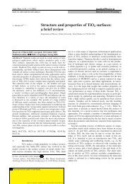
Fig. 11.<br />
Society.<br />
Non-contact AFM image <strong>of</strong> a TiO 2 (1 1 0)-(1 1) <strong>surface</strong>. From Fukui et al. [118]. # <strong>The</strong> American Physical<br />
A de®nite interpretation <strong>of</strong> images as shown in Fig. 10 is rather dif®cult. For the sake <strong>of</strong> studying<br />
<strong>surface</strong> structure and adsorbates, it is maybe better (and suf®cient) to focus on results obtained with the<br />
`normal' tip state that can be obtained reproduciblyby`cleaning' with high voltage/high current pulses.<br />
As mentioned above, onlyfew reports exist where satisfactoryimages have been obtained with<br />
negative sample bias (®lled-state images) [77]. <strong>The</strong>se were taken with a bias voltage that is too small to<br />
bridge the 3 eV gap. It is likelythat a real `contrast reversal' would occur under the tunneling<br />
conditions where the ®lled state <strong>of</strong> the VB are imaged, but, to this author's knowledge, such images<br />
have not yet been reported.<br />
Recently, non-contact AFM has been introduced as a complementary technique to study TiO 2<br />
<strong>surface</strong>s with atomic resolution. An image <strong>of</strong> the TiO 2 (1 1 0)-(1 1) <strong>surface</strong>, obtained byFukui et al.<br />
[118], is reproduced in Fig. 11. Frequencymodulation described in [119] was used as the feed back<br />
signal. While the contrast formation <strong>of</strong> atomicallyresolved AFM images using this technique is also<br />
somewhat controversial [120], one would assume that the physical geometry should dominate in AFM.<br />
<strong>The</strong> registry<strong>of</strong> (1 2) strands (see Section 2.2.2) in AFM images is consistent with the bright rows in<br />
Fig. 11 being the protruding bridging oxygens. Consequently, the black spots in Fig. 11 were assigned<br />
as oxygen vacancies in [118].<br />
2.2.1.4. Surface defects. <strong>The</strong> abilityto control the amount <strong>of</strong> defects is one <strong>of</strong> the main attractions <strong>of</strong> TiO 2<br />
as a `well-characterized' model system. Because imperfections such as vacancies introduce changes in the<br />
electronic structure (in particular a band gap feature at 0.8 eV below E Fermi , and a shoulder in the XPS Ti2p<br />
peak, see Section 3.2), theyhave been investigated with spectroscopic techniques for years. Much has been<br />
learned about the structure <strong>of</strong> defects, mainlybecause <strong>of</strong> recent investigations with scanning probe<br />
techniques. <strong>The</strong> following discussion considers steps; vacancies produced bythermal annealing,<br />
sputtering, and electron bombardment; as well as common impurities such as Ca and H.<br />
2.2.1.4.1. Step edges. Sputtering and annealing in UHV (at not too high temperatures) renders flat<br />
(1 1) <strong>surface</strong>s. As is expected for annealing <strong>of</strong> sputter-damaged <strong>surface</strong>s, the terrace size increases with