Gastric polyps and dysplasia
Gastric polyps and dysplasia
Gastric polyps and dysplasia
You also want an ePaper? Increase the reach of your titles
YUMPU automatically turns print PDFs into web optimized ePapers that Google loves.
MINI-SYMPOSIUM: UPPER GASTROINTESTINAL PATHOLOGY<br />
<strong>Gastric</strong> <strong>polyps</strong> <strong>and</strong> <strong>dysplasia</strong><br />
Paul J Kelly<br />
Gregory Y Lauwers<br />
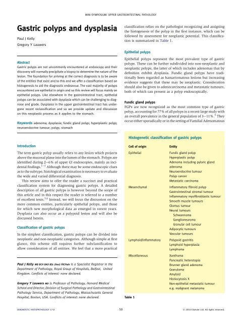
classification relies on the pathologist recognizing <strong>and</strong> assigning<br />
the histogenesis of the polyp in the first instance, which can be<br />
followed by assessment for neoplastic potential. This classification<br />
is summarized in Table 1.<br />
Epithelial <strong>polyps</strong><br />
Abstract<br />
<strong>Gastric</strong> <strong>polyps</strong> are not uncommonly encountered at endoscopy <strong>and</strong> their<br />
discovery will normally precipitate a biopsy to determine the nature of the<br />
lesion. The foundation for arriving at the correct diagnosis is to be aware<br />
of the entities that exist <strong>and</strong> to this end we offer a classification based on<br />
histogenesis to aid the diagnostic endeavour. The vast majority of <strong>polyps</strong><br />
encountered are epithelial in origin <strong>and</strong> so this review will focus mainly on<br />
epithelial <strong>polyps</strong>. Like elsewhere in the gastrointestinal tract, epithelial<br />
<strong>polyps</strong> can be associated with <strong>dysplasia</strong> which can be challenging to diagnose<br />
<strong>and</strong> grade. Dysplasia in the upper gastrointestinal tract has undergone<br />
recent reclassification <strong>and</strong> so we provide update <strong>and</strong> discussion<br />
on this neoplastic process as it applies to the stomach.<br />
Keywords adenoma; <strong>dysplasia</strong>; fundic gl<strong>and</strong> polyp; hyperplastic polyp;<br />
neuroendocrine tumour; polyp; stomach<br />
Epithelial <strong>polyps</strong> represent the most prevalent type of gastric<br />
<strong>polyps</strong>. These can be further subdivided into non-neoplastic <strong>and</strong><br />
neoplastic <strong>polyps</strong>, the latter of which includes adenomas that by<br />
definition exhibit <strong>dysplasia</strong>. Fundic gl<strong>and</strong> <strong>polyps</strong> have traditionally<br />
been regarded as hamartomatous lesions but increasing<br />
evidence suggests that these may be neoplastic. Consideration<br />
should also be given to adenocarcinoma <strong>and</strong> metastatic tumours,<br />
both of which can present as a polyp endoscopically.<br />
Fundic gl<strong>and</strong> <strong>polyps</strong><br />
FGPs are now recognized as the most common type of gastric<br />
polyp, accounting for 77% of all <strong>polyps</strong> in a recent large study with<br />
an overall prevalence in the general population of 3e11%. 5 They<br />
occur either sporadically or in the setting of Familial Adenomatous<br />
Histogenetic classification of gastric <strong>polyps</strong><br />
Introduction<br />
The term gastric polyp usually refers to any lesion which projects<br />
above the mucosal plane into the lumen of the stomach. Polyps are<br />
identified during 2e6% of upper GI endoscopies, mainly as incidental<br />
findings. 1e5 Although there may be some endoscopic clues<br />
as to the subtype, histological examination is necessary to evaluate<br />
the wide <strong>and</strong> varied differential diagnosis.<br />
This review aims to offer the reader a succinct <strong>and</strong> practical<br />
classification system for diagnosing gastric <strong>polyps</strong>. A detailed<br />
description of all gastric <strong>polyps</strong> is however beyond the scope of<br />
this article <strong>and</strong> in this respect the reader is referred to a number<br />
of excellent texts. 5,6 Instead, we will focus the discussion on the<br />
more common entities, particularly epithelial <strong>polyps</strong>, <strong>and</strong> those<br />
for which new morphological data as emerged in recent years.<br />
Dysplasia can also occur as a polypoid lesion <strong>and</strong> will also be<br />
discussed herein.<br />
Classification of gastric <strong>polyps</strong><br />
In the simplest classification, gastric <strong>polyps</strong> can be divided into<br />
neoplastic <strong>and</strong> non-neoplastic categories. Although simple at first<br />
glance, this scheme still requires further subclassification to<br />
allow consideration of all entities. We feel that a more practical<br />
Paul J Kelly MB BCH BAO BSc (Hon) FRCPath is a Specialist Registrar in the<br />
Department of Pathology, Royal Group of Hospitals, Belfast, United<br />
Kingdom. Conflicts of interest: none declared.<br />
Gregory Y Lauwers MD is Professor of Pathology, Harvard Medical<br />
School <strong>and</strong> Director, Division of Surgical Pathology <strong>and</strong> Gastrointestinal<br />
Pathology Service, Department of Pathology, Massachusetts General<br />
Hospital, Boston, USA. Conflicts of interest: none declared.<br />
Cell of origin<br />
Epithelial<br />
Mesenchymal<br />
Lymphoid/inflammatory<br />
Miscellaneous<br />
Table 1<br />
Entity<br />
Fundic gl<strong>and</strong> polyp<br />
Hyperplastic polyp<br />
Adenoma including pyloric gl<strong>and</strong><br />
adenoma<br />
Neuroendocrine tumour<br />
Polyp cancer<br />
Metastatic carcinoma<br />
Inflammatory fibroid polyp<br />
Gastrointestinal stromal tumour<br />
Inflammatory myofibroblastic tumour<br />
Smooth muscle tumours<br />
Glomus tumour<br />
Neural tumours<br />
Schwannoma<br />
Ganglioneuroma<br />
Granular cell tumour<br />
Adipocytic tumours<br />
Vascular tumours<br />
Polypoid gastritis<br />
Lymphoid hyperplasia<br />
Lymphoma<br />
Xanthoma<br />
Pancreatic heterotopia<br />
Brunner gl<strong>and</strong> adenoma<br />
Granuloma<br />
Amyloid<br />
Histiocytosis X<br />
Non-epithelial metastatic tumour<br />
e.g. malignant melanoma<br />
DIAGNOSTIC HISTOPATHOLOGY 17:2 50 Ó 2010 Elsevier Ltd. All rights reserved.
MINI-SYMPOSIUM: UPPER GASTROINTESTINAL PATHOLOGY<br />
a Typical fundic gl<strong>and</strong> polyp at low power showing numerous cystically-dilated oxyntic gl<strong>and</strong>s with overlying foveolar-type epithelium. b Surface of<br />
fundic gl<strong>and</strong> polyp lined by bl<strong>and</strong> foveolar epithelium.<br />
Figure 1<br />
Polyposis (FAP). Sporadic FGPs are much more common, <strong>and</strong><br />
their increased prevalence probably relates to their putative<br />
association with proton pump inhibitors. There has been some<br />
debate whether or not there is a negative association with<br />
Helicobacter pylori infection. 7 Most patients with FGPs are<br />
asymptomatic although non-specific gastrointestinal symptoms<br />
may occur in association with larger lesions. Endoscopically, FGPs<br />
appear as smooth, translucent, circumscribed mucosal elevations<br />
located in the body-fundic oxyntic mucosa. They can be single but<br />
are more commonly multiple (10) in a young person should<br />
prompt both the pathologist <strong>and</strong> clinician to consider the possibility<br />
of FAP. In this scenario, follow-up with colonoscopy or<br />
flexible sigmoidoscopy may have some merit. 9<br />
FGPs were previously considered hamartomatous lesions<br />
but the association of genetic alterations in both sporadic <strong>and</strong><br />
FAP-associated <strong>polyps</strong> suggests that both may be neoplasms.<br />
Mutations in the APC e b-catenin pathway have been<br />
encountered in both types of FGP however, in FAP, the <strong>polyps</strong><br />
are more frequently demonstrate adenomatous polyposis coli<br />
(APC) gene (90%) mutations, compared to b-catenin mutations<br />
(10%). 10 The converse is true in sporadic FGPs, which<br />
more often harbour activating mutations in the b-catenin<br />
gene. 11<br />
Dysplasia is rare in sporadic FGPs but is encountered in<br />
a significant number of FAP-associated <strong>polyps</strong>. Interestingly,<br />
sporadic FGPs found to harbour dysplastic foci are more<br />
frequently associated with APC gene mutations, suggesting that<br />
this subgroup is phenotypically akin to FAP-associated FGPs<br />
(Figure 2). 12<br />
Regression of FGPs has been noted to occur despite evidence<br />
accruing that these are neoplastic. If clinically appropriate, such<br />
as in the setting of large sporadic <strong>polyps</strong>, PPI therapy may be<br />
discontinued. Some studies have suggested however, that PPI<br />
therapy may be protective against <strong>dysplasia</strong> in FAP-associated<br />
FGPs. 2,13 The risk of <strong>dysplasia</strong> in sporadic <strong>polyps</strong> is very low <strong>and</strong><br />
although polypectomy is not necessary, biopsy is useful in order<br />
to exclude <strong>dysplasia</strong>; endoscopic surveillance is not recommended.<br />
2,9 Follow-up guidelines for patients with FAP are not<br />
well established but in this setting, annual screening <strong>and</strong><br />
surveillance is probably advisable as their risk of <strong>dysplasia</strong> is<br />
greater.<br />
Hyperplastic <strong>polyps</strong> <strong>and</strong> variants<br />
Hyperplastic <strong>polyps</strong> comprise approximately 17% of all gastric<br />
<strong>polyps</strong>, although a wide variation has been reported. 2,5,6,14 This<br />
variation likely reflects prescribing practices for proton pump<br />
inhibitors, surgical practice <strong>and</strong> the prevalence of Helicobacter<br />
infection.<br />
Hyperplastic <strong>polyps</strong> occur against a background of chronic<br />
gastritis <strong>and</strong> it is considered that these lesions develop as the result<br />
of an exaggerated mucosal response to injury. 15 An association<br />
with H. pylori infection has been reported but hyperplastic <strong>polyps</strong><br />
can also be associated with autoimmune gastritis <strong>and</strong> bile reflux. 9<br />
Solid organ transplant has also been reported as a risk factor. 16,17<br />
Hyperplastic <strong>polyps</strong> are typically observed in the antrum <strong>and</strong><br />
are often multiple. Less often they are noted in the cardia, where<br />
there is an association with reflux disease. Multiple hyperplastic<br />
<strong>polyps</strong> can be found in patients with Menetrier’s disease. Macroscopically,<br />
they are smooth, dome-shaped <strong>and</strong> usually measure<br />
between 0.5 <strong>and</strong> 1.5 cm but may exceed 2 cm. Larger lesions are<br />
prone to surface erosion which can result in chronic blood loss <strong>and</strong><br />
possibly iron-deficiency anaemia. <strong>Gastric</strong> outlet obstruction is<br />
a rare complication of large hyperplastic <strong>polyps</strong>. 18e20<br />
Histologically, hyperplastic <strong>polyps</strong> comprise variably elongated,<br />
distorted <strong>and</strong> branching foveolae lying within an oedematous<br />
<strong>and</strong> inflamed stroma (Figure 3). The foveolae are lined by<br />
a single layer of foveolar-type epithelium. Pyloric-type gl<strong>and</strong>s <strong>and</strong><br />
foci of intestinal metaplasia may be seen but in general the gastric<br />
DIAGNOSTIC HISTOPATHOLOGY 17:2 51 Ó 2010 Elsevier Ltd. All rights reserved.
MINI-SYMPOSIUM: UPPER GASTROINTESTINAL PATHOLOGY<br />
a Fundic gl<strong>and</strong> polyp with increased surface epithelial density. b Higher power shows cytonuclear atypia, consistent with low-grade <strong>dysplasia</strong>.<br />
Figure 2<br />
gl<strong>and</strong>s are not present. A rich vasculature is also common <strong>and</strong> can<br />
contribute to profuse bleeding during removal. Wisps of smooth<br />
muscle derived from the muscularis mucosa are usually seen. 8<br />
The presence of thickened <strong>and</strong> splayed muscle fibres in the core<br />
of the polyp should lead one to consider an alternative lesion such<br />
as a prolapse polyp 21 (Figure 4) or PeutzeJegher-type polyp.<br />
In most cases, the diagnosis of a hyperplastic polyp is<br />
straightforward. However, a challenge can be to differentiate<br />
between florid regenerative changes on the surface of an eroded<br />
polyp from a dysplastic or neoplastic process. To this end, one<br />
should carefully assess that the atypia observed is confined to an<br />
area of surface damage, where it is more likely to be reactive. It is<br />
pertinent to remind the reader that foci of <strong>dysplasia</strong> or<br />
intramucosal carcinoma can be encountered in up to 4% of<br />
hyperplastic <strong>polyps</strong>, <strong>and</strong> more commonly in large <strong>polyps</strong><br />
(>2 cm). 22e25 For this reason, it is important to assess all of the<br />
available material <strong>and</strong> liaise with the clinician if a <strong>dysplasia</strong> is<br />
identified, particularly if the polyp is large or there has been<br />
subtotal removal. Since there is also a not infrequent association<br />
with neoplasia elsewhere in the stomach (noted in 4e6% of<br />
cases), dysplastic changes in hyperplastic <strong>polyps</strong> should prompt<br />
endoscopic assessment of the surrounding mucosa. 4,9,25<br />
Variants of hyperplastic <strong>polyps</strong><br />
Polypoid foveolar hyperplasia e is regarded as a precursor<br />
lesion to hyperplastic <strong>polyps</strong> <strong>and</strong> shares a common aetiology in<br />
a Hyperplastic polyp comprising irregular, cystically dilated gl<strong>and</strong>s lined by foveolar cells set within an inflamed <strong>and</strong> oedematous stroma. There is<br />
evidence of surface erosion at the bottom of the field associated with a degree of reactive atypia. b The characteristic inflammatory cell infiltrate is<br />
evident adjacent to tortuous <strong>and</strong> distended gl<strong>and</strong>s.<br />
Figure 3<br />
DIAGNOSTIC HISTOPATHOLOGY 17:2 52 Ó 2010 Elsevier Ltd. All rights reserved.
MINI-SYMPOSIUM: UPPER GASTROINTESTINAL PATHOLOGY<br />
a At low power, the configuration of a prolapse-type gastric polyp is similar to a hyperplastic polyp. b On higher power thickened <strong>and</strong> splayed<br />
muscle fibres are more conspicuous <strong>and</strong> are associated with dilated <strong>and</strong> congested vascular channels, characteristic of trauma-induced changes.<br />
Cystically dilated misplaced gl<strong>and</strong>s are also noted, reminiscent of gastric cystica, are also associated with prolapsing mucosa.<br />
Figure 4<br />
the form of chronic mucosal injury. Typically, the lesions are<br />
between 1 <strong>and</strong> 2 mm <strong>and</strong> exhibit simple hyperplasia of the gastric<br />
foveolae without cystic change <strong>and</strong> gl<strong>and</strong> distortion. Both<br />
regression <strong>and</strong> progression to hyperplastic <strong>polyps</strong> has been<br />
described, depending on the persistence of the underlying<br />
stimulus.<br />
Gastritis cystica polyposa/profunda e is defined as a hyperplastic<br />
polyp is which there is epithelial misplacement in the<br />
muscularis mucosa or more deeply in the submucosa or muscularis<br />
propria. The application of the term polyposa refers to<br />
when the lesion is mainly intraluminal; profundus is the<br />
preferred term when most of the lesion is intramural. 6 Like<br />
elsewhere in the gastrointestinal tract, it is generally held that<br />
epithelial misplacement is a result of trauma-induced entrapment<br />
of gl<strong>and</strong>s in the deeper compartments of the stomach wall. The<br />
main diagnostic difficulty lies in distinguishing a well differentiated<br />
adenocarcinoma. Bl<strong>and</strong> cytomorphology, low or absent<br />
mitotic activity <strong>and</strong> lack of desmoplasia serve as indicators of<br />
benign misplaced epithelium.<br />
Neuroendocrine tumours e so called “carcinoid” tumours<br />
The term “neuroendocrine tumour” (NET) has now been adopted<br />
in preference to “carcinoid” <strong>and</strong> these comprise less than 2% of<br />
gastric polypoid lesions with a trend towards an increasing incidence.<br />
Several different types of neuroendocrine cells are present in<br />
the stomach but at least 70% of all gastric NETs derive from<br />
enterochromaffin-like (ECL) cells. 2,5,6,26,27 In the current WHO<br />
system, gastrointestinal NETs are further assessed <strong>and</strong> subclassified<br />
to establish malignant potential using several parameters including<br />
site of origin, size, angioinvasion, proliferative activity, metastasis<br />
<strong>and</strong> association with clinical hormonal syndromes. 27,28<br />
ECL cell NETs may arise sporadically, or against a background<br />
of ECL cell hyperplasia induced by hypergastrinaemia. Four<br />
types of gastric NET exist (Type IeIV) <strong>and</strong> differ according to<br />
aetiology <strong>and</strong> morphology. 27,28 Both type I <strong>and</strong> II tumours are<br />
associated with hypergastrinaemia <strong>and</strong> arise in the body or<br />
fundus. Type I tumours are associated with a background of<br />
chronic autoimmune atrophic gastritis which results in loss of<br />
negative feedback to the G cells in the gastric antrum, secondary<br />
to destruction of parietal cells. Type II gastric NETs are associated<br />
with gastrinomas in the context of ZollingereEllison<br />
syndrome <strong>and</strong> multiple endocrine neoplasia type I (3e15% of<br />
tumours). 29 Consequently, the background mucosa will appear<br />
different in type II tumours <strong>and</strong> will demonstrate parietal cell<br />
hypertrophy <strong>and</strong> hyperplasia. Type III tumours are sporadic <strong>and</strong><br />
generally do not have signature changes in the background<br />
mucosa; these are typically larger <strong>and</strong> exhibit more aggressive<br />
behaviour. Tumours associated with hypergastrinaemia are<br />
usually smaller, broad based <strong>and</strong> multifocal, while those that are<br />
sporadic (Type III) tend to be unifocal <strong>and</strong> larger. Type IV gastric<br />
NETs are rare, solitary <strong>and</strong> correspond to undifferentiated solid<br />
carcinomas that show early metastasis <strong>and</strong> poor outcome; there<br />
is no association with atrophic gastritis or ZES. 28<br />
Histologically, the most common forms of gastric NETs adopt<br />
ribbon <strong>and</strong> trabecular patterns. Insular patterns can also be seen.<br />
The cytomorphology is bl<strong>and</strong> <strong>and</strong> similar to typical “carcinoid”<br />
tumours occurring elsewhere in the gastrointestinal tract<br />
(Figure 5). The cells are usually chromogranin A positive but<br />
negative for chromogranin B. Synaptophysin is positive in 50%<br />
of cases. In addition, if a biopsy displays features of atrophic<br />
gastritis or hypertrophy, assessment should include examination<br />
of the NE cell compartments, as precursor lesions of type I <strong>and</strong> II<br />
gastric NETs, namely hyperplasia, <strong>dysplasia</strong> <strong>and</strong> micro-NETs,<br />
can also be recognized histologically <strong>and</strong> may give rise to<br />
polypoid lesions at endoscopy. A three-tier system for grading<br />
NETs of the gastrointestinal tract has been proposed based on<br />
assessment of mitotic activity <strong>and</strong> proliferative activity (as<br />
measured by Ki67 or MIB1). This has been somewhat controversial<br />
but has been included in British <strong>and</strong> European guidelines<br />
for management. 27 The presence of anaplasia <strong>and</strong> necrosis is also<br />
DIAGNOSTIC HISTOPATHOLOGY 17:2 53 Ó 2010 Elsevier Ltd. All rights reserved.
MINI-SYMPOSIUM: UPPER GASTROINTESTINAL PATHOLOGY<br />
a Small well-differentiated gastric neuroendocrine tumour can be seen against a background of atrophic gastritis (Type I ECL cell NET). Typical<br />
nests of ECL cells are seen at the bottom left of the field. b Higher power shows ill-defined nests of ECL cells in the lamina propria. The cells have<br />
bl<strong>and</strong> epithelioid nuclei with typical “salt <strong>and</strong> pepper” chromatin.<br />
Figure 5<br />
suggestive of higher grade <strong>and</strong> more likely to be found in<br />
sporadic NETs. 2<br />
Management of the gastric NETs depends on assessment of the<br />
parameters discussed above as well as tumour subtype (IeIV) <strong>and</strong><br />
grade (1e3). 27 Type I NETs are nearly always benign <strong>and</strong> can be<br />
managed by endoscopic resection <strong>and</strong> surveillance; those that are<br />
difficult to control may require antrectomy to remove the stimulus<br />
from antral G cells. Treatment of associated vitamin B12 deficiency<br />
is also requisite. It is usual for type II tumours to remain<br />
indolent <strong>and</strong> as such local excision following treatment of<br />
underlying ZES suffices. Type II tumours may be larger than their<br />
Type I counterpart, <strong>and</strong> lymph node metastases have been<br />
reported. Type III tumours are fundamentally different, are more<br />
aggressive are best treated by partial or total gastrectomy. Due to<br />
the advanced stage of disease at presentation, Type IV NETs may<br />
have more limited treatment options.<br />
Adenomas<br />
<strong>Gastric</strong> adenomas are defined by the WHO as circumscribed<br />
polypoid lesions composed of tubular <strong>and</strong>/or villous structures<br />
lined by dysplastic epithelium. 8,30 They can occur spontaneously<br />
or in the context of Familial Adenomatosis Polyposis (FAP). Many<br />
surgical pathologists use the terms [flat] <strong>dysplasia</strong> (such as that<br />
occurring against a background of chronic atrophic gastritis <strong>and</strong><br />
intestinal metaplasia) <strong>and</strong> adenoma interchangeably. We do not<br />
subscribe to the opinion that the term adenoma should be reserved<br />
to denote polypoid dysplastic lesions developing without associated<br />
chronic atrophic gastritis or intestinal metaplasia 7 since in<br />
most cases the lack of associated pathology is related to limited<br />
sampling of the surrounding mucosa. Moreover, <strong>dysplasia</strong> occurring<br />
in completely normal gastric mucosa is exceptionally rare.<br />
The prevalence of gastric adenomas is reported to be between<br />
0.65% <strong>and</strong> 3.75% in Western countries <strong>and</strong> between 9 <strong>and</strong> 27% in<br />
countries such as Japan <strong>and</strong> China, where there is a higher incidence<br />
of gastric carcinoma. 5,14,25,31 Most adenomas are solitary,<br />
exophytic lesions which can be sessile (more common) or<br />
pedunculated <strong>and</strong> usually measure less than 2 cm, although they<br />
can reach up to 4 cm in size. The risk of malignancy is related to<br />
size, grade of <strong>dysplasia</strong> <strong>and</strong> villosity of the growth pattern. Larger<br />
adenomas are more frequently associated with a component of<br />
high-grade <strong>dysplasia</strong>, <strong>and</strong> a significant proportion contains foci of<br />
malignant transformation, the incidence of which is 40e50% for<br />
lesions measuring greater than 2 cm. Smaller adenomas should<br />
not be dismissed with little more than a cursory assessment as<br />
these too can contain carcinomatous foci. 8,30,32<br />
As preinvasive lesions, it is recommended that adenomas are<br />
excised in total when it is safe to do so, either by endoscopic<br />
polypectomy or by endoscopic mucosal resection (EMR). As<br />
there is a risk of synchronous carcinomas in patients with an<br />
adenomatous polyp, 2 thorough evaluation of the entire stomach<br />
should be recommended <strong>and</strong> undertaken. Follow-up surveillance<br />
is also recommended when gastric adenomas are excised. 9<br />
Please refer to the <strong>Gastric</strong> <strong>dysplasia</strong> section for review of the<br />
microscopic features of adenomas/<strong>dysplasia</strong>.<br />
Polyposis syndromes<br />
A number of polyposis syndromes can affect the stomach<br />
including FAP, Juvenile polyposis, PeutzeJeghers syndrome,<br />
CronkiteeCanada syndrome <strong>and</strong> Cowden disease <strong>and</strong> are associated<br />
with hamartomatous <strong>polyps</strong>. These conditions are relatively<br />
rare <strong>and</strong> the patients may present clinically with<br />
manifestations unrelated to the stomach. Fundic gl<strong>and</strong> <strong>polyps</strong>,<br />
which are the most common type of gastric polyp are also<br />
associated with FAP (see above). A detailed discussion of these<br />
syndromes is beyond the scope of this review.<br />
Malignant epithelial <strong>polyps</strong> <strong>and</strong> metastatic tumours<br />
As is the case elsewhere in the gastrointestinal tract, carcinomas<br />
can appear endoscopically as <strong>polyps</strong>. After establishing that one<br />
DIAGNOSTIC HISTOPATHOLOGY 17:2 54 Ó 2010 Elsevier Ltd. All rights reserved.
MINI-SYMPOSIUM: UPPER GASTROINTESTINAL PATHOLOGY<br />
is dealing with an invasive adenocarcinoma as opposed to<br />
<strong>dysplasia</strong>, it is necessary to evaluate whether the tumour is<br />
primary or metastatic. If primary, one should evaluate if it is<br />
superficial (i.e. an early gastric cancer) or more deeply invasive.<br />
Usually biopsies from primary gastric carcinomas will be associated<br />
with background changes of <strong>dysplasia</strong>. Diffuse-type gastric<br />
carcinoma may not have obvious surrounding <strong>dysplasia</strong> <strong>and</strong> it is<br />
usually pertinent to consider the differential diagnosis of metastatic<br />
carcinoma, in particular metastatic lobular carcinoma from<br />
the breast. In cases where there is little or no evidence of an<br />
origin, correlation with all available clinical information,<br />
including the patient’s past medical history is important.<br />
Non-epithelial malignant tumours such as malignant melanomas<br />
should also be considered in the differential diagnosis of<br />
poorly differentiated carcinoma.<br />
Non-epithelial <strong>polyps</strong><br />
The various mesenchymal <strong>and</strong> stromal elements of the stomach<br />
can give rise to nodular proliferations <strong>and</strong> tumours that manifest<br />
endoscopically as <strong>polyps</strong>. This heterogeneous group of lesions is<br />
listed in Table 1. In our practice, other than glomus tumours,<br />
most lesions in this category morphologically comprise spindle<br />
cells <strong>and</strong> frequently immunohistochemistry is required to<br />
establish the final diagnosis. Several entities should be considered<br />
in the differential diagnosis of a gastric spindle cell lesion<br />
including inflammatory fibroid polyp (IFP), gastrointestinal<br />
stromal tumour (GIST) <strong>and</strong> inflammatory myofibroblastic<br />
tumour (IMT). Less common spindle cells lesions that may be<br />
sampled in an endoscopic biopsy include nerve sheath tumours<br />
such as schwannomas <strong>and</strong> ganglioneuromas <strong>and</strong> smooth muscle<br />
tumours. Only IFPs will be discussed further here <strong>and</strong> the reader<br />
is referred to other papers with respect to GISTs 33e35 <strong>and</strong> IMTs. 36<br />
Inflammatory fibroid polyp<br />
IFPs comprise between 1 <strong>and</strong> 3% of all gastric <strong>polyps</strong>. 5,14 They<br />
arise in the submucosa of the antropyloric region <strong>and</strong> appear as<br />
solitary, sessile or pedunculated lesions that are frequently<br />
ulcerated. Histologically, they comprise bl<strong>and</strong> spindle cells lying<br />
within a vascularized stroma that has an associated inflammatory<br />
infiltrate that is strikingly rich in eosinophils. Classically, the<br />
spindle cells are concentrically arranged around blood vessels<br />
but this is not always the rule (Figure 6). We have also seen<br />
variants of IFP which have a myxoid appearance, reminiscent of<br />
a nerve sheath myxoma. The spindle cells are usually CD34<br />
positive <strong>and</strong> CD117 negative. It is wise to perform a wider panel<br />
of immunohistochemistry in difficult cases as CD34 positivity is<br />
also a feature of GISTs; since GISTs may also be CD117 negative,<br />
it may also be pertinent to include DOG-1 in the immunopanel.<br />
In the past, IFPs were generally considered reactive lesions as<br />
there was an association with hypochlorhydria <strong>and</strong> achlorhydria.<br />
An allergic aetiology also seemed plausible due to the characteristic<br />
finding of eosinophils. A familial tendency has been noted<br />
following the discovery of a family in the UK, whose female<br />
members have a high incidence of the <strong>polyps</strong>. 37 More recently<br />
studies have demonstrated gain-of-function mutations in the<br />
PDGFR-a gene in IFPs, similar to those found in kit-negative<br />
GISTS, thereby alluding to a neoplastic process. 38,39<br />
In management terms, most IFPs are found incidentally <strong>and</strong><br />
do not recur after excision. Further treatment is not usually<br />
undertaken <strong>and</strong> patients do not require surveillance. 5<br />
Miscellaneous lesions presenting as <strong>polyps</strong><br />
A range of other lesions can present as <strong>polyps</strong> as demonstrated in<br />
Table 1. Of these xanthomata <strong>and</strong> pancreatic heterotopia will be<br />
discussed.<br />
Xanthomata<br />
These commonly appear as yellow plaques at endoscopy as<br />
opposed to well-defined <strong>polyps</strong> <strong>and</strong> are found in the body <strong>and</strong><br />
fundus. They are thought to develop in association with chronic<br />
gastritis, especially after gastric resection <strong>and</strong> thus are believed to<br />
represent a reaction pattern to tissue injury. They are small<br />
a Inflammatory fibroid polyp appearing as a moderately cellular submucosal tumour with limited extension into the lamina propria. Lymphoid<br />
aggregates are also present. b The characteristic morphology is more evident at higher power. Bl<strong>and</strong> spindle cells are more conspicuous <strong>and</strong> there<br />
is evidence of perivascular “whorling”. The stroma is rich in inflammatory cells, in particular eosinophils, which is typical.<br />
Figure 6<br />
DIAGNOSTIC HISTOPATHOLOGY 17:2 55 Ó 2010 Elsevier Ltd. All rights reserved.
MINI-SYMPOSIUM: UPPER GASTROINTESTINAL PATHOLOGY<br />
lesions, usually measuring less than 3 mm, <strong>and</strong> histologically<br />
appear as loose collections of lipid-laden macrophages in the<br />
lamina propria. Diagnosis is normally straightforward <strong>and</strong> their<br />
main diagnostic significance is related to the differential diagnosis<br />
which includes diffuse-type (signet ring) carcinoma <strong>and</strong><br />
granular cell tumour. A combination of histology, histochemistry<br />
(periodic-acid Schiff, mucicarmine) <strong>and</strong> immunohistochemistry<br />
(CD68, S100, cytokeratin) will allow differentiation of these<br />
entities.<br />
Pancreatic heterotopia<br />
Pancreatic heterotopia presents in two settings. The first refers to<br />
the gastro-oesophageal junction where small nodules of pancreatic<br />
acinar tissue may be noted. This is reported in up to 15% of<br />
people undergoing endoscopy for gastro-oesophageal reflux<br />
disease <strong>and</strong> it is plausible that it forms part of a metaplastic<br />
process. “True” pancreatic heterotopia usually occurs in the prepyloric<br />
region of the stomach. The polypoid lesion may appear<br />
umbilicated, due to the presence of a pancreatic duct. The histological<br />
appearance is essentially of submucosally-based normal<br />
pancreatic tissue. Of note, in some cases pancreatic ductal tissue<br />
may predominate or be the only tissue recognized. There may be<br />
prominence of somewhat disorganized smooth muscle fibres<br />
admixed with the pancreatic tissue which engenders the alternative<br />
term of “adenomyoma” for pancreatic heterotopia.<br />
Unless symptomatic, the patient does not require further<br />
therapy. Neoplastic transformation (i.e. pancreatic ductal adenocarcinoma<br />
or pancreatic neuroendocrine tumour) has been<br />
described but is sufficiently rare that there is no need for surgical<br />
excision or endoscopic surveillance.<br />
The main differential diagnoses are well-differentiated adenocarcinoma<br />
<strong>and</strong> gastritis cystica polyposa/profundus. Resolving<br />
the differential diagnosis with well-differentiated adenocarcinoma<br />
may be difficult, particularly as pancreatic acini <strong>and</strong> islets may not<br />
be apparent in some cases of heterotopia. In this situation it is<br />
useful to note the bl<strong>and</strong> cytology <strong>and</strong> the vaguely organoid or<br />
lobular appearance of heterotopia that can be appreciated on low<br />
power examination.<br />
The absent polyp<br />
Up to 16% of gastric biopsies taken from endoscopically identified<br />
<strong>polyps</strong> disclose no histological features that pertain to a recognized<br />
gastric polyp. Indeed many such biopsies will either be<br />
normal or show variable degrees of inflammation. 5 Occasionally,<br />
there may be oedema or mild foveolar hyperplasia, which may<br />
account for a polypoid endoscopic appearance but this is nonspecific.<br />
In such cases, it may be pertinent to liaise with the<br />
endoscopist or add a comment that the biopsy may not be representative<br />
of the lesion seen, or a more deeply seated lesion.<br />
However, care should be taken not to elevate the clinical concerns<br />
unnecessarily. It is good practice for endoscopic pictures to be<br />
included with the biopsy. 2<br />
<strong>Gastric</strong> <strong>dysplasia</strong><br />
Diagnosing <strong>and</strong> grading gastric <strong>dysplasia</strong> continues to be a source<br />
of difficulty. In the first instance, it is relatively infrequently<br />
encountered in routine practice, particularly in Western countries.<br />
Second, <strong>and</strong> not infrequently, there can be great difficulty in distinguishing<br />
florid regenerative atypia from <strong>dysplasia</strong>.<br />
Diagnosing <strong>and</strong> grading gastric <strong>dysplasia</strong> <strong>and</strong> differentiation<br />
from regenerative atypia<br />
Historically, criteria used to classify <strong>dysplasia</strong> <strong>and</strong> early invasive<br />
carcinoma in the upper gastrointestinal tract has differed<br />
between Western <strong>and</strong> Japanese pathologists, leading to difficulty<br />
in comparing prevalence data. Presently, there is a greater degree<br />
of consensus opinion, which has led to the development of<br />
several similar classification systems that in common adopt the<br />
two-tier grading system of low- <strong>and</strong> high-grade <strong>dysplasia</strong>. The<br />
classification systems in use are illustrated in Table 2.<br />
Low grade <strong>dysplasia</strong> shows only slight architectural deviation<br />
from non-neoplastic gl<strong>and</strong>ular epithelium <strong>and</strong> comprises multiple<br />
small gl<strong>and</strong>ular tubules with little branching or irregularity.<br />
Cytologically, there is a mild to moderate degree of atypia, with<br />
elongate or ovoid (so called “pencil-shaped”) hyperchromatic<br />
nuclei. There is nuclear crowding <strong>and</strong> pseudostratification but in<br />
Lymphoid proliferations<br />
The main lesions to consider are:<br />
Polypoid gastritis<br />
Lymphoid hyperplasia<br />
<strong>Gastric</strong> lymphoma<br />
Both polypoid gastritis <strong>and</strong> lymphoid hyperplasia are benign<br />
conditions, have preponderance for occurring in the antrum <strong>and</strong><br />
are associated with H. pylori infection. Consistent with this is that<br />
both lesions can regress with successful eradication of the<br />
bacteria. The histology is similar as nodular lymphoid aggregates<br />
are noted in both however, in the former, the inflammatory<br />
infiltrate is more mixed <strong>and</strong> may be accompanied by overlying<br />
epithelial changes. The histological finding of lymphoid rich<br />
lesions in the stomach should prompt the pathologist to consider<br />
gastric lymphoma. A finding of destructive infiltrates of atypical<br />
lymphoid cells should raise suspicion of lymphoma followed by<br />
judicious use of immunohistochemistry to establish the diagnosis.<br />
A second opinion from a haematopathologist may also be<br />
advisable.<br />
Classification schemes for evaluating <strong>dysplasia</strong> <strong>and</strong><br />
invasive adenocarcinoma in the upper gastrointestinal<br />
tract<br />
WHO classification<br />
Negative for <strong>dysplasia</strong><br />
Indefinite for <strong>dysplasia</strong><br />
Low-grade <strong>dysplasia</strong><br />
High-grade <strong>dysplasia</strong><br />
Invasive adenocarcinoma*<br />
* Includes intramucosal carcinoma.<br />
Table 2<br />
Vienna classification<br />
1. Negative for <strong>dysplasia</strong><br />
2. Indefinite for <strong>dysplasia</strong><br />
3. Non-invasive, low grade <strong>dysplasia</strong><br />
4. Non-invasive neoplasia<br />
4.1 High-grade <strong>dysplasia</strong><br />
4.2 Non-invasive carcinoma<br />
4.3 Suspicious for invasive carcinoma<br />
5. Invasive neoplasia<br />
5.1 Intramucosal carcinoma<br />
5.2 At least submucosal carcinoma<br />
DIAGNOSTIC HISTOPATHOLOGY 17:2 56 Ó 2010 Elsevier Ltd. All rights reserved.
MINI-SYMPOSIUM: UPPER GASTROINTESTINAL PATHOLOGY<br />
general the nuclei retain a tendency for basal orientation. The cells<br />
may appear mucin depleted. An important diagnostic feature is<br />
that the atypical features described extend to mucosal surface<br />
indicating lack of maturation (Figure 7).<br />
High-grade <strong>dysplasia</strong> displays more pronounced architectural<br />
aberrations with a greater degree of gl<strong>and</strong>ular crowding <strong>and</strong><br />
gl<strong>and</strong> irregularity. Typical patterns include prominent branching,<br />
budding <strong>and</strong> cribriforming (Figure 8). Small amounts of necrotic<br />
material or degenerate cell debris may be noted within gl<strong>and</strong>ular<br />
lumina <strong>and</strong> if extensive, one should search for evidence of<br />
intramucosal carcinoma. The constituent cells may appear<br />
cuboidal as opposed to columnar. There is generally a greater<br />
degree of nuclear pseudostratification with extension of nuclei<br />
towards the apical portion of the cell. The nuclei may be oval <strong>and</strong><br />
hyperchromatic as for low-grade <strong>dysplasia</strong> but more often will<br />
appear rounded, with vesicular chromatin <strong>and</strong> prominent<br />
nucleoli. Irregular, thickened nuclear membranes may be seen<br />
<strong>and</strong> may impart the appearance of “boulder-shaped” nuclei.<br />
Mitoses are more numerous <strong>and</strong> may be noted in the surface<br />
epithelium; atypical mitoses be also be noted more frequently. 40<br />
Regenerative atypia, particularly when florid, can be difficult<br />
to distinguish from <strong>dysplasia</strong>. In contrast to dysplastic mucosa,<br />
regenerating or reactive mucosa generally does not exhibit the<br />
same degree of architectural atypia or disorganization as<br />
a dysplastic process. Gl<strong>and</strong>s tend to respect their relative spaces<br />
<strong>and</strong> may be patulous with a corkscrew appearance, reminiscent<br />
of foveolar hyperplasia. To discern a reactive process, one should<br />
carefully examine the surrounding mucosa <strong>and</strong> assess the<br />
changes therein as features such as inflammation, oedema <strong>and</strong><br />
vascular congestion may point to a benign process. In regeneration,<br />
the cells present often appear immature <strong>and</strong> cuboidal with<br />
reduced amounts of cytoplasm. The nuclei may be slightly larger<br />
than in normal epithelium, but in general terms they are more<br />
monomorphic, evenly spaced without significant crowding or<br />
pseudostratification <strong>and</strong> tend to remain basally orientated. It is<br />
inferred that the nuclear to cytoplasmic ratio will be increased<br />
but this is less marked than in <strong>dysplasia</strong>. Nucleoli may be seen<br />
Figure 7 Low-grade adenomatous (Type I) <strong>dysplasia</strong>. The normal architecture<br />
is largely maintained but there is cellular <strong>and</strong> nuclear crowding.<br />
The hyperchromatic nuclei are pencil-shaped <strong>and</strong> show pseudostratification.<br />
The abnormalities can be seen within deep gl<strong>and</strong>s <strong>and</strong> on the<br />
surface indicating failure of maturation. This feature helps to establish<br />
a diagnosis of <strong>dysplasia</strong> over regenerative atypia.<br />
Figure 8 High-grade adenomatous (Type I) <strong>dysplasia</strong> characterized by<br />
a greater degree of architectural changes such as crowding <strong>and</strong> backto-back<br />
gl<strong>and</strong>ular arrangements. The nuclei are more pleomorphic <strong>and</strong><br />
have boulder-like outlines.<br />
but tend to be much smaller <strong>and</strong> may be multiple. Mitotic activity<br />
is usually increased but is confined to the proliferative “neck”<br />
zone <strong>and</strong> should not be present in surface epithelial cells. A key<br />
finding to appreciate is maturation <strong>and</strong> cellular differentiation as<br />
cells move from the proliferative zone to the surface 40 (Figure 9).<br />
Another useful finding is the lack of abrupt transition from<br />
benign epithelium to atypical epithelium, as is more commonly<br />
noted in <strong>dysplasia</strong>. 41<br />
Ancillary techniques such as p53 <strong>and</strong> Ki67 immunohistochemistry<br />
have been used to assist in distinguishing between<br />
florid reactive atypia <strong>and</strong> <strong>dysplasia</strong>. Strong, nuclear staining for<br />
p53 is thought to be a marker of neoplasia <strong>and</strong> can be detected in<br />
approximately one-third of gastric <strong>dysplasia</strong>. The finding of an<br />
area of strong p53 staining in conjunction with Ki67 labelling,<br />
especially at the surface (<strong>and</strong> hence distant from the normal<br />
proliferative zone), is suggestive of <strong>dysplasia</strong>. However, this<br />
staining pattern is not specific <strong>and</strong> it should be noted that p53<br />
staining has been detected in H. pylori gastritis <strong>and</strong> in up to 30%<br />
of cases of intestinal metaplasia. Therefore the results of immunohistochemistry<br />
should be correlated closely with the<br />
morphological impression gained from thorough H&E examination,<br />
<strong>and</strong> should never be used in isolation to render a diagnosis<br />
of <strong>dysplasia</strong>.<br />
Indefinite for <strong>dysplasia</strong> is used in many of the current classification<br />
systems. It is not a biological entity but a reflection of<br />
a diagnostic difficulty. From a clinical st<strong>and</strong>point, the term<br />
essentially indicates a need for close surveillance <strong>and</strong> repeat<br />
biopsy of the patient. The diagnosis can be used in a limited<br />
number of settings such as when there is a genuine difficulty in<br />
discerning reactive epithelial changes from <strong>dysplasia</strong>, when<br />
worrisome atypia is noted but is confined to a very small area or<br />
when the tissue is limited in amount or quality. When strict<br />
criteria are applied, this represents an uncommon diagnosis in<br />
our experience. In addition, a proffered diagnosis of “indefinite<br />
for <strong>dysplasia</strong>” should be accompanied by an explanatory note to<br />
the clinician regarding the diagnostic dilemma. Importantly, this<br />
category should not serve as a wastebasket group for all cases of<br />
regenerative atypia, as in most cases, an underlying inflammatory<br />
condition can be recognized.<br />
DIAGNOSTIC HISTOPATHOLOGY 17:2 57 Ó 2010 Elsevier Ltd. All rights reserved.
MINI-SYMPOSIUM: UPPER GASTROINTESTINAL PATHOLOGY<br />
Regenerative atypia. Although cells exhibit mild nuclear enlargement <strong>and</strong> mucin depletion, there is uniformity amongst all the atypical cells with<br />
no evidence of an abrupt transition to normal appearing epithelium, which is more typical of <strong>dysplasia</strong>. Note also the foveolar hyperplasia at the<br />
left side of the field <strong>and</strong> maturation of cells towards the surface. When all the features are taken together, a diagnosis of regenerative or reactive<br />
atypia can be rendered.<br />
Figure 9<br />
Types of gastric <strong>dysplasia</strong><br />
Four types of gastric <strong>dysplasia</strong> are recognized. The most<br />
common form of gastric <strong>dysplasia</strong> has an intestinal phenotype,<br />
resembling colonic adenomas. This is classified as Type I or<br />
“adenomatous” <strong>dysplasia</strong> (Figures 7 <strong>and</strong> 8). Type II (hyperplastic/foveolar)<br />
<strong>dysplasia</strong> is less common, <strong>and</strong> typically<br />
resembles foveolar epithelium. In a series of 69 cases of gastric<br />
<strong>dysplasia</strong>, foveolar-type accounted for approximately 22% of<br />
the cases. 42 The morphological features of low-grade foveolar<br />
<strong>dysplasia</strong> can be deceptively bl<strong>and</strong> <strong>and</strong> it may be difficult to<br />
differentiate from reactive foveolar atypia. This may explain<br />
why foveolar <strong>dysplasia</strong> is reported to be more commonly high<br />
grade than intestinal-type <strong>dysplasia</strong>. The constituent gl<strong>and</strong>s<br />
tend to show variation in size <strong>and</strong> shape with occasional cystic<br />
change <strong>and</strong> gl<strong>and</strong> branching. Papillary in-folding <strong>and</strong> gl<strong>and</strong><br />
serrations may be seen <strong>and</strong> are reminiscent of colonic hyperplastic<br />
<strong>polyps</strong> or serrated adenomas. The cells tend to have<br />
a cuboidal configuration with clear to faintly eosinophilic<br />
cytoplasm <strong>and</strong> vesicular nuclei <strong>and</strong> prominent nucleoli<br />
(Figure 10). 7,40<br />
The third type of <strong>dysplasia</strong> represents the pyloric gl<strong>and</strong><br />
adenoma (Figure 11). The characteristics of pyloric gl<strong>and</strong><br />
adenomas were first described by Borchard in 1990. 43 These<br />
lesions commonly arise against a background of autoimmune<br />
gastritis or H. pylori infection, <strong>and</strong> are more prevalent in elderly<br />
patients. They are composed of tightly packed pyloric-type<br />
gl<strong>and</strong>s lined by bl<strong>and</strong> appearing low columnar mucus-secreting<br />
epithelium with fine granular cytoplasm but the normal apical<br />
a Foveolar (Type II) <strong>dysplasia</strong>. Irregular gl<strong>and</strong> contours can be appreciated with short papillations <strong>and</strong> serrations. The lining epithelium resembles<br />
gastric foveolar epithelium. b At this power, nuclear atypia can be appreciated allowing a diagnosis of <strong>dysplasia</strong>.<br />
Figure 10<br />
DIAGNOSTIC HISTOPATHOLOGY 17:2 58 Ó 2010 Elsevier Ltd. All rights reserved.
MINI-SYMPOSIUM: UPPER GASTROINTESTINAL PATHOLOGY<br />
a Pyloric gl<strong>and</strong> adenoma. Tightly packed gl<strong>and</strong>s are present which are lined by pyloric-type epithelium. There is minimal atypia. b At higher power,<br />
mild nuclear enlargement can be appreciated along with small nucleoli. Despite the bl<strong>and</strong> morphology, these lesions are considered to be<br />
dysplastic.<br />
Figure 11<br />
mucin cap is absent. The immunohistochemical stain MUC6<br />
characteristically decorates the constituent cells. Assigning the<br />
grade of <strong>dysplasia</strong> can be difficult in pyloric gl<strong>and</strong> adenomas <strong>and</strong><br />
there is a significant risk of malignant transformation. 44,45 Thus,<br />
in light of the reported risks with these lesions, complete removal<br />
is necessary.<br />
Tubule neck (or globoid) <strong>dysplasia</strong> represents the fourth type of<br />
<strong>dysplasia</strong> <strong>and</strong> is believed to be a precursor to sporadic diffuse-type<br />
gastric carcinoma. It occurs in neck region of non-metaplastic<br />
gastric epithelium <strong>and</strong> the typical appearance is of as enlarged<br />
clear cells, of which some have signet-ring morphology<br />
(Figure 12). By definition, the atypical cells are confined by the<br />
a Tubule neck <strong>dysplasia</strong> in a case of sporadic diffuse-type carcinoma of the stomach. At this power only pit elongation <strong>and</strong> sharp transition with<br />
normal foveolar cells is noted. b The gastric pit (arrow) shows elongation <strong>and</strong> is lined by mucin-depleted cells showing nuclear pleomorphism. The<br />
adjacent lamina propria contains mononuclear inflammatory cells as well as individual malignant epithelial cells (arrowhead). This latter population<br />
shares features in common with the adjacent pit allowing one to interpret the atypia as representing tubule neck <strong>dysplasia</strong>. It is extremely<br />
rare <strong>and</strong> difficult to diagnose tubule neck.<br />
Figure 12<br />
DIAGNOSTIC HISTOPATHOLOGY 17:2 59 Ó 2010 Elsevier Ltd. All rights reserved.
MINI-SYMPOSIUM: UPPER GASTROINTESTINAL PATHOLOGY<br />
basement membrane. It is rare to make a diagnosis of tubule neck<br />
<strong>dysplasia</strong> as it is extremely subtle <strong>and</strong> difficult to recognize; in<br />
many cases, the diagnosis is only made on resection specimens of<br />
diffuse-type gastric carcinoma. 7,40,41 Accordingly, it is rarely seen<br />
on endoscopic biopsies <strong>and</strong> this point should be considered if one<br />
is contemplating the diagnosis on such material. Occasionally,<br />
clearing of neck cells, believed to be related to PPI effects, can<br />
mimic tubule neck <strong>dysplasia</strong> (personal observation GYL). These<br />
lesions ought to be differentiated from those seen in familial<br />
diffuse-type gastric cancer.<br />
Management of gastric <strong>dysplasia</strong><br />
Both low- <strong>and</strong> high-grade <strong>dysplasia</strong> are variably associated with<br />
an increased risk of malignant transformation. Rendering either<br />
of these diagnoses thus has significance for the patient in terms<br />
of further management <strong>and</strong> follow-up.<br />
It is reported that low-grade <strong>dysplasia</strong> persists in 19e50% of<br />
cases but regresses in 38e75%. 46e48 The risk of progression to<br />
high-grade <strong>dysplasia</strong> is suggested to be 17%. 49 There has also<br />
been variation in figures quoted for progression to adenocarcinoma<br />
but more recent studies suggest that this is in the order of<br />
0e9%. 49,50 Follow-up of low-grade <strong>dysplasia</strong> is recommended in<br />
the form of annual endoscopic surveillance <strong>and</strong> re-biopsy;<br />
advances in endoscopic techniques such as the introduction of<br />
chromoendoscopy, narrow b<strong>and</strong> imaging <strong>and</strong> confocal microscopy<br />
now allows improved surveillance with detection <strong>and</strong><br />
sampling of previously undetectable lesions. 41<br />
A diagnosis of high-grade <strong>dysplasia</strong> places the patient in<br />
a different clinical category associated with higher rates of<br />
persistence (14e58%) <strong>and</strong> progression (60e85%). Similarly,<br />
regression is less often observed being documented in 0e16%<br />
cases. 49e52 With more advanced techniques available for<br />
management, rendering a diagnosis of HGD (as well as intramucosal<br />
carcinoma) is no longer an indication for gastrectomy.<br />
Instead, following exclusion a more deeply infiltrative lesion by<br />
endoscopic ultrasound <strong>and</strong> adequate sampling, it is now possible<br />
to employ endoscopic mucosal resection for definitive<br />
management.<br />
Conclusion<br />
A huge variety of lesions may present incidentally at endoscopy<br />
as gastric <strong>polyps</strong>. Unfortunately, it is often the case that the<br />
biopsy specimen is submitted with little or no details as to the<br />
endoscopic impression of the lesion <strong>and</strong> thus the pathologist is<br />
asked to make a diagnosis in a vacuum based on limited clinical<br />
information <strong>and</strong> material. Rendering the correct diagnosis first<br />
requires knowledge of the individual entities that may exist <strong>and</strong><br />
to this end establishing the histogenesis of the lesion is a good<br />
starting point. Dysplasia can involve a number of different<br />
epithelial <strong>polyps</strong> <strong>and</strong> despite improved consensus in classifying<br />
<strong>and</strong> grading <strong>dysplasia</strong>, this remains challenging for many<br />
pathologists at the sign-out bench. Unfortunately, despite the<br />
range of ancillary tests that can be employed in histopathology,<br />
there is no substitute for careful H&E examination. Double<br />
reporting of upper gastrointestinal tract <strong>dysplasia</strong> is being<br />
increasingly used <strong>and</strong> is to be welcomed. In cases were diagnostic<br />
doubt persists we advocate a low threshold for seeking<br />
a second opinion.<br />
A<br />
REFERENCES<br />
1 Dekker W. Clinical relevance of gastric <strong>and</strong> duodenal <strong>polyps</strong>. Sc<strong>and</strong> J<br />
Gastroenterol 1990; 25(suppl 178): 7e12.<br />
2 Carmack SW, Genta RM, Graham DY, Lauwers GY. Management of<br />
gastric <strong>polyps</strong>: a pathology-based guide for gastroenterologists. Nat<br />
Rev Gastroenterol Hepatol 2009; 6: 331e41.<br />
3 Ghazi A, Ferstenberg H, Shinya H. Endoscopic gastroduodenal polypectomy.<br />
Ann Surg 1984; 200: 175e80.<br />
4 Laxen F, Sipponen P, Ihamaki T, Hakkiluoto A, Dortscheva Z. <strong>Gastric</strong><br />
<strong>polyps</strong>; their morphological <strong>and</strong> endoscopical characteristics <strong>and</strong><br />
relation to gastric carcinoma. Acta Pathol Microbiol Immunol Sc<strong>and</strong> A<br />
1982; 90: 221e8.<br />
5 Carmack SW, Genta RM, Schuler CM, Saboorian MH. The current<br />
spectrum of gastric <strong>polyps</strong>: a 1-year national study of over 120,000<br />
patients. Am J Gastroenterol 2009; 104: 1524e32.<br />
6 Odze RD, Goldblum JR. Surgical pathology of the GI tract, liver, biliary<br />
tract <strong>and</strong> pancreas. Philadelphia: Saunders Elsevier, 2009.<br />
7 Yantiss RK, O.R.. Neoplastic precursor lesions in the upper gastrointestinal<br />
tract. Diagnostic Histoplathology 2008; 14: 437e52.<br />
8 Park do Y, Lauwers GY. <strong>Gastric</strong> <strong>polyps</strong>: classification <strong>and</strong> management.<br />
Arch Pathol Lab Med 2008; 132: 633e40.<br />
9 Goddard AF, Badreldin R, Pritchard DM, Walker MM, Warren B. The<br />
management of gastric <strong>polyps</strong>. Gut 2010; 59: 1270e6.<br />
10 Abraham SC, Nobukawa B, Giardiello FM, Hamilton SR, Wu TT. Fundic<br />
gl<strong>and</strong> <strong>polyps</strong> in familial adenomatous polyposis: neoplasms with<br />
frequent somatic adenomatous polyposis coli gene alterations. Am J<br />
Pathol 2000; 157: 747e54.<br />
11 Abraham SC, Nobukawa B, Giardiello FM, Hamilton SR, Wu TT.<br />
Sporadic fundic gl<strong>and</strong> <strong>polyps</strong>: common gastric <strong>polyps</strong> arising through<br />
activating mutations in the beta-catenin gene. Am J Pathol 2001;<br />
158: 1005e10.<br />
12 Abraham SC, Park SJ, Mugartegui L, Hamilton SR, Wu TT. Sporadic<br />
fundic gl<strong>and</strong> <strong>polyps</strong> with epithelial <strong>dysplasia</strong>: evidence for preferential<br />
targeting for mutations in the adenomatous polyposis coli<br />
gene. Am J Pathol 2002; 161: 1735e42.<br />
13 Bianchi LK, Burke CA, Bennett AE, Lopez R, Hasson H, Church JM.<br />
Fundic gl<strong>and</strong> polyp <strong>dysplasia</strong> is common in familial adenomatous<br />
polyposis. Clin Gastroenterol Hepatol 2008; 6: 180e5.<br />
14 Stolte M, Sticht T, Eidt S, Ebert D, Finkenzeller G. Frequency, location,<br />
<strong>and</strong> age <strong>and</strong> sex distribution of various types of gastric polyp.<br />
Endoscopy 1994; 26: 659e65.<br />
15 Dirschmid K, Platz-Baudin C, Stolte M. Why is the hyperplastic polyp<br />
a marker for the precancerous condition of the gastric mucosa?<br />
Virchows Arch 2006; 448: 80e4.<br />
16 Amaro R, Neff GW, Karnam US, Tzakis AG, Raskin JB. Acquired<br />
hyperplastic gastric <strong>polyps</strong> in solid organ transplant patients. Am J<br />
Gastroenterol 2002; 97: 2220e4.<br />
17 Jewell KD, Toweill DL, Swanson PE, Upton MP, Yeh MM. <strong>Gastric</strong><br />
hyperplastic <strong>polyps</strong> in post transplant patients: a clinicopathologic<br />
study. Mod Pathol 2008; 21: 1108e12.<br />
18 Abraham SC, Singh VK, Yardley JH, Wu TT. Hyperplastic <strong>polyps</strong> of the<br />
stomach: associations with histologic patterns of gastritis <strong>and</strong> gastric<br />
atrophy. Am J Surg Pathol 2001; 25: 500e7.<br />
19 Cerwenka H, Bacher H, Mischinger HJ. Pyloric obstruction caused by<br />
prolapse of a hyperplastic gastric polyp. Hepatogastroenterology<br />
2002; 49: 958e60.<br />
20 Chen HW, Lu CH, Shun CT, Lin MT, Tsang YM. <strong>Gastric</strong> outlet obstruction<br />
due to giant hyperplastic gastric <strong>polyps</strong>. J Formos Med Assoc<br />
2005; 104: 852e5.<br />
DIAGNOSTIC HISTOPATHOLOGY 17:2 60 Ó 2010 Elsevier Ltd. All rights reserved.
MINI-SYMPOSIUM: UPPER GASTROINTESTINAL PATHOLOGY<br />
21 Gonzalez-Obeso E, Fujita H, Deshp<strong>and</strong>e, V, et al. <strong>Gastric</strong> “hyperplastic<br />
<strong>polyps</strong>”: a heterogeneous clinicopathological group including<br />
a distinct subset best categorized as mucosal prolapse polyp. Am J<br />
Surg Pathol., in press.<br />
22 Daibo M, Itabashi M, Hirota T. Malignant transformation of gastric<br />
hyperplastic <strong>polyps</strong>. Am J Gastroenterol 1987; 82: 1016e25.<br />
23 Dirschmid K, Walser J, Hugel H. Pseudomalignant erosion in hyperplastic<br />
gastric <strong>polyps</strong>. Cancer 1984; 54: 2290e3.<br />
24 Han AR, Sung CO, Kim KM, et al. The clinicopathological features of<br />
gastric hyperplastic <strong>polyps</strong> with neoplastic transformations:<br />
a suggestion of indication for endoscopic polypectomy. Gut Liver<br />
2009; 3: 271e5.<br />
25 Hattori T. Morphological range of hyperplastic <strong>polyps</strong> <strong>and</strong> carcinomas<br />
arising in hyperplastic <strong>polyps</strong> of the stomach. J Clin Pathol 1985; 38:<br />
622e30.<br />
26 Gilligan CJ, Lawton GP, Tang LH, West AB, Modlin IM. <strong>Gastric</strong> carcinoid<br />
tumors: the biology <strong>and</strong> therapy of an enigmatic <strong>and</strong> controversial<br />
lesion. Am J Gastroenterol 1995; 90: 338e52.<br />
27 Williams GT. Endocrine tumours of the gastrointestinal tract-selected<br />
topics. Histopathology 2007; 50: 30e41.<br />
28 Kloppel G, Perren A, Heitz PU. The gastroenteropancreatic neuroendocrine<br />
cell system <strong>and</strong> its tumors: the WHO classification. Ann N Y<br />
Acad Sci 2004; 1014: 13e27.<br />
29 Kloppel G, Heitz PU, Capella C, Solcia E. Pathology <strong>and</strong> nomenclature<br />
of human gastrointestinal neuroendocrine (carcinoid) tumors <strong>and</strong><br />
related lesions. World J Surg 1996; 20: 132e41.<br />
30 Watanabe H, J.J., Sobin LH, eds. Histological typing of oesophageal<br />
<strong>and</strong> gastric tumors. Berlin: Springer-Verlag, 1990.<br />
31 Nakamura T, Nakano G. Histopathological classification <strong>and</strong> malignant<br />
change in gastric <strong>polyps</strong>. J Clin Pathol 1985; 38: 754e64.<br />
32 Park DI, Rhee PL, Kim JE, et al. Risk factors suggesting malignant<br />
transformation of gastric adenoma: univariate <strong>and</strong> multivariate<br />
analysis. Endoscopy 2001; 33: 501e6.<br />
33 Agaimy A. Gastrointestinal stromal tumors (GIST) from risk stratification<br />
systems to the new TNM proposal: more questions than<br />
answers? A review emphasizing the need for a st<strong>and</strong>ardized GIST<br />
reporting. Int J Clin Exp Pathol 2010; 3: 461e71.<br />
34 Miettinen M, Lasota J. Gastrointestinal stromal tumors: review on<br />
morphology, molecular pathology, prognosis, <strong>and</strong> differential diagnosis.<br />
Arch Pathol Lab Med 2006; 130: 1466e78.<br />
35 Miettinen M, Sobin LH, Lasota J. Gastrointestinal stromal tumors of<br />
the stomach: a clinicopathologic, immunohistochemical, <strong>and</strong> molecular<br />
genetic study of 1765 cases with long-term follow-up. Am J Surg<br />
Pathol 2005; 29: 52e68.<br />
36 Coffin CM, Hornick JL, Fletcher CD. Inflammatory myofibroblastic<br />
tumor: comparison of clinicopathologic, histologic, <strong>and</strong> immunohistochemical<br />
features including ALK expression in atypical <strong>and</strong><br />
aggressive cases. Am J Surg Pathol 2007; 31: 509e20.<br />
37 Allibone RO, Nanson JK, Anthony PP. Multiple <strong>and</strong> recurrent inflammatory<br />
fibroid <strong>polyps</strong> in a Devon family (‘Devon polyposis<br />
syndrome’): an update. Gut 1992; 33: 1004e5.<br />
38 Schildhaus HU, Buttner R, Binot E, Merkelbach-Bruse S,<br />
Wardelmann E. Inflammatory fibroid <strong>polyps</strong> are true neoplasms with<br />
PDGFRA mutations. Pathologe 2009; 30(Suppl. 2): 117e20.<br />
39 Schildhaus HU, Cavlar T, Binot E, Buttner R, Wardelmann E, Merkelbach-Bruse<br />
S. Inflammatory fibroid <strong>polyps</strong> harbour mutations in the<br />
platelet-derived growth factor receptor alpha (PDGFRA) gene.<br />
J Pathol 2008; 216: 176e82.<br />
40 Misdraji J, Lauwers GY. <strong>Gastric</strong> epithelial <strong>dysplasia</strong>. Semin Diagn<br />
Pathol 2002; 19: 20e30.<br />
41 Srivastava A, Lauwers GY. <strong>Gastric</strong> epithelial <strong>dysplasia</strong>: the western<br />
perspective. Dig Liver Dis 2008; 40: 641e9.<br />
42 Park do Y, Srivastava A, Kim GH, et al. Adenomatous <strong>and</strong> foveolar<br />
gastric <strong>dysplasia</strong>: distinct patterns of mucin expression <strong>and</strong> background<br />
intestinal metaplasia. Am J Surg Pathol 2008; 32: 524e33.<br />
43 Borchard F, Ghanei A, Koldovsky U, et al. Gastrale Differenzierung in<br />
Adenomen der Magenschleimkaut Immunohistochemische und eleckronenmikroskopische<br />
Untersuchungen. Verh Dtsch Ges Pathol<br />
1990; 74: 528.<br />
44 Chen ZM, Scudiere JR, Abraham SC, Montgomery E. Pyloric gl<strong>and</strong><br />
adenoma: an entity distinct from gastric foveolar type adenoma. Am J<br />
Surg Pathol 2009; 33: 186e93.<br />
45 Vieth M, Kushima R, Borchard F, Stolte M. Pyloric gl<strong>and</strong> adenoma:<br />
a clinico-pathological analysis of 90 cases. Virchows Arch 2003; 442:<br />
317e21.<br />
46 Bearzi I, Brancorsini D, Santinelli A, Rezai B, Mannello B, Ranaldi R.<br />
<strong>Gastric</strong> <strong>dysplasia</strong>: a ten-year follow-up study. Pathol Res Pract 1994;<br />
190: 61e8.<br />
47 Burke AP, Sobin LH, Shekitka KM, Helwig EB. Dysplasia of the<br />
stomach <strong>and</strong> Barrett esophagus: a follow-up study. Mod Pathol<br />
1991; 4: 336e41.<br />
48 Lansdown M, Quirke P, Dixon MF, Axon AT, Johnston D. High grade<br />
<strong>dysplasia</strong> of the gastric mucosa: a marker for gastric carcinoma. Gut<br />
1990; 31: 977e83.<br />
49 Rugge M, Cassaro M, Di Mario F, et al. The long term outcome of<br />
gastric non-invasive neoplasia. Gut 2003; 52: 1111e6.<br />
50 Yamada H, Ikegami M, Shimoda T, Takagi N, Maruyama M. Long-term<br />
follow-up study of gastric adenoma/<strong>dysplasia</strong>. Endoscopy 2004; 36:<br />
390e6.<br />
51 Di Gregorio C, Mor<strong>and</strong>i P, Fante R, De Gaetani C. <strong>Gastric</strong> <strong>dysplasia</strong>. A<br />
follow-up study. Am J Gastroenterol 1993; 88: 1714e9.<br />
52 Saraga EP, Gardiol D, Costa J. <strong>Gastric</strong> <strong>dysplasia</strong>. A histological followup<br />
study. Am J Surg Pathol 1987; 11: 788e96.<br />
Practice points<br />
C<br />
C<br />
C<br />
C<br />
<strong>Gastric</strong> <strong>polyps</strong> can derive from any of the component cells of<br />
the stomach wall <strong>and</strong> therefore assigning histiogenesis to<br />
a lesion forms a useful starting point for diagnosis.<br />
Dysplasia can involve a number of “benign” gastric <strong>polyps</strong>, so<br />
all material should be carefully examined.<br />
<strong>Gastric</strong> <strong>dysplasia</strong> is relatively rare in routine practice <strong>and</strong><br />
remains challenging to diagnose <strong>and</strong> grade. Rendering this<br />
diagnosis has implications for patient follow-up <strong>and</strong><br />
management.<br />
Increasingly double reporting is being utilized in the diagnosis<br />
of <strong>dysplasia</strong> <strong>and</strong> is to be welcomed.<br />
DIAGNOSTIC HISTOPATHOLOGY 17:2 61 Ó 2010 Elsevier Ltd. All rights reserved.


