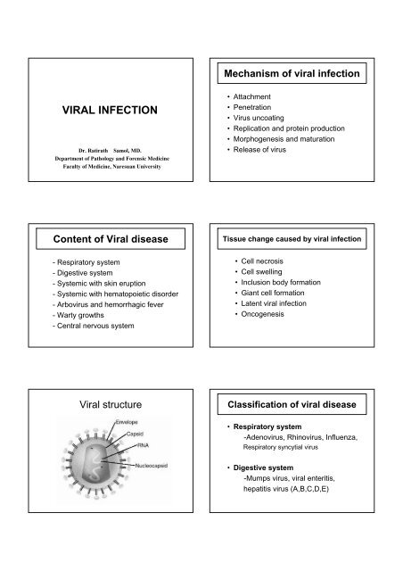
VIRAL INFECTION - Faculty of Medicine
VIRAL INFECTION - Faculty of Medicine
VIRAL INFECTION - Faculty of Medicine
Create successful ePaper yourself
Turn your PDF publications into a flip-book with our unique Google optimized e-Paper software.
Influenza Antigenic Changes• Antigenic Drift– Minor change, same subtype– Caused by point mutations in gene– May result in epidemicMechanism <strong>of</strong> theCytokine StormEvoked byInfluenza virus• Antigenic Shift– Major change, new subtype– Caused by exchange <strong>of</strong> gene segments– May result in pandemicInfluenza virus• Influenza• Avian influenza (Bird flu)• Mexico influenza or Swine influenza(2009 influenza)Influenza• Clinical feature:- rapid onset <strong>of</strong> fever, myalgia,headache, weakness, cough- progressive symptom 3-5 day- clinical subside 2 weeks• Complication: pneumonia, lunghemorrhage, hyaline membrane disease• Killed viral vaccineInfluenza• Self limited infection <strong>of</strong> upper airway,but can extending to lower air way• Destroy mucociliary function followedby secondary bacterial infectionAvian influenza (Bird flu)- Caused by influenza virus type A, H5N1- disease <strong>of</strong> bird, chicken, duck- transmitted to human by direct contactwith affected animals- virus dies from heat (70 ‘c)NORMAL TRACHEAL MUCOSALycke and Norrby Textbook <strong>of</strong> Medical Virology 19937 DAYS POST-<strong>INFECTION</strong>
Viral enteritis and diarrhea• Rotavirus•Norwalk virus• Corona virusNorwalk virus• (+) ssRNA, nonenveloped virus• Common in childhood• Clinical , transmission, pathogenesis,clinical course and treatment are similar toRotavirusRotavirus• Reovirus family• Most common cause <strong>of</strong> gastroenteritis inchildren < 1 year <strong>of</strong> age• Clinical :- fever, rhinorrhea, vomiting,abdominal pain, watery non-bloodydiarrhea• Pathogenesis: reduce absorption <strong>of</strong>water and sodium from GICoronavirus• Caused by respiratory tract infection anddiarrhea• Self limited disease• Intermittent epidermiologyRotavirus• Transmission by fecal-oral route• Persistent symptom about 1 week• Treatment: conservative with fluidreplacementHepatitis virus• Classified 6 types- Hepatitis virus types A, B, C, D, E, G
Measles virus• Clinical:- conjunctivitis, cough, coryza, fever- at day 2-3: “Koplik spot” at buccalmucosa- at day 4-5: reddish-brown rash,enlarged cervical LN- 1 week: subsidePathology <strong>of</strong> Measles• Multinucleated giantcells with intranuclearand intracytoplasmicinclusion (Warthin-Finkeldey cells) inlymphoid organs andother organsWarthin-Finkeldey cellsMeasles virus• Complication:- pneumonia- encephalitis- keratitis- abnormal hemorrhage- secondary bacterial infectionRubella (german measles)• Caused by Rubella virus• Incubation period 14-21 days• Systemic infection : rash, fever, malaise,coryza, arthritis and arthralgias• Exanthem - discrete, pinkish red, finemaculopapular eruption - begins on theface and spreads cephalocaudally• Dx: serology testingMeaslesRubella (german measles)Koplik spot
Congenital rubella syndrome• Infected pregnant woman ( first 20 th wks)• Fetal death, premature delivery, congenitalanomalies• Heart defects: PDA, VSD, pulmonaryvalvular stenosis• Eye and ear defects: cataract, glaucoma,deafness• CNS defects: microcephaly, mental retardHerpes simplex infectionTransmitted by direct/sexual contactDiagnosis: clinicallyscrap base <strong>of</strong> vesicle and a specialstain - Giemsa-stained (Tzanck smear)ballooned epithelial cells with intranuclearinclusions and multinucleated giantviral cultures take 24-72 hoursRubella (german measles)Primary herpes simplex infections• Complications - arthritis, purpura,thrombocytopenia, encephalitisHerpetic gingivostomatitishigh fever, irritability, anorexia, mouth pain,drooling in infants and toddlersgingiva becomes intensely erythematous,edematous, friable and tends to bleedsmall yellow ulcerations with red halos seen onbuccal and labial mucosa, tongue, gingivae,palate, tonsilsHerpes simplex infectionsPrimary herpes simplex infectionsHSV-1>90% <strong>of</strong> primary infections aresubclinicalFever blister or cold sores at facialskin eg. lip, nose, gingivostomatitisHSV-2genital pathogenneonatal herpesHSV-1 Cold soreSkin infectionsfever, malaise, localized lesionsdirect inoculation (usually cold sores) andgenitaliapainful vesicles on erythematous base -usually groupedHSV-2 Genital Herpes
Recurrent herpes simplex infectionHSV (gingivostomatitis)Triggers include fever, sunlight, localtrauma, menses, emotional stressSeen most commonly as cold soresProdrome <strong>of</strong> localized burning, itchingbefore eruption <strong>of</strong> grouped vesiclesHerpes simplex virusHSV (genital herpes)• Gross:- group <strong>of</strong> intraepithelial vesicles(blisters) and frequently crust covering- may be ulcer• Pathology:- pink to purple, glassy intranuclearinclusion (Cowdry type A)- mononucleated or multinucleated cellsHSV (cold sores)HSVglassy intranuclear inclusion (mononucleated or multinucleated cells)
Pathogenesis <strong>of</strong> HSVInitial infection in skin or mucous membranetravel along sensory nerve ending and retrograde axonalflow to neuron in dorsal root ganglia (latent infection)reactivate <strong>of</strong> latent infected neuron (eg. stress, fever, UV)Varicella zoster virus (VZV)• Cause <strong>of</strong> chickenpox, herpes zoster(shingles)• Incubation period ranges from 10-20 days• Transmitted by inhalation <strong>of</strong> airbornerespiratory droplets from an infected hostor direct contactnewly replicated virus is transported anterograde to a site ator near portal <strong>of</strong> entry into body causing localized vesiclesPathogenesis <strong>of</strong> HSVVaricella zoster virus (VZV)• Primary acute infection <strong>of</strong> VZV:“chickenpox”• Reactivation <strong>of</strong> latent VZV: “shingles orherpes zoster” distributes to sensorynervesNeonatal herpes infection• Infected fetus by contact vaginal secretioncontains viruses• Clinical features- mucocutaneous vesicles- virus spread to other organs eg. brain,liver, lung• High mortality rateVaricella (chickenpox)• Clinical : fever, diffuse vesicle on skin andmucous membrane• Complications : secondary bacterial skininfections, pneumonia, hepatitis,encephalitis, Reye syndrome• Severe in the immunocompromised host• Tx: Varicella-zoster immunogloblin
Herpes zoster (shingles)Varicella (chickenpox)After primary infection, virus lies dormantin genome <strong>of</strong> sensory nerve root cellPostulated triggers include mechanicaland trauma, infection, immunosuppressionLesions are grouped, thin-walled vesicleson erythematous base distributed alongdermatomelesions in multi-stagesPathogenesis <strong>of</strong> VZVShinglesPrimary infection ( chickenpox)VZV spread from mucosal and epidermal lesionsto local sensory nervesVZV remains latent in dorsal ganglion cells <strong>of</strong>sensory nervesReactivation <strong>of</strong> VZV results in syndrome <strong>of</strong> herpeszoster (shingles)Varicella zoster virus (VZV)Shingles• Gross:- chickenpox: diffuse, scattered vesicles- shingles: vesicles distribution along theperipheral nerve (dermatome)• Histology:- Intranuclear inclusion <strong>of</strong> infected cells(multinucleated cells)
VZVHand-foot-and-mouth diseaseIntranuclear inclusionCoxsackie Virus• Type A– Causes herpangina and hand- foot-and mouthdisease• Type B– Causes Pluerodynia• Both– Causes meningitis, myocarditis andpericarditis, also can cause juvenile diabetes• Coxsackie Virus A• Common in child• Infection <strong>of</strong> the throat• Causes red-ringedblisters or ulcers ontonsils, ro<strong>of</strong> <strong>of</strong> mouthand tongueHerpanginaHand-Foot-and Mouth diseaseCoxsackie Virus A, B or Enterovirus71Common in childProdome : low-grade fever, malaise, soremouth, anorexia1-2 days later :- oral lesions (tongue, throat, gum) : shallow,yellow ulcers surrounded by red halos- painful red blister on hand and footRx : supportive treatmentSystemic withhematopoietic disorder
Cytomegalovirus (CMV)• Produce a variety <strong>of</strong> disease dependon age, immune status• Cause <strong>of</strong> asymptomatic in healthyperson or severe systemic infection inneonates and immunocompromisedhost (opportunistic infection)Histology <strong>of</strong> CMV• Large purple intranuclear inclusionsurrounded by clear halo and smallerbasophilic intracytoplasmic inclusion• Organ involvement: salivary glands,kidney, liver, pancreas, brain, ect.Cytomegalovirus (CMV)CMV• Transmitted by:- intrauterine transmission- perinatal transmission- breast milk- respiratory droplets- semen and vaginal fluid- blood transfusion- organs transplantationCytomegalovirus (CMV)• Clinical feature depends on infectedorgans• Congenital CMV infection– 90% asymptomatic– 10% symptomatic eg. hemolytic anemia,jaundice, thrombocytopenia, pneumonia,hepatosplenomegaly, retinitis, brain damage,mental retard or deadEpstein-Barr Virus (EBV)• Cause <strong>of</strong> infectious mononucleosis• Self-limiting illness <strong>of</strong> children & young• EBV associated with hairy leukoplakia,lymphoma, and nasopharyngealcarcinoma• Transmitted by contact saliva
Epstein-Barr Virus (EBV)Atypical T lymphocytes• Clinical <strong>of</strong> infectious mononucleosis:- fever- generalized lymphadenopathy- hepatosplenomegaly- sore throat (patch on tonsil)- may be CNS lesion- may be hepatitis, pneumoniaPathogenesisEBV replicates in B-lymphocytes in tonsilB-lymphocytes disseminates in circulationAtypical T lymphocytes in blood circulationHuman Immunodeficiency Virus(HIV)•single-stranded, enveloped RNA viruses• HIV 1, HIV 2• can lead to acquired immunodeficiencysyndrome (AIDS)Human Immunodeficiency Virus(HIV)• Infection occurs by transfer <strong>of</strong> blood,semen, vaginal fluid, or breast milk• 4 major routes <strong>of</strong> transmission- sexual intercourse- contaminated needles- breast milk- vertical transmission
Human Immunodeficiency Virus(HIV)• Primarily infects in helper T cells (CD4+ T cells),macrophages, and dendritic cells• HIV infection leads to low levels <strong>of</strong> CD4+ T cellsby 3 main mechanisms- direct viral killing <strong>of</strong> infected CD4+ T cells- increased rates <strong>of</strong> apoptosis in infected CD4+T cells- killing <strong>of</strong> infected CD4+ T cells by CD8cytotoxic lymphocytes that recognize infectedcellsReplication cycleHuman Immunodeficiency Virus(HIV)• CD4+ T cell numbers decline below acritical level, cell-mediated immunity(CMI)is lost, and the body becomesprogressively more susceptible toopportunistic infectionsHIV infection 4 stages• Incubation period- asymptomatic , 2-4 weeks after infection• Acute infection- rapid viral replication- fever, lymphadenopathy- pharyngitis (sore throat)-rash, myalgia, malaise- mouth and esophageal soresHIV structureHIV infection 4 stages• Latency stage- shows few or no symptoms- low level viral particle in blood stream- duration 2 weeks -20 years
HIV infection 4 stages• AIDS- symptoms <strong>of</strong> various opportunistic infections- early : oral candidiasis (thrush) and tuberculosis- later : recurrences <strong>of</strong> HSV, shingles andpneumonia caused by Pneumocystis jirovecii- final stages : CMV, Mycobacterium aviumcomplex (MAC) and EBV induced B-celllymphomas or Kaposi ‘s sacromaDengue virus• Dengue virus 1-4• Dengue fever is a vector-borne disease• transmitted via bite <strong>of</strong> mosquitoeseg. Aedes aegypti, Aedes albopictus• Symptoms <strong>of</strong> dengue include:- high fever, headache-rash- nausea and vomiting-myalgia- leukopenia (neutropenia), thrombocytopeniaDengue virus• Infection with one serotype provideslifelong immunity to that particularserotype but not to the other three• Previous infection with simple denguefever greatly increases risk <strong>of</strong> developingDHFArbovirus andhemorrhagic feverWarty growths
Human papilloma virus (HPV)• Most common causes <strong>of</strong> sexuallytransmitted infection (STI) in the world• HPV types cause benign skin wart,anogenital warts, anogenital cancerHuman papilloma virus (HPV)• Transmitted by skin-to-skin contactduring sexual intercourse• Treated by electrocautery, lasertreatment• Histology : koilocytosis <strong>of</strong> infected cells• Warts: proliferative squamous epitheliumproducing nodules or flat lesionsHuman papilloma virus (HPV)Skin warts• Skin warts- HPV types 1,2- common warts (verruca vulgaris):hands, trunk, extremities- plantar warts: plantar- no associated with cancerHuman papilloma virus (HPV)Anogenital warts(condylomata acuminata)• Anogenital warts (condyloma acuminatum)- HPV types 6,11- warts at penis, vulva, vagina, anus- no associated with cancer• Anogenital cancer (squamous cell carcinoma)- HPV types 16,18,31,45- cause <strong>of</strong> cancer <strong>of</strong> anus, vulva, cervixand penis
Squamous cell carcinoma <strong>of</strong> cervixPathogenesistransmitted primarily through direct skin contactwith an infected individualvirus infects epidermis causing hyperplasia,hypertrophy and central degeneration <strong>of</strong> theepidermisepidermis has molluscum bodies contain largenumbers <strong>of</strong> maturing virionsKoilocytosis <strong>of</strong> infected cellsMolluscum contagiosumMolluscum virusintracytoplasmic inclusion bodies(molluscum bodies)• Cause <strong>of</strong> molluscum contagiosum•Self-limited viral disease <strong>of</strong> skin• Transmitted by skin or sexual contact• Gross: raised, umbilicated, cutaneousnodules at skin (most perineum)• Histology: intracytoplasmic inclusionbodies (molluscum bodies)
Intracytoplasmic inclusion bodies(molluscum bodies)PathogenesisPoliovirus infects oropharynx secreted in saliva andswallowMultiplies in intestinal mucosa and lymph nodesHematogenous spreadingInvade to CNS and replicate in spinal cord (spinalpoliomyelitis), or brain stem (bulbar poliomyelitis)* Virus destroys motor neuron in anterior horn <strong>of</strong>spinal cord or brain stem*PoliomyelitisCentral nervous system• Clinical features- flaccid paralysis- muscle atrophy- hyporeflexiaPoliovirus• Causes poliomyelitis• Transmitted by fecal-oral route• Most asymptomatic• 1/100 <strong>of</strong> infected patientsdisease(spinal poliomyelitis, bulbar poliomyelitis)Rabies virus• Cause <strong>of</strong> rabies (encephalitis)• Transmitted by saliva <strong>of</strong> dog or cat bites• Clinical:- malaise, headache, fever, dysphagiafallowed by period <strong>of</strong> acute neurologicsymptom including disorientation,hallucination, seizure, coma and deadfrom res. failure
Rabies virus• Post-exposure prophylaxis: combinedimmune serum and vaccine• High mortality rateArbo viral encephalitis virus• Japanese B caused by encephalitis• Animal host : pig• Carrier : Culex mosquitoes• Clinical : confusion, seizure, coma• Pathology : mononuclear cells aroundvessels, necrosis <strong>of</strong> neuron and braintissuePathogenesisRabies virus multiplies at site <strong>of</strong> local bitetravel to CNS through the peripheral nervesTHE ENDreplicates in neurons <strong>of</strong> CNSClinical symptomNegri bodiesIntracytoplasmic inclusion in neuron


