Multi-hierarchical self-assembly of a collagen mimetic peptide from ...
Multi-hierarchical self-assembly of a collagen mimetic peptide from ...
Multi-hierarchical self-assembly of a collagen mimetic peptide from ...

Create successful ePaper yourself
Turn your PDF publications into a flip-book with our unique Google optimized e-Paper software.
ARTICLES<br />
NATURE CHEMISTRY DOI: 10.1038/NCHEM.1123<br />
a b c<br />
50 nm 50 nm<br />
50 nm<br />
d<br />
e<br />
1 µm<br />
500 nm<br />
f<br />
g<br />
10 µm 1 µm<br />
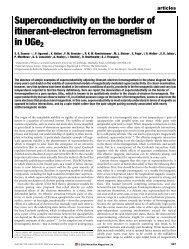
Figure 4 | Microscopy images <strong>of</strong> (Pro-Lys-Gly) 4 (Pro-Hyp-Gly) 4 (Asp-Hyp-Gly) 4 that show the fibrillar <strong>assembly</strong> <strong>of</strong> the system in phosphate buffer. a–c,<br />
TEM images <strong>of</strong> <strong>collagen</strong>-like nan<strong>of</strong>ibres in phosphate taken at ×40,000. a,b, Negatively stained images <strong>of</strong> the <strong>peptide</strong> in phosphate at a concentration <strong>of</strong><br />
1.0% by weight and stained with PTA, pH 6. c, Vitreous ice cryo-TEM image <strong>of</strong> <strong>collagen</strong>-like nan<strong>of</strong>ibres taken in phosphate at a concentration <strong>of</strong> 0.25%,<br />
which was diluted <strong>from</strong> a 0.5% by weight sample. d,e, AFM <strong>of</strong> <strong>collagen</strong>-like nan<strong>of</strong>ibres in phosphate as observed after spin coating onto freshly cleaved mica<br />
<strong>from</strong> solutions <strong>of</strong> <strong>peptide</strong> at concentrations <strong>of</strong> 1.0% (d) and 0.5% (e) byweight.f,g, SEM images <strong>of</strong> critical point dried hydrogel with a <strong>peptide</strong> concentration<br />
<strong>of</strong> 1.0% by weight that show the interconnected fibrous structure responsible for the gel forming properties at ×3,100 (f) and×30,000 (g).<br />
One final microscopy method, SEM, is important for understanding<br />
the qualitative long-range nanoscale behaviour <strong>of</strong> the<br />
system. Samples imaged by SEM were 1.0% by weight in phosphate<br />
buffer. In Fig. 4f, a dense fibre network that is homogeneous and<br />
extends across tens <strong>of</strong> microns is apparent. When the magnification<br />
is increased (Fig. 4g), the uniform nature <strong>of</strong> the nan<strong>of</strong>ibres within<br />
the network is more obvious. These results directly complement<br />
the fibre morphologies observed by TEM and AFM and also indicate<br />
the three-dimensional structure <strong>of</strong> the hydrogel.<br />
With the use <strong>of</strong> multiple microscopy techniques, the nanomorphology<br />
<strong>of</strong> (Pro-Lys-Gly) 4 (Pro-Hyp-Gly) 4 (Asp-Hyp-Gly) 4 was<br />
found to be nan<strong>of</strong>ibres <strong>of</strong> relatively uniform dimensions with<br />
824<br />
observed lengths <strong>of</strong> at least several hundred nanometres, widths <strong>of</strong><br />
4–5 nm, measured heights <strong>of</strong> 1.2+0.3 nm and a uniform longrange<br />
behaviour visible in the hydrated state.<br />
Hydrogel. With the first two levels <strong>of</strong> <strong>self</strong>-<strong>assembly</strong> confirmed, the<br />
final layer <strong>of</strong> analysis needed to describe the multi-<strong>hierarchical</strong><br />
<strong>assembly</strong> <strong>of</strong> (Pro-Lys-Gly) 4 (Pro-Hyp-Gly) 4 (Asp-Hyp-Gly) 4 was<br />
the assessment <strong>of</strong> the viscoelastic properties <strong>of</strong> the formed<br />
hydrogel (Fig. 5). Visually, the gels maintained their shape when<br />
they were removed <strong>from</strong> their containers, including the visible<br />
sustainability <strong>of</strong> the gel’s sharp edges. The image in Fig. 5d<br />
depicts the visual properties <strong>of</strong> the hydrogel. To analyse<br />
NATURE CHEMISTRY | VOL 3 | OCTOBER 2011 | www.nature.com/naturechemistry<br />
© 2011 Macmillan Publishers Limited. All rights reserved.