Multi-hierarchical self-assembly of a collagen mimetic peptide from ...
Multi-hierarchical self-assembly of a collagen mimetic peptide from ...
Multi-hierarchical self-assembly of a collagen mimetic peptide from ...

You also want an ePaper? Increase the reach of your titles
YUMPU automatically turns print PDFs into web optimized ePapers that Google loves.
ARTICLES<br />
PUBLISHED ONLINE: 28 AUGUST 2011 | DOI: 10.1038/NCHEM.1123<br />
<strong>Multi</strong>-<strong>hierarchical</strong> <strong>self</strong>-<strong>assembly</strong> <strong>of</strong> a <strong>collagen</strong><br />
<strong>mimetic</strong> <strong>peptide</strong> <strong>from</strong> triple helix to nan<strong>of</strong>ibre<br />
and hydrogel<br />
Lesley E. R. O’Leary, Jorge A. Fallas, Erica L. Bakota, Marci K. Kang and Jeffrey D. Hartgerink*<br />
Replicating the multi-<strong>hierarchical</strong> <strong>self</strong>-<strong>assembly</strong> <strong>of</strong> <strong>collagen</strong> has long-attracted scientists, <strong>from</strong> both the perspective <strong>of</strong> the<br />
fundamental science <strong>of</strong> supramolecular chemistry and that <strong>of</strong> potential biomedical applications in tissue engineering. Many<br />
approaches to drive the <strong>self</strong>-<strong>assembly</strong> <strong>of</strong> synthetic systems through the same steps as those <strong>of</strong> natural <strong>collagen</strong> (<strong>peptide</strong><br />
chain to triple helix to nan<strong>of</strong>ibres and, finally, to a hydrogel) are partially successful, but none simultaneously demonstrate<br />
all the levels <strong>of</strong> structural <strong>assembly</strong>. Here we describe a <strong>peptide</strong> that replicates the <strong>self</strong>-<strong>assembly</strong> <strong>of</strong> <strong>collagen</strong> through each<br />
<strong>of</strong> these steps. The <strong>peptide</strong> features <strong>collagen</strong>’s characteristic proline–hydroxyproline–glycine repeating unit, complemented<br />
by designed salt-bridged hydrogen bonds between lysine and aspartate to stabilize the triple helix in a sticky-ended<br />
<strong>assembly</strong>. This <strong>assembly</strong> is propagated into nan<strong>of</strong>ibres with characteristic triple helical packing and lengths with a lower<br />
bound <strong>of</strong> several hundred nanometres. These nan<strong>of</strong>ibres form a hydrogel that is degraded by <strong>collagen</strong>ase at a similar rate<br />
to that <strong>of</strong> natural <strong>collagen</strong>.<br />
Collagen, the most abundant protein in the human body, exemplifies<br />
multi-<strong>hierarchical</strong> <strong>self</strong>-<strong>assembly</strong>. In the case <strong>of</strong> type I<br />
<strong>collagen</strong>, <strong>self</strong>-<strong>assembly</strong> begins with three 1,000 amino acid<br />
<strong>peptide</strong> strands that adopt a polyproline type II helical structure<br />
and wind around one another to form a superhelical trimer that<br />
gives the well-known <strong>collagen</strong> triple helix. These triple helices<br />
then pack against one another in a quasihexagonal and staggered<br />
fashion to form nan<strong>of</strong>ibrous structures known as <strong>collagen</strong><br />
fibrils 1,2 . Collagen fibrils continue to <strong>self</strong>-assemble both linearly<br />
and laterally to give <strong>collagen</strong> fibres and a hydrogel network<br />
(Fig. 1). Together, the multiple levels <strong>of</strong> <strong>collagen</strong>’s structural hierarchy<br />
play a major role in the structural integrity <strong>of</strong> the extracellular<br />
matrix and provide binding sites for other proteins and cells.<br />
Collagen has been the target <strong>of</strong> bio<strong>mimetic</strong> design for decades<br />
because <strong>of</strong> the many difficulties associated with the use and characterization<br />
<strong>of</strong> <strong>collagen</strong> <strong>from</strong> natural sources and by expression. The<br />
use <strong>of</strong> recombinant systems requires either genetic modifications<br />
or a novel biosynthetic pathway in Escherichia coli to express hydroxyproline-containing<br />
<strong>collagen</strong>s 3–8 . There are many successes in the<br />
recapitulation <strong>of</strong> the <strong>collagen</strong> triple helix in short <strong>peptide</strong>s, both<br />
as a homotrimer 9–17 and, more recently, as a heterotrimer 18–24 .<br />
However, examples that take these <strong>collagen</strong>-like <strong>peptide</strong>s and use<br />
them to mimic the higher order <strong>assembly</strong> <strong>of</strong> <strong>collagen</strong> have faced a<br />
great deal <strong>of</strong> difficulty. In all previously reported systems, none<br />
has demonstrated discretely each level <strong>of</strong> <strong>collagen</strong> <strong>self</strong>-<strong>assembly</strong><br />
(triple helix, nan<strong>of</strong>ibre and hydrogel) within the same system.<br />
There are many examples <strong>of</strong> <strong>peptide</strong>s that form organized nanostructures<br />
without gelation 10,12,25–29 , and a few that show gelation<br />
without pro<strong>of</strong> <strong>of</strong> the presence <strong>of</strong> triple helices or nan<strong>of</strong>ibres 30,31 ;<br />
however, no system with triple-helix formation, nan<strong>of</strong>ibre formation<br />
and gelation is reported.<br />
Perhaps the best example <strong>of</strong> a fibre-forming <strong>collagen</strong>-like <strong>peptide</strong><br />
was demonstrated by Chaik<strong>of</strong>, Conticello and co-workers, who prepared<br />
a 36 amino acid <strong>peptide</strong> with the sequence (Pro-Arg-<br />
Gly) 4 (Pro-Hyp-Gly) 4 (Glu-Hyp-Gly) 4 (ref. 32). This zwitterionic<br />
<strong>peptide</strong> assembled into large organized fibres. However, even<br />
these <strong>collagen</strong> <strong>mimetic</strong> fibres have some drawbacks, which include<br />
(i) a mixed composition <strong>of</strong> fibres associated with a significant quantity<br />
<strong>of</strong> other amorphous material, (ii) the requirement for specific<br />
concentration and buffer composition outside <strong>of</strong> which the<br />
quality <strong>of</strong> <strong>assembly</strong> degrades or fails completely and (iii) phase<br />
separation and precipitation <strong>of</strong> the formed fibres as opposed to<br />
the formation <strong>of</strong> a hydrogel 32 .<br />
Recently, in our laboratory we investigated the structure <strong>of</strong><br />
several heterotrimeric <strong>collagen</strong> helices using NMR spectroscopy 18,24 ,<br />
a<br />
Peptide chain<br />
b<br />
Triple helix Nan<strong>of</strong>ibre Hydrogel<br />
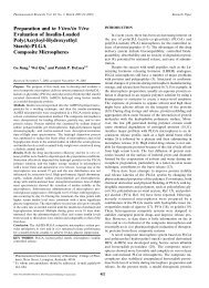
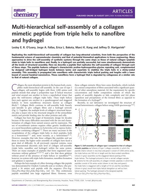
Figure 1 | Self-<strong>assembly</strong> <strong>of</strong> <strong>collagen</strong> type I compared to that <strong>of</strong> <strong>collagen</strong><br />
<strong>mimetic</strong> <strong>peptide</strong>s. a, Scheme<strong>of</strong>typeI<strong>collagen</strong><strong>assembly</strong>inwhichthe<br />
<strong>peptide</strong> chains (shown in red, blue and green) consist <strong>of</strong> 1,000 amino acids,<br />
the triple helices are 100 nm in length and the blunt-ended nan<strong>of</strong>ibres<br />
(shown in grey) assemble via the staggered lateral packing <strong>of</strong> the<br />
triple helices. The hydrogel pictured is <strong>from</strong> a rat-tail <strong>collagen</strong> sample.<br />
b, Scheme for the <strong>self</strong>-<strong>assembly</strong> <strong>of</strong> <strong>collagen</strong> <strong>mimetic</strong> <strong>peptide</strong>s in which the<br />
<strong>peptide</strong>s consist <strong>of</strong> 36 amino acids (shown in red, blue and green),<br />
the triple helix is staggered with a length <strong>of</strong> 10 nm and the nan<strong>of</strong>ibres<br />
(shown in grey) result <strong>from</strong> triple helical elongation as well as <strong>from</strong><br />
lateral packing. The hydrogel pictured is the designed <strong>peptide</strong><br />
(Pro-Lys-Gly) 4 (Pro-Hyp-Gly) 4 (Asp-Hyp-Gly) 4 .<br />
Departments <strong>of</strong> Chemistry and Bioengineering Rice University, Houston, Texas 77005, USA. *e-mail: jdh@rice.edu<br />
NATURE CHEMISTRY | VOL 3 | OCTOBER 2011 | www.nature.com/naturechemistry 821<br />
© 2011 Macmillan Publishers Limited. All rights reserved.
ARTICLES<br />
NATURE CHEMISTRY DOI: 10.1038/NCHEM.1123<br />
a<br />
4.3 Å<br />
5.2 Å<br />
b<br />
2.8 Å<br />
2.8 Å<br />
2.8 Å<br />
2.9 Å<br />
c<br />
N<br />
2.9 Å<br />
–<br />
O O<br />
O<br />
O<br />
H<br />
N<br />
N<br />
N<br />
N<br />
N<br />
H<br />
H<br />
H<br />
O<br />
O<br />
O<br />
O<br />
n<br />
n<br />
OH<br />
NH<br />
H 2 N + NH 2<br />
(Pro-Arg-Gly) n (Glu-Hyp-Gly) n<br />
N<br />
O<br />
N<br />
OH<br />
O<br />
N<br />
H<br />
O n<br />
N<br />
O<br />
H<br />
N<br />
O<br />
N<br />
H<br />
+<br />
NH 3<br />
O<br />
n<br />
(Pro-Hyp-Gly) n (Pro-Lys-Gly) n (Asp-Hyp-Gly) n<br />
N<br />
H<br />
–<br />
O<br />
O<br />
O<br />
N<br />
OH<br />
O<br />
N<br />
H<br />
O<br />
n<br />
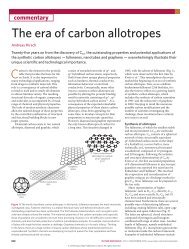
Figure 2 | Models <strong>of</strong> electrostatic interactions between charged amino acids in <strong>collagen</strong> <strong>mimetic</strong> <strong>peptide</strong>s. a,b, Models<strong>of</strong>Arg–Glu(a) andLys–Asp(b)<br />
charged pairs in <strong>collagen</strong> triple helices 18,24 . The <strong>peptide</strong> chains are shown in red, blue and pink for (a) and red, blue and green for (b), with the hydrogen<br />
atoms highlighted in white, oxygen in pink and nitrogen in blue. The hydrogen-bond lengths shown are measured <strong>from</strong> N to O. Arg–Glu pairs do not appear<br />
to form high-quality interactions because <strong>of</strong> the strong hydrogen bonding between Arg and a cross-strand carbonyl oxygen, which locks the side chain into<br />
place. In contrast, two conformers <strong>of</strong> lysine are present and both allow excellent hydrogen bonding to aspartic acid despite one <strong>of</strong> them displaying a similar<br />
hydrogen bond to a cross-strand carbonyl. c, Chemical structures <strong>of</strong> the common amino-acid triplets (Pro-Arg-Gly) n ,(Glu-Hyp-Gly) n ,(Pro-Hyp-Gly) n ,<br />
(Pro-Lys-Gly) n and (Asp-Hyp-Gly) n .<br />
in these systems, the arginine–glutamate interactions on which they<br />
rely are distant <strong>from</strong> one another and interact primarily by charge<br />
screening rather than by a specific salt-bridged hydrogen bond<br />
(Fig. 2a) 24 . One <strong>of</strong> the reasons for this is that the arginine side<br />
chain forms a tight hydrogen bond with the backbone carbonyl <strong>of</strong><br />
an adjacent <strong>peptide</strong> chain, which restrains it <strong>from</strong> making a more<br />
intimate contact with glutamate. In contrast, we observed very<br />
high-quality formation <strong>of</strong> lysine–aspartate salt-bridged hydrogen<br />
bonds (Fig. 2b) 18 . Based on these charge-pair observations and the<br />
work <strong>of</strong> Chaik<strong>of</strong> and Conticello, we prepared a new <strong>peptide</strong> in<br />
which the arginine residues are replaced with lysine and the<br />
glutamate residues with aspartate, to give the sequence (Pro-Lys-<br />
Gly) 4 (Pro-Hyp-Gly) 4 (Asp-Hyp-Gly) 4 . Our hypothesis was that the<br />
more effective interactions between lysine and aspartate, previously<br />
observed, would result in superior fibre- and hydrogel-forming<br />
characteristics. Indeed, this is what we observed.<br />
Here we report the synthesis and multi-<strong>hierarchical</strong> <strong>assembly</strong> <strong>of</strong><br />
the <strong>collagen</strong> <strong>mimetic</strong> <strong>peptide</strong> (Pro-Lys-Gly) 4 (Pro-Hyp-Gly) 4 (Asp-<br />
Hyp-Gly) 4 through each level <strong>of</strong> <strong>assembly</strong>, as depicted in Fig. 1b.<br />
This <strong>peptide</strong> demonstrated successfully the formation <strong>of</strong> a stable<br />
triple helix with a melting temperature <strong>of</strong> 40–41 8C. It exhibited<br />
nan<strong>of</strong>ibre morphologies, as observed in atomic force microscopy<br />
(AFM), scanning electron microscopy (SEM) and transmission electron<br />
microscopy (TEM), including both dry and hydrated techniques,<br />
and the nan<strong>of</strong>ibres formed were quite uniform, with<br />
virtually no other aggregations or morphologies observed.<br />
Furthermore, nan<strong>of</strong>ibrous <strong>self</strong>-<strong>assembly</strong> was observed easily under<br />
a wide range <strong>of</strong> buffers and ionic strengths, which indicates the<br />
robust nature <strong>of</strong> the <strong>self</strong>-<strong>assembly</strong> process. The nan<strong>of</strong>ibres displayed<br />
characteristic triple helical packing, as confirmed by fibre<br />
822<br />
diffraction, and <strong>self</strong>-assembled into hydrogels with good viscoelastic<br />
properties, as measured by oscillatory rheology and through comparisons<br />
to both natural and synthetic hydrogels. Finally, the prepared<br />
hydrogels were broken down by <strong>collagen</strong>ase type IV at a<br />
similar rate to rat-tail <strong>collagen</strong> in a simple functionality test 33 .As<br />
the system demonstrated control at each <strong>of</strong> level <strong>of</strong> <strong>collagen</strong> <strong>assembly</strong><br />
(triple helicity, fibre formation and hydrogel formation), we<br />
believe this <strong>peptide</strong>, as well as future systems based on it, have a<br />
large potential for use as tissue-engineering scaffolds.<br />
Results and discussion<br />
Once the <strong>peptide</strong> (Pro-Lys-Gly) 4 (Pro-Hyp-Gly) 4 (Asp-Hyp-Gly) 4<br />
was synthesized and purified successfully (complete details <strong>of</strong> the<br />
procedures are given in Methods and in the Supplementary<br />
Information), samples were made at specified concentrations<br />
between 0.2% (0.6 mM) and 1.0% (3 mM) by weight. Although<br />
many buffer systems with varying ionic strengths were explored<br />
(see Supplementary Information for further details), here we<br />
discuss primarily the results using 10 mM sodium phosphate<br />
buffer at pH 7 (referred to as phosphate). In this buffer system, all<br />
the samples made at concentrations <strong>of</strong> 0.5% (1.5 mM) by weight<br />
or more formed hydrogels within a few hours. Once hydrogel<br />
formation was observed, we began systematically to analyse the<br />
<strong>peptide</strong> at each level <strong>of</strong> <strong>self</strong>-<strong>assembly</strong>: triple helix, nan<strong>of</strong>ibre<br />
and hydrogel.<br />
Triple helix. To determine whether a <strong>collagen</strong> <strong>mimetic</strong> <strong>peptide</strong><br />
forms a triple helix, two circular dichroism (CD) experiments<br />
must be performed: a wavelength spectrum and a thermal<br />
unfolding curve. Collagen triple helices have a signature CD<br />
NATURE CHEMISTRY | VOL 3 | OCTOBER 2011 | www.nature.com/naturechemistry<br />
© 2011 Macmillan Publishers Limited. All rights reserved.
NATURE CHEMISTRY DOI: 10.1038/NCHEM.1123<br />
ARTICLES<br />
a 5<br />
b 4<br />
c<br />
MRE × 10 –3 (deg cm 2 dmol-residue –1 )<br />
0<br />
–5<br />
–10<br />
MRE × 10 –3 (deg cm 2 dmol-residue –1 )<br />
3<br />
2<br />
1<br />
0<br />
–15<br />
–0.15<br />
0.5% by weight<br />
–1<br />
1.0% by weight<br />
–20<br />
–2<br />
–0.2<br />
190 200 210 220 230 240 250<br />
10 20 30 40 50 60 70 80<br />
10 20 30 40 50 60 70 80<br />
Wavelength (nm) Temperature (ºC) Temperature (ºC)<br />
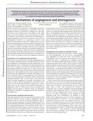
Figure 3 | Spectroscopy graphs illustrating the triple helical nature <strong>of</strong> the designed <strong>collagen</strong> <strong>mimetic</strong> <strong>peptide</strong>. a, CD spectrum <strong>of</strong> the fibre-forming<br />
<strong>collagen</strong>-like <strong>peptide</strong> (Pro-Lys-Gly) 4 (Pro-Hyp-Gly) 4 (Asp-Hyp-Gly) 4 at 0.5% and 1.0% by weight concentrations in phosphate at a temperature <strong>of</strong> 5 8C.<br />
The maximum at 225 nm and minimum near 200 nm are characteristic <strong>of</strong> polyproline type II helices. b, Thermal unfolding analysis curve for the <strong>peptide</strong> at<br />
1.0% by weight concentration in phosphate. c, First derivative <strong>of</strong> the mean residue ellipticity (MRE) versus temperature curve shown in (b). The cooperative<br />
transition at 41 8C demonstrates that the designed <strong>peptide</strong> forms a triple helix.<br />
MRE first derivative<br />
0.05<br />
0<br />
–0.05<br />
–0.1<br />
spectral pr<strong>of</strong>ile that consists <strong>of</strong> a maximum at 225 nm and a<br />
minimum near 200 nm, indicative <strong>of</strong> a polyproline type II helix.<br />
The thermal unfolding experiment monitors the spectral<br />
maximum as temperature is increased which, when a triple helix<br />
is present, shows a cooperative transition. For the <strong>peptide</strong> (Pro-<br />
Lys-Gly) 4 (Pro-Hyp-Gly) 4 (Asp-Hyp-Gly) 4 , CD spectra taken at all<br />
concentrations showed a strong maximum at 225 nm. The spectra<br />
for 0.5% and 1.0% by weight in phosphate are shown in Fig. 3a.<br />
Note the size difference between the maximum in the spectrum<br />
for 1.0% by weight compared to that for 0.5% by weight, even<br />
though the data is normalized for concentration. This indicates<br />
an increased percentage <strong>of</strong> the <strong>peptide</strong> is folded at the higher<br />
concentration. When melting experiments were performed <strong>from</strong> 5<br />
to 85 8C on samples at concentrations <strong>of</strong> 0.2%, 0.5% and 1.0% by<br />
weight, all the samples exhibited a cooperative transition in the<br />
melting pr<strong>of</strong>ile. Additionally, transitions for samples at higher<br />
<strong>peptide</strong> concentrations were stronger and more obvious than<br />
those for lower concentrations, which indicates that the higher<br />
concentration <strong>of</strong> <strong>peptide</strong> helps to drive triple-helix formation. The<br />
thermal unfolding curve and the first derivative <strong>of</strong> the curve for a<br />
1.0% by weight sample in phosphate are shown in Fig. 3b,c.<br />
(Melting studies for 0.2% and 0.5% by weight are given in<br />
the Supplementary Information.) A major transition occurs in the<br />
first derivative curve at 40–41 8C, which corresponds to the<br />
melting temperature <strong>of</strong> the <strong>peptide</strong>. However, a broad, minor<br />
transition is also visible between 10 and 30 8C. The minor<br />
transition may result <strong>from</strong> increased helicity on fibre elongation<br />
and lateral packing. A more detailed explanation for this is<br />
given below.<br />
Nan<strong>of</strong>ibre. After confirmation <strong>of</strong> the triple helical nature <strong>of</strong> the<br />
<strong>peptide</strong>, the next step was to understand the nanostructure <strong>of</strong> the<br />
<strong>self</strong>-assembled <strong>peptide</strong>. The microscopy techniques used included<br />
TEM, AFM and SEM. TEM is an integral technique used to view<br />
the morphology and measure the length and width <strong>of</strong> structures<br />
on the nanoscale. It is most commonly a dry technique that, to<br />
view carbon-based materials, requires the sample to be stained<br />
with a heavy metal, such as phosphotungstic acid (PTA). For this<br />
<strong>peptide</strong>, a 1.0% by weight concentration sample in phosphate was<br />
prepared, using the previously described procedure, and negatively<br />
stained with PTA. Images <strong>of</strong> these stained samples (Fig. 4a,b and<br />
Supplementary Information) reveal long nan<strong>of</strong>ibres, present both<br />
as single fibres and as fibre bundles. Figure 4a shows the variety<br />
<strong>of</strong> fibre widths present within this system when dried and stained.<br />
These fibres are the major species within the TEM sample, in<br />
contrast to previous reports <strong>of</strong> <strong>collagen</strong> <strong>mimetic</strong> nan<strong>of</strong>ibres that<br />
showed large aggregates and a variety <strong>of</strong> other non-fibrous<br />
structures in the TEM images. Figure 4b reveals the twisting nature<br />
<strong>of</strong> some <strong>of</strong> the nan<strong>of</strong>ibres in contrast to fibres with a smoother<br />
morphology. Although the negatively stained TEM images show<br />
the presence <strong>of</strong> nan<strong>of</strong>ibres for this <strong>peptide</strong> system, drying artefacts<br />
can cause samples to appear more densely packed or with a<br />
completely different structure than that present in the hydrated<br />
state. In addition, the use <strong>of</strong> a heavy-metal stain adds an additional<br />
level <strong>of</strong> uncertainty in assessing fibre size and morphology. For<br />
these reasons, we believe the presence <strong>of</strong> nan<strong>of</strong>ibres in a solution<br />
state can only be proved by imaging the system in a hydrated<br />
environment, specifically using vitreous ice cryo-TEM.<br />
The sample preparation for cryo-TEM differs greatly <strong>from</strong> that<br />
for dry TEM because cryo-TEM requires a thin aqueous film <strong>of</strong><br />
sample on the TEM grid before it is flash frozen in ethane slush.<br />
A representative TEM image <strong>from</strong> this preparation is given in<br />
Fig. 4c (for more images see the Supplementary Information). In<br />
contrast to the dry TEM images, especially Fig. 4a, the fibres seen<br />
in the vitreous ice cryo-TEM sample have uniform widths <strong>from</strong><br />
4 to 5 nm and fibre lengths <strong>from</strong> several hundred nanometres to<br />
many microns. However, similar to the dry cryo-TEM images, the<br />
fibres observed in cryo-TEM form the majority <strong>of</strong> the <strong>peptide</strong> population<br />
in the sample. (The spherical species seen in the cryo-TEM<br />
image are ethane artefacts that result <strong>from</strong> sample preparation,<br />
not <strong>peptide</strong> aggregates.) Therefore, both dry and cryo-TEM confirmed<br />
the presence <strong>of</strong> nan<strong>of</strong>ibres and that they were the major<br />
species within the system. Once the length and width <strong>of</strong> the nan<strong>of</strong>ibres<br />
formed <strong>from</strong> the <strong>peptide</strong> was determined by cryo-TEM, the<br />
height <strong>of</strong> the fibres was needed to understand the mechanism <strong>of</strong><br />
fibre formation. Tapping-mode AFM is the most efficient method<br />
for acquiring this data. Figures 4d,e are AFM images taken <strong>of</strong><br />
1.0% and 0.5% by weight samples in phosphate buffer. Nan<strong>of</strong>ibres<br />
are seen in both images; the sample <strong>of</strong> higher concentration<br />
exhibited a denser network <strong>of</strong> nan<strong>of</strong>ibres. Similar fibres were seen<br />
in all the buffers examined, including higher ionic strength<br />
buffers such as phosphate-buffered saline. The measured height<br />
pr<strong>of</strong>ile in phosphate buffer <strong>from</strong> the AFM images was 1.2+<br />
0.3 nm. This value is much lower than the fibre width <strong>of</strong> 4–5 nm<br />
measured by cryo-TEM, and the fibre lengths observed by AFM<br />
also appear smaller than those observed by TEM. A hypothesis<br />
for this difference is discussed below. One advantage <strong>of</strong> these<br />
images is that, as a result <strong>of</strong> their lower magnification, a larger<br />
area is visible and the uniformity <strong>of</strong> the population <strong>of</strong> <strong>self</strong>-assembled<br />
nan<strong>of</strong>ibres is more apparent.<br />
NATURE CHEMISTRY | VOL 3 | OCTOBER 2011 | www.nature.com/naturechemistry 823<br />
© 2011 Macmillan Publishers Limited. All rights reserved.
ARTICLES<br />
NATURE CHEMISTRY DOI: 10.1038/NCHEM.1123<br />
a b c<br />
50 nm 50 nm<br />
50 nm<br />
d<br />
e<br />
1 µm<br />
500 nm<br />
f<br />
g<br />
10 µm 1 µm<br />
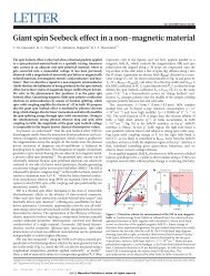
Figure 4 | Microscopy images <strong>of</strong> (Pro-Lys-Gly) 4 (Pro-Hyp-Gly) 4 (Asp-Hyp-Gly) 4 that show the fibrillar <strong>assembly</strong> <strong>of</strong> the system in phosphate buffer. a–c,<br />
TEM images <strong>of</strong> <strong>collagen</strong>-like nan<strong>of</strong>ibres in phosphate taken at ×40,000. a,b, Negatively stained images <strong>of</strong> the <strong>peptide</strong> in phosphate at a concentration <strong>of</strong><br />
1.0% by weight and stained with PTA, pH 6. c, Vitreous ice cryo-TEM image <strong>of</strong> <strong>collagen</strong>-like nan<strong>of</strong>ibres taken in phosphate at a concentration <strong>of</strong> 0.25%,<br />
which was diluted <strong>from</strong> a 0.5% by weight sample. d,e, AFM <strong>of</strong> <strong>collagen</strong>-like nan<strong>of</strong>ibres in phosphate as observed after spin coating onto freshly cleaved mica<br />
<strong>from</strong> solutions <strong>of</strong> <strong>peptide</strong> at concentrations <strong>of</strong> 1.0% (d) and 0.5% (e) byweight.f,g, SEM images <strong>of</strong> critical point dried hydrogel with a <strong>peptide</strong> concentration<br />
<strong>of</strong> 1.0% by weight that show the interconnected fibrous structure responsible for the gel forming properties at ×3,100 (f) and×30,000 (g).<br />
One final microscopy method, SEM, is important for understanding<br />
the qualitative long-range nanoscale behaviour <strong>of</strong> the<br />
system. Samples imaged by SEM were 1.0% by weight in phosphate<br />
buffer. In Fig. 4f, a dense fibre network that is homogeneous and<br />
extends across tens <strong>of</strong> microns is apparent. When the magnification<br />
is increased (Fig. 4g), the uniform nature <strong>of</strong> the nan<strong>of</strong>ibres within<br />
the network is more obvious. These results directly complement<br />
the fibre morphologies observed by TEM and AFM and also indicate<br />
the three-dimensional structure <strong>of</strong> the hydrogel.<br />
With the use <strong>of</strong> multiple microscopy techniques, the nanomorphology<br />
<strong>of</strong> (Pro-Lys-Gly) 4 (Pro-Hyp-Gly) 4 (Asp-Hyp-Gly) 4 was<br />
found to be nan<strong>of</strong>ibres <strong>of</strong> relatively uniform dimensions with<br />
824<br />
observed lengths <strong>of</strong> at least several hundred nanometres, widths <strong>of</strong><br />
4–5 nm, measured heights <strong>of</strong> 1.2+0.3 nm and a uniform longrange<br />
behaviour visible in the hydrated state.<br />
Hydrogel. With the first two levels <strong>of</strong> <strong>self</strong>-<strong>assembly</strong> confirmed, the<br />
final layer <strong>of</strong> analysis needed to describe the multi-<strong>hierarchical</strong><br />
<strong>assembly</strong> <strong>of</strong> (Pro-Lys-Gly) 4 (Pro-Hyp-Gly) 4 (Asp-Hyp-Gly) 4 was<br />
the assessment <strong>of</strong> the viscoelastic properties <strong>of</strong> the formed<br />
hydrogel (Fig. 5). Visually, the gels maintained their shape when<br />
they were removed <strong>from</strong> their containers, including the visible<br />
sustainability <strong>of</strong> the gel’s sharp edges. The image in Fig. 5d<br />
depicts the visual properties <strong>of</strong> the hydrogel. To analyse<br />
NATURE CHEMISTRY | VOL 3 | OCTOBER 2011 | www.nature.com/naturechemistry<br />
© 2011 Macmillan Publishers Limited. All rights reserved.
NATURE CHEMISTRY DOI: 10.1038/NCHEM.1123<br />
ARTICLES<br />
a<br />
1,000<br />
b<br />
1,000<br />
Storage modulus, G¢ (Pa)<br />
Loss modulus, G¢¢ (Pa)<br />
c<br />
100<br />
10<br />
1<br />
G¢ - 0.5%<br />
G¢¢ - 0.5%<br />
G¢ - 1.0%<br />
G¢¢ - 1.0%<br />
0.1<br />
0.1 1 10 100<br />
Strain (%)<br />
800<br />
700<br />
0.5 wt%<br />
1.0 wt%<br />
Storage modulus, G¢ (Pa)<br />
Loss modulus, G¢¢ (Pa)<br />
d<br />
100<br />
10<br />
1<br />
0.1<br />
G¢ - 0.5%<br />
G¢¢ - 0.5%<br />
G¢ - 1.0%<br />
G¢¢ - 1.0%<br />
0.1 1 10 100<br />
Frequency (rad s –1 )<br />
Storage modulus, G¢ (Pa)<br />
600<br />
500<br />
400<br />
300<br />
200<br />
100<br />
0<br />
20 30 37<br />
Temperature (ºC)<br />
Figure 5 | Rheology <strong>of</strong> the <strong>collagen</strong>-like <strong>peptide</strong> that demonstrates the temperature-dependent strength <strong>of</strong> the hydrogel. a, Strain sweep at 0.5% and 1.0%<br />
by weight <strong>peptide</strong> concentration in phosphate buffer at a temperature <strong>of</strong> 30 8C and a frequency <strong>of</strong> 1 rad s 21 shown as G ′ and G ′′ . b, Frequency sweep at<br />
0.5% and 1.0% by weight <strong>peptide</strong> concentration in phosphate at a temperature <strong>of</strong> 30 8C and 1% strain shown as G ′ and G ′′ . c, Temperature dependence <strong>of</strong><br />
rheological properties at 0.5 and 1.0% by weight <strong>peptide</strong> concentrations shown as G ′ . Data points were acquired at 1 rad s 21 and 1% strain. d, Photo <strong>of</strong> the<br />
shape-persistent nature <strong>of</strong> the gel with a concentration <strong>of</strong> 1.0% by weight in phosphate. The gel was prepared at a volume <strong>of</strong> 0.5 ml. Note the sustainability<br />
<strong>of</strong> the sharp gel edges.<br />
quantitatively the <strong>peptide</strong> hydrogels, rheological studies were<br />
performed. Strain and frequency-sweep experiments were performed<br />
to assess the gel properties and, specifically, the storage modulus (G ′ )<br />
and loss modulus (G ′′ ), which measure, respectively, the elastically<br />
stored energy and the energy lost as heat within the hydrogel on<br />
application <strong>of</strong> a shearing force. Representative graphs <strong>of</strong> each type <strong>of</strong><br />
experiment are shown in Fig. 5a,b and the first observation is that<br />
the G ′ is substantially larger than G ′′<br />
for both 0.5% and 1.0% by<br />
weight concentrations <strong>of</strong> the <strong>peptide</strong> in phosphate buffer. Therefore,<br />
(Pro-Lys-Gly) 4 (Pro-Hyp-Gly) 4 (Asp-Hyp-Gly) 4 forms a hydrogel in<br />
phosphate buffer at 0.5% by weight concentration and higher. The<br />
observed G ′ <strong>of</strong> this <strong>collagen</strong> <strong>mimetic</strong> system is similar to that<br />
typically observed for a <strong>collagen</strong> hydrogel formed <strong>from</strong> natural<br />
sources, such as rat-tail <strong>collagen</strong>, even though our <strong>peptide</strong> is<br />
approximately 30 times shorter (36 amino acids as compared to<br />
1,000) (ref. 34). It is also higher than that <strong>of</strong> Matrigel 35 and<br />
comparable to those <strong>of</strong> the popular b-sheet hydrogels described in<br />
the literature 36–41 .<br />
The <strong>collagen</strong> <strong>mimetic</strong> hydrogel was temperature sensitive. From<br />
the CD melting studies, we know that the triple helix unfolds at<br />
40–41 8C, and therefore a temperature-ramp rheological experiment<br />
<strong>from</strong> 20 to 60 8C was used to demonstrate the melting <strong>of</strong><br />
the hydrogel. Indeed, the G ′ values decreased above 40 8C, and<br />
by 50 8C the G ′′<br />
values exceeded the G ′ values, which indicates<br />
that the gel disassembled (see Supplementary Information).<br />
Figure 5c is a bar graph <strong>of</strong> the G ′ values for 0.5% and 1.0% by<br />
weight gels in phosphate at 20, 30 and 37 8C. The system was<br />
examined at different temperatures to gain insight into its behaviour<br />
before the gel melted. As shown in Fig. 5c, the gels have their<br />
highest G ′ at 30 8C and 37 8C, and a substantially lower observed<br />
storage modulus at 20 8C. The CD melting pr<strong>of</strong>ile shows a minor<br />
transition <strong>of</strong> the <strong>peptide</strong> between 10 and 30 8C prior to the actual<br />
triple-helix unfolding <strong>of</strong> the system. The temperature-dependent<br />
rheological results combined with the CD data suggest that, as<br />
the <strong>peptide</strong> slightly unfolds between 10 and 30 8C, the unfolded<br />
regions <strong>of</strong> fibre may interdigitate with other nan<strong>of</strong>ibres and<br />
result in a strengthened hydrogel.<br />
As a simple functional test <strong>of</strong> the <strong>collagen</strong> hydrogel <strong>mimetic</strong>, we<br />
compared its ability to be broken down by <strong>collagen</strong>ase (type IV,<br />
Invitrogen), the primary component <strong>of</strong> which is MMP2, a protease<br />
known to cleave specifically between the X and Gly residues <strong>of</strong> an<br />
X-Y-Gly repeat found in a triple helix 42 . (Pro-Lys-Gly) 4 (Pro-Hyp-<br />
Gly) 4 (Asp-Hyp-Gly) 4 hydrogels were prepared at a concentration<br />
<strong>of</strong> 2.0% by weight in phosphate buffer and treated with either <strong>collagen</strong>ase<br />
in Hank’s Balanced Salt Solution (HBSS) or with HBSS<br />
alone. The samples were allowed to incubate at room temperature<br />
(approximately 20 8C), 30 8C and 37 8C. Hydrogels <strong>of</strong> rat-tail <strong>collagen</strong><br />
were prepared in the same fashion, with and without <strong>collagen</strong>ase.<br />
As shown in Supplementary Table 1, hydrogels prepared <strong>from</strong><br />
our <strong>self</strong>-assembling <strong>peptide</strong> and rat-tail <strong>collagen</strong> degraded at similar<br />
NATURE CHEMISTRY | VOL 3 | OCTOBER 2011 | www.nature.com/naturechemistry 825<br />
© 2011 Macmillan Publishers Limited. All rights reserved.
ARTICLES<br />
a<br />
PKGPKGPKGPKGPOGPOGPOGPOGDOGDOGDOGDOG<br />
PKGPKGPKGPKGPOGPOGPOGPOGDOGDOGDOGDOG<br />
PKGPKGPKGPKGPOGPOGPOGPOGDOGDOGDOGDOG<br />
POGPOGPOGDOGDOGDOGDOGPKGPKGPKGPKGPOGPOGPOGPOGDOGDOGDOGDOG<br />
GPKGPKGPKGPKGPOGPOGPOGPOGDOGDOGDOGDOGPKGPKGPKGPKGPOGPOGPO<br />
OGPOGDOGDOGDOGDOGPKGPKGPKGPKGPOGPOGPOGPOGDOGDOGDOGDOGPKGP<br />
b<br />
c<br />
Fibre growth<br />
rates: samples <strong>of</strong> both types <strong>of</strong> hydrogels treated with <strong>collagen</strong>ase<br />
dissolved fully after one hour (37 8C) or four hours (30 8C and<br />
room temperature), whereas untreated controls did not.<br />
Fibre diffraction. To learn more about the packing morphology <strong>of</strong><br />
the <strong>self</strong>-assembled nan<strong>of</strong>ibres and, in turn, the fibre <strong>self</strong>-<strong>assembly</strong><br />
process (Fig. 6), X-ray fibre diffraction studies were carried out on<br />
a dried <strong>peptide</strong> sample (see Methods). As is apparent <strong>from</strong> the<br />
microscopy images (Fig. 4), neighbouring fibres lack a common<br />
orientation axis. To align the fibres partially, the drying <strong>peptide</strong><br />
solution was placed in a strong magnetic field to promote<br />
alignment during the drying process. This methodology has been<br />
shown to produce highly aligned protein fibres 43 , but had only<br />
limited success in our system. Figure 6d shows the recorded<br />
diffraction pattern. The dried pellet exhibits some alignment, as<br />
evidenced by the pseudo two-fold symmetry observed in the<br />
intensity versus azimuthal angle scan <strong>of</strong> the diffraction pattern<br />
(see Supplementary Information). However, no clear equatorial or<br />
e<br />
d<br />
2.8 Å<br />
4.3 Å<br />
11.5 Å<br />
2 4 6 8 10 12 14 16 18 20<br />
D spacing (Å)<br />
Figure 6 | Proposed mechanism <strong>of</strong> fibre <strong>self</strong>-<strong>assembly</strong>. a, Peptide sequence<br />
shown as single-letter amino acid code with P for proline, K for lysine, G for<br />
glycine, O for hydroxyproline and D for aspartate. The minimum repeating<br />
unit <strong>of</strong> the triple helical fibre has extensive ‘sticky’ ends. As additional<br />
<strong>peptide</strong>s (shaded grey) add to the minimum repeating unit, the percentage<br />
<strong>of</strong> amino acids that forms a high-quality triple helical structure increases<br />
rapidly. Positively charged lysine residues are in blue, negatively charged<br />
aspartates are in red and satisfied intrahelical electrostatic interactions are<br />
indicated by purple lassoes. Available interhelical charged-pair hydrogen<br />
bonds are shown by small arrows. b, Lysine–aspartate interaction between<br />
the i and i þ 3 amino acids <strong>of</strong> adjacent <strong>peptide</strong> strands. c, Quasihexagonal<br />
packing <strong>of</strong> growing fibres results in a bundle approximately 2 by 4 nm based<br />
on a triple helical cross-section <strong>of</strong> 1.2 nm. d,e, Fibre diffraction pattern (d)<br />
and its radially averaged intensity (e). Characteristic bands at 2.8 Å, 4.3 Å<br />
and 11.5 Å match well with previously reported fibre diffraction <strong>from</strong> natural<br />
<strong>collagen</strong>. a.u. ¼ arbitrary units.<br />
826<br />
Intensity (a.u.)<br />
NATURE CHEMISTRY DOI: 10.1038/NCHEM.1123<br />
meridional axis could be determined and thus the data were<br />
analysed by performing a radial integration <strong>of</strong> the diffraction<br />
pattern to yield a plot <strong>of</strong> the observed intensities as a function <strong>of</strong><br />
D spacing (Fig. 6e). The plot shows three distinct features: a<br />
weaker, sharp line near 2.8 Å, a diffuse intense reflection near<br />
4.3 Å and a strong well-defined band near 11.5 Å. The spacing <strong>of</strong><br />
the observed lines agrees well with that observed for <strong>collagen</strong><br />
<strong>from</strong> stretched kangaroo-tail tendons 44 . Based on this, we assigned<br />
the 11.5 Å band to the distance between two triple helices within<br />
the nan<strong>of</strong>ibres, the diffuse reflection at 4.3 Å to the distance<br />
between <strong>peptide</strong> chains inside a triple helix and the reflection at<br />
2.8 Å to the translation per triple helical triplet. This suggests that<br />
our <strong>collagen</strong>-like <strong>peptide</strong> fibres pack in a fashion similar to that <strong>of</strong><br />
natural <strong>collagen</strong>.<br />
Proposed mechanism <strong>of</strong> <strong>assembly</strong>. As mentioned above, the charge<br />
pairing <strong>of</strong> lysine and aspartate was shown previously to form direct<br />
electrostatic interactions in <strong>collagen</strong> <strong>mimetic</strong> <strong>peptide</strong>s 18 . Specifically,<br />
lysine’s side chain reaches in a C-terminal direction to make an<br />
intimate salt-bridge hydrogen bond with an aspartate on an<br />
adjacent lagging <strong>peptide</strong> <strong>of</strong>fset by three amino acids (Fig. 6b). Our<br />
<strong>peptide</strong> forms a homotrimer, so there is a potential for these<br />
charged amino-acid salt bridges to form between <strong>peptide</strong> strands<br />
and create an <strong>of</strong>fset, sticky-ended triple helix. Similar sticky-ended<br />
assemblies have been designed and reported, particularly for<br />
alpha-helical coiled coils 45,46 . Figure 6a shows the proposed<br />
repeating unit <strong>of</strong> <strong>peptide</strong> <strong>self</strong>-<strong>assembly</strong>. Lysine–aspartate<br />
interactions are highlighted with purple lassoes. This favourable<br />
interaction forces a dramatic sticky-ended triple helix in which<br />
only one-third <strong>of</strong> the possible lysine–aspartate pairs are satisfied.<br />
However, as additional <strong>peptide</strong>s are added to extend the triple<br />
helical system, the fraction <strong>of</strong> satisfied charge pairs increases. For<br />
example, adding just one more <strong>peptide</strong> increases the fraction <strong>of</strong><br />
satisfied charge pairs to one-half and an infinite-length triple<br />
helical fibre will have two-thirds <strong>of</strong> the salt-bridges satisfied<br />
through intrahelical interactions. In addition, for our <strong>collagen</strong><br />
<strong>mimetic</strong> system, fibre elongation satisfies a larger percentage <strong>of</strong><br />
inter<strong>peptide</strong> backbone hydrogen bonds donated <strong>from</strong> glycine,<br />
which are known to stabilize <strong>collagen</strong> triple helices 47–52 . In the<br />
three-<strong>peptide</strong> nucleation centre, only 50% <strong>of</strong> the glycine residues<br />
are capable <strong>of</strong> forming these inter<strong>peptide</strong> interactions; however, as<br />
the fibre grows, the percentage <strong>of</strong> glycines that participate in<br />
hydrogen bonds approaches 100%.<br />
As observed by TEM, SEM and AFM, the nan<strong>of</strong>ibres formed<br />
have dimensions greater than those <strong>of</strong> a single <strong>collagen</strong> triple<br />
helix. Therefore, several triple helices must bundle together to<br />
form the observed nan<strong>of</strong>ibres. This is supported by the fibre diffraction<br />
data, which clearly display the characteristic triple-helix<br />
packing band at 11.5 Å (Fig. 6d,e). The lysine and aspartate side<br />
chains not participating in intrahelix salt bridges (indicated by<br />
small arrows in Fig. 6a) are available for interhelix interactions,<br />
which promote helix bundling. In natural <strong>collagen</strong>, five helices are<br />
believed to pack in a quasihexagonal fashion to form fibrils that continue<br />
to assemble into mature fibres 1,2 . Based on the height and<br />
width for the (Pro-Lys-Gly) 4 (Pro-Hyp-Gly) 4 (Asp-Hyp-Gly) 4 nan<strong>of</strong>ibres<br />
measured by AFM and cryo-TEM, respectively, and a helixpacking<br />
distance <strong>from</strong> fibre diffraction, we hypothesize that our<br />
<strong>peptide</strong> system assembles in a similar fashion. Figure 6c illustrates<br />
this packing. The AFM-calculated nan<strong>of</strong>ibre height was 1.2+<br />
0.3 nm and the nan<strong>of</strong>ibre width observed by cryo-TEM was<br />
4–5 nm. Both <strong>of</strong> these measured values are within reason for our<br />
proposed quasihexagonal packing. However, for the fibre height<br />
the value measured by AFM appears to be significantly less than<br />
expected. There are several possible explanations for this. First, it<br />
is known that in AFM the measured heights <strong>of</strong> s<strong>of</strong>t organic<br />
materials are <strong>of</strong>ten less than expected because <strong>of</strong> flattening <strong>from</strong><br />
NATURE CHEMISTRY | VOL 3 | OCTOBER 2011 | www.nature.com/naturechemistry<br />
© 2011 Macmillan Publishers Limited. All rights reserved.
NATURE CHEMISTRY DOI: 10.1038/NCHEM.1123<br />
surface forces or <strong>from</strong> the AFM tip it<strong>self</strong> 53 . Another possible explanation<br />
is that the triple helices not in direct contact with the mica<br />
surface are removed during the washing step and leave behind <strong>collagen</strong><br />
ribbons only one triple helix high and shorter in length. In<br />
fact, our AFM-measured height is very nearly exactly that expected<br />
<strong>from</strong> a single triple helix. Nevertheless, the bundled fibrous structure<br />
is well supported by our X-ray diffraction data and the variances<br />
between cryo-TEM, stained TEM, AFM, SEM and X-ray diffraction<br />
can be attributed to necessary differences in sample preparation.<br />
Conclusion<br />
In this report, we describe the design and synthesis <strong>of</strong> a <strong>self</strong>assembling<br />
<strong>peptide</strong> that forms a sticky-ended <strong>collagen</strong>-like triple<br />
helix. At sufficient concentrations, these triple helices elongate<br />
and bundle into a homogeneous population <strong>of</strong> nan<strong>of</strong>ibres with<br />
triple helical packing similar to that <strong>of</strong> natural <strong>collagen</strong>, and these<br />
nan<strong>of</strong>ibres interact to form high-quality hydrogels that are degraded<br />
at a similar rate to that <strong>of</strong> rat-tail <strong>collagen</strong>. This <strong>collagen</strong>-based<br />
system simultaneously demonstrates triple-helix, nan<strong>of</strong>ibre and<br />
hydrogel formation and, as such, substantially recapitulates the<br />
multi-<strong>hierarchical</strong> <strong>self</strong>-<strong>assembly</strong> <strong>of</strong> natural <strong>collagen</strong>. As a result <strong>of</strong><br />
<strong>collagen</strong>’s major role in critical functions, such as tissue structure,<br />
repair and regeneration, we expect this <strong>peptide</strong>, and those based<br />
on its design, will play an important role in regenerative medicine<br />
and drug delivery.<br />
Methods<br />
Peptide synthesis. (Pro-Lys-Gly) 4 (Pro-Hyp-Gly) 4 (Asp-Hyp-Gly) 4 was synthesized<br />
using standard 9-fluorenylmethoxycarbonyl chemistry for solid-phase <strong>peptide</strong><br />
synthesis on an Advanced Chemtech Apex 396 multi<strong>peptide</strong> automated synthesizer<br />
at a scale <strong>of</strong> 0.15 mM on a glycine-preloaded Wang resin. Once synthesized, the<br />
<strong>peptide</strong> was purified on a Varian PrepStar220 high-performance liquid<br />
chromatograph (HPLC) using a preparative reverse-phase C-18 column and then<br />
dialysed against deionized water to remove salts. Once dialysed, the <strong>peptide</strong> was<br />
analysed by electron-spray ionization time <strong>of</strong> flight (TOF) mass spectrometry on a<br />
Bruker microTOF. The HPLC and mass spectrum are given in the Supplementary<br />
Information.<br />
Sample preparation. All <strong>peptide</strong> concentrations were measured by weight. All<br />
samples were adjusted to pH 7 with sodium hydroxide prior to the addition <strong>of</strong> buffer<br />
and then annealed for 15 minutes at 85 8C. Finally, the samples were incubated at<br />
room temperature for at least 12 hours prior to characterization to ensure complete<br />
<strong>assembly</strong>. Time-course rheological studies are given in the Supplementary<br />
Information to support this time scale.<br />
Circular dichroism. All spectra and thermal unfolding studies were performed on a<br />
Jasco J-810 spectropolarimeter equipped with a Peltier temperature-control system.<br />
Quartz cells were used with path lengths <strong>of</strong> 0.01 cm and 0.1 cm depending on the<br />
<strong>peptide</strong> concentration and buffer. Spectra were collected <strong>from</strong> 190 to 250 nm.<br />
Melting experiments were performed <strong>from</strong> 5 to 85 8C, monitoring at 225 nm, and<br />
the first derivative <strong>of</strong> the thermal unfolding curve was taken to determine the melting<br />
temperature <strong>of</strong> the sample. The molar residual ellipticity (MRE) is calculated <strong>from</strong><br />
the measured ellipticity using the equation:<br />
[u] =<br />
u × m<br />
(1)<br />
C × l × n r<br />
where u is the ellipticity in millidegrees, m is the molecular weight in g mol 21 , c is the<br />
concentration in mg ml 21 , l is the path length <strong>of</strong> the cuvette in cm and n r is the<br />
number <strong>of</strong> amino acids in the <strong>peptide</strong>. The spectrum for 1.0% by weight (Fig. 3a) is<br />
only shown <strong>from</strong> 250 to 205 nm because <strong>of</strong> the increase in background noise for<br />
samples <strong>of</strong> higher concentration at lower wavelengths.<br />
Atomic force microscopy. Samples were prepared and dropped onto freshly cleaved<br />
mica spinning on a Headway Research photoresist spinner. The sample was quickly<br />
rinsed with deionized water for 4–5 seconds and then spun for an additional ten<br />
minutes. AFM images were collected on a Digital Instruments Nanoscope IIIa AFM<br />
in tapping mode under ambient conditions. Height pr<strong>of</strong>iles were obtained using<br />
Nanoscope s<strong>of</strong>tware (20 measurements were taken per <strong>peptide</strong> concentration and<br />
buffer, then averaged and the standard deviation calculated).<br />
Transmission electron microscopy. Samples for TEM were prepared on Quantifoil<br />
R1.2/1.3 holey carbon mesh on copper grids. For dry TEM, PTA was used to stain<br />
the TEM grids using negative-staining techniques. A 2.0% by weight solution <strong>of</strong> PTA<br />
was prepared and adjusted to pH 6 with sodium hydroxide. All stains were made<br />
fortnightly and syringe filtered prior to use. For dry TEM sample preparation, the<br />
<strong>peptide</strong> solution was added to the carbon side <strong>of</strong> a TEM grid, allowed to dry for one<br />
minute and then indirectly blotted with filter paper to remove excess solution. The<br />
grid was allowed to dry for five minutes before it was inverted onto an aliquot <strong>of</strong> PTA<br />
solution, where it remained for ten minutes. The grid was then placed on filter paper<br />
to dry overnight.<br />
Vitreous ice TEM samples were prepared as follows. First, the TEM grids were<br />
glow discharged for one minute with a 5 mA discharge on a EMS 100 Glow<br />
Discharge Unit. The next stages <strong>of</strong> sample preparation were performed using a FEI<br />
Vitrobot type FP5350/60. The <strong>peptide</strong> solution (a diluted sample with a<br />
concentration <strong>of</strong> 0.25% by weight made <strong>from</strong> a 0.5% by weight sample) was added to<br />
the grid and immediately blotted for two seconds before being immersed in liquid<br />
ethane. Then the grid was transferred manually <strong>from</strong> liquid ethane to liquid<br />
nitrogen, in which it was stored until imaging. All TEM imaging was performed on a<br />
JEOL 2010 microscope (200 kV) and cryoimaging was taken at a temperature <strong>of</strong><br />
–176 8C using low-dose conditions.<br />
Scanning electron microscopy. 100 ml aliquots <strong>of</strong> each gel were placed in a 24-well<br />
plate. Gels were dehydrated in a series <strong>of</strong> ethanol–water solutions, progressing <strong>from</strong><br />
30% to 100% ethanol over the course <strong>of</strong> 24 hours. The dehydrated gels were critical<br />
point dried using an Electron Microscopy Sciences 850 critical point drier. They<br />
were then affixed to SEM pucks using conductive carbon tape. The pucks were<br />
sputter coated with 10 nm gold, rotated and then sputter coated with an additional<br />
5 nm gold using a CRC-150 sputter coater. Samples were imaged using a FEI Quanta<br />
400 ESEM at 20.00 kV.<br />
Rheology. All rheological studies were performed on a TA AR-G2 rheometer. Strain<br />
and frequency experiments were carried out using 12 mm stainless-steel parallelplate<br />
geometry with a gap size <strong>of</strong> 500 mm. Strain sweeps maintained a fixed<br />
frequency (1 rad s 21 ) and a variable strain (0.01–200%). Frequency sweeps utilized a<br />
fixed strain (1%) and varying frequencies (0.1–200 rad s 21 ).<br />
X-ray fibre diffraction. A freshly annealed 1.0% by weight sample was dried by<br />
placing 10 ml droplets between two capillaries held in the centre <strong>of</strong> a custom magnet<br />
<strong>assembly</strong>, as described by Sunde and co-workers, over a period <strong>of</strong> several days 43 .<br />
A dried <strong>peptide</strong> pellet attached to the end <strong>of</strong> the capillary was used for data<br />
collection. Data were collected at 1.54 Å using a Rigaku RUH3R rotating anode<br />
X-ray generator with a Rigaku R-axis IV þþ detector. The detector was placed at a<br />
distance <strong>of</strong> 180.0 mm <strong>from</strong> the sample, which was cooled using a N 2 stream to 100<br />
K. Diffraction patterns were acquired with exposure times that ranged <strong>from</strong> one to 40<br />
minutes; the highest exposure time yielded the best pattern. The data were analysed<br />
using the Fit2D s<strong>of</strong>tware package 54 . The position <strong>of</strong> the beam stop was calculated<br />
using the ring 11.5 Å and a median filter was applied to the data. Radial integration<br />
was carried out to produce a one-dimensional pr<strong>of</strong>ile <strong>of</strong> the observed intensities as a<br />
function <strong>of</strong> D spacing (Å) and angular integration to generate a plot <strong>of</strong> the observed<br />
intensities as a function <strong>of</strong> D spacing (Å) and azimuthal angle.<br />
Received 11 February 2011; accepted 25 July 2011;<br />
published online 28 August 2011<br />
ARTICLES<br />
References<br />
1. Ottani, V., Martini, D., Franchi, M., Ruggeri, A. & Raspanti, M. Hierarchical<br />
structures in fibrillar <strong>collagen</strong>s. Micron 33, 587–596 (2002).<br />
2. Ottani, V., Raspanti, M. & Ruggeri, A. Collagen structure and functional<br />
implications. Micron 32, 251–260 (2001).<br />
3. Pinkas, D. M., Ding, S., Raines, R. T. & Barron, A. E. Tunable, post-translational<br />
hydroxylation <strong>of</strong> <strong>collagen</strong> domains in Escherichia coli. ACS Chem. Biol. 6,<br />
320–324 (2011).<br />
4. Buechter, D. D. et al. Co-translational incorporation <strong>of</strong> trans-4-hydroxyproline<br />
into recombinant proteins in bacteria. J. Biol. Chem. 278, 645–650 (2003).<br />
5. Kohrer, C., Xie, L., Kellerer, S., Varshney, U. & Rajbhandary, U. L. Import <strong>of</strong><br />
amber and ochre suppressor tRNAs into mammalian cells: a general approach to<br />
site-specific insertion <strong>of</strong> amino acid analogues into proteins. Proc. Natl Acad. Sci.<br />
USA 98, 14310–14315 (2001).<br />
6. Liu, D. R., Magliery, T. J., Pasternak, M. & Schultz, P. G. Engineering a tRNA<br />
and aminoacyl–tRNA synthetase for the site-specific incorporation <strong>of</strong><br />
unnatural amino acids into proteins in vivo. Proc. Natl Acad. Sci. USA<br />
94, 10092–10097 (1997).<br />
7. Liu, D. R. & Schultz, P. G. Progress toward the evolution <strong>of</strong> an organism with an<br />
expanded genetic code. Proc. Natl Acad. Sci. USA 96, 4780–4785 (1999).<br />
8. Mendel, D., Cornish, V. W. & Schultz, P. G. Site-directed mutagenesis with an<br />
expanded genetic-code. Annu. Rev. Biophys. Biomol. Struct. 24, 435–462 (1995).<br />
9. Boudko, S. P. et al. Crystal structure <strong>of</strong> human type III <strong>collagen</strong> Gly991-Gly1032<br />
cystine knot-containing <strong>peptide</strong> shows both 7/2 and 10/3 triple helical<br />
symmetries. J. Biol. Chem. 283, 32580–32589 (2008).<br />
10. Kar, K. et al. Aromatic interactions promote <strong>self</strong>-association <strong>of</strong> <strong>collagen</strong> triplehelical<br />
<strong>peptide</strong>s to higher-order structures. Biochemistry 48, 7959–7968 (2009).<br />
11. Kramer, R. Z., Bella, J., Brodsky, B. & Berman, H. M. The crystal and molecular<br />
structure <strong>of</strong> a <strong>collagen</strong>-like <strong>peptide</strong> with a biologically relevant sequence. J. Mol.<br />
Biol. 311, 131–147 (2001).<br />
NATURE CHEMISTRY | VOL 3 | OCTOBER 2011 | www.nature.com/naturechemistry 827<br />
© 2011 Macmillan Publishers Limited. All rights reserved.
ARTICLES<br />
12. Krishna, O. D. & Kiick, K. L. Supramolecular <strong>assembly</strong> <strong>of</strong> electrostatically<br />
stabilized, hydroxyproline-lacking <strong>collagen</strong>-<strong>mimetic</strong> <strong>peptide</strong>s.<br />
Biomacromolecules 10, 2626–2631 (2009).<br />
13. Persikov, A. V., Ramshaw, J. A., Kirkpatrick, A. & Brodsky, B. Amino acid<br />
propensities for the <strong>collagen</strong> triple-helix. Biochemistry 39, 14960–14967 (2000).<br />
14. Persikov, A. V., Ramshaw, J. A. M., Kirkpatrick, A. & Brodsky, B. Electrostatic<br />
interactions involving lysine make major contributions to <strong>collagen</strong> triple-helix<br />
stability. Biochemistry 44, 1414–1422 (2005).<br />
15. Sakakibara, S. et al. Synthesis <strong>of</strong> (Pro-Hyp-Gly) n <strong>of</strong> defined molecular-weights –<br />
evidence for stabilization <strong>of</strong> <strong>collagen</strong> triple helix by hydroxyproline. Biochim.<br />
Biophys. Acta 303, 198–202 (1973).<br />
16. Shah, N. K., Ramshaw, J. A., Kirkpatrick, A., Shah, C. & Brodsky, B. A host–guest<br />
set <strong>of</strong> triple-helical <strong>peptide</strong>s: stability <strong>of</strong> Gly-X-Y triplets containing common<br />
nonpolar residues. Biochemistry 35, 10262–10268 (1996).<br />
17. Venugopal, M. G., Ramshaw, J. A., Braswell, E., Zhu, D. & Brodsky, B.<br />
Electrostatic interactions in <strong>collagen</strong>-like triple-helical <strong>peptide</strong>s. Biochemistry<br />
33, 7948–7956 (1994).<br />
18. Fallas, J. A., Gauba, V. & Hartgerink, J. D. Solution structure <strong>of</strong> an ABC <strong>collagen</strong><br />
heterotrimer reveals a single-register helix stabilized by electrostatic interactions.<br />
J. Biol. Chem. 284, 26851–26859 (2009).<br />
19. Gauba, V. & Hartgerink, J. D. Self-assembled heterotrimeric <strong>collagen</strong> triple<br />
helices directed through electrostatic interactions. J. Am. Chem. Soc. 129,<br />
2683–2690 (2007).<br />
20. Gauba, V. & Hartgerink, J. D. Surprisingly high stability <strong>of</strong> <strong>collagen</strong> ABC<br />
heterotrimer: evaluation <strong>of</strong> side chain charge pairs. J. Am. Chem. Soc. 129,<br />
15034–15041 (2007).<br />
21. Gauba, V. & Hartgerink, J. D. Synthetic <strong>collagen</strong> heterotrimers: structural mimics<br />
<strong>of</strong> wild type and mutant <strong>collagen</strong> type I. J. Am. Chem. Soc. 130, 7509–7515 (2008).<br />
22. Madhan, B., Xiao, J. X., Thiagarajan, G., Baum, J. & Brodsky, B. NMR<br />
monitoring <strong>of</strong> chain-specific stability in heterotrimeric <strong>collagen</strong> <strong>peptide</strong>s. J. Am.<br />
Chem. Soc. 130, 13520–13521 (2008).<br />
23. Ottl, J. et al. Design and synthesis <strong>of</strong> heterotrimeric <strong>collagen</strong> <strong>peptide</strong>s with a<br />
built-in cystine-knot. Models for <strong>collagen</strong> catabolism by matrixmetalloproteases.<br />
FEBS Lett. 398, 31–36 (1996).<br />
24. Russell, L. E., Fallas, J. A. & Hartgerink, J. D. Selective <strong>assembly</strong> <strong>of</strong> a high stability<br />
AAB <strong>collagen</strong> heterotrimer. J. Am. Chem. Soc. 132, 3242–3243 (2010).<br />
25. Cejas, M. A. et al. Thrombogenic <strong>collagen</strong>-<strong>mimetic</strong> <strong>peptide</strong>s: <strong>self</strong>-<strong>assembly</strong> <strong>of</strong><br />
triple helix-based fibrils driven by hydrophobic interactions. Proc. Natl Acad. Sci.<br />
USA 105, 8513–8518 (2008).<br />
26. Kar, K. et al. Self-association <strong>of</strong> <strong>collagen</strong> triple helix <strong>peptide</strong>s into higher order<br />
structures. J. Biol. Chem. 281, 33283–33290 (2006).<br />
27. Kar, K., Wang, Y. H. & Brodsky, B. Sequence dependence <strong>of</strong> kinetics and<br />
morphology <strong>of</strong> <strong>collagen</strong> model <strong>peptide</strong> <strong>self</strong>-<strong>assembly</strong> into higher order<br />
structures. Protein Sci. 17, 1086–1095 (2008).<br />
28. Kotch, F. W. & Raines, R. T. Self-<strong>assembly</strong> <strong>of</strong> synthetic <strong>collagen</strong> triple helices.<br />
Proc. Natl Acad. Sci. USA 103, 3028–3033 (2006).<br />
29. Paramonov, S. E., Gauba, V. & Hartgerink, J. D. Synthesis <strong>of</strong> <strong>collagen</strong>-like <strong>peptide</strong><br />
polymers by native chemical ligation. Macromolecules 38, 7555–7561 (2005).<br />
30. Yamazaki, C. M., Asada, S., Kitagawa, K. & Koide, T. Artificial <strong>collagen</strong> gels via<br />
<strong>self</strong>-<strong>assembly</strong> <strong>of</strong> de novo designed <strong>peptide</strong>s. Biopolymers 90, 816–823 (2008).<br />
31. Skrzeszewska, P. J. et al. Physical gels <strong>of</strong> telechelic triblock copolymers with<br />
precisely defined junction multiplicity. S<strong>of</strong>t Matter 5, 2057–2062 (2009).<br />
32. Rele, S. et al. D-periodic <strong>collagen</strong>-<strong>mimetic</strong> micr<strong>of</strong>ibers. J. Am. Chem. Soc. 129,<br />
14780–14787 (2007).<br />
33. Banwell, E. F. et al. Rational design and application <strong>of</strong> responsive alpha-helical<br />
<strong>peptide</strong> hydrogels. Nature Mater. 8, 596–600 (2009).<br />
34. Yang, Y. L., Leone, L. M. & Kaufman, L. J. Elastic moduli <strong>of</strong> <strong>collagen</strong> gels can<br />
be predicted <strong>from</strong> two-dimensional confocal microscopy. Biophys. J. 97,<br />
2051–2060 (2009).<br />
35. Mi, K. et al. Influence <strong>of</strong> a <strong>self</strong>-assembling <strong>peptide</strong>, RADA16, compared with<br />
<strong>collagen</strong> I and Matrigel on the malignant phenotype <strong>of</strong> human breast cancer cells<br />
in 3D cultures and in vivo. Macromol. Biosci. 9, 437–443 (2009).<br />
NATURE CHEMISTRY DOI: 10.1038/NCHEM.1123<br />
36. Greenfield, M. A., H<strong>of</strong>fman, J. R., de la Cruz, M. O. & Stupp, S. I. Tunable<br />
mechanics <strong>of</strong> <strong>peptide</strong> nan<strong>of</strong>iber gels. Langmuir 26, 3641–3647 (2010).<br />
37. Yokoi, H., Kinoshita, T. & Zhang, S. Dynamic re<strong>assembly</strong> <strong>of</strong> <strong>peptide</strong> RADA16<br />
nan<strong>of</strong>iber scaffold. Proc. Natl Acad. Sci. USA 102, 8414–8419 (2005).<br />
38. Zhang, S. G. et al. Self-complementary oligo<strong>peptide</strong> matrices support<br />
mammalian-cell attachment. Biomaterials 16, 1385–1393 (1995).<br />
39. Lamm, M. S., Rajagopal, K., Schneider, J. P. & Pochan, D. J. Laminated<br />
morphology <strong>of</strong> nontwisting beta-sheet fibrils constructed via <strong>peptide</strong><br />
<strong>self</strong>-<strong>assembly</strong>. J. Am. Chem. Soc. 127, 16692–16700 (2005).<br />
40. Ozbas, B., Kretsinger, J., Rajagopal, K., Schneider, J. P. & Pochan, D. J.<br />
Salt-triggered <strong>peptide</strong> folding and consequent <strong>self</strong>-<strong>assembly</strong> into hydrogels<br />
with tunable modulus. Macromolecules 37, 7331–7337 (2004).<br />
41. Aulisa, L., Dong, H. & Hartgerink, J. D. Self-<strong>assembly</strong> <strong>of</strong> multidomain <strong>peptide</strong>s:<br />
sequence variation allows control over cross-linking and viscoelasticity.<br />
Biomacromolecules 10, 2694–2698 (2009).<br />
42. Ottl, J. et al. Recognition and catabolism <strong>of</strong> synthetic heterotrimeric <strong>collagen</strong><br />
<strong>peptide</strong>s by matrix metalloproteinases. Chem. Biol. 7, 119–132 (2000).<br />
43. Serpell, L. C., Fraser, P. E. & Sunde, M. X-ray fiber diffraction <strong>of</strong> amyloid fibrils.<br />
Methods Enzymol. 309, 526–536 (1999).<br />
44. Okuyama, K. Revisiting the molecular structure <strong>of</strong> <strong>collagen</strong>. Connect. Tissue Res.<br />
49, 299–310 (2008).<br />
45. Pandya, M. J. et al. Sticky-end <strong>assembly</strong> <strong>of</strong> a designed <strong>peptide</strong> fiber provides<br />
insight into protein fibrillogenesis. Biochemistry 39, 8728–8734 (2000).<br />
46. Woolfson, D. N. Building fibrous biomaterials <strong>from</strong> alpha-helical and <strong>collagen</strong>like<br />
coiled-coil <strong>peptide</strong>s. Biopolymers 94, 118–127 (2010).<br />
47. Bella, J., Eaton, M., Brodsky, B. & Berman, H. M. Crystal and molecular<br />
structure <strong>of</strong> a <strong>collagen</strong>-like <strong>peptide</strong> at 1.9 Å resolution. Science 266,<br />
75–81 (1994).<br />
48. Okuyama, K., Okuyama, K., Arnott, S., Takayanagi, M. & Kakudo, M. Crystal<br />
and molecular structure <strong>of</strong> a <strong>collagen</strong>-like poly<strong>peptide</strong> (Pro-Pro-Gly) 10 . J. Mol.<br />
Biol. 152, 427–443 (1981).<br />
49. Pauling, L. & Corey, R. B. The structure <strong>of</strong> fibrous protein <strong>of</strong> the <strong>collagen</strong>-gelatin<br />
group. Proc. Natl Acad. Sci. USA 37, 272–281 (1951).<br />
50. Ramachandra, G. N. & Kartha, G. Structure <strong>of</strong> <strong>collagen</strong>. Nature 176,<br />
593–595 (1955).<br />
51. Ramachandran, G. N. & Kartha, G. Structure <strong>of</strong> <strong>collagen</strong>. Nature 174,<br />
269–270 (1954).<br />
52. Rich, A. & Crick, F. H. C. Molecular structure <strong>of</strong> <strong>collagen</strong>. J. Mol. Biol.<br />
3, 483–506 (1961).<br />
53. Ruozi, B., Tosi, G., Leo, E. & Vandelli, M. A. Application <strong>of</strong> atomic force<br />
microscopy to characterize liposomes as drug and gene carriers. Talanta<br />
73, 12–22 (2007).<br />
54. Wess, T. J., Hammersley, A., Wess, L. & Miller, A. Type-I <strong>collagen</strong> packing,<br />
conformation <strong>of</strong> the triclinic unit-cell. J. Mol. Biol. 248, 487–493 (1995).<br />
Acknowledgements<br />
This work was funded in part by National Science Foundation CAREER Award (DMR-<br />
0645474), the Robert A. Welch Foundation (Grant No. C1557) and the Norman<br />
Hackerman Advanced Research Program <strong>of</strong> Texas.<br />
Author contributions<br />
L.E.R.O. designed and performed the experiments (except SEM and fibre diffraction) and<br />
co-wrote the manuscript. J.A.F. performed and analysed fibre-diffraction experiments.<br />
E.L.B. performed the SEM experiments. M.K.K. performed the <strong>collagen</strong>ase experiments.<br />
J.D.H. supervised the research, evaluated all the data and co-wrote the manuscript.<br />
Additional information<br />
The authors declare no competing financial interests. Supplementary information<br />
accompanies this paper at www.nature.com/naturechemistry. Reprints and permission<br />
information is available online at http://www.nature.com/reprints. Correspondence and<br />
requests for materials should be addressed to J.D.H.<br />
828<br />
NATURE CHEMISTRY | VOL 3 | OCTOBER 2011 | www.nature.com/naturechemistry<br />
© 2011 Macmillan Publishers Limited. All rights reserved.