Journal of Medicine Vol 2 - Amrita Institute of Medical Sciences and ...
Journal of Medicine Vol 2 - Amrita Institute of Medical Sciences and ...
Journal of Medicine Vol 2 - Amrita Institute of Medical Sciences and ...
Create successful ePaper yourself
Turn your PDF publications into a flip-book with our unique Google optimized e-Paper software.
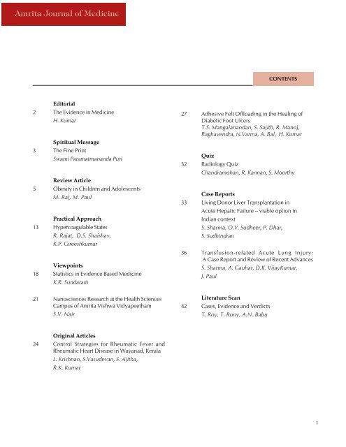
<strong>Amrita</strong> <strong>Journal</strong> <strong>of</strong> <strong>Medicine</strong><br />
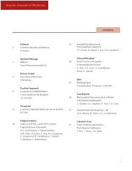
CONTENTS<br />
Editorial<br />
2 The Evidence in <strong>Medicine</strong><br />
H. Kumar<br />
Spiritual Message<br />
3 The Fine Print<br />
Swami Paramatman<strong>and</strong>a Puri<br />
Review Article<br />
5 Obesity in Children <strong>and</strong> Adolescents<br />
M. Raj, M. Paul<br />
Practical Approach<br />
13 Hypercoagulable States<br />
R. Rajat, D.S. Shaishav,<br />
K.P. Gireeshkumar<br />
Viewpoints<br />
18 Statistics in Evidence Based <strong>Medicine</strong><br />
K.R. Sundaram<br />
27 Adhesive Felt Offloading in the Healing <strong>of</strong><br />
Diabetic Foot Ulcers<br />
T.S. Mangalan<strong>and</strong>an, S. Sajith, R. Manoj,<br />
Raghavendra, N.Varma, A. Bal, H. Kumar<br />
Quiz<br />
32 Radiology Quiz<br />
Ch<strong>and</strong>ramohan, R. Kannan, S. Moorthy<br />
Case Reports<br />
33 Living Donor Liver Transplantation in<br />
Acute Hepatic Failure – viable option in<br />
Indian context<br />
S. Sharma, O.V. Sudheer, P. Dhar,<br />
S. Sudhindran<br />
36 Transfusion-related Acute Lung Injury:<br />
A Case Report <strong>and</strong> Review <strong>of</strong> Recent Advances<br />
S. Sharma, A. Gauhar, D.K. VijayKumar,<br />
J. Paul<br />
21 Nanosciences Research at the Health <strong>Sciences</strong><br />
Campus <strong>of</strong> <strong>Amrita</strong> Vishwa Vidyapeetham<br />
S.V. Nair<br />
Literature Scan<br />
42 Cases, Evidence <strong>and</strong> Verdicts<br />
T. Roy, T. Rony, A.N. Babu<br />
Original Articles<br />
24 Control Strategies for Rheumatic Fever <strong>and</strong><br />
Rheumatic Heart Disease in Wayanad, Kerala<br />
L. Krishnan, S.Vasudevan, S. Ajitha,<br />
R.K. Kumar<br />
1
<strong>Amrita</strong> <strong>Journal</strong> <strong>of</strong> <strong>Medicine</strong><br />
EDITORIAL<br />
Editorial Board<br />
Patrons<br />
Swami <strong>Amrita</strong> Swaroopan<strong>and</strong>a Puri<br />
Dr. Prem Nair<br />
Mr. Ron Gottsegen<br />
Dr. D.M. Vasudevan<br />
Chief Editor<br />
Dr. Harish Kumar<br />
Associate Editors<br />
Dr. An<strong>and</strong> Kumar<br />
Dr. Sudhindran<br />
Editorial Board Members<br />
Dr. V. Balakrishnan<br />
Mr. Sudhakar Jayaram<br />
. Dr. Dilip Panikar<br />
Dr. Prakash Kamath<br />
Dr. S.K. Nair<br />
Dr. Vaidyanathan<br />
Dr. M.G.K. Pillai<br />
Dr. Subramanian<br />
Dr. Kanthaswami<br />
Dr. Abraham Kuriakose<br />
Publications Officer<br />
Mrs. Jaya Sudhir Maharshi<br />
Cover Illustrations<br />
Mr. Dinesh M<br />
Mrs. Rajalakshmi Remesh<br />
Dept.<strong>of</strong> Graphics, AIMS<br />
The Evidence in <strong>Medicine</strong><br />
Harish Kumar<br />
We live in an era where “Evidence Based <strong>Medicine</strong>” is<br />
the new mantra. “Anecdotal <strong>Medicine</strong>” as the practice<br />
<strong>of</strong> medicine based on experience or observations is<br />
referred to, has become an unfashionable term <strong>and</strong> is<br />
frowned upon as quaint <strong>and</strong> unscientific. Today<br />
everything in medicine is acceptable only if it is evidencebased.<br />
This has resulted in a situation where data<br />
presented in a statistically skillful manner may make huge<br />
changes in the way we practice. For example if there is<br />
evidence that giving Aspirin to people with diabetes results<br />
in reduced risk <strong>of</strong> heart disease, it actually means that a<br />
small percentage <strong>of</strong> people who receive this treatment<br />
will benefit, but since we have no mechanism <strong>of</strong> assessing<br />
which patient with diabetes will benefit, we end up in a<br />
situation where we recommend that all people with<br />
diabetes should be on Aspirin. Due to this trend, many<br />
patients end up getting a long list <strong>of</strong> medications each <strong>of</strong><br />
which has evidence <strong>of</strong> benefit in a certain percentage <strong>of</strong><br />
users, but may or may not be beneficial for the specific<br />
patient under consideration.<br />
One day a man was walking down a street, when a friend<br />
called out to him <strong>and</strong> told him that he had seen his father<br />
lying dead in the next street. The man immediately went<br />
to the nearby bookshop <strong>and</strong> scanned all the newspapers<br />
<strong>and</strong> found no mention <strong>of</strong> his father’s death in any <strong>of</strong><br />
them. He concluded that since there was no mention in<br />
print <strong>of</strong> his friend’s observation that his father was dead,<br />
his friend must be wrong. This same analogue applies to<br />
medicine as well. We all believe that any thing that is<br />
printed in journals or textbooks is true <strong>and</strong> also that<br />
anything which is not in the books is false. But anyone<br />
with common sense <strong>and</strong> experience <strong>of</strong> life knows that<br />
what one sees <strong>and</strong> hears is as true as what one reads <strong>and</strong><br />
learns.<br />
The other aspect <strong>of</strong> evidence-based medicine is that many<br />
<strong>of</strong> the drug-trials are pharmasponsored <strong>and</strong> by their very<br />
nature, are biased. Since doctors are now very focused on<br />
“evidence”, the pharma companies have become very<br />
adept at providing us with evidence that will wilt under<br />
close scientific scrutiny.<br />
The plus side <strong>of</strong> evidence-based medicine is that doctors<br />
are now better read <strong>and</strong> the way we practice can be<br />
justified on scientific grounds. The practice <strong>of</strong> medicine<br />
has become more scientific <strong>and</strong> organized. Protocol driven<br />
management has made things more systematic <strong>and</strong><br />
introduced a degree <strong>of</strong> uniformity in practice, which<br />
didn’t exist before. But what we need is hard, cold facts<br />
to be combined with the warmth <strong>of</strong> our experience <strong>and</strong> a<br />
proper underst<strong>and</strong>ing <strong>of</strong> how best to apply the evidence<br />
to the individual patient in front <strong>of</strong> us for the betterment<br />
<strong>of</strong> his health.<br />
2
<strong>Amrita</strong> <strong>Journal</strong> <strong>of</strong> <strong>Medicine</strong><br />
The Fine Print<br />
Swami Paramatman<strong>and</strong>a Puri<br />
uri<br />
SPIRITUAL MESSAGE<br />
Although sadly forgotten by us most <strong>of</strong> the time, the<br />
frailty <strong>of</strong> the human body is clearly seen almost every<br />
day by those who work in the medical pr<strong>of</strong>ession. Amma<br />
tries to remind us that, “These bodies <strong>of</strong> ours are not<br />
eternal. They can perish at any moment. Try to remember<br />
that we are born as human beings to experience our true<br />
nature as consciousness, the hidden source <strong>and</strong> support<br />
<strong>of</strong> our mind.”<br />
Although the modern world tells us that there is no<br />
particular goal in life except material happiness, the wise<br />
sages <strong>of</strong> ancient India, after much reflection, conceived<br />
<strong>of</strong> human life as a place to make gradual efforts towards<br />
the goal <strong>of</strong> directly experiencing consciousness as one’s<br />
true self. Even though many physicians perform surgery,<br />
none so far have either found or seen that principle <strong>of</strong><br />
consciousness in the physical body. This doesn’t mean<br />
that it doesn’t exist, but only that it must be looked for<br />
where it truly is <strong>and</strong> with the proper instrument. Amma<br />
says that it must be sought within the mind, by the mind.<br />
Humans are uniquely endowed with the capacity to directly<br />
experience consciousness as it is, thereby resulting<br />
in the highest satisfaction <strong>and</strong> peace.<br />
If, in addition to our “normal” life, we are fortunate<br />
enough to feel an urge to pursue a path leading to this<br />
experience, we need a “plan <strong>of</strong> action” or a “flow chart.”<br />
Fortunately, Amma has given us some practical guidelines,<br />
which can <strong>and</strong> should be followed at the different<br />
stages <strong>of</strong> our life. She says,<br />
“Parents should start explaining spiritual ideas to children<br />
at an early age. They should learn to respect their<br />
elders by rising when elders enter the room, sitting down<br />
only after they take their seat, answering them politely,<br />
obeying their instructions, <strong>and</strong> refraining from making<br />
fun <strong>of</strong> them or answering them loudly or in a contrary<br />
way. They should learn prayers, listen to spiritual stories,<br />
do yogasanas, meditation <strong>and</strong> japa, <strong>and</strong> engage in seva,<br />
apart from their secular studies. Even if they acquire bad<br />
habits when they grow up, these good impressions which<br />
are dormant in their subconscious mind will bring them<br />
back to the right path in due course.”<br />
After growing up, most <strong>of</strong> us will desire to have a<br />
spouse <strong>and</strong> children, wealth <strong>and</strong> position, comforts <strong>and</strong><br />
possessions. The spiritual practices that we learned as<br />
children should be continued. Destructive passions such<br />
as anger <strong>and</strong> jealousy are to be gradually reduced, but<br />
not so gradually that nothing at all is done about them!<br />
This is the stage <strong>of</strong> life where there are plenty <strong>of</strong> opportu-<br />
nities for self-improvement. As they say, there are no<br />
problems, only opportunities.<br />
If one sincerely follows the instructions in this “user<br />
manual,” eventually one will feel a strong sense <strong>of</strong> detachment<br />
from worldly affairs <strong>and</strong> an urgency to prepare<br />
to meet one’s Maker. As Amma says,<br />
“Once the children are grown up <strong>and</strong> are able to take<br />
care <strong>of</strong> themselves, husb<strong>and</strong> <strong>and</strong> wife should lead a purely<br />
spiritual life, working for their spiritual improvement by<br />
engaging in meditation, japa <strong>and</strong> selfless service. If we<br />
spend the remainder <strong>of</strong> our lives in sadhana, the accumulated<br />
spiritual power will help both us <strong>and</strong> the world.<br />
Therefore, cultivate the habit <strong>of</strong> withdrawing the mind<br />
from countless worldly subjects <strong>and</strong> turn it totally towards<br />
God.”<br />
For many <strong>of</strong> us, the idea <strong>of</strong> eventually spending all<br />
our time in spiritual pursuits might seem very difficult if<br />
not impossible to do, <strong>and</strong> this might be so. In fact, very<br />
few indeed would be willing to sign on the dotted line!<br />
However, there is a saving grace, some “small print” at<br />
the bottom <strong>of</strong> this contract. It reads,<br />
“There are many ways to reach Me, <strong>and</strong> all are equally<br />
acceptable in My eyes. Try your best <strong>and</strong> leave the rest to<br />
Me.”<br />
Some <strong>of</strong> history’s greatest souls were just ordinary<br />
people like most <strong>of</strong> us. By living at home but, constantly<br />
making efforts to improve themselves, they experienced<br />
the same high state <strong>of</strong> consciousness that a sincere sannyasi<br />
does. Listen to this story.<br />
There once was a hermit who lived high up on a<br />
mountain-side in a tiny cave. His food was roots <strong>and</strong><br />
berries, a bit <strong>of</strong> bread given by a shepherd, or some milk<br />
brought by a woman who wanted his prayers. His only<br />
work was praying <strong>and</strong> thinking about God. For forty years<br />
he lived so, praying for the people, comforting them in<br />
their troubles, <strong>and</strong> most <strong>of</strong> all, worshipping in his heart.<br />
There was just one thing he cared about - to make his<br />
mind so pure <strong>and</strong> perfect that it could behold God.<br />
One day, he had a great longing to know how far<br />
along he had got with his work, how it looked to the<br />
Lord. So he prayed that he might be shown someone whose<br />
spirituality was neither more or less than his own. As he<br />
looked up from his prayer, a white-robed angel stood<br />
before him. The hermit bowed before the messenger with<br />
great awe <strong>and</strong> happiness; for he knew that his wish was<br />
answered. “Go to the nearest town,’’ the angel said,<br />
3
<strong>Amrita</strong> <strong>Journal</strong> <strong>of</strong> <strong>Medicine</strong><br />
The Fine Print<br />
“where there is a small farm where two women live. In<br />
them you will find two souls like your own.”<br />
When the hermit came to the door <strong>of</strong> the little farm,<br />
the two women who lived there were overjoyed to see<br />
him, for everyone loved <strong>and</strong> honoured the hermit. They<br />
put a chair for him on the porch <strong>and</strong> brought food <strong>and</strong><br />
drink. But the hermit was too eager to wait. He longed<br />
greatly to know what the souls <strong>of</strong> the two women were<br />
like, <strong>and</strong> from their looks he could see only that they<br />
were gentle <strong>and</strong> honest. One was old, <strong>and</strong> the other <strong>of</strong><br />
middle age.<br />
Presently he asked them about their lives. They told<br />
him the little there was to tell. They had worked hard<br />
always, in the fields with their husb<strong>and</strong>s, or in the house;<br />
they had many children; they had seen hard times, sickness<br />
<strong>and</strong> sorrow, but they had never despaired.<br />
“But what <strong>of</strong> your good deeds,’’ the hermit asked,<br />
“what have you done for God?’’<br />
“Very little,’’ they said, sadly, for they were too poor<br />
to give much. To be sure, twice every year, during harvest<br />
time, they gave some rice to their poorer neighbors.<br />
“That is very good,’’ the hermit said. “And is there<br />
any other good deed you have done?’’<br />
“Nothing,” said the older woman, “unless, unless —<br />
it might be called a good deed.” She looked at the younger<br />
woman, who smiled back at her.<br />
“What?’’ said the hermit.<br />
Still the woman hesitated; but at last she said, timidly,<br />
“It is not much to tell, only this, that it is twenty<br />
years since my sister-in-law <strong>and</strong> I came to live together<br />
in this house; we have brought up our families here. And<br />
in all the twenty years there has never been an angry word<br />
between us, or a look that was less than kind.’’<br />
The hermit bent his head before the two women <strong>and</strong><br />
gave thanks in his heart. “If my soul is as these,” he said,<br />
“I am blessed indeed.”<br />
A great light suddenly came into the hermit’s mind,<br />
<strong>and</strong> he saw how many ways there are <strong>of</strong> spiritual practice<br />
<strong>and</strong> serving God. Some go to churches, temples, or<br />
mosques, <strong>and</strong> some live in ashrams or caves, praying <strong>and</strong><br />
meditating. Some poor souls who had been very wicked,<br />
turn from their evilness with great sorrow, <strong>and</strong> serve Him<br />
with remorse. Some live faithfully <strong>and</strong> gently in humble<br />
homes, working, bringing up children, staying kind <strong>and</strong><br />
cheerful. And some bear pain patiently, for His sake.<br />
Endless, endless ways there are that only the Lord sees.<br />
4
<strong>Amrita</strong> <strong>Journal</strong> <strong>of</strong> <strong>Medicine</strong><br />
REVIEW ARTICLE<br />
Obesity in Children <strong>and</strong> Adolescents<br />
M. Raj, M. Paul<br />
INTRODUCTION<br />
The global disease pr<strong>of</strong>ile is changing<br />
at an astonishingly fast rate<br />
especially in low <strong>and</strong> middle-income<br />
countries. The looming epidemics <strong>of</strong><br />
obesity, cardiovascular disease (CVD)<br />
<strong>and</strong> diabetes form the center stage <strong>of</strong><br />
this transformation. Worldwide, obesity<br />
in children <strong>and</strong> adults has become<br />
a massive epidemic 1 . Obesity is an<br />
independent risk factor for CVD. Obesity<br />
is associated with an increased risk<br />
<strong>of</strong> morbidity <strong>and</strong> mortality as well as<br />
reduced life expectancy. Health service<br />
use <strong>and</strong> medical costs associated<br />
with obesity <strong>and</strong> related diseases have<br />
risen dramatically in recent times 2 .<br />
For children <strong>and</strong> adolescents, overweight<br />
<strong>and</strong> obesity are defined using<br />
age <strong>and</strong> sex specific normograms for<br />
Body Mass Index (BMI). Children<br />
whose body mass index that exceeds<br />
the age-gender-specific 95 th percentile<br />
are defined obese. Those whose BMI<br />
is between the 85 th <strong>and</strong> 95 th percentiles<br />
are overweight <strong>and</strong> are at increased risk<br />
for obesity related co-morbidities 3 . This<br />
article focuses on epidemiology, etiopathogenesis,<br />
comorbidities, prevention<br />
<strong>and</strong> treatment <strong>of</strong> obesity in children<br />
<strong>and</strong> adolescents.<br />
EPIDEMIOLOGY<br />
The prevalence <strong>of</strong> obesity in children<br />
is increasing worldwide. In 2003,<br />
the International Obesity Task Force<br />
reported that worldwide 1 out <strong>of</strong> 10<br />
child, aged 5-17 years, is overweight<br />
or obese 1 . Of the countries suffering<br />
from childhood obesity, Engl<strong>and</strong> tops<br />
the list. In Engl<strong>and</strong>, between 1974 <strong>and</strong><br />
2003, among children aged 5-10 years<br />
Dept. <strong>of</strong> Pediatric Cardiology, AIMS, Kochi.<br />
the prevalence <strong>of</strong> obesity increased<br />
from 1.8% to 6.0% in boys <strong>and</strong> from<br />
1.3% to 6.6% in girls 4 . A similar tendency<br />
was reported in Australia, where<br />
between 1985 <strong>and</strong> 1995 the prevalence<br />
<strong>of</strong> obesity among children aged<br />
7-15 years increased 4.6-fold among<br />
girls <strong>and</strong> 3.4-fold among boys 5 . Asian<br />
countries are not immune to this phenomenon.<br />
For example, in China, the<br />
prevalence <strong>of</strong> overweight <strong>and</strong> obesity<br />
among children aged 7-9 years increased<br />
from 1-2% in 1985 to 17%<br />
among girls <strong>and</strong> 25% among boys in<br />
2000 6 . In addition, obesity prevalence<br />
varies across socioeconomic strata. In<br />
developed countries, children <strong>of</strong> low<br />
socioeconomic status are more affected<br />
than their affluent counterparts 4,7 . The<br />
opposite is observed in developing<br />
countries: children <strong>of</strong> the upper socioeconomic<br />
strata are more likely than<br />
poor children to be obese 8,9 . Limited<br />
data is available from India regarding<br />
current trends in childhood obesity.<br />
An unpublished study conducted in<br />
24,000 school children in South India<br />
showed that the proportion <strong>of</strong> overweight<br />
children increased from 4.94%<br />
<strong>of</strong> the total students in 2003 to 6.57%<br />
in 2005 10 . Differences in overweight<br />
<strong>and</strong> overweight trends also occur by<br />
social class <strong>and</strong> by ethnic groups,<br />
emphasizing the importance <strong>of</strong> nongenetic<br />
variables 7,11,12 .<br />
ETIOPATHOGENESIS OF<br />
CHILDHOOD OBESITY<br />
Etiopathogenesis <strong>of</strong> childhood obesity<br />
is multifactorial. Interactions<br />
between genetic, neuroendocrine,<br />
metabolic, psychological, environmental<br />
<strong>and</strong> sociocultural factors are<br />
clearly evident in childhood obesity.<br />
GENE MUTATIONS AND<br />
OBESITY<br />
Naturally occurring single <strong>and</strong> polygenic<br />
gene mutations producing<br />
obesity are known in rodents like mice<br />
<strong>and</strong> rats. The prototypic obese mice<br />
with single gene defects are the obese<br />
(ob/ob, Lep ob ) <strong>and</strong> diabetes (db/db,<br />
Lepr db ) autosomal recessive mutations.<br />
These mutations produce phenotypes<br />
<strong>of</strong> severe hyperphagia, obesity, type 2<br />
diabetes, defective thermogenesis, <strong>and</strong><br />
infertility. The mutant gene responsible<br />
for the phenotype in Lep ob mice encodes<br />
a protein termed leptin, which<br />
is deficient in these animals 3 . Leptin<br />
deficiency has been documented in<br />
subsets <strong>of</strong> human obesity. Severe earlyonset<br />
human obesity caused by a<br />
mutant leptin receptor has also been<br />
identified. In the fatty (fat/fat) mouse,<br />
the recessively inherited mutation<br />
causes hyperinsulinemia without hyperglycemia<br />
<strong>and</strong> postpubertal obesity<br />
that is less severe than that seen in ob/<br />
ob or db/db mice. The yellow mutation<br />
<strong>of</strong> agouti mice is a dominant trait<br />
that causes yellow coat color, obesity,<br />
<strong>and</strong> diabetes 3 . The polygenic mouse<br />
models <strong>of</strong> obesity closely resemble the<br />
human obesity phenotypes than single<br />
gene models <strong>and</strong> have mutations that<br />
influence obesity, plasma cholesterol<br />
levels, body fat distribution, <strong>and</strong> propensity<br />
toward development <strong>of</strong> obesity<br />
on a high-fat diet.<br />
Genetic conditions known to be<br />
associated with predilection for obesity<br />
include Prader-Willi syndrome,<br />
Bardet-Biedl syndrome, <strong>and</strong> Cohen<br />
syndrome. It has been observed that<br />
obesity has a familial tendency. For<br />
young children, if one parent is obese,<br />
the odds ratio is approximately 3 for<br />
5
<strong>Amrita</strong> <strong>Journal</strong> <strong>of</strong> <strong>Medicine</strong><br />
Obesity in Children <strong>and</strong> Adolescents<br />
obesity in adulthood, but if both parents are obese, the<br />
odds ratio increases to more than 10. Before 3 years <strong>of</strong><br />
age, parental obesity is a stronger predictor <strong>of</strong> obesity in<br />
adulthood than the child’s weight status 13 .<br />
NEUROENDOCRINOLOGY OF<br />
ENERGY METABOLISM<br />
Food intake <strong>and</strong> energy expenditure is controlled by<br />
complex neuroendocrine interactions. The hormone leptin<br />
is an important component <strong>of</strong> this complex system. Leptin<br />
is made almost exclusively in adipose tissue <strong>and</strong> acts<br />
centrally in the hypothalamus. Low plasma concentrations<br />
<strong>of</strong> leptin <strong>and</strong> insulin (e.g., during fasting <strong>and</strong> weight<br />
loss) increase food intake <strong>and</strong> decrease energy expenditure<br />
by stimulating neuropeptide Y (NPY) synthesis, <strong>and</strong><br />
perhaps by inhibiting sympathetic activity <strong>and</strong> other catabolic<br />
pathways 3 . High leptin <strong>and</strong> insulin concentrations<br />
(e.g., during feeding <strong>and</strong> weight gain) decrease food intake<br />
<strong>and</strong> increase energy expenditure through release <strong>of</strong><br />
melanocortin <strong>and</strong> corticotropin-releasing hormone (CRH),<br />
among others. The major peptides that stimulate feeding<br />
are orexins A <strong>and</strong> B, which are secreted by the hypothalamus,<br />
<strong>and</strong> ghrelin, which is secreted by the stomach 3 .<br />
VITAL PERIODS IN<br />
DEVELOPMENT OF OBESITY<br />
There are vital periods <strong>of</strong> development for excessive<br />
weight gain. Intrauterine influences play a major role in<br />
the genesis <strong>of</strong> obesity by influencing proportions <strong>of</strong> fat<br />
<strong>and</strong> lean body mass, central nervous system appetite control,<br />
<strong>and</strong> pancreatic structure <strong>and</strong> function.<br />
Epidemiological studies have demonstrated a direct affirmative<br />
relationship between birth weight <strong>and</strong> BMI<br />
attained in later life 14 . In addition, lower birth weight for<br />
gestational age has been associated with later risk for<br />
more central deposition <strong>of</strong> fat, which also confers increased<br />
cardiovascular risk. Rapid weight gain during<br />
infancy is also associated with obesity later in childhood 15 .<br />
The combination <strong>of</strong> lower birth weight <strong>and</strong> higher attained<br />
BMI is most robustly associated with later CVD<br />
risk 16 .<br />
Extent <strong>and</strong> period <strong>of</strong> breastfeeding have been found to<br />
be inversely associated with risk <strong>of</strong> obesity in later childhood<br />
17-20 . The normal tendency during early puberty for<br />
insulin resistance may be a natural c<strong>of</strong>actor for unwarranted<br />
weight gain as well as various comorbidities <strong>of</strong><br />
obesity 21 . Early menarche is clearly associated with extent<br />
<strong>of</strong> obesity, with a tw<strong>of</strong>old increase in rate <strong>of</strong> early<br />
menarche associated with BMI greater than the 85 th percentile<br />
22 . The risk <strong>of</strong> obesity persisting into adulthood is<br />
higher among obese adolescents than among younger children<br />
13 . Observations suggest that up to 80% <strong>of</strong> overweight<br />
adolescents will become obese adults 23 .<br />
ENVIRONMENTAL RISK<br />
FACTORS FOR OBESITY<br />
Environmental risk factors for overweight <strong>and</strong> obesity,<br />
including family <strong>and</strong> parental issues, are numerous <strong>and</strong><br />
complicated. Poor cognitive stimulation in the home <strong>and</strong><br />
low socioeconomic status predicts development <strong>of</strong> obesity<br />
24 . Parental food choices influence child food<br />
preferences 25 , <strong>and</strong> degree <strong>of</strong> parental adiposity is a marker<br />
for children’s fat preferences 26 . Children <strong>and</strong> adolescents<br />
<strong>of</strong> lower socioeconomic status have been reported to be<br />
less likely to eat fruits <strong>and</strong> vegetables <strong>and</strong> to have a higher<br />
intake <strong>of</strong> total <strong>and</strong> saturated fat 27-29 . Early rebound <strong>of</strong> the<br />
BMI is associated with an augmented risk <strong>of</strong> higher BMI<br />
in adulthood. A recent study links early rebound <strong>of</strong> BMI<br />
to glucose intolerance <strong>and</strong> diabetes in adults 30 .<br />
SOCIETAL CHANGES AND OBESITY<br />
Widespread <strong>and</strong> intense societal changes during the<br />
last several decades have contributed to childhood obesity.<br />
Leisure activity is ever more sedentary <strong>and</strong> there has<br />
Table 1: Adverse Outcomes in Childhood Obesity<br />
Metabolic<br />
Cardiovascular<br />
Psychological<br />
Orthopedic<br />
Neurological<br />
Hepatic<br />
Pulmonary<br />
Renal<br />
Malignancy<br />
Type 2 diabetes mellitus, impaired glucose tolerance<br />
Metabolic syndrome, hyper insulinism<br />
Dyslipidemia, atherosclerosis<br />
Hypertension, left ventricular hypertrophy<br />
Depression, poor quality <strong>of</strong> life<br />
Slipped capital femoral epiphysis<br />
Blount’s disease, osteoarthritis<br />
Pseudotumor cerebri<br />
Nonalcoholic steatohepatitis, gall bladder disease<br />
Obstructive sleep apnea, asthma (exacerbation)<br />
Proteinuria, FSGS<br />
Of ovary, breast, colon<br />
6
<strong>Amrita</strong> <strong>Journal</strong> <strong>of</strong> <strong>Medicine</strong><br />
been a decrease in frequency <strong>and</strong> duration <strong>of</strong> physical<br />
activities <strong>of</strong> daily living for children 31 . The results <strong>of</strong> a<br />
r<strong>and</strong>omized trial to decrease television viewing for schoolaged<br />
children has provided the strongest evidence to<br />
support the role <strong>of</strong> limiting television in prevention <strong>of</strong><br />
obesity 32 .<br />
COMORBIDITIES RELATED TO OBESITY<br />
Obesity is associated with a number <strong>of</strong> comorbidities<br />
in adolescents <strong>and</strong> children. Table 1 presents common<br />
comorbid conditions related to obesity in adolescents <strong>and</strong><br />
children.<br />
METABOLIC SYNDROME<br />
Metabolic syndrome is a cluster <strong>of</strong> traits that include<br />
acanthosis nigricans, hyperinsulinemia, obesity, hypertension,<br />
<strong>and</strong> hyperlipidemia 33 . Metabolic syndrome is<br />
becoming common among children <strong>and</strong> adolescents <strong>and</strong><br />
its prevalence increases directly with the degree <strong>of</strong> obesity.<br />
Each component <strong>of</strong> the syndrome worsens with<br />
increasing obesity – an association that is independent<br />
<strong>of</strong> age, sex, <strong>and</strong> pubertal status 34 . The prevalence <strong>of</strong> the<br />
metabolic syndrome in adolescents is 4%, but it increases<br />
to 30% to 50% in overweight children 34,35 . The initiating<br />
event <strong>of</strong> the syndrome appears to be obesity leading to<br />
excess insulin production, which later leads to insulin<br />
resistance. Insulin resistance leads to increased hepatic<br />
synthesis <strong>of</strong> very-low-density lipoprotein, resistance <strong>of</strong><br />
the action <strong>of</strong> insulin on lipoprotein lipase in peripheral<br />
tissues, enhanced cholesterol synthesis, increased highdensity<br />
lipoprotein degradation, increased sympathetic<br />
activity, proliferation <strong>of</strong> vascular smooth muscle cells,<br />
<strong>and</strong> increased formation <strong>and</strong> decreased reduction <strong>of</strong><br />
plaque. The metabolic syndrome significantly influences<br />
cardiovascular disease risk in young individuals. Berenson<br />
et al 36 demonstrated a striking increase in the extent <strong>of</strong><br />
coronary atherosclerotic lesions with obesity <strong>and</strong> an increasing<br />
number <strong>of</strong> metabolic syndrome risk factors in<br />
young individuals. The significant adverse effect <strong>of</strong> worsening<br />
obesity on each component <strong>of</strong> the metabolic<br />
syndrome, underscores the deleterious effect <strong>of</strong> increasing<br />
BMI in this age group.<br />
TYPE 2 DIABETES MELLITUS<br />
The development <strong>of</strong> type 2 diabetes in obese adolescents<br />
has been well-documented 37 . Predictions imply that<br />
obesity driven type 2 diabetes might become the most<br />
common form <strong>of</strong> newly diagnosed diabetes in adolescent<br />
youth within 10 years 38 . There is strong evidence<br />
suggesting a global spread <strong>of</strong> type 2 diabetes in childhood<br />
39 . Evidence is emerging <strong>of</strong> an increase in type 2<br />
diabetes among urban Indian children as well 40 . Type 2<br />
diabetes mellitus had been primarily a disease <strong>of</strong> adulthood;<br />
however, type 2 diabetes now occurs in adolescents<br />
typically with a BMI >30 kg/m2 23 . Various studies demonstrate<br />
increased risk <strong>of</strong> nephropathy 41,42 <strong>and</strong> retinopathy<br />
43 compared to young people with type 1 diabetes,<br />
whilst recent data indicate early signs <strong>of</strong> cardiovascular<br />
disease in youth with type 2 diabetes 44 . The rapid increase<br />
in the incidence <strong>of</strong> type 2 diabetes points towards<br />
emergence <strong>of</strong> an epidemic <strong>of</strong> advanced cardiovascular<br />
disease due to the synergistic effects <strong>of</strong> other components<br />
<strong>of</strong> the metabolic syndrome, as well as chronic low-grade<br />
inflammation, as obese adolescents become obese young<br />
adults.<br />
CARDIOVASCULAR ABNORMALITIES<br />
It is well recognized that obesity substantially contributes<br />
to morbidity <strong>and</strong> mortality from cardiovascular<br />
disease across ages. Obesity may affect the heart through<br />
its influence on known risk factors such as dyslipidemia,<br />
hypertension, glucose intolerance, inflammatory markers,<br />
obstructive sleep apnea/hypoventilation, <strong>and</strong> the<br />
prothrombotic state, as well as through yet unrecognized<br />
mechanisms. L<strong>and</strong>mark studies like Bogalusa <strong>and</strong><br />
Muscatine have demonstrated that obesity during childhood<br />
<strong>and</strong> adolescence is a determinant <strong>of</strong> a number <strong>of</strong><br />
cardiovascular risk factors 36,45,46 . One study conducted on<br />
children <strong>and</strong> adolescents have shown that lean body mass,<br />
fat mass, <strong>and</strong> systolic blood pressure were independently<br />
associated with left ventricular mass, which is a strong<br />
independent predictor <strong>of</strong> coronary heart disease, stroke,<br />
<strong>and</strong> sudden death in adults 47 . Obstructive sleep apnea, a<br />
cardiovascular risk factor is also associated with obesity<br />
in children <strong>and</strong> adults 48 . As a whole, obesity predisposes<br />
or is associated with numerous cardiac complications<br />
such as coronary heart disease, heart failure, <strong>and</strong> sudden<br />
death through its impact on the cardiovascular system.<br />
PSYCHOSOCIAL ABNORMALITIES<br />
Psychosocial abnormalities are closely associated with<br />
obesity in children <strong>and</strong> adolescents. In a study by Pine,<br />
et al 49 adults who had been diagnosed with clinically<br />
defined major depression during their youth had a greater<br />
BMI than adults who did not suffer from depression during<br />
their youth. In another study, Goodman et al 50<br />
demonstrated that depression scores were highest in the<br />
children with the greatest increase in BMI. Observations<br />
point to the fact that obese children have smaller friend<br />
circles with the relationships being peripheral <strong>and</strong> isolated.<br />
Obesity related psychological issues have been<br />
shown to be associated with an increase in both suicidal<br />
ideation <strong>and</strong> number <strong>of</strong> suicide attempts in youth 51 .<br />
MEDICAL EVALUATION OF<br />
COMORBIDITIES<br />
All children <strong>and</strong> adolescents who are overweight require<br />
a detailed medical examination for identifying<br />
potential Comorbidities. A st<strong>and</strong>ard protocol is presented<br />
in Table 2.<br />
7
<strong>Amrita</strong> <strong>Journal</strong> <strong>of</strong> <strong>Medicine</strong><br />
Obesity in Children <strong>and</strong> Adolescents<br />
Table 2: <strong>Medical</strong> Evaluation <strong>of</strong> a Child or an Adolescent Who Is Overweight<br />
Evaluation <strong>of</strong> growth: Normal growth (especially height) makes metabolic or genetic form <strong>of</strong> overweight less likely<br />
Family history <strong>of</strong> premature coronary heart disease, dyslipidemia, diabetes<br />
Diet history, history <strong>of</strong> smoking<br />
History <strong>of</strong> sleep-disordered breathing<br />
Assessment <strong>of</strong> physical activity <strong>and</strong> sedentary behaviour<br />
Psychiatric assessment<br />
History <strong>of</strong> irregular menstrual periods, acne, <strong>and</strong> hirsutism in adolescent girls<br />
(evidence <strong>of</strong> polycystic ovarian syndrome).<br />
Skin disorders like intertrigo, monilial dermatitis, acne <strong>and</strong> acanthosis nigricans.<br />
Blood pressure measurement (multiple readings with attention to proper cuff size)<br />
Physical assessment for orthopedic abnormalities<br />
Urine analysis, fasting lipid pr<strong>of</strong>ile, Serum uric acid, C-Reactive Protein,<br />
Fasting glucose, fasting insulin, HbA1c level<br />
Liver function tests, thyroid function tests<br />
Renal function tests (if hypertension is present)<br />
Abdominal USG (for fatty liver, ovarian cysts)<br />
PREVENTION OF OBESITY<br />
The ideal preventive strategy for obesity is to prevent<br />
children with a normal, desirable BMI from becoming<br />
overweight or obese. Preventive strategies should start as<br />
early as newborn period. Both initiation <strong>and</strong> duration <strong>of</strong><br />
breast-feeding may reduce the risk <strong>of</strong> later overweight 52 .<br />
A reasonable goal for preschool interventions would be<br />
to aim toward weight gain <strong>of</strong> 1.0 kg/2 cm <strong>of</strong> growth.<br />
This rate <strong>of</strong> gain from preschool age (3 to 4 years) onward<br />
predicted desirable weight at 8 to 9 years <strong>of</strong> age,<br />
whereas a gain <strong>of</strong> 1.8 kg/2 cm predicted obesity at elementary<br />
school age 53 . The important role <strong>of</strong> healthful<br />
behaviors has increasingly been documented. Behavior<br />
modifications should focus on increasing consumption<br />
<strong>of</strong> fruits, vegetables <strong>and</strong> fiber-containing grain products,<br />
avoidance <strong>of</strong> high calorie/high fat food items, increasing<br />
daily physical activity, <strong>and</strong> limiting sedentary time.<br />
Interventions that include classroom <strong>and</strong> physical<br />
education sessions, changes in school meals, vending<br />
machines, <strong>and</strong> cafeterias, <strong>and</strong> after-school programs, can<br />
increase physical activity <strong>and</strong> improve dietary patterns in<br />
children <strong>and</strong> adolescents 54,55 . One recent study has emphasized<br />
reducing television, videotape/DVD, <strong>and</strong> video<br />
game use 32 . There are also successful examples <strong>of</strong> physical<br />
education interventions designed with higher-intensity<br />
or more motivating activities, specifically endurance training<br />
56 <strong>and</strong> popular dance 57 . Exposure to various media is<br />
critical. A substantial proportion <strong>of</strong> the advertising on<br />
children’s television promotes food, <strong>and</strong> there is a direct<br />
relationship between television viewing <strong>and</strong> obesity 58 .<br />
Reducing television viewing has reduced weight gain <strong>and</strong><br />
the prevalence <strong>of</strong> obesity in experimental trials 32,59 . It has<br />
been hypothesized that television promotes obesity<br />
through the consumption <strong>of</strong> food while watching television,<br />
the consumption <strong>of</strong> foods advertised on television,<br />
or reduced physical activity 58,60,61 .<br />
TREATMENT OF OBESITY<br />
In treating children with overweight <strong>and</strong> obesity, the<br />
immediate goal is to bring down the rate <strong>of</strong> weight gain,<br />
followed by a period <strong>of</strong> weight maintenance <strong>and</strong> finally<br />
weight reduction to improve BMI. Children 2 to 4 years<br />
old who are overweight or obese will achieve reductions<br />
in BMI percentile by achieving a rate <strong>of</strong> weight gain < 1<br />
kg/2 cm <strong>of</strong> height gain. Older Children who are obese<br />
without comorbidities may achieve BMI percentile reductions<br />
to below overweight cut-<strong>of</strong>fs with BMI<br />
maintenance. Children who are obese with comorbidities<br />
require an aggressive approach to bring in weight loss in<br />
concurrence with other treatment strategies. A gradual<br />
weight loss is preferred because it is achievable <strong>and</strong> more<br />
easily sustained giving long-term benefits. Older adolescents<br />
who have completed linear growth <strong>and</strong> have a BMI<br />
>30 kg/m 2 require more aggressive weight loss similar<br />
to that for adults 62 . The principles for the treatment <strong>of</strong><br />
obesity can be summarized as follows:<br />
1. Establish individual treatment goals <strong>and</strong> approaches<br />
on a case-to-case basis.<br />
2. Ensure family involvement through out the treatment<br />
period.<br />
8
<strong>Amrita</strong> <strong>Journal</strong> <strong>of</strong> <strong>Medicine</strong><br />
Table 3: Treatment Strategies for Sub groups<br />
BMI Status Classification Treatment Goal<br />
95 th percentile Obese Weight maintenance (younger children) or gradual<br />
weight loss (adolescents) to reduce BMI percentile<br />
>30 kg/m2 Adult obesity range Gradual weight loss (1–2 kg/mo) to achieve healthier<br />
BMI<br />
>95th percentile Obesity with comorbidity Gradual weight loss (1–2 kg/mo) to achieve healthier<br />
<strong>and</strong> comorbidity<br />
BMI; treatment <strong>of</strong> comorbidities<br />
present*<br />
*See Table 2.<br />
3. Provide regular assessment <strong>and</strong> ensure follow up compliance.<br />
4. Provide attention to behavioral, psychological, <strong>and</strong><br />
social factors in the treatment plan.<br />
5. Provide situations that can be implemented within<br />
the subject’s environment.<br />
DIETARY MANAGEMENT<br />
Dietary management should aim to provide appropriate<br />
calorie intake <strong>and</strong> optimum nutrition. Due emphasis<br />
should be given to developing <strong>and</strong> sustaining healthy<br />
eating patterns. A st<strong>and</strong>ard protocol is to recommend a<br />
fat intake <strong>of</strong> 30% to 40% kcal in children 1 to 3 years<br />
old, with a reduction to 25% to 35% in children 4 to 18<br />
years old; a carbohydrate intake <strong>of</strong> 45% to 65% kcal in<br />
all children <strong>and</strong> adults; <strong>and</strong> protein intakes <strong>of</strong> 5% to 20%<br />
kcal in children 1 to 3 years old with gradual increase to<br />
10% to 30% kcal in children 4 to 18 years old 63 . Care<br />
should be taken to provide adequate nutrition by <strong>of</strong>fering<br />
a variety <strong>of</strong> foods that are low in saturated fat (3 servings <strong>of</strong> milk or<br />
dairy products, <strong>and</strong> >6 servings <strong>of</strong> whole-grain <strong>and</strong> grain<br />
products per day as well as consuming adequate amounts<br />
<strong>of</strong> dietary fiber (age in years + 5 g/d) 23 . Limiting the<br />
intake <strong>of</strong> salt (
<strong>Amrita</strong> <strong>Journal</strong> <strong>of</strong> <strong>Medicine</strong><br />
Obesity in Children <strong>and</strong> Adolescents<br />
effects <strong>and</strong> requires fat-soluble vitamin supplementation<br />
<strong>and</strong> monitoring 69,70 . For rare genetic <strong>and</strong> metabolic disorders,<br />
pharmacological treatment may be useful. For<br />
example, recombinant leptin is useful in hereditary leptin<br />
deficiency. Octreotide may be useful in hypothalamic<br />
obesity 71 . Metformin has been used in insulin-resistant<br />
children <strong>and</strong> adolescents who are overweight, but longterm<br />
follow up data is lacking 72 .<br />
SURGICAL TREATMENT<br />
Many cases <strong>of</strong> severe adolescent obesity with or without<br />
co morbidities warrant aggressive approaches including<br />
surgical treatment. The bariatric procedures include gastric<br />
bypass, gastric binding <strong>and</strong> vertical b<strong>and</strong>ed<br />
gastroplasty. Indications for surgery used include a BMI<br />
>40 kg/m2 <strong>and</strong> severe associated comorbidities, such as<br />
obstructive sleep apnea, type 2 diabetes mellitus, <strong>and</strong><br />
pseudotumor cerebri. More severe elevation <strong>of</strong> BMI<br />
(>50kg/m2) may be an indication for surgical treatment<br />
in the presence <strong>of</strong> less severe comorbidities 23 . An experienced<br />
team approach including comprehensive medical<br />
<strong>and</strong> psychological evaluation is critical 73 . Surgical therapy<br />
should only be advised for full-grown adolescents with<br />
the severest obesity-related morbidity.<br />
SUMMARY<br />
Obesity in adolescents <strong>and</strong> children has risen to significant<br />
levels globally including developing countries<br />
with serious public health consequences. In addition to<br />
cardiovascular, emotional <strong>and</strong> social issues, it poses serious<br />
threat to the basic health care delivery system <strong>of</strong><br />
developing <strong>and</strong> under developed countries. Unless this<br />
massive epidemic is contained in the near future, the<br />
implications <strong>of</strong> this global phenomenon on future generations<br />
will be disastrous. The reversibility <strong>of</strong> this disease<br />
by proper interventions, should be seen as an opportunity<br />
<strong>and</strong> all efforts to avert this catastrophe duly attempted<br />
for the sake <strong>of</strong> generations to come.<br />
REFERENCES<br />
1. IOTF. Childhood obesity - the new crisis in public health. London:<br />
International Obesity Task Force; 2003.<br />
2. Poirier P, Giles TD, Bray GA, et al. Obesity <strong>and</strong> cardiovascular<br />
disease: pathophysiology, evaluation, <strong>and</strong> effect <strong>of</strong> weight loss.<br />
Circulation 2006 Feb 14;113(6):898-918.<br />
3. Donohoue PA. Obesity. In: Behrman RE, Kleigman RM, Jenson<br />
HB (eds). Nelson textbook <strong>of</strong> Pediatrics, 17 th ed. Philadelphia:<br />
WB Saunders. 2004;173-7.<br />
4. Stamatakis E, Primatesta P, Chinn S, et al. Overweight <strong>and</strong> obesity<br />
trends from 1974 to 2003 in English children: what is the<br />
role <strong>of</strong> socioeconomic factors? Arch Dis Child 2005;90:999-<br />
1004.<br />
5. Magarey AM, Daniels LA, Boulton TJ. Prevalence <strong>of</strong> overweight<br />
<strong>and</strong> obesity in Australian children <strong>and</strong> adolescents: reassessment<br />
<strong>of</strong> 1985 <strong>and</strong> 1995 data against new st<strong>and</strong>ard international<br />
definitions. Med J Aust 2001;174:561-4.<br />
6. Wang L, Kong L, Wu F, et al. Preventing chronic diseases in<br />
China. Lancet 2005;366:1821-4.<br />
7. Strauss RS, Pollack HA. Epidemic increase in childhood overweight,<br />
1986-1998. JAMA 2001;286:2845-8.<br />
8. Salmon J, Timperio A, Clel<strong>and</strong> V, et al. Trends in children’s<br />
physical activity <strong>and</strong> weight status in high <strong>and</strong> low socio-economic<br />
status areas <strong>of</strong> Melbourne, Victoria, 1985-2001. Aust N<br />
Z J Public Health 2005;29:337-42.<br />
9. Chhatwal J, Verma M, Riar SK. Obesity among pre-adolescent<br />
<strong>and</strong> adolescents <strong>of</strong> a developing country [India]. Asia Pac J Clin<br />
Nutr 2004;13:231-5.<br />
10. Raj M, Sundaram KR, Paul M, et al. Obesity in Indian Childrentime<br />
trends <strong>and</strong> relationship with hypertension. <strong>Journal</strong> in press,<br />
2007.<br />
11. Saxena S, Ambler G, Cole TJ, et al. Ethnic group differences in<br />
overweight <strong>and</strong> obese children <strong>and</strong> young people in Engl<strong>and</strong>:<br />
cross sectional survey. Arch Dis Child 2004;89:30-6.<br />
12. Gordon-Larsen P, Adair LS, Popkin BM. The relationship <strong>of</strong><br />
ethnicity, socioeconomic factors, <strong>and</strong> overweight in US adolescents.<br />
Obes Res 2003;11:121-9.<br />
13. Whitaker RC, Wright JA, Pepe MS, et al. Predicting obesity in<br />
young adulthood from childhood <strong>and</strong> parental obesity. N Engl<br />
J Med 1997;337:869-73.<br />
14. Parsons TJ, Power C, Logan S, et al. Childhood predictors <strong>of</strong><br />
adult obesity: a systematic review. Int J Obes Relat Metab Disord<br />
1999;23:S1-S107.<br />
15. Stettler N, Zemel BS, Kumanyika S, et al. Infant weight gain <strong>and</strong><br />
childhood overweight status in a multicenter, cohort study.<br />
Pediatrics 2002;109:194-9.<br />
16. Dietz WH. Overweight in childhood <strong>and</strong> adolescence. N Engl<br />
J Med 2004;350:855-7.<br />
17. Agras SW, Kraemer HC, Berkowitz RI, et al. Influence <strong>of</strong> early<br />
feeding style on adiposity at 6 years <strong>of</strong> age. J Pediatr<br />
1990;116:805-9.<br />
18. von Kries R, Koletzko B, Sauerwald T, et al. Breast feeding <strong>and</strong><br />
obesity: cross sectional study. BMJ 1999;319:147-50.<br />
19. Gilman MW, Rifas-Shiman SL, Camargo CA Jr, et al. Risk <strong>of</strong><br />
overweight among adolescents who were breastfed as infants.<br />
JAMA 2001;285:2461-7.<br />
20. Hediger ML, Overpeck MD, Kuczmarski RJ, et al. Association<br />
between infant breastfeeding <strong>and</strong> overweight in young children.<br />
JAMA 2001;285:2453-60.<br />
10
<strong>Amrita</strong> <strong>Journal</strong> <strong>of</strong> <strong>Medicine</strong><br />
21. Travers SH, Jeffers BW, Bloch CA, et al. Gender <strong>and</strong> Tanner<br />
stage differences in body composition <strong>and</strong> insulin sensitivity in<br />
early pubertal children. J Clin Endocrinol Metab 1995;80:<br />
172-8.<br />
22. Adair LS, Gordon-Larsen P. Maturational timing <strong>and</strong> overweight<br />
prevalence in US adolescent girls. Am J Public Health<br />
2001;91:642-4.<br />
23. Daniels SR, Arnett DK, Eckel RH, et al. Overweight in children<br />
<strong>and</strong> adolescents: pathophysiology, consequences, prevention,<br />
<strong>and</strong> treatment. Circulation 2005 Apr 19;111(15):1999-2012.<br />
24. Strauss RS, Knight J. Influence <strong>of</strong> the home environment on the<br />
development <strong>of</strong> obesity in children. Pediatrics 1999;103(6).<br />
25. Ray JW, Klesges RC. Influences on the eating behavior <strong>of</strong> children.<br />
Ann N Y Acad Sci.1993;699:57-69.<br />
26. Fisher JO, Birch LL. Fat preferences <strong>and</strong> fat consumption <strong>of</strong> 3- to<br />
5-year-old children are related to parental adiposity. J Am Diet<br />
Assoc 1995;95:759-64.<br />
27. Neumark-Sztainer D, Story M, Resnick MD, et al. Correlates <strong>of</strong><br />
inadequate fruit <strong>and</strong> vegetable consumption among adolescents.<br />
Prev Med 1996;25:497-505.<br />
28. Krebs-Smith SM, Cook A, Subar AF, et al. Fruit <strong>and</strong> vegetable<br />
intakes <strong>of</strong> children <strong>and</strong> adolescents in the United States. Arch<br />
Pediatr Adolesc Med.1996;150:81-6.<br />
29. Kennedy E, Powell R. Changing eating patterns <strong>of</strong> American<br />
children: a view from 1996. J Am Coll Nutr 1997;16:524-9.<br />
30. Bhargava SK, Sachdev AS, Fall CH, et al. Relation <strong>of</strong> serial<br />
changes in childhood body-mass index to impaired glucose tolerance<br />
in young adulthood. N Engl J Med 2004;350:865-75.<br />
31. Berkey CS, Rockett HR, Field AE, et al. Activity dietary intake,<br />
<strong>and</strong> weight changes in a longitudinal study <strong>of</strong> preadolescent<br />
<strong>and</strong> adolescent boys <strong>and</strong> girls Pediatrics 2000; 105(4).<br />
32. Robinson T. Reducing children’s television viewing to prevent<br />
obesity: a r<strong>and</strong>omized controlled trial. JAMA 1999;282:<br />
1561-7.<br />
33. DeFronzo RA, Ferrannini E. Insulin resistance. A multifaceted<br />
syndrome responsible for NIDDM, obesity, hypertension,<br />
dyslipidemia, <strong>and</strong> atherosclerotic cardiovascular disease. Diabetes<br />
Care. 1991;14:173-94.<br />
34. Weiss R, Dziura J, Burgert TS, et al. Obesity <strong>and</strong> the metabolic<br />
syndrome in children <strong>and</strong> adolescents. N Engl J Med. 2004 Jun<br />
3;350(23):2362-74.<br />
35. Cook S, Weitzman M, Auinger P, et al. Prevalence <strong>of</strong> a metabolic<br />
syndrome phenotype in adolescents: findings from the<br />
third National Health <strong>and</strong> Nutrition Examination Survey, 1988–<br />
1994. Arch Pediatr Adolesc Med. 2003;157:821-7.<br />
36. Berenson GS, Srinivasan SR, Bao W, et al. Association between<br />
multiple cardiovascular risk factors <strong>and</strong> atherosclerosis in children<br />
<strong>and</strong> young adults. The Bogalusa Heart Study. N Engl J<br />
Med. 1998;338:1650-6.<br />
37. Pinhas-Hamiel O, Dolan LM, Daniels SR, et al. Increased incidence<br />
<strong>of</strong> non-insulin-dependent diabetes mellitus among<br />
adolescents. J Pediatr. 1996;128:608-15.<br />
38. Type 2 diabetes in children <strong>and</strong> adolescents. American Diabetes<br />
Association. Pediatrics 2000;105(3 Pt 1):671-80.<br />
39. Pinhas-Hamiel O, Zeitler P. The global spread <strong>of</strong> type 2 diabetes<br />
mellitus in children <strong>and</strong> adolescents. J Pediatr<br />
2005;146(5):693-700.<br />
40. Ramach<strong>and</strong>ran A, Snehalatha C, Satyavani K, et al. Type 2 diabetes<br />
in Asian-Indian urban children. Diabetes Care<br />
2003;26(4):1022-5.<br />
41. Svensson M, Sundkvist G, Arnqvist HJ, et al. Signs <strong>of</strong> nephropathy<br />
may occur early in young adults with diabetes despite modern<br />
diabetes management: Diabetes Care 2003;26(10):2903-9.<br />
42. Yokoyama H, Okudaira M, Otani T, et al. Higher incidence <strong>of</strong><br />
diabetic nephropathy in type 2 than in type 1 diabetes in early<br />
onset diabetes in Japan. Kidney Int 2000;58(1):302-11.<br />
43. Yoshida Y, Hagura R, Hara Y, et al. Risk factors for the development<br />
<strong>of</strong> diabetic retinopathy in Japanese type 2 diabetic patients.<br />
Diabetes Res Clin Pract 2001;51(3):195-203.<br />
44. Gungor N, Thompson T, Sutton-Tyrrell K, et al. Early signs <strong>of</strong><br />
cardiovascular disease in youth with obesity <strong>and</strong> type 2 diabetes.<br />
Diabetes Care 2005;28(5):1219-21.<br />
45. Lauer RM, Lee J, Clarke WR. Factors affecting the relationship<br />
between childhood <strong>and</strong> adult cholesterol levels: the Muscatine<br />
Study. Pediatrics 1988;82:309-18.<br />
46. Lauer RM, Clarke WR. Childhood risk factors for high adult<br />
blood pressure: the Muscatine Study. Pediatrics 1989;84:<br />
633-41.<br />
47. Daniels SR, Kimball TR, Morrison JA, et al. Effect <strong>of</strong> lean body<br />
mass, fat mass, blood pressure, <strong>and</strong> sexual maturation on left<br />
ventricular mass in children <strong>and</strong> adolescents. Statistical, biological,<br />
<strong>and</strong> clinical significance. Circulation 1995;92:3249-54.<br />
48. Amin RS, Kimball TR, Bean JA, et al. Left ventricular hypertrophy<br />
<strong>and</strong> abnormal ventricular geometry in children <strong>and</strong><br />
adolescents with obstructive sleep apnea. Am J Respir Crit Care<br />
Med 2002;165:1395-9.<br />
49. Pine DS, Goldstein RB, Wolk S, et al. The association between<br />
childhood depression <strong>and</strong> adulthood body mass index. Pediatrics<br />
2001;107:1049-56.<br />
50. Goodman E, Whitaker RC. A prospective study <strong>of</strong> the role <strong>of</strong><br />
depression in the development <strong>and</strong> persistence <strong>of</strong> adolescent<br />
obesity. Pediatrics 2002;110:497-504.<br />
51. Eisenberg ME, Neumark-Sztainer D, Story M. Associations <strong>of</strong><br />
weight-based teasing <strong>and</strong> emotional well-being among adolescents.<br />
Arch Pediatr Adolesc Med 2003;157:733-8.<br />
52. Gillman MW, Rifas-Shiman SL, Camargo CA Jr, et al. Risk <strong>of</strong><br />
overweight among adolescents who were breastfed as infants.<br />
JAMA. 2001;285:2461-7.<br />
53. Williams CL, Strobino BA, Bollella M, et al. Cardiovascular risk<br />
reduction in preschool children. J Am Coll Nutr. 2004;23:<br />
117-23.<br />
54. Resnicow K, Robinson TN. School-based cardiovascular disease<br />
prevention studies: review <strong>and</strong> synthesis. Ann Epidemiol.<br />
1997;S7: S14-S31.<br />
11
<strong>Amrita</strong> <strong>Journal</strong> <strong>of</strong> <strong>Medicine</strong><br />
Obesity in Children <strong>and</strong> Adolescents<br />
55. Campbell K, Waters E, O’Meara S, et al. Interventions for preventing<br />
obesity in childhood. A systematic review. Obes Rev.<br />
2001; 2:149-57.<br />
56. Dwyer T, Coonan WE, Leitch DR, et al. An investigation <strong>of</strong> the<br />
effects <strong>of</strong> daily physical activity on the health <strong>of</strong> primary school<br />
students in South Australia. Int J Epidemiol. 1983;12:308-13.<br />
57. Flores R. Dance for health: improving fitness in African American<br />
<strong>and</strong> Hispanic adolescents. Public Health Rep.<br />
1995;110:189-93.<br />
58. Gortmaker SL, Must A, Sobol AM, et al. Television viewing as a<br />
cause <strong>of</strong> increasing obesity among children in the United States,<br />
1986–1990. Arch Pediatr Adolesc Med. 1996;150:356-62.<br />
59. Gortmaker SL, Peterson K, Wiecha J, et al. Reducing obesity via<br />
a school-based interdisciplinary intervention among youth: Planet<br />
Health. Arch Pediatr Adolesc Med. 1999;153:409-18.<br />
60. Robinson TN. Television viewing <strong>and</strong> childhood obesity. Pediatr<br />
Clin North Am. 2001;48:1017-25.<br />
61. Matheson DM, Killen JD, Wang Y, et al. Children’s food consumption<br />
during television viewing. Am J Clin Nutr.<br />
2004;79:1088-94.<br />
62. Barlow SE, Dietz WH. Obesity evaluation <strong>and</strong> treatment: expert<br />
committee recommendations. Pediatrics. 1998;102:29.<br />
63. Dietary Reference Intakes for Energy, Carbohydrate, Fiber, Fat,<br />
Fatty Acids, Cholesterol, Protein, <strong>and</strong> Amino Acids (Macronutrients).<br />
Washington, DC: National Academies Press; 2002.<br />
64. Williams CL, Hayman LL, Daniels SR, et al. Cardiovascular<br />
health in childhood: Circulation. 2002;106:143-60.<br />
65. Poirier P, Despres JP. Exercise in weight management <strong>of</strong> obesity.<br />
Cardiol Clin. 2001 Aug;19(3):459-70.<br />
66. Epstein LH, Paluch RA, Gordy CC, et al. Decreasing sedentary<br />
behaviors in treating pediatric obesity. Arch Pediatr Adolesc<br />
Med. 2000;154:220-6.<br />
67. Yanovski JA. Intensive therapies for pediatric obesity. Pediatr<br />
Clin North Am 2001;48:1041–53.<br />
68. Berkowitz RI, Wadden TA, Tershakovec AM, et al. Behavior<br />
therapy <strong>and</strong> sibutramine for the treatment <strong>of</strong> adolescent obesity:<br />
a r<strong>and</strong>omized controlled trial. JAMA 2003;289:1805-12.<br />
69. McDuffie JR, Calis KA, Uwaifo GI, et al. Three-month tolerability<br />
<strong>of</strong> orlistat in adolescents with obesity-related comorbid<br />
conditions. Obes Res. 2002;10:642-50.<br />
70. McDuffie JR, Calis KA, Booth SL, et al. Effects <strong>of</strong> orlistat on fatsoluble<br />
vitamins in obese adolescents. Pharmacotherapy<br />
2002;22:814-22.<br />
71. Lustig RH, Hinds PS, Ringwald-Smith K, et al. Octreotide therapy<br />
<strong>of</strong> pediatric hypothalamic obesity: a double-blind, placebo-controlled<br />
trial. J Clin Endocrinol Metab 2003;88:2586-92.<br />
72. Freemark M, Bursey D. The effects <strong>of</strong> metformin on body mass<br />
index <strong>and</strong> glucose tolerance in obese adolescents with fasting<br />
hyperinsulinemia <strong>and</strong> a family history <strong>of</strong> type 2 diabetes. Pediatrics<br />
2001;107:55.<br />
73. Inge TH, Krebs NF, Garcia VF, et al Bariatric surgery for severely<br />
overweight adolescents: concerns <strong>and</strong> recommendations.<br />
Pediatrics 2004;114:217-23.<br />
12
<strong>Amrita</strong> <strong>Journal</strong> <strong>of</strong> <strong>Medicine</strong><br />
PRACTICAL APPROACH<br />
Hypercoagulable States<br />
R. Rajat, D.S. Shaishav, K.P. Gireeshkumar<br />
Hypercoagulable states can be defined<br />
as a group <strong>of</strong> inherited or<br />
acquired conditions that are associated<br />
with a predisposition to venous<br />
thrombosis.<br />
The concept <strong>of</strong> a “state <strong>of</strong> hypercoagulability”<br />
dates back to 1854,<br />
when German pathologist Rudolph<br />
Virchow postulated that thrombosis<br />
results from three interrelated factors:<br />
(1) “decreased blood flow” (venous<br />
stasis); (2) “inflammation <strong>of</strong> or near<br />
the blood vessels” (vascular endothelial<br />
injury); <strong>and</strong> (3) “intrinsic<br />
alterations in the nature <strong>of</strong> the blood<br />
itself.” These “blood changes” alluded<br />
to in “Virchow’s triad” have become<br />
what are contemporarily known as<br />
“hypercoagulable states,” or<br />
“thrombophilias.”<br />
Patients with hypercoagulable states<br />
are at greater risk for developing a<br />
thrombotic event than those without<br />
such disorders, not all persons with a<br />
well-defined hypercoagulable state<br />
will develop an overt thrombosis <strong>and</strong><br />
not all persons with thrombosis have<br />
an identifiable hypercoagulable state.<br />
This chapter will focus on the most<br />
common hypercoagulable states <strong>and</strong><br />
their association with Venous Thromboembolism<br />
(VTE).<br />
THE ANTICOAGULANT<br />
SYSTEM<br />
The function <strong>of</strong> the natural anticoagulant<br />
system is to confine a normal<br />
hemostatic plug to the site <strong>of</strong> vessel<br />
wall injury <strong>and</strong> to prevent the beneficial<br />
thrombus from propagating to form<br />
a pathologic thrombus, which occludes<br />
the lumen <strong>of</strong> the vessel or embolizes<br />
to occlude distant vessels. The recognized<br />
anticoagulant components <strong>of</strong> this<br />
system include Antithrombin III, Protein<br />
C, <strong>and</strong> Protein S. The anticoagulant<br />
system is activated in parallel<br />
with the procoagulant system. Protein<br />
C is a circulating vitamin K-dependent<br />
zymogen, which is activated to APC<br />
(Activated Protein C), the active enzyme,<br />
by the thrombin- thrombomodulin<br />
complex. APC functions as a<br />
natural anticoagulant by inactivating<br />
(via proteolysis) procoagulant factors<br />
Va <strong>and</strong> VIIIa in the presence <strong>of</strong> Protein<br />
S. Antithrombin III is a Serine Protease<br />
inhibitor (SERPIN) <strong>and</strong> acts as a<br />
pseudosubstrate to irreversibly inhibit<br />
thrombin by covalently binding the<br />
Table 1: Classification <strong>of</strong> Hypercoagulable Conditions<br />
Primary (inherited)<br />
Antithrombin deficiency<br />
Protein C deficiency<br />
Protein S deficiencyFactor V Leiden (resulting in<br />
APC* resistance)<br />
Prothrombin 20210 mutation Hyperhomocystinemia<br />
Elevated factor VIII levels Dysfibrinogenemia<br />
Factor XII deficiency<br />
Disorders <strong>of</strong> plasmin generation<br />
*APC, activated protein C.<br />
Secondary (acquired)<br />
Pregnancy<br />
Immobility<br />
Trauma<br />
Postoperative state<br />
Use <strong>of</strong> oral contraceptives, estrogen, tamoxifen<br />
Antiphospholipid antibody syndrome<br />
Hyperhomocystinemia<br />
Other disease states·<br />
1. Malignancy<br />
2. Nephrotic syndrome<br />
3. Myeloproliferative disorders<br />
4. Congestive heart failure<br />
5. Heparin-induced thrombocytopenia<br />
with thrombosis Paroxysmal nocturnal<br />
hemoglobinuria<br />
6. Behçet’s disease<br />
Dept. <strong>of</strong> General <strong>Medicine</strong>, AIMS, Kochi.<br />
13
<strong>Amrita</strong> <strong>Journal</strong> <strong>of</strong> <strong>Medicine</strong><br />
Hypercoagulable States<br />
thrombin enzymatic active site. The rate <strong>of</strong> thrombin inhibition<br />
by Antithrombin III is increased markedly by<br />
glycosaminoglycans (e.g. heparin).<br />
Familial reductions in plasma Antithrombin III, Protein<br />
C, or Protein S activity due to either reduced plasma<br />
protein levels (i.e. altered protein expression), or normal<br />
levels <strong>of</strong> a dysfunctional protein (i.e. altered protein structure),<br />
are strongly associated with deep vein thrombosis<br />
<strong>and</strong> pulmonary embolism (venous thromboembolism),<br />
<strong>and</strong> validate the important role <strong>of</strong> these proteins in the<br />
natural anticoagulant system.<br />
Our underst<strong>and</strong>ing <strong>of</strong> these mechanisms continues to<br />
evolve as new genetic abnormalities are defined <strong>and</strong> new<br />
anticoagulant pathways are discovered.<br />
ANTITHROMBIN, PROTEIN C, AND<br />
PROTEIN S<br />
Deficiencies <strong>of</strong> Antithrombin, Protein C, <strong>and</strong> Protein<br />
S are inherited in an autosomal dominant pattern. All<br />
three generally present with a first episode <strong>of</strong> thrombosis<br />
between the ages <strong>of</strong> 10 <strong>and</strong> 50 years.<br />
Adults with heterozygous Protein C or Protein S deficiency<br />
may experience skin necrosis shortly after starting<br />
warfarin therapy without concomitant heparin therapy.<br />
Both Protein C <strong>and</strong> Protein S are vitamin K-dependent<br />
c<strong>of</strong>actors whose levels may drop precipitously after initiation<br />
<strong>of</strong> warfarin, leading to transient hypercoagulation.<br />
Several acquired conditions, including liver disease <strong>and</strong><br />
disseminated intravascular coagulation, can lead to decreased<br />
activity <strong>of</strong> Protein C <strong>and</strong> Protein S. A decreased<br />
Protein S level has also been specifically noted to occur<br />
with pregnancy, oral contraceptive use, <strong>and</strong> the nephrotic<br />
syndrome.<br />
Acquired Antithrombin deficiency is seen with liver<br />
disease, oral contraceptive use, the nephrotic syndrome,<br />
pregnancy, <strong>and</strong> disseminated intravascular coagulation.<br />
FACTOR V LEIDEN<br />
APC resistance is most commonly due to a point<br />
mutation in the gene encoding factor V. Factor V Leiden<br />
is the single most common inherited thrombophilic defect.<br />
Heterozygosity for factor V Leiden mutation imparts<br />
a sevenfold increased lifetime risk <strong>of</strong> venous thromboembolism,<br />
whereas homozygous expression confers an<br />
80-fold increased risk.<br />
Although Antithrombin, Protein S, <strong>and</strong> Protein C deficiencies<br />
usually present with thrombosis relatively early<br />
in life, the risk <strong>of</strong> thrombosis due to factor V Leiden increases<br />
with age. Coinheritance <strong>of</strong> other thrombophilic<br />
mutations, such as Protein C deficiency, Prothrombin<br />
20210, or Hyperhomocystinemia, further increases thrombotic<br />
risk.<br />
PROTHROMBIN 20210<br />
Prothrombin 20210 is an autosomal dominant inherited<br />
defect. Prothrombin 20210 confers a weaker<br />
thrombotic risk (about threefold) than factor V Leiden 1,2 .<br />
Coinheritance <strong>of</strong> both Factor V Leiden <strong>and</strong> Prothrombin<br />
20210 compounds the risk <strong>of</strong> venous thromboembolism,<br />
pregnancy-associated venous thromboembolism, <strong>and</strong> recurrent<br />
venous thromboembolism.<br />
Table 2: Unusual Venous Thrombotic Presentations <strong>of</strong> Certain Hypercoagulable States<br />
VTE Presentation<br />
Cerebral vein thrombosis<br />
Cerebral vein thrombosis in women using oral contraceptive<br />
pills<br />
Inferior vena cava, renal vein, mesenteric vein, portal <strong>and</strong><br />
hepatic vein thrombosis<br />
Migratory superficial thrombophlebitis (Trousseau’s syndrome)<br />
Recurrent superficial thrombophlebitis<br />
Warfarin skin necrosis<br />
Neonatal purpura fulminans<br />
Unexplained fetal loss (three or more first-trimester<br />
miscarriages or one second- or third-trimester unexplained<br />
death <strong>of</strong> a morphologically normal fetus)<br />
Hypercoagulable Condition<br />
Prothrombin G20210A, Antiphospholipid antibodies,<br />
Antithrombin deficiency, Essential Thrombocythemia,<br />
Paroxysmal Nocturnal Hemoglobinuria<br />
Prothrombin G20210A<br />
Antiphospholipid antibodies, Cancer, Antithrombin deficiency,<br />
Myeloproliferative syndromes, Paroxysmal<br />
Nocturnal Hemoglobinuria<br />
Cancer (particularly adenocarcinoma <strong>of</strong> the gastrointestinal<br />
tract)<br />
Factor V Leiden, Polycythemia Vera, Deficiencies <strong>of</strong> natural<br />
anticoagulants<br />
Protein C <strong>and</strong> Protein S deficiencies<br />
Homozygous Protein C <strong>and</strong> Protein S deficiencies<br />
Antiphospholipid antibodies<br />
14
<strong>Amrita</strong> <strong>Journal</strong> <strong>of</strong> <strong>Medicine</strong><br />
HYPERHOMOCYSTINEMIA<br />
Elevated levels <strong>of</strong> homocysteine, an intermediary in<br />
methionine metabolism, have been associated with both<br />
arterial <strong>and</strong> venous thrombosis. Hyperhomocystinemia,<br />
defined as fasting plasma levels greater than 15 micromole/L,<br />
is relatively common in the general population<br />
<strong>and</strong> can result from inherited enzyme deficiencies or acquired<br />
disorders 3 . Dietary deficiencies <strong>of</strong> folate <strong>and</strong><br />
vitamins B 6<br />
<strong>and</strong> B 12<br />
, chronic renal failure, pernicious anemia,<br />
<strong>and</strong> hypothyroidism have all been associated with<br />
elevated homocysteine levels.<br />
ANTIPHOSPHOLIPID ANTIBODY<br />
SYNDROME (APLAS)<br />
The antiphospholipid antibody syndrome is caused<br />
by a heterogeneous group <strong>of</strong> antibodies to various proteins<br />
complexed with negatively charged phospholipids.<br />
It can be a primary or secondary condition.<br />
Causes: APLAS is an autoimmune disorder <strong>of</strong> unknown<br />
cause. The search for possible triggers has uncovered a<br />
wide array <strong>of</strong> associated autoimmune or rheumatic diseases,<br />
infections, <strong>and</strong> drugs that are associated with the<br />
Lupus anticoagulant (LA) or Anticardiolipin (aCL) antibodies.<br />
1. Common autoimmune or rheumatic diseases <strong>and</strong><br />
percent with aPL antibodies<br />
a. SLE, Sjögren syndrome, Rheumatoid arthritis<br />
b. Autoimmune thrombocytopenic purpura<br />
d. Autoimmune hemolytic anemia - No figure<br />
available<br />
d. Psoriatic arthritis, Systemic sclerosis<br />
e. Mixed connective-tissue disease, Behçet syndrome<br />
f. Polymyalgia rheumatica or giant cell arteritis<br />
2. Infections<br />
3. Syphilis, Hepatitis C, HIV/HTLV infection<br />
4. Malaria, Bacterial septicemia<br />
5. Drugs<br />
a. Cardiac - Procainamide, quinidine, propranolol,<br />
hydralazine<br />
b. Neuroleptic or psychiatric - Phenytoin, chlorpromazine<br />
c. Other - Interferon alfa, quinine, amoxicillin<br />
6. Genetic predisposition<br />
CANCER AND THROMBOSIS<br />
The link between malignancy <strong>and</strong> thrombosis, first<br />
described by Trousseau, has been recognized since the<br />
1800s. Although <strong>of</strong>ten associated with adenocarcinoma,<br />
thrombosis has been described with many types <strong>of</strong> cancer<br />
<strong>and</strong> likely occurs through multiple mechanisms.<br />
Factor VIII<br />
Recently, elevated levels <strong>of</strong> factor VIII has been implicated<br />
as an independent risk factor for venous<br />
thromboembolism.<br />
SIGNS AND SYMPTOMS OF<br />
HYPERCOAGULABLE STATES<br />
The most common clinical manifestation <strong>of</strong> an underlying<br />
hypercoagulable state is lower-extremity deep<br />
venous thrombosis with or without pulmonary embolism.<br />
Because the clinical signs <strong>and</strong> symptoms associated with<br />
deep venous thrombosis <strong>and</strong> pulmonary embolism are<br />
insensitive <strong>and</strong> nonspecific, objective diagnostic confirmation<br />
by the use <strong>of</strong> an imaging method, such as contrast<br />
venography <strong>and</strong> duplex ultrasound, is m<strong>and</strong>atory.<br />
SCREENING LABORATORY EVALUATION<br />
Circumstances that require a Laboratory Workup for<br />
Thrombophilia<br />
1. Venous thrombosis before 40-50 years <strong>of</strong> age.<br />
2. Unprovoked thrombosis at any age.<br />
3. Recurrent thrombosis at any age.<br />
4. Unusual sites such as cerebral, mesenteric, portal,<br />
or hepatic veins.<br />
5. Positive family history for thrombosis.<br />
6. Thrombosis during pregnancy, recurrent pregnancy<br />
loss (>3 consecutive first-trimester pregnancy losses<br />
without an intercurrent term pregnancy).<br />
7. Unexplained abnormal laboratory test such as<br />
prolonged PTT.<br />
WHEN SHOULD TESTS BE PERFORMED?<br />
Ideally, testing should be performed in the outpatient<br />
setting at least 4 to 6 weeks after any acute thrombotic<br />
event. This is because acute illness states, including VTEs,<br />
can cause elevations <strong>of</strong> a number <strong>of</strong> acute-phase reactants,<br />
including factor VIII, C4b-binding protein,<br />
fibrinogen, <strong>and</strong> IgM anticardiolipin antibodies, all <strong>of</strong><br />
which may interfere with testing <strong>and</strong> <strong>of</strong>ten lead to falsepositive<br />
diagnoses. Heparins (unfractionated <strong>and</strong><br />
low-molecular-weight) can interfere with Antithrombin<br />
activity <strong>and</strong> with lupus anticoagulant assays, <strong>and</strong> warfarin<br />
predictably lowers Protein C <strong>and</strong> S activity levels 4 .<br />
Low activity levels <strong>of</strong> natural anticoagulants also occur<br />
as a result <strong>of</strong> liver disease, because Protein C, Protein S,<br />
<strong>and</strong> Antithrombin are all synthesized in the liver 5,6 . Antithrombin<br />
activity level may be reduced in nephrotic<br />
syndrome <strong>and</strong> active colitis, <strong>and</strong> protein S activity may<br />
also be reduced in the setting <strong>of</strong> HIV infection 4 .<br />
15
<strong>Amrita</strong> <strong>Journal</strong> <strong>of</strong> <strong>Medicine</strong><br />
Hypercoagulable States<br />
Table 3: Recommended Laboratory Evaluation for Patients Suspected <strong>of</strong>-<br />
Having an Underlying<br />
1 Activated protein C resistance<br />
2. Prothrombin G20210A mutation testing by PCR<br />
Hypercoagulable State<br />
1. Factor V Leiden PCR<br />
2. Antigenic assays for Antithrombin, Protein C, <strong>and</strong>/or<br />
Protein S<br />
3. Antithrombin, protein C, <strong>and</strong> protein S activity (functional)<br />
levels<br />
3. Confirmatory tests for lupus anticoagulants include<br />
at least one <strong>of</strong> the following: platelet neutralization<br />
procedure, hexagonal phase phospholipids, Textarin/<br />
Ecarin test, platelet vesicles.<br />
4. Factor VIII activity level<br />
5. Screening tests for lupus anticoagulants (sensititve aPTT,<br />
aPTT mixing studies, dilute Russell viper venom time)<br />
6. Anticardiolipin antibody testing by ELISA<br />
7. Fasting total plasma homocysteine level<br />
PCR=polymerase chain reaction; aPTT=activated partial thromboplastin time; ELISA=enzyme-linked immunosorbent assay.<br />
Recommendations for Lab Test (As per Guidelines for Investigation<br />
<strong>and</strong> Management British <strong>Journal</strong> <strong>of</strong> Haematology 2001)<br />
1. APTT, PT <strong>and</strong> Thrombin clotting time should be used as<br />
initial screening<br />
APTT may identify some patients with APLA, but is not<br />
sufficient to exclude them.<br />
PT is useful in interpretation <strong>of</strong> low protein C <strong>and</strong> protein<br />
S results.<br />
2. For protein C – chromatogenic assays while for protein<br />
S immunoreactive assays are preferred.<br />
3. For Prothrombin G 20210A – PCR based assay is required<br />
as there is no screening test.<br />
4. Labs must establish their own reference st<strong>and</strong>ard<br />
ranges for assays <strong>and</strong> tests <strong>and</strong> should undergo rigorous<br />
quality assessment.<br />
Comprehensive assays for APL Abs C both lupus<br />
anticoagulant <strong>and</strong> anticardiolipin Abs should be<br />
done<br />
Diagnosis <strong>of</strong> APLAS is confirmed by the occurrence <strong>of</strong><br />
one or more clinical manifestations in the presence <strong>of</strong><br />
positive antibody studies on two occasions more than<br />
3 months apart 8 . Antiphospholipid antibodies can be<br />
demonstrated by a lupus anticoagulant assay, such as<br />
the dilute Russell’s viper venom time test, or by an<br />
enzyme-linked immunosorbent assay for anticardiolipin<br />
antibodies.<br />
5. The hallmark result from laboratory tests that defines<br />
APLAS is the presence <strong>of</strong> aPL antibodies or abnormalities<br />
in phospholipid-dependent tests <strong>of</strong><br />
coagulation. The following laboratory tests should be<br />
performed in a patient suspected <strong>of</strong> having APS:<br />
a. aCL antibodies - Of the 3 known isotypes <strong>of</strong> aCL (i.e.<br />
immunoglobulin G [IgG], immunoglobulin M [IgM],<br />
immunoglobulin A [IgA]), IgG correlates most strongly<br />
with thrombotic events.<br />
b. Anti–beta-2 glycoprotein I antibodies<br />
c. Activated partial thromboplastin time (aPTT)<br />
d. LA tests such as dilute Russell viper venom time<br />
(DRVVT)<br />
e. Serologic test result for syphilis (false positive)<br />
f. CBC count (thrombocytopenia, Coombs-positive<br />
hemolytic anemia)<br />
TREATMENT<br />
There are no specific therapies to reverse most hypercoagulable<br />
states. FFP has traditionally been the source<br />
<strong>of</strong> factors to treat coagulation factor deficiencies.ATIII<br />
deficiency may be quickly corrected with infusions <strong>of</strong><br />
ATIII concentrates. Long-term therapy for congenital deficiency<br />
is generally not indicated, as an asymptomatic<br />
period may last decades. Once thrombosis has occurred,<br />
warfarin therapy is generally undertaken. Hyperhomo<br />
cysteinemia is treatable, <strong>and</strong> plasma homocysteine levels<br />
can be lowered in many individuals by folic acid or<br />
other B-complex vitamin supplementation. It is not known<br />
whether normalization <strong>of</strong> plasma homocysteine levels<br />
reverses the hypercoagulability completely. The presence<br />
<strong>of</strong> a hypercoagulable state should not affect acute VTE<br />
treatment [i.e. initial anticoagulation with intravenous<br />
unfractionated heparin or subcutaneous low-molecularweight<br />
heparin followed by oral anticoagulation with<br />
warfarin (except during pregnancy)] <strong>and</strong> monitored with<br />
the international normalized ratio (INR). A target international<br />
normalized ratio (INR) <strong>of</strong> 2.5 (therapeutic range<br />
2.0-3.0) is aimed for most patients, except those with a<br />
lupus anticoagulant. Because these antibodies can prolong<br />
the activated partial thromboplastin time, monitoring<br />
<strong>of</strong> unfractionated heparin therapy in this scenario should<br />
be performed by heparin assay (protamine titration or antifactor<br />
Xa activity assay). If such assays are not immediately<br />
available, the use <strong>of</strong> weight-based, subcutaneous lowmolecular-weight<br />
heparin should be considered instead<br />
<strong>of</strong> unfractionated heparin, because the former compounds<br />
16
<strong>Amrita</strong> <strong>Journal</strong> <strong>of</strong> <strong>Medicine</strong><br />
do not require monitoring. Moreover, 26.8% to 53% <strong>of</strong><br />
all patients with a lupus anticoagulant have an abnormal,<br />
prolonged baseline prothrombin time, <strong>and</strong> in many<br />
<strong>of</strong> these patients the international normalized ratio is not<br />
an adequate tool for monitoring warfarin therapy. In this<br />
situation, monitoring by chromogenic factor X activity<br />
assay is recommended 7 . Initiation <strong>of</strong> oral anticoagulation<br />
for primary VTE prophylaxis in asymptomatic carriers<br />
<strong>of</strong> any hypercoagulable state has not been advised, mainly<br />
because the annual absolute risk <strong>of</strong> idiopathic VTE is either<br />
low or not high enough to be favorably balanced<br />
against the annual risk <strong>of</strong> oral anticoagulation-related<br />
major <strong>and</strong> fatal hemorrhage 8 . However, because most VTEs<br />
(50% to 70%) in patients with a predisposition to hypercoagulability<br />
occur following a situational risk factor,<br />
such as major or orthopedic surgery, aggressive VTE prophylaxis<br />
should be prescribed to asymptomatic carriers<br />
<strong>of</strong> hypercoagulable states during high-risk situations 8 .<br />
MANAGEMENT OF ACUTE<br />
THROMBOEMBOLIC EVENTS<br />
1. Initial management <strong>of</strong> DVT / pulmonary embolism<br />
in patients with heritable thrombophilias – is same,<br />
with target INR 2.5, as in venous thrombosis in any other<br />
patient. 2. After 1 st venous thromboembolism, anticoagulant<br />
therapy is generally administered for 6 months. A<br />
shorter period <strong>of</strong> treatment may be acceptable when<br />
thrombosis is confined to distal veins (calf veins). It is<br />
recommended that when there is persistent thrombotic<br />
risk factors e.g. cancer or already identified high-risk<br />
thrombotic defects, usual period <strong>of</strong> anticoagulation can<br />
be extended, on an individual patient basis.<br />
MANAGEMENT OF RECURRENT VENOUS<br />
THROMBOSIS·<br />
Recurrent event occurring while the patient is not on<br />
anticoagulant, introduce coumarine at a target INR <strong>of</strong><br />
2.5, after initial treatment with heparin; when recurrent<br />
event has occurred while on anticoagulants, increase the<br />
target <strong>of</strong> INR to 3.5 (range 3 to 4) (Grade C recommendation)·<br />
Patients who have had two or more apparently<br />
spontaneous venous thrombotic events require consideration<br />
for indefinite anticoagulant prophylaxis.<br />
MANAGEMENT OF APLA SYNDROME<br />
A) Thrombosis:<br />
Acute management <strong>of</strong> arterial or venous thrombosis<br />
in-patient with antiphospholipid syndrome is no different<br />
from the treatment <strong>of</strong> other patients with similar<br />
complications. Thus the patient should receive heparin<br />
(1000 units/h). Prophylactic oral anticoagulant is advised<br />
following venous thrombosis for a prolonged period <strong>of</strong><br />
time since patients with antiphospholipid syndrome are<br />
prone to recurrent thrombosis. In patients with stroke or<br />
other arterial thrombotic event, aspirin (80-100 mg/day),<br />
aspirin plus clopidogrel, or oral anticoagulation has been<br />
used by various groups. In cases in which thrombosis<br />
continues despite adequate anticoagulation high doses<br />
<strong>of</strong> corticosteroids, initially, <strong>and</strong> cyclophosphamide have<br />
been used in addition to anticoagulation.<br />
B) Recurrent Pregnancy Losses:<br />
Management <strong>of</strong> women during pregnancy is controversial.<br />
Subcutaneous heparin (5000-15000 units) twice<br />
daily prophylaxis is recommended for patients with<br />
antiphospholipid syndrome. Some centers have reported<br />
successful pregnancy outcome with prednisone (20-60<br />
mg/day) <strong>and</strong> aspirin (80-100 mg/day). Another alternative<br />
management is immunoglobulin therapy (0.5 mg/<br />
kg/day) for 3-5 day each month.<br />
SUMMARY<br />
Venous thromboembolism is a common disease that<br />
causes significant morbidity <strong>and</strong> mortality. In recent years,<br />
the ability to diagnose inherited genetic defects <strong>and</strong> common<br />
acquired conditions predisposing to thrombosis has<br />
greatly increased. Venous thromboembolism is now understood<br />
to be a complex interaction <strong>of</strong> genetic <strong>and</strong><br />
acquired factors leading to thrombosis. Integrating the<br />
various factors to individually assess thrombotic risk still<br />
poses a challenging clinical problem that will likely become<br />
easier as more data accumulate. As the ability to<br />
accurately assess risk increases, the data can then be translated<br />
into more refined treatment regimens. Until then,<br />
only general guidelines regarding evaluation <strong>and</strong> management<br />
are available.<br />
REFERENCES<br />
1. Van Cott EM, Laposata M. Laboratory evaluation <strong>of</strong> hypercoagulable<br />
states. Hematol Oncol Clin North Am<br />
1998;12(6):1141-66.<br />
2. Nachman RL, Silverstein R. Hypercoagulable states. Ann Intern<br />
Med 1993;119(8):819-27.<br />
3. Welch GN, Loscalzo J. Homocysteine <strong>and</strong> atherothrombosis.<br />
N Engl J Med 1998;338(15):1042-50.<br />
4. Van CM, Laposata M. Laboratory evaluation <strong>of</strong> hypercoagulable<br />
states. Hematol Oncol Clin North America<br />
1998:12:1141-66.<br />
5. Van BHH, Lane DA. Antithrombin <strong>and</strong> inherited deficiency<br />
states. Semin Hematol 1997;34:188-204.<br />
6. Aiach M, Borgel D, Gaussem P, et al. Protein C <strong>and</strong> S deficiencies.<br />
Semin Hematol 1997:34:205-16.<br />
7. Kearon C, Crowther M, Hirsh J. Management <strong>of</strong> patients with<br />
hereditary hypercoagulable disorders. Annu Rev Med<br />
2000;51:169-85.<br />
8. Moll S, Ortel TL. Monitoring warfarin therapy in patients with<br />
lupus anticoagulants. Ann Intern Med. 1997;127:177-85.<br />
9. British <strong>Journal</strong> <strong>of</strong> Hematology, 2001;114:512-28. Guidelines<br />
for investigation <strong>and</strong> management <strong>of</strong> Heritable Thrombophilia.<br />
17
<strong>Amrita</strong> <strong>Journal</strong> <strong>of</strong> <strong>Medicine</strong><br />
VIEWPOINT<br />
Statistics in Evidence Based <strong>Medicine</strong><br />
K.R. Sundaram<br />
18<br />
In the fast evolving clinical research<br />
<strong>and</strong> decision-making process,<br />
a new paradigm has emerged in the<br />
recent past-”Evidence Based <strong>Medicine</strong>.”<br />
This is based on intuition,<br />
systematic as well as unsystematic<br />
clinical experience <strong>and</strong> pathophysiologic<br />
rationale as grounds for clinical<br />
decision making <strong>and</strong> stresses the examination<br />
<strong>of</strong> evidence from clinical<br />
research-mainly by literature search<br />
<strong>and</strong> gathering all available information<br />
(evidence) <strong>and</strong> applying formal<br />
scientific <strong>and</strong> statistical methods in<br />
evaluating the clinical literature. Easier<br />
access to the computer <strong>and</strong> Internet<br />
facilities have made literature search<br />
much easier <strong>and</strong> faster. This is a<br />
method which helps the clinicians to<br />
make decisions about the care <strong>of</strong> individual<br />
patients using the current best<br />
evidence consciously, <strong>and</strong> in a judicious<br />
manner.<br />
While reviewing the evidences in<br />
clinical trials <strong>and</strong> epidemiological investigations,<br />
several important<br />
questions need to be asked before taking<br />
a decision—similarity <strong>of</strong> the<br />
groups to be compared at the start <strong>of</strong><br />
the study, allocation <strong>of</strong> patients to different<br />
groups, whether r<strong>and</strong>om or not,<br />
validity <strong>of</strong> the diagnostic tests, drop<br />
out rate, the results on the treatment<br />
effect, its precision, its clinical importance<br />
<strong>and</strong> applicability etc. In the<br />
diagnostic test validity analysis, the<br />
concepts <strong>of</strong> pre-test probability <strong>and</strong><br />
likelihood ratios are extensively used.<br />
One <strong>of</strong> the important statistical methods<br />
commonly used in EBM is the risk<br />
analysis. The concept <strong>of</strong> “Number<br />
Needed to Treat” (NNT), which is<br />
computed from the risk analysis, has<br />
gained a lot <strong>of</strong> popularity now because<br />
Dept. <strong>of</strong> Biostatistics, AIMS, Kochi.<br />
<strong>of</strong> its simple <strong>and</strong> fashionable translation,<br />
which has attracted the<br />
clinicians. Perhaps the term itself was<br />
coined mainly for the sake <strong>of</strong> clinicians.<br />
When clinicians <strong>and</strong> policy<br />
makers are presented with research results<br />
in different formats like NNT,<br />
ARR (Attributable Risk Ratio) <strong>and</strong> RRT<br />
(Relative Risk Ratio), it was found that<br />
they make more conservative decisions<br />
when presented with NNTs than when<br />
they are presented with ARR or RRR.<br />
NNT gives the number <strong>of</strong> patients who<br />
need to be treated to achieve one additional<br />
favourable outcome (say, cure)<br />
or to avoid one additional bad outcome<br />
(say, death). Several Metaanalysis<br />
methods are extensively used<br />
in EBM to integrate the results <strong>of</strong> all<br />
available studies, published or reported<br />
otherwise in order to increase the statistical<br />
power <strong>of</strong> the results <strong>and</strong> thus<br />
to help the clinicians to reach a valid<br />
decision in patient care. In this article,<br />
the important statistical tools related<br />
to NNT, applied in EBM are discussed,<br />
explaining them with<br />
examples.<br />
STATISTICAL PARAMETERS<br />
IN NNT<br />
The basic parameter in defining<br />
NNT is ‘Risk’. Risk <strong>of</strong> an event due to<br />
a factor can be expressed in two ways<br />
–Proportion or Odds<br />
(1) Proportion = a ÷ (a+b)<br />
Example:<br />
a: Number <strong>of</strong> smokers who have lung<br />
cancer<br />
b: Number <strong>of</strong> smokers who didn’t have<br />
lung cancer<br />
(a+b): Total number <strong>of</strong> smokers<br />
with or without lung cancer<br />
a / (a+b) = Percentage <strong>of</strong> smokers<br />
with lung cancer<br />
(2) Odds = a ÷ b = Ratio <strong>of</strong><br />
event to non-event<br />
Example: Ratio <strong>of</strong> lung cancer cases<br />
to without lung cancer cases in smokers.<br />
Similarly, Benefit <strong>of</strong> an event due<br />
to a factor can be expressed in two<br />
ways<br />
(1) Proportion = a ÷ (a+b)<br />
Example:<br />
a: Number <strong>of</strong> patients who responded<br />
to the drug positively<br />
b: Number <strong>of</strong> patients who didn’t respond<br />
to the drug positively<br />
(a+b): Total number <strong>of</strong> patients<br />
who received the drug<br />
a / (a+b) = Percentage <strong>of</strong> patients<br />
who responded to the drug positively.<br />
(2) Odds = a ÷ b<br />
Example:<br />
Ratio <strong>of</strong> responded to the non-responded<br />
in those patients who<br />
received the drug.<br />
Combining these two measures<br />
(proportion <strong>and</strong> odds), Absolute Risk<br />
Reduction (ARR) / Absolute Benefit Increase<br />
(ABI) <strong>and</strong> Relative Risk (RR) /<br />
Relative Benefit (RB) can be defined<br />
as follows: -<br />
If R 1<br />
is the risk <strong>of</strong> the disease in the<br />
exposed group <strong>and</strong> R 2<br />
, in the unexposed<br />
group, (R 1<br />
-R 2<br />
) is called Absolute<br />
Risk Reduction (ARR), assuming that<br />
risk <strong>of</strong> disease in the exposed group is<br />
higher than that in the unexposed<br />
group.<br />
Or, if B 1<br />
is the benefit <strong>of</strong> positive<br />
response in the treated group <strong>and</strong> B 2<br />
,<br />
in the untreated group, (B 1<br />
-B 2<br />
) is called<br />
Absolute Benefit Increase assuming<br />
that benefit <strong>of</strong> positive response in the<br />
treated group is higher than that in the<br />
untreated group.
<strong>Amrita</strong> <strong>Journal</strong> <strong>of</strong> <strong>Medicine</strong><br />
Relative Risk Reduction (RRR) can be defined as<br />
ARR / R 1<br />
<strong>and</strong> Relative Benefit Increase (RBI) can be defined as<br />
ABI / B 1<br />
NNT can be defined based on ARR (ABI)<br />
NNT = 100 ÷ ARR (ABI) in case <strong>of</strong> percentages <strong>and</strong><br />
= 1 / ARR (ABI) in case <strong>of</strong> proportions.<br />
95 % confidence interval <strong>of</strong> NNT: 1/ARR L<br />
& 1/ARR Υ<br />
or 1 / ABI L<br />
& 1 / ABI U<br />
χ 2 = [|ad-bc| -n/2] 2 X n = 9.95>10.83(p
<strong>Amrita</strong> <strong>Journal</strong> <strong>of</strong> <strong>Medicine</strong><br />
Statistics in Evidence Based <strong>Medicine</strong><br />
Example –2: Results <strong>of</strong> a clinical trial comparing the effect <strong>of</strong> the st<strong>and</strong>ard drug <strong>and</strong> a new drug in improving the<br />
condition <strong>of</strong> the disease are given below:—<br />
DRUG - OUTCOME<br />
Improved Not improved Total<br />
A (St<strong>and</strong>ard) 20 30 50<br />
B (New) 35 15 50<br />
Total 55 45 100<br />
While interpreting NNT due consideration should be<br />
given to the other factors such as age, severity, duration<br />
<strong>and</strong> other epidemiological factors. Also, when different<br />
NNTs are to be compared with respect to different treatments,<br />
proper care should be taken to see whether the<br />
groups are comparable otherwise. NNT should be calculated<br />
when there is a comparator (placebo, no treatment<br />
or any other treatment), which is followed, in any st<strong>and</strong>ard<br />
experimental study. NNT gives more information<br />
than the Relative risk because it takes into consideration<br />
the baseline frequency <strong>of</strong> the outcome (with respect to<br />
the comparative group). It is a summary measure which<br />
can be easily understood <strong>and</strong> interpreted even by a nonmathematical<br />
/ statistical person. Comparison <strong>of</strong> NNT<br />
values for different treatments may help the clinicians in<br />
decision-making process. NNTs are very widely <strong>and</strong> increasingly<br />
used in many clinical trials <strong>and</strong> epidemiological<br />
experimental studies.<br />
Example-3: The following data gives the results <strong>of</strong> a<br />
study where continuous planned health education was<br />
given to 50 smokers for three months for stopping the<br />
smoking habit <strong>and</strong> another 50 smokers were not given<br />
any health education.<br />
SMOKING<br />
Health Stopped Not- Total<br />
education<br />
stopped<br />
Yes 30 20 50<br />
No 10 40 50<br />
Total 40 60 100<br />
REFERENCES<br />
1. Cook RJ, Sackett DL. The number needed to treat: a clinically<br />
useful measure <strong>of</strong> treatment effect. British <strong>Medical</strong> <strong>Journal</strong><br />
1995;310:452-4.<br />
2. Altman DG. Confidence intervals for the number needed to<br />
treat. BMJ 1998; 317:1309-12.<br />
3. Sackett DL, Deeks JJ, Altman DG. Down with odds ratios!<br />
Evidence Based <strong>Medicine</strong>, 1996,1:164.<br />
4. Davies HT. Interpreting measures <strong>of</strong> treatment effect. Hosp Med<br />
1998;59(6) 479-501.<br />
5. Moriarty PM. Relative ris kreduction versus number needed to<br />
treat as measures <strong>of</strong> lipid lowering trial results. Am J Cardiol<br />
1998;82(4)505-7.<br />
6. Rembold CM. Number needed to screen-development <strong>of</strong> a statistic<br />
for disease screening. BMJ 1998;317(7154)307-12.<br />
7. Vickers AJ, Tijssen JG, Kleijnen J, et al. Number needed to treat<br />
<strong>and</strong> placebo controlled trials. Lancet 1998;351(9099)310.<br />
8. Thomas R, Padma P, Braganza A, et al. Indian J Ophthalmol<br />
1996,44(2)113-5.<br />
9. Rajkumar SV, Sampathkumar P, Gustafson AB. Number needed<br />
to treat is a simple measure <strong>of</strong> treatment efficacy for clinicians.<br />
J Gen Intern Med 1996;11(6)357-9.<br />
10. Wiffen PJ, Moore RA. Demonstrating effectiveness – the concept<br />
<strong>of</strong> numbers needed to treat. J Clin Pharm Ther<br />
1996;21(1)23-7.<br />
20
<strong>Amrita</strong> <strong>Journal</strong> <strong>of</strong> <strong>Medicine</strong><br />
VIEW POINT<br />
Nanosciences Research at the Health <strong>Sciences</strong> Campus <strong>of</strong><br />
<strong>Amrita</strong> Vishwa Vidyapeetham<br />
S. V. Nair<br />
The word “Nanotechnology” was<br />
coined in 1974 by Norio Tanigutchi,<br />
pr<strong>of</strong>essor at the Tokyo Science University,<br />
with regard to ion sputter<br />
machining with nanometer precision<br />
(nanometer = 10 -9 meters), although<br />
the underst<strong>and</strong>ing <strong>and</strong> application <strong>of</strong><br />
nanotechnology is much older. The<br />
Romans manufactured gold<br />
nanoparticle reinforced glass 1 <strong>and</strong>,<br />
even long before this, metal<br />
nanoparticles have been used for medicinal<br />
purposes in ayurvedic sciences<br />
in India. Such metallic nanoparticles<br />
used in India have been <strong>of</strong> Na, K, Ca,<br />
Mg, V, Mn, Fe, Cu, <strong>and</strong> Zn, <strong>and</strong> their<br />
role in medicine continue to be a<br />
matter <strong>of</strong> current controversy <strong>and</strong> research<br />
2 . Furthermore, ancient<br />
ayurvedic sciences have proposed<br />
“nano” or “subtle” constituents such<br />
as “doshas” 3 in the human body, such<br />
that disease states are caused when<br />
the concentration <strong>of</strong> these nanoscale<br />
substances exceed some critical value.<br />
In this context, modern<br />
nanotechnology is in it’s infancy, with<br />
its serious evolution beginning only<br />
in the 1990s in the West, spurred by<br />
the earlier advent in the 80s <strong>of</strong> the<br />
scanning tunneling <strong>and</strong> scanning<br />
probe microscopy. Yet, in the short<br />
span <strong>of</strong> the last 15 years, there has<br />
been an explosion <strong>of</strong> studies in this<br />
area bringing modern <strong>and</strong> ancient sci-<br />
<strong>Amrita</strong> Centre for Nanosciences, AIMS, Kochi<br />
ence together in ways that were not<br />
thought possible. While India has had<br />
an ancient history in the nanosciences,<br />
much <strong>of</strong> this has languished over the<br />
past several centuries <strong>and</strong> modern<br />
nanotechnology was taken up actively<br />
only after about 2000. One <strong>of</strong> the early<br />
strong initiatives taken up by the Government<br />
<strong>of</strong> India was to establish the<br />
Nanoscience <strong>and</strong> Nanotechnology Initiative<br />
(NS & NT) in October 2001.<br />
By 2007, seven Centres <strong>of</strong> Excellence<br />
in Nanotechnology were established<br />
under this initiative <strong>and</strong> the Centre at<br />
<strong>Amrita</strong> Vishwa Vidyapeetham in<br />
Cochin is one <strong>of</strong> those seven, the newest<br />
one, <strong>and</strong> the only one in the<br />
biomedical area.<br />
AMRITA CENTRE FOR<br />
NANOSCIENCES<br />
The <strong>Amrita</strong> Centre for<br />
Nanosciences (ACNS) was established<br />
in May 2006 <strong>and</strong> inaugurated in February<br />
2007 by Pr<strong>of</strong>essor C.N.R. Rao,<br />
India’s visionary in nanosciences research.<br />
The Centre was funded to<br />
explore the role <strong>of</strong> nanomaterials in<br />
tissue engineering <strong>and</strong> stem cell research<br />
for the purpose <strong>of</strong> developing<br />
novel new implants that mimic natural<br />
tissues so as to preclude issues <strong>of</strong><br />
biological incompatibility <strong>and</strong> rejection.<br />
Subsequently, two new research<br />
programs were initiated: one, on surface<br />
nanostructuring <strong>of</strong> conventional<br />
materials to provide value added improvements<br />
to synthetic implants <strong>and</strong><br />
two, on use <strong>of</strong> quantum dots for targeting<br />
oral cancer cells for early diagnosis<br />
<strong>of</strong> a highly prevalent cancer in<br />
India. A 10,000-sq.ft space was dedicated<br />
to ACNS <strong>and</strong> the following<br />
laboratories were established:<br />
1. The nano-imaging lab containing<br />
the scanning electron <strong>and</strong> scanning<br />
probe microscopes.<br />
2. The nanochemistry lab for wet<br />
chemical processing <strong>of</strong> nano particles.<br />
3. A mechanical testing laboratory<br />
for characterization <strong>of</strong> the mechanical<br />
properties <strong>of</strong> nano<br />
materials.<br />
4. A chemical characterization laboratory<br />
for elemental characterization<br />
using FTIR, a Gel Permeation<br />
Chromatograph for determining<br />
the molecular weights <strong>of</strong><br />
polymer molecules, a nanoparticle<br />
sizer for laser-based determination<br />
<strong>of</strong> the charge <strong>and</strong> size <strong>of</strong><br />
nanoparticles <strong>and</strong> a Differential<br />
Scanning Calorimeter for the thermal<br />
characterization <strong>of</strong> polymeric<br />
materials.<br />
5. A polymer processing lab for synthesis<br />
<strong>of</strong> polymeric nanomaterials<br />
<strong>of</strong> novel new polymer systems.<br />
6. A nan<strong>of</strong>iber lab utilizing electrospinning<br />
for nan<strong>of</strong>iber processing.<br />
7. A polymer processing lab for the<br />
generation <strong>of</strong> polymeric nano composites<br />
by melt blending.<br />
There are other areas <strong>of</strong> research<br />
also that the Centre has plans to de-<br />
21
<strong>Amrita</strong> <strong>Journal</strong> <strong>of</strong> <strong>Medicine</strong><br />
Nanosciences Research at the Health <strong>Sciences</strong> Campus <strong>of</strong> <strong>Amrita</strong> Vishwa Vidyapeetham<br />
velop in the near future. This includes the toxicological<br />
effects <strong>of</strong> nanomaterials, use <strong>of</strong> naturally occurring materials<br />
in nanomedicine <strong>and</strong> the development <strong>of</strong><br />
nanostructured materials for solar energy applications.<br />
ACNS will also have a strong academic focus with a<br />
college <strong>of</strong> nanosciences devoted to the training <strong>of</strong> the<br />
next generation <strong>of</strong> Indian nanotechnologists,<br />
nanobiotechnologists <strong>and</strong> nanomedical scientists. A beginning<br />
to this is the start <strong>of</strong> a two-year Master <strong>of</strong><br />
Technology program in Nanomedical <strong>Sciences</strong> that is starting<br />
in August 2007 with 30 students admitted each year.<br />
The focus <strong>of</strong> this Master <strong>of</strong> Technology program is to<br />
provide a fundamental underst<strong>and</strong>ing <strong>of</strong> nanomaterials<br />
sciences that have applications in the medical sciences<br />
as well as a thorough training in the medical sciences<br />
relevant to current potential applications.<br />
TISSUE ENGINEERING<br />
The coinage “tissue engineering” immediately <strong>and</strong><br />
correctly implies a combination <strong>of</strong> the disciplines <strong>of</strong> biology<br />
<strong>and</strong> technology. Modern tissue engineering took<br />
its birth in the late 1980s in Boston, USA, through the<br />
pioneering efforts <strong>of</strong> Dr. Joseph Vacanti <strong>of</strong> Boston<br />
Children’s Hospital <strong>and</strong> Dr. Robert Langer <strong>of</strong> Massachusetts<br />
<strong>Institute</strong> <strong>of</strong> Technology 4 . The basic concept involves<br />
the development <strong>of</strong> a scaffold into which are introduced<br />
appropriate cells that eventually multiply into the tissuespecific<br />
cells <strong>and</strong> regenerate the target tissue. The dream<br />
is that this approach can eventually regenerate an entire<br />
organ although that reality is still fairly distant. Much <strong>of</strong><br />
the current research in the world is focused on how to<br />
exp<strong>and</strong> the cells in vitro in the scaffold so that one can<br />
generate sufficiently large volumes starting with limited<br />
cells <strong>and</strong> how to prevent cell death during subsequent<br />
implantation. Since mature cells can only be exp<strong>and</strong>ed<br />
to a very limited extent much research is focused on the<br />
use <strong>of</strong> stem cells, <strong>and</strong> even here, tissue-specific adult<br />
stem cells may be the choice <strong>of</strong> the future 4 .<br />
One role <strong>of</strong> nanotechnology in tissue engineering is<br />
the ability <strong>of</strong> nanomaterials to more efficiently exp<strong>and</strong><br />
cells. This has been observed in our labs at the ACNS<br />
where our preliminary observations show more efficient<br />
attachment <strong>and</strong> proliferation <strong>of</strong> cells on nanostructured<br />
surfaces 5 . A second role that nanomaterials can play is in<br />
controlling the chemical <strong>and</strong> mechanical properties <strong>of</strong><br />
scaffolds. Nanoparticle reinforcements can effectively<br />
influence the strength as well as the biodegradability <strong>of</strong><br />
the scaffold. This property becomes important when implantation<br />
is done before complete tissue regeneration.<br />
In this case, the scaffold must temporarily fulfill the<br />
mechanical functions <strong>of</strong> the tissue. Further, in many cases<br />
<strong>of</strong> tissue engineering, the scaffold is designed to be biodegradable<br />
<strong>and</strong> ideally the degradability is matched so<br />
that degradation is complete when the tissue has fully<br />
regenerated. There is work that shows that nanoparticles<br />
tend to enhance material degradability 6 <strong>and</strong> can therefore<br />
be used to tune the degradability <strong>of</strong> scaffolds.<br />
Currently at ACNS we are developing novel methods<br />
to process scaffolds out <strong>of</strong> many c<strong>and</strong>idate biodegradable<br />
materials, including naturally occurring materials,<br />
such as chitosan. One useful technique, developed in<br />
the 90s in the US is electrospinning 7 , which is capable <strong>of</strong><br />
readily generating nan<strong>of</strong>ibrous materials, which have a<br />
strong resemblance in structure to the extracellular matrix<br />
8 . At <strong>Amrita</strong> we are exploring different ways <strong>of</strong><br />
controlling nanostructure <strong>and</strong> microstructure using<br />
electrospinning 9 .<br />
EARLY DIAGNOSTICS AND DRUG<br />
DELIVERY<br />
An exciting discovery that propelled the field <strong>of</strong><br />
nanobiotechnology is that material particles in the<br />
nanosize regime have strong interactions with other biological<br />
nanomaterials, such as, protein, DNA, enzymes,<br />
antigens, antibodies <strong>and</strong> cellular receptors, allowing them<br />
to be used as highly efficient biological probes 10 . A particularly<br />
attractive choice is a semiconductor nanoparticle<br />
in the size regime under 10 nm. Such particles, termed<br />
quantum dots, exhibit luminescence <strong>of</strong> wavelength that<br />
is strongly particle size dependent <strong>and</strong> hence are perfectly<br />
suited for diagnostic imaging <strong>of</strong> specific cells. The Department<br />
<strong>of</strong> Biotechnology, Government <strong>of</strong> India is<br />
supporting a project at ACNS for the development <strong>of</strong> targeted<br />
quantum dots for early detection <strong>of</strong> oral cancer.<br />
The approach we are taking is to bi<strong>of</strong>unctionalize quantum<br />
dots with antibodies targeted to known cancer<br />
biomarkers either in tumor cells or, more interestingly,<br />
in what are known as cancer stem cells – rare pre-cancerous<br />
cells with indefinite potential for self-renewal 11 .<br />
NANOSURFACE BIOENGINEERING<br />
The most well recognized property <strong>of</strong> the nanoscale<br />
is its extremely high surface activity. Indeed a rough<br />
calculation will show that at a particle size <strong>of</strong> about 2<br />
nm 100% <strong>of</strong> the atoms reside on the surface, <strong>and</strong> therefore<br />
every single atom is capable <strong>of</strong> reacting with the<br />
environment. If this environment is biological, such as,<br />
when there are protein molecules or cells with nanoscale<br />
receptors on the surface, there is the potential for an unusually<br />
strong interaction <strong>of</strong> the material with the<br />
biological environment. Using this principle, at <strong>Amrita</strong><br />
we have been taking conventional implant materials like<br />
Ti <strong>and</strong> providing nanoscale features on the surface by<br />
various methodologies such as, laser treatments,<br />
nanochemistry <strong>and</strong> electrochemistry to provide surfaces<br />
ranging from the nanoscale to the microscale <strong>and</strong> then<br />
studying the cell-surface interactions 5 .<br />
22
<strong>Amrita</strong> <strong>Journal</strong> <strong>of</strong> <strong>Medicine</strong><br />
THE FUTURE<br />
The benefits <strong>of</strong> nanotechnology are certainly here to<br />
stay in many areas related to the health sciences. These<br />
are:<br />
1. Novel nanostructured implants.<br />
2. Tissue engineering using nanostructured scaffolds.<br />
3. Expansion <strong>of</strong> cells through nanostructuring.<br />
4. Early disgnostics <strong>of</strong> disease using targeted<br />
nanoparticles.<br />
5. Targeted <strong>and</strong> controlled release <strong>of</strong> drugs using<br />
nanoparticles rather than by systemic treatments.<br />
6. Use <strong>of</strong> nanoparticle probes for efficient, highly sensitive<br />
<strong>and</strong> high throughput biological assays.<br />
7. Increasing the efficiency <strong>of</strong> conventional drugs using<br />
nanoparticulate versions thereby substantially reducing<br />
their dosages <strong>and</strong> hence the side effects.<br />
8. Development <strong>of</strong> novel rapid healing wound dressings<br />
using nan<strong>of</strong>ibers.<br />
These are only some examples <strong>and</strong> are not an exhaustive<br />
list. At <strong>Amrita</strong> we are glad to be at the leading edge<br />
contributing to an exciting new era where technology <strong>and</strong><br />
medical sciences are no longer compartmentalized.<br />
REFERENCES<br />
1. Pradeep T. Nano: The Essentials, Tata McGraw Hill Publishing<br />
Co. Ltd., 2007, pg. 9.<br />
2. Kumar A, Nair AGC, Reddy AVR, et al. Unique Ayurvedic<br />
Metallic Herbal Preparations, Characterization, Biological Trace<br />
Element Research, vol. 109, No. 3, 2006, pgs. 231-4.<br />
3. Valiathan MS. India’s <strong>Medical</strong> Legacy, 38 th Founders Memorial<br />
Lecture at the Sriram <strong>Institute</strong> for Industrial Research, 2002.<br />
4. Charles A. Vacanti. A History <strong>of</strong> Tissue Engineering <strong>and</strong> a Glimpse<br />
into its Future, Tissue Engineering, May 2006, <strong>Vol</strong>. 12, No. 5,<br />
pgs. 1137-42.<br />
5. Divyarani VV, Anitha VC, Manju T, et al. Unpublished work,<br />
<strong>Amrita</strong> Centre for Nanosciences, 2007.<br />
6. Li Z, Yubao L, Aiping Y, et al. Preparation <strong>and</strong> in vitro investigation<br />
<strong>of</strong> chitosan/nano-hydroxyapatite composite used as bone<br />
substitute materials, J Mater Sc Mater Med, March 2005, <strong>Vol</strong>.<br />
16, No. 3, 213-9.<br />
7. Reneker DH, Chun I. Nanometer diameter <strong>of</strong> fibres <strong>of</strong> polymer,<br />
produced by electrospinning, Nanotechnology, <strong>Vol</strong>. 7, 1996,<br />
pgs. 216-33.<br />
8. Ramakrishna S, Fujihara K, Ma Z, et al. An Introduction to<br />
Electrospinning <strong>and</strong> Nan<strong>of</strong>ibers, World Scientific Publishers,<br />
2005, 291.<br />
9. Sajeev US, Menon D, Nair S, et al. Control <strong>of</strong> Nanostructures in<br />
PVA, PVA/Chitosan Blends <strong>and</strong> PCL through Electrospinning,<br />
to be published in the Bulletin <strong>of</strong> Materials Science, 2007.<br />
10. Katz E, Willner I. Integrated Nanoparticle-Biomolecule Hybrid<br />
Systems: Synthesis, Properties <strong>and</strong> Applications, Angew. Chem.<br />
Int. Ed., <strong>Vol</strong>. 43, 2004, pgs. 6042-108. Reya T, Morrison SJ,<br />
Clarke MF, et al. Stem Cells, Cancer <strong>and</strong> Cancer Stem Cells,<br />
Nature, 414 (6859), Nov. 2001,105-11.<br />
23
<strong>Amrita</strong> <strong>Journal</strong> <strong>of</strong> <strong>Medicine</strong><br />
ORIGINAL ARTICLE<br />
Control Strategies for Rheumatic Fever <strong>and</strong><br />
Rheumatic Heart Disease in Wayanad, Kerala<br />
L. Krishnan, S. Vasudevan, S. Ajitha, R.K. Kumar*<br />
ABSTRACT<br />
Rheumatic Fever (RF) <strong>and</strong> Rheumatic Heart Disease (RHD) are still an important public health problem in the developing world. The disease<br />
burden appears to be closely linked to human development. A recent survey in the Ernakulam District demonstrated that the estimated disease<br />
burden <strong>of</strong> RF <strong>and</strong> RHD in this region was among the lowest in the developing world. There are parts <strong>of</strong> Kerala where access to health care <strong>and</strong><br />
human development indices are much lower than the state average. For this reason the Indian Council <strong>of</strong> <strong>Medical</strong> Research (ICMR has chosen<br />
the district <strong>of</strong> Wayanad for determination <strong>of</strong> the magnitude <strong>of</strong> RF <strong>and</strong> RHD. The services <strong>of</strong> the entire health care infrastructure <strong>of</strong> the district<br />
(both government <strong>and</strong> private) including many community volunteers are being used to identify patients with RF <strong>and</strong> RHD. The results <strong>of</strong> this<br />
study will provide interesting epidemiological insights <strong>and</strong> help plan specific control strategies.<br />
24<br />
INTRODUCTION<br />
The incidence <strong>of</strong> Rheumatic fever<br />
<strong>and</strong> prevalence <strong>of</strong> Rheumatic heart<br />
diseases have declined in the developed<br />
countries around the world,<br />
although it still remains a major public<br />
health problem in developing<br />
countries. 1 In India, a study by<br />
Padmavati 2 revealed that the estimated<br />
average prevalence <strong>of</strong><br />
rheumatic heart disease in school surveys<br />
during 1984-1995 was 0.18-3.0<br />
per 1000. Another study by<br />
Lach<strong>and</strong>ani et al 3 revealed the prevalence<br />
<strong>of</strong> RHD to be 4.54 per 1000 in<br />
the year 2000. The RF/RHD registry<br />
project <strong>of</strong> <strong>Amrita</strong> <strong>Institute</strong> <strong>of</strong> <strong>Medical</strong><br />
<strong>Sciences</strong> <strong>and</strong> Research Centre<br />
(AIMS) at Kochi, Kerala during 2003<br />
– 2004 showed the prevalence <strong>of</strong> RHD<br />
to be 0.12 per 1000, which was the<br />
lowest ever reported in India.<br />
The control <strong>of</strong> Rheumatic fever /<br />
Rheumatic Heart disease (RF/RHD)<br />
includes a broad spectrum <strong>of</strong> activities<br />
aimed at primary prevention,<br />
secondary prevention, health education<br />
activities, epidemiological<br />
surveillance <strong>and</strong> creating registry.<br />
<strong>Amrita</strong> Kripa Charitable Hospital & AIMS, Kochi.<br />
* Dept. <strong>of</strong> Pediatric Cardiology, AIMS, Kochi.<br />
The Indian Council <strong>of</strong> <strong>Medical</strong><br />
Research (ICMR), New Delhi, under<br />
the government <strong>of</strong> India, which is the<br />
apex body for the planning, formulation,<br />
coordination, implementation<br />
<strong>and</strong> promotion <strong>of</strong> biomedical research,<br />
initiated The Jai Vigyan Mission mode<br />
project on RF/RHD control. As part <strong>of</strong><br />
this project, the ICMR funded the department<br />
<strong>of</strong> Paediatric Cardiology at<br />
<strong>Amrita</strong> <strong>Institute</strong> <strong>of</strong> <strong>Medical</strong> <strong>Sciences</strong><br />
<strong>and</strong> Research Centre, Kochi to implement<br />
the RF/RHD control programme<br />
in the district <strong>of</strong> Wayanad in Kerala.<br />
Its main objectives are to establish a<br />
RF/RHD registry, sensitize the community<br />
regarding the disease <strong>and</strong> its<br />
prevention <strong>and</strong> to estimate the prevalence<br />
<strong>of</strong> RHD among school children<br />
aged 5-15 years in the district. This<br />
paper gives an overview <strong>of</strong> the ongoing<br />
activities <strong>of</strong> the project being<br />
implemented by the <strong>Amrita</strong> Kripa<br />
Charitable Hospital (AKCH), Kalpetta<br />
at Wayanad.<br />
PROJECT AREA<br />
The State <strong>of</strong> Kerala with a population<br />
<strong>of</strong> over 31 million <strong>and</strong> density <strong>of</strong><br />
819 persons per sq.km is located in<br />
the southwest part <strong>of</strong> India. Its achievements<br />
in terms <strong>of</strong> the basic indicators<br />
<strong>of</strong> human development are in par with<br />
some <strong>of</strong> the developed countries.<br />
Kerala continues to rank at the top<br />
among Indian States with respect to<br />
the human development index, although<br />
there are few variations within<br />
districts across the state.<br />
Wayanad district, the project area<br />
is located in the northern part <strong>of</strong><br />
Kerala bordering Karnataka <strong>and</strong> Tamil<br />
Nadu <strong>and</strong> has a population size <strong>of</strong><br />
nearly 7,87,000. Some <strong>of</strong> the characteristics<br />
<strong>of</strong> Wayanad include relatively<br />
low literacy rates, relative poverty <strong>and</strong><br />
a high proportion <strong>of</strong> tribal population<br />
as compared to other districts in the<br />
State. Given these conditions, the<br />
Human Development Report <strong>of</strong><br />
Kerala 4 , recommends that special attention<br />
is needed for implementing<br />
policies <strong>and</strong> programmes to improve<br />
the basic well-being indicators such<br />
as quality <strong>of</strong> housing, access to water,<br />
good sanitation. This project being<br />
implemented at Wayanad could be a<br />
stepping-stone in that direction.<br />
CONTROL STRATEGIES<br />
The Jai Vigyan Mission mode for<br />
control <strong>of</strong> RF/RHD, adopts a time<br />
bound, goal oriented approach <strong>and</strong><br />
emphasizes on st<strong>and</strong>ardized <strong>and</strong> uniform<br />
methodologies. The metho<br />
dology adopted for this project can<br />
be summarized in two components:
<strong>Amrita</strong> <strong>Journal</strong> <strong>of</strong> <strong>Medicine</strong><br />
1. Passive surveillance: involves registration <strong>of</strong> RF/RHD<br />
cases in the district, facilitated through a series <strong>of</strong><br />
workshops for doctors <strong>and</strong> awareness programs for<br />
health workers.<br />
2. Active surveillance: involves screening a sample <strong>of</strong><br />
school children in the district for RHD.<br />
PASSIVE SURVEILLANCE<br />
I. WORKSHOP FOR DOCTORS<br />
The main aim <strong>of</strong> conducting a series <strong>of</strong> workshops for<br />
the doctors in the region was primarily to sensitize them<br />
regarding the project in general <strong>and</strong> their role in helping<br />
to implement the project. It was important that we get<br />
all the doctors, both from the government <strong>and</strong> private<br />
sectors to participate in the workshops as they are expected<br />
to report all the suspected or established cases <strong>of</strong><br />
RF/RHD to the project. However, to ensure maximum<br />
participation, the doctors in the area were grouped under<br />
three block panchayats, based on the geographical area<br />
in which they practice. As a result <strong>of</strong> which a total <strong>of</strong><br />
three workshops were conducted in the district with the<br />
support <strong>of</strong> pr<strong>of</strong>essional organizations like the Indian<br />
<strong>Medical</strong> Association <strong>and</strong> Kerala Government <strong>Medical</strong><br />
Officers Association. Specific guidelines for patient referrals<br />
<strong>and</strong> the role <strong>of</strong> doctors in the implementation <strong>of</strong><br />
the project were highlighted. The doctors were requested<br />
to either refer the patients directly to our project <strong>of</strong>fice at<br />
the AKCH or send the patient details by post through the<br />
self-addressed business reply card that was distributed to<br />
them during the workshops.<br />
II. AWARENESS PROGRAMS FOR<br />
COMMUNITY HEALTH CARE WORKERS<br />
The goal was to involve all the healthcare workers in<br />
the district. So we decided to utilize the services <strong>of</strong> the<br />
already existing network <strong>of</strong> grass root level healthcare<br />
workers in the region. It included the Kudumbasree volunteers,<br />
Anganwadi workers <strong>and</strong> primary healthcare<br />
workers <strong>of</strong> the department <strong>of</strong> health services.<br />
Through these awareness programs they are instructed<br />
about the signs <strong>and</strong> symptoms <strong>of</strong> RF/RHD, prevention<br />
strategies <strong>and</strong> advised to disseminate this information to<br />
other members <strong>of</strong> their group <strong>and</strong> the community. Apart<br />
from the PowerPoint presentations, an audiovisual documentary<br />
exclusively prepared for the project is being<br />
screened during the program for better underst<strong>and</strong>ing <strong>of</strong><br />
the disease <strong>and</strong> the project. They are also taught to identify<br />
the suspected or established cases <strong>of</strong> RF/RHD from<br />
the community <strong>and</strong> motivate them to report to the project<br />
<strong>of</strong>fice (AKCH) for evaluation <strong>and</strong> registration under the<br />
project registry.<br />
a. Use <strong>of</strong> Kudumbashree volunteers<br />
Kudumbashree is an innovative, women-centered poverty<br />
eradication programme being carried out since 1998<br />
in Kerala State. It gives prime importance for the economic<br />
empowerment <strong>of</strong> the indigent masses, especially<br />
the poor women <strong>of</strong> Kerala. Kudumbashree mainly includes<br />
three components micro credit, entrepreneurship <strong>and</strong><br />
empowerment. Its three-tier organizational structure is<br />
the Neighbourhood Groups (NHG) <strong>of</strong> women who come<br />
from poor families <strong>and</strong> who are identified based on risk<br />
indices. Each NHG with about 10 to 20 members selects<br />
a five-member volunteer committee having specific responsibilities,<br />
such as President, Secretary, Community<br />
Health <strong>Vol</strong>unteer, Income Generation Activities <strong>Vol</strong>unteer<br />
<strong>and</strong> Infrastructure volunteer.<br />
Wayanad district has well-organized <strong>and</strong> networked<br />
NHG’s, with an average <strong>of</strong> 350 groups in each Panchayat.<br />
Considering the fact that the members are from poor families<br />
<strong>and</strong> literally representing each household, we decided<br />
to disseminate information among this group. Since the<br />
kudumbashree is closely associated with the Panchayat<br />
<strong>and</strong> has the backing <strong>of</strong> the bureaucracy, we are conducting<br />
awareness classes for these Kudumbashree community<br />
health volunteers in each Panchayat.<br />
b. Use <strong>of</strong> Anganwadi workers<br />
Integrated Child Development Services (ICDS) Scheme<br />
is an inter-sectoral programme, which seeks to directly<br />
reach out to the community. The ICDS team comprises<br />
<strong>of</strong> the Anganwadi helpers, Anganwadi workers, supervisors,<br />
Child Development Project Officers (CDPOs) <strong>and</strong><br />
District Programme Officers (DPOs). Anganwadi Worker<br />
is a lady selected from the local community, <strong>and</strong> is a<br />
community based frontline voluntary worker <strong>of</strong> the ICDS<br />
Programme. She is also an agent <strong>of</strong> social change, mobilizing<br />
community support for better care <strong>of</strong> young children,<br />
girls <strong>and</strong> women.<br />
Anganwadi workers in Wayanad district have been<br />
grouped under three block Panchayats, each consisting<br />
<strong>of</strong> around 200 to 250 workers. Considering the total number<br />
<strong>and</strong> the well-established organization <strong>of</strong> the workers<br />
at the grass root level, we decided to conduct awareness<br />
programme in each block Panchayat.<br />
c. Use <strong>of</strong> primary healthcare workers from the department<br />
<strong>of</strong> state health services<br />
The grassroots level healthcare workers <strong>of</strong> the government<br />
health services constituting Junior Public health<br />
Nurses, Junior Health inspectors <strong>and</strong> health inspectors,<br />
were targeted to impart awareness program. This was<br />
because; they are the first level contact <strong>of</strong> the community<br />
with formal health care delivery system <strong>of</strong> the state.<br />
The gamut <strong>of</strong> services they provide is very wide encompassing<br />
promotive, preventive <strong>and</strong> curative services. They<br />
are assigned specified population or geographic area to<br />
provide comprehensive primary health care to the community<br />
<strong>and</strong> they function closely with the local Grama<br />
Panchayat ward members.<br />
25
<strong>Amrita</strong> <strong>Journal</strong> <strong>of</strong> <strong>Medicine</strong><br />
Control Strategies for Rheumatic Fever <strong>and</strong> Rheumatic Heart Disease in Wayanad, Kerala<br />
The district health services department had grouped<br />
these healthcare workers based on the geographical area<br />
<strong>of</strong> work into five groups. Each group consisting <strong>of</strong> around<br />
60-70 healthcare workers were functioning under one<br />
Community Health Center (CHC) in the district. To enable<br />
participation <strong>of</strong> all these healthcare workers, we<br />
decided to conduct awareness program in all five CHC’s.<br />
ACTIVE SURVEILLANCE<br />
I. School Survey<br />
A cross sectional survey <strong>of</strong> school children in the age<br />
group <strong>of</strong> 5 – 15 years is being conducted to screen for<br />
RHD. We began the survey from September 2006, on a<br />
target population <strong>of</strong> 25,000 school children. In order to<br />
generalize the results to the entire school children <strong>of</strong> the<br />
district, we selected the schools at r<strong>and</strong>om from the entire<br />
district. The schools were stratified based on the type<br />
<strong>of</strong> school (aided, unaided <strong>and</strong> government) <strong>and</strong> r<strong>and</strong>omly<br />
selected. The number <strong>of</strong> students screened would be proportional<br />
to the total number <strong>of</strong> children in that particular<br />
type <strong>of</strong> school. Our aim is to survey all students in each<br />
selected school. However, anticipating few children to<br />
be absent, a total <strong>of</strong> 60 schools with 27,481 children in<br />
the age group <strong>of</strong> 5 – 15 years were finalized for the survey.<br />
These children are screened in two stages:<br />
a. Primary screening<br />
The project team visits the selected school <strong>and</strong> collects<br />
demographic information <strong>of</strong> each child <strong>and</strong> records<br />
them in a prescribed format (screening form). The children<br />
are then called upon in a class wise manner for<br />
anthropometric measurement (height, weight <strong>and</strong> mid arm<br />
circumference), blood pressure recording <strong>and</strong> auscultation.<br />
The auscultation is performed by the project medical<br />
<strong>of</strong>ficer to identify abnormal heart sounds or murmur.<br />
b. Secondary screening<br />
If any child is found to have an abnormal heart sound<br />
or murmur from the primary screening, more information<br />
is collected regarding the history <strong>of</strong> Rheumatic fever<br />
<strong>and</strong> about secondary prophylaxis. Later these children are<br />
referred to AKCH. All children so referred will undergo<br />
detailed clinical examination <strong>and</strong> echocardiography by a<br />
cardiologist to confirm diagnoses. Children confirmed<br />
to have RF/RHD would be registered in the project<br />
registry.<br />
EVALUATION, REGISTRATION AND<br />
FEEDBACK<br />
Following the implementation <strong>of</strong> the ongoing abovementioned<br />
strategies, patients <strong>of</strong> suspected or established<br />
RF/RHD are reporting to the project <strong>of</strong>fice at AKCH directly<br />
or through the business reply cards with patient<br />
details received from doctors. The patients referred through<br />
the cards were contacted by the project field staff <strong>and</strong><br />
motivated to visit the project <strong>of</strong>fice for evaluation <strong>and</strong><br />
registration.<br />
Once the patient reports at AKCH directly, a physician<br />
initially evaluates the patients <strong>and</strong> necessary blood<br />
tests (ESR, ASO) are done to rule out RF/RHD. Later,<br />
suspected or established cases <strong>of</strong> RF/RHD would be scheduled<br />
for cardiologists’ evaluation on specified days <strong>of</strong> a<br />
month. Parallelly the children short-listed from school<br />
survey with abnormal heart sounds would also be scheduled<br />
on these days.<br />
A senior cardiologist from AIMS, Kochi would visit<br />
AKCH on the first weekend <strong>of</strong> every month for evaluation<br />
<strong>and</strong> echocardiogram <strong>of</strong> all patients scheduled under<br />
the project. The cardiologist thoroughly examines the<br />
patients <strong>and</strong> if necessary appropriate blood tests <strong>and</strong><br />
echocardiogram are done. The details <strong>of</strong> cardiology evaluation,<br />
echocardiogram findings <strong>and</strong> recommendations are<br />
recorded in pre-designed format (Form-2). Three copies<br />
<strong>of</strong> Form-2 are made, <strong>of</strong> which one is retained in the project<br />
<strong>of</strong>fice; one is given to the patient <strong>and</strong> one is sent to the<br />
referring doctors. In selected cases the cardiologist sends<br />
a detailed letter regarding the patient’s condition to the<br />
referring doctor.<br />
DISCUSSION<br />
The current RF/RHD control strategies being implemented<br />
at Wayanad is definitely advantageous for raising<br />
the health consciousness <strong>of</strong> the people. These multifaceted<br />
control strategies <strong>and</strong> awareness programmes<br />
targeting individuals in the community belonging to lower<br />
socio economic groups maybe effective since, it is known<br />
that crowded living conditions <strong>and</strong> poverty are associated<br />
with a high prevalence <strong>of</strong> RHD.<br />
The major limitation <strong>of</strong> this project would be the<br />
inability to provide treatment despite diagnosis <strong>of</strong> RF/<br />
RHD. Also, the existing government health care services<br />
are not equipped enough to provide the required treatment<br />
facilities, considering the high cost <strong>and</strong> the<br />
technology driven surgical treatment required for certain<br />
RF/RHD cases.<br />
REFERENCES<br />
1. Rheumatic fever Rheumatic Heart disease. Report <strong>of</strong> a WHO<br />
study group, Technical Report Series, WHO, Geneva<br />
1988;764:7-11.<br />
2. Padmavati S. Present Status <strong>of</strong> Rheumatic heart disease in India.<br />
Indian Heart J 1995, 47:395-8.<br />
3. Lach<strong>and</strong>ani A, Kumar HRP, Alam SM, et al. Prevalence <strong>of</strong> rheumatic<br />
fever <strong>and</strong> rheumatic heart disease in rural <strong>and</strong> urban<br />
school children <strong>of</strong> district Kanpur. Indian Heart J 2000, 52:192.<br />
4. Government <strong>of</strong> Kerala. Human Development Report 2005,<br />
Kerala. Centre for Development Studies, Thiruvananthapuram,<br />
Kerala.<br />
26
<strong>Amrita</strong> <strong>Journal</strong> <strong>of</strong> <strong>Medicine</strong><br />
ORIGINAL ARTICLE<br />
Adhesive Felt Offloading in the Healing <strong>of</strong><br />
Diabetic Foot Ulcers<br />
T.S. Mangalan<strong>and</strong>an, S. Sajith, R. Manoj, Raghavendra, N. Varma, A. Bal, H. Kumar<br />
ABSTRACT<br />
INTRODUCTION<br />
Increased plantar foot pressure is a leading cause <strong>of</strong> ulceration in the diabetic population. Healing these ulcers requires adequate blood supply,<br />
control <strong>of</strong> infection, excellent wound care <strong>and</strong> <strong>of</strong>floading or pressure redistribution <strong>of</strong> the ulcerative area. As diabetic foot care has evolved<br />
over the years, podiatrists have used numerous approaches including complete bed rest, cutout felt pads, specialized footwear <strong>and</strong> total<br />
contact casting to <strong>of</strong>fload these wounds.<br />
AIM<br />
To study the efficacy <strong>of</strong> medical grade adhesive felt in the healing <strong>of</strong> diabetic foot plantar ulcers, as compared to conventional techniques.<br />
MATERIALS AND METHODS<br />
The study was carried out from 1st <strong>of</strong> July ‘06 to 30th April ‘07. In both the felt <strong>and</strong> control groups, patients had relatively clean wounds, <strong>and</strong><br />
after initial surgical management, the ulcers were in the process <strong>of</strong> healing. In the felt group, the felt was cut out as per size <strong>of</strong> the ulcer, <strong>and</strong><br />
stuck around it, 2 to 3 mm away from the ulcer margins. This was changed once in 10 days, or as per requirement. The patient was ambulant<br />
with prescribed diabetic footwear. In the control group, patients were advised orthowedge or rocker bottom outsole diabetic footwear, bed<br />
rest, or walker mobilization. Appropriate wound dressings <strong>and</strong> antibiotics were given in both the groups. In both groups, the wounds were<br />
measured once in two weeks using a sterilized, disposable paper scale. End point was complete epithelialisation <strong>of</strong> the wounds, in both groups.<br />
RESULTS<br />
There were a total <strong>of</strong> 58 patients; 42 males <strong>and</strong> 16 females. Each <strong>of</strong> the ‘felt’ <strong>and</strong> ‘control’groups had 29 patients.<br />
The ulcer size varied in surface area from 1sqcm to 25 sqcms area in both the groups. Patients in both the groups were followed-up for a<br />
minimum period <strong>of</strong> 3 months, or till complete epithelialisation, whichever was earlier. Wounds in all patients, in both groups had completely<br />
epithelialised by the end <strong>of</strong> the study. There were no dropouts from either group. The mean time taken for wound healing in the felt group was<br />
3.9 weeks (range <strong>of</strong> 2.6 to 7.2 weeks) <strong>and</strong> in the conventional group it was 10. 3 weeks (range <strong>of</strong> 6.8 to 13.8 weeks); (p value <strong>of</strong> 0.0001).<br />
CONCLUSION<br />
The study shows significant reduction in the time taken for wound healing in diabetic foot plantar ulcers where ‘felt’ was used, as compared<br />
to conventional techniques. (p value 0.0001).<br />
It can thus be concluded that felt dressing is an excellent <strong>of</strong>floading technique in the management <strong>of</strong> diabetic foot plantar ulcers.<br />
INTRODUCTION<br />
Increased plantar foot pressure is<br />
a leading cause <strong>of</strong> ulceration in the<br />
diabetic population. Healing these<br />
ulcers requires adequate blood supply,<br />
control <strong>of</strong> infection, excellent<br />
wound care <strong>and</strong> <strong>of</strong>floading or pressure<br />
redistribution <strong>of</strong> the ulcerated<br />
area 1 . As diabetic foot care has evolved<br />
over the years, podiatrists have used<br />
numerous approaches including complete<br />
bed rest, cutout felt pads,<br />
Dept. <strong>of</strong> Endocrinology, Diabetes <strong>and</strong> Podiatric Surgery,<br />
AIMS, Kochi.<br />
specialized footwear <strong>and</strong> total contact<br />
casting to <strong>of</strong>fload these wounds 2 .<br />
However, in order to select an<br />
appropriate <strong>of</strong>floading modality, it is<br />
important to be aware <strong>of</strong> the potential<br />
causes <strong>of</strong> increased plantar pressure in<br />
the diabetic foot.<br />
Ground reactive forces (GRF) impact<br />
the plantar foot during weight<br />
bearing activities in all ambulatory<br />
individuals. Ground reactive forces can<br />
be perpendicular to the foot (known<br />
as vertical stress) or they can work parallel<br />
to the foot (known as shear<br />
stress). When these forces work together<br />
in a repetitive fashion, ulcers<br />
may form on the plantar foot in people<br />
with diabetes due to the inability to<br />
appreciate the increased stress on the<br />
foot 3, 4 .<br />
When people st<strong>and</strong>, each foot takes<br />
on 50 percent <strong>of</strong> the body weight.<br />
However, when people walk, they<br />
transfer all <strong>of</strong> the body weight from<br />
one foot to the other. During the<br />
stance phase <strong>of</strong> the gait cycle, the<br />
entire foot is only on the ground (foot<br />
flat) 23 percent <strong>of</strong> the time. The heel<br />
is in contact with the ground the first<br />
64 percent <strong>of</strong> the phase while the fore-<br />
27
<strong>Amrita</strong> <strong>Journal</strong> <strong>of</strong> <strong>Medicine</strong><br />
Adhesive Felt Offloading in the Healing <strong>of</strong> Diabetic Foot Ulcers<br />
foot is in contact the last 59 percent <strong>of</strong> the phase. This<br />
means all <strong>of</strong> the body weight is on one heel or one forefoot<br />
a significant period <strong>of</strong> time. This pressure can equal<br />
1.2 to 1.5 times the body weight depending on the walking<br />
speed 5 .<br />
When a deformity is present, there is increased pressure<br />
on the foot. Researchers have shown that diabetes<br />
causes a decrease in conduction speed in the tibial <strong>and</strong><br />
peroneal nerves. This correlates to increased lower extremity<br />
muscle weakness. Muscle weakness may lead to<br />
foot deformity <strong>and</strong> subsequently cause areas <strong>of</strong> increased<br />
pressure 6 .<br />
With regard to <strong>of</strong>floading; some material is usually<br />
placed on the foot such as padding or dressings. However,<br />
the first <strong>and</strong> most important <strong>of</strong>floading technique<br />
is to actually take pressure <strong>of</strong>f from the skin <strong>of</strong> the plantar<br />
aspect <strong>of</strong> the foot. Debriding the hyperkeratotic skin<br />
surrounding the wound edge is the one aspect <strong>of</strong><br />
<strong>of</strong>floading that one should address 7,8 . When left untreated,<br />
the thick <strong>and</strong> callused edges <strong>of</strong> a wound will roll<br />
inward <strong>and</strong> inhibit the wound edges from migrating towards<br />
the center. This hyperkeratotic edge may also cause<br />
increased plantar pressure. Based upon Dr. Paul Br<strong>and</strong>’s<br />
work that demonstrated the greatest amount <strong>of</strong> pressure<br />
placed on a foot ulcer is at the leading edge, Armstrong<br />
<strong>and</strong> Athanasiou later coined the term “edge effect” 13,16 .<br />
One might think the greatest pressure would be in the<br />
center <strong>of</strong> the wound but many plantar ulcers have such<br />
significant depth that the weight bearing forces do not<br />
affect the center <strong>of</strong> the wound. Due to the natural<br />
<strong>of</strong>floading <strong>of</strong> the central wound cavity, plantar foot pressures<br />
intensify at the leading edge <strong>of</strong> the wound. This is<br />
why many plantar foot ulcers need weekly, or at least<br />
frequent, debridement for <strong>of</strong>floading purposes 9,15 .<br />
Gait modification <strong>of</strong>fers another simple means <strong>of</strong><br />
<strong>of</strong>floading the foot. A reduction in walking speed or shuffling-type<br />
gait can reduce the pressure on the forefoot.<br />
Unfortunately, it is sometimes difficult for patients to<br />
change their walking patterns abruptly. Physical medicine<br />
can be helpful in retraining patients in gait when<br />
needed. All attempts should be made to keep these patients<br />
ambulatory with the exception <strong>of</strong> large wounds.<br />
Continued ambulation can reduce other morbidity. <strong>Medical</strong><br />
grade adhesive felt is an excellent technique for<br />
ambulatory <strong>of</strong>floading <strong>of</strong> plantar ulcers in diabetic patients<br />
10, 11 .<br />
Felts are a class <strong>of</strong> fabrics or fibrous structures obtained<br />
through the interlocking <strong>of</strong> wool, fur, cotton, rayon<br />
or some hair fibers under conditions <strong>of</strong> heat, moisture,<br />
<strong>and</strong> pressure. With the exception <strong>of</strong> felt, nonwoven<br />
materials are in the early stages <strong>of</strong> development. Nonwoven<br />
fabrics can be defined as textile fabrics made <strong>of</strong> a<br />
Plate: 1<br />
A<br />
B<br />
C<br />
D<br />
A: Shows a forefoot plantar ulcer prior to starting felt <strong>of</strong>floading treatment. B: Shows felt pad applied around the<br />
ulcer. C: Shows healing ulcer after about 3 weeks <strong>of</strong> treatment. D: Shows epithelialised ulcer.<br />
28
<strong>Amrita</strong> <strong>Journal</strong> <strong>of</strong> <strong>Medicine</strong><br />
fibrous layer having r<strong>and</strong>omly laid or oriented fibres or<br />
threads.<br />
Felts <strong>of</strong> the nonwoven class are considered to be the<br />
first textile goods produced, <strong>and</strong> many references may be<br />
found to felts <strong>and</strong> their uses in the histories <strong>of</strong> ancient<br />
civilizations. The nomadic tribes <strong>of</strong> north central Asia<br />
still produce felts for clothing <strong>and</strong> shelter, utilizing the<br />
primitive methods h<strong>and</strong>ed down from antiquity 1 . Felt is<br />
being used at various centers world over, for <strong>of</strong>floading<br />
diabetic plantar foot ulcers<br />
AIM<br />
To study the efficacy <strong>of</strong> medical grade adhesive felt<br />
in the healing <strong>of</strong> diabetic foot plantar ulcers as compared<br />
to conventional techniques.<br />
MATERIALS AND METHODS<br />
The study was carried out from 1st <strong>of</strong> July ‘06 to<br />
30th April ‘07. In both the felt <strong>and</strong> control groups, patients<br />
had relatively clean wounds, <strong>and</strong> after initial surgical<br />
management, the ulcers were in the process <strong>of</strong> healing.<br />
Inclusion criteria for the study were:<br />
a) Type 2 & type 1 diabetic patients.<br />
b) Plantar ulcers below 25 sq cm at study entry.<br />
c) Non infected ulcers <strong>and</strong> ulcers with superficial slough.<br />
d) Ankle Brachial Index (A.B.I) 0.9 to 1.2 at study<br />
entry.<br />
Exclusion criteria were:<br />
a) Ulcers over 25 sq cm surface area.<br />
b) Deeper/ spreading infections or surrounding cellulites.<br />
c) Non re-vascularisable Peripheral Occlusive Vascular<br />
Disease.<br />
d) Ulcers encroaching beyond plantar aspect.<br />
The patients who met these criteria were r<strong>and</strong>omly<br />
assigned to either felt group or control group.<br />
In the felt group, the felt was cut out as per size <strong>of</strong> the<br />
ulcer, <strong>and</strong> stuck around it, 2 to 3 mm away from the<br />
ulcer margins. This was changed once in 10 days, or as<br />
per requirement. The patient was ambulant with prescribed<br />
diabetic footwear. Here the footwear did not have<br />
an orthowedge or rocker bottom outsole modification.<br />
Felt used was 10 mm in thickness <strong>and</strong> had a ‘shore-hardness’<br />
<strong>of</strong> 24. Patients in both groups were followed-up<br />
once in two weeks, in the Out Patient Department.<br />
(Plate:1)<br />
In the control group, patients were advised orthowedge<br />
or rocker bottom outsole diabetic footwear, bed rest, or<br />
walker mobilization. Appropriate wound dressing was<br />
done by conventional techniques in both the groups. Sharp<br />
debridement, enzymatic debriding agents <strong>and</strong> topical<br />
antibiotics were used as per requirement <strong>of</strong> each patient.<br />
Systemic antibiotics were given when required.<br />
Routine investigations were carried out <strong>and</strong> strict glycemic<br />
control was achieved in all patients.<br />
In both groups, the wounds were measured once in<br />
two weeks using a sterilized, disposable paper scale. End<br />
point was complete epithelialisation <strong>of</strong> the wounds, in<br />
both groups. Statically, chi-square method was used to<br />
compare the two groups.<br />
RESULTS<br />
There were a total <strong>of</strong> 58 patients; 42 males <strong>and</strong> 16<br />
females r<strong>and</strong>omized to the two groups. Each <strong>of</strong> the ‘felt’<br />
<strong>and</strong> ‘control’ groups had 29 patients.<br />
The ulcer size varied in surface area from 1sqcm to 25<br />
sqcms area in both the groups. Mean area <strong>of</strong> ulcers was<br />
17 sqcm in the felt group <strong>and</strong> 17.5 sqcm in the control<br />
group at the time <strong>of</strong> entry into the study.<br />
As per the University <strong>of</strong> Texas (UT) classification<br />
(Table 1), ulcers ranged from 1 A to 3 D in both the<br />
groups (Table 2).<br />
Table 1: University <strong>of</strong> Texas Classification <strong>of</strong> Foot Ulcers<br />
0 1 2 3<br />
A no epithelial superficial ulcer involving ulcer involving<br />
break ulcer tendon, capsule. Bone, joint<br />
B infection infection infection infection<br />
C ischemia ischemia ischemia ischemia<br />
D ischemia ischemia ischemia ischemia<br />
& infection & infection & infection & infection<br />
29
<strong>Amrita</strong> <strong>Journal</strong> <strong>of</strong> <strong>Medicine</strong><br />
Adhesive Felt Offloading in the Healing <strong>of</strong> Diabetic Foot Ulcers<br />
Table 2: Distribution <strong>of</strong> patients as per<br />
UT classification<br />
Felt<br />
Control<br />
1A: 4 5<br />
1B: 9 7<br />
1C: 5 7<br />
2B: 7 6<br />
2C: 3 2<br />
3D: 1 2<br />
Peripheral neuropathy was seen in 25 patients <strong>of</strong> the<br />
felt group <strong>and</strong> 27 <strong>of</strong> the control group. This was tested by<br />
‘vibration perception test’ (VPT) using the Biothesiometer.<br />
Patients with VPT <strong>of</strong> 15 volts <strong>and</strong> above was taken as<br />
having peripheral neuropathy.<br />
2 Patients in the felt group <strong>and</strong> 3 patients in the control<br />
group had undergone prior peripheral angioplasty <strong>of</strong><br />
that lower limb for occlusive vascular disease, confirmed<br />
by a peripheral angiogram.<br />
Patients in both the groups were followed-up for a<br />
minimum period <strong>of</strong> 3 months, or till complete<br />
epithelialisation, whichever was earlier. Wounds in all<br />
patients, in both groups had completely epithelialised<br />
by the end <strong>of</strong> the study. There were no dropouts from<br />
either group.<br />
The mean time taken for wound healing in the felt<br />
group was 3.9 weeks (range <strong>of</strong> 2.6 to 7.2 weeks) <strong>and</strong> in<br />
the conventional group it was 10. 3 weeks (range <strong>of</strong> 6.8<br />
to 13.8 weeks); (p value <strong>of</strong> 0.0001).<br />
DISCUSSION<br />
The felt used in the study had a ‘shore hardness’ <strong>of</strong><br />
24. This is important for equally distributing plantar pressures<br />
<strong>and</strong> preventing build-up <strong>of</strong> plantar pressure over<br />
the area where the felt is stuck. ‘Shore hardness’ refers to<br />
the density <strong>of</strong> the material. Lesser the shore hardness,<br />
s<strong>of</strong>ter is the material 12, 14 .<br />
UT classification had shown comparable ulcer size<br />
<strong>and</strong> other parameters in both the groups. Peripheral neuropathy.<br />
In the felt group, the time taken for ulcer healing<br />
was markedly less, (p value 0.0001), as here the void<br />
created under the ulcer provides excellent pressure relief,<br />
where as some degree <strong>of</strong> plantar pressures do act on the<br />
ulcer when the patient is using a modified diabetic footwear,<br />
or a total contact cast. Since the felt is cut<br />
approximately 2 to 3 millimeter away from the margin<br />
<strong>of</strong> the ulcer, <strong>and</strong> is stuck around it, the material will not<br />
shift when the patient is walking. Thus this will obviously<br />
give complete relief <strong>of</strong> plantar pressure at this site.<br />
Hence wound healing progresses unhindered despite<br />
mobilizing the patient.<br />
In modified footwear this absolute pressure relief will<br />
not occur. Modifications <strong>of</strong> footwear for plantar pressure<br />
relief are the ‘orthowedge’ <strong>and</strong> ‘rocker-bottom’ modifications.<br />
The orthowedge shoes, which may be anterior,<br />
mid or posterior according to whether forefoot, midfoot<br />
or the hindfoot is to be <strong>of</strong>floaded. Rocker-bottom outsole<br />
modifications are also commonly prescribed for foot<br />
<strong>of</strong>floading <strong>and</strong> may be <strong>of</strong> six different types according to<br />
the part <strong>of</strong> the foot <strong>and</strong> the degree to which it has to be<br />
<strong>of</strong>floaded. However complete relief <strong>of</strong> plantar pressure<br />
cannot be obtained by these modifications as while walking<br />
15% to 20% or more plantar pressure is bound to be<br />
transmitted to the ulcer 2, 5 . Even in a total contact cast,<br />
which is a gold st<strong>and</strong>ard for plantar ulcer <strong>of</strong>floading, only<br />
about 86% <strong>of</strong> plantar pressure can be removed 11 .<br />
Patient compliance may also be a factor, since the<br />
felt which is stuck to the plantar skin will remain in position<br />
at all times, while a non compliant patient using a<br />
modified footwear can walk without it, leading to buildup<br />
local pressure <strong>and</strong> delayed wound healing. Similar<br />
studies done by other workers have also shown good results<br />
for ulcer healing, <strong>and</strong> are a proven modality <strong>of</strong><br />
<strong>of</strong>floading 1, 16, 17 . In a prospective clinical trial, by<br />
Armstrong DG et al, 63 patients with superficial noninfected,<br />
nonischemic diabetic plantar foot ulcers were<br />
r<strong>and</strong>omized to one <strong>of</strong> the commonly used modalities <strong>of</strong><br />
plantar ulcer <strong>of</strong>f-loading TCC, half-shoe, or RCW <strong>and</strong><br />
padded felt. Outcomes were assessed at wound healing<br />
or at 12 weeks, whichever came first. Primary outcome<br />
measures included proportion <strong>of</strong> complete wound healing<br />
at 12 weeks <strong>and</strong> activity. Results <strong>of</strong> this study also<br />
showed faster healing <strong>of</strong> plantar wounds, in the group<br />
where ‘felt’ was used.<br />
However as a healed plantar ulcer is also a high risk<br />
area for subsequent ulcerations, outsole modifications <strong>of</strong><br />
footwear like a orthowedge & rocker bottom, or insole<br />
modifications will be essential for prevention <strong>of</strong> ulcer<br />
recurrence 17 . This is so at the site <strong>of</strong> healing, the tissue is<br />
not as supple as normal tissue, <strong>and</strong> adhesions <strong>of</strong> the healed<br />
scar to deeper structures will make it less pliable <strong>and</strong><br />
mobile, leading to a high risk area for injuries. Also the<br />
vascularity <strong>of</strong> these healed ulcers may be lesser than that<br />
<strong>of</strong> the earlier tissue. Hence, this modified diabetic footwear<br />
is <strong>of</strong> paramount importance in the recurrence <strong>of</strong><br />
healed plantar wounds.<br />
In conclusion, the study shows significant reduction<br />
in the time taken for wound healing in diabetic foot plantar<br />
ulcers where ‘felt’ was used, as compared to<br />
conventional techniques. (p value: 0.0001).<br />
It can thus be stated that ‘felt’ is an excellent <strong>of</strong>floading<br />
technique in the management <strong>of</strong> diabetic foot plantar<br />
ulcers.<br />
30
<strong>Amrita</strong> <strong>Journal</strong> <strong>of</strong> <strong>Medicine</strong><br />
REFERENCES<br />
1. Nubé V, Molyneaux L, Bolton T, et al. The use <strong>of</strong> felt deflective<br />
padding in the management <strong>of</strong> plantar hallux <strong>and</strong> forefoot ulcers<br />
in patients with diabetes. The Foot, <strong>Vol</strong>ume 16, 1, 38-43.<br />
2. Off-loading techniques in the treatment <strong>of</strong> diabetic plantar neuropathic<br />
footulceration. Advances in Wound Care, Nov/Dec<br />
1999 by Cantanzariti, Alan R, Haverstock, Brent D, Grossman,<br />
Jordon P, Mendicino, Robert W.<br />
3. Armstrong DG, Lavery LA, Vela SA, et al. choosing a practical<br />
screening instrument to identify patients at risk for diabetic foot<br />
ulceration. Arch Intern Med 1998; 158:289-92.<br />
4. LoGerfo FW. Vascular disease, matrix abnormalities, <strong>and</strong> neuropathy:<br />
implications for limb salvage in diabetes mellitus. J Vase<br />
Surg 1987; 5:793-6.<br />
5. Payne CB. Biomechanics <strong>of</strong> the foot in diabetes mellitus: some<br />
theoretical considerations. J Am Podiatr Med Assoc 1998;88:<br />
285-9.<br />
6. Veves A, Sarnow MR, Giurini JM. et al, Differences in joint<br />
mobility <strong>and</strong> foot pressures between black <strong>and</strong> white diabetic<br />
patients. Diabet Med 1995; 12:585-9.<br />
7. Stess RM, Jensen SR, Mirmiran R. The role <strong>of</strong> dynamic plantar<br />
pressures in diabetic foot ulcers. Diabetes Care 1997;<br />
20:855-8.<br />
8. Hill MN, Feldman H1. Hilton SC, et al. Risk <strong>of</strong> foot complications<br />
in long term diabetic patients with <strong>and</strong> without ESRD: a<br />
preliminary study. ANNA J 1996;23:381-6.<br />
9. Helm PA, Walker SC, Pulliam GE. Recurrence <strong>of</strong> neuropathic<br />
ulceration following healing in a total contact cast. Arch Phys<br />
Med Rehabil 1991;72:967-70.<br />
10. Moss SE, Klein R, Klein BEK. The prevalence <strong>and</strong> incidence <strong>of</strong><br />
lower extremity amputation in a diabetic population. Arch Intern<br />
Med 1992; 152:610-6.<br />
11. Mueller MJ, Sinacore DR. Total-contact casting in the treatment<br />
<strong>of</strong> neuropathic ulcers. In: Bowker JH, Pfeifer MA, eds. Levin<br />
<strong>and</strong> O’Neal’s the Diabetic Foot. 6th ed. St Louis: Mosby;<br />
2001:301-320.<br />
12. Brodsky JW. Outpatient diagnosis <strong>and</strong> care <strong>of</strong> the diabetic foot.<br />
In: Heckman JD, ed. Instruction Course Lectures. Rosemont, Ill:<br />
American Academy <strong>of</strong> Orthopaedic Surgeons; 1993;42:121-<br />
139.<br />
13. Br<strong>and</strong> PW. The diabetic foot. In: Ellenberg M, Rifkin H, eds.<br />
Diabetes mellitus, theory <strong>and</strong> practice. 3rd ed. New York;<br />
<strong>Medical</strong> Examination Publishing, 1983, 803-28.<br />
14. Steed DL, Donohoe D, Webster MW, Lindsley L. Effect <strong>of</strong> extensive<br />
debridement <strong>and</strong> treatment on the healing <strong>of</strong> diabetic<br />
foot ulcers: diabetic ulcer study group. J Am Coll Surg 1996;<br />
183:61-4.<br />
15. American Diabetes Association. Consensus development conference<br />
on diabetic foot wound care. Diabetes Care 1999;<br />
22:1354-60.<br />
16. Armstrong DG, Lavery LA, Nixon BP, et al. It’s not what you<br />
put on, but what you take <strong>of</strong>f: Techniques for debriding<br />
<strong>and</strong> <strong>of</strong>floading the diabetic foot wound. Clin Inf Dis<br />
2004;39:S92-9.<br />
17. Armstrong DG, Nguyen HC, Lavery LA, et al. Off-loading the<br />
diabetic foot wound: a r<strong>and</strong>omized clinical trial. Diabetes Care<br />
2001;24(6):1019-21.<br />
31
<strong>Amrita</strong> <strong>Journal</strong> <strong>of</strong> <strong>Medicine</strong><br />
QUIZ<br />
Radiology Quiz<br />
Ch<strong>and</strong>ramohan, R. Kannan, S. Moorthy<br />
A 31 year old male patient was evaluated for recurrent pancreatitis. Figure shows MR cholangiopancreatography(MRCP)<br />
image <strong>of</strong> pancreatic <strong>and</strong> biliary ducts. What is the diagnosis?<br />
Answer on Page 44<br />
Dept. <strong>of</strong> Radiology, AIMS, Kochi.<br />
32
<strong>Amrita</strong> <strong>Journal</strong> <strong>of</strong> <strong>Medicine</strong><br />
CASE REPORT<br />
Living Donor Liver Transplantation in Acute Hepatic Failure<br />
– viable option in Indian context<br />
S. Sharma, O.V. Sudheer, P. Dhar, S. Sudhindran<br />
ABSTRACT<br />
OBJECTIVE<br />
Acute Hepatic Failure is a medical emergency with a high mortality rate. Worldwide experience has shown that timely liver transplantation can<br />
salvage a few <strong>of</strong> these patients. Cadaveric liver tranplantations in India are too infrequent to be considered a viable option in these patients<br />
who usually require an emergency liver replacement once medical measures begin to fail. Living donor liver transplantation can be <strong>of</strong>fered to<br />
the families <strong>of</strong> these desperately ill patients. A successful outcome can be expected in centers equipped with experienced surgeons <strong>and</strong><br />
necessary infrastructure to carry out this major surgical undertaking.<br />
INTRODUCTION<br />
Acute hepatic failure (AHF) is defined<br />
as the rapid development <strong>of</strong><br />
hepatocellular dysfunction – specifically<br />
coagulopathy <strong>and</strong> mental status<br />
changes within eight weeks <strong>of</strong> onset<br />
<strong>of</strong> jaundice in a patient without<br />
known preexisting liver disease 1 . Advances<br />
in intensive medical care <strong>and</strong><br />
development <strong>of</strong> artificial liver support<br />
systems have only resulted in modest<br />
improvements in outcomes with best<br />
survival rates <strong>of</strong> 15% - 30% 2 . Liver<br />
transplantation (LT) has revolutionized<br />
the treatment <strong>of</strong> irreversible AHF with<br />
80% short-term salvage <strong>and</strong> long-term<br />
survival rates ranging from 40%-<br />
75% 3 . Yet few actually undergo the<br />
procedure due to late referral, lack <strong>of</strong><br />
suitable donor or the development <strong>of</strong><br />
contraindications 4 .<br />
We present the case <strong>of</strong> one fortunate<br />
patient with AHF who survived,<br />
due to timely referral <strong>and</strong> readiness<br />
<strong>of</strong> the husb<strong>and</strong> to be a living donor.<br />
This report is perhaps the first emergency<br />
living related liver<br />
transplantation (LDLT) for AHF in<br />
South India.<br />
Dept. <strong>of</strong> G.I. Surgery, AIMS, Kochi.<br />
CASE HISTORY<br />
A 46-year-old lady reported to a<br />
hepatology centre with progressively<br />
increasing jaundice <strong>of</strong> 21days. Her<br />
etiological workup for viral hepatitis,<br />
acetaminophen toxicity, <strong>and</strong> Wilson’s<br />
disease was negative. She did confess<br />
to taking some ayurvedic preparations<br />
for arthralgia for the past 2 years. Despite<br />
aggressive supportive measures,<br />
the bilirubin levels <strong>and</strong> coagulation<br />
parameters worsened <strong>and</strong> she developed<br />
features <strong>of</strong> early encephalopathy.<br />
At this stage, she was referred to our<br />
centre, for liver transplantation.<br />
On arrival, she appeared icteric <strong>and</strong><br />
confused (Grade II encephalopathy).<br />
There were no stigmata <strong>of</strong> chronic liver<br />
disease <strong>and</strong> her abdominal examination<br />
was unremarkable. Her Serum<br />
Bilirubin was 34.1mg% <strong>and</strong> prothrombin<br />
time was prolonged with an INR<br />
<strong>of</strong> 3.1. She was admitted in the ICU<br />
for monitoring, <strong>and</strong> started on antibiotics<br />
(Piperacillin <strong>and</strong> Tazobactum)<br />
<strong>and</strong> antifungal (Fluconazole) agents <strong>and</strong><br />
was under consideration for possible<br />
transplantation. Within 12 hours <strong>of</strong><br />
admission, she slipped into Grade IV<br />
encephalopathy. CT scan <strong>of</strong> the brain<br />
did not reveal intracranial bleed. At<br />
this stage, the pretransplant workup<br />
was expedited with the aim <strong>of</strong> starting<br />
the transplant operation within 12<br />
hours. A 64 slice Multidetector CT<br />
scan (MDCT) <strong>of</strong> the donor to assess<br />
his anatomic suitability for the proposed<br />
right hepatectomy. The total<br />
liver volume calculated by CT<br />
volumetry was 1114cm 3 while the<br />
volume <strong>of</strong> the right lobe including the<br />
Middle Hepatic Vein (MHV) was<br />
675cm 3 . No vascular anomalies were<br />
identified which would be an impediment<br />
to donation. At surgery, a<br />
transcystic cholangiogram (IOC) was<br />
performed to delineate the biliary<br />
anatomy. Though the initial plan was<br />
to obtain the right lobe <strong>of</strong> the donor<br />
liver along with the MHV, the parenchymal<br />
transection had to be done to<br />
the right <strong>of</strong> the MHV to ensure adequate<br />
venous drainage <strong>of</strong> residual<br />
liver segments (particularly segment<br />
IV), in the donor. The cold ischaemia<br />
time (time from liver retrieval in the<br />
cold slush, until rewarming by recipient<br />
blood flow including the time<br />
spent in graft preparation on the bench<br />
table) was 103 minutes <strong>and</strong> the secondary<br />
warm ischaemia time was 61<br />
minutes. The combined operations<br />
were completed in 16 hours. The re-<br />
33
<strong>Amrita</strong> <strong>Journal</strong> <strong>of</strong> <strong>Medicine</strong><br />
Living Donor Liver Transplantation in Acute Hepatic Failure – viable option in Indian context<br />
King’s College criteria for transplantation in acute liver failure:<br />
Non – acetaminophen cause<br />
INR >6.7<br />
Or any three <strong>of</strong><br />
Unfavorable cause (eg. Drug induced, non A<br />
non B hepatitis, Halothane hepatitis)<br />
Age 40 years<br />
Acute/Subacute presentation<br />
Bilirubin level >300mmol/L<br />
Acetaminophen<br />
Arterial pH 300mmol/L<br />
INR > 6.5<br />
INR > 3.5<br />
cipient received 8 units <strong>of</strong> packed cells <strong>and</strong> 9 units <strong>of</strong><br />
plasma. The donor hepatectomy was accomplished without<br />
the need for red cell transfusion. The donor was<br />
extubated immediately postoperatively while the recipient<br />
was extubated the next day.<br />
Initial immunosuppression was with injectable methyl<br />
prednisolone alone. Oral steroids, Tacrolimus <strong>and</strong><br />
Mycophenolate M<strong>of</strong>etil were started on the third postoperative<br />
day. Tacrolimus dose was adjusted to maintain<br />
serum trough levels between 5 <strong>and</strong> 15ng/ml. The convalescence<br />
was rather smooth <strong>and</strong> she was discharged on<br />
24 th postoperative day. At follow up in 3 rd month, she is<br />
back to near normal life. The donor is back to his routine<br />
activities <strong>of</strong> daily living.<br />
DISCUSSION<br />
AHF is a clinical syndrome that results from sudden<br />
loss <strong>of</strong> hepatic parenchymal <strong>and</strong> metabolic functions <strong>and</strong><br />
manifests as coagulopathy <strong>and</strong> encephalopathy. The<br />
causes differ in various parts <strong>of</strong> the world. In U.K <strong>and</strong><br />
USA 5 acetaminophen <strong>and</strong> idiosyncratic drug reactions are<br />
the most commonly identified etiologic agents while in<br />
Japan <strong>and</strong> India acute hepatitis B is the leading cause <strong>of</strong><br />
AHF 6 . It is known that spontaneous survival rates from<br />
AHF due to acetaminophen toxicity <strong>and</strong> Hepatitis A are<br />
high 4 , while the rates for other viral hepatitis <strong>and</strong> idiosyncratic<br />
drug reactions are low.<br />
AHF constitutes a medical emergency associated with<br />
a high mortality due to the development <strong>of</strong> cerebral<br />
edema, coagulopathy <strong>and</strong> bleeding complications. The<br />
role <strong>of</strong> LT in AHF has been established beyond doubt.<br />
The essential challenge is identifying the crucial timing<br />
for transplantation. This involves maintaining the fine<br />
balance between possible reversibility <strong>of</strong> the condition<br />
through hepatic regeneration versus development <strong>of</strong> complications,<br />
which may either contraindicate transplant or<br />
render the condition irretrievable despite a transplant! A<br />
useful tool to assess whether a patient is likely to become<br />
irreversible is the Kings college criteria 7 , for early<br />
identification <strong>of</strong> indices with a poor prognosis.<br />
Traditional cadaveric graft if available <strong>and</strong> in a good<br />
condition, is considered ideal because patients with AHF<br />
have a huge metabolic dem<strong>and</strong>. However, even in countries<br />
with an established cadaveric organ-sharing program,<br />
a suitable donor may not be easily available in the emergency<br />
setting <strong>of</strong> AHF. Over the last few years, the use <strong>of</strong><br />
living donor liver transplantation (LDLT) has been evaluated<br />
<strong>and</strong> the benefits in the setting <strong>of</strong> AHF are<br />
well-established 8 . The advantages are that graft function<br />
is usually excellent as cold ischaemia time can be kept<br />
to a minimum <strong>and</strong> the procedure can be completed on<br />
an emergency basis before the patient’s condition deteriorates<br />
irreversibly. The potential disadvantage is possible<br />
inadequacy <strong>of</strong> available liver volume; hence the safer left<br />
liver grafts (Fig.1) have given way to the functionally larger<br />
right liver, albeit at the cost <strong>of</strong> significantly higher donor<br />
morbidity <strong>and</strong> mortality 9 .<br />
Donor related complications can be kept to a minimum<br />
by careful preoperative estimation <strong>of</strong> the donor liver<br />
volume <strong>and</strong> a careful delineation <strong>of</strong> the vascular <strong>and</strong> biliary<br />
anatomy <strong>of</strong> the two lobes <strong>of</strong> the liver. The size <strong>of</strong> the<br />
graft required by the recipient is estimated by the graft to<br />
recipient weight ratio (GRWR), which is the weight <strong>of</strong><br />
the graft divided by the weight <strong>of</strong> the recipient multiplied<br />
by 100 (ideal GRWR = 1), <strong>and</strong> balanced against<br />
the 40% <strong>of</strong> the original liver volume that has to be retained<br />
in the donor. CT volumetry is invaluable in this<br />
regard 10 . It is imperative while planning the parenchymal<br />
transection to ensure adequate vascular inflow <strong>and</strong> outflow<br />
<strong>and</strong> biliary drainage <strong>of</strong> all the segments in both the<br />
lobes <strong>of</strong> the liver to avoid functional parenchymal loss in<br />
either the donor or recipient. The information obtained<br />
34
<strong>Amrita</strong> <strong>Journal</strong> <strong>of</strong> <strong>Medicine</strong><br />
Liver Right Lobe<br />
(given to recipient)<br />
Right Hepatic Vein<br />
(given to recipient)<br />
Right Hepatic Artery<br />
(given to recipient)<br />
Oversewn Right<br />
Hepatic Vein<br />
(stays in donor)<br />
Left<br />
Hepatic<br />
Artery<br />
(stays in<br />
donor)<br />
Common Hepatic Vein<br />
(stays in donor)<br />
Left Hepatic Vein<br />
(stays in donor)<br />
Liver Left Lobe<br />
(stays in donor)<br />
Right Bile Duct<br />
(given to recipient)<br />
Right Portal Vein<br />
(given to recipient)<br />
Common<br />
Portal Vein<br />
(given to recipient)<br />
Left Portal Vein<br />
(stays in donor)<br />
Common Hepatic Artery<br />
(stays in donor)<br />
Left Bile Duct<br />
(stays in donor)<br />
Right Bile Duct Stump<br />
(stays in donor)<br />
Common Bile Duct<br />
(stays in donor)<br />
with the use <strong>of</strong> a MDCT <strong>and</strong> intaoperative cholangiogram<br />
greatly facilitate this planning.<br />
The outcome after LDLT is inversely proportional to<br />
the degree <strong>of</strong> liver failure as evidenced by the grade <strong>of</strong><br />
encephalopathy <strong>and</strong> degree <strong>and</strong> number <strong>of</strong> organ dysfunctions<br />
in the recipient. Early referral to a transplant center<br />
by the treating physician before irreversible neurologic<br />
damage or uncontrollable septic complications have occurred<br />
can significantly impact outcome. Soon after arrival<br />
our patient slipped into grade IV encephalopathy. Fortunately,<br />
we managed an emergency transplantation within<br />
hours <strong>of</strong> deterioration <strong>of</strong> her neurological status, which<br />
probably accounted for her prompt extubation postoperatively.<br />
CONCLUSION<br />
LT has the potential to be a life saving surgery for<br />
patients <strong>of</strong> AHF who deteriorate rapidly <strong>and</strong> would otherwise<br />
have dismal prognosis even with the best medical<br />
care. The most important determinant <strong>of</strong> the outcome <strong>of</strong><br />
LT is timely referral before end organ damage has occurred.<br />
In our country, Gastroenterologists frequently deal<br />
with many patients with AHF secondary to viral infections<br />
or drug toxicity (due to use <strong>of</strong> traditional <strong>and</strong><br />
alternative medicines for various ailments). Too few cadaveric<br />
organ donations take place in our country for<br />
salvaging these otherwise healthy patients. Lack <strong>of</strong> a viable<br />
donor network for elective supply <strong>of</strong> organs vitiates<br />
against possible reliability in an emergency setting. Living<br />
donor liver transplantation performed in experienced<br />
centers <strong>of</strong>fers a viable solution to a condition with an<br />
otherwise dismal prognosis.<br />
REFERENCES<br />
1. Ho<strong>of</strong>nagle JH, Carithers RC, Shapiro C, et al. Fulminant Hepatic<br />
Failure; Summary <strong>of</strong> a workshop. Hepatology 1995;21:240.<br />
2. Stockman HB, Ijzermans JN. Prospects for the temporary treatment<br />
<strong>of</strong> acute liver failure. Eur J Gastroenterol Hepatol 2002;<br />
14:195-03.<br />
3. Lu A, Monge H, Drazan K, et al. Liver transplantation for fulminant<br />
hepatitis at Stanford University. J Gastroenterol<br />
2002;37:82-7.<br />
4. Bernal W, Wendon J, Rela M. Use <strong>and</strong> outcome <strong>of</strong> liver transplantation<br />
in acute acetaminophen induced acute liver failure.<br />
Hepatology 1998;27:1050-55.<br />
5. Ostapowicz G, Fontana RJ, Schiodt FV, et al. Results <strong>of</strong> a prospective<br />
study <strong>of</strong> acute liver failure at 17 tertiary health care<br />
centers in USA. Ann Intern Med 2002;137:947.<br />
6. Acharya SK, P<strong>and</strong>a SK, Saxena A, et al. Acute Hepatic Failure in<br />
India: A perspective from the East. J Gastroenterol Hepatol<br />
2000;15:473.<br />
7. O’Grady JG, Alex<strong>and</strong>er GJ, Hayllen KM, et al. Early indicators<br />
<strong>of</strong> prognosis in fulminant hepatic failure. Gastroenterology 1989;<br />
97:439-45.<br />
8. Uemoto S, Inomata Y, Sukurai T. Living Donor liver transplantation<br />
for fulminant hepatic failure. Transplatation<br />
2000;233:502-8.<br />
9. Lui CI, Fam ST, Lo CM. Right lobe liver donor liver transplantation<br />
improves survival in patients with Acute Liver Failure. Br J<br />
Surg 2002;89:317-22.<br />
10. Schani TD, Bodian C, Schwartz M. Accuracy <strong>and</strong> significance<br />
<strong>of</strong> CT scan assessment <strong>of</strong> hepatic volume in patients undergoing<br />
liver transplantation. Transplantation 2000;69:545-50.<br />
35
<strong>Amrita</strong> <strong>Journal</strong> <strong>of</strong> <strong>Medicine</strong><br />
CASE REPORT<br />
Transfusion-related Acute Lung Injury:<br />
A Case Report <strong>and</strong> Review <strong>of</strong> Recent Advances<br />
S. Sharma, A.Gauhar, D.K. VijayKumar, J. Paul*<br />
ABSTRACT<br />
Transfusion-related acute lung injury (TRALI) is an emerging as a common cause <strong>of</strong> transfusion related adverse events. However the awareness<br />
about this entity in medical fraternity is low <strong>and</strong> consequently it is an under diagnosed <strong>and</strong> very underreported complication <strong>of</strong> transfusion<br />
therapy. We report a case <strong>of</strong> 46-year old lady who developed acute hemodynamic <strong>and</strong> respiratory instability following a single unit blood<br />
transfusion in the postoperative period. She responded to symptomatic management with vasopressor <strong>and</strong> ventilator support. The diagnosis <strong>of</strong><br />
TRALI relies on excluding other diagnoses such as sepsis, volume overload, <strong>and</strong> cardiogenic pulmonary edema. All plasma-containing blood<br />
products have been implicated in TRALI, with the majority <strong>of</strong> cases linked to whole blood, packed RBCs, platelets, <strong>and</strong> fresh-frozen plasma.<br />
The pathogenesis <strong>of</strong> TRALI may be explained by a “two-hit” hypothesis, involving priming <strong>of</strong> the inflammatory machinery <strong>and</strong> then activation<br />
<strong>of</strong> this primed mechanism. Treatment is supportive, with a prognosis substantially better than most causes <strong>of</strong> clinical acute lung injury.<br />
KEY WORDS: Transfusion-related; acute lung injury, blood transfusion, acute respiratory distress syndrome; non-cardiogenic pulmonary<br />
edema.<br />
KEY MESSAGES: The article is written with an intention to spread awareness among the medical fraternity to this <strong>of</strong>ten missed, misdiagnosed<br />
entity that is more frequent than we would like ourselves to believe.<br />
36<br />
INTRODUCTION<br />
Transfusion related acute lung injury<br />
(TRALI) is a frequently<br />
misdiagnosed, yet potentially fatal<br />
reaction following transfusion <strong>of</strong> blood<br />
products. There is a lot <strong>of</strong> confusion<br />
in the literature regarding this entity<br />
because till recently there was no uniform<br />
nomenclature, definition or<br />
diagnostic features described in relation<br />
to it.<br />
We describe a case report <strong>of</strong> TRALI,<br />
not because it is infrequent, unique<br />
or has never been described before,<br />
but to familiarize our colleagues with<br />
it. The intention <strong>of</strong> this article is to<br />
compile available information to selfeducate<br />
ourselves to a potentially<br />
preventable life-threatening condition<br />
<strong>and</strong> the current guidelines for its management.<br />
CASE HISTORY<br />
A 46-year-old lady, who had undergone<br />
surgery for ovarian<br />
Dept. <strong>of</strong> Surgical Oncology, AIMS, Kochi.<br />
* Dept. <strong>of</strong> Anaesthesia, AIMS, Kochi.<br />
malignancy, reported breathlessness on<br />
the first post-operative day. Her complaints<br />
had started within 20-25<br />
minutes <strong>of</strong> completion <strong>of</strong> a transfusion<br />
<strong>of</strong> single unit <strong>of</strong> packed red blood<br />
cells (PRBC). She also had chest discomfort<br />
<strong>and</strong> rapidly progressed to<br />
become unstable haemodynamically<br />
with a falling oxygen saturation<br />
(30/min),<br />
<strong>and</strong> mild fever (100° F). Immediate<br />
investigations done are shown in<br />
Table1.<br />
The patient required ventilatory<br />
support with a positive end expiratory<br />
pressure (PEEP) <strong>of</strong> 10 for 3 days along<br />
with a hemodynamic support with<br />
Dopamine, Dobutamine <strong>and</strong> Noradrenaline<br />
(these could be tapered <strong>and</strong><br />
withdrawn within the next 48 hours).<br />
The initial differential diagnosis<br />
was between a transfusion mismatch,<br />
myocardial infarction, pulmonary<br />
embolism or a fluid overload. Post<br />
transfusion recipient <strong>and</strong> bag blood<br />
sample were sent immediately to the<br />
blood bank where a mismatch was<br />
ruled out. Also patient had no typical<br />
features <strong>of</strong> a cross match reaction like<br />
bronchospasm, rashes, hemoglobinuria,<br />
renal shutdown, or falling<br />
hemoglobin levels, etc. Fluid overload<br />
was ruled out in face <strong>of</strong> a normal CVP<br />
<strong>and</strong> normal ECHO. A normal ECG,<br />
normal ECHO <strong>and</strong> near normal cardiac<br />
enzymes ruled out the possibility<br />
<strong>of</strong> an acute myocardial ischemic<br />
event. Pulmonary embolism was<br />
eliminated from the differential diagnosis<br />
on the basis <strong>of</strong> bilateral extensive<br />
pulmonary infiltrates, normal D-<br />
dimer values <strong>and</strong> no clinical signs <strong>of</strong><br />
deep vein thrombosis.<br />
In this setting a possibility <strong>of</strong><br />
TRALI was raised. Patient’s clinical<br />
features, course <strong>of</strong> events, response <strong>of</strong><br />
the acute episode to supportive management,<br />
all were supportive <strong>of</strong> this<br />
diagnosis.<br />
The patient eventually recovered<br />
from this acute reaction over next 5<br />
days <strong>and</strong> was discharged to home on<br />
the 10 th postoperative day.
<strong>Amrita</strong> <strong>Journal</strong> <strong>of</strong> <strong>Medicine</strong><br />
Table 1: Summary <strong>of</strong> Immediate investigations done at the time <strong>of</strong> acute symptoms.<br />
S. No. Investigation Parameters Value Comments<br />
1. ABG<br />
2. Invasive hemodynamic<br />
monitoring<br />
pH 7.447<br />
pCO 2<br />
23.8<br />
pO 2<br />
30.7<br />
SpO 2<br />
62.2<br />
Hb 10.4<br />
Hct 32<br />
Arterial BP 85/48<br />
CVP 12<br />
Measured with O 2<br />
on flow at a rate <strong>of</strong> 10 L/min<br />
through nasal prongs<br />
Invasive monitoring instituted in view <strong>of</strong> the<br />
deteriorating hemodynamic status<br />
3. ECG<br />
4. ECHO<br />
5. Chest X-Ray<br />
6. Troponin T<br />
7. CK - MB<br />
Normal sinus tachycardia (HR – 144/min) with no evidence <strong>of</strong> ischemic changes<br />
Normal study with no evidence <strong>of</strong> RWMA, normal LA parameters, LVEF – 55%<br />
Bilateral extensive pulmonary infiltrates, no effusion<br />
0.352 NR: 0 – 0.2 ng/mL<br />
47 NR: 0 – 23 IU/mL<br />
DISCUSSION<br />
Barnard 1 in 1951 described the first case <strong>of</strong> fatal pulmonary<br />
edema accompanying transfusion therapy. The<br />
confusion surrounding TRALI is due to the numerous<br />
eponyms that have been used in the past to refer to this<br />
clinical entity. The syndrome had previously been referred<br />
to as pulmonary hypersensitivity reaction 2,3 , allergic pulmonary<br />
edema 4 , non-cardiogenic pulmonary edema 5-7<br />
<strong>and</strong> pulmonary leukoagglutinin reaction 8 .<br />
The term was first coined by Popovsky et al 9 in 1983<br />
to refer to non-cardiogenic pulmonary edema complicating<br />
transfusion therapy in 1985 in a case series report <strong>of</strong><br />
36 cases from Mayo clinic, which occurred from 1982 to<br />
1985.<br />
Thus it is still under-recognized <strong>and</strong> under-reported<br />
for a multitude <strong>of</strong> reasons, which vary from a lack <strong>of</strong><br />
precise definition to misdiagnosis to lack <strong>of</strong> awareness.<br />
EPIDEMIOLOGY<br />
TRALI has emerged as one <strong>of</strong> the most common serious<br />
complications <strong>of</strong> blood transfusion 10-13 . With the<br />
reduction <strong>of</strong> clerical errors <strong>and</strong> with more effective screening<br />
<strong>and</strong> prevention <strong>of</strong> the transmission <strong>of</strong> infectious agents,<br />
TRALI has surpassed hemolytic reactions as the leading<br />
cause <strong>of</strong> transfusion-related mortality in developed countries<br />
11-14 . Published incidence <strong>of</strong> TRALI ranged from<br />
0.02% to 0.05% per blood product unit transfused <strong>and</strong><br />
from 0.08% to 0.16% per patient who received a transfusion<br />
15-19 . The true incidence <strong>of</strong> TRALI is not known<br />
because there is significant underreporting <strong>of</strong> cases 20, 21 .<br />
DEFINITION & CLINICAL PRESENTATION<br />
TRALI is defined as non-cardiogenic pulmonary edema<br />
temporally related to the transfusion <strong>of</strong> blood products<br />
22,23<br />
.<br />
Almost any blood component containing about 50<br />
ml or more <strong>of</strong> plasma is implicated, use <strong>of</strong> red blood<br />
cells (RBCs), <strong>and</strong> pooled platelets from several donors<br />
seems to have a particularly high risk 10,12,19,24,25 . Rarely,<br />
cryoprecipitate, intravenous immunoglobulin, <strong>and</strong> stem<br />
cell preparations have been implicated <strong>and</strong> it does not<br />
seem to occur with washed RBC 18,26,27 . Interestingly the<br />
incidence is least with Fresh Frozen Plasma (FFP), <strong>and</strong><br />
maximum with platelet concentrates.<br />
Symptoms <strong>of</strong> TRALI appear usually within 2 to 6 hours<br />
from initiation <strong>of</strong> transfusion, but cases <strong>of</strong> presumed TRALI<br />
have been described up to 48 hours after transfusion 28,29 .<br />
Clinically the patient presents with features <strong>of</strong> acute onset<br />
respiratory <strong>and</strong> hemodynamic complications in the<br />
absence <strong>of</strong> features <strong>of</strong> circulatory overload like dyspnea,<br />
tachypnea, frothy sputum, fever, hypotension, or, much<br />
more rarely, hypertension 30,31 . Laboratory findings for<br />
TRALI are inconsistent <strong>and</strong> include acute transient neutropenia<br />
32 , presence <strong>of</strong> matching leukocyte<br />
antigen-antibody in the donor <strong>and</strong> recipient 33 , <strong>and</strong> increased<br />
neutrophil-priming activity in transfused blood<br />
19<br />
. In patients with an endotracheal tube in place, high<br />
protein concentration found in edema fluid sampled<br />
within the first hour <strong>of</strong> intubation may help differentiate<br />
TRALI from fluid overload <strong>and</strong> cardiogenic pulmonary<br />
edema 23 .<br />
37
<strong>Amrita</strong> <strong>Journal</strong> <strong>of</strong> <strong>Medicine</strong><br />
Transfusion-related Acute Lung Injury: A Case Report <strong>and</strong> Review <strong>of</strong> Recent Advances<br />
Table 2: Proposed scheme <strong>of</strong> investigations for an adverse event following a transfusion (Adapted from reference no 43)<br />
S. No. Investigation Comment<br />
1. ABO typing To confirm type<br />
2. Direct anti-globulin test To exclude cross match incompatibility<br />
3. Complete blood counts Transient neutropenia is seen with TRALI.<br />
4. Peripheral blood film Hemolytic cells may be seen in cross match reaction<br />
5. Chest X-ray Needed to exclude pulmonary edema, pneumonia, other reasons for hypoxia<br />
6. Blood cultures Bacterial contamination is a differential diagnosis<br />
7. Anti-body panel Includes anti HLA-1 & HLA-2, anti granulocyte, anti monocyte, anti IgA<br />
8. D-dimer / FDP To evaluate for Deep vein thrombosis<br />
9. ECHO For cardiac function status <strong>and</strong> fluid overload<br />
10. ECG /Cardiac enzymes For cardiac function status (to exclude Myocardial infarction)<br />
11. Undiluted pulmonary From endotracheal tube if present – can be diagnostic if fluid to<br />
edema fluid serum protein ratio is >0.75<br />
ETIOLOGY & PATHOGENESIS<br />
The exact etiology <strong>of</strong> TRALI is unknown, but 2 distinct<br />
mechanisms have been suggested. The traditional<br />
theory proposes an antibody-mediated reaction between<br />
recipient granulocytes <strong>and</strong> antigranulocyte antibodies from<br />
donors who were sensitized during pregnancy (multiparous<br />
women) or by previous transfusion 9,12,34,35 .<br />
Recently, an alternative mechanism has been suggested,<br />
implicating pro-inflammatory molecules,<br />
predominantly lipid products <strong>of</strong> cell degradation, known<br />
to accumulate during storage <strong>of</strong> cellular blood products<br />
19,36,37<br />
.<br />
Both models are based on a two-hit hypothesis wherein<br />
a first hit is required as an initial priming event followed<br />
by a second initiator event. Of note, the 2 hypotheses <strong>of</strong><br />
TRALI pathogenesis are not mutually exclusive <strong>and</strong> even<br />
may act synergistically with underlying patient factors to<br />
produce acute lung injury.<br />
The first insult (first hit) is priming <strong>and</strong> adherence <strong>of</strong><br />
neutrophils to the pulmonary endothelium. C<strong>and</strong>idate<br />
conditions for producing the first insult in TRALI include<br />
surgery, sepsis, trauma, massive transfusions, hematologic<br />
malignancies, cardiac surgeries, induction chemotherapy<br />
<strong>and</strong> cardiopulmonary bypass 23 .<br />
The second insult (second hit) activates these primed<br />
neutrophils, resulting in the release <strong>of</strong> reactive oxygen<br />
species that cause capillary leak <strong>and</strong> pulmonary edema<br />
38<br />
. For the second hit, parity <strong>of</strong> the blood donor, relationship<br />
to the blood donor, <strong>and</strong> the age <strong>of</strong> the blood products<br />
can all be potential risk factors 23 .<br />
Although the antibody theory remains more widely<br />
accepted <strong>and</strong> published, in some cases there is definite<br />
evidence <strong>of</strong> biologically active lipids in the etiogenesis<br />
<strong>of</strong> TRALI.<br />
DIAGNOSIS<br />
The first step in management <strong>of</strong> TRALI is to make a<br />
correct diagnosis. It requires a high index <strong>of</strong> clinical suspicion<br />
<strong>and</strong> awareness about this condition in event <strong>of</strong><br />
any adverse event temporally related to blood transfusion<br />
to diagnose <strong>and</strong> treat this condition effectively. Figure<br />
1 outlines the algorithm for diagnosis <strong>of</strong> a suspected case<br />
<strong>of</strong> TRALI. Too <strong>of</strong>ten, hypoxia that develops after transfusion<br />
therapy is ascribed to volume overload, <strong>and</strong> diuretics<br />
are empirically administered. Mild-to-moderate cases <strong>of</strong><br />
TRALI may be misdiagnosed as volume overload, <strong>and</strong><br />
the chance to make a diagnosis <strong>of</strong> TRALI, <strong>and</strong> possibly<br />
prevent future cases, is lost.<br />
The differential diagnosis <strong>of</strong> TRALI includes,<br />
but is not limited to, transfusion related circulatory overload,<br />
anaphylactoid reaction to transfusate, bacterial<br />
contamination <strong>of</strong> tranfusate, <strong>and</strong> hemolytic transfusion<br />
reaction 39 .<br />
There are no specific investigations since there is no<br />
specific abnormality associated with TRALI. However<br />
investigations are required to rule out other possibilities<br />
<strong>of</strong> a transfusion related reaction. Thus in regular clinical<br />
practice, TRALI is a diagnosis by exclusion because it<br />
has no specific symptoms, signs or investigations.<br />
The only routine laboratory parameter that has<br />
been associated, albeit infrequently, with TRALI is leucopenia<br />
39 .<br />
Edema fluid/plasma protein ratio measured by taking<br />
matched samples <strong>of</strong> edema fluid from an endotracheal<br />
tube <strong>and</strong> plasma sample for protein measurements can<br />
38
<strong>Amrita</strong> <strong>Journal</strong> <strong>of</strong> <strong>Medicine</strong><br />
Box 1: Criteria for diagnosis <strong>of</strong> TRALI as per the current consensus (22).<br />
Criteria for ALI (AECC guidelines 1994)<br />
1. Timing: Acute onset<br />
2. Pulmonary artery occlusion pressure d”18 mm Hg when measured or lack <strong>of</strong><br />
clinical evidence <strong>of</strong> left atrial hypertension.<br />
3. Chest radiograph: Bilateral infiltrates seen on frontal chest radiograph<br />
4. Hypoxemia: Ratio <strong>of</strong> PaO 2<br />
/FiO 2<br />
d” 300 mm Hg regardless <strong>of</strong> the positive<br />
end-expiratory pressure level, or oxygen saturation <strong>of</strong> d” 90% on room air.<br />
In addition for TRALI<br />
1. Onset within 6 hours <strong>of</strong> transfusion <strong>of</strong> blood products<br />
2. No pre-existing ALI prior to transfusion<br />
3. TRALI still possible if another ALI risk is present<br />
4. Massive transfusion should not exclude the possibility <strong>of</strong> TRALI<br />
Fig. 1: Flow chart to evaluate a case <strong>of</strong> Acute Lung Injury within 6 hours <strong>of</strong> transfusion<br />
Acute lung injury developing during<br />
transfution or within 6 hours <strong>of</strong> completion<br />
Rule out cardiogenic pulmonary edema <strong>and</strong> fluid overload<br />
Clinical examination & Chest Radiograph<br />
Echocardiography + Pulmonary artery catheterization<br />
If present obtain undiluted pulmonary edema fluid <strong>and</strong><br />
matched plasma sample for protein analysis.<br />
TRALI still suspected<br />
Notify blood bank<br />
Keep recently used blood bags <strong>and</strong> sed to blood bank<br />
Draw blood from patient <strong>and</strong> send to blood bank<br />
be diagnostic <strong>of</strong> increased permeability pulmonary edema.<br />
In hydrostatic pulmonary edema it is 0.75<br />
with increased permeability pulmonary edema 40 . This<br />
method is valid only for undiluted pulmonary edema fluid,<br />
not BAL.<br />
A scheme <strong>of</strong> proposed investigations in a case <strong>of</strong> any<br />
adverse clinical event temporally associated with blood<br />
or blood product transfusion is given in Table 2<br />
TREATMENT & PROGNOSIS<br />
In majority, TRALI is a self-limiting condition that is<br />
believed to have a better short-term prognosis than other<br />
causes <strong>of</strong> acute lung injury 23 . Most patients recover with<br />
supportive care although about less than 70% patients<br />
will require mechanical ventilation with a hospital mortality<br />
<strong>of</strong> 5-15% 39 .<br />
39
<strong>Amrita</strong> <strong>Journal</strong> <strong>of</strong> <strong>Medicine</strong><br />
Transfusion-related Acute Lung Injury: A Case Report <strong>and</strong> Review <strong>of</strong> Recent Advances<br />
Management <strong>of</strong> TRALI is supportive, as it is for any<br />
patient with permeability pulmonary edema, <strong>and</strong> <strong>of</strong>ten<br />
includes ventilatory support. Patients with TRALI are <strong>of</strong>ten<br />
normotensive to hypotensive with normal or low filling<br />
pressures 23 . Diuretics may be contraindicated, <strong>and</strong> IV<br />
fluids should be administered as necessary, titrating with<br />
the arterial BP to a mean pressure <strong>of</strong> 60 mm Hg with<br />
appropriate urine output 39,41 . Invasive hemodynamic<br />
monitoring may be necessary in especially severe cases<br />
to guide fluid management 23,39 . For mild TRALI cases,<br />
supplemental oxygen <strong>and</strong> supportive care may be sufficient<br />
for treatment. For the more severe case, IV fluids<br />
<strong>and</strong> mechanical ventilation are necessary. Lung protective<br />
(low tidal volume with low plateau pressures)<br />
ventilatory strategies should be employed when ventilating<br />
TRALI patients 39,41 . There are reports, but no<br />
prospective r<strong>and</strong>omized trials, <strong>of</strong> use <strong>of</strong> glucocorticoids<br />
in the management <strong>of</strong> TRALI <strong>and</strong> as <strong>of</strong> now their role in<br />
this setting remains unsettled 42 .<br />
Recurrent TRALI cases have been described 42 , so the<br />
indications for future transfusions in a TRALI patient<br />
should be scrutinized <strong>and</strong> the patient monitored carefully<br />
if a transfusion is needed at all.<br />
REFERENCES<br />
1. Barnard RD. Indiscriminate transfusion: a critique <strong>of</strong> case reports<br />
illustrating hypersensitivity reactions. N Y State J Med<br />
1951;51:2399–2402.<br />
2. Thompson JS, Severson CD, Parmely MJ, et al. Pulmonary ‘hypersensitivity’<br />
reactions induced by the transfusion <strong>of</strong> non-HLA<br />
leukoagglutinins. N Engl J Med 1971;20:1120–5.<br />
3. Wolf CFW, Canale VC. Fatal pulmonary hypersensitivity reaction<br />
to HL-A incompatible blood transfusion: report <strong>of</strong> a case<br />
<strong>and</strong> review <strong>of</strong> the literature. Transfusion 1976; 16:135–40.<br />
4. Kern<strong>of</strong>f PBA, Durrant IJ, Rizza CR, et al. Severe allergic pulmonary<br />
oedema after plasma transfusion. Br J Haematol<br />
1972;23:777–81.<br />
5. Carilli AD, Ramanamurty MV, Chang YS, et al. Noncardiogenic<br />
pulmonary edema following blood transfusion. Chest<br />
1978;74:310–2.<br />
6. Culliford AT, Thomas S, Spencer FC. Fulminating<br />
noncardiogenic pulmonary edema: a newly recognized hazard<br />
during cardiac operations. J Thorac Cardiovasc Surg<br />
1980;80:868–75.<br />
7. Hashim SW, Kay HR, Hammond GL, et al. Noncardiogenic<br />
pulmonary edema after cardiopulmonary bypass: an anaphylactic<br />
reaction to fresh frozen plasma. Am J Surg 1984;147:560–4.<br />
8. Ward HN. Pulmonary infiltrates associated with leukoagglutinin<br />
transfusion reactions. Ann Intern Med 1970;73:689–94.<br />
9. Popovsky MA, Abel MD, Moore SB. Transfusion-related acute<br />
lung injury associated with passive transfer <strong>of</strong> antileukocyte<br />
antibodies. Am Rev Respir Dis 1983;128:185–9.<br />
10. Popovsky MA, Moore SB. Diagnostic <strong>and</strong> pathogenetic considerations<br />
in transfusion-related acute lung injury. Transfusion<br />
1985;25:573–7.<br />
11. Sazama K. Reports <strong>of</strong> 355 transfusion-associated deaths: 1976<br />
through 1985. Transfusion 1990;30:583–90.<br />
12. Kopko PM, Marshall CS, MacKenzie MR, et al. Transfusion<br />
related acute lung injury: report <strong>of</strong> a clinical look-back investigation.<br />
JAMA 2002;287:1968–71.<br />
13. Popovsky MA. Breathlessness <strong>and</strong> blood: a combustible combination.<br />
Vox Sang 2002;83(suppl 1):147–50.<br />
14. Askari S, Nollet K, Debol SM, et al. Transfusion-related acute<br />
lung injury during plasma exchange: suspecting the unsuspected.<br />
J Clin Apheresis 2002;17:93–6.<br />
15. Popovsky MA. Transfusion <strong>and</strong> lung injury. Transfus Clin Biol<br />
2001;8:272–7.<br />
16. Mariani SM. Conference report–transfusions <strong>and</strong> TRALI: what<br />
are the risks today? Highlights from the 71st Annual Meeting <strong>of</strong><br />
the American Society for Clinical Laboratory Science, July 22–<br />
26, 2003. Philadelphia, PA: Medscape Gen Med 2003; 5:6.<br />
17. Silliman CC. Transfusion-related acute lung injury. Transfus Med<br />
Rev 1999;13:177-86.<br />
18. Webert KE, Blajchman MA. Transfusion-related acute lung injury.<br />
Transfus Med Rev 2003;17:252–62.<br />
19. Silliman CC, Boshkov LK, Mehdizadehkashi Z, et al. Transfusion-related<br />
acute lung injury: epidemiology <strong>and</strong> a prospective<br />
analysis <strong>of</strong> etiologic factors. Blood 2003;101:454–62.<br />
20. Wallis JP. Transfusion-related acute lung injury (TRALI): underdiagnosed<br />
<strong>and</strong> under-reported. Br J Haematol 2003;<br />
90:573–75.<br />
21. Lee JH. Transfusion-related fatalities: reports to US FDA, 1990–<br />
1998. ABC Newsletter, 1999.<br />
22. Toy P, Popovsky MA, Abraham E, et al. Transfusion-related<br />
acute lung injury: definition <strong>and</strong> review. Crit Care Med<br />
2005;33(4):721– 6.<br />
23. Looney MR, Gropper MA, Matthay MA. Transfusion-related<br />
acute lung injury – a review. Chest 2004;126(1):249-58.<br />
24. Ennelfriet CP, Reesink HW. Br<strong>and</strong> A, et al. Transfusion-related<br />
acute lung injury (TRALI). Vox Sang 2001;81:269-83.<br />
25. Gajic O, Rana R, Mendez JL, et al. Acute lung injury after blood<br />
transfusion in mechanically ventilated patients. Transfusion<br />
2004;44:1468-74.<br />
26. Reese EP Jr, McCullough JJ, Craddock PR. An adverse pulmonary<br />
reaction to cryoprecipitate in a hemophiliac. Transfusion<br />
1975;15:583-8.<br />
27. Suassuna JH, da Costa MA, Faria RA, et al. Noncardiogenic<br />
pulmonary edema triggered by intravenous immunoglobulin in<br />
cancer-associated thrombotic thrombocytopenic purpurahemolytic<br />
uremic syndrome [letter]. Nephron 1997;77:368-70.<br />
28. Kopko PM, Holl<strong>and</strong> PV. Transfusion-related acute lung injury.<br />
Br J Haematol 1999;105:322-9.<br />
40
<strong>Amrita</strong> <strong>Journal</strong> <strong>of</strong> <strong>Medicine</strong><br />
29. Levy GJ, Shahot MM, Hart ME, et al. Transfusion associated<br />
noncardiogenic pulmonary edema: report <strong>of</strong> a case <strong>and</strong> a warning<br />
regarding treatment. Transfusion 1986;26:278-81.<br />
30. Toy P, Gajic O. Transfusion-related acute lung injury. Anesth<br />
Analg 2004;99:1623-4.<br />
31. Bernard GR, Artigas A, Brigham KL, et al. The American-European<br />
Consensus Conference on ARDS: definitions, mechanisms,<br />
relevant outcomes, <strong>and</strong> clinical trial coordination. Am J Respir<br />
Crit Care Med 1994;149(3, pt1):818-24.<br />
32. Yomtovian R, Kline W, Press C, et al. Severe pulmonary hypersensitivity<br />
associated with passive transfusion <strong>of</strong> a<br />
neutrophil-specific antibody. Lancet 1984;1:244-6.<br />
33. Kopko PM, Paglieroni TG, Popovsky MA, et al. TRALI: correlation<br />
<strong>of</strong> antigen-antibody <strong>and</strong> monocyte activation in<br />
donor-recipient pairs. Transfusion 2003;43:177- 184.<br />
34. Palfi M, Berg S, Ernerudh J, et al. A r<strong>and</strong>omized controlled trial<br />
<strong>of</strong> transfusion-related acute lung injury: is plasma from multiparous<br />
blood donors dangerous? Transfusion 2001;41:317-22.<br />
35. Seeger W, Schneider U, Kreusler B, et al. Reproduction <strong>of</strong><br />
transfusion related acute lung injury in an ex viva lung model.<br />
Blood 1990;76:1438-44.<br />
36. Wallis JP, Luhenko A, Wells AW, et al. Plasma <strong>and</strong> lipids from<br />
stored packed red blood cells cause acute lung injury in an<br />
animal model. J Clin Invest 1998;101:1458-67.<br />
37. Geelhoed GW, Bennett SH. “Shock lung” resulting from perfusion<br />
<strong>of</strong> canine lungs with stored hank blood. Am Surg<br />
1975;41:661-82.<br />
38. Wyman TH, Bjornsen AJ, Elzi DJ, et al. A two-insult in vitro<br />
model <strong>of</strong> PMN-mediated pulmonary endothelial damage: requirements<br />
for adherence <strong>and</strong> chemokine release. Am J Physiol<br />
Cell Physiol 2002; 283:C1592–C1603.<br />
39. Boshkov LK. Transfusion-Related Acute Lung Injury <strong>and</strong> the<br />
ICU. Crit Care Clin 21 (2005) 479– 95.<br />
40. Matthay MA. Pathophysiology <strong>of</strong> pulmonary edema. Clin Chest<br />
Med 1985;6:301–14.<br />
41. ARDS Network. Ventilation with lower tidal volumes as compared<br />
with traditional tidal volumes for acute lung injury <strong>and</strong><br />
the acute respiratory distress syndrome: The Acute Respiratory<br />
Distress Syndrome Network. N Engl J Med 2000;342:1301–8.<br />
42. Boshkov N, Montgomery J, Sage D, et al. Recurrent transfusion<br />
related acute lung injury. Transfusion 2001;41:1421–5.<br />
43. Sh<strong>and</strong>er A, Popovsky MA. Underst<strong>and</strong>ing the Consequences <strong>of</strong><br />
Transfusion-Related Acute Lung Injury. Chest 2005;128:598S–<br />
604S.<br />
41
<strong>Amrita</strong> <strong>Journal</strong> <strong>of</strong> <strong>Medicine</strong><br />
LITERATURE SCAN<br />
Cases, Evidence <strong>and</strong> Verdicts<br />
T. Roy, T. Rony, A.N. Babu<br />
The sixth installment <strong>of</strong> this series<br />
reviews three recent articles<br />
related to neuroprotection in cerebrovascular<br />
disease considering either<br />
primary or secondary prevention<br />
agents.<br />
Y. Lampl MD, M. Boaz, PhD , R.<br />
Gilad MD , M. Lorberboym MD , R.<br />
Dabby,MD , A. Rapoport MD, M.<br />
Anca-Hershkowitz MD, M. Sadeh,<br />
MD :Minocycline treatment in acute<br />
stroke An open-label, evaluatorblinded<br />
study. Neurology 2007;<br />
69:1404–1410<br />
BACKGROUND<br />
Ischemic animal model studies<br />
have shown a neuroprotective effect<br />
<strong>of</strong> minocycline.<br />
OBJECTIVE<br />
To analyze the effect <strong>of</strong><br />
minocycline treatment in human<br />
acute ischemic stroke.<br />
METHODS<br />
An open-label, evaluator-blinded<br />
study was performed. Minocycline at<br />
a dosage <strong>of</strong> 200 mg was administered<br />
orally for 5 days. The therapeutic window<br />
<strong>of</strong> time was 6 to 24 hours after<br />
onset <strong>of</strong> stroke. Data from NIH Stroke<br />
Scale (NIHSS), modified Rankin Scale<br />
(mRS), <strong>and</strong> Barthel Index (BI) were<br />
evaluated. The primary objective was<br />
to compare changes from baseline to<br />
day 90 in NIHSS in the minocycline<br />
group vs. placebo.<br />
RESULTS<br />
One hundred fifty-two patients<br />
were included in the study. Seventy-<br />
Dept. <strong>of</strong> Digital Health, AIMS, Kochi.<br />
four patients received minocycline<br />
treatment, <strong>and</strong> 77 received placebo.<br />
NIHSS <strong>and</strong> mRS were significantly<br />
lower <strong>and</strong> BI scores were significantly<br />
higher in minocycline-treated patients.<br />
This pattern was already<br />
apparent on day 7 <strong>and</strong> day 30 <strong>of</strong> follow-up.<br />
Deaths, myocardial<br />
infarctions, recurrent strokes, <strong>and</strong><br />
hemorrhagic transformations during<br />
follow-up did not differ by treatment<br />
group.<br />
CONCLUSIONS<br />
Patients with acute stroke had significantly<br />
better outcome with<br />
minocycline treatment compared<br />
with placebo. The findings suggest a<br />
potential benefit <strong>of</strong> minocycline in<br />
acute ischemic stroke.<br />
Somchai Laowattana, MD, PhD;<br />
<strong>and</strong> Stephen M. Oppenheimer, MD,<br />
PhD<br />
Protective effects <strong>of</strong> beta-blockers<br />
in cerebrovascular disease. Neurology<br />
2007;68:509–514<br />
OBJECTIVE<br />
Because activated sympathetic<br />
tone is associated with poorer outcome<br />
after stroke, we investigated<br />
whether beta-blocker treatment was<br />
associated with lesser stroke severity<br />
<strong>and</strong> improved outcome.<br />
METHOD<br />
This study prospectively evaluated<br />
111 patients with stroke. Stroke severity<br />
on presentation gauged by<br />
Canadian Neurologic Scale (CanNS)<br />
<strong>and</strong> medication use verified from<br />
medical records. Power spectral<br />
analysis <strong>of</strong> heart rate variability estimated<br />
cardiac sympathovagal tone.<br />
Coagulation <strong>and</strong> inflammatory activity<br />
were assessed.<br />
RESULTS<br />
On multiple linear regression,<br />
betablocker use was the sole independent<br />
predictor <strong>of</strong> less severe stroke on<br />
presentation (95% CI: 0.12 to 1.86:<br />
p =0.03). When CanNS was dichotomized,<br />
multiple logistic regression<br />
revealed that beta-blocker use (odds<br />
ratio [OR] 3.70, 95% CI: 1.24 to<br />
11.01, p= 0.02) <strong>and</strong> female gender<br />
(OR 2.96, 95% CI: 1.14 to 7.69, p=<br />
0.03) were independent predictors <strong>of</strong><br />
CanNS score >8.5. There was no<br />
difference in blood pressure <strong>and</strong> blood<br />
glucose between these two groups.<br />
Beta-blocker treatment was associated<br />
with lower sympathovagal tone<br />
(p = 0.001), thrombin (p = 0.009),<br />
hemoglobin A1C levels (p =0.02),<br />
<strong>and</strong> erythrocyte sedimentation rate (p<br />
= 0.003).<br />
CONCLUSION<br />
Beta-blocker use is associated with<br />
less severe stroke on presentation <strong>and</strong><br />
may be cerebroprotective due to a<br />
sympatholytic effect associated with<br />
decreased thrombin, inflammation,<br />
<strong>and</strong> hemoglobin A1C.<br />
Xiaobin Wang, Xianhui Qin,<br />
Hakan Demirtas, Jianping Li,<br />
Guangyun Mao, Yong Huo, Ningling<br />
Sun, Lisheng Liu, Xiping Xu<br />
Efficacy <strong>of</strong> folic acid supplementation<br />
in stroke prevention: a<br />
meta-analysis.<br />
Lancet <strong>Vol</strong> 369 June 2, 2007<br />
42
<strong>Amrita</strong> <strong>Journal</strong> <strong>of</strong> <strong>Medicine</strong><br />
BACKGROUND<br />
The efficacy <strong>of</strong> treatments that lower homocysteine<br />
concentrations in reducing the risk <strong>of</strong> cardiovascular disease<br />
remains controversial. A meta-analysis <strong>of</strong> relevant<br />
r<strong>and</strong>omized trials to assess the efficacy <strong>of</strong> folic acid<br />
supplementation in the prevention <strong>of</strong> stroke was performed.<br />
METHODS<br />
Data was collected from eight r<strong>and</strong>omized trials <strong>of</strong><br />
folic acid that had stroke reported as one <strong>of</strong> the endpoints.<br />
Relative risk (RR) was used as a measure <strong>of</strong> the<br />
effect <strong>of</strong> folic acid supplementation on the risk <strong>of</strong> stroke<br />
with a r<strong>and</strong>om effect model. The analysis was further<br />
stratified by factors that could affect the treatment effects.<br />
FINDINGS<br />
Folic acid supplementation significantly reduced the<br />
risk <strong>of</strong> stroke by 18% (RR 0·82, 95% CI 0·68–1·00;<br />
p=0·045). In the stratified analyses, a greater beneficial<br />
effect was seen in those trials with a treatment duration<br />
<strong>of</strong> more than 36 months (0·71, 0·57–0·87; p=0·001), a<br />
decrease in the concentration <strong>of</strong> homocysteine <strong>of</strong> more<br />
than 20% (0·77, 0·63–0·94; p=0·012), no fortification<br />
or partly fortified grain (0·75, 0·62–0·91; p=0·003), <strong>and</strong><br />
no history <strong>of</strong> stroke (0·75, 0·62–0·90; p=0·002). In the<br />
corresponding comparison groups, the estimated RRs were<br />
attenuated <strong>and</strong> insignificant.<br />
INTERPRETATION<br />
The findings indicate that folic acid supplementation<br />
can effectively reduce the risk <strong>of</strong> stroke in primary prevention.<br />
COMMENTARY<br />
The first article by Lampl et al suggests that administration<br />
<strong>of</strong> minocycline at the acute stage <strong>of</strong> stroke is<br />
associated with better clinical outcome as measured by<br />
three st<strong>and</strong>ard scales for neurological disability.<br />
Minocycline, a semisynthetic second generation derivative<br />
<strong>of</strong> tetracycline, was shown to have a clear beneficial<br />
neuroprotective effect in animal models <strong>of</strong> a variety <strong>of</strong><br />
conditions. The proposed mechanisms <strong>of</strong> minocycline<br />
include its anti-inflammatory effect, reduction <strong>of</strong> microglial<br />
activation 1 matrix metalloproteinase reduction 2 nitric<br />
oxide production 3 , <strong>and</strong> inhibition <strong>of</strong> apoptotic cell death 4 .<br />
There are, however, a number <strong>of</strong> limitations to the<br />
study. The trial was neither double-blinded nor controlled<br />
<strong>and</strong> the total number <strong>of</strong> patients was quite small. It also<br />
remains to be defined whether differing routes <strong>of</strong> administration<br />
<strong>of</strong> the drug, changes in dosing or treatment<br />
window might have led to changes in the result. Nevertheless,<br />
the study shows that minocyline has promise <strong>and</strong><br />
warrants further study.<br />
The second article by Somchai Laowattana et al concluded<br />
that Beta-blocker use was associated with less<br />
severe stroke on presentation <strong>and</strong> may be cerebroprotective<br />
due to a sympatholytic effect associated with decreased<br />
thrombin, inflammation, <strong>and</strong> hemoglobin A1C. Acute<br />
stroke has been associated with sympathetic activation 5<br />
<strong>and</strong> higher norepinephrine levels have been correlated<br />
with more severe functional impairment. In this study,<br />
beta-blocker treatment correlated with lower cardiac<br />
sympathovagal tone, indicating that a potential mechanism<br />
may be the antisympathetic effect <strong>of</strong> these<br />
medications. The protective effect may also be related to<br />
anti-inflammatory mechanism as suggested by the lower<br />
ESR values in these patients. Stroke patients on betablockers<br />
also had lower thrombin levels <strong>and</strong> hemoglobin<br />
A1Clevels. It is interesting that this study suggests a decrease<br />
in severity <strong>of</strong> stroke for patients on beta-blockers,<br />
while other reports have raised concerns <strong>of</strong> a greater incidence<br />
<strong>of</strong> stroke in patients taking beta-blockers for<br />
hypertension control compared to other medications. If<br />
one assumes that both <strong>of</strong> these findings are legitimate,<br />
then the clinician is left with a dilemma – weighing both,<br />
it may best to go with the option that decreases incidence<br />
as opposed to reducing severity.<br />
Finally, the meta-analysis by Wang et al suggests that<br />
folic acid supplementation may reduce the risk <strong>of</strong> stroke<br />
through primary prevention in certain subsets <strong>and</strong> clinical<br />
circumstances. It showed that a decrease <strong>of</strong> less than<br />
20% in the concentration <strong>of</strong> homocysteine did not significantly<br />
affect the relative risk <strong>of</strong> stroke, whereas a<br />
significant reduction in the risk <strong>of</strong> stroke occurred with a<br />
decrease in homocysteine concentration <strong>of</strong> 20% or more.<br />
Wang concluded that folic acid supplementation significantly<br />
reduces the risk <strong>of</strong> stroke by 18% (95% CI<br />
68–100, p=0·045), <strong>and</strong> that the benefit was greater in<br />
those trials with a longer treatment duration (>36<br />
months), larger homocysteine-lowering effects (>20%),<br />
no or partial grain fortification, <strong>and</strong> no history <strong>of</strong> stroke.<br />
However, it should be noted that the non-stratified analysis<br />
only attained marginal statistical significance, <strong>and</strong> this<br />
meta-analysis has inherent limitations due to its inability<br />
to adjust for individual variables (such as differences<br />
in supplement composition between studies) 6 . These findings<br />
should be viewed in light <strong>of</strong> the current controversy<br />
about the role <strong>of</strong> homocysteine reduction for cardiovascular<br />
disease – a number <strong>of</strong> trials such as the HOPE – 2<br />
study failed to find any benefit on cardiovascular endpoints<br />
through folate supplementation. Interestingly, this<br />
same study did show a decrease in stroke by roughly 25% 7 .<br />
In conclusion, stroke remains a potentially devastating<br />
clinical event for which preventive therapies remain<br />
suboptimal. The reviewed studies provide some promising<br />
approaches to consider in pharmacological<br />
neuroprotection. Further studies are needed for more definitive<br />
insights.<br />
43
<strong>Amrita</strong> <strong>Journal</strong> <strong>of</strong> <strong>Medicine</strong><br />
REFERENCES<br />
1. Stirling DP, Khodarahmi K, Liu J, et al. Minocycline treatment<br />
reduces delayed oligodendrocyte death, attenuates axonal dieback,<br />
<strong>and</strong> improves functional outcome after spinal cord injury.<br />
J Neurosci 2004;24:2182–90.<br />
2. Ryan ME, Usman A, Ramaurthy NS, et al. Excessive matrix<br />
metalloproteinase activity in diabetes: inhibition by tetracycline<br />
analogues with zinc reactivity. Curr Med Chem 2001;8:305–<br />
16.<br />
3. Amin AR, Attur MG, Thakker GD, et al. A novel mechanism <strong>of</strong><br />
action <strong>of</strong> tetracyclines: effects on nitric oxide synthesis. Proc<br />
Natl Acad Sci USA 1996;93:14014–9.<br />
4. Lee SM, Yune TY, Kim SJ, et al. Minocycline reduces cell death<br />
<strong>and</strong> improves functional recovery after traumatic spinal cord<br />
injury in the rat. J Neurotrauma 2003;20:1017–27.<br />
5. Hachinski VC, Smith KE, Silver MD, et al. Acute myocardial<br />
<strong>and</strong> plasma catecholamine changes in experimental stroke. Stroke<br />
1986;17:387–90.<br />
6. Mark SD, Wang W, Fraumeni JF Jr, et al. Lowered risks <strong>of</strong><br />
hypertension <strong>and</strong> cerebrovascular disease after vitamin/mineral<br />
supplementation: the Linxian Nutrition Intervention Trial. Am J<br />
Epidemiol 1996;143:658–64.<br />
7. Lonn E, Yusuf S, Arnold MJ, et al. Homocysteine lowering with<br />
folic acid <strong>and</strong> B vitamins in vascular disease. N Engl J Med<br />
2006;354:1567–77.<br />
Radiology Quiz (Answer)<br />
DIAGNOSIS: Pancreas divisum<br />
DISCUSSION:<br />
MR cholangiopancreatography(MRCP) image shows the main pancreatic duct(PD) crossing the common bile duct(CBD)<br />
<strong>and</strong> entering into minor papilla proximal to CBD insertion suggestive <strong>of</strong> Pancreas divisum. Minor duct joins with the<br />
CBD.<br />
Embryologically pancreas develops from two components namely the dorsal <strong>and</strong> ventral pancreas. When the rotation<br />
<strong>of</strong> bowel occurs, the main pancreatic duct in the dorsal pancreas combines with the ventral component to form the main<br />
duct <strong>of</strong> Wirsung, which enters through the main papilla <strong>and</strong> ampulla <strong>of</strong> Vater. In most instances, the minor duct <strong>of</strong><br />
Santorini becomes atretic or if patent, it enters into minor papilla.<br />
Pancreas divisum, the most common congenital variant <strong>of</strong> the pancreatic anatomy, the ventral <strong>and</strong> dorsal pancreatic<br />
ducts fail to fuse in utero, resulting in drainage <strong>of</strong> the bulk <strong>of</strong> pancreatic fluid (80-95%) via the duct <strong>of</strong> Santorini through<br />
the relatively small minor papilla <strong>and</strong> predispose for pancreatitis.<br />
ERCP is the st<strong>and</strong>ard <strong>of</strong> reference for imaging the pancreaticobiliary system because <strong>of</strong> its high image resolution <strong>and</strong><br />
the advantage <strong>of</strong> allowing therapeutic intervention 1 . ERCP, however, is expensive <strong>and</strong> invasive, with a reported complication<br />
rate <strong>of</strong> 5%.<br />
CT scan findings <strong>of</strong> pancreatic divisum include: the increase in the craniocaudal length <strong>of</strong> pancreas <strong>and</strong> the occasional<br />
presence <strong>of</strong> a fat cleft separating the dorsal <strong>and</strong> ventral elements 2 .<br />
MR cholangiopancreatography(MRCP) has been described as providing direct cholangiography <strong>and</strong> pancreatography<br />
noninvasively <strong>and</strong> without use <strong>of</strong> contrast medium 3 . With use <strong>of</strong> a heavily T2-weighted pulse sequence, solid organs <strong>and</strong><br />
moving fluids have low signal intensity, <strong>and</strong> stationary fluids such as bile <strong>and</strong> pancreatic juice have high signal intensity.<br />
Nonfusion <strong>of</strong> ventral <strong>and</strong> dorsal pancreatic ducts in pancreas divisum can be recognized more readily after secretinstimulated<br />
MRCP. Exogenous administration <strong>of</strong> secretin stimulates the secretion <strong>of</strong> fluids <strong>and</strong> bicarbonates by the<br />
exocrine pancreas, with a consequent increase in the volume <strong>of</strong> fluid inside the pancreatic ducts. This increase in fluid<br />
content is used to improve the visualization <strong>of</strong> pancreatic ductal anatomy on MRCP.<br />
Various endoscopic approaches used for the treatment <strong>of</strong> pancreas divisum with acute recurrent pancreatitis include<br />
endoscopic sphincterotomy <strong>of</strong> the minor ampulla, with or without sphincterotomy <strong>of</strong> the major ampulla; ductal balloon<br />
dilatation; <strong>and</strong> pancreatic duct stent placement.<br />
REFERENCES<br />
1. Frank HM, Ana L. K., Anubha W., et al. MRI <strong>of</strong> Pancreatitis <strong>and</strong><br />
Its Complications: Part 2, Chronic Pancreatitis. AJR 2004;<br />
183:1645-52.<br />
2. Soulen MC et al: Pancreatic divisum: CT scanning <strong>and</strong> ERCP<br />
correlation. Radiology 1986:161;145.<br />
3 Patrice MB, Caroline R, Patrice T, et al. Pancreas Divisum:<br />
Evaluation with MR Cholangiopancreatography. Radiology 1996;<br />
199:99-103.<br />
4. Manfredi R, Costamagna G, Brizi MG, et al. Severe chronic<br />
pancreatitis versus suspected pancreatic disease: dynamic MR<br />
cholangiopancreatography after secretin stimulation.<br />
Radiology 2000; 214(3):849-55.<br />
44


