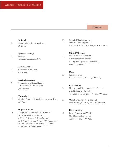
Journal of Medicine Vol 4 - Amrita Institute of Medical Sciences and ...
Journal of Medicine Vol 4 - Amrita Institute of Medical Sciences and ...
Journal of Medicine Vol 4 - Amrita Institute of Medical Sciences and ...

You also want an ePaper? Increase the reach of your titles
YUMPU automatically turns print PDFs into web optimized ePapers that Google loves.
<strong>Amrita</strong> <strong>Journal</strong> <strong>of</strong> <strong>Medicine</strong><br />
CONTENTS<br />
Editorial<br />
2 Commercialization <strong>of</strong> <strong>Medicine</strong><br />
H. Kumar<br />
23 Extended Maxillectomy by<br />
Transm<strong>and</strong>ibular Approach<br />
S.S. Chatni, R. Sharan, S. Iyer, M.A. Kuriakose<br />
Spiritual Message<br />
3 Patience<br />
Swami Paramatman<strong>and</strong>a Puri<br />
Review Article<br />
5 Carcinoma <strong>of</strong> the Ovary<br />
Chithrathara<br />
Practical Approach<br />
9 Comprehensive Rehabilitation:<br />
A New Dawn for the Disabled<br />
J.N. Panicker<br />
Viewpoint<br />
13 Caution! Counterfeit <strong>Medicine</strong>s are on the Rise<br />
B.P. Rao<br />
Clinical Whodunit<br />
29 Nasal Cure for a Myopathy –<br />
A Neuroendocrine Puzzle?<br />
G. Siby, U.K. Syam, A. An<strong>and</strong>kumar,<br />
Hiran, G. Aneesh<br />
Quiz<br />
33 Radiology Quiz<br />
Ch<strong>and</strong>ramohan, R. Kannan, S. Moorthy<br />
Case Reports<br />
34 Rhinocerebral Mucormycosis in a Patient<br />
with Diabetic Nephropathy<br />
A. Mathew, J.C. Varghese, P. Nair, V.N. Unni<br />
37 Mutiple Endocrine Neoplasia – 2B<br />
N.M. Detroja, B. Nisha, A.G. Unnikrishnan<br />
Original Articles<br />
18 Analysis <strong>of</strong> GSTM1 <strong>and</strong> CYP1A1 Genes<br />
Tropical Chronic Pancreatitis<br />
A.G. Unnikrishnan, S. Ramach<strong>and</strong>ran,<br />
M.R. Pillai, H. Kumar, P. Nair, R.V. Jayakumar,<br />
U. Guruprasad, R. N<strong>and</strong>akumar, T. Joseph,<br />
I. Hariharan, V. Balakrishnan<br />
Literature Scan<br />
41 Cases, Evidence <strong>and</strong> Verdicts -<br />
The Glitazone Controversy<br />
T. Roy, T. Rony, A.N. Babu<br />
1
<strong>Amrita</strong> <strong>Journal</strong> <strong>of</strong> <strong>Medicine</strong><br />
EDITORIAL<br />
Editorial Board<br />
Patrons<br />
Swami <strong>Amrita</strong> Swaroopan<strong>and</strong>a Puri<br />
Dr. Prem Nair<br />
Mr. Ron Gottsegen<br />
Dr. D.M. Vasudevan<br />
Chief Editor<br />
Dr. Harish Kumar<br />
Associate Editors<br />
Dr. An<strong>and</strong> Kumar<br />
Dr. Sudhindran<br />
Editorial Assistance<br />
Dr. V.P. Praveen<br />
Dr. P. Gowri<br />
Editorial Board Members<br />
Dr. V. Balakrishnan<br />
Mr. Sudhakar Jayaram<br />
. Dr. Dilip Panikar<br />
Dr. Prakash Kamath<br />
Dr. S.K. Nair<br />
Dr. Vaidyanathan<br />
Dr. M.G.K. Pillai<br />
Dr. Subramanian<br />
Dr. Kanthaswami<br />
Dr. Abraham Kuriakose<br />
Publications Officer<br />
Mrs. Jaya Sudhir Maharshi<br />
Visualiser<br />
Mr. Biju Chembalayat<br />
Cover Illustrations<br />
Mr. Biju Chembalayat<br />
Mrs. Rajalakshmi Remesh<br />
Dept.<strong>of</strong> Graphics, AIMS<br />
Commercialization <strong>of</strong><br />
<strong>Medicine</strong><br />
Harish Kumar<br />
People come to see a doctor when they are feeling ill,<br />
either physically or mentally. The duty <strong>of</strong> a physician is<br />
to then prescribe the necessary remedies to restore the<br />
equilibrium <strong>and</strong> make the person feel better. It is a sacred<br />
duty – the aim <strong>of</strong> which is to take an individual<br />
from a state <strong>of</strong> “ashanti” to “shanti”. Once he is healed<br />
or better the patient will gratefully compensate the physician<br />
in cash or kind, as per his means. The physician<br />
gains not only monetarily, but also is enriched by the<br />
gratefulness <strong>and</strong> good wishes <strong>of</strong> the diseased person <strong>and</strong><br />
the family <strong>of</strong> those whom he has healed. The whole focus<br />
<strong>of</strong> the patient-physician interaction is that the patient<br />
must feel better, <strong>and</strong> there is an attitude <strong>of</strong> surrender <strong>and</strong><br />
trust on the part <strong>of</strong> the patient.<br />
But in today’s world, medicine has become commercialized.<br />
Pharmaceutical <strong>and</strong> other commercial companies<br />
are interested only in pr<strong>of</strong>its <strong>and</strong> they can generate, <strong>and</strong><br />
sustain these pr<strong>of</strong>its only with the active support <strong>of</strong> the<br />
doctor community. It is a well known, but not <strong>of</strong>ten admitted,<br />
reality that commercial interests frequently<br />
dominate prescription patterns not only in India, but also<br />
all over the world. This has caused a subtle shift in the<br />
attitude <strong>of</strong> many doctors whose primary focus is now on<br />
the commercial gains from the pr<strong>of</strong>ession <strong>and</strong> the service<br />
provided to the patient has become secondary. The main<br />
casualty in this new commercial scenario is the doctorpatient<br />
trust. Patients are increasingly suspicious <strong>of</strong> the<br />
intentions <strong>of</strong> doctors <strong>and</strong> ever-increasing medico-legal<br />
litigations are the outward symptom <strong>of</strong> this malaise. The<br />
‘trust’ <strong>and</strong> ‘surrender’ have been replaced by a business<br />
like transaction, which takes away the sacredness <strong>of</strong> healing.<br />
The other side <strong>of</strong> the coin is the benefit that has<br />
accrued to the science <strong>of</strong> medicine due to this commercialization.<br />
Enormous sums <strong>of</strong> money are being pumped<br />
into research in various areas <strong>of</strong> medicine by commercial<br />
organizations <strong>and</strong> our treatment <strong>and</strong> investigation modalities<br />
are improving at an ever-increasing pace.<br />
The majority <strong>of</strong> physicians are not commercial <strong>and</strong><br />
there is still hope if efforts are made to better physicianpatient<br />
bonding. But unfortunately, today the success <strong>of</strong><br />
a doctor is measured by the amount <strong>of</strong> material wealth<br />
he has accumulated from the practice <strong>of</strong> his pr<strong>of</strong>ession.<br />
If this trend continues then doctors <strong>and</strong> commercial organizations<br />
doing business in the medical field may<br />
become richer but the pr<strong>of</strong>ession <strong>and</strong> its public image<br />
are becoming poorer by the day.<br />
2
<strong>Amrita</strong> <strong>Journal</strong> <strong>of</strong> <strong>Medicine</strong><br />
Patience<br />
Swami Paramatman<strong>and</strong>a Puri<br />
SPIRITUAL MESSAGE<br />
Working in the healthcare field, one has a great deal<br />
<strong>of</strong> interaction with other people – doctors, nurses, patients<br />
<strong>and</strong> their families, administrators <strong>and</strong> so on, <strong>and</strong><br />
therefore, one has vast opportunities for self-improvement.<br />
Amma says that life is like a school <strong>and</strong> every situation<br />
gives us a chance to learn about our self <strong>and</strong> the world in<br />
which we live. We should always try to remain conscious<br />
<strong>of</strong> that fact as we travel through life. One <strong>of</strong> the essential<br />
qualities that we need to develop if we want our life to<br />
be successful is patience, <strong>and</strong> there is no shortage <strong>of</strong> opportunities<br />
for us to do so in the healthcare setting.<br />
Unfortunately, patience is hard to come by in those<br />
<strong>of</strong> us who have been raised in an increasingly comfortable<br />
<strong>and</strong> technological world. Modern life seems to be<br />
about speed, enjoyment <strong>and</strong> comfort. We go to great<br />
lengths to make sure that we do not have to undergo any<br />
inconvenience, delay or boredom. We find waiting <strong>and</strong><br />
adjusting with other people <strong>and</strong> circumstances to be very<br />
painful. Our mind is like a child’s mind – impatient <strong>and</strong><br />
impetuous. And all this impatience leads to uncontrollable<br />
anger <strong>and</strong> pain.<br />
We have heard from elders that when life was not so<br />
fast <strong>and</strong> comfortable, people were a lot more patient <strong>and</strong><br />
a lot less angry. They were also willing to do a lot more<br />
for refining their personality <strong>and</strong> for making spiritual<br />
progress. Apparently, the ancient culture <strong>of</strong> India still<br />
has a few things to teach us!<br />
Anger can be seen everyday at home, in school, at the<br />
workplace, when shopping, <strong>and</strong> even on the road. We<br />
have all heard <strong>of</strong> road rage. This is an instance <strong>of</strong> an<br />
abnormal lack <strong>of</strong> patience resulting in intense anger. We<br />
may not feel that we are an angry person, but a naturally<br />
self-controlled person is a rarity. Even so-called spiritual<br />
people can have a lot <strong>of</strong> hidden anger. Sometimes, out <strong>of</strong><br />
compassion, mahatmas will show us how much anger<br />
we really have so that we can become aware <strong>of</strong> it <strong>and</strong> try<br />
to improve.<br />
There once was a sadhu named Suthra who<br />
was an exceptionally bold <strong>and</strong> compassionate person.<br />
One day, a friend came to him <strong>and</strong> said, “A famous holy<br />
man who is very much revered by everybody in this<br />
neighbourhood has come. Let us go <strong>and</strong> see him.”<br />
Suthra agreed, <strong>and</strong> they walked to the holy man’s hut,<br />
greeting him upon their arrival by bowing down. The<br />
holy man invited them to be seated. After a few minutes<br />
silence, Suthra asked the holy man, “Have you any fire? I<br />
need some.”<br />
The holy man said, “No, I have no fire here at present.”<br />
Again, after a few minutes <strong>of</strong> silence, Suthra again<br />
asked the holy man, “O sadhu, have you any fire?”<br />
“I have already told you that I have none,” said the<br />
holy man, slightly annoyed.<br />
But this did not seem to make any impression on<br />
Suthra. He again asked the man, “Sir, I have great need <strong>of</strong><br />
some fire, so let me have some.”<br />
At this, the holy man became really irritated <strong>and</strong> replied<br />
with great heat, “O foolish man! Please stop asking<br />
me for fire! Can’t you underst<strong>and</strong> what I say? I have told<br />
you three times that I do not have any fire. Isn’t that<br />
enough? Or will you go on repeating the same stupid<br />
question over <strong>and</strong> over again?”<br />
Suthra kept silent while the holy man scolded him.<br />
Then he said, “Brother, I really need some fire. Are you<br />
absolutely sure that you don’t have any?”<br />
Now thoroughly enraged, the sadhu stood up <strong>and</strong><br />
picked up a stick. He beat Suthra with it until the stick<br />
broke. Suthra then smiled <strong>and</strong> said, “My question is now<br />
answered. I saw <strong>and</strong> smelled some smoke when I entered<br />
your presence, <strong>and</strong> so I knew that there was fire here.<br />
And now, as anyone can see, the fire has blazed up <strong>and</strong><br />
is burning with angry flames. Yet strangely enough, you<br />
still maintain that you have none.”<br />
Underst<strong>and</strong>ing now that Suthra was referring to his<br />
anger, the sadhu immediately calmed down. Hanging his<br />
head in shame, he said in a humble voice, “Thank you,<br />
Mahatmaji, for your lesson. I will take it to heart <strong>and</strong> try<br />
to mend my ways.”<br />
In order to purify our mind, we must first know what<br />
is there in our mind to purify. This is the function <strong>of</strong><br />
divine grace. A mahatma will let us know what is inside<br />
us either through his/her mere presence or through our<br />
circumstances. Amma knows that we are impatient <strong>and</strong><br />
have a lot <strong>of</strong> anger as well as pride. She uses “crowds” as<br />
one <strong>of</strong> the means to purify our minds. Due to the huge<br />
crowds that come to see Amma, we must wait a long<br />
time in order to have her darshan. That itself gives us a<br />
chance to develop patience, humility <strong>and</strong> devotion.<br />
Amma does not just make us be patient, but is herself<br />
the very image <strong>of</strong> patience. Look how she patiently sits<br />
until the last person has had her darshan. Many <strong>of</strong> us<br />
have seen her begin darshan at 8:00pm <strong>and</strong> continue until<br />
the next day at noon, not getting up even for a minute.<br />
Who can or will do such a thing, giving up sleep <strong>and</strong><br />
3
<strong>Amrita</strong> <strong>Journal</strong> <strong>of</strong> <strong>Medicine</strong><br />
Patience<br />
comfort day after day? Who else can listen to problems<br />
for hours on end? Who can sincerely smile continuously?<br />
Who else can travel around India <strong>and</strong> the world for 8<br />
months in the year, keeping up an impossible schedule?<br />
If we can’t learn a little patience from such a being, we<br />
are really a hopeless case. One may search for a long<br />
time, but one would be hard put to find a role model<br />
such as Amma who practices what she preaches.<br />
Amma tells <strong>of</strong> seeing people waiting all day in line at<br />
the government hospital in order to see a doctor. But for<br />
making a bit <strong>of</strong> spiritual progress or self-improvement,<br />
most people have no patience. Many <strong>of</strong> us want everything<br />
right now. People come to Amma <strong>and</strong> insist that<br />
they should have darshan immediately or that their problems<br />
should disappear without the least delay! They have<br />
no time to hang around. With such a lack <strong>of</strong> patience,<br />
what benefit will they get from Amma’s company?<br />
In relation to the Highest Truth, without patience, we<br />
cannot gain the direct experience <strong>of</strong> our true self, the<br />
Atma. The ever-bubbling restless mind prevents us from<br />
doing so. The state that Amma demonstrates in her own<br />
life <strong>and</strong> which she wants us to also experience is one <strong>of</strong><br />
perfect stillness, peace, shanti.<br />
Instead <strong>of</strong> getting angry whenever we must wait or<br />
when we meet with resistance, let us use such situations<br />
to practice patience. Let us think <strong>of</strong> Amma’s example <strong>of</strong><br />
natural patience <strong>and</strong> try to follow in her footsteps. Then<br />
we will find every difficulty becoming an opportunity<br />
for spiritual practice. When we naturally become as patient<br />
as Amma, then we have reached a lasting goal. We<br />
will be happy <strong>and</strong> have great satisfaction, <strong>and</strong> we will<br />
make others happy too.<br />
4
<strong>Amrita</strong> <strong>Journal</strong> <strong>of</strong> <strong>Medicine</strong><br />
Carcinoma <strong>of</strong> the Ovary<br />
Chithrathara<br />
REVIEW ARTICLE<br />
ABSTRACT<br />
In India, ovarian carcinoma, the<br />
second most common gynecological<br />
malignancy, is showing a rising trend.<br />
Epithelial ovarian carcinoma (EOC)<br />
forms about 90% <strong>of</strong> malignant ovarian<br />
tumors. The retrospective data <strong>and</strong><br />
the meta-analysis favoured upfront<br />
surgical cytoreduction. Neoadjuvant<br />
chemotherapy (NAC) <strong>and</strong> interval surgery<br />
has come into practice to reduce<br />
the incidence <strong>of</strong> non-therapeutic laparotomies.<br />
Sound clinical judgement<br />
<strong>and</strong> CT scan can reliably predict the<br />
inoperability <strong>and</strong> helps to select patients<br />
for NAC. Any form <strong>of</strong> minimally<br />
invasive surgery is to be used with caution.<br />
Literature also projects the<br />
surgeon as an independent prognostic<br />
factor <strong>and</strong> the surgery is best contemplated<br />
in centers <strong>of</strong> excellence. Early<br />
disease requires comprehensive surgical<br />
staging. Borderline serous tumors<br />
require counselling for restaging versus<br />
surveillance. Changing the cycling<br />
<strong>of</strong> drugs, targeted <strong>and</strong> tailored therapy<br />
are promising future treatment options.<br />
INTRODUCTION<br />
Ovarian cancer is the second most<br />
common gynecological cancer. Epithelial<br />
ovarian cancers (EOC) constitute<br />
90% <strong>of</strong> malignant ovarian tumors.<br />
Most unfortunately 3/4 th <strong>of</strong> the patients<br />
are diagnosed at an advanced stage with<br />
disseminated intra-abdominal disease.<br />
Even though complete remission can<br />
be achieved in 90% <strong>of</strong> patients, relapse<br />
is the rule. Relapses are salvaged<br />
with repeated cycles <strong>of</strong> chemotherapy<br />
<strong>and</strong> surgery is used only in selected<br />
cases. The longer the disease free interval,<br />
the better the response to further<br />
chemotherapy. The discovery <strong>of</strong> newer<br />
<strong>and</strong> newer chemotherapy molecules<br />
Dept. <strong>of</strong> Surgical Oncology, AIMS, Kochi.<br />
has converted ovarian carcinoma to a<br />
‘chronic disease’. The cachectic patient<br />
finally succumbs to death due to uncontrolled<br />
intra-abdominal disease<br />
with multiple level intestinal obstructions.<br />
INCIDENCE<br />
EOC is a disease <strong>of</strong> senescent ovary.<br />
The age adjusted incidence rates increase<br />
from 15/100,000 to 57/100,000<br />
during the 8 th decade. Age adjusted<br />
incidence rates in Kerala are little lower<br />
than western countries. The population-based<br />
data available from<br />
Thiruvananthapuram showed incidence<br />
rates <strong>of</strong> 5.3/100,000 in the urban<br />
area <strong>and</strong> 3.5/100,000 in rural area.<br />
Hospital based cancer registry shows<br />
that it is 6/100 women cancers.<br />
HEREDITARY OVARIAN<br />
CARCINOMA<br />
5-10% <strong>of</strong> ovarian cancers are linked<br />
to genetic predisposition. The breastovarian<br />
cancer syndrome accounts for<br />
approximately 90% <strong>of</strong> hereditary ovarian<br />
cancer cases <strong>and</strong> is frequently<br />
associated with BRCA1 & BRCA2. The<br />
hereditary non-polyposis colorectal<br />
cancer accounts for 5% <strong>of</strong> cases. Firm<br />
conclusions could not be drawn on the<br />
benefit <strong>of</strong> genetic testing <strong>and</strong> early<br />
detection 1 . Presently the most effective<br />
strategy in the prevention <strong>of</strong><br />
ovarian carcinoma is prophylactic bilateral<br />
oopherectomy, which also<br />
causes a substantial reduction in breast<br />
cancer risk 2 .<br />
MANAGEMENT OF<br />
EPITHELIAL OVARIAN<br />
TUMORS<br />
Aggressive surgical cytoreduction<br />
<strong>and</strong> platinum based chemotherapy<br />
forms the mainstay <strong>of</strong> management <strong>of</strong><br />
EOC. Even though there are no r<strong>and</strong>omized<br />
trials, literature is replete<br />
with retrospective data, which gives<br />
ample evidence for the use <strong>of</strong> upfront<br />
cytoreductive surgery. The results <strong>of</strong><br />
two meta-analysis also favour primary<br />
surgery. It gives accurate initial staging<br />
<strong>and</strong> there is theoretical advantage<br />
<strong>of</strong> better action <strong>of</strong> chemotherapy based<br />
on Gompertzian model. Removal <strong>of</strong><br />
the tumor in total <strong>and</strong> a comprehensive<br />
surgical staging is indicated in<br />
early stage ovarian cancer. Keyhole <strong>and</strong><br />
vaginal surgery, limited incisions, etc.<br />
are to be avoided especially in suspected<br />
early stage ovarian malignancy<br />
since these compromise cure <strong>and</strong> endanger<br />
the life <strong>of</strong> the woman.<br />
The prognostic determinants include<br />
stage, histological subtype <strong>and</strong><br />
grade, medical co-morbidities, performance<br />
status, response to<br />
chemotherapy <strong>and</strong> post-operative residual<br />
disease. The only consistent<br />
alterable variant is post-operative residual<br />
disease. So it is the first surgery<br />
that determines the outcome. Many are<br />
<strong>of</strong> the opinion that post-operative residual<br />
disease is a reflection <strong>of</strong><br />
biological aggressiveness <strong>of</strong> the tumor.<br />
But Eisenkope <strong>and</strong> others have demonstrated<br />
that the surgical effort <strong>and</strong><br />
accompanying survival is surgeon dependent<br />
3 . The approach <strong>and</strong> thus the<br />
result can vary among surgeons depending<br />
on their perspective <strong>and</strong><br />
philosophy 4 .<br />
The GOG studies give important<br />
insights into the impact <strong>of</strong><br />
cytoreductive surgery in ovarian carcinoma.<br />
They defined the optimal<br />
debulking surgery as the removal <strong>of</strong> all<br />
gross or visible tumor. When patient<br />
was left with no residual tumor a progression<br />
free survival <strong>of</strong> 40 months was<br />
observed, compared to 20 months in<br />
5
<strong>Amrita</strong> <strong>Journal</strong> <strong>of</strong> <strong>Medicine</strong><br />
Carcinoma <strong>of</strong> the Ovary<br />
patients with residual disease <strong>of</strong> less than 1 cm. It is<br />
shown that a possibly superior chemotherapeutic regimen<br />
containing taxanes cannot compensate for the tumor<br />
left behind after surgery. (19 JSO)<br />
The maximum goal <strong>of</strong> cytoreductive (debulking) surgery<br />
should be the complete removal <strong>of</strong> all visible disease<br />
<strong>and</strong> the minimum is to reduce the tumor to less than 1<br />
cm (optimal debulking). To meet this target one may <strong>of</strong>ten<br />
have to go for intestinal resections, rarely splenectomy,<br />
peritoneal excisions including diaphragmatic peritoneum<br />
<strong>and</strong> genito-urinary tract resections. Surgical procedures,<br />
which impair quality <strong>of</strong> life, for e.g. ostomies, should be<br />
avoided as far as possible. Precise <strong>and</strong> more expeditious<br />
removal <strong>of</strong> widespread peritoneal implants including diaphragmatic<br />
implants is facilitated by the use <strong>of</strong> Cavitron<br />
ultrasonic surgical aspirator (CUSA) <strong>and</strong> argon beam laser.<br />
Other modalities used are carbon dioxide laser <strong>and</strong><br />
loop electrosurgical excision procedures 5,6 . However in<br />
best h<strong>and</strong>s <strong>and</strong> best centers, many times the initial laparotomy<br />
turns out to be non therapeutic. The mortality<br />
<strong>and</strong> the morbidity due to haemorrhage in the presence <strong>of</strong><br />
friable growth may be overwhelming. In order to circumvent<br />
this problem <strong>and</strong> in view <strong>of</strong> the excellent response<br />
<strong>of</strong> tumor observed even after the futile initial laparotomy,<br />
the concept <strong>of</strong> interval laparotomy has come into practice.<br />
There is no r<strong>and</strong>omized trial data published in favour<br />
<strong>of</strong> interval cytoreduction. However several phase II <strong>and</strong><br />
retrospective data showed 3-year survival rate <strong>of</strong> 50%,<br />
which is comparable to optimal primary debulking. With<br />
neoadjuvant chemotherapy (NAC) 50% complete remission<br />
rate is reported <strong>and</strong> the complete / optimal resection<br />
is possible in 75% <strong>of</strong> cases. Our own data shows that<br />
the rate <strong>of</strong> optimal cytoreduction is 50% for primary surgery<br />
<strong>and</strong> 88.6% for patients who received NAC 7 . However<br />
it has to be clear that good reduction is not equivalent to<br />
initial small volume disease.<br />
For interval debulking also, just like primary surgery,<br />
one has to be prepared for any extent <strong>of</strong> surgery to completely<br />
remove the disease to microscopic level. So the<br />
minimum prerequisites for an ovarian cancer laparotomy<br />
are adequate infrastructure with adequate theatre facilities,<br />
dedicated team with adequate expertise <strong>and</strong> oncology<br />
concept. It has to be ascertained that no facility should<br />
<strong>of</strong>fer surgery for patients with ovarian cancer if adequate<br />
st<strong>and</strong>ards <strong>of</strong> care cannot be met with.<br />
Meticulous <strong>and</strong> systematic pre-operative evaluation<br />
has to be done to assess the operability <strong>and</strong> to avoid a<br />
non-therapeutic laparotomy.<br />
THE PREDICTORS OF SURGICAL<br />
OUTCOME ARE:<br />
1. Clinical evaluation: age above 50 years, gross ascites<br />
<strong>and</strong> fixed large pelvic masses are unfavourable<br />
clinical factors.<br />
2. Abdominal ultrasound: large volume ascites <strong>and</strong><br />
hydroureteronephrosis increase the inoperability rates.<br />
3. CT scan: Bristow 2000 selected 13 radiographic features<br />
along with performance status 8 . The important<br />
radiographic criteria considered are number <strong>of</strong> metastasis,<br />
peritoneal thickening, large ascites, large<br />
metastatic deposits on diaphram, suprarenal nodes,<br />
etc. Each parameter was assigned a numerical value.<br />
They reported that with a predictive index > 4, the<br />
specificity was 85% (inappropriate unexploration<br />
15%) <strong>and</strong> the sensitivity approached 100% (unnecessary<br />
exploration 0%). According to Dowdy, diffuse<br />
peritoneal thickening <strong>and</strong> large volume ascites independently<br />
predicted surgical outcome.<br />
4. Diagnostic laparoscopy:<br />
Inoperability criteria in advanced ovarian cancer:-<br />
Absolute<br />
a. Stage 1V disease or<br />
b. Metastasis <strong>of</strong> more than 1 cm at sites where optimal<br />
cytoreduction is not possible, e.g. at porta<br />
hepatis, around superior mesenteric artery, etc.<br />
Relative<br />
a. Uncountable (100) peritoneal metastases<br />
b. Estimated total metastatic load <strong>of</strong> >1000gm (both<br />
intra <strong>and</strong> extraperitoneal)<br />
c. Presence <strong>of</strong> more than 10gms peritoneal metastatic<br />
plaques<br />
d. Large volume ascites (5L)<br />
e. Those with performance status 2 or 3<br />
The time interval between diagnostic laparoscopy <strong>and</strong><br />
definitive surgery or chemotherapy should be as short as<br />
possible.<br />
ROLE OF RETROPERITONEAL LYMPH<br />
NODE DISSECTION (RPLND)<br />
Lymph nodal involvement in ovarian cancer is 20-<br />
40% (40 JSO) in apparently early disease to as high as<br />
70-80% in advanced disease. Early stages LND is recommended<br />
as a part <strong>of</strong> staging (some studies show improved<br />
survival also) <strong>and</strong> in advanced disease involved nodes<br />
are removed to attain R0 – R1 status.<br />
ROLE OF HYSTERECTOMY<br />
No studies so far tested the benefit <strong>of</strong> uninvolved<br />
uterus 9 . However, uterus should not be removed in instances<br />
<strong>of</strong> suboptimal tumor removal, since in the event<br />
<strong>of</strong> a subsequent recurrence, the tumor will directly invade<br />
the bladder.<br />
6
<strong>Amrita</strong> <strong>Journal</strong> <strong>of</strong> <strong>Medicine</strong><br />
CURRENT CONCEPTS IN BORDERLINE<br />
SEROUS TUMORS<br />
Invasive serous carcinoma <strong>of</strong> ovary is divided into lowgrade<br />
<strong>and</strong> high-grade tumors by the 2-tier system <strong>of</strong> grading<br />
designed by the M D Anderson Cancer Center (2004).<br />
The term microinvasive serous carcinoma was coined by<br />
John Hopkins University <strong>and</strong> is subclassified into<br />
noninvasive <strong>and</strong> invasive. Currently, non-invasive<br />
micropapillary tumor is considered as a variant <strong>of</strong> serous<br />
tumor <strong>of</strong> low malignant potential (STLMP) <strong>and</strong> invasive<br />
is synonymous with low-grade serous tumour (LGSC) 10 .<br />
Data suggests that STLMP <strong>and</strong> LGSC share a common<br />
pathogenesis whereas the high-grade serous carcinoma<br />
(HGSC) has a distinct <strong>and</strong> different pathogenesis.<br />
MOLECULAR EVIDENCE<br />
K-ras <strong>and</strong> BRAF mutations are detected in 1/3 <strong>of</strong><br />
STLMP <strong>and</strong> LGSC <strong>and</strong> are mutually exclusive. A k-ras<br />
mutation seen in STLMP is rare in HGSC <strong>and</strong> the BRAF<br />
mutation is non-existent. CHEK2 is a protein kinase that<br />
is involved in cell cycle arrest. Genotyping revealed a<br />
strong positive association between the CHEK2 1157T<br />
missense variant <strong>and</strong> ovarian cystadenomas, STLMP <strong>and</strong><br />
LGSC. Such association is not found in HGSC. On the<br />
other h<strong>and</strong> p53 mutation occur frequently in HGSC, but<br />
rarely seen in STLMP <strong>and</strong> LGSC.<br />
CLINICAL EVIDENCE<br />
30% <strong>of</strong> STLMP is associated with implants, more<br />
common in micropapillary type. In70-80% <strong>of</strong> patients<br />
with STLMP who relapse, the histology <strong>of</strong> recurrence is<br />
LGSC.<br />
It is a great dilemma for the gynecologic oncologist<br />
whether to do a restaging, when a patient with STLMP is<br />
referred after removal <strong>of</strong> only the primary tumor. This is<br />
a controversial point; current recommendation is to go<br />
for a comprehensive counselling for restaging versus surveillance<br />
11,12 . But a concomitant surgical staging along<br />
with primary tumor removal would be preferable in ideal<br />
circumstances. This can assess the prognosis <strong>and</strong> obviate<br />
the need for restaging if it turns out to be invasive carcinoma<br />
on final paraffin sections.<br />
CHEMOTHERAPY OF EPITHELIAL<br />
OVARIAN CANCER (EOC)<br />
Early stage disease:<br />
Early stage disease is classified as low-risk or highrisk,<br />
based on prognostic factors. Low-risk are patients<br />
with stage 1A/1B, grade1 with non-clear cell histology.<br />
This group has a 5-year survival rate <strong>of</strong> more than 90%<br />
<strong>and</strong> can be left alone without any adjuvant treatment.<br />
High-risk factors are stage 1C/11, grade 2 & 3, clear<br />
cell histology <strong>and</strong> dense adhesions. High-risk patients have<br />
a relapse rate <strong>of</strong> about 40-50 % <strong>and</strong> warrants adjuvant<br />
treatment. Paclitaxel <strong>and</strong> carboplatin combination given<br />
every 3-4weeks for 6 cycles is the st<strong>and</strong>ard practice at<br />
this time.<br />
CHEMOTHERAPY OF ADVANCED EOC<br />
Consensus exists as to the use <strong>of</strong> platinum based chemotherapy<br />
in this situation. Though controversies exist<br />
with regard to which platinum, which taxane, whether<br />
anthracycline is to be added or not, etc., the st<strong>and</strong>ard<br />
practice at present is 6cycles <strong>of</strong> paclitaxel (175mg/m2<br />
over 3 hours) <strong>and</strong> carboplatin (AUC 6)<br />
CHEMOTHERAPY IN RELAPSED OVARIAN<br />
CANCER<br />
Treatment options depend on whether it is a platinum<br />
sensitive relapse (platinum free interval more than<br />
6 months) or a platinum resistant/refractory relapse. Platinum<br />
sensitive patients can still respond to platinum based<br />
chemotherapy combinations. Judicious use <strong>of</strong> drugs to<br />
balance the potential benefits, toxicity <strong>and</strong> patient acceptance<br />
need emphasis in the treatment <strong>of</strong> relapsed<br />
ovarian carcinoma. Though most <strong>of</strong> the drugs claim a<br />
response rate <strong>of</strong> 20-30%, liposomal doxorubicin st<strong>and</strong>s<br />
as one <strong>of</strong> the preferred options in the treatment <strong>of</strong> relapsed<br />
EOC. Topotican, docetaxel, gemcitabine, oral<br />
etoposide / altretamine are the most commonly used other<br />
second line drugs.<br />
THE FUTURE<br />
Targeted <strong>and</strong> tailored therapy is likely to play an important<br />
role in the management <strong>of</strong> EOC in the future.<br />
Blockage <strong>of</strong> epidermal growth factor pathways with<br />
Erlotinib, use <strong>of</strong> antiangiogenesis drugs like bevazucimab<br />
etc. is gaining momentum. Conventional drugs like<br />
paclitaxel used as weekly schedule was found to be welltolerated<br />
<strong>and</strong> active in patients with platinum resistant<br />
disease, opening up innovative ways <strong>of</strong> administering<br />
currently available conventional drugs.<br />
CONCLUSION<br />
Surgery <strong>and</strong> chemotherapy play important role in the<br />
management <strong>of</strong> ovarian carcinoma.<br />
Upfront surgery/chemotherapy is decided mainly on<br />
clinical judgement <strong>and</strong> CT criterias. Currently, there is<br />
limited role for laparoscopy in the management <strong>of</strong> ovarian<br />
tumors. Lack <strong>of</strong> adequate sensitivity <strong>and</strong> specificity<br />
<strong>of</strong> currently used methods curtail early detection, so prevention<br />
with preservation <strong>of</strong> ovaries needs further studies.<br />
Tailored <strong>and</strong> targeted therapy can replace the current st<strong>and</strong>ard<br />
<strong>of</strong> care in future.<br />
7
<strong>Amrita</strong> <strong>Journal</strong> <strong>of</strong> <strong>Medicine</strong><br />
Carcinoma <strong>of</strong> the Ovary<br />
REFERENCES<br />
1. Kauf ND, Satagopan JM, Robson ME, et al. Risk reducing<br />
salpingo-oopherectomy in woman with a BRCA1 or BRCA2<br />
mutation. N Eng J Med 2002;346:609.<br />
2. Grann VR, Jacobson JS, Thomason D, et al. Effect <strong>of</strong> prevention<br />
strategies on survival <strong>and</strong> quality adjusted survival <strong>of</strong> women<br />
with BRCA1/2 mutations; an updated decision analysis. J Clin<br />
Oncol 2002;20:2520.<br />
3. Eisenkop SM, Nalick RM, _ang SJ, et al. peritoneal implant elimination<br />
during cytoreductive surgery for ovarian cancer: impact<br />
on survival. Gynecol Oncol 1993;51:224-9.<br />
4. Podratz KC, Aletti G, Cliby WA. Advanced ovarian cancer surgical<br />
management: Indian J <strong>of</strong> Gynecol Oncol 2006; 6 suppl<br />
1:11-2.<br />
5. Patsner B. Carbon dioxide laser vaporization <strong>of</strong> diaphragmatic<br />
metastases for cytoreduction <strong>of</strong> ovarian epithelial tumor. Gynecol<br />
Oncol 1990;76:724-7.<br />
6. Fanning J, Hilgers RD. Loop electrosurgical excision procedure<br />
for intensified cytoreduction <strong>of</strong> ovarian cancer. Gynecol<br />
Oncol1995;57:188-90.<br />
7. Sharma S, Vijayakumar K, Chitrathara K, et al. Neoadjuvant<br />
chemotherapy in advanced epithelial ovarian cancer: a retrospective<br />
study. Indian J <strong>of</strong> <strong>Medical</strong> <strong>and</strong> Paediatric Oncology<br />
2007;28 No 1:7-13.<br />
8. Bristow RE, Duska LR, Lambrou NC, et al. A model for predicting<br />
surgical outcome in patients with advanced ovarian carcinoma<br />
using computed tomography. Cancer 2000;89:1532-40.<br />
9. Munstedt K, Franke FE. Role <strong>of</strong> primary surgery in advanced<br />
ovarian cancer. Review. World J <strong>of</strong> Surgical Oncology<br />
2004;2:32.<br />
10. Burks R, Sherman M, Kurman R. Micropapillary serous carcinoma<br />
<strong>of</strong> the ovary. A distinctive low-grade carcinoma related to<br />
serous borderline tumors. Am J Surg Pathol 1996;20:1319-30.<br />
11. Lin PS, Gershenson DM, Bevers MW, et al. The current status <strong>of</strong><br />
surgical staging <strong>of</strong> ovarian serous borderline tumors. Cancer<br />
1999 Feb 15;85(4):905-11.<br />
12. Fauvet R, Boccara J, Dufournet C, et al. Restaging surgery for<br />
women with borderline ovarian tumors: results <strong>of</strong> a French<br />
multicenter study. Cancer 2004 Mar 15;100(6):1145-51.<br />
8
<strong>Amrita</strong> <strong>Journal</strong> <strong>of</strong> <strong>Medicine</strong><br />
PRACTICAL APPROACH<br />
Comprehensive Rehabilitation:<br />
A New Dawn for the Disabled<br />
J.N. Panicker<br />
CASE VIGNETTE<br />
A previously healthy 25 year-old<br />
manual worker, the sole breadwinner<br />
<strong>of</strong> a family, falls from the third floor<br />
<strong>of</strong> a building at a construction site.<br />
Luckily, other than being a bit dazed,<br />
he has not suffered any head injuries.<br />
But he has severe back pain <strong>and</strong> is<br />
unable to move his legs. His lower<br />
limbs are flaccid <strong>and</strong> weak <strong>and</strong> he has<br />
a sensory level at the 12 th thoracic<br />
dermatome with loss <strong>of</strong> all sensations<br />
below. He has tenderness at the 9 th<br />
thoracic vertebra <strong>and</strong> also retention <strong>of</strong><br />
urine. X-ray <strong>and</strong> subsequently MRI<br />
confirm the fears <strong>of</strong> a fracture, showing<br />
burst fracture <strong>of</strong> the 9 th thoracic<br />
vertebra <strong>and</strong> spinal cord transection.<br />
He undergoes intensive therapy <strong>and</strong><br />
surgery for fracture site stabilization.<br />
Active management is complete <strong>and</strong><br />
he is now awaiting discharge- as a<br />
paraplegic.<br />
“WHAT NEXT?”<br />
During the initial days following<br />
a medical or surgical condition, patients<br />
are actively managed <strong>and</strong><br />
regularly briefed by treating doctors.<br />
Patients tend to assume that total cure<br />
is certain <strong>and</strong> will happen in the immediate<br />
future. However many times,<br />
at the time <strong>of</strong> discharge, they find<br />
themselves to be stabilized at a level<br />
<strong>of</strong> functioning that is far from the state<br />
<strong>of</strong> “normalcy” <strong>and</strong> “cure” that they<br />
had assumed. Unmet expectations<br />
lead to frustrations <strong>and</strong> patients start<br />
asking questions- “What next?”,<br />
“Where do I go for help now?” or<br />
“Who will help me?”. With time,<br />
the questions become more broad-<br />
Dept. <strong>of</strong> Neurology, AIMS, Kochi.<br />
“How do I face my people?”, “Will I<br />
be able to work?” or “Who will look<br />
after my family?”. Most <strong>of</strong> the time,<br />
the treating team <strong>of</strong> doctors has no<br />
concrete answers to these relevant yet<br />
complex questions. It is at this critical<br />
juncture that a comprehensive<br />
rehabilitation team should take over.<br />
REHABILITATION:<br />
STARTING THE PROCESS<br />
Rehabilitation is the active participation<br />
<strong>of</strong> the disabled person <strong>and</strong><br />
others to reduce the impact <strong>of</strong> disease<br />
<strong>and</strong> disability on daily life. It is a creative<br />
process by which all possible<br />
means are utilized to reduce the impact<br />
<strong>of</strong> disease by optimizing <strong>and</strong><br />
maximizing social participation. By<br />
this process, a disabled person is made<br />
into a differently abled person. Importantly,<br />
it is the third stage <strong>of</strong><br />
Fig.1: The Rehabilitation Team<br />
medical care after prevention <strong>and</strong> cure.<br />
Any patient who is left with disabilities<br />
following a medical or surgical<br />
ailment is a potential c<strong>and</strong>idate for<br />
entering a rehabilitation programme<br />
that is specific for his needs.<br />
The first step in rehabilitation is to<br />
identify the problems being faced by<br />
a patient which are consequent to his<br />
illness. This includes defining the dimensions<br />
<strong>of</strong> disablement. Impairment<br />
is the loss or abnormality <strong>of</strong> anatomical,<br />
physiological or psychological<br />
structure or function. Disability,<br />
which results from impairment, is the<br />
restriction or lack <strong>of</strong> ability to perform<br />
an activity in the manner or range considered<br />
normal. H<strong>and</strong>icap is the<br />
disadvantage for a given individual in<br />
his social context, resulting from an<br />
impairment or disability, which lim-<br />
9
<strong>Amrita</strong> <strong>Journal</strong> <strong>of</strong> <strong>Medicine</strong><br />
Comprehensive Rehabilitation: A New Dawn for the Disabled<br />
its or prevents the fulfillment <strong>of</strong> a role or participation in<br />
society that is considered normal (International Classification<br />
<strong>of</strong> Impairments Disabilities <strong>and</strong> H<strong>and</strong>icap, WHO<br />
1980). In the patient described in the case vignette, paraplegia<br />
<strong>and</strong> inability to pass urine are examples <strong>of</strong> impairment,<br />
difficulty to walk is his disability <strong>and</strong> inability to look<br />
after the family <strong>and</strong> earn a livelihood are likely to be his<br />
h<strong>and</strong>icaps.<br />
THE REHABILITATION TEAM<br />
Rehabilitation relies upon several disciplines that work<br />
together <strong>and</strong> cohesively to achieve aims <strong>and</strong> goals. Team<br />
effort is essential <strong>and</strong> the only criterion for becoming a<br />
member <strong>of</strong> the team is interest in contributing to the<br />
medicosocial rehabilitation <strong>of</strong> the patient. All team<br />
members are equal in position <strong>and</strong> the patient is the central<br />
figure (Fig.1). In institutional based rehabilitation,<br />
the members <strong>of</strong> the multidisciplinary team include:<br />
1. Doctor plays many roles in rehabilitation (Table 1).<br />
Doctors on the team may include the original treating<br />
physician such as neurologist, neurosurgeon,<br />
internist, pediatrician, geriatrician, orthopedic surgeon<br />
or plastic surgeon, who may be interested in<br />
continuing to care for the patient during the rehabilitation<br />
phase <strong>of</strong> treatment, <strong>and</strong>/or physiatrists, who<br />
have specialization in rehabilitation. In a<br />
multidisciplinary team, the doctor is generally the<br />
team leader <strong>and</strong> his most important function is perhaps<br />
to coordinate the entire rehabilitation efforts<br />
to ensure smooth <strong>and</strong> goal-directed progress.<br />
2. Physical therapist is responsible for strengthening<br />
muscle power, improving range <strong>of</strong> joint movements<br />
<strong>and</strong> reducing spasticity through a variety <strong>of</strong> exercises.<br />
He also plays an important role in training patients<br />
to improve sitting <strong>and</strong> st<strong>and</strong>ing balance, gait <strong>and</strong><br />
transfers from bed. He may use various physical<br />
modalities such as heat, cold, electrical stimulation,<br />
LASER <strong>and</strong> massage for pain relief. Pool based therapies<br />
(hydrotherapy) facilitates many <strong>of</strong> the functions<br />
<strong>of</strong> the therapist.<br />
3. Occupational therapist trains patients in maximizing<br />
self-care activities such as dressing, bathing, toilet<br />
<strong>and</strong> eating. He also trains patients to transfer from<br />
the bed, improve h<strong>and</strong> functions <strong>and</strong> explore various<br />
vocational skills. He evaluates the home <strong>of</strong> the<br />
patient <strong>and</strong> suggests modifications to make it barrier-free<br />
to facilitate movement <strong>of</strong> the disabled. He<br />
can advice regarding proper wheelchair prescription<br />
<strong>and</strong> also in driving skills.<br />
4. Prosthetist/Orthotist is responsible for designing, fabricating<br />
<strong>and</strong> fitting orthoses (caliper, splint) <strong>and</strong><br />
prostheses (artificial limb).<br />
Table 1: Role <strong>of</strong> the Doctor in Rehabilitation<br />
1. Clinical Assessment <strong>and</strong> Confirmation <strong>of</strong> the diagnosis<br />
2. Identification <strong>of</strong> the medical, surgical <strong>and</strong> rehabilitation-specific problems faced by the patient<br />
3. Evaluation <strong>of</strong> the extent <strong>and</strong> severity <strong>of</strong> disabilities<br />
4. Goal setting<br />
5. Liaison between various team members to facilitate a coordinated effort<br />
6. Planning physical, occupational therapy <strong>and</strong> types <strong>of</strong> calipers/splints<br />
7. Planning <strong>and</strong> carrying out relevant investigations: electrophysiology, radiology, urodynamic<br />
studies<br />
8. <strong>Medical</strong> management <strong>of</strong> spasticity, urinary bladder/bowel/sexual dysfunction, seizures, pain<br />
9. Planning specific measures for spasticity management such as botulinum toxin injections <strong>and</strong><br />
bacl<strong>of</strong>en pump implantation<br />
10. Surgical correction <strong>of</strong> deformities<br />
11. Liaison with other medical <strong>and</strong> surgical specialists<br />
10
<strong>Amrita</strong> <strong>Journal</strong> <strong>of</strong> <strong>Medicine</strong><br />
5. Psychologist plays a crucial role in assessing personality<br />
<strong>and</strong> behavioural problems that may impede the<br />
rehabilitation process. In brain-injured patients, he<br />
conducts detailed assessment <strong>of</strong> cognitive functions<br />
<strong>and</strong> can plan a formal cognitive retraining<br />
programme. The psychologist also plays an important<br />
role in counseling patients <strong>and</strong> their caregivers.<br />
6. Speech therapist assesses the types <strong>and</strong> severity <strong>of</strong><br />
dysphonia, dysarthria <strong>and</strong> aphasia <strong>and</strong> plans a scientific<br />
speech therapy programme. In patients with<br />
little or no clear speech, augmentative <strong>and</strong> alternative<br />
communication (AAC) techniques can be taught<br />
to the patient to improve communication. Speech<br />
therapist also assesses swallowing dysfunction <strong>and</strong><br />
carries out comprehensive programmes for improving<br />
swallowing.<br />
7. Social worker <strong>of</strong>ten serves as the bridge between patients,<br />
caregiver <strong>and</strong> the rehabilitation team. He<br />
analyses the home environment <strong>and</strong> social support<br />
system into which the patient will go back after discharge.<br />
He facilitates the rehabilitation programme<br />
by tackling conflicts- both potential <strong>and</strong> real. He<br />
assesses the financial status <strong>and</strong> may suggest various<br />
vocations that the patient can learn according to his<br />
disabilities. He can also give guidance regarding<br />
welfare schemes <strong>of</strong> the government <strong>and</strong> various agencies.<br />
8. Vocational counselor advises regarding employment<br />
<strong>and</strong> income generation for the rehabilitated patient.<br />
Vocations may include c<strong>and</strong>le making, h<strong>and</strong>icraft<br />
<strong>and</strong> woodwork, baking <strong>and</strong> computer-based jobs,<br />
paper bag making <strong>and</strong> operating telephone booths.<br />
9. Recreation therapist uses recreational activities such<br />
as games, dramatics <strong>and</strong> picnics to improve group<br />
activities, social skills <strong>and</strong> tackle behavioural problems.<br />
10. Rehabilitation Nurse forms the cornerstone <strong>of</strong> any<br />
rehabilitation ward <strong>and</strong> has special training in the<br />
long-term care <strong>of</strong> bed-bound patients, prevention <strong>and</strong><br />
management <strong>of</strong> pressure ulcers <strong>and</strong> bowel/bladder<br />
incontinence. In some rehabilitation centres, specialized<br />
nurses may be available who focus only on<br />
pressure ulcer (tissue viability nurses) or bladder/<br />
bowel (continence advisors) management.<br />
11. Dietician<br />
The rehabilitation team would require occasional inputs<br />
from other specialists such as biomedical/<br />
rehabilitation engineers, neurologists, neurosurgeons,<br />
orthopedic surgeons, urologists, gastroenterologists,<br />
pain <strong>and</strong> palliative specialists, obstetricians <strong>and</strong> gynecologists<br />
<strong>and</strong> plastic surgeons <strong>and</strong> they become<br />
part <strong>of</strong> the extended team.<br />
GOAL SETTING: AN ESSENTIAL<br />
COMPONENT OF REHABILITATION<br />
The success <strong>of</strong> a rehabilitation programme <strong>of</strong>ten lies<br />
in negotiating appropriate aims <strong>and</strong> goals that a given<br />
patient is expected to achieve. The aim <strong>of</strong> a rehabilitation<br />
programme is the overall outcome that the<br />
rehabilitation team <strong>and</strong> the patient expect to be achievable.<br />
It is decided upon by consensus. A rehabilitation<br />
programme that addresses all the problems <strong>of</strong> the patient<br />
is then planned <strong>and</strong> carried out by following a series <strong>of</strong><br />
short-term goals. In a successful programme, the patient<br />
serially attains one goal after another <strong>and</strong> ultimately<br />
achieves the aim.<br />
Before goals are set, several factors should be taken<br />
into consideration. These include age <strong>of</strong> the patient, comorbidities<br />
such as coronary artery disease or osteoarthritis<br />
that may limit activities, complications such as heterotopic<br />
ossification that may preclude active joint<br />
mobilization, <strong>and</strong> the expected overall outcome from the<br />
underlying disease. Of equal importance is the motivation<br />
<strong>of</strong> the patient <strong>and</strong> caregiver for active rehabilitation,<br />
dedication <strong>of</strong> the rehabilitation team <strong>and</strong> the<br />
infrastructural <strong>and</strong> financial resources available to execute<br />
the programme. The patient <strong>and</strong> caregivers play a paramount<br />
role in setting the goals <strong>of</strong> rehabilitation. The entire<br />
process is flexible <strong>and</strong> goals may be reset at any time<br />
during the programme according to needs or complications<br />
that may arise. Goal setting is both a science <strong>and</strong><br />
art <strong>and</strong> it is useful to remember a few principles, known<br />
by the acronym S.M.A.R.T. Goals should be:<br />
1. Specific<br />
2. Measurable<br />
3. Achievable<br />
4. Relevant to the rehabilitation aim<br />
5. Timed: achievable within a defined period <strong>of</strong> time<br />
Once goals are set <strong>and</strong> a time frame is planned, members<br />
<strong>of</strong> the team start working with the patient. Patients<br />
are followed up using simple rating scales such as the<br />
Barthel Index for activities <strong>of</strong> daily living <strong>and</strong> Functional<br />
Independence Measure (FIM). The rehabilitation team<br />
meets regularly to review the progress <strong>of</strong> the patient.<br />
The aims for the patient presented in the case vignette<br />
may be: ability to walk with assistance <strong>of</strong> bilateral knee<br />
ankle foot orthosis (calipers) using a walker (mobility<br />
aid) for short distances, <strong>and</strong> to achieve optimal urinary<br />
11
<strong>Amrita</strong> <strong>Journal</strong> <strong>of</strong> <strong>Medicine</strong><br />
Comprehensive Rehabilitation: A New Dawn for the Disabled<br />
bladder drainage. A sequence <strong>of</strong> short-term goals to be<br />
achieved over a 3 months period may be: strengthening<br />
<strong>of</strong> upper limb muscles, achieving independent sitting with<br />
support, then independent sitting without support, learning<br />
proper donning <strong>and</strong> d<strong>of</strong>fing <strong>of</strong> calipers, learning to<br />
transfer from bed to chair <strong>and</strong> bed to st<strong>and</strong>ing position,<br />
st<strong>and</strong>ing with full support, then st<strong>and</strong>ing with support<br />
using a walker <strong>and</strong> finally mobilizing with the aid. To<br />
achieve optimal urinary bladder drainage, the goal may<br />
be to become pr<strong>of</strong>icient in self-drainage <strong>of</strong> urine by clean<br />
intermittent catheterization. Though the doctor, physiotherapist<br />
<strong>and</strong> occupational therapist may spend most time<br />
with the patient, other members <strong>of</strong> the team would be<br />
having important roles. The orthotist would design the<br />
knee ankle foot orthosis after taking measurements <strong>and</strong><br />
would also follow up to ensure that it is fitting well. The<br />
social worker would focus on the support system available<br />
from the family <strong>and</strong> the construction company <strong>and</strong><br />
also identify potential conflicts that may arise during rehabilitation.<br />
A home visit would be arranged during which<br />
the occupational therapist would assess any architectural<br />
barriers that may restrict movement with a walker <strong>and</strong><br />
simple modifications would be suggested to make it barrier-free.<br />
The social worker <strong>and</strong> vocational counselor would<br />
discuss alternate sources <strong>of</strong> income generation. The patient<br />
would be taught clean intermittent<br />
self-catheterization by the physician <strong>and</strong> this would be<br />
monitored by the rehabilitation nurse. The dietician<br />
would plan a diet with optimal calorie intake. By the<br />
end <strong>of</strong> 3 months, he would be ready to reenter society<br />
with confidence <strong>and</strong> dignity, knowing well that he is<br />
independent for most <strong>of</strong> his requirements <strong>and</strong> would be<br />
able to look after the needs <strong>of</strong> the family.<br />
DELIVERY OF REHABILITATION<br />
The initial period <strong>of</strong> rehabilitation is based in institutions.<br />
It should be attached to a hospital so that patients<br />
may receive efficient management <strong>of</strong> medical complications.<br />
However it should ideally be physically separated<br />
from the hustle <strong>and</strong> bustle <strong>of</strong> the hospital to allow it to<br />
develop a separate identity. Subsequently, patients should<br />
be encouraged to return to society as earning members,<br />
retaining the optimal level <strong>of</strong> independence for activities<br />
<strong>of</strong> daily living that he achieved during rehabilitation.<br />
Community based rehabilitation should be encouraged<br />
in which members <strong>of</strong> society, in addition to the patient<br />
<strong>and</strong> care givers, take an active role in helping people with<br />
disability become socially reintegrated into the community.<br />
REHABILITATION IN INDIA<br />
India is a virgin ground for setting up a network for<br />
comprehensive rehabilitation. The strongly motivated<br />
family support system, which is still intact in India today,<br />
plays an important role in ensuring the success <strong>of</strong> a<br />
rehabilitation programme. Alternative systems <strong>of</strong> medicine<br />
such as Ayurveda <strong>and</strong> Yoga should be incorporated<br />
into the comprehensive rehabilitation programme <strong>and</strong><br />
India should pioneer an evidence based programme involving<br />
judicious use <strong>of</strong> various systems <strong>of</strong> medicine for<br />
the overall management <strong>of</strong> patients. These factors may<br />
shorten the period <strong>of</strong> time required for achieving goals<br />
<strong>and</strong> will also improve patient satisfaction.<br />
FUTURE TRENDS IN REHABILITATION<br />
The explosive foray <strong>of</strong> computers <strong>and</strong> technology into<br />
all realms <strong>of</strong> society has pervaded the field <strong>of</strong> rehabilitation<br />
sciences as well. The future is bright for newer<br />
technologies such as assistive devices that facilitate use<br />
<strong>of</strong> computes by quadriplegics, environmental control<br />
units, novel wheelchairs <strong>and</strong> vehicle modifications. In<br />
the coming years, stem cell therapy, brain-computer interfaces,<br />
called neuroprostheses, bionic arm,<br />
microelectrode array based technologies <strong>and</strong> robotics will<br />
become part <strong>of</strong> the armamentarium <strong>of</strong> the rehabilitation<br />
team.<br />
CONCLUSION<br />
Comprehensive rehabilitation is essential to complete<br />
the package <strong>of</strong> medical care <strong>of</strong>fered to patients. It is the<br />
responsibility <strong>of</strong> every health care worker to ensure that<br />
patients with residual disabilities be made aware <strong>of</strong> the<br />
potentials <strong>of</strong> rehabilitation so as to achieve optimal independence.<br />
They should be directed to a proper<br />
rehabilitation centre to continue care. The starting <strong>of</strong><br />
multidisciplinary institutional based rehabilitation at<br />
<strong>Amrita</strong> <strong>Institute</strong> <strong>of</strong> <strong>Medical</strong> <strong>Sciences</strong> will go a long way<br />
in meeting this need.<br />
REFERENCES<br />
1. Greenwood RJ, Barnes MP, McMillan TM, Ward CD, editors.<br />
H<strong>and</strong>book <strong>of</strong> Neurological Rehabilitation. Hove: Psychology<br />
Press; 2003.<br />
2. Sunder S. Textbook <strong>of</strong> Rehabilitation. New Delhi: Jaypee Brothers;<br />
2002.<br />
3. Barnes MP. Principles <strong>of</strong> Neurological Rehabilitation. J Neurol<br />
Neurosurg Psychiatry 2003;74:3-7.<br />
12
<strong>Amrita</strong> <strong>Journal</strong> <strong>of</strong> <strong>Medicine</strong><br />
VIEWPOINT<br />
Caution! Counterfeit <strong>Medicine</strong>s are on the Rise<br />
B.P. Rao<br />
ABSTRACT<br />
Fraudulent medicines present a serious health <strong>and</strong> economic problem worldwide. Currently 6 to 10% <strong>of</strong> the global medicines trade is<br />
counterfeit. In India the figure may be around 20 to 25%. Obviously such drugs fail to meet the prescribed st<strong>and</strong>ards in safety, quality, <strong>and</strong><br />
efficacy <strong>and</strong> fail to comply with the national <strong>and</strong> international laws related to Good Manufacturing <strong>and</strong> Trade Practices <strong>of</strong> Pharmaceutical<br />
Products. Over 50% are sold via E- trading. Lack <strong>of</strong> legislation, lack <strong>of</strong> enforcement, lack <strong>of</strong> anti-counterfeit culture, <strong>and</strong> lengthy court<br />
procedures are some <strong>of</strong> the perpetuating factors for counterfeiting pharmaceutical products. The trail from raw material to appearance on a<br />
pharmacy shelf involve as many as four countries <strong>and</strong> is well organized just as drug trafficking. International efforts are required to crack down<br />
the network <strong>of</strong> manufacture <strong>and</strong> sale <strong>of</strong> counterfeit medicines. International <strong>Medical</strong> Products Anti-Counterfeiting Taskforce (IMPACT) has<br />
been set up in the United States <strong>of</strong> America <strong>and</strong> many European countries. Development <strong>of</strong> Global Anti- Counterfeit Task Force is on the anvil.<br />
Pharmacovigilance programmes are extended to include reporting <strong>and</strong> investigating suspected cases <strong>of</strong> counterfeit drugs. Public awareness <strong>and</strong><br />
political will are crucial factors in the fight against counterfeiting <strong>of</strong> medicines.<br />
INTRODUCTION<br />
Alarm bell is ringing. Listen! It is<br />
echoing “Caution! Counterfeit <strong>Medicine</strong>s<br />
are on the Rise”. It is estimated<br />
that 6 to 10% <strong>of</strong> the global medicines<br />
trade is counterfeit. Around 1% in<br />
Europe; 10-30% in parts <strong>of</strong> Asia,<br />
Africa <strong>and</strong> parts <strong>of</strong> Latin America; over<br />
20% in former Soviet Republics. A<br />
global increase <strong>of</strong> more than 90% is<br />
expected by 2010.<br />
According to estimate made by<br />
Associated Chambers <strong>of</strong> Commerce<br />
<strong>and</strong> Industry <strong>of</strong> India (Assoc hem) 20%<br />
<strong>of</strong> medicines sold in India are fake.<br />
That means one in every five medicines<br />
consumed by an Indian is likely<br />
to be a fake one. Over 50% <strong>of</strong> them<br />
are sold via the Internet trading. In<br />
India alone spurious medicines market<br />
has grown to Rs. 4,000 crores in<br />
2006 as against Rs.3,000 crores in<br />
2005.<br />
Dept. <strong>of</strong> Pharmacology, AIMS, Kochi.<br />
THE RED ALERT AND THE<br />
ACTION PLANS<br />
The red alert on counterfeit medicines<br />
was first sounded during the<br />
Conference <strong>of</strong> Experts on the Rational<br />
Use <strong>of</strong> Drugs, held in Nairobi in November<br />
1985. In 1988, the Forty- first<br />
World Health Assembly adopted a<br />
resolution (WHA 41.16) requesting the<br />
Director- General “To initiate<br />
programmes for the prevention <strong>and</strong><br />
detection <strong>of</strong> the export, import <strong>and</strong><br />
smuggling <strong>of</strong> counterfeit medicines<br />
<strong>and</strong> requested governments <strong>and</strong> pharmaceutical<br />
firms to cooperate with the<br />
Secretary – General <strong>of</strong> the United Nations<br />
in cases when the provisions <strong>of</strong><br />
the International Drug Treaties are violated”.<br />
Accordingly, in 1995 World Health<br />
Organization <strong>and</strong> pharmaceutical<br />
firms jointly formulated a global<br />
project to combat the menace <strong>of</strong> counterfeit<br />
drugs. They developed simple<br />
measures like inspection <strong>and</strong> cost effective<br />
methods <strong>of</strong> analysis <strong>of</strong><br />
suspected samples <strong>and</strong> these are being<br />
promoted.<br />
Evidence is emerging <strong>of</strong> unholy<br />
nexus between pharmaceutical counterfeiting<br />
<strong>and</strong> international narcotic<br />
lobbies. Recently, an “International<br />
<strong>Medical</strong> Products Anti-counterfeiting<br />
Taskforce (IMPACT)” has been established<br />
in the United States <strong>of</strong> America<br />
<strong>and</strong> in many European countries to<br />
initiate programmes for the prevention<br />
<strong>and</strong> detection <strong>of</strong> the export, import <strong>and</strong><br />
smuggling <strong>of</strong> falsely labelled, spurious/<br />
counterfeit or subst<strong>and</strong>ard<br />
pharmaceutical preparations <strong>and</strong><br />
monitor cases <strong>of</strong> violation <strong>of</strong> the provisions<br />
<strong>of</strong> the International Drug<br />
Treaties.<br />
DEFINITION<br />
A counterfeit medicine is one,<br />
which is deliberately <strong>and</strong> fraudulently<br />
mislabelled with respect to its identity<br />
<strong>and</strong> / or source.<br />
MEDICINES MAY BE<br />
CONSIDERED AS<br />
COUNTERFEITED:<br />
1. Insufficient quantity <strong>of</strong> medicine(s)<br />
2. Subst<strong>and</strong>ard ingredients<br />
13
<strong>Amrita</strong> <strong>Journal</strong> <strong>of</strong> <strong>Medicine</strong><br />
Caution! Counterfeit <strong>Medicine</strong>s are on the Rise<br />
3. No medicine as labelled<br />
4. Inappropriate, but harmless ingredient(s) in place <strong>of</strong><br />
the labeled medicine(s)<br />
5. Inappropriate as well as potentially harmful<br />
ingredient(s) in place <strong>of</strong> the labeled medicine(s)<br />
6. Added - unlabeled harmless /potentially toxic / banned<br />
ingredients e.g., steroids with herbals<br />
7. Expired medicines repacked <strong>and</strong> relabeled<br />
8. <strong>Medicine</strong>s mimicking the original product in package<br />
design <strong>and</strong> label (fake packaging <strong>of</strong> legally manufactured<br />
medicine)<br />
THE POOR VICTIMS<br />
The entire human population are aggrieved <strong>and</strong> their<br />
lives are at risk. Many case <strong>of</strong> fatalities <strong>and</strong> morbidity<br />
have been reported from India. Category wise consequences<br />
are given below:<br />
1. Consumers, the primary victims:<br />
May be innocuous<br />
May result in permanent disability<br />
May be fatal<br />
2. Legitimate manufacturers:<br />
Undermine the confidence in their product<br />
Incur loss <strong>of</strong> sales<br />
3. Government:<br />
Fiscal loss <strong>of</strong> revenue to purchase<br />
spurious medicines<br />
Lead to collapse <strong>of</strong> health sector<br />
4. Health care pr<strong>of</strong>essionals:<br />
Loss <strong>of</strong> patient’s confidence in their services<br />
Loss <strong>of</strong> revenue<br />
THE CONCERN<br />
Initially counterfeiting was limited to comparatively<br />
expensive medicines. Today scenario has changed. Prescription,<br />
nonprescription, generic, br<strong>and</strong>ed,<br />
over-the-counter, life saving; all are being counterfeited.<br />
Many cases <strong>of</strong> fatalities have been reported from India<br />
due to the use <strong>of</strong> counterfeit medicines.<br />
FACILITATING FACTORS FOR COUNTERFEITING<br />
1. High dem<strong>and</strong> for medicines<br />
2. High cost <strong>of</strong> medicines <strong>and</strong> price variation<br />
3. Easy for transportation<br />
4. Scarcity <strong>and</strong> erratic supply <strong>of</strong> basic<br />
medicines<br />
5. Weak drug regulatory control in the<br />
fields <strong>of</strong> manufacture <strong>and</strong> marketing<br />
6. Poor socioeconomic status <strong>of</strong> the population - easy<br />
prey to corruption<br />
7. Lack <strong>of</strong> know how <strong>and</strong> lack <strong>of</strong> resources for quality<br />
assessment<br />
8. Lack <strong>of</strong> public awareness <strong>and</strong> political will<br />
9. Boost in the unscrupulous E- advertising <strong>and</strong> trading<br />
practices<br />
STRATEGIES TO COUNTER THE MENACE OF COUN-<br />
TERFEITING<br />
1. At the International Level:<br />
a. World Trade Organization’s General Agreement on<br />
Tariffs <strong>and</strong> Trade - Trade Related Aspects <strong>of</strong> Intellectual<br />
Property Rights Agreement 1994 (GATT -Trips<br />
Agreement 1994)<br />
All types <strong>of</strong> Intellectual property rights like patents,<br />
copyright <strong>and</strong> trademarks are protected globally by<br />
GATT- TRIPS Agreement 1994 signed by all member<br />
countries <strong>of</strong> the World Trade Organisation (WTO).<br />
This agreement covers all areas <strong>of</strong> business <strong>and</strong> sociey<br />
like pharmaceceuticals, s<strong>of</strong>tware, music, arts etc. The<br />
Agreement details all the rules related to enforcement<br />
<strong>of</strong> international laws in cases <strong>of</strong> fraud or<br />
counterfeiting like collection <strong>and</strong> documentation <strong>of</strong><br />
evidence, provisional measures to be adopted in such<br />
cases, penalties to be meted out etc. It is further<br />
agreed that that courts should have the right, under<br />
certain conditions, to order the disposal or destruction<br />
<strong>of</strong> pirated or counterfeit goods.<br />
b. International <strong>Medical</strong> Products Anti-Counterfeiting<br />
Taskforce (IMPACT)<br />
International <strong>Medical</strong> Products Anti-Counterfeiting<br />
Taskforce (IMPACT) has been set up in the United<br />
States <strong>of</strong> America <strong>and</strong> many European countries. Development<br />
<strong>of</strong> Global Anti- Counterfeit Task Force is<br />
on the anvil.<br />
c. Pharmacovigilance systems should be extended to<br />
include reporting <strong>of</strong> <strong>and</strong> investigating suspected cases<br />
<strong>of</strong> counterfeit medicines.<br />
d. Custom Offence<br />
1. Treat willful Counterfeiting <strong>of</strong> trademark as criminal<br />
<strong>of</strong>fence<br />
2. Enact <strong>and</strong> implement appropriate legislation to<br />
protect intellectual property right <strong>and</strong> to counter<br />
the movement <strong>of</strong> counterfeit <strong>and</strong> pirated goods<br />
across the border under the clause “custom <strong>of</strong>fence”<br />
14
<strong>Amrita</strong> <strong>Journal</strong> <strong>of</strong> <strong>Medicine</strong><br />
e. Awareness<br />
Create awareness to the public about the existence <strong>of</strong><br />
counterfeit pharmaceutical products <strong>and</strong> the related<br />
health hazards. This is necessary in order to mobilize<br />
the political will for effective implementation <strong>of</strong><br />
counter measures.<br />
2. At the National Level:<br />
a. Legal administrative network: Set up a legal administrative<br />
framework to define <strong>and</strong> control the legitimate<br />
drug market <strong>and</strong> the drug distribution system.<br />
b. Quality assurance: Apart from licence for Product<br />
Manufacture <strong>and</strong> Marketing from the local authorities,<br />
certification on Good Manufacturing Practice<br />
(GMP) to be made m<strong>and</strong>atory. These steps help to<br />
ensure the quality <strong>of</strong> the products <strong>and</strong> to establish the<br />
sites <strong>of</strong> origin <strong>of</strong> the pharmaceutical product(s).<br />
c. Quality Assessment:<br />
Provide easily identifiable, <strong>and</strong> interpretable markers<br />
<strong>of</strong> quality assessment ª% develop low technology, easy<br />
methods <strong>of</strong> quality assessment e.g. colorimetric<br />
assay.<br />
d. Rational Therapy <strong>and</strong> Rational Use <strong>of</strong> Drugs:<br />
Promote rational therapy <strong>and</strong> rational use <strong>of</strong> drugs <strong>and</strong><br />
prepare or update Essential Drugs List.<br />
e. Price:<br />
Initiate measures to reduce the price <strong>of</strong> lifesaving<br />
medicines.<br />
f. Pharmacovigilance:<br />
Helps to identify subst<strong>and</strong>ard <strong>and</strong> fake medicines<br />
Include pharmacovigilance in the medical <strong>and</strong> paramedical<br />
curriculum.<br />
Design web sites to give safety alerts<br />
without generating unwarranted public alarm.<br />
g. Education:<br />
Organize a programme <strong>of</strong> education on counterfeit<br />
medicines to community leaders, teachers, media,<br />
police, traditional healers, <strong>and</strong> local healthcare pr<strong>of</strong>essionals.<br />
h. Punishment:<br />
Consider counterfeiting as an explicit criminal <strong>of</strong>fence<br />
liable for punishment.<br />
3. At the Pharmaceutical Industries Level:<br />
Pharmaceutical firms very <strong>of</strong>ten do not publicize the<br />
problem because they are afraid that the existing market<br />
may be lost. Sometimes the loss <strong>of</strong> revenue may<br />
be up to 30%.<br />
a. Quality Assurance:<br />
Assure quality <strong>of</strong> raw materials as well as finished<br />
products<br />
Follow WHO Guidelines on Good Manufacturing<br />
Practices in the processes <strong>of</strong> procurement <strong>of</strong> raw materials,<br />
<strong>and</strong> manufacture <strong>of</strong> medicines.<br />
b. Protection from tampering:<br />
Use tamper resistant <strong>and</strong> tamper evident holograms,<br />
colour shifting inks, water marks, tapes etc to authenticate<br />
the package <strong>and</strong> the product.<br />
c. Good trading practices:<br />
Follow legal Good Trading Practices in the distribution<br />
<strong>and</strong> marketing <strong>of</strong> medicines.<br />
Avoid distribution <strong>of</strong> products through a large number<br />
<strong>of</strong> intermediaries <strong>and</strong> complex transactions.<br />
1.Certification on Good Manufacturing Practice (GMP)<br />
should be made m<strong>and</strong>atory.<br />
2. Promote rational therapy <strong>and</strong> rational use <strong>of</strong> drugs<br />
<strong>and</strong> prepare or update Essential Drugs List<br />
4. At the Wholesaler Level:<br />
1. Direct Route:<br />
A legitimate role exists for the responsible wholesaler<br />
to see that the product reaches the end-user by the<br />
most direct route that is practicable.<br />
2. Registration:<br />
Each facility within the distribution chain must be<br />
registered, licensed, inspected <strong>and</strong> required to maintain<br />
complete records <strong>of</strong> the source from which<br />
consignments are purchased.<br />
3. Records:<br />
To facilitate the investigation <strong>of</strong> suspected counterfeit<br />
products <strong>and</strong> their origin, maintenance <strong>of</strong> acquisition<br />
<strong>and</strong> sale records is very important.<br />
4. Number <strong>of</strong> Transactions:<br />
There should not normally be more transactions between<br />
the wholesaler <strong>and</strong> dispensing pharmacist than<br />
between the manufacturer <strong>and</strong> wholesaler.<br />
5. Pharmacist:<br />
As a condition <strong>of</strong> licensing, distributor should employ<br />
a suitably qualified person, preferably a<br />
pharmacist, to be responsible for documentation <strong>and</strong><br />
quality assessment <strong>of</strong> the products purchased.<br />
5. At The Pharmacist Level:<br />
The Pr<strong>of</strong>essional bodies <strong>of</strong> Pharmacists have a high<br />
responsibility.<br />
Purchase <strong>and</strong> sale:<br />
The Pr<strong>of</strong>essional bodies <strong>of</strong> pharmacists should exhort<br />
pharmacists:<br />
To purchase stocks from sources reputed for integrity.<br />
Not to purchase <strong>and</strong> sell suspicious products.<br />
15
<strong>Amrita</strong> <strong>Journal</strong> <strong>of</strong> <strong>Medicine</strong><br />
Caution! Counterfeit <strong>Medicine</strong>s are on the Rise<br />
To report any suspicion on spurious / counterfeit<br />
products to the authorities/ pharmacovigilance cell in<br />
the area.<br />
To fully cooperate with enforcement agencies.<br />
a. Communication:<br />
The Pr<strong>of</strong>essional bodies should maintain effective channels<br />
for communication with enforcement agencies<br />
<strong>and</strong> legitimate manufacturers.<br />
b. Education:<br />
The Pr<strong>of</strong>essional bodies should take the initiative in<br />
consumer <strong>and</strong> public education spread information<br />
on suspected counterfeiting activity to their members.<br />
They should devise <strong>and</strong> disseminate simple safety tips<br />
for the consumers.<br />
6. At the Consumer Level:<br />
Consumers are advised to be particularly vigilant about<br />
their medications.<br />
These products may be expired, contain incorrect ingredients<br />
or amounts <strong>of</strong> active ingredients, or be enclosed<br />
in packaging that does not match the product inside.<br />
Consumers are advised to be particularly vigilant about<br />
their medications.<br />
SIMPLE SAFETY TIPS FOR THE CONSUMERS<br />
1. Look at Your <strong>Medicine</strong>:<br />
It is very difficult for healthcare pr<strong>of</strong>essional, let alone<br />
the patient to identify a counterfeit product just by<br />
looking at a medication or its packaging.<br />
a. Read the instructions printed on the product, the package<br />
or the package insert.<br />
The label or the package insert should provide all the<br />
basic information about the product <strong>and</strong> the manufacturer.<br />
These are required for the safe <strong>and</strong> effective<br />
use <strong>of</strong> medicines.<br />
b. Signs <strong>of</strong> tampering<br />
Package / container is different, unduly soiled or<br />
damaged.<br />
Contents show signs <strong>of</strong> decaying like discolouration,<br />
cracks, precipitate etc.<br />
c. Information about the product:<br />
Name <strong>of</strong> the product (generic <strong>and</strong> registered).<br />
Strength.<br />
Total quantity in a pack.<br />
Unit dose, <strong>and</strong> dosage per day, dosage interval.<br />
Route <strong>of</strong> administration.<br />
Synopsis on pharmacology.<br />
Warnings, interactions - possible drug - food <strong>and</strong> drug<br />
– drug interactions.<br />
d. Information about the manufacturer:<br />
Name <strong>of</strong> the manufacturer /distributor.<br />
Licence number <strong>of</strong> the manufacturing firm <strong>and</strong> / or<br />
distributor.<br />
Lack <strong>of</strong> basic information about the product in question<br />
or an unpr<strong>of</strong>essionally prepared document with<br />
factual errors <strong>and</strong> /or spelling <strong>and</strong> grammar mistakes<br />
should arouse suspicion <strong>of</strong> a spurious medicine.<br />
2. Talk To Your Pharmacist:<br />
If the package is different or damaged.<br />
If there is variation in shape, colour,<br />
consistency, taste, smell, or feel.<br />
If it does not produce the expected<br />
results or it produces. This can be due a<br />
number <strong>of</strong> reasons, including:<br />
a. Your body’s response has changed<br />
over time (requiring a higher or lower dose).<br />
b. You have a new medical condition<br />
that changes the medication’s<br />
effectiveness, or<br />
c. An interaction has occurred among<br />
medications, vitamins, or herbal<br />
supplements you are taking or<br />
specific foods you are eating<br />
d. you have received a counterfeit<br />
medicine.<br />
If it produces unusual experiences<br />
It may be due to side effects, interactions<br />
or allergy.<br />
3. Advice on Procurement <strong>of</strong> <strong>Medicine</strong>s:<br />
a. Authorised dealer<br />
Chances <strong>of</strong> getting original medicines are high when<br />
medicines are purchased from reputed, known, authorized<br />
pharmacies.<br />
b. Beware <strong>of</strong> E- trading<br />
Consumers are advised not to be lured by the advertisements<br />
that appear in the Internet. It is very difficult<br />
for the public to know whether the site is approved<br />
for E- trading or not. To stay safe, if medicine is to be<br />
purchased through e- trading outlet, chose a web site<br />
that posts the National Association <strong>of</strong> Boards <strong>of</strong> Pharmacies<br />
VIPPS (Verified Internet Pharmacy Practice<br />
Sites) symbol if such a system exists.<br />
c. Receipt<br />
Always dem<strong>and</strong> bill after purchase. Verify whether<br />
batch number <strong>and</strong> expiry date are noted down on<br />
the bill.<br />
16
<strong>Amrita</strong> <strong>Journal</strong> <strong>of</strong> <strong>Medicine</strong><br />
1.Consumers are advised not to be lured by the<br />
advertisements that appear in the Internet.<br />
2.Chances <strong>of</strong> getting original medicines are high when<br />
medicines are purchased from reputed, known,<br />
authorized pharmacies.<br />
3.Always dem<strong>and</strong> for bill on purchase<br />
SUMMARY<br />
No doubt, counterfeit medicines are a threat to very<br />
human existence. We have reached a stage were we are<br />
not sure whether the glucostrip we carry for self estimation<br />
blood glucose level is genuine or not, the vaccine<br />
we carry to protect the precious neonate is genuine or<br />
not or the antibiotic which we carry for our beloved is<br />
genuine or not. It is estimated that 10% <strong>of</strong> the medicines<br />
in the global market are counterfeits. Global eradication<br />
<strong>of</strong> counterfeiting should be the goal, a very difficult task<br />
to achieve. Counterfeiting should be considered as organized<br />
crime with deep rooted international links.<br />
International trade regulations, effective utilization <strong>of</strong><br />
Government machinery for making legislation <strong>and</strong> implementation,<br />
active involvement <strong>of</strong> Pharmaceutical<br />
industries <strong>and</strong> health pr<strong>of</strong>essionals <strong>and</strong> more important<br />
consumer participation etc are absolutely essential to<br />
achieve partial success at least to contain the global problem<br />
<strong>of</strong> counterfeiting.<br />
REFERENCES<br />
1. Uppsala Reports January 2007.<br />
2. The Hindu: Kerala: Economy / Business Saturday, 13<br />
January 2007.<br />
3. Goodman <strong>and</strong> Gilman’s The Pharmacological Basis <strong>of</strong> Therapeutics<br />
eleventh Edition 2006.<br />
4. Clinical Pharmacology 9 th Edition P.N. Bennet <strong>and</strong><br />
M.J. brown 2003.<br />
5. Drug <strong>and</strong> Therapeutic Committees A practical Guide 2003.<br />
6. Quality Assurance <strong>of</strong> Phramaceuticals volume 1. World Health<br />
Organization Geneva 1997.<br />
17
<strong>Amrita</strong> <strong>Journal</strong> <strong>of</strong> <strong>Medicine</strong><br />
Analysis <strong>of</strong> GSTM1 <strong>and</strong> CYP1A1 Genes in<br />
Tropical Chronic Pancreatitis: A Pilot Study<br />
ORIGINAL ARTICLE<br />
A.G. Unnikrishnan*, S. Ramach<strong>and</strong>ran**, M.R. Pillai***, H. Kumar*, P. Nair****, R.V. Jayakumar*,<br />
U. Guruprasad*, R. N<strong>and</strong>akumar****, T. Joseph**, I. Hariharan**, V. Balakrishnan****<br />
ABSTRACT<br />
Background/Objective The etiopathogenesis <strong>of</strong> tropical chronic pancreatitis (TCP) remains unknown. Oxidative stress <strong>and</strong> exposure to<br />
environmental toxins (xenobiotics) have been proposed as risk modifiers. The objective was to study the prevalence <strong>of</strong> selected abnormalities<br />
in the xenobiotic metabolizing genes in subjects with TCP.<br />
METHODS<br />
Twenty-one patients attending the Pancreas Clinic at our center were studied <strong>and</strong> the results <strong>of</strong> gene analysis were compared with a control<br />
group <strong>of</strong> 400 healthy volunteers. The outcome studied was the prevalence <strong>of</strong> deletions <strong>and</strong> mutations in the xenobiotic-metabolizing genes<br />
GSTM1 <strong>and</strong> CYPA1 <strong>and</strong> the DNA repair gene XRCC1 in subjects with TCP.<br />
RESULTS<br />
Our results show that 9.5% <strong>of</strong> subjects with TCP had deletions <strong>of</strong> GSTM1 <strong>and</strong> 33% had polymorphisms <strong>of</strong> CYP1A1. In the control group<br />
(n=400) the prevalence <strong>of</strong> these polymorphisms were 28% <strong>and</strong> 16% respectively. Compared with controls, polymorphisms <strong>of</strong> CYP1A1 were<br />
commoner in subjects with TCP, though this was not statistically significant (p= 0.064). The prevalence <strong>of</strong> GSTM1 polymorphisms was higher<br />
in the control group, but this too was not statistically significant (p= 0.077).<br />
CONCLUSIONS<br />
These results <strong>of</strong> this small pilot study do not imply that a genetic susceptibility to environmental disruptors could be an important factor in<br />
the pathogenesis <strong>of</strong> TCP. Larger studies are needed to assess the links between these genes <strong>and</strong> TCP, especially considering that both are<br />
associated with a higher risk <strong>of</strong> pancreatic cancer.<br />
Keywords<br />
Endocrine disruption, pancreatic diabetes, oxidative stress, xenobiotic, chronic pancreatitis, pancreatic cancer<br />
ACKNOWLEDGEMENT:<br />
We thank Surya Ramach<strong>and</strong>ran <strong>and</strong> Indhu Hariharan for help in carrying out the analysis.<br />
INTRODUCTION<br />
The etiopathogenesis <strong>of</strong> Tropical<br />
chronic pancreatitis (TCP) has been a<br />
mystery 1,2 . Malnutrition <strong>and</strong> cassava<br />
intake had been the preferred hypotheses<br />
earlier, but these hypotheses have<br />
now been strongly questioned 3,4 . In<br />
recent times, genetic factors have led<br />
to a paradigm shift in the underst<strong>and</strong>ing<br />
<strong>of</strong> the illness 5 , TCP is increasingly<br />
being recognized as a heterogeneous<br />
disease with multiple risk factors 3,5 .<br />
*Dept. <strong>of</strong> Endocrinology <strong>and</strong> Diabetes, AIMS, Kochi.<br />
**Regional Cancer Center, Triv<strong>and</strong>rum.<br />
***Rajiv G<strong>and</strong>hi Center for Biotechnoly,<br />
Triv<strong>and</strong>rum.<br />
****Dept. <strong>of</strong> Gastroenterolgy, AIMS, Kochi.<br />
These risk factors could either be an<br />
extrinsic toxin, or an intrinsic abnormality<br />
increasing the subjects’<br />
vulnerability to extrinsic factors.<br />
Xenobiotics (environmental toxins<br />
like. cigarette smoke <strong>and</strong> occupational<br />
chemicals) have been proposed as extrinsic<br />
factors involved in pancr<br />
eatitis 6 . Xenobiotic compounds include<br />
solvents, fuels, phenols,<br />
polyaromatic hydrocarbons, herbicides,<br />
<strong>and</strong> halogenated alkanes.<br />
However their mere presence in the<br />
atmosphere is not sufficient, <strong>and</strong> increased<br />
susceptibility to these<br />
xenobiotics, it has been postulated,<br />
could be conferred by micronutrient<br />
deficiency <strong>and</strong> oxidant stress 7 . Studies<br />
on the anti-oxidant status <strong>of</strong> subjects<br />
with TCP have shown abnormal<br />
antioxidant status in these subjects 7, 8 .<br />
Micronutrient antioxidants react with<br />
glutathione that is present in tissues<br />
to accelerate disposal <strong>of</strong> reactive oxygen<br />
species as well as xenobiotic<br />
metabolites that may be derived via<br />
the cytochromes P450 pathway 7 . Pancreatitis<br />
can be caused by the<br />
heightening <strong>of</strong> oxidative-detoxification<br />
reactions induced by cytochrome<br />
p450-1 activity in the liver or pancreas.<br />
Theophylline clearance, which is a<br />
marker <strong>of</strong> cytochrome<br />
p450-1 activity in vivo, has already<br />
been shown to be increased in subjects<br />
with TCP 9 .<br />
18
<strong>Amrita</strong> <strong>Journal</strong> <strong>of</strong> <strong>Medicine</strong><br />
Therefore, we postulated that genetic defects that increase<br />
the damage caused by environmental disruptors<br />
could play a role in the genesis <strong>of</strong> TCP. We studied the<br />
role <strong>of</strong> 2 genes important in the susceptibility <strong>and</strong> recovery<br />
from xenobiotic-induced oxidative stress. The two i.e.<br />
GSTM1 <strong>and</strong> CYP1A1 are important in oxidative-detoxification<br />
<strong>of</strong> environmental toxins <strong>and</strong> have already been<br />
implicated in the pathogenesis <strong>of</strong> pancreatitis 6 . This study<br />
was carried out in the state <strong>of</strong> Kerala in India, which is<br />
the region with one <strong>of</strong> the highest prevalence rates <strong>of</strong><br />
subjects with TCP in the world 10 . In general, pesticiderelated<br />
illness has been reported from Kerala 11 .<br />
METHODS<br />
We studied 21 patients with tropical chronic pancreatitis<br />
(TCP), based on the following criteria: (1) recurrent<br />
pain (2) large intraductal calculi, particularly in the head<br />
region; (3) ultrasonological <strong>and</strong> ERCP evidence <strong>of</strong> pancreatic<br />
calcification; (4) absence <strong>of</strong> any other etiological<br />
factors like alcoholism, etc; (5) diabetes mellitus (may<br />
or may not be present). After obtaining informed consent,<br />
EDTA blood samples were collected from 21<br />
patients. Genomic DNA was isolated from peripheral<br />
blood leucocytes following st<strong>and</strong>ard protocols. The methodology<br />
used for genotype analysis <strong>of</strong> the CYP1A1, <strong>and</strong><br />
GSTM1 was Polymerase Chain Reaction (PCR) with specific<br />
primers, using well-described protocols used by the<br />
collaborating center in earlier studies (Table 1) 12-14 . We<br />
also carried out genotype analysis <strong>of</strong> these genes in 400<br />
healthy control subjects derived from the healthy volunteers.<br />
The Institutional Ethics Committee had cleared the<br />
study. The two groups (cases <strong>and</strong> controls) were compared<br />
using Fischer’s Exact test to compare frequencies.<br />
Two-tailed P values less than 0.05 were considered statistically<br />
significant.<br />
Table 1: Methodology used for PCR for GSTM1 <strong>and</strong> CYP1A1<br />
GSTM1 Analysis<br />
PCR was carried out in a total volume <strong>of</strong> 50 microliter containing 0.5-1.0 microgram <strong>of</strong> genomic DNA,<br />
120 ng <strong>of</strong> each <strong>of</strong> the primers (forward & reverse) for GSTM1 <strong>and</strong> beta-globin (control gene), PCR buffer<br />
(1X) [50mM KCl, 10mM Tris-HCl (pH 9.0), 1.5mM MgCl2 <strong>and</strong>0.1% Triton X-100], 200 micromolar<br />
<strong>of</strong> each dNTP <strong>and</strong> 1.25 units <strong>of</strong> Taq DNA polymerase. Negative controls consisted <strong>of</strong> a similar reaction<br />
mixture with the template replaced with sterile water. Initial denaturation at 94 deg C for 5 min<br />
followed by 30 cycles <strong>of</strong> denaturation at 94 deg C for 30 sec, annealing at 64 deg C for 60 sec <strong>and</strong><br />
extension at 72 deg C for 60 sec. A final extension was carried out at 72 deg C for 5 min.<br />
The presence <strong>of</strong> 232 bp (GSTM1) <strong>and</strong> 265bp (beta-globin) b<strong>and</strong>s implies the presence <strong>of</strong> GSTM1<br />
while the presence <strong>of</strong> only 265bp b<strong>and</strong> implies GSTM1 deletion.<br />
Primer sequence:<br />
GSTM1: A: 5'- GAA CTC CCT GAA AAG CTA AAG C - 3'<br />
B: 5'- GTT GGG CTC AAA TAT ACG GTG G-3'<br />
Beta-globin: C: 5'- CAA CTT CAT CCA CGT TCA CC-3'<br />
D: 5'- GAA GAG CCA AGG ACA GGT AC-3'<br />
CYP1A1 m1 & m2<br />
10 microliters <strong>of</strong> the purified PCR product was digested with 20 units <strong>of</strong> MspI (CYP1A1 m1) <strong>and</strong> NcoI<br />
(CYP1A1 m2) restriction enzymes at 37 degrees C for 1 hour. The RFLP products were then<br />
electrophoresed on an agarose gel <strong>and</strong> visualized using ethidium bromide staining. 100bp DNA molecular<br />
weight marker was used to assess the size <strong>of</strong> the PCR-RFLP products.<br />
MspI<br />
(CYP1A1m1)<br />
NcoI<br />
(CYP1A1m2)<br />
Evaluation <strong>of</strong> RFLP:<br />
200 & 140bp=> homozygous variant<br />
340, 200 & 140 bp=> heterozygous variant.<br />
340 bp=> wild type<br />
232 bp=> wild type<br />
232 & 263 bp=> heterozygous variant.<br />
263 bp=> homozygous variant<br />
19
<strong>Amrita</strong> <strong>Journal</strong> <strong>of</strong> <strong>Medicine</strong><br />
Analysis <strong>of</strong> GSTM1 <strong>and</strong> CYP1A1 Genes in Tropical Chronic Pancreatitis: A Pilot Study<br />
RESULTS<br />
The mean age <strong>of</strong> the subjects was 43.4 +/- 12 years.<br />
Seven among the 21 subjects were females <strong>and</strong> 14 were<br />
males. Eleven subjects were <strong>of</strong> a lower socioeconomic<br />
status, while 10 subjects were <strong>of</strong> a middle-income socioeconomic<br />
status. Six out <strong>of</strong> the 21 subjects gave a<br />
history <strong>of</strong> cassava intake. Four among the 21 were smokers.<br />
Two subjects with TCP had developed pancreatic<br />
cancer (Table 2).<br />
The results <strong>of</strong> the genotype analyses are shown in<br />
Table 1. Deletions <strong>of</strong> the GSTM1 were found in two<br />
(9.5%) subjects. Seven (33%) subjects had polymorphisms<br />
in the CYP1A1 gene, <strong>of</strong> which one was <strong>of</strong> a<br />
homozygous type, the rest being heterozygous. In a series<br />
<strong>of</strong> 400 blood samples obtained from healthy<br />
volunteers who have had no incidence <strong>of</strong> cancer or any<br />
other known systemic disease, we genotyped CYP1A1<br />
<strong>and</strong> GSTM1 genes. The GSTM1 gene was deleted in 28%<br />
(n=112/400) <strong>of</strong> the normal population <strong>and</strong> actively<br />
present in 72%. For the CYP 1A1 gene 16% (n=64) <strong>of</strong><br />
the 400 normal population were polymorphic, <strong>of</strong> which<br />
2% were homozygous polymorphic <strong>and</strong> 14% were heterozygous<br />
polymorphic. As compared with the control<br />
population, the prevalence <strong>of</strong> CYP1A1 polymorphisms<br />
was higher in the subjects with TCP (NS; p= 0.064;<br />
CI=95%), but this did not reach statistical significance.<br />
The difference in the prevalence <strong>of</strong> GSTM1 too was not<br />
statistically significant when subjects with TCP were<br />
compared with healthy controls. (p= 0.077 ). Overall, 7<br />
out <strong>of</strong> the 21 subjects (33%) had an abnormality in either<br />
the CYP1A1, or GSTM1 or both.<br />
DISCUSSION<br />
The results <strong>of</strong> this small study suggest that environment-disrupting<br />
genes are present in about one third <strong>of</strong><br />
subjects with TCP. However, there was no statistically<br />
significant difference when compared with a control population.<br />
Hence our results largely negate, but do not<br />
completely rule out the effects <strong>of</strong> these functionally rel-<br />
Table 2<br />
Case<br />
no. Remarks GST M1 CYP 1A1<br />
1 56 year male PRESENT WILD<br />
2 15 year old girlWith diabetes PRESENT WILD<br />
3 30 year old male DELETED HETEROZYGOUS POLYMORPHIC<br />
4 65 year old ladyWith diabetes PRESENT WILD<br />
5 30 year old maleTakes Cassava PRESENT WILD<br />
6 42 year old maleWith DiabetesTakes CassavaSmoker PRESENT WILD<br />
7 52 year old ladyWith Diabetes,Takes Cassava PRESENT HETEROZYGOUS POLYMORPHIC<br />
8 53 year old maleWith DiabetesTakes Cassava PRESENT WILD<br />
9 23 year old ladySevere Pain, Required Stenting PRESENT WILD<br />
10 23 year old ladyObstructive Jaundice PRESENT WILD<br />
11 67 year old male PRESENT WILD<br />
12 36 year old male PRESENT HETEROZYGOUS<br />
Pancreaticojejunostomy For Recurrent Pain<br />
POLYMORPHIC<br />
13 31 year old male PRESENT HETEROZYGOUS POLYMORPHIC<br />
14 51 year old maleWith Diabetes Smoker PRESENT WILD<br />
15 51 year old maleWith pancreatic cancer PRESENT WILD<br />
16 42 year old ladyWith Diabetes PRESENT WILD<br />
17 56 year old ladyWith Diabetes DELETED WILD<br />
18 51 year old maleWith pancreatic cancerSmoker PRESENT WILD<br />
19 39 year old maleWith diabetesTakes Cassava PRESENT HETEROZYGOUS POLYMORPHIC<br />
20 19 year old male PRESENT HETEROZYGOUS POLYMORPHIC<br />
21 47 year old maleSmoker,Takes Cassava PRESENT HOMOZYGOUS POLYMORPHIC<br />
20
<strong>Amrita</strong> <strong>Journal</strong> <strong>of</strong> <strong>Medicine</strong><br />
evant polymorphisms in low penetrance genes. It is possible<br />
that these genes might cause additive or even<br />
synergistic effects on an individual who already has other<br />
risk factors for TCP. This fits in well with the current<br />
model <strong>of</strong> TCP as a heterogeneous, multifactorial disorder<br />
15,16 . Our study is limited by the small sample size,<br />
which prevents us from making statistically significant<br />
conclusions. The small sample size also meant that correlations<br />
between the genotype <strong>and</strong> phenotype would not<br />
be possible.<br />
shown that GSTM1 is not involved in the pathogenesis<br />
<strong>of</strong> hereditary pancreatitis 19 . The role <strong>of</strong> GSTM1 null mutations<br />
in chronic pancreatitis is controversial, <strong>and</strong> indeed,<br />
whether they protect or predispose to injury itself is a<br />
subject <strong>of</strong> debate 20, 21 .<br />
One <strong>of</strong> the most interesting outcomes <strong>of</strong> this study<br />
was the increased incidence (though not statistically significant)<br />
<strong>of</strong> polymorphisms in the CYP 1A1 gene.<br />
The glutathione S-transferases (GSTs) are a family <strong>of</strong><br />
enzymes that help in detoxifying a wide range <strong>of</strong> chemicals<br />
<strong>and</strong> thus protecting the body from oxidative stress,<br />
including carcinogens. In some GST genes there are polymorphisms,<br />
which alter the activity <strong>of</strong> these enzymes.<br />
GSTM1 genes exhibit deletion polymorphisms. Homozygous<br />
deletions <strong>of</strong> those genes, called GSTM1 null<br />
genotypes, result in a lack <strong>of</strong> enzyme activity. A decrease<br />
in GST enzyme activity could result in inefficient detoxification<br />
<strong>of</strong> various carcinogens, which could lead to genetic<br />
340 bp<br />
200 bp<br />
140 bp<br />
1 2 3 4 M<br />
Fig.2: Ethidium bromide stained agarose gel showing PCR<br />
products corresponding to the alleles <strong>of</strong> the CYP1A1m1<br />
genes. Lane 1 shows the wild type, lanes 2 <strong>and</strong> 3 show a<br />
heterozygous polymorphism, while lane 4 shows a homozygous<br />
polymorphism<br />
1 2 3 4 M<br />
265 bp<br />
232 bp<br />
Fig.1: Ethidium bromide stained agarose gel showing PCR<br />
products corresponding to the alleles <strong>of</strong> the GSTM1 gene.<br />
“M” is the DNA Size marker. Lanes 2, 3 <strong>and</strong> 4 show<br />
presence <strong>of</strong> the GSTM1 gene, <strong>and</strong> Lane 1 shows deletion<br />
<strong>of</strong> the gene.<br />
damage <strong>and</strong> increased cancer risk 17 . GSTs have already<br />
been linked to pancreatic cancer 18 . In our study, the<br />
GSTM1 deletions were present in only about 10% <strong>of</strong> the<br />
subjects, lower than the value <strong>of</strong> 28% in controls; this<br />
suggests that GSTM1 does not appear to be an important<br />
gene in this setting <strong>of</strong> TCP. GSTM1 has been linked to<br />
chronic alcoholic pancreatitis 6 . In a recent study, it was<br />
Polycyclic aromatic hydrocarbons (PAHs), a class <strong>of</strong><br />
chemicals that includes potent carcinogens, have been<br />
postulated to have a role in the genesis <strong>of</strong> TCP 3 . Automobile<br />
exhaust, industrial emissions <strong>and</strong> smoke from<br />
burning wood, charcoal <strong>and</strong> tobacco contain high levels<br />
<strong>of</strong> PAHs. The major metabolic pathway for ingested or<br />
inhaled PAHs to water-soluble derivatives is oxidative<br />
activation by CYP1A1 followed by detoxification by phase<br />
II enzymes. If these are not removed form the body by<br />
this system, PAHs <strong>and</strong> their metabolites cause genetic<br />
<strong>and</strong> tissue damage 22 . Most environmental carcinogens<br />
undergo initial metabolism by CYPs (Phase I enzymes)<br />
into either inactive metabolites or into chemically reactive<br />
electrophilic metabolites which can bind to DNA<br />
<strong>and</strong> trigger a carcinogenic response; these reactive metabolites<br />
may be converted by both Phase I enzymes or by<br />
Phase II enzymes (e.g. GST) into inert, <strong>and</strong> biologically<br />
inactive products 23 . Very recent studies <strong>of</strong> CYP1A2 genes<br />
have shown that they could be an important factor in the<br />
pathogenesis <strong>of</strong> pancreatic cancer 24 . In the only Indian<br />
21
<strong>Amrita</strong> <strong>Journal</strong> <strong>of</strong> <strong>Medicine</strong><br />
Analysis <strong>of</strong> GSTM1 <strong>and</strong> CYP1A1 Genes in Tropical Chronic Pancreatitis: A Pilot Study<br />
study looking into this aspect, theophylline kinetics, a<br />
marker for the potentially toxic CYP-450I pathway <strong>of</strong> drug<br />
metabolism, was studied in 11 controls <strong>and</strong> 11 patients<br />
with TCP: the results showed that theophylline clearance<br />
was faster among subjects with TCP, suggesting that the<br />
toxic pathways were activated 9 .<br />
Our results justify the need for a larger study, preferably<br />
a case control study with a large sample size studying<br />
multiple environment-disrupting genes, to assess the genotype-phenotype<br />
correlations. Such genotype analyses might<br />
detect high-risk pr<strong>of</strong>iles, <strong>and</strong> may even facilitate future<br />
diagnostic <strong>and</strong> intervention strategies for the disease.<br />
REFERENCES<br />
1. Mohan V, Premalatha G, Pitchumoni C. Tropical chronic pancreatitis:<br />
an update. J Clin Gastroenterol 2003;36:337-46.<br />
2. Chari S, Mohan V, Jayanthi V, et al. Comparative study <strong>of</strong> the<br />
clinical pr<strong>of</strong>iles <strong>of</strong> alcoholic chronic pancreatitis <strong>and</strong> tropical<br />
chronic pancreatitis in Tamil Nadu, south India. Pancreas<br />
1992;7:52-8.<br />
3. Barman KK, Premalatha G, Mohan V. Tropical chronic<br />
pancreatitis. Postgrad Med J 2003;79:606-15.<br />
4. Mathangi D, Deepa R, Mohan V, et al. Long-term ingestion <strong>of</strong><br />
cassava (tapioca) does not produce diabetes or pancreatitis in<br />
the rat model. Int J Pancreatol 2000;27:203-8.<br />
5. Ch<strong>and</strong>ak GR, Idris MM, Reddy DN, et al. Mutations in the<br />
pancreatic secretory trypsin inhibitor gene (PSTI/SPINK1) rather<br />
than the cationic trypsinogen gene (PRSS1) are significantly<br />
associated with tropical calcific pancreatitis. J Med Genet<br />
2002;39:347-51.<br />
6. Burim RV, Canalle R, Takahashi CS, et al. Polymorphisms in<br />
glutathione S-transferases GSTM1, GSTT1 <strong>and</strong> GSTP1 <strong>and</strong><br />
cytochromes P450 CYP2E1 <strong>and</strong> CYP1A1 <strong>and</strong> susceptibility to<br />
cirrhosis or pancreatitis in alcoholics. Mutagenesis 2004;19:291-<br />
8.<br />
7. Braganza J, Sch<strong>of</strong>ield D, Snehalatha C, et al. Micronutrient<br />
antioxidant status in tropical compared with temperate-zone<br />
chronic pancreatitis. Sc<strong>and</strong> J Gastroenterol 1993;28:1098-104.<br />
8. Segal I, Sharer N, Kay P, et al. Iron, accurate <strong>and</strong> copper status<br />
<strong>of</strong> Sowetan Blacks with calcific chronic pancreatitis. QJM<br />
1996;89:45-53.<br />
9. Chaloner C, S<strong>and</strong>le L, Mohan V, et al. Evidence for induction <strong>of</strong><br />
cytochrome P-450I in patients with tropical chronic pancreatitis.<br />
Int J Clin Pharmacol Ther Toxicol 1990;<br />
28:235-40.<br />
10. Balaji L, T<strong>and</strong>on R, T<strong>and</strong>on B, et al. Prevalence <strong>and</strong> clinical<br />
features <strong>of</strong> chronic pancreatitis in southern India. Int J Pancreatol<br />
1994;15:29-34.<br />
11. An<strong>and</strong> A. Kerala pesticide tragedy. Natl Med J India<br />
2001;14:123-4.<br />
12. Joseph T, Kusumakumary P, Chacko P, et al. Genetic<br />
polymorphism <strong>of</strong> CYP1A1, CYP2D6, GSTM1 <strong>and</strong> GSTT1 <strong>and</strong><br />
susceptibility to acute lymphoblastic leukaemia in Indian<br />
children. Pediatr Blood Cancer 2004;43:560-7.<br />
13. Sreelekha T, Ramadas K, P<strong>and</strong>ey M, et al. Genetic polymorphism<br />
<strong>of</strong> CYP1A1, GSTM1 <strong>and</strong> GSTT1 genes in Indian oral cancer.<br />
Oral Oncol 2001;37:593-8.<br />
14. Chacko P, Joseph T, Mathew B, et al. Role <strong>of</strong> xenobiotic<br />
metabolizing gene polymorphisms in breast cancer susceptibility<br />
<strong>and</strong> treatment outcome. Mutat Res 2005;581:153-63.<br />
15. Balakrishnan V, Nair P, Radhakrishnan L, et al. Tropical<br />
pancreatitis - a distinct entity, or merely a type <strong>of</strong> chronic<br />
pancreatitis? Indian J Gastroenterol 2006;25:74-81.<br />
16. Balakrishnan V, R N, R L. Tropical Pancreatitis: What is<br />
Happening to it ? In: Balakrishnan V, Sudhindran S, Kumar H,<br />
Unnikrishnan AG, eds. Chronic Pancreatitis <strong>and</strong> Pancreatic<br />
Diabetes in India. Bangalore: Indian Pancreatitis Study Group,<br />
2006:23-54.<br />
17. Wilkinson J, 4th, Clapper M. Detoxication enzymes <strong>and</strong><br />
chemoprevention. Proc Soc Exp Biol Med 1997; 216:192-200.<br />
18. Duell EJ, Holly EA, Bracci PM, et al. A Population-Based, Case-<br />
Control Study <strong>of</strong> Polymorphisms in Carcinogen-Metabolizing<br />
Genes, Smoking, <strong>and</strong> Pancreatic Adenocarcinoma Risk. J Natl<br />
Cancer Inst 2002;94:297-306.<br />
19. Schneider A, Togel S, Barmada M, et al. Genetic analysis <strong>of</strong> the<br />
glutathione s-transferase genes MGST1, GSTM3, GSTT1, <strong>and</strong><br />
GSTM1 in patients with hereditary pancreatitis. J Gastroenterol<br />
2004; 39:783-7.<br />
20. Hennie MJR, Robert JFL, Wilbert HMP, et al. Glutathione S-<br />
transferase mu null genotype affords protection against alcohol<br />
induced chronic pancreatitis. American <strong>Journal</strong> <strong>of</strong> <strong>Medical</strong><br />
Genetics Part A 2003;120A:34-9.<br />
21. Whitcomb DC. Value <strong>of</strong> genetic testing in the management <strong>of</strong><br />
pancreatitis. Gut 2004;53:1710-17.<br />
22. Goodsell DS. The Molecular Perspective: Polycyclic Aromatic<br />
Hydrocarbons. Stem Cells 2004;22:873-4.<br />
23. Conney AH. Enzyme Induction <strong>and</strong> Dietary Chemicals as<br />
Approaches to Cancer Chemoprevention: The Seventh DeWitt<br />
S. Goodman Lecture. Cancer Res 2003;63:7005-31.<br />
24. Li D, Jiao L, Li Y, et al. Polymorphisms <strong>of</strong> cytochrome P4501A2<br />
<strong>and</strong> N-acetyltransferase genes, smoking, <strong>and</strong> risk <strong>of</strong> pancreatic<br />
cancer1. Carcinogenesis 2005:bgi171.<br />
22
<strong>Amrita</strong> <strong>Journal</strong> <strong>of</strong> <strong>Medicine</strong><br />
ORIGINAL ARTICLE<br />
ABSTRACT<br />
Extended Maxillectomy by Transm<strong>and</strong>ibular Approach<br />
S.S. Chatni, R. Sharan, S. Iyer, M.A. Kuriakose<br />
OBJECTIVE<br />
The objective <strong>of</strong> this study was to evaluate the effectiveness <strong>of</strong> m<strong>and</strong>ibulotomy approach for maxillary sinus tumors with infra-temporal fossa<br />
extension.<br />
STUDY DESIGN<br />
This is a retrospective review <strong>of</strong> case records <strong>of</strong> patients who had undergone maxillectomy using trans-m<strong>and</strong>ibular approach for tumors with<br />
infratemporal extension, for a period <strong>of</strong> 20 months from January 2004. For objective assessment <strong>of</strong> the morbidity pr<strong>of</strong>ile <strong>of</strong> the procedure, all<br />
evaluable patients were recalled <strong>and</strong> evaluated using pre-defined parameters.<br />
METHODS<br />
From the institutional head <strong>and</strong> neck oncology database, all patients who have undergone maxillectomy <strong>and</strong> those requiring m<strong>and</strong>ibulotomy<br />
approach for this procedure were identified. Details <strong>of</strong> the patient demographics, tumor details, radiological extend <strong>of</strong> the tumor, pathology<br />
report on resection margin, <strong>and</strong> patient status on follow up were obtained from the hospital records. Those patients who are disease free <strong>and</strong><br />
have a minimum follow up <strong>of</strong> four months were evaluated for objective determination <strong>of</strong> the post-operative morbidity <strong>and</strong> effectiveness <strong>of</strong> the<br />
approach to obtain a surgical resection with uninvolved margin.<br />
RESULTS<br />
During the 36 months period from January 2004, 45 patients had undergone maxillectomy for malignant paranasal sinus tumors. Of these in<br />
ten patients the procedure was carried out using trans-m<strong>and</strong>ibular approach <strong>and</strong> remaining using either Weber-Ferguson or mid-facial<br />
degloving approach. The indication for the trans-m<strong>and</strong>ibular approach was maxillary sinus tumors with infra-temporal fossa involvement.<br />
Trans-m<strong>and</strong>ibular approach was the sole procedure in six patients <strong>and</strong> additional Weber-Ferguson was required in two <strong>and</strong> craniotomy in two<br />
patients.<br />
All patients had complete clearance with negative margins at the infratemporal fossa region. Varying degree <strong>of</strong> trismus was present in all<br />
patients. None <strong>of</strong> the patients had any complication at the m<strong>and</strong>ibulotomy site. In two patients there were minor occlusal disturbances. The<br />
cosmetic result <strong>of</strong> the approach was satisfactory. Eight patients are free <strong>of</strong> disease, one patient has local recurrence <strong>and</strong> one patient died <strong>of</strong><br />
recurrent disease.<br />
CONCLUSIONS<br />
M<strong>and</strong>ibulotomy is an effective approach for maxillary tumors with infra-temporal fossa extension. It can be combined with other approaches<br />
as required depending on the extent <strong>of</strong> tumor. The procedure has acceptable morbidity <strong>and</strong> the aesthetic <strong>and</strong> functional results are satisfactory.<br />
Key words: Maxillectomy, Infratemporal fossa, M<strong>and</strong>ibulotomy, paranasal sinus cancer, skull base.<br />
Extended Maxillectomy by Transm<strong>and</strong>ibular approach<br />
INTRODUCTION<br />
Local recurrence is the most common<br />
pattern <strong>of</strong> treatment failure in maxillary<br />
sinus malignancies. This is<br />
particularly true in those maxillary sinus<br />
tumors with extension into<br />
infra-temporal fossa 1,2 . Disease extension<br />
through the posterior wall into<br />
the pterygoid plates <strong>and</strong> infratemporal<br />
fossa are considered poor prognostic<br />
factors 3 . In these selected patients<br />
Dept. <strong>of</strong> Head <strong>and</strong> Neck Surgery, AIMS, Kochi.<br />
Fig.1:CT scan showing disease extension<br />
through posterior wall <strong>of</strong> left<br />
maxilla <strong>and</strong> eroding the pterygoid<br />
plates<br />
Fig.2 : CT scan showing disease in left<br />
maxilla, extending to the infratemporal<br />
fossa <strong>and</strong> anteriorly to the s<strong>of</strong>t<br />
tissues <strong>of</strong> the cheek<br />
23
<strong>Amrita</strong> <strong>Journal</strong> <strong>of</strong> <strong>Medicine</strong><br />
Extended Maxillectomy by Transm<strong>and</strong>ibular Approach<br />
(Fig.1, Fig.2), compartmental resection <strong>of</strong> the infratemporal<br />
fossa is required to obtain for an oncologically sound<br />
resection. This is difficult to obtain using the st<strong>and</strong>ard<br />
anterior approaches using Weber-Ferguson or facial degloving<br />
incisions 4,5 . M<strong>and</strong>ibulotomy approach is a<br />
well-established technique to approach infratemporal<br />
fossa 6 . Herein we report effectiveness in performing extended<br />
maxillectomy with infra-temporal fossa clearance<br />
using m<strong>and</strong>ibulotomy approach.<br />
OBJECTIVE<br />
The objective <strong>of</strong> this study was to describe the technique<br />
<strong>of</strong> m<strong>and</strong>ibulotomy approach for excision <strong>of</strong> maxillary sinus<br />
tumor with extension to the infratemporal fossa as<br />
well as to evaluate the effectiveness <strong>and</strong> morbidity associated<br />
with the procedure.<br />
MATERIALS AND METHODS<br />
Head <strong>and</strong> neck oncology database <strong>of</strong> <strong>Amrita</strong> <strong>Institute</strong> <strong>of</strong><br />
<strong>Medical</strong> Science was reviewed to identify all patients who<br />
have undergone maxillectomy during three-year period<br />
from January 2004 to January 2007. Reviewing <strong>of</strong> the<br />
patient records identified those patients who have undergone<br />
maxillectomy with m<strong>and</strong>ibulotomy approach.<br />
Patient demographics, treatment details <strong>and</strong> follow up<br />
disease status were obtained from the patient records.<br />
The preoperative imaging studies were reviewed to assess<br />
the extent <strong>of</strong> tumour. The surgical resection margin was<br />
recorded from the histopathological report. The patients<br />
were recalled to assess morbidity pr<strong>of</strong>ile using a pre-defined<br />
data sheet. The data point included- patient<br />
complaints, facial scar, complication at the<br />
m<strong>and</strong>ibulotomy site, occlusal disturbances, mouth opening<br />
<strong>and</strong> disease status.<br />
SURGICAL TECHNIQUE<br />
A midline lip-splitting incision, which was extended to<br />
the neck, was utilized for the procedure (Fig.3). Intraorally,<br />
the mucosal incision from the lower lip was<br />
extended to the alveolus <strong>and</strong> then to the ipsilateral premolar<br />
region as an inter-dental incision. After pre-adapting<br />
bone plates, a para-median m<strong>and</strong>ibulotomy was performed<br />
at the premolar <strong>and</strong> canine inter-dental area. Care<br />
is taken to identify <strong>and</strong> preserve the mental nerve. The<br />
inter-dental incision was then extended posteriorly on<br />
the medial side <strong>of</strong> m<strong>and</strong>ible to the retro molar region in<br />
Fig.4: Intra-oral incision extended to the upper gingivobuccal<br />
sulcus anteriorly upto the proposed site <strong>of</strong> palatal<br />
cut. Medial Pterygoid plate divided at its insertion<br />
a sub-periosteal plane. The incision was extended to the<br />
upper gingivo-buccal sulcus <strong>and</strong> then anteriorly till the<br />
intended site <strong>of</strong> palatal incision (Fig.4).<br />
Fig.5: Vessel loop around the lingual nerve <strong>and</strong> the metal<br />
instrument pointing at the inferior alveolar nerve<br />
Fig.3: Midline lip-split incision extending into the neck<br />
The s<strong>of</strong>t tissues are elevated <strong>of</strong>f the medial side <strong>of</strong> m<strong>and</strong>ible<br />
in a sub-periosteal plane; the medial pterygoid is<br />
divided at its insertion on the m<strong>and</strong>ible (Fig.4). The inferior<br />
alveolar nerve <strong>and</strong> the lingual nerve are identified<br />
<strong>and</strong> preserved (Fig.5). Further the lateral pterygoid muscle<br />
is detached from the condyle <strong>of</strong> the m<strong>and</strong>ible. The stylom<strong>and</strong>ibular<br />
ligament <strong>and</strong> periosteal attachment at the<br />
posterior boarder <strong>of</strong> m<strong>and</strong>ible was then released. The<br />
m<strong>and</strong>ible can then be swung laterally to achieve good<br />
exposure <strong>of</strong> the infratemporal fossa <strong>and</strong> the middle cranial<br />
base. The internal maxillary artery can be visualized<br />
24
<strong>Amrita</strong> <strong>Journal</strong> <strong>of</strong> <strong>Medicine</strong><br />
All the patients in this series had reconstruction <strong>of</strong> the<br />
defects with free tissue transfer. Three patients received<br />
adjuvant radiotherapy, five patients received adjuvant<br />
concurrent chemoradiotherapy <strong>and</strong> two patients received<br />
no adjuvant therapy.<br />
Fig.6: Specimen with adequate posterior s<strong>of</strong>t tissue<br />
margins<br />
between the two heads <strong>of</strong> pterygoid muscles, which was<br />
ligated at this stage. The pterygoid plates are detached<br />
from the skull base to include in the specimen. The upper<br />
cheek flap is raised over the maxilla through the sub<br />
labial incision <strong>and</strong> further cuts are made to perform a<br />
maxillectomy. En bloc resection with adequate posterior<br />
s<strong>of</strong>t tissue margins is thus achieved (Fig.6).<br />
RESULTS<br />
Transm<strong>and</strong>ibular approach was utilized in ten out <strong>of</strong> 45<br />
maxillectomies performed during the study period<br />
Table 1 summarizes the patient demographics <strong>and</strong> tumor<br />
details. Two patients were recurrent cases, having received<br />
prior treatment elsewhere. Table 2 describes the radiological<br />
extent, the surgical approaches used <strong>and</strong> the<br />
adequacy <strong>of</strong> resection. Of note, none <strong>of</strong> the patients had<br />
positive margin at the infratemporal fossa region.<br />
Fig.7: Postoperative picture showing reasonable cosmesis<br />
<strong>and</strong> trismus<br />
Table 3 shows the complications related to the approach<br />
that were evaluated during the patients’ revisits. In spite<br />
<strong>of</strong> the jaw stretching exercises during the postoperative<br />
period, trismus was present in varying grades in all patients.<br />
The overall aesthetic result was very good (Fig.7).<br />
One patient who had salvage surgery for recurrent spindle<br />
cell sarcoma <strong>of</strong> left maxillary sinus died after 20 months<br />
Table1: Patient demographics <strong>and</strong> tumor details<br />
No Age Sex Site Histology<br />
1 46 M Maxilla- Left Recurrent Spindle cell sarcoma<br />
2 63 M Maxilla- Right Squamous cell carcinoma<br />
3 32 F Hard palate-Left Mucoepidermoid carcinoma<br />
4 37 F Hard <strong>and</strong> s<strong>of</strong>t Palate-Left Adenoid cystic carcinoma<br />
5 45 M Maxilla- Right Recurrent Inflammatory pseudotumor<br />
6 45 F Maxilla- Left Squamous cell carcinoma<br />
7 70 M Maxilla- Left Adenoid cystic carcinoma<br />
8 50 M Maxilla- Right Mucoepidermoid carcinoma<br />
9 32 M Maxilla-Right Squamous cell carcinoma<br />
10 64 M Hard <strong>and</strong> s<strong>of</strong>t palate-Left Squamous cell carcinoma<br />
25
<strong>Amrita</strong> <strong>Journal</strong> <strong>of</strong> <strong>Medicine</strong><br />
Extended Maxillectomy by Transm<strong>and</strong>ibular Approach<br />
Table2: Extent <strong>of</strong> Pathology <strong>and</strong> Adequacy <strong>of</strong> the Approach Used<br />
No Tumor Extent Surgical Approach Resection Area <strong>of</strong><br />
Extra-cranial Intra-cranial Orbital status positive/<br />
close margin<br />
1 Extension into ITF None Intra-orbital; Weber-Ferguson with R0 None<br />
<strong>and</strong> Nasal Cavity extra-conal trans- conjunctival<br />
incision <strong>and</strong> Paramedian<br />
m<strong>and</strong>ibulotomy<br />
2 Extension into ITF None None Para-median R1 Posterior<br />
palatal<br />
<strong>and</strong> Nasal Cavity m<strong>and</strong>ibulotomy mucosal<br />
margin<br />
3 Hard palate, None None Para-median R0 None<br />
nasal cavity <strong>and</strong> ITF<br />
m<strong>and</strong>ibulotomy<br />
4 Extension to pterygo- None None Para-median R0 None<br />
maxillary region<br />
m<strong>and</strong>ibulotomy<br />
5 Extensions medially Extra-dural Intra-orbital; Para-median R0 None<br />
to nasal cavity <strong>and</strong> extra-conal m<strong>and</strong>ibulotomy <strong>and</strong><br />
nasopharynx, Periorbita fronto-temporal<br />
posteriorly to ITF preserved craniotomy<br />
<strong>and</strong> superiorly to<br />
the middle cranial<br />
fossa<br />
6 Maxillary sinus with None Intra-orbital; Para-median R1 Orbital<br />
extensions to ITF <strong>and</strong> extra-conal m<strong>and</strong>ibulotomy margin<br />
inferior orbital wall<br />
Periorbita<br />
preserved<br />
7 Extensions to ITF <strong>and</strong> Widening <strong>of</strong> None Para-median R1 V2 nerve<br />
middle cranial base foramen m<strong>and</strong>ibulotomy <strong>and</strong> cut margin<br />
rotundum<br />
temporal craniotomy<br />
8 Extension to posterior None None Paramedian R0 None<br />
wall <strong>of</strong> maxilla<br />
m<strong>and</strong>ibulotomy<br />
9 Extension to ITF <strong>and</strong> None Periorbita Paramedian R0 None<br />
ethmoids preserved m<strong>and</strong>ibulotomy <strong>and</strong><br />
lateral rhinotomy<br />
10 Minimal extension None None Paramedian R0 None<br />
to ITF<br />
m<strong>and</strong>ibulotomy<br />
26
<strong>Amrita</strong> <strong>Journal</strong> <strong>of</strong> <strong>Medicine</strong><br />
Table3: Morbidity pr<strong>of</strong>ile <strong>of</strong> m<strong>and</strong>ibulotomy approach for maxillectomy<br />
No Scar Occlusion Osteotomy Cranial nerve Mouth<br />
complications deficits opening<br />
(cms)<br />
1 Inconspicuous Normal Plate infection V 3 2<br />
sacrificed<br />
2 Inconspicuous Normal None Nil 2.5<br />
3 Inconspicuous Normal None Nil 2.5<br />
4 Inconspicuous Minimal None V3 1.5<br />
deviation<br />
Anesthesia<br />
5 Inconspicuous Normal None Lingual nerve 1.5<br />
hypo aesthesia<br />
6 Inconspicuous Normal None Nil 2<br />
7 Inconspicuous Minimal None Nil 2.5<br />
deviation<br />
8 Inconspicuous Normal None Nil 2<br />
9 Inconspicuous Normal None V 3 2.5<br />
sacrificed<br />
10 Inconspicuous Normal None None 3<br />
following regional <strong>and</strong> distant metastasis. All other patients<br />
are alive till date with a median follow-up <strong>of</strong> 12<br />
months, minimum follow-up being 2 months <strong>and</strong> maximum<br />
29 months.<br />
DISCUSSION<br />
Extension <strong>of</strong> maxillary sinus carcinoma to pterygoid plates<br />
<strong>and</strong> infratemporal fossa is considered upstage the tumor<br />
to T4a 7 <strong>and</strong> considered to have a poor prognosis 8 . There<br />
are also reports suggesting that infratemporal fossa involvement<br />
is not a poor prognostic factor in maxillary<br />
sinus cancer 9 .<br />
When the paranasal sinus cancer involves pterygoid or<br />
the infratemporal fossa, for adequate surgical clearance<br />
compartmental resection <strong>of</strong> the infratemporal fossa contents<br />
along with the maxilla is required (Fig.1, Fig.2).<br />
With para-median m<strong>and</strong>ibulotomy, the medial pterygoid<br />
muscle can be divided right from its insertions at the<br />
medial m<strong>and</strong>ible <strong>and</strong> lateral pterygoid from the condyle<br />
<strong>and</strong> the temporal crest <strong>of</strong> infratemporal fossa. These<br />
muscles can be left attached to their origins at the pterygoid<br />
plates, which would be detached from the skull-base<br />
<strong>and</strong> removed with the maxilla. This posterior approach<br />
<strong>of</strong>fers maximum visualization <strong>of</strong> the infratemporal fossa<br />
region, which is poorly exposed through the conventional<br />
Weber-Ferguson, based anterior approach for<br />
maxillectomy. In addition, extension <strong>of</strong> the maxillary<br />
sulcus incision anteriorly as in is degloving approach allows<br />
performing the entire procedure through the lower<br />
lip-splint incision. When the tumor extends to the orbital<br />
floor or with intracranial extension, Weber-Ferguson<br />
(Fig.8) <strong>and</strong> bicoronal incisions (Fig.9) can be incorporated<br />
with the m<strong>and</strong>ibulotomy approach, as the tumor<br />
dictates.<br />
Although several techniques have been described to access<br />
infratemporal fossa 10 . The technique adopted by us<br />
is that described by R. M. Tiwari with some modifications<br />
in skin <strong>and</strong> mucosal incisions 11 . Transm<strong>and</strong>ibular<br />
approach to retro maxillary region was described by<br />
Barbosa in 1961 12 . This approach was through the ascending<br />
ramus <strong>of</strong> the m<strong>and</strong>ible <strong>and</strong> did not become<br />
popular. Transm<strong>and</strong>ibular approach to the skull base was<br />
described by Biller et al in 1981 13 <strong>and</strong> later adopted by<br />
Krespi et al in 1984 14 . They adopted a median<br />
m<strong>and</strong>ibulotomy <strong>and</strong> summarized that this approach gives<br />
a good exposure to the lateral <strong>and</strong> midline compartments<br />
<strong>of</strong> the middle cranial base <strong>and</strong> a good vascular control in<br />
27
<strong>Amrita</strong> <strong>Journal</strong> <strong>of</strong> <strong>Medicine</strong><br />
Extended Maxillectomy by Transm<strong>and</strong>ibular Approach<br />
m<strong>and</strong>ibulotomy over a median m<strong>and</strong>ibulotomy are well<br />
established.<br />
Conclusion: M<strong>and</strong>ibulotomy is the approach <strong>of</strong> choice<br />
for maxillary tumors with posterior extension. It gives<br />
good access for the clearance <strong>of</strong> the infratemporal fossa.<br />
It can be combined with other approaches as required<br />
depending on the extent <strong>of</strong> tumor. The cosmesis is good<br />
with reconstruction <strong>of</strong> the maxillectomy defects <strong>and</strong> the<br />
morbidity <strong>of</strong> the approach is minimal.<br />
Fig.8: M<strong>and</strong>ibulotomy combined with Weber Ferguson<br />
approach<br />
REFERENCES<br />
1. Qureshi SS, Chaukar DA, Talole SD, et al. Squamous cell carcinoma<br />
<strong>of</strong> the maxillary sinus: a Tata Memorial Hospital experience.<br />
Indian J Cancer 2006 Jan-Mar;43(1):26-9.<br />
2. Hicsonmez A, Andrieu MN, Karaca M, et al. Treatment outcome<br />
<strong>of</strong> nasal <strong>and</strong> paranasal sinus carcinoma. Otolaryngol 2005<br />
Dec;34(6):379-83.<br />
3. Dulguerov P, Jacobsen MS, Allal AS, et al. Nasal <strong>and</strong> paranasal<br />
sinus carcinoma: are we making progress? A series <strong>of</strong> 220 patients<br />
<strong>and</strong> a systematic review. Cancer 2001 Dec<br />
15;92(12):3012-29.<br />
Fig.9: M<strong>and</strong>ibulotomy combined with bicoronal incision<br />
for craniotomy<br />
the neck. Para pharyngeal space, infratemporal fossa, clivus,<br />
nasopharynx <strong>and</strong> cervical spine can be exposed by<br />
this approach. Local recurrence due to inadequate posterior<br />
clearance in maxillary sinus cancers was well<br />
documented <strong>and</strong> various approaches were described to<br />
approach the infratemporal <strong>and</strong> retro maxillary regions.<br />
However they suffer from drawbacks <strong>of</strong> lateral m<strong>and</strong>ibular<br />
osteotomy or limited exposure. Lawson et al for the<br />
first time in 1990 reported combined median<br />
m<strong>and</strong>ibulotomy <strong>and</strong> Weber- Ferguson approach for total<br />
maxillectomy for the purpose <strong>of</strong> enbloc resection <strong>of</strong> pterygoid<br />
plates <strong>and</strong> infratemporal fossa muscles along with<br />
the maxillectomy specimen 15 . Again, no other reports <strong>of</strong><br />
such approach are found in the literature after that. In<br />
2001, R. M. Tiwari described the transm<strong>and</strong>ibular approach<br />
to total maxillectomy. He advocated a paramedian<br />
m<strong>and</strong>ibulotomy <strong>and</strong> the anteromedial dissection could<br />
be through the upper sub labial extension <strong>of</strong> the same<br />
approach 11 . Other incisions can be added as necessary.<br />
As described by the same author, clearance <strong>of</strong> the retro<br />
maxillary area enbloc with the maxilla is the single most<br />
advantage <strong>of</strong> the technique. In addition to this, a Weber-<br />
Ferguson incision can be totally avoided in many instances.<br />
However, other approaches can be combined with<br />
this approach if necessary as shown in our series. Trismus<br />
is a common sequel but this is not related only to<br />
the approach because any surgery involving the infratemporal<br />
fossa results in trismus. Advantages <strong>of</strong> a Para-median<br />
4. Choi EC, Choi YS, Kim CH, et al. Surgical outcome <strong>of</strong> radical<br />
maxillectomy in advanced maxillary sinus cancers. Yonsei Med<br />
J 2004 Aug 31;45(4):621-8.<br />
5. Bilsky MH, Bentz B, Vitaz T, et al. Crani<strong>of</strong>acial resection for<br />
cranial base malignancies involving the infratemporal fossa.<br />
Neurosurgey 2005 Oct; 57(4 Suppl):339-47.<br />
6. Jack LG, Lyon LG, Keith MW. Transm<strong>and</strong>ibular approach. In:<br />
Paul J Donald. Surgery <strong>of</strong> the Skull Base. Philadelphia,<br />
PA:Lippincott-Raven; 1998:341-6.<br />
7. AJCC staging manual 2002.<br />
8. Carrillo JF, Guemes A, Ramirez-Ortega MC, et al. Prognostic<br />
factors in maxillary sinus <strong>and</strong> nasal cavity carcinoma. Eur J Surg<br />
Oncol 2005 Dec;31(10):1206-12.<br />
9. Nazar G, Rodrigo JP, Llorente JL, et al. Prognostic Factors <strong>of</strong><br />
Maxillary Sinus Malignancies. Am J Rhinology 2004<br />
Jul;18(4):233-8.<br />
10. Llorente JL, Nazar G, Cabanillas R, et al. Subtemporal-preauricular<br />
approach in the management <strong>of</strong> infratemporal<br />
<strong>and</strong> nasopharyngeal tumours. J Otolaryngology 2006 Jun<br />
35(3):173-9.<br />
11. Tiwari RM. Transm<strong>and</strong>ibular Approach To Total Maxillectomy.<br />
Indian <strong>Journal</strong> <strong>of</strong> Otolaryngology <strong>and</strong> Head <strong>and</strong> Neck Surgery<br />
2001;53:187-9.<br />
12. Barbosa; 1961.<br />
13. Biller RF, Shugar MA, Krespi YP. A new technique for wide<br />
field exposure <strong>of</strong> the base <strong>of</strong> the skull. Arch Otolaryngol<br />
1981;107:698-702.<br />
14. Krespi YP, Sisson GA. Transm<strong>and</strong>ibular exposure <strong>of</strong> the skull<br />
base. Am J Surg Oct 1984;148(4):534-8.<br />
28
<strong>Amrita</strong> <strong>Journal</strong> <strong>of</strong> <strong>Medicine</strong><br />
CLINICAL WHODUNIT<br />
Nasal Cure for a Myopathy- - A Neuroendocrine Puzzle?<br />
Siby G*, Syam UK*, An<strong>and</strong>kumar A*, Hiran**, Aneesh G***<br />
ACKNOWLEDGEMENT<br />
The authors acknowledge departments <strong>of</strong> Head <strong>and</strong> neck surgery, Neurosurgery, Endocrinology <strong>and</strong> Pathology. We<br />
also thank Raju for his assistance in graphics <strong>of</strong> this manuscript.<br />
THE SCENARIO<br />
31 years old school teacher from a<br />
middle class family from Assam was<br />
admitted in head <strong>and</strong> neck surgery department,<br />
AIMS, Kochi, for evaluation<br />
<strong>of</strong> left nasal block with on <strong>and</strong> <strong>of</strong>f<br />
epistaxis <strong>of</strong> 1yr duration. She was referred<br />
to neurology department for<br />
weakness <strong>of</strong> all limbs <strong>of</strong> 4 yr duration<br />
for which she was extensively investigated<br />
<strong>and</strong> treated in many major<br />
hospitals in India without much improvement.<br />
“HERSTORY” DOWN THE<br />
LANE<br />
This young lady was apparently up<br />
<strong>and</strong> about till 2002 during which, she<br />
used to take active participation in<br />
sports <strong>and</strong> dance. Since 2002 she<br />
started having recurrent generalized<br />
musculoskeletal pains with no history<br />
<strong>of</strong> arthritis or deformities or skin lesions.<br />
Subsequently she developed<br />
symmetrical proximal muscle weakness<br />
involving the lower limbs,<br />
however no distal weakness was noticed.<br />
This was followed by similar<br />
involvement <strong>of</strong> the upper limbs. Weakness<br />
gradually progressed <strong>and</strong> she<br />
became bedridden for the last 6 months<br />
despite treatment.<br />
She did not have any diurnal variation<br />
<strong>of</strong> her symptoms; neither did she<br />
have any wasting, fasciculations,<br />
cramps or any other neuroaxial involvement.<br />
There was no history suggestive<br />
<strong>of</strong> any other systemic involvement till<br />
*Dept. <strong>of</strong> Neurology, **Dept. <strong>of</strong> Pathology,<br />
***Dept. <strong>of</strong> Endocrinology, AIMS, Kochi.<br />
Table 1:<br />
last year. She also had no history suggestive<br />
<strong>of</strong> a toxic exposure or any<br />
similar illness in her family.<br />
Neurological examination 4 yrs after<br />
the onset <strong>of</strong> illness revealed nasal<br />
twang <strong>of</strong> the voice <strong>and</strong> quadriparesis<br />
affecting predominantly proximal<br />
muscle groups <strong>of</strong> all extremities, neck<br />
& truncal muscles. It was associated<br />
with diffuse pain <strong>and</strong> bony tenderness.<br />
Reflexes were normally elicitable <strong>and</strong><br />
plantars were bilaterally flexor. Sensory<br />
system examination revealed no abnormalities.<br />
Rest <strong>of</strong> the neurological examination<br />
was normal.<br />
WHAT COULD BE THE<br />
NEUROLOGICAL<br />
LOCALIZATION?<br />
Clinical possibility <strong>of</strong> a pure motor<br />
LMN syndrome was considered<br />
<strong>of</strong> which a muscle disorder appeared<br />
more likely. Other possibilities to be<br />
considered were NM Junction disorder,<br />
anterior horn cell disease,<br />
Hemogram<br />
Normal<br />
Liver functions<br />
Normal<br />
Renal functions<br />
Normal<br />
Sodium<br />
140 meq/l<br />
Potassium<br />
4.2meq/l<br />
Bicarbonate<br />
24 mmol/L<br />
Magnesium 2.4 mg/dl (1.7-2.5)<br />
Albumin<br />
4 gm/dl<br />
Serum Phosphorus 1.8 mg/dl (2.7-4.5)<br />
Urine Phosphorous 5.5<br />
Alkaline PO4 312 u/l (39-117)<br />
Calcium 8.8 mg/dl (8.6-10)<br />
25- OH Vit D 144.7 nmol/L (25-125)<br />
PTH 77.75 pg/ml (16-62)<br />
TSH 0.88 mIU/ml (0.2-4.2)<br />
24 hrs urine Ca 171mg/24 hrs (100-300)<br />
24 hrs urine phosphorus 434mg/24 hrs (400-1000)<br />
24 hrs urine creatinine 972-mg/24 hrs<br />
Tmp GFR 0.7 (2.5-4.5)<br />
29
<strong>Amrita</strong> <strong>Journal</strong> <strong>of</strong> <strong>Medicine</strong><br />
proximal motor neuropathy <strong>and</strong> metabolic bone disease.<br />
Going a step further, the temporal pr<strong>of</strong>ile <strong>of</strong> the illness,<br />
absence <strong>of</strong> fluctuating weakness/ wasting/<br />
fasciculations/ normal reflexes, narrow down the possibility<br />
to a muscle disease. Prior records from various<br />
hospitals showed that electrodiagnostic studies, serum<br />
CPK <strong>and</strong> muscle biopsy were all normal. She had also<br />
received steroids, but showed no response, thereby<br />
favouring the possibility <strong>of</strong> a metabolic myopathy rather<br />
than an inflammatory one.<br />
FUEL FOR THOUGHT<br />
She was investigated at various hospitals <strong>and</strong> found<br />
to have a raised alkaline phosphatase, normal calcium<br />
<strong>and</strong> low phosphorus, which was considered as secondary<br />
to hypovitaminosis D <strong>and</strong> hence she was put on Vit D<br />
<strong>and</strong> Calcium supplements without any improvement (she<br />
rather worsened).<br />
WHY NO RESPONSE AFTER 4 YEARS OF<br />
APPROPRIATE TREATMENT WITH<br />
VITAMIN D AND CALCIUM???<br />
Fig.1: Multiple fractures <strong>of</strong> ribs <strong>and</strong> pelvis<br />
Fig. 1a<br />
Nasal Cure for a Myopathy- A Neuroendocrine Puzzle?<br />
SEEKING ANSWERS FROM LABORATORY<br />
AID’S:<br />
Metabolic workup (Table 1) revealed hypophosphatemia<br />
with normal calcium & a high alkaline<br />
phosphatase. However, Serum Vit D3 <strong>and</strong> PTH levels<br />
were mildly elevated. Her tubular maximum for phosphate<br />
(TmP/GFR) corrected for GFR (index <strong>of</strong> renal<br />
threshold for phosphate, which is independent <strong>of</strong> plasma<br />
Fig. 1b<br />
phosphate) was reduced. X-rays revealed multiple fractures<br />
at the site <strong>of</strong> maximum pain (Fig.1a & 1b). Whole<br />
body skeletal scintigraphy was performed which was suggestive<br />
<strong>of</strong> a metabolic bone disease rather than an<br />
infiltrative disorder.<br />
Table 2: Biochemical findings in the major acquired conditions characterized by hypophosphataemia <strong>and</strong> musculoskeletal<br />
symptoms 2<br />
Condition Plasma indices Urinary indices<br />
Nutritional osteomalacia Calcium or low/normal Calcium<br />
25 OH Vit D Phosphate normalc<br />
1,25 Di OH Vit D , normal or a<br />
PTH<br />
ALP b<br />
Oncogenic osteomalacia Calcium normal Calcium normal<br />
25 OH Vit D normal Phosphate normalc<br />
1,25 Di OH Vit D or normal<br />
PTH normal or<br />
ALP b<br />
Primary hyperparathyroidism Calcium Calcium normal d or<br />
25 OH Vit D normal Phosphate normalc<br />
1,25 Di OH Vit D normal or<br />
PTH<br />
Humoral hypercalcaemia <strong>of</strong> Calcium Calcium<br />
malignancy (PTHrPe)<br />
25 OH Vit D normal<br />
1,25 Di OH Vit D normal or<br />
PTH normal or<br />
ALP normal<br />
a. Depends on any recent exposure to vitamin D. b. Occasionally within normal limits. c. But renal phosphate<br />
clearance always high. d. Owing to increased renal tubular reabsorption. e. Effects mediated by PTH-related<br />
peptide. Differences between the condition <strong>and</strong> primary hyperparathyroidism broadly reflect the effects <strong>of</strong> relative end<br />
organ resistance to PTHrP compared with PTH.<br />
30
<strong>Amrita</strong> <strong>Journal</strong> <strong>of</strong> <strong>Medicine</strong><br />
SUMMARIZING AT THIS JUNCTURE<br />
Young female presenting with a chronic metabolic<br />
myopathy & multiple fractures which is non-responsive<br />
to appropriate dose <strong>of</strong> Vitamin D & calcium supplements,<br />
with persistent hypophosphatemia, phosphaturia (suggested<br />
by the reduced Tmp /GFR) <strong>and</strong> a mildly abnormal<br />
serum PTH & 25OH Vit D (despite significant hypophosphatemia)..<br />
POSSIBILITIES???<br />
Hypophosphatemic disorders 1 like –<br />
1) Autosomal dominant hypophosphatemic rickets.<br />
2) Acquired hypophosphataemic osteomalacia<br />
(Table 2).<br />
However, the age <strong>of</strong> onset, absence <strong>of</strong> significant family<br />
history <strong>and</strong> the characteristic biochemical pr<strong>of</strong>ile<br />
further narrow down the diagnosis to oncogenic osteomalacia.<br />
WHAT NEXT???<br />
The patient was evaluated for an occult neoplasm.<br />
Usual sites were screened (Chest X-ray, USG abdomen,<br />
bone scan, genital examination) but yielded no additional<br />
information.<br />
END OF THE ROAD???<br />
PATIENTS ARE THE BEST TEACHERS<br />
(UNMASKING THE ETIOLOGY)<br />
Now the patient presents with recurrent epistaxis <strong>and</strong><br />
nasal block. On presentation she is bedridden for the last<br />
6 months due to the pre-existing neurological problem<br />
mentioned above. She was evaluated by an ENT team<br />
outside <strong>and</strong> was diagnosed to have a nasopharyngeal mass<br />
lesion for which she was referred to head <strong>and</strong> neck team<br />
here for further management.<br />
MRI <strong>of</strong> head <strong>and</strong> neck (Fig.2) showed a nasal mass<br />
with bone destruction <strong>of</strong> vomer, planum sphenoidale,<br />
<strong>and</strong> medial orbital wall with dural infiltration <strong>of</strong> planum<br />
sphenoidale.<br />
WHAT COULD THE MASS BE???<br />
FINAL COUNT DOWN<br />
It is possible that the patient has 2 separate disease<br />
processes with the tumour being a separate entity. But,<br />
putting the traditional teaching <strong>of</strong> “Occam’s razor” into<br />
use, possibility <strong>of</strong> neuroendocrine tumours arising from<br />
the nasal region like esthesioblastoma or other s<strong>of</strong>t tissue<br />
tumours were considered. However tumours like<br />
esthesioblastoma are not reported to be associated with<br />
osteomalacia, hence s<strong>of</strong>t tissue tumours could be considered<br />
as the first possibility.<br />
Hence the rare possibility <strong>of</strong> phosphaturic mesenchymal<br />
tumours, which are characterized by renal phosphate<br />
wasting (otherwise called oncogenic osteomalacia), was<br />
considered. Most tumours associated with oncogenic<br />
osteomalacia are benign, consisting <strong>of</strong> mesenchymal cells<br />
or mixed connective tissue, but occasional malignant<br />
ones are also reported 3 .<br />
Bone lesions associated with oncogenic osteomalacia<br />
include hemangiopericytoma, osteosarcoma,<br />
chondroblastoma, chondromyxoid fibroma, giant cell<br />
tumour, malignant fibrous histiocytoma & giant cell tumour<br />
4 . S<strong>of</strong>t tissue tumours are reported to be <strong>of</strong>ten<br />
vascular, with abundant spindle or giant cells.<br />
Tumours are <strong>of</strong>ten small <strong>and</strong> not easily detectable on<br />
physical examination or on routine radiography. They<br />
appear to have a propensity for head <strong>and</strong> neck region,<br />
<strong>and</strong> detailed tomography or resonance imaging <strong>of</strong> the sinuses<br />
& jaw areas are advocated 3 . Successful localization<br />
<strong>of</strong> the causative tumours has been reported with octreotide<br />
scintigraphy or Tc 99 -sestamibi scintigraphy 5 .<br />
Fig.3a: H & E X 200 Admixed fat <strong>and</strong> giant cells<br />
Fig.2: Nasal malignant tumour with bone destruction <strong>of</strong><br />
vomer planum sphenoidal, medial orbital wall<br />
Fig.3b: H & E X 400 Giant cells <strong>and</strong> spindle cells<br />
31
<strong>Amrita</strong> <strong>Journal</strong> <strong>of</strong> <strong>Medicine</strong><br />
Nasal Cure for a Myopathy- A Neuroendocrine Puzzle?<br />
THE SURGEON’S PEN<br />
Combined efforts from both Head & Neck with Neurosurgery<br />
teams were required for a successful resection<br />
<strong>of</strong> the tumour. Biopsy revealed (Fig.3) a benign mesenchymal<br />
tumour with giant cells<br />
FGF-23<br />
Inhibits 1-alpha-hydroxylase causing<br />
a reduction in 1,25-dihydroxyvitamin<br />
D<br />
FINAL JUDGMENT<br />
Phosphaturic mesenchymal tumour - mixed connective<br />
tissue variant.<br />
THE PHYSICIAN’S COURT<br />
She was discharged on oral phosphate solution with<br />
calcium & Vit D supplements. On follow up after 4<br />
months, she had full functional recovery with normal<br />
serum Calcium& phosphorous levels. She is being maintained<br />
on oral phosphorus solution, calcium <strong>and</strong> Vitamin<br />
D3 supplements. If a tumour is not found or if an identified<br />
tumour is not resectable, it is recommended that<br />
1a-calcidiol, calcitriol or active Vitamin D metabolites<br />
<strong>and</strong> oral phosphates may be used 3 .<br />
DISCUSSION<br />
Osteomalacia is a well-recognized cause <strong>of</strong> metabolic<br />
myopathy particularly in women <strong>and</strong> the treatment is<br />
simple when the etiology is found with no difficulty.<br />
Oncogenic osteomalacia is a paraneoplastic manifestation<br />
<strong>of</strong> a specific mesenchymal tumor affecting s<strong>of</strong>t tissues<br />
or bone.<br />
The characteristic metabolic abnormalities <strong>of</strong> low serum<br />
phosphorus concentration, elevated alkaline<br />
phosphatase, <strong>and</strong> inappropriately high urine phosphate<br />
levels indicate hypophosphatemic osteomalacia due to<br />
renal phosphate wasting. Serum PTH levels are frequently<br />
normal but are reported to be variable also1. Despite<br />
hypophosphatemia, which is a major physiological stimulus<br />
for 1,25-dihydroxy-vitamin D production,<br />
1,25-dihydroxy-vitamin D levels is low or normal due to<br />
the suppression <strong>of</strong> 25OH vitamin D1a hydroxylase by<br />
FGF-23 6 . These metabolic abnormalities were similar to<br />
those observed in our patient.<br />
Muscle weakness in oncogenic osteomalacia is multifactorial.<br />
Hypophosphatemia, 1,25-dihydroxy-vitamin<br />
D deficiency, <strong>and</strong> disuse atrophy due to pain <strong>and</strong> fractures<br />
can all contribute to muscle weakness 7 .<br />
Physiology minded physicians have been mind-boggled<br />
by the complexity involved in the pathogenesis <strong>of</strong> oncogenic<br />
osteomalacia.<br />
FGF-23 (Fig-4) plays a central role in the proposed<br />
mechanism <strong>of</strong> renal phosphate wasting in these disorders.<br />
The tumor overexpresses FGF-23, which appears to<br />
be a phosphaturic peptide, “phosphatonin”. It impairs<br />
the reabsorption <strong>of</strong> phosphate in the renal tubules <strong>and</strong><br />
inhibits the 1-hydroxylation <strong>of</strong> 25-hydroxy-vitamin D in<br />
the kidney causing a decrease in the synthesis <strong>of</strong> 1,25-<br />
Inhibits reabsorption <strong>of</strong> phosphorus<br />
in renal tubules<br />
Inhibits NPT2a Sodium-Phosphate<br />
Co transporter<br />
PHOSPHATURIA<br />
Fig.4: Role <strong>of</strong> FGF-23 in oncogenic osteomalacia 8<br />
dihydroxy vitamin D3, the deficiency <strong>of</strong> which is thought<br />
to cause renal phosphate wasting. FGF-23 also decreases<br />
NPT2a (sodium- phosphate renal co transporter) at proximal<br />
tubular cell thereby facilitating phosphaturia 6 .<br />
In conclusion, treatment <strong>of</strong> oncogenic osteomalacia<br />
is a truly gratifying experience as there are not many<br />
medical conditions in which complete reversal <strong>of</strong> debilitating<br />
symptoms is possible with surgery (within few<br />
months). We propose that the basic investigation panel<br />
for a metabolic myopathy should include phosphorus<br />
levels, which can prevent a delay in diagnosis. Awareness<br />
<strong>of</strong> this condition <strong>and</strong> proper evaluation is a<br />
prerequisite for treatment <strong>of</strong> this condition without delay,<br />
as in our case the patient was doctor-shopping for<br />
almost 5 yrs. Neoplasm resection results in rapid correction<br />
<strong>of</strong> phosphate metabolism <strong>and</strong> gradual improvement<br />
<strong>of</strong> the metabolic bone disease <strong>and</strong> muscle weakness.<br />
REFERENCES<br />
1. Carpenter TO. Treatment <strong>of</strong> X-linked hypophosphatemic rickets.<br />
Pediatr Clin North Am 1997;44:443-66.<br />
2. Clunie GPR, Fox PE, Stamp TCB. Four cases <strong>of</strong> acquired<br />
hypophosphataemic (‘oncogenic’) osteomalacia. Problems <strong>of</strong><br />
diagnosis, treatment <strong>and</strong> longterm management. Rheumatology<br />
2000;39:1415-21<br />
3. Carpenter TO. Oncogenic osteomalacia-A complex dance <strong>of</strong><br />
factors. N Engl J Med 2003;348;17:1705-8.<br />
4. Park YK, Unni KK, Beabout JW, et al. Phosphaturic mesenchymal<br />
tumour. J Korean Med Sci 1994;9(4):289-98.<br />
5. Nguyen BD, Wang EA. Indium-III Pentetreotide scintigraphy <strong>of</strong><br />
mesenchymal tumour with oncogenic osteomalacia. Clin Nucl<br />
Med 1999;24;130-1.<br />
6. Kronenberg HM. NPT2A-Key to phosphate homeostasis. NEJM<br />
2002;347:1022-24.<br />
7. Shafeeq SL, et al. Oncogenic osteomalacia: Muscular weakness<br />
<strong>and</strong> multiple fractures. Neurology 2006;67:364-5.<br />
8. Jonsson KB, et al. FGF-23 in oncogenic osteomalacia & X-linked<br />
hypophosphatemia. NEJM 2003;348:1656-63.<br />
32
<strong>Amrita</strong> <strong>Journal</strong> <strong>of</strong> <strong>Medicine</strong><br />
QUIZ<br />
Radiology Quiz<br />
Ch<strong>and</strong>ramohan, R. Kannan, S. Moorthy<br />
A 36 years old female with uncontrolled hypertension with retinopathy <strong>and</strong> generalized fatigue for 5 yrs referred for<br />
CT angiogram. CT angiogram <strong>of</strong> renal <strong>and</strong> abdomino pelvic images are shown. What is the most likely diagnosis?<br />
Answer on Page 40<br />
Dept. <strong>of</strong> Radiology, AIMS, Kochi.<br />
33
<strong>Amrita</strong> <strong>Journal</strong> <strong>of</strong> <strong>Medicine</strong><br />
CASE REPORT<br />
Rhinocerebral Mucormycosis in a<br />
Patient with Diabetic Nephropathy<br />
A. Mathew , J.C. Varghese, P. Nair *, V.N. Unni<br />
ABSTRACT<br />
Diabetes Mellitus <strong>and</strong> immunosuppressed states predispose patients to fungal infections like mucormycosis: We report<br />
a case <strong>of</strong> rhino cerebral mucormycosis in a patient with diabetic nephropathy <strong>and</strong> moderate renal failure.<br />
Key words : Rhinocerebral Mucormycosis, Diabetic Nephropathy<br />
INTRODUCTION<br />
Rhinocerebral mucormycosis is a<br />
life threatening infection that is associated<br />
with immunosuppressed states.<br />
The disease usually spreads rapidly;<br />
rarely, it may pursue an indolent<br />
course 1 . In diabetic subjects, it is characteristically<br />
associated with diabetic<br />
ketoacidosis. This case report describes<br />
fulminant <strong>and</strong> progressive rhinocerebral<br />
mucormycosis in a subject with<br />
diabetic nephropathy (without<br />
ketoacisosis). In addition to the case<br />
report, a brief discussion with the relevant<br />
review <strong>of</strong> the literature is also<br />
presented.<br />
CASE REPORT<br />
A 45 years old manual labourer, a<br />
known diabetic since 15 years <strong>and</strong><br />
hypertensive since six years, was admitted<br />
to our hospital with pain <strong>and</strong><br />
progressive swelling <strong>of</strong> the left side<br />
<strong>of</strong> the face since one week. He was<br />
found to have very high blood sugar<br />
levels <strong>and</strong> moderate renal failure, <strong>and</strong><br />
was referred to the Nephrologist for<br />
further treatment.<br />
Dept. <strong>of</strong> Nephrology, * Dept. <strong>of</strong> Otorhinolaryngology,<br />
AIMS, Kochi.<br />
On admission he was febrile, conscious<br />
<strong>and</strong> oriented. BP was 160/100<br />
mmHg. He had erythema <strong>and</strong> oedema<br />
over the left malar region <strong>of</strong> the face.<br />
Evaluation by ENT surgeon showed<br />
features <strong>of</strong> left maxillary sinusitis with<br />
sloughing <strong>of</strong> nasal mucosa. Optic<br />
fundi revealed non-proliferative diabetic<br />
retinopathy.<br />
Investigations showed neutrophilic<br />
leucocytosis (21,000/cu.mm) with<br />
shift to the left <strong>and</strong> prominent toxic<br />
granules in neutrophils. Blood sugars<br />
were uncontrolled (FBS: 523mg/dl,<br />
PPBS: 698 mg/dl); he had proteinuria<br />
(1800 mg in 24 hours) <strong>and</strong> a moderate<br />
renal failure (S.Creatinine: 3.3mg/<br />
dl). Ultrasonogram showed normal<br />
sized kidneys with increased<br />
echogenicity. Roentgenogram <strong>of</strong><br />
paranasal sinuses showed haziness <strong>of</strong><br />
the left maxillary sinus. CT Scan <strong>of</strong><br />
the paranasal sinuses showed s<strong>of</strong>t tissue<br />
density occupying the left<br />
maxillary antrum <strong>and</strong> extending into<br />
the middle meatus with occlusion <strong>of</strong><br />
the left osteomeatal complex (Fig.1).<br />
There was also extensive involvement<br />
<strong>of</strong> the anterior ethmoidal sinuses. He<br />
was started on antibiotics (Augmentin<br />
<strong>and</strong> Ofloxacin) as well as<br />
antihypertensives <strong>and</strong> blood sugars<br />
were controlled with Insulin<br />
Fig.1: CT Scan <strong>of</strong> the paranasal sinuses<br />
showing s<strong>of</strong>t tissue density<br />
occupying the left maxillary antrum<br />
<strong>and</strong> extending into the middle meatus<br />
with occlusion <strong>of</strong> the left osteomeatal<br />
complex.<br />
When he was initially seen by ENT<br />
surgeons he had only signs <strong>of</strong> left maxillary<br />
sinusitis. Within 36 hours, he<br />
developed an oroantral fistula on the<br />
left side along with sinus formation<br />
in the left lacrymal sac region (Fig.2).<br />
He was taken up for a Cald -Well Luc<br />
Surgery <strong>and</strong> left ethmoidectomy. Left<br />
maxillary antrum contained pus <strong>and</strong><br />
sloughed mucosa <strong>and</strong> vascularity was<br />
impaired. Sloughed middle turbinate<br />
was excised <strong>and</strong> extensive debridement<br />
was done; specimens were sent<br />
for bacterial <strong>and</strong> fungal cultures <strong>and</strong><br />
histopathology.<br />
Histopathological examination <strong>of</strong><br />
the sloughed nasal mucosa revealed<br />
34
<strong>Amrita</strong> <strong>Journal</strong> <strong>of</strong> <strong>Medicine</strong><br />
the presence <strong>of</strong> broad non-spate fungal hyphae consistent<br />
with mucormycosis<br />
(Figure 3a & b). He was started on Amphotericin B<br />
(1 mg/Kg/day as IV infusions).<br />
Inspite <strong>of</strong> antifungal therapy, antibiotics <strong>and</strong> other<br />
supportive measures, patient continued to have high-grade<br />
fever <strong>and</strong> worsening <strong>of</strong> the sensorium. On 17 th day after<br />
admission, he developed further deterioration in sensorium<br />
<strong>and</strong> expired on the 19 th day.<br />
DISCUSSION<br />
Fig.2: Oroantral fistula on the left side along with sinus<br />
formation in the left lacrymal sac region.<br />
Fig.3b<br />
Fig.3a<br />
Fig.3a&b: Sloughed nasal mucosa showing the presence<br />
<strong>of</strong> broad non-septate fungal hyphae consistent with mucormycosis.<br />
Inspite <strong>of</strong> daily irrigation <strong>and</strong> suction, the general<br />
condition <strong>of</strong> the patient did not improve. On 8 th day after<br />
admission his sensorium deteriorated rapidly. Though he<br />
did not show any focal neurological signs, fundus examination<br />
showed bilateral papilloedema. A non-contrast CT<br />
Scan <strong>of</strong> the brain showed a right frontal lobe hematoma<br />
causing mild oedema <strong>and</strong> mass effect with extension <strong>of</strong><br />
the bleed into lateral, 3 rd <strong>and</strong> 4 th ventricles (Fig.4). A digital<br />
subtraction angiogram was done but no definite source<br />
<strong>of</strong> bleed could be demonstrated. Neurology <strong>and</strong> Neurosurgery<br />
opinion was sought <strong>and</strong> conservative management<br />
was advised.<br />
Fig.4: Non contrast CT Scan <strong>of</strong> the brain showing a right<br />
frontal lobe hematoma causing mild oedema <strong>and</strong> mass<br />
effect with extension <strong>of</strong> the bleed into lateral, 3 rd <strong>and</strong> 4 th<br />
ventricles.<br />
The Zygomycetes are a class <strong>of</strong> fungi that can cause a<br />
variety <strong>of</strong> infections in humans. These fungi are ubiquitous<br />
in nature <strong>and</strong> can be found on decaying vegetations<br />
<strong>and</strong> in the soil. The genera most commonly seen in human<br />
infections are Rhizopus, Absidia, Mucor <strong>and</strong><br />
Cunninghamella. The hyphae <strong>of</strong> Zygomycetes fungi are<br />
distinct in nature, a feature that allows presumptive identification<br />
from clinical specimens. The hyphae are broad,<br />
irregularly branched <strong>and</strong> have very rare septations.<br />
Mucormycosis is characterised by infarction <strong>and</strong> necrosis<br />
<strong>of</strong> host tissues that results from invasion <strong>of</strong> the<br />
vasculature by the hyphae. The infection usually spreads<br />
very fast, but there are rare descriptions <strong>of</strong> infections with<br />
an indolent course 1 . Almost all patients with invasive<br />
mucormycosis have some underlying disease that predisposes<br />
to the infection <strong>and</strong> influences the clinical<br />
presentation. The most common underlying conditions<br />
are metabolic acidosis, diabetes mellitus, hematological<br />
malignancies, solid organ transplantation, acquired immunodeficiency<br />
syndrome (AIDS), trauma, burns,<br />
treatment with corticosteroids or Desferoxamine 2 .<br />
The most common clinical presentation <strong>of</strong> mucormycosis<br />
is rhinocerebral infection 1 . The infection is thought<br />
to start with inhalation <strong>of</strong> spores into the paranasal sinuses<br />
(PNS) <strong>of</strong> a susceptible host. Hyperglycemia, usually<br />
with an associated metabolic acidosis, is the most common<br />
underlying condition found in patients with<br />
rhinocerebral mucormycosis 2 . A review <strong>of</strong> 179 cases <strong>of</strong><br />
rhinocerebral mucormycosis found that 126 (70%) <strong>of</strong> the<br />
patients had diabetes mellitus <strong>and</strong> most had ketoacidosis<br />
at the time <strong>of</strong> presentation 2 . There are very rare reports<br />
<strong>of</strong> rhinocerebral mucormycosis in the absence <strong>of</strong> any apparent<br />
risk factors 3 .<br />
The infection usually presents as an acute sinusitis<br />
with fever, purulent nasal discharge <strong>and</strong> pain over the<br />
sinuses; all the sinuses may get involved. Spread <strong>of</strong> infection<br />
from sinuses to contiguous structures such as the<br />
palate, orbit <strong>and</strong> brain commonly occurs very quickly.<br />
The indicators <strong>of</strong> spread beyond the sinuses are tissue<br />
necrosis <strong>of</strong> the palate resulting in palatal eschars, destruction<br />
<strong>of</strong> the turbinates, perinasal swelling, erythema<br />
<strong>and</strong> cyanosis <strong>of</strong> the facial skin overlying the involved sinuses.<br />
Periorbital oedema, proptosis <strong>and</strong> blindness occur with<br />
involvement <strong>of</strong> the orbit. Spread <strong>of</strong> the infection from<br />
35
<strong>Amrita</strong> <strong>Journal</strong> <strong>of</strong> <strong>Medicine</strong><br />
Rhinocerebral Mucormycosis in a Patient with Diabetic Nephropathy<br />
the ethmoid sinuses to the frontal lobe results in<br />
obtundation. Spread from the sphenoid sinuses to the<br />
adjacent cavernous sinus can result in thrombosis <strong>of</strong> the<br />
sinus itself <strong>and</strong> involvement <strong>of</strong> the carotid artery.<br />
Isolated involvement <strong>of</strong> the Central Nervous System.<br />
Though central nervous system (CNS) infection typically<br />
arises from an adjacent PNS infection, there have<br />
been more than 30 cases <strong>of</strong> isolated CNS mucormycosis<br />
described in literature 4 . Over two-thirds <strong>of</strong> patients with<br />
isolated CNS mucormycosis have been intravenous drug<br />
abusers, who presumably have injected material contaminated<br />
with zygomycetes spores directly into the blood<br />
stream. Most <strong>of</strong> the patients presented with lethargy <strong>and</strong><br />
focal neurological deficits. The vast majority <strong>of</strong> patients<br />
had involvement <strong>of</strong> the basal ganglia although isolated<br />
involvement <strong>of</strong> the frontal lobe has also been described 4 .<br />
RENAL MUCORMYCOSIS<br />
Isolated involvement <strong>of</strong> the kidneys with mucormycosis<br />
has been reported <strong>and</strong> is presumed to occur via<br />
seeding <strong>of</strong> the kidneys during an episode <strong>of</strong> fungaemia 5 .<br />
Many <strong>of</strong> the patients with isolated renal involvement also<br />
are infected with human immunodeficiency virus. Patients<br />
with this form <strong>of</strong> mucormycosis usually present<br />
with flank pain <strong>and</strong> fever <strong>and</strong> involvement can be unilateral<br />
or bilateral 6 .<br />
MANAGEMENT<br />
The diagnosis <strong>of</strong> mucormycosis is made by identification<br />
<strong>of</strong> the organism in tissues with an inflammatory<br />
reaction <strong>and</strong> <strong>of</strong>ten necrosis <strong>of</strong> the tissue involved. A clinician<br />
must think <strong>of</strong> this entity <strong>and</strong> pursue invasive testing<br />
in order to establish a diagnosis as early as possible.<br />
Rhinocerebral infection should be suspected in any highrisk<br />
patient that presents with sinusitis. Though<br />
rhinocerebral mucormycosis has classically been associated<br />
with diabetic ketoacidosis, it can also occur in the<br />
absence <strong>of</strong> ketosis, as in this case. Endoscopic evaluation<br />
<strong>of</strong> the sinuses should be performed to look for tissue necrosis<br />
<strong>and</strong> to obtain specimens. The specimens should<br />
be inspected for hyphae using Calc<strong>of</strong>luor white <strong>and</strong><br />
methanamine silver stains. However the absence <strong>of</strong> hyphae<br />
should not dissuade clinicians from considering a<br />
diagnosis <strong>of</strong> mucormycosis when the clinical picture is<br />
highly suggestive.<br />
Further evaluation includes imaging <strong>of</strong> the head with<br />
either CT or MRI to gauge sinus involvement <strong>and</strong> to evaluate<br />
the contiguous structures <strong>of</strong> the brain. For isolated<br />
renal involvement, percutaneous biopsy or nephrectomy<br />
can establish a diagnosis 6 .<br />
The mainstay <strong>of</strong> treatment <strong>of</strong> mucormycosis is early<br />
surgical intervention, if this can be performed. Aggressive<br />
(<strong>of</strong>ten radical <strong>and</strong> disfiguring) surgical debridement<br />
should be undertaken as soon as the diagnosis <strong>of</strong> the<br />
rhinocerebral mucormycosis is strongly suspected. Adjunctive<br />
therapy with Amphotericin B (1mg/ Kg/day as<br />
IV infusions) is advised. Liposomal formulations have<br />
been used in some patients <strong>and</strong> this is indicated in patients<br />
intolerant to conventional Amphotericin B 7 .<br />
Despite early diagnosis <strong>and</strong> aggressive combined surgical<br />
<strong>and</strong> medical therapy, the prognosis for recovery from<br />
mucormycosis is not favourable. Absence <strong>of</strong> pulmonary<br />
involvement, surgical debridement, <strong>and</strong> a cumulative dose<br />
<strong>of</strong> Amphotericin-B more than 2000mg were associated<br />
with a better outcome 2 . In rhinocerebral mucormycosis,<br />
patients usually present with advanced disease <strong>and</strong> the<br />
prognosis is poor. The most significant factors associated<br />
with death were delayed diagnosis, the presence <strong>of</strong><br />
hemiplegia or hemiparesis, bilateral sinus involvement,<br />
leucopenia, renal disease <strong>and</strong> treatment with<br />
Desferoxamine 2 . Overall mortality from rhinocerebral<br />
mucormycosis ranges from 25 to 50 percent, with the<br />
best prognosis in patients with infection confined to the<br />
sinuses.<br />
As Physicians encounter <strong>and</strong> treat a large number <strong>of</strong><br />
diabetic patients with renal failure, as well as follow up<br />
renal transplant recipients (on immunosuppression), it<br />
would be essential to be fully aware <strong>of</strong> this fungal infection;<br />
a very early diagnosis <strong>and</strong> prompt treatment are<br />
extremely crucial to save the lives <strong>of</strong> these patients.<br />
REFERENCES<br />
1. Harril WC, Stewart MG, Lee AG, et al. Chronic Rhinocerebral<br />
Mucormycosis. Laryngoscope 1996;106:1292-97.<br />
2. McNulty JS. Rhinocerebral Mucormycosis: Predisposing factors.<br />
Laryngoscope 1982;92:1140-3.<br />
3. Radner AB, Witt MD, Edward JE. Acute Invasive rhinocerebral<br />
mucormycosis in an otherwise healthy patient. Case report <strong>and</strong><br />
review. Clin Infect Dis 1995;20:163-7.<br />
4. Hopkins RJ, Rothman M, Fiore A, et al. Cerebral mucormycosis<br />
associated with intravenous drug use. Three case reports <strong>and</strong><br />
review. Clin. Infect. Dis. 1994;19:1133-7.<br />
5. Langston C, Roberts DA, Porter GA, et al. Renal Phycomycosis.<br />
J Urol 1973;109:941-4.<br />
6. Levy E, Bia Mj. Isolated Renal Mucormycosis. Case report <strong>and</strong><br />
review. J. Am. Soc. Nephrol 1995;5:2014-7.<br />
7. Herbrecht R, Letscher – Bru V, Bowden RA. Treatment <strong>of</strong> 21<br />
cases <strong>of</strong> invasive mucormycosis with Amphotericin B colloidal<br />
dispersion. Eur. J. Clin. Microbiol. Infect. Dis. 2001;20:460-4.<br />
36
<strong>Amrita</strong> <strong>Journal</strong> <strong>of</strong> <strong>Medicine</strong><br />
CASE REPORT<br />
Multiple Endocrine Neoplasia 2B<br />
N.M. Detroja, B. Nisha, A.G. Unnikrishnan<br />
ABSTRACT<br />
Multiple endocrine neoplasia type 2 B (MEN2B) is a very rare condition characterized by bilateral pheochromocytomas, medullary thyroid<br />
carcinoma (MTC) <strong>and</strong> mucosal neuromas in association with a marfanoid habitus. A high degree <strong>of</strong> suspicion is essential in evaluating subjects<br />
who present with isolated components <strong>of</strong> the syndrome, since both pheochromocytomas <strong>and</strong> MTC are potentially life-threatening conditions.<br />
We report a case <strong>of</strong> MEN2B who presented with mucosal neuromas years ago to a local doctor, followed several years later by the eventual<br />
development <strong>of</strong> the full-blown syndrome.<br />
ACKNOWLEDGMENTS<br />
We are thankful to Dr. Gopalkrishnan Nair <strong>and</strong> Dr. Sheela Nampoothiri for their contribution in the case management.<br />
CASE<br />
A 40-year-old gentleman came with<br />
history <strong>of</strong> swelling in front <strong>of</strong> neck first<br />
noticed 6 months back. The swelling<br />
had been rapidly increasing in size for<br />
past 3 months. Clinically, he was euthyroid<br />
<strong>and</strong> there were no pain or<br />
pressure symptoms. He was evaluated<br />
outside; FNAC <strong>of</strong> the thyroid nodule<br />
was suggestive <strong>of</strong> medullary thyroid<br />
carcinoma (MTC) <strong>and</strong> was referred to<br />
our department for further management.<br />
His mother had noticed multiple<br />
small swelling over tongue <strong>and</strong> lips<br />
since childhood; however, this was not<br />
evaluated <strong>and</strong> he had earlier undergone<br />
plastic surgery <strong>of</strong> lips for the same in<br />
1984 (Fig.1 <strong>and</strong> 2).<br />
He was overweight <strong>and</strong> had a<br />
marfanoid habitus. His blood pressure<br />
was persistently normal. He had acanthosis<br />
nigricans <strong>and</strong> mucosal neuromas<br />
over the lips <strong>and</strong> tongue. A solitary<br />
thyroid nodule measuring around 3 x<br />
3 cm in size was present over the right<br />
lobe <strong>of</strong> the thyroid. This nodule was<br />
hard, moving <strong>and</strong> non-tender. He also<br />
had bilateral cervical lymphadenopathy.<br />
Fig.2: Mucosal neuromas over tongue<br />
In view <strong>of</strong> the FNAC <strong>of</strong> MTC <strong>and</strong> mucosal neuroma he was thought to<br />
have Multiple Endocrine Neoplasia 2B (MEN2B) <strong>and</strong> was investigated further.<br />
His thyroid functions were normal <strong>and</strong> S. Calcitonin <strong>and</strong> Carcino<br />
Embryogenic Antigen (CEA) were elevated (Table 1).<br />
Table 1: Laboratory investigations <strong>of</strong> the patient<br />
Free T4 1.37 ng/dL (normal 0.93 – 1.71)<br />
TSH 1.32 mU/L (normal 0.27 – 4.2)<br />
Calcitonin<br />
CEA<br />
1349 pg/mL (normal 0 – 18.2 pg/ml<br />
190.5 ng/mL (normal
<strong>Amrita</strong> <strong>Journal</strong> <strong>of</strong> <strong>Medicine</strong><br />
Multiple Endocrine Neoplasia 2B<br />
CT scan neck showed multiple nodules involving both<br />
lobes <strong>of</strong> thyroid with evidence <strong>of</strong> multiple enhancing<br />
lymphadenopathy in the subm<strong>and</strong>ibular level 2, 3, 4 right<br />
side <strong>and</strong> level 1 <strong>and</strong> 5 on both sides. The 24-hour urinary<br />
metanephrine <strong>and</strong> VMA were surprisingly normal. CT<br />
abdomen showed a 6.2 x 5.8 cm sized heterogeneously<br />
enhancing lesion with central necrosis on the left adrenal<br />
gl<strong>and</strong>. Right adrenal gl<strong>and</strong> also showed evidence <strong>of</strong> 1.2 x<br />
1.3 cm enhancing nodule (Fig.3).<br />
Fig.3: CT chest showing bilateral adrenal mass lesion<br />
In spite <strong>of</strong> normal VMA <strong>and</strong> metanephrine patient<br />
underwent laparoscopic bilateral adrenalectomy in view<br />
<strong>of</strong> presence <strong>of</strong> bilateral adrenal mass <strong>and</strong> strong possibility<br />
<strong>of</strong> the pheochromocytoma in this clinical setting.<br />
Before surgery he was adequately prepared with alpha<br />
blockade <strong>and</strong> given peri <strong>and</strong> postoperative steroid coverage<br />
<strong>and</strong> later on, after adrenalectomy, he was stabilised<br />
on oral hydrocortisone <strong>and</strong> fludrocortisone. His diagnosis<br />
was confirmed with histopathology showing bilateral<br />
pheochromocytoma. After that he underwent total thyroidectomy<br />
with bilateral modified radical neck<br />
dissection. Histopathology confirmed the diagnosis <strong>of</strong><br />
MTC with metastasis to bilateral cervical lymph nodes.<br />
Post operatively he was started on L-thyroxine replacement<br />
<strong>and</strong> then fractionated radiotherapy was given to<br />
tumor bed <strong>and</strong> nodal areas.<br />
MUTATION ANALYSIS<br />
His blood was sent for MEN2B mutation analysis<br />
which was done by PCR DNA sequencing which revealed<br />
a heterozygous M918T mutation in exon 16 <strong>of</strong> RET protooncogene<br />
<strong>of</strong> chromosome 10. His diagnosis <strong>of</strong> MEN 2B<br />
was confirmed.<br />
CARRIER ASCERTAINMENT<br />
His 3-year-old son’s blood was also sent to the same<br />
laboratory <strong>and</strong> it also yielded the same M918T mutation.<br />
The son had few mucosal neuromas in the tongue.<br />
His BP was normal <strong>and</strong> basal calcitonin was 14 pg/mL.<br />
He underwent total thyroidectomy with central compartment<br />
dissection.<br />
Histopathological examination showed C cell hyperplasia.<br />
His urinary metanephrines <strong>and</strong> CT abdomen are<br />
normal now.<br />
DISCUSSION<br />
Multiple endocrine neoplasia syndromes are classified<br />
into two main categories: MEN type 1 <strong>and</strong> MEN<br />
type 2<br />
MEN 2 includes two forms:<br />
MEN 2A (Werner’s syndrome)<br />
-Medullary thyroid cancer (100%)<br />
-Pheochromocytoma (50%) <strong>and</strong><br />
-Parathyroid tumors (10-20%)<br />
MEN2A variants<br />
-MEN2A with cutaneous lichen amyloidosis<br />
-Familial Medullary thyroid cancer<br />
-MEN2A with Hirschsprung’s disease<br />
MEN2B<br />
-Medullary thyroid cancer (100%)<br />
-Pheochromocytoma (50%)<br />
-Ganglioneuroma <strong>and</strong> mucosal neuromas (>98%)-<br />
-Marfanoid habitus (>95%)<br />
MEN2 related syndromes are uncommon with probably<br />
fewer than 1500 kindreds worldwide 1 . Fewer cases<br />
<strong>of</strong> MEN2 have been reported in Asia, except for Japan,<br />
where many families exist 2 . They are autosomal dominant<br />
conditions due to activating mutations <strong>of</strong> extracellular<br />
cysteine domain or intracellular tyrosine kinase domain<br />
<strong>of</strong> RET protooncogene with the result that the intracellular<br />
tyrosine kinase is activated constitutively in the<br />
absence <strong>of</strong> its natural lig<strong>and</strong> ‘Glial derived nerotropic<br />
factor’. MTC occurs in all the patients <strong>of</strong> MEN2B. In<br />
general MTC develops earlier <strong>and</strong> it is more aggressive in<br />
MEN2B compare to MEN2A. Children have been described<br />
with metastatic MTC shortly after birth 3,4 . Death<br />
related to complications <strong>of</strong> metastatic MTC may occur<br />
before onset <strong>of</strong> the third decade 5,6 . Pheochromocytomas<br />
in MEN2B occur in more than 50% <strong>of</strong> affected individuals<br />
<strong>and</strong> also may develop at an early age. Mucosal<br />
neuromas located on the tongue tip, within the lips, <strong>and</strong><br />
on the eyelids makes for a characteristic facies identifiable<br />
even in the childhood 7 . But because <strong>of</strong> the rarity <strong>of</strong><br />
the condition <strong>and</strong> unawareness amongst the doctors it is<br />
<strong>of</strong>ten missed as in the present case.<br />
The primary goal <strong>of</strong> screening for MEN2 is to identify<br />
<strong>and</strong> treat the several manifestations <strong>of</strong> MEN2 before they<br />
become life threatening. A secondary goal is to provide<br />
genetic counseling to family members about the potential<br />
for transmission to the next generation. The two<br />
38
<strong>Amrita</strong> <strong>Journal</strong> <strong>of</strong> <strong>Medicine</strong><br />
life-threatening manifestations <strong>of</strong> MEN2 are metastasis<br />
from MTC <strong>and</strong> sudden death caused by pheochromocytoma.<br />
Carriers <strong>of</strong> RET mutation are classified into three<br />
categories <strong>of</strong> risk. The highest risks are those with MEN2B<br />
<strong>and</strong> a codon 918, 883 or 922 mutation. They should<br />
undergo total thyroidectomy <strong>and</strong> central lymph node dissection<br />
ideally in the first month <strong>of</strong> life <strong>and</strong> latest before<br />
6 months <strong>of</strong> age as metastases have been reported as early<br />
as first year <strong>of</strong> life. The high-risk cases are those with<br />
mutations in 609, 611, 618, 620 <strong>and</strong> 634 <strong>and</strong> require<br />
total thyroidectomy before 5 years <strong>of</strong> age. The intermediate<br />
risk is for those with 768, 790, 791 or 804 mutation<br />
<strong>and</strong> they require thyroidectomy when stimulated calcitonin<br />
values are high. Some do surgery at the age <strong>of</strong> 10<br />
years. Tests for pheochromocytoma has to be done yearly 8 .<br />
In summary, early carrier ascertainment <strong>and</strong> treatment<br />
by total thyroidectomy at an early age appear to identify<br />
<strong>and</strong> treat patients before there is identifiable lymph node<br />
metastasis. Thus it is possible that death from hereditary<br />
MTC will be largely eliminated by this early intervention,<br />
making it the first example <strong>of</strong> successful use <strong>of</strong><br />
genetic ascertainment to eliminate death from malignancy.<br />
However, it will be many years before the impact<br />
<strong>of</strong> this earlier intervention on cure <strong>of</strong> MTC will be demonstrated.<br />
REFERENCES<br />
1. Eng C, Clayton D, Schuffenecker I, et al. The relationship between<br />
specific RET proto-oncogen mutations <strong>and</strong> disease<br />
phenotype in multiple endocrine neoplasia type 2. International<br />
RET mutation consortium analysis. JAMA 1996;276:1575-79.<br />
2. Yamamoto M, Takai S, Miki T, et al. Close linkage <strong>of</strong> MEN2A<br />
with RBP3 locus in Japanese Kindreds. Hum Genet<br />
1989;82:287-8.<br />
3. Moyes CD, Alex<strong>and</strong>er FW. Mucosal neuroma syndrome presenting<br />
in a neonate. Dev Med Child Neurol 1977;19:518-34.<br />
4. Samaan NA, Draznin MB, Halpin RE, et al. Multiple endocrine<br />
syndrome 2b in early childhood. Caccer 1991;68:1832-34.<br />
5. Sizemore GW, Carney JA, Gharib H, et al. Multiple endocrine<br />
neoplasia type2B: Eighteen-year follow-up <strong>of</strong> a four-generation<br />
family. Henry Ford Hosp J 1991;40:1832-34.<br />
6. Vasen HFA, Van der Feltz M, Raue F, et al. The natural course <strong>of</strong><br />
multiple endocrine type 2b: a study <strong>of</strong> 18 cases. Arch Intern<br />
Med 1992;152:1250-2.<br />
7. Rashid M, Kairi MR, Dexter RN, et al. Mucosal neuroma, pheochromocytoma<br />
<strong>and</strong> medullary thyroid carcinoma: multiple<br />
endocrine neoplasia type 3. <strong>Medicine</strong> (Baltimore) 1975;<br />
54:89-112.<br />
8. Br<strong>and</strong>i ML, Gagel RF, Angeli A, et al. Guidelines for diagnosis<br />
<strong>and</strong> therapy <strong>of</strong> MEN type 1 <strong>and</strong> type 2. J Clin Endocrinal Metab<br />
2001;86:5658-71.<br />
39
<strong>Amrita</strong> <strong>Journal</strong> <strong>of</strong> <strong>Medicine</strong><br />
Radiology Quiz<br />
Ch<strong>and</strong>ramohan, R. Kannan, S. Moorthy<br />
DIAGNOSIS:<br />
Polyarteritis Nodosa.<br />
DISCUSSION:<br />
Polyarteritis Nodosa is a systemic fibrinoid necrotizing vasculitis <strong>of</strong> medium sized <strong>and</strong> small muscular arteries (<strong>and</strong><br />
sometimes small veins) that can involve any organ <strong>and</strong> in varying degrees.<br />
PAN occurs twice as frequently in men as in women, <strong>and</strong> it is found in all age groups but most commonly in the 5th–<br />
7th decades.<br />
The kidneys may be involved in 70%–80% <strong>of</strong> cases; the gastrointestinal tract, peripheral nerves, <strong>and</strong> skin in 50%;<br />
skeletal muscles <strong>and</strong> mesentery in 30%; <strong>and</strong> the central nervous system in 10%.<br />
Many <strong>of</strong> the clinical symptoms are related to organ ischemia secondary to arterial branch occlusions. Aneurysm<br />
rupture is a less common cause <strong>of</strong> pain. Arthralgias are noted in 50% <strong>of</strong> patients, as are peripheral neuropathies. Renal<br />
involvement including proteinuria <strong>and</strong> hypertension are found in 75%. Branch vessel occlusions can lead to multiple<br />
renal infarcts.<br />
A definitive diagnosis may be made in certain clinical settings by performing tissue biopsy from a symptomatic organ<br />
site.<br />
Angiography is considered the gold st<strong>and</strong>ard for diagnosis. The main angiographic findings in polyarteritis nodosa are<br />
small aneurysms, vascular ectasia, <strong>and</strong> occlusive vascular disease manifesting as luminal irregularity, stenosis, or occlusion<br />
<strong>of</strong> small <strong>and</strong> medium-sized arteries <strong>of</strong> the viscera. These abnormalities are detected in 40–90% <strong>of</strong> patients who have<br />
just developed clinical symptoms.<br />
The most well known angiographic feature is the presence <strong>of</strong> so-called microaneurysms in medium or small arteries.<br />
CECT can demonstrate aneurysms <strong>of</strong> the hepatic, pancreatic, <strong>and</strong> renal arteries <strong>and</strong> lesions inside these organs caused<br />
by either aneurysm rupture or thrombosis. MDCT can be used as a noninvasive imaging technique for diagnosing small<br />
aneurysms in patients with suspected polyarteritis nodosa <strong>and</strong> for following up these patients after immunosuppressive<br />
treatment.<br />
In this particular case multiple microaneurysms were detected in both renal arteries. Multiple small infarcts were<br />
noted in both the kidneys producing a moth eaten appearance. Aneurysms were also noted in the splenic artery <strong>and</strong><br />
internal iliac artery branches <strong>and</strong> lab investigations revealed Positive ANA, elevated as DNA antibody <strong>and</strong> elevated ESR<br />
were detected.<br />
40
<strong>Amrita</strong> <strong>Journal</strong> <strong>of</strong> <strong>Medicine</strong><br />
LITERATURE SCAN<br />
Cases, Evidence <strong>and</strong> Verdicts - The Glitazone Controversy<br />
T. Roy, T. Rony, A.N. Babu<br />
There have been a number <strong>of</strong> reports<br />
<strong>and</strong> significant controversy over the<br />
cardiovascular safety <strong>of</strong> the<br />
thiozolidinediones, <strong>and</strong> in particular,<br />
rosiglitazone. The fifth installment <strong>of</strong><br />
this series reviews three recent articles<br />
related to PPARg agonists –<br />
Thiazolidinediones or Glitazones.<br />
Abstracts <strong>of</strong> the studies as they appeared<br />
in print are presented, followed<br />
by an analytical commentary.<br />
Erl<strong>and</strong> Erdmann MD, FESC, FACC,<br />
John A. Dorm<strong>and</strong>y FRCS, DSc, Bernard<br />
Charbonnel MD, Massimo<br />
Massi-Benedetti MD, Ian K. Moules<br />
BSc (Hons), Allan M. Skene PhD <strong>and</strong><br />
PROactive Investigators<br />
The Effect <strong>of</strong> Pioglitazone on Recurrent<br />
Myocardial Infarction in 2,445<br />
Patients With Type 2 Diabetes <strong>and</strong><br />
Previous Myocardial Infarction<br />
Results From the PROactive<br />
(PROactive 05) Study<br />
<strong>Journal</strong> <strong>of</strong> the American College <strong>of</strong><br />
Cardiology <strong>Vol</strong> 49, No. 17, 1 May<br />
2007<br />
INTRODUCTION<br />
People with type 2 diabetes have an<br />
increased incidence <strong>of</strong> MI compared<br />
with the general population. Those<br />
with diabetes <strong>and</strong> MI have a worse<br />
prognosis than nondiabetic patients<br />
with cardiovascular disease.<br />
OBJECTIVES<br />
This analysis from the PROactive<br />
(PROspective pioglit Azone Clinical<br />
Trial In macroVascular Events) study<br />
Dept. <strong>of</strong> Digital Health, AIMS, Kochi.<br />
assesses the effects <strong>of</strong> pioglitazone on<br />
mortality <strong>and</strong> macrovascular morbidity<br />
in patients with type 2 diabetes <strong>and</strong><br />
a previous myocardial infarction.<br />
METHODS<br />
The PROactive study was a prospective,<br />
multicenter, double-blind,<br />
placebo-controlled trial <strong>of</strong> 5,238 patients<br />
with type 2 diabetes <strong>and</strong><br />
macrovascular disease. Patients were<br />
r<strong>and</strong>omized to either pioglitazone or<br />
placebo in addition to their other glucose-lowering<br />
<strong>and</strong> cardiovascular<br />
medication. Treatment <strong>of</strong> diabetes,<br />
dyslipidemia, <strong>and</strong> hypertension was<br />
encouraged according to the International<br />
Diabetes Federation guidelines.<br />
Patients were followed for a mean <strong>of</strong><br />
2.85 years. The primary end point was<br />
the time to first occurrence <strong>of</strong><br />
macrovascular events or death. Of the<br />
total cohort, the subgroup <strong>of</strong> patients<br />
who had a previous MI (n = 2,445<br />
[46.7%]; n = 1,230 in the pioglitazone<br />
group <strong>and</strong> n = 1,215 in the placebo<br />
group) was evaluated using<br />
prespecified <strong>and</strong> post-hoc analyses.<br />
RESULTS<br />
Pioglitazone had a statistically significant<br />
beneficial effect on the<br />
prespecified end point <strong>of</strong> fatal <strong>and</strong><br />
nonfatal MI (28% risk reduction [RR];<br />
p = 0.045) <strong>and</strong> acute coronary syndrome<br />
(ACS) (37% RR; p = 0.035).<br />
There was a 19% RR in the cardiac<br />
composite end point <strong>of</strong> nonfatal MI<br />
(excluding silent MI), coronary<br />
revascularization, ACS, <strong>and</strong> cardiac<br />
death (p = 0.033). The difference in<br />
the primary end point defined in the<br />
main PROactive study did not reach<br />
significance in the MI population<br />
(12% RR; p = 0.135). The rates <strong>of</strong><br />
heart failure requiring hospitalization<br />
were 7.5% (92 <strong>of</strong> 1,230) with<br />
pioglitazone <strong>and</strong> 5.2% (63 <strong>of</strong> 1,215)<br />
with placebo. Fatal heart failure rates<br />
were similar (1.4% [17 <strong>of</strong> the 92] with<br />
pioglitazone versus 0.9% [11 <strong>of</strong> the<br />
63] with placebo).<br />
CONCLUSIONS<br />
In high-risk patients with type 2 diabetes<br />
<strong>and</strong> previous MI, pioglitazone<br />
significantly reduced the occurrence <strong>of</strong><br />
fatal <strong>and</strong> nonfatal MI <strong>and</strong> ACS.<br />
The PROactive (PROspective<br />
pioglitAzone Clinical Trial In<br />
macroVascular Events) was a prospective,<br />
r<strong>and</strong>omized, multicenter,<br />
double-blind, placebo-controlled trial<br />
involving 5,238 patients with type 2<br />
diabetes <strong>and</strong> macrovascular disease. A<br />
subanalysis <strong>of</strong> those with a previous<br />
history <strong>of</strong> myocardial infarction (MI)<br />
(n = 2,445) showed that pioglitazone<br />
had a statistically significant benefit<br />
on recurrent MI (28% risk reduction)<br />
<strong>and</strong> acute coronary syndrome (37%<br />
risk reduction). We conclude that<br />
pioglitazone was effective in the secondary<br />
prevention <strong>of</strong> atherosclerotic<br />
disease in high-risk patients with type<br />
2 diabetes <strong>and</strong> a previous MI.<br />
Steven E. Nissen, M.D., <strong>and</strong> Kathy<br />
Wolski, M.P.H. Effect <strong>of</strong> Rosiglitazone<br />
on the Risk <strong>of</strong> Myocardial Infarction<br />
<strong>and</strong> Death from Cardiovascular<br />
Causes.<br />
NEJM:<strong>Vol</strong> 356:2457-2471 June<br />
14,2007<br />
41
<strong>Amrita</strong> <strong>Journal</strong> <strong>of</strong> <strong>Medicine</strong><br />
Cases, Evidence <strong>and</strong> Verdicts- The Glitazone Controversy<br />
BACKGROUND<br />
Rosiglitazone is widely used to treat patients with type 2<br />
diabetes mellitus, but its effect on cardiovascular morbidity<br />
<strong>and</strong> mortality has not been determined.<br />
METHODS<br />
We conducted searches <strong>of</strong> the published literature, the<br />
Web site <strong>of</strong> the Food <strong>and</strong> Drug Administration, <strong>and</strong> a<br />
clinical-trials registry maintained by the drug manufacturer<br />
(GlaxoSmithKline). Criteria for inclusion in our<br />
meta-analysis included a study duration <strong>of</strong> more than 24<br />
weeks, the use <strong>of</strong> a r<strong>and</strong>omized control group not receiving<br />
rosiglitazone, <strong>and</strong> the availability <strong>of</strong> outcome data for<br />
myocardial infarction <strong>and</strong> death from cardiovascular<br />
causes. Of 116 potentially relevant studies, 42 trials met<br />
the inclusion criteria. We tabulated all occurrences <strong>of</strong><br />
myocardial infarction <strong>and</strong> death from cardiovascular<br />
causes.<br />
RESULTS<br />
Data were combined by means <strong>of</strong> a fixed-effects model.<br />
In the 42 trials, the mean age <strong>of</strong> the subjects was approximately<br />
56 years, <strong>and</strong> the mean baseline glycated<br />
hemoglobin level was approximately 8.2%. In the<br />
rosiglitazone group, as compared with the control group,<br />
the odds ratio for myocardial infarction was 1.43 (95%<br />
confidence interval [CI], 1.03 to 1.98; P=0.03), <strong>and</strong> the<br />
odds ratio for death from cardiovascular causes was 1.64<br />
(95% CI, 0.98 to 2.74; P=0.06).<br />
CONCLUSIONS<br />
Rosiglitazone was associated with a significant increase<br />
in the risk <strong>of</strong> myocardial infarction <strong>and</strong> with an increase<br />
in the risk <strong>of</strong> death from cardiovascular causes that had<br />
borderline significance. The study was limited by a lack<br />
<strong>of</strong> access to original source data, which would have enabled<br />
time-to-event analysis. Despite these limitations,<br />
patients <strong>and</strong> providers should consider the potential for<br />
serious adverse cardiovascular effects <strong>of</strong> treatment with<br />
rosiglitazone for type 2 diabetes.<br />
The DREAM (Diabetes REduction Assessment with<br />
ramipril <strong>and</strong> rosiglitazone Medication) Trial Investigators.<br />
Effect <strong>of</strong> rosiglitazone on the frequency <strong>of</strong> diabetes<br />
in patients with impaired glucose tolerance or impaired<br />
fasting glucose: a r<strong>and</strong>omised controlled trial.<br />
The Lancet, <strong>Vol</strong>ume 368, Issue 9549, 18 November 2006-<br />
24 November 2006, Page 1770<br />
BACKGROUND<br />
Rosiglitazone is a thiazolidinedione that reduces insulin<br />
resistance <strong>and</strong> might preserve insulin secretion. The aim<br />
<strong>of</strong> this study was to assess prospectively the drug’s ability<br />
to prevent type 2 diabetes in individuals at high risk <strong>of</strong><br />
developing the condition.<br />
METHODS<br />
5269 adults aged 30 years or more with impaired fasting<br />
glucose or impaired glucose tolerance, or both, <strong>and</strong> no<br />
previous cardiovascular disease were recruited from 191<br />
sites in 21 countries <strong>and</strong> r<strong>and</strong>omly assigned to receive<br />
rosiglitazone (8 mg daily; n=2365) or placebo (2634)<br />
<strong>and</strong> followed for a median <strong>of</strong> 3 years. The primary outcome<br />
was a composite <strong>of</strong> incident diabetes or death.<br />
Analyses were done by intention to treat.<br />
RESULTS<br />
At the end <strong>of</strong> study, 59 individuals had dropped out<br />
from the rosiglitazone group <strong>and</strong> 46 from the placebo<br />
group. 306 (11·6%) individuals given rosiglitazone <strong>and</strong><br />
686 (26·0%) given placebo developed the composite primary<br />
outcome (hazard ratio 0·40, 95% CI 0·35–0·46;<br />
p
<strong>Amrita</strong> <strong>Journal</strong> <strong>of</strong> <strong>Medicine</strong><br />
<strong>and</strong> nonfatal MI <strong>and</strong> acute coronary syndromes in highrisk<br />
patients with type 2 diabetes <strong>and</strong> previous MI. Insulin<br />
resistance contributes to the development <strong>of</strong> hyperglycemia<br />
as well as to a cluster <strong>of</strong> characteristic CVD risk<br />
factors, including an atherogenic lipid pr<strong>of</strong>ile; hypertension;<br />
<strong>and</strong> a prothrombotic, proinflammatory vascular<br />
environment 2 . Improving insulin sensitivity can lower<br />
blood glucose, improve plasma lipids, lower blood pressure,<br />
<strong>and</strong> improve many <strong>of</strong> the characteristic vascular<br />
abnormalities common in those with type 2 diabetes 3 .<br />
Pioglitazone has a favorable effect on lipid levels (increases<br />
HDL, lowers triglycerides <strong>and</strong> beneficially<br />
changing the composition <strong>of</strong> LDL particles), blood pressure<br />
<strong>and</strong> mediators <strong>of</strong> inflammation <strong>and</strong> endothelial<br />
dysfunction 4 . It also reduces intima-media thickness <strong>of</strong><br />
the carotid artery 5 .<br />
The second article we have presented is a meta- analysis<br />
which heralded the onset <strong>of</strong> the current controversy regarding<br />
cardiovascular safety when it concluded that<br />
rosiglitazone was associated with a significant increase<br />
in risk <strong>of</strong> myocardial infarction <strong>and</strong> risk <strong>of</strong> death from<br />
cardiovascular causes that had borderline significance.<br />
The mechanisms for the apparent increase postulated by<br />
the authors were as follows:<br />
A mean increase in LDL cholesterol <strong>of</strong> 18.6% among<br />
patients treated for 26 weeks with an 8-mg daily dose, as<br />
compared with placebo 6 .<br />
(a) They are known to precipitate congestive heart<br />
failure in susceptible patients 7 ; which can increase the<br />
myocardial oxygen dem<strong>and</strong> <strong>and</strong> thus could provoke<br />
ischaemic events.<br />
(b) Modest reduction in hemoglobin<br />
However the conclusions were based on limited access<br />
to trial results from publicly available sources, not<br />
on patient level source data. Results were based on a<br />
relatively small number <strong>of</strong> events, resulting in odds ratio<br />
that could be affected by small changes in the classification<br />
<strong>of</strong> events. The confidence interval for the odds ratio<br />
for MI <strong>and</strong> death are wide, resulting in uncertainty about<br />
the magnitude <strong>of</strong> the observed hazard.<br />
Adding to the uncertainty, the recently published<br />
RECORD 8 study (which was reported after the<br />
metanalysis) concluded that there was no evidence <strong>of</strong><br />
increased mortality, either from any cause or from cardiovascular<br />
causes. There was however a significant<br />
increase in risk <strong>of</strong> heart failure. This was a large, r<strong>and</strong>omized,<br />
long-term study involving patients with type 2<br />
diabetes. This was designed to assess the cardiovascular<br />
safety <strong>of</strong> rosiglitazone combined with metformin or sulfonylurea,<br />
as compared with the combination <strong>of</strong><br />
metformin <strong>and</strong> sulfonylurea. This interim report was generated<br />
in response to the meta-analysis described above,<br />
<strong>and</strong> is based on 4447 participants with a mean follow up<br />
<strong>of</strong> 3.75 years. However this study was significantly underpowered<br />
because <strong>of</strong> the withdrawal <strong>of</strong> patients from<br />
their assigned treatment <strong>and</strong> losses to follow–up. Accompanying<br />
editorials in the NEJM expressed concern that if<br />
anything, the RECORD data suggested the possibility <strong>of</strong><br />
harm <strong>and</strong> certainly did not dispel such concerns 9,10,11 .<br />
The DREAM study, reported in the third article we<br />
have considered, concluded that rosiglitazone at 8 mg<br />
daily for 3 years together with lifestyle modifications<br />
substantially reduces the risk <strong>of</strong> diabetes by 60% in individuals<br />
at high risk <strong>of</strong> diabetes. It also increased the<br />
likelihood <strong>of</strong> regression to normoglycaemia in adults with<br />
impaired fasting glucose or impaired glucose tolerance,<br />
or both. The explanations postulated by the authors were<br />
as follows. Rosiglitazone increases the effectiveness <strong>of</strong><br />
endogenous insulin. It could slow the fall in beta cell<br />
function with time by reducing the physiological dem<strong>and</strong><br />
for insulin secretion or by direct beta cell cytoprotective<br />
effect 12 . There was a seven-fold increase in heart failure<br />
incidence in the rosiglitazone group, though in absolute<br />
terms this was still relatively modest with an incidence<br />
<strong>of</strong> 0.5%. There was no other statistically significant difference<br />
in cardiovascular complications though for major<br />
outcomes like myocardial infarction, there was a trend<br />
towards increased incidence with rosiglitazone.<br />
Whether the observed cardiovascular risks <strong>of</strong><br />
rosiglitazone represent a “class effect” <strong>of</strong><br />
thiazolidinediones is to be considered. PROACTIVE study<br />
(article A) showed a significant effect favoring<br />
pioglitazone. Pioglitazone has a more favourable effect<br />
on lipids than rosiglitazone. Relative to rosiglitazone,<br />
pioglitazone improves triglycerides, HDL-C, non-HDL-<br />
C, <strong>and</strong> LDL particle concentration <strong>and</strong> size. However<br />
muraglitazar, dual PPARg <strong>and</strong> PPAR a agonist was disapproved<br />
by FDA due to increase cardiovascular events 13 .<br />
The weight <strong>of</strong> evidence to date suggests that the risks are<br />
not uniform between the thiazolidinediones, with<br />
pioglitazone apparently showing benefits, <strong>and</strong><br />
rosiglitazone harm.<br />
The Endocrinologic <strong>and</strong> Metabolic Drugs Advisory<br />
Committee <strong>and</strong> the Drug Safety <strong>and</strong> Risk Management<br />
Advisory Committee <strong>of</strong> the Food <strong>and</strong> Drug Administration<br />
(FDA) convened on 30 July 2007 to discuss the<br />
myocardial ischemic risk associated with rosiglitazone<br />
treatment 14 . They concluded that the use <strong>of</strong> rosiglitazone<br />
was associated with a greater risk <strong>of</strong> myocardial ischemic<br />
events than placebo, metformin, or sulfonylureas. Ultimately,<br />
the committee voted to recommend not that<br />
rosiglitazone be removed from the market but rather that<br />
label warnings <strong>and</strong> extensive educational efforts be instituted<br />
immediately. The committee also requested further<br />
studies, but disconcertingly, none <strong>of</strong> the several proposed<br />
analyses <strong>of</strong> the ongoing clinical trials is likely to define<br />
an absolute risk for myocardial ischemic events in patients<br />
with diabetes who are taking this drug.<br />
43
<strong>Amrita</strong> <strong>Journal</strong> <strong>of</strong> <strong>Medicine</strong><br />
Cases, Evidence <strong>and</strong> Verdicts- The Glitazone Controversy<br />
What would be reasonable conclusions to draw from<br />
these varying results?<br />
1. Thiazolidinediones are approved monotherapy agents<br />
in treatment <strong>of</strong> type 2 diabetes mellitus. They are also<br />
indicated for use in combination with sulfonylurea,<br />
metformin, or insulin when diet <strong>and</strong> exercise plus the<br />
single agent does not result in adequate glycemic control.<br />
However they should be used cautiously, if at<br />
all, in those with cardiovascular disease.<br />
2. They may be used in those with IFG/IGT with high<br />
risk <strong>of</strong> diabetes.<br />
3. Pioglitazone would clearly seem to be preferable to<br />
rosiglitazone in view <strong>of</strong> its favorable lipid lowering<br />
pr<strong>of</strong>ile <strong>and</strong> less cardiovascular events <strong>and</strong> can be considered<br />
the agent <strong>of</strong> choice if a thiazolidinedione is<br />
required.<br />
4. The treating clinician should carefully consider the<br />
need for any thiazolidinedione before prescribing one<br />
<strong>and</strong> should refrain from using rosiglitazone in the absence<br />
<strong>of</strong> exceptionally compelling circumstances.<br />
REFERENCES<br />
1. Miyazaki Y, Mahankali A, Matsuda M, et al. Improved glycemic<br />
control <strong>and</strong> enhanced insulin sensitivity in type 2 diabetic subjects<br />
treated with pioglitazone. Diabetes Care 2001;24:710-9.<br />
2. Kendall DM, Sobel BE, Coulston AM, et al. The insulin resistance<br />
syndrome <strong>and</strong> coronary artery disease. Coron Artery Dis<br />
2003;14:335-48.<br />
3. Hamdy O, Ledbury S, Mullooly C, et al. Lifestyle modification<br />
improves endothelial function in obese subjects with the insulin<br />
resistance syndrome. Diabetes Care 2003;26:2119-25.<br />
4. Kendall DM, Buse JB, Boyle PJ, et al. Impact <strong>of</strong> adjunctive<br />
thiazolidinedione therapy on blood lipid levels <strong>and</strong> glycemic<br />
control in patients with type 2 diabetes. Curr Med Res Opin<br />
2004;20:215-23.<br />
5. Langenfeld MR, Forst T, Hohberg C, et al. Pioglitazone decreases<br />
carotid intima-media thickness independently <strong>of</strong> glycemic<br />
control in patients with type 2 diabetes mellitus. Results from a<br />
controlled r<strong>and</strong>omized study. Circulation 2005;111:2525-31.<br />
6. Av<strong>and</strong>ia (rosiglitazone maleate) tablets: prescribing information.<br />
Research Triangle Park, NC: GlaxoSmithKline, 2007 (package<br />
insert).<br />
7. Nesto RW, Bell D, Bonow RO, et al. Thiazolidinedione use,<br />
fluid retention, <strong>and</strong> congestive heart failure: a consensus statement<br />
from the American Heart Association <strong>and</strong> American<br />
Diabetes Association: October 7, 2003. Circulation<br />
2003;108:2941-8.<br />
8. Philip DH, D Phil, Stuart J, et al. Rosiglitazone Evaluated for<br />
Cardiovascular Outcomes – An Interim Analysis. NEJM<br />
2007;357:28-38.<br />
9. Bruce MP, Curt DF. The Record on rosiglitazone <strong>and</strong> the risk <strong>of</strong><br />
myocardial infarction. NEJM 2007;357:67-9.<br />
10. David MN. Rosiglitazone <strong>and</strong> cardiotoxicity – Weighing the<br />
Evidence. NEJM 2007;357:64-6.<br />
11. Jeffrey MD, Stephen M, Gregory DC. Rosiglitazone- Continued<br />
Uncertainity about safety. NEJM 2007;357:63-4.<br />
12. Leiter LA. Beta-cell preservation: a potential role for<br />
thiazolidinediones to improve clinical care in type 2 diabetes.<br />
Diabet Med 2005;22:963-72.<br />
13. Nissen SE, Wolski K, Topol EJ. Effect <strong>of</strong> muraglitazar on death<br />
<strong>and</strong> major adverse cardiovascular events in patients with type 2<br />
diabetes mellitus. JAMA 2005;294:2581-6.<br />
14 Clifford J. Rosen, M.D. The Rosiglitazone Story — Lessons from<br />
an FDA Advisory Committee Meeting Published at<br />
www.nejm.org August 8,2007(10.1056/NEJMp078167).<br />
44