The American College of Radiology BI-RADS - radiographia.ru
The American College of Radiology BI-RADS - radiographia.ru
The American College of Radiology BI-RADS - radiographia.ru

Create successful ePaper yourself
Turn your PDF publications into a flip-book with our unique Google optimized e-Paper software.
<strong>The</strong> <strong>American</strong> <strong>College</strong> <strong>of</strong> <strong>Radiology</strong> <strong>BI</strong>-<strong>RADS</strong> ® ATLAS<br />
and MQSA: Frequently Asked Questions<br />
(Updated: 4/11/08)<br />
General<br />
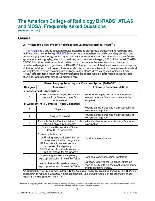
Q. What is the Breast Imaging Reporting and Database System (<strong>BI</strong>-<strong>RADS</strong> ® )?<br />
A. <strong>BI</strong>-<strong>RADS</strong>® is a quality assurance guide designed to standardize breast imaging reporting and<br />
facilitate outcome monitoring. <strong>BI</strong>-<strong>RADS</strong>® serves as a comprehensive guide providing standardized<br />
breast imaging terminology, report organization and assessment st<strong>ru</strong>cture, as well as a classification<br />
system for mammography, ultrasound, and magnetic resonance imaging (MRI) <strong>of</strong> the breast. <strong>The</strong> <strong>BI</strong>-<br />
<strong>RADS</strong> ® Atlas also includes the fourth edition <strong>of</strong> the mammography lexicon and audit system. It<br />
provides radiologists with guidance on <strong>BI</strong>-<strong>RADS</strong> ® through the use <strong>of</strong> illustrated cases, sample reports,<br />
statistical definitions, and explanations for performing mammography audits. It is a systematic method<br />
for radiologists to report mammogram findings using 7 standardized categories, or levels. Each <strong>BI</strong>-<br />
<strong>RADS</strong> ® category has a follow-up recommendation associated with it to help radiologists and other<br />
physicians appropriately manage a patient's care.<br />
Breast Imaging Reporting and Database System (<strong>BI</strong>-<strong>RADS</strong> ® )<br />
Category Assessment Follow-up Recommendations<br />
a. Assessment is Incomplete<br />
0<br />
Need Additional Imaging Evaluation<br />
and/or Prior Mammograms for<br />
Comparison<br />
Additional imaging and/or prior images are<br />
needed before a final assessment can be<br />
assigned<br />
b. Assessment is Complete – Final Categories<br />
1 Negative<br />
Routine annual screening mammography (for<br />
women over age 40)<br />
2 Benign Finding(s)<br />
Routine annual screening mammography (for<br />
women over age 40)<br />
3<br />
4<br />
5<br />
6<br />
Probably Benign Finding – Initial Short-<br />
Interval Follow-Up Suggested<br />
Suspicious Abnormality – Biopsy<br />
Should Be Considered<br />
Optional subdivisions:*<br />
4A: Finding needing intervention with<br />
a low suspicion for malignancy<br />
4B: Lesions with an intermediate<br />
suspicion <strong>of</strong> malignancy<br />
4C: Findings <strong>of</strong> moderate concern,<br />
but not classic for malignancy<br />
Highly Suggestive <strong>of</strong> Malignancy –<br />
Appropriate Action Should Be Taken<br />
Known Biopsy-Proven Malignancy –<br />
Appropriate Action Should Be Taken<br />
Initial short-term follow up (usually 6-month)<br />
examination<br />
Usually requires biopsy<br />
Requires biopsy or surgical treatment<br />
Category reserved for lesions identified on<br />
imaging study with biopsy pro<strong>of</strong> <strong>of</strong> malignancy<br />
prior to definitive therapy<br />
* A subdivision may be used in addition to the Category 4 final assessment; MQSA does not allow a<br />
subdivision to replace a Category 4 final assessment. Use <strong>of</strong> subdivision is at the discretion <strong>of</strong> the<br />
facility it is not required by the FDA.<br />
This document is copyright protected by the <strong>American</strong> <strong>College</strong> <strong>of</strong> <strong>Radiology</strong>. Any attempt to reproduce, copy, modify, alter or otherwise change or use this document without the express<br />
written permission <strong>of</strong> the <strong>American</strong> <strong>College</strong> <strong>of</strong> <strong>Radiology</strong> is prohibited.<br />
– 1 –<br />
Z:\<strong>BI</strong>-<strong>RADS</strong>\FAQs\Current FAQs\<strong>BI</strong>-<strong>RADS</strong> FAQs.doc Revised: 5/9/08
Q. Where can I find information about <strong>BI</strong>-<strong>RADS</strong> ® ?<br />
A. Information about <strong>BI</strong>-<strong>RADS</strong>® is located on the ACR’s website or you can call the ACR at<br />
(800) 227-6440.<br />
Q. Where can I find information on how to purchase the <strong>BI</strong>-<strong>RADS</strong> ® Atlas?<br />
A. You can find information on ordering the complete atlas by clicking on <strong>BI</strong>-<strong>RADS</strong>® at the ACR<br />
Store web page or calling Publication Sales at (800) 227-7762. Information on <strong>BI</strong>-<strong>RADS</strong> ® and other<br />
ACR publications is available at this link to the Materials & Publications Catalog.<br />
Q. How should I reference the ACR <strong>BI</strong>-<strong>RADS</strong> ® Atlas in a publication I’m preparing?<br />
A. Please use the following citation in your references:<br />
D'Orsi CJ, Bassett LW, Berg WA, et al: Breast Imaging Reporting and Data System: ACR <strong>BI</strong>-<strong>RADS</strong>-<br />
Mammography (ed 4), Reston, VA, <strong>American</strong> <strong>College</strong> <strong>of</strong> <strong>Radiology</strong>, 2003<br />
Q. What is <strong>BI</strong>-<strong>RADS</strong> ® Category 6 (Known Biopsy-Proven Malignancy), and when would a<br />
facility use it?<br />
A. <strong>BI</strong>-<strong>RADS</strong> ® Category 6 has been added for breast findings confirmed to be malignant by biopsy<br />
but prior to definitive therapies such as surgical excision, radiation therapy, chemotherapy or<br />
mastectomy. This category is also appropriate for second opinions on findings previously biopsied<br />
and shown to be malignant or for the monitoring <strong>of</strong> response to neoadjuvant chemotherapy prior to<br />
surgical excision. Unlike <strong>BI</strong>-<strong>RADS</strong> ® categories 4 and 5, there is no associated intervention required to<br />
confirm malignancy.<br />
However, if an exam is performed prior to therapy on a woman with known breast cancer and<br />
something other than the original malignancy is seen, the interpreting physician should provide an<br />
appropriate final assessment (e.g., Categories 0, 4, and 5) instead <strong>of</strong> Category 6 so that immediate<br />
action can occur.<br />
A major rationale for adding Category 6 is that examinations meriting this assessment should be<br />
excluded from auditing. Auditing that includes such examinations would inappropriately indicate<br />
inflated cancer detection rates, positive predictive values, and other outcomes parameters. More<br />
information on Category 6 is available on the <strong>BI</strong>-<strong>RADS</strong>® page on the ACR website.<br />
Q. Does the ACR have a position on putting universal disclaimers on mammography reports,<br />
such as: “X percent <strong>of</strong> cancers are not detected by mammography.” or “Dense breast tissue<br />
limits the sensitivity <strong>of</strong> mammography.”?<br />
A. Page 4 <strong>of</strong> the introduction to the 2003 ACR <strong>BI</strong>-<strong>RADS</strong> ® Atlas states: “…universal disclaimers are<br />
not necessary since it is well established that a negative mammogram cannot exclude cancer and a<br />
clinically suspicious area should be biopsied even if the mammogram is negative.” However, it is<br />
appropriate to include a statement regarding extremely dense breast tissue. <strong>The</strong> chapter on Report<br />
Organization (p. 179-180) states: “For consistency, breast composition should be described for all<br />
patients using the following patterns:<br />
1. <strong>The</strong> breast is almost entirely fat (75%<br />
glandular).”<br />
This document is copyright protected by the <strong>American</strong> <strong>College</strong> <strong>of</strong> <strong>Radiology</strong>. Any attempt to reproduce, copy, modify, alter or otherwise change or use this document without the express<br />
written permission <strong>of</strong> the <strong>American</strong> <strong>College</strong> <strong>of</strong> <strong>Radiology</strong> is prohibited.<br />
– 2 –<br />
Z:\<strong>BI</strong>-<strong>RADS</strong>\FAQs\Current FAQs\<strong>BI</strong>-<strong>RADS</strong> FAQs.doc Revised: 5/9/08
<strong>BI</strong>-<strong>RADS</strong> ® , ACR Accreditation & MQSA<br />
Q. Where can I find information about the ACR’s Breast Imaging Accreditation Programs?<br />
A. Information about the ACR’s Breast Imaging Accreditation Programs is located on the websites<br />
for the Mammography Accreditation Program, the Breast Ultrasound Accreditation Program, and the<br />
Stereotactic Breast Biopsy Accreditation Program.<br />
Q. Where can I go for assistance or clarification <strong>of</strong> the mammography final assessment<br />
categories required by the Mammography Quality Standards Act (MQSA)?<br />
A. <strong>The</strong> best source <strong>of</strong> clarification or assistance regarding the MQSA final assessment categories is<br />
the FDA. For policy questions, you should check the FDA’s Policy Guidance Help System first. You<br />
may also call the FDA Facility Hotline at (800) 838-7715, fax a question(s) to them at (410) 290-6351<br />
or email them at MQSAhotline@hcmsllc.com.<br />
Q. Under MQSA, is it necessary to include an assessment code (i.e., 0, 1, 2, 3, 4, 5, or 6) in<br />
addition to the assessment category on all mammography reports?<br />
A. No. <strong>The</strong> FDA regulations only require that each mammographic report include the text<br />
corresponding to the overall final assessment category, not the numeric code. <strong>The</strong>se categories are<br />
“Negative”, “Benign”, “Probably Benign”, “Suspicious”, “Highly Suggestive <strong>of</strong> Malignancy”, and<br />
“Incomplete: Need Additional Imaging Evaluation.” More recently, the FDA has also approved<br />
alternative standards to allow several other final assessment categories: “Known Biopsy Proven<br />
Malignancy” and “Post Procedure Mammograms for Marker Placement.” <strong>The</strong> FDA requires that the<br />
final assessments be written verbatim as described in their regulations, using phrasing as detailed in<br />
the table below. Although the ACR encourages the use <strong>of</strong> numeric codes along with the final<br />
assessment text, there are no requirements that they be assigned to these assessments. For more<br />
information, see the FDA's Policy Guidance Help System.<br />
MQSA Final Assessment<br />
Category<br />
Incomplete: Need Additional<br />
Imaging Evaluation<br />
<strong>BI</strong>-<strong>RADS</strong> ®<br />
FDA-Approved Final Assessment<br />
Category<br />
Descriptions<br />
0 • Incomplete: Need Additional Imaging Evaluation<br />
• Incomplete: Needs Additional Imaging Evaluation<br />
• Incomplete: Additional Imaging Evaluation Needed<br />
• Incomplete: Need Additional Imaging Evaluation -<br />
Comparison with Prior Studies<br />
• Incomplete: Need Additional Imaging Evaluation and/or<br />
Prior Mammograms for Comparison<br />
• Incomplete: Need Prior Mammograms for Comparison<br />
• Need Additional Imaging Evaluation (the term<br />
"Incomplete" can be inferred in this example as this is<br />
the only Incomplete <strong>BI</strong>-<strong>RADS</strong> ® assessment category)<br />
• Incomplete Mammogram: Need Additional Imaging<br />
Evaluation<br />
Negative 1 • Negative<br />
• Negative Mammogram<br />
This document is copyright protected by the <strong>American</strong> <strong>College</strong> <strong>of</strong> <strong>Radiology</strong>. Any attempt to reproduce, copy, modify, alter or otherwise change or use this document without the express<br />
written permission <strong>of</strong> the <strong>American</strong> <strong>College</strong> <strong>of</strong> <strong>Radiology</strong> is prohibited.<br />
– 3 –<br />
Z:\<strong>BI</strong>-<strong>RADS</strong>\FAQs\Current FAQs\<strong>BI</strong>-<strong>RADS</strong> FAQs.doc Revised: 5/9/08
MQSA Final Assessment<br />
Category<br />
<strong>BI</strong>-<strong>RADS</strong> ®<br />
Category<br />
FDA-Approved Final Assessment<br />
Descriptions<br />
Benign 2 • Benign<br />
• Benign Finding<br />
• Benign Findings<br />
• Benign Abnormality<br />
• Benign Abnormalities<br />
• Benign Mammogram<br />
Probably Benign 3 • Probably Benign<br />
• Probably Benign Finding<br />
• Probably Benign Findings<br />
• Probably Benign Abnormality<br />
• Probably Benign Abnormalities<br />
• Probably Benign - Short Interval Follow-up Suggested<br />
• Probably Benign Finding - Short Interval Follow-up<br />
Suggested<br />
• Probably Benign Mammogram<br />
Suspicious 4 • Suspicious<br />
• Suspicious Finding<br />
• Suspicious Findings<br />
• Suspicious Abnormality<br />
• Suspicious Abnormalities<br />
• Suspicious for Malignancy<br />
• Suspicious <strong>of</strong> Malignancy<br />
• Suspicious Abnormality - Biopsy Should Be Considered<br />
• Suspicious Finding - Biopsy Should Be Considered<br />
• Suspicious Mammogram<br />
Highly Suggestive <strong>of</strong><br />
Malignancy<br />
Known Biopsy Proven<br />
Malignancy<br />
5 • Highly Suggestive <strong>of</strong> Malignancy<br />
• Highly Suggestive for Malignancy<br />
• Highly Suggestive <strong>of</strong> Malignancy - Appropriate Action<br />
Should Be Taken<br />
6 • Known Biopsy Proven Malignancy<br />
• Known Biopsy Proven Cancer<br />
• Known Malignancy<br />
• Known Cancer<br />
Post Procedure Mammograms none • Post Procedure Mammograms for Marker Placement<br />
for Marker Placement<br />
(This question and answer was adapted from the FDA’s Policy Guidance Help System.)<br />
Q. Is there a new <strong>BI</strong>-<strong>RADS</strong> ® code for “Post Procedure Mammograms for Marker Placement?”<br />
A. No, there is no <strong>BI</strong>-<strong>RADS</strong> ® code for the FDA-approved alternative standard for “Post Procedure<br />
Mammograms for Marker Placement.” This is not a <strong>BI</strong>-<strong>RADS</strong> ® final assessment. This assessment<br />
category may only be used for post-procedure mammograms obtained for the purpose <strong>of</strong> confirming<br />
the deployment and position <strong>of</strong> breast tissue markers, which typically have been placed at the time <strong>of</strong><br />
core biopsy. In addition, this assessment should be excluded from auditing. For more information, see<br />
the FDA's Policy Guidance Help System.<br />
This document is copyright protected by the <strong>American</strong> <strong>College</strong> <strong>of</strong> <strong>Radiology</strong>. Any attempt to reproduce, copy, modify, alter or otherwise change or use this document without the express<br />
written permission <strong>of</strong> the <strong>American</strong> <strong>College</strong> <strong>of</strong> <strong>Radiology</strong> is prohibited.<br />
– 4 –<br />
Z:\<strong>BI</strong>-<strong>RADS</strong>\FAQs\Current FAQs\<strong>BI</strong>-<strong>RADS</strong> FAQs.doc Revised: 5/9/08
Q. Does the ACR have sample lay letters for the FDA-approved “Post Procedure<br />
Mammograms for Marker Placement” final assessment?<br />
A. No. Because “Post Procedure Mammograms for Marker Placement” is not a formal <strong>BI</strong>-<strong>RADS</strong> ®<br />
final assessment, the ACR has not developed sample lay letters for these situations. Facilities using<br />
this assessment will need to develop their own lay letters for these patients.<br />
Q. <strong>The</strong> MQSA regulations require a facility to provide mammography reports to referring<br />
healthcare providers and lay summaries to its patients within 30 days <strong>of</strong> the date <strong>of</strong> the exam.<br />
<strong>The</strong> regulations also require that if the final assessment is “Suspicious” or “Highly Suggestive<br />
<strong>of</strong> Malignancy”, the facility should provide the mammography report and lay summary as<br />
soon as possible (ASAP). <strong>The</strong> FDA guidance interprets ASAP as within 3 days for the report to<br />
the healthcare provider and within 5 days for the lay letter to the patient. Is this guidance<br />
timeframe within 3 and 5 days <strong>of</strong> the patient’s mammography exam?<br />
A. No. This is within 3 and 5 days <strong>of</strong> the report date, as long as it is no longer than 30 days from the<br />
date <strong>of</strong> the exam.<br />
Mammography<br />
Q. We know that we must send our patients the results <strong>of</strong> their mammograms in writing. Does the<br />
ACR have examples <strong>of</strong> letters for this purpose?<br />
A. Yes, the ACR has samples <strong>of</strong> lay letters for all <strong>of</strong> the <strong>BI</strong>-<strong>RADS</strong> ® final assessment categories at<br />
the following link: Mammography Sample Lay Report Letters.<br />
Q. Our patient has a palpable abnormality but the mammogram is “Negative.” Should we<br />
code the mammogram as a <strong>BI</strong>-<strong>RADS</strong> ® Category 1 or 0?<br />
A. Page 254 <strong>of</strong> the <strong>BI</strong>-<strong>RADS</strong> ® Atlas recommends that, in general, reports be coded with final<br />
assessments based on the imaging findings. Hence, a negative mammogram (no findings to report)<br />
should be assessed as negative (Category 1). However, when patient management is influenced by<br />
clinical findings, the management recommendation should take both imaging and clinical<br />
findings into consideration and the clinical findings may be detailed in the report. For example,<br />
it would be acceptable to render a negative (Category 1) assessment but describe the presence and<br />
nature <strong>of</strong> a palpable abnormality, with recommendations for annual screening mammography and<br />
management <strong>of</strong> the palpable abnormality based on findings at clinical breast examination.<br />
Q. Does <strong>BI</strong>-<strong>RADS</strong> ® Category 0 (Need Additional Imaging Evaluation and/or Prior<br />
Mammograms for Comparison) have a subcategory that addresses technical errors on<br />
mammograms (e.g., motion, artifacts, insufficient tissue, etc.) to indicate that the mammogram<br />
needs repeating (i.e., “technical recall”)?<br />
A. No, there is no <strong>of</strong>ficial subcategory for technical errors in <strong>BI</strong>-<strong>RADS</strong> ® Category 0. You may create<br />
your own subcategory for internal analysis purposes. For example, you may want to create a<br />
subcategory <strong>of</strong> “0-TR” for technical recalls and include the technical reasons for the recall in the body<br />
<strong>of</strong> the report. Keep in mind that the FDA regulations require you to assign only the FDA-approved<br />
categories as final assessments. However, if you provide a subcategory for technical recalls within<br />
the overall assessment category <strong>of</strong> “0,” the technical recalls should not be counted in the medical<br />
audit <strong>of</strong> category “0”s.<br />
Q. A patient has a diagnostic mammogram with a final assessment <strong>of</strong> Category 4. <strong>The</strong> patient<br />
then has two biopsies <strong>of</strong> the same lesion, one core and one surgical. <strong>The</strong> core biopsy<br />
pathology is ADH; the excisional biopsy pathology is malignant. Should the diagnostic<br />
This document is copyright protected by the <strong>American</strong> <strong>College</strong> <strong>of</strong> <strong>Radiology</strong>. Any attempt to reproduce, copy, modify, alter or otherwise change or use this document without the express<br />
written permission <strong>of</strong> the <strong>American</strong> <strong>College</strong> <strong>of</strong> <strong>Radiology</strong> is prohibited.<br />
– 5 –<br />
Z:\<strong>BI</strong>-<strong>RADS</strong>\FAQs\Current FAQs\<strong>BI</strong>-<strong>RADS</strong> FAQs.doc Revised: 5/9/08
mammogram be statistically categorized as both a FALSE POSITIVE and a TRUE POSITIVE, or<br />
just a TRUE POSITIVE?<br />
A. If the exam was a positive and cancer was discovered within the year, it is a considered a t<strong>ru</strong>e<br />
positive exam. <strong>The</strong> t<strong>ru</strong>th is determined by the discovery <strong>of</strong> malignancy by any means, within 365<br />
days from the exam date.<br />
Q. We commonly issue addenda and/or comparison reports after initial mammography<br />
reports have been issued. Are we required to provide a final assessment category with each <strong>of</strong><br />
these reports? Must we also send the addendum or comparison report to the referring health<br />
care provider and a letter to the patient, even if there is no change in the final assessment<br />
category or recommended course <strong>of</strong> action?<br />
A. Yes, to both questions. <strong>The</strong> report issued after additional x-ray testing that is covered under<br />
MQSA (i.e., coned, repeat, magnification views) or following comparison with old images must<br />
provide a final assessment category for the case. A report must be communicated to the referring<br />
health care provider or the self-referred patient. In addition, a lay summary <strong>of</strong> the addendum or<br />
comparison report must be provided to the patient, even if there is no change in the final assessment<br />
category or recommended course <strong>of</strong> action.<br />
For the specific case where there is no significant change in the report, a simple statement that the<br />
comparison has been performed and that there is no overall change (ensuring to include the<br />
unchanged final assessment) would satisfy the requirement. If the addendum states that the referring<br />
health care provider has been notified <strong>of</strong> the results <strong>of</strong> the patient’s examination, the addendum lay<br />
summary can be a simple statement informing the patient <strong>of</strong> that fact. (This question and answer was<br />
adapted from the FDA’s Policy Guidance Help System.)<br />
Q. We wish to use the ACR’s Category 4 subdivisions (4A – Finding needing intervention with<br />
a low suspicion for malignancy, 4B – Lesions with an intermediate suspicion <strong>of</strong> malignancy,<br />
and 4C – Findings <strong>of</strong> moderate concern, but not classic for malignancy). Can our reports use<br />
these subcategories instead <strong>of</strong> the Category 4 assessment (Suspicious Abnormality – Biopsy<br />
Should Be Considered)?<br />
A. No. While you have the option <strong>of</strong> using one <strong>of</strong> the 3 subcategories in addition to a final<br />
assessment <strong>of</strong> "Suspicious", the FDA will not allow you to use the subcategories instead <strong>of</strong> the<br />
"Suspicious" assessment category on the mammography report. (This question and answer was<br />
adapted from the FDA’s Policy Guidance Help System.)<br />
Q. Do mammograms performed on men require a <strong>BI</strong>-<strong>RADS</strong> ® final assessment and/or code?<br />
A. Yes, all mammography examinations, regardless <strong>of</strong> the patient’s gender, are required to have a<br />
final assessment assigned to them. However, management recommendations may differ from those<br />
made for women because annual screening mammography is not usually appropriate for men.<br />
Ultrasound<br />
Q. Does MQSA require that <strong>BI</strong>-<strong>RADS</strong> ® categories be assigned to breast ultrasound<br />
examinations?<br />
A. No, MQSA does not apply to breast ultrasound; however, the ACR does recommend using <strong>BI</strong>-<br />
<strong>RADS</strong>® final assessment codes for breast ultrasound examinations. See “Multiple Procedures”<br />
section below for discussion on issuing a combined report with mammography.<br />
This document is copyright protected by the <strong>American</strong> <strong>College</strong> <strong>of</strong> <strong>Radiology</strong>. Any attempt to reproduce, copy, modify, alter or otherwise change or use this document without the express<br />
written permission <strong>of</strong> the <strong>American</strong> <strong>College</strong> <strong>of</strong> <strong>Radiology</strong> is prohibited.<br />
– 6 –<br />
Z:\<strong>BI</strong>-<strong>RADS</strong>\FAQs\Current FAQs\<strong>BI</strong>-<strong>RADS</strong> FAQs.doc Revised: 5/9/08
MRI<br />
Q. Does MQSA require that <strong>BI</strong>-<strong>RADS</strong> ® categories be assigned to MR examinations?<br />
A. No, MQSA does not apply to MRI; however, the ACR does recommend using <strong>BI</strong>-<strong>RADS</strong>® final<br />
assessment codes for MR examinations.<br />
Q. A patient’s breast MRI exam resulted in a <strong>BI</strong>-<strong>RADS</strong> ® Category 2 assessment (Benign<br />
Finding(s)); her mammography exam resulted in a <strong>BI</strong>-<strong>RADS</strong> ® Category 4 assessment<br />
(Suspicious Abnormality – Biopsy Should Be Considered). <strong>The</strong> patient has a previous history<br />
<strong>of</strong> malignancy following lumpectomy, and her physician believes that the area needs biopsy.<br />
In cases where the mammography report disagrees with the breast MRI exam, is it appropriate<br />
to recommend a biopsy in the impression <strong>of</strong> the breast MRI report based on the positive<br />
mammogram?<br />
A. Yes, you may include a recommendation for biopsy in your breast MRI report. If your report<br />
system has a combined module that includes all three lexicons (mammography, ultrasound, and<br />
breast MRI), appropriate letters will be sent to clinicians and patients based on the most serious <strong>BI</strong>-<br />
<strong>RADS</strong> ® category (in this case “4”). Also, as a general <strong>ru</strong>le, imaging studies should not be used to<br />
contradict a biopsy from another breast imaging study.<br />
Multiple Procedures<br />
Q. Does MQSA require an “Overall Assessment” for multiple procedures?<br />
A. No, an overall assessment summarizing multiple procedures is not required under MQSA. You<br />
may visit the FDA's Policy Guidance Help System's section on “Medical Records and<br />
Reports/Contents <strong>of</strong> Records and Reports” for more information.<br />
Q. If we perform a "non-mammographic" breast imaging study on the same day as the<br />
mammographic examination, are we required to issue a separate mammography report with<br />
its own final assessment category and recommendations? Or, may we issue a combined<br />
report whose final assessment category and recommendations represent the overall<br />
assessment <strong>of</strong> all the breast imaging studies that were performed that day?<br />
A. <strong>The</strong> facility has the option <strong>of</strong> issuing either separate or combined reports. (You may want to check<br />
with your billing <strong>of</strong>fice first; some third party payers may require individual reports.)<br />
Ideally, the report <strong>of</strong> diagnostic mammographic views and ultrasound should be included in the same<br />
report (with separate paragraphs detailing each) and one integrated final assessment that takes into<br />
consideration all breast imaging findings. (This is allowable under MQSA; see FDA's Policy Guidance<br />
Help System for more information.) If a single final assessment is issued, it should be based on the<br />
interpreting physician’s review <strong>of</strong> all studies. <strong>The</strong> hierarchy <strong>of</strong> the <strong>BI</strong>-<strong>RADS</strong> ® categories is as follows:<br />
5, 4, 0, 3, 2, 1. <strong>The</strong> <strong>BI</strong>-<strong>RADS</strong>® Atlas provides several examples <strong>of</strong> such combined reports (see page<br />
221 “Illustrated Case #4” in the Mammography section, and page 11 in the “Illustrated Cases-<br />
Combined” section).<br />
Q. Under Centers for Medicare and Medicaid Services (CMS) guidelines, we can now charge<br />
for screening and diagnostic exams done on the same patient on the same day. May we<br />
combine the two exams into one report or must we issue two separate reports?<br />
A. <strong>The</strong> facility has the option <strong>of</strong> issuing either separate or combined reports. If 2 reports are issued,<br />
each must contain its own overall final assessment. <strong>The</strong> facility can report both exams on the "same<br />
piece <strong>of</strong> paper." If the facility decides to issue a single combined report, the facility needs to be aware<br />
<strong>of</strong> the following:<br />
This document is copyright protected by the <strong>American</strong> <strong>College</strong> <strong>of</strong> <strong>Radiology</strong>. Any attempt to reproduce, copy, modify, alter or otherwise change or use this document without the express<br />
written permission <strong>of</strong> the <strong>American</strong> <strong>College</strong> <strong>of</strong> <strong>Radiology</strong> is prohibited.<br />
– 7 –<br />
Z:\<strong>BI</strong>-<strong>RADS</strong>\FAQs\Current FAQs\<strong>BI</strong>-<strong>RADS</strong> FAQs.doc Revised: 5/9/08
1. A single combined report must contain a single overall final assessment.<br />
2. <strong>The</strong> combined report should make it clear to the referring physician that it is combining the results<br />
<strong>of</strong> the screening and diagnostic studies. This is also important if questions ever arise about whether<br />
the exams were billed correctly.<br />
3. Issuing a single report with a single final assessment will skew the facility’s medical audit results<br />
unless the exam is audited as both screening category 0 and diagnostic using the final assessment<br />
category rendered.<br />
4. Though some computerized reporting systems may consider this a single exam (rather than two),<br />
FDA would still allow facilities to count both exams toward meeting the continuing experience<br />
requirement.<br />
(This question and answer was adapted from the FDA’s Policy Guidance Help System.)<br />
Q. If the final assessment <strong>of</strong> a mammography exam is "incomplete" (<strong>BI</strong>-<strong>RADS</strong> ® Category 0)<br />
and the woman is referred for additional testing, does MQSA require the facility to revise or<br />
amend the original report if, as a result <strong>of</strong> this referral, the assessment is changed to one <strong>of</strong><br />
the other categories?<br />
A. No. However, if the other test uses x-rays and is covered under MQSA (e.g., mammographic<br />
coned or magnification views), the facility performing these views must issue a report (either<br />
separately or as an addendum to the original mammography report) reflecting the final assessment.<br />
<strong>The</strong> ACR <strong>BI</strong>-<strong>RADS</strong> ® Atlas provides further recommendations on this topic: “An incomplete<br />
assessment requires further evaluation with additional mammographic views, comparison films,<br />
ultrasound or, less commonly, MRI. When additional imaging studies are completed, a final<br />
assessment is rendered. Ideally, the report <strong>of</strong> diagnostic mammographic views and ultrasound will be<br />
included in the same report, with separate paragraphs detailing each, and one integrated final<br />
assessment that takes into consideration all breast imaging findings.”<br />
Q. An original screening mammogram received a <strong>BI</strong>-<strong>RADS</strong> ® Category 0 assessment (Need<br />
Additional Imaging Evaluation and/or Prior Mammograms for Comparison) due to an abnormal<br />
asymmetry. <strong>The</strong> subsequent diagnostic mammogram was also <strong>BI</strong>-<strong>RADS</strong> ® Category 0. An<br />
ultrasound exam was performed and received a <strong>BI</strong>-<strong>RADS</strong> ® Category 1 (Negative) assessment.<br />
Even though the ultrasound was negative, I want to further evaluate this patient with MRI<br />
since the mammogram was <strong>of</strong> concern. If I issue a combined final assessment for all <strong>of</strong> the<br />
procedures, how should I determine the appropriate <strong>BI</strong>-<strong>RADS</strong> ® code to use to ensure proper<br />
care?<br />
A. This question implies two non-recommended uses <strong>of</strong> <strong>BI</strong>-<strong>RADS</strong> ® Category 0.<br />
First, <strong>BI</strong>-<strong>RADS</strong> ® Category 0 should generally not be used for diagnostic mammograms; typically, it<br />
should be used only for screening mammograms. <strong>The</strong>refore, if diagnostic mammography is<br />
performed in conjunction with ultrasound, an integrated <strong>BI</strong>-<strong>RADS</strong> ® assessment should be given<br />
(rather than a Category 0 assessment for the mammography followed by a Category 1 assessment<br />
for the ultrasound). <strong>The</strong> final assessment would depend on the mammographic and sonographic<br />
imaging findings and whether these are described in the diagnostic breast imaging report. See<br />
examples:<br />
• If no findings are described in either the mammography or ultrasound portions <strong>of</strong> the report, <strong>BI</strong>-<br />
<strong>RADS</strong> ® Category 1 (Negative) is the appropriate assessment.<br />
• If specific benign findings are described in either the mammography or ultrasound portion <strong>of</strong> the<br />
report, <strong>BI</strong>-<strong>RADS</strong> ® Category 2 (Benign Finding(s)) is the appropriate assessment.<br />
• If diagnostic mammography depicts a focal asymmetry with no associated mass, calcifications, or<br />
architectural distortion; if there is no sonographic or palpable correlate to the mammographic<br />
This document is copyright protected by the <strong>American</strong> <strong>College</strong> <strong>of</strong> <strong>Radiology</strong>. Any attempt to reproduce, copy, modify, alter or otherwise change or use this document without the express<br />
written permission <strong>of</strong> the <strong>American</strong> <strong>College</strong> <strong>of</strong> <strong>Radiology</strong> is prohibited.<br />
– 8 –<br />
Z:\<strong>BI</strong>-<strong>RADS</strong>\FAQs\Current FAQs\<strong>BI</strong>-<strong>RADS</strong> FAQs.doc Revised: 5/9/08
finding; and if there are no previous mammograms available for comparison, it may be<br />
appropriate to render a <strong>BI</strong>-<strong>RADS</strong> ® Category 3 (Probably Benign Finding – Initial Short-Interval<br />
Follow-Up Suggested) assessment.<br />
• If diagnostic mammography indicates the presence <strong>of</strong> a suspicious abnormality despite absence<br />
<strong>of</strong> a sonographic correlate, <strong>BI</strong>-<strong>RADS</strong> ® Category 4 (Suspicious Abnormality – Biopsy Should Be<br />
Considered) is the appropriate assessment.<br />
Second, <strong>BI</strong>-<strong>RADS</strong> ® Category 0 should not be routinely used for diagnostic breast imaging<br />
findings that warrant further evaluation with MRI. Rather, the radiologist should issue a <strong>BI</strong>-<strong>RADS</strong> ®<br />
final assessment for the integrated mammography and ultrasound report before the MRI is<br />
performed. If further evaluation with MRI is warranted, the radiologist should incorporate this<br />
recommendation into the patient management recommendations within the impression <strong>of</strong> the<br />
integrated mammography and ultrasound report. This provides the following advantages:<br />
• If the recommended MRI examination is not performed, the integrated diagnostic breast imaging<br />
report will stand as issued.<br />
• On the other hand, if MRI is performed as recommended, it would not be necessary to re-interpret<br />
the mammogram and ultrasound. A negative or benign MRI assessment will sustain a similar<br />
assessment made for mammography and ultrasound. If the MRI exam shows more abnormal<br />
findings than those identified by mammography and ultrasound, the MRI assessment would<br />
supersede that made for mammography and ultrasound.<br />
Also note that breast MRI is not appropriate follow-up in many situations including:<br />
• In lieu <strong>of</strong> biopsy <strong>of</strong> a suspicious finding on mammography and/or ultrasound<br />
• As an alternative to short interval follow-up <strong>of</strong> probably benign findings on mammography and/or<br />
ultrasound<br />
• In lieu <strong>of</strong> biopsy <strong>of</strong> a suspicious finding on mammography and/or ultrasound (i.e., suspicious<br />
calcifications)<br />
• To further evaluate findings which should be recognized as benign on mammography and/or<br />
ultrasound, such as gynecomastia or multiple bilateral partially circumscribed, partially obscured<br />
masses; most lymph nodes and fat necrosis can be so recognized mammographically and/or<br />
sonographically<br />
MRI is rarely helpful in further evaluations <strong>of</strong> possible distortion which is too vague to target for<br />
stereotactic or sonographic biopsy.<br />
Q. Axillary adenopathy is seen on screening mammography with no suspicious findings in<br />
the breasts. What should the <strong>BI</strong>-<strong>RADS</strong> ® final assessment be?<br />
A. In the absence <strong>of</strong> known infectious or inflammatory cause, isolated unilateral axillary<br />
adenopathy should receive a <strong>BI</strong>-<strong>RADS</strong> ® Category 4 assessment (Suspicious Abnormality – Biopsy<br />
Should Be Considered). Unilateral axillary adenopathy suggests occult breast carcinoma or, much<br />
less commonly, metastatic melanoma, ovarian cancer, or other metastatic cancer. Consequently:<br />
• A careful search <strong>of</strong> the ipsilateral breast images is warranted<br />
• Bilateral axillary ultrasound should be performed first to help confirm that the finding is<br />
asymmetric/unilateral<br />
• Clinical evaluation for infection or inflammation in the ipsilateral breast, axilla, arm, and hand is<br />
recommended at the time <strong>of</strong> ultrasound, as mastitis, breast abscess, an infected skin lesion, and<br />
cat scratch fever are all potential sources <strong>of</strong> benign unilateral axillary adenopathy<br />
If no known infectious or inflammatory sources are present, a <strong>BI</strong>-<strong>RADS</strong> ® Category 4 assessment<br />
(Suspicious Abnormality – Biopsy Should Be Considered) is appropriate, with intent to biopsy after<br />
This document is copyright protected by the <strong>American</strong> <strong>College</strong> <strong>of</strong> <strong>Radiology</strong>. Any attempt to reproduce, copy, modify, alter or otherwise change or use this document without the express<br />
written permission <strong>of</strong> the <strong>American</strong> <strong>College</strong> <strong>of</strong> <strong>Radiology</strong> is prohibited.<br />
– 9 –<br />
Z:\<strong>BI</strong>-<strong>RADS</strong>\FAQs\Current FAQs\<strong>BI</strong>-<strong>RADS</strong> FAQs.doc Revised: 5/9/08
further evaluation and review <strong>of</strong> clinical history. It is then appropriate to proceed with ultrasoundguided<br />
fine needle aspiration biopsy (FNAB) or core biopsy, and it may be advisable to perform<br />
ipsilateral whole breast ultrasound during that visit to search for an occult primary breast carcinoma.<br />
If a benign cause can be elucidated, <strong>BI</strong>-<strong>RADS</strong> ® Category 2 (Benign Finding(s)) is a reasonable<br />
assessment.<br />
Bilateral axillary adenopathy should be assessed as <strong>BI</strong>-<strong>RADS</strong> ® Category 2 (Benign Finding(s)) in<br />
some situations and as <strong>BI</strong>-<strong>RADS</strong> ® Category 4 (Suspicious Abnormality – Biopsy Should Be<br />
Considered) in others.<br />
• Bilateral axillary adenopathy is frequently reactive/infectious in origin such as with inflammatory<br />
conditions (sarcoid, systemic lupus erythematosis, psoriasis, other) and HIV. In such situations,<br />
the finding is <strong>BI</strong>-<strong>RADS</strong> ® Category 2 (Benign Finding(s)).<br />
• Patients with known lymphoma may also have bilateral axillary adenopathy. In this situation, the<br />
<strong>BI</strong>-<strong>RADS</strong> ® assessment should be based on the breasts themselves, but should indicate known<br />
lymphoma (e.g., “<strong>BI</strong>-<strong>RADS</strong> ® Category 2 (Benign Finding(s)) with known lymphoma”). It may be<br />
helpful to contact the referring healthcare provider to clarify whether or not there is such a history<br />
before issuing a final report.<br />
If there is no known explanation for the bilateral adenopathy, and particularly if it is new, then it may<br />
be a sign <strong>of</strong> lymphoma and a <strong>BI</strong>-<strong>RADS</strong> ® Category 4 assessment (Suspicious Abnormality – Biopsy<br />
Should Be Considered) rendered with a recommendation for ultrasound-guided FNAB or core biopsy.<br />
[Note: Ideally the specimen should be kept in saline or RPMI 1640 if lymphoma is suspected, to<br />
facilitate fluorescence-activated cell sorting.].<br />
Medical Audits<br />
Q. Does the ACR have information regarding physician mammography outcome analysis<br />
s<strong>of</strong>tware?<br />
A. Yes, the ACR has a list <strong>of</strong> lice nsed vendors on our website at <strong>BI</strong>-<strong>RADS</strong>® S<strong>of</strong>tware Vendors List.<br />
All have medical audit s<strong>of</strong>tware.<br />
Q. Where can I find data on patient outcome statistics for screening and diagnostic<br />
mammography?<br />
A. Information is available on the <strong>BI</strong>-<strong>RADS</strong>® pages <strong>of</strong> the ACR website. See table 2 at the following<br />
link: Analysis <strong>of</strong> Medica l Audit Data: Desirable Goals. More recent information is also available from<br />
the following sources:<br />
• Rosenberg RD, et al., Performance benchmarks for screening mammography, <strong>Radiology</strong><br />
2006; 241:55-66.<br />
• EA Sickles, et al., Performance benchmarks for diagnostic mammography, <strong>Radiology</strong> 2005;<br />
235:775-790.<br />
• <strong>The</strong> National Cancer Institute’s Breast Cancer Surveillance Consortium Performance<br />
Benchmarks for Screening and Diagnostic Mammography. Available at:<br />
( http://breastscreening.cancer.gov/data/benchmarks/screening/) and<br />
(http://breastscreening.cancer.gov/data/benchmarks/diagnostic.html)<br />
This document is copyright protected by the <strong>American</strong> <strong>College</strong> <strong>of</strong> <strong>Radiology</strong>. Any attempt to reproduce, copy, modify, alter or otherwise change or use this document without the express<br />
written permission <strong>of</strong> the <strong>American</strong> <strong>College</strong> <strong>of</strong> <strong>Radiology</strong> is prohibited.<br />
– 10 –<br />
Z:\<strong>BI</strong>-<strong>RADS</strong>\FAQs\Current FAQs\<strong>BI</strong>-<strong>RADS</strong> FAQs.doc Revised: 5/9/08
Q. Are there more recent benchmarks, than those provided in the 2003 ACR <strong>BI</strong>-<strong>RADS</strong> ® Atlas,<br />
for auditing my mammography practice?<br />
A. Yes Audit results from actual practice <strong>of</strong> the Breast Cancer Surveillance Consortium (BCSC)<br />
participants were published in 2006. This study included results from 807 radiologists at 187 facilities<br />
and over 2.5 million screening mammographic examinations (1). Results have also been analyzed<br />
from the diagnostic mammography practice in this consortium for 646 radiologists at 151 facilities and<br />
over 332,000 examinations (2). In general, the audit is designed for screening, though some practices<br />
audit their overall practice, including diagnostic examinations. If a facility’s audit includes both<br />
screening and diagnostic mammograms, expected outcomes will vary depending on the percent <strong>of</strong><br />
exams which are diagnostic. Suggested outcomes from combined studies can be found in Sohlich et<br />
al (3).<br />
<strong>The</strong> table below summarizes the BCSC results for pure screening (1), exams recalled from screening<br />
(<strong>BI</strong>-<strong>RADS</strong> ® Category 0) for additional evaluation (2), and pure diagnostic examinations for patients<br />
with a palpable lump (2). <strong>The</strong> following definitions are used:<br />
• Stage 0 Cancers – ductal carcinoma in situ (DCIS)<br />
• Stage I Cancers – invasive cancer ≤ 2 cm in size and negative nodes<br />
• Minimal Cancers – includes DCIS and invasive cancer ≤ 1 cm in size<br />
• Cancer Detection Rate – # <strong>of</strong> women with examinations called positive (<strong>BI</strong>-<strong>RADS</strong> ® Categories 0,<br />
4, or 5 on screening; <strong>BI</strong>-<strong>RADS</strong> ® Categories 4 or 5 on diagnostic workup) who are diagnosed with<br />
cancer, divided by the total number <strong>of</strong> examinations] times 1000<br />
®<br />
• Recall Rate – # <strong>of</strong> positive examinations on screening (<strong>BI</strong>-<strong>RADS</strong> Categories 0, 4, and 5) divided<br />
by the total number <strong>of</strong> examinations.<br />
• Sensitivity – percentage <strong>of</strong> women called positive (<strong>BI</strong>-<strong>RADS</strong> ® Categories 0, 4, or 5 on screening;<br />
<strong>BI</strong>-<strong>RADS</strong> ® Categories 4 or 5 on diagnostic workup) who are diagnosed with cancer within one<br />
year <strong>of</strong> screening<br />
• PPV2 – percentage <strong>of</strong> women with examinations recommending biopsy (<strong>BI</strong>-<strong>RADS</strong> ® Categories 4<br />
or 5) which have a tissue diagnosis <strong>of</strong> cancer within one year<br />
Screening Screening Recalls Diagnostic (Lump)<br />
Stage 0 or I Cancers 76 % 83 % 40 %<br />
Minimal Cancer 52 % 63 % 16 %<br />
Positive Axillary Node(s) 19 % 16 % 33 %<br />
Cancer Detection Rate 4.4 3.1 47<br />
Recall Rate 9.7 % N/A N/A<br />
Sensitivity 80 % 86 % 84 %<br />
Mean Invasive size (cm) 1.3 1.1 2.1<br />
PPV2 25 % 23 % 48 %<br />
<strong>The</strong> following should be considered when auditing one’s practice:<br />
• A clinically useful audit includes calculation <strong>of</strong> several rather than only one or two metrics, the<br />
more the better.<br />
• For low-volume practices, and especially for individual radiologists who work in low-volume<br />
practices, some metrics will lack precision because the number <strong>of</strong> cancers is small. <strong>The</strong><br />
workaround is to audit for the most recent two or three years rather than just the most recent<br />
year.<br />
This document is copyright protected by the <strong>American</strong> <strong>College</strong> <strong>of</strong> <strong>Radiology</strong>. Any attempt to reproduce, copy, modify, alter or otherwise change or use this document without the express<br />
written permission <strong>of</strong> the <strong>American</strong> <strong>College</strong> <strong>of</strong> <strong>Radiology</strong> is prohibited.<br />
– 11 –<br />
Z:\<strong>BI</strong>-<strong>RADS</strong>\FAQs\Current FAQs\<strong>BI</strong>-<strong>RADS</strong> FAQs.doc Revised: 5/9/08
• Most practices will not be able to acquire sufficiently accurate data on false-negative cases to<br />
accurately calculate sensitivity. Linkage with a regional tumor registry or a captive non-mobile<br />
patient population (such as found in a large HMO) is necessary to obtain such information.<br />
• Practices which use integrated <strong>BI</strong>-<strong>RADS</strong> ® assessments for concurrently-performed diagnostic<br />
mammography/ultrasound examinations should expect different outcomes from published<br />
benchmarks. <strong>The</strong> above published data involves the performance <strong>of</strong> mammography alone.<br />
References:<br />
1. Rosenberg RD, et al., Performance benchmarks for screening mammography, <strong>Radiology</strong> 2006;<br />
241:55-66.<br />
2. EA Sickles, et al., Performance benchmarks for diagnostic mammography, <strong>Radiology</strong> 2005;<br />
235:775-790.<br />
3. Sohlich RE, Sickles EA, Burnside ES, Dee KE. Interpreting data from audits when screening and<br />
diagnostic mammography outcomes are combined. AJR Am J Roentgenol 2002; 178:681-686.<br />
Q. Is it necessary for a facility to separate the medical audit into screening and diagnostic<br />
patients?<br />
A. No. Although a facility will obtain more statistically relevant information by separating their<br />
analyses for screening and diagnostic examinations, FDA regulations allow the facility to combine all<br />
patients into one group <strong>of</strong> audit data. <strong>The</strong> ACR recommends that screening and diagnostic<br />
examinations be audited separately.<br />
Q. Does MQSA require follow up <strong>of</strong> Category 0 patients?<br />
A. No. FDA regulations specify that facilities must “collect and review outcome data for all<br />
mammograms performed, including follow up on the disposition <strong>of</strong> all positive mammograms and<br />
correlation <strong>of</strong> pathology results with the interpreting physician.” <strong>The</strong> FDA considers mammograms<br />
with final assessments <strong>of</strong> “Suspicious" or "Highly suggestive <strong>of</strong> malignancy" to be positive. <strong>The</strong>refore,<br />
the FDA does not require facilities to follow-up other cases such as those recommended for shortterm<br />
follow up or ultrasound, or cases that are in the assessment category <strong>of</strong> "Incomplete, Need<br />
additional imaging evaluation." (For more information, see the FDA's Policy Guidance Help System.)<br />
However, the ACR believes that a meaningful audit <strong>of</strong> screening examinations requires that the<br />
®<br />
recommendations for recall imaging (<strong>BI</strong>-<strong>RADS</strong> Category 0) also be considered “positive” and thus<br />
recommends that the facility collect and review outcome data on Category 0 exams.<br />
Q. When doing medical audits, are pathology-proven “high risk lesions” described on page<br />
®<br />
300 in the <strong>BI</strong>-<strong>RADS</strong> Atlas (i.e., ADH, ALH, LS, PDP and PT) considered positive?<br />
A. No, these are considered negative pathology results.<br />
Q. We always do a post-procedure mammogram after an ultrasound-guided biopsy. We bill<br />
for the mammogram separately from the biopsy procedure and use the FDA’s final<br />
assessment <strong>of</strong> “Post Procedure Mammograms for Marker Placement.” However, because this<br />
final assessment is not included in the current ACR <strong>BI</strong>-<strong>RADS</strong> ® Atlas, our s<strong>of</strong>tware vendors<br />
have not provided this option in their medical audit s<strong>of</strong>tware. Consequently, we cannot<br />
include these studies in our annual medical audit. Do you have any suggestions how we can<br />
include these cases?<br />
A. Some facilities consider the post-clip mammogram as part <strong>of</strong> the ultrasound interventional<br />
procedure and consequently will not bill for it or code it. Other facilities that choose to code it (and bill<br />
This document is copyright protected by the <strong>American</strong> <strong>College</strong> <strong>of</strong> <strong>Radiology</strong>. Any attempt to reproduce, copy, modify, alter or otherwise change or use this document without the express<br />
written permission <strong>of</strong> the <strong>American</strong> <strong>College</strong> <strong>of</strong> <strong>Radiology</strong> is prohibited.<br />
– 12 –<br />
Z:\<strong>BI</strong>-<strong>RADS</strong>\FAQs\Current FAQs\<strong>BI</strong>-<strong>RADS</strong> FAQs.doc Revised: 5/9/08
for it) separately wait for the result <strong>of</strong> the biopsy and then code the mammogram accordingly. For<br />
example, if the pathology was benign, they would code the mammogram as a Category 2; if the<br />
pathology was malignant, they would code the mammogram as a Category 6. In either case, it is not<br />
appropriate to include these outcomes in your medical audit because the purpose <strong>of</strong> the examination<br />
is only to assess for successful treatment (proper clip placement) rather than to assess for presence<br />
or absence <strong>of</strong> malignancy. MQSA only requires facilities to follow-up on positive studies: Category 2<br />
is a negative result; Category 6 is exempt from auditing.<br />
Implants<br />
Q. A patient with implants has a mammography examination which is interpreted as <strong>BI</strong>-<br />
<strong>RADS</strong> ® Category 2 (Benign Finding(s)). However, there is an implant <strong>ru</strong>pture. Should this be<br />
considered a <strong>BI</strong>-<strong>RADS</strong> ® 3 (Probably Benign Finding – Initial Short-Interval Follow-Up<br />
Suggested) because <strong>of</strong> the <strong>ru</strong>pture? Should the lay letter to the patient inform her <strong>of</strong> the<br />
<strong>ru</strong>ptured implant?<br />
A. No to the first question. Since “implant <strong>ru</strong>pture” is a benign finding, the mammogram should<br />
receive a <strong>BI</strong>-<strong>RADS</strong> ® Category 2 (Benign Finding(s)) final assessment. However, the<br />
recommendations usually associated with this assessment would not be correct in this situation. We<br />
suggest including something other than "routine follow-up" in the body <strong>of</strong> your report. This should not<br />
be a problem since no patient follow-up recommendations are part <strong>of</strong> this assessment category as<br />
they are with the malignancy categories (i.e., Categories 4 and 5). Yes, you should also clarify the<br />
follow-up for the patient in her lay letter. This issue is currently under consideration by the ACR <strong>BI</strong>-<br />
<strong>RADS</strong> ® committee.<br />
Miscellaneous<br />
®<br />
Q. Are there <strong>BI</strong>-<strong>RADS</strong> assessments and recommendations available for breast PET scans<br />
and breast-specific gamma imaging (BSGI) exams? Are there plans to include these in the <strong>BI</strong>-<br />
<strong>RADS</strong> ® Lexicon in the future?<br />
A. No, because these procedures are so new, the ACR currently does not have assessments and<br />
®<br />
recommendations for breast PET scans and BSGI exams. <strong>The</strong> ACR will update the <strong>BI</strong>-<strong>RADS</strong> Atlas<br />
with new modalities as they become more established and widely available. However, the language<br />
for mammography, ultrasound and MRI assessment categories may be used for such exams as long<br />
as recommendations for patient management are clearly stated in the reports.<br />
Q. We performed a stereotactic core biopsy that demonstrated a small focus <strong>of</strong> atypical<br />
lobular hyperplasia (ALH). As part <strong>of</strong> this procedure, we performed a post-clip placement<br />
®<br />
mammogram. Should we code the mammogram as a <strong>BI</strong>-<strong>RADS</strong> 6 (Known Biopsy-Proven<br />
®<br />
Malignancy) or a <strong>BI</strong>-<strong>RADS</strong> 4 (Suspicious Abnormality) since we may be recommending a<br />
surgical excisional biopsy <strong>of</strong> the stereotactic biopsy site? Do we need to send the patient the<br />
results <strong>of</strong> this mammogram?<br />
A. No to both<br />
questions. Since this mammogram is part <strong>of</strong> the interventional procedure, it is not<br />
required to have a final assessment category. For the same reason, a patient lay letter is not<br />
required. By the way, the diagnosis <strong>of</strong> ALH is considered benign (negative) rather than malignant<br />
(positive).<br />
This document is copyright protected by the <strong>American</strong> <strong>College</strong> <strong>of</strong> <strong>Radiology</strong>. Any attempt to reproduce, copy, modify, alter or otherwise change or use this document without the express<br />
written permission <strong>of</strong> the <strong>American</strong> <strong>College</strong> <strong>of</strong> <strong>Radiology</strong> is prohibited.<br />
– 13 –<br />
Z:\<strong>BI</strong>-<strong>RADS</strong>\FAQs\Current FAQs\<strong>BI</strong>-<strong>RADS</strong> FAQs.doc Revised: 5/9/08