Bone Substitute Materials - Biomet
Bone Substitute Materials - Biomet
Bone Substitute Materials - Biomet

Create successful ePaper yourself
Turn your PDF publications into a flip-book with our unique Google optimized e-Paper software.
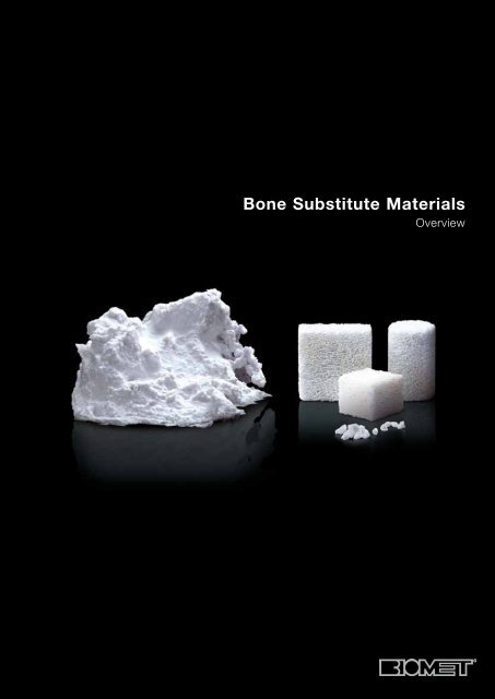
<strong>Bone</strong> <strong>Substitute</strong> <strong>Materials</strong><br />
Overview
Content<br />
Introduction 3<br />
Overview 4<br />
Endobon 6<br />
Calcium Phosphate 8<br />
Calcibon Paste 10<br />
Calcibon Granules 12<br />
Ordering Information 14<br />
References 16<br />
What can patients and surgeons expect from us? Reliable products,<br />
and expertise, which ensures secure application. Moreover, developments<br />
needed in the future.<br />
<strong>Biomet</strong> is one of the leading orthopaedic companies worldwide. We<br />
develop and produce products for orthopaedic and trauma surgery.<br />
Our predominant competence enables us to accompany our clients<br />
continuously in clinical surgeries.<br />
This vicinity has an impact: We take impulses in, and our own Research<br />
and Development Department is a direct contact partner for new ideas.<br />
Here we connect the clinically documented quality of our implants with<br />
the future orientated possibilities of bioactive materials. Through this we<br />
become an innovation force and are able to face the continuous developments<br />
in our markets more flexibly.<br />
The results are products and performances, that aid the surgeons’<br />
community, to support the healing process of their patients in a<br />
medically optimal, scientifically proven, and cost-effective manner.<br />
2<br />
Legal notice<br />
This material is intended for educational purposes only. Any medical information included herein<br />
is for information purposes and shall not be used in any way as substitute for professional advice<br />
provided by a physician or other health care provider.
<strong>Bone</strong> <strong>Substitute</strong> <strong>Materials</strong> from<br />
<strong>Biomet</strong><br />
Time-tested Products<br />
Autologous graft, bone bank or bone substitute material?<br />
The first criterion when treating a bone defect is always<br />
a rapid recovery process, without as few complications<br />
for the patient as possible. More over economical and<br />
technical aspects are essential. The treatment with<br />
bone substitutes proves to be well suited for the<br />
treatment of bone defects with respect to availability,<br />
duration of surgery and incurred costs, linked with<br />
excellent compatibility and documented treatment<br />
achievements.<br />
Quality and Unlimited Quantity<br />
<strong>Biomet</strong> developed a portfolio of bone substitute materials<br />
(based on mainly hydroxyapatite calcium phosphates). Unlike<br />
autologous material, they have the advantage to have a<br />
rapid availability in constant quality and unlimited quantity.<br />
Furthermore, the application of bone substitute materials<br />
avoids a second surgery for harvesting autologous material:<br />
The duration of the surgery and the anesthesia are reduced<br />
and patients are not faced with possible complications and<br />
pain at the donor site.<br />
<strong>Bone</strong> substitute materials have advantages in comparison<br />
to bone bank materials: There are no logistics issues in<br />
providing the materials for surgery and they are available in<br />
adequate quantity.<br />
A Solution for Each Indication<br />
Not all bone substitute materials are the same: For each<br />
separate indication, suitable bone substitute materials<br />
were thoroughly researched and developed. Particularly<br />
the utilisation of different substances with specific<br />
characteristics and functionalities supports the healing<br />
process in the long run. <strong>Biomet</strong>'s bone substitute materials<br />
have proven to be of high quality for a number of<br />
years already. Their excellent biocompatibility and their<br />
clinical success are documented extensively. Praxisorientated<br />
trainings and consulting services aid to support<br />
surgeons and their personnel in order to ensure consistent<br />
successful application.<br />
3
Overview <strong>Bone</strong> <strong>Substitute</strong> <strong>Materials</strong>:<br />
A Solution for Each Indication<br />
A precondition for successful clinical use of bone substitutes<br />
are the exact indication and the correct application. They<br />
are suitable for non-infected, metaphyseal, cancellous bone<br />
defects. <strong>Bone</strong> substitutes should only be implanted into a<br />
vital bony bed. When utilizing bone substitute materials,<br />
it is always vital to carry out a correct reposition and an<br />
adequate osteosynthesis.<br />
Product Endobon Calcibon<br />
Blocks, Cylinder,<br />
Granules<br />
Paste<br />
(liquid & powder)<br />
Basic Material Hydroxyapatite (ceramic) Calciumdeficient hydroxyapatite<br />
Material Properties<br />
Biological<br />
Stable osseous integration<br />
Ready-to-use<br />
Augmentable<br />
Porous<br />
Synthetic<br />
Stable osseous integration<br />
Characteristics Osteoconductive Osteoconductive<br />
Biodegradable<br />
Indications<br />
1. Tibia plateau fractures<br />
2. Osteotomies<br />
1. Distal radius fractures<br />
2. Calcanus fractures<br />
3. Tibia plateau fractures<br />
4
Calcibon Granules<br />
Granules<br />
Calciumdeficient hydroxyapatite<br />
Synthetic<br />
Stable osseous integration<br />
Ready-to-use<br />
Augmentable<br />
Porous<br />
Osteoconductive<br />
Biodegradable<br />
1. Mixing with spongiosa<br />
2. Large bone defects, e.g. hip revision<br />
5
Endobon<br />
Primary stable<br />
Osteoconductive<br />
Interconnecting pore system<br />
Augmentable<br />
Endobon is a natural hydroxyapatite ceramic (HA ceramic),<br />
which is particularly suitable as a bone graft substitute. It is<br />
of biological origin and osteoconductive. The newly formed<br />
bone can grow directly onto the ceramic surface and into<br />
the implant. The interconnecting pore system of Endobon<br />
allows the new bone to grow through the whole implant.<br />
Indications<br />
Endobon is an excellent bone substitute material for<br />
impression fractures close to the joint, like tibia and<br />
radius fractures, for fractures of the calcaneus and for<br />
osteotomies.<br />
Material Properties<br />
Endobon is a primary stable hydroxyapatite ceramic. The<br />
inner structure with its interconnecting system of macro<br />
and micro pores with pore sizes of 100 to 1,500 μm<br />
allows the newly formed bone to grow through the whole<br />
implant. This finally leads to a stable osseous integration of<br />
the Endobon implant.<br />
Endobon has a porosity of 45-85 Vol.% and a density<br />
of 0.4 to 1.6 g/cm 3 resembling the values of natural bone.<br />
Quality and Production<br />
Endobon is manufactured in a two-stage high-temperature<br />
process:<br />
• 1. Pyrolysis above 900°C<br />
• 2. Sintering above 1,200°C<br />
This leads to a complete combustion and ablation of all<br />
bacteria, viruses, and prions from the original material.<br />
Endobon has been successfully used in clinical applications<br />
for more than 15 years.<br />
Usage<br />
Endobon is available in the form of blocks, cylinders, or<br />
granules of different sizes. The latter are especially suited<br />
for augmentation of autologous bone chips.<br />
All forms are easily and rapidly usable during surgery.<br />
The blocks and cylinders are especially suitable for<br />
fractures below load bearing joints, e.g. tibia plateau<br />
fractures. The granules should be used for filling irregularly<br />
formed defects.<br />
6
6 Weeks Post-operatively<br />
6 weeks after the implantation of Endobon, integration of<br />
the implant with the surrounding vital bony bed can be seen<br />
on the microscopic analysis.<br />
3 Months Post-operatively<br />
After 3 months, the microscopic analysis shows a nearly<br />
complete osseous integration of the implant.<br />
6 Months Post-operatively<br />
After 6 months, the histological slide shows direct contact<br />
of the newly formed bone (1) and the ceramic surface (2).<br />
2<br />
1<br />
7
Calcium Phosphate – The Calcibon Family<br />
Synthetic<br />
Primary stable<br />
Osteoconductive<br />
Biodegradable<br />
Calcibon is a synthetic, biodegradable bone substitute<br />
material. It belongs to the family of calcium phosphates,<br />
a group of osteoconductive bone substitutes. The cured<br />
Calcibon material is a micro-crystalline, calciumdeficient<br />
hydroxyapatite. The chemical composition and the crystalline<br />
structure of the cured material mimic the mineral part<br />
of natural bone.<br />
Material Properties<br />
Calcibon powder is synthesized from calcium and phosphate<br />
salts. Its different components are Tri-calcium-phosphate,<br />
calcium-hydrogen-phosphate and calcium carbonate.<br />
The Biodegradability<br />
Calcibon has the potential to be biodegraded by the<br />
surrounding vital bony bed through remodelling. Calcibon is<br />
thereby broken down by osteoclasts and the new bone is<br />
reconstructed by osteoblasts.<br />
The remodelling process depends on the individual and<br />
the environment where Calcibon is implanted. A vital bony<br />
bed, close contact with the surrounding bone, and a functional<br />
stimulation improve the rate of the remodelling.<br />
8
Calcibon – Two Products: Paste or Granules<br />
The synthetic calcium-phosphate material Calcibon is<br />
available in two different forms: as a paste, mixed from a<br />
liquid and a powder component, or ready-to-use as<br />
granules.<br />
Despite identical origins the two products, Calcibon and<br />
Calcibon Granules differ in their properties regarding<br />
further processing. Both products are introduced in more<br />
detail on the following pages.<br />
• Osteoclasts recognize<br />
bone mineral like surface<br />
• Osteoclasts start<br />
degradation<br />
Mechanical stimulation<br />
Differentiating factors<br />
Mesenchymal stem cells<br />
Pre-osteoblasts<br />
New bone matrix<br />
• Differentiation of<br />
mesenchymal stem cells<br />
• Activation of<br />
pre-osteoblasts<br />
• Osteoclasts release<br />
differentiating factors<br />
and thereby induce<br />
differentiation<br />
and activation<br />
of pre-osteoblasts<br />
Osteoblasts<br />
• Osteoblasts synthesis<br />
and deposit new bone<br />
matrix with subsequent<br />
mineralization<br />
9
Calcibon Paste<br />
High compressive strength<br />
Good clinical results<br />
Easy handling<br />
Hardens in aqueous environment<br />
Calcibon consists of two components: Calcibon liquid<br />
and Calcibon powder. Mixing these two components<br />
results in a smooth and malleable paste ideal for filling bone<br />
defects. The paste hardens at body temperature in-situ and<br />
reaches a very high compressive strength.<br />
Processing<br />
The Calcibon paste is mixed directly before application.<br />
Calcibon liquid is an aqueous di-sodium-hydrogen-phosphate<br />
solution, which initiates the curing process within the<br />
paste.<br />
The processing cycle of Calcibon has been optimized in<br />
consideration of the Operating Room environment:<br />
• Short mixing time 1 minute<br />
• Sufficient application time 4 minutes<br />
• Acceptable final setting time 5 minutes<br />
Overall required time<br />
10 minutes<br />
The Compressive Strength<br />
One of the outstanding properties of Calcibon is its<br />
extremely high compressive strength. In-vitro testing shows<br />
that after 6 hours, the cured final material shows a compressive<br />
strength of approximately 15 MPa, already in the<br />
range of the strength of cancellous bone (10-20 MPa). This<br />
compressive strength continues to increase over time and<br />
results in a final strength of up to 45 MPa after 3 days.<br />
Thus, the compressive strength is in the area of cortical<br />
bone (25-100 MPa).<br />
Compressive strength versus time<br />
After 6 hours the compressive strength of Calcibon is comparable to<br />
that of cancellous bone. After 3 days the final compressive strength<br />
of up to 45 MPa is reached.<br />
10<br />
Compressive strength (MPa)<br />
60<br />
45 MPa = 3 days<br />
40<br />
20<br />
15 MPa = 6 hours<br />
0 50 100<br />
Time (h)
Indications<br />
Calcibon is indicated for refilling of noninfected, metaphyseal,<br />
cancellous bone defects caused either by<br />
trauma, benign tumour, surgery, or congenital. Calcibon<br />
may also be used to refill cavities created in connection with<br />
kyphoplasty, if classified as fracture type A1 (according to<br />
Magerl classification).<br />
Osteoblasts<br />
Osteocyte<br />
The most common indications for the clinical use of<br />
Calcibon are distal radius fractures, tibia plateau fractures,<br />
and calcaneus fractures.<br />
Usage<br />
The cohesion time of Calcibon is extremely short. Already<br />
after mixing, the binding forces inside the paste are high<br />
enough to ensure the form stability of the material, even in<br />
an aqueous environment.<br />
Calcibon<br />
2 weeks post-operatively<br />
Detailed picture of the bone formation at the contact site Calcibon implant-bone.<br />
The Calcibon surface is covered with an osteoid-layer (*).<br />
<strong>Bone</strong><br />
Even though Calcibon should be implanted into a dry and<br />
blood-free environment, the short cohesion time allows an<br />
implantation into a wet bony bed, where the material will<br />
keep its form and harden adequately.<br />
Calcibon<br />
16 weeks post-operatively<br />
Parts of the Calcibon implant material have been biodegraded followed<br />
by bone in-growth. Remodelling activity at the interface, as characterised<br />
by the presence of remodelling lacunae (RL) and osteoclast-like cells<br />
(black arrows), was still seen.<br />
Calcibon<br />
24 weeks post-operatively<br />
Continued biodegradation of Calcibon and subsequent bone in-growth.<br />
The new bone is mature and cannot be discerned from original trabecular<br />
bone.<br />
11
Calcibon Granules<br />
Refilling of lange bone defects<br />
Porosity supports remodelling process<br />
Augmentation with other material<br />
Utilization in revision surgery<br />
Calcibon Granules are a line extension of the existing<br />
Calcibon product range. Like the paste, the granules are<br />
also manufactured synthetically, are biodegradable and<br />
biocompatible. The main differences: The granules are<br />
ready-to-use, however, due to their porosity the compressive<br />
strength is reduced. In addition they are highly suitable<br />
to be used for augmentation in combination with other<br />
materials.<br />
Material Properties<br />
The production process of Calcibon Granules is at first<br />
glance identical to the one of the Calcibon powder. Nevertheless,<br />
the mixing of the two components already takes<br />
place during production. The granules are hence a completely<br />
hardened material. Due to a special manufacturing<br />
technique porous granules with micro and macro pores are<br />
produced.<br />
The porous Calcibon Granules have macro pores of 150-<br />
550 µm. These allow cells, e.g., osteoclasts and osteoblasts,<br />
to migrate into the granules. The activities of these<br />
cells enable the cellular biodegradation of the Calcibon<br />
Granules and the formation of new autologous bone.<br />
The macro pores are separated from each other by thin<br />
walls, which in turn consist of a micro porous network.<br />
These micro pores allow the cells involved in the remodelling<br />
process to be supplied with nutrients.<br />
12
Indications<br />
Generally, Calcibon Granules are intended for filling and<br />
reconstructing large, non-infected, metaphyseal, cancellous<br />
bone defects. The origin of these defects can vary<br />
greatly; from e.g., trauma, benign tumour, surgery, to<br />
congenital.<br />
Calcibon Granules can be used as filling material for<br />
revision hip surgery, e.g., for the reconstruction of the<br />
acetabulum and to fill the shaft. Moreover, the granules can<br />
be used as filling material for cages in spinal surgery.<br />
Use<br />
Calcibon Granules are ready-to-use. No mixing time has to<br />
be taken into account.<br />
2 ml Calcibon Granules mixed with 1 ml PRP (Platelet-Rich-Plasma of<br />
<strong>Biomet</strong> Biologics)<br />
Calcibon Granules can be applied directly to the aseptic<br />
bone bed or augmented with other material, such as<br />
bone marrow, bone grafts, blood, and PRP (Platelet-Rich-<br />
Plasma, extracted with the GPS III System).<br />
Scanning electron microscope image of the interconnecting micro pore<br />
system. Magnification x 3,000<br />
13
Ordering Information<br />
Calcibon Synthetic calcium-phosphate<br />
Product Description Quantity Art. Nr.<br />
Calcibon 5 g circa 4 ml paste 30 3005 0001<br />
Calcibon 10 g circa 8 ml paste 30 3010 0001<br />
Calcibon 20 g circa 16 ml paste 30 3020 0001<br />
Calcibon Granules 10 10 ml Granules 30 3210 0001<br />
Calcibon Granules 20 20 ml Granules 30 3220 0001<br />
Calcibon Granules 50 50 ml Granules 30 3250 0001<br />
Endobon Hydroxyapatite<br />
Product Description Size Art. Nr.<br />
Block 5 5.0 x 5.0 x 10.0 mm 30 2037 0001<br />
Block 12.5 12.5 x 12.5 x 10.0 mm 30 2030 0001<br />
Block 20 20 x 20 x 10.0 mm 30 2031 0001<br />
Product Description Size Art. Nr.<br />
Cylinder 9 Ø 8.50 x 20 mm 30 2022 0001<br />
Cylinder 10 Ø 9.55 x 20 mm 30 2023 0001<br />
Cylinder 11 Ø 10.60 x 20 mm 30 2024 0001<br />
Cylinder 12 Ø 11.70 x 20 mm 30 2025 0001<br />
Product Description Granules size Art. Nr.<br />
Granules I 5 ml 1.5 - 2.8 mm 30 2034 0001<br />
Granules II 5 ml 2.8 - 5.6 mm 30 2035 0001<br />
Granules II 10 ml 2.8 - 5.6 mm 30 2069 0001<br />
14
Notes<br />
15
References<br />
IW850011<br />
12/2009<br />
Endobon<br />
Calcibon and Calcibon Granules<br />
1. Baer W., Schaller P., Carl H.D.: „Spongy hydroxyapatite in hand<br />
surgery – A five year follow-up". J. Hand. Surg. (Br) 2002; 27B:<br />
101–103.<br />
2. Briem D., Linhart W., Lehmann W., Meenen N.M., Rueger J.M.:<br />
„Langzeitergebnisse nach Anwendung einer porösen<br />
Hydroxyapatitkeramik (Endobon) zur operativen Versorgung von<br />
Tibiakopffrakturen”. Unfallchirurg 2002; 105: 128–133.<br />
3. Großpeter A.S., Pretzsch M., Frh. van Salis-Soglio G.: „Die<br />
Behandlung knöcherner Defekte mit der Hydroxylapatitkeramik<br />
Endobon – Eine mittelfristige klinische und radiologische<br />
Verlaufsbeobachtung bei 58 Patienten”. Orthopädische Praxis 2004;<br />
40: 290–294.<br />
4. Helber M.U., Ulrich C.: „Metaphysärer Defektersatz mit Hydroxylapatitkeramik<br />
– 3- bis 4 Jahresnachuntersuchungsergebnisse”.<br />
Unfallchirurg 2000; 103: 749–753.<br />
5. Kehr P., Gosset F.: „Endobon as a bone substitute in spine surgery.<br />
Preliminary study in 11 patients”. Europ. J. Orthop. Surg. Traumatol<br />
2000; 10: 217–221.<br />
6. Khodadadyan-Klostermann C., Liebig T., Melcher I., Raschke M.,<br />
Haas N.P.: „Osseous integration of hydroxyapatite grafts in metaphyseal<br />
bone defects of the proximal tibia” (CT-Study). Acta. Chir.<br />
Orthop. Traumatol Cech. 2002; 69(1): 16– 21.<br />
7. Müller-Mai C., Voigt C., Hering A., Rahmanzadeh R., Gross U.:<br />
„Madreporische Hydroxylapatitgranulate zur Füllung ossärer Defekte”.<br />
Unfallchirurg 2001; 104: 221–229.<br />
8. Sailer R., Lutz M., Zimmermann R., Hackl W., Gabl M., Blauth M.:<br />
„Minimalinvasive Therapie der dislozierten distalen metaphysären<br />
Radiuskompressionsfraktur: klinische und radiologische Ergebnisse<br />
nach gedeckter Reposition, Stiftfixation und stabiler Defektauffüllung<br />
mit einer porösen Hydroxylapatitkeramik”. Akt. Traumatol 2003; 33:<br />
26-30.<br />
1. Driessens F.C.M., Boltong M.G., Wenz R.: „Calcium phosphate<br />
bone cements: State of the art 2000”. 12th Conference of the<br />
European Society of Biomechanics, Dublin, Ireland, 27–30th August<br />
2000.<br />
2. Hillmeier J., Meeder P.J., Nöldge G., Kasperk C.: „Minimally invasive<br />
reduction and internal stabilization of osteoporotic vertebral body<br />
fractures (Balloon Kyphoplasty)”. Operat. Orthop. Traumatol 2003;<br />
15: 343–362.<br />
3. Hillmeier J., Meeder P.J., Nöldge G., Kock H.J., Da Fonseca K.,<br />
Kasperk C.: „Augmentation von Wirbelkörperfrakturen mit einem<br />
neuen Calciumphosphat-Zement nach Ballon-Kyphoplastie”.<br />
Orthopäde 2004; 33: 31–39.<br />
4. Hillmeier J., Grafe I., Da Fonseca K., Meeder P.J., Nöldge G.,<br />
Libicher M., Kock H.J., Haag M., Kasperk C.: „Die Wertigkeit<br />
der Ballonkyphoplastie bei der osteoporotischen Wirbelkörperfraktur”.<br />
Orthopäde 2004; 33: 893–904.<br />
5. Linhart W., Briem D., Peters A., Lehmann W., Windolf J., Rueger J.M.:<br />
„Resorbierbare Kalziumphosphatzemente”. Trauma Berufskrankheit<br />
2004; online: 29. Oktober.<br />
6. Ooms E.M., Wolke J.G.C., van der Waerden J.P.C.M., Jansen<br />
J.A.: „Trabecular bone response to injectable calcium phosphate<br />
(Ca-P) cement”. J-Biomed-Mater Res 2002; 61: 9–18.<br />
7. Ooms E.M., Wolke J.G.C., van de Heuvel M.T., Jeschke B., Jansen<br />
J.A.: „Histological evaluation of the bone response to calcium<br />
phosphate implanted in cortical bone”. Biomaterials 2003; 24:<br />
989–1000.<br />
8. Ooms E.M., Egglezos E.A., Wolke J.G.C., Jansen J.A.:<br />
„Soft-tissue response to injectable calcium phosphate cements”.<br />
Biomaterials 2003; 24: 749–757.<br />
9. Schnettler R., Stahl J.P., Alt V., Pavlidis T., Dingeldein E., Wenisch S.:<br />
„Calcium phosphate-based bone substitutes“. Europ. J. Trauma<br />
2004; 30: 219–229.<br />
This material is intended for the sole use and benefit of physicians. It is not to be<br />
redistributed, duplicated or disclosed without the express written consent of <strong>Biomet</strong>.<br />
Worldwide distributor<br />
<strong>Biomet</strong> Deutschland GmbH<br />
Gustav-Krone-Str. 2<br />
D-14167 Berlin<br />
Tel.: +49 / 30 / 845 81-0<br />
Fax: +49 / 30 / 845 81-110<br />
www.biomet.de<br />
Responsible manufacturers:<br />
Calcibon, Calcibon Granules<br />
<strong>Biomet</strong> Deutschland GmbH<br />
Gustav-Krone-Str. 2<br />
D-14167 Berlin<br />
www.biomet.de<br />
0123<br />
Endobon<br />
<strong>Biomet</strong> France S.A.R.L.<br />
58 Avenue de Lautagne<br />
B.P. 75<br />
F-26903 Valence Cedex<br />
www.biomet.fr 0459