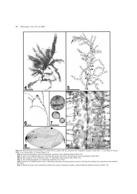
46 Phycologia, Vol. 42 (1), 2003 Figs 4–9. <strong>Reticulocaulis</strong> mucosissimus. ac, axial cell; jc, jacket cell; pc, periaxial cell. Vegetative features. Scale bars 2 cm (Fig. 4); 50 m (Figs 5, 6); 10 m (Fig. 7); 50 m (Figs 8, 9). Fig. 4. Female gametophyte (pressed herbarium specimen) <strong>from</strong> Masirah Island; MAS 138. Fig. 5. Apex of an indeterminate axis, showing <strong>the</strong> apical cell (arrow) and periaxial cells (arrowheads); KM 4481. Fig. 6. <strong>The</strong> transition <strong>from</strong> cylindrical inner- to spheroidal outer-cortical cells; MAS 138. Fig. 7. Two-celled propagules on a Hawaiian specimen; IA 17225. Fig. 8. Detail of an inflated jacket cell in a Hawaiian specimen, showing primary (arrows) and lateral secondary pit connections (arrowheads); IA 23471. Fig. 9. Sheath of jacket cells around <strong>the</strong> central-axial strand, showing secondary cortical filament initiation (arrows); MAS 138.
Schils et al.: <strong>Reticulocaulis</strong> in <strong>the</strong> Indian Ocean 47 nections results in a zigzag arrangement of carpogonial branch cells when viewed dorsally or ventrally (Fig. 12). <strong>The</strong> carpogonial branch curves sharply towards <strong>the</strong> axis bearing it and <strong>the</strong> carpogonium arises adaxially on cell #2, <strong>the</strong> hypogynous cell (Figs 10, 11). <strong>The</strong> initially short and reflexed trichogyne can elongate to over 500 m (Figs 12, 13; Abbott 1985). Cell #2 initiates a cluster of 4–6 branched filaments of tightly packed nutritive cells (Fig. 14), whereas cells #3 and #4 tend to bear a primary, slightly branched lateral, a second slightly more branched lateral and 1–3 small clusters of ramified nutritive cells (Fig. 13). Primary laterals, 6–16 cells in length and branched to two orders, form adaxially on most of <strong>the</strong> remaining carpogonial branch cells, <strong>the</strong> longest occurring on <strong>the</strong> most proximal cells (Figs 13, 14). Any of <strong>the</strong> cells proximal to cell #4 may ultimately bear ei<strong>the</strong>r an abaxial or an adaxial second sterile filament. Upon presumed fertilization, <strong>the</strong> carpogonial branch cells and <strong>the</strong> basal cells of <strong>the</strong> sterile laterals inflate, and both <strong>the</strong> pit connections and <strong>the</strong> nuclei of <strong>the</strong>se cells enlarge substantially (Fig. 15). <strong>The</strong> gonimoblast initial develops directly <strong>from</strong> <strong>the</strong> fertilized carpogonium (Fig. 16); at <strong>the</strong> same time, <strong>the</strong> nutritive cells fuse directly with <strong>the</strong> hypogynous cell through <strong>the</strong>ir pit connections, which retain <strong>the</strong>ir original size or expand only slightly as <strong>the</strong> pit plugs break down (Fig. 17). <strong>The</strong> passageways that are now open between <strong>the</strong> hypogynous cell and <strong>the</strong> nutritive-cell clusters presumably become paths for direct nutrient transport to <strong>the</strong> developing gonimoblast. <strong>The</strong> carposporophyte remains compact, does not intermingle with vegetative tissue, and lacks a pericarp. Ovoid carposporangia (40 30 m) terminate <strong>the</strong> branches of <strong>the</strong> compact gonimoblast (Fig. 18); cystocarps at various stages of development are found scatte<strong>red</strong> within <strong>the</strong> cortex and reach 330 m in diameter. Spermatangia are produced in terminal dendroid clusters on separate male gametophytes, <strong>the</strong> fertile axes often being accompanied by a sterile sibling cortical filament of one or two cells (Fig. 19). Spermatangial mo<strong>the</strong>r cells initiate 1–3 spermatangia (Fig. 20). Tetrasporangial thalli were not collected in <strong>the</strong> course of this study and are unrecorded for <strong>the</strong> <strong>genus</strong>. In line with findings for o<strong>the</strong>r genera of <strong>the</strong> Naccariaceae (Jones & Smith 1970; Boillot & L’Hardy-Halos 1975), <strong>Reticulocaulis</strong> is presumed to have a heteromorphic life history involving a diminutive system of prostrate filaments bearing terminal tetrahedral tetrasporangia. Growth of Hawaiian R. mucosissimus in culture, reported by Abbott (1999, p. 123), resulted in a microscopic filamentous phase but no production of tetrasporangia. <strong>Reticulocaulis</strong> obpyriformis Schils, sp. nov. Affinis R. mucosissimis Abbott (1985) sed differt characteribus pluribus. Gametophyta monoica; thallus pallido-roseolus pallidus, usque ad 15 cm altus, rami indeterminatis laxe et irregulatim ramificantibus. Cellulae corticis obpyriformes cylindricae; rami breves cellulis parvis sphaericis in filamento corticato, rarus evolutantes in axes indeterminatos; interdum trichomata in cellulis terminalibus rel subterminalibus corticis portata; cellulae axiales intra 1 mm sub apice latae ad 70(–80) m. Spermatangia evoluta e filamenti corticalis cellulis distalibus. Praesentia duorum ramorum carpogonialium in cellula basali frequentior quam in R. mucosissimo. Filamenta lateralia secunda persaepe in cellulis proximis ramorum carpogonialium. Similar to R. mucosissimus Abbott (1985) but with <strong>the</strong> following distinguishing characters: gametophytes monoecious; thalli pale pink, to 15 cm high; branching of indeterminate axes loose and irregular. Cortical cells obpyriform and cylindrical; cortical filaments bearing short laterals consisting of small spherical cells and potentially developing into indeterminate axes; hairs occasional on terminal and subterminal cortical cells; axial cells broadening to 70(–80) m within 1 mm of <strong>the</strong> apices. Spermatangia developing directly <strong>from</strong> catenate series of distal cortical cells. Supporting cells bearing two carpogonial branches occur more frequently than in R. mucosissimus. Secondary laterals common on proximal carpogonial branch cells. HOLOTYPE: GENT, SMM 446 (Fig. 21) TYPE LOCALITY: West of Bidholih, south coast of Socotra Island (Figs 1, 3). Sample site ALG-40 (12.303N, 53.843E): a rocky platform at 19 m cove<strong>red</strong> with thin layers of sand and punctuated by deeper sand patches (Schils, 30 April 2000). ETYMOLOGY: obpyriformis, refers to <strong>the</strong> inverse pear shape of <strong>the</strong> cortical cells. <strong>The</strong> thalli are terete, pale pink, and up to 15 cm in length (Fig. 21). Branching is irregularly radial, with a sparse development of up to four orders of indeterminate laterals. <strong>The</strong> domeshaped apical cell divides obliquely, <strong>the</strong> immediate derivatives forming a sinusoidal pattern before <strong>the</strong> axial cells become aligned (Fig. 23). Within 1 mm of <strong>the</strong> apices, <strong>the</strong> axial cells broaden to attain length–width ratios of 4 : 1 (Fig. 22). <strong>The</strong> superior periaxial cell is cut off at about <strong>the</strong> third axial cell behind <strong>the</strong> apex, <strong>the</strong> ‘phyllotaxy’ on successive segments being alternate (Fig. 23). Inferior periaxial cells, rhizoidal downgrowths and laterals develop <strong>from</strong> about <strong>the</strong> 40th axial cell downwards, at which time <strong>the</strong> phyllotaxy of <strong>the</strong> determinate laterals tends to become an irregular ¼ spiral, because <strong>the</strong> inferior periaxials set in at a 90 angle to <strong>the</strong> superior periaxial cells. Derivatives of <strong>the</strong> inferior periaxial cells become more strongly developed than those of <strong>the</strong> superior cells and initiate <strong>the</strong> occasional indeterminate branch when <strong>the</strong> cortical filament continues growing and initiates periaxial cells. Third-order periaxial cells are very infrequently initiated in older parts of <strong>the</strong> thallus; <strong>the</strong>y develop cortical filaments and jacket cells like <strong>the</strong> o<strong>the</strong>r periaxial cells. <strong>The</strong> lower cells of <strong>the</strong> cortical filaments are p<strong>red</strong>ominantly obpyriform (Figs 22, 24), although cylindrical to barrelshaped cells also occur (Fig. 24). <strong>The</strong> sizes and contours of <strong>the</strong> cortical cells change ra<strong>the</strong>r abruptly distally, <strong>from</strong> being elongated, obpyriform or cylindrical, and up to 90 m long by 27 m wide, to being small, spherical and 4–6 m in diameter. Hairs develop occasionally on terminal and subterminal cortical cells (Fig. 25), but propagules were not observed. Certain cortical filaments bear short moniliform laterals of small spherical to ovoid cells (Fig. 24); <strong>the</strong>se laterals can bear spermatangia, less often carpogonial branches, or may transform directly into indeterminate axes (<strong>the</strong> atypical way of indeterminate lateral formation: Fig. 23). Several orders of rhizoidal downgrowths develop <strong>from</strong> <strong>the</strong> periaxial cells, <strong>the</strong> cells becoming inflated and linked by lateral secondary pit connections (Fig. 26) and forming a sheath around <strong>the</strong> axial strand (Fig. 27), in which <strong>the</strong> pit connections attenuate and become obscure. <strong>The</strong>se jacket cells are spheroidal and may give rise to secondary cortical filaments. In older parts of <strong>the</strong> thallus, <strong>the</strong> jacket cells become densely cove<strong>red</strong>