The red algal genus Reticulocaulis from the Arabian
The red algal genus Reticulocaulis from the Arabian
The red algal genus Reticulocaulis from the Arabian

Create successful ePaper yourself
Turn your PDF publications into a flip-book with our unique Google optimized e-Paper software.
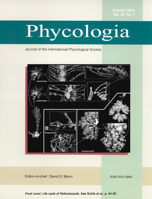
Phycologia (2003) Volume 42 (1), 44–55 Published 7 March 2003<br />
<strong>The</strong> <strong>red</strong> <strong>algal</strong> <strong>genus</strong> <strong>Reticulocaulis</strong> <strong>from</strong> <strong>the</strong> <strong>Arabian</strong> Sea, including<br />
R. obpyriformis sp. nov., with comments on <strong>the</strong> family Naccariaceae<br />
TOM SCHILS∗, OLIVIER DE CLERCK AND ERIC COPPEJANS<br />
Phycology Research Group, Biology Department, Ghent University, Krijgslaan 281 (S8), 9000 Ghent, Belgium<br />
T. SCHILS, O.DE CLERCK AND E. COPPEJANS. 2003. <strong>The</strong> <strong>red</strong> <strong>algal</strong> <strong>genus</strong> <strong>Reticulocaulis</strong> <strong>from</strong> <strong>the</strong> <strong>Arabian</strong> Sea, including R.<br />
obpyriformis sp. nov., with comments on <strong>the</strong> family Naccariaceae. Phycologia 42: 44–55.<br />
<strong>Reticulocaulis</strong> obpyriformis Schils, sp. nov. is described <strong>from</strong> <strong>the</strong> south coast of Socotra Island (Yemen), and a second<br />
species, R. mucosissimus, is recorded <strong>from</strong> a similar upwelling area in <strong>the</strong> <strong>Arabian</strong> Sea (Masirah Island, Oman). <strong>The</strong>se are<br />
<strong>the</strong> first published records of Naccariaceae for <strong>the</strong> Indian Ocean and end <strong>the</strong> monospecific, Hawaiian-endemic status of<br />
<strong>Reticulocaulis</strong>. Features distinguishing R. obpyriformis <strong>from</strong> R. mucosissimus include its more sparsely branched thallus,<br />
obpyriform ra<strong>the</strong>r than cylindrical inner cortical cells, <strong>the</strong> presence of short moniliform laterals of small spherical cells on<br />
<strong>the</strong> cortical filaments, monoecious ra<strong>the</strong>r than dioecious gametophytes, and <strong>the</strong> direct development of spermatangia <strong>from</strong><br />
catenate mo<strong>the</strong>r cells. <strong>The</strong> morphology and anatomy of <strong>the</strong> gametophytes of this heteromorphic <strong>genus</strong> are discussed in<br />
relation to those of o<strong>the</strong>r naccariacean genera.<br />
INTRODUCTION<br />
Recent phycological studies in <strong>the</strong> <strong>Arabian</strong> Sea and <strong>the</strong> nor<strong>the</strong>rn<br />
Indian Ocean have resulted in <strong>the</strong> description of new taxa<br />
(Wynne 1999a) and a plethora of new records (Wynne & Banaimoon<br />
1990; Wynne & Jupp 1998; Wynne 1999b, 2000) indicative<br />
of a unique marine benthic flora. <strong>The</strong> south-west<br />
monsoon that results in upwelling along <strong>the</strong> south-eastern<br />
coastline of <strong>the</strong> <strong>Arabian</strong> Peninsula (Currie et al. 1973; Ormond<br />
& Banaimoon 1994) is an important physical phenomenon<br />
influencing <strong>the</strong>se neritic ecosystems and <strong>the</strong>ir biotas, particularly<br />
those of Masirah Island (Oman) and <strong>the</strong> Socotra Archipelago<br />
(Yemen), which support a seasonally rich diversity<br />
of gelatinous <strong>red</strong> algae (Schils & Coppejans 2002). Among<br />
<strong>the</strong> more unexpected of <strong>the</strong> algae recently discove<strong>red</strong>, <strong>the</strong>re<br />
are two species of <strong>Reticulocaulis</strong>, a hi<strong>the</strong>rto monotypic <strong>genus</strong><br />
thought to be confined to Hawaii in <strong>the</strong> central Pacific Ocean<br />
and a member of <strong>the</strong> relatively little-known and infrequently<br />
encounte<strong>red</strong> family Naccariaceae.<br />
Following <strong>the</strong> recommendations of Kylin (1928), Svedelius<br />
(1933) and Feldmann & Feldmann (1942), <strong>the</strong> Naccariaceae<br />
is generally included in <strong>the</strong> order Bonnemaisoniales, based on<br />
details of gonimoblast development and <strong>the</strong> presence of nutritive-cell<br />
clusters on <strong>the</strong> carpogonial branch (Chihara &<br />
Yoshizaki 1972), an ordinal placement supported by ultrastructural<br />
characters of <strong>the</strong> pit plugs (Pueschel & Cole 1982).<br />
Womersley (1996), however, commented that <strong>the</strong> family might<br />
not be related to <strong>the</strong> Bonnemaisoniaceae, because of some<br />
seemingly major differences in <strong>the</strong> carposporophyte, such as<br />
a diffuse ra<strong>the</strong>r than compact gonimoblast and <strong>the</strong> complete<br />
absence of a pericarp. Abbott (1999) recently placed <strong>the</strong> Naccariaceae<br />
in <strong>the</strong> Gigartinales without specifying her reasons<br />
for <strong>the</strong> transfer. <strong>The</strong> Naccariaceae currently comprises <strong>the</strong><br />
genera Atractophora P. Crouan & H. Crouan, Naccaria Endlicher<br />
and <strong>Reticulocaulis</strong> I.A. Abbott. Despite consisting of<br />
only seven species, of which five belong to Naccaria, <strong>the</strong><br />
∗ Corresponding author (tom.schils@rug.ac.be).<br />
family is widely distributed throughout <strong>the</strong> Atlantic and Pacific<br />
Oceans. Although a single robust female gametophyte of<br />
N. naccarioides (J. Agardh) Womersley & I.A. Abbott is<br />
known <strong>from</strong> <strong>the</strong> Indian Ocean coast of Western Australia<br />
(GEN-10793e, MELU: leg. G.T. Kraft & G.W. Saunders, 7<br />
October 1995, Pinaroo, Western Australia, 32.20S, 115.45E),<br />
<strong>the</strong> present article is <strong>the</strong> first published report on a member<br />
of <strong>the</strong> Naccariaceae <strong>from</strong> anywhere in <strong>the</strong> Indian Ocean.<br />
MATERIAL AND METHODS<br />
<strong>The</strong> east coast of Masirah Island (Oman; 20.42N, 58.79E;<br />
Figs 1, 2) and <strong>the</strong> south coast of Socotra (Yemen; 12.47N,<br />
53.87E; Figs 1, 3) are influenced by a seasonal coastal upwelling<br />
<strong>from</strong> May to September, during <strong>the</strong> south-west monsoon.<br />
<strong>The</strong> specimens of this report were found in similar habitats<br />
around Masirah and Socotra, viz. rocky platforms at 10–<br />
20 m depth on which macroalgae were <strong>the</strong> most abundant<br />
benthic organisms, interspersed with isolated small hard and<br />
soft coral colonies. Gelatinous <strong>red</strong> algae such as Dudresnaya<br />
P. Crouan & H. Crouan, Gibsmithia Doty, Platoma Schousboe<br />
ex Schmitz and P<strong>red</strong>aea De Toni species (Schils & Coppejans<br />
2002), were particularly conspicuous during <strong>the</strong> early-winter<br />
and late-spring periods, o<strong>the</strong>r associated algae being Amphiroa<br />
J.V. Lamouroux spp., Callophycus Trevisan sp., Caulerpa<br />
peltata J.V. Lamouroux, Euptilota fergusonii Cotton, Galaxaura<br />
marginata (Ellis & Solander) J.V. Lamouroux, Halimeda<br />
J.V. Lamouroux spp., Lobophora variegata (J.V. Lamouroux)<br />
Womersley ex Oliveira, Rhodymenia Greville spp., Spatoglossum<br />
asperum J. Agardh, and Udotea indica A. Gepp & E.<br />
Gepp.<br />
Specimens of <strong>Reticulocaulis</strong> were collected by <strong>the</strong> first author<br />
during field trips to Masirah Island on 2–30 November<br />
1999 and Socotra on 26 March–7 May 2000, <strong>the</strong> subtidal habitats<br />
being accessed by means of SCUBA. <strong>The</strong> collected algae<br />
were pressed on herbarium sheets, with portions preserved in<br />
a 5% formalin–seawater solution. Herbarium sheets, wet spec-<br />
44
Schils et al.: <strong>Reticulocaulis</strong> in <strong>the</strong> Indian Ocean 45<br />
Figs 1–3. Collection sites of <strong>Reticulocaulis</strong> in <strong>the</strong> <strong>Arabian</strong> Sea. Scale<br />
bars 1000 km (Fig. 1); 20 km (Figs 2, 3).<br />
Fig. 1. <strong>The</strong> <strong>Arabian</strong> Peninsula showing Masirah Island and Socotra.<br />
Fig. 2. Sample site 9 (asterisk; 20.199N, 58.715E), near Ras Zarri,<br />
off Masirah Island, Oman.<br />
Fig. 3. Sample site ALG-40 (asterisk; 12.303N, 53.843E), west of<br />
Bidholih, off Socotra, Yemen.<br />
imens and microscope slides are deposited in GENT (Ghent<br />
University Herbarium, Krijgslaan 281/S8, 9000 Ghent, Belgium).<br />
Slides and formalin-preserved samples of Hawaiian R.<br />
mucosissimus I.A. Abbott were kindly supplied by I. A. Abbott<br />
of <strong>the</strong> Bernice Bishop Museum. Herbarium sheets of N.<br />
corymbosa J. Agardh and N. wiggii (Turner) Endlicher were<br />
borrowed <strong>from</strong> <strong>the</strong> National Herbarium of <strong>the</strong> Ne<strong>the</strong>rlands<br />
(L). Material for microscopical examination was stained with<br />
aniline blue, fast green or Lugol’s Iodine (for rhodoplasts).<br />
Material for nuclear and pit-connection studies was stained<br />
using Wittmann’s aceto-iron-haematoxylin–chloral hydrate<br />
(Wittmann 1965), following <strong>the</strong> procedures of Hommersand<br />
& F<strong>red</strong>ericq (1988). Anatomical and reproductive characteristics<br />
were observed <strong>from</strong> tissue squashes (whole-mounts in a<br />
50% corn syrup–water solution, containing a few drops of<br />
phenol) using light microscopy (Leitz Diaplan). Photographs<br />
were taken with a Wild MPS51 35 mm camera and on an<br />
Olympus DP50 digital camera.<br />
RESULTS<br />
<strong>Reticulocaulis</strong> mucosissimus I.A. Abbott 1985, p. 555<br />
SPECIMENS EXAMINED: Oman. Masirah Island (Figs 1, 2): sample site<br />
9 (20.199N, 58.715E), close to Ras Zarri. A rocky platform at 9<br />
m depth with scatte<strong>red</strong> rocky outcrops in an area of strong surge<br />
(Schils, 9 November 1999). MAS 138: female (Fig. 4) and male<br />
gametophytes. Hawaii. Mahukona, north-west coast of Hawaii.<br />
Plants growing on dead coral at a depth of 9 m (K. J. McDermid,<br />
26 May 1998). Formalin sample IA 23471 (female gametophyte)<br />
and slide KM 4481 (female gametophyte); Kawailoa, Oahu Island<br />
(W. H. Magruder & S. Carper, 10 May 1985). Slide IA 17225:<br />
female gametophyte.<br />
Thalli are bright <strong>red</strong>, mucilaginous and attached by a discoid<br />
holdfast (Fig. 4). Omani plants reach 13 cm in length and<br />
grow <strong>from</strong> dome-shaped apical cells that divide obliquely, <strong>the</strong><br />
immediate daughter cells being aligned in a nearly straight<br />
row (Fig. 5). <strong>The</strong> axial cells are slender and elongate, those<br />
lying 1 mm away <strong>from</strong> <strong>the</strong> apical cells having length–width<br />
ratios of 4 : 1. <strong>The</strong> first periaxial cell (<strong>the</strong> ‘superior’ periaxial)<br />
is cut off three axial cells <strong>from</strong> <strong>the</strong> apex, and superior<br />
periaxial cells on successive segments are produced in an irregular<br />
¼ spiral. A second periaxial cell (<strong>the</strong> ‘inferior’ periaxial)<br />
is always positioned proximal to <strong>the</strong> first. It is generally<br />
cut off in cells positioned 15–20 cells away <strong>from</strong> <strong>the</strong> apex<br />
(Fig. 5) and at a 90 angle to <strong>the</strong> first periaxial cell. At <strong>the</strong><br />
same time, several rhizoidal outgrowths develop <strong>from</strong> both<br />
periaxial cells; <strong>the</strong>se outgrowths branch. Besides differing in<br />
<strong>the</strong> timing of <strong>the</strong>ir initiation, <strong>the</strong> shapes of <strong>the</strong> two periaxial<br />
cells are also dissimilar: <strong>the</strong> superior periaxial cell becomes<br />
elongated and rectilinear, whereas <strong>the</strong> inferior one remains<br />
spherical (Fig. 5). <strong>The</strong> inferior lateral becomes <strong>the</strong> more developed<br />
of <strong>the</strong> two laterals and occasionally gives rise to indeterminate<br />
branches as it continues growing and initiates<br />
periaxial cells. Infrequently, an axial cell can initiate a third<br />
periaxial cell, which develops like <strong>the</strong> superior lateral. <strong>The</strong><br />
derivatives of <strong>the</strong> periaxial cells (<strong>from</strong> about <strong>the</strong> 15th axial<br />
cell) differentiate rapidly by branching and cell elongation<br />
into determinate filaments that constitute <strong>the</strong> cortex. <strong>The</strong> inner<br />
cortical cells are cylindrical (Fig. 6), whereas <strong>the</strong> outer cells<br />
remain ovoid to (sub)spherical.<br />
Two-celled propagules, reaching 16.5 m in diameter (Fig.<br />
7) and developing terminally on many of <strong>the</strong> cortical filaments,<br />
were observed on slide IA 17225 of a specimen <strong>from</strong><br />
Hawaii. One or two axial cells below <strong>the</strong> site where <strong>the</strong> second<br />
periaxial cell first forms, both periaxial cells initiate rhizoidal<br />
downgrowths. <strong>The</strong> periaxial cells and <strong>the</strong> rhizoidal<br />
downgrowths inflate into what were termed ‘jacket cells’ by<br />
Abbott (1985), viz. cells that mutually cross-connect by lateral<br />
secondary pit connections (Fig. 8) and constitute a sheath<br />
around <strong>the</strong> central-axial strand (Fig. 9). While maturing, <strong>the</strong><br />
pit connections of <strong>the</strong> jacket cells attenuate and become difficult<br />
to distinguish, which results in a seemingly parenchymatous<br />
covering. Before <strong>the</strong> covering is complete, <strong>the</strong> jacket<br />
cells initiate secondary cortical filaments that are ei<strong>the</strong>r fasciculate<br />
or unbranched, as well as secondary rhizoidal downgrowths.<br />
In older parts of <strong>the</strong> thallus, <strong>the</strong> jacket cells become<br />
densely cove<strong>red</strong> by <strong>the</strong>se secondary rhizoidal filaments, which<br />
rarely branch and form uniseriate rows that cross one ano<strong>the</strong>r,<br />
but actually constitute a single layer.<br />
<strong>The</strong> rhodoplasts are discoid but like erythrocytes in shape<br />
(2–4 m in diameter), having centres that are thinner than <strong>the</strong><br />
margins.<br />
Female gametophytes have carpogonial branches that are of<br />
accessory origin; <strong>the</strong>y were found throughout <strong>the</strong> thallus in<br />
various stages of development. Near <strong>the</strong> apex, carpogonial<br />
branches arise singly <strong>from</strong> ei<strong>the</strong>r of <strong>the</strong> periaxial cells. Fur<strong>the</strong>r<br />
down <strong>the</strong> thallus, <strong>the</strong>y also develop <strong>from</strong> o<strong>the</strong>r jacket cells<br />
(rhizoidal filament cells) and <strong>the</strong> lower cortical filament cells.<br />
Pairs of carpogonial branches on a single supporting cell are<br />
infrequently seen. <strong>The</strong> branches consist of 7–13 equally staining<br />
cells, which, following <strong>the</strong> terminology of Lindstrom<br />
(1984), can be designated by numbers starting with <strong>the</strong> carpogonium<br />
(#1). Eccentric positioning of <strong>the</strong> primary pit con-
46 Phycologia, Vol. 42 (1), 2003<br />
Figs 4–9. <strong>Reticulocaulis</strong> mucosissimus. ac, axial cell; jc, jacket cell; pc, periaxial cell. Vegetative features. Scale bars 2 cm (Fig. 4); 50 m<br />
(Figs 5, 6); 10 m (Fig. 7); 50 m (Figs 8, 9).<br />
Fig. 4. Female gametophyte (pressed herbarium specimen) <strong>from</strong> Masirah Island; MAS 138.<br />
Fig. 5. Apex of an indeterminate axis, showing <strong>the</strong> apical cell (arrow) and periaxial cells (arrowheads); KM 4481.<br />
Fig. 6. <strong>The</strong> transition <strong>from</strong> cylindrical inner- to spheroidal outer-cortical cells; MAS 138.<br />
Fig. 7. Two-celled propagules on a Hawaiian specimen; IA 17225.<br />
Fig. 8. Detail of an inflated jacket cell in a Hawaiian specimen, showing primary (arrows) and lateral secondary pit connections (arrowheads);<br />
IA 23471.<br />
Fig. 9. Sheath of jacket cells around <strong>the</strong> central-axial strand, showing secondary cortical filament initiation (arrows); MAS 138.
Schils et al.: <strong>Reticulocaulis</strong> in <strong>the</strong> Indian Ocean 47<br />
nections results in a zigzag arrangement of carpogonial branch<br />
cells when viewed dorsally or ventrally (Fig. 12). <strong>The</strong> carpogonial<br />
branch curves sharply towards <strong>the</strong> axis bearing it and<br />
<strong>the</strong> carpogonium arises adaxially on cell #2, <strong>the</strong> hypogynous<br />
cell (Figs 10, 11). <strong>The</strong> initially short and reflexed trichogyne<br />
can elongate to over 500 m (Figs 12, 13; Abbott 1985).<br />
Cell #2 initiates a cluster of 4–6 branched filaments of<br />
tightly packed nutritive cells (Fig. 14), whereas cells #3 and<br />
#4 tend to bear a primary, slightly branched lateral, a second<br />
slightly more branched lateral and 1–3 small clusters of ramified<br />
nutritive cells (Fig. 13). Primary laterals, 6–16 cells in<br />
length and branched to two orders, form adaxially on most of<br />
<strong>the</strong> remaining carpogonial branch cells, <strong>the</strong> longest occurring<br />
on <strong>the</strong> most proximal cells (Figs 13, 14). Any of <strong>the</strong> cells<br />
proximal to cell #4 may ultimately bear ei<strong>the</strong>r an abaxial or<br />
an adaxial second sterile filament.<br />
Upon presumed fertilization, <strong>the</strong> carpogonial branch cells<br />
and <strong>the</strong> basal cells of <strong>the</strong> sterile laterals inflate, and both <strong>the</strong><br />
pit connections and <strong>the</strong> nuclei of <strong>the</strong>se cells enlarge substantially<br />
(Fig. 15). <strong>The</strong> gonimoblast initial develops directly <strong>from</strong><br />
<strong>the</strong> fertilized carpogonium (Fig. 16); at <strong>the</strong> same time, <strong>the</strong><br />
nutritive cells fuse directly with <strong>the</strong> hypogynous cell through<br />
<strong>the</strong>ir pit connections, which retain <strong>the</strong>ir original size or expand<br />
only slightly as <strong>the</strong> pit plugs break down (Fig. 17). <strong>The</strong> passageways<br />
that are now open between <strong>the</strong> hypogynous cell and<br />
<strong>the</strong> nutritive-cell clusters presumably become paths for direct<br />
nutrient transport to <strong>the</strong> developing gonimoblast. <strong>The</strong> carposporophyte<br />
remains compact, does not intermingle with vegetative<br />
tissue, and lacks a pericarp. Ovoid carposporangia (40<br />
30 m) terminate <strong>the</strong> branches of <strong>the</strong> compact gonimoblast<br />
(Fig. 18); cystocarps at various stages of development are<br />
found scatte<strong>red</strong> within <strong>the</strong> cortex and reach 330 m in diameter.<br />
Spermatangia are produced in terminal dendroid clusters on<br />
separate male gametophytes, <strong>the</strong> fertile axes often being accompanied<br />
by a sterile sibling cortical filament of one or two<br />
cells (Fig. 19). Spermatangial mo<strong>the</strong>r cells initiate 1–3 spermatangia<br />
(Fig. 20).<br />
Tetrasporangial thalli were not collected in <strong>the</strong> course of<br />
this study and are unrecorded for <strong>the</strong> <strong>genus</strong>. In line with findings<br />
for o<strong>the</strong>r genera of <strong>the</strong> Naccariaceae (Jones & Smith<br />
1970; Boillot & L’Hardy-Halos 1975), <strong>Reticulocaulis</strong> is presumed<br />
to have a heteromorphic life history involving a diminutive<br />
system of prostrate filaments bearing terminal tetrahedral<br />
tetrasporangia. Growth of Hawaiian R. mucosissimus<br />
in culture, reported by Abbott (1999, p. 123), resulted in a<br />
microscopic filamentous phase but no production of tetrasporangia.<br />
<strong>Reticulocaulis</strong> obpyriformis Schils, sp. nov.<br />
Affinis R. mucosissimis Abbott (1985) sed differt characteribus pluribus.<br />
Gametophyta monoica; thallus pallido-roseolus pallidus,<br />
usque ad 15 cm altus, rami indeterminatis laxe et irregulatim ramificantibus.<br />
Cellulae corticis obpyriformes cylindricae; rami breves<br />
cellulis parvis sphaericis in filamento corticato, rarus evolutantes in<br />
axes indeterminatos; interdum trichomata in cellulis terminalibus rel<br />
subterminalibus corticis portata; cellulae axiales intra 1 mm sub<br />
apice latae ad 70(–80) m. Spermatangia evoluta e filamenti corticalis<br />
cellulis distalibus. Praesentia duorum ramorum carpogonialium<br />
in cellula basali frequentior quam in R. mucosissimo. Filamenta<br />
lateralia secunda persaepe in cellulis proximis ramorum carpogonialium.<br />
Similar to R. mucosissimus Abbott (1985) but with <strong>the</strong> following<br />
distinguishing characters: gametophytes monoecious; thalli pale<br />
pink, to 15 cm high; branching of indeterminate axes loose and<br />
irregular. Cortical cells obpyriform and cylindrical; cortical filaments<br />
bearing short laterals consisting of small spherical cells and<br />
potentially developing into indeterminate axes; hairs occasional on<br />
terminal and subterminal cortical cells; axial cells broadening to<br />
70(–80) m within 1 mm of <strong>the</strong> apices. Spermatangia developing<br />
directly <strong>from</strong> catenate series of distal cortical cells. Supporting cells<br />
bearing two carpogonial branches occur more frequently than in R.<br />
mucosissimus. Secondary laterals common on proximal carpogonial<br />
branch cells.<br />
HOLOTYPE: GENT, SMM 446 (Fig. 21)<br />
TYPE LOCALITY: West of Bidholih, south coast of Socotra Island (Figs<br />
1, 3). Sample site ALG-40 (12.303N, 53.843E): a rocky platform<br />
at 19 m cove<strong>red</strong> with thin layers of sand and punctuated by deeper<br />
sand patches (Schils, 30 April 2000).<br />
ETYMOLOGY: obpyriformis, refers to <strong>the</strong> inverse pear shape of <strong>the</strong><br />
cortical cells.<br />
<strong>The</strong> thalli are terete, pale pink, and up to 15 cm in length (Fig.<br />
21). Branching is irregularly radial, with a sparse development<br />
of up to four orders of indeterminate laterals. <strong>The</strong> domeshaped<br />
apical cell divides obliquely, <strong>the</strong> immediate derivatives<br />
forming a sinusoidal pattern before <strong>the</strong> axial cells become<br />
aligned (Fig. 23).<br />
Within 1 mm of <strong>the</strong> apices, <strong>the</strong> axial cells broaden to attain<br />
length–width ratios of 4 : 1 (Fig. 22). <strong>The</strong> superior periaxial<br />
cell is cut off at about <strong>the</strong> third axial cell behind <strong>the</strong> apex, <strong>the</strong><br />
‘phyllotaxy’ on successive segments being alternate (Fig. 23).<br />
Inferior periaxial cells, rhizoidal downgrowths and laterals develop<br />
<strong>from</strong> about <strong>the</strong> 40th axial cell downwards, at which time<br />
<strong>the</strong> phyllotaxy of <strong>the</strong> determinate laterals tends to become an<br />
irregular ¼ spiral, because <strong>the</strong> inferior periaxials set in at a<br />
90 angle to <strong>the</strong> superior periaxial cells. Derivatives of <strong>the</strong><br />
inferior periaxial cells become more strongly developed than<br />
those of <strong>the</strong> superior cells and initiate <strong>the</strong> occasional indeterminate<br />
branch when <strong>the</strong> cortical filament continues growing<br />
and initiates periaxial cells. Third-order periaxial cells are<br />
very infrequently initiated in older parts of <strong>the</strong> thallus; <strong>the</strong>y<br />
develop cortical filaments and jacket cells like <strong>the</strong> o<strong>the</strong>r periaxial<br />
cells.<br />
<strong>The</strong> lower cells of <strong>the</strong> cortical filaments are p<strong>red</strong>ominantly<br />
obpyriform (Figs 22, 24), although cylindrical to barrelshaped<br />
cells also occur (Fig. 24). <strong>The</strong> sizes and contours of<br />
<strong>the</strong> cortical cells change ra<strong>the</strong>r abruptly distally, <strong>from</strong> being<br />
elongated, obpyriform or cylindrical, and up to 90 m long<br />
by 27 m wide, to being small, spherical and 4–6 m in<br />
diameter. Hairs develop occasionally on terminal and subterminal<br />
cortical cells (Fig. 25), but propagules were not observed.<br />
Certain cortical filaments bear short moniliform laterals of<br />
small spherical to ovoid cells (Fig. 24); <strong>the</strong>se laterals can bear<br />
spermatangia, less often carpogonial branches, or may transform<br />
directly into indeterminate axes (<strong>the</strong> atypical way of indeterminate<br />
lateral formation: Fig. 23).<br />
Several orders of rhizoidal downgrowths develop <strong>from</strong> <strong>the</strong><br />
periaxial cells, <strong>the</strong> cells becoming inflated and linked by lateral<br />
secondary pit connections (Fig. 26) and forming a sheath<br />
around <strong>the</strong> axial strand (Fig. 27), in which <strong>the</strong> pit connections<br />
attenuate and become obscure. <strong>The</strong>se jacket cells are spheroidal<br />
and may give rise to secondary cortical filaments. In older<br />
parts of <strong>the</strong> thallus, <strong>the</strong> jacket cells become densely cove<strong>red</strong>
48 Phycologia, Vol. 42 (1), 2003<br />
Figs 10–15. <strong>Reticulocaulis</strong> mucosissimus. Carpogonial and carposporophyte morphology (MAS 138). ac, axial cell; bc, basal cell of carpogonial<br />
branch; cfc, cortical filament cells; cp, carpogonium; cs, carposporangium; jc, jacket cell; nc, nutritive-cell cluster; sc, supporting cell of<br />
carpogonial branch; sl, sterile lateral; tri, trichogyne. Scale bars 10 m.<br />
Fig. 10. Seven-celled carpogonial branch before elongation of trichogyne <strong>from</strong> <strong>the</strong> carpogonium, with sterile laterals growing <strong>from</strong> <strong>the</strong> lower cells.<br />
Fig. 11. Young carpogonial branch, on which <strong>the</strong> carpogonium has produced a reflexed trichogyne and sterile laterals have arisen <strong>from</strong> most<br />
of <strong>the</strong> proximal cells. A jacket cell has also been initiated by <strong>the</strong> supporting cell.<br />
Fig. 12. Dorsal view of a mature carpogonial branch, showing <strong>the</strong> zigzag arrangement of <strong>the</strong> cells and densely cluste<strong>red</strong> nutritive cells borne<br />
on <strong>the</strong> hypogynous cell (cell #2).<br />
Figs 13, 14. Lateral views of carpogonial branches bearing nutritive-cell clusters on <strong>the</strong> hypogenous cell and on cell #3, cell #4 (shaded) and<br />
lengthy sterile laterals on more proximal cells.<br />
Fig. 15. Early carposporophyte development, showing <strong>the</strong> nutritive-cell clusters (shaded) and carposporangium initiation. <strong>The</strong> carpogonial<br />
branch cells and <strong>the</strong> basal cells of <strong>the</strong> sterile laterals inflate and pit connections widen. Cortical filaments arise <strong>from</strong> jacket cells.
Schils et al.: <strong>Reticulocaulis</strong> in <strong>the</strong> Indian Ocean 49<br />
Figs 16–20. <strong>Reticulocaulis</strong> mucosissimus. Cystocarpic and spermatangial features. (MAS 138). cfc, cortical filament cell; cp, carpogonium; cs,<br />
carposporangium; gc, gonimoblast cell; gi, gonimoblast initial; hy, hypogynous cell; nc, nutritive cell; sp, spermatangium; spm, spermatangial<br />
mo<strong>the</strong>r cell; tri, trichogyne. Scale bars 10 m.<br />
Fig. 16. Division of <strong>the</strong> (presumably fertilized) carpogonium to produce <strong>the</strong> gonimoblast initial. Nutritive-cell filaments are borne on <strong>the</strong><br />
hypogynous cell, and <strong>the</strong> trichogyne is still attached to <strong>the</strong> carpogonium.<br />
Fig. 17. Fusion of <strong>the</strong> nutritive-cell clusters with <strong>the</strong> hypogynous cell through primary pit connections (arrowhead), in which <strong>the</strong> pit plugs<br />
progressively break down (arrows), resulting in broad open passageways. Gonimoblast cells are larger and more angular than nutritive cells<br />
and abut <strong>the</strong> clusters next to <strong>the</strong> remnant trychogyne.<br />
Fig. 18. Ovoid terminal carposporangia borne on angular penultimate cells of branched gonimoblast filaments.<br />
Fig. 19. Spermatangia forming in dendroid clusters on one of a pair of ultimate branches of a cortical filament, <strong>the</strong> cells of <strong>the</strong> second branch<br />
remaining sterile.<br />
Fig. 20. Detail of a dendroid spermatangial cluster: <strong>the</strong> spermatangia are borne mostly in pairs on subterminal mo<strong>the</strong>r cells.<br />
by rhizoidal filaments. <strong>The</strong> rhizoidal filaments develop <strong>from</strong><br />
periaxial cells and o<strong>the</strong>r jacket cells; <strong>the</strong>y branch (Fig. 28),<br />
and some initiate secondary cortical filaments (Figs 22, 27,<br />
28).<br />
<strong>The</strong> rhodoplasts are discoid, have a distinctive ‘erythrocyte’<br />
appearance (Fig. 29), and are 2–4 m in diameter. As in R.<br />
mucosissimus, <strong>the</strong> rhizoidal and jacket cells contain fewer rhodoplasts<br />
than do <strong>the</strong> cortical cells, and older axial cells virtually<br />
lack <strong>the</strong>m altoge<strong>the</strong>r.<br />
<strong>The</strong> gametophytes are monoecious. Spermatangia develop<br />
on terminal (Fig. 25) and subterminal cortical cells, with up<br />
to nine fertile axial cells forming in series (Fig. 30). Unlike<br />
in R. mucosissimus, <strong>the</strong> spermatangia tend to be borne directly<br />
on fertile axial cells ra<strong>the</strong>r than on terminal mo<strong>the</strong>r cells of<br />
dendroid cortical filaments. Carpogonial branches are scatte<strong>red</strong><br />
throughout <strong>the</strong> thallus in various states of development.<br />
<strong>The</strong> carpogonial branch is 7–13 cells long, <strong>the</strong> supporting cell<br />
being one of <strong>the</strong> periaxial cells, a jacket cell (rhizoidal fila-
50 Phycologia, Vol. 42 (1), 2003<br />
Figs 21–24. <strong>Reticulocaulis</strong> obpyriformis. Habit and vegetative features (SMM 446). ac, axial cell; cfc, cortical filament cell; cpb, carpogonial<br />
branch; pc, periaxial cell. Scale bars 2 cm (Fig. 21); 100 m (Fig. 22); 10 m (Figs 23, 24).<br />
Fig. 21. Holotype (a pressed monoecious specimen).<br />
Fig. 22. Bead-like, inflated axial cells jacketed by derivatives of <strong>the</strong> periaxial cells and by rhizoids that give rise to unbranched secondary<br />
cortical filaments (arrowheads). Primary cortical filaments of obpyriform cells and a carpogonial branch are borne on <strong>the</strong> periaxial cells.<br />
Fig. 23. Direct transformation of a short moniliform branch of a cortical filament into an indeterminate lateral, as indicated by <strong>the</strong> sinusoidal<br />
development of <strong>the</strong> axis behind <strong>the</strong> apical cell (arrow) and <strong>the</strong> alternate production of periaxial cells and cortical filaments.<br />
Fig. 24. Obpyriform and cylindrical cortical cells bearing single or pai<strong>red</strong> moniliform laterals of restricted growth.<br />
ment cell) or a lower cortical filament cell. <strong>The</strong> presence of<br />
two carpogonial branches on a single supporting cell occurs<br />
more frequently than in R. mucosissimus (Fig. 31). <strong>The</strong> hypogynous<br />
cell produces 4–6 branched clusters of densely aggregated<br />
nutritive cells. Cells #3 and #4 generally each bear<br />
two longer branched laterals and 1–3 small nutritive-cell clusters.<br />
<strong>The</strong> carpogonial branch cells proximal to cell #4 bear a<br />
long primary sterile lateral and may ultimately come to bear<br />
an abaxial or adaxial second sterile lateral. As <strong>the</strong> carpogonial<br />
branch matures, sterile laterals become progressively more<br />
branched. Upon fertilization, <strong>the</strong> carpogonial branch cells and<br />
<strong>the</strong> basal cells of <strong>the</strong> sterile laterals inflate, both <strong>the</strong> pit connections<br />
and nuclei of <strong>the</strong>se cells enlarging substantially. <strong>The</strong><br />
gonimoblast initial develops directly <strong>from</strong> <strong>the</strong> fertilized carpogonium.<br />
<strong>The</strong> nutritive cells did not stain because <strong>the</strong>ir contents<br />
were rapidly emptied, and thickened strands between <strong>the</strong><br />
nutritive-cell clusters and <strong>the</strong> hypogynous cell were not seen.<br />
Mature carposporophytes were not observed and hence no<br />
measurements of cystocarpic structures (diameter of cystocarps<br />
and carposporangia) could be made.<br />
Tetrasporophytes are unknown.<br />
DISCUSSION<br />
<strong>The</strong> <strong>Arabian</strong> collections of <strong>Reticulocaulis</strong> extend <strong>the</strong> known<br />
distribution of <strong>the</strong> Naccariaceae <strong>from</strong> <strong>the</strong> Atlantic and <strong>the</strong> Pacific<br />
to <strong>the</strong> north-western Indian Ocean. Both species occur<br />
<strong>the</strong>re in habitats similar to that occupied by R. mucosissimus<br />
in Hawaii, <strong>the</strong> Hawaiian populations forming part of a ‘spring<br />
flora’, which consists mainly of gelatinous species of Acrosymphyton<br />
L.G. Sjöstedt, Dudresnaya, Gibsmithia and<br />
Schmitzia P.C. Silva growing in areas scou<strong>red</strong> by waves 4–10<br />
m in height (I.A. Abbott, personal communication). <strong>The</strong><br />
strong seasonality of members of <strong>the</strong> Naccariaceae has been<br />
documented previously (Dixon & Irvine 1977; Womersley<br />
1996) and we suspect that seasonal growth in <strong>the</strong> nor<strong>the</strong>rn<br />
Indian Ocean may be related to day-length changes and water<br />
temperature. <strong>The</strong> occurrence of R. mucosissimus in Hawaii<br />
and Oman corresponds to previous reports of a biogeographical<br />
affinity of certain <strong>Arabian</strong> Sea biota with distant regions<br />
in <strong>the</strong> Pacific (Coles 1995: Hawaii; Wynne 2000: Japan; Schils<br />
& Coppejans 2002: Australia). <strong>The</strong>se disjunct distributions<br />
can be explained by (1) undersampling of subtidal habitats
Schils et al.: <strong>Reticulocaulis</strong> in <strong>the</strong> Indian Ocean 51<br />
Figs 25–28. <strong>Reticulocaulis</strong> obpyriformis. Habit and vegetative features (SMM 446). ac, axial cell; cfc, cortical filament cell; jc, jacket cell; pc,<br />
periaxial cell. Scale bars 10 m (Fig. 25); 20 m (Fig. 26); 100 m (Fig. 27); 20 m (Fig. 28).<br />
Fig. 25. Hairs (arrows) and spermatangia (arrowheads) developing <strong>from</strong> terminal and subterminal cortical cells.<br />
Fig. 26. Primary pit connection (arrow) and lateral secondary pit connections (arrowheads) of an inflated jacket cell cove<strong>red</strong> by a narrow<br />
rhizoidal filament.<br />
Fig. 27. Axial cells surrounded by a sheath of jacket cells, which develop branched (black arrow) and unbranched secondary cortical filaments<br />
(arrowhead). Primary cortical filaments (open arrow) are borne on <strong>the</strong> periaxial cells.<br />
Fig. 28. Jacket cells that initiate multiple rhizoidal filaments (arrowheads), which branch (arrows), and secondary cortical filaments.<br />
within <strong>the</strong> Indo-Pacific (Schils & Coppejans 2002); and (2)<br />
being relicts of Miocene distributions, which were alte<strong>red</strong> as<br />
a result of changes in <strong>the</strong> current patterns (Hommersand 1986)<br />
that formerly connected <strong>the</strong>se regions, separating <strong>the</strong> refugia<br />
that are subject to seasonal temperate water (Schils et al.<br />
2001). However, because of <strong>the</strong> seasonal appearance of <strong>the</strong><br />
Naccariaceae and <strong>the</strong>ir generally infrequent occurrence, few<br />
data are available and it is currently not possible to favour<br />
ei<strong>the</strong>r of <strong>the</strong> two hypo<strong>the</strong>ses.<br />
<strong>The</strong> <strong>Reticulocaulis</strong> species, R. mucosissimus and R. obpyriformis,<br />
are easily distinguished by various anatomical and<br />
morphological features (Table 1). In erecting <strong>the</strong> <strong>genus</strong>, Abbott<br />
(1985) distinguished <strong>Reticulocaulis</strong> <strong>from</strong> <strong>the</strong> closely related<br />
Naccaria by <strong>the</strong> different developmental pattern of <strong>the</strong><br />
‘jacket cells’ (see below), <strong>the</strong> longer and more elaborately<br />
branched carpogonial branches and <strong>the</strong> compact vs diffuse<br />
carposporophyte. However, Abbott (1985) was comparing R.<br />
mucosissimus with N. naccarioides (J. Agardh) Womersley &<br />
I.A. Abbott (previously regarded as <strong>the</strong> type species of Neoardissonia<br />
Kylin) and Naccaria hawaiiana I.A. Abbott, ra<strong>the</strong>r<br />
than with <strong>the</strong> generitype, N. wiggii (Turner) Endlicher. This<br />
becomes an important consideration when evaluating <strong>the</strong> contrast<br />
Abbott made between <strong>the</strong> axial sheath of <strong>Reticulocaulis</strong><br />
and <strong>the</strong> ‘axial pseudoparenchyma’ of Naccaria. Abbott (1985)<br />
regarded <strong>the</strong> former as resulting <strong>from</strong> <strong>the</strong> cross-connection of<br />
enlarged periaxial- and rhizoidal-cell derivatives lying parallel<br />
to <strong>the</strong> central-axial filament in <strong>Reticulocaulis</strong>, whereas <strong>the</strong><br />
multilaye<strong>red</strong> axial sheath in Naccaria originates <strong>from</strong> several<br />
successive basal cells of <strong>the</strong> cortical filaments. Examination<br />
of material of N. wiggii (L 0276772: leg. P. & H. Huvé, 13<br />
May 1963, Calanque de Sormiou, Marseilles, France; Fig. 32)<br />
and N. corymbosa (L 0276776: leg. A. J. Bernatowicz, 16<br />
March 1953, Gunners Bay, east end of St David’s Island, Bermuda)<br />
shows that both have similar secondary pit connections<br />
between axial-strand cells and that <strong>the</strong>se become attenuate and<br />
obscure while maturing, as in <strong>Reticulocaulis</strong>. <strong>The</strong> sheath of<br />
jacket cells around <strong>the</strong> central axes of N. wiggii and N. hawaiiana<br />
is composed of inflated periaxial, rhizoidal and inner<br />
cortical cells (Womersley & Abbott 1968; Boillot & L’Hardy-<br />
Halos 1975, figs 8, 13). Millar (1990) notes that <strong>the</strong> degree
52 Phycologia, Vol. 42 (1), 2003<br />
Figs 29–33. <strong>Reticulocaulis</strong> obpyriformis and Naccaria wiggii. bc, basal cell of carpogonial branch; cp, carpogonium; hy, hypogynous cell; sc, supporting<br />
cell of carpogonial branch; stc, sterile cell; tri, trichogyne. Scale bars 10 m (Fig. 29); 20 m (Fig. 30); 50 m (Figs 31, 32); 10 m (Fig. 33).<br />
Figs 29–31. <strong>Reticulocaulis</strong> obpyriformis, SMM 446.<br />
Fig. 29. Discoid rhodoplasts (arrowheads) with thickened rims that give <strong>the</strong>m an appearance similar to that of erythrocytes; <strong>the</strong> plastids<br />
densely fill an inner cortical cell and <strong>the</strong>re are also surrounding reserve vacuoles (arrows).<br />
Fig. 30. Spermatangia (arrowheads) developing on terminal and intercalary cortical mo<strong>the</strong>r cells.<br />
Fig. 31. Two carpogonial branches borne on a single supporting cell of a cortical lateral.<br />
Figs 32, 33. Naccaria wiggii, L 0276772.<br />
Fig. 32. Primary pit connection (arrow) and lateral secondary pit connections (arrowheads) on a jacket cell.<br />
Fig. 33. A four-celled carpogonial branch, on which sterile cells arise <strong>from</strong> cells #2 and #3 but which lacks nutritive-cell clusters.
Schils et al.: <strong>Reticulocaulis</strong> in <strong>the</strong> Indian Ocean 53<br />
Table 1. Comparison of morphological and anatomical features in <strong>Reticulocaulis</strong> mucosissimus and R. obpyriformis.<br />
R. mucosissimus R. obpyriformis<br />
dark pink<br />
thallus reaching 13 cm<br />
densely branched; thallus contour tapers pyramidally at <strong>the</strong> apices<br />
because of <strong>the</strong> organisation of <strong>the</strong> short laterals<br />
ra<strong>the</strong>r straight apices<br />
branching an irregular 1/4 spiral<br />
early (15–20th axial cell) appearance of second periaxial cell<br />
angular to globose jacket cells<br />
gradual acropetal transition of cortical cells <strong>from</strong> cylindrical to<br />
spherical; short moniliform branches of cortical filaments absent;<br />
terminal hairs lacking<br />
dioecious<br />
secondary laterals or rhizoidal filaments on proximal carpogonial<br />
branch cells relatively infrequent<br />
axial cells slender<br />
two-celled propagules occasional on outer cortical cells<br />
pale pink<br />
thallus reaching 15 cm<br />
sparsely branched thallus; even <strong>the</strong> small indeterminate laterals do<br />
not branch densely<br />
sinusoidal apices<br />
branching initially alternate, later (<strong>from</strong> second periaxial cell formation<br />
onwards) an irregular 1/4 spiral<br />
late ( 40th axial cell) appearance of second periaxial cell<br />
spherical jacket cells<br />
abrupt acropetal transition of cortical cells <strong>from</strong> cyclindrical or obpyriform<br />
to small and spherical or ovoid; short moniliform<br />
branches of cortical filaments present; terminal or subterminal<br />
hairs occasional<br />
monoecious<br />
secondary laterals or rhizoidal filaments on proximal carpogonial<br />
branch cells common<br />
axial cells broadly inflated<br />
two-celled propagules absent<br />
of inflation of descending-filament cells in N. naccarioides<br />
varies in <strong>the</strong> few recorded specimens according to where in<br />
Australia <strong>the</strong>y come <strong>from</strong>, thus perhaps undermining <strong>the</strong> absolute<br />
taxonomic value of <strong>the</strong> very criterion for which <strong>Reticulocaulis</strong><br />
was named.<br />
Additional features separating <strong>Reticulocaulis</strong> and Naccaria<br />
include differences in which of <strong>the</strong> periaxial cells grows out<br />
into <strong>the</strong> dominant lateral on each axial cell: supposedly it is<br />
primarily <strong>the</strong> superior in Naccaria and <strong>the</strong> inferior in <strong>Reticulocaulis</strong>.<br />
However, this criterion may not be reliable because<br />
Millar (1990) argues that <strong>the</strong> dominance of ei<strong>the</strong>r determinate<br />
fascicle in Naccaria appears to be strongly affected by age or<br />
habitat.<br />
O<strong>the</strong>r characters, however, clearly distinguish <strong>Reticulocaulis</strong><br />
<strong>from</strong> Naccaria (Table 2). <strong>The</strong> carpogonial branches are<br />
longer (7–13 cells vs 2–8 cells) in <strong>Reticulocaulis</strong> and develop<br />
<strong>from</strong> <strong>the</strong> periaxial cells, <strong>the</strong> jacket cells and <strong>the</strong> lower cells of<br />
<strong>the</strong> cortical fascicles, whereas in Naccaria species <strong>the</strong>y can<br />
arise <strong>from</strong> <strong>the</strong> periaxial cells (in N. hawaiiana: Abbott 1985,<br />
fig. 11), <strong>from</strong> intercalary supporting cells at various levels in<br />
<strong>the</strong> cortex (in N. wiggii: specimen L 0276772), or <strong>from</strong> rhizoids<br />
(in N. naccarioides: Womersley 1996, p. 356). <strong>The</strong> degree<br />
to which sterile laterals arise and develop on carpogonial<br />
branch cells appears to be variable in Naccaria species such<br />
as N. hawaiiana (Abbott 1999), N. naccarioides (Millar 1990;<br />
Womersley 1996) and N. wiggii (Hommersand & F<strong>red</strong>ericq<br />
1990; Fig. 33), but <strong>the</strong> sterile laterals in Naccaria never approach<br />
<strong>the</strong> degree of development seen in <strong>Reticulocaulis</strong>. <strong>The</strong><br />
production of nutritive-cell clusters on <strong>the</strong> hypogynous cell is<br />
more consistent in <strong>Reticulocaulis</strong> than in Naccaria [e.g. observations<br />
of N. wiggii, L 0276772; N. corymbosa J. Agardh,<br />
L 0276776: leg. A.J. Bernatowicz, 16 March 1953, Gunners<br />
Bay, east end of St David’s Island, Bermuda, and N. naccarioides,<br />
Womersley & Abbott (1968)], in which <strong>the</strong>ir presence<br />
is variable even on single plants; at times <strong>the</strong>y can be absent<br />
altoge<strong>the</strong>r (Fig. 33). <strong>The</strong> nutritive-cell clusters on <strong>the</strong> two cells<br />
(carpogonial branch cells #3 and #4) proximal to <strong>the</strong> hypogynous<br />
cell in <strong>Reticulocaulis</strong> are lacking in Naccaria. Nutritive-cell<br />
clusters are also more numerous and more densely<br />
packed in <strong>Reticulocaulis</strong> than in Naccaria (Abbott 1985).<br />
Perhaps <strong>the</strong> greatest difference between Naccaria and <strong>Reticulocaulis</strong><br />
lies in <strong>the</strong> structure of <strong>the</strong> cystocarp, which grows<br />
diffusely among cortical filaments in Naccaria (Dixon & Irvine<br />
1977; Hommersand & F<strong>red</strong>ericq 1990; Millar 1990;<br />
Womersley 1996) but remains tightly compact in <strong>Reticulocaulis</strong>,<br />
although post-fertilization stages, such as <strong>the</strong> fusion of <strong>the</strong><br />
fertilized carpogonium and hypogynous cell by widening of<br />
<strong>the</strong> pit connection, are similar in both genera (Millar 1990;<br />
Womersley 1996). Formation in Naccaria of a fusion cell that<br />
incorporates <strong>the</strong> fertile axial cell (Hommersand & F<strong>red</strong>ericq<br />
1990; Womersley 1996, fig. 160H), however, is not seen in<br />
<strong>Reticulocaulis</strong> and constitutes ano<strong>the</strong>r major difference between<br />
<strong>the</strong> two genera. <strong>The</strong> difference in <strong>the</strong> sizes of <strong>the</strong> mature<br />
cystocarp structures of R. mucosissimus between those<br />
reported here (carposporangium and cystocarp diameter) and<br />
those reported in Abbott (1985, p. 557, fig. 6) is probably <strong>the</strong><br />
result of Abbott having made measurements on immature cystocarps.<br />
<strong>The</strong> specimens <strong>from</strong> Hawaii examined in this article<br />
bore cystocarp structures covering <strong>the</strong> range reportedly found<br />
in <strong>the</strong> Omani specimens.<br />
Spermatangial organization appears to differ between species<br />
of Naccaria to a degree equal to that seen between <strong>the</strong><br />
two species of <strong>Reticulocaulis</strong>. In R. mucosissimus <strong>the</strong> male<br />
gametophytes bear dense terminal clusters, in which branched<br />
laterals terminate in spermatangial mo<strong>the</strong>r cells (Abbott 1985,<br />
fig. 4; Figs 19, 20), whereas in R. obpyriformis <strong>the</strong>y develop<br />
directly on outer cortical cells, as in N. hawaiiana (Abbott<br />
1985, fig. 7).<br />
<strong>The</strong> R. obpyriformis type of spermatangial arrangement is<br />
also characteristic of <strong>the</strong> recently described Liagorothamnion<br />
mucoides Huisman, D.L. Ballantine & M.J. Wynne (2000), an<br />
enigmatic monotypic <strong>genus</strong> that <strong>the</strong> authors provisionally put<br />
in its own tribe (<strong>the</strong> Liagorothamnieae) within <strong>the</strong> family Ceramiaceae.<br />
<strong>The</strong> authors state that <strong>the</strong> post-fertilization process<br />
in Liagorothamnion is ‘difficult to observe’ and ‘open to interpretation’<br />
but that it apparently involves fusion of <strong>the</strong> fertilized<br />
carpogonium by means of connecting cells or short<br />
filaments with <strong>the</strong> supporting cell, which is located at <strong>the</strong> base<br />
of a whorl-branchlet. This process is very similar to that reported<br />
for Atractophora (Millar 1990), to which Liagorothamnion<br />
thus shows a number of striking similarities. Both genera<br />
are mucilaginous, produce four whorl-laterals per axial cell,
54 Phycologia, Vol. 42 (1), 2003<br />
Table 2. Comparison of morphological and anatomical characteristics among <strong>the</strong> genera of <strong>the</strong> Naccariaceae (Zerlang 1889; Kylin 1928; Kylin<br />
1956; Fan 1961; Womersley & Abbott 1968; Dixon & Irvine 1977; Abbott 1985; Hommersand & F<strong>red</strong>ericq 1990; Millar 1990; Womersley<br />
1996; Abbott 1999; this article).<br />
Feature Atractophora Naccaria <strong>Reticulocaulis</strong><br />
Branching pattern of determinate<br />
branches<br />
whorls of four periaxial cells per<br />
axial cell<br />
two periaxial cells per axial cell;<br />
branching on an irregular spiral<br />
of 1/4<br />
two periaxial cells per axial cell;<br />
branching on an irregular spiral<br />
of 1/4<br />
Sheath of inflated cells around absent present present<br />
axial strand<br />
Secondary pit connections absent present between jacket cells present between jacket cells<br />
Adventitious and rhizoidal filaments<br />
present present present<br />
Hairs on terminal cortical filament<br />
present present or absent present or absent<br />
cells<br />
Gametophytes monoecious monoecious or dioecious monoecious or dioecious<br />
Carpogonial branch (including three- to four-celled two- to eight-celled seven- to thirteen-celled<br />
carpogonium)<br />
Laterals on carpogonial branch<br />
cells<br />
short short primary laterals lengthy, secondary<br />
sterile filaments present<br />
Nutritive cells present absent or restricted to <strong>the</strong> hypogynous<br />
cell<br />
Immediate post-fertilization<br />
events<br />
Gonimoblast composition<br />
Localized swelling of <strong>the</strong> branches<br />
by mature cystocarps<br />
Position of spermatangial mo<strong>the</strong>r<br />
cells<br />
fusion of carpogonium with supporting<br />
cell by a short process<br />
fusion of carpogonium with hypogynous<br />
cell by widening of<br />
pit connection<br />
diffuse gonimoblast intermixed<br />
with vegetative filaments<br />
always present on <strong>the</strong> hypogynous<br />
cell, small clusters on<br />
<strong>the</strong> carpogonial branch cells<br />
#3, #4<br />
fusion of carpogonium with hypogynous<br />
cell by widening of<br />
pit connection<br />
diffuse gonimoblast intermixed<br />
compact gonimoblast lacking<br />
with vegetative filaments<br />
vegetative filaments<br />
present present absent<br />
? dendroid distal clusters or in catenate<br />
series<br />
dendroid distal clusters or in catenate<br />
series<br />
surround <strong>the</strong>ir central-axial filaments with a jacket of uninflated<br />
cells, have three- to four-celled recurved carpogonial<br />
branches bearing more than two sterile groups and produce a<br />
carposporophyte that surrounds <strong>the</strong> axial strand and intermingles<br />
with vegetative filaments. Liagorothamnion may thus<br />
prove to have a closer alliance with <strong>the</strong> Naccariaceae than<br />
with <strong>the</strong> Ceramiaceae.<br />
Abbott (1985) has suggested that <strong>Reticulocaulis</strong> ‘might be<br />
looked upon as <strong>red</strong>uced <strong>from</strong> Acrosymphyton-like forms in<br />
terms of <strong>the</strong> carpogonial branch . . .’ and hence related to <strong>the</strong><br />
Dumontiaceae, a family <strong>the</strong>n placed in <strong>the</strong> order Cryptonemiales<br />
and now a member of <strong>the</strong> Gigartinales (Saunders &<br />
Kraft 1997). <strong>The</strong> proposed affinity between <strong>Reticulocaulis</strong> and<br />
Acrosymphyton is not supported by recent evidence. According<br />
to a phylogenetic analysis of <strong>the</strong> Dumontiaceae (Tai et al.<br />
2001), Acrosymphyton (Acrosymphytaceae; Lindstrom 1987)<br />
is sister to <strong>the</strong> Gelidiales, whereas Bonnemaisonia is basal to<br />
<strong>the</strong> included Gigartinales and Gelidiales. Ongoing molecular<br />
research (G.W. Saunders & C.A. Maggs, personal communication)<br />
shows that <strong>the</strong> Naccariaceae is almost certainly not<br />
monophyletic (Naccaria groups weakly with <strong>the</strong> Bonnemaisoniaceae<br />
and may not belong in <strong>the</strong> Bonnemaisoniales,<br />
whereas Atractophora receives solid support as a member of<br />
<strong>the</strong> Bonnemaisoniaceae), and it clearly does not belong in ei<strong>the</strong>r<br />
<strong>the</strong> Nemaliales or <strong>the</strong> Gigartinales. Saunders & Kraft<br />
(1997, p. 130) suggested that DNA studies of <strong>the</strong> Naccariaceae,<br />
to establish its ordinal affinities, should be a top priority<br />
for molecular systematists, and this recommendation still<br />
holds.<br />
ACKNOWLEDGEMENTS<br />
We greatly appreciate <strong>the</strong> useful comments of Gerry Kraft and<br />
Max Hommersand, which helped improve <strong>the</strong> manuscript. We<br />
also give sincere thanks to Isabella Abbott for her help and<br />
enthusiasm in providing Hawaiian specimens, and <strong>the</strong> Senckenberg<br />
Research Institute, Germany (Michael Apel and Friedhelm<br />
Krupp), <strong>the</strong> UNDP Socotra Marine Team, and <strong>the</strong> Ardoukoba<br />
Organization (France) for providing excellent organization<br />
and a pleasant ambience during both field trips. Special<br />
thanks are due to <strong>the</strong> fellow ‘missionaries’, whose<br />
cheerful and professional spirit kept our little commune going<br />
efficiently. T.S. is much indebted to <strong>the</strong> diving partners Mohammed<br />
Ismail, Ali Bin Naser Al Rasibi and André Germé,<br />
who all became intrigued with hunting static sea life. <strong>The</strong><br />
Latin translation was kindly supplied by Paul Goetghebeur.<br />
T.S. and O.D.C. are indebted to <strong>the</strong> Fund for Scientific Research<br />
Flanders (Belgium) for research assistant and postdoctoral<br />
research grants, respectively. Financial support was provided<br />
by <strong>the</strong> FKFO project 3G002496.<br />
REFERENCES<br />
ABBOTT I.A. 1985. Vegetative and reproductive morphology in <strong>Reticulocaulis</strong><br />
gen. nov. and Naccaria hawaiiana sp. nov. (Rhodophyta,<br />
Naccariaceae). Journal of Phycology 21: 554–561.<br />
ABBOTT I.A. 1999. Marine <strong>red</strong> algae of <strong>the</strong> Hawaiian Islands. Bishop<br />
Museum Press, Honolulu. 477 pp.<br />
BOILLOT A. & L’HARDY-HALOS M.-T. 1975. Observations en culture<br />
d’une Rhodophycée Bonnemaisoniale: le Naccaria wiggii (Turner)
Schils et al.: <strong>Reticulocaulis</strong> in <strong>the</strong> Indian Ocean 55<br />
Endlicher. Bulletin de la Société Phycologique de France 20: 30–<br />
36.<br />
CHIHARA M. & YOSHIZAKI M. 1972. Bonnemaisoniaceae: <strong>the</strong>ir gonimoblast<br />
development, life history and systematics. In: Contributions<br />
to <strong>the</strong> systematics of benthic marine algae of <strong>the</strong> North Pacific (Ed.<br />
by I.A. Abbott & M. Kurogi), pp. 243–251. Japanese Society of<br />
Phycology, Kobe, Japan.<br />
COLES S.L. 1995. Corals of Oman. CYK Publications, Muscat, Oman.<br />
100 pp.<br />
CURRIE R.J., FISHER A.E. & HARGREAVES P.M. 1973. <strong>Arabian</strong> Sea upwelling.<br />
In: <strong>The</strong> biology of <strong>the</strong> Indian Ocean (Ed. by B. Zietzschel<br />
& S.A. Gerlach), pp. 37–52. Springer, New York.<br />
DIXON P.S. & IRVINE L.M. 1977. Seaweeds of <strong>the</strong> British Isles. Vol. 1.<br />
Rhodophyta – part 1. Introduction, Nemaliales, Gigartinales. British<br />
Museum (Natural History), London. 252 pp.<br />
FAN K.-C. 1961. Morphological studies of <strong>the</strong> Gelidiales. University<br />
of California Publications in Botany 32: 315–368.<br />
FELDMANN J.&FELDMANN G. 1942. Recherches sur les Bonnemaisoniacées<br />
et leur alternance de générations. Annales des Sciences<br />
Naturelles, Botanique, ser. 11, 3: 75–175.<br />
HOMMERSAND M.H. 1986. <strong>The</strong> biogeography of <strong>the</strong> South African marine<br />
<strong>red</strong> algae: a model. Botanica Marina 29: 257–270.<br />
HOMMERSAND M.H. & FREDERICQ S. 1988. An investigation of cystocarp<br />
development in Gelidium pteridifolium with a revised description<br />
of <strong>the</strong> Gelidiales (Rhodophyta). Phycologia 27: 254–272.<br />
HOMMERSAND M.H. & FREDERICQ S. 1990. Sexual reproduction and<br />
cystocarp development. In: Biology of <strong>the</strong> <strong>red</strong> algae (Ed. by K.M.<br />
Cole & R.G. Sheath), pp. 305–345. Cambridge University Press,<br />
New York.<br />
HUISMAN J.M., BALLANTINE D.L. & WYNNE J.M. 2000. Liagorothamnion<br />
mucoides gen. et sp. nov. (Ceramiaceae, Rhodophyta) <strong>from</strong> <strong>the</strong><br />
Caribbean Sea. Phycologia 39: 507–516.<br />
JONES W.E. & SMITH R.M. 1970. <strong>The</strong> occurrence of tetraspores in <strong>the</strong><br />
life history of Naccaria wiggii (Turn.) Endl. British Phycological<br />
Journal 5: 91–95.<br />
KYLIN H. 1928. Entwicklungsgeschichtliche Florideenstudien. Lund<br />
Universitets Årsskrift, Ny Följd, Andra Afdelningen 24(4): 1–127.<br />
KYLIN H. 1956. Die Gattungen der Rhodophyceen. CWK Gleerups<br />
Förlag, Lund, Sweden. 673 pp.<br />
LINDSTROM S.C. 1984. Neodilsea natashae sp. nov. (Dumontiaceae,<br />
Rhodophyta) with comments on <strong>the</strong> family. Phycologia 23: 29–37.<br />
LINDSTROM S.C. 1987. Acrosymphytaceae, a new family in <strong>the</strong> order<br />
Gigartinales sensu lato (Rhodophyta). Taxon 36: 50–53.<br />
MILLAR A.J.K. 1990. Marine <strong>red</strong> algae of <strong>the</strong> Coffs Harbour region,<br />
nor<strong>the</strong>rn New South Wales. Australian Systematic Botany 3: 293–<br />
593.<br />
ORMOND R.F.G. & BANAIMOON S.A. 1994. Ecology of intertidal macro<strong>algal</strong><br />
assemblages on <strong>the</strong> Hadramout coast of sou<strong>the</strong>rn Yemen,<br />
an area of seasonal upwelling. Marine Ecology Progress Series 105:<br />
105–120.<br />
PUESCHEL C.M. & COLE K.M. 1982. Rhodophycean pit plugs: an ultrastructural<br />
survey with taxonomic implications. American Journal<br />
of Botany 69: 703–720.<br />
SAUNDERS G.W. & KRAFT G.T. 1997. A molecular perspective on <strong>red</strong><br />
<strong>algal</strong> evolution: focus on <strong>the</strong> Florideophycidae. Plant Systematics<br />
and Evolution, supplement 11: 115–138.<br />
SCHILS T. & COPPEJANS E. 2002. Gelatinous <strong>red</strong> algae of <strong>the</strong> <strong>Arabian</strong><br />
Sea, including Platoma heteromorphum sp. nov. (Gigartinales, Rhodophyta).<br />
Phycologia 41: 254–267.<br />
SCHILS T., DE CLERCK O. & COPPEJANS E. 2001. Macro<strong>algal</strong> assemblages<br />
of <strong>the</strong> Socotra Archipelago with biogeographical notes on<br />
<strong>the</strong> <strong>Arabian</strong> Sea flora. Phycologia 40 (supplement): 50–51.<br />
SVEDELIUS N. 1933. On <strong>the</strong> development of Asparagopsis armata<br />
Harv. and Bonnemaisonia asparagoides (Woodw.) Ag. A contribution<br />
to <strong>the</strong> cytology of <strong>the</strong> haplobiontic Rhodophyceae. Nova<br />
Acta Regiae Societatis Scientiarum Upsaliensis, ser. 4, 9: 1–61.<br />
TAI V., LINDSTROM S.C. & SAUNDERS G.W. 2001. Phylogeny of <strong>the</strong><br />
Dumontiaceae (Gigartinales, Rhodophyta) and associated families<br />
based on SSU rDNA and internal transcribed spacer sequence data.<br />
Journal of Phycology 37: 184–196.<br />
WITTMANN W. 1965. Aceto-iron-haematoxylin–chloral hydrate for<br />
chromosome staining. Stain Technology 40: 161–164.<br />
WOMERSLEY H.B.S. 1996. <strong>The</strong> marine benthic flora of sou<strong>the</strong>rn Australia.<br />
Rhodophyta – part IIIB. Gracilariales, Rhodymeniales, Corallinales<br />
and Bonnemaisoniales. Australian Biological Resources<br />
Study, Canberra. 392 pp.<br />
WOMERSLEY H.B.S. & ABBOTT I.A. 1968. Structure and reproduction<br />
of Neoardissonea Kylin (Rhodophyta, Naccariaceae). Journal of<br />
Phycology 4: 173–177.<br />
WYNNE M.J. 1999a. Pseudogrinnellia barratiae gen. et sp. nov., a new<br />
member of <strong>the</strong> <strong>red</strong> <strong>algal</strong> family Delesseriaceae <strong>from</strong> <strong>the</strong> Sultanate<br />
of Oman. Botanica Marina 42: 37–42.<br />
WYNNE M.J. 1999b. New records of benthic marine algae <strong>from</strong> <strong>the</strong><br />
Sultanate of Oman. Contributions of <strong>the</strong> University of Michigan<br />
Herbarium 22: 189–208.<br />
WYNNE M.J. 2000. Fur<strong>the</strong>r connections between <strong>the</strong> benthic marine<br />
<strong>algal</strong> floras of <strong>the</strong> nor<strong>the</strong>rn <strong>Arabian</strong> Sea and Japan. Phycological<br />
Research 48: 211–220.<br />
WYNNE M.J. & BANAIMOON S.A. 1990. <strong>The</strong> occurrence of Jolyna laminarioides<br />
(Phaeophyta) in <strong>the</strong> <strong>Arabian</strong> Sea and <strong>the</strong> Indian Ocean<br />
and a new report of Melanothamnus somalensis (Rhodophyta). Botanica<br />
Marina 33: 213–218.<br />
WYNNE M.J. & JUPP B.P. 1998. <strong>The</strong> benthic marine <strong>algal</strong> flora of <strong>the</strong><br />
Sultanate of Oman: new records. Botanica Marina 41: 7–14.<br />
ZERLANG O.E. 1889. Entwicklungsgeschichtliche Untersuchungen<br />
über die Florideengattungen Wrangelia und Naccaria. Flora 72:<br />
371–407.<br />
Accepted 28 July 2002