FARMACOLOGIA DEL SISTEMA INMUNE
FARMACOLOGIA DEL SISTEMA INMUNE
FARMACOLOGIA DEL SISTEMA INMUNE

You also want an ePaper? Increase the reach of your titles
YUMPU automatically turns print PDFs into web optimized ePapers that Google loves.
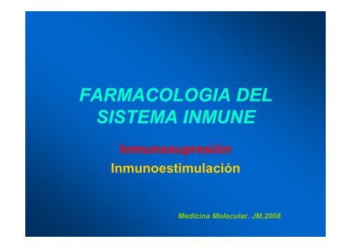
<strong>FARMACOLOGIA</strong> <strong>DEL</strong><br />
<strong>SISTEMA</strong> <strong>INMUNE</strong><br />
Inmunosupresión<br />
Inmunoestimulación<br />
Medicina Molecular. JM,2008
Las dos caras de Janus
INMUNOSUPRESIÓN
CICLOSPORINA
Jean Francois Borel
Scanning electron micrograph of Tolypocladium inflatum
Estructura de la ciclosporina
INMUNOESTIMULACION
The Two-Signal Model of T-cell Activation<br />
DC<br />
T cell<br />
CD4 or CD8<br />
1<br />
MHC<br />
TCR<br />
2<br />
COSTIMULATION
• Experimental evidences for the two signal model of T cell activation<br />
• Experimental evidences that CD28 is a costimulatory receptor<br />
• Effects of CD28-mediated costimulation on T cells<br />
• Mechanism of action<br />
• The ligands for CD28: B7 molecules<br />
• The complexity of the B7/CD28 system: CD28 plays a role in<br />
promoting T cell tolerance!!
Marc Jenkins and Ronald Schwartz in the late 80’s :<br />
The first definitive experimental demonstration that TCR engagement alone<br />
was insufficient for T cell activation.<br />
Proliferative response of T cell clones<br />
(pigeon cytochrome c peptide 81-104 presented by I-E k )<br />
to normal or ECDI(chemical crosslinker)-fixed peptide-pulsed APCs<br />
Jenkins M.K., and Schwartz R.H. J. Exp. Med. 165:302-319, 1987.<br />
ECDI-treated APCs fail to stimulate proliferation by normal T cell clones :<br />
Not the result of extensive modification of the MHC class II molecule<br />
ECDI treatment inactivated an accessory (costimulatory) function of the APC
T cell clones stimulated in vitro by EDCI-treated APCs<br />
were induced into a state of unresponsiveness (Anergy).<br />
Jenkins M.K., and Schwartz R.H. J. Exp. Med. 165:302-319, 1987.
Cells with this costimulatory function:<br />
macrophages, activated B cells and dendritic cells<br />
(professional antigen presenting cells).<br />
Subsequently,it was shown that:<br />
Naïve T cells have similar requirement for these<br />
costimulatory signals<br />
in order to produce IL-2 and progress through the cell cycle.
This requirement for costimulation and the<br />
restricted pattern of expression of the costimulatory ligands to professional APCs,<br />
was proposed to be a mechanism for maintenance of peripheral T cell tolerance.<br />
Self-reactive T cell<br />
Anergy<br />
TCR<br />
Costimulatory<br />
Receptors<br />
Costimulatory<br />
Ligands<br />
peptide/<br />
MHC I<br />
Parenchymal cell
In vitro T cell responses in CD28 KO mice<br />
Shahinian A. et al.<br />
Science 261:609, 1993.<br />
Adapted from Green J. et al.<br />
Immunity 1:501, 1994.
The importance of CD28-mediated costimulation has also been demonstrated in vivo.<br />
Blockade of CD28-mediated costimulation has been shown to:<br />
• Prolong graft survival<br />
• Block primary antibody response<br />
• Prevent or reduce in intensity experimentally, antigen-induced models of<br />
autoimmunity including arthritis, encephalomyelitis, myocarditis, thyroiditis and<br />
myasthenia gravis
Major effects of CD28-mediated costimulation in T cells<br />
Proliferation<br />
IL-2 (transcription, mRNA stabilization)<br />
IL-2R up-regulation<br />
G1 cell cycle kinases<br />
Cell cycle inhibitor p27Kip<br />
Survival<br />
Bcl-xL<br />
Effector function<br />
CD40-L, OX-40, 41BB, ICOS<br />
cytokines expression<br />
cytotoxic molecules<br />
The major effects of CD28-mediated costimulation are to augment and sustain T cell<br />
responses initiated by antigen receptor signal by promoting T-cell survival and<br />
enabling cytokines to initiate T cell clonal expansion and differentiation.
Mechanism<br />
The conventional view<br />
CD28 delivers intracellular signals that synergize distally<br />
with the TCR signaling pathways, at the level of JUN-kinase.
The new concept (based on the visualization of T cell activation)<br />
Kupfer et al. have shown that T cells polarize toward the APCs, thereby forming a highly<br />
structured synapse (the immune synapse).<br />
This process involves a redistribution of surface and intracellular molecules.<br />
Some molecules, such as TCR, CD4, src kinases are enriched in a central zone, whereas<br />
others, such as LFA-1 and CD45, remain outside.<br />
• helps maintaining the interaction between the TCR and the<br />
peptide/MHC complexes<br />
(low concentration and low affinity for the TCR)<br />
• helps in the local accumulation of intracellular signaling complexes
Wulfing, C. and and Davies, M.M. Science 282:2266, 1998.<br />
1. Actine cytoskeleton reorients toward the T cell-APC interface<br />
soon after the start of T cell activation<br />
Beads are attached to the surface of T cells and monitor the movement of the cytoskeleton in the T cells.<br />
Figure 1. Bead movement toward the interface. Single frames of a video microscopy experiment (36) of<br />
the interaction of 5C.C7 T cells, loaded with 4.5-µm anti-ICAM-1 (YN1) beads,<br />
with peptide-loaded CH27 B cell lymphoma cells (35) are shown. The CH27 cell is substantially larger<br />
than the T cells. The T cell intracellular calcium concentration is overlaid in a false color scale from blue<br />
(low concentration) to red (high concentration) to mark the onset of T cell activation, set to time 0:00 min.<br />
Although the beads, one of which is marked with an arrow, are bound to the posterior end of<br />
the central T cell before and at the time of its activation, they can be seen to move toward the<br />
T cell-B cell interface in subsequent frames.
Thus, in addition to a role in initial activation of T cells,<br />
B7/CD28 interactions are important in promoting T cell<br />
tolerance because they have a critical role in the homeostasis<br />
of CD4+ CD25+ regulatory T cells which play an essential<br />
role in regulating self-tolerance.
B7.1 and B7.2: What’s the difference?<br />
Expression: Primarily limited to APCs<br />
B7.1 is absent from unstimulated cells.<br />
B7.2 is constitutively expressed at low levels on unstimulated DCs<br />
and blood monocytes.<br />
Activation of APCs induces B7 expression.<br />
In general, the induction of B7.2 expression follows faster kinetics<br />
and usually reaches higher values than does B7.1 induction.<br />
In vivo, anti B7.2 mAb was shown to be more efficient than anti-B7.1 mAb to<br />
block the expansion of Ag-specific T cells.<br />
B7.1 KO vs B7.2 KO:<br />
When immunized intravenously without adjuvant, B7.2-deficient mice failed to<br />
form germinal centers, whereas B7.1-deficient mice gave Ab responses<br />
comparable with wild-type mice.<br />
B7.2 is probably the major initial ligand for CD28 while B7.1<br />
which is expressed later sustains T cell activation.
Mab ANTI CTLA-4<br />
(Ipililumab)
A homologue of CD28 named cytotoxic T lymphocyte-4 (CTLA-4) had<br />
been identified as a gene product of cytotoxic T cells (1987).<br />
We now know that it is expressed by both CD4+ and CD8+ T cells.<br />
In 1991, it was found that CTLA-4 binds also to B7.1 and later on also to<br />
B7.2. The affinity of binding of the B7 molecules to CTLA-4 is much<br />
higher than that of CD28 (50-2000-fold).<br />
A soluble version of CTLA-4 termed CTLA-4-Ig was made (ectodomain of<br />
CTLA-4 fused to the tail of an immunoglubulin). This reagent has been<br />
used extensively in vitro and in vivo to block the interaction of the B7<br />
molecules with CD28.<br />
In contrast to CD28, which is expressed constitutively on naïve T cells,<br />
CTLA-4 expression is induced in activated T cells.<br />
The obvious and intriguing question was why T cells have 2 receptors for<br />
the same ligands: a constitutive, abundant and low affinity receptor (CD28)<br />
and an inducible, not very abundant, high affinity receptor (CTLA-4).
mAb against CTLA-4 was found to costimulation by anti-CD28.<br />
CTLA-4 synergizes with CD28 in order to enhance costimulation and<br />
prolong IL-2 production and proliferation by T cells.<br />
Fab fragments of anti-CTLA-4 T cell responses in vitro.<br />
The enhancement was a result of the blockade of an inhibitory signal<br />
delivered by CTLA-4.<br />
Beads coated with anti-CD3, anti-CD28 and anti-CTLA-4 mAbs resulted in<br />
a much lower T cell response than beads coated with anti-CD3 and anti-<br />
CD28.<br />
Cross-linking CTLA-4 inhibits T cell responses mediated by TCR and<br />
CD28 stimulation.<br />
The inhibitory function of CTLA-4 was definitely shown in CTLA-4 KO<br />
mice.
Phenotype of CTLA-4 KO mice<br />
Fatal lymphoproliferative disorder. Mice die at 18-28 days of age due to lymphocytic<br />
infiltration into nonlymphoid tissues. This phenotype is one of the most aggressive<br />
lymphoproliferative disorder reported in gene-targeted mice.<br />
Virtually all of the peripheral T cells display an activated phenotype<br />
Chambers C.A. et al.<br />
Immuunol. Rev. 153:27, 1996.
Phenotype of CTLA-4 KO mice<br />
This is not due to a failure to negatively select thymocytes expressing TCRs with high<br />
affinity for self peptides/MHC complexes that would permit the emigration of highly<br />
autoreactive T cells to the periphery.<br />
Thymocyte development is normal in CTLA-4 KO mice.<br />
CTLA-4 is necessary for the regulation of peripheral T cell tolerance.
B7/CD28/CTLA-4<br />
Activated T cell<br />
AP2<br />
AP2<br />
CD28<br />
TCR<br />
CTLA-4<br />
P<br />
P<br />
B7.1 (CD80)<br />
B7.2 (CD86)<br />
peptide/<br />
MHC<br />
Activated Professional APC
CTLA-4 is not readily detectable in naïve T cells<br />
but is rapidly upregulated upon T cell activation.<br />
CTLA-4 mRNA can be readily detected within 1 h of<br />
TCR engagement and peaks at about 24–36 h.<br />
However, CTLA-4 is not readily detectable at the cell surface<br />
until 24–48 h after activation.<br />
An accurate assessment of the kinetics of<br />
CTLA-4 protein expression is complicated<br />
by the fact that it is not primarily expressed at the cell surface.<br />
Surface expression of CTLA-4 is tightly regulated as a result<br />
of the presence of a tyrosine-based intracellular localization<br />
motif in its cytoplasmic tail which allows an association with<br />
the medium subunit of the clathrin-coated<br />
pit adaptor complex, AP2,thus providing a mechanism for<br />
CTLA-4 cellular localization. CTLA-4 expression on the<br />
T cell surface is stabilized and increased by tyrosine<br />
phosphorylation of the endocytosis motif, which inhibits<br />
AP2 association. It is noteworthy that the intracellular portion<br />
of CTLA-4 is 100% conserved among many different species<br />
of animals, suggesting that control of intracellular trafficking<br />
may be extremely important for its function. This motif results<br />
in both the rapid endocytosis of CTLA-4 from the cell surface to<br />
endosomal compartments, as well as the targeting of at<br />
least some CTLA-4 to the lysosomes for degradation.<br />
It is unclear whether endocytosed CTLA-4 can recycle back<br />
to the surface or if this represents a terminal pathway for the<br />
protein.
Dynamic integration of TCR, CD28 and CTLA-4<br />
As previously discussed, CD28 is constitutively expressed on T cells, whereas CTLA-4 appears after<br />
activation. Because of this, and perhaps as a result of our innate appreciation for symmetry, the idea arose<br />
that CD28 engagement allowed initiation, while CTLA-4 provided for termination of immune responses.<br />
Surprisingly, the majority of the in vitro data has demonstrated an inhibitory role for CTLA-4 in the early<br />
stages of T cell activation. IL-2 production, expression of early markers such as CD69 and CD25, and a<br />
number of other aspects of activation are inhibited upon CTLA-4 cross-linking. These events take place<br />
within hours of T cell activation with anti-CD3 and CD28. In fact, the inhibition of the induction of<br />
IL-2 transcription was detected 4 h after stimulation.<br />
This suggests either that there is a physiologically relevant intracellular pool of CTLA-4 present<br />
in naïve T cells or that protein expression is induced rapidly upon activation.<br />
The possibility that CTLA-4 can inhibit early stages of T cell activation has led to the<br />
development of models that stress that the dynamic interplay of costimulatory and TCR signals<br />
depends on the activation state of the T cell as well as the activation state of the antigen-presenting cell.
The threshold model:When B7 levels are low and TCR signals are weak, the amount of CTLA-4 induced<br />
is low but may be sufficient to minimize costimulation and prevent activation. Under these conditions<br />
CTLA-4 may set a threshold for activation. In other words, it plays a role in setting the stimulatory<br />
threshold required for a T cell to progress to full activation.
As in the classical two-signal model,<br />
an encounter of a naïve T cell with a cell<br />
expressing appropriate MHC/antigen complex<br />
but lacking B7 does not result in activation of<br />
the T cell owing to lack of costimulation.<br />
The cells receiving a TCR signal in the absence<br />
of CD28-mediated costimulation may be<br />
rendered anergic.<br />
However, engagement of the TCR can lead<br />
to rapid induction and/or mobilization of<br />
small amounts of CTLA-4. Under conditions<br />
where there is an incompletely activated<br />
APC expressing only low amounts of B7,<br />
CTLA-4 could, by virtue of its higher affinity,<br />
outcompete CD28 for B7 and/or deliver<br />
inhibitory signals. This could effectively<br />
raise the threshold of CD28 and/or TCR<br />
signals needed for full activation.<br />
Chambers, C.A. et al. Ann.Rev.Immunol. 19:565, 2001
There are two scenarios in which CTLA-4 may play a role in establishing a threshold for<br />
CD28 and/or TCR signals needed for activation of naïve T cells.<br />
Both presume that low levels of CTLA-4 pre-exist or can be rapidly induced in naïve T cells upon<br />
engagement of the TCR and CD28.<br />
The first scenario deals with the regulation of the response of T cells to tonic signaling by<br />
self-peptide/MHC interactions.<br />
Continuous TCR interactions with self-peptide/MHC provide important signals for the survival of<br />
peripheral T cells.<br />
Some of these tonic interactions, under conditions of low levels of CD28/B7 interaction, might be<br />
sufficiently stimulatory to lead to the activation of CD4+ T cells and the induction and/or mobilization of<br />
CTLA-4. Based on the analysis of the CTLA-4-/- mice, it is speculated that CTLA-4 might prevent the<br />
signals generated by these interactions from leading to full activation of CD4+ T cells.<br />
Thus, CTLA-4 may be involved in maintaining naïve CD4+ T cells and previously activated T cells<br />
in a resting state.<br />
This model is supported by the phenotype of CTLA-4-/- mice. The expansion of T cells that occurs in these<br />
mice is polyclonal. This suggests that the expansion is not the result of a failure to terminate responses<br />
to few environmental pathogens. Thus, the phenotype of CTLA-4-/- mice results from the activation and<br />
expansion of T cells reactive to low-affinity self-ligands due to a decreased activation threshold.
The second scenario suggests a role for CTLA-4 in maintaining peripheral tolerance of T cells with<br />
specificity for tissue-specific antigens that are not expressed in the thymus and have not been deleted as<br />
a consequence of negative selection. CTLA-4 may provide an additional level of regulation to ensure<br />
peripheral tolerance by preventing activation when a T cell encounters a normal self-antigen in the<br />
context of low B7 expression.<br />
This scenario may explain the observation that CTLA-4 blockade or deficiency accelerates the onset and<br />
severity of insulitis and diabetes in nonobese diabetic (NOD) mice expressing a transgenic ß TCR cloned<br />
from an islet-ß-cell-specific CD4+ T cell clone isolated from a NOD mouse. T cells bearing this<br />
TCR are efficiently selected, rather than deleted in the thymus, demonstrating that central tolerance is not<br />
effective for T cells with this specificity. The observation that blockade or loss of CTLA-4 dramatically<br />
accelerates disease in this model system indicates a role for CTLA-4 in maintaining an activation threshold<br />
for autoreactive T cells bearing TCRs specific to autoantigens.
The attenuation model:When B7 levels are high and TCR signals are strong, the higher levels of CTLA-4<br />
induced after activation may be able to attenuate the response of activated cells. This model suggest that<br />
after activation and subsequent entry into the cell cycle, CTLA-4 can limit the capacity of a T cell<br />
to divide.
Under conditions where the APC is activated<br />
and expressing high levels of B7,<br />
CD28 costimulation may dominate and<br />
activation proceeds. However, this results in<br />
induction and/or mobilization of CTLA-4<br />
that may be proportional to the strength of the<br />
TCR signal, resulting in differential inhibition.<br />
CTLA-4 preferentially attenuates the expansion<br />
of T cells that have been strongly activated.<br />
This notion is supported by the effect of<br />
CTLA-4-blockade on the proliferative capacity,<br />
or average number of daughter cells per responder,<br />
for T cells primed with agonist or weak agonist<br />
peptides in adjuvant.<br />
Blockade significantly increased the proliferative<br />
capacity of T cells primed with the agonist ligand<br />
but had a minimal effect on the cells responding<br />
to the weak agonist peptide..<br />
Chambers, C.A. et al. Ann.Rev.Immunol. 19:565, 2001
CTLA-4 would prevent this high-affinity population from dominating the primed pool by restricting<br />
proliferation early in the response. Engagement of CTLA-4 would then serve to broaden the pool of<br />
T cells by limiting clonal representation of the high-affinity population, thus allowing more equal<br />
representation of the cells bearing lower affinity TCRs in the early stages of the clonal evolution of<br />
the response.<br />
It appears that increased CTLA-4 expression upon activation modulates T cell responses<br />
differentially and might serve to limit the burst size of responding T cells. Overall, the results suggest<br />
that the quality of the TCR signal is critical to determining if and/or how dramatically CTLA-4 regulates<br />
the proliferative capacity of any antigen-specific clone selected from the T cell repertoire.<br />
While the TCR and CD28 might primarily determine the range of T cells responding to antigen,<br />
CTLA-4 would limit clonal representation of T cells with high-affinity TCRs.
Role played by CTLA-4 in regulating antigen-specific T cell responses. (a)<br />
The threshold and attenuation models make different predictions about the<br />
function of CTLA-4, depending on the stimulatory conditions encountered during<br />
activation. Shown are graphical representations that relate the effect of CTLA-4–<br />
mediated inhibition on T cell expansion to the degree of stimulation (TCR +<br />
CD28 signals). Threshold model: T cells that are stimulated below a given<br />
activation threshold (dashed lines) will not commit to entering the cell cycle.<br />
According to the threshold model, CTLA-4 may participate in setting a threshold<br />
for activation, thereby keeping cells that receive low amounts of stimulation from<br />
responding (red lines). In the absence of CTLA-4 signaling, the T cell activation<br />
threshold would be shifted, so that cells that receive weaker amounts of stimulation<br />
could now become activated and proliferate (green lines).
T cell–mediated immunotherapy represents a promising treatment for human malignancies.<br />
In cancer patients, the absence of efficient tumor-specific immunity can be related to inadequate<br />
APC function or to T cell tolerance/ignorance towards tumor antigens.<br />
Mice were injected intravenously with 5 x 10⁴ or 105 B16-F10 melanoma cells.<br />
Treatment using irradiated F10/g cells (GM-CSF transfected B16-F10 cells; Presumably, GM-CSF<br />
production at the site of vaccination might attract host APCs and enhance their function in vivo.) and<br />
antibodies was started after 24 h.<br />
These results indicate that CTLA-4 blockade and GM-CSF–producing<br />
vaccines act synergistically to cause rejection of poorly immunogenic tumors.<br />
van Elsas A. J. Exp. Med. 190:355, 1999
Therapy with anti-CTLA-4 and GM-CSF B16 melanoma vaccine<br />
leads to tumor rejection but also induces autoimmune responses<br />
(depigmentation).