Evidence for Effects on Neurology and Behavior - BioInitiative Report
Evidence for Effects on Neurology and Behavior - BioInitiative Report
Evidence for Effects on Neurology and Behavior - BioInitiative Report

Create successful ePaper yourself
Turn your PDF publications into a flip-book with our unique Google optimized e-Paper software.
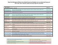
Bawin et al. [1973] exposed cats to 147-MHz RFR amplitude-modulated at 8 <strong>and</strong> 16 Hz at<br />
1 mW/cm 2 . They reported changes in both sp<strong>on</strong>taneous <strong>and</strong> c<strong>on</strong>diti<strong>on</strong>ed EEG patterns.<br />
Interestingly, the effects were not observed at lower or higher frequencies of modulati<strong>on</strong>.<br />
Takashima et al. [1979] also studied the EEG patterns in rabbits exposed to RFR fields (1-30<br />
MHz) amplitude-modulated at either 15 or 60 Hz. Acute exposure (2-3 h, field strength 60-500<br />
Vrms/m) elicited no observable effect. Chr<strong>on</strong>ic exposure (2 h/day <str<strong>on</strong>g>for</str<strong>on</strong>g> 4-6 weeks at 90-500<br />
Vrms/m) produced abnormal patterns including high amplitude spindles, bursts, <strong>and</strong> suppressi<strong>on</strong><br />
of normal activity (shift to pattern of lower frequencies) when recorded within a few hours after<br />
exposure.<br />
In an experiment by Chou <strong>and</strong> Guy [1979a], no significant change in electrical activity<br />
from the hypothalamus was detected in rabbits exposed to 2450-MHz RFR at 100 mW/cm 2<br />
(SAR at electrode ~25 W/kg). In a chr<strong>on</strong>ic exposure experiment, Chou et al. [1982b] exposed<br />
rabbits to c<strong>on</strong>tinuous-wave 2450-MHz RFR at 1.5 mW/cm 2 (2 h/day, 5 days/week <str<strong>on</strong>g>for</str<strong>on</strong>g> 90 days).<br />
Electroencephalograph <strong>and</strong> evoked potentials were measured at the sensory-motor <strong>and</strong> occipital<br />
cortex at various times during the exposure period. They reported large variati<strong>on</strong>s in the data <strong>and</strong><br />
a tendency toward a decreased resp<strong>on</strong>se amplitude in the latter part of the experiment, i.e., after<br />
a l<strong>on</strong>ger period of exposure.<br />
In a more recent study, Chizhenkova [1988] recorded in the unanesthetized rabbits slow<br />
wave EEG in the motor <strong>and</strong> visual cortex, evoked potential in the visual cortex to light flashes,<br />
<strong>and</strong> single unit activity in the visual cortex during <strong>and</strong> after exposure to c<strong>on</strong>tinuous-wave RFR<br />
(wavelength = 12.5 cm, 40 mW/cm 2 , 1 min exposure to the head) using glass electrodes. She<br />
reported that RFR increased the incident of slow wave <strong>and</strong> spindles in the EEG, which are<br />
characteristics of slow wave sleep in animals. However, the radiati<strong>on</strong> facilitated light-evoked<br />
resp<strong>on</strong>ses in the visual cortex. Cells in the visual cortex also showed changes in firing rates<br />
(increase or decrease depending <strong>on</strong> the neur<strong>on</strong> studied). Driving resp<strong>on</strong>ses of visual cortical<br />
neur<strong>on</strong>s to light flashes, i.e., resp<strong>on</strong>ses to sequence of light flashes of increasing frequency, were<br />
also enhanced by the RFR exposure. The author interpreted the data as showing a decrease in the<br />
threshold of visual evoked potential <strong>and</strong> an increase in excitability of visual cortical cells as a<br />
result of RFR exposure.<br />
NEUROCHEMICAL EFFECTS OF<br />
RADIOFREQUENCY RADIATION<br />
Neurochemical studies of RFR include those <strong>on</strong> the c<strong>on</strong>centrati<strong>on</strong>s <strong>and</strong> functi<strong>on</strong>s of<br />
neurotransmitters, receptor properties, energy metabolism, <strong>and</strong> calcium efflux from brain tissues.<br />
Changes in Neurotransmitter Functi<strong>on</strong>s<br />
In most studies <strong>on</strong> the effects of RFR <strong>on</strong> neurotransmitter functi<strong>on</strong>s, <strong>on</strong>ly the c<strong>on</strong>centrati<strong>on</strong><br />
of neurotransmitters (usually measured as amount/gm wet weight of brain tissue) was measured<br />
in the brains of animals after irradiati<strong>on</strong>. Data <strong>on</strong> change in c<strong>on</strong>centrati<strong>on</strong> al<strong>on</strong>e tells little about<br />
the nature of the effect, since it could result from different causes. For example, a decrease in the<br />
c<strong>on</strong>centrati<strong>on</strong> could be due to an enhanced release or a decrease in synthesis of the<br />
neurotransmitter as the result of RFR exposure. For a more in<str<strong>on</strong>g>for</str<strong>on</strong>g>mative study, the turnover rate<br />
35