Evidence for Effects on Neurology and Behavior - BioInitiative Report
Evidence for Effects on Neurology and Behavior - BioInitiative Report
Evidence for Effects on Neurology and Behavior - BioInitiative Report
Create successful ePaper yourself
Turn your PDF publications into a flip-book with our unique Google optimized e-Paper software.
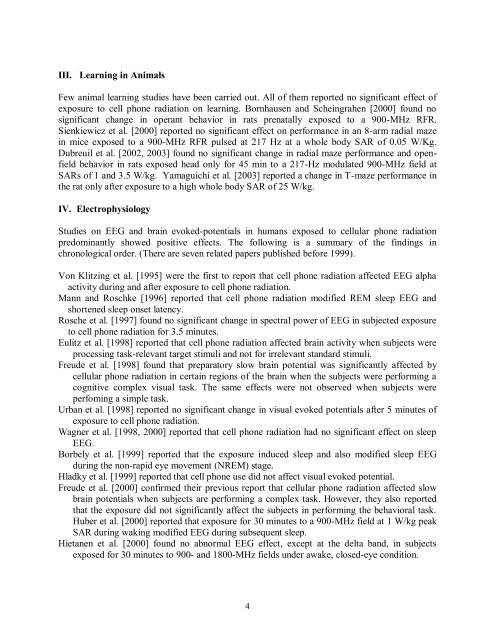
III. Learning in Animals<br />
Few animal learning studies have been carried out. All of them reported no significant effect of<br />
exposure to cell ph<strong>on</strong>e radiati<strong>on</strong> <strong>on</strong> learning. Bornhausen <strong>and</strong> Scheingrahen [2000] found no<br />
significant change in operant behavior in rats prenatally exposed to a 900-MHz RFR.<br />
Sienkiewicz et al. [2000] reported no significant effect <strong>on</strong> per<str<strong>on</strong>g>for</str<strong>on</strong>g>mance in an 8-arm radial maze<br />
in mice exposed to a 900-MHz RFR pulsed at 217 Hz at a whole body SAR of 0.05 W/Kg.<br />
Dubreuil et al. [2002, 2003] found no significant change in radial maze per<str<strong>on</strong>g>for</str<strong>on</strong>g>mance <strong>and</strong> openfield<br />
behavior in rats exposed head <strong>on</strong>ly <str<strong>on</strong>g>for</str<strong>on</strong>g> 45 min to a 217-Hz modulated 900-MHz field at<br />
SARs of 1 <strong>and</strong> 3.5 W/kg. Yamaguichi et al. [2003] reported a change in T-maze per<str<strong>on</strong>g>for</str<strong>on</strong>g>mance in<br />
the rat <strong>on</strong>ly after exposure to a high whole body SAR of 25 W/kg.<br />
IV. Electrophysiology<br />
Studies <strong>on</strong> EEG <strong>and</strong> brain evoked-potentials in humans exposed to cellular ph<strong>on</strong>e radiati<strong>on</strong><br />
predominantly showed positive effects. The following is a summary of the findings in<br />
chr<strong>on</strong>ological order. (There are seven related papers published be<str<strong>on</strong>g>for</str<strong>on</strong>g>e 1999).<br />
V<strong>on</strong> Klitzing et al. [1995] were the first to report that cell ph<strong>on</strong>e radiati<strong>on</strong> affected EEG alpha<br />
activity during <strong>and</strong> after exposure to cell ph<strong>on</strong>e radiati<strong>on</strong>.<br />
Mann <strong>and</strong> Roschke [1996] reported that cell ph<strong>on</strong>e radiati<strong>on</strong> modified REM sleep EEG <strong>and</strong><br />
shortened sleep <strong>on</strong>set latency.<br />
Rosche et al. [1997] found no significant change in spectral power of EEG in subjected exposure<br />
to cell ph<strong>on</strong>e radiati<strong>on</strong> <str<strong>on</strong>g>for</str<strong>on</strong>g> 3.5 minutes.<br />
Eulitz et al. [1998] reported that cell ph<strong>on</strong>e radiati<strong>on</strong> affected brain activity when subjects were<br />
processing task-relevant target stimuli <strong>and</strong> not <str<strong>on</strong>g>for</str<strong>on</strong>g> irrelevant st<strong>and</strong>ard stimuli.<br />
Freude et al. [1998] found that preparatory slow brain potential was significantly affected by<br />
cellular ph<strong>on</strong>e radiati<strong>on</strong> in certain regi<strong>on</strong>s of the brain when the subjects were per<str<strong>on</strong>g>for</str<strong>on</strong>g>ming a<br />
cognitive complex visual task. The same effects were not observed when subjects were<br />
perfoming a simple task.<br />
Urban et al. [1998] reported no significant change in visual evoked potentials after 5 minutes of<br />
exposure to cell ph<strong>on</strong>e radiati<strong>on</strong>.<br />
Wagner et al. [1998, 2000] reported that cell ph<strong>on</strong>e radiati<strong>on</strong> had no significant effect <strong>on</strong> sleep<br />
EEG.<br />
Borbely et al. [1999] reported that the exposure induced sleep <strong>and</strong> also modified sleep EEG<br />
during the n<strong>on</strong>-rapid eye movement (NREM) stage.<br />
Hladky et al. [1999] reported that cell ph<strong>on</strong>e use did not affect visual evoked potential.<br />
Freude et al. [2000] c<strong>on</strong>firmed their previous report that cellular ph<strong>on</strong>e radiati<strong>on</strong> affected slow<br />
brain potentials when subjects are per<str<strong>on</strong>g>for</str<strong>on</strong>g>ming a complex task. However, they also reported<br />
that the exposure did not significantly affect the subjects in per<str<strong>on</strong>g>for</str<strong>on</strong>g>ming the behavioral task.<br />
Huber et al. [2000] reported that exposure <str<strong>on</strong>g>for</str<strong>on</strong>g> 30 minutes to a 900-MHz field at 1 W/kg peak<br />
SAR during waking modified EEG during subsequent sleep.<br />
Hietanen et al. [2000] found no abnormal EEG effect, except at the delta b<strong>and</strong>, in subjects<br />
exposed <str<strong>on</strong>g>for</str<strong>on</strong>g> 30 minutes to 900- <strong>and</strong> 1800-MHz fields under awake, closed-eye c<strong>on</strong>diti<strong>on</strong>.<br />
4


