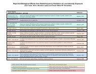
Evidence for Effects on Neurology and Behavior - BioInitiative Report
Evidence for Effects on Neurology and Behavior - BioInitiative Report
Evidence for Effects on Neurology and Behavior - BioInitiative Report

Create successful ePaper yourself
Turn your PDF publications into a flip-book with our unique Google optimized e-Paper software.
of a neurotransmitter should be investigated. This involves the measurement of the rate of<br />
decrease in c<strong>on</strong>centrati<strong>on</strong> of the neurotransmitter when its synthesis is blocked <strong>and</strong>/or the rate of<br />
accumulati<strong>on</strong> of the metabolites of the neurotransmitter. More recently, the rate of release of a<br />
neurotransmitter from a local brain regi<strong>on</strong> can be studied by the microdialysis technique.<br />
Snyder [1971] reported a significant increase in the c<strong>on</strong>centrati<strong>on</strong>s of serot<strong>on</strong>in <strong>and</strong> its<br />
metabolite, 5-hydroxyindolacetic acid, in the brain of rats after 1 h of exposure to c<strong>on</strong>tinuouswave<br />
3000-MHz RFR at 40 mW/cm 2 (SAR 8 W/kg). However, decreases in both neurochemicals<br />
were observed in the brain of rats exposed 8 h/day <str<strong>on</strong>g>for</str<strong>on</strong>g> 7 days at 10 mW/cm 2 . Thus,<br />
these results indicated an increase in the synthesis <strong>and</strong> turnover of brain serot<strong>on</strong>in after acute<br />
exposure <strong>and</strong> a decrease after prol<strong>on</strong>ged exposure to RFR. Furthermore, warming the animals by<br />
placing them in an incubator heated at 34 o C had no significant effect <strong>on</strong> the turnover rate of<br />
serot<strong>on</strong>in in the brain.<br />
Catravas et al. [1976] also reported an increase in diencephal<strong>on</strong> serot<strong>on</strong>in c<strong>on</strong>centrati<strong>on</strong> <strong>and</strong><br />
activity of tryptophan hydroxylase, the synthesis enzyme <str<strong>on</strong>g>for</str<strong>on</strong>g> serot<strong>on</strong>in, in the rat after 8 daily (8<br />
h/day) exposures to RFR at 10 mW/cm 2 . No significant changes in activity of m<strong>on</strong>oamine<br />
oxidase, the degradati<strong>on</strong> enzyme of serot<strong>on</strong>in, was observed in the brain of the irradiated rats.<br />
Zeman et al. [1973] investigated the effects of exposure to pulsed 2860-MHz RFR <strong>on</strong> -<br />
amino-butyric acid (GABA) in the rat brain. No significant difference was observed in GABA<br />
c<strong>on</strong>centrati<strong>on</strong> nor the activity of its synthesis enzyme, L-glutamate decarboxylase, in the brains<br />
of chr<strong>on</strong>ic (10 mW/cm 2 , 8 h/day <str<strong>on</strong>g>for</str<strong>on</strong>g> 3-5 days, or 4 h/day, 5 days/week <str<strong>on</strong>g>for</str<strong>on</strong>g> 4 or 8 weeks) or<br />
acutely exposed (40 mW/cm 2 <str<strong>on</strong>g>for</str<strong>on</strong>g> 20 min, or 80 mW/cm 2 <str<strong>on</strong>g>for</str<strong>on</strong>g> 5 min) rats compared with those of<br />
the sham-exposed animals.<br />
Rats exposed to c<strong>on</strong>tinuous-wave 1600-MHz RFR at 30 mW/cm 2 <str<strong>on</strong>g>for</str<strong>on</strong>g> 10 min were reported<br />
to have altered c<strong>on</strong>centrati<strong>on</strong>s of catecholamines (norepinephrine <strong>and</strong> dopamine) <strong>and</strong> serot<strong>on</strong>in<br />
in specific regi<strong>on</strong>s of the brain [Merritt et al., 1976]. Norepinephrine was decreased <strong>on</strong>ly in the<br />
hypothalamus, whereas decrease in serot<strong>on</strong>in was seen in the hippocampus <strong>and</strong> decreases in<br />
dopamine were observed in the striatum <strong>and</strong> hypothalamus. These effects were suggested to be<br />
caused by an uneven distributi<strong>on</strong> of RFR in different regi<strong>on</strong>s of the brain. In a further study, rats<br />
exposed to similar radiati<strong>on</strong> (20 or 80 mW/cm 2 ) were found to have a reducti<strong>on</strong> of<br />
norepinephrine c<strong>on</strong>centrati<strong>on</strong> in the basal hypothalamus, whereas no significant changes in<br />
dopamine <strong>and</strong> serot<strong>on</strong>in c<strong>on</strong>centrati<strong>on</strong>s were observed even though the brain temperature<br />
increased up to 5 o C [Merritt et al., 1977]. In another study [Grin, 1974], rats were exposed to<br />
2375-MHz RFR at power densities of 50 <strong>and</strong> 500 W/cm 2 <str<strong>on</strong>g>for</str<strong>on</strong>g> 30 days (7 h/day). At 50 W/cm 2 ,<br />
brain epinephrine was increased <strong>on</strong> the 20th day of exposure, but returned to normal by day 30.<br />
There were slight increases in norepinephrine <strong>and</strong> dopamine c<strong>on</strong>centrati<strong>on</strong>s throughout the<br />
exposure period. At 500 W/cm 2 , c<strong>on</strong>centrati<strong>on</strong>s of all three neurotransmitters were increased at<br />
day 5, but declined c<strong>on</strong>tinually after further exposure.<br />
Various studies have been carried out to investigate the neurochemical effects of RFR<br />
irradiati<strong>on</strong> <strong>on</strong> acetylcholine in the brain. A decrease in whole brain c<strong>on</strong>centrati<strong>on</strong> of acetylcholine,<br />
suggesting an increased release of the neurotransmitter, has been reported in mice<br />
exposed to a single 2450-MHz RFR pulse, which deposited 18.7 J in the brain <strong>and</strong> increased the<br />
brain temperature by 2 to 4 o C [Modak et al., 1981]. Several studies investigated the effect <strong>on</strong><br />
acetylcholinesterase (AChE), the degradati<strong>on</strong> enzyme <str<strong>on</strong>g>for</str<strong>on</strong>g> acetylcholine. Acute (30 min) exposure<br />
to 9700-MHz RFR was reported to inhibit the membrane-bound AChE activity in a vagal-heart<br />
preparati<strong>on</strong> [Young, 1980]. This effect was attributed to a release of bound calcium from the<br />
postjuncti<strong>on</strong>al membrane. In another study [Baranski, 1972], acute exposure to pulsed RFR<br />
36