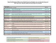
Evidence for Effects on Neurology and Behavior - BioInitiative Report
Evidence for Effects on Neurology and Behavior - BioInitiative Report
Evidence for Effects on Neurology and Behavior - BioInitiative Report

Create successful ePaper yourself
Turn your PDF publications into a flip-book with our unique Google optimized e-Paper software.
hippocampus was blocked by pretreatment of the animals with the opiate-antag<strong>on</strong>ists nalox<strong>on</strong>e<br />
<strong>and</strong> naltrex<strong>on</strong>e, suggesting involvement of endogenous opioids in the effect. Endogenous opioids<br />
are a group of peptides synthesized by the nervous system <strong>and</strong> have pharmacological properties<br />
like opiates. They are involved in a variety of physiological functi<strong>on</strong>s such as stress reacti<strong>on</strong>s,<br />
temperature-regulati<strong>on</strong>, motivati<strong>on</strong>al behaviors, etc. Our further research showed that the effects<br />
of RFR <strong>on</strong> central cholinergic activity could be classically c<strong>on</strong>diti<strong>on</strong>ed to cues in the exposure<br />
envir<strong>on</strong>ment [Lai et al., 1987c]. These effects of RFR <strong>on</strong> cholinergic functi<strong>on</strong>s are similar to<br />
those reported in animals after exposure to stressors [Finkelstein et al., 1985; Lai, 1987; Lai et al.,<br />
1986c].<br />
When different power densities of RFR were used, a dose-resp<strong>on</strong>se relati<strong>on</strong>ship could be<br />
established from each brain regi<strong>on</strong> [Lai et al., 1989a]. Data were analyzed by probit analysis,<br />
which enables a statistical comparis<strong>on</strong> of the dose-resp<strong>on</strong>se functi<strong>on</strong>s of the different brain<br />
regi<strong>on</strong>s. It was found that a higher dose-rate was required to elicit a change in HACU in the<br />
striatum, whereas the resp<strong>on</strong>ses of the fr<strong>on</strong>tal cortex <strong>and</strong> hippocampus were similar. Thus, under<br />
the same irradiati<strong>on</strong> c<strong>on</strong>diti<strong>on</strong>s, different brain regi<strong>on</strong>s could have different sensitivities to RFR.<br />
In further experiments to investigate the c<strong>on</strong>tributory effect of different parameters of RFR<br />
exposure, we found that the radiati<strong>on</strong> caused a durati<strong>on</strong>-dependent biphasic effect <strong>on</strong> cholinergic<br />
activity in the brain. After 20 instead of 45 min of RFR exposure as in earlier experiments, an<br />
increase in HACU was observed in the fr<strong>on</strong>tal cortex, hippocampus, <strong>and</strong> hypothalamus of the rat<br />
[Lai et al., 1989b], <strong>and</strong> these effects could be blocked by pretreatment with the opiate antag<strong>on</strong>ist<br />
naltrex<strong>on</strong>e, suggesting the effects are also mediated by endogenous opioids.<br />
Experiments [Lai et al., 1988] were then carried out to compare the effects of exposure in<br />
two different systems that produced different energy absorpti<strong>on</strong> patterns in the body of the<br />
exposed animal. Rats were exposed to pulsed (2 s pulses, 500 pps) or c<strong>on</strong>tinuous-wave 2450-<br />
MHz RFR in the circular waveguide <strong>and</strong> the miniature anechoic chamber exposure systems<br />
designed by Guy [Guy, 1979; Guy et al., 1979] with the whole body average SAR kept at a<br />
c<strong>on</strong>stant level of 0.6 W/kg. In the circular waveguide rats were exposed to circularly polarized<br />
RFR from the side of the body. In the miniature anechoic chamber rats were exposed dorsally<br />
with plane-polarized RFR. The circular waveguide produced a more localized energy absorpti<strong>on</strong><br />
pattern than the miniature anechoic chamber. Detailed dosimetry studies in the body <strong>and</strong> brain of<br />
rats exposed in these two exposure systems had been carried out [Chou et al., 1984, 1985a].<br />
After 45 min of exposure to the RFR, a decrease in HACU was observed in the fr<strong>on</strong>tal cortex in<br />
all exposure c<strong>on</strong>diti<strong>on</strong>s studied (circular waveguide vs miniature anechoic chamber, pulsed vs<br />
c<strong>on</strong>tinuous-wave). However, regardless of the exposure system used, HACU in the hippocampus<br />
decreased <strong>on</strong>ly after exposure to pulsed, but not c<strong>on</strong>tinuous-wave RFR. Striatal HACU was<br />
decreased after exposure to either pulsed or c<strong>on</strong>tinuous-wave RFR in the miniature anechoic<br />
chamber, but no significant effect was observed when the animal was exposed in the circular<br />
waveguide. No significant effect <strong>on</strong> HACU was found in the hypothalamus under all the<br />
exposure c<strong>on</strong>diti<strong>on</strong>s studied. Thus, each brain regi<strong>on</strong> resp<strong>on</strong>ded differently to RFR exposure<br />
depending <strong>on</strong> the parameters. <str<strong>on</strong>g>Effects</str<strong>on</strong>g> <strong>on</strong> the fr<strong>on</strong>tal cortex were independent of the exposure<br />
system or use of pulsed or c<strong>on</strong>tinuous- wave RFR. The hippocampus <strong>on</strong>ly resp<strong>on</strong>ded to pulsed<br />
but not to c<strong>on</strong>tinuous-wave RFR. Resp<strong>on</strong>se of the striatum depended <strong>on</strong> the exposure system<br />
used. The neurochemical changes were correlated with the dosimetry data of Chou et al. [1985a]<br />
<strong>on</strong> the local SARs in different brain areas of rats exposed to RFR in these two exposure systems.<br />
The dosimetry data showed that the septum, where the cell bodies of the hippocampal<br />
cholinergic pathway are located, had the lowest local SAR am<strong>on</strong>g eight brain areas measured in<br />
38