TNPJ220m - ProZyme
TNPJ220m - ProZyme
TNPJ220m - ProZyme

You also want an ePaper? Increase the reach of your titles
YUMPU automatically turns print PDFs into web optimized ePapers that Google loves.
TechNote #TNPJ220<br />
091206AA<br />
NOTE: Reflects some changes from original (presented at ISAC XXIII) in order to provide a<br />
stand-alone presentation for first-time viewers.<br />
Slide 1 - Introduction<br />
Welcome. Thank you all for being here. I assume you’re here because you’d like to learn<br />
more about using PhycoLink ® Conjugation and Purification Kits to make your own<br />
conjugates. We’d like to start by thanking a long-time customer for giving us our title<br />
when he told us that our kits “just make the best DARN conjugates.” We simply couldn’t<br />
have said it any better ourselves.<br />
Many of you know <strong>ProZyme</strong> as the manufacturer of phycobiliproteins (e.g. RPE, APC, etc.)<br />
sold under the PhycoPro tradename, which are widely used in flow cytometry and other<br />
fluorescence applications. You may also be familiar with our PhycoLink line of fluorescent<br />
conjugates (antibodies, streptavidin, etc.), which are value-added products for use in<br />
fluorescence applications.<br />
What we hope to accomplish today is to impart to you some of our experience,<br />
accumulated over many years working with a particular class of fluorescent pigments, the<br />
phycopigment proteins, and to dispel some of the mystery from what has largely been<br />
considered a black box in terms of making conjugates from these fluors.
Slide 2 - Overview<br />
We’re going to start out by discussing some of the properties that define a good conjugate.<br />
Then we’ll consider some of the characteristics of the phycopigment proteins (PPP’s). I<br />
need to stop to define this term. You’re probably familiar with the class of fluorescent<br />
molecules known as the phycobiliproteins, large, water-soluble proteins derived from<br />
cyanobacteria and eukaryotic algae, having absorbance and emission wavelengths in the<br />
visible range; examples include RPE and APC. With our recent licensing of the PerCP<br />
pigment protein from Becton, Dickinson and Company, Inc., in order to be<br />
inclusive/correct we’ve had to expand this term. We’ll now refer to this class of molecules<br />
as the phycopigment proteins, PPP’s or three P’s—I’ll use these terms interchangeably<br />
throughout the presentation.<br />
Next, we’ll talk about using our PhycoLink Conjugation Kits to make conjugates from these<br />
molecules. Then, we’ll consider some of the scenarios for which it may be desirable to<br />
utilize our PhycoLink Purification Kits in order to purify the conjugate you’ve made. I’d<br />
like to mention at this point that I’m not going to read our protocols to you. All of the<br />
procedures and supporting figures/data to which I’ll refer in the course of today’s<br />
presentation are available on our website for you to review at your leisure. Also, we’ve<br />
recently rewritten all of our conjugation kit booklets in order to remove any ambiguity in<br />
cases where we could have been more clear. So, for those of you who may not have
worked with one of our kits in a while, I encourage you to download the revised versions<br />
of the booklet. Just click into the PPP of choice to find the product insert (booklet), FAQs,<br />
TechNotes, etc.<br />
http://www.prozyme.com/phycolink/pj-kits.html<br />
Then, we’ll discuss some tools for evaluating conjugates, which are equally applicable to<br />
conjugates you have made using our kits as well as those purchased from commercial<br />
sources. Lastly, if time permits, I’d like to close by showing examples of customerfurnished<br />
data which illustrates the utility of PPP conjugates in applications including flow<br />
cytometry, fluorescence microscopy and imaging cytometry.
Slide 3 - Why make your own?<br />
I assume you’re here because you either need or want to make your own conjugates,<br />
possibly for one or more of these reasons:<br />
<br />
<br />
<br />
<br />
<br />
Want a direct conjugate in order to avoid two-step staining.<br />
Antibody unavailable in the color you want.<br />
Have only limited quantities of antibody to work with (maybe you’re making your<br />
own, or it’s expensive to buy purified antibody).<br />
Need a bright tag for a dim (i.e. low affinity/abundance) marker.<br />
Cost-effective alternative to purchasing commercial conjugates, especially if you use<br />
a lot of the same conjugate (e.g. as a gating reagent in every tube). Also, custom<br />
conjugations are expensive and usually require a significant amount of antibody<br />
(5 mg)!!!
Slide 4 - Best Darn Conjugates<br />
Now, let’s consider some of the features that we want in that best darn conjugate:<br />
<br />
<br />
<br />
<br />
<br />
High affinity - the binding of your target protein to its cognate ligand should not be<br />
compromised as a result of the conjugation process. For example, if your protein is<br />
an antibody, the antigen binding site should not be obstructed by the label.<br />
Desired absorbance/emission & minimal overlap - usually you have predefined<br />
parameters in mind with regard to absorbance and emission wavelengths to ensure<br />
compatibility with your particular instrument as well as to minimize spectral overlap<br />
with other colors in your panel; especially important when doing multi-color (or to<br />
use the latest term, “polychromatic”) analysis.<br />
Sensitive - the signal should be robust over a range of concentrations, and have low<br />
background.<br />
Minimal self-quenching - the signal should remain proportional to concentration<br />
over a range of concentrations and antigen expression levels.<br />
Bright - and what we overwhelming hear from customers is that the best darn<br />
conjugates must be bright!
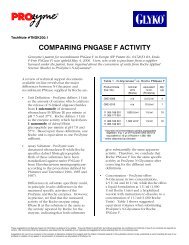
Slide 5 - Conjugate Brightness<br />
So, let’s take some time now to consider the factors that combine to determine any<br />
conjugate brightness: molar absorptivity, quantum efficiency, fluorochrome density and<br />
finally, self-quenching; the first two and the fourth are characteristics of the fluorochrome<br />
and the third is a result of the conjugation process:<br />
<br />
Molar Absorptivity (aka Molar Extinction Coefficient). It is a measure of how well a<br />
molecule absorbs energy at a given wavelength (usually expressed at its<br />
absorbance maximum). In reality, molecules absorb over a range of wavelengths<br />
constituting the molecule’s absorptivity spectrum, of which we’ll see some<br />
examples in a moment. Values can range over several orders of magnitude<br />
between the least and most absorptive molecules, making this factor of primary<br />
importance in determining brightness.<br />
NOTE: Even fluors with the highest molar absorptivities will miss a large portion of<br />
the incident light (waste light in diagram). This is not necessarily a bad thing as it<br />
allows us to multiplex.<br />
<br />
Quantum Efficiency. This is the proportion of absorbed photons emitted as<br />
fluorescence. It is never 100%, as a portion is always dissipated as heat (note the<br />
difference in the thickness of the arrows depicting Absorbed Light vs. Fluorescence
in the diagram). Due to poor reproducibility among methods of measurement, it is<br />
difficult to compare values. Useful fluors have values between 0.25 - 0.9+, making<br />
the maximum difference between the worst and best fluors only 3- to 4-fold. This<br />
factor is therefore of secondary importance in determining conjugate brightness, but<br />
still worthy of consideration.<br />
<br />
Fluorochrome Density. This is the number of fluorochrome molecules per<br />
conjugate molecule. It is often desirable to try to maximize this, but there can be<br />
drawbacks. One is decreased solubility. Fluorescein is a good example—too many<br />
and a conjugate precipitates.<br />
[I would like to pause a moment to explain what is actually going on here,<br />
since this is one of the most misunderstood concepts about conjugates.<br />
Many manufacturers would have you believe that their conjugates are 1:1,<br />
that is, one ligand is conjugated to one fluorochrome. As a result, customers<br />
have an oversimplified picture of what the conjugates actually look like.<br />
Although the molar ratio of antibody-to-fluor in the conjugate may be<br />
reported as 1:1—and that’s probably rarely the case, the conjugate is really a<br />
population of many antibody and fluorochrome molecules, say a mix of 1:1,<br />
2:2, 3:3; or even 2:3 and 3:2.<br />
Only one of these antibody molecules participates per antigen binding<br />
event, but what is measured is the cumulative fluorescence of all of the<br />
fluorochrome molecules in the conjugate. Per binding event, many fluors<br />
are measured, making that very bright conjugate.]<br />
<br />
Self-quenching. Another drawback to increasing fluorochrome density is selfquenching<br />
(fluorescein is a good example). Self-quenching can occur when the<br />
emission spectrum of a fluorochrome overlaps significantly with its absorbance<br />
spectrum, resulting in reabsorption of an emitted photon before it reaches the<br />
detector. The result is decreased apparent fluorescence signal, because the<br />
probability of a photon being re-emitted is decreased.<br />
Self-quenching is increased by any process that decreases the randomness of the<br />
distribution of molecules in solution, and is most problematic when many small<br />
fluorochromes with short Stokes shifts are incorporated into a single conjugate (a<br />
strategy commonly used by manufacturers to compensate for low molar<br />
absorptivity, quantum efficiency, etc.).<br />
See TechNote TNPJ210 for a more exhaustive discussion with representative values:<br />
http://www.prozyme.com/pdf/tnpj200.pdf
Slide 6 - Phycopigment Proteins<br />
Now let’s see how the PPP’s measure up in terms of these characteristics. The PPP’s have<br />
optimum spatial chromophore arrangement which optimizes each of these factors:<br />
NOTE: The subunits and their interactions are beyond the scope of this presentation, but<br />
the effects can be summed up in terms of the factors we have defined.<br />
<br />
The PPP’s have extremely high molar absorptivities. They’ve evolved to be<br />
excellent light antennae; their very survival has depended on their ability to<br />
scavenge light from the low-light environments in which their parent organisms<br />
live. The PPP’s also have a large number of chromophores per molecule (~35 in<br />
RPE) vs. only one per molecule in small, synthetic dyes (e.g. CyDye Fluors, Alexa<br />
Fluor ® dyes, fluorescein, rhodamine, etc.). Fluorescein and Rhodamine B have<br />
values less than 100 (with units of x10 3 cm 2 mol -1 ). The Alexa Fluor ® dyes and<br />
CyDye Fluors typically have values ranging from about 100 to 250. By contrast,<br />
the PPP’s are up to several orders of magnitude higher: RPE has a value of nearly<br />
2000, while BPE comes in over 2400 (at its max)!<br />
I’d like to digress for a moment and mention that, although RPE has traditionally<br />
been the phycoerythrin of choice for flow cytometry, with the
introduction/installation of instruments equipped with green lasers (e.g. 532 nm),<br />
BPE, with its excitation maximum at 545 nm, might be a worthwhile alternative,<br />
given its extremely high molar absorptivity.<br />
<br />
<br />
In terms of quantum efficiencies, the PPP’s have been shown to be among the most<br />
efficient of all fluors, and for a very good reason: the PPP’s are responsible for<br />
shuttling light in an energy cascade to chlorophyll in the photosynthetic reaction<br />
center through a process known as FRET (fluorescence resonance energy<br />
transfer)—the organism’s survival depends on this being an efficient process.<br />
The PPP’s exhibit minimal self-quenching; the fluors are distributed optimally along<br />
a protein backbone.<br />
Additionally, PerCP has an extremely long Stokes shift (nearly 200 nm; absorbance<br />
482 nm and emission 675 nm). It would be difficult to rival this, even with a<br />
tandem dye!<br />
<br />
Lastly, the PPP’s can be conjugated to antibodies or other proteins without<br />
alteration of their spectral properties.
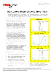
Slide 7 - Molar Absorptivity<br />
This figure provides a comparison of just one of the factors we have been discussing,<br />
molar absorptivity. You’re probably used to looking at normalized absorption curves, but<br />
this is unnormalized data to allow a true comparison of different fluors. As you can see,<br />
relative to RPE and APC, Fluorescein and Alexa Fluor ® 633 are barely off axis! The<br />
conclusion is clear: the PPP’s are the brightest fluors available!
Slide 8 - Phycopigment Proteins<br />
Here they are in their full glory—Peridinin-chlorophyll-protein complex (PerCP),<br />
Y-Phycoerythrin (YPE), R-Phycoerythrin (RPE), R-Phycocyanin (RPC), C-Phycocyanin<br />
(CPC) and Allophycocyanin (APC) (left to right). For the convenience of researchers, we<br />
have made some them available as kits for conjugation.
Slide 9 - PhycoLink ® Conjugation Kits<br />
These are the Product Codes for the various colors we offer. Our newest kit is PerCP, with<br />
which you’re no doubt familiar. We’re pleased that Becton, Dickinson and Company, Inc.,<br />
who continues to hold the patent on this fluorochrome, recognized the increasing need for<br />
end-users to make their own conjugates of this useful fluor and agreed to license to us for<br />
use in conjugation kits for research use only. We also offer a tandem fluorochrome:<br />
RPE-Tandem-665, which consists of RPE covalently coupled to the Oyster ® 645 dye.
Slide 10 - PhycoLink ® Conjugation Kits<br />
Consider some of the features and benefits of our PhycoLink Conjugation Kits:<br />
<br />
<br />
<br />
<br />
<br />
<br />
First, they are complete. They contain everything you need to conjugate up to<br />
1 mg of your antibody (or other target protein).<br />
They are universal in that they work with essentially any type of antibody<br />
molecule or other sulfhydryl-containing protein. The kits work exceptionally well<br />
with polyclonal and the majority of monoclonal antibodies. We’ve even had<br />
reports from customers who have successfully conjugated IgM’s using our kits!<br />
And, we’ve successfully made F(ab’) 2 conjugates ourselves.<br />
They’re fast; the entire procedure takes less than 2 ½ hours to complete.<br />
The kits are extremely easy to use. We provide you with complete step-by-step<br />
protocols.<br />
Our kits utilize the PPP’s, and as I’ve shown this should result in the brightest,<br />
most sensitive conjugates.<br />
The kits are configured to be flexible to allow different scale conjugations from a<br />
single kit. This is especially useful in cases where you wish to perform a trial
conjugation (e.g. 100 g) and then scale-up.<br />
<br />
Lastly, they’re economical. Making your own conjugates represents a<br />
cost-effective alternative to purchasing commercial conjugates, especially if you use<br />
a lot of the same conjugate; also, custom conjugations can cost thousands of dollars<br />
for similar quantities.
Slide 11 - Everything needed to conjugate up to 1 mg of antibody<br />
PhycoLink kits contain everything you need to conjugate up to 1 mg of antibody (2 mg in<br />
the case of the PerCP kit): most importantly, the activated phycopigment protein; and all<br />
the other necessary reagents; two different gel filtration methods (desalting columns and<br />
spin columns) to enable different scale conjugations; and pre-made exchange and storage<br />
buffers, plus the recipes for making more.
Slide 12 - Fast & Easy Protocol<br />
The procedure takes less than 2 ½ hours from start to finish and consists of four easy steps:<br />
antibody preparation, antibody reduction, covalent conjugation and conjugate finishing.
Slide 13 - Antibody Preparation & Reduction<br />
It’s important to start out with a purified antibody solution, preferably at 1-10 mg/ml for<br />
best results. Essentially any neutral buffer is compatible (e.g. phosphate, Tris, MES), but<br />
the solution should be free of BSA or other sulfhydryl-containing proteins. If you’re<br />
planning to conjugate an antibody you’ve purchased from an outside source, always<br />
request a copy of the Certificate of Analysis from the vendor which specifies the<br />
formulation, including any carrier proteins. Additives such as sodium azide or glycerol, up<br />
to a certain amount, shouldn’t interfere, but we’ve got a whole series of Frequently Asked<br />
Questions (FAQ’s) on our website which discuss buffer considerations, concentrations and<br />
other common questions:<br />
http://www.prozyme.com/faqs/faqs.html#phycolink<br />
Start by reducing your purified antibody to expose free sulfhydryls. Our standard protocol<br />
is preferential for the hinge disulfides. Again, this is important in that the conjugation<br />
process won’t interfere with or diminish the antibody’s antigen binding capacity.<br />
Before proceeding to the covalent conjugation step it’s important to remove the excess<br />
reducing agent. This is accomplished with either the desalting or spin columns provided.
Slide 14 - Covalent Conjugation<br />
Once you’ve reduced and desalted your antibody, covalently couple it to the activated<br />
PPP. The activated PPP provided in the kit is a fully tested product in and of itself and is<br />
available for purchase separately.<br />
The conjugation conditions have been optimized to ensure the reaction works as reliably<br />
as possible with the majority of antibodies. A molar excess of activated PPP is added to<br />
drive the reaction so that practically every antibody molecule is tagged.<br />
This slide shows an oversimplified diagram of what is actually occurring. In reality, there<br />
are likely to be several lysine residues modified per PPP molecule (in other words, several<br />
SMCCs per PPP). Also, each antibody contains more than one free sulfhydryl residue,<br />
although usually only 1-2 activated PPPs react per antibody due to steric hindrance. This<br />
has the potential to produce antibody-PPP molecules of varying composition.<br />
Although reagent manufacturers often report the molar ratio of antibody-to-fluor in their<br />
conjugates as 1:1, this can be misleading and should not be taken to mean that every<br />
single antibody-PPP molecule is composed of only one antibody plus one PPP. The<br />
conjugate is usually a heterogeneous population of different antibody-fluorochrome<br />
molecules, each characterized by their own antibody-to-PPP ratios (e.g. 1:1, 2:2, 3:3; or<br />
even 2:3 or 3:2). The aggregate ratio of the overall population may average out to 1:1, or
to a higher or lower ratio.<br />
Following covalent conjugation, any remaining free sulfhydryl groups are covalently<br />
blocked by treatment with NEM, and the conjugate may either be exchanged into Storage<br />
Buffer for immediate use or purified using one of our Purification Kits.
Slide 15 - Effect of PPP:Ab Molar Ratio on Conjugate Yield<br />
This slide summarizes the effect of varying the molar ratio of PPP to antibody on conjugate<br />
yield. For a full discussion, please refer to the FAQ on our website:<br />
http://www.prozyme.com/faqs/pjpamtfaq.html<br />
In this experiment, 1 mg of purified goat IgG was conjugated with various amounts of<br />
activated APC (x-axis). The reddish-brown curve shows the percentage of APC in<br />
precipitated conjugate while the dark blue line shows the percentage of soluble conjugate.<br />
While low molar ratios of APC to Ab give high incorporation of APC into conjugate, as you<br />
can see (reddish-brown line) they can result in poor yields due to excessive cross-linking<br />
and precipitation; whereas high molar ratios of APC to Ab result in a greater percentage of<br />
soluble conjugate (dark blue line). We’ve selected ratios for our kits that should produce<br />
high yields with very little precipitation if you follow our standard protocol. NOTE: there<br />
will be excess PPP not incorporated into the conjugate as a result. Further, every PPP has<br />
its own optimal molar ratio; what’s shown here is specific to APC.
Slide 16 - Alternatives to the Standard Method<br />
In some cases, it may be necessary or desirable to use an alternative conjugation method<br />
for a number of reasons: your protein doesn’t contain any endogenous sulfhydryls; is<br />
resistant to DTT reduction; reoxidizes quickly; is sterically hindered or loses<br />
activity/function when reduced; or to minimize losses or dilution when performing smallscale<br />
conjugations.
Slide 17 - Alternatives to the Standard Method (continued)<br />
There are three different methods for conjugating your protein using the activated PPP<br />
provided in our conjugation kits when an alternative to our standard protocol is needed.<br />
The choice of which method to use will depend on several factors: whether your protein<br />
contains available sulfhydryls; whether or not you wish you reduce your protein; and the<br />
scale of the conjugation reaction.<br />
The first two methods (Iminothiolane and SPDP-TCEP) work with both sulfyhdryl- and<br />
non-sulfhydryl-containing proteins. The Iminothiolane method has the advantage that it<br />
avoids a reduction step, but has the disadvantage that it works through amine modification<br />
which, in the case of antibodies, has the potential to interfere with the antigen binding site.<br />
This method is also subject to the same issue of loss and dilution as our standard method<br />
when performing small-scale conjugations. The SPDP-TCEP method has an advantage in<br />
this regard in that it doesn’t require desalting but, like Iminothiolane, has the potential to<br />
result in interference with the antigen binding site.<br />
The third method (Direct TCEP), is a new one which we hope to have incorporated into<br />
our kits within the next couple of months. This method may be suitable when performing<br />
small-scale (e.g. 0.25 mg) conjugations of sulfyhdyryl-containing proteins. As a non-thiol<br />
containing reducing agent, TCEP doesn’t require desalting and thus avoids the losses<br />
and/or dilution associated with DTT removal. However, it should be noted that TCEP is
purported to be a more potent reducing agent than DTT, which may affect the<br />
characteristics of a given antibody.<br />
For more details concerning alternative conjugation methods, see our TechNote TNPJ300:<br />
http://www.prozyme.com/pdf/tnpj300.pdf
Slide 18 - Scaling Up<br />
Let’s assume you’ve successfully made a conjugate (e.g. 0.1 mg), tested it in your<br />
application and now wish to scale-up.<br />
For scale-ups within the capacity of our kits, we recommend following the procedure<br />
appropriate for the amount of antibody: 0.1 mg to 0.25 mg conjugations are best<br />
performed using the Spin Column procedure; 0.5 mg to 1 mg conjugations are best<br />
performed using the Desalting Column procedure; 0.25 mg to 0.5 mg conjugations may be<br />
performed using either procedure, depending on antibody concentration.<br />
Scale-ups beyond the capacity of our kits may be performed by purchasing additional<br />
activated PPP. While a full discussion of the parameters relevant to scale-up is beyond the<br />
scope of this presentation, by maintaining antibody concentration and molar ratios, we’ve<br />
been able to demonstrate an essentially linear scale-up from 0.5 mg to ~2 mg to 10 mg and<br />
beyond. We invite you to give us a call if you are contemplating a scale-up, as individual<br />
antibodies have unique considerations and actual results may vary.<br />
In case you’d prefer to spend your time on other exciting aspects of your research,<br />
<strong>ProZyme</strong> offers affordable custom conjugation services for large-scale projects.
Slide 19 - Why Purify?<br />
In many cases, it’s possible to use your conjugates “as is”, with some unincorporated PPP.<br />
This often doesn’t pose a problem, especially when staining cell surface antigens, as<br />
unbound PPP is washed away in the process.<br />
However, depending on the application, purification of unincorporated activated PPP may<br />
be desirable in order to achieve higher sensitivity through a reduction of non-specific<br />
binding. Flow cytometric staining of intracellular antigens is an example of an application<br />
which may benefit by the use of purified conjugates. It’s also necessary to purify your<br />
conjugate if you intend to perform lot-to-lot comparisons as the unincorporated activated<br />
PPP will interfere in the evaluation.<br />
The disadvantages of purification should be taken into consideration when deciding<br />
whether or not to purify as losses can exceed 50% when processing small quantities. In<br />
general, our recommendation is that you not purify unless absolutely necessary.<br />
Sometimes only a small portion needs to be purified for evaluation.
Slide 20 - PhycoLink ® Purification Kits<br />
If you’ve decided to purify your conjugate, we offer purification kits to allow rapid and<br />
convenient purification by means of size exclusion chromatography. Our kits contain<br />
everything necessary to remove unincorporated reactants from your conjugate and include<br />
detailed protocols that have been thoroughly tested for trouble-free purification. When<br />
you purchase a kit, you also receive a CD-ROM with photographs to guide you through<br />
the various steps.<br />
Importantly, our purification kits are specially formulated for use with PPP conjugates.<br />
The PPP’s are very large molecules (e.g. RPE is ~240 kDa; APC is ~104 kDa). So, the<br />
challenge is to purify away one large molecule (the activated PPP) from another large<br />
molecule (your conjugate). This isn’t effectively done with any ordinary matrix. We’ve<br />
identified the best matrix for this purpose and have selected one that achieves optimal<br />
separation while minimizing non-specific binding of PPP’s, known to be a problem with<br />
certain matrices.<br />
NOTE: Affinity purification is not recommended for PPP conjugates as the harsh elution<br />
conditions diminish fluorescent properties.<br />
Presently, our purification kits are available in two sizes:
KPK13: 13-ml column for purification of 0.25 mg conjugates<br />
KPK80: 80-ml column for purification of 1.0 mg conjugates<br />
but other column sizes are generally available that work with the kit connectors.<br />
Better resolution can be achieved with the 80-ml column than the 13-ml column due to its<br />
greater length, and hence, resolving capability. However, it is possible to obtain<br />
significantly purified conjugate with either column by selecting only early fractions.<br />
A single purification kit can be used many times; the hardware is reusable and the matrix is<br />
stable when stored in ethanol at 4°C. Some investigators prefer to designate a different<br />
matrix batch for each conjugate. Matrix refills may be purchased from <strong>ProZyme</strong> for only a<br />
fraction of the cost of the original kit.<br />
The set-up is based on a Luer-Lock system, so it is possible to substitute a different<br />
column to accommodate the amount of conjugate. For example, one of our customers,<br />
who happens to be attending this meeting, Dr. Jian Ling from the Southwest Medical<br />
Research Center in San Antonio, Texas, wished to purify a 50-g conjugate he had made<br />
using one of our conjugation kits. We were concerned that, due to the small volume, the<br />
losses with our 13-ml column would be too great. Dr. Ling independently acquired a 4-ml<br />
column, followed our setup procedures (using our matrix) and was able to achieve<br />
adequate separation of his conjugate from the unreacted components in order to perform<br />
an intracellular flow experiment requiring high sensitivity.<br />
(Thank you Dr. Ling for allowing us to use your name as an example.)
Slide 21 - Mouse IgG-RPE: 80-ml Column Profile<br />
This is an example of a typical column profile obtained with our 80-ml column. We’re<br />
looking at fractions of a 1-mg mouse IgG-RPE. As you can see, the majority of the<br />
conjugate elutes as a leading peak, with smaller conjugate complexes running between the<br />
leading peak and the broad peak of unincorporated RPE. Very little unincorporated<br />
antibody (green line) is present due to the optimized protocols.<br />
In choosing which fractions to pool, there’s a trade-off between purity and yield. Clearly,<br />
the early fractions will be the most pure, but if your application can tolerate it, less pure<br />
fractions may be included in order to increase yield.
Slide 22 - Conjugate Evaluations<br />
Please refer to TechNote TNPJ200 for a complete set of tools for evaluating your purified<br />
conjugate:<br />
http://www.prozyme.com/pdf/tnpj200.pdf<br />
This TechNote contains techniques for determining protein concentration, molarity of<br />
individual components, molecular weight and conjugate composition. For those of you<br />
who may be a little rusty in terms of your concentration calculations, the TechNote<br />
includes a worksheet with all the formulas necessary to convert absorbance measurements<br />
into concentration data; all you have to do is fill in the blanks.<br />
These characterization techniques may be applied both to conjugates made using our kits<br />
as well as those purchased from commercial sources. Although conjugate manufacturers<br />
would like us to believe in the simplified case that conjugates are 1:1, this probably isn’t so<br />
often the case. Therefore, you may be well served to characterize the conjugates you’re<br />
purchasing to determine the fluorochrome-to-antibody ratio, particularly if the intended<br />
use is for quantitative analysis.
Slide 23 - Technical Support<br />
All of the protocols, FAQs and TechNotes referenced, are available on our website:<br />
http://www.prozyme.com/phycolink/pj-kits.html<br />
Our kits work well with the majority of antibodies, and customers rarely have problems.<br />
However, I want to emphasize that we’re here to help at any step along the way. We<br />
appreciate that your time is valuable, so the first thing we ask you to do, if you don’t get<br />
the expected results, is to complete the troubleshooting form in the FAQ section on our<br />
website (and submit it to us by email or fax):<br />
http://www.prozyme.com/faqs/pjperffaq.html<br />
That way, we’ll have all the information concerning your experiment and be as prepared<br />
as possible to provide a solution to the problem.
Slide 24 - Phenotype of the BB Rat T Lymphopenia<br />
Now, I’d like to turn to some data supplied by our customers. This first example is work<br />
done in the laboratory of Dr. Phillippe Poussier at Sunnybrook Health Sciences Centre in<br />
Toronto, Ontario.<br />
Shown are lymph node mononuclear cells from the diabetes-prone BB rat (right panels)<br />
versus control animals (left panels), stained with either anti-TCR / FITC (top row) or<br />
CD4-RPE and CD8-APC (bottom row). Note, the Gimap5 mutation in the BB rat results in<br />
severe T lymphopenia, including distortion of the normal CD4:CD8 ratio.<br />
The antibodies used in this experiment were made in-house from hybridomas (all mouse<br />
IgG1-kappa) purchased from the European Tissue Culture Collection; and the CD4-RPE<br />
and CD8-APC conjugates were made using PhycoLink Conjugation Kits. Dr. Poussier’s lab<br />
typically performs 1-mg conjugations, but has done smaller-scale conjugations with<br />
acceptable results. The conjugates are used without purification, yet as you can see, bright<br />
staining of the respective populations is achieved, with very little background.<br />
According to Dr. Poussier, his lab makes their own conjugates, as opposed to purchasing<br />
commercial conjugates, because “it is cheaper in the long run especially if several<br />
investigators who use the same reagents share the costs.”
Slides 25 through 31<br />
The next series of slides was kindly provided by Diagnostic Hybrids, based in Athens,<br />
Ohio, who are developing a clinical diagnostic assay for the rapid detection of viral<br />
infection based on a conventional fluorescence microscopy platform. Their assay makes<br />
use of highly-specific anti-viral monoclonal antibodies conjugated to RPE, which when<br />
viewed through a fluorescein filter set appears yellow. They are planning to submit a<br />
510(k) application to the FDA in order to obtain clearance for these assays as in vitro<br />
diagnostic (IVD) Reagents and are currently looking for partner sites who would be<br />
interested in performing clinical testing.<br />
The PPP’s traditionally have been excluded from use as tags for fluorescence microscopy<br />
due to concerns about photobleaching. Diagnostic Hybrids has observed absolutely no<br />
evidence of photobleaching of their RPE conjugates, even under the continuous<br />
illumination conditions inherent in this platform.
Slide 25 - D 3 uet Flu A/Respiratory Screen<br />
Influenza A infection grown on R-Mix Too ® mixed cell monolayer stained with the<br />
Flu A/respiratory screening reagent and viewed using a fluorescein filter set. The<br />
Flu A/respiratory screening reagent contains antibodies to Flu A labeled with RPE and<br />
antibodies to six other major respiratory viruses labeled with fluorescein.
Slide 26 - D 3 uet Flu A/Respiratory on Flu A/B Co-infection<br />
Influenza A and influenza B co-infection grown on R-Mix ® mixed cell monolayer stained<br />
with the Flu A/respiratory screening reagent and viewed using a fluorescein filter set.
Slide 27 - D 3 uet Flu A/Respiratory on Flu A/Adeno Co-infection<br />
Influenza A and adenovirus co-infection grown on R-Mix ® mixed cell monolayer stained<br />
with the Flu A/respiratory screening reagent and viewed using a fluorescein filter set.
Slide 28 - D 3 uet Flu A/Respiratory on Flu A/Para2 Co-infection<br />
Influenza A and parainfluenza 2 co-infection grown on R-Mix ® mixed cell monolayer<br />
stained with the Flu A/respiratory screening reagent and viewed using a fluorescein filter<br />
set.
Slide 29 - D 3 uet Flu A/Respiratory on Flu A/RSV Co-infection<br />
Influenza A and respiratory syncytial virus co-infection grown on R-Mix ® mixed cell<br />
monolayer stained with the Flu A/respiratory screening reagent and viewed using a<br />
fluorescein filter set.
Slide 30 - D 3 uet VZV (Varicella-zoster Virus)<br />
Left-hand panel: VZV grown on H&V-Mix ® cell monolayer stained with VZV antibodies<br />
labeled with RPE and viewed using a fluorescein filter set.<br />
Right-hand panel: VZV infected cells scraped and mounted on slide and stained with VZV<br />
antibodies labeled with RPE and viewed using a fluorescein filter set.
Slide 31 - D 3 uet HSV-2 (Herpes Simplex Virus type 2)<br />
HSV-2 grown on A549 cell monolayer stained with HSV-2 antibodies labeled with RPE and<br />
viewed using a fluorescein filter set.
Slides 32 and 33<br />
Immunicon Corporation kindly granted permission to use the images shown in the next<br />
pair of slides. They have developed an IVD assay for the capture, analysis and<br />
quantitation of rare, circulating, breast cancer tumor cells (CTC’s) from blood.<br />
Immunicon’s CellTracks ® AutoPrep System is an automated sample preparation system for<br />
the immunomagnetic capture and fluorescence staining of CTC’s based on their expression<br />
of a set of specific markers. Immunicon’s CellTracks Analyzer II is a semi-automated<br />
fluorescence microscope for the enumeration and characterization of the isolated cells.<br />
Accurate quantitation and analysis relies on the use of target-specific antibodies conjugated<br />
to fluorochromes, including the phycobiliproteins RPE and APC. This is another example<br />
of the suitability of this class of pigment molecules for this type of fluorescence<br />
application.
Slides 32 - CellTracks ® AutoPrep System for Rare Cell Analysis<br />
This slide illustrates the processing steps involved in the capture and staining of CTC’s<br />
performed by Immunicon’s CellTracks AutoPrep System.<br />
The final enriched sample is dispensed into the MagNest ® cell presentation device, which<br />
presents the captured cells in a single focal plane for quantitative analysis using the<br />
CellTracks Analyzer II.
Slide 33 - CellTracks Analyzer II for for Rare Cell Analysis<br />
Sample image gallery obtained with the CellTracks Analyzer II.<br />
Discrimination of CTC’s from leukocytes is made possible by the use of lineage-specific<br />
markers: cytokeratin-RPE (CK-PE) identifies CTC’s, while CD45-APC, identifies leukocytes.
Slide 34 - Trademarks and Licenses<br />
We show trademarks and licenses which may have been mentioned during the course of<br />
the presentation.<br />
Thank you to our PhycoLink customers who provided data for inclusion in this<br />
presentation:<br />
Phillippe Poussier, M.D., Associate Professor, Departments of Medicine and Immunology<br />
Sunnybrook Health Sciences Centre<br />
Jimmy Page, Ph.D., Manager, Immunological Detection<br />
Diagnostic Hybrids, Inc<br />
E-mail: jimmy_page@dhiusa.com<br />
Immunicon Corporation<br />
E-mail: sales@immunicon.com
Slide 35 - Conclusion<br />
We’ve come to the end of our time, so I’d like to thank you once again for coming and I’ll<br />
now open the floor up to any questions.<br />
<strong>ProZyme</strong> customers are an important source of information regarding advanced or<br />
specialized uses of our products. We encourage you to contact us if you have any<br />
suggestions about product performance or new applications and techniques.<br />
TOLL FREE<br />
(800) 457-9444 (US & CANADA)<br />
PHONE<br />
(510) 638-6900<br />
FAX<br />
(510) 638-6919<br />
E-MAIL<br />
info@prozyme.com<br />
WEB<br />
www.prozyme.com<br />
Or, contact your local distributor; a list of <strong>ProZyme</strong>’s distributors, with contact information<br />
may be found at:<br />
http://www.prozyme.com/distrib.html
Disclaimer<br />
These suggestions and data are based on information we believe to be reliable. They are offered in good faith, but without<br />
guarantee, as conditions and methods of use of our products are beyond our control. We recommend that the prospective user<br />
determine the suitability of our materials and suggestions before adopting them on a commercial scale.<br />
Suggestions for use of our products or the inclusion of descriptive material from patents and the citation of specific patents in this<br />
publication should not be understood as recommending the use of our products in violation of any patent or as permission to<br />
license to use any patents of <strong>ProZyme</strong>, Inc.