Intentional replantation ^ a `last resort' treatment or a ...
Intentional replantation ^ a `last resort' treatment or a ...
Intentional replantation ^ a `last resort' treatment or a ...

Create successful ePaper yourself
Turn your PDF publications into a flip-book with our unique Google optimized e-Paper software.
Peer<br />
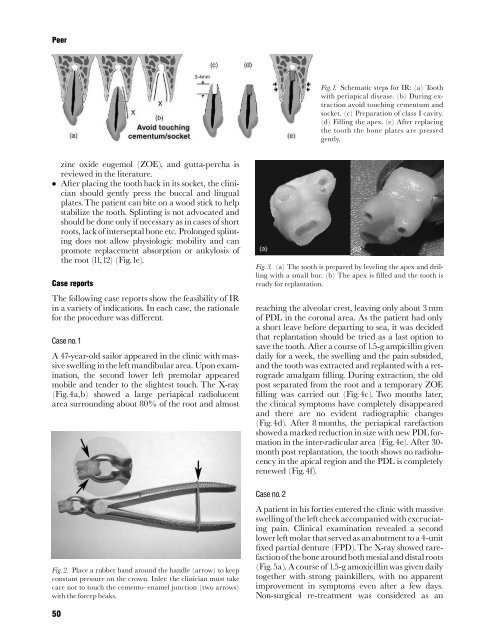
Fig. 1. Schematic steps f<strong>or</strong> IR: (a) Tooth<br />
with periapical disease. (b) During extraction<br />
avoid touching cementum and<br />
socket. (c) Preparation of class Icavity.<br />
(d) Filling the apex. (e) After replacing<br />
the tooth the bone plates are pressed<br />
gently.<br />
zinc oxide eugemol (ZOE), and gutta-percha is<br />
reviewed in the literature.<br />
After placing the tooth back in its socket, the clinician<br />
should gently press the buccal and lingual<br />
plates.The patient can bite on a wood stick to help<br />
stabilize the tooth. Splinting is not advocated and<br />
should be done only if necessary as in cases of sh<strong>or</strong>t<br />
roots, lack of interseptal bone etc. Prolonged splinting<br />
does not allow physiologic mobility and can<br />
promote replacement abs<strong>or</strong>ption <strong>or</strong> ankylosis of<br />
the root (11,12) (Fig.1e).<br />
Case rep<strong>or</strong>ts<br />
The following case rep<strong>or</strong>ts show the feasibility of IR<br />
in a variety of indications. In each case, the rationale<br />
f<strong>or</strong> the procedure was di¡erent.<br />
Case no. 1<br />
A 47-year-old sail<strong>or</strong> appeared in the clinic with massive<br />
swelling in the left mandibular area. Upon examination,<br />
the second lower left premolar appeared<br />
mobile and tender to the slightest touch. The X-ray<br />
(Fig.4a,b) showed a large periapical radiolucent<br />
area surrounding about 80% of the root and almost<br />
Fig. 2. Place a rubber band around the handle (arrow) to keep<br />
constant pressure on the crown. Inlet: the clinician must take<br />
care not to touch the cemento^enamel junction (two arrows)<br />
with the f<strong>or</strong>cep beaks.<br />
Fig. 3. (a) The tooth is prepared by leveling the apex and drilling<br />
with a small bur. (b) The apex is filled and the tooth is<br />
ready f<strong>or</strong> <strong>replantation</strong>.<br />
reaching the alveolar crest, leaving only about 3 mm<br />
of PDL in the c<strong>or</strong>onal area. As the patient had only<br />
a sh<strong>or</strong>t leave bef<strong>or</strong>e departing to sea, it was decided<br />
that <strong>replantation</strong> should be tried as a last option to<br />
save the tooth. After a course of 1.5-g ampicillin given<br />
daily f<strong>or</strong> a week, the swelling and the pain subsided,<br />
and the tooth was extracted and replanted with a retrograde<br />
amalgam ¢lling. During extraction, the old<br />
post separated from the root and a temp<strong>or</strong>ary ZOE<br />
¢lling was carried out (Fig.4c). Two months later,<br />
the clinical symptoms have completely disappeared<br />
and there are no evident radiographic changes<br />
(Fig.4d). After 8 months, the periapical rarefaction<br />
showed a marked reduction in size with new PDL f<strong>or</strong>mation<br />
in the inter-radicular area (Fig.4e). After 30-<br />
month post <strong>replantation</strong>, the tooth shows no radiolucency<br />
in the apical region and the PDL is completely<br />
renewed (Fig.4f).<br />
Case no. 2<br />
A patient in his f<strong>or</strong>ties entered the clinic with massive<br />
swelling of the leftcheek accompaniedwith excruciating<br />
pain. Clinical examination revealed a second<br />
lower left molar that servedas an abutment to a 4-unit<br />
¢xed partial denture (FPD).The X-ray showed rarefactionofthebone<br />
aroundbothmesialanddistalroots<br />
(Fig.5a). A course of 1.5-g amoxicillin was given daily<br />
together with strong painkillers, with no apparent<br />
improvement in symptoms even after a few days.<br />
Non-surgical re-<strong>treatment</strong> was considered as an<br />
50