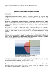
Intentional replantation ^ a `last resort' treatment or a ...
Intentional replantation ^ a `last resort' treatment or a ...
Intentional replantation ^ a `last resort' treatment or a ...

Create successful ePaper yourself
Turn your PDF publications into a flip-book with our unique Google optimized e-Paper software.
<strong>Intentional</strong> <strong>replantation</strong><br />
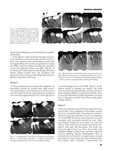
Fig. 4. (a, b) Mandibular premolar presented<br />
with periapical disease. (c) After<br />
<strong>replantation</strong>. (d) Two months after IR,<br />
still no radiographic changes. (e) Eight<br />
months after IR, the periapical rarefaction<br />
showed a marked reduction. (f)<br />
Thirty months post <strong>replantation</strong> the<br />
PDL is completely renewed.<br />
optionbut handling the post inthe distal rootcouldbe<br />
problematic.<br />
As the patient could not tolerate the pain anym<strong>or</strong>e,<br />
it was decided to extract the tooth, check f<strong>or</strong> root fractures<br />
and replant it after perf<strong>or</strong>ming an extra-<strong>or</strong>al<br />
retrograde ¢lling.The tooth was separated by cutting<br />
the FPD, extracted without breaking the p<strong>or</strong>celain<br />
crown and replanted without any splint (Fig.5b). A<br />
few hours after replanting, the pain disappeared completely.<br />
Fifteen months later, the periapical and<br />
pan<strong>or</strong>amic X-rays shows good healing with noperiradicular<br />
rarefactions (Fig.5c,d).<br />
Case no. 3<br />
A 70-year-old patient presented with complaints of<br />
discomf<strong>or</strong>t around the second lower right incis<strong>or</strong>.<br />
An examination revealed the presence of a sinus tract<br />
near the tooth.The X-ray rep<strong>or</strong>t showed a periapical<br />
area encircling approximately 50% of the apical root<br />
Fig. 5. (a) Mandibular second molar presented with periapical<br />
disease. (b) Immediately after IR. (c, d) Fifteen months later,<br />
periapical and pan<strong>or</strong>amic X-rays show complete healing.<br />
Fig. 6. Replantation of mandibular incis<strong>or</strong> presented with periapical<br />
disease and sinus tract. (a) Periapical radiolucency encircling<br />
50% of root. (b) After IR and retrograde amalgam filling.<br />
(c) Four years later, periapical X-ray shows good healing.<br />
area with disappearance of the PDL (Fig.6a). As the<br />
patient refused to undergo any surgery, the tooth<br />
was extracted and replanted after an extra-<strong>or</strong>al retrograde<br />
amalgam ¢lling was perf<strong>or</strong>med (Fig.6b). After<br />
4 years, thetooth shows no clinicalproblems, the sinus<br />
tract is long gone and the X-ray shows good apical<br />
healing (Fig.6c).<br />
Case no. 4<br />
A 40-year-old patient arrived12 years ago to the communal<br />
clinic with complaints of discomf<strong>or</strong>t around<br />
the lower second left molar. Examination revealed a<br />
sinus tract near the tooth that served as an abutment<br />
to a 3-unit FPD. The X-ray showed an old root canal<br />
<strong>treatment</strong> with rarefaction around the roots. After<br />
removing the bridge, the root canals were retreated<br />
(Fig.7a). Five years later, the ¢stula returned accompanied<br />
with pain and discomf<strong>or</strong>t. The tooth was<br />
extracted, apicoectomy was done extra-<strong>or</strong>ally with<br />
amalgam ¢llings on both roots, and then replanted<br />
with no splinting (Fig.7b). Radiographs taken at<br />
3-month intervals showed no improvement around<br />
the mesial root and a deep mesial periodontal pocket<br />
was evident. After 11months, a hemisection was<br />
51