Antigen sampling and presentation - EPFL
Antigen sampling and presentation - EPFL
Antigen sampling and presentation - EPFL

Create successful ePaper yourself
Turn your PDF publications into a flip-book with our unique Google optimized e-Paper software.
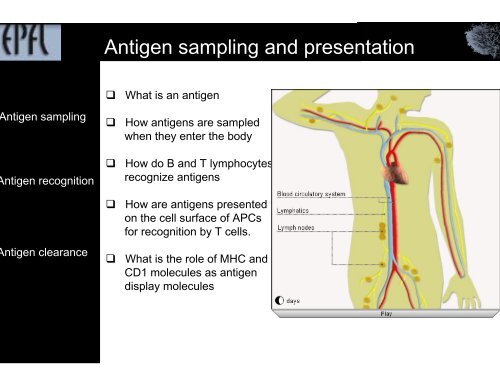
<strong>Antigen</strong> <strong>sampling</strong> <strong>and</strong> <strong>presentation</strong><br />
ntigen <strong>sampling</strong><br />
ntigen recognition<br />
ntigen clearance<br />
<br />
<br />
<br />
<br />
<br />
What is an antigen<br />
How antigens are sampled<br />
when they enter the body<br />
How do B <strong>and</strong> T lymphocytes<br />
recognize antigens<br />
How are antigens presented<br />
on the cell surface of APCs<br />
for recognition by T cells.<br />
What is the role of MHC <strong>and</strong><br />
CD1 molecules as antigen<br />
display molecules
How are antigens recognized by the immune system ?<br />
efinition<br />
ature<br />
<br />
<br />
antigen refers to any substance that is<br />
recognized by T or B lymphocytes via<br />
their cell surface receptors <strong>and</strong>/or<br />
antibodies secreted by B cells<br />
<strong>Antigen</strong>s can be:<br />
‣ proteins & glycoproteins<br />
‣ polysaccharides<br />
‣ lipoproteins & lipopolysaccharides<br />
‣ nucleic acids<br />
‣ chemical compounds (nicotine,<br />
heavy metals, narcotics).<br />
oreignness<br />
Self antigens refer to all antigens that<br />
are normal constituents of the body.<br />
Non-self antigens refer to all antigens that<br />
are not normal constituents of the body.<br />
munoenicity<br />
Immunogens refer to antigens that are able to stimulate a specific adaptive immune response<br />
when introduced into the body. Not all antigens are immunogens.<br />
To be immunogenic, antigens must fulfill certain criteria<br />
Hapten are small molecules that need a carrier to be immunogenic
What is an epitope<br />
cell epitope<br />
<strong>Antigen</strong>s are generally larger than the binding sites of the B cell or T cell's antigen receptors<br />
that specifically recognize <strong>and</strong> bind them.<br />
The particular parts of the antigen that are specifically recognized by an antigen receptor<br />
is called epitope or antigenic determinant<br />
B cell epitopes are either:<br />
‣ conformational corresponding to<br />
surface exposed regions of native<br />
antigen.<br />
‣ linear corresponding to surface<br />
exposed sequences of the antigen<br />
‣ epitopes that are preferentially<br />
recognized within a given individual<br />
are called dominant epitopes<br />
cell epitope T cell epitopes are<br />
‣ peptides, bound to MHC molecules &<br />
recognized by αβ TCR (T cell receptors)<br />
‣ lipids or sugars bound to CD1 molecules<br />
& recognized by γδ T cell receptors<br />
eneration of<br />
epitopes<br />
<br />
native antigens have to be processed into fragments so that internal motifs become<br />
accessible to MHC molecules.
<strong>Antigen</strong> <strong>sampling</strong><br />
ntigen<br />
mpling sites<br />
ntigen<br />
mpling cells<br />
Since foreign antigens <strong>and</strong> microorganisms can enter the body at multiple sites,<br />
antigen-<strong>sampling</strong> cells are strategically located at all the possible sites of antigen entry:<br />
‣ skin<br />
‣ mucosal surfaces<br />
‣ the lymph nodes for antigens that<br />
escape <strong>sampling</strong> in skin or mucosal<br />
surfaces <strong>and</strong> find their way from the<br />
tissue spaces to the lymph.<br />
‣ in the spleen for antigens that gain<br />
access to the blood, for instance<br />
following bites by insects<br />
the resident parenchymal antigen-<strong>sampling</strong><br />
cells.<br />
‣ These cells take up antigen <strong>and</strong><br />
deliver it intact to underlying APCs.<br />
‣ Example: the epithelial M cells<br />
concentrated MALT.<br />
the migratory hematopoietic antigen-<strong>sampling</strong><br />
cells.<br />
‣ These cells take up antigen <strong>and</strong> transport<br />
it to distant lymphoid organs.<br />
‣ APCs have competence for antigen processing<br />
<strong>and</strong> <strong>presentation</strong>
<strong>Antigen</strong> <strong>sampling</strong> in skin<br />
The skin harbors two populations of<br />
antigen-<strong>sampling</strong> cells:<br />
‣ Langerhans cells, which reside within<br />
the epidermis<br />
‣ Dermal dendritic cells, which reside<br />
within the dermis.
<strong>Antigen</strong> <strong>sampling</strong> in skin<br />
angerhans<br />
ells<br />
<br />
a unique population of immature DCs<br />
characterized by<br />
‣ the presence of large granules<br />
Birbek granules<br />
‣ E-cadherin<br />
‣ langerin<br />
‣ CD1a,<br />
‣ lack DC-SIGN & mannose receptor<br />
‣ recruited from peripheral blood as<br />
`DC precursors & differentiate in the<br />
epidermis into resident LCs<br />
‣ LCs eventually pick up antigens<br />
(including apoptotic skin cells) <strong>and</strong><br />
migrate by the afferent lymphatics<br />
to the regional lymph nodes,
<strong>Antigen</strong> <strong>sampling</strong> in skin<br />
ermal DCs Dermal dendritic cells are immature<br />
DCs<br />
‣ express different markers as LCs<br />
‣ lack Birbeck granules <strong>and</strong> langerin.<br />
‣ express DC-SIGN & mannose receptor<br />
‣express high levels of DEC-205<br />
Epidermis<br />
Dermis
<strong>Antigen</strong> <strong>sampling</strong> cells: Dendritic cells<br />
mature DCs<br />
CD80,CD86<br />
<br />
<br />
<br />
Immature DCs occur in all tissues where antigen may enter<br />
into the body<br />
high capacity for antigen uptake but a limited capacity for<br />
antigen <strong>presentation</strong><br />
Immature DCs express a large number of receptros:<br />
‣ chemokine receptors (CCR1, CCR2, CCR5, CCR6)<br />
‣ endocytic receptors by which they can capture antigens.<br />
• non-opsonic receptors (DC-SIGN, DEC-205,<br />
mannose receptor) specific for microbial products.<br />
• integrin receptors, which are involved in the uptake of<br />
apoptotic bodies.<br />
• opsonic receptors: Fc & complement receptors<br />
‣ sensing receptors by which they can integrate information<br />
about the antigen<br />
• TLRs<br />
• Cytokine receptors
<strong>Antigen</strong> <strong>sampling</strong> cells: Dendritic cells<br />
ature DCs<br />
<br />
Mature DCs are exclusively found in the T cell areas of secondary lymphoid organs.<br />
They are characterized by a high propensity for antigen <strong>presentation</strong><br />
<strong>and</strong> T cell activation but a reduced ability to capture antigens.<br />
CD80, CD86<br />
Mature DCs express the following :<br />
‣ Chemokine receptor CCR7, so that they can leave<br />
non lymphoid peripheral tissues <strong>and</strong> migrate to the T cell areas<br />
of the secondary lymphoid organs where lig<strong>and</strong>s for CCR7 are<br />
constitutively expressed.<br />
‣ Adhesion molecules (CD48, CD58) required for interacting with<br />
T cells.<br />
‣ Cell surface MHC class II molecules required for antigen<br />
<strong>presentation</strong>.<br />
‣ Co-stimulatory molecules (CD80, CD86, CD40) required for<br />
activation of reactive T cells.<br />
CD80<br />
‣ By contrast, they no longer express endocytic receptors
<strong>Antigen</strong> Recognition<br />
Two main strategies have been developed by the adaptive immune system to recognize antigen<br />
‣ Immunoglobulins (Igs), expressed by the B lymphocytes.<br />
‣ T-cell receptors (TCRs), expressed by the T lymphocytes.<br />
<br />
<strong>Antigen</strong> recognition by B cells clearly<br />
differs from antigen recognition by T cells:<br />
ative<br />
ntigens<br />
‣ B cells recognize intact antigens.<br />
Processed<br />
antigens<br />
‣ T cells recognize antigen fragments<br />
exposed on the surfaces of host cells<br />
by antigen presenting molecules
<strong>Antigen</strong> Recognition by B lymphocytes<br />
<br />
Thymus dependent (TD).<br />
‣ TD antigens refer to antigens against which<br />
antigen-specific B cells cannot produce<br />
antibodies without the cognate help of<br />
antigen-specific CD4 + T cells (CD40/CD40L)<br />
‣ Most TD antigens are oligovalent protein<br />
antigens that bind specifically to the BCR.<br />
Thymus independent type 1 (TI-1).<br />
‣TI-1 antigens are polyclonal, non-antigen<br />
specific activators of B cells that engage<br />
co-stimulatory receptors on B cells but not the<br />
BCRs.<br />
‣ Prototype: lipopolysaccharide (LPS)<br />
from gram-negative bacterial cell wall<br />
which binds to TLR4..<br />
Thymus independent type 2 (TI-2).<br />
‣ TI-2 antigens are high Mr molecules with<br />
repetitive epitopes that cause extensive<br />
cross linking of BCRs <strong>and</strong> stimulate B cell<br />
proliferation <strong>and</strong> differentiation<br />
without the cognate help of T lymphocytes.<br />
‣ Prototype high molecular weight polysaccharide such as the dextran B512 (Dx), LPS.
<strong>Antigen</strong> Recognition by T lymphocytes<br />
TCR-MHC<br />
interaction<br />
Peptide<br />
recognition<br />
T cells detect antigens via T-cell receptors (TCRs) that recognize antigen when presented<br />
as short fragments bound to antigen-presenting molecules on the surface of antigenpresenting<br />
cells (APCs)<br />
T cells exist as two main populations which<br />
have their own antigen recognition strategy.<br />
‣ T cells bearing αβ TCRs recognize peptides<br />
of protein antigens in association with MHC<br />
molecules<br />
• CD4 + T helper cells recognize peptides<br />
of 10-20 amino acids presented by<br />
MHC class II molecules<br />
• CD8 + T cells recognize peptides<br />
of 8 to10 amino acids presented by<br />
MHC class I molecule.<br />
O 4 -molecule<br />
lipid<br />
ecognition<br />
‣ T cells bearing γδ TCRs recognize small<br />
phosphorylated molecules <strong>and</strong> lipidic<br />
bacterial antigens bound to evolutionary<br />
conserved <strong>and</strong> nonpolymorphic class I-like<br />
antigen-presenting molecules known as<br />
CD1 molecules
<strong>Antigen</strong> Recognition by T lymphocytes<br />
T cells detect antigens via T-cell receptors (TCRs) that recognize antigen when presented<br />
as short fragments bound to antigen-presenting molecules on the surface of antigenpresenting<br />
cells (APCs)<br />
T cells exist as two main populations which<br />
have their own antigen recognition strategy.<br />
‣ T cells bearing αβ TCRs recognize peptides<br />
of protein antigens in association with MHC<br />
molecules<br />
• CD4 + T helper cells recognize peptides<br />
of 10-20 amino acids presented by<br />
MHC class II molecules<br />
• CD8 + T cells recognize peptides<br />
of 8 to10 amino acids presented by<br />
MHC class I molecule.<br />
‣ T cells bearing γδ TCRs recognize small<br />
phosphorylated molecules <strong>and</strong> lipidic<br />
bacterial antigens bound to evolutionary<br />
conserved <strong>and</strong> nonpolymorphic class I-like<br />
antigen-presenting molecules known as<br />
CD1 molecules
<strong>Antigen</strong> Presentation<br />
hat is antigen<br />
esentation?<br />
hy antigen<br />
esentation?<br />
<br />
<br />
<br />
What is antigen <strong>presentation</strong>?<br />
‣ <strong>Antigen</strong> <strong>presentation</strong> is the process by which<br />
cells display antigen in the form of short fragments<br />
bound to antigen-presenting molecules<br />
on their cell surface for recognition by T lymphocytes<br />
<strong>Antigen</strong> <strong>presentation</strong> involves two broad steps:<br />
‣ antigen processing: the intracellular degradation of<br />
antigens.<br />
‣ intracellular loading of resulting antigen fragments<br />
onto intracellular MHC molecules followed by the<br />
transport <strong>and</strong> display of these complexes to the cell<br />
surface<br />
What is antigen <strong>presentation</strong> for?<br />
‣ <strong>Antigen</strong> <strong>presentation</strong> is required for:<br />
• initiation of T cell-mediated immune responses.<br />
• induction of T cell tolerance.<br />
• triggering of T cell effector functions.<br />
‣ The outcome of antigen <strong>presentation</strong> is determined by<br />
• the nature of the antigen<br />
• the type of cells presenting the antigen,<br />
• the antigen-presenting molecules these cells express<br />
• the origin of antigen to be presented.
<strong>Antigen</strong> Presentation: The molecules<br />
HC<br />
olecules<br />
MHC<br />
molecules<br />
<br />
<strong>Antigen</strong> presenting molecules are glycoproteins which display antigens on the surface of cell<br />
for recognition by T cells.<br />
MHC molecules have specifically evolved to bind breakdown products of protein antigens.<br />
They comprise two main types of molecules.<br />
‣ MHC class I molecules are expressed on almost all cells <strong>and</strong> present peptides to<br />
MHC-class I restricted CD8+ T cells.<br />
‣ MHC class II molecules are normally expressed on a limited number of cells<br />
(dendritic cells, B cells, macrophages) <strong>and</strong> present peptides to MHC-class II restricted<br />
CD4+ T cells.<br />
CD1<br />
molecules<br />
<br />
CD1 molecules are expressed on a limited number of cell types <strong>and</strong> have specifically evolved<br />
to bind breakdown products of lipid <strong>and</strong> glycolipid antigens.
<strong>Antigen</strong> Presentation: The molecules<br />
<br />
The HLA complex is located on the short arm of chromosome 6 <strong>and</strong> covers almost 4 Mb.<br />
‣ It is the most gene-dense region & contains over 220 identified genes,<br />
of which: more than 40 encode MHC molecules.<br />
‣ about half are MHC-unrelated genes with immmunological functions.<br />
‣ Many pseudogenes or genes with unknown function.<br />
HLA Class I<br />
<br />
The human class I region spans 2 Mb. It contains<br />
‣ HLA-A coding for the α chain of<br />
‣ HLA-B "classical" MHC I molecules<br />
‣ HLA-C<br />
‣ a number of class Ib coding for the a chain<br />
of "non-classical" MHC class I molecules.<br />
‣ pseudogenes such as HLA-H, -J, -K <strong>and</strong> -L,<br />
‣ MHC-unrelated genes with unknown functions
<strong>Antigen</strong> Presentation: The molecules<br />
<br />
The HLA complex is located on the short arm of chromosome 6 <strong>and</strong> covers almost 4 Mb.<br />
‣ It is the most gene-dense region & contains over 220 identified genes,<br />
of which: more than 40 encode MHC molecules.<br />
‣ about half are MHC-unrelated genes with immmunological functions.<br />
‣ Many pseudogenes or genes with unknown function.<br />
LA Class II<br />
The human class II region spans ~ 750 kb.<br />
It contains:<br />
‣ three groups of "classical" class II genes,<br />
called HLA-DR, -DP <strong>and</strong> -DQ.<br />
‣ two pairs of "nonclassical" class II genes,<br />
called HLA-DM <strong>and</strong> HLA-DO.<br />
‣ a series of genes that play major role into<br />
the class I antigen <strong>presentation</strong> pathway.<br />
‣ pseudogenes such as DPA2, DPB2, DQA2 <strong>and</strong><br />
DQB2 genes,<br />
‣ a dense cluster of non-immune genes located at<br />
its centromeric end <strong>and</strong> termed the extended class I
<strong>Antigen</strong> Presentation: The molecules<br />
the two outermost domains (α1 <strong>and</strong> α2 in MHC<br />
class I; α1 <strong>and</strong> β1 in MHC class II) are<br />
MHC-unique domains, each composed of<br />
‣ four-str<strong>and</strong>ed β sheet<br />
‣ a single α-helix<br />
The peptide<br />
binding<br />
domain<br />
<br />
<br />
<br />
MHC domains fold together to form a narrow<br />
groove or cleft consisting of an eight-str<strong>and</strong>ed<br />
β-pleated sheet topped by two α helices,<br />
oriented roughly parallel to each other.<br />
This groove forms the peptide binding site<br />
where peptides are bound <strong>and</strong> presented to<br />
T cells.<br />
In both MHC classes, the peptide binding<br />
"MHC superdomain" is supported by a pair<br />
of immunoglobulin-like domains,<br />
corresponding to α3 <strong>and</strong> β2m in MHC<br />
class I <strong>and</strong> α2 <strong>and</strong> β2 in MHC class II.
<strong>Antigen</strong> Presentation: The molecules<br />
MHC expression is co-dominant<br />
MHC class I<br />
expression<br />
MHC class I<br />
‣ consist of an α chain<br />
<strong>and</strong> β2 microglobulin<br />
‣ Up to 6 different MHC class<br />
I molecules can be expressed<br />
on the same cell<br />
MHC class II<br />
expression<br />
MHC class II<br />
‣ consist of an α <strong>and</strong> a β chain<br />
‣ α <strong>and</strong> β chain can pair<br />
either in cis (both from the<br />
same chromosome)<br />
or in trans (one from each<br />
chromosome) association.
Which cells present antigen?<br />
All cells except red blood cells express MHC class I molecules, can present antigens<br />
<strong>and</strong> can be considered as antigen-presenting cells<br />
Naive T cell<br />
activation<br />
Effector/<br />
memory<br />
T cell<br />
activation<br />
One restricts however the term antigen presenting cells to those cells that play a role in<br />
antibody responses.<br />
Professional antigen-presenting cells refer<br />
to cells that<br />
‣present antigens at their cell surface for<br />
initiating primary T cell responses.<br />
‣ deliver a costimulatory signal, a necessary<br />
condition for activating naïve CD4+ T cells<br />
<strong>and</strong> CD8+ T cells <strong>and</strong> promoting their<br />
differentiation into effector T cells.<br />
‣ Dendritic cells (DCs) are the best professional<br />
APCs, since they are the only APCs able to<br />
present antigens to naïve T cells.<br />
Non-professional antigen-presenting cells refer<br />
to cells that do occasionally present antigens<br />
along with costimulatory molecules, but whose<br />
primary function is different.<br />
<br />
Target cells refer to cells that present antigens in the context of MHC class I molecules<br />
in order to activate cytotoxic effector T cells <strong>and</strong> eventually be killed by them.<br />
‣ infected or malignant cells.
Professional APCs for CD4 T cells<br />
hat is<br />
equired ?<br />
reside in or able to enter the T cell areas of lymphoid organs where naive CD4 T cells are confined<br />
are endocytically active in order to acquire exogenous antigens<br />
express MHC class II molecules (signal 1)<br />
express co-stimulatory molecules to activate naive CD4 T cells (signal 2)<br />
Sensing receptor<br />
MHC/pep/ TCR<br />
Naive T<br />
cell<br />
Effector T<br />
cell<br />
CD86/ CD28<br />
Endocytic receptor<br />
Sensing receptor<br />
MHC/pep/TCR<br />
Naive T<br />
cell<br />
Anergic T<br />
cell<br />
Endocytic receptor
DCs polarize the T helper cell response<br />
Microbes<br />
Tissue factors<br />
IL-12<br />
Th1<br />
IFN-γ<br />
IFN-α<br />
IL-18<br />
MHC/pep/TCR<br />
Th<br />
IFN-γ<br />
TNF-β<br />
CD86/CD28<br />
?<br />
Th2<br />
PGE2<br />
Histamine<br />
TSLP (thymic stromal<br />
lymphopoietin)<br />
Th<br />
IL-4<br />
IL-5<br />
IL-13<br />
IL-10<br />
Th3/Treg<br />
IL-10<br />
TGF-β<br />
Th<br />
TGF-β<br />
IL-10
How do cells present antigen?<br />
<br />
Cells have evolved three pathways for presenting antigen<br />
ndogenous<br />
athway<br />
‣ The endogenous pathway allows cells<br />
to present endogenously synthesized<br />
proteins on MHC class I molecules for<br />
recognition by CD8 + T cells.<br />
Exogenous<br />
pathway<br />
‣ The exogenous pathway allows cells<br />
to present internalized exogenous<br />
antigens on MHC class II molecules for<br />
recognition by CD4 + T cells.<br />
Cross<br />
pathway<br />
‣ The cross pathway allows cells to<br />
present exogenous antigens on MHC<br />
class I molecules for recognition by<br />
CD8 + T cells.
The MHC I <strong>presentation</strong> pathway<br />
ynthesis<br />
f MHC<br />
lass I<br />
<br />
MHC class I biosynthesis starts<br />
by the separate translation of<br />
the α <strong>and</strong> β2m chains in the RER.<br />
alnexin<br />
Newly synthesized class I heavy<br />
chains associate with calnexin, an<br />
ER transmembrane chaperone<br />
protein that protects <strong>and</strong> stabilizes<br />
partly folded heavy chains until they<br />
assemble with β2m.<br />
Peptide<br />
Complex<br />
loading<br />
When β2m binds to the heavy chain,<br />
calnexin is replaced a the "peptideloading<br />
complex“ including:<br />
‣ calreticulin<br />
‣ ERp57<br />
‣ tapasin
The MHC II <strong>presentation</strong> pathway<br />
ndocytic<br />
athway<br />
The endocytic protein-processing pathway<br />
‣ Peptides that bind MHC class II molecules are<br />
generated in the endocytic pathway. Following<br />
internalization, the antigen is enclosed in an<br />
endosome that converts to an early endosome,<br />
<strong>and</strong> then to a late endosome, in which the<br />
antigen unfolds due to the low pH.<br />
‣ Their fusion with lysosomes creates a highly<br />
degradative environment which allows the<br />
denaturation <strong>and</strong> processing of endocytosed<br />
antigens into short antigenic peptides.<br />
iosynthetic<br />
athway<br />
The biosynthetic pathway of MHC class II molecules<br />
‣ MHC II molecules are made up of two<br />
transmembrane chains (α <strong>and</strong> β) synthesized<br />
<strong>and</strong> assembled in the ER with a third protein<br />
called the invariant chain (li). The Ii chain prevents<br />
play<br />
the loading of peptides on class II moelcules<br />
‣The αβ/li trimeric complexes are transported from<br />
the ER to the Golgi complex <strong>and</strong> then sent to<br />
the endocytic compartments. In these compartments,<br />
li is degraded, leaving MHC II molecules free to<br />
acquire peptide from endocytosed antigens
The cross <strong>presentation</strong> pathway<br />
eneration of<br />
D8 + T cells<br />
om<br />
ogenous<br />
tigens<br />
The cross <strong>presentation</strong> pathway is the process by which DCs present internalized<br />
exogenous antigens on MHC class I molecules for recognition by CD8 + T cells.<br />
It allows these cells to generate cytotoxic CD8 + T cells against virus, when DCs are not the<br />
target of virus <strong>and</strong> thus do not express viral antigens.<br />
The cross-<strong>presentation</strong> pathway requires the encounter between:<br />
‣ MHC I molecules generated in the endoplasmic reticulum (ER)<br />
‣ Peptides derived from in endocytosed antigens.<br />
This pathway requires<br />
‣exogenous antigens to be re-routed for loading on MHC I molecules,<br />
despite their initial sequestration in the endocytic compartments.<br />
Nucleus<br />
Nucleus<br />
Plasma membrane<br />
Plasma membrane
Summary<br />
MHC are antigen-presenting molecules that present protein antigens to T lymphocytes.<br />
HC molecules<br />
‣ MHC class I molecules present peptides (from cytosolic proteins) to CD8 + T cells.<br />
‣ MHC class II molecules present peptides (from endocytosed proteins) to CD4 + T cells.<br />
HC<br />
olymorphism<br />
ndogenous<br />
athway<br />
In any individual, the set of MHC molecules are sufficient to alert T cells to all possible infection<br />
‣MHC polygeny : more than one MHC molecule of each class are expressed in cells<br />
‣ MHC polymorphism : each MHC molecule exist as multiple variants so in heterozygote<br />
individuals, each MHC molecule exist as two variants.<br />
‣ Binding properties of MHC molecules: each MHC variant is capable of binding a diverse<br />
set of peptides.<br />
The MHC I or endogenous pathway allows cells to exhibit a representative fraction of their<br />
cytosolic content for inspection by MHC I-restricted cytotoxic CD8 + T cells.<br />
‣ Cytosolic antigens are degraded by proteasome. Peptides are transported to the ER by TA<br />
‣ MHC I molecules acquire peptides in the ER while they are synthesized.<br />
rossresentation<br />
athway<br />
xogenous<br />
athway<br />
The cross-<strong>presentation</strong> pathway is done by DCs. It allows DCs to initiate CD8 + T cell response<br />
against viruses that infect cells other than DCs.<br />
The MHC II or exogenous pathway allows DCs, B cells <strong>and</strong> macrophages to exhibit a<br />
representative fraction of their extracellular environement for inspection by MHC IIrestricted<br />
helper CD4 + T cells.<br />
‣ Endocytosed antigens are degraded along the endocytic pathway<br />
‣ MHC II molecules acquire peptides in lysosome/late endosome on their way to the<br />
plasma membrane.
Learning objectives<br />
Explain the differences between professional <strong>and</strong> non-professional APCs<br />
Describe how antigens are sampled in the skin, at mucosal surfaces <strong>and</strong> from blood<br />
Describe how antigens are processed for <strong>presentation</strong> to helper CD4 T lymphocytes<br />
Describe how antigens are processed for <strong>presentation</strong> to cytotoxic CD8 T<br />
lymphocytes<br />
Explain the differences between the MHC class I <strong>and</strong> class II pathways.<br />
Identify the signals that up regulate the co-stimulatory molecules on professional<br />
APCs<br />
Describe the properties of the different dendritic cell sub-types.<br />
Describe the properties of immature <strong>and</strong> mature dendritic cells.