Antigen sampling and presentation - EPFL
Antigen sampling and presentation - EPFL
Antigen sampling and presentation - EPFL

Create successful ePaper yourself
Turn your PDF publications into a flip-book with our unique Google optimized e-Paper software.
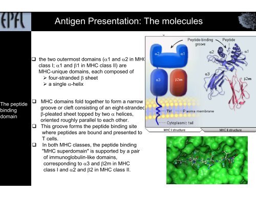
<strong>Antigen</strong> Presentation: The molecules<br />
the two outermost domains (α1 <strong>and</strong> α2 in MHC<br />
class I; α1 <strong>and</strong> β1 in MHC class II) are<br />
MHC-unique domains, each composed of<br />
‣ four-str<strong>and</strong>ed β sheet<br />
‣ a single α-helix<br />
The peptide<br />
binding<br />
domain<br />
<br />
<br />
<br />
MHC domains fold together to form a narrow<br />
groove or cleft consisting of an eight-str<strong>and</strong>ed<br />
β-pleated sheet topped by two α helices,<br />
oriented roughly parallel to each other.<br />
This groove forms the peptide binding site<br />
where peptides are bound <strong>and</strong> presented to<br />
T cells.<br />
In both MHC classes, the peptide binding<br />
"MHC superdomain" is supported by a pair<br />
of immunoglobulin-like domains,<br />
corresponding to α3 <strong>and</strong> β2m in MHC<br />
class I <strong>and</strong> α2 <strong>and</strong> β2 in MHC class II.