October 2012 - MicrobeHunter.com
October 2012 - MicrobeHunter.com
October 2012 - MicrobeHunter.com

Create successful ePaper yourself
Turn your PDF publications into a flip-book with our unique Google optimized e-Paper software.
Technique<br />
Gram Staining<br />
distance increments, but would have<br />
taken longer and risked more movement<br />
of the amoeba between the individual<br />
images and possible difficulty in obtaining<br />
a good stack. If you try this method,<br />
please make sure that you are focusing<br />
in a direction of increasing the distance<br />
between the objective and the slide in<br />
order to avoid colliding them together<br />
accidentally and possibly causing severe<br />
damage to the objective. The eight<br />
video frames <strong>com</strong>prising the stack are<br />
shown in figures 2-9.<br />
■<br />
References<br />
1. Wikipedia. "Speckle imaging". http://en.wikipedia.org/wiki/Speckle_imaging.<br />
2. Walker, D. Video microscopy trials with the USB Live View output of a DLSR camera. Micscape<br />
Magazine, June 2008.<br />
3. Cypionka, H. Videostacking.<br />
http://www.mikroskopie.de/mikforum/read.php?2,48956,48956#msg-48956, July 26, 2008.<br />
4. Cypionka, H. Videostacking.<br />
http://www.mikroskopie-forum.de/index.php?topic=9190.0<br />
May 18, 2011.<br />
5. Krebs, C. Netzelia, testate amoeba, ingesting alga.<br />
http://www.photomacrography.net/forum/viewtopic.php?t=15584<br />
January 01, <strong>2012</strong>.<br />
Gram staining is a quite powerful staining technique, but requires careful standardization<br />
of the bacterial culture.<br />
Oliver Kim<br />
Gram staining is one of the most<br />
well-known staining procedures<br />
for bacteria. It was discovered<br />
by the Danish scientist Hans<br />
Christian Gram (1850–1938). Gram accidentally<br />
discovered this staining procedure<br />
while he was working with Carl<br />
Fiedländer (who discovered the bacterial<br />
cause of pneumonia in 1882).<br />
Gram staining allows for the differentiation<br />
of bacteria in to Gram positive<br />
and Gram negative bacteria. The Gram<br />
positive bacteria stain dark blue or purple,<br />
while the Gram negative bacteria<br />
display a red color. The staining properties<br />
are due to the properties of the cell<br />
wall. Gram positive bacteria have a<br />
thick cell wall, which is able to retain<br />
the blue-black color <strong>com</strong>plexes inside<br />
the cell. The Gram negative bacteria<br />
have a much thinner cell wall, which<br />
allows the <strong>com</strong>plexes to be washed out<br />
by alcohol. For this reason, the Gram<br />
stain was for many years a central technique<br />
in bacterial identification. While<br />
molecular techniques have largely taken<br />
over the identification process, Gram<br />
staining remains a popular technique for<br />
microbiological education.<br />
In his 1884 publication, Gram described<br />
the method as a means to selectively<br />
stain pneumonia bacteria of lung<br />
tissue sections. The surrounding lung<br />
tissue was not stained, making the bacteria<br />
much easier to locate. In his publication<br />
from 1884, he states the<br />
advantage of this staining procedure:<br />
“In my procedure the nucleus and other<br />
tissue elements remain unstained, while<br />
the cocci are strongly stained. This makes<br />
them much easier to locate than previously,<br />
since in ordinary preparations from<br />
pneumonia patients, where such a large<br />
amount of exudate occurs, they are impossible<br />
to see.”<br />
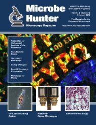
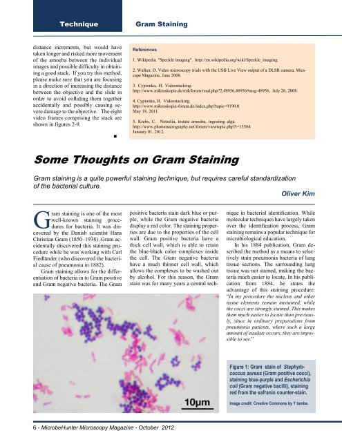
Figure 1: Gram stain of Staphylococcus<br />
aureus (Gram positive cocci),<br />
staining blue-purple and Escherichia<br />
coli (Gram negative bacilli), staining<br />
red from the safranin counter-stain.<br />
Image credit: Creative Commons by Y tambe.<br />
6 - <strong>MicrobeHunter</strong> Microscopy Magazine - <strong>October</strong> <strong>2012</strong>