Chondromucosal Nasal Flap With the Transposition Flap of Von ...
Chondromucosal Nasal Flap With the Transposition Flap of Von ...
Chondromucosal Nasal Flap With the Transposition Flap of Von ...

You also want an ePaper? Increase the reach of your titles
YUMPU automatically turns print PDFs into web optimized ePapers that Google loves.
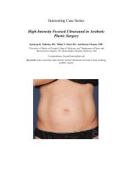
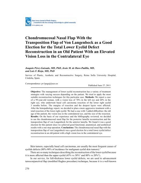
<strong>Chondromucosal</strong> <strong>Nasal</strong> <strong>Flap</strong> <strong>With</strong> <strong>the</strong><strong>Transposition</strong> <strong>Flap</strong> <strong>of</strong> <strong>Von</strong> Langenbeck as a GoodElection for <strong>the</strong> Total Lower Eyelid DefectReconstruction in an Old Patient <strong>With</strong> an ElevatedVision Loss in <strong>the</strong> Contralateral EyeJoaquín Pérez-Guisado, MD, PhD, Jesús M. de Haro-Padilla, MD,and Luis F. Rioja, MD, PhDService <strong>of</strong> Plastic, Aes<strong>the</strong>tic and Reconstructive Surgery, Reina S<strong>of</strong>ía University Hospital,Córdoba, Spain.Correspondence: pv1peguj@uco.esPublished June 27, 2011Objective: The management <strong>of</strong> lower eyelid reconstruction has a variety <strong>of</strong> treatmentstrategies with varying success depending on <strong>the</strong> patient. We tried to apply <strong>the</strong> mostsuitable reconstruction techniques for this particular case. Methods: We report a case<strong>of</strong> a 99-year-old woman, with a vision loss <strong>of</strong> 70% in <strong>the</strong> left eye and 40% in <strong>the</strong>right eye, who underwent basal cell carcinoma resection <strong>of</strong> <strong>the</strong> lower right eyelid3 months before. The margins <strong>of</strong> resection and <strong>the</strong> deepest layers were affected.After <strong>the</strong> histopathology report, we decided to plan a more aggressive treatment with atotal resection <strong>of</strong> <strong>the</strong> lower right eyelid. We had a case with 3 added difficulties: <strong>the</strong> oldage <strong>of</strong> <strong>the</strong> patient, <strong>the</strong> vision loss in <strong>the</strong> contralateral eye, and <strong>the</strong> size <strong>of</strong> <strong>the</strong> resection.Results: On <strong>the</strong> basis <strong>of</strong> our experience and <strong>the</strong> bibliography reviewed, we decidedto use <strong>the</strong> chondromucosal nasal flap for <strong>the</strong> posterior lamella reconstruction and <strong>the</strong>transposition flap <strong>of</strong> von Langenbeck for <strong>the</strong> anterior lamella. We found it was a goodelection for this patient since we achieved good functional, anatomical, and aes<strong>the</strong>ticalresults with a one-step operation. Conclusions: The chondromucosal nasal flap with <strong>the</strong>transposition flap <strong>of</strong> von Langenbeck was a good election for a total lower eyelid defectreconstruction in an old patient with a high vision loss in <strong>the</strong> contralateral eye.Skin tumors, especially basal cell carcinomas, are usually <strong>the</strong> most frequent causes <strong>of</strong>eyelids defects (80%-90% <strong>of</strong> incidence for malignant eyelid skin tumors). 1There are so many techniques describing <strong>the</strong> reconstruction <strong>of</strong> <strong>the</strong> lower eyelid becauseit is more affected than <strong>the</strong> upper eyelid (45% vs 38% 1 and 80% vs 20% 2 ).In our service, for full-thickness lower eyelid defects, we are used to advancementtarsoconjunctival flap (modified Hughes procedure) technique, because it is a well-known270
PÉREZ-GUISADO ET ALand versatile technique that can be used to reconstruct lower eyelid defects <strong>of</strong> less than 50%<strong>of</strong> lid length, as well as defects that fall into <strong>the</strong> 50% to 75% category. 3Large defects affecting more than 50% <strong>of</strong> <strong>the</strong> lower lid can be reconstructed with this2-stage procedure. 4 Never<strong>the</strong>less, when it concerns to lower eyelid defects <strong>of</strong> full-thicknessmeasuring greater than 75%, <strong>the</strong> situation requires a more aggressive approach. 5CLINICAL CASEThe patient is a 99-year-old woman, with a vision loss <strong>of</strong> 70% in <strong>the</strong> left eye and 40%in <strong>the</strong> right eye, who underwent basal cell carcinoma resection <strong>of</strong> <strong>the</strong> lower right eyelid3 months before. The resection had an extension <strong>of</strong> 0.7 × 0.3cm 2 , affecting <strong>the</strong> centralmargin <strong>of</strong> <strong>the</strong> lower eyelid and was treated with direct closure. The hystopathology report<strong>of</strong> <strong>the</strong> resection informed solid (nodular) type <strong>of</strong> basal cell carcinoma characterized byislands <strong>of</strong> cells with peripheral palisading and disorganized central cells. The margins <strong>of</strong>resection and <strong>the</strong> deepest layers were affected.After <strong>the</strong> hystopathology report, we decided to plan a more aggressive treatment around<strong>the</strong> scar <strong>of</strong> <strong>the</strong> previous resection. The scar was not well defined (Fig 1A), so we thought <strong>the</strong>best approach would be a total resection <strong>of</strong> <strong>the</strong> lower eyelid with a full-thickness resectionaround <strong>the</strong> scar that would imply a measurement greater than 75% <strong>of</strong> <strong>the</strong> full lower eyelidextension.Figure 1. (A) Scar after 3 months <strong>of</strong> <strong>the</strong> first resection; (B) Preoperative markings and lower eyelidresection; (C) <strong>Chondromucosal</strong> nasal flap harvest.We had a case with 3 limitations for applying <strong>the</strong> advancement tarsoconjunctival flap:<strong>the</strong> old age <strong>of</strong> <strong>the</strong> patient, <strong>the</strong> elevated vision loss in <strong>the</strong> contralateral eye, and <strong>the</strong> highsize <strong>of</strong> <strong>the</strong> resection. The patient was advanced in years with a severe vision loss in <strong>the</strong>contralateral eye, so <strong>the</strong> occlusion <strong>of</strong> <strong>the</strong> right eye and <strong>the</strong> risk <strong>of</strong> a second step operationwould not be <strong>the</strong> best options. The defect would be too big for applying this technique.Reviewing <strong>the</strong> bibliography, we thought this patient could be a good candidate for <strong>the</strong> totallower eyelid reconstruction with <strong>the</strong> nasal chondromucosal flap, because it does not needeye occlusion and <strong>the</strong> reconstruction is achieved with one-step operation. 6Because <strong>of</strong> <strong>the</strong> age <strong>of</strong> <strong>the</strong> patient, <strong>the</strong> operative procedure was performed under localanaes<strong>the</strong>sia with sedation instead <strong>of</strong> general anaes<strong>the</strong>sia. Although this is a large surgicalfield to perform under local anaes<strong>the</strong>sia, in our hospital, <strong>the</strong> final decision about <strong>the</strong> mostsuitable anaes<strong>the</strong>sia depends on <strong>the</strong> anaes<strong>the</strong>tist, who considered <strong>the</strong> local anaes<strong>the</strong>sia withsedation as <strong>the</strong> most appropriated method for this patient.271
ePlasty VOLUME 11Preoperative markings were done for harvest <strong>of</strong> <strong>the</strong> upper lateral cartilage in <strong>the</strong> nasalchondromucosal flap, which is based on <strong>the</strong> lateral terminal branch <strong>of</strong> <strong>the</strong> dorsal nasal artery,and for transposition flap <strong>of</strong> von Langenbeck, and <strong>the</strong>n <strong>the</strong> resection was done (Fig 1B).The piece <strong>of</strong> resection was 2.8 × 1.9cm 2 . The flap was designed along <strong>the</strong> lateral nasal walland was based on <strong>the</strong> terminal branch <strong>of</strong> <strong>the</strong> dorsal nasal artery, to include <strong>the</strong> subcutaneoustissues down to <strong>the</strong> periosteum and <strong>the</strong> cranial portion <strong>of</strong> <strong>the</strong> upper lateral cartilage. Theskin was incised for 2.5 cm along <strong>the</strong> border between <strong>the</strong> lateral nasal wall and <strong>the</strong> cheekfrom <strong>the</strong> inner canthus to <strong>the</strong> ala nasi. Then, <strong>the</strong> subcutaneous tissue was dissected, fromlateral to medial, onto a line beyond <strong>the</strong> midline <strong>of</strong> <strong>the</strong> nose. The subcutaneous dissectionwas extended superiorly to <strong>the</strong> glabellar area and inferiorly to or beyond <strong>the</strong> lower margin<strong>of</strong> <strong>the</strong> upper lateral cartilages. A portion <strong>of</strong> <strong>the</strong> lateral cartilage with 1.2 × 2.1cm 2 <strong>of</strong> sizewas dissected. The flap was harvested from <strong>the</strong> pedicle including <strong>the</strong> portion <strong>of</strong> <strong>the</strong> lateralcartilage dissected (Fig 1C).The flap was <strong>the</strong>n transposed to reconstruct <strong>the</strong> posterior lamella <strong>of</strong> <strong>the</strong> lower eyelid(Fig 2A).Figure 2. (A) <strong>Chondromucosal</strong> nasal flap transposition to reconstruct <strong>the</strong> posteriorlamella; (B) <strong>Transposition</strong> flap <strong>of</strong> von Langenbeck.<strong>With</strong> <strong>the</strong> help <strong>of</strong> <strong>the</strong> loupe magnification, flap mucosa was sutured to <strong>the</strong> conjunctivalmargin. <strong>Transposition</strong> flap <strong>of</strong> von Langenbeck was used for repairing <strong>the</strong> anterior lamellaand correcting ectropion 7 (Fig 2B), lateral and medial periosteal fixation was performed tominimize <strong>the</strong> risk <strong>of</strong> lid malposition. The transposition flap <strong>of</strong> von Langenbeck consisted ina superiorly based lateral transposition cutaneous flap. Its base was immediately adjacentto <strong>the</strong> defect, with a length and a width approximately equal to <strong>the</strong> length and width <strong>of</strong> <strong>the</strong>defect. The flap was harvested and transposed into <strong>the</strong> defect.The narrow skin bridge created by <strong>the</strong> extensive lateral nasal incision and <strong>the</strong> medialextent <strong>of</strong> <strong>the</strong> lid resection was reinforced with <strong>the</strong> stitches from <strong>the</strong> medial extent <strong>of</strong> <strong>the</strong>transposition flap. Wound closure was accomplished by a simple discontinuous subcutaneouspattern (4-0 polyglactin), continuous cutaneous pattern for <strong>the</strong> transposition flap <strong>of</strong>von Langenbeck (5-0 nylon), and discontinuous cutaneous pattern for <strong>the</strong> rest (4-0 nylon).The results, just after (Fig 4A), 3 months after (Fig 4B), and 6 months (Fig 4C) after <strong>the</strong>surgery, were aes<strong>the</strong>tically and functionally excellent, and as we can see, with <strong>the</strong> absence<strong>of</strong> any malposition, entropion or ectropion. This patient had a good result probably due inpart to her aged skin, so maybe <strong>the</strong> procedure described would be a good alternative just inold people but not in young people.272
ePlasty VOLUME 11Concerning to <strong>the</strong> anterior lamella reconstruction:1. Semicircular rotational flap (Tenzel): A Tenzel flap is indicated for central or medialdefects <strong>of</strong> <strong>the</strong> eyelid affecting up to 60% <strong>of</strong> <strong>the</strong> eyelid margin, so it was not enoughfor <strong>the</strong> size <strong>of</strong> our defect. Moreover, if this flap is used to repair excessively largedefects, a greater amount <strong>of</strong> unsupported tissue results in <strong>the</strong> lateral lid, potentiallyresulting in eyelid malposition including entropion or ectropion. 82. Bipedicle upper eyelid flap (Tripier): The height <strong>of</strong> <strong>the</strong> defect that can be repaired islimited to 10 to 15 mm 8 ; <strong>the</strong>refore, it was not appropriate for our reconstruction.3. Temporal forehead flap (Fricke): Although this transposition flap from <strong>the</strong> temporalforeheadarea was initially described by Fricke for reconstruction <strong>of</strong> <strong>the</strong> upper eyelid,it could also be used for lower eyelid reconstruction. 4 Never<strong>the</strong>less, our experiencehas taught us that due to <strong>the</strong> closer distance from <strong>the</strong> flap to <strong>the</strong> upper eyelid than <strong>the</strong>lower eyelid this technique has better results for <strong>the</strong> upper eyelid.We found both transposition flap <strong>of</strong> von Langenbeck and rotation cheek flap (Mustardéflap) as <strong>the</strong> most appropriated techniques for this case. We finally decided to usetransposition flap <strong>of</strong> von Langenbeck because this technique is less traumatic and better toavoid or even correct a residual ectropion.We can conclude <strong>the</strong> chondromucosal nasal flap with <strong>the</strong> transposition flap <strong>of</strong> vonLangenbeck was a good election for a total lower eyelid defect reconstruction in an oldpatient with a high vision loss in <strong>the</strong> contralateral eye. We achieved good functional,anatomical, and aes<strong>the</strong>tical results with a one-step operation.REFERENCES1. Zaragoza Casares P, Llanes Menéndez F, Gómez Fernández T, Zato Gómez de Liaño MA, Zaragoza GarcíaP. Carcinoma basocelular palpebral. Estudium Ophtalmol. 2006;4.2. Clara Gómez Cabrera, Nereyda Martínez, Martha Herrera Soto e Ileana Agramonte. Cryo<strong>the</strong>rapy in smallmalignant eyelids tumors. Revist Cub Oftalmol. 2003;16:1.3. Spinelli HM, Jelks GW. Periocular reconstruction: a systematic approach. Plast Reconstr Surg. 1993;91:1017-24.4. Codner MA, McCord CD, Mejia JD, Lalonde D. Upper and lower eyelid reconstruction. Plast Reconstr Surg.2010;126:231e-45e.5. Newman MI, Spinelli HM. Reconstruction <strong>of</strong> <strong>the</strong> eyelids, correction <strong>of</strong> ptosis, and canthoplasty. In: ThorneCH (ed). Grabb and Smith’s Plastic Surgery. 7th ed. Philadelphia, PA: Lippincott Williams & Wilkins;2007:397-416.6. Scuderi N, Ribuffo D, Chiummariello S. Total and subtotal upper eyelid reconstruction with <strong>the</strong> nasalchondromucosal flap: a 10-year experience. Plast Reconstr Surg. 2005;115:1259-65.7. Weerda H. The eyelids. In: Weerda H, ed. Reconstructive Facial and Plastic Surgery: A Problem SolvingManual. Stuttgart, Germany: Georg Thieme Verlag; 2001:95-100.8. Tucker SM. Specialized local facial flaps for <strong>the</strong> eyelids and lips. In: Dolan RW, ed. Facial Plastic, Reconstructive,and Trauma Surgery. Basel, Switzerland: Marcial Denker; 2004:201-32.274