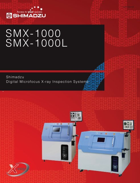
SMX-1000 SMX-1000L
SMX-1000 SMX-1000L
SMX-1000 SMX-1000L

You also want an ePaper? Increase the reach of your titles
YUMPU automatically turns print PDFs into web optimized ePapers that Google loves.
Compact X-ray Inspection SystemsCombining Ease of Use with Advanced Functionality
<strong>SMX</strong>-<strong>1000</strong><strong>SMX</strong>-<strong>1000</strong>LShimadzu Digital Microfocus X-ray Inspection SystemsCompact X-ray Inspection SystemsCombine Ease of Use with Advanced FunctionalityShimadzu's new <strong>SMX</strong>-<strong>1000</strong> and <strong>SMX</strong>-<strong>1000</strong>L produce extremely clear, distortion-free imagesthanks to a digital flat-panel detector (FPD) and sealed microfocus X-ray tube. Also, newlydeveloped easy-to-use software lets you efficiently perform target inspections with just a fewsimple selections. Simply select the type of sample used from a list and the image displayconditions are automatically set. No complex tasks are required.Additionally, images from the system's CCD-camera make navigation easy, together with stepfeed, teaching, image thumbnails, and comprehensive measurement functions. If desired, anoptional VCT unit can be added to easily capture CT images.ApplicationsThe <strong>SMX</strong>-<strong>1000</strong>/<strong>SMX</strong>-<strong>1000</strong>L Digital Microfocus X-ray TV Systems are ideal for non-destructive, fluoroscopic inspection ofhigh-density surface-mounted PCBs and the status (discontinuities and contact) of the fine connections in BGA, CSP,and system LSIs.The <strong>SMX</strong>-<strong>1000</strong>L enables continuous inspection of L-size PCBs or large numbers of components on pallets.Clear ImagesThe combination of a leading-edge digital flat-paneldetector (FPD) with Shimadzu image-processingtechnology produces crisp, clear images with nodistortion.Inclined FluoroscopyDefects normally invisible using vertical (0o)fluoroscopy often become visible if viewed from anangle (up to 60o). These are the first models toinclude this useful function as standard.Easy OperationEvery effort was made to include only the functionsessential for easy and intuitive unit operation in aproduction environment.Tomographic ImagesAdd the optional VCT unit to easily produce clear,cone-beam CT images that allow moresophisticated analysis.ContentsP 4 - FeatureP 8 - Operation FunctionsP 11 - OptionsP 6 - Image AdjustmentsP10 - MeasurementP 12 - Specifications
FeaturesFPD Provides Clear, Distortion-Free Images!The FPD together with the microfocus X-ray tube produce clear, high-resolution fluoroscopic images, even at high magnification.1. Distortion-Free Images 4. Excellent Contrast ResolutionFPD imageFPD image 12-bit (4096 gradations)An FPD image is free of the distortion typically producedby an image intensifier, ensuring accurate reproductionof surface shapes. (Gridlines were added to showlinearity.)2. One Million PixelsFPD image (<strong>1000</strong> x <strong>1000</strong> pixels)Brightness andcontrast adjustmentBrightness andcontrast adjustmentShimadzu's one million pixel FPD provides over threetimes as much information than a 300,000 pixel imageintensifier.3. No ShadingFPD imageWhen conditions are set to observe high-X-ray absorbing interior parts of a motor using animage tube, the image's peripheral plastic parts with low X-ray absorption appear white.However, simple adjustments of brightness and contrast allow the flat-panel detector toaccurately observe all areas of a single image taken using a single set of fixed X-rayconditions. This improvement in visibility is possible thanks to a 16-fold increase in the amountof information in the 12-bit FPD image, compared to the 8-bit image-tube image.The FPD image achieves uniform brightness across theentire image, without shading.4
Fluoroscopy at up to 60° Inclination!A maximum FPD tilt angle of 60° allows fluoroscopy over an extensive range, whilemaintaining constant magnification.Tracking minimizes displacement of the fluoroscopy position, even when the C-arm istilted. Never lose track of observation points again.FPD tilted 60°FPD not tiltedFPDUp to 60°FPD<strong>SMX</strong>-<strong>1000</strong>Solder ball joint defects that cannotbe identified at zero degrees (whenviewed from above) can be easilyidentified when viewed obliquely at a60° angle.50x magnification at 0° 50x magnification at 60°Large Door in a Compact, Unified DesignAmple opening and large table make operation easy!New double-sliding doors provide a large 535 mm x 400 mmopening 2.2 times larger than previous Shimadzu models andlarger than any competitor.(<strong>SMX</strong>-<strong>1000</strong>L: 680 mm x 430 mm)Generous 400 mm x 350 mm table accepts even largesurface-mounted PCBs.(<strong>SMX</strong>-<strong>1000</strong>L: 570 mm x 670 mm L-size PCBs)* Stroke is reduced 50 mm in each direction.5
Image AdjustmentsEasy Mouse Movements for CompletePreset Functions for Image ConditionsInstantly set the image display conditions for the target (material) bysimply selecting the appropriate work type from a list.When BGA is selectedX 10BGAPatternDigital zoom of fluoroscopicimages (live images, savedimages) is displayed in realtime.When Pattern is selected6
Control of FunctionsCCD Camera for Fast PositioningUse of a CCD camera image for stage positioning allows easymovement of the stage to the target position.PanoramaSimply click any point on the CCD image tomove the stage to that position.Use the zoom function to accurately specify thetarget position.These models allow stage and target positioningwhile viewing only the monitor, dramaticallyimproving ease-of-operation, compared to theconventional method of positioning using ajoystick while viewing a laser marker throughthe observation window.Panorama function displays a full image of alarge sample that cannot be viewed in a singleframe.Click any point on the image's live display tomove the stage on its X-Y axis, tilt or zoom inand out. Stage movement is slowed by simplymoving the cursor towards the image center.Stage travel speed is automatically optimized tomatch the current fluoroscopy magnification.Sub-Image Display AreaDisplays a reference image during inspection.Zoom function allows display of this image atsame magnification as main display area.Mouse-Based On-Screen Image PositioningAll stage and device positioning canbe controlled via the mouse, allowingthe operator to concentratecompletely on examining the image inthe monitor. Simply click on a point tobring it to the center of the display.Move scroll-wheelLeft-clickFast FastSlowFast FastX-Y movementFastZoom-inSlowZoom-outFastChangemagnificationRight-clickFastTiltSlowTiltFastTilting7
Operation FunctionsMultiple Functions to Improve OperatorStep FeedMoves the stage sequentially in equal steps.Ensures efficient inspection of evenly-spaced samples, such as those on a pallet.Operator can easily evaluate images of sequentially displayed samples.• Simply click points with the mouseto enter the visual evaluation resultsfor each point.• After inspection of all set points iscompleted, easy-to-read colorbasedresults are displayed in atable, as shown below. Refer to thistable when sorting samples.• Enter the feed pitch and number ofrepetitions.• Inspection is repeated as stagemoves in a Z-pattern.stageTeachingTeaching pre-registers inspection points and registers the observationconditions for each point. This function then automatically plays backthe registered procedure to significantly enhance inspection efficiencyduring repeated inspections of multiple samples of the same type.Inspection position changes automatically during teaching,allowing operator to concentrate on evaluating images.To repeat an earlier inspection, simply selecta previously-registered teaching file.Up to 10,000 points can be registered in a single file.Current inspection position8
EfficiencyImage DirectoryImages saved in a file can be displayed as thumbnails.As shown below, display mode includes a variety of useful, operator-friendly functions.Easily search for stored images using a Windows ®Explorer-like interface.Use the mouse scroll wheel to select a 2 x 2, 4 x 4, or 6 x 6display format.Double-click on any thumbnail to display the full <strong>1000</strong> x<strong>1000</strong>-pixel image (original size).Inspection conditions used to take an imageare saved with the image and can be displayedwith the image.Select any thumbnail and click [Condition settings] toautomatically register the inspection conditions used tocapture that image, allowing the same inspection to beeasily repeated. This guarantees identical images, even ifthe operator changes.9
MeasurementUseful Measurement Functions are StandardMeasuring FunctionsDimensionsMeasures the distance, angle and curvature between two points. Eliminating the conventional need for dimensional compensation, the <strong>SMX</strong>-<strong>1000</strong> automatically calculates compensation data based on each fluoroscopy magnification to ensure efficient dimension measurement.The flat-panel detector (FPD)generates digital imagesfree of distortion or shading toachieve better dimensionalmeasurementaccuracy thanpreviously possible.The measurement results displayed onthe screen can be saved as a file in .csvformat for statistical processingwith a commercial spreadsheetapplication.BGA Void RatioSet the threshold value and start the measurement to display themeasured results for each bump on the image. Pass/failevaluation is conducted automatically according to the presetevaluation criteria. The results can be saved in .csv format.Items (16) and (19) fail.Area RatioMeasures the area ratio of objects in thedesignated ROI (region of interest).The area ratio is calculated as objectarea/ROI area. This function is useful forevaluating die bond or solder pastewettability.ROIROI areaObject areaStatic imageWire Sweep RatioSimply click on each end of a wire and thepoint of maximum sweepto calculate the wire sweep ratio. Theresults can be saved in .csv format.WirePoint ofmaximum sweepL1L2Wire sweep ratio =L2 / L1 x 100 (%)10
OptionsComprehensive Range of OptionsOptionsVCT UnitP/N 362-62900(<strong>SMX</strong>-<strong>1000</strong>) P/N 362-62900-01(<strong>SMX</strong>-<strong>1000</strong>L)Attach the removable VCT unit and easily obtain clear,cone-beam CT images in addition to fluoroscopic images.Specifications (Main)1. CT max. size/weight PCB: 50 x 100 x 2 mm;component: 30 dia. x 25 mm;100 g max.2. Cone-beam CT field of view (FOV) 5 to 30 mm dia.3. Scanning method Cone-beam CT (normal mode)4. Reconstruction matrix 512 x 512 x 508 (max.)5. Number of views 600, 12006. Display MPR7. Stage: X direction (horizontal) ± 15 mmY direction (horizontal) 40 mmRotational resolution 2/<strong>1000</strong>°VCT UnitMPR Image of CapacitorRotation/Inclination Unit P/N 362-63762Attach the removable rotation/inclination unit and obtain X-ray fluoroscopy images of small-sizecomponents from multiple directions to minimize inspection mistakes.Specifications (Main)1. Max. load 20 g2. Rotation Continuous3. Inclination ±30°Main ControllerOperation Box A : P/N 362-63982Combining Main Controller with the <strong>SMX</strong>-<strong>1000</strong>/<strong>SMX</strong>-<strong>1000</strong>L allows manual operation of the XY stageusing buttons and a joystick.Specifications (Main)1. X-Y feed Joystick control: 6 speeds2. Controls zoom, angle of inclination, and rotation/inclination unit operation (button operation)VCT Controller : P/N 362-63983Operation box B is included with the VCT unit.Main ControllerSpecifications (Main)1. X-Y feed Joystick control: 6 speeds2. Controls zoom and angle of inclination (button operation)XIV4-E Image Analysis SoftwareVCT ControllerP/N 362-55937-51Provides even more sophisticated image processing and analysis, as well as macros for process automation. This software reads images takenand saved by the <strong>SMX</strong>-<strong>1000</strong>/<strong>SMX</strong>-<strong>1000</strong>L units.11
<strong>SMX</strong>-<strong>1000</strong>/<strong>SMX</strong>-<strong>1000</strong>L General SpecificationsP/NSpatial ResolutionMaximum Sample SizeStrokeMaximum Sample WeightDetector InclinationMaximum OutputDetectorInspection Visual FieldMagnificationPower SupplyDimensionsWeight<strong>SMX</strong>-<strong>1000</strong><strong>SMX</strong>-<strong>1000</strong>L362-61700362-620005 μm (chart resolution)350 x 400 mm570 x 670 mm300 x 350 mm520 x 620 mm5 kg60° max90 kV (sealed X-ray tube)Digital flat-panel detector (FPD)Approx. 2 mm to 35 mmApprox. 6x to 158x (dynamic images to 127x)AC 100 VW995 x D990 x H1285 mmmW1490 x D1525 x H1325 mmApprox. 500 kgApprox. 950 kg<strong>SMX</strong>-<strong>1000</strong>/<strong>SMX</strong>-<strong>1000</strong>L External DimensionsUnit: mm12851325Comply with all local regulationswhen installing an <strong>SMX</strong> system.990<strong>SMX</strong>-<strong>1000</strong>9951525 1490<strong>SMX</strong>-<strong>1000</strong>LShimadzu Corporation is amember of JIMA(Japan Inspection InstrumentsManufacturers’ Association)Testing MachinesMicro Strength TesterMST-I Micro-AutographTabletop Universal TesterEZGraphMicro Compression TesterMCT-WWindows ® is a registeredtrademark of MicrosoftCorporation in the USand other countries.Founded in 1875, Shimadzu Corporation, a leader in thedevelopment of advanced technologies, has a distinguishedhistory of innovation built on the foundation of contributing tosociety through science and technology. We maintain a globalnetwork of sales, service, technical support and applicationscenters on six continents, and have established long-termrelationships with a host of highly trained distributors locatedin over 100 countries. For information about Shimadzu, and tocontact your local office, please visit our Web site atwww.shimadzu.comJQA-0376SHIMADZU CORPORATION. International Marketing Division3. Kanda-Nishikicho 1-chome, Chiyoda-ku, Tokyo 101-8448, JapanPhone: 81(3)3219-5641 Fax. 81(3)3219-5710URL http://www.shimadzu.comThe contents of this brochure are subject to change without notice.Printed in Japan 4199-12605-30AIT