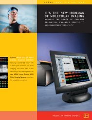
KODAK IMAGE STATION IN-VIVO IMAGING SYSTEMS - Raytest
KODAK IMAGE STATION IN-VIVO IMAGING SYSTEMS - Raytest
KODAK IMAGE STATION IN-VIVO IMAGING SYSTEMS - Raytest

You also want an ePaper? Increase the reach of your titles
YUMPU automatically turns print PDFs into web optimized ePapers that Google loves.
<strong>KODAK</strong> <strong>IMAGE</strong> <strong>STATION</strong><br />
<strong>IN</strong>-<strong>VIVO</strong> IMAG<strong>IN</strong>G <strong>SYSTEMS</strong><br />
High sensitivity optical molecular imaging and high<br />
resolution digital radiography capabilities in a single<br />
multimodal system<br />
The NEW Image Station In-Vivo Imaging Systems provide high performance optical molecular imaging of<br />
near-IR fluorescence, isotopic, and luminescence labels in small animals. The Image Station In-Vivo FX<br />
system features an integrated x-ray module that allows precise co-registration of anatomical x-ray<br />
images with optical molecular and isotopic imaging modalities for improved anatomical localization of<br />
biomarkers of interest.<br />
Greatly enhances anatomical localization of molecular imaging<br />
agent signals in small animals, organs, and tissues<br />
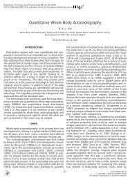
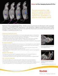
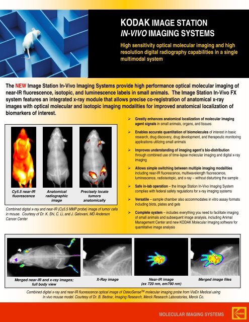
Cy5.5 near-IR<br />
fluorescence<br />
Anatomical<br />
radiographic<br />
image<br />
Combined digital x-ray and near-IR (Cy5.5 MMP probe) image of tumor cells<br />
in mouse. Courtesy of Dr. K. Shi, C. Li, and J. Gelovani, MD Anderson<br />
Cancer Center<br />
Merged near-IR and x-ray images;<br />
full body view<br />
Precisely locate<br />
tumors<br />
anatomically<br />
Enables accurate quantitation of biomolecules of interest in basic<br />
research, drug discovery, drug development, and therapeutic monitoring<br />
applications utilizing small animals<br />
Improves understanding of imaging agent’s bio-distribution<br />
through combined use of time-lapse molecular imaging and digital x-ray<br />
imaging<br />
Allows simple switching between multiple imaging modalities<br />
including near-IR fluorescence, multiwavelength fluorescence,<br />
luminescence, radioisotopic, and x-ray – without disturbing the sample<br />
Safe in-lab operation – the Image Station In-Vivo Imaging System<br />
complies with federal safety regulations for x-ray imaging systems<br />
Versatile – sample chamber also accommodates in vitro assay formats<br />
including blots, plates and gels<br />
Complete system – includes everything you need to facilitate imaging<br />
of small animals and subsequent image analysis, including Animal<br />
Management Center and new <strong>KODAK</strong> Molecular Imaging software for<br />
quantitative image analysis<br />
X-Ray image Near-IR image<br />
(ex 720 nm, em790 nm)<br />
Merged image files<br />
Combined digital x-ray and near-IR fluorescence optical image of OsteoSense TM molecular imaging probe from VisEn Medical using<br />
in vivo mouse model. Courtesy of Dr. B. Bednar, Imaging Research, Merck Research Laboratories, Merck Co.<br />
MOLECULAR IMAG<strong>IN</strong>G <strong>SYSTEMS</strong>
System Configuration<br />
The <strong>KODAK</strong> Image Station In-Vivo FX Imaging System is comprised of a 4<br />
million pixel, cooled CCD camera in a light tight imaging cabinet. 20 x 20 cm<br />
field of view allows imaging of 4 or more mice simultaneously. An external<br />
illuminator provides specific, selectable excitation from 380 nm to 780 nm.<br />
Proprietary wide angle filters allow high sensitivity imaging at near-IR<br />
wavelengths without optical artifacts.<br />
The integrated x-ray module contains the x-ray source, safety switches, and<br />
status indicators, and is controlled by the <strong>KODAK</strong> Molecular Imaging<br />
software package.<br />
The patented Animal Management Center with integrated sample chamber<br />
and phosphor screen allows environmental control and sequential imaging in<br />
x-ray and fluorescence modes without moving the sample.<br />
Kodak’s new Molecular Imaging Software package provides comprehensive<br />
image analysis capabilities for both in vivo and in vitro applications, including<br />
automated region of interest analysis and relative intensity measurement.<br />
FDA Compliance<br />
The <strong>KODAK</strong> Image Station In-Vivo FX Imaging System meets or exceeds<br />
compliance to FDA Department of Health and Human Services regulation<br />
CFR 1020.40 for radiation safety.<br />
For More Information<br />
<strong>KODAK</strong> Image Station In-Vivo FX Imaging System<br />
<strong>KODAK</strong> Image Station In-Vivo Imaging system is supplied complete with cooled CCD<br />
camera, imaging chamber, multi-wavelength fluorescence illumination, integrated x-ray<br />
module, <strong>KODAK</strong> Molecular Imaging Software, and more.<br />
For more information on the Image Station In-Vivo FX Imaging System and<br />
the complete line of <strong>KODAK</strong> Image Station digital imaging systems, please<br />
visit www.kodak.com/go/molecular or call toll free in the United States<br />
and Canada 1-877-747-4357, express code 7. Outside of the United<br />
States, call +1-203-786-5657.<br />
System Specifications<br />
Camera: CCD CCD 2048 x 2048 pix els, cooled to -29oC Lens 10x zoom, 20 - 200 mm<br />
Illumination:<br />
Fluorescence Selectable multiw av elength epi-illumination, 380 - 780 nm<br />
White Light White light epi-illumination<br />
Emission Filters Selectable w ide angle filters, 440 nm - 830 nm<br />
Performance:<br />
Field of View 2 x 2 cm to 20 x 20 cm<br />
Optical Resolution 10 microns/pix el<br />
Data Acquisition 16-bit single capture; n-bit data acquisition<br />
Detection Modes: Luminescence, Near-IR Fluorescence, Multiw av elength<br />
Fluorescence, Absorbance, Radioisotopic, X-Ray<br />
Exposure Modes: Single capture, Multiple Capture, Progressiv e, Time Lapse<br />
Digital X-Ray Module:<br />
Energy Range Approx imately 12 - 35 kVP<br />
Max imum Current Approx imately 150 µA<br />
Spot Size 33 degrees<br />
Field of Illumination 20 x 20 cm<br />
Ordering Information (Catalog Numbers)<br />
Description PC Mac<br />
Image Station In-Viv o FX Sy stem 858 0540 897 4545<br />
Image Station In-Viv o F Sy stem 886 0488 894 3953<br />
The Image Station In-Vivo FX system includes:<br />
• Imaging chamber with cooled 2048 x 2048 pixel, 16-bit CCD camera<br />
• Controllable x-ray module<br />
• External illumination source<br />
• 5-position excitation filter slider with four excitation filters of choice<br />
• 4-position emission filter wheel with four proprietary wide angle<br />
emission filters of choice<br />
• Radiographic Phosphor Screen<br />
• Radioisotopic Phosphor Screen<br />
• Thermal Control Unit<br />
• Animal Management Center<br />
• Animal Chambers<br />
• Atmospheric Control Ports<br />
• PC or MAC<strong>IN</strong>TOSH computer with 17” flat screen monitor<br />
• <strong>KODAK</strong> Molecular Imaging Software, 3 network licenses or 3 seat<br />
standalone license<br />
• On-site instrument installation and user training<br />
• User documentation and 12-month limited warranty<br />
The Image Station In-Vivo F system contains all components listed<br />
above, with the exception of the x-ray module and radiographic phosphor<br />
screen.<br />
While the Image Station In-Vivo Imaging System can be used for in vivo and in vitro molecular imaging of materials, users should be<br />
aware that the methods of preparing and viewing the materials for molecular imaging may be subject to various patent rights.<br />
MOLECULAR IMAG<strong>IN</strong>G <strong>SYSTEMS</strong>