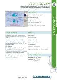
Quantitative whole-body Autoradiography part 1 of 9
Quantitative whole-body Autoradiography part 1 of 9
Quantitative whole-body Autoradiography part 1 of 9

You also want an ePaper? Increase the reach of your titles
YUMPU automatically turns print PDFs into web optimized ePapers that Google loves.
Regulatory Toxicology and Pharmacology 31, S1–S3 (2000)<br />
doi:10.1006/rtph.2000.1379, available online at http://www.idealibrary.com on<br />
<strong>Quantitative</strong> Whole-Body <strong>Autoradiography</strong><br />
R. A. J. Coe<br />
Whole-Body <strong>Autoradiography</strong>, BioDynamics Research Limited, Walton Manor, Walton, Milton Keynes,<br />
Buckinghamshire MK7 7AJ, United Kingdom<br />
INTRODUCTION<br />
Distribution studies with new radiolabeled test compounds<br />
in animals form an important <strong>part</strong> in the preclinical<br />
drug development programs <strong>of</strong> many companies. The<br />
data obtained from these studies <strong>of</strong>ten form the basis for<br />
the assessment <strong>of</strong> human organ and tissue exposure to<br />
the test compound and the removal <strong>of</strong> residues/metabolites<br />
from those organs and tissues after drug administration.<br />
The data may also provide pharmacokinetic information<br />
with regard to any specific binding to, or<br />
selective affinity for, a tissue or organ by the test compound<br />
or its metabolites. The data may provide some<br />
indication as to the potential toxicological and pharmacological<br />
sites <strong>of</strong> action. In addition, much <strong>of</strong> the data<br />
derived from these studies are submitted to various regulatory<br />
authorities as <strong>part</strong> <strong>of</strong> the process <strong>of</strong> providing new<br />
safe drugs. This regulatory process has changed over the<br />
years and this will be discussed later.<br />
The techniques most commonly used for these distribution<br />
studies are quantitative <strong>whole</strong>-<strong>body</strong> autoradiography<br />
(QWBA) and quantitative tissue distribution<br />
[QTD—by dissection and liquid scintillation counting<br />
(LSC)]. In this paper it is my intention to very briefly<br />
review quantitative <strong>whole</strong>-<strong>body</strong> autoradiography and<br />
to provide a background against which the other papers<br />
presented in this issue may be viewed.<br />
QWBA<br />
Whole-<strong>body</strong> autoradiography in its present form has<br />
developed from Ullberg’s original method (1954) and in<br />
the 46 years since then, many scientists have defined<br />
and refined the materials and methods that we use.<br />
The autoradiographic phenomenon was first noted<br />
by Niepce de St. Victor (1867) and subsequently reevaluated<br />
by Becquerel (1896a,b,c,d). Subsequent experiments<br />
using the autoradiographic process by London<br />
(1904a,b,c), Lacassagne and Lattes (1929), Lomholt<br />
(1930), and Leblond (1943) led Ullberg (1954) to publish<br />
his elegant method for the study <strong>of</strong> soluble xenobiotics<br />
in vivo. It was realized, at that time, that all the<br />
potential data present in the autoradiograms were not<br />
being used. Ullberg himself, in association with Berlin<br />
(1963), made many <strong>of</strong> the early attempts to quantify<br />
Received February 10, 2000<br />
S1<br />
the concentration <strong>of</strong> radioactivity detected. Because <strong>of</strong><br />
the materials in use (X-ray film) only semiquantitative<br />
results could be achieved and QTD remained the major<br />
method <strong>of</strong> obtaining quantitative data. Cross et al.<br />
(1974) and Longshaw and Fowler (1977, 1978) defined<br />
some <strong>of</strong> the parameters affecting the accuracy <strong>of</strong> autoradiographic<br />
data by <strong>whole</strong>-<strong>body</strong> autoradiography, and<br />
Cross et al. (1974) produced a practical densitometer<br />
and calibration source for quantifying autoradiograms.<br />
Other scientists were assessing the sensitivity <strong>of</strong> X-ray<br />
film to � radiation (Coe, 1982; Franklin, 1980, 1983,<br />
1985) while Geary et al. (1985) suggested a different<br />
isotope standards scale for use in QWBA albeit with<br />
the emphasis on use in microautoradiography and receptor<br />
binding studies. Alternative methods were proposed<br />
to overcome some <strong>of</strong> the pitfalls <strong>of</strong> the Cross<br />
method, for example, the stoichiometric analysis <strong>of</strong> the<br />
exposed X-ray film’s silver content (Stevens, 1980).<br />
Section thickness was also thought to be an important<br />
factor in the process <strong>of</strong> quantifying autoradiograms,<br />
and Williams (1987) established the infinite thickness<br />
<strong>of</strong> a section containing carbon-14. Schweitzer et al.<br />
(1987) proposed an elegant but simple quantitation<br />
method using a calibration scale made from blood samples<br />
“spiked” with radioactivity.<br />
The continuing interest in quantifying autoradiograms<br />
led to the commercial development <strong>of</strong> a number<br />
<strong>of</strong> s<strong>of</strong>tware programs that could be used to analyze<br />
images captured or derived from film-based autoradiograms<br />
(Seescan, UK; Loates Associates, U.S.A.; Imaging<br />
Research Inc., Canada), while other scientists devised<br />
their own (Goochee et al., 1983; D’Argy et al.,<br />
1990). Subject to the standardization <strong>of</strong> certain experimental<br />
parameters close correlation between data obtained<br />
from QWBA and QTD could be achieved (Coe<br />
and Attwood, 1995; Zane et al., 1996; Steinke, 1997;<br />
Lordi et al., 1999). During the late 1980s and early<br />
1990s the phosphor storage imaging plate became<br />
available together with its scanner and it quickly became<br />
apparent that this was a significant advance.<br />
This detection medium was shown to be more linear, to<br />
be more sensitive, and to have a wider dynamic range<br />
than any previous detector for analyzing the radioactive<br />
content <strong>of</strong> <strong>whole</strong>-<strong>body</strong> sections. Early work by<br />
0273-2300/00 $35.00<br />
Copyright © 2000 by Academic Press<br />
All rights <strong>of</strong> reproduction in any form reserved.
S2 R. A. J. COE<br />
Sonoda et al. (1983), Miyahara (1989), Shigematsu<br />
(1992), Mori and Hamaoka (1994), Motoji et al. (1995),<br />
and Poitchoiba et al. (1995) quickly established some <strong>of</strong><br />
the parameters that needed to be standardized or investigated.<br />
A collaborative study performed by over 20<br />
Japanese companies (Forum, 1993; Tanaka, 1994) indicated<br />
that QWBA could usefully replace many <strong>of</strong> the<br />
more traditional methods <strong>of</strong> obtaining the data for<br />
ADME studies, especially the quantitative analysis <strong>of</strong><br />
metabolites by TLC. At about the same time other<br />
forms <strong>of</strong> direct nuclear counting <strong>of</strong> the radioactivity in<br />
<strong>whole</strong>-<strong>body</strong> cryosections were becoming commercially<br />
available (Biospace, Canberra Packard) and there has<br />
been a review <strong>of</strong> these by Schweitzer (1995).<br />
REGULATORY AFFAIRS<br />
Over a similar period <strong>of</strong> time, there was a perception<br />
among scientists that each <strong>of</strong> the many regulatory<br />
authorities throughout the world had different experimental<br />
requirements as to the data to be submitted.<br />
The World Health Organization (WHO) started an investigation<br />
into the costs <strong>of</strong> supplying such different<br />
sets <strong>of</strong> data and eventually began a review <strong>of</strong> the process.<br />
Internationally recognized experts reviewed the<br />
methods, guidelines, and data required for acceptance<br />
<strong>of</strong> the data by the regulatory authorities. These were<br />
published in a series <strong>of</strong> globally agreed guidelines covering<br />
all aspects <strong>of</strong> the data required for submissions<br />
and to date these have gone from International Committee<br />
for Harmonisation (ICH) 1 to ICH 4. Of most<br />
interest to autoradiographers is ICH 4 signed in Tokyo<br />
in 1998, as this is the document that defines how our<br />
absorption, distribution, metabolism, and excretion<br />
studies may be performed. It is interesting to note that<br />
there are no designated experimental methods for any<br />
given data collection. Any method may be used, provided<br />
that it has been validated. The Japanese guidelines<br />
are quite clear and I quote “. . .6. Newer methods,<br />
such as quantitative <strong>whole</strong>-<strong>body</strong> autoradiography, can<br />
be used to estimate the <strong>whole</strong>-<strong>body</strong> distribution <strong>of</strong> test<br />
substances, if they are properly validated. Such methods<br />
can also be used to determine the concentration <strong>of</strong><br />
test substances in organs/tissues. It is recommended to<br />
elucidate the chemical forms derived from the test<br />
substance in those organs and tissues in which high<br />
concentrations or accumulations are recognized and in<br />
those which may be a target <strong>of</strong> the toxic or pharmacological<br />
effects <strong>of</strong> the test substance. . .” (my emphasis).<br />
The “Guidelines for Non-Clinical Pharmacokinetic<br />
Studies“ were published by the Ministry <strong>of</strong> Health and<br />
Welfare (MHW), Japan, on the July 29, 1998, and<br />
presented at the 5th ISSX Meeting by Dr. Ohno. Slide<br />
number 23 <strong>of</strong> Dr. Ohno’s presentation contained the<br />
following information and I quote:<br />
“Figure on the correlation <strong>of</strong> the tissue distribution data by<br />
quantitative <strong>whole</strong>-<strong>body</strong> autoradiography and those by classical<br />
method using liquid scintillation counting.<br />
We will accept the use <strong>of</strong> new technology when the validity <strong>of</strong><br />
the new methods was indicated. <strong>Quantitative</strong> <strong>whole</strong>-<strong>body</strong> autoradiography<br />
was one <strong>of</strong> them. Use <strong>of</strong> quantitative <strong>whole</strong>-<strong>body</strong><br />
autoradiography:<br />
MHW will accept radioluminography for quantitative tissue<br />
distribution studies as an alternative to classical method using<br />
liquid scintillation counting, if the method is properly validated”<br />
(my emphasis).<br />
There is growing evidence, both factual and anecdotal,<br />
that the regulatory authorities are accepting<br />
data derived from QWBA in ADME and human densitometry<br />
submissions in place <strong>of</strong> the more traditionally<br />
derived data (McCracken et al., 1999; Ellender, 1999).<br />
As noted earlier, an intercompany collaborative<br />
study had been conducted in Japan and the resultant<br />
data were accepted by the regulatory authorities as<br />
being comparable to that derived from more traditional<br />
methods. A similar study was performed in Europe,<br />
starting in 1993 with the proposal to do the study, the<br />
selection <strong>of</strong> the laboratories taking <strong>part</strong>, and the experimental<br />
parameters to be investigated. In 1994 there<br />
were discussions as to how the results would be published,<br />
followed in 1995 by a selective poster presentation<br />
<strong>of</strong> some <strong>of</strong> the results, at the First European <strong>Autoradiography</strong><br />
Meeting in Edinburgh. At this stage<br />
agreement was also reached with the Japanese validation<br />
group (via Dr. Shigematsu). Other selective data<br />
have been presented at the Society for Whole-Body<br />
<strong>Autoradiography</strong> Meeting in Ann Arbor, Michigan, and<br />
at the Second European <strong>Autoradiography</strong> Meeting in<br />
Heidelberg in 1998.<br />
The papers that follow are the in-depth results <strong>of</strong><br />
that collaborative study and cover such topics as the<br />
technical validation <strong>of</strong> the system, standardization <strong>of</strong><br />
the WBA method, sensitivity <strong>of</strong> the phosphor plate and<br />
its detection limits, self-absorption, method cross-validation<br />
(QWBA vs LSC) use with isotopes other than<br />
carbon-14, precision <strong>of</strong> measurement <strong>of</strong> tissue concentration<br />
(intercompany cross-validation), and a new<br />
study design for QWBA. The authors are to be congratulated<br />
on their efforts to elucidate the parameters <strong>of</strong><br />
this relatively new technology and the need for its<br />
standardization to ensure that the systems currently<br />
in use may be said to be fully validated and acceptable<br />
to the regulatory authorities worldwide.<br />
REFERENCES<br />
Becquerel, H. (1896a). Sur les radiations émises par phosphorescence.<br />
C. R. Hebd. Séance. Acad. Sci. Paris 122, 5420–5421.<br />
Becquerel, H. (1896b). Sur les radiations invisibles émises par les<br />
corps phosphorescents. C. R. Hebd. Séance. Acad. Sci. Paris 122,<br />
501.<br />
Becquerel, H. (1896c). Sur les radiations invisibles émises par les<br />
sels d’uranium. C. R. Hebd. Séance. Acad. Sci. Paris 122, 689.<br />
Becquerel, H. (1896d). Émission de radiation nouvelles par<br />
l’uranium métallique. C. R. Hebd. Séance. Acad. Sci. Paris 122,<br />
1086.<br />
Berlin, M., and Ullberg, S. (1963). Accumulation and retention <strong>of</strong><br />
mercury in the mouse. Arch. Environ. Health 6, 589–616.
Coe, R. A. J. (1982). An evaluation <strong>of</strong> X-ray films suitable for autoradiographs<br />
using � 14 C radiation. Int. J. Appl. Radiot. Isotopes 36,<br />
193–196.<br />
Coe, R. A. J., and Attwood, J. (1995). The Correlation <strong>of</strong> Data Derived<br />
from <strong>Quantitative</strong> Tissue Distribution Studies and <strong>Quantitative</strong><br />
Whole Body <strong>Autoradiography</strong> Using a Film Based Image Analysis<br />
System. Poster at 1st European <strong>Autoradiography</strong> Meeting, Edinburgh.<br />
Cross, S. A. M., Groves, A. D., and Hesselbo, T. (1974). A quantitative<br />
method for measuring radioactivity in tissues sectioned for <strong>whole</strong><br />
<strong>body</strong> radiography. Int. J. Appl. Radiot. Isotopes 25, 381–386.<br />
D’Argy, R., Goran, O., Sperber, G. O., Bengt, S., Larsson, B. S., and<br />
Ullberg, S. (1990). Computer assisted quantification and image<br />
processing <strong>of</strong> <strong>whole</strong> <strong>body</strong> autoradiograms. J. Pharmacol. Methods<br />
24, 165–181.<br />
Ellender,M. (1999). Interpretation <strong>of</strong> Animal Data in the Calculation<br />
<strong>of</strong> Radiation Doses to Human Volunteers from New Radio-labelled<br />
Compounds. Presentation at European Society for <strong>Autoradiography</strong><br />
Meeting, October 1999.<br />
Forum (1993). Collaborative Studies on Distribution and Metabolism<br />
<strong>of</strong> Radiolabelled Drugs Using Radioluminography: Topics <strong>of</strong> New<br />
Techniques. The 1993 Annual Meeting <strong>of</strong> the Xenobiotic Metabolism<br />
and Disposition Group, Chiba, Japan.<br />
Franklin, E. R. (1980). The response <strong>of</strong> selected X-ray films to 14 C-�<br />
radiation. Int. J. Appl. Radiot. Isotopes 31, 256–258.<br />
Franklin, E. R. (1983). Factors affecting the sensitivity <strong>of</strong> X-ray films<br />
used for <strong>whole</strong> <strong>body</strong> autoradiography. Xenobiotica 13, 163.<br />
Franklin, E. R. (1985). The use <strong>of</strong> measurements <strong>of</strong> radiographic film<br />
response <strong>of</strong> X-ray film in quantitative and semi-quantitative autoradiography.<br />
Int. J. Appl. Radiot. Isotopes 36, 193–196.<br />
Geary, W. A., II, Toga, A. W., and Wooten, G. F. (1985). <strong>Quantitative</strong><br />
film autoradiography for tritium: Methodological considerations.<br />
Brain Res. 337, 99–118.<br />
Goochee, C., Rosband, W., and Sokol<strong>of</strong>f, H. (1983). Application <strong>of</strong><br />
computer assisted images processing to autoradiographic methods<br />
for studying brain functions. TINS 6, 256–260.<br />
Lacassagne, A., and Lattes, J. (1924). R’éparition du polonium (injecté<br />
sous la peau) dans l’organisme de rats porteurs de griffes<br />
cancereuses. C. R. Séance Soc. Biol. 90, 352–353.<br />
Leblond, C. P. (1943). Localisation <strong>of</strong> newly administered iodine in the<br />
thyroid gland as indicated by radioiodine. J. Anat. 77, 149–152.<br />
Lomholt, S. (1930). Investigation into the distribution <strong>of</strong> lead in the<br />
organism on the basis <strong>of</strong> a photographic (radiochemical) method.<br />
J. Pharmacol. Exp. Ther. 40, 235–245.<br />
London, E. S. (1904a). Etudes sur la valeur physiologique et<br />
pathologique de l’ émanation du radium. Arch. Elec. Med. 12,<br />
363–372.<br />
London, E. S. (1904b). O fiziolagopatologicheskom znachenii emanastii<br />
radya (Physiologopathological importance <strong>of</strong> radium emanation).<br />
Obshchestrie okhraneniia marodnogo zdraviia 15/III.<br />
London, E. S. (1904c). O fiziologopatologicheskrom znachenii emanastii<br />
radya (Physiologopathological importance <strong>of</strong> radium emanation).<br />
Russki. Vrach (St. Petersburg) 3, 869–872.<br />
Longshaw, S., and Fowler, J. S. L. (1977). The development <strong>of</strong> a permanent<br />
robust stable standard radioactive source for use in <strong>whole</strong><strong>body</strong><br />
autoradiography. Acta Pharmacol. Toxicol. V41(Suppl. 1), 52.<br />
Longshaw, S., and Fowler, J. S. L. (1978). A poly (methyl 14 C)<br />
methacrylate source for use in <strong>whole</strong>-<strong>body</strong> autoradiography and<br />
beta-radiography. Xenobiotica 8, 289–295.<br />
Lordi, A., Solon, E., and Thonoor, M. (1999). Method Validation <strong>of</strong><br />
<strong>Quantitative</strong> Whole Body <strong>Autoradiography</strong> against <strong>Quantitative</strong><br />
Tissue Dissection Techniques. Presentation at Society for Whole-<br />
Body <strong>Autoradiography</strong> Meeting, St. Louis, MO, 1999.<br />
McCracken, N. W., Young, C. G., and Devereux, K. (1999). The Use <strong>of</strong><br />
<strong>Quantitative</strong> Whole Body <strong>Autoradiography</strong> in Dosimetry Calcula-<br />
QUANTITATIVE WHOLE-BODY AUTORADIOGRAPHY<br />
tions. Poster at Society <strong>of</strong> Whole-Body <strong>Autoradiography</strong> Meeting,<br />
St. Louis, MO, 1999.<br />
MHW (1998). Japanese Guidelines For Non-clinical Pharmacokinetic<br />
Studies. Japanese Ministry <strong>of</strong> Health and Welfare (Medicinal<br />
Council 496).<br />
Miyahara, J. (1989). The imaging plate: A new radiation imaging<br />
sensor. Chem. Today 223, 29–36.<br />
Motoji, N., Hayama, E., and Shigematsu, A. (1995a). Studies on the<br />
quantitative autoradiography. I Radioluminography for quantitative<br />
autoradiography <strong>of</strong> 14 C. Biol. Pharm. Bull. 18(1), 89 –93; Eur.<br />
J. Drug Metab. Pharmacokinetic 20(3), 89–105.<br />
Motoji, N., Hayama, E., Shigematsu, A., Tazaki, S., Mori, N., and<br />
Miyahara, J., (1995b). Studies on the quantitative autoradiography.<br />
II. Radioluminography for quantitative autoradiography <strong>of</strong><br />
3<br />
H. Biol. Pharm. Bull. 18(1), 94–99.<br />
Motoji, N., Hayama, Y., Niikura, Y., and Shigematsu, A. (1995c).<br />
Studies on the quantitative autoradiography. III. Radioluminography<br />
for quantitative comparison <strong>of</strong> a novel tissue-mould measurement<br />
technique ‘paste-mould method’ to the semi quantitative<br />
<strong>whole</strong>-<strong>body</strong> <strong>Autoradiography</strong> (WBA) using the same animals. Biol.<br />
Pharm. Bull. 18(1), 100–107.<br />
Mori, K., and Hamaoka, T. (1994). IP <strong>Autoradiography</strong> System (BAS)<br />
Protein, Nucleic Acid and Enzyme, Vol. 39, No. 11, pp. 181–191.<br />
[English translation from Tanpakusshitu, Kakasan, Koso]<br />
Neipce de St. Victor (1867). Sur une nouvelle action de lumiere. C. R.<br />
Hebd. Séance. Acad. Sci. Paris 65, 505.<br />
Dr. Y. Ohno (1998). Presentation at 5th ISSX Meeting, Cairns,<br />
October 1998.<br />
Potchoiba, M. J., Tensfeldt, T. G., Nocerini, M. R., and Silber, B. M.<br />
(1995). A novel quantitative method for determining the biodistribution<br />
<strong>of</strong> radiolabelled xenobiotics using <strong>whole</strong>-<strong>body</strong> autoradiography<br />
cryosectioning and autoradioluminography. J. Pharmacol.<br />
Exp. Ther. 272(2), 953–962.<br />
Schweitzer, A., Fahr, A., and Niederberger, W. (1987). A simple<br />
method for the quantitation <strong>of</strong> 14 C-<strong>whole</strong>-<strong>body</strong> autoradiograms.<br />
Int. J. Appl. Radiot. Isotopes 38, 329–333.<br />
Schweitzer, A. (1995). Spatial imaging <strong>of</strong> radioactivity in animal<br />
tissues and organs. In Pharmacokinetics: Regulatory–Industrial–<br />
Academic Perspectives, 2nd ed. Dekker, New York.<br />
Shigematsu, A., Aihara, M., Motoji, N., Hatori, Y., Hamai, Y.,<br />
Asaumi, M., Iwai, S., Ogawa, M., and Miura, K. (1999). Proposition<br />
for assessment <strong>of</strong> quantitative <strong>whole</strong>-<strong>body</strong> autoradiography Exp.<br />
Mol. Pathol. 67, 75–90.<br />
Sonoda, M., Takana, M., Miyahara, J., and Kato, H. (1983). Computed<br />
radiography utilising scanning laser stimulated luminescence.<br />
Radiology 148, 833 – 838.<br />
Steinke, W. (1997). Cross Validation <strong>of</strong> Radioluminology versus Liquid<br />
Scintillation Counting in <strong>Quantitative</strong> Distribution Studies in<br />
Rats. Presentation at Society for Whole-Body <strong>Autoradiography</strong><br />
Meeting, Ann Arbor, MI, 1997.<br />
Stevens, G. W. W. (1980). Personal communication.<br />
Tanaka, M. (1994). Collaborative studies on the distribution and<br />
metabolism <strong>of</strong> radiolabelled drugs using radioluminology Xenobiol.<br />
Metab. Dispos. 9(3), 393–407.<br />
Ullberg, S. (1954). Studies on the distribution and fate <strong>of</strong> 35 S-labelled<br />
benzylpenicillin in the <strong>body</strong>. Acta Radiol. Suppl. 118, 1–110.<br />
Williams, M. A. (1987). Infinite thickness in autoradiographs <strong>of</strong><br />
carbon 14 J. Microsc. 145, RP1–RP2.<br />
Zane, P. A., O’Buck, A. J., Robertson, P., Tripp, S. L., and Traina,<br />
V. M. (1996). Validation <strong>of</strong> Procedures for <strong>Quantitative</strong> Whole Body<br />
<strong>Autoradiography</strong> Using Digital Imaging. Poster at Society <strong>of</strong><br />
Whole-Body <strong>Autoradiography</strong> Meeting, Indianapolis, IN.<br />
S3