Proceedings Nov 2008 - St Vincents Clinic - St Vincent's Hospital
Proceedings Nov 2008 - St Vincents Clinic - St Vincent's Hospital
Proceedings Nov 2008 - St Vincents Clinic - St Vincent's Hospital
You also want an ePaper? Increase the reach of your titles
YUMPU automatically turns print PDFs into web optimized ePapers that Google loves.
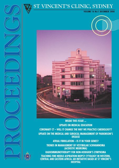
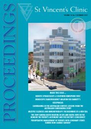
PROCEEPROCEEDINGSPROCEEST VINCENT’S CLINIC, SYDNEYVOLUME 16 No:1 DECEMBER <strong>2008</strong>INSIDE THIS ISSUE ...UPDATE ON MEDICAL EDUCATIONCORONARY CT – WILL IT CHANGE THE WAY WE PRACTICE CARDIOLOGY?UPDATE ON THE MEDICAL AND SURGICAL MANAGEMENT OF PARKINSON’SDISEASEATRIAL FIBRILLATION – IS IT IN YOUR GENES?TRENDS IN MANAGEMENT OF VESTIBULAR SCHWANNOMA(ACOUSTIC NEUROMA)RADIOIMMUNOTHERAPY FOR NON-HODGKIN’S LYMPHOMATEACHING FINE NEEDLE ASPIRATION BIOPSY CYTOLOGY IN WESTERN,CENTRAL AND EASTERN AFRICA: AN INITIATIVE BASED AT ST VINCENT’SHOSPITALST VINCENT’S CLINIC DARLINGHURST, SYDNEY
ST VINCENT’S CLINIC, SYDNEY VOLUME 16 No:1 DECEMBER <strong>2008</strong>COPYRIGHTPROCEEDINGPROCEEDINGSEditorial 2Dr John H. O’Neill MD, FRACPConsultant Neurologist,Editor, <strong>Proceedings</strong>ArticlesUpdate on Medical Education 4Associate Professor Eva SegelovMedical Oncologist and Directorof Medical <strong>St</strong>udent Education<strong>St</strong> Vincent’s <strong>Clinic</strong>al SchoolCoronary CT – Will it change the way 8we practice Cardiology?Associate Professor Jane McCrohonMBBS, FRACP, PhdUNSW Cardiologist, <strong>St</strong> Vincent’s CampusProfessor Michael Feneley,MD, FRACP, FCSANZ, FACCDirector, Heart-Lung Program,<strong>St</strong> Vincent’s HosptalDirector Cardiology, <strong>St</strong> Vincent’s CampusUpdate on the Medical and Surgical 12Management of Parkinson’s DiseaseDr <strong>St</strong>ephen Tisch MBBS PhD FRACP<strong>St</strong>aff Specialist, Department of Neurology,<strong>St</strong> <strong>Vincents</strong> <strong>Hospital</strong>Consultant Neurologist,<strong>St</strong> Vincent’s Private <strong>Hospital</strong> and <strong>Clinic</strong>Conjoint Senior Lecturer University of NSWAtrial Fibrillation – Is it in your Genes? 18Diane Fatkin, MD, BSc (Med),FRACP, FCSANZCardiology Department, <strong>St</strong> Vincent’s <strong>Hospital</strong>,Sr Bernice Research Program in Inherited HeartDiseases, Victor Chang Cardiac Research InstituteTrends in Management of Vestibular 22Schwannoma (Acoustic Neuroma)Dr Nigel Biggs FRACS, MBBS (Hons)VMO <strong>St</strong> Vincent’s General <strong>Hospital</strong>,VMO <strong>St</strong> Vincent’s Private <strong>Hospital</strong><strong>St</strong> Vincent’s <strong>Clinic</strong><strong>2008</strong> <strong>St</strong> Vincent’s <strong>Clinic</strong> Foundation 26GrantsRadioimmunotherapy for Non-Hodgkin’s 27LymphomaDr Edwin Szeto BSc (Med) Hons, MBBS,FRACPNuclear Medicine Physician<strong>St</strong> Vincent’s <strong>Clinic</strong>Teaching Fine Needle Aspiration Biopsy 33Cytology in Western, Central and EasternAfrica: an initiative based at <strong>St</strong> Vincent’s <strong>Hospital</strong>Dr Andrew S. Field MBBS (Hons), FRCPA, FIACDipCyt(RCPA)Deputy Director and Senior ConsultantDepartment of Anatomical Pathology<strong>St</strong> Vincent’s <strong>Hospital</strong>BOARD OF DIRECTORSDr Brett Courtenay (Chair)Mrs Maureen McCabe OAMSr Teresita Marcelo RSCMs. Patricia TysonMr. John WilcoxSr Genevieve Walsh RSCBOARD OF TRUSTEESMr Ted Harris AC (President)Mr Ian BurninghamDr Maxwell ColemanDr Brett CourtenayMr Robert CusackMr Peter Falk OAMMr Peter Ferris AMSr Margaret Fitzgerald RSCMr Peter HuntProfessor Reginald Lord AMMr David MeagherMrs Roslyn Packer AOMs Michelle WilsonS T V INCENT’ S C LINICEXECUTIVE DIRECTORMs Michelle WilsonMEDICAL COUNCILDr Gordon O’Neill (chair)Associate Professor Judith FreundDr Michael KingDr Malcolm PellDr Ian SuttonS T V INCENT’ S C LINIC F OUNDATIONSCIENTIFIC COMMITTEEDr Peter Bentivoglio (Chair)Dr David GolovskyProfessor Reginald Lord AMProfessor Sandy Middleton(except Multi-Disciplinary Grants)Dr Dudley O’SullivanMr. John Geoghegan(Multi-Disciplinary Grants)All literary matter in the Journal is covered bycopyright, and must not be reproduced, storedin a retrieval system, or transmitted in anyform by electronic or mechanical means,photocopying, or recording, without writtenpermission.ST VINCENT’S CLINIC438 Victoria <strong>St</strong>reet, Darlinghurst, Sydney, NSW 2010, AustraliaPhone: (02) 8382 6222 Fax: (02) 8382 6402Email: clinic@stvincents.com.auWebsite: www.clinic.stvincents.com.auST VINCENT’S CLINIC, PROCEEDINGS VOLUME 16 NO: 1 DECEMBER <strong>2008</strong> 1
EDITORIALDr John O’Neill MD, FRACPCONSULTANT NEUROLOGISTEDITOR, PROCEEDINGSIn this year of the death of SrBernice Elphick, I felt it was fittingin this twentieth issue of <strong>Proceedings</strong>to provide a brief history of <strong>St</strong>Vincent’s <strong>Clinic</strong>, now in its nineteenthyear of operation.The concept of a <strong>Clinic</strong> originallycommenced in the mid 1980s. At thattime, there was a certain amount ofpolitical unrest in medical circles. Asmall group of doctors associated with <strong>St</strong>Vincent’s <strong>Hospital</strong> met to consider thepossibility of commencing a new clinicsimilar to some of the clinics in NorthAmerica. The success of <strong>St</strong> Vincent’sPrivate <strong>Hospital</strong>, then only 10 years old,was a great example of what could bedone with ingenuity, vision and goodmanagement. The pioneering groupcomprised Drs John Roarty, Derek Berg,Thomas Hugh, Bruce Taylor, the lateDavid Wilson and Professor James Biggs.The group felt that, with thecooperation of the Private <strong>Hospital</strong>, sucha new venture could be realised. It wouldaim to be a multi-disciplinary clinic ofexcellence with different specialtygroups working closely together bothinternally and externally for greaterpatient benefit. It aimed to provideradiotherapy and all forms ofinvestigative procedures. It would be afurther forum for education. It was vitalto the concept of the <strong>Clinic</strong> that aresearch foundation be created.This concept was presented to SrBernice who, as usual, perused thedocumentation very carefully beforestrongly endorsing the project andincluding a day surgery unit. At thattime she informed the medical groupthat the Private <strong>Hospital</strong> had justacquired an adjacent property, an oldhardware store on the corner of Oxford<strong>St</strong>, on which it was planned to expandthe medical imaging department withthe installation of a new computerisedscanner. A small sub-committee wasestablished, consisting of Sr Bernice, theaforementioned representatives of thedoctors under the chairmanship of DrJohn Roarty and representatives of Civil& Civic, the construction firm who built<strong>St</strong> Vincent’s Private <strong>Hospital</strong>. Thecontribution of Mr Richard Hammondfrom Civil & Civic was quiteremarkable. He was assisted by MrRichard Bate and the group also had theassistance of Mr Jim Baillie on legalissues.After many lengthy and sometimesdifficult meetings with the medical staffat large, approval was finally given forthe <strong>Clinic</strong> to go ahead and thispioneering joint venture between theSisters of Charity and the doctors wasformed. The support of Sr Bernice withthe doctors at that time was quiteoutstanding. The expertise, wisdom anddiplomacy of the original Chairman ofthe Board, Mr Peter Ferris, wasfundamental in progression of thedevelopment.The <strong>Clinic</strong> was opened in August,1990. Two years later, the <strong>St</strong> Vincent’s<strong>Clinic</strong> Foundation was established. Theobjective of the Foundation was topromote and fund research work donewithin the <strong>Clinic</strong> and on the Campus.Both the <strong>Clinic</strong> and Foundation havegrown in strength and prestige over theyears. In <strong>2008</strong>, the foundation provided$640,000 funding for 16 separateresearch grants and 2 multi-disciplinarypatient-focussed research grants. The listis provided on page 26 of this issue andon the inside back cover is anapplication form for persons who maywish to make a donation to assist withthe continuing research work supervisedby <strong>St</strong> Vincent’s <strong>Clinic</strong> Foundation. Itshould be noted that the LadiesCommittee of <strong>St</strong> Vincent’s Private<strong>Hospital</strong> and <strong>St</strong> Vincent’s <strong>Clinic</strong>Foundation continue to actively supportthe raising of funds for the Foundation.The development of the <strong>Clinic</strong> wouldnot have been possible for the Mission ofthe Sisters of Charity without theinclusion of the Sisters of CharityOutreach Centre. Sr Bernice wasinstrumental in the inclusion of theOutreach Centre within the <strong>Clinic</strong> andit too started from nothing to becomethe wonderful charitable organisationthat it is today.2 ST VINCENT’S CLINIC, PROCEEDINGS VOLUME 16 NO: 1 DECEMBER <strong>2008</strong>
It can be said that the two majorpioneers of <strong>St</strong> Vincent’s <strong>Clinic</strong> were SrBernice and Dr John Roarty (picturedabove) and commemorative portraits toboth of them rightly hang on level 4 ofthe <strong>Clinic</strong>.This twentieth issue continues thetradition of the <strong>Clinic</strong> serving as a forumfor continuing education and containsseven excellent articles.Dr Eva Segelov describes thechallenges faced and the processesadopted in the current training of ourmedical students.Dr Jane McCrohan and AssociateProfessor Michael Feneley wrote the firstof two cardiology articles, their articledescribing the new technology of multisliceCT scanning in the evaluation of arange of cardiac conditions and inparticular the coronary arteries. In thesecond article, Associate Professor DianeFatkin discusses atrial fibrillation, anarrhythmia increasingly prevalent in theageing population and, untreated, amajor risk factor for stroke in thatpopulation. She looks particularly at therole of genetics in the development ofatrial fibrillation and she was supportedin that work by the <strong>St</strong> Vincent’s <strong>Clinic</strong>Foundation.Dr <strong>St</strong>ephen Tisch is a newlyappointed neurologist to the Campus,returning from post-graduate studies inthe UK where he obtained specialtraining in the medical and surgicalmanagement of Parkinson’s disease andother movement disorders. His articleprovides an overview of themanagement of Parkinson’s disease.Dr Nigel Biggs, ENT Surgeon andEdwin Szeto, specialist in nuclearmedicine, have provided learned articleson, respectively, the currentmanagement of acoustic neuroma andthe use of radio-immunotherapy in themanagement of Non-Hodgkin’sLymphoma.Finally, Dr Andrew Field,Cytopathologist with Sydpath, describeshis involvement in the teaching ofcytopathology in third world countriesand the benefits derived therein.ST VINCENT’S CLINIC, PROCEEDINGS VOLUME 16 NO: 1 DECEMBER <strong>2008</strong> 3
Associate ProfessorEva SegelovUpdate on MedicalEducationAssociate Professor Eva SegelovMedical Oncologist and Director ofMedical <strong>St</strong>udent Education<strong>St</strong> Vincent’s <strong>Clinic</strong>al SchoolI NTRODUCTIONOver the past decade there havebeen major changes in medicaleducation in universities andpostgraduate colleges relating tocurriculum content, mode of delivery,and assessment. Australian medicalschools have followed internationaltrends by updating their programs,adapting to the needs of both learnersand teachers in the era of moderntechnology. This article describessome of the major changes in medicaleducation and relates them to currentprograms within the <strong>St</strong> Vincent’s<strong>Clinic</strong>al School of Faculty ofMedicine, University of New SouthWales.C HANGE INP EDAGOGYMedical education hasmodernized to keep pacewith the changes in generaleducation theory that haveimpacted on learning and teaching inprimary and high schools, as well asrecognizing the role of education in post-University continuing professionaldevelopment. Some pedagogical changesinclude:• The concept of training students to belife long learners, with recognition ofcareer-long continuation ofprofessional development; 1, 2• Recognition that not all learningstyles are similar, as with teachingstyles, but that increased studentengagement in active-mode learningleads to more effective outcomes anddevelopment of critical thinking skills;4 ST VINCENT’S CLINIC, PROCEEDINGS VOLUME 16 NO: 1 DECEMBER <strong>2008</strong>
❛Give a man a fish and you feed him for a day. Teach a man tofish and you feed him for a lifetime.❜Chinese proverb• Recognition that such topics as ethics,professionalism, teamwork, culturalsensitivity, communication skills andreflective practice can be taught andassessed, at least in part; 3, 4• Embracing huge changes intechnology, particularly electronicmedia;• Recognition that continuedprofessional development and teachersupport is needed to enhanceeffectiveness of teachers in medicine,requiring resources and expertise. 5, 6Concurrently, changes in bothworkplace and workforce have drivenneed for educational reform. Inmedicine, these include:• Worldwide workforce shortage,affecting most areas of medicine aswell as nursing and allied health;• Lifestyle changes and emphasis onwork-life balance;• Demographic shift with growingageing population;• Redistribution of health care provisioninto community and outpatientsettings;• Introduction of new technologies indiagnosis and treatment e.g. MRI,molecular diagnostics etc;• Opening of multiple new medicalschools (by 2009, Australia will have19 universities offering a medicaldegree, each with different entrycriteria and varying missionstatements and desired outcomes).S ELECTIONIt is not denied that to studymedicine, “high academic ability isessential”, 7 but rather that this alone isinsufficient. The challenge is to adopttools which robustly measure bothcognitive and non-cognitive abilities,such that they predict not only forperformance during the course but alsoas a medical practitioner. 8,9 MostAustralian universities now admitstudents based on aptitude testingthrough the national UMAT(Undergraduate Medical and HealthSciences Admission Test) 10 orGAMSAT (Graduate AustralianMedical School Admissions Test). 11In addition, most medical schoolsrequire an interview, either semistructuredof 30-60 minutes durationwith two-three panel members (medicaland lay), or the Multistation MiniInterview (MMI), an OSCE-style(Objective <strong>St</strong>ructured <strong>Clinic</strong>alExamination) series of around eightstations of 5-10 minutes duration,manned by individual interviewers.Interviews can assess fluency andcomprehension of spoken English andallow community input. Some degree ofstandardization can be achieved withinterviewer training and structuredmarking criteria. However, the processremains subjective, with potential forgender, cultural and other biases.Caution is also given regarding testingattitudes and ethics that may in fact belearned during the course. The influenceof coaching (not uncommon in such acompetitive environment) isunmeasured, a problem particularly asthe resource intensive nature of theinterview process does not easily allowfor significant changes from year to year.Above all, the predictive validity isuncertain, although many studies areunderway relating interviewperformance to subsequent studentgrades and as a predictor of later clinicalperformance. Recent data suggest thatMMI have better predictive power andare more cost and time efficient. 8Quotas to promote entry of studentsfrom under-represented anddisadvantaged community groups existfor most Australian medical schools,partly driven by Commonwealth fundingmodels for university places. The aim isto produce doctors from various cultural,geographical and socially disadvantagedpopulations, as it is thought that suchstudents will return to serve thecommunity they represent. Early resultsshow a higher return to localcommunities from student educated atrural medical schools.For the six year undergraduate medicinecourse at UNSW, entry is by applicationwith an initial short portfolio. The threecomponents: interview, UMAT, and HSC(or equivalent) contribute equally to thefinal ranking. Conjoint staff are encouragedto become interviewers, with trainingprovided. There is a scheme for entry ofrural students, indigenous students andstudents accepting bonded places(commitment to work in areas of need aftersubstantive training completed).C URRICULUMCONTENT ANDORGANISATIONThe traditional model of teachingbasic biological sciences beforeprogressing to clinical sciences has beenreplaced in modern medical schools byintegration of these around clinicalscenarios. There is ongoing, heateddebate (e.g. the optimal amount ofanatomy teaching) but there is alsorecognition that the scope of medicaleducation has broadened. Not only arethere many new diseases, diagnostics andtherapeutics, but globalization has led tomore emphasis on public andinternational health, medical error,health systems and health economics.Exposing students to subjects such asethics and legal, professionalism,evidence based medicine and skills forlife long learning (library and internetskills) has broad practical implicationse.g. gaining consent, dealing withpatients taking alternate therapies, etc.Modern curricula have well definedoutcomes and detailed learning plans bywhich these can be achieved. This is anadvance over old syllabi which tended tobe an exhaustive list of diseases aboutwhich students needed to ‘knoweverything”.In 2004, UNSW launched a totallyrevamped, strongly science-based six yearundergraduate curriculum, with outcomesarticulated by well defined graduatecapabilities grouped under three mainheadings: Applied Knowledge and Skills,Interactional Abilities and PersonalST VINCENT’S CLINIC, PROCEEDINGS VOLUME 16 NO: 1 DECEMBER <strong>2008</strong> 5
“We did dissection every Monday from 11-5 and then again onThursday afternoons, and I don’t think I learned anything at all”name withheld, GPAttributes (http://www.med.unsw.edu.au). As well as core terms in Medicine,Surgery, Women’s and Children’s Health,Psychiatry and Community Health,students have the opportunity to select termsin areas of interest in senior years. There isa 32 week research project (the IndependentLearning Project) which can be undertakenin any relevant field. <strong>St</strong> Vincent’s <strong>Clinic</strong>alSchool is a popular choice for a wide varietyof clinical and basic research projects.M ETHOD OFDELIVERYThe most well known of the newcurriculum designs is Problem BasedLearning (PBL), which was a radicaldeparture from traditional didacticteaching. The principles are to instill auseable knowledge base, skills inproblem solving, self-directed learningand collaboration. This is achievedthrough case based, integrated learningusing the student’s own enquiry, often insmall groups facilitated by a tutor, withspecified learning objectives. Groupwork is also a feature to aid practice inthe workplace which involves being partof a team.Most contemporary Australianmedical curricula have adopted theseprinciples, which mirror changes inprimary and high school educationtheory and tertiary education in manyother faculties. Research is ongoingcomparing ‘new’ and ‘old’ curriculashowing benefit for the newermethods. 12, 14Delivery mode has also changed, withtwo main drivers:1. e-learning: the advent of internet hasrevolutionized education in many ways,for example: unprecedented access tolearning materials; content delivery attiming and location of student’s choice;facilitating communication betweenindividuals and groups of students andstaff; etc. 15 Technologies such asvideoconferencing and blogging, havealso changed the landscape. There is agrowing body of evidence (evenrandomized controlled trials!) thatincorporating these technologies areeffective. 16Lectures at UNSW are podcast andthere is emphasis on web based provision oflearning materials. <strong>St</strong> Vincent’s <strong>Clinic</strong>alSchool has a videoskills program wherepaired groups of students video theirperformance of a history and physical examon a patient, with immediate playback. Ourhandheld transponders allow instantanonymous feedback for formative testing.UNSW encourages professionaldevelopment for conjoint staff to maximizeuse of modern technologies in teaching.2. Simulation: there is solid evidence onthe benefit of simulation in learning. 17This ranges from use of models forlearning and practicing procedural skills(e.g. basic and advanced life support,urinary catheterisation, lumbarpuncture), to the use of surrogatepatients to teach examination skills (e.g.breast and gynaecological assessment). 18Use of surrogate and standardizedpatients has been shown to enhancereliability of assessment during clinicalexams.<strong>St</strong> Vincent’s <strong>Clinic</strong>al School is a keystakeholder in the Don Harrison PatientSafety Simulation Centre, with an extensiveprogram of clinical skills taught across allPhases of the New Medicine Program in thesimulated environment. <strong>St</strong>andardizedpatients are used in some stations of theclinical examination, to compareperformance of students across the clinicalschools of the Faculty of Medicine.A SSESSMENTThis is also an area of major changewith recognition of the need forformative assessment (designed to givecontinuous feedback to both studentsand teachers on their progress towardsthe development of knowledge,understanding, skills and attitude) aswell as summative assessment (tasksdesigned to determine grades or marks ata certain time point). 19 New assessmenttools have been introduced into bothstudent and post- graduate medicalevaluation, with more emphasis onmultisource feedback and broadassessment including professionalism,ethics, teamwork and communicationskills. 20, 21 Whilst research into thesenew tools shows superior predictabilityand reliability, it is recognized that theyare time consuming. Two commonlyused modern assessment tools are:1. miniCEX (mini-<strong>Clinic</strong>alEXamination) – currently used toassess international medical graduatesand one of the mechanisms currentlybeing piloted for standardizedassessment of all Junior MedicalOfficers. It involves directobservation of the performance of atask (history or physical examinationor procedural skill or communication)with immediate verbal and writtenfeedback. The whole process shouldtake no more than 15 minutes butshould be repeated on multipleoccasions with different tasks toensure reliability.2. RIME paradigm – originally developedin the VA system in the USA, this isa framework of feedback to thelearner along a pathway ofdevelopment as a professional; fromReporter (describes what ishappening) to Interpreter (interpretswhat is happening) to Manager (takesaction) to Educator and Evaluator(reflects on performance and teachesothers), this uses constructivelanguage to advance students,recognizing the concept of life longlearning.The New Medicine Program at UNSW,which reaches full implementation across allthree Phases in 2009, uses a number ofmodern assessment tools including mini-CEX to allow students to receive feedbackon their performance. Peer feedback is alsoencouraged. <strong>St</strong> Vincent’s <strong>Clinic</strong>al Schoolhas run pilot student programs using theRIME mechanism. Training in newassessment methods is available at outregular Train the Trainer workshops.6 ST VINCENT’S CLINIC, PROCEEDINGS VOLUME 16 NO: 1 DECEMBER <strong>2008</strong>
M EDICAL EDUCATIONRESEARCHMedical education research has comeof age as a valid area of investigation andscholarship. Qualitative and quantitativestudies measuring the impact of variousteaching strategies are published in highquality medical education journals suchas Medical Teacher and AcademicMedicine. There is also a growinginterest in publishing medical educationarticles in journals such as BMJ andMJA. Medical education researchprojects attracting competitive researchfunding with recent creation ofspecialized categories within theNHMRC and ARC granting codes formedical education research. Nationaland international conferences are wellattended by all stakeholders in healthprofessional education so that futurechanges can be evidence-driven.Measuring educational outcomes is achallenging and relevant to a wide rangeof health professionals who are facingsimilar challenges in their professionaleducation and development programs.Evaluation of the New MedicineProgram at UNSW is ongoing, recognizingthat measuring educational outcomes iscomplex. New teaching methods are trialledand rigorously evaluated, 22 under thedirection of the Program Evaluation andImprovement Group. Long term impact isof particular importance.S UMMARYMany changes have driven theevolution of medical educationinternationally from didactic, teacherbased curricula to student-centeredcurricula focussing on teaching lifelonglearning skills. Many different types oflearning packages defined by specificlearning objectives and outcomes, withcorresponding assessment, have beendeveloped. Although these changes mayat face value be challenging to our owneducational pedagogy, it is anticipatedthat students will develop skills that willallow them to manage the demands ofmodern medical practice.The <strong>St</strong> Vincent’s <strong>Clinic</strong>al Schoolwelcomes appointment of conjoint staff andencourages all consultants across thecampus to become affiliated. In particular afocus on one-on-one teaching of seniorstudents in private rooms duringconsultations has been successful over thepast several years, with excellent feedbackfrom students and consultants. A survey ofpatient attitudes is planned for the nearfuture but most consultants report thatpatients welcome students in these controlledconditions. For further information, pleasesee http://www.med.unsw.edu.au/medweb.nsf/page/Conjoints, or contact the <strong>Clinic</strong>alSchool 83822023.R EFERENCES1. Schrock, J.W. and R.K. Cydulka, Lifelonglearning. Emerg Med Clin North Am, 2006.24(3): p. 785-95.2. Simon, F.A. and C.A. Aschenbrener,Undergraduate medical educationaccreditation as a driver of lifelong learning. JContin Educ Health Prof, 2005. 25(3): p. 157-61.3. McElhinney, T.K., Medical ethics in medicaleducation: finding and keeping a place at thetable. J Clin Ethics, 1993. 4(3): p. 273-5.4. Goldie, J., Integrating professionalismteaching into undergraduate medicaleducation in the UK setting. Med Teach, <strong>2008</strong>.30(5): p. 513-27.5. McLean, M., F. Cilliers, and J.M. Van Wyk,Faculty development: yesterday, today andtomorrow. Med Teach, <strong>2008</strong>. 30(6): p. 555-84.6. Molodysky, E., Identifying and trainingeffective clinical teachers. Australian FamilyPhysician, 2006. 35(112): p. 53-55.7. Tutton, P. and M. Price, Selection of medicalstudents. BMJ, 2002. 324(7347): p. 1170-1.8. Rosenfeld, J.M., et al., A Cost EfficiencyComparison Between The Multiple Mini-Interview and Traditional AdmissionsInterviews. Adv Health Sci Educ Theory Pract,2006.9. Powis, D.A., M. Bore, and D. Munro,Selecting medical students: evidence basedadmissions procedures for medical students arebeing tested. BMJ, 2006. 332(7550): p. 1156.10. http://www.acer.edu.au/tests/university/umat/.11. http://www.gamsat.acer.edu.au/.12. Lycke, K.H., P. Grottum, and H.I. <strong>St</strong>romso,<strong>St</strong>udent learning strategies, mental models andlearning outcomes in problem-based andtraditional curricula in medicine. Med Teach,2006. 28(8): p. 717-22.13. Srinivasan, M., et al., Comparing problembasedlearning with case-based learning: effectsof a major curricular shift at two institutions.Acad Med, 2007. 82(1): p. 74-82.14. Peters, A.S., et al., Long-term outcomes ofthe New Pathway Program at HarvardMedical School: a randomized controlled trial.Acad Med, 2000. 75(5): p. 470-9.15. Ellaway, R. and K. Masters, AMEE Guide32: e-Learning in medical education Part 1:Learning, teaching and assessment. MedTeach, <strong>2008</strong>. 30(5): p. 455-73.16. Davis, J., et al., Computer-based teaching isas good as face to face lecture-based teachingof evidence based medicine: a randomizedcontrolled trial. Med Teach, <strong>2008</strong>. 30(3): p.302-7.17. Criss, E.A., Patient simulators. Changing theface of EMS education. Jems, 2001. 26(12): p.24-31.18. <strong>St</strong>ewart, F. and J. Cleland, The introductionof standardized clinical surgical teaching:students’ and tutors’ perceptions of newteaching and learning aids. Med Teach, <strong>2008</strong>.30(5): p. 508-12.19. Perera, J., et al., Formative feedback tostudents: the mismatch between facultyperceptions and student expectations. MedTeach, <strong>2008</strong>. 30(4): p. 395-9.20. O’Sullivan, A.J. and S.M. Toohey,Assessment of professionalism inundergraduate medical students. Med Teach,<strong>2008</strong>. 30(3): p. 280-6.21. Schubert, S., et al., A situational judgementtest of professional behaviour: developmentand validation. Med Teach, <strong>2008</strong>. 30(5): p.528-33.22. Gibson, K.A., et al., Enhancing evaluation inan undergraduate medical education program.Acad Med, <strong>2008</strong>. 83(8): p. 787-93.ST VINCENT’S CLINIC, PROCEEDINGS VOLUME 16 NO: 1 DECEMBER <strong>2008</strong> 7
Assoc Prof Jane McCrohonProfessor Michael FeneleyI NTRODUCTIONDramatic improvements intechnology have occurred in the lastdecade across all cardiac imagingmodalities but perhaps none hasbetter commanded the attention ofthe medical fraternity than that ofcardiac CT. This review will providean overview of the current state ofplay in the use of this technology inclinical practice and where newdevelopments in scanner technologyare likely to lead over the next fewyears. At a time when the <strong>St</strong>Vincent’s campus is rapidlydeveloping its cardiac imagingprogram, the future looks bright interms of our ability to bring cuttingedge techniques in imaging to thediverse range of cardiac patients inour care.Coronary CT – Willit change the way wepractice cardiology?B ACKGROUNDCardiovascular disease is theleading cause of mortalityworldwide 1 with atherosclerosisas the majorcontributor in this process. Atheroscleroticlesions develop through stagesfrom fatty streaks to plaques of varyingmorphologies. These plaques may beobstructive (flow limiting and oftensymptomatic) or non-obstructive; stableor unstable (based on the content oflipid and inflammatory components)and either fatty, fibrous or calcific innature (or a combination of these).In the diagnosis and management ofcoronary artery disease, we havetraditionally relied on functionalassessment or stress tests (exercise ECG,stress echo or myocardial perfusionAssociate Professor Jane McCrohonMBBS, FRACP, PhdUNSW Cardiologist,<strong>St</strong> Vincent’s CampusProfessor Michael Feneley,MD, FRACP, FCSANZ, FACCDirector, Heart-Lung Program,<strong>St</strong> Vincent’s HosptalDirector Cardiology,<strong>St</strong> Vincent’s Campusscans). Different methods of stressassessment use different end points toassess flow reserve of the myocardium.When a stress test is performed toinvestigate the possibility of coronaryartery disease, we are assessing thepresence or absence of a flow limitingstenosis of sufficient severity to inducemyocardial ischaemia and itsaccompanying sequelae in the‘ischaemic cascade’ (diastolicdysfunction, perfusion deficits, reducedmyocardial systolic thickening, ECGchanges and finally patient symptoms ofchest tightness, effort intolerance and/ordyspnoea). Functional assessmenttherefore relies on the existence ofsignificant coronary artery narrowing(usually >70 per cent luminal narrowing)with a sufficient reduction in myocardialblood flow during stress to yield a positiveresult. The accuracy of different testsvaries across different populations basedon the clinical likelihood of disease andother patient factors. Depending on thepresence and extent of myocardialischaemia and/or patient symptoms,many patients will then be referred forinvasive coronary angiography andpotential revascularization.Unfortunately, as many as half of firstcoronary events (including myocardialinfarction and death) occur in previouslyasymptomatic people 2 and occur due tothe rupture of unstable non-occlusiveplaques which cannot be detected withfunctional assessment alone or evenconventional invasive coronaryangiography. This concept is not newand was well defined by Glagov in 1987whereby there is outward or ‘positive’arterial remodeling of the arterial walland preservation of lumen size until 408 ST VINCENT’S CLINIC, PROCEEDINGS VOLUME 16 NO: 1 DECEMBER <strong>2008</strong>
per cent or greater of the internal elasticarea is affected by atheromatous plaque(Figure 1). 3 It is now recognised thatthese types of plaques are frequentlyunstable and their detection is nowpossible with new modalities such asintravascular ultrasound (IVUS),magnetic resonance and most recentlyCT. CT coronary angiography (CTA)therefore provides additionalinformation beyond the lumen andalthough not promoted as a screeningtest for asymptomatic low riskindividuals, may provide an opportunityto better risk stratify our patients atintermediate coronary risk.The paradigm for cardiac CT use andits integration with other imagingmodalities remains in evolution and islikely to change as the technology andour familiarity with the techniqueincrease. Current recommendations orclinical appropriateness criteria (REF)have been provided by variousinternational bodies such as theAmerican College of Cardiology andRadiology (ACC and ACR) and theSociety of Cardiovascular ComputedTomography (SCCT) and the mainindications for cardiac CT at this pointin time are summarized in Table 1.Whether CTA should be done as aninitial test in a patient with symptomssuggestive of coronary disease or used asa ‘gatekeeper’ following functionaltesting is indeterminate. Recent workhas suggested that regardless of theanatomical accuracy of a technique (CTor conventional angiography), there isoften a poor correlation between theangiographic stenosis grade and thefunctional effect on downstream flow tothe myocardium and patient symptoms. 4Functional testing prior to treatment istherefore likely to remain a keycomponent in patient management inthe years ahead. Emerging prognosticdata for CT and correlation of clinicaloutcomes based on test accuracy willinfluence these decision processes, aswill the potential for simultaneous CTassessment of myocardial perfusionwhich could finally provide the covetedholy grail of anatomical and functionalevaluation of coronary disease in a singletest. The role of integrated imaging, suchas PET-CT, as a provider of bothfunctional and anatomical informationin a single examination is also beingexplored.T HETable 1: Current appropriate indications for Cardiac CT*F UNDAMENTALSOFCTCT images are generated by ionizingradiation. An x-ray beam passes throughthe body at different angles and isreceived by a detector array on the otherside of the patient. The x-ray beamreaching the detector array is digitized toproduce pixels of a known size and eachpixel contains gray scale informationEVALUATION OF CHEST PAIN SYNDROME INTERMEDIATEPROBABLITY GROUPASSESSMENT OF CORONARY ARTERY ANOMALIESEQUIVOCAL OR UNINTERPRETABLE STRESS TESTEVALUATION OF CORONARY ARTERY BYPASS GRAFTSCONGENITAL HEART DISEASEEVALUATION OF CORONARY ARTERY DISEASE IN NEW ONSETHEART FAILUREEVALUATION OF CARDIAC MASS / PERICARDIUMPATIENTS WITH LIMITED IMAGE QUALITY FROM OTHERTECHNIQUESPULMONARY VEIN, CORONARY VEIN MAPPINGASSESSMENT OF GREAT VESSELS* Adapted from Reference 6Compensatory expansion maintainslumen sizetillate in disease (positive remodeling)Normal lumenMinimal CADModerate CADSevere CADFigure 1: The Glagov PhenomenonTraditional lumenal angiography will only detect disease that encroaches the lumen. In the presence of positive remodeling of the arterial wall inatherosclerosis, lumenal area can be preserved and labeled as ‘normal’ on catheter angiography even in the presence of significant plaque burden. In contrast,CTA can clearly visualise the entire spectrum of atherosclerotic remodellingST VINCENT’S CLINIC, PROCEEDINGS VOLUME 16 NO: 1 DECEMBER <strong>2008</strong> 9
ased on the beams attenuation throughdifferent tissues. This attenuationinformation is represented in HounsfieldUnits (HU), and ranges widely from air(-1000HU) to water (0HU) and bonecortex (+1000HU).Cardiac CT is usually performed on amultislice CT (MSCT) scanner with arotating x-ray source and stationarydetector arrays. Continuous or stepwisetable movement allows the collection ofa volume data set. Recent developmentsin scanner technology have seen a rapidincrease in the number of slices acquiredper breath hold from 4, 16, 64 and now256 and 320 slice acquisitions. Suchdevelopments along with increasedgantry speeds and improved acquisitionand reconstruction techniques have ledto improved coverage, faster scan timesand increased temporal and spatialresolution. Current scanners nowachieve isotropic voxels of 0.35mm 3 andtemporal resolution from 40-200ms(compared to spatial and temporalresolution of 0.1-0.2mm and 5-10ms,respectively, with catheter angiography).Patient preparation is essential incoronary CT, with most centresadvocating heart rates of 97per cent). This confirms its key role inthe ‘ruling out’ of significant disease. It isimportant to remember, however, thatmuch of this data was acquired in singlecentrestudies with a high level ofexpertise and variable patientpopulations. Perhaps the mostmeaningful clinical accuracy data hasemerged from the CORE 64 CT trial,which was a multicentre, ‘real world’trial of patients scheduled to haveinvasive catheter angiography. Theprevalence of disease in this trial was56% (much higher than in other studies)and much higher than many of thepatients currently being sent for CTA inmany centres. The performance of CTAeven in this higher risk group was verygood, with sensitivity, specificity,positive predictive value and negativepredictive value of 85, 90, 91, and 83%,respectively, when compared withinvasive angiography. 5Factors associated with reduceddiagnostic accuracy include the presenceof heavy calcification, rapid or irregularheart rates (atrial fibrillation or ectopics)and inadequate breath holdingtechnique. Although there will alwaysbe some limitations, many of these arebecoming less problematic withdevelopments in technology and morerapid acquisitions.Minimising radiation exposure hasbeen a key concern leading totechniques such as x-ray dosemodulation based on body size and ECGmodulation, where the tube current ismaximal or turned on only during thediastolic phase(s) of interest. One of thelatest generation scanners, a 320 sliceCT scanner, is capable of acquiring datawithin a single heart beat usingprospective gating and only 10-30% ofthe R-R interval, producing radiationlevels as low as 4-7mSv for coronaryangiography and 1mSv for coronarycalcium scoring. Similarly low radiationdoses can be achieved, albeit over aminimum of two heart beats, usingprospective gating on the 256 slicescanner. This compares with 6-10mSvradiation dose for invasive diagnosticcatheter coronary angiography and 15-20mSv for nuclear SPECT perfusionimaging.As with any CT examination,children and females are at greater riskof radiation exposure and knowledge ofrenal function and diabetic status areessential to minimize the risk of contrastnephropathy.Figure 2: Analysis of CT coronaryangiography data involves a detailed review of2D, 3D and cross-sectional images10 ST VINCENT’S CLINIC, PROCEEDINGS VOLUME 16 NO: 1 DECEMBER <strong>2008</strong>
C ORONARY A RTERYC ALCIUM S CORINGThis is usually performed as a singlebreath hold scan just before the CTAacquisition, and is a low radiation, lowresolution scan. Most reports define thecalcium score based on the patient’s ageand gender, and place the patient in aquartile of coronary artery disease riskbased on previous data registries (Figure1). <strong>St</strong>ated simply, a calcium score of zerois associated with an extremely lowlikelihood of significant coronaryatherosclerosis (although not zero,particularly in younger patients) and>400 with a high likelihood of extensiveatherosclerosis.Recent data have suggested thatcoronary calcium scores provideincremental risk stratification inindividual patients beyond that offeredby the Framingham Risk Score alone.For example, in one study, 45% ofpatients having a high Framingham RiskScore could be reclassified into low orintermediate risk by their coronary arterycalcium scores. 7Interestingly, despite the correlationof increasing calcium score with age andatherosclerotic burden in a givenindividual, calcium within a plaque ismore likely to represent stable or slowlyevolving pathology, whereas minorspecks of calcium or its absence is felt tobe characteristic of the more lipid-richplaques associated with less stableplaque physiology. The role of calciumscoring and plaque characterization is anarea of intense research interest at thispoint in time using CT and othertechniques such as intravascularultrasound and MRI.Multimodality Cardiac imaging – therole for coronary CTIt is now well recognised that any newtechnology should be demonstrated toprovide incremental information andimprovements in patient care overexisting techniques before its routine useis recommended. In cardiology,techniques such as nuclear medicinehave provided us with a wealth ofprognostic information over many years.Such information has been used to drawconclusions and develop similar data innew modalities such as MRI and CT.Prognostic and cost-effectiveness datafor coronary CT are rapidly emerging,and will no doubt shape the use andreimbursement of this technology in theyears ahead.The available information does notcurrently support the routine clinicalapplication of CT coronary angiographyin asymptomatic individuals, althoughresearch studies exploring its possiblerole in low, intermediate and high riskasymptomatic subjects are in progress.The technique is still in its early stagesand long-term follow up will be neededbefore a screening use of coronary CT islikely to be embraced.Two recent studies have clearlyshown that the extent and severity ofcoronary atherosclerosis on coronary CTis associated with a worse prognosiswhen compared with the absence ofcoronary atherosclerosis. 8,9 In particular,a worse prognosis was associated withmore proximal disease, stenoses >50 percent, the number of arteries involvedand the presence of left main disease.The scoring used to assess plaqueseverity and extent across all branches ofthe coronary tree was shown to predictall-cause mortality independently ofother traditional risk factors. The factthat >50 per cent of myocardial infarctsoccur on non-occlusive plaque isprobably largely related to the higherincidence of these plaques overall, withthe study by Min et al 8 confirming thatthose patients with the greater extent ofplaque (independent of stenosis severity)had a poorer prognosis. Conversely, thestudy also confirmed that the absence ofcoronary plaque conferred a highnegative predictive value of 97.8-99.7%for events over the 15 month averagefollow-up period.T HEF UTURECardiac CT will continue to evolverapidly over the next few years due to itsaccessibility, ease of use and preferenceby patients and referrers for a noninvasivemeans of visualizing thecoronary arteries. It is unlikely however,that anatomical imaging alone willprovide sufficient guidance in manycases where disease is present and in thepatient population most cardiologistsreview in their rooms. Supplementaryfunctional imaging will be required toguide management decisions after manyabnormal CT scans, but the emergingtechnique of CT myocardial perfusionimaging offers a golden opportunity for atruly comprehensive single test forischaemic heart disease. Whether thetechnology meets expectations remainsto be seen, and how we incorporate therange of cardiac imaging modalities nowavailable to us into improved patientcare is likely to be even more importantin years to come than the technologyitself.R EFERENCES1. Yusuf S, Hawken S, Ounpuu S et al. Effectof potentially modifiable risk factors associatedwith myocardial infarction in 52 countries(the INTERHEART study): case controlstudy. Lancet 2004;364 (9438):937-522. O’Rourke RA, Brundage BH, Froelicher VFet al. American College of Cardiology /American Heart Association ExpertConsensus Document on EBCT for thediagnosis and prognosis of coronary arteydisease. J Am Coll Cardiol 2000;36(1):326-40.3. Glagov, S., Weisenberg, E., Zarins, C. K.,<strong>St</strong>ankunavicius,R., Kolettis, G. J.Compensatory enlargement of humanatherosclerotic coronary arteries. NEJM 1987;316:1371–1375.4. Meijboom WB, Van Mieghem CAG, vanPelt N et al. Comprehensive Assessment ofCoronary Artery <strong>St</strong>enosis. JACC <strong>2008</strong>;52:636-43.5. Miller JM, Rochitte CE, Dewe M et al.Coronary Artery Evaluation using 640RowMultidetector Computer TomographyAngiography (CORE 64): results of amulticenter, international trial to assess thediagnostic accuracy compared withconventional coronary angiography (abstr)Circulation 2007;116:II2630.6. ACCF/ACR/SCCT/SCMR/ASNC/NASCI/SCAI/SIR 2006 appropriateness criteria forcardiac computed tomography and cardiacmagnetic resonance imaging. A report of theAmerican College of Cardiology FoundationQuality <strong>St</strong>rategic Directions CommitteeAppropriateness Criteria Working Group. JAm Coll Radiol. 2006 Oct;3(10):751-71.7. Pundaiute F, Schuijf JD, Jukema JW et al.Prognostic value of multislice computedtomography coronary angiography in patientswith known or suspected coornary arterydisease. JACC 2007;49:62-70.8. Min JK, Shaw LJ, Devereux RB et al.Prognostic value of multidetector coronarycomputed tomographic angiography forprediction of all-cause mortality. JACC2007;50: 1161-70.9. Greenland P, La Bree L, Azen SP et al.Coronary artery calcium score combined withFramingham score for risk prediction inasymptomatic individuals. JAMA.2004;291(2):210-5.ST VINCENT’S CLINIC, PROCEEDINGS VOLUME 16 NO: 1 DECEMBER <strong>2008</strong> 11
Dr <strong>St</strong>ephen TischI NTRODUCTIONParkinson’s disease (PD) is acommon neurodegenerative disorderwith a prevalence in Sydney of 3.4 percent in people aged 55 years or over. 1PD is characterised by the cardinalmotor symptoms of tremor, rigidity,bradykinesia and gait disorder withpostural instability. Levodopa remainsthe most effective oral medication forthe motor symptoms of PD but isassociated with the development ofmotor complications includingdyskensias and ON/OFF phenomenausually within 3-5 years of treatment.Motor complications from Levodopaare worse in younger patients andwhen higher doses of Levodopa areused. 2 The increased recognition oflevodopa induced motor fluctuationsand the availability of alternativedopamine agonist therapies has led toa reappraisal of both early and latertreatment of PD. For those patientswith advanced PD and establishedmotor fluctuations, significanttreatment advances have been madewith the use of continuousApomorphine infusion or deep brainstimulation (DBS) surgery. Astreatments for motor symptoms of PDhave improved, there has beenincreased recognition of residual nonmotorsymptoms affecting cognition,sleep, mood, autonomic and sexualfunction which may contribute todisability. These aspects may also beimproved with specificpharmacotherapy and interventions.In this review treatment strategies forearly and advanced PD will bepresented with an emphasis onpharmacotherapy and surgeryavailable within the Australiancontext. The major limitation ofcurrent available treatment optionsfor PD is the lack of neuroprotectiveor neurorestorative therapy, andfuture therapies in development willalso be discussed.Dr <strong>St</strong>ephen Tisch MBBS PhD FRACP<strong>St</strong>aff Specialist, Department ofNeurology, <strong>St</strong> <strong>Vincents</strong> <strong>Hospital</strong>Consultant Neurologist, <strong>St</strong> Vincent’sPrivate <strong>Hospital</strong> and <strong>Clinic</strong>Conjoint Senior Lecturer Universityof NSWUpdate on the Medicaland Surgical Managementof Parkinson’s DiseaseL EVODOPAANDM OTORF LUCTUATIONSLevodopa was first used for PD inthe early 1970s and remains themost effective oral dopaminereplacement medication for themotor symptoms of PD. It has neverbeen subjected to a randomisedcontrolled trial. Unlike North Americawhere Sinemet (Levodopa/carbidopa) isthe only available Levodopa preparation,Australia like Europe has Madopar(Levodopa/benserazide) which is veryuseful since occasional patients willtolerate one but not the other. Motorfluctuations occur in about 10 per centof Levodopa treated patients with eachyear of treatment, so that by five yearsabout 50 per cent are affected. Levodopahas not been shown to be toxic todopaminergic neurons in vivo nor does itaccelerate the neurodegenerativeprocess, rather motor fluctuations arisedue to sensitizing plasticity with thestriatum due to pulsatile dopminergicstimulation. Motor fluctuations consistof dyskinesias (choreoform, dystonic,diphasic), wearing off and end of dosedeterioration, ON/OFF phenomena,delayed ON and dose failures. Patientsmay undergo frequent rapid transitionsbetween a rigid immobile OFF state anda mobile ON state, which may be furthercompromised by dyskinesias. Thedevelopment of motor fluctuations oftenheralds an escalation in disability anddependency, particularly when frequentsevere OFF periods cause immobility andfalls or severe dyskinesias interfere withfunction during the ON phase. Motorfluctuations are more likely to develop inyounger patients and after 6 years oftreatment affect almost 100 per cent ofpatients with PD onset < 40 years.Larger doses of Levodopa promote the12 ST VINCENT’S CLINIC, PROCEEDINGS VOLUME 16 NO: 1 DECEMBER <strong>2008</strong>
development of motor fluctuations. Inthe ELLDOPA trial the rate ofdyskinesia was 2.3 per cent in patientstreated with 300 mg/d of levodopa and16.5 per cent with 600 mg/day after ninemonths of treatment. 3 The use of lowerLevodopa doses particularly in early PDhas reduced the incidence of severemotor fluctuations. Despite thelimitations of Levodopa it is the mostpotent oral dopamine replacementtherapy and forms the mainstay oftreatment for most patients.C ATECHOL-O-METHYLT RANSFERASEI NHIBITORSCatechol-O-methyl transferase(COMT) inhibitors reduce theperipheral degradation of Levodopathereby boosting levels reaching thebrain. COMT inhibitors prolong theaction of Levodopa and reduce end ofdose wearing off. The down side ofCOMT inhibitors is that they mayexacerbate dyskinesias. Other side effectsinclude diarrhoea and harmless browndiscolouration of the urine. Entacapone(Comtan) is co-adminstered with someor all of the daily Levodopa doses, and istypically initiated when patients begin toexperience end of dose wearing off. Thecombined Entacapone Levodopa/carbidopa preparation (<strong>St</strong>alevo) ispreferred by many patients 4 and isfinding an increasing role in the initialtreatment of PD in the hope that lowerLevodopa total dose and morecontinuous dopaminergic stimulationmay forestall motor fluctuations,however, to date this is unproven.Tolcopone, a potent and efficaciousCOMT inhibitor was withdrawn inAustralia in 1999 due to cases of fatalhepatotoxicity. It is more effective thanEntacapone in increasing ON time andmay have limited role in Entacaponerefractory patients provided carefulmonitoring of liver function isperformed. 5,6M ONOAMINE O XIDASEB INHIBITORSMonoamine oxidase B inhibitors(MAO-B) also enhance the effects ofLevodopa by inhibiting brain enzymaticdegradation of dopamine and blockingreuptake of dopamine at the synapticcleft. Unlike COMT inhibitors MAO-Bdo not require co-administration ofLevodopa to produce symptomaticimprovement and are sometimes used asfirst line treatment for mildly affectedPD patients and delay the need forLevodopa by about nine months. 7 Morecommonly MAO-B are used as adjuvanttherapy in Levodopa treated patients toreduce OFF time, but may worsendyskinesias, psychotic phenomena orprovoke postural hypotension. Aprospective UK study showed a higherfive year mortality among patientstreated with a combination of Selegelineand Levodopa 8 however other studieshave not shown increased mortality.Rasagaline is a highly bioavailableirreversible MOA-B inhibitor isavailable in Europe and North Americabut not yet in Australia. As adjuvanttherapy Rasagaline prolongs ON time byabout 25 per cent 9 , an effect comparableto that of Entacapone. 10D OPAMINEA GONISTSDopamine agonists include the ergotderived agents (Bromocriptine,Pergolide, Cabergoline) and thesynthetic non-ergot agents (Ropinirole,Pramipexole, Rotigotine, Apomorphine).The literature surroundingdopamine agonists is extensive but a fewkey points can be made, and aresupported by a recent Cochranereview. 11 As monotherapy the dopamineagonists are less potent than Levodopabut have a lower incidence of motorfluctuations particularly dyskinesias.Dopamine agonists induce side effectsincluding nausea, postural hypotension,somnolence, hallucinations andbehavioural dysregulation (in particularpathological gambling), to a greaterextent than Levodopa. Dopamineagonists are useful as first line treatmentin younger patients and those withmilder symptoms, to postpone therequirement of Levodopa. In additionthey are valuable as adjuvant treatmentin Levodopa treated patients to reducemotor fluctuations by increasing theduration and quality of ON time,sometimes at the expense of worseningdyskinesias. Within the Australiancontext Pramipexole (Sifrol) in June<strong>2008</strong> became the first non-ergotdopamine agonist PBS listed for PD.This development is of heightenedsignificance because of increasing safetyconcerns surrounding the ergot agonistswhich may cause cardiacvalvulopathy, 12,13 and are being phasedout, where possible in the majority ofpatients. Older agonists can besuccessfully switched overnight to acorresponding dose of non-ergolineagonist. 14 Rotigotine is a potent noveldopamine agonist administered as atransdermal patch applied every 24hours with potential advantages in termsof continuous dopaminergic stimulationand patient preference. Initialexperience of Rotigotine has been verypositive. 15 It is licensed in Australia, butis expensive and is not yet PBS listed.A POMORPHINEApomorphzine was first synthesised inthe 1850s by the reaction of morphinewith hydrochloric acid and wasoriginally marketed as an emetic. It hasno narcotic properties but is a highpotency dopamine agonist with affinityto D1 and D2 receptors. It is the onlydopamine agonist with a magnitude ofclinical effect on motor symptomsequivalent to Levodopa. Apomorphineis not orally bioavailable and has a shorthalf life and therefore requiressubcutaneous administration either asintermittent injections or continuoussubcutaneous infusion. BecauseApomorphine is emetogenic,premedication with Domperidone isrequired. Intermittent Apomorphineinjections are useful as “rescue” therapyfor OFF periods, providing rapidimprovement in mobility. Patients withmore frequent OFF periods anddyskinesia benefit more from continuousinfusions, which increase ON time byabout 50%, and reduce dyskinesias,particularly in those patients able todiscontinue Levopdopa. 16-18 Theinfusion is usually limited to the wakingday, but can in some circumstances begiven 24 hours a day. Apomorphineinfusions require a small infusion pumpto be worn (Figure 1) and inpatientinitiation by an experienced team. Onceestablished, the infusions are usuallytrouble free. Some patients develop skinnodules at the injection sites andoccasional haemolytic anaemia canoccur. Apomorphine is licensed inAustralia for intermittent injection andcontinuous infusion and is PBS listed.ST VINCENT’S CLINIC, PROCEEDINGS VOLUME 16 NO: 1 DECEMBER <strong>2008</strong> 13
A MANTADINE A NDA NTICHOLINERGICSAmantadine is a non-competitiveNMDA receptor antagonist, and theonly class of drug which acts on brainglutamate receptors which are known toplay an important role in PD. It hasmodest intrinsic antiparkinsonian effectsand is sometimes used as a first line drugin mildly affected patients however thebeneficial effects are often unsustained.More recently its anti-dyskineticproperties have been recognised. Whenused as adjuvant therapy Amantadinereduces dyskinesias by up to 50-60 percent. 19,20 Problems with Amantadineinclude insomnia, confusion and ankleoedema.Anticholinergic agents includingBenzhexol (Trihexphenidyl) andBenztropine are probably underutilisedand provide an alternative initialmonotherapy in mildly affected ortremor dominant patients. 7 Atropinelikeside effects and worsening ofpsychotic or cognitive aspects limit theirusefulness and make them unsuitable theelderly.M EDICALM ANAGEMENTM OTOROFF LUCTUATIONSI N PDEarly motor fluctuations such as endof dose wearing OFF can be managedinitially by shortening the Levodopadose interval. Peak dose dyskinesias mayrespond to a reduction of individualLevodopa doses. Delayed ON or dosefailures may be helped by takingLevodopa 30-60 minutes before food andeating smaller more frequent meals tooffset competitive inhibition ofLevodopa protein transport at the gutand brain. Similarly, dispersibleFigure 1. ProgrammableApomorphine infusion pump, whichdelivers a continuous subcutaneousinfusion of Apomorphine via abutterfly needle. The pump is worn bypatients in a belt carry case.Levodopa preparations (e.g. Madoparrapid) may be useful to hasten the onsetof Levodopa effect. Nocturnal OFFperiods can be treated with slow releaseLevodopa (e.g. Sinemet CR) at bedtimeand additional immediate releaseLevodopa during the night. Whenmanipulation of Levodopa fails tocontrol increasing OFF periods, theaddition of Entacapone or theintroduction of a dopamine agonist isusually beneficial, however these agentsmay worsen dyskinesia. However,dopamine agonists and COMTinhibitors may allow reduction of totalLevodopa dose, which over time canhelp reduce dyskinesias. A commonmistake when adding agonists is to usetoo low a maintenance dose. Thestarting doses are low to avoid sideeffectsparticularly nausea andhypotension, and adequate upwardtitration should be performed until atherapeutic effect is achieved or sideeffects are reached. Dyskinesias can betreated directly by the addition ofAmantadine. Painful OFF dystonia in alimb can be helped in selected patientswith botulinum toxin injections. Asmotor fluctuations worsen combinationadjuvant therapy with dopamineagonists and COMT inhibitors canprovide additional benefit. Increasingly,such combination therapy is being usedearlier in the course of disease in aneffort to limit the Levodopa requirementand minimise Levodopa induced motorcomplications.When disabling motor fluctuationsare no longer manageable with oralmedications, intermittent subcutaneousinjections or continuous infusion ofApomorphine should be considered. Analternative is continuous duodenalinfusion of Levodopa (Duodopa) whichin small open label studies has beenshown to significantly decrease OFFtime and dyskinesias. 21,22 Duodoparequires a jejunostomy tube and infusionpump, and complications related to thetube are common. 23 Duodopa is anorphan drug in Australia and itsavailability is limited. In the real worldthe pharmacotherapy of motorfluctuations in PD is challenging becauseof the complex neuropsychiatric,cognitive, behavioural and autonomicaspects of the disease and in response todrug therapies.N ON-MOTORS YMPTOMS ANDP HARMACOTHERAPYNon-motor symptoms of PD arediverse and include cogntitive,psychiatric, behavioural, autonomic andsleep disturbances. Cognitive declineand hallucinations may be helped bycentral acetylcholinesterase inhibitors.Hallucinations, paranoia and otherpsychotic phenomena may be improvedby reducing dopaminergic medicationsparticularly the dopamine agonists andstopping other provocative medicationssuch as anticholinergics. Atypicalantipsychotic agents such as Quetiapineand Clozapine which do not worsenmotor symptoms are useful. Depression isa frequent problem and useful agentsinclude Amitriptyline and Venlafaxine.Dream enactment behaviour (REMsleep behaviour disorder) is alsocommon in PD and often predatescardinal motor symptoms. It is disruptiveand potentially injurious to the sleeppartner and usually responds toClonazepam. Excessive daytimesomnolence is common, can beexacerbated by dopminergic medicationsand Modafinil is useful in some patients.Autonomic problems includeconstipation, urinary dysfunction,postural hypotension, impotence andsialorrhoea. Detruser hyperreflexia mayresult in urinary frequency and urgencywhich usually improves withOxybutinin. Orthostatic hypotensioncan be managed by liberalising fluid andsalt intake, Fludrocortisone orPhysostigmine. Impotence may respondto Sildenafil. Sialorrheoa is usually worsein the OFF state, and optimisingdopaminergic medications may besufficient. Additional treatments includeone per cent Atropine eye dropssublingually or botulinum toxininjection into the salivary glands.14 ST VINCENT’S CLINIC, PROCEEDINGS VOLUME 16 NO: 1 DECEMBER <strong>2008</strong>
F UNCTIONALN EUROSURGERY FORPDThe development of deep brainstimulation has led to renaissance offunctional neurosurgical intervention forPD. Traditional lesional surgeries such asthalamotomy and pallidotomy, althougheffective, have been almost entirelysuperseded by DBS, which is safer moreadaptable and largely reversible. DBSinvolves implantation of electrodes usingstereoetactic surgical technique toprecise targets within the brain. Theelectrodes are connected to animplanted pulse generator whichresembles a cardiac pacemaker and thewhole system is internalised under theskin (Figure 2). Chronic high frequencystimulation is delivered whichelectrically silences the brain targetnucleus mimicking the effect of a lesion.The main targets for DBS are thethalamus, globus pallidus and thesubthalamic nucleus (STN). ThalamicDBS is effective for tremor but does notimprove rigidity, bradykinesia or gait. Itis usually carried out unilaterally, andretains a limited role in asymmetrictremor dominant PD patients. DBS ofthe medial segment of the globuspallidus is particularly effective fordyskinesia but also alleviates tremor,rigidity and to lesser extent bradykinesia.Surgery is usually bilateral although maybe performed unilaterally for thecontralateral hemibody. Typically it isnot possible to decrease dopaminergicmedications after pallidal DBS, andmedications may even be increasedbecause of freedom from dyskinesia.Pallidal DBS tends not to provokeneuropsychiatric disturbances and is welltolerated even in older or more severelyaffected patients. For these reasons thepostoperative management is morestraightforward and there is a lowincidence of complications.Because of the high efficacy inalleviating motor symptoms,dopaminergic medications can bereduced on average by about 50 per centwhich is believed to contribute to theanti-dyskinetic effect.W HICHPD PATIENTSA RE S UITABLE F ORDBS?DBS is indicated in PD patients withdisabling motor fluctuations despiteoptimal medical treatment. For STNDBS it is very important that Levodoparesponsiveness is preserved; the patientmust still have good ON phases even ifthese are short lived or contaminated bydyskinesias. It is well established thatSTN DBS is not effective in thosepatients who no longer respond toLevodopa and indeed the magnitude ofimprovement after STN DBS correlateswell with preoperative motorimprovement following Levodopachallenge. 24 Gait balance is also veryimportant and those patients withLevodopa refractory postural instabilityand falls respond poorly to STN DBS.Other important considerations are thepatient’s age, cognition, psychiatricstate, speech, general medical health,expectations, employment and familysupports.O UTCOMESA FTERSTN DBS FORA DVANCED PDSTN DBS is highly effective formotor symptoms of PD in many patientseliminating OFF periods completelyallowing patients to resume previousactivities or continue employment. Theeffects are durable and the bulk ofimprovement is maintained after fiveyears, with some deteriorationattributable to underlying diseaseprogression. 25 Large controlled studieshave confirmed that STN DBS for PDimproves quality of life. 26There are a number of potentialproblems associated with STN DBS.Neuropsychiatric changes can occurincluding post-op confusion, hypomania,depression, apathy and occasionallysuicide. 27 There are minor butmeasurable changes in cognition afterSTN DBS mainly concerning reductionsof verbal fluency. 28 These changes arenot clinically significant, however it iswell recognised that in elderly patients 29or those with pre-existing cognitiveimpairment 30 marked cognitivedeterioration can occur, emphasising theneed for careful patient selection.Speech is not consistently improved bySTN DBS and in some patients worsens.The dramatic physical improvement canput strain patient’s sense of self, familydynamics and relationships and carersBilateral STN DBS is the mosteffective operation to alleviate all themotor symptoms of PD including tremor,rigidity, bradykinesia and gait akinesia.Dyskinesias are also reduced but thiseffect is progressive and in the short termSTN DBS may actually provokedyskinesia during the adjustment phase.Figure 2. T2 weighted stereotactic axial brain MRI image showing STN nuclei marked for calculationof sterotactic coordinates. Panel below shows electrode implantation using rigid stereotatic frame. Rightpanel shows complete DBS system including implanted pulse generator, connecting cables and brainelectrodes, which is all internalised under the skin.ST VINCENT’S CLINIC, PROCEEDINGS VOLUME 16 NO: 1 DECEMBER <strong>2008</strong> 15
may feel obsolete. 31 With careful patientselection and post-operative adjustmentof medications and neurostimulatorsettings, these problems can beminimised, and the majority of patientsdo very well. STN DBS requires andexperienced team includingneurosurgeon, neurologist, movementdisorders nurse as well as involvement ofneuropsychologist, psychiatrist, speechtherapists and allied health professionals.Bilateral STN DBS is the preferredoperation for most PD patients and themost widely performed. The selectioncriteria are more stringent than forpallidal DBS which provides a valuablealternative procedure particularly forthose with a predominance ofdyskinesias.F UTURE D IRECTIONSA ND T REATMENTSO N T HE H ORIZONThe existing therapies for PDincluding DBS are symptomatic and donot modify underlying diseaseprogression. Neuroprotective therapy ifstarted early in the disease or even inpresymptomatic individuals mightprevent progression of PDneuropathology and the development ofclinical disease. Neurorestorativetherapies aim to reverse existingpathology by promoting regeneration ofhealthy neurons. Various treatmentstrategies have been proposed includingantioxidants, fetal nigral transplantation,neurotrophic growth factors and stemcells. Some therapies have reachedhuman clinical studies. Fetal nigraltransplantation into the striatum hasyielded mixed results with some patientsshowing dramatic sustainedimprovement 32 and others developingsevere complications includinguncontrollable dyskinesias. 33 Infusions ofglial derived neurotrophic growth factor(GDNF) into the striatum producedsignificant improvement in clinicalsymptoms within three months in fivepatients with advanced PD andfunctional imaging showed increaseddopamine in the striatum. 34 A largerrandomised multicentre study foundstriatal GDNF infusion ineffective 35however technical differences in thetype of catheter used may havecontributed. Delivery of GDNF to thebrain using viral vectors andencapsulated cells is also being explored.A recent study using stereotacticinjection of adenovirus-associatedneurturin (another neurotrophic growthfactor closely related to GDNF) in 12PD patients found significantimprovement in the OFF Levodopamotor scores and increased ON time atone year. 36 <strong>St</strong>ems cells are capable ofdifferentiating into dopaminergicneurons which have been shown to bebeneficial in animal models of PD. 37 Theeffective means of delivery andregulation of stem cells for human PDtreatment has not yet been resolved.The issue of tissue and celltransplantation for PD has become morecomplicated recently by the discovery ofdistinctive PD neuropathology inlongstanding fetal grafted tissue, 38,39suggesting a potentially transmissiblecomponent of PD neuropathology whichcould pose additional obstacles tosuccessful transplant therapies.REFERENCES1. Chan DK, Cordato D, Karr M, Ong B, LeiH, Liu J, Hung WT. Prevalence ofParkinson’s disease in Sydney. Acta NeurolScand. 2005;111:7-112. Jankovic J. Motor fluctuations and dyskinesiasin Parkinson's disease: clinical manifestations.Mov Disord. 2005;20 Suppl 11:S11-63. Parkinson <strong>St</strong>udy Group. Levodopa and theprogression of Parkinson’s disease. N Engl JMed 2004; 351: 2498-5084. Brooks DJ, Agid Y, Eggert K, Widner H,Ostergaard K, Holopainen A; TC-INIT<strong>St</strong>udy Group. Treatment of end-of-dosewearing-off in parkinson's disease: stalevo(levodopa/ carbidopa/entacapone) andlevodopa/DDCI given in combination withComtess/Comtan (entacapone) provideequivalent improvements in symptom controlsuperior to that of traditional levodopa/DDCItreatment. Eur Neurol. 2005;53:197-2025. Lees AJ, Ratziu V, Tolosa E, Oertel WH.Safety and tolerability of adjunctive tolcaponetreatment in patients with early Parkinson’sdisease. J Neurol Neurosurg Psychiatry.2007;78:944-86. Lees AJ. Evidence-based efficacy comparisonof tolcapone and entacapone as adjunctivetherapy in Parkinson’s disease. CNS NeurosciTher. <strong>2008</strong>;14:83-937. Lees A. Alternatives to levodopa in the initialtreatment of early Parkinson’s disease. DrugsAging. 2005;22:731-408. Lees AJ. Comparison of therapeutic effectsand mortality data of levodopa and levodopacombined with selegiline in patients withearly, mild Parkinson’s disease. Parkinson’sDisease Research Group of the UnitedKingdom. BMJ. 1995;311:1602-79. Parkinson <strong>St</strong>udy Group. A randomisedplacebo-controlled trial of rasgaline inlevodopa-treated patients with Parkinson’sdisease and motor fluctuations: the PRESTOstudy. Arch Neurol. 2005;62:241-810. Rascol O, Brooks DJ, Melamed E, Oertel W,Poewe W, <strong>St</strong>occhi F, Tolosa E; LARGOstudy group. Rasagaline as an adjunct tolevodopa in patients with Parkinson’s diseaseand motor fluctuations (LARGO, Lastingeffect in Adjunct therapy with RasagalineGiven Once daily, study); a randomised,double-blind, parallel-group trial. Lancet2005;365:947-5411. <strong>St</strong>owe RL, Ives NJ, Clarke C, van Hilten J,Ferreira J, Hawker RJ, Shah L, Wheatley K,Gray R. Dopamine agonist therapy in earlyParkinson’s disease. Cochrane Database SystRev. <strong>2008</strong> Apr 16;(2):CD00656412. Zanettini R, Antonini A, Gatto G, GentileR, Tesei S, Pezzoli G. Valvular heart diseaseand the use of dopamine agonists forParkinson’s disease. N Engl J Med. 2007 Jan4;356:39-4613. Schade R, Andersohn F, Suissa S,Haverkamp W, Garbe E. Dopamine agonistsand the risk of cardiac-valve regurgitation. NEngl J Med. 2007;356:29-3814. <strong>St</strong>ewart D, Morgan E, Burn D, Grosset D,Chaudhuri KR, MacMahon D, NeedlemanF, Macphee G, Heywood P. Dopamineagonist switching in Parkinson’s disease. HospMed. 2004;65:215-915. Pham DQ, Nogid A. Rotigotine transdermalsystem for the treatment of Parkinson’sdisease. Clin Ther. <strong>2008</strong>;30:813-2416. Colzi A, Turner K, Lees AJ. Continuoussubcutaneous waking day apomorphine in thelong term treatment of levodopa inducedinterdose dyskinesias in Parkinson’s disease. JNeurol Neurosurg Psychiatry. 1998;64:573-617. Katzenschlager R, Hughes A, Evans A,Manson AJ, Hoffman M, Swinn L, Watt H,Bhatia K, Quinn N, Lees AJ. Continuoussubcutaneous apomorphine therapy improvesdykinesias in Parkinon’s disease: a prospectivestudy using single dose challenges. Mov Disord.2005;20: 151-718. García Ruiz PJ, Sesar Ignacio A, AresPensado B, Castro García A, Alonso FrechF, Alvarez López M et al. Efficacy of longtermcontinuous subcutaneous apomorphineinfusion in advanced Parkinson’s disease withmotor fluctuations: A multicenter study. MovDisord. <strong>2008</strong>;23:1130-113619. Verhagen Metman L, Del Dotto P, van denMunckhof P, Fang J, Mouradian MM, ChaseTN. Amantadine as treatment for dyskinesiasand motor fluctuations in parkinon’s disease.Neurology 1998;50:1323-620. Luginger E, Beneficial effects of amantadineon L-dopa-induced dyskinesias in Parkinson’sdisease. Mov Disord. 2000;15:873-821. Nyholm D, Nilsson Remahl AI, Dizdar N,Constantinescu R, Holmberg B, Jansson R,Aquilonius SM, Askmark H. Duodenallevodopa infusion monotherapy vs oral16 ST VINCENT’S CLINIC, PROCEEDINGS VOLUME 16 NO: 1 DECEMBER <strong>2008</strong>
polypharmacy in advanced Parkinson’sdisease. Neurology. 2005;64:216-2322. Eggert K, Schrader C, Hahn M, <strong>St</strong>amelou M,Rüssmann A, Dengler R, Oertel W, Odin P.Continuous jejunal levodopa infusion inpatients with advanced parkinson disease:practical aspects and outcome of motor andnon-motor complications. ClinNeuropharmacol. <strong>2008</strong>;31:151-6623. Nyholm D, Lewander T, Johansson A,Lewitt PA, Lundqvist C, Aquilonius SM.Enteral levodopa/carbidopa infusion inadvanced Parkinson disease: long-termexposure. Clin Neuropharmacol. <strong>2008</strong>;31:63-7324. Charles PD, Van Blercom N, Krack P, LeeSL, Xie J, Besson G, Benabid AL, Pollak P.Predictors of effective bilateral subthalamicnucleus stimulation for PD. Neurology.2002;59:932-425. Krack P, Batir A, Van Blercom N,Chabardes S, Fraix V, Ardouin C, KoudsieA, Limousin PD, Benazzouz A, LeBas JF,Benabid AL, Pollak P. Five-year follow-up ofbilateral stimulation of the subthalamicnucleus in advanced Parkinson’s disease. NEngl J Med. 2003;349:1925-3426. Deuschl G, Schade-Brittinger C, Krack P,Volkmann J, Schäfer H, Bötzel K, DanielsC, Deutschländer A, Dillmann U, Eisner W,Gruber D, Hamel W, Herzog J, Hilker R,Klebe S, Kloss M, Koy J, Krause M, KupschA, Lorenz D, Lorenzl S, Mehdorn HM,Moringlane JR, Oertel W, Pinsker MO,Reichmann H, Reuss A, Schneider GH,Schnitzler A, <strong>St</strong>eude U, <strong>St</strong>urm V,Timmermann L, Tronnier V, Trottenberg T,Wojtecki L, Wolf E, Poewe W, Voges J;German Parkinson <strong>St</strong>udy Group,Neurostimulation Section. A randomized trialof deep-brain stimulation for Parkinson’sdisease. N Engl J Med. 2006;355:896-90827. Houeto JL, Mesnage V, Mallet L, Pillon B,Gargiulo M, du Moncel ST, Bonnet AM,Pidoux B, Dormont D, Cornu P, Agid Y.Behavioural disorders, Parkinson’s disease andsubthalamic stimulation. J Neurol NeurosurgPsychiatry. 2002;72:701-728. Witt K, Daniels C, Reiff J, Krack P,Volkmann J, Pinsker MO, Krause M,Tronnier V, Kloss M, Schnitzler A,Wojtecki L, Bötzel K, Danek A, Hilker R,<strong>St</strong>urm V, Kupsch A, Karner E, Deuschl G.Neuropsychological and psychiatric changesafter deep brain stimulation for Parkinson’sdisease: a randomised, multicentre study.Lancet Neurol. <strong>2008</strong>;7:605-1429. Saint-Cyr JA, Trépanier LL, Kumar R,Lozano AM, Lang AE. Neuropsychologicalconsequences of chronic bilateral stimulationof the subthalamic nucleus in Parkinson’sdisease. Brain. 2000;123:2091-10830. Hariz MI, Johansson F, Shamsgovara P,Johansson E, Hariz GM, Fagerlund M.Bilateral subthalamic nucleus stimulation in aparkinsonian patient with preoperative deficitsin speech and cognition: persistentimprovement in mobility but increaseddependency: a case study. Mov Disord.2000;15:136-931. Schüpbach M, Gargiulo M, Welter ML,Mallet L, Béhar C, Houeto JL, Maltête D,Mesnage V, Agid Y. Neurosurgery inParkinson disease: a distressed mind in arepaired body? Neurology. 2006;66:1811-632. Hauser RA, Freeman TB, Snow BJ, NauertM, Gauger L, Kordower JH, Olanow CW.Long-term evaluation of bilateral fetal nigraltransplantation in Parkinson’s disease. ArchNeurol. 1999 ;56:179-8733. Olanow CW, Goetz CG, Kordower JH,<strong>St</strong>oessl AJ, Sossi V, Brin MF, Shannon KM,Nauert GM, Perl DP, Godbold J, FreemanTB. A double-blind controlled trial ofbilateral fetal nigral transplantation inParkinson’s disease. Ann Neurol. 2003;54:403-1434. Gill SS, Patel NK, Hotton GR, O'SullivanK, McCarter R, Bunnage M, Brooks DJ,Svendsen CN, Heywood P. Direct braininfusion of glial cell line-derived neurotrophicfactor in Parkinson’s disease. Nat Med.2003;:589-9535. Lang AE, Gill S, Patel NK, Lozano A, NuttJG, Penn R, Brooks DJ, Hotton G, Moro E,Heywood P, Brodsky MA, Burchiel K, KellyP, Dalvi A, Scott B, <strong>St</strong>acy M, Turner D,Wooten VG, Elias WJ, Laws ER, DhawanV, <strong>St</strong>oessl AJ, Matcham J, Coffey RJ, TraubM. Randomized controlled trial ofintraputamenal glial cell line-derivedneurotrophic factor infusion in Parkinson’sdisease. Ann Neurol. 2006;59:459-6636. Marks WJ Jr, Ostrem JL, Verhagen L, <strong>St</strong>arrPA, Larson PS, Bakay RA, Taylor R, Cahn-Weiner DA, <strong>St</strong>oessl AJ, Olanow CW,Bartus RT. Safety and tolerability ofintraputaminal delivery of CERE-120 (adenoassociatedvirus serotype 2-neurturin) topatients with idiopathic Parkinson’s disease:an open-label, phase I trial. Lancet Neurol.<strong>2008</strong>;7:400-837. Redmond DE Jr, Bjugstad KB, Teng YD,Ourednik V, Ourednik J, Wakeman DR,Parsons XH, Gonzalez R, Blanchard BC,Kim SU, Gu Z, Lipton SA, Markakis EA,Roth RH, Elsworth JD, Sladek JR Jr,Sidman RL, Snyder EY. Behavioralimprovement in a primate Parkinson’s modelis associated with multiple homeostatic effectsof human neural stem cells. Proc Natl AcadSci U S A. 2007;104:12175-8038. Li JY, Englund E, Holton JL, Soulet D,Hagell P, Lees AJ, Lashley T, Quinn NP,Rehncrona S, Björklund A, Widner H,Revesz T, Lindvall O, Brundin P. Lewybodies in grafted neurons in subjects withParkinson’s disease suggest host-to-graftdisease propagation. Nat Med. <strong>2008</strong>;14:501-339. Kordower JH, Chu Y, Hauser RA, FreemanTB, Olanow CW. Lewy body-like pathologyin long-term embryonic nigral transplants inParkinson’s disease. Nat Med. <strong>2008</strong>;14:504-6ST VINCENT’S CLINIC, PROCEEDINGS VOLUME 16 NO: 1 DECEMBER <strong>2008</strong> 17
Dr Diane FatkinAtrial Fibrillation –I NTRODUCTIONAtrial fibrillation (AF) is the mostcommon cardiac arrhythmia and amajor predisposing factor forthrombembolic stroke and heartfailure. Despite advances in drugtherapies and non-pharmacologicalintervention strategies, effectivetreatment of AF remains a significantclinical challenge. A betterunderstanding of the causes of AFshould open new avenues fordiagnosis, treatment and prevention.Recent epidemiological data showingfamilial aggregation of AF suggest thatinherited gene variants are likely tocontribute to AF pathogenesis in asignificant proportion of cases.Elucidation of the genes involved andthe mechanisms linking geneticdefects with atrial arrhythmogenesis isa “hot” area of international researchin cardiovascular genetics. Our groupis studying families in which AFappears as an inherited trait. Bystudying families we hope to identifykey molecular determinants of AF,which will provide a framework forelucidation of disease pathways thatunderlie the more commonlyoccurringsporadic forms of AF.Diane Fatkin, MD, BSc (Med),FRACP, FCSANZCardiology Department, <strong>St</strong> Vincent’s<strong>Hospital</strong>, Sr Bernice ResearchProgram in Inherited Heart Diseases,Victor Chang Cardiac ResearchInstituteAssociated Investigators:Raj Subbiah, MB BS, BSc(Med),PhD, FRACP; Bruce Walker, MB BS,PhD, FRACP; Dennis Kuchar, MD,MB BS, FRACP; Charles Thorburn,MA, MB ChB, FRCP, FRACPCardiologists, <strong>St</strong> Vincent’s <strong>Hospital</strong>and <strong>St</strong> Vincent’s <strong>Clinic</strong>Jamie Vandenberg, MB BS,BSc(Med), PhDMark Cowley Lidwell CardiacElectrophysiology Research Program,Victor Chang Cardiac ResearchInstituteIs it in your Genes?C LINICAL B URDENOF A TRIALF IBRILLATIONAF is a cardiac rhythmdisorder characterised byrapid and irregular activationof the atria (Figure 1). Theloss of effective contraction promotesblood stasis and thrombus formation inthe atria as well as reduced leftventricular filling. Consequently, AFresults in an increased risk of stroke andheart failure. 1,2 AF can occur at any age,but is more common in the elderly,affecting up to 10 per cent of those overthe age of 80 years. 3 In individuals aged40 years, the lifetime risk of developingAF is >20 per cent. 4 Given theincreasing proportion of the elderly inour community, together with a rapidlyrising number of young people withpredisposing factors for AF, themorbidity, mortality and costs of thiscondition are already high and arepredicted to escalate substantially in thefuture. 5C AUSES OF A TRIALF IBRILLATIONThere is a long list of cardiac andsystemic disorders that predispose to AFdevelopment. These include coronaryartery disease, valvular heart disease,cardiomyopathies, hypertension, chronicrespiratory disorders, diabetes andthyroid disease. In addition to thesewell-known risk factors, a number ofnovel risk factors have been identified,including obesity, obstructive sleepapnoea, heavy alcohol intake andpsychosocial factors. 6 AF can occur inthe absence of any of these predisposingconditions in approximately 10 to 20 percent of cases (“lone” AF). 7 Geneticfactors have not traditionally beenconsidered in the differential diagnosisof AF aetiology.F AMILIALA GGREGATIONA TRIALF IBRILLATIONOFCase reports of families with AF haveappeared in the literature since 1936. 8Despite these observations, familialclustering of AF has often been regardedas coincidental. Two largeepidemiological studies have evaluatedthe prevalence of a family history of AFin community-based cohorts. The first ofthese was performed in more than 2,200individuals in the offspring cohort of theFramingham Heart <strong>St</strong>udy inMassachusetts, USA. 9 The investigatorsfound that those individuals who hadone or both parents with documentedAF had an almost doubled chance of AFdevelopment when compared toindividuals without a parental history ofAF. In a second study of over 5,000individuals in Iceland, the risk of AF infirst-degree relatives of patients with AFwas increased nearly two-fold overall,but rose to five-fold when AF in theindex case was diagnosed at a relativelyearly age (
for these genetic analyses. In the firstmajor study of this type, Gudbjartssonand colleagues 11 reported a genome-widecase-control association analysis that wasperformed in 550 patients with AF and4,476 control subjects. A singlesignificant association was found with aset of SNPs located in chromosome4q25. This finding was replicated inthree separate Caucasian studypopulations and one Chinese studypopulation. Interestingly, this set ofSNPs was in a region of the genome thatwas remote from any known genes, andso a mechanistic explanation for a linkwith AF has yet to be determined.Identification of SNPs that can be usedto predict susceptibility to AF, drugresponsiveness and outcome will be amajor focus of research over the nextdecade. Gene discovery in families withmonogenic disease is a starting point forfinding key genes for subsequent SNPassociation studies in cohorts of patientswith more common complex forms ofAF.Dr Diane Fatkincommon exposure of family members toenvironmental and lifestyle factors, thesestudies provided the first strong evidencethat genetic factors might be involved inAF pathogenesis.A TRIALF IBRILLATION AS AC OMPLEX D ISEASEBoth inherited and acquired factorshave been implicated in the aetiology ofmany common diseases. Although therole of genetics in AF has only recentlybegun to be investigated, it is anticipatedthat AF will also prove to be a complexdisorder and that a spectrum of geneticdefects of varying severity will beinvolved (Figure 2). Families in whichAF segregates as a Mendelian traitrepresent a minority of all cases. In thesefamilies, there is generally a single genemutation that is sufficient to causedisease. Although genetic factors may beinvolved in patients of varying ages,monogenic inherited forms of AF shouldbe sought particularly in young patientswith lone AF and a positive familyhistory. More commonly, especially inolder individuals, there may be one ormore genetic variants that alter diseasesusceptibility, together with acquired comorbidconditions that predispose to AF.In this setting, there may be a positivefamily history but with a non-Mendelianinheritance pattern, or the family historymay be negative. Elucidation of thesegenetic defects will require large, wellcharacterisedpopulations of patientswith AF as well as healthy controlsubjects. The development of singlenucleotide polymorphism (SNP) arraysand HapMap data showing the genomewideorganization of SNPs into discreteclusters has provided powerful new toolsG ENE M UTATIONS INF AMILIAL A TRIALF IBRILLATIONIn order to identify those individualsin whom there may be a genetic basis forAF, a critical first step is to recognisefamilial disease and a detailed familyhistory should be taken from all patientswho have a new onset of AF,irrespective of the presence ofconcurrent risk factors. The medicalhistory of the index case (proband) andfirst-degree family members may includeAF and/or associated features such asthromboembolic events, heart failure,pacemaker insertion or sudden death.Figure 2: AF "pyramid" showing predicted relative importance of genetic factors (single genemutations and SNPs) and atrial environmental factors [E], such as atrial stretch, in patients of varyingage with AF.ST VINCENT’S CLINIC, PROCEEDINGS VOLUME 16 NO: 1 DECEMBER <strong>2008</strong> 19
ECG screening may reveal individualswho have asymptomatic AF or otherchanges, such as sinus bradycardia,atrioventicular conduction block, orshort/long QT intervals. Echocardiographymay show abnormalities ofleft atrial size or left ventricular size andfunction. Once the clinical status offamily members is determined, a familypedigree can be drawn and evaluated forits inheritance characteristics.Autosomal dominant inheritance hasbeen observed most commonly infamilies with adult-onset AF.A number of different approaches canbe used to identify disease-causing genes.In large families, DNA samples from allindividuals can be included in a genomewidelinkage analysis. This will enable achromosomal locus to be mapped inwhich there are polymorphic markersthat link with disease in the family. Adatabase search is then performed toselect a short-list of candidate geneswithin the disease interval forsubsequent evaluation. Mutationscreening of candidate genes isperformed using methods such as DNAsequence analysis. In small families thatare unsuitable for linkage studies due toinsufficient numbers, mutation screeningof candidate genes is performed usingDNA samples from the family probands.To be a candidate for AF, a gene isrequired to be expressed in the atriumand have functional properties thatcould foreseeably be involved in atrialarrhythmogenesis. Using these strategies,a number of chromosomal loci anddisease genes for adult-onset familial AFhave been found (reviewed in 12 ). Themajority of mutations have been locatedin genes that encode cardiac ionchannels, particularly K+ ion channelsand the Na+ ion channel. Very fewmutations in each of these disease geneshave been reported, however, andstudies performed in cohorts of patientswith familial and lone AF havedemonstrated that mutations in thesegenes collectively account for a verysmall proportion of all AF cases. Thegenes that cause AF in the vast majorityof families have yet to be determined.D ISCOVERY OF AN OVEL S TRETCH-S ENSITIVEM UTATIONIdentification of disease-causingmutations in families with AF is acurrent focus of our research. Thesestudies are performed jointly by a team ofclinicians and scientists at the <strong>St</strong>Vincent’s <strong>Hospital</strong>s, <strong>St</strong> Vincent’s <strong>Clinic</strong>,and the Victor Chang Cardiac ResearchInstitute. A large number of familieswith AF are participating in our studiesand active recruitment is ongoing. Wehave recently performed mutationscreening of genes encoding cardiac K+ion channels in our patient cohort andhave identified one novel missensemutation, R14C, in the KCNQ1 gene inone family. 13 (Figure 3). There was ahigh prevalence of hypertensionamongst members of this family, withenlarged left atria observed in many ofthe individuals in the older generation.Hypertension and left atrial dilatationare both known to be independent riskfactors for AF. We found, however, thatAF developed only in those individualswho had hypertension, left atrialdilatation and the KCNQ1 mutation(Figure 4). The mutation was present inall individuals in the family who had AFand was not seen in any DNA samplesfrom a large cohort of healthyvolunteers. We evaluated the functionalconsequences of the KCNQ1 mutationusing electrophysiological studies inCHO cells. The KCNQ1 gene encodesthe a-subunit of the I Ks channel thatcontributes to the repolarisation phase ofthe cardiac action potential. Patchclampstudies of the normal and mutantKCNQ1 showed no effect on I Ksactivation under baseline conditions.However, when cells were subjected toosmotic stress to mimic the effects of cellstretch, there was a marked increase inI Ks current. When these data wereincluded in a computer model of thehuman atrial action potential, it waspredicted that the mutation would havea gain-of-function effect and shorten theaction potential duration. These changeswould predispose to the development ofAF by a re-entrant arrhythmiamechanism.I NTERACTIONSB ETWEEN G ENETICF ACTORS ANDA TRIAL S TRETCHA unifying feature of the cardiac andsystemic disorders that predispose to AFis the presence of atrial pressure and/orvolume overload that results in increasedwall stress and chamber dilatation.Atrial size increases with age, 14 as doesthe prevalence of these co-morbidities. 5Atrial wall stretch has numerous shorttermand long-term sequelae includingaltered activity of stretch-activatedchannels and intracellular Ca 2+concentration, release of localFigure 3: DNA sequence traces showingnormal KCNQ1 sequence and a KCNQ1mutation (C to T substitution that results in aR14C amino acid change).Figure 4: Pedigree of a family with AF due to a stretch-sensitive KCNQ1 mutation. Males aredenoted by squares, females by circles. Filled symbols indicate hypertension (left half) and AF (righthalf) or uncertain phenotype (diagonal lines). The family proband is indicated by an arrow. Thepresence (+) or absence (-) of the KCNQ1 mutation is indicated.20 ST VINCENT’S CLINIC, PROCEEDINGS VOLUME 16 NO: 1 DECEMBER <strong>2008</strong>
neurohumoral factors, cell loss, fibrosis,ischaemia, oxidative damage, alteredmyofibrillar energetics, de-differentiationof cardiomyocytes and altered geneexpression. 15 Collectively, these changescontribute to impaired atrial contractilefunction and further atrial dilatation.Atrial size has been shown to be anindependent risk factor for AF 3 andsustained AF can result in electrical andstructural remodelling of the atria andchamber dilatation. 15 There is strongevidence that atrial size is an importantdeterminant of AF risk but not allindividuals who have atrial dilatationdevelop AF. Our discovery of a stretchsensitive mutation in a family with AFsuggested, for the first time, thatinteractions between an inherited ionchannel mutation and atrial“environmental” factors might promoteAF. These observations open up newavenues for genetics research and patientmanagement. We suggest that inpopulations of individuals who haveatrial dilatation due to conditions suchas hypertension, the presence of stretchsensitiveSNPs might select a subgroupat increased risk of AF. The finding ofstretch-sensitive genetic variants infamilies and in at-risk populations hassignificant clinical implications sinceaggressive therapy to modify theunderlying cause of atrial dilatation mayenable AF to be prevented.C ONCLUSIONSThere is increasing recognition thatgenetic factors may have a role in AFdevelopment and a family history shouldbe taken in all patients with a new onsetof AF. <strong>St</strong>udies of families in which AFresults from a single inherited genemutations provide a valuable resource foridentifying the molecular defects that areinvolved in AF pathogenesis andparticipation in genetic research studiesis encouraged. In patients without awell-defined family history, AF mayresult from the combined effects of oneor more genetic factors that modifydisease susceptibility as well as acquiredfactors. Elucidation of the interactionsbetween genetic and acquired factors,such as atrial stretch, may not onlyenable selection of patients with anincreased propensity for AF but alsopoint to new strategies for prevention ofAF.A CKNOWLEDGMENTSThe authors acknowledge thegenerous support of the <strong>St</strong> Vincent’s<strong>Clinic</strong> Foundation for these studies.R EFERENCES1. Wolf PA, Abbott RD, Kannel WB. Atrialfibrillation as an independent risk factor forstroke: the Framingham <strong>St</strong>udy. <strong>St</strong>roke1991:22:983-988.2. Wang TJ, Larson MG, Levy D, et al.Temporal relations of atrial fibrillation andcongestive heart failure and their jointinfluence on mortality: the Framingham Heart<strong>St</strong>udy. Circulation 2003: 107:2920-2925.3. Kannel WB, Wolf PA, Benjamin EJ, et al.Prevalence, incidence, prognosis, andpredisposing conditions for atrial fibrillation:population-based estimates. Am J Cardiol.1998;82:2N-9N.4. Lloyd-Jones DM, Wang TJ, Leip EP, et al.Lifetime risk for development of atrialfibrillation. The Framingham Heart <strong>St</strong>udy.Circulation 2004;110:1042-1046.5. Tsang TSM, Miyasaka Y, Barnes ME, et al.Epidemiological profile of atrial fibrillation: acontemporary perspective. Prog Cardiovasc Dis.2005;48:1-8.6. Chen LY, Shen WK. Epidemiology of atrialfibrillation: a current perspective. HeartRhythm 2007;4:S1-S6.7. Brand FN, Abbott RD, Kannel WB, et al.Characteristics and prognosis of lone atrialfibrillation. JAMA 1985;254:3449-3453.8. Orgain ES, Wolff L, White PD.Uncomplicated auricular fibrillation andflutter: frequent occurrence and goodprognosis in patients without other evidenceof cardiac disease. Arch Int Med. 1936;57:493-513.9. Fox CS, Parise H, D’Agostino RB et al.Parental atrial fibrillation as a risk factor foratrial fibrillation in offspring. JAMA 2004;291:2851-2855.10. Arnar DO, Thorvaldsson S, Manolio TA, etal. Familial aggregation of atrial fibrillation inIceland. Eur Heart J. 2006;27:708-712.11. Gudbjartsson DF, Arnar DO, Helgadottir A,et al. Variants conferring risk of atrialfibrillation on chromosome 4q25. Nature2007;448:353-357.12. Fatkin D, Otway R, Vandenberg JI. Genesand atrial fibrillation: a new look at an oldproblem. Circulation 2007;116:782-792.13. Otway R, Vandenberg JI, Guo G, et al.<strong>St</strong>retch-sensitive KCNQ1 mutation: a linkbetween genetic and environmental factors inthe pathogenesis of atrial fibrillation? J AmColl Cardiol. 2007;49:578-586.14. Pan NH, Tsao HM, Chang NC, et al. Agingdilates atrium and pulmonary veins. Chest<strong>2008</strong>;133:190-6.15. Casaclang-Verzosa G, Gersh BJ, TsangTSM. <strong>St</strong>ructural and functional remodeling ofthe left atrium. J Am Coll Cardiol. <strong>2008</strong>;51:1-11.ST VINCENT’S CLINIC, PROCEEDINGS VOLUME 16 NO: 1 DECEMBER <strong>2008</strong> 21
Dr Nigel BiggsTrends in Management ofVestibular Schwannoma(Acoustic Neuroma)I NTRODUCTIONThe management paradigm ofvestibular schwannoma has changedsignificantly over the last two decades.The widespread availability ofMagnetic Resonance Imaging (MRI)and its increased utilisation hasresulted in an increased diagnosis ofsmall tumours. The instance of suchtumours is now thought to be closer to1:80,000 per year. 1 The availability ofhigh resolution scanning has alsoenabled many of these tumours to betracked progressively over time andfurther information regarding thegrowth rate of such tumours is nowavailable. As a consequence of thisthere has been a shift in the policy oftreating all diagnosed tumours. Theincreased usage of stereotacticradiotherapy (radiosurgery is amisnomer) has also alteredtherapeutic choices to both patientsand clinicians. As patients arebecoming more informed about thesechoices the decision making processhas become more complex.There is also little doubt thattreatment outcomes are better in theunits with a large experience such as<strong>St</strong> Vincent’s. The otolaryngology andskull base unit led by Professor PaulFagan has experience of managingover 1200 vestibular schwannomasand other cerebellopontine angle(CPA) tumours.Dr Nigel Biggs FRACS, MBBS(Hons)VMO <strong>St</strong> Vincent’s General <strong>Hospital</strong>,VMO <strong>St</strong> Vincent’s Private <strong>Hospital</strong><strong>St</strong> Vincent’s <strong>Clinic</strong>V ESTIBULARS CHWANNOMA –G ROWTH A NDD IAGNOSISThe natural history of vestibularschwannoma remainsunpredictable. Although therehas been research looking atmarkers of growth, there have been noconsistent findings. The average growthrate is quoted as 1mm to 2mm per year;however widespread experiencedemonstrates a broad range of growthrates between no growth and severalmillimetres per year.Vestibular schwannoma isincreasingly detected when small due tothe availability of MRI scanning and ahigher index of suspicion on behalf ofotolaryngologists. Any adult presentingwith a unilateral or asymmetricsensorineural hearing loss of greater than10 decibels in two or more frequencies isrecommended to have theircerebellopontine angle imaged. Themost cost effective form of imaging is anon-contrast MRI scan of the internalauditory canal (IAC) with fine slicesthrough the area. A mass detected on T2weighted images is then usually followedup with contrasted films. Auditorybrainstem evoked responses andcontrasted CT scanning of the area isnow only used where MR imaging is notpossible.Management can be divided intothree main areas: non-surgical treatment(observation), stereotactic radiotherapyand surgery. The clinical decisionmaking for treatment is based on anumber of factors including the status ofhearing, vestibular function, presence orabsence of other cranial nerve deficitsand co-morbidity. The most important22 ST VINCENT’S CLINIC, PROCEEDINGS VOLUME 16 NO: 1 DECEMBER <strong>2008</strong>
factors still remain the size and locationof the tumour. The size of the tumourrefers to the measured diameter withinthe cerebellopontine angle.L ARGE V ESTIBULARS CHWANNOMALarge tumours are generally describedas those greater than 3cm in diameter inthe CPA (Figure 1). Patients presentingwith tumours of this size often havesymptoms of hearing loss, tinnitus andsome vestibular function. The degree ofthis may vary according to the positionof the tumour in relation to the cochlearvestibular nerve. More medially basedtumours may present with only a minorhearing loss, and rarely very largetumours can present with virtuallynormal hearing. Conversely, quite smalltumours when presenting in the lateralaspect of the IAC often demonstrateearly symptoms and signs. In general,tumours of greater than 3cm arecompressing the brainstem and forcingthe facial nerve to take a more extendedcourse to the internal auditory meati.The facial nerve is therefore at greaterrisk. Increased brainstem compressionover time as the tumour grows oftenresults in ataxia and compression of the4th ventricle which may lead tohydrocephalus.Most national and internationalcentres which manage vestibularschwannoma recommend surgery forlarge tumours. It is more difficult tocontrol large tumours with radiotherapyand there is concern over initialradiation induced swelling of thetumour, which may precipitatehydrocephalus after commencement oftreatment (Figure 2). The goal ofsurgery for tumours of this size is removalof the tumour with preservation ofadjacent cranial nerves. Hearingpreservation in tumours of this size isvery unlikely and efforts should beconcentrated on preservation of facialnerve function. Surgical approaches fortumours of this size are usuallytranslabyrinthine or retrosigmoid. Theadvantages and disadvantages of eachapproach and utilisation of approachvaries from unit to unit. Thetranslabyrinthine approach is the mostcommonly used approach at <strong>St</strong> Vincent’s<strong>Hospital</strong> and provides the best outcomes.The primary advantages of this approachis that the facial nerve can be readilyidentified at the lateral end of theinternal auditory canal, there is a shorterdistance between surgeon and thetumour compared to the retrosigmoidapproach. Finally, there is little or nocompression of the cerebellum requiredin exposing the tumour.The advantageof the retrosigmoid approach is a widerview of the CPA and particularly theinferior aspects along with the lowercranial nerves.The aim of surgery is tumour removaland it is rare that an elective sub-totaldissection would be performed. Tumourremoval however is not performed at thecost of sacrificing the facial nerve. Insituations where a small fragment of1mm to 2mm in size is left firmlyadherent to the facial nerve, it is feltthat sacrificing the facial nerve toremove this residual tumour is notnecessary. Long term follow up of thesepatients rarely demonstrates anyrecurrence of tumour. Subtotal removalswhere a larger volume of tumour is leftand the blood supply not removedhowever, can lead to significantrecurrence. As surgery for very largetumours can be quite lengthy, in somecircumstances a two stage approach isundertaken where the translabyrinthinebone removal is performed days prior totumour resection. Facial nerve outcomeis usually dictated by the experience ofthe surgical team and the size of thetumour. Tumours in excess of 3cm indiameter have significantly poorer facialnerve outcomes than smaller tumours(House – Brackmann grades 3-6). Withlarger lesions there is also an increasedrisk of post operative complications suchFigure 1. T1 weighted axial MR scan with contrast of the CPAdemonstrating a very large right vestibular schwannoma with significantbrainstem distortion.Figure 2. T2 weighted axial MR scan of a patient having had previousretrosigmoid approach and subtotal vestibular schwannoma removal followedby Gamma Knife stereotactic radiotherapy. Note the centrally demarcatedirradiated area with the surrounding margin of growing tumour (arrow).ST VINCENT’S CLINIC, PROCEEDINGS VOLUME 16 NO: 1 DECEMBER <strong>2008</strong> 23
as stroke, other cranial nerve palsies orsecondary hydrocephalus. Fortunately,the instance of such complications ifvery low.M EDIUM V ESTIBULARS CHWANNOMAThese are usually classified as tumoursof 1cm to 3cm within thecerebellopontine angle (Figure 3). Thedecision making process for tumours ofthis size is often determined by the sizeand position of the tumour. Tumours2cm or larger are contacting thebrainstem and are therefore more likelyto require treatment, whereas tumourscloser to 1cm in size, where there is stillCSF visible between the tumour and thebrainstem, may be managed moreconservatively. Other patient relatedfactors and, in particular, hearing isalways considered in these cases. Thedegree of pre-existing hearing loss, ageand general health of the patient isconsidered. Elderly patients withtumours of this size tend to be managedmore conservatively when compared toyoung patients who are more likely torequire treatment in the long term.Surgical outcomes when managingtumours of this size are often comparedto those of radiotherapy. Facial nervepreservation rates of tumours of this sizeat <strong>St</strong> Vincent’s <strong>Hospital</strong> is over 95percent. There is, however, argumentthat facial nerve preservation rates withradiotherapy are as high as 98 percent. 2Furthermore, with radiotherapy in apercentage of these cases auditoryfunction is preserved. Trigeminalneuralgia however remains a problem forthis modality with more than fivepercent of patients reporting this posttreatment.3 Trigeminal neuralgiafollowing microsurgical resection fortumours of this size is less than onepercent in our experience. Morecontroversial is the risk of malignancyfollowing radiotherapy. While it isaccepted that in the elderly this risk isnot relevant, radiotherapy in a patient intheir fourth or fifth decade does carry along term risk. With increasing use ofradiotherapy, more reports are beingpublished on malignant transformationor malignancy within the radiation field.There have been at least 20 reports inthe recent literature and more areexpected. 4S MALL V ESTIBULARS CHWANNOMAFor intracanalicular tumours andtumours with minimal extent in theCPA (30 dB 50%C >50dB >50%D Any
widespread with excellent tumourcontrol rates and better hearingresults. 6,7 As previously mentioned,there is the long term risk of inductionof malignancy within the radiation field.More recently there has been a shifttowards conservative management ofsuch tumours with the so called “watchand wait” approach. Long term studies inlarge numbers of patients managed thisway have demonstrated a largeproportion of these tumours grow soslowly that they often never requiretreatment or treatment can be delayedby many years. 8 Furthermore, many ofthese patients preserve their hearing sothat when compared to the results ofradiotherapy for tumours of this size it isdifficult to see the benefits of anyintervention. 9 This is also backed up byquality of life studies in this area.Patients who undergo any form oftreatment always demonstratedeterioration in quality of life comparedto the conservatively managed group. 10The good outcomes associated with awatch and wait approach have impactedon the role of early surgical intervention.Current policy for these tumours is towatch and wait where there is anyfunctional hearing in patients of any agegroup and in the elderly this approach isusually adopted irrespective of degree ofhearing loss. Based on this approach, theindications for intervention become:evidence of rapid growth on serial MRimaging, disabling imbalance, vertigo orstrong patient desire for treatment due tothe presence of an intracranial tumour.To try to detect those patients requiringintervention, a scanning regime of firstrepeating the scan at six months andthen yearly scans is adopted. Thosepatients requiring treatment thenundergo definitive translabyrinthineexcision to maximise facial nervepreservation and minimise risk ofresidual disease.remains the modality of choice formedium to large tumours withradiotherapy reserved for the elderly ormedically infirm.R EFERENES1. Ramsden RT. <strong>St</strong>ereotactic radiosurgery forvestibular schwannoma. ENT News 200715(6): 52-42. Pollock BE, Driscoll CL, Foote RL et al.Patient outcomes after vestibular schwannomamanagement: a prospective comparison ofmicrosurgical resection and stereotacticradiosurgery. Neurosurgery. 2006 Jul;59(1):77-853. Hempel JM, Hempel E, Wowra B et al.Functional outcome after gamma knifetreatment in vestibular schwannoma. Eur ArchOtorhinolaryngol. 2006 Aug;263(8):714-8.Epub 2006 Jun 24. Balasubramaniam A, Shannon P, Hodaie Met al. Glioblastoma multiforme afterstereotactic radiotherapy for acoustic neuroma:case report and review of the literature. NeuroOncol. 2007 Oct;9(4):447-53. Epub 2007 Aug17.5. Meyer TA, Canty PA, Wilkinson EP et al.Small acoustic neuromas: surgical outcomesversus observation or radiation. Otol Neurotol.2006 Apr;27(3):380-92.6. Bush ML, Shinn JB, Young AB et al. Long-Term Hearing Results in Gamma KnifeRadiosurgery for Acoustic Neuromas.Laryngoscope. <strong>2008</strong> Mar 26.7. Myrseth E, Møller P, Pedersen PH etal.Vestibular schwannomas: clinical results andquality of life after microsurgery or gammaknife radiosurgery. Neurosurgery. 2005 May;56(5):927-35; discussion 927-35.8. <strong>St</strong>angerup SE, Caye-Thomasen P, Tos M etal. Change in hearing during 'wait and scan'management of patients with vestibularschwannoma. J Laryngol Otol. 2007 Dec 19:1-9.9. Kim S, Flanagan S, Biggs N et al. HearingResults in Conservative Treatment ofAcoustic Tumours. Presentation ASOHNSScientific Meeting, Adelaide April 2007.10. Sandooram D, Grunfeld EA, McKinney C,et al. Quality of life following microsurgery,radiosurgery and conservative management forunilateral vestibular schwannoma. ClinOtolaryngol Allied Sci. 2004 Dec;29(6):621-7.C ONCLUSIONThere has been a significant shift inthe management of vestibularschwannoma. A more conservativewatch and wait policy for small tomedium tumours has raised questionsover the role of early interventionhearing preservation procedures andstereotactic radiotherapy. SurgeryST VINCENT’S CLINIC, PROCEEDINGS VOLUME 16 NO: 1 DECEMBER <strong>2008</strong> 25
<strong>2008</strong> <strong>St</strong> Vincent’s <strong>Clinic</strong> Foundation GrantsThe Ladies’ Committee Sr Mary Bernice Research Grant – $100 000<strong>St</strong> Vincent’s <strong>Clinic</strong>al School – Prof Terence Campbell“The state dependence of drug binding to the human-ether-a-go-go related gene K+ channel”Adult <strong>St</strong>em Cell Research – $100 000Victor Chang Cardiac Research Institute – Prof Robert Graham“G-CSF in Angina patients with ischaemic heart disease to stimulate neovascularisation-GAIN II”The Tancred Research Grant – $50 000Garvan Institute of Medical Research – Dr Jerry Greenfield“Defining the insulin-signalling defect in human insulin resistance and type 2 diabetes”The K & A Collins Cancer Research Grant – $50 000<strong>St</strong> Vincent’s <strong>Hospital</strong> – Dr Michael Buckland“Epigenetic changes in human brain tumours”The Di Boyd Cancer Research Grant – $25 000<strong>St</strong> Vincent’s <strong>Hospital</strong> – Dr David Brown“Macrophage inhibitory cytokine-1: a potential screening test for colonic polyps”The Froulop Vascular Research Grant – $40 000<strong>St</strong> Vincent’s <strong>Hospital</strong> – Dr Alan Farnsworth“Left atrial reduction for chronic atrial fibrillation and enlarged left atrium associated with mitral valve disease: long-term echo- andelectro- cardiographic results”Annual Awards –• <strong>St</strong> Vincent’s <strong>Hospital</strong> – Assoc Prof Debbie Marriott ($35 000)“The prospective surveillance of invasive fungal infections in Australian Intensive Care Units”• <strong>St</strong> Vincent’s <strong>Hospital</strong> Prof Bruce Brew ($25 000)“The kynurenine pathway of tyrptophan metabolism influences adult stem cell proliferation and differentiation”• <strong>St</strong> Vincent’s <strong>Hospital</strong> Prof David Ma ($25 000)“Identification of diagnostic and prognostic indicators in acute myeloid leukaemia with normal karyotype by microRNA expression”• <strong>St</strong> Vincent’s <strong>Hospital</strong> Dr John Moore ($25 000)“The role of Protein Kinase C epsilon in philadephia positive acute leukaemia and its affect during glivec treatment”• <strong>St</strong> Vincent’s <strong>Hospital</strong> – Assoc Prof Nicholas Pocock ($25 000)“Dual Time Point 18F-FDG PET for the Evaluation of Suspected Malignancy”• Victor Chang Cardiac Research Institute – Dr Catherine Suter ($25 000)“Small RNA species and cancer development”Travelling Scholarship – $10 000The Committee recommended that 4 Travelling Scholarships each of $10 000 are awarded:Department of Otolaryngology/Head and Neck Surgery – Dr Richard HarveyRhinology/Endoscopic skull base fellowship ay the Medical University of South Carolina, South Carolina, USADepartment of Colorectal Surgery – Dr Rohan Ghett<strong>Clinic</strong>al Observer Mount Sinnai <strong>Hospital</strong> Toronto and <strong>Clinic</strong>al Observer Leeds General infirmaryDepartment of Cardiology – Dr Joseph SuttieDPhil in Cardiovascular Medicine at Oxford University – “Cardiac Magnetic Resonance Imaging in HypertrophicCardiomyopathy”Heart Transplant Program – Dr Andrew JabbourCollaborative research at Zensun Science and Technology Laboratory and Fudan-Zensun Cellular Signalling Researchlaboratory, School of Life Science, Fudan University, Shanghai, ChinaMulti-disciplinary Patient Focussed Research Grants<strong>St</strong> Vincent’s Private <strong>Hospital</strong> – $25 000 – Prof Kim Walker“A Multidisciplinary Model of Care to Improve Warfarin Anticoagulation”<strong>St</strong> Vincent’s <strong>Hospital</strong> – $25 000 – Ms. Serena Knowles“Multi-disciplinary implementation of an evidence-based practice: Collaborative quality improvement in Intensive Care Unit (ICU)patient care”<strong>St</strong> Vincent’s <strong>Hospital</strong> – $25 000 – Ms Valerie Bramagh & Ms. Nicky Sygall“<strong>St</strong>roke Education and Support Program -Community follow- up for patients of Acute <strong>St</strong>roke Care Unit (ASCU)”26 ST VINCENT’S CLINIC, PROCEEDINGS VOLUME 16 NO: 1 DECEMBER <strong>2008</strong>
Dr Edwin SzetoRadioimmunotherapyfor Non-Hodgkin’sLymphomaI NTRODUCTIONRadioimmunotherapy (RIT) isdefined as a treatment modality inwhich cytotoxic radiation is deliveredto tumour cells via target-specificantibodies. 1 Radiolabelledmonoclonal antibodies targeting theCD20 antigen in B-cell lymphomahas demonstrated efficacy and safety.How to best use these agents indifferent clinical settings remainsuncertain.Dr Edwin Szeto BSc (Med) Hons,MBBS, FRACPNuclear Medicine Physician<strong>St</strong> Vincent’s <strong>Clinic</strong>B ACKGROUNDNon-Hodgkin’s lymphoma(NHL) is the fifth mostcommon malignancy in bothAustralia and the USA andranks sixth in cancer mortality. Eightyfive per cent of NHLs are B-celllymphomas. Most cases fall into twohistological subtypes, diffuse large B-celllymphoma (DLBCL) and follicularlymphoma (FL). FL is the second mostcommon type of NHL comprising 35 percent to 40 per cent of all adultlymphomas. The term “indolent”lymphoma is often used to describe FL(and a number of other less commonhistological forms) due to the slowclinical course with a long mediansurvival of eight to 10 years. Whilepatients might initially respond totherapy, the disease is punctuated bymultiple episodes of recurrence (withprogressively lower response rates andshorter duration of response after eachadditional treatment) leading ultimatelyto death due to refractory disease,transformation to an aggressive large B-cell pathology or complications oftherapy. 2, 3I MMUNOTHERAPYRituximab is a chimeric anti-CD20monoclonal antibody which targets theCD20 antigen which is present on thesurface of mature B-cells and on theneoplastic cells of approximately 93 percent of patients with B-cell malignanciessuch as NHL. The CD20 antigen is notpresent on stem cells, pro B-cells, plasmaST VINCENT’S CLINIC, PROCEEDINGS VOLUME 16 NO: 1 DECEMBER <strong>2008</strong> 27
cells or other non-lymphoid tissues. 4Furthermore, this antigen does notcirculate in the plasma as free proteinand is not shed into the circulation afterantibody binding, so that there is verylittle free antigen that would blockantibody delivery to sites of disease. 5The results of randomised phase IIItrials indicate that the addition ofRituximab to combinationchemotherapy prolongs progression-freeand overall survival compared tochemotherapy alone. 6In Australia, TGA approvedRituximab immunotherapy is available topatients with NHL for the followingindications:1. CD20 positive, relapsed or refractorylow grade or follicular, B-cell NHL.2. CD20 positive, previously untreateddiffuse large B-cell NHL, incombination with chemotherapy.3. CD20 positive, previously untreated<strong>St</strong>age III/IV follicular, B-cell NHL, incombination with chemotherapy.There are ongoing trials to determinethe role of Rituximab as frontlinetreatment in asymptomatic patients withlow tumour burden and its role asmaintenance therapy versusobservation/retreatment.However, all tumour cells may not bebound by monoclonal antibodies andmight be resistant to its anti-tumour andimmune activating mechanisms. Asubstantial number of patients withindolent lymphomas do not respond toRituximab. 7R ADIOIMMUNOTHERAPYIn an attempt to augment theeffectiveness of antibody preparations,radioisotopes, toxins and other moleculeshave been attached to antibodies. B-celllymphoma is a particularly goodcandidate for RIT because the disease isinherently radiosensitive, and themalignant cells in the blood, bonemarrow, spleen and lymph nodes areaccessible to monoclonal antibodies.Furthermore, even if a tumour is bulky orportions lack CD20 expression, thenearby targeted radiation will causecytotoxicity (the so-called “bystander”effect) while limiting undesirable nonspecificradiation. 8RIT in refractory/relapsed indolentNHLRIT has been proven to be so effectivethat the USA Food and DrugAdministration approved the first everradioimmunoconjugate for the treatmentof a malignancy in February 2002, when90Yttrium-Ibritumomab (Zevalin) waslicensed for the treatment of indolent ortransformed, relapsed or refractory B-celllymphoma. Subsequently, 131 Iodine-Tositumomab (Bexxar) was alsoapproved for indolent or transformed B-cell NHL relapsed or refractory afterRituximab. 9 The approved indication forZevalin in Europe involves patients withfollicular lymphoma who have relapsedor are refractory after prior Rituximabcontainingtreatments. Both of theseradiolabelled anti-CD20 monoclonalantibodies are deliberately mouseantibodies in order to accelerate antibodyclearance and limit the effects ofprolonged total body irradiation. 10 Table1 summarises the main physicalcharacteristics of these two products.When comparing the results of RITwith the single agent Rituximab inheavily pre-treated patients, RIT confersa better or similar response rate with theconvenience of single-doseadministration. 11Table 2 summarises the pivotalclinical trials involving RIT for theapproved clinical setting ofrefractory/relapsed indolentlymphomas. 12-20 The results for 90 Y-131Iodine-Tositumomab 90Yttrium-IbritumomabLinker None TiuxetanIsotope decay Beta & gamma BetaHalf-life 8.0 days 2.7 daysPath-length 0.8 mm 5.0 mmEnergy 0.61 MeV 2.3 MeVNon-tumour distribution Thyroid BoneImaging Possible Not PossibleTable 1Ibritumomab and 131 I-Tositumomab aresimilar. Yttrium-90 is a pure beta -emitter; the long path-length of its particles (5.0mm) is advantageous intumours with a heterogeneous or lowdistribution of CD20 antigen and ispotentially more efficacious against alarger tumour mass. 1, 21 In addition, beinga pure -emitter, patients treated withYttrium-90 do not requirehospitalisation. Iodine-131 is a beta andgamma () emitter, and the path-lengthof its particles is shorter than that ofYttrium-90; therefore it has beensuggested to be better suited for therapyin minimal residual disease. 22 However,there is a wealth of knowledge andexperience with the use of Iodine-131 inother clinical settings (notably in thyroidcancer treatment). This, together with itslow cost, availability and the ability toharness its emissions for imagingpurposes are particularly advantageous(imaging allows patient specifictherapeutic dose calculations based onbioclearance curves and isotoperetention times). Due to its emissions,patients receiving Iodine-131 requirehospitalisation to reduce radiationexposure to healthcare workers and thepatient’s family.<strong>St</strong> Vincent’s <strong>Hospital</strong> Experience<strong>St</strong> Vincent’s <strong>Hospital</strong> Sydney was aparticipant in the Phase II MulticentreAustralian Trial of 131 I-Rituximab RITin relapsed or refractory indolent NHL. 20Following closure of the trial on 31December 2004 and the analysis of theexcellent outcomes, with objectiveoverall response rate (ORR) of 76 percent and complete response (CR) of 53per cent, 131 I-Rituximab is available viathe TGA’s Special Access Scheme onlyin the institutions that participated inthe trial (<strong>St</strong> Vincent’s <strong>Hospital</strong> is theonly hospital in NSW). The trial criteriafor patient selection were all patientswith relapsed or refractory NHL whoqualify for TGA approved Rituximabimmunotherapy, and 131 I-Rituximabremains available “free” to patients whosatisfy this criteria. The radioimmunoconjugateproduced in the sterilefacilities of the Nuclear Medicinedepartment at <strong>St</strong> Vincent’s <strong>Hospital</strong> isthe equivalent of 131 I-Tositumomab.Although 90 Y-Ibritumomab may bepurchased and used in Australia, the costis $25,000 per patient per treatment.28 ST VINCENT’S CLINIC, PROCEEDINGS VOLUME 16 NO: 1 DECEMBER <strong>2008</strong>
SafetyThe efficacies of RIT are achievedwith minimal toxicity. The toxicityprofile of these RIT agents has becomefirmly established by the trials listed inTable 2. The Australian trialdemonstrated toxicity was principallyhematologic, with grade 4thrombocytopenia occurring in four percent and grade 4 neutropenia in 16 percent of patients. The median time toplatelet nadir was six weeks, seven weeksfor neutrophils and eight weeks forhemoglobin. No patient was hospitalisedwith infection. 20 Factors predicting forhematological toxicity include thepresence of bone marrow involvementand the number of previous therapies.Non-hematological toxicity (asthenia,nausea and chills) is typically mild. 23, 24Human anti-mouse antibody(HAMA) may develop but is rare. Noadverse event associated with thedevelopment of HAMA has beenreported. Furthermore, the developmentof HAMA does not appear tocompromise response to therapy. 16There has been some concern thatsystemic exposure to radiation may resultin an increased frequency ofmyelodysplastic syndrome or acutemyeloid leukaemia following RIT.Reviews have shown that the incidencesare in line with the expected frequencybased on patients’ prior chemotherapy. 25N OVEL C ONCEPTS OFR ADIOIMMUNOTHERAPYA. RIT retreatmentThere is evolving evidence thatpatients treated with RIT can be safelyand effectively retreated in certainsituations. Kaminski et al. 17demonstrated ORR of 56 per cent inpatients retreated with 131I-Tositumomab (CR 25 per cent).Interestingly overall duration of responsein those achieving CR was actuallylonger than seen with initial therapy (35months vs. 14.5 months). There was novariable that predicted response toretreatment or duration of response.Further study is required.B. RIT as frontline therapyTable 3 summarises several trialsusing RIT as frontline treatment forindolent NHL. In all these studies,patients achieved very high ORR (75per cent – 95 per cent) with minimaltoxicities. 26-29 The study reporting thehighest ORR and CR (95 per cent and75 per cent respectively) was the onlystudy evaluating RIT as a single agent inpatients with previously untreated <strong>St</strong>ageIII/IV follicular lymphoma. 28 It is notedthat the Australian TGA has approvedRituximab immunotherapy for thisindication. Radiolabelled Rituximabwould theoretically augment theresponses achieved with Rituximabalone. Further studies are required toevaluate this regimen and others.C. RIT in aggressive histologiesIndolent lymphoma sometimestransforms into a diffuse aggressiveReference <strong>St</strong>udy phase RIT used Patients Responses DOR (months) TTP (months)Vose et al. II multicentre 131I-Tositumomab 47 Low grade ORR 57% 9.9 median NR2000 (12) CR 27% 8.2 low gradeTransformed ORR 60% 12.1 transformedCR 50%Witzig et al. I, II 90Y-Ibritumomab 57 ORR 74% 5.4 6.8 median2002 (13) CR 15%Wiseman et al. II 90Y-Ibritumomab 30 ORR 83% 11.7 median 9.8 median2002 (14) CR 37%Witzig et al. III 90Y-Ibritumomab 143 90Y: ORR 80%; CR 30% 90 Y 64% ≥ 6/12 90Y 11.22002 (15) 131I-Tositumomab 131I: ORR 56%; CR 16% 131 I 47% ≥ 6/12 131I 10.1(p=0.04) (p=0.03) (p=0.173)Davies et al. II 131I-Tositumomab 41 ORR 76% Overall 1.3 yrs2004 (16) CR 37% Those achieving CR 3.4 yrs NRKaminski et al. II multicentre 131I-Tositumomab 32 ORR 56% 15.2 for responders NR2005 (17) CR 25%Wiseman & Retrospective 90Y-Ibritumomab 211 Long-term response 37% 28.1 median 29.3 medianWitzig 2005 (18)53.9 for ongoingrespondersFisher et al. Integrated analysis 131ITositumomab 250 ORR 47-68% 45.8 median for long-term 32% 12 months2005 (19) of 5 clinical trials CR 20-38% respondersLeahy et al. II multicentre 131I-Tositumomab 91 ORR 76% 20 for CR 132006 (20) CR 53% 7 for PRORR: overall response; CR: complete response; PR: partial response; DOR: duration of response; TTP: time to progression; NR: not reportedTable 2: RIT for the treatment of refractory/relapsed indolent lymphomasST VINCENT’S CLINIC, PROCEEDINGS VOLUME 16 NO: 1 DECEMBER <strong>2008</strong> 29
histology which responds poorly tostandard chemotherapy and has amedian survival of less than two years.The only modality shown to improveoutcomes in this setting is allogenic orautologous stem cell transplant. 8 Thereis an obvious need for improvedtreatment modalities.Mantle cell lymphoma combines thechemoresistant properties of indolentlymphoma with a more aggressiveclinical pattern.Table 4 summarises several trialsreporting the use of RIT in the abovesettings. 30-34 In Mantle cell lymphoma,the results suggest a significant benefitfor the addition of RIT to standardchemotherapy. The favourable results indiffuse large B-cell lymphoma (DLBCL)has led several groups to evaluate RIT asconsolidation after standardchemotherapy and Rituximab. In 2006,the TGA in Australia approved the useof Rituximab in DLBCL in combinationwith chemotherapy. RadiolabelledRituximab should theoretically augmentthe efficacy of Rituximab alone.D. RIT in hemopoietic celltransplantationHigh dose chemotherapy followed byautologous hemopoietic celltransplantation (AHCT) has beenshown to be superior to conventionalsalvage chemotherapy in patients withrelapsed aggressive NHL and ispotentially curative. 35 The role ofAHCT in indolent NHL is promisingbut remains investigational.The potential to re-infuse malignantlymphoid cells with the stem cell graft isan important concern with AHCT.Immunotherapy with antibodies directedagainst lymphoid cells has beenincorporated into transplantationstrategies with the aim of eliminatingmalignant cells from the graft, andRituximab has emerged as an importantReference <strong>St</strong>udy phase RIT used Patients Responsesagent for this purpose. Total bodyirradiation (TBI) has been used as part ofsome strategies for AHCT due to theradiosensitivity of NHL cells, but its usehas been limited due to significanttoxicities. RIT represents an alternativetreatment modality that combines thespecificity of monoclonal antibodies andthe efficacy of radiation.Table 5 summarises the key trialsusing RIT in stem cell transplantationconditioning. 36-47 The field of stem celltransplantation allows the exploration ofhigher than standard doses of RIT, sincemyelosuppression ceases to be the doselimitingtoxicity. In these studies, therehave been no reports of adverse effectson stem cell engraftment and therecovery from treatment induced aplasia.All study designs allow a minimum of 14days between administration of RIT andthe infusion of stem cells.The use of RIT does not appear toincrease other organ toxicity. 9 Overall,Press et al. 2003 (23) II: CHOP (6 cycles) followed 131I-Tositumomab 90 ORR 90%; CR 67%by 131 I-TositumomabDe Monaco et al. 2005 (27) II: CHOP-Rituximab 90Y-Ibritumomab 8 After CHOP-Rituximab:(3 cycles) followed by ORR 75%; CR 25%90Y-Ibritumomab-RituximabAfter 90 Y-Ibritumomab-Rituximab:CR 87.5%Kaminski et al. 2005 (28) II 131I-Tositumomab 76 ORR 95%; CR 75%5 year OS: 89%Leonard et al. 2005 (29) II: Fludarabine (3 cycles) 131I-Tositumomab 35 After Fludarabine:followed by 131 I-TositumomabORR 89%; CR 3 patientsAfter 131 I-Tositumomab:ORR 100%; CR 30 patientsORR: overall response rate; CR: complete response; OS: overall survivalTable 3: RIT as frontline treatment of Indolent LymphomaReference Histology RIT used Patients ResponsesZelenetz et al. 2002 (30) Diffuse lage cell 131I-Tositumomab 71 ORR 39%; CR 25%Morschhauser et al (2004 (31) Diffuse large cell 90Y-Ibritumomab ORR 44%Younes et al 2005 (32) Mantle cell 90Y-Ibritumomab 22 ORR 36%; CR 14%Smith et al. 2006 (33) Mantle cell 90Y-Ibritumomab 56 CHOP + Rituximab:ORR 72%; CR 14%CHOP + Rituximab + 90 Y-Ibritumomab:ORR 84%; CR 45%Jurczak et al. 2006 (34) Mantle cell 90Y-Ibritumomab 29 CR 75% overallCR 95% in previously untreated patientsTable 4: RIT in aggressive histologies30 ST VINCENT’S CLINIC, PROCEEDINGS VOLUME 16 NO: 1 DECEMBER <strong>2008</strong>
high-dose RIT appears to be efficaciousand safe for conditioning prior to AHCTin patients with relapsed or refractoryNHL, with improved overall survivaland progression-free survival.C ONCLUSIONRIT has added significantly to thetherapeutic armamentarium in B-cellNHL treatment. RIT has the advantageof a short administration time withacceptable toxicity. It is currentlyavailable in Australia for CD20 positive,relapsed or refractory low grade orfollicular B-cell lymphoma. As moreexperience is acquired with the use ofRIT, there will likely be a shift towardsearlier administration in the diseasecourse, as well as in combination withchemotherapy and in the myeloablativesetting, both in indolent lymphoma andin other lymphoma subtypes.R EFERENCES1. Juweid ME. Radioimmunotherapy of B-cellnon-Hodgkin’s lymphoma: from clinical trialsto clinical practice. J. Nucl. Med. 93: 1507-1529, 2002.2. Rao AV et al. Radioimmunotherapy for Non-Hodgkin’s lymphoma. <strong>Clinic</strong>al Medicine &Research. 3: 157-165, 2005.3. Johnson P et al. Patterns of survival inpatients with recurrent follicular lymphoma: a20-year study from a single center. J. Clin.Oncol. 13: 140-147, 1995.4. Anderson K et al. Expression of human B-cell-associated antigens on leukemias andlymphomas: A model of human B-celldifferentiation. Blood: 1424-1433, 1984.5. Press O et al. Monoclonal antibody 1F5 (anti-CD20) ser-therapy of human B celllymphomas. Blood 69: 584-591, 1987.6. Winter JN. Defining the role ofimmunotherapy and radioimmunotherapy inthe treatment of low-grade lymphomas.Current Opinion in Haematology 14: 360-368,2007.7. Friedberg JW. Unique toxicities andresistance mechanisms associated withmonoclonal antibody therapy. Haematology(Am. Soc. Hematol. Educ. Program): 329-334,20058. Schaefer-Cutillo J et al. <strong>Nov</strong>el concepts inRadioimmunotherapy fpr Non-Hodgkin’slymphoma. Oncology 21: 203-223, 2007.9. Emmanoulides C. Radioimmunotherapy forNon-Hodgkin’s lymphoma: HistoricalPerspective and Current <strong>St</strong>atus. J. Clin. Exp.Hematopathol. 47: 43-60, 2007.10. Lemieux B, Coiffier B. Radioimmunotherapyin low-grade non-Hodgkin’slymphoma. Best Practice & Research <strong>Clinic</strong>alHaematology 18: 81-95, 2005.11. Santos ES. Current results and futureapplications of radioimmunotherapymanagement of non-Hodgkin’s lymphoma.Leukemia & Lymphoma 47: 2453-2476, 2006.12. Vose JM et al. Multicenter phase II study ofiodine-131 tositumomab for chemotherapyrelapsed/refractorylow-grade and transformedlow-grade B-cell non-Hodgkin’s lymphomas. J.Clin. Oncol. 18: 1316-1323, 2000.13. Witzig TE et al. Treatment with ibritumomabtiuxetan radioimmunotherapy in patients withrituximab-refractory non-Hodgkin’slymphoma. J. Clin. Oncol. 20: 3262-3269,2002.14. Wiseman GA et al. Ibritumomab tiuxetanradioimmunotherapy for patients with relapsedor refractory non-Hodgkin’s lymphoma andmild thrombocytopenia: a phase II multicentertrial. Blood 99: 4336-4342, 2002.15. Witzig TE et al. Randomised controlled trialof Yttrium-90-labelled ibritumomab tiuxetanradioimmunotherapy versus rituximabimmunotherapy for patients with relapsed orrefractory low-grade, follicular, or transformedB-cell non-Hodgkin’s lymphoma. J. Clin.Oncol. 20: 2453-2463, 2002.16. Davies AJ et al. Tositumomab and iodine I131 tositumomab for recurrent indolent andtransformed B-cell non-Hodgkin’s lymphoma.J. Clin. Oncol. 22: 1469-1479, 2004.17. Kaminski MS et al. Re-treatment with I-131tositumomab in patients with non-Hodgkin’slymphoma who had previously responded to I-131 tositumomab. J. Clin. Oncol. 23: 7985-7993, 2005.18. Wiseman GA, Witzig TE. Yttrium-90 (Y90)ibritumomab tiuxetan (Zevalin) induces longtermdurable responses in patients withrelapsed or refractory B-cell non-Hodgkin’slymphoma. Cancer Biother. Radiopharm. 20:185-188, 2005.19. Fisher RI et al. Tositumomab and iodine-131tositumomab produces durable completeremissions in a subset of heavily pretreatedpatients with low-grade and transformed non-Hodgkin’s lymphomas. J. Clin. Oncol. 23:7565-7573, 2005.Reference <strong>St</strong>udy phase RIT used Patients Responses Follow-up Overall survival PFSPress et al. 1993 (36) I 131I-Rituximab 43 ORR 95%; CR 84% 26 months > 21 months >11 monthsLiu et al. 1998 (37) I, II 131I-Rituximab 29 ORR 86%; CR 79% 42 months 68% 42%Press et al. 2000 (38) I, II 131I-Tositumomab + cytoxan & VP-16 52 89% disease-free state 2 years 83% 68%Gopal et al. 2002 (39) I, II 131I-Tositumomab + cytoxan & VP-16 16 ORR 82%; CR 73% 3 years 93% 61%Gopal et al 2003 (40) Non-randomised 131I-Tositumomab & HDCT vs 27 131I: ORR 93%; CR 85% 5 years 131I: 67% 131I: 48%HDCT & TBI TBI: NR TBI: 53% TBI: 29%Winter et al. 2004 (41) I, II 90Y-Ibritumomab + BEAM 22 NR 3 years 60% 47%Devizzi et al. 2005 (42) II 90Y-Ibritumomab 13 NR 6 months 84% 76%Krishnan et al. 2005 (43) II 90Y-Ibritumomab + BEAM 24 NR 13 months 94% 74%Nademanee et al. 2005 (44) I, II 90Y-Ibritumomab + Etoposide + 31 NR 22 months 92% 78%CyclophosphamideShimoni et al. 2005 (45) II 90Y-Ibritumomab + BEAM autograft or 16 ORR 100%; CR 87% 12 months 63% 44%Fludarabine allograftVanazzi et al. 2005 (46) I, II 90Y-Ibritumomab 12 ORR 50%; CR 41% 8-12 months NR NRVose et al. 2005 (47) I 131I-Tositumomab + BEAM 23 ORR 65%; CR 57% 38 months 55% 39%ORR: overall response; CR: complete response; PFS: progression free survival; TBI: total body irradiation; NR: not reportedTable 5: RIT in stem cell transplantationST VINCENT’S CLINIC, PROCEEDINGS VOLUME 16 NO: 1 DECEMBER <strong>2008</strong> 31
20. Leahy MF et al. Multicenter Phase II <strong>Clinic</strong>al<strong>St</strong>udy of Iodine-131-RituximabRadioimmunotherapy in Relapsed orRefractory Indolent Non-Hodgkin’sLymphoma. J. Clin. Oncol. 24: 4418-4424,2006.21. Dillman RO. Radiolabelled anti-CD20monoclonal antibodies for the treatment of B-cell lymphoma. J. Clin. Oncol. 20: 3545-3557,2002.22. Zelenetz AD. Radioimmunotherapy forlymphoma. Current Opinion in Oncology 11:375-380, 1999.23. Gregory SA et al. The iodine I-131tositumomab therapeutic regimen: Summaryof safety in 995 patients withrelapsed/refractory low grade (LG) andtransformed LG non-Hodgkin’s lymphoma(NHL). Proc. Am. Soc. Clin. Oncol. 22: 6732,2004.24. Witzig TE et al. Safety of yttrium-90ibritumomab tiuxetan radioimmunotherapyfor relapsed low-grade, follicular, ortransformed non-Hodgkin’s lymphoma. J.Clin. Oncol. 21: 1263-1270, 2003b.25. Davies AJ. Radioimmunotherapy for B-celllymphoma: Y90 ibritumomab tiuxetan andI131 tositumomab. Oncogene 26: 3614-3628,2007.26. Press OW et al. A phase 2 trial of CHOPchemotherapy followed bytositumomab/iodine I 131 tositumomab forpreviously untreated follicular non-Hodgkin’slymphoma: Southwest Oncology GroupProtocol S9911. Blood 102: 1601-1612, 2003.27. De Monaco NA et al. Phase II trial ofabbreviated CHOP-Rituximab followed by90Y-ibritumomab tiuxetan (Zevalin) andRituximab followed by 90Y-ibritumomab andRituximab in patients with previouslyuntreatedfollicular non-Hodgkin’s lymphoma(NHL). Blood 106: 689a (abstr. 2449), 2005.28. Kaminski MS et al. 131 I-Tositumomabtherapy as initial treatment for follicularlymphoma. N. Engl. J. Med. 353: 441-449,2005.29. Leonard JP et al. Abbreviated chemotherapywith fludarabine followed by tositumomab andiodine I 131 tositumomab for untreatedfollicular lymphoma. J. Clin. Oncol. 23: 5696-5704, 2005.30. Zelenetz AD et al. Patients with transformedlow grade lymphoma attain durable responsesfollowing outpatient radioimmunotherapywith tositumomab and iodine I 131tositumomab (Bexxar) (abstract 1384). Blood100: 357a, 2002.31. Morschhauser F et al. Yttrium-90Ibritumomab tiuxetan for patients withrelapsed/refractory diffuse large B celllymphoma not appropriate for autologous stemcell transplantation: Results of an open-labelphase II trial (abstract 130). Blood 104: 41a,2004.32. Younes A et al. Activity of Yttrium 90 (90Y)ibritumomab tiuxetan (Zevalin) in 22 patientswith relapsed and refractory mantle celllymphoma (MCL). Blood 106: 689a (abstr.),2005.33. Smith MR et al. Phase II study of rituximab +CHOP followed by 90 Y-ibritumomab tiuxetanin patients with previously untreated mantlecell lymphoma: An Eastern CooperativeOncology Group <strong>St</strong>udy (E1499). J. Clin.Oncol. 24 (Suppl.): 7503a (abstr.), 2006.34. Jurczak W et al. 90Y-Zevalin ( 90 Y-ibritumomab tiuxetan) radioimmunotherapy(RIT) consolidation of FCM inductionchemotherapy in mantle cell lymphoma(MCL) patients: Results from the PLRG uponcompleted enrolment. Blood 108 (Suppl):777a (abstr.), 2006.35. Philip T et al. Autologous bone marrowtransplantation as compared with salvagechemotherapy in relapses of chemotherapysensitivenon-Hodgkin’s lymphoma. N. Engl.J. Med. 333: 1540-1545, 1995.36. Press OW et al. Radiolabelled-antibodytherapy of B-cell lymphoma with autologousbone marrow support. N. Engl. J. Med. 329:1219-1224, 1993.37. Liu SY et al. Follow-up of relapsed B-celllymphoma patients treated with iodine-131-labeled anti-CD20 antibody and autologousstem-cell rescue. J. Clin. Oncol. 16: 3270-3278, 1998.38. Press OW et al. A phase I/II trial of iodine-131-tositumomab (anti-CD20), etoposide,cyclophosphamide, and autologous stem elltransplantation for relapsed B-cell lymphomas.Blood 96: 2934-2942, 2000.39. Gopal AK et al. High-dose chemoradioimmunotherapywith autologous stemcell support for relapsed mantle celllymphoma. Blood 99: 3158-3162, 2002.40. Gopal AK et al. High-dose radioimmunotherapyversus conventional high-dose therapyand autologous hematopoietic stem celltransplantation for relapsed follicular non-Hodgkin’s lymphoma: a multivariate cohortanalysis. Blood 102: 2351-2357, 2003.41. Winter JN et al. Zevalin dose-escalationfollowed by high-dose beam andautotransplant in cd20+ relapsed or refractorynon-Hodgkin’s lymphoma (NHL). Biol. BloodMarrow Transplant. 10s: 28 (abstr.), 2004.42. Devizzi L et al. High-dose myeloablativeZevalin radioimmunotherapy with tandemstem-cell autografting has minimal toxicityand full feasibility in an outpatient setting.Blood 106: 768a (abstr.), 2005.43. Krishnan A et al. The outcome of ZBEAM, aregimen combining 90Y ibritumomab tiuxetanwith high dose chemotherapy in elderlypatients with non-Hodgkin’s lymphoma(NHL). J. Clin. Oncol. 23: 576s (abstr.), 2005.44. Nademanee A et al. A phase 1/2 trial of highdose yttrium-90-ibritumomab tiuxetan incombination with high-dose etoposide andcyclophosphamide followed by autologousstem cell transplantation in patients with poorrisk and relapsed non-Hodgkin’s lymphoma.Blood 106: 2896-2902, 2005.45. Shimoni A et al. Ibritumomab tiuxetan(Zevalin) in the conditioning regimen forautologous and reduced-intensity allogeneicstem-cell transplantation in patients withchemo-refractory non-Hodgkin’s lymphoma.Blood 106: 329a (abstr.), 2005.46. Vanazzi A et al. High dose Zevalin (90yttrium ibritumomab tiuxetan) treatment withPBSC support in refractory-resistant NHLpatients: preliminary results of a phase I/IIstudy. Blood 106: 146a (abstr.), 2005.47. Vose JM et al. Phase I trial of iodine-131tositumomab with high-dose chemotherapyand autologous stem-cell transplantation forrelapsed non-Hodgkin’s lymphoma. J. Clin.Oncol. 23: 461-467, 2005.32 ST VINCENT’S CLINIC, PROCEEDINGS VOLUME 16 NO: 1 DECEMBER <strong>2008</strong>
Dr Andrew S. FieldTeaching Fine NeedleAspiration Biopsy Cytology inWestern, Central and EasternAfrica: an initiative based at<strong>St</strong> Vincent’s <strong>Hospital</strong>Dr Andrew S. Field MBBS (Hons),FRCPA, FIAC DipCyt(RCPA)Deputy Director and SeniorConsultantDepartment of Anatomical Pathology<strong>St</strong> Vincent’s <strong>Hospital</strong>Fine needle aspiration biopsy(FNAB) is the technique of usinga very small 23 to 25 gaugeneedle to puncture the skin toobtain material from palpable lumps,most commonly in the breast, thyroidand lymph nodes, and to sample deeperlesions in the lungs, abdomen and liverusing ultrasound or CT guidance. Forpalpable lesions the FNAB can becarried out in clinics or doctors roomsand no local anaesthetic is required,although it can be used. The needle canbe introduced simply by itself heldbetween the fingers or attached to asyringe held in a holder which allowsaspiration while holding the lesionfirmly with the other hand. The FNABtechnique was developed mainly inScandinavia in the late 1950s and waspioneered in Australia by ProfessorSvante Orell, a Swedish cytopathologistof international fame, who trained at theKarolinska <strong>Hospital</strong> in <strong>St</strong>ockholm andthen emigrated to Australia to work inPerth, Newcastle and finally Adelaide.In the <strong>St</strong> Vincent’s setting, weperform FNAB on palpable sites andradiologists carry out the biopsies ofdeeper sites. The fine needle aspirationmaterial is used to make both air driedGiemsa stained and alcohol fixedPapanicolaou stained cytology slides andthen the material can be used in a widerange of ancillary testing appropriate tothe organ site, including the preparationof histological cell blocks forimmunoperoxidase studies, flowcytometry, molecular studies,ST VINCENT’S CLINIC, PROCEEDINGS VOLUME 16 NO: 1 DECEMBER <strong>2008</strong> 33
cytogenetics, cultures and electronmicroscopy.In a setting where there are limitedmedical resources, such as large parts ofsub-Saharan Africa, Asia and Centraland Southern America, the FNABtechnique is a powerful diagnostic toolsince it requires very little infrastructureand equipment. 1 The challenge is totrain cytopathologists in these countriesto perform the FNAB and report thecytopathology.In September 2006 I delivered alecture on the role of cytopathology inthe developing world at a full daysymposium at the InternationalAcademy of Pathology (IAP) Congressin Montreal. At the end of thatCongress there was a meeting of morethan 60 pathologists from Africa, whohad been given bursaries by the USCanadian Division of the IAP to attendthe Congress, and other interestedpathologists, to discuss what could bedone immediately to assist our colleaguesin these developing countries. I wasinvited to provide a FNAB tutorial atMulago <strong>Hospital</strong> in Kampala, Uganda byDr Michael Odida, one of thepathologists at the Makerere UniversityMedical School. Mulago <strong>Hospital</strong>, whereBurkitt of Burkitt’s lymphoma fame hadworked, is the largest hospital and theonly referral hospital in Uganda, anation of more than 30 million people.I invited Dr William Geddie from theUniversity Health Network in Toronto,Canada to join me on the faculty. BillGeddie and I had met at a two weektutorial on performing and interpretingFNAB, at the Karolinska <strong>Hospital</strong> in1989, run by one of the founders ofFNAB cytology, Dr Torsten Lowhagen.We modelled our planned tutorial onthe way in which we have been taughtby Torsten Lowhagen and on similartutorials that we have taught at inAustralia and Canada.In January 2007 we presented a fourday, 32 hour tutorial at Mulago <strong>Hospital</strong>(Figure 1). The tutorial focussed onpractical sessions on how to perform aFNAB effectively to obtain optimalmaterial, how to smear the material onthe slides so as to maximise theirdiagnostic potential and then how to usea diagnostic approach to interpret theslides.Bill and I demonstrated how toperform FNABs, initially in a crowdedtreatment room on the Head and Neckward and then in Thyroid and Breastoutpatient clinics. My first FNAB wason a 14 year old boy, who had beensitting in a ward for two weeks duringwhich time he had had an unsuccessfulFNAB and incisional biopsy of a largegrapefruit sized neck mass. My FNABshowed a classic Hodgkin’s Lymphomaand not the clinically expected Burkitt’slymphoma, and the boy has beensubsequently treated successfully in thesmall Oncology centre at the hospital.These practical sessions, whichdemonstrated how FNAB could beperformed in an outpatients setting andprovide an immediate provisional reportof the air dried Giemsa stained smears“at the bedside”, really showed to thegathered pathologists and surgeons whatcould be achieved with the FNAB intheir clinical practice.This galvanised the audience duringthe following two days of lectures. Theycould see what a powerful tool FNAB isfor triaging patients and, in most cases,providing a definitive diagnosis. TheFigure 1: Dr Bill Geddie, second from left in front row, and Dr Andrew Field, fifth from left, back row, with the registrants, in front of Mulago <strong>Hospital</strong>,Kampala, Uganda, January 200734 ST VINCENT’S CLINIC, PROCEEDINGS VOLUME 16 NO: 1 DECEMBER <strong>2008</strong>
huge backlog of patients in the packedwards waiting to have incisional biopsiesin the overloaded operating theatres andthe large numbers of new patientspresenting daily to outpatients could betriaged, diagnosed and in many casestreatment could be commenced or thepatient could be sent home for palliativecare. For example, FNAB of cervicallymphadenopathy patients can triagethem as having tuberculosis, AIDSrelated lymphadenopathy, lymphoma ormetastatic carcinoma (Figure 2a). Thisrelieves the burden of hospitalizedpatients on the hospital system as thesepatients do not require hospitalizationfor clinical assessment and surgicalbiopsy.The positive response to the tutorialfrom the 18 registrants wasoverwhelming. It obviously takes morethan a week long tutorial to train acytopathologist but the practical tutorialempowers pathologists to performFNAB, build up their experience andeventually teach. To support them intheir development, the <strong>St</strong> Vincent’s<strong>Hospital</strong> Pathology Service, Sydpath,has paid for couriering slides to <strong>St</strong><strong>Vincents</strong>, where I have reported thecases, emailing these opinions to theUgandan pathologists, who are chargedwith making the final diagnosis. Lately, Ihave received emailed digitalmicroscopic images and emailed back myopinions on these cases to thepathologists.One of the registrants at the Kampalatutorial, Dr Francis Faduyile, spent fivemonths in 2007 in our <strong>St</strong> Vincent’sDepartment of Anatomical Pathologystudying cytopathology. Recently hesent us images of the FNAB of a largeorange sized lesion in the cheek of a fiveyear old boy, which had destroyed hisright maxilla, caused massive proptosis ofhis right eye and derangement of histeeth. A diagnosis of cementifyingfibroma was made. This is a locallydestructive but non-metastasizing lesion,and at this stage we are hopeful that theboy will be treated in London by aleading maxillofacial surgeon we havecontacted.Subsequently Bill Geddie and Iprovided a five day tutorial in October2007 at the Lagos University Teaching<strong>Hospital</strong> in Lagos, Nigeria where we had38 registrants including threepathologists from Ghana and trainee andexperienced pathologists from all overNigeria. This tutorial was memorable forthe intense interest shown by theregistrants in the FNAB technique, theirenthusiasm and their demonstrateddedication to their health system whichis extremely under resourced andstretched to the limits. Manypathologists and other medicalpractitioners seek a future outsideNigeria but those that remain arestrongly committed to their hospitalsand their country while working in verypoorly equipped laboratories. During ourfive days of lectures the electricity failedon many occasions. The extremely noisybackup generator was right outside thelecture theatre. These interruptions havea serious impact on running modernlaboratory equipment and even internetaccess. Again the practicaldemonstrations of the efficacy andaccuracy of FNAB in an outpatientsetting had a profound effect on theregistrants (Figure 2b). We alsopresented a lecture on the clinical role ofFNAB to the Lagos Medical Society toan audience of 400 doctors and nursesand two TV camera crews.Figure 2(a): Dr Andrew Field performing a FNAB on a seven year old Masai girl with right cervical lymphadenopathy, with the simple needle technique,at Muhimbili University Teaching <strong>Hospital</strong> in Dar Es Salaam, January 08.ST VINCENT’S CLINIC, PROCEEDINGS VOLUME 16 NO: 1 DECEMBER <strong>2008</strong> 35
In January <strong>2008</strong> a third tutorial washeld at Muhimbili University of Healthand Allied Sciences in Dar Es Salaam,Tanzania where we had 32 registrantsincluding 16 of Tanzania’s 18 surgicalpathologists along with threepathologists from Uganda and a numberof senior surgeons who were very keen totake part in the actual fine needleaspirations. Dr Matthew Zarka, Head ofCytopathology at the Mayo <strong>Clinic</strong>,Scottsdale, Arizona joined me as thefaculty. Again the response wasexcellent.The tutorials have been run underthe auspices of the Papanicolaou Societyof Cytology, a US based internationalsociety associated with the annualUnited <strong>St</strong>ates and Canadian IAPDivision Meeting, but the faculty hasbeen self funded. The local hospitals andpathologists have provided basic lectureand clinical facilities and always a livelytutorial dinner. Sydpath has, for eachtutorial, provided me with 25kgs ofmedical supplies including needles,syringes, slides, coverslips and gloves,enough to fill my Samsonite suitcase.The makers of DiffQuick stain, Lab AidsPty Ltd, have similarly donated fourlitres of their product, which helped fillthe suitcase and perplexed theoccasional customs official.What are our future plans? BillGeddie and I will run a FNAB tutorialin Nairobi in October <strong>2008</strong> in the fourdays prior to the Association ofPathologists of Eastern, Central andSouthern Africa biannual scientificmeeting in Mombasa in Kenya, and thentutorials in March, 2009 in Kano,Nigeria and Dar Es Salaam, Tanzania,with a further six tutorials planned withthe International Academy of Cytologyand Papanicolaou Society in 2009 andJanuary 2010.The aim of the tutorials is to train alarge number, a “critical mass”, ofAfrican pathologists in FNAB,gynaecological and general cytology, sothat they can not only practicecytopathology, but also teach cytologistsand trainee pathologists. To further this,we have actively supported them in thecreation of their own cytology societieswhich can then organise localprogrammes for scientists to train ascytologists and resident pathologists totrain as cytopathologists, with anexamination and accreditation systemcreating career pathways. A detailedsyllabus for training cytologistsdeveloped recently by the AustralianSociety of Cytology has been providedto pathologists in Tanzania.The pathologists are paid poorly inthe public sector and all moonlight inthe private sector. Now they cangenerate income by running FNABclinics and reporting cervical Pap smearsbased in laboratories requiring minimalinfrastructure, compared to ahistopathology laboratory.Cytopathology and histopathology arecomplementary but histopathologylaboratories in these African countriesare grossly under resourced inequipment, staff and consumables.The tutorials have been anopportunity to teach the FNABtechnique which is eminently suited tothe medical system in under resourcedcountries because it provides rapid,accurate and inexpensive diagnoses ofpatients, particularly in the outpatientsetting. The pathologists trained at thesetutorials will continue to develop theirFNAB expertise which will assist theirpatients and the highly stressed medicalsystems of their respective Africannations.R EFERENCES1. Malami SA, Iliyasu Y. Fine NeedleAspiration Cytology in Nigeria. Acta Cytol<strong>2008</strong>: 52:400-403Figure 2(b): DrAndrew Fieldperforming a FNAB ona woman’s right breast,with suspected breastcarcinoma using thesyringe holder, in anoutpatient setting. Notethe advanced carcinomainvolving the left breast.Lagos UniversityTeaching <strong>Hospital</strong>,October 0736 ST VINCENT’S CLINIC, PROCEEDINGS VOLUME 16 NO: 1 DECEMBER <strong>2008</strong>
HOW YOU CAN PLAY YOUR PARTST VINCENT’’S CLINIC FOUNDATIONAll donations (over $2.00) to <strong>St</strong> Vincent’s <strong>Clinic</strong>Foundation are tax deductible and can be made in a numberof ways.T HE S T V INCENT’ S C LINICF OUNDATIONSupporting Excellence in<strong>Clinic</strong>al ResearchEstablished in 1992, the <strong>St</strong> Vincent’s <strong>Clinic</strong> Foundationhas strengthened the research and educational aims ofthe <strong>Clinic</strong>. The Foundation provides funds and supportfor medical research into matters of clinical significanceas well as providing support for public and medical education.The Foundation has made a funding commitment of over$1million over a three year period for ADULT stem cellresearch on the Campus. Research in this area is cutting edgeand has the potential to provide the basis for major scientificbreakthroughs and the development of new treatments.Since 1992, the Foundation has provided over $3.8 millionin financial support for over 115 research projects. TheFoundation has successfully supported vital research into diseaseand illness including:• Heart disease• Cancer (prostate, breast, colon and pancreas)• Asthma• Arthritis• Deep vein thrombosis• Diabetes• Obesity• Liver disease• Pulmonary disease• Pain• Depression and suicide• Alzheimers Disease• Adult stem cell researchAdditionally, the Foundation supports research into thefunction of genes and cellular activities in the development andprogression of diseases or illness.The Foundation is proud to be able to provide financialassistance to medical students who wish to undertake researchwhilst they are studying and to provide a travelling scholarshipfor recent graduates to travel for a period of time overseas whilstfurthering their studies.This research helps to develop the body of knowledge thatunderpins medical practice, which in turn means better patientcare. The development of a strong research base helps in thepractice of excellent patient care.Your support is needed now• An annual donation of $200• An annual donation/one off donation of $___________• <strong>St</strong> Vincent’s <strong>Clinic</strong> Foundation has also developed theopportunity for donors to nominate the Foundation intheir Estate. Please call us for further information.Name: __________________________________________Address: ________________________________________________________________________________________Telephone: ______________________________________Email: __________________________________________A cheque made payable to <strong>St</strong> Vincent’s <strong>Clinic</strong>Foundation for the amount of $200 or $________Please debit my Credit CardNominate Card: Bankcard / Mastercard / VisaName on Card: _______________________________Expiry date: _____ / _____ Amount: _____________Card number:Signature: _______________________________________Please send to:Friends will receive aninvitation to the AGM,Foundation functions,copies of “<strong>Proceedings</strong>” andother material related tothe Foundation.If you do not wish toreceive this material,please tick the box.<strong>St</strong> Vincent’s <strong>Clinic</strong> Foundation438 Victoria <strong>St</strong>reetDarlinghurst NSW 2010If you are not already a Friend of <strong>St</strong> Vincent’s <strong>Clinic</strong>Foundation (no charge) and would love to become aFriend, please tick the box.We careand weknowthatyou doST VINCENT’S CLINIC, PROCEEDINGS VOLUME 16 NO: 1 DECEMBER <strong>2008</strong> 37


