Proceedings JULY 2002 - St Vincents Clinic - St Vincent's Hospital
Proceedings JULY 2002 - St Vincents Clinic - St Vincent's Hospital
Proceedings JULY 2002 - St Vincents Clinic - St Vincent's Hospital

You also want an ePaper? Increase the reach of your titles
YUMPU automatically turns print PDFs into web optimized ePapers that Google loves.
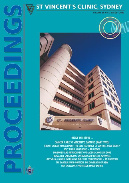
PROCEEDINGSST VINCENT’S CLINIC, SYDNEYINSIDE THIS ISSUE ...VOLUME 10 No:1 AUGUST <strong>2002</strong>CANCER CARE ST VINCENT’S CAMPUS (PART TWO)BREAST CANCER MANAGEMENT: THE NEW TECHNIQUE OF SENTINEL NODE BIOPSYSOFT TISSUE NEOPLASMS – AN UPDATEDIAGNOSIS AND MANAGEMENT OF BLADDER CANCER IN <strong>2002</strong>RENAL CELL CARCINOMA: OVERVIEW AND RECENT ADVANCESLARYNGEAL CANCER: INCREASING ROLE FOR CONSERVATION – AN OVERVIEWTHE SANDRA DAVID ORATION: THE GOVERNOR OF NSWHER EXCELLENCY PROFESSOR MARIE BASHIRST VINCENT’S CLINIC DARLINGHURST, SYDNEY
The Mission of <strong>St</strong> Vincent’s<strong>Clinic</strong> FoundationThe people of Sydney and beyond are fortunate to benefit from one of the mostcomprehensive health care services available at the <strong>St</strong> Vincent’s Campus atDarlinghurst. These facilities are part of the 34-strong health and aged carefacilities under the direction of the Sisters of Charity of Australia.Integral to the Darlinghurst campus is the services and facilities provided by <strong>St</strong>Vincent’s <strong>Clinic</strong> and <strong>St</strong> Vincent’s <strong>Clinic</strong> Foundation. The aim of <strong>St</strong> Vincent’s <strong>Clinic</strong>and <strong>St</strong> Vincent’s <strong>Clinic</strong> Foundation are patient care, medical teaching and clinicalresearch. The three aims are interlinked and each serves to strengthen the others.Established in 1992, <strong>St</strong> Vincent’s <strong>Clinic</strong> Foundation provides funds and support formedical research into matters of clinical significance as well as providing support forpublic and medical education.Every advance in medical science has started with a commitment by a medicalpractitioner or a scientist to alleviating the pain and suffering of mankind.Australia is rich in research bodies which focus on clinical laboratory based research.However, funding is sparse for research conducted in the course of patient care. This iswhere <strong>St</strong> Vincent’s <strong>Clinic</strong> Foundation can focus some of the community’s goodwill.Since 1992, the Foundation has spent over $2.5 million and provided financialsupport for over 100 research projects. The Foundation has successfully supported vitalresearch into disease and illness including cancer, diabetes, kidney disease, heartdisease, arthritis, mental health, youth suicide, deep vein thrombosis and obesity, toname just a few. Additionally the Foundation supports research into the function ofgenes and cells in many diseases. The Foundation also provides financial support formedical students who wish to undertake research during their study.The Foundation depends on donations to continue to support this importantresearch.We need your support to assist <strong>St</strong> Vincent’s <strong>Clinic</strong> Foundation to continue to providefinancial support to our researchers.
ST VINCENT’S CLINIC, SYDNEY VOLUME 10 No:1 AUGUST <strong>2002</strong>PROCEEDINGSEditorialDr John H. O’Neill MD, FRACPConsultant Neurologist, Editor,<strong>Proceedings</strong> 2ArticlesBreast Cancer Management: The NewTechnique of Sentinel Node Biopsy –Evolution or RevolutionPaul Crea FRCS, FRACSSurgical Oncologist<strong>St</strong> Vincent’s <strong>Clinic</strong> 3Soft Tissue Neoplasms – An Update:Head and Neck Sarcomas – The <strong>St</strong>Vincent’s ExperienceMichael Jensen MBBS, FRACS, FACSVisiting Surgical Oncologist<strong>St</strong> Vincent’s <strong>Hospital</strong> 9Diagnosis and Management of BladderCancer in <strong>2002</strong>Gordon O’Neill MBBS, FRACS (Urol)Consultant Urologist<strong>St</strong> Vincent’s <strong>Clinic</strong> 15Renal Cell Carcinoma: Overview andRecent AdvancesRaji Kooner MBBS (Hons) (Syd),FRACSUrological Surgeon<strong>St</strong> Vincent’s <strong>Clinic</strong><strong>St</strong> Vincent’s <strong>Hospital</strong> 20Laryngeal Cancer: Increasing Role forConservation – An OverviewIan Cole FRACS, FRCSOtolaryngologist Head and NeckSurgeon<strong>St</strong> Vincent’s <strong>Hospital</strong> 24The Sandra David Oration: 30The Governor of NSWHer Excellency Professor Marie BashirCOPYRIGHTAll literary matter in the Journal iscovered by copyright, and must notbe reproduced, stored in a retrievalsystem, or transmitted in any formby electronic or mechanical means,photocopying, or recording, withoutwritten permission.BOARD OF DIRECTORSDr Brett Courtenay (Chairman)Sr Margaret Beirne RSCSr Suzette Clark RSCMs Maureen McCabeProf Terence CampbellMr John WilcoxMs Patricia TysonBOARD OF TRUSTEESMr Ted Harris AC (President)Sr Mary Bernice Elphick RSC AM OBEMr Peter Ferris AMMrs Leith Myerson MBEMr David MeagherMrs Roslyn PackerMr Arthur Fitzerald AMMr Peter FalkSr Margaret Beirne RSCMr <strong>St</strong>even RubicMs Michelle WilsonDr Brett CourtenayDr Thomas HughProf Douglas Tracy AOProf John Hickie AOS T V INCENT’ S C LINICEXECUTIVE DIRECTORMs Michelle WilsonMEDICAL COUNCILDr Peter BentivoglioDr Janet RimmerDr Judith FreundDr Katherine SamarasDr Phillip BrennerDr Jock HarknessS T V INCENT’ S C LINIC FOUNDATIONSCIENTIFIC COMMITTEEDr Brett CourtenayDr Thomas HughProf Douglas Tracy AOProf John Hickey AODr Dudley O’SullivanDr David GolovskyADDRESSST VINCENT’S CLINIC438 Victoria <strong>St</strong>reet, Darlinghurst, Sydney, NSW 2010, AustraliaPhone: (02) 8382 6222 Fax: (02) 8382 6402ST VINCENT’S CLINIC, PROCEEDINGS VOLUME 10 NO: 1 AUGUST <strong>2002</strong> 1
EDITORIALDr John O’Neill MD, FRACPCONSULTANT NEUROLOGISTEDITOR, PROCEEDINGSThis Issue is the second of a 2-part series (See “<strong>Proceedings</strong>”,August 2001) on certainaspects of cancer care on <strong>St</strong>Vincent’s Campus.The first article by Dr Paul Creacritically analyses “Sentinel NodeBiopsy”, a new technique crucial todecision-making in breast cancermanagement. Recent literature hadsuggested that if, at initial surgery, therewas no evidence of cancer involving thesentinel node of the axilla then survivalfollowing mastectomy alone was noworse than if this procedure wascombined with surgical axillaryclearance (radical mastectomy).Avoidance of the latter procedure wouldobviously improve cosmetic outcomeand greatly reduce morbidity for thepatient. Dr Crea reviews this literatureand describes his own carefuldevelopment of the technique at <strong>St</strong>Vincent’s <strong>Clinic</strong>. He reports his personalmanagement of 200 consecutive cases ofbreast cancer, using the technique ofsentinel node biopsy, over a period frommid-1999. This is a scholarly work andepitomises excellence in a combinedclinical and research project which hasenormous practical importance forwomen who develop breast cancer.Dr Michael Jensen reviews theliterature on soft tissue neoplasms and,in particular, malignant sarcomas. Hereviews his personal data in managementof 83 consecutive cases over a 15-yearperiod. This experience equates to 20 –25% of all cases seen in NSW over asimilar period of time. Dr Jensen’sexperience in this field is clearlyremarkable and his own personal resultsappear to be equally as good as havebeen reported in the internationalliterature.Drs Gordon O’Neill and Raji Koonerof the Urology Department of <strong>St</strong>Vincent’s <strong>Clinic</strong> have producedexcellent reviews on the diagnosis andmanagement of bladder cancer (DrO’Neill) and renal cell carcinoma (DrKooner).Dr Ian Cole, ENT Surgeon, hasreviewed the literature on managementof laryngeal cancer, incorporating hisown personal experience (Table 2). Thearticle emphasises current surgicaltechniques aimed at maximising survivalbut also conserving laryngeal functionand hence quality of life for patients whosuffer with this condition.This year, the 8th Sandra DavidMemorial Lecutre was given by theGovernor of NSW, Her ExcellencyProfessor Marie Bashir. She discusses thechallenge society faces in the area ofmental health (especially depression) inyoung people. Having been both aclinician (Psychiatrist) andadministrator in mental health,especially involving children and youngpeople, her insights and reflections onthis problem make excellent reading andfood for thought.Established in 1992, the <strong>St</strong> Vincent’s<strong>Clinic</strong> Foundation provides funds andsupport for medical research as well asfor public and medical education. Sinceinception, the Foundation has spentover $2.5 million for these purposes,supporting over 100-research projects.Much of the hard fundraising for thisachievement has been, and continues tobe, undertaken by the <strong>St</strong> Vincent’sPrivate <strong>Hospital</strong> Ladies Committee. Atthe end of this Issue is a list of therecipients of the <strong>2002</strong> Research Grants.These projects are currently under study.P RIVACYS TATEMENT<strong>St</strong> Vincent’s <strong>Clinic</strong> distributes<strong>Proceedings</strong> to keep you up to date aboutresearch activities at <strong>St</strong> Vincent’s <strong>Clinic</strong>.<strong>St</strong> Vincent’s <strong>Clinic</strong> collects personalinformation from its donors and otherinterested supporters to provide you withinformation about our activities.We value your privacy and treat anyinformation we hold as confidential.We don’t share your information withany third parties, unless we are requiredto by law or on a confidential basis toagents that we use in the ordinaryoperation of our business (such as fordata processing, printing or mailing).We have developed a privacy policywhich has more information about howwe deal with privacy issues.You have a right to obtain access tothe information we hold about you. Ifyou would like to access thatinformation, or if you would like a copyof our privacy policy or moreinformation abut <strong>St</strong> Vincent’s <strong>Clinic</strong>,please contact us on 8382 6405.2 ST VINCENT’S CLINIC, PROCEEDINGS VOLUME 10 NO: 1 AUGUST <strong>2002</strong>
Dr Paul CreaBreast Cancer ManagementThe new techniques of sentinelnode biopsy – evolution or revolutionI NTRODUCTIONThe latest figures from the NSWCancer Registry show that in 1999there were 3,493 new cases of breastcancer diagnosed, of which 25 weremale. Breast cancer accounted for27% of female cancers and led to820 deaths (16% of female cancerdeaths). Over the past 30 years,there has been a gradual increase inincidence from 51 to 77 per100,000. The death rate however,has fallen from 18.5 to 16.1 per100,000, probably representing theonset of population screening andimproved treatment. However, thereremains an overall risk for a femaledeveloping breast cancer of 8.9% (1in 12) by the age of 75. Currently,there are about 35,000 women inAustralia living with breast cancer,their average 5 year survival being79%.There is a high level of socialawareness in Australia of both thesefigures and the consequences ofsurgery, radiotherapy andchemotherapy. This has led tostrong calls for efforts to be made tosignificantly improve both.Dr Paul Crea, FRCS, FRACSSurgical Oncologist<strong>St</strong> Vincent’s <strong>Clinic</strong>T HE B REAST –M ASTECTOMYOn a September morning in1811, Fanny Burney, anEnglish essayist living inParis, underwent amastectomy in Paris without the benefitsof anaesthesia. A vivid account of herterrifying ordeal is preserved in lettersshe wrote to her sister in London. Shesurvived the operation and the diseasefor many years.It was not until the 1880s thatWilliam Halsted, of Johns Hopkins<strong>Hospital</strong> in Baltimore, published alandmark report detailing the operationof radical mastectomy. This achieved thefirst significant success in the control ofbreast cancer. The surgery was extensiveand disfiguring but its benefit was soonevident and it became the operativeprocedure of choice and the standardtreatment of breast cancer for the next70 years. It was seen as vastly preferableto the alternative of a slow death fromthe consequences of advanced cancer.In the early 1950s, variousinstitutions began to question the needfor such extensive surgery andinvestigated the possibility of decreasingits extent without compromising theresults in terms of survival. Somemodifications to the standard radicalmastectomy succeeded such as thepreservation of the pectoralis majormuscle (modified radical or Patey’smastectomy). Others failed, such asattempts to conserve the breast withoutadding the safety of radiotherapy, whichled to an increased rate of localrecurrence.Today the most commonly usedbreast conservation procedure is the socalled“lumpectomy”. This is somewhatof a misnomer as it can lead to anautomatic presumption that thetreatment of the primary cancer in thebreast requires no more than theremoval of the lump itself. Theprocedure would better be termed a“partial” or “segmental mastectomy”.With the onset of breast screening andincreasing community awareness, thediagnosis of breast cancer is now beingmade at a much earlier stage. Thisrenders conservative surgery a suitableoption in more than 80% of casesresulting in an equal survival rate to theprevious more radical procedure and ithas become accepted as the standardalternative.ST VINCENT’S CLINIC, PROCEEDINGS VOLUME 10 NO: 1 AUGUST <strong>2002</strong> 3
T HEA XILLARYG LANDS –B ACKGROUND TOS ENTINEL N ODEB IOPSYOnce the primary cancer within thebreast is treated by either partial or totalmastectomy, the second arm in themanagement of breast cancer has alwaysbeen the removal of the axillary glands.This was done for the dual purpose ofdetermining whether spread hadoccurred to them and, potentially, asmanagement if involved glands werefound.Evaluation of data from large serieshas shown that the overall incidence ofaxillary metastases is 46%. 1 This,however, drops sharply with thedecreasing size of the primary tumour aswell as its characteristics, such as gradingand the absence of visible vascularinvasion.Two important factors, however, needto be considered. The first being thateven though the incidence of axillarymetastases can be up to 40%, theincidence of clinical uncontrolledaxillary disease if axillary dissection isnot performed is 21% 2 , indicating thathalf of the axillary lymph nodes maynever present as clinical disease. Thesecond factor is that axillary nodalinvolvement can still occur, thoughuncommonly, even in patients with earlyprimary tumours.Recurrence in the axilla after axillaryclearance is between 1 & 3%. 3 So,although axillary clearance is a veryeffective way of controlling thispotential for axillary disease, it commitsa large number of women, whose axillaryglands will prove to be free of metastaticdisease, to unnecessary surgery.The false negative rate of histologicalevaluation of the axillary glands isgenerally considered to be low, at lessthan 2%. However, this is most likely anunderestimation, in the absence ofimmunohistochemical testing. Also, thelong held practice that the status of theaxillary nodes would dictate thedirection of further adjuvant treatment isnow not as valid, with the nature of theprimary tumour now being considered anequally important factor in this decision.Some further inaccuracy in staging alsooccurs due to the lack of evaluation ofthe status of the internal mammarynodes which can harbour metastaticdisease in 10-20% of patients. (It is,however, accepted that routinedissection of the internal mammarychain will not improve survival.)The morbidity of axillary dissection iswell known and has been recentlysummarised in a publication from theNational Breast Cancer Centre. Itincludes:1) Seroma – this is common,uncomfortable and generally notinfluenced by the type and durationof drainage.2) Wound infection – an incidence ofabout 10% is reported though thismay not be significantly altered iflesser procedures are undertaken.3) Reduced shoulder mobility andstiffness – this will occur to somedegree in virtually all patients and itsseverity will depend on age and preexistingconditions, such as arthritis,as well as post-operative motivationand mobilisation.4) Paraesthesiae – this is virtuallyuniversal and is associated withdivision of the intercosto-brachialnerve, although its preservation willnot eliminate it. A small number ofpatients will experience debilitatingneuropathic pain resistant to standardforms of analgesia.5) Motor nerve damage – this can occurin four areas:(i) medial pectoral nerve: injury tothis is common during dissectionaround the pectoralis minormuscle and leads to wasting of itslower fibres though producingminimal morbidity.(ii) nerve to serratus anterior: damageto this will result in winging ofthe scapula.(iii) nerve to latissimus dorsi: damageusually results in minimalmorbidity with some associatedloss of power.(iv) brachial plexus palsy can occur infewer than 1% of cases but itspresentation is very dramatic inthe loss of all shoulder abductionpost operatively. Its cause isunknown, occurring even withthe most careful armmanagement during surgery.Virtually all cases will recoverhowever, within three months.6) Lymphoedema – this is the mostsignificant and well known side effectof axillary dissection. It is clinicallyobvious in about 10-20% of cases andprobably occurs, if measured, in 30-40%. It is well recognised in thecommunity and held in fear by allwomen, leading some to totally rejectaxillary surgery.All non-invasive attempts to assessthe status of the axillary glands,including clinical examination,mammography, ultrasound, CT, MRIand PET scanning have not succeeded.Clearly, a need existed to explore thepossibility of limiting axillary surgerywhilst maintaining safety. One of thefirst such reports was in the early 1980swhen Forrest in Edinburgh described atechnique of axillary (pectoral node)sampling. None of the 67 patients withnegative sampling converted to positiveon subsequent axillary dissection. 5S ENTINELN ODEB IOPSY –D EVELOPMENTThe “sentinel node” concept was firstdescribed over twenty years ago in thetreatment of penile cancer and,subsequently, in melanoma by DonaldMorton 6 who used an injection of bluedye to identify the gland. MethyleneBlue and fluorescent dyes were tried andwere unsuccessful. The two mostcommonly used dyes today areIsosulphan Blue and Patent Blue-V. Inthe management of breast cancer, thetechnique of sentinel node biopsy wasfirst reported by Krag in 1993 7 usingradionuclide scanning and by Giuliano 8in 1994, using blue dye and,subsequently, by Albertini 9 using acombination of both.In 1997, Veronesi 10 from theEuropean Institute of Oncology in Milanreported the largest study using lymphoscintigraphyand intraoperative use ofthe gamma probe. Many multinationalstudies since then have validated theconcept that the spread of cancer cells tothe axillary basin is not a random eventbut occurs sequentially, beginning with4 ST VINCENT’S CLINIC, PROCEEDINGS VOLUME 10 NO: 1 AUGUST <strong>2002</strong>
the sentinel node (or nodes) and then tohigher levels.In 1999, Cody 11 , of Memorial SloanKettering Cancer Center, reported theexperience of 1500 cases of sentinelnode biopsy from a single institution.This recognised the existence of a steeplearning curve and recommended thatan audit of each surgeon’s performancebe done to achieve a false negative rateof about 5%. Worldwide consensuscurrently suggests that each surgeonnewly undertaking this procedure shoulddo the first 30-40 cases by both sentinelnode biopsy and subsequent full axillarydissection to evaluate his level of skill.S ENTINEL N ODEB IOPSY – PERSONALE XPERIENCEThe remainder of this paper willanalyse the results of a personal series ofthe first 200 cases of sentinel nodebiopsy accrued since the introduction ofthis technique at <strong>St</strong> Vincent’s <strong>Clinic</strong> inmid 1999, following a visit to theEuropean Institute of Oncology.The initial requirement was theintroduction of a totally new procedurewithin the Campus setting. Thisinvolved the collaboration of variousdepartments and the study parameterswere that it would be carried out by asingle breast surgeon within a privatesurgical practice. To minimise variancesonly two Nuclear Medicine departmentswere used (<strong>St</strong> Vincent’s Campus {publicand private hospitals} and the Mater<strong>Hospital</strong>) and two pathology practices(Douglass Hanly Moir and Sydpath).Evaluation of the procedure would bejudged by the accuracy of threeparameters:1. accuracy of pre-operative radionuclidescanning (the choice and use ofisotope);2. accuracy of intraoperative localisation(the choice and use of the gammaprobe);3. accuracy of pathological testing(intraoperative frozen section).Evaluation of three end points wouldthen be made:Figure 1:Lymphoscintigraphy scanningfollowing injection of radionuclidecolloid.Figure 2: Lymphoscintogramshowing injection site (largehotspot) and Sentinel Node(small hotspot)Figure 3: “Navigator” Intraoperativeprobe used toidentify Sentinel Node site inlower axillary area.1. the reliability of the assessment of thesentinel node, as judged on frozensection and subsequent histologicalanalysis, in predicting the status ofthe remainder of the axillary glands;2. the incidence of local axillary bedrecurrence;3. long term prognosis (survival).M ATERIALS A NDM ETHODSRadionuclide Scanning: From variousliterature reviews a decision was madethat the best radiopharmaceutical was99m Technetium Antimony SulphideColloid having a particle size of 50-200nm, a half life of 6 hours and animaging energy of 140-150KeV. Itscharacteristics were optimal forabsorption into the lymphatic channelsand trapping within the interstitial spacewithin the lymph nodes. It would beST VINCENT’S CLINIC, PROCEEDINGS VOLUME 10 NO: 1 AUGUST <strong>2002</strong> 5
delivered by subdermal peri-areolar orperi-tumoural injections in 4 divideddoses of 0.05 – 0.1mls to a total dose ofbetween 10 – 50MBq. The area ofinjection would be massaged andscanning undertaken over the following90 minutes. The images acquired wouldthen be taken with the patients tosurgery (Fig 1 & 2).Intraoperative Localisation: There were4 different handheld probes evaluatedand the USSC Navigator was selectedand has been subsequently used in allcases (Fig 3). It has a peak sensitivity of140KeV (window of 120-150) and theresponse is transduced into an acousticsignal and digital read out.Figure 4: Intra-operative localisation of Sentinel Node.(Blue dye was concurrently used in 6cases, with injection after induction ofanaesthesia {Patent Blue V 2.5%} peritumourly,2mls diluted to 5mls withnormal saline.)Using the gamma probe and with thevisual aid of the scan, the sentinel nodesite was then identified intraoperatively(Fig 4) allowing either a small incisionto be made into the lower axillary skinor at the lateral end of the incision fortumours in the upper outer quadrant (Fig5). The active node (or nodes) wasremoved and tested away from thepatient (Fig 6) and then sent for frozensection. Lack of residual activity in thebiopsy cavity was then checked.Pathological Evaluation: There wereslight variations in the techniques offrozen section analysis. This was eitherdone by bisecting the node or taking2mm slices and examining the largest.After H & E staining, the whole face ofthe node was examined to allowvisualisation of the subcapsular area forpossible micrometastases. All residualtissue was then fixed in formalin andprocessed in routine paraffin blocks,staining with H & E and, if negative,proceeding to Keratin Immunoperoxidasestaining (Cam 5.2,cytokeratin 19, or AE1/AE3).The procedure of sentinel nodebiopsy was discussed at length withoperable patients. It was offered to allpatients with tumours less than 4cm insize and in the absence of multifocaldisease or palpable axillary nodes.(Initially, it was considered unsuitable incases of previous breast surgery or insituductal carcinoma; this view changedduring the course of the study.)Figure 5: Resection of identified Sentinel Node.After a full explanation of theprocedure and an opportunity to readsome literature, a full and informedconsent was obtained from each patientto either proceed to complete axillarydissection if the sentinel node was foundto be positive on frozen section, noaxillary dissection if negative, orvariations of these if the testing failed toshow a node, reflecting the patient’swishes. Two hundred cases were enteredinto the study. In the first thirty of these,total axillary dissection was plannedirrespective of the status of the sentinelnode unless otherwise requested by thepatient.The first 30:R ESULTSA total of 52 cases of primary breastcancer were seen in the 4 months to theend of November 1999. Of these, 22were excluded for various reasons and 30proceeded to sentinel node biopsy.Treatment of the breast tissue was bytotal mastectomy in 15 and partialmastectomy in the other 15. From these,24 patients proceeded to standardaxillary dissection. Of the remaining 6, 4requested sentinel node biopsy only and2, Level I-II dissection.Sentinel node was identified byscanning in 21 of 30 patients (70%). Afurther 6 were found by intraoperativegamma probe for a total localisation rateof 90% (27/30). In these 27 patients, thenode was found to be positive in 8, andall were confirmed on subsequent H &E. In the other 19, in whom the nodewas reported as normal on frozen section,3 converted to positive on H & E. Allwere in subcapsular foci only.Successful sentinel node localisationwas undertaken in 21 patients. Of the 86 ST VINCENT’S CLINIC, PROCEEDINGS VOLUME 10 NO: 1 AUGUST <strong>2002</strong>
Figure 6: Resected specimen (Sentinel Node) Activity confirmed away from operative field.TABLE 1: INSTITUTIONS AND PATHOLOGY SERVICES USEDNUCLEAR MEDICINE SCANS PERFORMED AT: %<strong>St</strong> Vincent’s <strong>Clinic</strong> 111 55.5<strong>St</strong> Vincent’s General 74 37Mater <strong>Hospital</strong> 15 7.5PATHOLOGY SERVICES %Douglass Hanly Moir 104 52Sydpath 96 48TABLE 2: DYE INJECTION SITETRACER INJECTION SITE %Peritumoral 142 71Subareolar/between Nipple and Tumour 49 24.5Around Hookwire 4 2Unknown 4 2Blue Dye 1 0.5in whom the node was positive, this wasthe only node positive in 6 (a total of130 other nodes removed were normal).In the other 2, only three of a total of 22nodes removed were positive.In the 13 patients with negativenodes on frozen section, 11 remainednegative on H & E. In the other 2, thefirst had 7 cancer cells found in asubcapsular focus, the other 32 nodesbeing negative and, in the second, one ofeight other axillary nodes was found tobe positive, representing a skipmetastasis.These preliminary results showed afalse negative rate on frozen section of15% (2/13) and a positive predictivevalue of the status of the axilla of 95%(20/21). These figures comparedfavourably with world results andencouraged proceeding with thetechnique.The first two hundred:The accrual of 200 cases took justover 2 years. During this period of timethere were continual refinements madeto the technique, though the basicstructure and methodology remainedunchanged. This report will detail apreliminary analysis of these cases.Rigorous efforts were undertaken witheach patient to provide them with abalanced explanation of its pro’s andcon’s. As time passed, individual patientsbecame much better informed of thenature of the procedure through themedia as well as by self-initiatedliterature and internet searching. Oftensecond visits were requested as eachpatient took time to examine theinformation supplied in an effort toarrive at the best decision. Of thosesuitable for selection, the vast majorityopted to undertake sentinel node biopsy.Table 1 shows the institutions where theprocedures were undertaken and thepathology services used.The performance of pre-operativeradionuclide visualisation became muchmore streamlined as departmentmembers refined the finer details of themode of injection, scanning andmassaging (especially difficult withhookwire localisation). Peri-areolarinjections became more common,especially in tumours of the upper outerquadrants, to allow distancing of theprimary hotspot from the sentinel node(Table 2). Surgical techniques were alsorefined as it soon became apparent thatthe sentinel node may not be within thevisualised Level I area of the axilla or bethe most obvious lower axillary node.Pre-operative scanning was successful inidentifying a sentinel node in 167 cases(83.5%). With subsequent use of theintra-operative probe, the sentinel nodewas identified in a further 20 cases thusincreasing the total success rate ofpickup to 93.5% (Table 3). The type ofoperative procedures undertaken on thebreast and the axillary glands were nowvirtually independent of each other, as isshown in Table 4. Breast conservingsurgery was undertaken in 69.5%(139/200). Sentinel node biopsy, withno further axillary dissection wasperformed in 61.5% (121/200). Twentyonepatients requested some modifiedform of lower axillary dissection, even ifthe sentinel node was negative.Results were analysed on an ongoingbasis and the first factor to be consideredwas the rate of false negative frozensections. This group was identified onsubsequent paraffin and immunoperoxidasestains (Table 5). In the 187cases in whom the sentinel node wassuccessfully identified, 154 were frozensection negative (82.4%) and one wasequivocal. Of these however, 17subsequently proved to be paraffinpositive (11%). On histopathology, 5 ofthis group had definite metastaticdeposits, usually being found in a nodalpole away from the cut surface, the other12 showed only small subcapsular foci.Of the 136 patients shown to be frozensection and paraffin section negative, 88were tested for keratin staining(immuno-peroxidase) and positivity wasfound in 5, again all were subcapsulardeposits only. In only one case was thegland found to contain widespreadreplacement and this was with lobularcarcinoma, whose cells are notoriouslydifficult to identify on frozen section.ST VINCENT’S CLINIC, PROCEEDINGS VOLUME 10 NO: 1 AUGUST <strong>2002</strong> 7
D ISCUSSIONTABLE 3: SUCCESS RATE – SENTINEL NODE IDENTIFICATIONThe technique of sentinel nodebiopsy has major physical andpsychological advantages and attemptsto address the commonly heldcommunity dread of lymphoedema. Italso significantly decreases morbidityand length of hospital stay. VariousNorth American centres are nowundertaking it as a day only procedureunder local anaesthesia.Several questions remainincompletely resolved. The first is thelong term safety of the procedure asjudged by the rate of local recurrence inthe axillary bed. In the first 100 cases, nolocal axillary recurrence was found attheir one year follow-up visit. Thesecond question would be the long-termassessment of end-point survival in thisgroup of patients compared to the timehonoured standard of modified radicalmastectomy. We also have to decidewhat level of false-negative rate isacceptable. A major unansweredquestion is what form of treatmentshould be recommended in the caseswhere only subcapsular metastatic fociare found. Their relevance in dictatingadjuvant treatment is unknown as theymay have always been present in thepast but were not identified due to thelack of immunoperoxidase staining –currently this group is recommended foradjuvant chemotherapy.Several international studies arecurrently being undertaken to try andresolve these matters but their resultswill not be available for several moreyears. As happened with laparoscopiccholecystectomy, the force of patientdemands may see the universalacceptance of this procedure well beforethat. If so, great care must be taken toavoid the pitfalls and the use of thisprocedure should be limited to thosewho are adequately experienced toundertake it.B IBLIOGRAPHY1. Carter C L, Allen C, Henson D E. Relation oftumour size, lymph node status, and Survival in24,740 breast cancer cases. Cancer 1989;63:181-72. Fisher B, Wolmark N, Bauer M, Redmond C,Gebhardt M. The accuracy of clinical nodalstaging and of limited axillary dissection as adeterminant of histological nodal status incarcinoma of the breast. Surg Gynecol Obstet1981; 152: 765-723. De Boer R, Hillen H F P , Roumen R M I I,Rullen I I JT , Van der Sangen M J C, VoogdNUCLEAR MEDICINE SCAN FOR SENTINEL NODE %Nodes Detected 167 83.5No Nodes Detected 32 16N/A (Blue Dye only) 1 0.5INTRA-OPERATIVE PROBE %Sentinel Node Found 187 93.5Sentinel Node Not Found 13 6.5TABLE 4: OPERATIVE PROCEDURESBREAST OPERATION %Partial Mastectomy 139 69.5Total Mastectomy 58 29Subcutaneous Mastectomy 3 1.5AXILLARY SURGERY %Sentinel Node Biopsy Only 121 60.5Level I-II Dissection 15 7.5Lower Axillary Sampling 6 3Total Axillary Dissection 58 29TABLE 5: SENTINEL NODE ANALYSIS – PRESENCE/ABSENCE OFMETASTATIC DEPOSITS IN SENTINEL NODE. ACCURACY OF RESULTS ONSUBSEQUENT LEVELS OF TESTINGFROZEN SECTION 187 %Negative 154 82.4Positive 32 17Equivocal 1 0.6PARAFFIN SECTION 154 %(Frozen Section Neg)Negative 136 88.3Positive 17 11.0Equivocal 1 0.7IMMUNOPEROXIDASE (Paraffin Neg) 136 %Negative 83 61Positive 5 3.7Not Tested 48 35.3A C. Detection, treatment and outcome ofaxillary recurrence after axillary clearance forinvasive breast cancer. Br J Surg 2001;88:118-1224. Veronesi U, Cascinelli N, Buffalino R,Morabitu A, Greco M, Galuzzo D et al. Risk ofinternal mammary node metastases and itsrelevance on prognosis in breast cancerpatients. Ann Surg 1983; 198:681-6845. Forrest A P M, <strong>St</strong>ewart H I, Roberts M M,<strong>St</strong>eele R I C. Simple mastectomy and axillarynode sampling (pectoral node biopsy) in themanagement of primary breast cancer. AnnSurg 1982; 196: 371-76. Morton D L, Wen D-R, Wong J H, EconomouJ S, Cagle L A, <strong>St</strong>orm F K, et al. Technicaldetails of intraoperative lymphatic mapping forearly stage melanoma. Arch Surg 1992; 127:392-97. Krag D N, Weaver D L, Alex J C, et al.Surgical resection and radiolocalisation of thesentinel lymph node in breast cancer using agamma probe. Surg Oncol 1993; 2. 335-408. Giuliano A E, Kirgan D M, Guenther J M, etal. Lymphatic mapping and sentinellymphadonectomy for breast cancer. Ann Surg1994; 220: 391-4019. Albertini J J, Lyman G H, Cox C. et al.Lymphatic mapping and sentinel node biopsyin the patient with breast cancer. JAMA1996; 276: 1818-2210. Veronesi U, Paganelli G, Galimberti V, VialeG, Zurrida S, Bedoni M, et al. Sentinel Nodebiopsy to avoid axillary dissection in breastcancer with clinically negative lymph nodes.Lancet 1997;349: 1864-711. Cody HS, Borgen PI. <strong>St</strong>ate of the artapproaches to sentinel node biopsy for breastcancer: study design, patient selection,technical and quality control at MemorialSloan Kettering Cancer Centre. Surg Oncol1999; 8: 8591.8 ST VINCENT’S CLINIC, PROCEEDINGS VOLUME 10 NO: 1 AUGUST <strong>2002</strong>
Dr Michael JensenSoft Tissue Neoplasms –An Update: Head andNeck Sarcomas – the<strong>St</strong> Vincent’s ExperienceO VERVIEWSarcomas are uncommon malignanttumours, of apparent mesenchymalorigin, accounting for 1% of allmalignancy. The majority in adultsarise within large muscle groups of theextremities; other sites include thebuttocks, chest wall, retroperitoneumand head and neck. The relative rarityand morphologic heterogeneity of softtissue neoplasms render them liable tomisdiagnosis and present a significantchallenge to pathologists andoncologists. Clear histologicaldistinction from both benign softtissue tumours as well as thelocally aggressive non-metastasisingmalignancies, is essential tomanagement. Diagnosis is aided bythe use of immunohistochemistry andgenetic markers, in addition to lightmicroscopy and ultrastructural studieswith electron microscopy. Over theperiod 1985 – 2000, I havedocumented 83 cases of soft tissuesarcoma of the head and neck treatedat <strong>St</strong> Vincent’s <strong>Hospital</strong>. Thisexperience will be detailed in thesecond part of this article.Dr Michael Jensen, MBBS, FRACS,FACSVisiting Surgical Oncologist<strong>St</strong> <strong>Vincents</strong> <strong>Hospital</strong>N ON –S ARCOMATOUS S OFTT ISSUE N EOPLASMSThis section of the articledescribes a variety of soft tissueneoplasms to be distinguishedfrom sarcomas.FibromatosesArising in the subcutaneous or deepfascia, an aggressive fibromatosis must bedistinguished from a true malignancy.On CT scanning, a peculiar strandingeffect extends from the nonencapsulated “tumour” mass. Althoughseen in limbs or trunk, fibromatosesmore commonly present in the head andneck (especially in the soft tissues at thebase of the neck or shoulder girdle),occasionally presenting submucosally,e.g. in the bucco-alveolar sulcus.Characterised by local infiltrativegrowth, a fibromatosis nevermetastasises. Spontaneous regression hasbeen reported. Often painful, theselesions are treated by simple excision. Ifsurrounding and involving nerves suchas the brachial plexus, a local course ofradiation therapy may be considered.The deep fibromatoses include:(i) abdominal wall and (ii) intraabdominaldesmoid tumours.The extra abdominal desmoid tumourusually arises in association withST VINCENT’S CLINIC, PROCEEDINGS VOLUME 10 NO: 1 AUGUST <strong>2002</strong> 9
girdles of adults (male dominant) usuallyin the fifth to seventh decades. Withrare exception, the lesion is solitary andasymptomatic and, if well excised, rarelyrecurs. 4 These tumours have a densepseudocapsule with a varying amount ofosteoid bone formation.Fig 2 is a CT scan of a 75 year oldmale depicting an ossifying neoplasmarising within the right axilla.ElastofibromaA degenerative “pseudotumour”, thisusually arises in elderly patients andmanifests as a slow growing solid mass offibroelastic tissue between the lowerportion of the scapula and chest wall. Itmay reach 5cm to 10cm in greatestdiameter. This reactive process is usuallyattached to the thoracic fascia,periosteum and ligaments of the 7th and8th ribs.Infiltrating AngiolipomaA benign, mixed mesenchymaltumour often mistaken for sarcoma, theinfiltrating angiolipoma of skeletalmuscle is subdivided into small, largeand mixed vessel types. It presents as apainful soft tissue mass, the large vesselvariant commonly found in the lowerlimb, occasionally in trunk and head andneck. This tumour is comparativelymore common than the rareangiosarcoma of muscle.These lesions do not metastasise andpose no threat to life. Surgery on suchlesions should be preceded byarteriography and may be associatedwith considerable deformity and loss offunction. 5,6,7Introduction:Figure 2 CT scandemonstrating extensivecourse calcification of amesenchymalchondrosarcoma.S ARCOMASA sarcoma usually presents as apseudoencapsulated, circumscribed mass.The pseudocapsule contains a mixture oftumour cells, compressed tissue andinflammatory cells with littledesmoplastic reaction. With increasingsize and tumour grade there is atendency for seeding of malignant cellsalong tissue planes of least resistance,including muscle or neurovascularbundles, well beyond the apparentpseudocapsule.Histological subtypes of sarcomaoccur in different age groups. A usefulrecommendation for the reporting of softtissue sarcomas was published by theAssociation of Directors of Anatomicaland Surgical Pathology. 8The typical paediatric sarcoma is arhabdomyosarcoma either of theembryonal or alveolar subtype. Theembryonal variety (70% to 80%) isparticularly chemotherapy sensitive andpresents mostly in the head and neck,especially the orbits and paranasalsinuses of young children (birth to 15years). It also occurs in the genitourinarytract and occasionally theretroperitoneum. In children, wherecomplete remission is not achieved withchemotherapy, the AMORE protocol isused. This protocol describes ablativesurgery, moulage technique withbrachytherapy and reconstructivesurgery. 9The alveolar type accounts for 10-20% of all rhabdomyosarcomas and isseen usually in the 10-30 year age group.Also encountered in the teenage to30 year age group are synovial,epithelioid and clear cell sarcomas aswell as mesenchymal chondrosarcomaand primitive neuroectodermal tumours.Over 40 years of age, thepredominant sarcomas are the malignantfibrous histiocytoma, liposarcoma,fibrosarcoma, leiomyo sarcoma,malignant nerve sheath nerve tumourand pleiomorphic rhabdomyo sarcoma.The majority of these sarcomas havea similar natural history, presentcommonly in the periphery, withprognosis largely determined by gradingand size.Recent studies with immunohistochemistryand genetic markers have ledpathologists to reclassify pleiomorphicsarcomas usually reported in the fortyplus age group. Many formerly regardedas malignant fibrous histiocytomas(M.F.H.), have been deconstructed tonow include leiomyosarcoma,liposarcoma and rhabdomyosarcoma.MFH becomes a diagnosis of exclusionbut has some identifiable features thatset it apart from other pleiomorphicsarcomas. 10,11In the elderly, a variety of aggressiveangiosarcoma commonly arises in thescalp, unusually may spread to lymphnodes and is invariably fatal with localuncontrolled disease after several years.The tumour presents as a vascularmalformation with poorly definedmargins often mistaken for simplehaemangioma. These lesions willrespond to radiation therapy with localcontrol in the treatment field butST VINCENT’S CLINIC, PROCEEDINGS VOLUME 10 NO: 1 AUGUST <strong>2002</strong> 11
inevitable recurrence at the margin. Asimilar result occurs with surgery.Primitive Neuroendocrine TumoursThis undifferentiated small, roundcell, mesenchymal neoplasm with littleevidence of ultrastructural specialisation,is commonly described as anextraskeletal Ewing’s sarcoma. Thesetumours share identical patterns ofproto-oncogene expression with Ewing’ssarcoma with common expression of theMIC2 gene. 12,13Commonly a paediatric malignancy,these tumours present in unusual sites inyoung adults but fortunately arechemotherapy sensitive. Protocols oftreatment involve extensive chemotherapyfollowed by radical ablativesurgery with local radiation therapy andconsolidation chemotherapy.Extraskeletal ChondrosarcomaAn aggressive malignancy with verypoor prognosis, the mesenchymalchondrosarcoma typically occur inthe15-35 age group. This tumour oftenpresents with a peculiar calcifiedappearance – a high grade variant of softtissue sarcoma. Local control may beachieved with radical surgery andradiation therapy. Distant soft tissue andbony metastases are common. 14A typical example of mesenchymalchondrosarcoma is shown in Figure 3.This tumour is to be distinguishedfrom low grade myxoid chondrosarcoma,which when completely excised, has anexcellent prognosis with a high degree ofpermanent local control and smaller riskof metastatic disease, e.g., chondrosarcomaof the larynx, (where 70% arisefrom the cricoid and 20-30% fromthyroid cartilage. 15 Most myxoidextraskeletal chondrosarcomas arise inthe deep tissues of the extremities ofolder patients, usually within themusculature.Surgical clearance of chondrosarcomais dependent upon the site ofpresentation, more difficult when arisingin relatively inaccessible sites such aspelvis or orbit.LeiomyosarcomaThis malignancy requires specialmention in two anatomical sub-sites.Superficial low grade leiomyosarcomas,presenting in the dermis orFigure 3 Typical example of mesenchymal chondrosarcoma.subcutaneous tissue, are usually cured bywide local excision. Where margins arelimited, radiation therapy is added postoperatively.16Leiomyosarcoma is the commonlyseen aggressive, retroperitonealmalignancy and often invades adjacentstructures. It may be possible to effect aradical resection, including the adjacentviscera and, when involved, majorvessels such as IVC or aorta, withappropriate vascular reconstruction.With limited margins, post operativeradiation therapy is planned. Refinedtechniques utilise removable, displacementprostheses to protect small bowel.Brachytherapy is used to supplementconventional external beam therapy.Leiomyosarcoma also arises from thewall of major vessels, most commonlythe inferior venacava (IVC). It may besuprahepatic (a cause of Budd ChiariSyndrome), or infrahepatic – above orbelow the renal vessels. InfrahepaticIVC leiomyosarcoma is potentiallyresectable with mobilisation androtation of the caudate lobe of liver andvascular reconstruction. Limited livermetastasis may occur and does notexclude long term survival.Diagnosis:M ANAGEMENTFine needle cytology of superficial softtissue tumour facilitates a diagnosis ofmalignancy and helps rule outmelanoma, lymphoma and metastaticcarcinoma.Precise histological typing andgrading usually require open surgicalbiopsy, although this is now possiblewith image directed core biopsy. Smalllesions, say under 2cms, may be excisedfor histological diagnosis. The majorityof tumours will require incision biopsythrough a judiciously placed incision.Histopathological difficulty arises wherepreoperative radiation andchemotherapy were used: this may alterprognostic information, where tumournecrosis is a particular problem.Treatment:Surgical resection remains the centraltheme of management of adult sarcomain clear distinction to the paediatricsarcomas, where the majority of tumoursare first treated with and responddramatically to chemotherapy,particularly those of round cell type.Local CT scanning and MRI imagingadd considerably to the pre-operativeplanning and management of sarcomas.Pulmonary CT scanning, to excludemetastatic disease, is undertaken prior tomajor ablative surgery.The high local recurrence rate thatfollowed simple excision of high gradetumours led to the development of “softpart” or compartment resection. Theprinciples of this technique, popularisedby L Bowden and R Booher in 1958 17 ,were originally derived from theconcepts of George Pack. Whereanatomically feasible, this excisionincludes wide ablation of the malignancywith at least one tissue plane12 ST VINCENT’S CLINIC, PROCEEDINGS VOLUME 10 NO: 1 AUGUST <strong>2002</strong>
macroscopically clear of the tumour,including the removal of entire musclebundles from point of origin to insertion,with sharp tissue plane dissection. Theresection encompasses, when necessary,neurovascular bundles. An improvedlocal control rate, up to 75%, isachieved. 18As an alternative, and to minimisedeformity, radiation therapy is applied tosupplement surgery, where deliberatelylimited surgical margins are applied. Awide “shrinking field” technique ofradiation therapy, pre or post surgery, isnecessitated to achieve the same highdegree of local control possible with“compartment” surgery.The “shrinking field” prescribes astandard tumour dose of 50Gy to enclosethe extended, potential, tumour bed,boosted by higher local peritumour dosesand was popularised by Herman Suit. 19Current limb sparing techniques, withmicrovascular free grafts forreconstruction, significantly reducemorbidity.A description of:a. tumour site and depth – dermal,subcutaneous, intramuscular,retroperi-toneal,b. type of resection,c. size of tumour,d. histological grading ande. the status of surgical margins:provide useful information to thesurgical oncologist in determining thenecessity for adjuvant radiation therapyand/or further surgery.Amputation is restricted to grosscircumferential tumours, particularlywhere bone invasion is involved.Hemipelvectomy, forequarter andhindquarter operations are limited torelatively rare tumours.is minimal with most internationalstudies unable to demonstrate asignificant survival advantage for the useof adjuvant chemotherapy. Programsusing chemotherapy with high gradesarcoma are currently being evaluated.Modern efforts in adult sarcoma therapyconcentrate on clever reconstructivesurgery and new techniques of radiation.In the future, intensity modulatedradiation therapy using beams that aremodulated during the delivery of thetreatment field have great potential forthe treatment of sarcomas in relativelyinaccessible sites, particularly the headand neck, utilising radiated volumes in ahighly contoured fashion and sparingvital structures. 20S ARCOMAS OF THEH EAD AND N ECK:T HE S T V INCENT’ SE XPERIENCEIn our combined series from the Headand Neck group at <strong>St</strong> Vincent’s, 83 headand neck soft tissue sarcomas werereported in the 15 year period from 1985to 2000. 21This compares with historical datafrom the New South Wales CancerRegistry, which records 538 patientswith the diagnosis of head and necksarcoma in the 25 year period 1970 –1995. This includes all soft tissue andbone sarcomas. During the same period,208 deaths were recorded. There were asmall number of paediatric tumours andteenage malignancies with the vastmajority of sarcomas arising in the 50plus age group.This adult hospital excludes patientsunder 15. As shown in Fig. 4, there was aclear bimodal distribution of patients inthis series.A significant group, particularly malesin the 15 to 30 year age group, presentedwith the diagnosis of alveolarrhabdomyosarcoma. This group didpoorly in spite of aggressive combinationtreatment with pre-treatment chemotherapy,surgery and radiation therapy. Alocal control rate of only 40% at 3 yearswas achieved.Compartment excision, as earlierdefined, was rarely possible in the headand neck. Low grade sarcomas under3cms in size were treated by surgicalexcision alone. This particularly appliedto tumours arising in the scalp and neckand included dermatofibrosarcomaprotuberans and small superficialleiomyosarcomas. 22Topographical data in our seriesdemonstrated the vast majority of headand neck sarcomas involved the softtissues of the scalp and neck. Thedifferent pathological subtypes areshown in Fig. 5. The most commonpathology was MFH and angiosarcoma,with a smaller number of leiomyosarcoma,pleiomorphic rhab-domyosarcomaand dermatofibrosarcomaprotuberans. Wide excision andradiation therapy were used for all highgrade tumours. 22, 23S UMMARYThe vast majority of sarcomas presentin the periphery. Using careful planningtechniques, especially MRI scanning, ahigh degree of local control is achieved.The difficult tumours, such asretroperitoneal sarcoma and the highgrade relentlessly recurring variants arehighlighted.Although chemotherapy is themainstay of treatment for paediatricmalignancy, its impact in adult sarcomasFigure 4ST VINCENT’S CLINIC, PROCEEDINGS VOLUME 10 NO: 1 AUGUST <strong>2002</strong> 13
The poorest subgroup were the 10elderly patients with angiosarcomas,mostly of the scalp and face whodemonstrated relentless local recurrentdisease. Although temporary infieldcontrol could be achieved by wide fieldsof radiation therapy and surgery, diseaseinevitably recurred at the periphery ofthe treatment fields. Many patientsdeveloped lymph node metastases, mostdied with aggressive local behaviourinvading orbit, ear canal, oral cavity, etc.Our current therapy for these patientsuses wide field conformal radiationtherapy to cover a large area of the headand neck, aiming for local control. 24, 25Chondro-osseous tumours were aninteresting subgroup of 14 patients. Thechondro-osseous group were dominatedby chondrosarcomas with the largestnumber involving the cricoid. Most werelow grade tumours with an excellentprognosis where complete excision wasusually possible.C ONCLUSIONIf we exclude the two poorlybehaving groups (the 15-30 year old agegroup with alveolar rhabdomyosarcomaand the elderly patients withangiosarcoma), the remaining majority(65 patients with head and necksarcoma) have a similar prognosis topatients with sarcoma arising in theperiphery, with a high local control rateapproaching 70%.Figure 5Our experience concurs with theliterature and highlights theheterogeneity of soft tissue neoplasms, aspresented in this article. The importanceof histological diagnosis and thenecessity to distinguish benign soft tissuetumours and the aggressive, locallyinvasive malignancies has been stressed.Surgery, either as a stand alonecompartment excision or with plannedradiation therapy utilising brachytherapyin combination with external beam,remains the most important modality inthe management of adult sarcoma.R EFERENCES1. Jones I T, Jagerman D G, Fazio V W. et alDesmoid Tumours in Hereditary Polyposis.Ann Surg 204: 94, 1986.2. Weiss, SW, Rao,VK, Well DifferentiatedLiposarcoma (atypical lipomatous tumours) ofdeep soft tissues of the extremities, retroperitoneum,and miscellaneous sites ; a follow upstudy of 92 cases with analysis of the incidenceof dedifferentiation, Am. J.of Surg.Path., Vol16: 1051 – 1058, 1992.3. McCormick,D, Mentzel,T, BehamA,Fletcher,CDM: Dedifferentiated liposarcoma;clinicopathological analysis of 32 casessuggesting a better prognostic group amongpleomorphic sarcomas, Am. J. Surg. Pathol,Volume 18, 1213 – 1223, 1994.4. Goodlad J R and Fletcher C D M, RecentDevelopments in Soft Tissue Tumours,Histopathology 27, 103-120, 1995.5. Allen P W and Enzinger,FM. Haemangiomaof skeletal muscle, An analyses of 89 cases,Cancer 29; 8 – 29, 1972.6. Dionne G P, and Seemayer T A, Infiltratinglipomas and angiolipomas revisited, Cancer33, 732 – 738, 1974.7. Vasantha K Rao ,M D and SharonWeiss, MD. Analysis of Histological Features and<strong>Clinic</strong>al Outcomes in 51 Cases. TheAmerican Journal of Surgical Pathology,Volume 16, 764 – 771, 1992.8. Recommendations for Reporting Soft TissueSarcoma. Am.J. Clin. Pathol. 111. : 594 – 598,19999. Schouwenberg P F, Kupperman D, Bakker F P,Blank L E, de Boer H B, New combinedtreatment of surgery, radiation therapy andreconstruction in head and neck,rhabdomyosarcoma in children, the AMOREProtocol. Head and Neck, Vol. 20, 283 – 292,July 1998.10. Christopher D.M. Fletcher, PleiomorphicMalignant Fibrous Histiocytoma : Fact orFiction? Am. J. Surg. Pathol. 16(3): 213-228,1992.11. SharonWeiss and Hiroshi Iwasaki:Pleiomorphic Sarcomas, Current Issues: XXIIICongress of the International Academy ofPathology. Oct 15, 2000. Nagoya Japan, 2000.12. Ambros I M, Ambros P F, <strong>St</strong>rehl S et al.MIC2 is a specific marker for Ewing’s sarcomaand peripheral primitive neuroectodermaltumours. Cancer; 67, 1886 – 1893, 1991.13. <strong>St</strong>evenson A J, Chatten J, Bertoni F,Miettinen M. CD99 (p30/32mic2)neuroectodermal /Ewing’s sarcoma antigen asan immunohistochemical marker. Appl.Immunohistochem. 2; 231-240.14. Yasuaki Nakashima MD, Krishnan K, Unni ,MB, BS, Thomas C, Shives, MD, Ronald G,Swee, MD, David C, Dahlin MD,Mesenchymal Chondro-sarcoma of Bone andSoft Tissue, Cancer 57; 2444-2453, 1986.15. <strong>St</strong>ephen Rodrigues, <strong>St</strong>uart Miller and AndrewWhyte, Chondrosarcoma Of The Larynx,Aust. J. Otolaryngology. Vol. 4, (1), 47-49,January 2001.16. I.Dahl and L. Angervall, Cutaneous andSubcutaneous Leoimyosarcoma A <strong>Clinic</strong>aland Pathological <strong>St</strong>udy of 47 Patients, Path.Europe Vol. 9 : 307 – 315, 197417. Bowden L, Boohor R J, The Principles andTechniques of Resection of Soft Parts forSarcoma. Surgery, 44. 963 – 977, 195818. El Castro, MD: <strong>St</strong>even I. Hajdu, MD: JosephA. Fortner MD: New York. Surgical ExcisionFibrosarcoma of the Extremities. Archives ofSurgery Vol. 107, 284-286 1973.19. Suit H, Mankin H, Wood W, et al: Radiationin the treatment of primary soft tissuesarcoma. Cancer 55, 265 – 67, 1985.20. Kenny L, Peters L, Roger A, et al ModernRadiotherapy for Modern Surgeon; AnUpdate, ANZ J. of Surg., Vol 72 131 -137,<strong>2002</strong>.21. Sarcomas of the Head/Neck, Jensen et al, inpress.22. Snehal G. Patel ,MS,FRCS, Ashok R.Shaha,MD, FACS, and Jatin P. Shah, MD,FACS, FRCS (Hon). Soft Tissue Sarcomas ofthe Head and Neck: An Update Am. J. ofOtolaryng., Vol 22 (1), 2-18, 2001.23. Dagher R, Helman L, Rhabdomyosarcoma;An Overview . Oncologist, Vol 4 (1),34-44,1999.24. Bullen R, Larson P O, Landeck A E, et al.Angiosarcoma of the head and neck managedby biopsies and radiation therapy, Dermatol.Surg. Vol 24 (10), 1105 – 1110, 199825. Veness M and Cooper S. Treatment ofcutaneous angiosarcomas of the head andneck. Aust. Radiol. Vol 39 (3), 277 – 281,Aug 1995.14 ST VINCENT’S CLINIC, PROCEEDINGS VOLUME 10 NO: 1 AUGUST <strong>2002</strong>
Dr Gordon O’NeillDiagnosis and Managementof Bladder Cancer in <strong>2002</strong>Dr Gordon O’Neill MBBS, FRACS(Urol)Consultant Urologist<strong>St</strong> Vincent’s <strong>Clinic</strong>I NTRODUCTIONThe treatment of bladder cancerremains challenging despitesignificant improvements inpreventing disease progressionand improving survival. It represents aspectrum from superficial welldifferentiated disease to highlymalignant tumours with a dismalprognosis. Bladder cancer is the fourthmost common cancer in men and theeighth most common cancer in women.The overall incidence has increased indeveloped countries in the last 30 years,yet mortality rates have declined. This isprimarily due to the positive impact ondisease progression with intravesicalimmunotherapy.The median age at diagnosis is 65 andtransitional cell carcinoma (TCC) israrely diagnosed before the age of 40.TCC however has been reported inadolescents and tends to be welldifferentiated. Males are affected threetimes more than females. The diseasetends to be more aggressive in Afro-Americans and has a higher incidence incities compared with rural populations.Tobacco smoking is the mostimportant risk factor, accounting for upto 50% of TCCs. Other risk factorsinclude exposure to polycyclic aromatichydrocarbons and benzene productsfound in the dye and leather industry.Hairdressers and painters are at greaterrisk of developing disease.Cyclophosphamide has been linked tosecondary TCC and phenacatin, whilstmore commonly linked to upper tractTCC, has been implicated in thedevelopment of bladder cancer. There isan unclear association with caffeine andsaccharin.ST VINCENT’S CLINIC, PROCEEDINGS VOLUME 10 NO: 1 AUGUST <strong>2002</strong> 15
Squamous cell carcinoma accounts for8% of primary bladder malignancies andis usually associated with chronicinflammation. Its incidence is high (upto 30%) in Egypt where schistosomiasisis prevalent. Adenocarcinoma isextremely rare (1-2%) and is usuallyassociated with bladder extrophy orurachal remnants.Figure 1: Photo ofbladder tumour on IVPP RESENTATIOND IAGNOSISANDHaematuria is the presentingsymptom in 90% of patients withbladder cancer and may be gross ormicroscopic. Other common symptomsinclude urinary frequency, urgency, loinpain from ureteric obstruction andurinary tract infection. Patients withhaematuria, whether macroscopic ormicroscopic, should be fully assessedwith a complete history and physicalexamination. Urine should be sent formicroscopy, culture and cytology. Theupper tracts should be assessed with anIVP (Figure 1) and renal ultrasound inpreparation for cystoscopy. Tumoursfound at cystoscopy (Figure 2) arecompletely resected where possible andrandom biopsies are necessary if cytologyis positive and there is no obviousmacroscopic tumour. Following thediagnosis of bladder cancer, otherinvestigations are necessary, such asFBC, UEC, LFTs and chest x-ray. Inhigher grade and stage disease CTscanning may reveal the extent of localdisease, the presence oflymphadenopathy and visceralmetastases. Patients with an elevatedalkaline phosphatase should also beevaluated with a bone scan.Unfortunately, understaging is commonand many patients with locally advanceddisease have micrometastatic disease atthe time of diagnosis.G RADE AND S TAGEAn understanding of the histologicalgrading and staging of TCC isimperative in its management. Tumoursare usually divided into three grades:grade 1 tumours being welldifferentiated; grade 2 moderatelydifferentiated; and grade 3 poorlydifferentiated. Grade (G) is particularlyFigure 2: Photo ofbladder tumour downcystoscopeimportant in superficial tumours as aprognostic sign for recurrence andprogression. Grade 1 tumours are rarelyinvasive and most invasive bladdertumours are high grade.Carcinoma in situ (CIS) is a highgrade flat tumour confined to thebladder mucosa which may involve largeareas of the bladder. Aggressivetreatment is warranted as 54% ofpatients will have progressed to invasivedisease at five years. 5% of patients withCIS will have metastatic disease at thetime of presentation.The most common and useful stagingsystem is the TMN system which wasfurther modified in 1997. The T stagesare particularly important, each subcategory pertaining to a differentprognosis and treatment strategy. TAtumours are confined to the bladdermucosa, T1 have invaded superficiallyinto the lamina propria and T2 tumoursare muscle invasive. T3 tumours are nowdefined as those that extend beyond thebladder to involve the perivesical fat(see Table).T HEN ATURALH ISTORY OF TCC74% of TCCs of the bladder aresuperficial at the time of diagnosis. Ofthese 70% are stage TA and 30% arestage T1. Metastatic disease is found in5-20% of patients at diagnosis and isusually associated with invasive localdisease. Approximately 5% of patientswill have upper tract tumours at the timeof diagnosis which may increase to 20%within five years. Superficial bladdercancer is usually associated with a defect16 ST VINCENT’S CLINIC, PROCEEDINGS VOLUME 10 NO: 1 AUGUST <strong>2002</strong>
of the entire bladder mucosa and as aresult recurrences are common. Despitecomplete tumour resection, two thirds ofpatients will develop recurrences withinfive years and 88% by 15 years.Progression from superficial bladdercancer to deep muscle invasion occurs in15% of patients with regular endoscopicfollow up. The natural history ofinvasive disease treated with localtherapy alone results in distantmetastases within two to three years inthe majority of patients. Metastaticdisease is often fast growing and lethalwith five year survival rates less than5%.The incidence of lymph nodeinvolvement in bladder cancer is linkedto a number of prognostic factors, stageand grade being the most important: T15%; T2 30%; T4 50%. Other prognosticfactors include tumour size, tumourmultiplicity and the presence of CIS.Many biological markers have beenexamined and p53 antigen expressionappears to be the most clinically useful.Increased concentration of “wild” typep53 has been shown to predict asignificantly increased risk of recurrenceand death and may be useful in future toidentify a sub population of patients withparticularly aggressive tumours who maybenefit from adjuvant therapy.M ANAGEMENTOFS UPERFICIALB LADDER C ANCERGeneral OverviewThe standard treatment of stage TAbladder cancer is complete endoscopicresection with or without intravesicaltherapy. Intravesical therapy is indicatedwhen there is diffuse bladder mucosalinvolvement or frequent recurrences.Recurrence rates are related to the gradeof the tumour. 80% of grade 3 tumourswill have recurred by three years.<strong>St</strong>age T1 bladder cancer is usuallymanaged with transurethral resectionand adjuvant intravesical chemotherapyor immunotherapy. Grade is highlysignificant as 50% of patients withT1G3 tumours will progress to muscleinvasive disease with regular follow up.In these patients, consideration shouldbe given to early radical cystectomyCurrent TMN <strong>St</strong>aging for Primary Bladder Cancer, includingModifications made in 1997Primary Tumour (T)TxTOTaTisT1T2T3T4Primary tumour cannot be assessedNo evidence of primary tumourNon invasive papillary carcinomaCarcinoma in situTumour invades subepithelial connective tissueTumour invades muscleT2aT2bbefore the onset of muscle invasivedisease which is associated with a greaterincidence of micrometastasis at the timeof operation.CIS should be treated aggressivelyand is often associated with invasivedisease. Ideally, all visibly affected areasshould be resected or cauterised, but thisis not always possible. The mostcommon agent used to treat CIS isBacille Calmette-Guerin (BCG).I will now discuss in more detailintravesical chemotherapy andimmunotherapy.Tumour invades (inner half) superficial muscleTumour invades deep muscleTumour invades perivesical tissueT3aT3bMicroscopicallyMacroscopically (extravesical mass)Tumour invades adjacent structuresT4aT3bRegional Lymph Nodes (N)NxNON1N2N3Distant Metastasis (M)MxMOM1Tumour invades prostate, uterus, vaginaTumour invades pelvic wall, abdominal wallRegional nodes cannot be assessedNo regional lymph node metastasisMetastasis in a single lymph node, 2cm or less in greatest dimensionMetastasis in a single lymph node, more than 2cm, not more than 5cm ingreatest dimension; or multiple lymph nodes, none more than 5cm ingreatest dimensionMetastasis in a lymph node more than 5cm in greatest dimensionDistant metastasis cannot be assessedNo distant metastasisDistant metastasis presentIntravesical TherapyIntravesical therapy for themanagement of bladder cancer wasintroduced in the late 1950s with thespecific goals of eradicatingexisting/residual tumour, preventingrecurrence of tumour after completetumour resection and preventingprogression of disease.The suspected biological behaviour ofthe patient’s tumour remains animportant factor in the decision ofintravesical therapy. In general,intravesical therapy is not required forgrade 1 TA lesions which have aST VINCENT’S CLINIC, PROCEEDINGS VOLUME 10 NO: 1 AUGUST <strong>2002</strong> 17
progression rate of
Bladder Preservation TreatmentOptionsThere has been much interest inalternative ways of treating bladdercancer aimed at bladder preservation.Monotherapy options include limitedsurgical procedures such as transurethralresection of tumour (TURT) alone andpartial cystectomy. Both of these optionsare only applicable to patients with smallunifocal tumours and are not advised asdefinitive treatment for most patients.Chemotherapy alone, whilst oftenachieving a complete response in somestudies, has been found not to bedurable.Using radiotherapy as monotherapyin bladder preservation includes bothinterstitial brachytherapy and externalbeam radiation. Interstitialbrachytherapy is suitable for only a smallsub-set of patients and when used incombination with external beamradiotherapy is limited to those havingtumours
Dr Raji KoonerRenal Cell Carcinoma:Overview and RecentAdvancesI NTRODUCTIONRenal cell carcinoma (RCC) isprimarily a surgically managed disease.It has multiple potential presentingsymptoms and signs. The diagnosis isoften made incidentally, mostcommonly during an ultrasound orCT scan for abdominal discomfort.Surgical treatment can be technicallychallenging and efforts to treatadvanced disease have been at theforefront of medical and surgicaloncology. There have been excitingadvances in the development of newminimally invasive strategies for themanagement of patients withlocalised kidney carcinoma. Theadvent of laparoscopic surgery hasprovided new and significantly lessmorbid surgical alternatives inpatients with localised RCC.Recent advances in immunologicalforms of therapy involving Interleukin2 and allogenic stem celltransplantation therapy haveprovided new hope for patients withadvanced RCC. There have also beensignificant advances in the moleculargenetics of RCC.RCC accounts for roughly 3% ofadult cancers. It occurs mostcommonly in the fifth to sixth decadeand has a male/female ratio of 2:1.Dr Raji Kooner MBBS (Hons) (Syd),FRACSUrological Surgeon<strong>St</strong> Vincent’s <strong>Clinic</strong><strong>St</strong> Vincent’s <strong>Hospital</strong>A ETIOLOGYThe cause of RCC is unknown.Cigarette smoking is the onlyrisk factor consistently linkedto RCC. RCC occurs in twoforms, inherited and sporadic. There arethree forms of hereditary RCC. Themost common is clear cell familialkidney cancer. The others are ahereditary form of papillary RCC andRCC associated with Von HippelLindau disease. In recent years varioustumour suppressor genes and oncogeneshave been identified in patients withRCC. These recent advances willpotentially lead to better methods forearly diagnosis, prevention andpotentially also for therapy.P ATHOLOGYRCC’s may vary in size from a fewcentimeters to lesions that fill theabdomen. RCC is classified as clear cell(majority), granular, papillary andsarcomatoid. The clear appearance isdue to the presence of cholesterol in thecytoplasm of RCC cells. RCC’s arevascular tumours that tend to spreadeither by direct invasion to the renalcapsule into perinephric fat and adjacentstructures or by direct extension into therenal vein.S TAGINGThe classical staging system byRobson is as follows:-<strong>St</strong>age I: Tumour is confined within thekidney parenchyma.<strong>St</strong>age II: Tumour involves theperinephric fat.<strong>St</strong>age IIIA: Tumour involves the mainrenal vein or inferior vena cava.<strong>St</strong>age IIIB: Tumour involves regionallymph nodes.20 ST VINCENT’S CLINIC, PROCEEDINGS VOLUME 10 NO: 1 AUGUST <strong>2002</strong>
<strong>St</strong>age IIIC: Tumour involves both localvessels and regional lymph nodes.<strong>St</strong>age IVA: Tumour involves adjacentorgans.<strong>St</strong>age IVB: Distant metastases.This system is conceptuallystraightforward but does not account forthe generally good prognosis of patientswith renal vein thrombosis in theabsence of true vein wall invasion.These patients have an approximate60% five year survival rate.C LINICALF INDINGSRCC’s are the great masqueradersamongst the more common neoplasms.They are associated with a wide varietyof presenting signs and symptoms. Theclassically described triad of grosshaematuria, flank pain and a palpablemass occur in only 15% of patients. Themajority of patients are detectedincidentally. RCC is associated with awide spectrum of paraneoplasticsyndromes including erythrocytosis,hypercalcaemia, hypertension and nonmetastatic hepatic dysfunction.I MAGINGS TUDIESCT is the current gold standard usedin the assessment of renal masses. Atypical finding is a mass that becomesenhanced with the use of intravenouscontrast material (Figure 1). An IVPused alone is only 75% accurate andhence only performed in selected cases.Ultrasound examination is a noninvasive, relatively inexpensiveinvestigation that is useful indetermining whether a mass is cystic orsolid in nature. Renal angiography israrely required but may be useful inspecific clinical situations, for exampleguiding the operative approach in apatient with a RCC in a solitary kidneywhen attempts to perform a partialnephrectomy may be indicated.Magnetic resonance imaging (MRI) isequivalent to CT for staging of RCC. Itsprimary advantage is evaluation ofpatients with suspected vascularextension. Fine needle aspiration ofprimary renal cell masses is rarelyperformed.D IFFERENTIALD IAGNOSISThe majority of solid lesions in thekidney are RCC’s. The differentialFigure 1. 5cm right renal cell carcinomadiagnoses include complex cysts,angiomyolipomas, oncocytoma,lymphoma and metastatic lesions.T REATMENT(i) Localised DiseaseSurgical removal of the early stagelesion remains the only potentiallycurative therapy available for RCCpatients. Appropriate therapy dependsalmost entirely on the stage of thetumour presentation and thereforerequires a thorough staging evaluation.Radical nephrectomy is the primarytreatment for localised RCC. Its goal isto achieve the removal of the tumourand to take a wide margin of normaltissue. Various incisions provide optimalaccess for the radical nephrectomy.These include:-1. Anterior subcostal2. Thoraco-abdominal3. Midline4. The classic flank incisionThe role of regional lymphadenectomyin RCC remainscontroversial. Pre-operative renal arteryembolisation is very occasionally used inpatients with large tumours in which therenal artery may be difficult to reachearly in the procedure. 5% of patientswith RCC will present with inferiorvenal caval involvement. Aggressivetherapy is warranted if there is noevidence of metastases. Occasionally theuse of cardio-pulmonary bypass isrequired to resect extensive cavalthrombi.Laparoscopic Radical Nephrectomy(including personal experience)Laparoscopic radical nephrectomy hasbeen a major recent advance in thesurgical management of RCC (Figures2a – f). The five year cure rate has beensimilar to open nephrectomy. The majorand minor complication rates are similarbetween open and laparoscopic surgery.Although laparoscopic surgery mayrequire a slightly longer operating timethere are significant benefits to thepatient with a decrease in blood loss,post-operative analgesic requirements,hospital stay and time to return tonormal activities when compared toopen radical nephrectomy.The author presented his initialexperience with laparoscopicnephrectomy at a podium presentationof the Royal Australasian College ofUrology Annual Meeting (2001). Themean blood loss for the first 22procedures was 200 ml, medianoperating time 3.1 hours with a medianhospital stay of five days. Most patientswere back to work and active within twoweeks. There were no majorcomplications and with a mean followup of eighteen months there has been notumour recurrence. This cohort ofexperience demonstrates that goodresults are achievable with significantbenefit for the patient in terms of lengthof stay and initial recovery. Laparoscopicnephrectomy is now the treatment ofchoice in the author’s hands for patientswith localised RCC.ST VINCENT’S CLINIC, PROCEEDINGS VOLUME 10 NO: 1 AUGUST <strong>2002</strong> 21
Partial NephrectomyPartial nephrectomy is an acceptableform of treatment in patients with asolitary kidney or renal impairment.Recent studies have shown that even forpatients with a normal contralateralkidney and a single lesion of 4 cm or less,partial nephrectomy has a similaroutcome to radical nephrectomy.Other ApproachesObservation is a reasonable treatmentin elderly patients with tumours of lessthan 3 cm in diameter. The risk ofmetastases of a RCC prior to reachingthis size is exceptionally low.Two recent advances in thetreatment of localised RCC include theuse of radiofrequency ablation (RFA)and cryotherapy. Both forms oftreatment can be applied percutaneously,laparo-scopically or via opensurgery. Cryotherapy results in the tissuefreezing to minus 20 degrees and RFAheats the tissue to 60 and 70 degrees.Both therapies cause cell death. Whilstthese therapies are at a very early stage ofdevelopment they hold significantpromise as a minimally invasivepotential treatment for RCC.(ii) Disseminated DiseaseRCC is one of the few tumours thatdemonstrates spontaneous regression in asmall number of patients. There is hencean enormous amount of research intothe use of various agents such as biologicresponse modifiers.A – SurgeryThere is little evidence to support therole of nephrectomy in inducingspontaneous regression of metastases.Radical nephrectomy is used as apalliative procedure in patients withsevere haemorrhage, unremitting painand occasionally in the management ofparaneoplastic syndrome. Patientspresenting with a solitary metastatic sitethat is amenable to surgical resectionmay be candidates for combinednephrectomy and removal of themetastatic focus. In carefully selectedpatients the five year survival hasapproached 30% in some series. The roleof nephrectomy as a means of reducingtumour burden prior to theadministration of biological responsemodifier therapy and the more recentuse of adjuvant nephrectomy in patientswith evidence of clinical response tobiological response modifier therapy,remain controversial and the subject ofongoing clinical trials.Figure 2a.Laparoscopicnephrectomy.Renal veinexposed withadrenal andgonadal veinsclipped.Figure 2b. Renalartery exposed.Figure 2c. Renalartery clipped.B – Radiation TherapyRCC is a relatively radio-resistanttumour. This, however, can provideeffective palliation of metastatic disease.C – Hormonal TherapyRecent studies have shown poorresponse to the use of progestational,androgenic and anti-oestrogenic agents.22 ST VINCENT’S CLINIC, PROCEEDINGS VOLUME 10 NO: 1 AUGUST <strong>2002</strong>
Figure 2d. Renalvein stapled.Figure 2e.Tumour inentrapmentbag.Figure 2f.Tumourenclosed inentrapmentbag.D – ChemotherapyRCC is amongst the mostchemotherapy resistant epithelialcancers. Recent studies have shownresponse rates of less than 10% andhence chemotherapy is rarely used inpatients with RCC.E Biological Response ModifiersThe use of metastatic RCC as amodel for the investigation of variousbiologic response modifiers is aconsequence of both the lack of effectivetherapy and the long term recognizedbiologic “eccentricities” of this tumour.The rare phenomenon of spontaneousregression of metastatic RCC has ledmany to believe this phenomena to beimmunologically mediated. A variety ofclinical approaches to theimmunotherapy of RCC have beentested. Recent agents used includeinterferons (alpha, beta, gamma),adoptive cellular therapy with tumourinfiltrating lymphocytes, autolymphocytetherapy and Interleukin-2(IL-2). IL-2 is the only approved agentby the US Food and DrugAdministration office for the treatmentof metastatic RCC. It is a cytokineproduced by activated T cells that hasconsiderable immunostimulatory andantineoplastic activity. Some studieshave demonstrated a 10% complete anda 9% partial response rate (total 19%)with the use of IL-2. Recent data hasshown an improved outcome whenradical nephrectomy is combined withthe use of IL-2 as opposed to the use ofIL-2 alone in the metastatic setting.PrognosisPatients with RCC who are found tohave disease when it is localised to thekidney can have up to a 95% five andten year survival rate. Patients withrenal vein thrombosis in the absence oftrue vein wall invasion carry anapproximate 60% five year survival rate.Those who present to their urologicalsurgeon with locally advanced diseasemay have a 65% chance of surviving fiveyears. Patients who present withadvanced disease have a 20% two yearsurvival rate.SummaryThere have been exciting advances inthe development of new minimallyinvasive strategies for the managementof patients with localised kidney cancer.The advent of laparoscopic surgery hasprovided new and significantly lessmorbid surgical strategies for themanagement of patients with RCC.Recent advances in immunologicalforms of therapy have provided newhope for patients with advanced RCC.ST VINCENT’S CLINIC, PROCEEDINGS VOLUME 10 NO: 1 AUGUST <strong>2002</strong> 23
Dr Ian ColeLaryngeal Cancer –Increasing Role forConservation – An OverviewI NTRODUCTIONLaryngeal cancer can be consideredto have one of the better prognoseswhen compared to other cancers ofthe upper aero-digestive tract(UADT). The five year relativesurvival rate was 68.2% for males and61.8% for females in NSW between1980 and 1995. Two thirds oftumours arise in the glottis (vocalcord) and many present early due tothe change in the quality of thevoice. This is in contrast to the muchpoorer prognosis of the anatomicallyclosely related and relatively “silent”tumours of the laryngo (hypo)pharynx. Cancers in these two areasmake up about one third of allmucosal cancers in the head andneck. Unfortunately overall survivalhas not changed in the last 20 years,however the quality of life hascertainly improved due to betterspeech rehabilitation afterlaryngectomy and as a result of newlaryngeal conservation surgery. Acombination of chemotherapy andradiotherapy has also been shown toreduce the number of patientsrequiring a total laryngectomy incarefully selected cases.Dr Ian Cole FRACS, FRCSOtolaryngologist Head and NeckSurgeon<strong>St</strong> Vincent’s <strong>Hospital</strong>A NATOMYAlthough we associate thelarynx with speech, itsprime function is as theprotector of the lowerairway, especially while swallowing. Ithas the appearance of a “ship’sventilator” protruding backwards intothe hypopharynx (Figure 1). The lateralfood channels or pyriform sinuses passon each side of the larynx to meet thepostcricoid area and eventually theoesophagus.The larynx is divided forembryological and anatomical reasonsinto three parts. The Supraglottis, withan abundant lymphatic drainage, isseparated from the Glottis (vocal cords)by a space known as the ventricle(Figure 2). It includes the whole of theepiglottis, aryepiglottic folds and itsunderlying ligament and the false cords.The Glottis begins at the lateral extentof the ventricle (Figure 3) and extendsdown to 1cm below the free edge of thevocal folds. The Subglottis extends fromthis point to the lower border of thecricoid cartilage.The hypopharynx is intimatelyrelated to the larynx and cancers of thisarea can spread to directly involve thelarynx or invade via the paraglotticspace (Figure 4).24 ST VINCENT’S CLINIC, PROCEEDINGS VOLUME 10 NO: 1 AUGUST <strong>2002</strong>
Figure 1. The inlet of the larynxFigure 2. The vocal folds and ventricles seen witha 70 degree telescopeFigure 3. Larynx bisected in coronal plane,demonstrating the ligaments and paraglotticspacesTable 1. Geographical variation in prevalence ofLaryngeal cancer (per 100,000 males only)HIGH RISKLOW RISKThailand 18.4 USA (Whites) 6.7France 17.6 UK 4.9Italy 16.0 Australia 1.9 – 3.7Brazil 15.8 Singapore 1.9India 14.5 Japan 0.6Spain 13.3USA (Blacks) 10.6Figure 4. The relationship of the pyriform sinus and the postcricoidarea to the larynx. (The former occupies the posterior half of theparaglottic space.)E PIDEMIOLOGYCancer of the larynx occurs morecommonly in the sixth and seventhdecade of life.The male / female ratio used to be10/1 but, after 1960, there was anoticeable increase in incidence infemales so that the present ratio is morelike 5/1. 1The incidence is approximately1/10th that of lung cancer. For malesthis is 4 to 5 per 100,000 for laryngealand 1 per 100,000 for hypopharyngealcancer.Table 2. Incidence (%) of anatomical sites of cancerwithin larynx and hypopharynx (1986-2001).Multiple sites indicate difficulty determining siteof origin due to the large size of the cancer.(Personal series)LarynxGlottis - 252 (63%)Supraglottis - 127 (31%)Multiple sites - 15 (5%)Subglottis - 3 (1%)HypopharynxPyriform Sinus -110 (76%)Post.Pharyngeal Wall - 15 (10%)Postcricoid - 13 (9%)Multiple sites - 7 (5%)ST VINCENT’S CLINIC, PROCEEDINGS VOLUME 10 NO: 1 AUGUST <strong>2002</strong> 25
It is significantly associated with bothcigarette smoking and alcohol abuse.There is also a higher risk in thoseexposed to fossil fuel burning individualstoves and in blue collar workers in theconstruction and textile industries. 2There is quite a marked geographicalvariation (Table 1). Australia can beconsidered a low risk country. 3P RESENTATION<strong>Clinic</strong>al presentation and ultimateprognosis depends very much on the siteof origin within the larynx. Glotticcancer presents early with hoarseness,whereas in the supraglottis, sore throat ismore likely. The ratio favors the glottis(2/3), although this is reversed in thoseEuropean countries with a highincidence of UADT tract cancer.A clinically fixed vocal fold mayindicate spread across the ventricle asshown in Figure 5.In pyriform sinus cancers, whichmake up 85% of those involving thehypopharynx, sore throat usuallyprogresses over a period of months todysphagia . Examining the records of 37cancers at this site from the <strong>St</strong>.Vincent’s <strong>Hospital</strong> department ofRadiation Oncology show a mean periodof 122 days or 4 months (median 60days. SD.112) for symptoms before adiagnosis was made.The relative incidence ofinvolvement of the different sites withinthe larynx and hypopharynx in apersonal series over a twenty year periodis shown in Table 2. <strong>St</strong>ridor and referredpain to the ear are late symptoms.Metastatic spread to the lymph nodes isunusual in glottic cancer (10-40%),whereas it is 40-50% for supraglotticcancer and 60-70% for pyrifom sinuscancer.<strong>Clinic</strong>al Tumour staging is shown inTable 3.H ISTORICALF ACTSHistory may have taken a differentturn if certain events that eventually ledto the death of Crown Prince Frederick(III) of Germany in 1888 could havebeen averted. The Crown Prince, son ofEmperor William I of Germany, marriedFigure 5. Transglottic cancer (vertical H& E section). Cancer extending across the ventricle but notinvading thyroid cartilage (TC).Table 3. T staging of Laryngeal ( and Hypo-pharyngeal) Cancer atpresentation (International Union Against Cancer, FourthEdition)T1 Vocal fold fully mobile (one subsite)T2 Impaired VC mobility (or two subsites)T3 Vocal fold fixationT4 Through cartilage (or outside region)Queen Victoria’s eldest daughter,Princess Victoria in 1857. He was aliberal, peace loving and enlightenedmonarch who was a great anglophile.Unfortunately he died of laryngealcancer after only 90 days in office,blighting the hopes of every lover ofliberty in Europe.In May 1887, Morell Mackenzie, aneminent laryngologist practicing inLondon, was asked to provide a secondopinion. He performed multiple biopsiesunder local anaesthetic and had themexamined by an equally eminentpathologist, Professor Virchow. Thediagnosis of cancer could not beconfirmed and, on Morell Mackenzie’srecommendation, external surgery(Laryngofissure) was postponed.Eventually a tracheotomy becamenecessary two months before he died.Germany came under the control ofWilliam II, an immature and impatientman, who was brought up in the Prussianimperialist mould. The non-aggressiontreaty with Russia lapsed and in August1914 World War I started.The first total laryngectomy (TL) wascarried out by Theodore Billroth in1837, however survival statistics wereabysmal. In a series of seven TL carriedout by The Crown Prince’s personallaryngologist in Germany, Professor VonBergman, no patient survived.It is interesting to speculate what theCrown Prince’s survival might havebeen considering his father lived to over90 and his son, William II to 82. 4G LOTTICC ANCERSurvival rate in early (<strong>St</strong>age 1/11)cancer involving just the vocal cords(glottis) is extremely good, being 90% at12 months and over 80% at 36 months.For stage T1 (mobile vocal cord) localcontrol rate with primary radiotherapy26 ST VINCENT’S CLINIC, PROCEEDINGS VOLUME 10 NO: 1 AUGUST <strong>2002</strong>
and surgical salvage is 96-100%. Forstage T2 (reduced mobility or extensionto subglottis), it is 52-76%.Local control is so good because thelymphatic supply of the glottis is poorand cancers remain within the confinesof the thyroid cartilage.Early cancer (stages 1 and 11) istreated with a single modality, i.e.radiotherapy or conservative surgerywhereas advanced tumours (stage 111and 1V) are managed with supracricoid(Conservation) or total laryngectomyfollowed by irradiation.S UPRAGLOTTICC ANCERThese cancers are less likely to invadethe thyroid cartilage but can spreadthrough the epiglottis to invade the preepiglotticspace.Because of the rich lymphatic supplyof this part of the larynx, spread to thecervical lymph nodes occurs in almost50%. Management therefore shouldinclude treatment of the neck bilaterally,followed by radiation therapy forpatients with spread of cancer intolymph nodes.Local control with organconservation, either using partial(supraglottic) laryngectomy, laser surgeryor a chemotherapy/radiation protocolalso give excellent results and loss of thewhole larynx can be avoided in manypatients (see below).The overall 2-year survival rateregardless of staging is 50-69%. Survivalwith and without neck disease is 46%and 84%, respectively. Only 30% ofpatients with extracapsular nodal spreadsurvive 2 years. 7There is no appreciable difference insurvival rates reported from around theworld, which are 56%-69%. 8P YRIFORM S INUSC ANCERTwo thirds of cases of pyriform sinuscancer present in stages 3 and 4 andhave already invaded the larynx.Unrecognized submucosal spread occursin almost 60%. 9Because of the high incidence ofFigure 6. Five year disease specific survival by site (mucosal surface of aerodigestive tract).associated co-morbidity and secondprimary cancers, the survival of thesepatients is poorer than any other site inthe head and neck (Figure 6). It istherefore even more important to try toimprove the quality of life by larynxconservation in these patients.Theile et al (Brisbane) popularizedthe total laryngo-pharyngectomy andreplacement with a free flap using anenteric graft such as jejunum. 10 A tubedradial forearm skin flap can also be used,however it is better as a patch graft whensome pharynx has been spared.V OICER EHABILITATIONF OLLOWING T OTALL ARYNGECTOMYEven at the first total laryngectomyby Billroth his associate, Gaussenbaurhad designed an instrument to allow airto by-pass the removed larynx, howeverit relied on preserving an opening intothe pharynx (pharyngostome) to allowair to enter and vibrate the pharyngealmuscles.Success was finally achieved on areliable basis only in 1982, when Blomand Singer 11 and Panje 12 first introducedthe tracheo-oesophageal puncture(TOP). It allowed a one way valve to beinserted from trachea to the pharynxand air could be driven through tovibrate the repaired crico-pharyngealmuscle. This proved to be successful inover 70% of laryngectomies. 13Initially TOP’s were carried out as asecondary procedure but they are nowincreasingly inserted at the time ofsurgery.It was recognized that 40% of patientswere unable to obtain a good voice dueto crico-pharyngeal spasm. This hasbeen corrected by myotomy and/orpharyngeal plexus neurectomy, whichenables the muscle to relax. Hand freetracheal stoma valves now allow forspeech without blocking the stoma withthumb or fingers (Figure 7). The trend isto use long stay valves, which can be inplace for many months withoutchanging. Hopefully in the future ananti-fungal agent will be incorporated toprevent deterioration from Candidaimpregnation.L ARYNXC ONSERVATIONInduction chemotherapyNo benefit in survival has beenachieved by addition of chemotherapy,however the results of inductionchemotherapy (Cisplatinum and 5Fluorouracil) followed by definitiveradiation therapy were compared tothose of conventional laryngectomy andpostoperative radiation.ST VINCENT’S CLINIC, PROCEEDINGS VOLUME 10 NO: 1 AUGUST <strong>2002</strong> 27
This study by the Department ofVeteran Affairs Laryngeal Cancer <strong>St</strong>udyGroup has shown that in advanced(<strong>St</strong>age 111/1V) laryngeal cancer, 2/3(64%) of larynges could be saved. 14In pyriform sinus cancer, lessspectacular results were achieved. Threeand five year estimates for laryngealpreservation were 42% and 35%. 15Our protocol is to use theneoadjuvant organ preservationtreatment for stage 111/1V supraglotticand some transgottic cancers providingthere is no radiological evidence forcartilage invasion.C ONSERVATIONS URGERYTransoral CO2 Laser surgery.This surgery has become increasinglypopular over the last 5 years because ofthe low morbidity and short hospital staycompared to conventional externalsurgery. Often tracheotomy is avoided.<strong>St</strong>einer from Goettingen, Germany haspopularized this surgery and introducedthe concept of sectional removal of largetumors by cutting across and removingthem in segments rather thanattempting to laser around them (16,17).For early glottic cancer, survival was96.7% and local control 85.8% .It iscontraindicated when the framework ofthe larynx is invaded.Figure 7. Sagital section demonstrating the prosthesis in position and a hand free tracheostome valvewith humidifier. Sudden force of expired air during speech closes the valve automatically. It otherwiseremains open during normal breathing.Three year recurrence free and overallsurvival rate for early supraglottic canceris 87% and 85%, respectively.It is a safe and time and cost-effectivealternative to both external excisionalsurgery with planned post-operativedefinitive radiation and, in some cases,radiation alone.External conservation surgerySurgical conservation includes partiallaryngeal surgery (supraglottic andvertical hemi-laryngectomies),popularised in the 1960’s and still in usetoday and the more recently describedsupracricoid procedures for previouslyunresectable disease.Early glottic cancer that invadescartilage on CT is best treated with oneof the vertical partial laryngectomies. Itis also a successful procedure for salvageafter radiation failure. No evidence ofdisease rate at two and three years was85% and 73%, respectively, withultimate local control of 72% (median of5.75 years) in a consecutive series of 21patients. 19Supraglottic conservation laryngectomyis a useful procedure for cancerinvolving the infra-hyoid epiglottisespecially with pre-epiglottic spaceinvolvement.Patients may aspirate for a period oftime after this operation and thereforeselection is important. Results aresimilar to radiotherapy.Supracricoid proceduresSupracricoid laryngectomies haveproved a significant advance in themanagement of certain laryngeal andpyriform sinus cancers. Althoughoriginally described in 1974 in France(Piquet), they have only recently beenpopularized in the English-languageliterature. 20 These procedures preservethe cricoid cartilage and hyoid bone butallow removal of the whole thyroidcartilage in cases of advanced (T4)squamous cell carcinoma of thetransglottis. The ability to speak whileeating and drinking (no stoma) plays avital role in French social life.Laccourreye O. et al have alsopopularized the supracricoid hemilaryngo-pharyngectomyfor earlypyriform sinus cancer. 21 This has been asuccessful procedure at <strong>St</strong>. Vincent’s<strong>Hospital</strong>, allowing preservation of thelarynx, removal of tracheotomy andresumption of swallowing in mostpatients within three weeks. 2228 ST VINCENT’S CLINIC, PROCEEDINGS VOLUME 10 NO: 1 AUGUST <strong>2002</strong>
T HEF UTUREThe weight of evidence for the effectof concomitant chemotherapy andradiotherapy on survival is sufficient tomake it a more attractive treatmentwhen compared to radiation alone whenradiotherapy is used for curative extentin locally advanced cancer. 23Molecular studies on oral cancer (andin laryngeal cancer in unpublished data)at the Garvan Institute have suggestedthat over expression of Cyclin D1 andunder expression of p16 genes predict apoor prognosis. 24Laser surgery will play a much greaterpart in the treatment of primary cancerin the future because of its costeffectiveness as one stage treatment andits low morbidity in reducing the bulk ofdisease before radiotherapy.R EFERENCES1. The Changing Incidence of Head and NeckCancers. Larynx: ICD-8 161.Head and NeckCancer. Edit. Rhys Evans PH, Robin PE andFielding JWL Castle House.Publications.1983;2: 20-22.2. Maier H. et al Laryngol. Rhinol. Otol.1991; 70:93-98)3. Waterhouse J.et al. Cancer Incidence in FiveContinents. Vol.V. IARC ScientificPublications. Lyons. 1989.4. <strong>St</strong>evenson R Scott. Morell Mackenzie. WilliamHeinemann, Medical books Ltd. London. 1946.5. Cole IE. Unpublished data. Concord <strong>Hospital</strong>.1982-91.6. Glanz H. Advance in Otolaryngology.1984; 32:1-32.7. Myers MN and Aijaz A. Management ofCarcinoma of the Supraglottic Larynx: Currentconcepts, and future trends. Laryngoscope 1996;106: 559-567.8. NSW Cancer Council. Survival from Cancerin NSW in 1980-95.www.cancercouncil.com.au/cncinfo/research/reports/survival/guide.htm9. Ho Chui M et al. Submucosal tumourextension in Hypo-pharyngeal Cancer. Arch.Otolaryngol/ Head and Neck Surg 1997; 123:964.10. Theile DR, Robinson DW, Theile DE, ComanWB. Free jejunal interposition reconstructionafter pharyngolaryngectomy: 201 consecutivecases. Head and Neck 1995;17:83-88.11.Singer MI and Blom ED. An endoscopictechnique for restoration of voice afterLaryngectomy. Ann.Otol.1980;89: 529-533.12. Panje WR. Prosthetic voice rehabilitationfollowing laryngectomy: The voice button.Ann.Otol.1981;90: 116-121.13. Cole I and Miller S. Total Laryngectomy withprimary voice restoration. Aust.N.Z.J. Surg.1992;62: 279-282.14. Department of Veteran Affairs LaryngealCancer <strong>St</strong>udy. N Engl.J. Med.1991; 324: 1685-90.15. Lefebvre J L et al. Larynx preservation inPyriform Sinus Cancer: Preliminary Results of aEuropean Organazationn for Research andTreatment of Cancer Phase 111 Trial. J. Nat.Cancer Institute.1996:88, No.13: 890-898.16. <strong>St</strong>einer WE. Therapy of Hypo-pharyngealcancer. part 111: The concept of minimallyinvasive therapy of cancers of the UADT withspecial reference to Hypo-pharyngeal cancerand trans-oral laser microsurgery HNO. 1995;43(3):104-11217. Ambrosch P, Kron M and <strong>St</strong>einer W. CO2laser microsurgery for early Supraglotticcarcinoma. Ann.Otol.Rhinol.Laryngol.1998:107:680-68818. Eckel HE et al. Potential role of trans-oral lasersurgery for larynx carcinoma. Lasers in Surgeryand Medecine.1998; 23; 79-86.19. Mooney W, Cole IE, Albsoul N and PearsonSA. Salvage Vertical Partial Laryngectomy forradiation failure in early glottic carcinoma.Accepted ANZJS 2001.20. Coman W, Gallagher R et al. Supracricoidlaryngectomy: a significant advance in themanagement of Laryngeal cancer.Aust.N.Z.J.Surg;1998; 68:630-63421.Laccourreye H et al. Supracricoid hemilaryngo-pharyngectomy.Analysis of 240 casesAnn.Otol.Rhinol.& Laryngol. 1987:96:217-22122. Cole IE, Gallagher R and Bova R. Supracricoidhemi-laryngo-pharyngectomy for earlycarcinoma of the Pyriform sinus. In publication2001.23. Browman GP et al, <strong>Clinic</strong>al Review Head andNeck.2001;4:579-58924. Bova RJ, Quinn DI, Nankervis JS, Cole IE,Sheridan BF, Jensen MJ, Morgan GJ, HughesCJ and Sutherland RL. Cyclin D1 and p16expression predict reduced survival incarcinoma of the anterior tongue. <strong>Clinic</strong>alCancer Research.1999; 5: 2810-2819.ST VINCENT’S CLINIC, PROCEEDINGS VOLUME 10 NO: 1 AUGUST <strong>2002</strong> 29
The Best Years of Your Life:Reflections on Depression in Young PeopleThe 2001 Sandra David Memorial LectureHer Excellency Professor Marie Bashir AC Governor of NSWThe Sandra Davidcommemorative series hasaddressed a range ofchallenging areas of scienceand medicine, medicine and the law,issues of genetics and ethics. It wassuggested that my contribution mightaddress an issue of young peoples’ mentalhealth which is an area where biological,genetic and psychosocial factors may allbe involved in a complex interplaywhich in adverse circumstances mayproduce pathological or dysfunctionalconsequences and, when understood andre-oriented in therapeutic strategies, mayinfluence restoration of function.In view of the fact that a significantpart of my professional life has beenspent working as a clinician and thenadministrator in the domain of mentalhealth, particularly that of children andyoung people, it is appropriate that Ishare with you some thoughts ondepression in young people across thoseyears of adolescence and youngadulthood, so often believed by enviousonlookers to be “the best years of yourlife!”First, it may be useful to consider theoverall context of mental health from apublic health perspective.It is now recognised that the impactof mental illness within the Australianpopulation is becoming a serious andcostly public health issue and that, overa lifetime, one in five persons will beaffected by a significant mental healthproblem. Further, in comprehensive,multi-site epidemiological studies inNorth America (the epidemiologicalcatchment area study), evidence hasaccumulated that a majority of personsdiagnosed with mental health disordershad experienced their initial episode inchildhood or adolescence.Further, a report published in 1996 –“The Global Burden of Disease <strong>St</strong>udy” –a 4-year research collaboration betweenthe World Health Organisation(W.H.O), the World Bank and HarvardSchool of Public Health, indicated thatby the year 2020 depression – depressivedisorders – would become the secondlargest contributor to disease burden indeveloped countries and the largestcontributor in developing countries.“Burden of Disease”, a public healthconcept, conceptualised by W.H.O.,refers to the impact of illness, injury,disability and premature mortality onhealthy life. This estimate therefore hasimplications for health economics,service planning, community andprofessional education, prevention, earlyidentification and vigilance inidentifying co-morbidities (e.g. alcoholand substance abuse). It demandsevidence-based management andmonitoring of outcomes and response totreatment. Indeed, epidemiologicalevidence is accumulating worldwide ofthe considerable dimensions of theproblem. The American Association forWorld Health has estimated that thefrequency of people suffering frommental, neurological or addictivedisorders was approximately 400 millionprior to September 11, 2001.Epidemiological studies released lastyear by the Australian Institute ofHealth and Welfare indicate that thefirst episode of depression is oftenexperienced in adolescence, a time ofsignificant biological change and diversepsychosocial demands. Further, it can beprone to recurrence.It is estimated that approximately24% of young people in Australia willhave suffered at least one majordepressive episode by the age of 18 years(Australian Institute of Health andWelfare – Australia’s Young People –Their health and wellbeing 1999, cat.No. Phe-19, AIHW, Canberra p.87).The study further revealed that“depression is the leading cause of theburden of disease for young Australianwomen and occurs at three times therate than that for young men”. In aprompt response to this revelation, theNSW Department for Womencommissioned a report (published inAugust 2001) entitled “Young Women’sHealth; Depression and Risk TakingBehaviour”, to analyse this highprevalence of depression among womenand girls, the associated risk takingbehaviours which impact so substantiallyon overall health and to seek solutions.(This is an impressive paper, rich indata).Since depression in the young,particularly young women, frequentlyco-exists with anxiety, the impact maybe considerable, negatively influencingother aspects of the individual’s healthand wellbeing (including risk forpremature sexual activity, teenagepregnancies, alcohol abuse, trafficaccidents, drug experimentation,smoking, or eating disorders). It isimportant to note that substance abusein young people has often been precededby significant depression for a number ofyears and that the addition of drugs andalcohol are likely to perpetuate or evenaggravate the depression. This is highlyrelevant to the work of drug programsand the drug courts.In regard to unwanted pregnancies,Health Insurance Commission datareveal that in the year 1999-2000 therewas approximately 32,289 terminationsin N.S.W. In N.S.W. 27% of allpregnancies end in abortion and theproportion for teenagers is 46%.While depression may occur at threefold the rate in young women as inyoung men and may contributesubstantially to suicidal behaviour,young men may reach a level ofdepression equally or more severe and,when suicidal ideation or intent follow,completed suicide – usually due to morelethal means in young men – occurs at amuch higher rate than with youngwomen. Depression is a major risk factorfor suicide and figures for Australia areamong the highest in the industrialisedworld. Over the period 1979-1997 thesuicide rate for young Australian malesincreased by 71% (from 14 to 24 per30 ST VINCENT’S CLINIC, PROCEEDINGS VOLUME 10 NO: 1 AUGUST <strong>2002</strong>
100,000). Over the same period thefemale rate rose from 4.5 to 5.9 per100,000 which was not regarded asstatistically significant.Whilst there may be other factorsimplicated in suicide, it is estimated thataround 80% of young people who suicidehave been suffering from depression anda majority of those have sought help,usually from a General Practitioner inthe months prior to their death.A number of depression awarenessand suicide prevention programs havebeen implemented over recent years,however, and may be starting to makean impact. Preliminary analysis of the1999 Australian suicide data released afew months ago suggests that, for thefirst time in 13 years, a trend towards aslight reduction is noted in adolescentsand in young adult males.A major review of the mid 1990’sspanning a 10-year period, however,indicated a rising incidence ofdepression in young people and anearlier age at first onset. Some analystsmay argue that these pessimisticdevelopments could be a manifestationof more accurate recognition bypractitioners due to a greaterunderstanding of the problem and amore pro-active medical educationstrategy on the one hand, together withan awareness on the part of family orfriends to obtain treatment earlier, withdiminishing concern for potential stigmathan previously.Certainly, a fear of stigma bedevilspeople with mental health problems stilland it has been a longstanding factor inthose reluctant to seek professionalassistance.A diagnosis of depression as a clinicalentity in children and young people hadbeen rejected by a number of eminentand influential internationalpractitioners in the field of child andadolescent psychiatry as recently as theearly 1980’s due to the considerableinfluence of the psychoanalytic model.The proponents theorised that a youngperson would not yet have adequatelydeveloped a mature super-ego, that is awell-defined sense of conscience and, byassociation, an inability to experienceshame and guilt. They believed that thiswas essential in order for depression todevelop. This unproven theory failed toacknowledge the biological andpsychosocial relationships to depression.At this stage it is appropriate toclarify the definition of clinicaldepression as a disorder which can havebiological, cognitive and behaviouralconsequences. This is in contrast to“benign” depression: a normal, transient,unhappy mood which may includedespondency, mild to moderate sadnessand which may be related to a situationof loss or bereavement with which onecan readily identify. Major depressivedisorder, the clinical condition, ischaracterised by a constellation of welldefined symptoms present for at least 4-6weeks.Considerable scientific evidence nowexists that depression in children andyoung people is characterised bysymptom patterns resembling those ofadults. These are noted in theoperational criteria of the internationalclassification systems (ICD-10 and DSMIV). Feeling consistently miserable orirritable, diminished pleasure or interestin activities previously enjoyed, reducedconcentration, appetite or weightchange, increased or decreased sleep,diminished energy, feelings of guilt andworthlessness and morbid thoughts ofdeath or dying can usually be elicited ina sensitive assessment.There may be family concern atdeteriorating scholastic performance andconflict over behavioural problems,including experimentation with drugsand alcohol.However, depressed adolescents tendnot to like to talk spontaneously aboutfeelings and may respond angrily toenquiries. Frequently attributederroneously to adolescent turmoil or“growing pains”, the problem maydevelop unnoticed, lasting up to 9months or longer. For many the impactof the depressive experience can haveserious consequences which includenegative effects on family and personalrelationships, deteriorating self esteem,serious disruption to education and workprospects, as well as social withdrawaland homelessness. Many studies attest tothe fact that there are considerablyhigher rates of mood disorders andsubstance abuse amongst homeless youthcompared with home based youngpeople.Whilst research studies may havedifficulty methodologically in clarifyingwhether the state of homelessness predatedthe development of a majormental health problem, or is theconsequence of these problems,community staff working closely withhomeless young people strongly supportthe latter view – that the mental healthproblems arose first.Professional colleagues who haveprovided consultative services overmany years to the Mathew TalbotHostel comment frequently on thesignificantly much younger age of thehomeless men seeking accommodationin recent times.More severe forms of depressive oraffective disorder (known as bipolar ormanic depressive disorder and alsounipolar disorder) are influenced bygenetic predisposition and may have anacute or fulminating presentation insome adolescents. Some episodes may beassociated with the psychotic features ofhallucinatory phenomena and delusionalbeliefs. These young people arefrequently at risk of the misdiagnosis ofschizophrenia and consequently ofincomplete treatment.This severe form of depressivedisorder, though uncommon (around 1-3%) in adolescence, requires carefulmanagement and meticulous follow up,presenting, as it does, a major upheavalto the important developmental taskswhich face adolescents during thiscritical age period.For all the reasons presented, inparticular the lost years, the wastedpotential, the significant risks – manycreative programs to meet this challengehave been developed in Australiafollowing an awareness of thedimensions of the problem and theimplications of failure to respond.The National Health & MedicalResearch Council (N.H.&M.R.C.)established an expert working party thatproduced a number of strategies toenhance skills in the identification,diagnosis, prevention and managementof depression in young people. Theseincluded:• <strong>Clinic</strong>al practice guidelines (acomprehensive reference document)and four supplementary publications.• Guidelines for general practitioners (ashorter version of the referencedocument).• Guidelines for mental healthprofessionals, particularly communitybased workers.• A comic book for consumers (BlueDaze), particularly for young peoplewho have limited literacy skills.• An information booklet, also forconsumers which includes a series ofshort vignettes (multi culturallysensitive) illustrating the varyingforms in which depression in youngpeople may present.ST VINCENT’S CLINIC, PROCEEDINGS VOLUME 10 NO: 1 AUGUST <strong>2002</strong> 31
General Practitioners who enjoyworking with young people (and who donot feel defensive with them) havehoned their skills in this area andspecially appointed health professionals,under the “School Link” program, havebeen funded by <strong>St</strong>ate health &education to work (after intensivetraining and continuing supervisorysupport) within the school environment,identifying and supporting those youngpeople who are at risk.Can we identify these young people,about to enter “the best years of theirlives” – a time of burgeoningintelligence, physical beauty, idealism,altruism, loyalty to friends and dreams ofthe future – but already at risk?Proven risk factors include a historyof a previous episode of depression, afamily history of depression or alcoholabuse, a negative emotionalenvironment (such as persistent parentalconflict), examination failure, loss of ajob, unemployment and loss of animportant relationship.In Australian Society, certain groupsbecause of traumatic life experiences,often associated with loss, deprivation,poverty and marginalisation are atgreater risk. These include youngindigenous Australians, young refugees,young homosexual people and youngpeople in custody and those who sufferfrom chronic physical illness. Thisdepression can be acted out in a range ofself-harming ways.Suicides of indigenous young malesexceed all other groups and haveoccurred as young as 8 years of age.Reported cases have describeddetermined efforts such as climbingelectricity poles to grasp the live wires.<strong>Clinic</strong>s at the Aboriginal MedicalService Redfern, conducted up till a fewdays before my swearing-in as Governor,regularly included young people fromfamilies which had experienced removal,and where family members had gone onto experience loss after loss includingsexual abuse in their placements awayfrom home in the aftermath of theirremoval.On the other hand, research studieshave also identified the presence ofprotective factors which reduce thelikelihood of depression, such as havinga good relationship with a parent, goodpeer attachments and havingemployment.Pathways of treatment at anindividual level in this age group areenhanced within the framework of atrusting therapeutic alliance – oftenfragile – always necessary.It is noteworthy that despite theabundance of anti-depressantmedications, and the oft acclaimedgreater efficacy of new age medications,successful response in the more commontype of depression is limited in this agegroup, apart from a subgroup in whomanxiety and irritability are prominentfeatures.Of significant importance arecounselling approaches which includeeducating young people to understandand to manage better the depressogenicfactors which may be influential in theirlife, working with the family and, ifrequired, with the appropriate person inthe school.Issues of confidentiality andsensitivity are paramount. On occasionsthe counsellor, or general practitioner oradolescent physician involved may findthemselves in the privileged role ofadvocate.It is essential also to consider thepublic health/population health contextwhich was introduced at the beginningof this presentation.Support is required across the wholepopulation for families identified at riskand for vulnerable children beginningeven in the antenatal period, earlychildhood and primary school years.These recommendations are based onthe positive evidence of longitudinalstudies – involving good earlyintervention programs.Most inspiring is that this model isbuilding bridges across the intersectoralboundaries of government departments,non-government organisations, churchesand volunteers, creating genuinepartnerships and the beginning ofrenewed social capital.And what of the wider environmentand its capacity for toxic influence?During the years of the Cold War,young people around the worldfrequently expressed fears of a nuclearholocaust and the reckless disregard forhuman life which they perceived frominternational leaders. In the years ofadolescent idealism, contemporary socialfactors still concern them.I am now privileged as Governor tomeet regularly at Government Housewith hundreds of year 11 and year 12young people from across the state.These are very impressive young peoplefrom public and private schools.Invariably they raise issues ofreconciliation, of threats to theenvironment, of adolescent depressionand suicide. Some speak of a need forspiritual renewal in order to make acontribution to their lives.It has been pointed out that the risein depression in adolescents, and I quote,“has occurred in developed countries at atime of political stability, incommunities by and large untouched bywar and with social welfare systems”.Theorists, including the celebratedDurkheim, have postulated that duringsuch periods, anomic influences and areduction in social cohesion may prevail.Other explanations have been soughtin an examination of contemporarysocietal values and the tendency to focuson the rights and wellbeing of self withless concern for others, in anenvironment which promotes materialgratification.Whatever the complex contributoryfactors, solutions must be sought – andhopefully – found. This is a challenge forthe whole of our society.R EFERENCES1. Robins LN, Reger DA eds. PsychiatricDisorders In America: The EpidemiologicCatchment Area <strong>St</strong>udy New York; Free Press1991.2. Murray JL & Lopez AD. The Global Burden OfDisease; A Comprehensive Assessment OfMortality And Disability From Diseases, InjuriesAnd Risk Factors In 1990 And Projected To 2020Harvard School Of Public Health, W.H.O.19963. Australian Institute of Health and Welfare –Australia’s Young People – Their Health AndWellbeing 1999 Cat. No. PHE-19 AIHW,Canberra P.874. Young Women’s Health: Depression And RiskTaking Behaviour. N.S.W. Dept. For WomenReport August 20015. Young Women’s Health: Depression And RiskTaking Behaviour. N.S.W. Dept. For WomenReport August 20016. ‘Suicides In Australia’ Cat. 3309 1921-1998:Australian Bureau of <strong>St</strong>atistics7. Birmaher B, Rayn D, Williamson DE, BrentDA, Kaufman J, Dahl RE, et al. Childhood andadolescent depression, a review of the past 10 yearspart 1. Journal of the American Academy OfChild And Adolescent Psychiatry 1996: 35:1427 – 14398. Parker G & Roy K. Adolescent depression: areview. Australian & New Zealand Journal OfPsychiatry 2001; 35:572-580.9. Robins LN, Reger DA, eds. Psychiatric disordersin America: the epidemiologic catchment areastudy New York ; Free Press, 1991.10. Kamieniecki GW. Relevance of psychologicaldistress and psychiatric disorders among homelessyoung in Australia: a comparative reviewAustralia & New Zealand Journal OfPsychiatry 2001: 35; 352-258.11. National Health & Medical Research Council(N.H.&M.R.C.) Publication CP38 <strong>Clinic</strong>alPractice Guidelines On Depression Young People”12.Parker G, Roy K. Adolescent depression: a review.Australian & New Zealand Journal ofPsychiatry 2001; 35:572-580.32 ST VINCENT’S CLINIC, PROCEEDINGS VOLUME 10 NO: 1 AUGUST <strong>2002</strong>
ST VINCENT’S CLINIC FOUNDATIONRESEARCH GRANTS <strong>2002</strong>SISTER MARY BERNICE RESEARCH AWARD 1 - FUNDED BY ST VINCENT’S PRIVATEHOSPITAL AND CLINIC LADIES’ COMMITTEE - $100 000.00A/Professor Robyn Ward“Identification of a new hereditary colorectal and breast cancer syndrome – the methylator phenotype”K & A COLLINS AWARD - $50 000.00Dr Alan Meagher“Serrated Polyps – Defining their role in the development of colorectal cancer”SR. MARY BERNICE AWARD 2 – FUNDED BY ST VINCENT’S PRIVATE HOSPITAL ANDCLINIC LADIES’ COMMITTEE – $25 000.00Dr Helen Tao“Multilineage potential of human bone marrow cells”DI BOYD CANCER RESEARCH AWARD – $20 000.00Dr Robyn Lukeis“Whole genomic DNA analysis of colorectal tumours – detection of whole genomic gains and losses by comparativegenomic hybridisation (CGH)”ANNUAL RESEARCH AWARDS – $20 000.00 EACHDr L Girgis“Gene expression in mechanically loaded human oesteoarthritic chondrocytes”Dr J LowThe value of combining sensitive d-dimer assay and clinical score as a negative predictor in the diagnosis of deepvenous thrombosis (DVT)Dr A BiankinNovel gene discovery in the development and progression of pancreatic cancerProf RSA LordInvestigation of structural and functional characteristics of vascular dendritic cells in situ and in vitroDr D FatkinMolecular pathogenesis in dilated cardiomyopathyADDITIONAL AWARD – $15 000.00Dr Vince Lamaro“Effect of inanimate laproscopic training laboratory on patient management and staff training”