Proceedings JULY 2003 - St Vincents Clinic - St Vincent's Hospital
Proceedings JULY 2003 - St Vincents Clinic - St Vincent's Hospital
Proceedings JULY 2003 - St Vincents Clinic - St Vincent's Hospital
- No tags were found...

You also want an ePaper? Increase the reach of your titles
YUMPU automatically turns print PDFs into web optimized ePapers that Google loves.
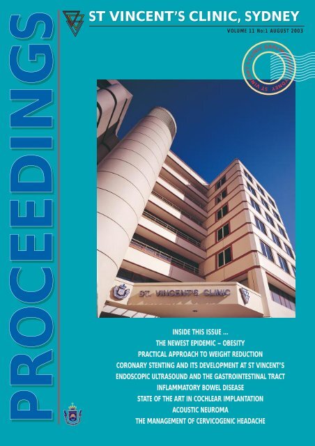
PROCEEDINGSST VINCENT’S CLINIC, SYDNEYVOLUME 11 No:1 AUGUST <strong>2003</strong>INSIDE THIS ISSUE ...THE NEWEST EPIDEMIC – OBESITYPRACTICAL APPROACH TO WEIGHT REDUCTIONCORONARY STENTING AND ITS DEVELOPMENT AT ST VINCENT’SENDOSCOPIC ULTRASOUND AND THE GASTROINTESTINAL TRACTINFLAMMATORY BOWEL DISEASESTATE OF THE ART IN COCHLEAR IMPLANTATIONACOUSTIC NEUROMATHE MANAGEMENT OF CERVICOGENIC HEADACHEST VINCENT’S CLINIC DARLINGHURST, SYDNEY
ST VINCENT’S CLINIC, SYDNEY VOLUME 11 No:1 AUGUST <strong>2003</strong>PROCEEDINGSEditorialDr John H. O’Neill MD, FRACPConsultant Neurologist, Editor,<strong>Proceedings</strong> 2Articles... And the Winner in the Newest EpidemicCategory is ... ObesityDr Katherine Samaras MBBS, PhD, FRACP<strong>St</strong> Vincent’s <strong>Clinic</strong> 3Practical Approach to Weight ReductionDr David Mann BSc (Hon),Dip Nutr & Diet, PhDHead of Department, Nutrition andDietetics Department<strong>St</strong> Vincent’s <strong>Clinic</strong> 7Coronary <strong>St</strong>enting and its Development at<strong>St</strong> Vincent’sDr Paul Roy MBBS (Sydney), FRACP,FACC, FSCA, FRCP (London)Cardiovascular Interventionalist<strong>St</strong> Vincent’s <strong>Clinic</strong> 10Endoscopic Ultrasound – a new way oflooking at the Gastrointestinal TractDr David Williams<strong>St</strong>aff Specialist, Gastroenterologist<strong>St</strong> Vincent’s <strong>Hospital</strong> 13Inflammatory Bowel Disease and the <strong>St</strong> Vincent’sExperience with the Use of ThalidomideDr Carolyn BariolConsultant GastroenterologistFaculty of Medicine, University of NSW 19<strong>St</strong>ate of the Art in Cochlear ImplantationDr Phillip Chang FRACSVisiting ENT Surgeon<strong>St</strong> Vincent’s <strong>Clinic</strong>Dr David Flint FRACSNeurotology Fellow<strong>St</strong> Vincent’s <strong>Hospital</strong> 23Acoustic Neuroma: Pneumocephalous, a rarecomplicationP.M. Valente B.BioMed Sci (Hons)Visiting Fellow in Neuro-Otology<strong>St</strong> Vincent’s <strong>Hospital</strong> 28A Randomised <strong>Clinic</strong>al Trial Provides Evidencefor the Long-Term Effectiveness of Physiotherapyin The Management of Cervicogenic HeadachePaul Kelly MAPA, GRAD DIP MAN Ther,MPAAPhysiotherapy Department, <strong>St</strong> Vincent’s <strong>Clinic</strong>Neil Munro MAPA, GRAD DIP MAN THER,MPAAPhysiotherapy Department, <strong>St</strong> Vincent’s <strong>Clinic</strong>Carolyn Grinter DIP OTPhysiotherapy Department<strong>St</strong> Vincent’s <strong>Clinic</strong> 31COPYRIGHTAll literary matter in the Journal iscovered by copyright, and must notbe reproduced, stored in a retrievalsystem, or transmitted in any formby electronic or mechanical means,photocopying, or recording, withoutwritten permission.BOARD OF DIRECTORSDr Brett Courtenay (Chair)Professor Terence CampbellSr. Suzette Clark rscSr. Adele Cotrell- Dormer rscMs. Maureen McCabeMs. Patricia TysonMr John WilcoxBOARD OF TRUSTEESMr Ted Harris AC (President)Dr Brett CourtenaySr. Mary Bernice Elphick rsc AM OBEMr Peter FalkMr Peter Ferris AMMr Arthur Fitzgerald AMSr. Margaret Fitzgerald rscProfessor John Hickie AODr Thomas HughProfessor Reginald LordMr David MeagherMrs. Leith Myerson MBEMrs Roslyn PackerMr <strong>St</strong>even RubicMs Michelle WilsonS T V INCENT’ S C LINICEXECUTIVE DIRECTORMs Michelle WilsonMEDICAL COUNCILDr Peter BentivoglioA/Professor Judith FreundDr Jock HarknessDr Gordon O'NeillDr Janet RimmerDr Katherine SamarasS T V INCENT’ S C LINIC FOUNDATIONSCIENTIFIC COMMITTEEDr Peter Bentivoglio (Chair)Dr David GolovskyProfessor John Hickie AODr Thomas HughProfessor Reginald LordDr Dudley O'SullivanST VINCENT’S CLINIC438 Victoria <strong>St</strong>reet, Darlinghurst, Sydney, NSW 2010, AustraliaPhone: (02) 8382 6222 Fax: (02) 8382 6402Email: clinic@stvincents.com.auWebsite: www.clinic.stvincents.com.auST VINCENT’S CLINIC, PROCEEDINGS VOLUME 11 NO: 1 AUGUST <strong>2003</strong> 1
EDITORIALDr John O’Neill MD, FRACPCONSULTANT NEUROLOGISTEDITOR, PROCEEDINGSThis Issue of <strong>Proceedings</strong>comprises eight articles fromseveral different fields ofmedicine. Unfortunately itwas not possible to obtain a summarisedtranscript of the 2002 Sandra DavidOration which was given by Dr A Kass,MD, of the Johns Hopkins MedicalInstitute, Baltimore. This oration wasdedicated to the memory of the late RayKelly who was one of Dr Kass’ Fellowsduring Ray’s period of post-graduatestudy in the United <strong>St</strong>ates.Obesity is a topical issue in oursociety. It is now epidemic in Australiawith a prevalence of approximately 60%.It is timely, therefore, to have an articleon that subject, by Dr KatherineSamaras, endocrinologist. Her article iscomplimented by a practical approach toweight loss as suggested by Dr DavidMann, nutritionist. The articles shouldprovoke thought, and action, amongst agood proportion of readers!Coronary stents are common amongstour aging population and Dr Paul Roy,cardiologist, has reviewed this topicespecially with respect to itsdevelopment at <strong>St</strong> Vincent’s. Dr Roy is avery busy clinician and the excellence ofcoronary stenting at <strong>St</strong> Vincent’s can belargely attributed to his carefullycompiled database, constant self-audit,retraining and revision of the technique.Dr David Williams, gastroenterologist,has reviewed the subject ofendoscopic ultrasound, a specialisedtechnique performed in Sydney only at<strong>St</strong> Vincent’s (by David) and Concord<strong>Hospital</strong>s. This new technique hasrevolutionised the investigation ofsuspected pancreatic tumours andmediastinal masses as well as greatlyassisted in the staging of gastrointestinalcancer. Dr Carolyn Bariol,gastroenterologist, has provided anoverview of inflammatory bowel diseaseand specifically has reviewed an earlytrial at <strong>St</strong> Vincent’s using thalidomidefor those conditions. This is promising tobe a very effective therapy.Cochlear implants have had anenormous positive impact on the lives ofpatients with profound deafness. Thefirst such implant in Australia wasperformed by Dr John Tonkin, ENTsurgeon at <strong>St</strong> Vincent’s. Our youngestcochlear implant specialist at <strong>St</strong>Vincent’s, Dr Phillip Chang (ENTsurgeon), has reviewed the currentsituation with respect to that specialisedsurgical procedure. <strong>St</strong> Vincent’scontinues to be a leader in the combinedENT and Neurosurgical approach tocertain skull-base tumours. I haveincluded in this Issue a case report by DrPedro Valente, visiting Fellow under DrPaul Fagan, on the subject of a rarecomplication of acoustic neuromasurgery.Spinal problems are common in ourcommunity and the physiotherapydepartment of <strong>St</strong> Vincent’s <strong>Clinic</strong>provides wonderful support for the manyspinal patients who come through ourdoors. Members of that departmentformed part of a collaborative trial runby Professor Jull, physiotherapist,analysing the benefits of physiotherapyfor cervicogenic headache. The results ofthat study are reported.<strong>St</strong> Vincent’s Private <strong>Hospital</strong> and<strong>Clinic</strong> Ladies Committee havecontinued their wonderful fundraisingfor research on <strong>St</strong> <strong>Vincent's</strong> campus.Over the last financial year they made$160,000 available to fund threeseparate Sr Mary Bernice ResearchGrants. The successful grant applicationsfor <strong>2003</strong> are listed at the end of thisIssue. In addition to the above, this yearthe <strong>St</strong> <strong>Vincent's</strong> <strong>Clinic</strong> Foundationannounced it would commit a minimumof $1,000,000 over the next three yearsto fund adult stem cell research on <strong>St</strong><strong>Vincent's</strong> campus.2 ST VINCENT’S CLINIC, PROCEEDINGS VOLUME 10 NO: 1 AUGUST 2002
Dr Katherine SamarasAnd the winner in thenewest epidemiccategory is ... OBESITYI NTRODUCTIONIn almost every facet of the modernmedical practice, we are confronted bythe compounding problem of obesity.Obesity predicts the development ofType 2 Diabetes, ischaemic heartdisease and is closely associated withhypertension, lipid disorders, sleepapnoea, infertility and degenerativejoint disease. Obesity exacerbates thedisability of neurological, orthopaedic,respiratory and rheumatologicalconditions. Obesity is also associatedwith higher risks of common cancers,such as bowel, ovarian and breastcancer. Obesity significantly increasesthe morbidity associated withanaesthesia and surgicalcomplications, both early and late.Dr Katherine Samaras MBBS, PhD,FRACPConsultant Gastroenterologist<strong>St</strong> Vincent’s <strong>Clinic</strong>Recent Australian data haveshown prevalence rates foroverweight and obesity tohave reached epidemicproportions. Approximately 50 per centof all women and 60 per cent of all men,aged 18 or over, are either overweight orobese. The statistics are even higher incertain ethnic groups. Rates ofchildhood obesity are also increasingrapidly. Australians are now the secondmost obese nation in the world, closelyfollowing the trends found in the United<strong>St</strong>ates. Since obesity (specifically centralabdominal obesity) predicts importantdiseases such as diabetes, hypertensionand cardiac disease, the current rise inobesity prevalence is likely to befollowed by increased rates of theseconditions and their sequelae.One of the great difficulties intreating obesity is the apparent lack ofany intervention that provides long termamelioration. Diet and exerciseprogrammes have been proven to lacklong term efficacy. Obesity ischaracterised by high rates of what hasbeen termed by some as “recidivism”,implying that patients who actively loseweight through dietary restriction andincreased energy expenditure, lapse backinto “old habits” with consequent weightregain. The physiology of the adipocyteand whole body metabolismdemonstrates that the problem is farmore complex than adherence to dietaryand physical activity guidelines. Thenotion that the higher fat mass thatmust be re-established (the “adipostat”)will be elaborated upon subsequently.ST VINCENT’S CLINIC, PROCEEDINGS VOLUME 11 NO: 1 AUGUST <strong>2003</strong> 3
Understanding this physiology allows usto guide our patients through obesity to ahealthier body mass, but is the mostcompelling argument for strategies forobesity prevention.In patients who are overweight orobese, a reduction of 5kg alone, is oftensufficient to ameliorate the metabolicconsequences of obesity. Patients oftenhold the absurd notion that they have toreach a specific weight for their height,dictated by life insurance tables.This may be an unreasonable ask forthe patient and is usually not necessaryfor health benefits.Appropriate short and long term goalswill depend on the dietary strategyselected.D EFININGOVERWEIGHT ANDOBESITYWhilst the body mass index (BMI) isa useful indicator of obesity, it is a crudeestimate of body fat mass, as it isconfounded by lean tissue mass. TheBMI is a far less accurate indicator of fatmass in muscular men, for example, orethnic groups with a high lean tissuemass, such as South Pacific Islanders.The BMI provides insufficient clinicalinformation regarding an individual’srisk from the sequelae of obesity. Thewaist circumference is a criticallyimportant measure in this regard, as it isa better clinical risk indicator and allowsthe identification of central obesity. Thewaist circumference is a simple measureand requires only a tape measure. Waistcircumference is measured at thenarrowest point between the ribs and theiliac crest. In some patients, especiallythe centrally obese, such a point won’tbe identifiable. The level of theumbilicus is a suitable alternative.Central obesity is present when the waistcircumference exceeds 90 cm in a femaleand 100 cm in a male.U NDERSTANDINGTHE AETIOLOGY OFOBESITYPopulation studies demonstrate thatgenetic factors explain up to 60 per centof the differences between individuals intotal body fat mass and its distribution. 1Despite the strong genetic contributionto body fat mass, only a few geneticmutations have been described. Theseaccount for less than a dozen cases ofobesity world wide, almost allmanifesting as morbid obesity in earlychildhood. These include mutations inthe leptin gene, the leptin receptor geneand other genes involved in theregulation of hunger and satiety.Obesity is mostly due to a long termexcess of energy intake over energyexpenditure in individuals who have agenetic predisposition for excellentenergy conservation. These “thrifty”genes were quite useful in circumstancesof privation, such as famine or cycles ofwar, when food availability was low andthis was an excellent survival advantage.The genes that regulate the effectivestorage of energy are unlikely to bemutations, but genetic polymorphismsthat have arisen from long term adaptivechanges or selection by attrition duringadverse conditions such as cycles offamine or war. The current physicalenvironment in Australia is, however,one of perpetual feast for most, marriedto increasing sedentariness. Such anenvironment will permit geneticallypredisposed individuals to accumulate alarger amount of body fat, compared toothers who do not possess such “thrifty”genes, and this is a useful model forunderstanding the disparity in body fatstores between individuals in a societywhere most observe similar lifestyles.Whilst this permits us to understandwhy certain individuals develop obesity,it does not help understand why manypatients do not seem to be able tomaintain any hard won loss of fat mass.Here, the notion of the “adipostat” ishelpful. This refers to a physiologicalstate of homeostasis that is established atthe high body fat mass. A loss of fat massis associated with significant metabolicperturbations, all guaranteed to returnthe body to the higher fat mass. Thisincludes physical changes, such asreductions in metabolic rate,thermogenesis, but also profoundchemical changes, both within theadipocyte and brain. The adipocyte haslong been considered a simple, inertstorage vessel for fat. Recent research hasdemonstrated the adipocyte is an activesynthetic site for numerous cytokinesand other chemicals that have not onlylocal metabolic effects, but also impacton numerous aspects of body function,from appetite regulation through to thetiming of sexual maturity and fertility.When an adipocyte releases its fatcontent, a number of events are initiatedthat, in the long term, promote therestitution of fat to the adipocyte. Ifadhering to a programme restrictive dietand increased physical activity were nothard enough in our perpetualfeast/sedentary society, humanphysiology plots against the obeseindividual. This framework is thestrongest argument for the activeprevention of obesity, especially in theabsence of any effective “cures”.Endocrine problems such ashypothyroidism, and Cushing’ssyndrome can promote weight gain andshould be considered in the clinicalevaluation of the obese patient.Cushing’s syndrome is however a rarecause of obesity.E STIMATING THEEFFECT OFENVIRONMENTTwin research conducted at <strong>St</strong>Thomas’ <strong>Hospital</strong> London and on the <strong>St</strong>Vincent’s campus has estimated theeffect of various environmental factorson total body and central abdominal fat.Use of the twin model provides a robustmodel for estimating environmentalinfluence independent of geneticinfluences and other environmentalfactors. Using this model, we were ableto show that physical activity was thestrongest environmental influence ontotal and central abdominal fat. 2Postmenopausal oestrogen replacementtherapy and cigarette smoking werelesser influences 3 and no influence ofdietary composition 4 was found in thesecross-sectional studies of postmenopausalwomen.Our group was the first to report aprotective gene-environment interactionbetween physical activity and body fatmass. In those genetically predisposed toobesity a higher level of physical activityis associated with significantly lower fatmeasures, much greater an effectcompared to those without any risk ofobesity. 2 These findings suggest thathigher levels of physical activity may beuseful in preventing obesity in thegenetically susceptible.4 ST VINCENT’S CLINIC, PROCEEDINGS VOLUME 11 NO: 1 AUGUST <strong>2003</strong>
Current research we are undertakingon the <strong>St</strong> Vincent’s campus, incollaboration with <strong>St</strong> Thomas’ iscurrently examining for geneenvironmentinteractions between bodyfat and other aspects of the metabolicsyndrome (insulin resistance syndrome)and other lifestyle factors.M ANAGEMENTThe first step in starting a weightmanagement regimen is to establishreasonable weight and life style targets.Patients often have unrealisticexpectations of what is possible, givenwhat is known about the genetics andphysiology of obesity. The body massindex (weight divided by heightsquared) will indicate the degree ofobesity. Cut-offs for the various levels ofobesity are indicated in Table 1.T HE BASICS IN DIETAND EXERCISEThe challenge in weight managementis to guide the patient to effective, longterm restructuring of their eating habits,food choices and physical activity.Body weight is governed by thesimple law of energy conservation, givensome qualifying factors elaborated uponin the above. If energy intake exceedsenergy output, there will be weight gain,and visa versa. The simplest means ofreducing energy intake is to reduce theintake of energy dense foods, includinghigh fat and high carbohydrate foods.Patients need to become savvyinterpreters of food labels, especiallynewer “low-fat” foods, which cancontain large amounts of sugars assubstitutes for fat. Patients should eatmore fibre-containing foods (such asvegetables and fruits), as well as leanprotein foods (such as lean meat,chicken or fish). The most importantconcept is total caloric reduction, mosteasily achieved by fat and carbohydratereduction. Many patients mistakenly fallinto the trap of not consideringcarbohydrate calories however. Highcarbohydrate diets can promote weightgain or prevent weight loss if caloriesconsumed exceed energy output. Ofcourse, a reduction in alcohol calorieswill also help.GroupThe assistance of a nutritionistexperienced in weight reduction is animportant starting point. There are alsonumerous ethical patient resources andinformation leaflets on nutritionavailable.On the energy output side of theequation is physical activity. Anyreduction in sedentariness is a startingpoint. Many mistakenly advise obesepatients to undertake far too strenuousphysical activity in the initial phases ofweight reduction. The effort involved inwalking on the flat for an overweight orobese individual is significantly greaterthan for a lean one. Swedish studieshave demonstrated that overweightwomen walking 100m flat grade areworking at 70 per cent of their maximalaerobic capacity, compared to 50 percent when lean people jog. A suitableinitial programme may suggest anincrease in any walking activity inperforming usual daily duties. Longerterm a goal of at least 30 minuteswalking daily is required, especially forweight maintenance.Some patients will require specialisedpsychological assistance, includingcognitive behavioural therapy orhypnotherapy.Very low energy diets (VLED)promote rapid weight reduction throughsevere caloric restriction and should beused only under careful medicalsupervision. These diets replace mealswith a vitamin-fortified milk proteinbaseddrink and allow only starch-freevegetables and low calorie beverages.These diets are useful for the initialphase of treatment of morbidly obeseindividuals, or obese patients withsignificant co-morbidities who have hadlittle success despite some lifestylechanges. These diets can produceelectrolyte abnormalities, particularly inpatients receiving antihypertensives ordiuretics. Patients with diabetes mayrequire medication adjustment. Such adiet is contraindicated in pregnancy.BMI (kg/m2)Healthy weight 20-24.9Overweight 25.0-29.9Obese 30.0-34.9Morbidly obese >35.0Table 1. Classification for obesity using body mass index.D RUGSThe therapeutic drug options inobesity are increasing. There arecurrently two drugs available for weightmanagement: orlestat and sibutramine.Orlestat is a pancreatic lipaseinhibitor, reversibly inhibiting itshydrolytic activity on ingested fats andreducing intestinal fat absorption by 30per cent. This medication can be auseful adjunct in association with areduced fat, calorie restricted diet.Adverse effects include flatulence,bloating, loose or frequent motions, oilyseepage.Sibutramine is a centrally-actingserotonin and noradrenaline reuptakeinhibitor which increases satiety andmetabolic rate. Adverse effects includehypertension, flushing, headaches.Potentially dangerous interactions canoccur with other drugs such asmonoamine oxidase inhibitors and otherantidepressants.S URGERYSurgical approaches to weightreduction revolve around reducingconsumption by limiting capacity(stomach stapling and, more recently,Lap-banding) or reducing absorption(intestinal bypass). There is significantevidence of comorbidity reduction withthese techniques, as there is with anysuccessful approach to weight reduction.These techniques could be considered inselected patients with significantcomorbities who have been unable toreduce weight through other means.ST VINCENT’S CLINIC, PROCEEDINGS VOLUME 11 NO: 1 AUGUST <strong>2003</strong> 5
C ONCLUSIONSObesity has reached epidemicproportions in Australia. This willimpact on almost every aspect ofmedicine and have consequences on theprevalence of diabetes, heart disease,hypertension and other relatedconditions. Health strategists arerecognising the importance of the activeprevention of obesity and there is astrong imperative for governments andcommunity planners to buildenvironments that promote activity(rather than prevent it). Research intothe regulation of body fat is required tobetter understand the mechanisms thatpromote weight regain. Currently,achieving and maintaining healthyweight will rely on the changesindividuals make and maintain in anotherwise obesogenic environment.R EFERENCES1. Samaras K, Spector TD, Nguyen TV, Baan K,Campbell LV, Kelly PJ. Genetic factorsdetermine the amount and distribution of fat inwomen after the menopause. J Clin EndocrinolMetab 1997; 82:781-5.2. Samaras K, Kelly PJ, Chiano MN, Spector TD,Campbell LV. Genetic and environmentalinfluences on total and central abdominal fat:the effect of physical activity in female twins.Annals of Internal Medicine 1999; 130:873-8823. Samaras K, Spector TD, Kelly PJ, CampbellLV. Tobacco smoking and estrogenreplacement therapy are associated with lowertotal and central fat in monozygotic twins. Int JObesity 1998;22:149-156.4. Samaras K, Kelly PJ, Chiano MC, Arden N,Spector TD, Campbell LV. Genes vsenvironment: The relationship betweendietary fat and total and central fat. DiabetesCare 1998;21:2069-2076<strong>St</strong> Vincent’s <strong>Clinic</strong> FoundationResearch Grants <strong>2003</strong>LADIES’ COMMITTEE SR MARY BERNICE AWARD 1 – $100,000Prof Bruce BrewThe involvement of quinolinic acid and other tryptophancatabolites in the pathogenesis of Alzheimer’s Disease.LADIES’ COMMITTEE SR MARY BERNICE AWARD 2 – $30,000Dr Joanne JosephAn evaluation of in-vivo platelet and monocyte activation inpatients receiving standard compared to drug-elutine (sirolimus)stents for stable coronary artery disease.LADIES’ COMMITTEE SR MARY BERNICE AWARD 3 – $30,000Prof Ken HoAnabolic hormones in the therapy of glucocorticoid-inducedprotein wasting.K+A COLLINS CANCER RESEARCH GRANT – $50,000Dr D SegaraInvestigation of the WNT signalling pathway in the developmentand progression of pancreatic cancer.DI BOYD CANCER RESEARCH GRANT – $20,000Prof David Ma/Dr Helen TaoGene expression profiling may reflect the clinical outcome in selectedhaematological malignant diseases.ANNUAL GRANTSDr Diane Fatkin – $20,000Evaluation of early disease in familial dilated cardiomyopathyProf Terence Campbell – $20,000<strong>St</strong>ructure of the HERG K+ channel drug binding siteDr Alan Meagher – $20,000Analysis of the class II HLA binding specifications of the p53vaccine (Pentrix) peptidesDr Nirmal Patel – $20,000Inner ear neural stem cell transfer and neurotrophin-3 (NT3)production in the mouse modelTRAVELLING FELLOWSHIP GRANT –Dr Andrew Biankin $10,000Postdoctoral Fellow in pancreatic cancer under the supervision ofProfessor <strong>St</strong>even Leach in the Division of Surgical Oncology atJohns-Hopkins University <strong>Hospital</strong>Dr David Brown $10,000Postdoctoral study in neuroimmunology with Dr Evan Snyder atHarvard Medical School, Boston. Research in the field ofneuroimmunology, examining the function of macrophageinhibitory cytokine-1 (MIC-1) in the central nervous systemSTUDENT RESEARCH GRANT – $3,000Ms Lily WangDoes quinolinic acid cause astrocyrte apoptosis?TOTAL $333,000Note: The Committee agreed to recommend the granting of 4 annual awardsrather than 5 awards and to divert some of the award money to funding anadditional Travelling Scholarship as it was felt that both recipients qualifiedequally for the grant.6 ST VINCENT’S CLINIC, PROCEEDINGS VOLUME 11 NO: 1 AUGUST <strong>2003</strong>
Dr David MannPractical approachto weight reductionDr David Mann BSc (Hon), DipNutr & Diet, PhDHead of Department,Nutrition and Dietetics,<strong>St</strong> Vincent’s <strong>Clinic</strong>I NTRODUCTIONThere are as many diets as thereare diseases associated withbeing over weight, yet overweight patients rarely aremotivated to change their lifestyles toachieve weight loss. Often there is aresistance for people to follow weightreduction diets simply because diets areperceived to be complicated and difficultand, in many cases, this certainly is true.Added on top of this, weight loss israrely long term and there is almostalways a regaining of the lost weight.A significant problem encountered bypatients attempting weight reductioncan be the impracticality of the diet. Insome cases, the time required to preparefor the diet is prohibitive or the mealsrequired on the diet may be unsuitableor repetitive. Some diets are extremelylimiting in food selection. It may be thata person must change their mealpatterns altogether or that the meals theindividual eats may not conform to therest of the family’s meals. These are veryreal problems that deter any wellmeaning dieter.These obstacles should not be lookedupon as excuses for dieters to avoid theissues of weight loss but rather practicalsolutions should be formulated to openthe way for the dieter to achieve theirgoals. Clearly, a more main stream,practical approach to weight reduction isneeded.The ideal approach should not onlybe a practical one but one that actuallyST VINCENT’S CLINIC, PROCEEDINGS VOLUME 11 NO: 1 AUGUST <strong>2003</strong> 7
teaches or reconditions the patient intoa new habit with eating. Whilst mostclinicians may be aware of this, very fewknow how to go about modifying aperson’s eating habits.Within the scope of this article, Ihope to highlight some approaches thatI have taken in my practice that haveproven to be successful with mostpatients. The format and presentation ofthis article may be somewhat unusualbut I also hope to provide some real lifeexamples that actually apply to patients.The topics covered may becontroversial and ideas may be novelhowever when taking a long term viewto behavior modification the ideas andsuggestions will be apparent.S ETTINGG OALSAs clinicians, we are veryquick to impress on patientsthe importance of the idealweight or the correct bodymass index (BMI). Whilst I fully agreewith these standards, and thesestandards give a good guideline to workby, we must use these standards only as aguideline. It is extremely difficult toapply these standards to a real person. Inaddition, the patients in our clinics maybe morbidly obese, that is greater than20kg over weight and often much more.The ideal weight or the correct BMIseem so far away and unreachable thatwe may actually be discouraging ourpatients to achieve their goals.I find that initially, not setting a goalallows the patient to be in a better frameof mind and allows them to focus moreimportantly on the issues of cooking andarranging for meals on the diet. Focusingtheir attention and effort in doing thecorrect style of cooking or to add extraflavors or put in some incidentalexercises instead will give better weightloss results. If an aim has to be discussed,we should always aim for an initialmodest weight loss such as 5 to 6kg. Itshould then be pointed out that afterthey have attained this level, they canset a new target. I also find that lettingthe patient take control of this targetallows them to feel more in control oftheir diet and weight loss.C OUNTINGC ALORIESTraditionally, numerous diets workedby having the dieter count calories, addup grams of fat intake or drink 2 litres ofwater or a range of other activities thatare difficult to achieve in a normalworking day. Should a dieter add upgrams of fat intake each day? They firstlyneed to have a fat counter at hand allday, every day. The values of foodconsumed needs to be weighed ormeasured fairly accurately and then asearch is undertaken for that food in thefat counter. This is then recorded and atthe end of the day the total is calculated.This is far too time consuming andtotally impractical for a full time worker.Most patients I find are fed up with this“paranoia” approach to dieting. Theseactivities distract a dieter from thinkingabout other more important areas oftheir eating behavior which, if givenmore time to focus on, would give betteroverall results. Occasionally, I dorecommend that patients purchase a fatcounter but only to have a glancethrough to see the approximate level offat in different foods.I try always to keep the diet elementsto the basics and any unnecessaryrequirements should be avoided. I findthat the opposite of what some dietstend to do actually works better.E STABLISHING ANE ATING P ATTERNThere is a common belief thatreducing the volume of food intake willassist in reducing weight. To a largeextent this may be true, however, giventhe relatively free supply of food in oursociety, what actually happens is that areduction in food intake will result in afeeling of hunger and this will result insubsequent over eating.We have often seen patients who maybe either too busy or short of time andmiss breakfast. Often such patients willskip lunch or have a very small lunch.When such patients arrive home, theirhunger is so fierce that they will go on anon-stop eating binge and, of course,often pick high calorie food.This pattern of “dieting” is socommon and only results in furtherweight gain. In addition, reducing foodvolume not only encourages poor eatinghabits but it may result in lowered selfesteem for the patient who finds theirattempt at weight loss is unsuccessful.This type of patient needs a great dealof reassurance and encouragement forthem to try establishing an even eatingpattern. This can be done by providingsome practical options. For example,instead of trying to have breakfast athome, I suggest to the patient to onlyever have breakfast at work or on theway to work. They may bring a loaf ofbread into work on Monday morning,keep it in the fridge and when theyarrive at work each morning they put onsome toast and make a cup of tea beforethey start work.For lunch, where there is insufficienttime to buy or make lunch, I recommendto patients who may be working near amilk bar or sandwich shop to arrange fora prepared and possibly even deliveredlunch to avoid hunger.There is then the difficulty ofcooking. More and more I am seeingcouples who both work (the “DoubleIncome No Kids” category), arrive homelate and do not have an opportunity ofcooking each night. Instead, they buytake away food or simply dine out atrestaurants. I direct their attention tocooking products such as ChickenTonight Cooking Sauces (this is onlyone example of many more). These aresauces that are pre-made and onlyrequire the addition of meat/chicken/fishand some vegetables. The best partabout these products is that almost all ofthem are very low fat and do not containpreservatives or other additives. There isa salt content, however the addition ofmeats and vegetables brings the salt toan acceptable level.I also encourage patients to precooktheir meals using these types of products,so that on a Sunday afternoon a coupleof hours in the kitchen means that theycan cook all of next weeks meals in onego and that they will only need tomicrowave to reheat during the week.Hence, cutting down on cooking timeand best of all cut out buying take awayfood.The aim of providing patients withthese suggestions is to make it possiblefor them not only to be able to follow adiet but also it gradually reconditionsthem into a good eating pattern again. A8 ST VINCENT’S CLINIC, PROCEEDINGS VOLUME 11 NO: 1 AUGUST <strong>2003</strong>
egular eating pattern will be able toavoid hunger and hence food choicescan be more appropriate.N ORMAL T IMES ANDS OCIAL T IMESThe affluent society in which we liveallows us to have ample amounts of food,especially high calorie food, around us atall times. As living organisms, humansare instinctively driven to eat; oursurvival is based on our ability to havestrong instincts to eat.The following suggestion may soundunusual but I have used it in my practicefor a long time and it has suited almosteveryone. It is an extremely importantpart of my re-education process that Ifind other diets neglect.All diets are quick to tell dieters thefood to limit themselves to or point outall the bad foods that they must avoid.Very few diets actually teach people howto still make the most of those reallyenjoyable foods (including restaurantmeals, drinking alcohol, oily foods,desserts, etc.) that we all want. Up tonow dieters have achieved weight loss bywithholding from enjoyment. Is it anywonder that people avoid dieting!My recommendation is that weshould be teaching people how to eatenjoyable foods but at a level which,when balanced with the low caloriefoods, will either achieve weight loss ormaintain a constant weight. Mostpatients’ initial reaction is that thisconcept cannot be successful. We are sofirmly conditioned that enjoyable foodsshould always be avoided, that anysuggestion of giving in to pleasure makesus feel uneasy. Because everyone has asocial life, everybody goes out to eat ordrink in one way or another. It would bedifficult to avoid or modify in any waythese social occasions. My advice topatients is to learn to relax on theseoccasions and enjoy the variety of foodand drinks that is available but to revertback to the diet immediately afterwards.Some patients report that they feel soguilty they have deviated from the dietthat they do not return to it. Thedefeated feeling takes over them sostrongly that their self-esteem is severelybadly affected and self-control isabandoned. As strange as it may seem, ifthe dieter learns to “let go” and enjoythese socials they will almost never causeany harm and will continue to loseweight on their normal diet.Further, if we could conditionourselves to thinking that the “enjoyablefoods” are reserved for when we shouldbe having a good time, this willautomatically reduce the frequency ofthe high calorie food intake and allowsfor easier weight control.A CCESSIBLE F OODP RODUCTSFood or food products and ingredientsthat are suggested must always beavailable in supermarkets. Everyone goesto the supermarkets; not everyone willgo to a health food store and not allhealth food stores carry the sameproducts. Dieters are very quick todeviate off the diet if they find that theirattempts at purchasing the right productscannot be achieved at the first attempt.Fortunately, the food products andcooking ingredients that are available tous in the supermarkets are not only ofgood quality with respect to low fatcontent, but they are also of good qualitywith respect to taste and flavor.Whatever the products, the ease withwhich any individual is able to obtainthese products is extremely important. Itis counter-productive to recommend aproduct that patients could only obtainfrom a special shop that is only in somesuburbs and only at certain times of theyear.I also often recommend the use offrozen meals either as an emergencymeal or just a lazy night meal. There arenow so many different brands andvarieties to choose from and all verygood quality, low in fat and excellent inflavor. Again, the aim of these types ofproducts are to make the process offollowing a diet easier.R EASSURANCE ANDM ORAL SUPPORTMany clinicians underestimate thevalue of moral support during a weightreduction diet. Female patients require agreat deal of support by way ofexplaining about fluid retention andsweet cravings during monthly menstrualperiods. Men require a great deal ofsupport in respect of the change in sizerather than the change in weight. Menwith a higher per cent muscle contentwho are currently doing muscle-bulkingexercises will lose weight very slowly onthe scales but will very quickly change insize.During a diet these problems need tobe explained to dieters to ensure thattheir emotions do not divert in thewrong direction. In fact, generalexplanation of the physiological changesthat takes place with the dieter’s bodyduring the diet helps to alleviate anyworries or concerns that a dieter mayhave about their weight loss.The reassurance is important to avoidpatients having thoughts that their dietis not working and the explanations willhelp them to understand how their bodyworks and how the body can respond tofood. This type of information is helpfulfor patients to ultimately understand thewhole picture to be able to control theirweight long after they have finishedlosing weight.C ONCLUSIONOften, the task of following a dietseems so insurmountable that manydieters simply avoid the issue of weightloss or weight control. As clinicians, wemust be able to take into considerationall aspects of dieting and thecomplexities involved to help thepatient clear the way to achieving theirgoals. The above is only an example ofsome of the problems encountered by adieter and hopefully some solutions tomake their job a little easier.The constraints of this article doesnot allow more in-depth discussions onmany other aspects of dieting such as therelationship between emotions andeating behavior, the problems of the“picking” behavior and explanations todieters as to why some diets areinappropriate.ST VINCENT’S CLINIC, PROCEEDINGS VOLUME 11 NO: 1 AUGUST <strong>2003</strong> 9
Dr Paul R. RoyThe new era ofcoronary stentingand its developmentat <strong>St</strong> Vincent’sDr Paul R. Roy MBBS (Syd),FRACP, FACE, FSCA, FRCP (Lon)Cardiovascular Interventionalist<strong>St</strong> Vincent’s <strong>Clinic</strong>When John Morgan and Idid the first coronaryballoon angioplasty at <strong>St</strong>Vincent’s in 1980 wenever envisaged what would happenover the next 20 years. Then, everyangioplasty was associated with an acuteclosure rate of about 5 per cent due todissection. Urgent surgery for thosepatients was associated with a higherthan normal cardiac surgical mortality. Itwas very disruptive and irritating for oursurgical colleagues. The results ofballoon angioplasty were limited in the1980s by the rate of dissection and acuteclosure, by subacute closure, and longterm by a restenosis rate of 25 – 30 percent.The first coronary stent wasimplanted by Puel in Montpellier,France in 1986. I attended the first livecourse in coronary stenting in Lausannein 1987. Acute and subacute thrombosisduring the course appeared to be asignificant problem and when I returnedto <strong>St</strong> Vincent’s our initial enthusiasm forstenting was lukewarm. The stents thatwere used in the Lausanne course wereWallstents and they were notimmediately made commerciallyavailable because of the worry ofthrombosis.10 ST VINCENT’S CLINIC, PROCEEDINGS VOLUME 11 NO: 1 AUGUST <strong>2003</strong>
During the period 1990 – 1993 animproved stent, known as the PalmasSchatz <strong>St</strong>ent became available. Thesestents were hand crimped on to theballoon in the cardiac catheterlaboratory. Though they were animprovement on the previous stents,occasionally the crimped stent came offthe balloon before it was deployed in thecoronary artery and this of course causedembolization of the stent to anunwanted site in the vascular tree.A cumbersome balloon mountedstent, known as the Cook <strong>St</strong>ent alsobecame available at this time and wasshown to be useful, for bailout in acutedissection. 1 This stent was used here at <strong>St</strong>Vincent’s in a live demonstration coursein 1993.During our live demonstration coursein 1995, David Baron and I had theopportunity to be the first in this countryto use a factory produced stent with thestent crimped onto the balloon in theproduction process, rather than crimpedon by the operator in the catheter lab.This was known as the AVE Microstentand was the forerunner of many suchballoon mounted stents.In 1994 – 1995 the <strong>St</strong>ress andBenestent <strong>St</strong>udies 2 demonstrated a 30per cent reduction in restenosis rateswhen stents were used in preference to‘plain old’ balloon angioplasty.A subsequent fall in restenosis rates to15 – 20 per cent, lead gradually towidespread use of stents over the period1995 – 2002. During this period stenttechnology advanced rapidly withsmaller, more secure stents with very lowcrossing profiles, making it possible forthese stents to be tracked down intosmaller and more tortuous vessels thanwas previously possible.Anticoagulation to prevent post stentthrombosis also changed and advancedduring this period. Recent studies havedemonstrated the effectiveness ofAspirin (which inhibits cyclo oxygenaseand thus decreases prostaglandin andthromboxane formation) in combinationwith Clopidogrel (an ADP receptorantagonist) to protect against acute andsubacute thrombosis. 3 These two drugstogether block two important pathwaysfor platelet aggregation but do not blockplatelet aggregation completely. If,during the course of a stentingprocedure, there is a complex situationwhere there is a high risk of thrombosiswe also have available 2 B 3 Ainhibitors. These drugs inhibit plateletreceptors and prevent platelets bindingto fibrinogen to form a clot. When givenintravenously they can totally block theplatelet aggregation process.By 2002 our major problems withcoronary stenting were restenosis (15 –20 per cent) and the inability to reopensome chronic total occlusions.Comparisons of stents versus bypasssurgery for triple vessel disease haveshown similar follow up death andmyocardial infarct rates but a higherneed for further procedures in the stentgroup. This of course has been duemostly to restenosis. 4A revolution in stent technology hasoccurred in the last two years effectivelyaddressing the problem of restenosis.Restenosis is a result of neointimalhyperplasia. This occurs when the usualhealing process and re-endothelializationinside the stented segment of the arterygoes ‘overboard’. The process can beroughly compared to a keloid scarringeffect.Neointimal growth involvesdedifferentiation of vascular smoothmuscle cells from a contractile state to asecretory state leading to cellularproliferation, migration from the mediainto the intima and synthesis ofintracellular matrix. The pathophysiologyof restenosis consists of thecomplex interaction of cytokines andgrowth factors with cellular and acellularelements. Until now, blockade of anyone factor has been insufficient toinhibit the restenosis cascade. Attentionhas now been focused on disruptingessential central cellular processes thatwould subsequently affect ‘downstream’events that ultimately lead to restenosis.A vast amount of creative energy hasgone into the development of a drugcoated stent which in early trials hasreduced the restenosis rate to 1 – 3 percent.Initially the stainless steel struts of thestent made attachment of a drugdifficult. A polymer was ultimatelydesigned which was coated onto thestent, allowing application of a drug insuch a way that the drug would ‘elute’slowly from the stent over thirty days.The drug currently used is Sirolimus, animmunosuppressive agent widely usedpost organ transplantation. Rapamycin,its original name, was discovered in soilsamples brought home by researchersfrom Easter Island (named by the Dutchon Easter Day 1722). Transplantimmunologists, Randall Morris andClare Gregory, at <strong>St</strong>anford University,observed that transplanted rat heartstreated with Sirolimus had cleancoronary arteries instead of the usualintimal thickening observed in thecoronary arteries of transplanted hearts.With many steps in between and withcollaboration between experts in bothdrug and device development, Johnson& Johnson produced the first Sirolimus<strong>St</strong>ent, i.e., a stent covered with apolymer from which the drug Sirolimusis released slowly into the local tissueover 30 days. The FIM (first in man)study of this stent was an open labelstudy of 45 patients with both fast andslow release coating of the stent. At 12months there was zero restenosis ineither group. 5The Ravel <strong>St</strong>udy was a double-blindrandomized trial of a drug eluting stentversus a bare metal stent in 238 patients.There was again 0 per cent restenosisversus 26 per cent in the bare metalstent. 6In the Sirius Trial reported by LeonM. et al, at the TCT meeting inWashington in September 2002, 1,058patients were randomized to Sirolimus orbare stent. These were a group of moredifficult patients with diabetes, longlesions, previous angioplasty and surgery.The results were again outstanding, witha Sirolimus <strong>St</strong>ent restenosis rate of 3.2per cent compared to 35.4 per cent inthe bare stent.In May 2002, at the World Congressof Cardiology Live DemonstrationCourse which took place here at <strong>St</strong>Vincent’s, David Baron, David Muller,<strong>St</strong>ephanie Wilson and myself were thefirst in this country to obtain access tothe Sirolimus drug coated stent. FromMay until December 2002, we implanted215 Sirolimus stents at <strong>St</strong> Vincent’s<strong>Clinic</strong> in a variety of low and high risklesions. Only one patient has thus farpresented with restenosis.So far this is the first anti-proliferativeintervention that we have had whichdramatically improves patient outcomes.Sirolimus has a cytostatic mechanism ofST VINCENT’S CLINIC, PROCEEDINGS VOLUME 11 NO: 1 AUGUST <strong>2003</strong> 11
action that leaves vessel cells healthyand viable without killing them. Itpermits natural and normal healing ofthe vessel wall and re-endothelialisation.It acts in the G phase of cell metabolism(Figure 1). This means that theSirolimus causes cytotaxis but notcytotoxicity and allows the cell torecover in due course.Cytostatic agentCell cycle arrestedCheckpointPoint at which cell commits tocompleting the cell cycleAt this stage we have greatexpectations for improved patientoutcomes with drug-eluting stents. Wedo, however, need to be cautious thatthis drug doesn’t simply delay restenosisfor a year or two. We need to wait forlong term restenosis rates before we gettoo enthusiastic.If it turns out to be as good as itappears, some of the classic ‘enemies’ ofpercutaneous intervention such asmultivessel disease, left main stenosis,small diameter vessels, bifurcations andfemoral and tibial arteries may well beconquered by drug-eluting stents,leaving chronic total occlusions as ourmajor challenge and hence very fewpatients who will need bypass surgery.We should all be very grateful for theenormous effort that has gone intoovercoming the substantial challenges ofdeveloping such a clever device.Our group here at <strong>St</strong> Vincent’srecently began a world first trial of a newstent with a new drug coating which wehope will be a refinement of the alreadysuccessful Sirolimus <strong>St</strong>ent.Current work in progress in otherplaces involves attempts to producestents which may be able to carry severaldrugs as well as Rapamycin. Efforts arealso in progress to produce biodegradablestents, which will also be able to carrydrugs to a specific site. All of thesethings are going to lead to a greatimprovement in our ability to treatpatients with vascular disease, not onlyin the coronaries, but in other vascularbeds as well.SirolimusG0 phaseCell works butis not activelyreplicatingR EFERENCES:1. GS, Cannon AD, Agrawal SKIntracoronary <strong>St</strong>enting for acute andthreatened closure complicating percutaneoustransluminal coronary angioplasty.Circulation 85 : 916 – 927, 19922. Serrys PW, Jargere PD, Kiemeny F et alThe Benestent <strong>St</strong>udy Group: A comparisonof balloon expandable stent implantation withballoon angioplasty in patients with coronaryartery disease.New England Journal Medicine 331 : 489 – 495,19943. <strong>St</strong>einhuble SR, Berger PB et alJ.A.M.A November 20, 2002 – Vol. 288No. 19: 2411 – 24204. O’Neill WW, Grines CLSurgery or <strong>St</strong>ents? The gap continues tonarrow.Lancet 2002, 360 : 961 – 9625. Sousa JE et alCirculation 2001 – 104 : 2007 – 20116. Morice MC et alEuropean Heart Journal 2001 – 22 (suppl) 484G1 phaseCell enlargesand makesnew proteinsS phaseDNAreplicationM phaseCell division(mitosis)G2 phasePreparationfor divisionCytotoxicagentCell diesFigure 1:12 ST VINCENT’S CLINIC, PROCEEDINGS VOLUME 11 NO: 1 AUGUST <strong>2003</strong>
Dr David Williams Endoscopic ultrasound –a new way of looking atthe gastrointestinal tractI NTRODUCTIONEndoscopic ultrasound (EUS) isnow an important diagnostic tool inthe management of gastrointestinaldiseases and, with refinements intechnology, it has found diverseclinical applications. Whilst it is nowwidely adopted into clinical practicein USA, Japan and Europe, EUS isavailable in only a few centres inAustralia. <strong>St</strong> Vincent’s <strong>Hospital</strong>Campus has been providing an EUSservice since 2000 and this article isintended to provide an overview of itscurrent indications.Dr David Williams MBBS, FRACP<strong>St</strong>aff Specialist Gastroenterologist<strong>St</strong> Vincent’s <strong>Hospital</strong>I NSTRUMENTSEndoscopic ultrasound (EUS) isperformed with small highfrequency ultrasound transducersmounted into the end of anendoscope, thereby producing clear anddetailed images of the intestinal walllayer structure and surrounding organs. Itis performed as an outpatient procedureunder conscious sedation. No othercurrent imaging method can reveal thegut wall from oesophagus to rectum as aseries of histological correlates. Withstandard EUS frequencies of 5-30 MHz,the intestinal wall is imaged as a multilayeredstructure, corresponding tomucosa, submucosa, muscularis andserosa respectively. This has tremendousrelevance to GI cancer staging anddetermining the nature of submucosallesions.Several echoendoscopes are available,depending on the study required:1. Radial scanner: The most widelyused instrument is a mechanicaldriven radial scanner (7.5-20MHz)that provides a 360° view orientedperpendicular to the tip of theendoscope and allows detailed imagesof the intestinal wall and surroundingstructures.2. Linear Array scanner: Thisinstrument provides 110° scanning inthe same plane as the long axis of theendoscope and has colour Dopplercapability. Therefore, this instrumentallows real time EUS-guided needlebiopsy through the intestinal wall ofextraluminal lymph nodes andpancreas lesions, and underpinstherapeutic applications such asendoscopic pancreatic pseudocystdrainage.3. Catheter miniprobes: These smalldiameter, high frequency (20-30MHz) probes can be passed down thechannel of an endoscope and providehigh resolution imaging of smallmucosal lesions of the GI tract andbile duct.C LINICALI NDICATIONSTable 1 highlights the conditions inwhich EUS can make a difference withrespect to clinical outcomes, and severalof these indications are discussed indetail.<strong>St</strong>aging GI CancerEUS is ideally suited to the TNMstaging classification system of GIcancer, as it can not only define extentof tumour involvement through the gutwall but can also identify loco-regionalnodal metastases and determine tumourvascular invasion (Figure 1). Sinceprognosis of such cancers correlates withstage at diagnosis, accurate pretreatmentstaging is essential intreatment selection, avoidance ofinappropriate surgery and determinationof prognosis.• Oesophageal cancer: EUS is the bestloco-regional staging modality foroesophageal cancer and is most usefulwhere stage dependent treatmentprotocols are in place (Figures 2 & 3).Numerous studies have demonstratedsuperiority of EUS compared withComputerised Tomography (CT),with reported T(umour) & N(odal)staging accuracy in the order of 90 percent and 80 per cent respectively. Acareful systematic review of theliterature has reinforced thisadvantage. 1 Case series alsodemonstrate the ability of EUS todetect coeliac nodal and small livermetastases that are missed on CT,with consequent tumour upstagingand significant impact on patientmanagement. EUS can also betterdefine T4 disease (invasion of pleura,aorta, great vessels) that would provenot to be amenable to attemptedresection.Whilst well designed clinical trialshave yet to fully establish the effecton clinical outcome, the increasinguse of neoadjuvant chemoradio-ST VINCENT’S CLINIC, PROCEEDINGS VOLUME 11 NO: 1 AUGUST <strong>2003</strong> 13
Table 1. When endoscopic ultrasound can make a differenceCancer stagingOesophagus<strong>St</strong>omachRectumEUS-guided tissue diagnosisSubmucosal lesionsPancreasBiliary treeEUS-guided therapyDetermine appropriate treatment (stage dependent protocols)Surgery (<strong>St</strong>g I, IIA), neoadjuvant therapy (IIB, III), palliation (IV)Endoscopic mucosal resection (<strong>St</strong>g 0,1), surgery (II, III)Local (<strong>St</strong>g I) v. wide resectionMediastinal node cytology can preclude curative surgery in lung cancerConfirm and characterise endoscopic abnormalitiesGuide lesion management: excision v. observationConfirm suspicious lesions on CT or MRILocalise small lesionsCharacterise cysts<strong>St</strong>age pancreatic and ampullary cancerEvaluate for chronic pancreatitisScreen for CBD stonesDetect cholangiocarcinoma and gallbladder cancerDrainage pancreatic pseudocysts, coeliac plexus neurolysis(Adapted from Lightdale C, Endoscopic ultrasound: when does it make a difference? <strong>Clinic</strong>al Update American Society forGI Endoscopy 1999;6(4);1-4)therapy places a higher value on pretreatmentstaging. In patientsundergoing surgery, an R0 resection(ie no residual disease in the area ofprimary tumour at end of operation)is associated with five year survivalrates of 20-35 per cent as comparedwith R1/R2 resections (ie. residualmicroscopic/ macroscopic disease, 0-10 per cent five year survival). In arecent study, EUS correctly predictedtumour response to chemoradiationin 87 per cent patients who hadtumour regression, with positivepredictive value of 80 per cent. 2 Assuch EUS can be used to selectpatients who are likely to benefit fromsurgical resection after neoadjuvanttherapy.• Superficial GI cancers: EUSdiagnosis of early GI tract malignancyis a recent and important developmentin endoscopy.a. Early oesophageal cancer: Becauseof screening programs on patientswith Barrett’s oesophagus, more casesof early oesophageal cancer are beingrecognised. High frequency catheterUS probes can now improve thestaging of early cancer over radialscanning instruments and can allowdifferentiation of tumour penetrationto submucosa. Early cancers that donot penetrate into the submucosa canbe treated with curative intent byFigure 1: Diagrammatic representation of T and N staging of GI cancers based on layeredstructure of the gut wall as seen on EUS. T1, limited to mucosa/submucosa; T2, extending tomuscularis; T3, extending beyond bowel wall; T4, invading adjacent structuresusing endoscopic mucosal resection(EMR) or mucosal ablation withargon plasma coagulation. In a largecentre, EUS staging results wereassociated with a change ofmanagement plan in 40 per centpatients with Barrett’s related earlycancer and allowed for curativeendoscopic treatments rather thanoesoph-agectomy in selected cases. 3b. Gastric cancer: As with theoesophagus, the evaluation of depthof cancer invasion is important inchoosing preferable treatment such asEMR or surgical resection. For lesionsthat are confined to mucosa, EMRcan be an effective treatmentmodality and if pathology confirmsthe EUS diagnosis, the patient isspared radical surgery.c. MALT lymphoma: Mucosaassociatedlymphoid tissue (MALT)lymphoma is related to Helicobacterpylori infection. EUS staging can14 ST VINCENT’S CLINIC, PROCEEDINGS VOLUME 11 NO: 1 AUGUST <strong>2003</strong>
Figure 2: Oesophageal cancer, T1. A small hypoechoic tumour infiltratesinto the echogenic submucosa but does not penetrate muscularispredict response to therapy wherebydisease limited to mucosa orsubmucosa is likely to regress after H.pylori eradication. In contrast, diseasethat penetrates to deeper layers willlikely not regress and will requirechemoradiation.Figure 3: Oesophageal cancer, T3N1. An eccentric bulky tumour isseen extending through full thickness of the oesophageal wall. A smallrounded malignant node is noted adjacent to the tumour.• Pancreatic cancer: The majorchallenge in evaluating patients withsuspected pancreatic cancer is to bestselect those patients for potentiallycurative surgery or to identify thosewho will not benefit from surgicalexploration. Despite advances incross-sectional imaging (CT, MRI)that can now better define vascularinvasion, EUS remains an importantdiagnostic tool (Figure 4). EUS canprovide added accuracy in 1) patientswith symptoms suggesting pancreaticdisease but negative CT findings andin 2) patients with equivocal findings,even after triple phase multi-detectorCT. EUS is very sensitive for thedetection of tumours
Figure 5: EUS-guided FNA biopsy of pancreas lesion using the linearscannerFigure 6A: EUS-guided FNA biopsy.Figure 6B: Malignant appearing subcarinal adenopathy as seen on EUS in apatient with non-small cell lung cancer.Figure 6C: EUS-guided FNA biopsy of subcarinal node. Nodalaspirate confirmed metastatic diseaseE US-GUIDED‘Tissue is the issue’B IOPSY –F NAUsing the linear array scanner,precision needle placement under realtime ultrasound guidance can beachieved (Figure 5). This enables theendoscopist to obtain samples fromtissues away from the gut lumen, withcurrent indications including samplingpancreas tumours, lymph nodes,submucosal lesions, ascites and livermetastases. It is a remarkably safeprocedure with reported complicationrates
Figure 7: Gastrointestinal stromal tumour (GIST). EUS image ofa gastric submucosal stromal tumour arising from the muscularis(4th layer).and specificity for detection ofmalignant nodes. Several studies haveconfirmed its superiority to CT scanalthough there are no published studiescomparing PET with EUS. 8 Nonetheless,outpatient EUS FNA is quick, safe andprovides tissue for cytological analysis.Micrometastases can result in falsenegative EUS FNA because of the smallnumber of cancer cells present.However, it is anticipated thatdevelopments in real time PCRtechniques on nodal aspirates from EUSFNA may allow detection ofmicrometastases in pathologicallybenign nodes of patients with non-smallcell lung cancer.S UBMUCOSALL ESIONSIntramural GI tumours arising belowmucosa can present difficultmanagement decisions. However, EUScan discriminate extramural compressionfrom mural disease, has the ability todelineate the origin of tumours fromwithin the wall layer structure andsonographic characteristics can confirmthe pathological nature of the lesion. Forexample, lipomas are bright, echogenictumours arising from the 3 rd(submucosal) layer, whereas stromaltumours (GIST) are hypoechoic andusually emerge from the 4 th (muscularis)layer (Figure 7). Sonographic featuresthat arouse suspicion of malignanttransformation of GIST tumours includesize >3cm, irregular margins, internalFigure 8: CBD stones. EUS image of 2 small calculi seen in a non-dilated commonbile ductcystic areas and peritumoural nodes.C OMMON B ILE D UCTS TONESEUS has high sensitivity andspecificity for the detection of CBDstones, equal to or better thanendoscopic retrograde cholangiopancreatography(ERCP), without the risk ofERCP-induced pancreatitis. Thisaccuracy persists even for small stones innon-dilated ducts (Figure 8). In blindedcomparative studies, ERCP sensitivityfor stone detection was 79-90 per centcompared to 88-100 per cent for EUS. 9False negative results for ERCP werecaused by small stones in dilated ducts, ascenario in which EUS has excellentoperating characteristics. In a follow-upstudy of 238 patients who were initiallyfree of stones on EUS, 97 per cent hadno biliary events after 12 months. 10Therefore, when EUS is negative forCBD stones, ERCP or cholangiographycan be avoided.Prior to laparoscopic cholecystectomyfor symptomatic cholelithiasis, EUS isbest indicated in intermediate riskpatients (ie. history of acute cholangitisor biliary pancreatitis; 8-10mmdilatation of CBD; unexplainedanomalies in LFT's) where CBD stonesare identified in 20-50 per cent of cases.When stones are confirmed there is thepotential of performing ERCP andsphincterotomy at the same procedure.Equally, unsuspected lesions can beidentified, such as gallbladder microlithiasisor pancreatoampullary tumours.T HERAPEUTICA NDI NTERVENTIONALE USEUS directed needle puncture offers apotentially expanding role for newendoscopic therapies, with reports ofEUS-guided cholangiopancreatographyand EUS-directed intratumouralinjection therapy. Several establishedendoscopic treatments now incorporateEUS:• Drainage of pancreatic pseudocysts:Endoscopic transgastric ortransduodenal drainage can beachieved usually when a bulginglesion is seen at endoscopy, althoughthere is risk of bleeding andperforation. EUS can determineoptimal drainage site and preventearly complications by definingpresence of intervening vessels,determine that the pseudocyst is not>1cm away from gut lumen(increased risk of perforation) and cancharacterise cyst contents (exclusionof cystic neoplasm or abscess). Usinga dedicated large channel linearscanning echoendoscope cystpuncture and stent insertion can beachieved under direct US guidance.• Coeliac plexus neurolysis(CPN):CPN has been used for many years tomanage abdominal pain fromST VINCENT’S CLINIC, PROCEEDINGS VOLUME 11 NO: 1 AUGUST <strong>2003</strong> 17
Table 2. <strong>Clinic</strong>al application of EUS at <strong>St</strong> <strong>Vincent's</strong> <strong>Hospital</strong> CampusINDICATIONNo.Cancer stagingOesophagus 64<strong>St</strong>omach 28Rectum 14Pancreas 49Ampulla 31Pancreas cyst 38Pancreatic parenchyma 84Submucosal lesions 96CBD/<strong>St</strong>one disease 58*Neuroendocrine tumours 7EUS-guided FNAMediastinal nodes 7Neuroendocrine tumour 1Liver metastases 1Coeliac axis mass 1* CBD stones identified in 9 patients with normal transcutaneous US and cholangiographyadvanced malignancy, using a surgicalor transcutaneous approach. Thecoeliac axis is a landmark that isreadily imaged by EUS via atransgastric approach. Wiersemareported EUS-guided transgastricCPN using absolute alcohol forpatients with pancreatic cancer,showing significant reduction in painscores that lasted 12 weeks in theabsence of significant complications. 11E US A TS T V INCENT' SH OSPITAL C AMPUSEUS services are not well developedin Australia, hampered by high capitalcosts, the relative lack of stagedependent treatment protocols and theneed for intensive training, even forexperienced endoscopists. Nonetheless,<strong>St</strong> <strong>Vincent's</strong> <strong>Hospital</strong> Campus has beenproviding an endoscopic ultrasoundservice since 2000, with emphasis on GIcancer staging, evaluation of submucosallesions, evaluation of pancreasparenchyma and exclusion of CBDstones. Table 2 highlights the EUSstudies undertaken at <strong>St</strong> <strong>Vincent's</strong><strong>Hospital</strong> since the service started to thepresent.The technique of EUS-guided FNAbiopsy has not been readily available,although has been able to providecytological diagnoses when attempted(mediastinal nodal metastases in lungcancer, n=4; sarcoidosis, n=1; reactivemediastinal adenopathy, n=2; pancreaticneuroendocrine tumour, n=1; metastasesleft lobe liver in gastric cancer, n=1;metastatic SCC, n=1). However, a newlinear scanner has now been acquiredand it is anticipated that EUS-guidedFNA biopsy can be more readilyincorporated into diagnostic protocols,particularly for mediastinal staging oflung cancer.S UMMARYEUS has come of age as anendoscopic diagnostic modality. It allowsclear examination of the gut wall fortumour invasion and can visualise andbiopsy tissues adjacent to the intestinaltract such as lymph nodes and pancreas.As such, EUS has broad clinicalapplications and can enhance diagnosis,improve cancer staging and impactclinical decision making. Up until thepresent time, EUS has limitedavailability in Australia but the service iswell established at <strong>St</strong> <strong>Vincent's</strong> <strong>Hospital</strong>and will continue to expand its utility.R EFERENCES1. Harris KM, Kelly S, Berry E, et al. Systematicreview of endoscopic ultrasound in gastrooesophagealcancer. Health TechnologyAssessment 1998;2:182. Willis J, Cooper GS, Isenberg G, et al.Correlation of EUS measurement withpathological assessment of neoadjuvanttherapy response in oesophageal cancer.Gastrointest Endosc 2002;55:655-613. Canto M, Cruz-Correa MR, Heitmeller RF, etal. What is the accuracy of EUS lymph nodestaging in patients with Barrett's oesophagusand high grade dysplasia or early cancer?Gastrointest Endosc 2001;53:AB1724. Kochman ML. EUS in pancreatic cancer.Gastrointest Endosc 2002;56(4):S6-125. Harewood GC & Wiersema MJ. A costanalysis of endoscopic ultrasound in theevaluation of pancreatic head carcinoma. Am JGastroenterol 2001;96:2651-66. Zimmer T, Ziegler K, Bader M, et al.Localisation of neuroendocrine tumours of theupper gastrointestinal tract. Gut 1994;35:471-57. Modlin IM and Tang LH. Approaches to thediagnosis of gut neuroendocrine tumours: thelast word (today). Gastroenetrology1997;112:583-908. Fickling W & Wallace M. EUS in lung cancer.Gastrointest Endosc 2002;56(4):S18-219. Palazzo L & O'Toole D. EUS in common bileduct stones. Gastrointest Endosc2002;56(4):S49-5710. Napoleon B, Keriven-Souquet O, Pujol B, etal. Does normal endoscopic ultrasound reallyavoid ERCP in patients with suspicion of bileduct stones? Gastrointest Endosc1996;43:A54411. Wiersema MJ & Wiersema LM.Endosonography-guided coeliac plexusneurolysis. Gastrointest Endosc 1996;44:639-4218 ST VINCENT’S CLINIC, PROCEEDINGS VOLUME 11 NO: 1 AUGUST <strong>2003</strong>
Dr Carolyn BariolI NTRODUCTIONInflammatory Bowel Disease,including Crohn’s Disease andUlcerative Colitis, is a chronicinflammatory condition of thegastrointestinal tract. It affects anestimated 23,000 individuals inAustralia and, although the overallmortality of those affected is nogreater than that of the generalpopulation, it can have a dramaticimpact on quality of life. Knowledgeof the genetics and immunopathogenesisof the disease is growing.The use of immunomodulatingtherapies, such as Infliximab, hasresulted in improved outcomes in adisease that has traditionally beendifficult to treat.A pilot study examining the use ofthalidomide in inflammatory boweldisease at <strong>St</strong> Vincent’s Campus hasyielded encouraging results in bothulcerative colitis and Crohn’s disease,and provides further insights into thepathogenesis of this complexcondition.Carolyn Bariol is a consultantgastroenterologist currentlycompleting a research project intothe genetics of colorectal cancerpresursor lesions at <strong>St</strong> Vincent’sCampus for the Faculty of Medicine,UNSW.Inflammatory BowelDisease and <strong>St</strong> Vincent’sexperience with the useof ThalidomideE PIDEMIOLOGY ANDR ISK FACTORSInflammatory bowel disease (IBD)can occur at any age but the peakincidence is 15-30 years with asecond smaller peak at between 50and 80 years. There is no genderspecificity.(A) GeneticsThere is good evidence that geneticfactors play a role in IBD. First-degreerelatives are 3-20 times more likely todevelop the disease than the backgroundpopulation. Twin studies suggest thatthere is higher concordance for Crohn’sdisease(CD) than ulcerative colitis(UC).Recent progress has been made in termsof identifying novel genetic markers ofsusceptibility to these conditions.Linkage analysis has establishedCARD15/NOD2 as the first Crohn’sdisease susceptibility gene. 1 This gene islocated on chromosome 16q12 andmutations have been observed in bothfamilial and sporadic cases of the disease.cDNA nucleotide micorarrays or “DNAchip technologies” have identified anumber of upregulated genes amongindividuals with ulcerative colitisincluding some pro-inflammatorycytokines and several HLA IItranscripts. 2 Genetic mutations,however, do not adequately explain thedevelopment of CD or UC in theabsence of environmental triggers.(B) Environmental factorsMany environmental factors havebeen identified as contributing to thepathogenesis of IBD in susceptibleindividuals.(i) Cigarette smoking<strong>St</strong>udies have repeatedly shown anegative correlation for smoking andulcerative colitis, and in contrast apositive correlation between smokingand Crohn’s disease. In CD, smokers aremore likely to have relapses, requirecorticosteroids and immunosuppressivetherapies. 3 Those with UC who ceasesmoking have an increase in theirdisease activity. 4(ii) Infectious agents/MicroorganismsExposure of the colonic mucosa tothe faecal stream is a necessaryrequirement for development of IBD.This has been demonstrated in animalexperiments where geneticallysusceptible mice raised in a germ-freeenvironment do not develop thedisease 5 , and in human faecal diversionexperiments. Various infectious agentsare being actively pursued (Measles virusand Mycobacterium paratuberculosis)but to date a causative link remainsunproven.(iii) Appendicectomy<strong>St</strong>udies suggest that childhoodappendicectomy confers protectionagainst the development of ulcerativecolitis. Those who have had theirappendix removed for an inflammatoryindication before age 20 have a reducedrisk of developing UC. The risk ofdeveloping Crohns’s disease isunaltered. 6P ATHOGENESISThe pathogenesis of inflammatorybowel disease is the result of adysregulation and upregulation of thenormal immune responses to dietary andmicrobial antigens normally found in theintestinal lumen. Mucosal injury is theresult of a multi-step processcommencing with antigen presentationto CD4+ helper T lymphocytes. As theimmune response is triggered, activatedCD4+ T cells secrete cytokines, whichin turn recruit and stimulate additionalST VINCENT’S CLINIC, PROCEEDINGS VOLUME 11 NO: 1 AUGUST <strong>2003</strong> 19
immune cells, eventually leading tomucosal damage. Some of the immuneabnormalities studied in inflammatorybowel disease are briefly mentionedbelow.Leucocyte and epithelial responsesIn terms of lymphocyte populations,an increase in T-cell proliferation andincreased numbers of circulating B cellshave been reported. Autoantibodiessuch as p-ANCA (increased inulcerative colitis) and ASCA (anti-Saccharomyces cervisiae antibodies –increased in Crohn’s disease) have beenidentified and may be useful indistinguishing the two diseases. 7Enhanced expression of adhesionmolecules and increased leucocytebinding to endothelial cells is alsodescribed, as are alterations in intestinalmucous and increased intestinalpermeability.Immunoregulatory and inflammatorycytokinesIn Crohn’s disease, Th1 cells (CD4+helper cells that induce cell-mediatedimmunity) are upregulated. This resultsin increased secretion of the proinflammatorycytokines, TNF andIFN. Other recently established proinflammatorycytokines with a role inthe pathogenesis of IBD are IL-12, IL-16, IL-18, macrophage migrationinhibiting factor (MIF) and transforminggrowth factor beta (TGF).T REATMENTSFor many years, the treatment ofinflammatory bowel disease hasconsisted of corticosteroids for acuteexacerbations, 5-amino-salicylates asmaintenance therapy for colonic disease,and steroid-sparing agents such asazathioprine for steroid dependentpatients. IBD is a notoriously difficultcondition to treat due to problems withdrug toxicities, patient non-complianceand the cost of long-term therapies. Theheterogeneity of disease and the need fortailored drug combinations haveprevented the establishment of largecontrolled trials for standard therapies.Our expanding knowledge about theimmunopathogenesis of this disease hasinitiated the use of variousimmunomodulating therapies. Theintroduction of Infliximab, anintravenous chimeric anti-TNFantibody (first used for Crohn’s diseasein 1998), brought extremely encouragingresults in terms of disease remission andfistula closure although there were initialconcerns regarding an increase inlymphoproliferative malignancies.Thalidomide, which reduces theproduction of TNF has been used onthe <strong>St</strong> Vincent’s Campus for severalyears in specialised settings such asaphthous ulceration in HIV patients andgraft versus host disease in bone marrowtransplantation. This knowledge and itsapplication in Crohn’s disease by DrsAntony Wettstein and Alan Meagher in1996 led to an impressive longtermclinical remission in a women withchronic gastrointestinal haemorrhagerequiring multiple transfusions and inwhom all conventional treatment hadfailed. This novel use for thalidomidewas subsequently reported in the Lancet 8and an open-label pilot study wascommenced soon after with thefinancial support of the <strong>St</strong> Vincent’s<strong>Clinic</strong> Foundation. A summary of thepublication 9 resulting from this study isset out below.E ARLY STUDIES ONTHE SAFETY ANDEFFICACY OFTHALIDOMIDE FORSYMPTOMATICINFLAMMATORYBOWEL DISEASEThalidomide, -N-phthalimidoglutarimide,was synthesized in 1956 andfirst used as a sedative and anti-emeticuntil foetal abnormalities resulted in thedrug being withdrawn promptly from themarket. Since then, ongoing clinicalresearch has proven thalidomide to beeffective in several inflammatory andimmune-mediated conditions, includingerythema nodosum leprosum(ENL), andBehcet’s disease. Its effect may be due toinhibition of release of the cytokine,tumour necrosis factor alpha fromactivated inflammatory cells. Increasedlevels of inflammatory cytokines,including TNF, have been isolated inthe stools and intestinal mucosa ofpatients with Crohn’s disease andulcerative colitis. It may act bystimulating neutrophil accumulation,granuloma formation, upregulation ofadhesion molecules on endothelium,and prothrombotic effects. Thalidomidereduces production of TNF fromstimulated monocytes in vitro in a dosedependentfashion by enhancing thedegradation of TNF mRNA.We have performed an open labelstudy assessing the efficacy and safety ofthalidomide for the treatment of chronicsymptomatic inflammatory boweldisease.MethodsPatients were eligible for this study ifthey had histologically proveninflammatory bowel disease for at least 6months and were aged 16-80 years.Female patients were limited to thoseunable to conceive (post-menopausal,surgically sterilised or pasthysterectomy). Male patients remainedsexually abstinent or used barriermethods of contraception throughoutthe study period and for one month posttrial.Thalidomide was prescribed at astarting dose of 100mg and increasedstepwise by 100mg to a maximum of400mg per day according to the patients’symptoms and side effect profile. Thetreatment period was twelve weeks.Patients underwent a complete medicalhistory and physical examination(including neurological assessment)prior to commencement of thalidomide,and then at weeks 2, 4, 8, and 12. Datarecorded at each visit included stoolfrequency, and consistency, Crohn’sdisease activity index (CDAI), serumUEC, FBC, LFT, amylase, ESR and Creactive protein. Laboratory assays forTNF, Il6 and Il8 were performed atweeks 0 and week 12. All patientsunderwent full colonoscopy at trialcommencement and immediately posttrial.Macroscopic appearance andhistology of biopsies were scored usingpreviously described numerical systems.All patients underwent nerveconduction studies at cessation of thetrial. Individuals were also reviewed 8-12weeks after cessation of thalidomide toreassess stool frequency and consistency.ResultsDemographic dataEleven patients were enrolled (9male, 2 post-menopausal females , meanage 33, range 20-77). Six patients hadCD, four UC, one indeterminate20 ST VINCENT’S CLINIC, PROCEEDINGS VOLUME 11 NO: 1 AUGUST <strong>2003</strong>
Table 1 <strong>Clinic</strong>al dataPretreatment Week 12 p value<strong>Clinic</strong>al<strong>St</strong>ool frequency (no stools /day) 4.3 (2 -9) 2.3 (1-9) 0.0012<strong>St</strong>ool consistency (1-3) 2.1 (1-3) 1.2 (1-2) 0.02CDAI 117 (82-157) 48 (8-112) 0.0008Endoscopic grade (0-3) 2.3 (2-3) 0.9 (0-3) 0.011Histological grade (0-8) 5.9 (3-7) 3.8 (1-8) 0.03SerologyESRmm/h 20.8 (2-50) 8.7 (2-18) 0.044CRPmg/L 13.8 (1-32) 7.2 (1-21) 0.023Il6 pg/mL 17.2 (1-54) 15 (1-60) 0.39IL8 pg/mL 22.9 (1-89) 21.3 (3-76) 0.7TNFa pg/mL 25.9 (2-48) 27.1 (4-40) 0.32Results are expressed as mean(range)colitis(IC). Seven patients were steroiddependent, five took azathioprine andall took 5-amino salicylate compoundsin established doses.<strong>Clinic</strong>al data (see table 1)Two patients with ulcerative colitiswithdrew within three weeks of startingthalidomide due to intolerable mooddisturbances. They were excluded fromfurther analysis. Nine patientscompleted the study. Of these, eightexperienced an improvement of stoolfrequency and stool consistency withthalidomide. One patient with CD didnot respond. The median dose ofthalidomide during the study was 100mg(range 100-400) and all responderscompleted the study at a dose of 100mgdaily. At cessation of the trial, fourpatients had completely healed mucosaand three had a reduction ininflammation.Comparative biopsies before and aftertreatment were assessed. In those whohad pre- and post-treatment biopsies,there was a significant reduction ininflammatory grade.Laboratory dataWith thalidomide there was asignificant reduction in C reactiveprotein and ESR. CDAI improved froma mean of 117 to 48. The assays forserum TNF demonstrated nosignificant difference.Nerve conduction studies performedpost-trial showed minor abnormalities inthe sensory action potential and motorconduction velocity in the right ulnarand common peroneal nerve of onepatient. The clinical importance of theseresults are uncertain. He remainsasymptomatic.Post-trial clinical data<strong>St</strong>ool frequency returned toapproximate pre-trial levels in allpatients 8-12 weeks following cessationof thalidomide (mean 2.3 to 4.4stools/day, p 0.005)DiscussionThis study strongly suggests thatthalidomide is effective as short termtherapy in symptomatic inflammatorybowel disease. A significant clinicalresponse in eight of nine patients whocompleted three months of therapy wasobserved. Symptomatic improvementwas noted with stool frequencydecreasing from 4.3 to 2.3 motions perday. The clinical findings weresupported by improvement incolonoscopic and histological featuresand the reduction in laboratory markersof inflammation. On review two monthspost-trial, the stool frequency returnedto pre-treatment levels indicating thatthe effect of thalidomide is of short termduration.Two similar open-labelled studieshave revealed promising results inCrohn’s disease patients treated withthalidomide for moderate to severelyactive disease. The first described a 56per cent response rate and 33 per centremission rate in luminal disease after 12weeks 10 and the second a 70 per centresponse rate and 20 per cent remissionrate after 12 weeks. 11 Both used CDAIreduction alone as their criteria forresponse. The side effect profiles werevery similar to our own, however therewas no data on maintenance of responseonce thalidomide was ceased.The short term nature of thethalidomide response noted in our studyis of concern, and longer term studieswith screening for neuropathy areneeded. Minimizing the dose over anextended period is a potential solution.The initial case reported by our grouphas been relapse-free on a total weeklythalidomide dose of 100mg. In time, theidentification of a less toxic thalidomidemolecule such as CC-3052 12 may enablesafe use of a non-neurotoxic, nonteratogenic,anti-inflammatory agent forinflammatory bowel disease.The side effects of thalidomidecannot be overemphasized, particularlyteratogenicity and peripheralneuropathy. The incidence of peripheralneuropathy is related to dose, with mostcases of neuropathy occurring after 40 to50g. One study showed that 0.5 per centof patients who received thalidomidedeveloped a painful peripheralneuropathy. This may be irreversible inabout fifty per cent of patients. 13 CarefulST VINCENT’S CLINIC, PROCEEDINGS VOLUME 11 NO: 1 AUGUST <strong>2003</strong> 21
monitoring of all patients treated withthalidomide is advised using nerveconduction studies. Minor adverseeffects in this study were well tolerated.Night sedation was considered anadvantage by some patients.TNF is released by activatedinflammatory cells and is responsible inpart for the immune reactions producingenterocolonic inflammation. The effectof thalidomide may be demonstrated inthis study by the reduction of otherinflammatory markers including ESRand CRP. However, the serum levels ofTNF did not decrease in this study overa three month period. Other possiblemechanisms of action of thalidomidemay account for the lack of serum TNFresponse, including inhibition ofangiogenesis induced by basic fibroblastgrowth factor (bFGF) and vascularendothelial growth factor (VEGF).Specific immunostaining for TNF atthe mucosal level would help determineits role in the mechanism of action ofthalidomide in inflammatory boweldisease.Future trials are needed to measurethe effect of thalidomide on local releaseof colonic TNF. <strong>St</strong>udies of clinicalresponse with infusion of anti-TNFantibody in Crohn’s disease have beenmarked by concerns of lymphomatoustransformation, reactivation oftuberculosis 14 and the development ofhuman anti-chimeric antibodies. Theencouraging results of this study suggestthat thalidomide may provide aneffective oral alternative to intravenousmonoclonal chimeric anti-TNFantibody.R EFERENCES1. Hugot JP, Chamaillard M, Zouali H, Lesage S,Cezard JP, Belaiche J, et al. Association ofNOD2 leucine-rich repeat variants withsusceptibility to Crohn’s disease. Nature2001;411(6837):599-603.2. Lawrance IC, Fiocchi C, Chakravarti S.Ulcerative colitis and Crohn’s disease:distinctive gene expression profiles and novelsusceptibility candidate genes. Hum Mol Genet2001;10(5):445-56.3. Cosnes J, Beaugerie L, Carbonnel F, Gendre JP.Smoking cessation and the course of Crohn’sdisease: an intervention study.Gastroenterology 2001;120(5):1093-9.4. Beaugerie L, Massot N, Carbonnel F, Cattan S,Gendre JP, Cosnes J. Impact of cessation ofsmoking on the course of ulcerative colitis. AmJ Gastroenterol 2001;96(7):2113-6.5. Taurog JD, Richardson JA, Croft JT, SimmonsWA, Zhou M, Fernandez-Sueiro JL, et al. Thegermfree state prevents development of gut andjoint inflammatory disease in HLA-B27transgenic rats. J Exp Med 1994;180(6):2359-64.6. Andersson RE, Olaison G, Tysk C, Ekbom A.Appendectomy and protection againstulcerative colitis. N Engl J Med2001;344(11):808-14.7. Ruemmele FM, Targan SR, Levy G, DubinskyM, Braun J, Seidman EG. Diagnostic accuracyof serological assays in pediatric inflammatorybowel disease. Gastroenterology1998;115(4):822-9.8. Wettstein AR, Meagher AP. Thalidomide inCrohn’s disease. Lancet 1997;350(9089):1445-6.9. Bariol C, Meagher AP, Vickers CR, Byrnes DJ,Edwards PD, Hing M, et al. Early studies on thesafety and efficacy of thalidomide forsymptomatic inflammatory bowel disease. JGastroenterol Hepatol 2002;17(2):135-9.10. Ehrenpreis ED, Kane SV, Cohen LB, CohenRD, Hanauer SB. Thalidomide therapy forpatients with refractory Crohn’s disease: anopen-label trial. Gastroenterology1999;117(6):1271-7.11. Vasiliauskas EA, Kam LY, Abreu-Martin MT,Hassard PV, Papadakis KA, Yang H, et al. Anopen-label pilot study of low-dose thalidomidein chronically active, steroid-dependentCrohn’s disease. Gastroenterology1999;117(6):1278-87.12. Marriott JB, Westby M, Cookson S, GuckianM, Goodbourn S, Muller G, et al. CC-3052: awater-soluble analog of thalidomide and potentinhibitor of activation-induced TNF-alphaproduction. J Immunol 1998;161(8):4236-43.13. Fullerton PM, O’Sullivan DJ. Thalidomideneuropathy: a clinical electrophysiological, andhistological follow-up study. J NeurolNeurosurg Psychiatry 1968;31(6):543-51.14. Keane J, Gershon S, Wise RP, Mirabile-LevensE, Kasznica J, Schwieterman WD, et al.Tuberculosis associated with infliximab, atumor necrosis factor alpha-neutralizing agent.N Engl J Med 2001;345(15):1098-104.Table 2 Adverse reactionsSymptomnumberSedation 11Xerostomia 11Skin dryness 6Mood disturbance 2Constipation 3Deep venous thrombosis 1Loss of libido 122 ST VINCENT’S CLINIC, PROCEEDINGS VOLUME 11 NO: 1 AUGUST <strong>2003</strong>
Dr Phillip ChangDr David Flint<strong>St</strong>ate of the art inCochlear ImplantationI NTRODUCTIONA cochlear implant is an electronicdevice surgically implanted in theskull that provides auditory sensationby direct stimulation of the cochlearnerve. This technology allows for therestoration of hearing in selectedpatients with either a congenital oracquired sensorineural hearing loss.Dr John Tonkin of <strong>St</strong> Vincent’s<strong>Hospital</strong>, Sydney, implanted the firstcochlear implant device in Australiain 1977. 1 Professor Graeme Clark ofMelbourne implanted Australia’s firstmulti-channel cochlear implantdevice in 1978. Multi-channelcochlear implants first gained Foodand Drug Administration clearance inthe United <strong>St</strong>ates for adults in 1985and for children in 1990. Many of therecent advances in the field ofcochlear implantation have takenplace in Australia. <strong>St</strong> Vincent’s<strong>Hospital</strong>, Sydney, has played acardinal role in the research anddevelopment of auditory implantationtechnology particularly in the realmof auditory brainstem implantation.Over the last five years there havebeen dramatic advances in thesoftware and hardware of cochlearimplants together with improvedsurgical techniques. This has led to asignificant widening of the candidacyof patients who could potentiallybenefit from their use.Dr Phillip Chang FRACSVisiting ENT Surgeon<strong>St</strong> <strong>Vincents</strong> <strong>Clinic</strong>Dr David Flint FRACSNeurotology Fellow<strong>St</strong> <strong>Vincents</strong> <strong>Hospital</strong>C OCHLEAR IMPLANTDESIGNThe hardware of the cochlearimplant consists of both anexternal and implantablecomponent (Figure 1). Thesoftware consists of various strategiesutilised to process and deliver auditoryinformation to the auditory system.The externally worn component ofthe cochlear implant is presentlycontained entirely behind the ear(Figures 2 and 3). It contains an earlevel microphone, a speech processor,Figure 3: External component of the cochlearimplant – Newer smaller ear-level device.Dr Phillip ChangFigure 1: Cochlear implant consists of anexternal component worn behind the ear and aninternal component embedded within the temporalbone. The electrode is introduced into the cochlea.Figure 2: External component of the cochlearimplant – Superseded larger body-worn device.ST VINCENT’S CLINIC, PROCEEDINGS VOLUME 11 NO: 1 AUGUST <strong>2003</strong> 23
Figure 4: The internal portion of the cochlearimplant is seated within the thickness of thecranial vault and has both a cochlear and a ballelectrode.and an electromagnetic transmitter. Inaddition the batteries are containedwithin the external part of the implant.The implanted internal componentlies within a bony seat created withinthe thickness of the cranial vaultpostero-superior to the auricle (Figure4). This component consists of asubcutaneous receiver coil, amicroprocessor based stimulator, and theelectrode wire that is inserted into thecochlea (Figure 5).The cochlear implant works by virtueof a microphone that picks up speechand environmental sounds. These arefiltered, analysed and digitised intocoded signals by the speech processorwithin the external device. The signalsare sent from the speech processor to thetransmitting magnetic coil. The coilsends these signals as FM signals acrossthe intact scalp from the external to theinternal component.The internal portion of the implantdevice has a magnetic disk which servesas the receiver of the signal. Thisinformation passes to the internalstimulator and then to the electrodearray. By a series of electrical impulsesthe array directly stimulates the cochlearnerve situated in the bony core or themodiolus of the cochlea. The soundinformation is subsequently sent alongthe cochlear nerve to the higher centersof the brain for interpretation.The cochlear implant has a speechprocessor that has the ability to utilisemore than one software strategy in orderto process sound. This allows optimaldigital processing of environmental andspeech sounds. Different strategiesemphasize different pitch, loudness andtiming cues. Cochlear implant recipientscan therefore choose a certain quality ofauditory perception when listening toFigure 5: The electrode of the implant lieswithin the lumen of the cochlea and directlystimulates the cochlear nerve within the core ormodiolus of the cochlea. The damaged inner earhair cells are therefore bypassed.particular sounds in particularenvironments. This advance permitsimproved speech perception and soundappreciation (e.g. music).R ECENTA DVANCESOver the last 30 years there has beena dramatic decrease in the size of boththe external and internal hardwarecomponents of the cochlear implant.In the early days of cochlearimplantation the external componentwas literally the size of a briefcase. Theexternal portion is now available as anear level device smaller in size than aconventional hearing aid. The smallersize of the device facilitates easier useand mobility for patients.The internal implantablereceiver/stimulator is also smallerpermitting insertion via smallerincisions. This is a particular advantagein the implantation of younger infants.A smaller internal device also minimisesits extrusion and inadvertent trauma.Cochlear implant devices in theirtechnological infancy had only oneelectrode with which to stimulate thecochlear nerve. Multi-channel devicesnow consist of up to 22 sites ofstimulation along the electrode. Thisallows excitation along the whole lengthof the cochlear nerve within thecochlea. By doing so the frequencyrecognitionor tonotopical arrangementof auditory stimulation is maintained.Delivery of superior stimulation to thecentral nervous system has led toimproved auditory sensation forimplanted patients.Over recent years there have beensignificant advances in electrode design.Figure 6: The most advanced cochlear implanthas a pre-curled electrode permitting it to liecloser to the cochlear nerve.The ideal position of the electrodewithin the lumen of the cochlea isagainst its inner wall so that it lies asclose as possible to the cochlear nervewithin the core or modiolus of thecochlea. In such a position the implantrequires less energy to stimulate thecochlear nerve, battery life is longer andthere is less “cross talk” between each ofthe 22 stimulation sites. To achieve thiselectrode position the latest electrodehas a preformed curl held straight with astylet (Figure 6). Following insertion ofthe electrode into the cochlea the styletis removed and the electrode gently curlsand intimately embraces the cochlearnerve.S URGERYThe procedure for insertion of thecochlear implant is performed in sterileconditions under a general anaesthetic.It takes approximately two hours.A small incision is made within a skincrease behind the ear (Figure 7). Apocket is created beneath the skin andmuscle for the internal portion of theimplant as it sits in a bony seat drilledwithin the thickness of the skull. It isheld in position by sutures which passthrough tiny separate holes drilled in thecranial vault.The middle ear is accessed by theremoval of the bone of the mastoidbehind the ear via a limitedmastoidectomy. The facial nerve isidentified in its course through thetemporal bone and a slot is createdadjacent to this in order to enter themiddle ear cavity.24 ST VINCENT’S CLINIC, PROCEEDINGS VOLUME 11 NO: 1 AUGUST <strong>2003</strong>
Within the middle ear cavity theround window marks the entrance to thecochlea. The electrode is insertedthrough the enlarged round window andpassed round the two and a half turns ofthe cochlea. A free muscle graft is placedaround the electrode to create a seal atthe round window thereby separatingthe potentially unclean middle ear fromthe sterile inner ear.With the electrode in place and therest of the internal component of theimplant seated and fixed the overlyingsoft tissue is closed in layers. A headbandage is placed.On the first post-operative day thehead dressing is removed prior to thepatient’s discharge. Oral analgesics maybe required for the first two daysfollowing surgery.C OMPLICATIONSWith current surgical techniques andtechnology complications associatedwith cochlear implantation areuncommon. 2 Damage to the facial nerveis possible but exceedingly uncommon.Risk of damage to the facial nerve ishigher in cases of inner earmalformation as the facial nerve may liein a more unpredictable position.Cerebrospinal leakage can occur eitherthrough the cochlea or the through thebony well created for the internalcomponent. Device migration, extrusionor failure is very rare. The implantedpatient is at no increased risk formeningitis with the implant devicescurrently available in Australia.S WITCH O N ANDP ROGRAMMING OFTHE I MPLANTThe device is switched on andprogrammed by a trained audiologist twoto three weeks following surgery. Thisallows time for the wound to heal andthe mild soft tissue swelling to settle.The cochlear implant is connecteddirectly to a lap-top computer via amagnetic coil for programming. Each ofthe 22 sites of stimulation of theelectrode in the cochlea is programmedin turn. Both a threshold level and acomfortable loudness level are programmedFigure 7: The cochlear implant is inserted via asmall post-auricular incision.into the device using either behaviouralor objective responses from the patient.As the patient becomes familiar withthe implant through its use over theensuing weeks, the programming of thecochlear implant is refined to attain thebest sound quality for the patient. Theswitch-on is potentially an emotionalevent for patients as they experience thesensation of sound either for the firsttime or after being deprived of sound foryears. Adequate and appropriatecounselling about expectation is critical.At the time of switch-on patients ofteninitially describe speech with theimplant as having an electronic orrobot-like quality. With time and usethe quality of audition rapidlyapproaches that of natural and normalhearing.C ANDIDACYCochlear implants provide a meansby which the sensation of hearing can beoffered to patients with a sensorineuralhearing loss who receive minimal benefitfrom hearing aids. The candidacy forcochlear implantation has dramaticallywidened over the last five years withbroadening age and audiological criteria.Age: The minimum age for cochlearimplantation continues to decrease andis presently under the age of one year. Byvirtue of neural plasticity centralauditory pathways develop in the first sixyears of life in response to auditorystimulation with the developmentoccurring particularly rapidly within thefirst three years of life.Most children implanted at anappropriate early age develop speech andlanguage skills at the same rate as theirhearing peers. In general they are able toFigure 8: Wider candidacy for cochlearimplantation means that there is presently nominimum or maximum age criterion.attend mainstream schooling withoutadditional tutelage.The cochlea is adult size at birth andtherefore the size of the electrode of thecochlear implant is standard. Thesmaller implantable componentpresently available facilitates surgery onyounger infants.At the other end of the age spectrumthere is no maximum age forimplantation. The elderly are able tobenefit from cochlear implantationequally as well as younger implantees(Figure 8).Level of Hearing Loss: As theperformance of cochlear implants hasimproved, the audiological criteria havewidened to include patients with greaterresidual hearing. Formerly only thosepatients with a profound deafness wereconsidered candidates for implantation.Currently those patients with severehearing losses (~70dB HL) and whohave limited benefit from hearing aiduse are being considered for cochlearimplantation.Aetiology of Deafness: Severecongenital malformations (such ascochlear aplasia or the absence of thecochlear nerve) preclude cochlearimplantation. Meningitis as a cause ofdeafness necessitates the earlyconsideration of cochlear implantation.An early sequela of meningitis isossification of the cochlea. Extensiveossification of the cochlea lumencontraindicates cochlear implantation.Limited ossification of the cochlealumen can be overcome by furtherwidening the entrance to the cochlea ora more extensive drill out of the cochlea.Aside from inner ear malformations andST VINCENT’S CLINIC, PROCEEDINGS VOLUME 11 NO: 1 AUGUST <strong>2003</strong> 25
Cost Effectiveness of the Cochlear Implant Relative to Selected Medical Technologiesmost cost effective$86,198HemodialysisUS$ $10,000 $20,000 $30,000 $40,000 $50,000 $60,000 $70,000 $80,000 $90,000Cost per Quality-Adjusted Life-Year (QALY)meningitis the results from cochlearimplantation are independent of theaetiology of the sensorineural hearingloss. Most sensorineural losses are theend result of loss of the hair cells in thecochlea leaving the spiral ganglion ofthe cochlear nerve intact and amenableto stimulation by cochlear implantation.Other disabilities: Patients withcognitive, motor and other sensorydisabilities can presently benefit fromcochlear implantation. Each of thesecases is fully assessed on an individualbasis by the cochlear implant team.Patients must be able to tolerate ageneral anaesthetic, have an appropriatelevel of expectation and be motivated inthe rehabilitation required.I NVESTIGATIONSEach patient considered for acochlear implant undergoes acomprehensive candidacy assessment.This includes a medical, audiologicaland psychosocial evaluation.The medical assessment includes botha clinical and radiological examination.CT scanning of the temporal bones isperformed to determine mastoidaeration, position of the facial nerve,development and patency of thecochlea. MR imaging is used to excludea retro-cochlear cause for the hearingimpairment, a contraindication forcochlear implantation.Audiological testing is based on bothbehavioural and objective testing.Subjective testing may be difficult ininfants and developmentally delayedpatients. In these patients reliableobjective testing in the form ofleast cost effective$7,968 Neonatal Intensive Care (1 to 1.5kg)$11,485 Coronary Angioplasty (severe angina)$15,928 COCHLEAR IMPLANT$18,500 Repair of Asymptomatic Intercranial Aneurysms$28,435Estrogen-Progestin Replacement Therapy$29,200Implantable Defibrillator$38,970Cardiac Transplant$49,700Knee Replacement$64,033CABG (1-vessel disease, moderate angina)$83,011Peritoneal DialysisFigure 9: The cochlearimplant represents one of themost cost-effective medicalinterventions.Figure 10: An experimentalpartially-inserted cochlear implant device is undertrial. The device is seen partially inserted into theouter turns of a cadaveric cochlea.automated brainstem responseaudiology, evoked cochleography andotoacoustic emissions is essential.All patients are fully assessed from apsychosocial point of view to ensure thatexpectation of the implantee is realisticand that there is a necessary degree ofdedication to the rehabilitation required.This applies to both adult and paediatricpatients.B ENEFITThere is presently no othertechnology that is even comparable tocochlear implantation in being able toreplicate any of the other human senses.This technology has the ability toprovide the sensation of hearing to alevel and quality comparable to normaland natural hearing in selected patients.Cochlear implants are among themost highly cost-effective medicaltechnologies available (Figure 9).Benefits have also been shown in thequality of life, improved mental health,added safety and education, increasedindependence and improved jobopportunities. 3Adult patients who have hearing lossthat has occurred following theacquisition of speech and language(post-lingual hearing impaired) performexceedingly well. In general thesepatients are able to detect all audibleenvironmental sounds and speech. Mostunderstand speech through auditionalone without the assistance of lipreadingand are therefore able to use thetelephone. Pre-lingually hearingimpaired adults generally have morelimited improvement in speechperception, but can benefit fromimproved perception of environmentalsounds.In the case of infants with hearingimpairment the earlier a child isidentified with a hearing impairmentand is implanted the better that childwill perform. Infants implanted at anappropriately early age develop normalor near-normal language and speech.The sensation of hearing with a cochlearimplant is adopted as the natural hearingof these children. Up to 80 per cent ofthose infants implanted early achieveword recognition with out any othercues (open-set word recognition). 4Optimal results of cochlear implantationin infants rely on early training with anauditory-verbal rehabilitationprogramme.Favourable factors for cochlearimplantation are young age atimplantation, short duration of deafness,greater residual hearing, post-lingualdeafness, and an acquired rather than acongenital hearing loss. Consistent useof the device, an educationalenvironment, and social or familysupport are also helpful positive factors.26 ST VINCENT’S CLINIC, PROCEEDINGS VOLUME 11 NO: 1 AUGUST <strong>2003</strong>
F UTURED IRECTIONSIn years to come the hardware andsoftware will continue to improveresulting in even better functionaloutcomes for implanted patients.Bilateral cochlear implants willpotentially improve speech perception innoise and allow for the localization ofsound. Prototypes of the totallyimplantable cochlear implants (TICI)with no external components have beendeveloped. This will have a significantadvantage from a cosmetic, comfort andconvenience point of view.Another advance on the horizon is ahybrid electro-acoustical stimulator(Figure 10). A short atraumaticelectrode array is presently being trialled.It is partially inserted into the cochleawhereby only stimulating that portion ofthe cochlea that codes for higherfrequencies. This will be suitable for highfrequency hearing losses such as thosesecondary to presbyacusis, noise-inducedhearing loss and ototoxicity.By far the greatest advance in thefuture related to sensorineural hearingloss will be the time when cochlearimplant technology will be obsolete. Atthis time damage to inner ear hair cellswill be either avoided by geneticengineering or repaired with neuralregeneration. Until this time cochlearimplant technology will continue toserve to restore the sensation of hearingto adults and children alike.R EFERENCES1. Fagan P, Chin R, Tonkin J, Atlas M, Platt-Hepworth S, McNeill C. Deafness in adults:the role of the cochlear implant. ModernMedicine of Australia 1998;August:76-85.2. Souliere C, Quigley S, Langman A. CochlearImplantation in Children. Otolaryngology<strong>Clinic</strong>s of North America 1994;27(3):533-556.3. Balkany T, Hodges A, Miyamoto R, Gibbon K,Odabasi O. Cochlear implantation in children.Otolarygology <strong>Clinic</strong>s of North America2001;34(2):455-467.4. Balkany T, Hodges A, Luntz M. Update onCochlear Impantation. Otolaryngology <strong>Clinic</strong>sof North America 1996;29(2):277-289.The Mission of <strong>St</strong> Vincent’s<strong>Clinic</strong> FoundationThe people of Sydney and beyond are fortunate to benefit fromone of the most comprehensive health care services availableat the <strong>St</strong> <strong>Vincent's</strong> Campus at Darlinghurst. These facilitiesare part of the health and aged care facilities under the direction ofthe Sisters of Charity of Australia.Integral to the Darlinghurst campus is the services and facilitiesprovided by <strong>St</strong> <strong>Vincent's</strong> <strong>Clinic</strong> and <strong>St</strong> <strong>Vincent's</strong> <strong>Clinic</strong> Foundation.The aims of <strong>St</strong> <strong>Vincent's</strong> <strong>Clinic</strong> and <strong>St</strong> <strong>Vincent's</strong> <strong>Clinic</strong> Foundationare patient care, medical teaching and clinical research. The threeaims are interlinked and each serves to strengthen the others.Established in 1992, <strong>St</strong> <strong>Vincent's</strong> <strong>Clinic</strong> Foundation provides fundsand support for medical research into matters of clinical significanceas well as providing support for public and medical education.Every advance in medical science has started with a commitmentby a medical practitioner or a scientist to alleviating the pain andsuffering of mankind.Australia is rich in research bodies which focuses on clinicallaboratory based research. However, funding is sparse for researchconducted in the course of patient care. This where <strong>St</strong> <strong>Vincent's</strong><strong>Clinic</strong> Foundation can focus some of the community's goodwill.Since 1992, the Foundation has spent over $3.5 million andprovided financial support for over 115 research projects. TheFoundation has successfully supported vital research into disease andillness including cancer, diabetes, kidney disease, heart disease,arthritis, asthma, pulmonary disease, liver disease, pain, mentalhealth, youth suicide, deep vein thrombosis, obesity, Alzheimer'sDisease and adult stem cell research. Additionally the Foundationsupports research into the function of genes and cells in manydiseases. The Foundation also provides financial support for medicalstudents who wish to undertake research during their study as well as atravelling scholarship providing support for a researcher studyingoverseas.The Foundation depends on donations to continue to support thisimportant research. We need your support to assist <strong>St</strong> <strong>Vincent's</strong> <strong>Clinic</strong>Foundation to continue to provide financial support to ourresearchers.ST VINCENT’S CLINIC, PROCEEDINGS VOLUME 11 NO: 1 AUGUST <strong>2003</strong> 27
P.M. ValenteI NTRODUCTIONThe term acoustic neuroma (AN)is something of a misnomer, for thistumour is neither a neuroma noracoustic, but rather correctly knownas a Vestibular Schwannoma (VS) 1 .Its origin is from the sheath ofSchwann cells (neurilemma) of theVIII nerve, in the region of thetransition zone between the centraland peripheral myelin, also known asthe Obersteiner-Redlich zone. 2 Themanagement of VS still presentssignificant difficulties despite moderninvestigations such as computertomography (CT), magneticresonance imaging (MRI), theoperating microscope 3 , facial nervemonitoring 4 and modern techniquesof tumour excision.Major complications (Table 1) stilloccur occasionally but only the minorcomplications occur with anyfrequency 5 . A rare complication ofacoustic neuroma surgery is acutepneumocephalous. A patentextracranial to intracranialcommunication and a driving pressurefor air flow through thiscommunication must be present forambient air to enter the cranial vault.Several conditions meet these criteriaand predispose to the postoperativedevelopment of pneumocephalous. 6 Areview of the <strong>St</strong>. <strong>Vincents</strong> Private<strong>Hospital</strong>, Sydney (SVPH) experienceand a detailed report of a case arepresented.P.M. Valente B.BioMed Sci (Hons)Visiting Fellow in Neuro-otology<strong>St</strong> Vincent’s <strong>Hospital</strong>Acoustic Neuroma:Pneumocephalous, arare complicationC ASER EPORTA49 year-old man presentedwith a left sided hearingdeficit due to an acousticneuroma (Figure 1a-b). Itmeasured 14mm in the coronal plane,10mm antero-posterior and 8mmcraniocaudal and extended into thecerebellopontine angle medially and tothe fundus of the internal auditory canallaterally. A middle-fossa approach wasundertaken in order to preserve hearing.Prior to closure of the craniotomy flap, asuction drain with multiple side holeswas inserted.On the first morning post-surgery thepatient sat up suddenly and thereaftersuffered a progressive loss ofconsciousness. It is believed that thissuction drain became partially dislodgedat the time the patient increased hisintracranial pressure upon sitting up inbed. CT (Figure 2a-d) revealed thecause, a collection of gas over the frontallobes. The suction drain was removedand the patient treated with 100 percent oxygen. Thereafter he made a slowbut uneventful recovery with fullhearing and facial nerve function.D ISCUSSIONAcoustic neuromas account for 78 percent of all tumours of thecerebellopontine angle. 7 They areusually unilateral (sporadic form),although bilateral lesions may be notedas in Von Recklinghausen’s Disease(Neurofibro-matosis Type-II form). Thetumour most often presentssymptomatically between the ages of 30and 40 years, with a female to malepredilection of 3 to 2. The size of thetumours ranges up to 5.0cm, althoughmore commonly the size outside thecanal is about 2.5 to 3.0cm. 8 It isimportant for the clinician to be awarethat there is a remarkable degree ofvariability in the manifestations of thesetumors. 9The first case of pneumocephalous(cranial aerocele) was reported by Lecatin 1741. 10 In 1884, Chiari diagnosedpneumocephalous in a postmortemexamination of a subject withethmoiditis, and demonstrated afistulous connection between theethmoid cavity and the frontal lobe ofthe brain. 11 In 1913, Luckett 12 providedthe first radiographic evidence ofintracranial air in a living person. 13Pneumocephalous has been describedafter penetrating head trauma andintracranial surgery, although it has beenreported with traumatic nasotrachealintubation, nasogastric tube placement,epidural anesthesia, nitrous oxideanesthesia, 14 meningitis, chronicsubdural hematoma and encephalocele. 15Pneumocephalous has also been reportedas a rare complication of a number ofotolaryngologic procedures, 16 includingfunctional endoscopic sinus surgery,intranasal ethmoidectomy, nasalseptoplasty, turbinate resection, acousticneuroma resection, infections andcongenital anomalies. 17 It should also benoted that CSF leakage associated withpneumocephalous is also notuncommon.Two mechanisms through which itmay occur have been proposed. 18 Thefirst requires the presence of a duraldefect, through which CSF leaks untilCSF becomes replaced by air, leading topneumocephalous. A second mechanismis known as the “ball-valve effect.”Raised pressure in the middle ear by noseblowing, sneezing, swallowing, coughing,or Valsalva’s manoeuver creates apositive pressure gradient, forcing airinto the intracranial space. Thismechanism attributes a valve-likefunction to the Eustachian tube. In bothmechanisms there must be a connectionbetween the intra- and extracranialspace. This can either be a congenital28 ST VINCENT’S CLINIC, PROCEEDINGS VOLUME 11 NO: 1 AUGUST <strong>2003</strong>
List of Complications• Diminished or destroyed hearing• Vestibular disorders– Dizziness, vomiting, unsteadiness– fatigue• Tinnitus• Headaches and neck aches– Occipital neuralgia• Facial nerve paralysis– Difficulty eating, drinking, blinking, smiling– Severe droop of half the face– Lost or altered sense of taste– Dental problems• Vocal problems due to vocal cord weakness• Eye problems– Dry eye– Double vision• CSF leak (20% risk with translabyringhine and15% with Retrosigmoid approach).• Cognitive and emotional difficulties– Short-term memory loss– Inabilty to concentrate– Sense of confusion– Language difficulties– Drop in IQ• Hydrocephalus• Meningitis• Blood clots, seizures, strokes• Incomplete tumour removal• Depression• Death• PneumocephalousFigure 1a-b: Left acoustic neuroma just medial to thecerebellopontine angle.Table 1: List of possible complications for Acoustic NeuromaSurgerybony defect (e.g. in the tegmentympani), skull erosion thataccompanies hyperpneumatization orthrough surgery.In other cases reported from thisinstitute 19 it was speculated that air wastrapped within the ear canal and theCPA after resection of the tumour whenthe dura is closed. Since then, the CPAis routinely filled with water prior toclosure of the dura. In the case reportedhere, it was thought that the suctiondrain placed within the craniotomy flapbecame partially dislodged at the timethe patient increased his intracranialpressure upon sitting up in bed, forcingpart of the multi-perforated suction drainto be exposed to the outer environmentand henceforth, air being collected intothe drain due to positive pressurechanges which resulted in collection ofair into the frontal lobes.The presentations ofpneumocephalous are often vague andnonspecific. 20 The patient may complainof a headache, nausea, vomiting,lethargy, and an altered state ofconsciousness and show signs ofmeningism. The diagnosis is oftenunsuspected and made only after acomputed tomographic scan is obtained.Computed tomography is a highlyaccurate diagnostic tool and can detectas little as 0.5 mL of air in theintracranial compartment. 21In most cases, spontaneous resolutionoccurs and rarely is further surgeryrequired. However, if surgical reexplorationis indicated, it is bestperformed via the blind sac subtotalpetrosectomy of Fisch.ST VINCENT’S CLINIC, PROCEEDINGS VOLUME 11 NO: 1 AUGUST <strong>2003</strong> 29
Figure 2a-d: Postoperative pneumocephalous in a 49 year-old male after acoustic neuroma surgeryafter a surgical drain became dislodged causing loss of conscious and anoxia.R EFERENCES1 Report of the NIH Consensus DevelopmentConference on Acoustic Neuroma, December11 - 13, 1991.(Eldridge R Parry D VestibularSchwannoma (acoustic neuroma). Consensusdevelopment conference. Neurosurgery 30:962-4, 1992.)2 <strong>St</strong>erkers J.M., et al. The origin of acousticneuromas. Acta Otolaryngology. 103:427-431,1987.3 Wiet R.J., Teixido M., Liang J.G.Complications in acoustic neuroma surgery[Review]. Otolaryngologic <strong>Clinic</strong>s of NorthAmerica. 25(2):289-412, 1992 April.4 Slattery W.H., Francis S., Chang K.Perioperative Morbidity of Acoustic NeuromaSurgery. Otology & Neurotology. 22(6):895-902, 2001 November.5 Wiet RJ, Teixido M, Liang JG. Complications inacoustic neuroma surgery. Otolaryngologic<strong>Clinic</strong>s North America 25: 389-412, 1992.6 Schrijver H.M., Berendse H.W.Pneumocephalus by Valsalva’s maneuver.Neurology. 2:345-6, January <strong>2003</strong>.7 Lee K.J. Essential Otolaryngology - Head &Neck Surgery. 7th Ed. 1999. Pages 737-45.Appleton & Lange Publishers, USA.8 UCSF Acoustic Neuroma Home Page9 Selesnick SH, Jackler RK. <strong>Clinic</strong>almanifestations and audiologic diagnosis ofacoustic neuromas. Otolaryngologic <strong>Clinic</strong>s ofNorth America 25: 521-51, 1992.14 Miller C.F., Furman W.R. Symptomaticpneumocephalus after translabyrinthineacoustic neuroma excision and nitrous oxideanesthesia. Anesthesiology. 58(3):281-283,1983.15 Clevens R.A. et al. Incidence andmanagement of tension pneumocephalus afteranterior craniofacial resection: Case reportsand review of literature. Otolaryngology - Head& Neck Surgery. 120(4): 579-583, April 1999.16 Haran R.P., Chandy M.J. SymptomaticPneumocephalus after Transsphenoidalsurgery. Surgical Neurology. 48:575-8, 1997.17 Ajalloveyan M., Doust B., Atlas M.D., FaganP.A. Pneumocephalus after acoustic neuromasurgery. The American Journal of Otology.19:824-827, 1998.18 Schrijver H.M., Berendse H.W.Pneumocephalus by Valsalva’s maneuver.Neurology. 2:345-6, January <strong>2003</strong>.19 Ajalloveyan M., Doust B., Atlas M.D., FaganP.A. Pneumocephalus after acoustic neuromasurgery. The American Journal of Otology.19:824-827, 1998.20 Chee N.W., Niparko J.K. Imaging Quiz Case 1.Archives of Otolaryngology- Head & NeckSurgery. 126(12):1499-1503, 2000.21 Martin R.J., Holthouse D.J., Wayne T.G.Localizing the source of pneumocephalus: adiagnostic problem. Journal of <strong>Clinic</strong>alNeuroscience. 9(2): 216-218. 2002.10 Jelsma F, Moore DF. Cranial aerocele.American Journal Surgery. 1954;87:437-451.11 Dandy WE. Pneumocephalus (intracranialpnuematocele or aerocele). Arch Surg.1926;12:949-982.12 Luckett WH. Air in the ventricles of the brain,following a fracture of the skull: report of acase. Surg Gynecol Obstet. 1913;17:237-240.13 Chee N.W., Niparko J.K. Imaging Quiz Case 1.Archives of Otolaryngology- Head & NeckSurgery. 126(12):1499-1503, 200030 ST VINCENT’S CLINIC, PROCEEDINGS VOLUME 11 NO: 1 AUGUST <strong>2003</strong>
Paul KellyNeil MunroCarolyn GrinterB ACKGROUNDA national multicentre randomisedtrial was undertaken to assess theeffectiveness of physiotherapy in themanagement of cervicogenicheadache. This was conducted in fiveAustralian states and in Sydney thetrial centre base was the <strong>St</strong> Vincent’s<strong>Clinic</strong> Physiotherapy Department andinvolved six therapists.A randomised clinical trialprovides evidence for the longtermeffectiveness ofphysiotherapy in the managementof cervicogenic headacheThe research was funded by theNHMRC, Physiotherapy ResearchFoundation, The University ofQueensland Foundation and The <strong>St</strong>Vincent’s <strong>Clinic</strong> Foundation. Thiswas significant for the <strong>St</strong> Vincent’s<strong>Clinic</strong> Foundation as it was the firstgrant given by this body tophysiotherapy research. The objectiveof the trial was to determine theeffectiveness of manipulative therapyand a low-load exercise regime whenused alone and in combination ascompared with a control group. Fromthe physiotherapy clinic Paul Kellyand Neil Munro were involved withassessment and manipulative therapywhilst Carolyn Grinter was involvedwith collection of data. The studyshowed that the conservativetreatments of manipulative therapyand a specific exercise program areeffective for the management ofcervicogenic headaches and that theeffects are maintained in the longterm. The article can be located in“Spine” Vol 27 No.17 2002.Many thanks to The <strong>St</strong> Vincent’s<strong>Clinic</strong> Foundation for theircontribution. The following is asummary from Professor Jull,physiotherapist.Paul Kelly MAPA, GRAD DIP MAN Ther,MPAAPhysiotherapy Department,<strong>St</strong> Vincent’s <strong>Clinic</strong>Neil Munro MAPA, GRAD DIP MANTHER, MPAAPhysiotherapy Department,<strong>St</strong> Vincent’s <strong>Clinic</strong>Carolyn Grinter DIP OTPhysiotherapy Department<strong>St</strong> Vincent’s <strong>Clinic</strong>Cervicogenic headache is aterm to describe headachesarising from musculoskeletaldisorders in the neck. Theterm represents a potential spectrum ofpain sources and pathologies in theupper cervical structures responsible forheadache. It is characteristically aunilateral or unilaterally dominantheadache without sideshift which isassociated with neck pain andcommonly provoked by sustained orawkward head and neck postures ormovement. Cervicogenic headache is acomparatively common headache typeand epidemiological estimates indicate amonth prevalence of 2.5 per cent in thegeneral population while that formigranes is 4 per cent. CervicogenicNeil Munro and Paul Kellyheadache is more prevalent in femalesbut it can affect any age group. Theremay a history of neck trauma but onsetcan be insideous and associated withcervical degenerative disease ormechanical overload on the neck fromsedentary activities such as prolongedcomputer use.The last decade in particular haswitnessed a sustained demand on allhealth care professions to bring togetherevidence which supports or indeedrejects the effectiveness of theirinterventions. Conservative treatmentsuch as physiotherapy management iscommonly used and recommended forcervicogenic headache but there hasbeen little research into the effects ofST VINCENT’S CLINIC, PROCEEDINGS VOLUME 11 NO: 1 AUGUST <strong>2003</strong> 31
such management methods. Inconsequence, a national multicentrerandomised clinical trial was undertakento assess the effectiveness ofphysiotherapy interventions.The clinical trial aimed to assess theeffectiveness of two physiotherapymethods for the management ofcervicogenic headache. The first wasmanipulative therapy as a directtreatment of painful cervical jointdysfunction. The second was a new formof low load exercise, which addressedmuscle impairment linked with jointpain and dysfunction in cervicogenicheadache. The exercise programfocussed on the re-education of the deepand postural muscle control of thecervico-brachial region using theprinciples of segmental stabilizationtraining rather than conventionalstrength training. The therapies weretested alone and in combination toinvestigate whether or not there was anadditive effect of the combinedtherapies. Other questions of clinicalrelevance were also investigated andthese included whether there wererelationships between changes infrequency of headache characteristics atbaseline, which might identify thosesubjects who responded to treatment.Methods: The study was a prospective,multi-centre RCT with unblindedtreatment and blinded outcomeassessment. A randomized permutedblock design was used and anindependent body implemented therandomization process. Two hundredsubjects participated in the study.Subjects, 18 to 60 years of age, wererecruited through advertising or referralfrom general medical practitioners andselected using the criteria forcervicogenic headache described bySjaastad et al. The active treatmentperiod was 6 weeks. Co-medication froma defined list of analgesics and nonsteroidal anti-inflamatory drugs waspermitted for all groups. Outcomemeasures for the trial included headachesymptomology (frequency, intensity, andduration), a neck apin index, medicationintake, patient satisfaction scales andphysical tests of the cervical spine. Thelatter included pain reported on activecervical movements, pain scores oncervical joint palpation, performance ina cranio-cervical flexion muscle test anda photographic measure of the forwardhead posture position. Measures weretaken at baseline, in the weekimmediately post treatment (week 7)and at three, six and 12 months after theintervention period.Results and Discussion: The analysisplan focussed on changes in the outcomemeasures from baseline assessment andanalyses were performed on an intentionto treat principle. There was only a 3.5per cent loss to follow-up and the losswas spread across intervention groups.Both manipulative therapy and specificexercise significantly reduced headachefrequency, intensity, neck pain andmedication intake immediatelyfollowing treatment compared to thecontrol group. Those differences werestill evident at the 12 month follow-up(all P < 0.05). Effect sizes were moderateto high and clinically relevant. Thetreatment effects were very similar forthe three treatment arms. A significantadditive effect for the combined therapywas not demonstrated, althoughapproximately 10 per cent more subjectswho received the combined treatmentobtained either complete relief orreached the benchmark of a 50 per centor better reduction in headachefrequency. At the 12 month follow-up,72per cent of subjects in the activetreatment groups reported a 50 per centor greater reduction in headachefrequency, with 42 per cent reporting 80-100 per cent relief at this time. Overall,these results support the clinicaleffectiveness of the physiotherapytreatments for the majority of subjects.The interventions resulted in significantreductions in the pain experienced onneck movements and manual palpationof the cervical joints (all P < 0.05).Manipultaive therapy provided a betterimprovement in segmental joint motionthan therapeutic exercise but, incontrast, manipulative therapy had noeffect on the muscle impairment testedwith the cranio-cervical flexion test.The photographic measure of posturalform did not change for any interventiongroup across the trial period.Further analysis indicated that thechanges in headache symptoms couldnot be entirely explained by the changesin the physical impairments and furtherresearch is required into he mechanismsof action of the treatments. Thepresence of dizziness or lightheadednesswere the only symptoms which seperatedthe subjects who did or did not respondto treatment. This indicates that addingspecific training for kinesthetic sensemay enhance future treatment programs.The age and gender of the subject orchronicity of the headache did notmitigate against a sucessful outcome.Conclusions: This trial has providedevidence of the long term effectivenessof physiotherapy treatment methods ofmanipulative therapy and a specifictherapeutic exercise regimen forcervicogenic headache. The resultsindicated that some features of thecervicogenic headache syndrome wereimproved more with one, the other, orthe combined intervention. Thereforethe most appropriate recommendationfor a rehabilitation program forcervicogenic headache is the use of bothmanipulative therapy and the specificexercise in combination.R EFERENCES1. Sjaastad O, Fredriksen TA, Pfaffenrath V.Cervicogenic headache: Diagnostic criteria.Headache 1998a;38:442-445.2. Nilsson N. The prevalence of cervicogenicheadache in a random population sample of20-59 year olds. Spine 1995a;20(17):1884-1888.3. Rasmussen BK, Jensen R, Schroll M, Olesen J.Epidemiology of headache in a generalpopulation – a prevalence study. Journal of<strong>Clinic</strong>al Epidemiology 1991;44(11):1147-1157.4. Koes B, Hoving JL. The value of therandomized clinical trial in the field ofphysiotherapy. Manual Therapy1998;3(4):179-186.5. Pollmann W, Keidel M, Pfaffenrath V.Headache and the cervical spine: a criticalreview. Cephalagia 1997;17:801-816.6. Maitland GD, Hengeveld E, Banks K, EnglishK. Maitland's Vertebral Manipulation. 6th ed.London: Butterworth, 2000.7. Jull G, Barrett C, Magee R, Ho P. Furthercharacterisation of muscle dysfunction incervical headache, Cephalagia 1999;19(3):179-185.8. Richardson CA, Jull GA, Hodges PW, HidesJA. Therapeutic Exercise for Spinal Segmental<strong>St</strong>abilization in Low Back Pain: Sceintific Basisand <strong>Clinic</strong>al Approach. Edinburgh: ChurchillLivingstone, 1999.9. Sjaastad O, Fredriksen TA, Pfaffenrath V.Cervicogenic headache: diagnostic criteria.Headache 1990b;30(11):725-726.10. Leak AM, Cooper J, Dyer S, Williams KA,Turner-<strong>St</strong>okes L, Frank AO. The NorthwickPark Neck Pain Questionnaire, devised tomeasure neck pain and disability. BritishJournal of Rheumatology 1994;33:469-474.11. Newell DJ. Intention-to-treat analysis:implications for quantitative and qualitativeresearch. International Journal of Epidemiology1992;21:837-841.32 ST VINCENT’S CLINIC, PROCEEDINGS VOLUME 11 NO: 1 AUGUST <strong>2003</strong>