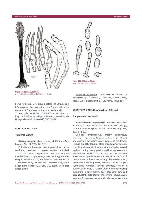
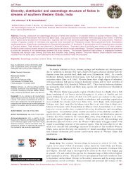
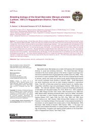
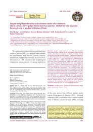
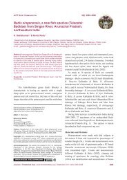
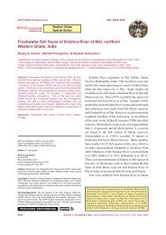
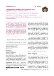
Foliicolous fungi <strong>of</strong> Silent ValleyHosagoudar & Rijucab10µmFigure 119. Teratosperma anacardiia - Foot cell; b - Conidiophore; c- ConidiaPodoconis anacardii (Hansf.) Hughes, Mycol. Pap. 48:65, 1952 (Fig. 119).Colonies epiphyllous, thin to dense, up to 1mm indiameter, rarely confluent. Hyphae mostly straight, palebrown, branching irregular at acute to wide angles, looselyreticulate, cells 1–3 μm broad. Conidiophores borne onfoot cells. Foot cells sessile to stipitate, borne laterally tothe hyphae, enlarged apically with irregularly producedspinules, 7–10x4–9 μm. Conidiophores produced on thefoot cells, macronematous, mononematous, erect, darkbrown, septate, simple, straight, smooth, 50–73x2–4μm. Conidiogenous cells pale brown, monoblastic,integrated, terminal, percurrent, annellated. Conidiasolitary, dry, acrogenous, obclavate, rostrate, mostly2-septate, constricted at the septa, 10–21μm long,truncate at the base, 1–2 appendaged on the basal cell,4–5 μm broad at the broadest part, apical cell pale andbroadly round at the tip, up to 2μm broad.Materials examined: 14.xii.2003 on leaves <strong>of</strong>Myristica beddomei King. (Myristicaceae), Champatty,Silent Valley, Kerala, V.B. Hosagoudar et al. HCIO 45769,TBGT 1518.The genus SpiropesSpiropes armatellicola Hosag. & D.K. Agarwal, J:Econ. Taxon. Bot. 26: 603, 2002 (Fig. 120).Colonies mostly epiphyllous, dense. up to 5mm indiameter, confluent. Hyphae superficial, pale brown,branched, surrounded around appressoria and mycelium<strong>of</strong> the host, 1–2 µm broad. Conidiophores solitary,simple, mononematous, erect, straight to flexuous,paler towards the apex, conidial scars scattered, 60–112x 4–7 µm. Conidiogenous cells polyblastic, integrated,terminal and intercalary, conspicuous. Conidia straight toa6.6µmFigure 120. Spiropes armatellicolaa - Conidiophores; b - Conidiaslightly curved, obclavate, rostrate at the apex, truncateat the base, pale brown, uniseptate, rostrate above theseptum, ovate below the septum, slightly hinged at thebase, 24–29 µm long, 6–8 µm broad at the broadestportion, up to 3µm broad at the base, beak 8–16 µmlong and up to 1.5µm broad at the tip.Materials examined: 12.xii.2003 on the colonies <strong>of</strong>Armatella cryptocaryae Hosag. on leaves <strong>of</strong> Litsea sp.(Lauraceae), Sairandhri, Silent Valley, V.B. Hosagoudar etal. HCIO 46327, TBGT 1973.Spiropes japonicus (P. Henn.) M.B. Ellis, Mycol. Pap.114: 22, 1968; Dematiaceous Hyphomycetes p. 256,1971; Katumoto, Trans. Mycol. Soc. Japan 24: 251, 1983;Hosag., Abraham, and Biju, C.K. New Botanist 23: 213,1996 (Fig. 121).Colonies amphigenous, dense, velvety, up to 3mm indiam., confluent. Conidiophores synnematous, compact,erect, cylindrical, 245–520x19–30 μm; conidiophoresspread out in the apical and upper half <strong>of</strong> the synnemata,brown to dark brown, paler towards the apex, septate,smooth, 3v4 μm wide; Conidiogenous cells polyblastic,terminal and intercalary, sympodial cylindrical cicatrized,scars numerous and conspicuous; conidia solitary, dry,acropleurogenous, simple, fusiform to obclavate, paleb3774<strong>Journal</strong> <strong>of</strong> <strong>Threatened</strong> <strong>Taxa</strong> | www.threatenedtaxa.org | 05 March 2013 | 5(3): 3701–3788
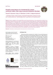
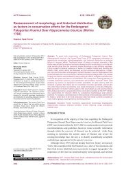
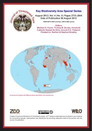
Foliicolous fungi <strong>of</strong> Silent ValleyHosagoudar & Riju8µmbaacb7µmFigure 122. Oidium doidgeaea - Conidiophores; b - ConidiaFigure 121. Spiropes japonicusa - Conidiogenous cells; b - Synnema; c - Conidiabrown to brown, 3–6 pseudoseptate, 40–70 μm long,8-9μm wide at the broadest portion, 2–4 μm wide at theapex and 3–5 μm broad at the base, wall smooth.Materials examined: 12.xii.2003 on Meliolaceousfungi on Mallotus sp., (Euphorbiaceae), Sairandhri, V.B.Hosagoudar et al. HCIO 46317, TBGT 1963.Powdery MildewsThe genus OidiumOidium doidgeae Bapp., Hosag. & Udaiyan, NewBotanist 22: 124, 1995 (Fig. 122).Colonies amphigenous, mostly epiphyllous, dense,confluent, persistent. Hyphae septate, branched,3.5–7.5 μm wide. Appressoria lobed and oppsite.Conidiophores straight, erect, 57–84 11m long; foot cellsstraight, cylindrical, slightly flexuous, 22–38x7.5–11.511μm, followed by a shorter cell. Conidia solitary, ovoid,ellipsoidal tocylindrical, 22–38x11–19 11μm. Germ tubeapical, simple.Materials examined: 15.xii.2003 on leaves <strong>of</strong>Triumfetta sp., (Tiliaceae), Sairandhri, Silent Valley,Kerala, V.B. Hosagoudar et al. HCIO 45913, TBGT 1675.Sphaeropsidales (Anamorphs <strong>of</strong> Asterina)The genus AsterostomellaAsterostomella daphniphylli Hosag.& Ravikumarin Hosag.& Goos,Mycotaxon 52: 471,1994; Hosag.,Chandraprabha & Agarwal, Asterinales <strong>of</strong> Kerala, p. 229,2011 (Fig. 123).Colonies amphigenous, mostly epiphyllous,crustose to velvety, up to 2mm in diameter, confluentand covering the entire upper surface <strong>of</strong> the leaves.Hyphae straight, flexuous, <strong>of</strong>ten crooked when solitary,branching alternate to irregular at acute angles, severalhyphae running closely parallel and forming a compactmycelial mat, cells 9–15.5x4.5–7.5 µm. Appressoriaalternate and produced only on the outer surface <strong>of</strong>the compact hyphae, mostly straight but rarely curved,unicellular ovate to globose, entire, 6–12.5x6–9.5 µm.Pycnothyria numerous, loosely crowded, circular inoutline, <strong>of</strong>ten ovate, 130–190 µm in diameter, coveringmembrane initially brown, later becoming dark andopaque, splitting stellately at the center or having a wideopening. Pycnothyriospores oval, ellipsoidal, pyriform,<strong>Journal</strong> <strong>of</strong> <strong>Threatened</strong> <strong>Taxa</strong> | www.threatenedtaxa.org | 05 March 2013 | 5(3): 3701–37883775